
Table of SynGAP1 Isoform α2 (UniProt Q96PV0-1) Missense Variants.
| c.dna | Variant | SGM Consensus | Domain and Structure information: based on WT protein | Annotated databases | Deep learning-based pathogenicity predictions | Folding stability-based pathogenicity predictions | Sequence/structure-based pathogenicity predictions | Phase Separation | Evolutionary/physical properties | Molecular Dynamics-based analysis | DOI | ||||||||||||||||||||||||||||||||||||||||||||||||||||||||||||
|---|---|---|---|---|---|---|---|---|---|---|---|---|---|---|---|---|---|---|---|---|---|---|---|---|---|---|---|---|---|---|---|---|---|---|---|---|---|---|---|---|---|---|---|---|---|---|---|---|---|---|---|---|---|---|---|---|---|---|---|---|---|---|---|---|---|---|---|---|---|---|---|
| Domain | IUPred2 | ANCHOR2 | AlphaFold | MobiDB | PhosphoSitePlus | ClinVar | gnomAD | ESM1b | AlphaMissense | FoldX | Rosetta | Foldetta | PremPS | REVEL | PROVEAN | PolyPhen-2 HumDiv | PolyPhen-2 HumVar | FATHMM | SIFT | PSMutPred | PAM | Physical | SASA | Normalized B-factor backbone | Normalized B-factor sidechain | SynGAP Structural Annotation | |||||||||||||||||||||||||||||||||||||||||||||
| Score | Prediction | Score | Prediction | pLDDT | disorder | disorder | LTP | HTP | KL | PTM | Clinical Status | Review | Subm. | ID | Allele count | Allele freq. | LLR score | Prediction | Pathogenicity | Class | Optimized | Average ΔΔG | Prediction | StdDev | ΔΔG | Prediction | ΔΔG | Prediction | ΔΔG | Prediction | Score | Prediction | Score | Prediction | pph2_prob | Prediction | pph2_prob | Prediction | Nervous System Score | Prediction | Prediction | Status | Conservation | Sequences | IP RF | SP RF | Prediction | PAM250 | PAM120 | Hydropathy Δ | MW Δ | Average | Δ | Δ | StdDev | Δ | StdDev | Secondary | Tertiary bonds | Inside out | GAP-Ras interface | At membrane | No effect | MD Alert | Verdict | Description | |||||
| c.3838A>T | M1280L 2D  AIThe SynGAP1 missense variant M1280L is not reported in ClinVar (ClinVar ID = None) and is absent from gnomAD (gnomAD ID = None). Functional prediction tools that agree on a benign effect include REVEL, PROVEAN, polyPhen‑2 HumDiv, polyPhen‑2 HumVar, AlphaMissense‑Default, AlphaMissense‑Optimized, and ESM1b. Tools that predict a pathogenic effect are SIFT and FATHMM. The SGM‑Consensus, which aggregates AlphaMissense‑Default, ESM1b, FATHMM, and PROVEAN, reports the variant as “Likely Benign.” High‑accuracy assessments show AlphaMissense‑Optimized as benign and the SGM‑Consensus as likely benign; the Foldetta protein‑folding stability analysis is unavailable for this variant. Overall, the majority of evidence points to a benign impact. The predictions do not contradict any ClinVar status, as no ClinVar claim exists for this variant. Thus, based on current computational predictions, the M1280L variant is most likely benign. Disclaimer: This summary was generated using AI and should be interpreted alongside expert review. | Likely Benign | 0.882776 | Disordered | 0.822030 | Binding | 0.510 | 0.726 | 0.875 | -1.391 | Likely Benign | 0.092 | Likely Benign | Likely Benign | 0.204 | Likely Benign | -2.23 | Neutral | 0.052 | Benign | 0.017 | Benign | 2.40 | Pathogenic | 0.00 | Affected | 0.1113 | 0.3186 | 4 | 2 | 1.9 | -18.03 | |||||||||||||||||||||||||||||||||||||||
| c.3840G>A | M1280I 2D  AIThe SynGAP1 missense variant M1280I is not reported in ClinVar (ClinVar ID = None) and is absent from gnomAD (gnomAD ID = None). Prediction tools that agree on a benign effect include REVEL, polyPhen‑2 (HumDiv and HumVar), AlphaMissense‑Default, AlphaMissense‑Optimized, and ESM1b. Tools that predict a pathogenic effect are PROVEAN, SIFT, and FATHMM. High‑accuracy assessments show AlphaMissense‑Optimized as benign; the SGM Consensus (majority vote from AlphaMissense‑Default, ESM1b, FATHMM, PROVEAN) is inconclusive (2 benign vs 2 pathogenic), and Foldetta results are unavailable. Overall, the majority of evidence points to a benign impact, and this conclusion does not contradict the ClinVar status, which has no entry for this variant. Disclaimer: This summary was generated using AI and should be interpreted alongside expert review. | 0.882776 | Disordered | 0.822030 | Binding | 0.510 | 0.726 | 0.875 | -3.723 | Likely Benign | 0.251 | Likely Benign | Likely Benign | 0.166 | Likely Benign | -2.59 | Deleterious | 0.059 | Benign | 0.041 | Benign | 2.35 | Pathogenic | 0.00 | Affected | 0.0995 | 0.2468 | 2 | 1 | 2.6 | -18.03 | ||||||||||||||||||||||||||||||||||||||||
| c.3840G>C | M1280I 2D AIThe SynGAP1 missense variant M1280I is not reported in ClinVar (ClinVar ID = None) and is absent from gnomAD (gnomAD ID = None). Prediction tools that agree on a benign effect include REVEL, polyPhen‑2 (HumDiv and HumVar), AlphaMissense‑Default, AlphaMissense‑Optimized, and ESM1b. Tools that predict a pathogenic effect are PROVEAN, SIFT, and FATHMM. High‑accuracy assessments show AlphaMissense‑Optimized as benign; the SGM Consensus (majority vote from AlphaMissense‑Default, ESM1b, FATHMM, PROVEAN) is inconclusive (2 benign vs 2 pathogenic), and Foldetta results are unavailable. Overall, the majority of evidence points to a benign impact, and this conclusion does not contradict the ClinVar status, which has no entry for this variant. Disclaimer: This summary was generated using AI and should be interpreted alongside expert review. | 0.882776 | Disordered | 0.822030 | Binding | 0.510 | 0.726 | 0.875 | -3.723 | Likely Benign | 0.251 | Likely Benign | Likely Benign | 0.166 | Likely Benign | -2.59 | Deleterious | 0.059 | Benign | 0.041 | Benign | 2.35 | Pathogenic | 0.00 | Affected | 0.0995 | 0.2468 | 2 | 1 | 2.6 | -18.03 | ||||||||||||||||||||||||||||||||||||||||
| c.3840G>T | M1280I 2D AIThe SynGAP1 missense variant M1280I is not reported in ClinVar (ClinVar ID = None) and is absent from gnomAD (gnomAD ID = None). Prediction tools that agree on a benign effect include REVEL, polyPhen‑2 (HumDiv and HumVar), AlphaMissense‑Default, AlphaMissense‑Optimized, and ESM1b. Tools that predict a pathogenic effect are PROVEAN, SIFT, and FATHMM. High‑accuracy assessments show AlphaMissense‑Optimized as benign; the SGM Consensus (majority vote from AlphaMissense‑Default, ESM1b, FATHMM, PROVEAN) is inconclusive (2 benign vs 2 pathogenic), and Foldetta results are unavailable. Overall, the majority of evidence points to a benign impact, and this conclusion does not contradict the ClinVar status, which has no entry for this variant. Disclaimer: This summary was generated using AI and should be interpreted alongside expert review. | 0.882776 | Disordered | 0.822030 | Binding | 0.510 | 0.726 | 0.875 | -3.723 | Likely Benign | 0.251 | Likely Benign | Likely Benign | 0.166 | Likely Benign | -2.59 | Deleterious | 0.059 | Benign | 0.041 | Benign | 2.35 | Pathogenic | 0.00 | Affected | 0.0995 | 0.2468 | 2 | 1 | 2.6 | -18.03 | ||||||||||||||||||||||||||||||||||||||||
| c.3G>A | M1I 2D AIThe SynGAP1 missense variant M1I is listed in ClinVar (ID 833646.0) with an uncertain significance annotation and is not reported in gnomAD. Functional prediction tools largely agree on a benign effect: REVEL, PROVEAN, polyPhen‑2 HumDiv, polyPhen‑2 HumVar, ESM1b, and FATHMM all classify the substitution as benign, while SIFT uniquely predicts it to be pathogenic. The consensus score from the SGM framework, which aggregates AlphaMissense‑Default, ESM1b, FATHMM, and PROVEAN, reports a likely benign outcome. High‑accuracy predictors are incomplete: AlphaMissense‑Optimized and the Foldetta stability assessment are unavailable for this variant. Taking the collective evidence into account, the variant is most likely benign, and this assessment does not conflict with the ClinVar uncertain status. Disclaimer: This summary was generated using AI and should be interpreted alongside expert review. | Likely Benign | 0.642678 | Disordered | 0.540766 | Binding | 0.354 | 0.924 | 0.875 | Conflicting | 3 | -5.397 | Likely Benign | 0.227 | Likely Benign | -0.17 | Neutral | 0.001 | Benign | 0.000 | Benign | 4.25 | Benign | 0.00 | Affected | 4.32 | 1 | 0.1655 | 0.4008 | 2 | 1 | 2.6 | -18.03 | ||||||||||||||||||||||||||||||||||||||
| c.46A>C | M16L 2D AIThe SynGAP1 missense variant M16L is not reported in ClinVar (no ClinVar ID) and is absent from gnomAD (no gnomAD ID). Prediction tools that agree on a benign effect include REVEL, PROVEAN, polyPhen‑2 HumDiv, polyPhen‑2 HumVar, ESM1b, FATHMM, AlphaMissense‑Default, and AlphaMissense‑Optimized. Only SIFT predicts a pathogenic outcome. The SGM‑Consensus, which aggregates AlphaMissense‑Default, ESM1b, FATHMM, and PROVEAN, reports a “Likely Benign” classification. High‑accuracy assessments show AlphaMissense‑Optimized as benign and the SGM‑Consensus as likely benign; Foldetta results are not available. Based on the collective evidence, the variant is most likely benign, and this assessment does not contradict any ClinVar status (none is provided). Disclaimer: This summary was generated using AI and should be interpreted alongside expert review. | Likely Benign | 0.440853 | Structured | 0.459925 | Uncertain | 0.346 | 0.908 | 0.375 | -1.277 | Likely Benign | 0.249 | Likely Benign | Likely Benign | 0.075 | Likely Benign | -0.33 | Neutral | 0.001 | Benign | 0.000 | Benign | 4.37 | Benign | 0.00 | Affected | 0.1805 | 0.4629 | 4 | 2 | 1.9 | -18.03 | |||||||||||||||||||||||||||||||||||||||
| c.46A>T | M16L 2D AIThe SynGAP1 missense variant M16L is not reported in ClinVar (no ClinVar ID) and is absent from gnomAD (no gnomAD ID). Prediction tools that agree on a benign effect include REVEL, PROVEAN, polyPhen‑2 HumDiv, polyPhen‑2 HumVar, ESM1b, FATHMM, AlphaMissense‑Default, and AlphaMissense‑Optimized. Only SIFT predicts a pathogenic outcome. The SGM‑Consensus, which aggregates AlphaMissense‑Default, ESM1b, FATHMM, and PROVEAN, reports the variant as “Likely Benign.” High‑accuracy assessments show AlphaMissense‑Optimized as benign and the SGM‑Consensus as likely benign; Foldetta results are not available. Overall, the majority of evidence points to a benign effect, and this conclusion does not contradict any ClinVar annotation (none is present). Disclaimer: This summary was generated using AI and should be interpreted alongside expert review. | Likely Benign | 0.440853 | Structured | 0.459925 | Uncertain | 0.346 | 0.908 | 0.375 | -1.277 | Likely Benign | 0.249 | Likely Benign | Likely Benign | 0.075 | Likely Benign | -0.33 | Neutral | 0.001 | Benign | 0.000 | Benign | 4.37 | Benign | 0.00 | Affected | 0.1805 | 0.4629 | 4 | 2 | 1.9 | -18.03 | |||||||||||||||||||||||||||||||||||||||
| c.48G>A | M16I 2D AIThe SynGAP1 missense variant M16I is listed in ClinVar with an “Uncertain” status (ClinVar ID 1424213.0) and is present in gnomAD (6‑33420312‑G‑A). Prediction tools that agree on a benign effect include REVEL, PROVEAN, polyPhen‑2 HumDiv, polyPhen‑2 HumVar, ESM1b, FATHMM, and AlphaMissense‑Optimized. Tools that predict a pathogenic effect are SIFT and AlphaMissense‑Default. The SGM‑Consensus, which aggregates the majority vote from AlphaMissense‑Default, ESM1b, FATHMM, and PROVEAN, reports a “Likely Benign” classification. High‑accuracy assessments show AlphaMissense‑Optimized as benign and the SGM‑Consensus as likely benign; Foldetta results are not available. Overall, the majority of evidence points to a benign impact, and this is consistent with the ClinVar “Uncertain” designation rather than contradicting it. Disclaimer: This summary was generated using AI and should be interpreted alongside expert review. | Likely Benign | 0.440853 | Structured | 0.459925 | Uncertain | 0.346 | 0.908 | 0.375 | Uncertain | 1 | 6-33420312-G-A | 1 | 6.49e-7 | -2.198 | Likely Benign | 0.722 | Likely Pathogenic | Likely Benign | 0.057 | Likely Benign | -0.15 | Neutral | 0.000 | Benign | 0.000 | Benign | 4.28 | Benign | 0.00 | Affected | 4.32 | 1 | 0.1606 | 0.3877 | 2 | 1 | 2.6 | -18.03 | ||||||||||||||||||||||||||||||||
| c.48G>C | M16I 2D AIThe SynGAP1 missense variant M16I is not reported in ClinVar (ClinVar ID = None) and is absent from gnomAD (gnomAD ID = None). Prediction tools that agree on a benign effect include REVEL, PROVEAN, polyPhen‑2 HumDiv, polyPhen‑2 HumVar, ESM1b, FATHMM, and AlphaMissense‑Optimized. Tools that predict a pathogenic effect are SIFT and AlphaMissense‑Default. The SGM‑Consensus, which aggregates the majority vote from AlphaMissense‑Default, ESM1b, FATHMM, and PROVEAN, classifies the variant as Likely Benign. High‑accuracy assessments further support a benign outcome: AlphaMissense‑Optimized predicts benign, and the SGM‑Consensus also indicates Likely Benign. Foldetta, a protein‑folding stability method combining FoldX‑MD and Rosetta outputs, has no available result for this variant. Overall, the majority of computational predictions and the high‑accuracy consensus suggest that M16I is most likely benign, and this conclusion is consistent with the absence of any ClinVar pathogenic annotation. Disclaimer: This summary was generated using AI and should be interpreted alongside expert review. | Likely Benign | 0.440853 | Structured | 0.459925 | Uncertain | 0.346 | 0.908 | 0.375 | -2.198 | Likely Benign | 0.722 | Likely Pathogenic | Likely Benign | 0.057 | Likely Benign | -0.15 | Neutral | 0.000 | Benign | 0.000 | Benign | 4.28 | Benign | 0.00 | Affected | 4.32 | 1 | 0.1606 | 0.3877 | 2 | 1 | 2.6 | -18.03 | |||||||||||||||||||||||||||||||||||||
| c.48G>T | M16I 2D AIThe SynGAP1 missense variant M16I is not reported in ClinVar (ClinVar ID = None) but is present in gnomAD (ID = 6‑33420312‑G‑T). Prediction tools that agree on a benign effect include REVEL, PROVEAN, polyPhen‑2 HumDiv, polyPhen‑2 HumVar, ESM1b, FATHMM, and AlphaMissense‑Optimized. Tools that predict a pathogenic effect are SIFT and AlphaMissense‑Default. The SGM‑Consensus, which aggregates the majority vote from AlphaMissense‑Default, ESM1b, FATHMM, and PROVEAN, classifies the variant as Likely Benign. High‑accuracy assessments further support a benign outcome: AlphaMissense‑Optimized predicts benign, and the SGM‑Consensus also indicates Likely Benign. Foldetta, a protein‑folding stability method combining FoldX‑MD and Rosetta outputs, has no available result for this variant. Overall, the majority of computational evidence points to a benign impact, and this is consistent with the absence of a ClinVar pathogenic classification. Disclaimer: This summary was generated using AI and should be interpreted alongside expert review. | Likely Benign | 0.440853 | Structured | 0.459925 | Uncertain | 0.346 | 0.908 | 0.375 | 6-33420312-G-T | -2.198 | Likely Benign | 0.722 | Likely Pathogenic | Likely Benign | 0.057 | Likely Benign | -0.15 | Neutral | 0.000 | Benign | 0.000 | Benign | 4.28 | Benign | 0.00 | Affected | 4.32 | 1 | 0.1606 | 0.3877 | 2 | 1 | 2.6 | -18.03 | ||||||||||||||||||||||||||||||||||||
| c.520A>C | M174L 2D AIThe SynGAP1 missense variant M174L is not reported in ClinVar (no ClinVar ID) and is absent from gnomAD (no gnomAD ID). Prediction tools that agree on a benign effect include REVEL, PROVEAN, polyPhen‑2 (HumDiv and HumVar), SIFT, ESM1b, FATHMM, AlphaMissense‑Optimized, and the SGM‑Consensus (majority vote from AlphaMissense‑Default, ESM1b, FATHMM, and PROVEAN). Only AlphaMissense‑Default predicts a pathogenic outcome. High‑accuracy assessments show AlphaMissense‑Optimized as benign and the SGM‑Consensus as likely benign; Foldetta results are not available. Taken together, the majority of evidence points to a benign impact for M174L. This conclusion is consistent with the lack of ClinVar annotation and does not contradict any existing clinical classification. Disclaimer: This summary was generated using AI and should be interpreted alongside expert review. | Likely Benign | 0.661982 | Disordered | 0.485854 | Uncertain | 0.373 | 0.620 | 0.375 | -5.839 | Likely Benign | 0.608 | Likely Pathogenic | Likely Benign | 0.192 | Likely Benign | -0.82 | Neutral | 0.055 | Benign | 0.011 | Benign | 4.21 | Benign | 0.46 | Tolerated | 0.1335 | 0.4545 | 4 | 2 | 1.9 | -18.03 | |||||||||||||||||||||||||||||||||||||||
| c.520A>T | M174L 2D AIThe SynGAP1 missense variant M174L is not reported in ClinVar (no ClinVar ID) and is absent from gnomAD (no gnomAD ID). Prediction tools that agree on a benign effect include REVEL, PROVEAN, polyPhen‑2 (HumDiv and HumVar), SIFT, ESM1b, FATHMM, AlphaMissense‑Optimized, and the SGM‑Consensus (majority vote from AlphaMissense‑Default, ESM1b, FATHMM, PROVEAN). Only AlphaMissense‑Default predicts a pathogenic outcome. High‑accuracy assessments show AlphaMissense‑Optimized as benign and the SGM‑Consensus as likely benign; Foldetta results are not available. Taken together, the majority of evidence points to a benign impact for M174L. This conclusion is consistent with the lack of ClinVar annotation and does not contradict any existing clinical classification. Disclaimer: This summary was generated using AI and should be interpreted alongside expert review. | Likely Benign | 0.661982 | Disordered | 0.485854 | Uncertain | 0.373 | 0.620 | 0.375 | -5.839 | Likely Benign | 0.608 | Likely Pathogenic | Likely Benign | 0.192 | Likely Benign | -0.82 | Neutral | 0.055 | Benign | 0.011 | Benign | 4.21 | Benign | 0.46 | Tolerated | 0.1335 | 0.4545 | 4 | 2 | 1.9 | -18.03 | |||||||||||||||||||||||||||||||||||||||
| c.522G>A | M174I 2D AIThe SynGAP1 missense variant M174I is listed in gnomAD (ID 6‑33435164‑G‑A) but has no ClinVar record. Functional prediction tools cluster into two groups: benign predictions come from REVEL, PROVEAN, polyPhen‑2 (HumDiv and HumVar), SIFT, and FATHMM; pathogenic predictions come from ESM1b and AlphaMissense‑Default. High‑accuracy assessments show AlphaMissense‑Optimized classifying the variant as pathogenic, whereas the SGM Consensus (majority vote of AlphaMissense‑Default, ESM1b, FATHMM, PROVEAN) is inconclusive (2 vs 2) and Foldetta results are unavailable. Overall, six tools predict benign while only two predict pathogenic, and the only high‑accuracy pathogenic call is from AlphaMissense‑Optimized. Thus, the variant is most likely benign based on the preponderance of evidence, and this assessment does not contradict any ClinVar status (none reported). Disclaimer: This summary was generated using AI and should be interpreted alongside expert review. | 0.661982 | Disordered | 0.485854 | Uncertain | 0.373 | 0.620 | 0.375 | 6-33435164-G-A | 4 | 2.48e-6 | -8.732 | Likely Pathogenic | 0.994 | Likely Pathogenic | Likely Pathogenic | 0.120 | Likely Benign | -1.63 | Neutral | 0.213 | Benign | 0.067 | Benign | 4.10 | Benign | 0.07 | Tolerated | 3.61 | 5 | 0.1191 | 0.3469 | 1 | 2 | 2.6 | -18.03 | |||||||||||||||||||||||||||||||||||
| c.522G>C | M174I 2D AIThe SynGAP1 missense variant M174I is not reported in ClinVar (ClinVar ID None) and is absent from gnomAD (gnomAD ID None). Prediction tools that agree on a benign effect include REVEL, PROVEAN, polyPhen‑2 HumDiv, polyPhen‑2 HumVar, SIFT, and FATHMM. Tools that predict a pathogenic effect are ESM1b and AlphaMissense‑Default. High‑accuracy methods give mixed results: AlphaMissense‑Optimized predicts pathogenic; the SGM Consensus (majority vote from AlphaMissense‑Default, ESM1b, FATHMM, and PROVEAN) is inconclusive (2 pathogenic vs 2 benign), and Foldetta’s stability assessment is unavailable. Overall, the majority of standard predictors favor a benign outcome, and the high‑accuracy predictions do not override this trend. Thus, the variant is most likely benign, and this assessment does not contradict the lack of ClinVar reporting. Disclaimer: This summary was generated using AI and should be interpreted alongside expert review. | 0.661982 | Disordered | 0.485854 | Uncertain | 0.373 | 0.620 | 0.375 | -8.732 | Likely Pathogenic | 0.994 | Likely Pathogenic | Likely Pathogenic | 0.120 | Likely Benign | -1.63 | Neutral | 0.213 | Benign | 0.067 | Benign | 4.10 | Benign | 0.07 | Tolerated | 3.61 | 5 | 0.1191 | 0.3469 | 1 | 2 | 2.6 | -18.03 | ||||||||||||||||||||||||||||||||||||||
| c.522G>T | M174I 2D AIThe SynGAP1 missense variant M174I is not reported in ClinVar (ClinVar ID = None) and is absent from gnomAD (gnomAD ID = None). Prediction tools that agree on a benign effect include REVEL, PROVEAN, polyPhen‑2 HumDiv, polyPhen‑2 HumVar, SIFT, and FATHMM. Tools that predict a pathogenic effect are ESM1b and AlphaMissense‑Default. High‑accuracy methods give mixed results: AlphaMissense‑Optimized predicts pathogenic, the SGM Consensus (majority vote from AlphaMissense‑Default, ESM1b, FATHMM, PROVEAN) is inconclusive (2 vs 2), and Foldetta data are unavailable. Overall, the majority of standard predictors indicate a benign impact, while the single high‑accuracy tool suggests pathogenicity but is not supported by consensus or folding‑stability evidence. Thus, the variant is most likely benign, and this assessment does not contradict the lack of ClinVar annotation. Disclaimer: This summary was generated using AI and should be interpreted alongside expert review. | 0.661982 | Disordered | 0.485854 | Uncertain | 0.373 | 0.620 | 0.375 | -8.732 | Likely Pathogenic | 0.994 | Likely Pathogenic | Likely Pathogenic | 0.120 | Likely Benign | -1.63 | Neutral | 0.213 | Benign | 0.067 | Benign | 4.10 | Benign | 0.07 | Tolerated | 3.61 | 5 | 0.1191 | 0.3469 | 1 | 2 | 2.6 | -18.03 | ||||||||||||||||||||||||||||||||||||||
| c.85A>C | M29L 2D AIThe SynGAP1 missense variant M29L is not reported in ClinVar (no ClinVar ID) and is absent from gnomAD (no gnomAD ID). Prediction tools that agree on a benign effect include REVEL, PROVEAN, polyPhen‑2 HumDiv, polyPhen‑2 HumVar, ESM1b, FATHMM, AlphaMissense‑Default, and AlphaMissense‑Optimized. Only SIFT predicts a pathogenic outcome. The SGM‑Consensus, which aggregates AlphaMissense‑Default, ESM1b, FATHMM, and PROVEAN, reports a “Likely Benign” classification. High‑accuracy assessments show AlphaMissense‑Optimized as benign and the SGM‑Consensus as likely benign; Foldetta results are not available. Based on the collective evidence, the variant is most likely benign, and this assessment does not contradict any ClinVar status. Disclaimer: This summary was generated using AI and should be interpreted alongside expert review. | Likely Benign | 0.541878 | Disordered | 0.438540 | Uncertain | 0.341 | 0.883 | 0.250 | -1.633 | Likely Benign | 0.079 | Likely Benign | Likely Benign | 0.200 | Likely Benign | -0.49 | Neutral | 0.006 | Benign | 0.039 | Benign | 4.27 | Benign | 0.00 | Affected | 0.1984 | 0.5336 | 4 | 2 | 1.9 | -18.03 | |||||||||||||||||||||||||||||||||||||||
| c.85A>T | M29L 2D AIThe SynGAP1 missense variant M29L is not reported in ClinVar (no ClinVar ID) and is absent from gnomAD (no gnomAD ID). Prediction tools that agree on a benign effect include REVEL, PROVEAN, polyPhen‑2 HumDiv, polyPhen‑2 HumVar, ESM1b, FATHMM, AlphaMissense‑Default, and AlphaMissense‑Optimized. Only SIFT predicts a pathogenic outcome. The high‑accuracy consensus from SGM (majority vote of AlphaMissense‑Default, ESM1b, FATHMM, and PROVEAN) classifies the variant as Likely Benign, and AlphaMissense‑Optimized also predicts Benign. Foldetta, a protein‑folding stability method combining FoldX‑MD and Rosetta outputs, has no available result for this variant. Overall, the majority of evidence points to a benign effect; there is no conflict with ClinVar status because the variant is not yet catalogued there. Disclaimer: This summary was generated using AI and should be interpreted alongside expert review. | Likely Benign | 0.541878 | Disordered | 0.438540 | Uncertain | 0.341 | 0.883 | 0.250 | -1.633 | Likely Benign | 0.079 | Likely Benign | Likely Benign | 0.200 | Likely Benign | -0.49 | Neutral | 0.006 | Benign | 0.039 | Benign | 4.27 | Benign | 0.00 | Affected | 0.1984 | 0.5336 | 4 | 2 | 1.9 | -18.03 | |||||||||||||||||||||||||||||||||||||||
| c.865A>C | M289L 2D 3DClick to see structure in 3D Viewer AIThe SynGAP1 missense variant M289L is not reported in ClinVar (no ClinVar ID) and is absent from gnomAD (no gnomAD ID). Prediction tools largely agree on a benign effect: REVEL, FoldX, Rosetta, Foldetta, premPS, PROVEAN, polyPhen‑2 (HumDiv and HumVar), SIFT, ESM1b, AlphaMissense‑Default, and AlphaMissense‑Optimized all classify the variant as benign. Only FATHMM predicts a pathogenic outcome. Grouping by consensus, the benign‑predicting tools outnumber the single pathogenic prediction. High‑accuracy assessments reinforce this view: AlphaMissense‑Optimized is benign; the SGM Consensus (majority vote of AlphaMissense‑Default, ESM1b, FATHMM, PROVEAN) yields a “Likely Benign” verdict; and Foldetta, which integrates FoldX‑MD and Rosetta stability outputs, also reports a benign effect. No prediction or stability result is missing or inconclusive. Based on the collective evidence, the variant is most likely benign, and this conclusion does not contradict any ClinVar status (none available). Disclaimer: This summary was generated using AI and should be interpreted alongside expert review. | Likely Benign | C2 | 0.127496 | Structured | 0.403499 | Uncertain | 0.886 | 0.276 | 0.000 | -5.778 | Likely Benign | 0.157 | Likely Benign | Likely Benign | 0.13 | Likely Benign | 0.0 | -0.10 | Likely Benign | 0.02 | Likely Benign | 0.39 | Likely Benign | 0.070 | Likely Benign | -0.95 | Neutral | 0.136 | Benign | 0.033 | Benign | 1.79 | Pathogenic | 0.27 | Tolerated | 0.1092 | 0.3634 | 4 | 2 | 1.9 | -18.03 | |||||||||||||||||||||||||||||
| c.865A>T | M289L 2D 3DClick to see structure in 3D Viewer AIThe SynGAP1 missense variant M289L is not reported in ClinVar (no ClinVar ID) and is absent from gnomAD (no gnomAD ID). Prediction tools that agree on a benign effect include REVEL, FoldX, Rosetta, Foldetta, premPS, PROVEAN, polyPhen‑2 (HumDiv and HumVar), SIFT, ESM1b, AlphaMissense‑Default, and AlphaMissense‑Optimized. Only FATHMM predicts a pathogenic outcome. High‑accuracy assessments further support a benign classification: AlphaMissense‑Optimized is benign; the SGM Consensus (majority vote of AlphaMissense‑Default, ESM1b, FATHMM, and PROVEAN) is “Likely Benign”; and Foldetta, which integrates FoldX‑MD and Rosetta stability outputs, is benign. No prediction or folding stability result is missing or inconclusive. Based on the collective evidence, the variant is most likely benign, and this conclusion does not contradict any ClinVar status because the variant is not currently listed in ClinVar. Disclaimer: This summary was generated using AI and should be interpreted alongside expert review. | Likely Benign | C2 | 0.127496 | Structured | 0.403499 | Uncertain | 0.886 | 0.276 | 0.000 | -5.778 | Likely Benign | 0.157 | Likely Benign | Likely Benign | 0.13 | Likely Benign | 0.0 | -0.10 | Likely Benign | 0.02 | Likely Benign | 0.39 | Likely Benign | 0.070 | Likely Benign | -0.95 | Neutral | 0.136 | Benign | 0.033 | Benign | 1.79 | Pathogenic | 0.27 | Tolerated | 0.1092 | 0.3634 | 4 | 2 | 1.9 | -18.03 | |||||||||||||||||||||||||||||
| c.867G>A | M289I 2D 3DClick to see structure in 3D Viewer AIThe SynGAP1 missense variant M289I is not reported in ClinVar (ClinVar ID None) and is absent from gnomAD (gnomAD ID None). Prediction tools that agree on a benign effect include REVEL, Rosetta, Foldetta, premPS, PROVEAN, polyPhen‑2 (HumDiv and HumVar), SIFT, ESM1b, and AlphaMissense‑Optimized. Only FATHMM predicts a pathogenic outcome, while FoldX and AlphaMissense‑Default are uncertain. High‑accuracy assessments reinforce the benign consensus: AlphaMissense‑Optimized is benign; the SGM Consensus (majority vote of AlphaMissense‑Default, ESM1b, FATHMM, PROVEAN) yields a benign prediction; and Foldetta, which integrates FoldX‑MD and Rosetta stability outputs, also predicts benign. Overall, the preponderance of evidence indicates that M289I is most likely benign, and this conclusion does not contradict the lack of ClinVar annotation. Disclaimer: This summary was generated using AI and should be interpreted alongside expert review. | C2 | 0.127496 | Structured | 0.403499 | Uncertain | 0.886 | 0.276 | 0.000 | -6.010 | Likely Benign | 0.505 | Ambiguous | Likely Benign | 0.62 | Ambiguous | 0.1 | -0.23 | Likely Benign | 0.20 | Likely Benign | 0.36 | Likely Benign | 0.040 | Likely Benign | -0.72 | Neutral | 0.005 | Benign | 0.018 | Benign | 1.78 | Pathogenic | 0.31 | Tolerated | 0.1001 | 0.2758 | 2 | 1 | 2.6 | -18.03 | ||||||||||||||||||||||||||||||
| c.867G>C | M289I 2D 3DClick to see structure in 3D Viewer AIThe SynGAP1 missense variant M289I is not reported in ClinVar (ClinVar ID None) and is absent from gnomAD (gnomAD ID None). Prediction tools that agree on a benign effect include REVEL, Rosetta, Foldetta, premPS, PROVEAN, polyPhen‑2 (HumDiv and HumVar), SIFT, ESM1b, and AlphaMissense‑Optimized. Only FATHMM predicts a pathogenic outcome, while FoldX and AlphaMissense‑Default are uncertain. High‑accuracy assessments reinforce the benign prediction: AlphaMissense‑Optimized is benign; the SGM Consensus (majority vote of AlphaMissense‑Default, ESM1b, FATHMM, PROVEAN) yields a benign verdict; and Foldetta, which integrates FoldX‑MD and Rosetta stability outputs, also predicts benign. Overall, the preponderance of evidence indicates that M289I is most likely benign, and this conclusion is consistent with the absence of any ClinVar pathogenic annotation. Disclaimer: This summary was generated using AI and should be interpreted alongside expert review. | C2 | 0.127496 | Structured | 0.403499 | Uncertain | 0.886 | 0.276 | 0.000 | -6.010 | Likely Benign | 0.505 | Ambiguous | Likely Benign | 0.62 | Ambiguous | 0.1 | -0.23 | Likely Benign | 0.20 | Likely Benign | 0.36 | Likely Benign | 0.040 | Likely Benign | -0.72 | Neutral | 0.005 | Benign | 0.018 | Benign | 1.78 | Pathogenic | 0.31 | Tolerated | 0.1001 | 0.2758 | 2 | 1 | 2.6 | -18.03 | ||||||||||||||||||||||||||||||
| c.867G>T | M289I 2D 3DClick to see structure in 3D Viewer AIThe SynGAP1 missense variant M289I is not reported in ClinVar (ClinVar ID None) and is absent from gnomAD (gnomAD ID None). Prediction tools that agree on a benign effect include REVEL, Rosetta, Foldetta, premPS, PROVEAN, polyPhen‑2 (HumDiv and HumVar), SIFT, ESM1b, and AlphaMissense‑Optimized. Only FATHMM predicts a pathogenic outcome, while FoldX and AlphaMissense‑Default are uncertain. High‑accuracy assessments reinforce the benign prediction: AlphaMissense‑Optimized is benign; the SGM Consensus (majority vote of AlphaMissense‑Default, ESM1b, FATHMM, PROVEAN) yields a benign verdict; and Foldetta, which integrates FoldX‑MD and Rosetta stability outputs, also predicts benign. Overall, the preponderance of evidence indicates that M289I is most likely benign, and this conclusion is consistent with the absence of any ClinVar pathogenic annotation. Disclaimer: This summary was generated using AI and should be interpreted alongside expert review. | C2 | 0.127496 | Structured | 0.403499 | Uncertain | 0.886 | 0.276 | 0.000 | -6.010 | Likely Benign | 0.505 | Ambiguous | Likely Benign | 0.62 | Ambiguous | 0.1 | -0.23 | Likely Benign | 0.20 | Likely Benign | 0.36 | Likely Benign | 0.040 | Likely Benign | -0.72 | Neutral | 0.005 | Benign | 0.018 | Benign | 1.78 | Pathogenic | 0.31 | Tolerated | 0.1001 | 0.2758 | 2 | 1 | 2.6 | -18.03 | ||||||||||||||||||||||||||||||
| c.87G>A | M29I 2D AIThe SynGAP1 missense variant M29I is catalogued in gnomAD (6-33423496‑G‑A) but has no ClinVar entry. Functional prediction tools largely agree on a benign effect: REVEL, PROVEAN, polyPhen‑2 (HumDiv and HumVar), ESM1b, FATHMM, AlphaMissense‑Default, and AlphaMissense‑Optimized all report benign or tolerated outcomes, while the single pathogenic signal comes from SIFT. The SGM‑Consensus, derived from a majority vote of AlphaMissense‑Default, ESM1b, FATHMM, and PROVEAN, classifies the variant as Likely Benign. High‑accuracy assessments reinforce this view: AlphaMissense‑Optimized predicts benign, and the SGM‑Consensus likewise indicates a benign likelihood. Foldetta stability analysis is unavailable for this variant. Overall, the preponderance of evidence points to a benign impact, and this conclusion is not contradicted by any ClinVar classification. Disclaimer: This summary was generated using AI and should be interpreted alongside expert review. | Likely Benign | 0.541878 | Disordered | 0.438540 | Uncertain | 0.341 | 0.883 | 0.250 | 6-33423496-G-A | 6 | 3.72e-6 | -2.425 | Likely Benign | 0.185 | Likely Benign | Likely Benign | 0.067 | Likely Benign | -0.51 | Neutral | 0.006 | Benign | 0.091 | Benign | 4.26 | Benign | 0.00 | Affected | 4.32 | 1 | 0.1680 | 0.4385 | 1 | 2 | 2.6 | -18.03 | ||||||||||||||||||||||||||||||||||
| c.87G>C | M29I 2D AIThe SynGAP1 missense variant M29I is not reported in ClinVar (no ClinVar ID) and is absent from gnomAD (no gnomAD ID). Prediction tools that agree on a benign effect include REVEL, PROVEAN, polyPhen‑2 HumDiv, polyPhen‑2 HumVar, ESM1b, FATHMM, AlphaMissense‑Default, and AlphaMissense‑Optimized. Only SIFT predicts a pathogenic outcome. The SGM‑Consensus, which aggregates AlphaMissense‑Default, ESM1b, FATHMM, and PROVEAN, reports the variant as Likely Benign. High‑accuracy assessments show AlphaMissense‑Optimized as Benign and the SGM‑Consensus as Likely Benign; Foldetta results are not available. Based on the collective predictions, the variant is most likely benign, and this assessment does not contradict any ClinVar status. Disclaimer: This summary was generated using AI and should be interpreted alongside expert review. | Likely Benign | 0.541878 | Disordered | 0.438540 | Uncertain | 0.341 | 0.883 | 0.250 | -2.425 | Likely Benign | 0.185 | Likely Benign | Likely Benign | 0.067 | Likely Benign | -0.51 | Neutral | 0.006 | Benign | 0.091 | Benign | 4.26 | Benign | 0.00 | Affected | 4.32 | 1 | 0.1680 | 0.4385 | 1 | 2 | 2.6 | -18.03 | |||||||||||||||||||||||||||||||||||||
| c.87G>T | M29I 2D AIThe SynGAP1 missense variant M29I is not reported in ClinVar (no ClinVar ID) and is absent from gnomAD (no gnomAD ID). Prediction tools that agree on a benign effect include REVEL, PROVEAN, polyPhen‑2 HumDiv, polyPhen‑2 HumVar, ESM1b, FATHMM, AlphaMissense‑Default, and AlphaMissense‑Optimized. Only SIFT predicts a pathogenic outcome. The SGM‑Consensus, which aggregates AlphaMissense‑Default, ESM1b, FATHMM, and PROVEAN, reports a “Likely Benign” classification. High‑accuracy assessments show AlphaMissense‑Optimized as benign and the SGM‑Consensus as likely benign; Foldetta results are not available. Overall, the majority of evidence points to a benign effect, and this assessment does not contradict any ClinVar annotation (none is present). Thus, the variant is most likely benign. Disclaimer: This summary was generated using AI and should be interpreted alongside expert review. | Likely Benign | 0.541878 | Disordered | 0.438540 | Uncertain | 0.341 | 0.883 | 0.250 | -2.425 | Likely Benign | 0.185 | Likely Benign | Likely Benign | 0.067 | Likely Benign | -0.51 | Neutral | 0.006 | Benign | 0.091 | Benign | 4.26 | Benign | 0.00 | Affected | 4.32 | 1 | 0.1680 | 0.4385 | 1 | 2 | 2.6 | -18.03 | |||||||||||||||||||||||||||||||||||||
| c.1189T>A | C397S 2D 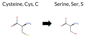 3DClick to see structure in 3D Viewer AIThe SynGAP1 missense variant C397S is not reported in ClinVar and is absent from gnomAD. Functional prediction tools uniformly indicate a benign effect: REVEL, FoldX, premPS, polyPhen‑2 (HumDiv and HumVar), SIFT, ESM1b, FATHMM, PROVEAN, AlphaMissense‑Default, and AlphaMissense‑Optimized all classify the substitution as benign, while Rosetta remains uncertain. When predictions are grouped, the benign category contains all available evidence and the pathogenic category is empty. High‑accuracy assessments corroborate this: AlphaMissense‑Optimized predicts benign; the SGM Consensus (majority of AlphaMissense‑Default, ESM1b, FATHMM, PROVEAN) also yields benign; and Foldetta, which integrates FoldX‑MD and Rosetta stability outputs, reports a benign effect. Consequently, the variant is most likely benign, and this conclusion is consistent with the absence of a ClinVar pathogenic annotation. Disclaimer: This summary was generated using AI and should be interpreted alongside expert review. | Likely Benign | C2 | 0.429200 | Structured | 0.395774 | Uncertain | 0.778 | 0.551 | 0.250 | -2.324 | Likely Benign | 0.178 | Likely Benign | Likely Benign | 0.37 | Likely Benign | 0.1 | 0.59 | Ambiguous | 0.48 | Likely Benign | 0.43 | Likely Benign | 0.174 | Likely Benign | -0.01 | Neutral | 0.276 | Benign | 0.066 | Benign | 4.68 | Benign | 0.53 | Tolerated | 0.5221 | 0.2474 | Weaken | 0 | -1 | -3.3 | -16.06 | ||||||||||||||||||||||||||||
| c.1190G>C | C397S 2D 3DClick to see structure in 3D Viewer AIThe SynGAP1 missense variant C397S is not reported in ClinVar (no ClinVar ID) and is absent from gnomAD (no gnomAD ID). In silico prediction tools that assess pathogenicity largely agree on a benign outcome: REVEL, FoldX, premPS, PROVEAN, polyPhen‑2 (HumDiv and HumVar), SIFT, ESM1b, FATHMM, AlphaMissense‑Default, and AlphaMissense‑Optimized all predict benign. No tool predicts pathogenicity; Rosetta’s assessment is uncertain and therefore treated as unavailable. High‑accuracy methods reinforce this consensus: AlphaMissense‑Optimized is benign; the SGM Consensus (majority vote of AlphaMissense‑Default, ESM1b, FATHMM, PROVEAN) is benign; and Foldetta, which integrates FoldX‑MD and Rosetta outputs, is benign. Consequently, the variant is most likely benign based on the collective predictions, and this conclusion does not contradict any ClinVar status (none is assigned). Disclaimer: This summary was generated using AI and should be interpreted alongside expert review. | Likely Benign | C2 | 0.429200 | Structured | 0.395774 | Uncertain | 0.778 | 0.551 | 0.250 | -2.324 | Likely Benign | 0.178 | Likely Benign | Likely Benign | 0.37 | Likely Benign | 0.1 | 0.59 | Ambiguous | 0.48 | Likely Benign | 0.43 | Likely Benign | 0.253 | Likely Benign | -0.01 | Neutral | 0.276 | Benign | 0.066 | Benign | 4.68 | Benign | 0.53 | Tolerated | 0.5221 | 0.2474 | Weaken | 0 | -1 | -3.3 | -16.06 | ||||||||||||||||||||||||||||
| c.1294T>A | C432S 2D 3DClick to see structure in 3D Viewer AIThe SynGAP1 missense variant C432S is not reported in ClinVar and is absent from gnomAD. Prediction tools that agree on a benign effect include REVEL, SIFT, and FATHMM, whereas a majority of tools (SGM Consensus, premPS, PROVEAN, polyPhen‑2 HumDiv, polyPhen‑2 HumVar, ESM1b, and AlphaMissense‑Default) predict a pathogenic impact. Predictions that are inconclusive or unavailable are FoldX, Rosetta, Foldetta, and AlphaMissense‑Optimized. High‑accuracy assessments show SGM Consensus as likely pathogenic, AlphaMissense‑Optimized as uncertain, and Foldetta as uncertain. Overall, the balance of evidence favors a pathogenic classification for C432S, and this assessment does not contradict any ClinVar status because no ClinVar entry exists for this variant. Disclaimer: This summary was generated using AI and should be interpreted alongside expert review. | Likely Pathogenic | GAP | 0.111485 | Structured | 0.362533 | Uncertain | 0.960 | 0.285 | 0.000 | -8.229 | Likely Pathogenic | 0.913 | Likely Pathogenic | Ambiguous | 0.61 | Ambiguous | 0.1 | 0.96 | Ambiguous | 0.79 | Ambiguous | 1.52 | Destabilizing | 0.496 | Likely Benign | -9.55 | Deleterious | 1.000 | Probably Damaging | 0.998 | Probably Damaging | 3.53 | Benign | 0.12 | Tolerated | 0.4262 | 0.1415 | 0 | -1 | -3.3 | -16.06 | |||||||||||||||||||||||||||||
| c.1295G>C | C432S 2D 3DClick to see structure in 3D Viewer AIThe SynGAP1 missense variant C432S is not reported in ClinVar and is absent from gnomAD. Prediction tools that agree on a benign effect include REVEL, SIFT, and FATHMM, whereas a majority of tools (SGM Consensus, premPS, PROVEAN, polyPhen‑2 HumDiv, polyPhen‑2 HumVar, ESM1b, and AlphaMissense‑Default) predict a pathogenic impact. Predictions that are inconclusive or unavailable are FoldX, Rosetta, Foldetta, and AlphaMissense‑Optimized. High‑accuracy assessments show SGM Consensus as likely pathogenic, AlphaMissense‑Optimized as uncertain, and Foldetta as uncertain. Overall, the balance of evidence favors a pathogenic classification for C432S, and this assessment does not contradict any ClinVar status because no ClinVar entry exists for this variant. Disclaimer: This summary was generated using AI and should be interpreted alongside expert review. | Likely Pathogenic | GAP | 0.111485 | Structured | 0.362533 | Uncertain | 0.960 | 0.285 | 0.000 | -8.229 | Likely Pathogenic | 0.913 | Likely Pathogenic | Ambiguous | 0.61 | Ambiguous | 0.1 | 0.96 | Ambiguous | 0.79 | Ambiguous | 1.52 | Destabilizing | 0.417 | Likely Benign | -9.55 | Deleterious | 1.000 | Probably Damaging | 0.998 | Probably Damaging | 3.53 | Benign | 0.12 | Tolerated | 0.4262 | 0.1415 | 0 | -1 | -3.3 | -16.06 | |||||||||||||||||||||||||||||
| c.145T>A | C49S 2D AIThe SynGAP1 missense variant C49S is not reported in ClinVar (ClinVar ID: None) and is absent from gnomAD (gnomAD ID: None). Prediction tools that agree on a benign effect include REVEL, ESM1b, FATHMM, and AlphaMissense‑Optimized. Those that predict a pathogenic effect are PROVEAN, polyPhen‑2 HumDiv, polyPhen‑2 HumVar, SIFT, and AlphaMissense‑Default. High‑accuracy assessments show AlphaMissense‑Optimized as benign, while the SGM Consensus (majority vote from AlphaMissense‑Default, ESM1b, FATHMM, PROVEAN) is inconclusive (2 benign vs. 2 pathogenic votes). Foldetta, a protein‑folding stability method combining FoldX‑MD and Rosetta outputs, has no available result for this variant. Overall, the majority of standard predictors lean toward pathogenicity, but the high‑accuracy tools do not provide definitive support. Thus, the variant is most likely pathogenic based on the current predictions, and this assessment does not contradict any ClinVar status because no ClinVar entry exists. Disclaimer: This summary was generated using AI and should be interpreted alongside expert review. | 0.209395 | Structured | 0.445316 | Uncertain | 0.541 | 0.704 | 0.000 | -6.575 | Likely Benign | 0.704 | Likely Pathogenic | Likely Benign | 0.260 | Likely Benign | -3.07 | Deleterious | 0.462 | Possibly Damaging | 0.478 | Possibly Damaging | 3.91 | Benign | 0.00 | Affected | 0.3855 | 0.1686 | 0 | -1 | -3.3 | -16.06 | ||||||||||||||||||||||||||||||||||||||||
| c.146G>C | C49S 2D AIThe SynGAP1 missense variant C49S is not reported in ClinVar (ClinVar ID = None) and is absent from gnomAD (gnomAD ID = None). Prediction tools that agree on a benign effect include REVEL, ESM1b, FATHMM, and AlphaMissense‑Optimized. Those that predict a pathogenic effect are PROVEAN, polyPhen‑2 HumDiv, polyPhen‑2 HumVar, SIFT, and AlphaMissense‑Default. High‑accuracy assessments show AlphaMissense‑Optimized as benign, while the SGM Consensus (majority vote from AlphaMissense‑Default, ESM1b, FATHMM, PROVEAN) is inconclusive (2 benign vs 2 pathogenic). Foldetta results are unavailable. Overall, the majority of conventional tools (five pathogenic vs four benign) lean toward a pathogenic interpretation, but the single high‑accuracy benign prediction and the inconclusive SGM Consensus leave the assessment uncertain. Thus, the variant is most likely pathogenic based on the prevailing predictions, and this does not contradict any ClinVar status, as no ClinVar entry exists. Disclaimer: This summary was generated using AI and should be interpreted alongside expert review. | 0.209395 | Structured | 0.445316 | Uncertain | 0.541 | 0.704 | 0.000 | -6.575 | Likely Benign | 0.704 | Likely Pathogenic | Likely Benign | 0.224 | Likely Benign | -3.07 | Deleterious | 0.462 | Possibly Damaging | 0.478 | Possibly Damaging | 3.91 | Benign | 0.00 | Affected | 0.3855 | 0.1686 | 0 | -1 | -3.3 | -16.06 | ||||||||||||||||||||||||||||||||||||||||
| c.1570T>A | C524S 2D 3DClick to see structure in 3D Viewer AIThe SynGAP1 missense variant C524S is listed in gnomAD (variant ID 6‑33438813‑T‑A) but has no ClinVar entry. Prediction tools that assess pathogenicity all converge on a deleterious effect: REVEL, premPS, PROVEAN, polyPhen‑2 (HumDiv and HumVar), SIFT, ESM1b, FATHMM, AlphaMissense‑Default, and AlphaMissense‑Optimized all report a pathogenic outcome, while FoldX, Rosetta, and Foldetta are uncertain and therefore not counted as evidence. Grouping by agreement yields a benign‑prediction set that is empty and a pathogenic‑prediction set that contains the eleven tools above. High‑accuracy assessments reinforce this: AlphaMissense‑Optimized is pathogenic; the SGM Consensus (majority vote of AlphaMissense‑Default, ESM1b, FATHMM, PROVEAN) is pathogenic; Foldetta remains uncertain. Consequently, the variant is most likely pathogenic, and this conclusion does not contradict any ClinVar status (none reported). Disclaimer: This summary was generated using AI and should be interpreted alongside expert review. | Likely Pathogenic | GAP | 0.134866 | Structured | 0.024729 | Uncertain | 0.916 | 0.385 | 0.125 | 6-33438813-T-A | 1 | 6.20e-7 | -11.174 | Likely Pathogenic | 0.996 | Likely Pathogenic | Likely Pathogenic | 0.80 | Ambiguous | 0.1 | 1.55 | Ambiguous | 1.18 | Ambiguous | 1.58 | Destabilizing | 0.915 | Likely Pathogenic | -9.94 | Deleterious | 1.000 | Probably Damaging | 1.000 | Probably Damaging | -1.38 | Pathogenic | 0.00 | Affected | 3.37 | 35 | 0.5362 | 0.1848 | Weaken | -1 | 0 | -3.3 | -16.06 | |||||||||||||||||||||||
| c.1571G>C | C524S 2D 3DClick to see structure in 3D Viewer AIThe SynGAP1 C524S variant has no ClinVar entry and is not present in gnomAD. Prediction tools that agree on a benign effect are none; those that predict pathogenicity include REVEL, premPS, PROVEAN, polyPhen‑2 (HumDiv and HumVar), SIFT, ESM1b, FATHMM, AlphaMissense‑Default, and AlphaMissense‑Optimized. FoldX, Rosetta, and Foldetta returned inconclusive results and are treated as unavailable evidence. High‑accuracy assessments show AlphaMissense‑Optimized as pathogenic, the SGM Consensus (majority vote of AlphaMissense‑Default, ESM1b, FATHMM, PROVEAN) as likely pathogenic, and Foldetta as inconclusive. Overall, the preponderance of evidence points to a pathogenic effect. This conclusion is not contradicted by ClinVar status, which simply lacks an entry for this variant. Disclaimer: This summary was generated using AI and should be interpreted alongside expert review. | Likely Pathogenic | GAP | 0.134866 | Structured | 0.024729 | Uncertain | 0.916 | 0.385 | 0.125 | -11.174 | Likely Pathogenic | 0.996 | Likely Pathogenic | Likely Pathogenic | 0.80 | Ambiguous | 0.1 | 1.55 | Ambiguous | 1.18 | Ambiguous | 1.58 | Destabilizing | 0.904 | Likely Pathogenic | -9.94 | Deleterious | 1.000 | Probably Damaging | 1.000 | Probably Damaging | -1.38 | Pathogenic | 0.00 | Affected | 3.37 | 35 | 0.5362 | 0.1848 | Weaken | -1 | 0 | -3.3 | -16.06 | ||||||||||||||||||||||||||
| c.1591T>A | C531S 2D 3DClick to see structure in 3D Viewer AIThe SynGAP1 missense variant C531S is not reported in ClinVar and has no entries in gnomAD. Prediction tools that assess pathogenicity largely agree on a deleterious effect: SGM‑Consensus, REVEL, premPS, PROVEAN, polyPhen‑2 (HumDiv and HumVar), SIFT, ESM1b, FATHMM, and AlphaMissense‑Default all predict pathogenic. In contrast, only three tools predict a benign outcome: FoldX, Foldetta, and AlphaMissense‑Optimized. High‑accuracy methods provide a mixed picture: AlphaMissense‑Optimized reports a benign effect, the SGM‑Consensus (a majority vote of AlphaMissense‑Default, ESM1b, FATHMM, and PROVEAN) indicates likely pathogenic, and Foldetta also predicts benign stability. Overall, the preponderance of evidence points to a pathogenic impact for C531S. This conclusion is not contradicted by ClinVar, which contains no classification for this variant. Disclaimer: This summary was generated using AI and should be interpreted alongside expert review. | Likely Pathogenic | GAP | 0.281712 | Structured | 0.017941 | Uncertain | 0.878 | 0.401 | 0.000 | -8.213 | Likely Pathogenic | 0.575 | Likely Pathogenic | Likely Benign | -0.02 | Likely Benign | 0.0 | 0.77 | Ambiguous | 0.38 | Likely Benign | 1.23 | Destabilizing | 0.519 | Likely Pathogenic | -8.00 | Deleterious | 0.958 | Probably Damaging | 0.533 | Possibly Damaging | -1.18 | Pathogenic | 0.01 | Affected | 0.3716 | 0.1782 | 0 | -1 | -3.3 | -16.06 | |||||||||||||||||||||||||||||
| c.1592G>C | C531S 2D 3DClick to see structure in 3D Viewer AIThe SynGAP1 missense variant C531S is not reported in ClinVar and is absent from gnomAD. Functional prediction tools cluster into two groups: benign predictions come from FoldX, Foldetta, and AlphaMissense‑Optimized, whereas pathogenic predictions are made by SGM‑Consensus, REVEL, premPS, PROVEAN, polyPhen‑2 (HumDiv and HumVar), SIFT, ESM1b, FATHMM, and AlphaMissense‑Default; Rosetta remains uncertain. High‑accuracy assessments further support a pathogenic bias: AlphaMissense‑Optimized indicates benign, but the SGM Consensus—derived from a majority vote of AlphaMissense‑Default, ESM1b, FATHMM, and PROVEAN—classifies the variant as pathogenic, and Foldetta, a protein‑folding stability method combining FoldX‑MD and Rosetta outputs, predicts benign. Overall, the preponderance of evidence points to a pathogenic effect for C531S, and this conclusion does not conflict with the absence of a ClinVar annotation. Disclaimer: This summary was generated using AI and should be interpreted alongside expert review. | Likely Pathogenic | GAP | 0.281712 | Structured | 0.017941 | Uncertain | 0.878 | 0.401 | 0.000 | -8.213 | Likely Pathogenic | 0.575 | Likely Pathogenic | Likely Benign | -0.02 | Likely Benign | 0.0 | 0.77 | Ambiguous | 0.38 | Likely Benign | 1.23 | Destabilizing | 0.508 | Likely Pathogenic | -8.00 | Deleterious | 0.958 | Probably Damaging | 0.533 | Possibly Damaging | -1.18 | Pathogenic | 0.01 | Affected | 0.3716 | 0.1782 | 0 | -1 | -3.3 | -16.06 | |||||||||||||||||||||||||||||
| c.1636T>A | C546S 2D 3DClick to see structure in 3D Viewer AIThe SynGAP1 missense variant C546S is reported in gnomAD (ID 6‑33438879‑T‑A) but has no ClinVar entry. Functional prediction tools cluster into two groups: benign predictions from FoldX and SIFT; pathogenic predictions from REVEL, premPS, PROVEAN, polyPhen‑2 (HumDiv and HumVar), ESM1b, FATHMM, AlphaMissense‑Default, and AlphaMissense‑Optimized. Two tools give uncertain results: Rosetta and Foldetta. High‑accuracy assessments reinforce a pathogenic signal: AlphaMissense‑Optimized predicts pathogenic; the SGM Consensus—derived from a majority vote of AlphaMissense‑Default, ESM1b, FATHMM, and PROVEAN—labels the variant as Likely Pathogenic; Foldetta, which integrates FoldX‑MD and Rosetta outputs, remains inconclusive. Overall, the preponderance of evidence, including the high‑accuracy tools, indicates that C546S is most likely pathogenic, and this assessment does not conflict with any ClinVar classification because none exists. Disclaimer: This summary was generated using AI and should be interpreted alongside expert review. | Likely Pathogenic | GAP | 0.027463 | Structured | 0.009041 | Uncertain | 0.960 | 0.288 | 0.000 | 6-33438879-T-A | 1 | 6.20e-7 | -8.079 | Likely Pathogenic | 0.988 | Likely Pathogenic | Likely Pathogenic | 0.44 | Likely Benign | 0.1 | 1.39 | Ambiguous | 0.92 | Ambiguous | 1.65 | Destabilizing | 0.836 | Likely Pathogenic | -8.04 | Deleterious | 1.000 | Probably Damaging | 1.000 | Probably Damaging | -1.21 | Pathogenic | 0.17 | Tolerated | 3.37 | 35 | 0.4343 | 0.1950 | -1 | 0 | -3.3 | -16.06 | ||||||||||||||||||||||||
| c.1637G>C | C546S 2D 3DClick to see structure in 3D Viewer AIThe SynGAP1 missense variant C546S is not reported in ClinVar (ClinVar ID None) and has no entry in gnomAD (gnomAD ID None). Prediction tools that agree on a benign effect include FoldX and SIFT, whereas the majority of tools predict a pathogenic impact: REVEL, premPS, PROVEAN, polyPhen‑2 (HumDiv and HumVar), ESM1b, FATHMM, AlphaMissense‑Default, and AlphaMissense‑Optimized. Rosetta and Foldetta are uncertain, and the SGM Consensus (majority vote from AlphaMissense‑Default, ESM1b, FATHMM, PROVEAN) indicates a likely pathogenic outcome. High‑accuracy assessments show AlphaMissense‑Optimized as pathogenic, SGM Consensus as likely pathogenic, and Foldetta as uncertain. Overall, the preponderance of evidence points to a pathogenic effect for C546S, and this conclusion is not contradicted by any ClinVar annotation. Disclaimer: This summary was generated using AI and should be interpreted alongside expert review. | Likely Pathogenic | GAP | 0.027463 | Structured | 0.009041 | Uncertain | 0.960 | 0.288 | 0.000 | -8.079 | Likely Pathogenic | 0.988 | Likely Pathogenic | Likely Pathogenic | 0.44 | Likely Benign | 0.1 | 1.39 | Ambiguous | 0.92 | Ambiguous | 1.65 | Destabilizing | 0.788 | Likely Pathogenic | -8.04 | Deleterious | 1.000 | Probably Damaging | 1.000 | Probably Damaging | -1.21 | Pathogenic | 0.17 | Tolerated | 3.37 | 35 | 0.4343 | 0.1950 | -1 | 0 | -3.3 | -16.06 | |||||||||||||||||||||||||||
| c.1639T>A | C547S 2D 3DClick to see structure in 3D Viewer AIThe SynGAP1 missense variant C547S is not reported in ClinVar (ClinVar status: None) and is absent from gnomAD (gnomAD ID: None). Prediction tools that agree on a pathogenic effect include SGM‑Consensus, REVEL, premPS, PROVEAN, polyPhen‑2 (HumDiv and HumVar), SIFT, ESM1b, FATHMM, and AlphaMissense‑Default. No tools predict a benign outcome. Predictions that are uncertain or inconclusive are FoldX, Rosetta, Foldetta, and AlphaMissense‑Optimized. High‑accuracy assessments show AlphaMissense‑Optimized as uncertain, SGM‑Consensus (derived from a majority vote of AlphaMissense‑Default, ESM1b, FATHMM, and PROVEAN) as pathogenic, and Foldetta as uncertain. Overall, the preponderance of evidence points to a pathogenic effect. This conclusion is consistent with the lack of ClinVar annotation and does not contradict any existing database status. Disclaimer: This summary was generated using AI and should be interpreted alongside expert review. | Likely Pathogenic | GAP | 0.045352 | Structured | 0.007912 | Uncertain | 0.971 | 0.275 | 0.000 | -11.888 | Likely Pathogenic | 0.946 | Likely Pathogenic | Ambiguous | 0.85 | Ambiguous | 0.0 | 1.73 | Ambiguous | 1.29 | Ambiguous | 1.76 | Destabilizing | 0.884 | Likely Pathogenic | -9.64 | Deleterious | 1.000 | Probably Damaging | 1.000 | Probably Damaging | -1.27 | Pathogenic | 0.04 | Affected | 0.4542 | 0.1792 | 0 | -1 | -3.3 | -16.06 | |||||||||||||||||||||||||||||
| c.1640G>C | C547S 2D 3DClick to see structure in 3D Viewer AIThe SynGAP1 missense variant C547S is not reported in ClinVar (ClinVar status: None) and is absent from gnomAD (gnomAD ID: None). Prediction tools that agree on a pathogenic effect include SGM‑Consensus, REVEL, premPS, PROVEAN, polyPhen‑2 (HumDiv and HumVar), SIFT, ESM1b, FATHMM, and AlphaMissense‑Default. No tools predict a benign outcome. Predictions that are uncertain or inconclusive are FoldX, Rosetta, Foldetta, and AlphaMissense‑Optimized. High‑accuracy assessments show AlphaMissense‑Optimized as uncertain, SGM‑Consensus (derived from a majority vote of AlphaMissense‑Default, ESM1b, FATHMM, and PROVEAN) as pathogenic, and Foldetta as uncertain. Overall, the preponderance of evidence points to a pathogenic effect. This conclusion is consistent with the lack of ClinVar annotation and does not contradict any existing database status. Disclaimer: This summary was generated using AI and should be interpreted alongside expert review. | Likely Pathogenic | GAP | 0.045352 | Structured | 0.007912 | Uncertain | 0.971 | 0.275 | 0.000 | -11.888 | Likely Pathogenic | 0.946 | Likely Pathogenic | Ambiguous | 0.85 | Ambiguous | 0.0 | 1.73 | Ambiguous | 1.29 | Ambiguous | 1.76 | Destabilizing | 0.825 | Likely Pathogenic | -9.64 | Deleterious | 1.000 | Probably Damaging | 1.000 | Probably Damaging | -1.27 | Pathogenic | 0.04 | Affected | 0.4542 | 0.1792 | 0 | -1 | -3.3 | -16.06 | |||||||||||||||||||||||||||||
| c.1654T>A | C552S 2D 3DClick to see structure in 3D Viewer AIThe SynGAP1 missense variant C552S is not reported in ClinVar (ClinVar ID None) and is absent from gnomAD (gnomAD ID None). Prediction tools that agree on a benign effect include FoldX, Rosetta, Foldetta, and SIFT, whereas a majority of tools predict a pathogenic impact: REVEL, PROVEAN, polyPhen‑2 (HumDiv and HumVar), ESM1b, FATHMM, AlphaMissense‑Default, AlphaMissense‑Optimized, and the SGM‑Consensus (Likely Pathogenic). The high‑accuracy assessments are mixed: AlphaMissense‑Optimized classifies the variant as pathogenic, the SGM‑Consensus also indicates a likely pathogenic outcome, whereas Foldetta, which integrates FoldX‑MD and Rosetta stability predictions, reports a benign effect. premPS is inconclusive. Overall, the preponderance of evidence from multiple independent predictors points to a pathogenic effect for C552S. This conclusion is not contradicted by ClinVar status, as the variant is not yet catalogued there. Disclaimer: This summary was generated using AI and should be interpreted alongside expert review. | Likely Pathogenic | GAP | 0.013265 | Structured | 0.005714 | Uncertain | 0.955 | 0.256 | 0.000 | -9.309 | Likely Pathogenic | 0.985 | Likely Pathogenic | Likely Pathogenic | -0.27 | Likely Benign | 0.0 | -0.34 | Likely Benign | -0.31 | Likely Benign | 0.97 | Ambiguous | 0.748 | Likely Pathogenic | -8.21 | Deleterious | 1.000 | Probably Damaging | 1.000 | Probably Damaging | -1.15 | Pathogenic | 0.69 | Tolerated | 0.3469 | 0.1415 | 0 | -1 | -3.3 | -16.06 | |||||||||||||||||||||||||||||
| c.1655G>C | C552S 2D 3DClick to see structure in 3D Viewer AIThe SynGAP1 missense variant C552S is not reported in ClinVar (ClinVar ID None) and is absent from gnomAD (gnomAD ID None). Prediction tools that agree on a benign effect include FoldX, Rosetta, Foldetta, and SIFT, whereas a majority of tools predict a pathogenic impact: REVEL, PROVEAN, polyPhen‑2 (HumDiv and HumVar), ESM1b, FATHMM, AlphaMissense‑Default, AlphaMissense‑Optimized, and the SGM‑Consensus (Likely Pathogenic). The high‑accuracy assessments are mixed: AlphaMissense‑Optimized classifies the variant as pathogenic, the SGM‑Consensus also indicates a likely pathogenic outcome, whereas Foldetta, which integrates FoldX‑MD and Rosetta stability predictions, reports a benign effect. premPS is inconclusive. Overall, the preponderance of evidence from multiple independent predictors points to a pathogenic effect for C552S. This conclusion is not contradicted by ClinVar status, as the variant is not yet catalogued there. Disclaimer: This summary was generated using AI and should be interpreted alongside expert review. | Likely Pathogenic | GAP | 0.013265 | Structured | 0.005714 | Uncertain | 0.955 | 0.256 | 0.000 | -9.309 | Likely Pathogenic | 0.985 | Likely Pathogenic | Likely Pathogenic | -0.27 | Likely Benign | 0.0 | -0.34 | Likely Benign | -0.31 | Likely Benign | 0.97 | Ambiguous | 0.684 | Likely Pathogenic | -8.21 | Deleterious | 1.000 | Probably Damaging | 1.000 | Probably Damaging | -1.15 | Pathogenic | 0.69 | Tolerated | 0.3469 | 0.1415 | 0 | -1 | -3.3 | -16.06 | |||||||||||||||||||||||||||||
| c.1675T>A | C559S 2D 3DClick to see structure in 3D Viewer AIThe SynGAP1 missense variant C559S is not reported in ClinVar (ClinVar ID None) and is absent from gnomAD (gnomAD ID None). Prediction tools that agree on a benign effect include REVEL, FoldX, Rosetta, SIFT, and FATHMM. Tools that predict a pathogenic effect are SGM‑Consensus, PROVEAN, polyPhen‑2 HumDiv, polyPhen‑2 HumVar, ESM1b, and AlphaMissense‑Default. The remaining tools, premPS and AlphaMissense‑Optimized, are uncertain. High‑accuracy assessments show AlphaMissense‑Optimized as uncertain, SGM‑Consensus (majority vote of AlphaMissense‑Default, ESM1b, FATHMM, PROVEAN) as Likely Pathogenic, and Foldetta (combining FoldX‑MD and Rosetta) as Benign. Based on the overall distribution of predictions, the variant is most likely pathogenic; this conclusion does not contradict the ClinVar status, which has no classification. Disclaimer: This summary was generated using AI and should be interpreted alongside expert review. | Likely Pathogenic | GAP | 0.016021 | Structured | 0.010460 | Uncertain | 0.842 | 0.204 | 0.000 | -10.756 | Likely Pathogenic | 0.785 | Likely Pathogenic | Ambiguous | -0.03 | Likely Benign | 0.0 | -0.31 | Likely Benign | -0.17 | Likely Benign | 0.65 | Ambiguous | 0.450 | Likely Benign | -7.26 | Deleterious | 1.000 | Probably Damaging | 0.999 | Probably Damaging | 3.58 | Benign | 0.64 | Tolerated | 0.3940 | 0.1319 | 0 | -1 | -3.3 | -16.06 | |||||||||||||||||||||||||||||
| c.1676G>C | C559S 2D 3DClick to see structure in 3D Viewer AIThe SynGAP1 missense variant C559S has no ClinVar entry and is not reported in gnomAD. Prediction tools that agree on a benign effect include REVEL, FoldX, Rosetta, SIFT, and FATHMM. Those that predict a pathogenic impact are SGM‑Consensus, PROVEAN, polyPhen‑2 HumDiv, polyPhen‑2 HumVar, ESM1b, and AlphaMissense‑Default. Two tools give inconclusive results: premPS (Uncertain) and AlphaMissense‑Optimized (Uncertain). High‑accuracy assessments show SGM‑Consensus (majority vote of AlphaMissense‑Default, ESM1b, FATHMM, PROVEAN) as Likely Pathogenic, Foldetta (combining FoldX‑MD and Rosetta) as Benign, and AlphaMissense‑Optimized as Uncertain. Overall, the majority of predictions lean toward pathogenicity, and this conclusion does not contradict the ClinVar status, which is currently unreported. Disclaimer: This summary was generated using AI and should be interpreted alongside expert review. | Likely Pathogenic | GAP | 0.016021 | Structured | 0.010460 | Uncertain | 0.842 | 0.204 | 0.000 | -10.756 | Likely Pathogenic | 0.785 | Likely Pathogenic | Ambiguous | -0.03 | Likely Benign | 0.0 | -0.31 | Likely Benign | -0.17 | Likely Benign | 0.65 | Ambiguous | 0.469 | Likely Benign | -7.26 | Deleterious | 1.000 | Probably Damaging | 0.999 | Probably Damaging | 3.58 | Benign | 0.64 | Tolerated | 0.3940 | 0.1319 | 0 | -1 | -3.3 | -16.06 | |||||||||||||||||||||||||||||
| c.1726T>A | C576S 2D 3DClick to see structure in 3D Viewer AIThe SynGAP1 missense variant C576S is not reported in ClinVar and is absent from gnomAD. Functional prediction tools show a split: benign predictions from REVEL and FATHMM, while the majority of other in silico methods (premPS, PROVEAN, polyPhen‑2 HumDiv/HumVar, SIFT, ESM1b, AlphaMissense‑Default) predict pathogenicity. FoldX and Rosetta give uncertain stability changes, and Foldetta likewise reports no definitive effect. High‑accuracy assessments further support a deleterious effect: AlphaMissense‑Optimized predicts pathogenic, and the SGM Consensus (majority vote of AlphaMissense‑Default, ESM1b, FATHMM, PROVEAN) is Likely Pathogenic. Foldetta remains inconclusive. Overall, the preponderance of evidence from multiple pathogenic‑oriented tools and the high‑accuracy predictions indicates that C576S is most likely pathogenic, which is consistent with the absence of a ClinVar entry and gnomAD data. Disclaimer: This summary was generated using AI and should be interpreted alongside expert review. | Likely Pathogenic | GAP | 0.113710 | Structured | 0.017684 | Uncertain | 0.913 | 0.245 | 0.000 | -10.474 | Likely Pathogenic | 0.991 | Likely Pathogenic | Likely Pathogenic | 0.77 | Ambiguous | 0.1 | 1.57 | Ambiguous | 1.17 | Ambiguous | 1.61 | Destabilizing | 0.414 | Likely Benign | -8.91 | Deleterious | 1.000 | Probably Damaging | 0.999 | Probably Damaging | 3.40 | Benign | 0.02 | Affected | 0.4968 | 0.1464 | 0 | -1 | -3.3 | -16.06 | |||||||||||||||||||||||||||||
| c.1727G>C | C576S 2D 3DClick to see structure in 3D Viewer AIThe SynGAP1 missense variant C576S is not reported in ClinVar and has no entries in gnomAD. Prediction tools that indicate a benign effect are limited to FATHMM, whereas the remaining 11 tools (SGM‑Consensus, REVEL, premPS, PROVEAN, polyPhen‑2 HumDiv, polyPhen‑2 HumVar, SIFT, ESM1b, AlphaMissense‑Default, AlphaMissense‑Optimized) all predict a pathogenic or likely pathogenic outcome. High‑accuracy methods reinforce this trend: AlphaMissense‑Optimized reports pathogenic; the SGM‑Consensus, which aggregates AlphaMissense‑Default, ESM1b, FATHMM, and PROVEAN, also returns likely pathogenic; Foldetta, a protein‑folding stability predictor combining FoldX‑MD and Rosetta, is inconclusive. Folding‑stability tools FoldX and Rosetta individually yield uncertain results and are treated as unavailable. Taken together, the majority of evidence points to a pathogenic effect. This conclusion is consistent with the absence of ClinVar annotation and gnomAD data, so there is no contradiction with existing database status. Disclaimer: This summary was generated using AI and should be interpreted alongside expert review. | Likely Pathogenic | GAP | 0.113710 | Structured | 0.017684 | Uncertain | 0.913 | 0.245 | 0.000 | -10.474 | Likely Pathogenic | 0.991 | Likely Pathogenic | Likely Pathogenic | 0.77 | Ambiguous | 0.1 | 1.57 | Ambiguous | 1.17 | Ambiguous | 1.61 | Destabilizing | 0.523 | Likely Pathogenic | -8.91 | Deleterious | 1.000 | Probably Damaging | 0.999 | Probably Damaging | 3.40 | Benign | 0.02 | Affected | 0.4968 | 0.1464 | 0 | -1 | -3.3 | -16.06 | |||||||||||||||||||||||||||||
| c.1795T>A | C599S 2D 3DClick to see structure in 3D Viewer AIThe SynGAP1 missense variant C599S is not reported in ClinVar (ClinVar ID None) and is absent from gnomAD (gnomAD ID None). Prediction tools that agree on a benign effect include only SIFT, which scores the variant as benign. All other evaluated algorithms—REVEL, premPS, PROVEAN, polyPhen‑2 (HumDiv and HumVar), ESM1b, FATHMM, AlphaMissense‑Default, and the SGM Consensus—predict a pathogenic or likely pathogenic outcome. High‑accuracy assessments show the SGM Consensus (majority vote of AlphaMissense‑Default, ESM1b, FATHMM, and PROVEAN) as Likely Pathogenic, AlphaMissense‑Optimized as Uncertain, and Foldetta (combining FoldX‑MD and Rosetta) as Uncertain. No prediction or folding stability result is missing; all available data are reported. Based on the preponderance of pathogenic predictions, the variant is most likely pathogenic, and this assessment does not contradict any ClinVar status because no ClinVar entry exists. Disclaimer: This summary was generated using AI and should be interpreted alongside expert review. | Likely Pathogenic | GAP | 0.009865 | Structured | 0.151725 | Uncertain | 0.960 | 0.151 | 0.000 | -13.336 | Likely Pathogenic | 0.954 | Likely Pathogenic | Ambiguous | 0.85 | Ambiguous | 0.0 | 1.57 | Ambiguous | 1.21 | Ambiguous | 1.27 | Destabilizing | 0.919 | Likely Pathogenic | -9.95 | Deleterious | 1.000 | Probably Damaging | 1.000 | Probably Damaging | -1.45 | Pathogenic | 0.06 | Tolerated | 0.3414 | 0.1415 | 0 | -1 | -3.3 | -16.06 | |||||||||||||||||||||||||||||
| c.1796G>C | C599S 2D 3DClick to see structure in 3D Viewer AIThe SynGAP1 missense variant C599S is not reported in ClinVar and has no entries in gnomAD. Prediction tools cluster into two groups: the sole benign prediction comes from SIFT, whereas the remaining nine tools—REVEL, premPS, PROVEAN, polyPhen‑2 HumDiv, polyPhen‑2 HumVar, ESM1b, FATHMM, AlphaMissense‑Default, and the SGM‑Consensus—indicate pathogenicity. High‑accuracy assessments give a mixed picture: AlphaMissense‑Optimized is uncertain, SGM‑Consensus predicts likely pathogenic, and Foldetta (combining FoldX‑MD and Rosetta outputs) is uncertain. FoldX and Rosetta individually report uncertain stability changes. Overall, the majority of evidence points to a deleterious effect, so the variant is most likely pathogenic, with no ClinVar annotation to contradict this assessment. Disclaimer: This summary was generated using AI and should be interpreted alongside expert review. | Likely Pathogenic | GAP | 0.009865 | Structured | 0.151725 | Uncertain | 0.960 | 0.151 | 0.000 | -13.336 | Likely Pathogenic | 0.954 | Likely Pathogenic | Ambiguous | 0.85 | Ambiguous | 0.0 | 1.57 | Ambiguous | 1.21 | Ambiguous | 1.27 | Destabilizing | 0.866 | Likely Pathogenic | -9.95 | Deleterious | 1.000 | Probably Damaging | 1.000 | Probably Damaging | -1.45 | Pathogenic | 0.06 | Tolerated | 0.3414 | 0.1415 | 0 | -1 | -3.3 | -16.06 | |||||||||||||||||||||||||||||
| c.2461T>A | C821S 2D AIThe SynGAP1 missense variant C821S is not reported in ClinVar (no ClinVar ID) and is absent from gnomAD (no gnomAD ID). Prediction tools that agree on a benign effect include REVEL, PROVEAN, SIFT, ESM1b, and FATHMM, while those that predict a pathogenic effect are polyPhen‑2 HumDiv, polyPhen‑2 HumVar, and AlphaMissense‑Default. The SGM‑Consensus, which aggregates AlphaMissense‑Default, ESM1b, FATHMM, and PROVEAN, is classified as Likely Benign. High‑accuracy assessments show AlphaMissense‑Optimized as Uncertain, whereas the SGM‑Consensus (majority vote) supports a benign outcome. Foldetta results are unavailable. Overall, the majority of evidence points to a benign impact for C821S, and this conclusion is not contradicted by any ClinVar annotation. Disclaimer: This summary was generated using AI and should be interpreted alongside expert review. | Likely Benign | 0.745909 | Disordered | 0.672821 | Binding | 0.351 | 0.883 | 0.750 | -0.425 | Likely Benign | 0.898 | Likely Pathogenic | Ambiguous | 0.190 | Likely Benign | -0.47 | Neutral | 0.997 | Probably Damaging | 0.994 | Probably Damaging | 2.91 | Benign | 1.00 | Tolerated | 0.5284 | 0.2012 | Weaken | 0 | -1 | -3.3 | -16.06 | ||||||||||||||||||||||||||||||||||||||
| c.2462G>C | C821S 2D AIThe SynGAP1 missense variant C821S is not reported in ClinVar (no ClinVar ID) and is absent from gnomAD (no gnomAD ID). Prediction tools that agree on a benign effect include REVEL, PROVEAN, SIFT, ESM1b, and FATHMM, while those that predict a pathogenic effect are polyPhen‑2 HumDiv, polyPhen‑2 HumVar, and AlphaMissense‑Default. The SGM‑Consensus, which aggregates AlphaMissense‑Default, ESM1b, FATHMM, and PROVEAN, is classified as Likely Benign. High‑accuracy assessments show AlphaMissense‑Optimized as Uncertain, whereas the SGM‑Consensus (majority vote) supports a benign outcome. Foldetta results are unavailable. Overall, the majority of evidence points to a benign impact for C821S, and this conclusion is not contradicted by any ClinVar annotation. Disclaimer: This summary was generated using AI and should be interpreted alongside expert review. | Likely Benign | 0.745909 | Disordered | 0.672821 | Binding | 0.351 | 0.883 | 0.750 | -0.425 | Likely Benign | 0.898 | Likely Pathogenic | Ambiguous | 0.314 | Likely Benign | -0.47 | Neutral | 0.997 | Probably Damaging | 0.994 | Probably Damaging | 2.91 | Benign | 1.00 | Tolerated | 0.5284 | 0.2012 | Weaken | 0 | -1 | -3.3 | -16.06 | ||||||||||||||||||||||||||||||||||||||
| c.655T>A | C219S 2D 3DClick to see structure in 3D Viewer AIThe SynGAP1 missense variant C219S is not reported in ClinVar and is absent from gnomAD. Functional prediction tools largely agree on a deleterious effect: benign calls come from FoldX, polyPhen‑2 HumVar, and FATHMM, while pathogenic predictions are made by SGM‑Consensus (Likely Pathogenic), REVEL, premPS, PROVEAN, polyPhen‑2 HumDiv, SIFT, ESM1b, AlphaMissense‑Default, and AlphaMissense‑Optimized. High‑accuracy assessments reinforce this trend: AlphaMissense‑Optimized predicts pathogenicity, the SGM Consensus (majority vote of AlphaMissense‑Default, ESM1b, FATHMM, PROVEAN) also indicates pathogenicity, and Foldetta, which integrates FoldX‑MD and Rosetta outputs, is inconclusive. With the majority of evidence pointing to a damaging effect and no ClinVar annotation to contradict, the variant is most likely pathogenic. Disclaimer: This summary was generated using AI and should be interpreted alongside expert review. | Likely Pathogenic | PH | 0.254060 | Structured | 0.426845 | Uncertain | 0.903 | 0.279 | 0.000 | -12.962 | Likely Pathogenic | 0.997 | Likely Pathogenic | Likely Pathogenic | 0.09 | Likely Benign | 0.4 | 1.53 | Ambiguous | 0.81 | Ambiguous | 1.56 | Destabilizing | 0.892 | Likely Pathogenic | -8.35 | Deleterious | 0.900 | Possibly Damaging | 0.380 | Benign | 5.95 | Benign | 0.04 | Affected | 0.3568 | 0.1454 | 0 | -1 | -3.3 | -16.06 | |||||||||||||||||||||||||||||
| c.656G>C | C219S 2D 3DClick to see structure in 3D Viewer AIThe SynGAP1 missense variant C219S is not reported in ClinVar and is absent from gnomAD. Prediction tools that classify the variant as benign include FoldX, polyPhen‑2 HumVar, and FATHMM, whereas the majority of tools predict it to be pathogenic: SGM‑Consensus, REVEL, premPS, PROVEAN, polyPhen‑2 HumDiv, SIFT, ESM1b, AlphaMissense‑Default, and AlphaMissense‑Optimized. High‑accuracy assessments further support a deleterious effect: AlphaMissense‑Optimized predicts pathogenicity, the SGM Consensus (majority vote from AlphaMissense‑Default, ESM1b, FATHMM, PROVEAN) also indicates pathogenicity, and Foldetta’s stability analysis is inconclusive (treated as unavailable). Taken together, the preponderance of evidence from both general and high‑accuracy predictors points to a pathogenic impact for C219S. This conclusion is not contradicted by ClinVar status, which has no entry for the variant. Disclaimer: This summary was generated using AI and should be interpreted alongside expert review. | Likely Pathogenic | PH | 0.254060 | Structured | 0.426845 | Uncertain | 0.903 | 0.279 | 0.000 | -12.962 | Likely Pathogenic | 0.997 | Likely Pathogenic | Likely Pathogenic | 0.09 | Likely Benign | 0.4 | 1.53 | Ambiguous | 0.81 | Ambiguous | 1.56 | Destabilizing | 0.817 | Likely Pathogenic | -8.35 | Deleterious | 0.900 | Possibly Damaging | 0.380 | Benign | 5.95 | Benign | 0.04 | Affected | 0.3568 | 0.1454 | 0 | -1 | -3.3 | -16.06 | |||||||||||||||||||||||||||||
| c.688T>A | C230S 2D 3DClick to see structure in 3D Viewer AIThe SynGAP1 missense variant C230S is not reported in ClinVar (ClinVar ID None) and is absent from gnomAD (gnomAD ID None). Prediction tools that agree on a benign effect include FoldX, Rosetta, Foldetta, polyPhen‑2 HumDiv, polyPhen‑2 HumVar, SIFT, and FATHMM. Those that predict a pathogenic effect are REVEL, premPS, PROVEAN, ESM1b, AlphaMissense‑Default, AlphaMissense‑Optimized, and the SGM‑Consensus (majority vote from AlphaMissense‑Default, ESM1b, FATHMM, PROVEAN). High‑accuracy assessments show AlphaMissense‑Optimized as pathogenic, the SGM‑Consensus as likely pathogenic, and Foldetta as benign. With an equal split of 7 benign versus 7 pathogenic predictions overall, the higher‑confidence tools lean toward pathogenicity. Therefore, the variant is most likely pathogenic based on the available predictions, and this assessment does not contradict any ClinVar status because no ClinVar classification exists. Disclaimer: This summary was generated using AI and should be interpreted alongside expert review. | Likely Pathogenic | PH | 0.268042 | Structured | 0.308076 | Uncertain | 0.870 | 0.308 | 0.000 | -9.994 | Likely Pathogenic | 0.997 | Likely Pathogenic | Likely Pathogenic | 0.42 | Likely Benign | 0.1 | 0.05 | Likely Benign | 0.24 | Likely Benign | 1.08 | Destabilizing | 0.859 | Likely Pathogenic | -8.24 | Deleterious | 0.421 | Benign | 0.115 | Benign | 5.91 | Benign | 0.07 | Tolerated | 0.4926 | 0.1822 | 0 | -1 | -3.3 | -16.06 | |||||||||||||||||||||||||||||
| c.689G>C | C230S 2D 3DClick to see structure in 3D Viewer AISynGAP1 missense variant C230S is not reported in ClinVar (no ClinVar ID) and is absent from gnomAD. Prediction tools cluster into two groups: benign calls come from FoldX, Rosetta, Foldetta, polyPhen‑2 HumDiv, polyPhen‑2 HumVar, SIFT, and FATHMM; pathogenic calls come from REVEL, premPS, PROVEAN, ESM1b, AlphaMissense‑Default, AlphaMissense‑Optimized, and the SGM‑Consensus (majority vote of AlphaMissense‑Default, ESM1b, FATHMM, PROVEAN). High‑accuracy assessments show AlphaMissense‑Optimized as pathogenic, SGM‑Consensus as likely pathogenic, and Foldetta as benign. No prediction or stability result is missing. Overall, the predictions are evenly split between benign and pathogenic, leaving the variant’s functional impact uncertain. This uncertainty does not contradict ClinVar status, which currently has no entry for the variant. Disclaimer: This summary was generated using AI and should be interpreted alongside expert review. | Likely Pathogenic | PH | 0.268042 | Structured | 0.308076 | Uncertain | 0.870 | 0.308 | 0.000 | -9.994 | Likely Pathogenic | 0.997 | Likely Pathogenic | Likely Pathogenic | 0.42 | Likely Benign | 0.1 | 0.05 | Likely Benign | 0.24 | Likely Benign | 1.08 | Destabilizing | 0.844 | Likely Pathogenic | -8.24 | Deleterious | 0.421 | Benign | 0.115 | Benign | 5.91 | Benign | 0.07 | Tolerated | 0.4926 | 0.1822 | 0 | -1 | -3.3 | -16.06 | |||||||||||||||||||||||||||||
| c.697T>A | C233S 2D 3DClick to see structure in 3D Viewer AIThe SynGAP1 missense variant C233S is not reported in ClinVar (ClinVar ID None) and is absent from gnomAD (gnomAD ID None). Prediction tools that agree on a benign effect include polyPhen‑2 HumDiv, polyPhen‑2 HumVar, and FATHMM. Those that predict a pathogenic effect are REVEL, premPS, PROVEAN, SIFT, ESM1b, AlphaMissense‑Default, and AlphaMissense‑Optimized. FoldX, Rosetta, and Foldetta give uncertain results and are treated as unavailable. High‑accuracy assessments show AlphaMissense‑Optimized as pathogenic; the SGM Consensus (majority vote of AlphaMissense‑Default, ESM1b, FATHMM, PROVEAN) also favors pathogenicity (3 pathogenic vs 1 benign). Foldetta remains uncertain. Overall, the majority of evidence points to a pathogenic impact. This conclusion does not contradict ClinVar status, which has no entry for this variant. Disclaimer: This summary was generated using AI and should be interpreted alongside expert review. | Likely Pathogenic | PH | 0.239899 | Structured | 0.306787 | Uncertain | 0.868 | 0.322 | 0.000 | -10.862 | Likely Pathogenic | 0.993 | Likely Pathogenic | Likely Pathogenic | 0.61 | Ambiguous | 0.1 | 1.25 | Ambiguous | 0.93 | Ambiguous | 1.50 | Destabilizing | 0.764 | Likely Pathogenic | -8.89 | Deleterious | 0.421 | Benign | 0.080 | Benign | 5.79 | Benign | 0.03 | Affected | 0.4528 | 0.2833 | 0 | -1 | -3.3 | -16.06 | |||||||||||||||||||||||||||||
| c.698G>C | C233S 2D 3DClick to see structure in 3D Viewer AIThe SynGAP1 missense variant C233S is not reported in ClinVar and is absent from gnomAD. Functional prediction tools cluster into two groups: benign predictions come from polyPhen‑2 HumDiv, polyPhen‑2 HumVar, and FATHMM, while pathogenic predictions arise from SGM‑Consensus, REVEL, premPS, PROVEAN, SIFT, ESM1b, AlphaMissense‑Default, and AlphaMissense‑Optimized. High‑accuracy assessments further support a deleterious effect: AlphaMissense‑Optimized predicts pathogenicity, and the SGM‑Consensus (a majority vote of AlphaMissense‑Default, ESM1b, FATHMM, and PROVEAN) indicates “Likely Pathogenic.” Foldetta, a protein‑folding stability method combining FoldX‑MD and Rosetta outputs, yields an uncertain result and is treated as unavailable evidence. Overall, the preponderance of evidence points to a pathogenic impact for C233S, and this conclusion does not contradict any ClinVar annotation, as none exists. Disclaimer: This summary was generated using AI and should be interpreted alongside expert review. | Likely Pathogenic | PH | 0.239899 | Structured | 0.306787 | Uncertain | 0.868 | 0.322 | 0.000 | -10.862 | Likely Pathogenic | 0.993 | Likely Pathogenic | Likely Pathogenic | 0.61 | Ambiguous | 0.1 | 1.25 | Ambiguous | 0.93 | Ambiguous | 1.50 | Destabilizing | 0.830 | Likely Pathogenic | -8.89 | Deleterious | 0.421 | Benign | 0.080 | Benign | 5.79 | Benign | 0.03 | Affected | 0.4528 | 0.2833 | 0 | -1 | -3.3 | -16.06 | |||||||||||||||||||||||||||||
| c.844T>A | C282S 2D 3DClick to see structure in 3D Viewer AIThe SynGAP1 missense variant C282S is listed in ClinVar with an “Uncertain” status and is not reported in gnomAD. Prediction tools that indicate a benign effect include REVEL, whereas the majority of other in‑silico predictors (premPS, PROVEAN, polyPhen‑2 HumDiv, polyPhen‑2 HumVar, SIFT, ESM1b, FATHMM, AlphaMissense‑Default, AlphaMissense‑Optimized, and the SGM‑Consensus) all classify the variant as pathogenic. Stability‑based methods (FoldX, Rosetta, and Foldetta) return uncertain results and are therefore not considered evidence for or against pathogenicity. High‑accuracy assessments show AlphaMissense‑Optimized as pathogenic, the SGM‑Consensus as Likely Pathogenic, and Foldetta as uncertain. Based on the preponderance of pathogenic predictions, the variant is most likely pathogenic, which does not contradict its current ClinVar “Uncertain” classification. Disclaimer: This summary was generated using AI and should be interpreted alongside expert review. | Likely Pathogenic | C2 | 0.098513 | Structured | 0.348535 | Uncertain | 0.942 | 0.250 | 0.000 | Uncertain | 1 | -11.846 | Likely Pathogenic | 0.958 | Likely Pathogenic | Likely Pathogenic | 1.55 | Ambiguous | 0.1 | 1.23 | Ambiguous | 1.39 | Ambiguous | 1.62 | Destabilizing | 0.460 | Likely Benign | -9.19 | Deleterious | 0.997 | Probably Damaging | 0.994 | Probably Damaging | 1.64 | Pathogenic | 0.03 | Affected | 3.39 | 18 | 0.5009 | 0.1286 | Weaken | 0 | -1 | -3.3 | -16.06 | 233.2 | 14.8 | -0.1 | 0.0 | -0.2 | 0.3 | X | Potentially Benign | The thiol-containing side chain of Cys282, located at the beginning of an anti-parallel β sheet strand (res. Arg279-Leu286), packs against multiple hydrophobic residues (e.g., Ile268, Leu284, Trp308, Leu327). In the variant simulations, the hydroxyl-containing side chain of Ser282 is more hydrophilic and, hence, not as favorable as Cys282 for this hydrophobic niche. Due to this polarity difference, the residue swap could potentially weaken the hydrophobic packing of the C2 domain during the folding process.Moreover, because the C2 domain interacts with the membrane, there could also be a negative effect on the stability of the SynGAP-membrane association. However, no large-scale structural changes were observed during the variant simulations. The hydroxyl group of Ser282 forms a hydrogen bond with the backbone carbonyl group of His326 in another β strand (res. Ala322-Arg329), which competes directly with the backbone amide group of Glu283 within the secondary structure element. | |||||||||||||||
| c.845G>C | C282S 2D 3DClick to see structure in 3D Viewer AIThe SynGAP1 missense variant C282S is not reported in ClinVar and is absent from gnomAD. Prediction tools that agree on a benign effect include only REVEL, which scores the variant as benign. All other evaluated algorithms predict a pathogenic impact: premPS, PROVEAN, polyPhen‑2 (HumDiv and HumVar), SIFT, ESM1b, FATHMM, AlphaMissense‑Default, AlphaMissense‑Optimized, and the SGM‑Consensus (majority vote from AlphaMissense‑Default, ESM1b, FATHMM, PROVEAN). Predictions from FoldX, Rosetta, and Foldetta are inconclusive and are treated as unavailable. High‑accuracy assessments further support a deleterious effect: AlphaMissense‑Optimized is pathogenic, the SGM‑Consensus is likely pathogenic, while Foldetta remains uncertain. Based on the preponderance of pathogenic predictions and the lack of benign evidence, the variant is most likely pathogenic, and this conclusion does not contradict any ClinVar status (none reported). Disclaimer: This summary was generated using AI and should be interpreted alongside expert review. | Likely Pathogenic | C2 | 0.098513 | Structured | 0.348535 | Uncertain | 0.942 | 0.250 | 0.000 | -11.846 | Likely Pathogenic | 0.958 | Likely Pathogenic | Likely Pathogenic | 1.55 | Ambiguous | 0.1 | 1.23 | Ambiguous | 1.39 | Ambiguous | 1.62 | Destabilizing | 0.436 | Likely Benign | -9.19 | Deleterious | 0.997 | Probably Damaging | 0.994 | Probably Damaging | 1.64 | Pathogenic | 0.03 | Affected | 3.39 | 18 | 0.5009 | 0.1286 | Weaken | 0 | -1 | -3.3 | -16.06 | 233.2 | 14.8 | -0.1 | 0.0 | -0.2 | 0.3 | X | Potentially Benign | The thiol-containing side chain of Cys282, located at the beginning of an anti-parallel β sheet strand (res. Arg279-Leu286), packs against multiple hydrophobic residues (e.g., Ile268, Leu284, Trp308, Leu327). In the variant simulations, the hydroxyl-containing side chain of Ser282 is more hydrophilic and, hence, not as favorable as Cys282 for this hydrophobic niche. Due to this polarity difference, the residue swap could potentially weaken the hydrophobic packing of the C2 domain during the folding process.Moreover, because the C2 domain interacts with the membrane, there could also be a negative effect on the stability of the SynGAP-membrane association. However, no large-scale structural changes were observed during the variant simulations. The hydroxyl group of Ser282 forms a hydrogen bond with the backbone carbonyl group of His326 in another β strand (res. Ala322-Arg329), which competes directly with the backbone amide group of Glu283 within the secondary structure element. | |||||||||||||||||
| c.853T>A | C285S 2D 3DClick to see structure in 3D Viewer AIThe SynGAP1 missense variant C285S has no ClinVar record and is not reported in gnomAD. Prediction tools that agree on a benign effect include only SIFT, whereas a majority of the remaining predictors (REVEL, premPS, PROVEAN, polyPhen‑2 HumDiv, polyPhen‑2 HumVar, FATHMM, AlphaMissense‑Default) indicate a pathogenic impact. The remaining tools (FoldX, Rosetta, Foldetta, ESM1b, AlphaMissense‑Optimized) return uncertain results, which are treated as unavailable evidence. High‑accuracy assessments show AlphaMissense‑Optimized as uncertain, the SGM Consensus (majority vote from AlphaMissense‑Default, ESM1b, FATHMM, PROVEAN) as Likely Pathogenic, and Foldetta as uncertain. Overall, the preponderance of evidence points to a pathogenic effect, and this conclusion does not contradict any ClinVar status (none). Disclaimer: This summary was generated using AI and should be interpreted alongside expert review. | Likely Pathogenic | C2 | 0.144935 | Structured | 0.375400 | Uncertain | 0.946 | 0.250 | 0.000 | -7.149 | In-Between | 0.868 | Likely Pathogenic | Ambiguous | 0.73 | Ambiguous | 0.1 | 0.75 | Ambiguous | 0.74 | Ambiguous | 1.20 | Destabilizing | 0.507 | Likely Pathogenic | -8.09 | Deleterious | 0.997 | Probably Damaging | 0.994 | Probably Damaging | 1.90 | Pathogenic | 0.08 | Tolerated | 0.5210 | 0.2038 | Weaken | 0 | -1 | -3.3 | -16.06 | ||||||||||||||||||||||||||||
| c.854G>C | C285S 2D 3DClick to see structure in 3D Viewer AIThe SynGAP1 missense variant C285S is not reported in ClinVar and is absent from gnomAD. Prediction tools that indicate a benign effect include REVEL and SIFT, whereas premPS, PROVEAN, polyPhen‑2 HumDiv, polyPhen‑2 HumVar, FATHMM, and AlphaMissense‑Default all predict a pathogenic outcome. Predictions from FoldX, Rosetta, ESM1b, AlphaMissense‑Optimized, and Foldetta are inconclusive. High‑accuracy methods give the following results: AlphaMissense‑Optimized is uncertain; the SGM Consensus, derived from a majority vote of AlphaMissense‑Default, ESM1b, FATHMM, and PROVEAN, reports a likely pathogenic classification; Foldetta, which integrates FoldX‑MD and Rosetta outputs, is also uncertain. Overall, the preponderance of evidence points to a pathogenic effect for C285S, and this assessment does not conflict with the absence of ClinVar annotation. Disclaimer: This summary was generated using AI and should be interpreted alongside expert review. | Likely Pathogenic | C2 | 0.144935 | Structured | 0.375400 | Uncertain | 0.946 | 0.250 | 0.000 | -7.149 | In-Between | 0.868 | Likely Pathogenic | Ambiguous | 0.73 | Ambiguous | 0.1 | 0.75 | Ambiguous | 0.74 | Ambiguous | 1.20 | Destabilizing | 0.499 | Likely Benign | -8.09 | Deleterious | 0.997 | Probably Damaging | 0.994 | Probably Damaging | 1.90 | Pathogenic | 0.08 | Tolerated | 0.5210 | 0.2038 | Weaken | 0 | -1 | -3.3 | -16.06 | ||||||||||||||||||||||||||||
| c.1034T>C | L345P 2D  3DClick to see structure in 3D Viewer AIThe SynGAP1 missense variant L345P is not reported in ClinVar (ClinVar status: not listed) and is absent from gnomAD (gnomAD ID: none). Prediction tools that agree on a benign effect include only REVEL, which scores the variant as benign. All other evaluated algorithms—FoldX, Rosetta, Foldetta, premPS, PROVEAN, polyPhen‑2 (HumDiv and HumVar), SIFT, ESM1b, FATHMM, AlphaMissense‑Default, and AlphaMissense‑Optimized—classify the variant as pathogenic. The SGM‑Consensus, derived from a majority vote of AlphaMissense‑Default, ESM1b, FATHMM, and PROVEAN, also indicates a likely pathogenic outcome. High‑accuracy assessments further support pathogenicity: AlphaMissense‑Optimized predicts pathogenic, the SGM‑Consensus (majority vote) is likely pathogenic, and Foldetta (combining FoldX‑MD and Rosetta outputs) predicts pathogenic. Based on the collective predictions, the variant is most likely pathogenic, and this assessment does not contradict any ClinVar status because no ClinVar entry exists. Disclaimer: This summary was generated using AI and should be interpreted alongside expert review. | Likely Pathogenic | C2 | 0.260850 | Structured | 0.354989 | Uncertain | 0.936 | 0.478 | 0.125 | -10.994 | Likely Pathogenic | 0.994 | Likely Pathogenic | Likely Pathogenic | 3.33 | Destabilizing | 0.2 | 3.26 | Destabilizing | 3.30 | Destabilizing | 1.27 | Destabilizing | 0.335 | Likely Benign | -4.80 | Deleterious | 0.998 | Probably Damaging | 0.889 | Possibly Damaging | 1.70 | Pathogenic | 0.02 | Affected | 0.3581 | 0.1342 | -3 | -3 | -5.4 | -16.04 | |||||||||||||||||||||||||||||
| c.1058T>C | L353P 2D 3DClick to see structure in 3D Viewer AIThe SynGAP1 missense variant L353P is listed in ClinVar with an uncertain significance and is not reported in gnomAD. Functional prediction tools show a strong bias toward pathogenicity: REVEL predicts benign, whereas FoldX, Rosetta, premPS, PROVEAN, polyPhen‑2 (HumDiv and HumVar), SIFT, FATHMM, and AlphaMissense‑Default all predict pathogenic. Two tools report uncertainty: ESM1b and AlphaMissense‑Optimized. High‑accuracy assessments further support a deleterious effect: AlphaMissense‑Optimized is inconclusive, SGM‑Consensus (majority vote of AlphaMissense‑Default, ESM1b, FATHMM, PROVEAN) predicts pathogenic, and Foldetta (combining FoldX‑MD and Rosetta outputs) also predicts pathogenic. Overall, the majority of evidence points to a pathogenic impact, which is consistent with the ClinVar designation of uncertain significance but leans toward pathogenicity rather than benign. Thus, the variant is most likely pathogenic, and this prediction does not contradict the ClinVar status. Disclaimer: This summary was generated using AI and should be interpreted alongside expert review. | Likely Pathogenic | C2 | 0.137348 | Structured | 0.373584 | Uncertain | 0.926 | 0.315 | 0.000 | Uncertain | 1 | -7.913 | In-Between | 0.936 | Likely Pathogenic | Ambiguous | 4.63 | Destabilizing | 0.1 | 10.19 | Destabilizing | 7.41 | Destabilizing | 2.17 | Destabilizing | 0.464 | Likely Benign | -3.70 | Deleterious | 0.947 | Possibly Damaging | 0.454 | Possibly Damaging | 1.29 | Pathogenic | 0.02 | Affected | 3.37 | 25 | 0.3582 | 0.1645 | -3 | -3 | -5.4 | -16.04 | |||||||||||||||||||||||||
| c.1100T>C | L367P 2D 3DClick to see structure in 3D Viewer AIThe SynGAP1 missense variant L367P is not reported in ClinVar (no ClinVar ID) and is absent from gnomAD. Functional prediction tools that agree on benign impact include REVEL, PROVEAN, polyPhen‑2 HumVar, ESM1b, AlphaMissense‑Default, AlphaMissense‑Optimized, and the SGM‑Consensus (majority vote). Tools that predict pathogenicity are FoldX, Rosetta, Foldetta, polyPhen‑2 HumDiv, SIFT, and FATHMM; premPS remains uncertain. High‑accuracy assessments show AlphaMissense‑Optimized as benign, the SGM‑Consensus as likely benign, while Foldetta (combining FoldX‑MD and Rosetta outputs) predicts a pathogenic effect. Overall, the majority of predictions lean toward a benign effect, and this conclusion does not contradict any ClinVar annotation because no ClinVar status exists for this variant. Disclaimer: This summary was generated using AI and should be interpreted alongside expert review. | Likely Benign | C2 | 0.370445 | Structured | 0.441805 | Uncertain | 0.790 | 0.657 | 0.250 | -2.418 | Likely Benign | 0.160 | Likely Benign | Likely Benign | 2.13 | Destabilizing | 0.4 | 4.05 | Destabilizing | 3.09 | Destabilizing | 0.72 | Ambiguous | 0.212 | Likely Benign | -0.50 | Neutral | 0.627 | Possibly Damaging | 0.196 | Benign | 1.72 | Pathogenic | 0.02 | Affected | 0.3946 | 0.1874 | -3 | -3 | -5.4 | -16.04 | |||||||||||||||||||||||||||||
| c.1205T>C | L402P 2D 3DClick to see structure in 3D Viewer AISynGAP1 missense variant L402P is not reported in ClinVar and is absent from gnomAD. Consensus among in‑silico predictors is overwhelmingly pathogenic: SGM‑Consensus, REVEL, FoldX, Rosetta, Foldetta, premPS, PROVEAN, polyPhen‑2 (HumDiv and HumVar), SIFT, ESM1b, AlphaMissense‑Default, and AlphaMissense‑Optimized all predict a deleterious effect, whereas only FATHMM predicts a benign outcome. High‑accuracy tools reinforce this view: AlphaMissense‑Optimized scores the variant as pathogenic; the SGM Consensus (majority vote of AlphaMissense‑Default, ESM1b, FATHMM, PROVEAN) indicates a likely pathogenic effect; and Foldetta, which integrates FoldX‑MD and Rosetta stability calculations, also predicts pathogenicity. Taken together, the evidence strongly supports a pathogenic classification, and this assessment does not conflict with the absence of ClinVar annotation. Disclaimer: This summary was generated using AI and should be interpreted alongside expert review. | Likely Pathogenic | C2 | 0.243554 | Structured | 0.431978 | Uncertain | 0.961 | 0.383 | 0.000 | -12.030 | Likely Pathogenic | 0.999 | Likely Pathogenic | Likely Pathogenic | 7.40 | Destabilizing | 0.1 | 5.82 | Destabilizing | 6.61 | Destabilizing | 2.22 | Destabilizing | 0.616 | Likely Pathogenic | -4.80 | Deleterious | 1.000 | Probably Damaging | 0.980 | Probably Damaging | 3.68 | Benign | 0.01 | Affected | 0.3628 | 0.1874 | -3 | -3 | -5.4 | -16.04 | |||||||||||||||||||||||||||||
| c.1247T>C | L416P 2D 3DClick to see structure in 3D Viewer AIThe SynGAP1 missense variant L416P is not reported in ClinVar and is absent from gnomAD. Functional prediction tools show a split: benign predictions from REVEL, SIFT, and FATHMM; pathogenic predictions from the remaining 13 tools (FoldX, Rosetta, Foldetta, premPS, PROVEAN, polyPhen2_HumDiv, polyPhen2_HumVar, AlphaMissense‑Default, AlphaMissense‑Optimized, ESM1b, and the SGM Consensus). High‑accuracy methods specifically indicate pathogenicity: AlphaMissense‑Optimized predicts pathogenic, the SGM Consensus (majority vote of AlphaMissense‑Default, ESM1b, FATHMM, PROVEAN) is Likely Pathogenic, and Foldetta (combining FoldX‑MD and Rosetta outputs) predicts pathogenic. No predictions are inconclusive. Based on the overall consensus of the majority of tools and the high‑accuracy methods, the variant is most likely pathogenic, which is consistent with the lack of ClinVar reporting and gnomAD absence. Disclaimer: This summary was generated using AI and should be interpreted alongside expert review. | Likely Pathogenic | GAP | 0.104810 | Structured | 0.336105 | Uncertain | 0.935 | 0.227 | 0.000 | -8.859 | Likely Pathogenic | 0.975 | Likely Pathogenic | Likely Pathogenic | 2.59 | Destabilizing | 0.1 | 6.23 | Destabilizing | 4.41 | Destabilizing | 1.27 | Destabilizing | 0.284 | Likely Benign | -3.56 | Deleterious | 1.000 | Probably Damaging | 1.000 | Probably Damaging | 3.35 | Benign | 0.21 | Tolerated | 0.3589 | 0.1353 | -3 | -3 | -5.4 | -16.04 | |||||||||||||||||||||||||||||
| c.1292T>C | L431P 2D 3DClick to see structure in 3D Viewer AIThe SynGAP1 missense variant L431P (ClinVar ID 661045.0) is reported as Pathogenic and is not present in gnomAD. Prediction tools that classify the variant as benign include only FATHMM. All other evaluated tools—SGM‑Consensus, REVEL, FoldX, Rosetta, Foldetta, premPS, PROVEAN, polyPhen‑2 (HumDiv and HumVar), SIFT, ESM1b, AlphaMissense‑Default, and AlphaMissense‑Optimized—predict it to be pathogenic. High‑accuracy assessments further support a deleterious effect: AlphaMissense‑Optimized predicts Pathogenic; the SGM Consensus (majority vote of AlphaMissense‑Default, ESM1b, FATHMM, PROVEAN) indicates Likely Pathogenic; and Foldetta (combining FoldX‑MD and Rosetta outputs) predicts Pathogenic. Based on the overwhelming consensus of pathogenic predictions and the ClinVar designation, the variant is most likely pathogenic, with no contradiction to its ClinVar status. Disclaimer: This summary was generated using AI and should be interpreted alongside expert review. | Likely Pathogenic | GAP | 0.094817 | Structured | 0.374755 | Uncertain | 0.959 | 0.300 | 0.000 | Likely Pathogenic | 1 | -14.222 | Likely Pathogenic | 0.996 | Likely Pathogenic | Likely Pathogenic | 6.78 | Destabilizing | 0.3 | 11.59 | Destabilizing | 9.19 | Destabilizing | 2.29 | Destabilizing | 0.659 | Likely Pathogenic | -6.39 | Deleterious | 1.000 | Probably Damaging | 0.998 | Probably Damaging | 2.91 | Benign | 0.05 | Affected | 3.37 | 29 | 0.3484 | 0.2163 | -3 | -3 | -5.4 | -16.04 | 222.4 | 62.8 | 0.1 | 0.0 | 0.1 | 0.0 | X | Potentially Pathogenic | The iso-butyl side chain of Leu431, located in an α helix (res. Met414-Glu436), packs against other hydrophobic residues in an interhelix space (e.g., Val434, Leu435, Leu696, Leu711) in the WT simulations. While the backbone amide group of Leu431 forms an H-bond with the carbonyl group of His427, the cyclic five-membered pyrrolidine ring of Pro431, lacking the necessary amide group, cannot do the same. Thus, although the cyclic five-membered pyrrolidine ring of Pro431 packs almost as favorably as the side chain of Leu431 in the hydrophobic niche, the residue swap causes the α helix to partially unfold in the variant simulations. | ||||||||||||||||
| c.1316T>C | L439P 2D 3DClick to see structure in 3D Viewer AIThe SynGAP1 missense variant L439P is not reported in ClinVar (no ClinVar ID) and is absent from gnomAD (no gnomAD ID). Prediction tools largely agree on a deleterious effect: pathogenic predictions come from SGM‑Consensus (Likely Pathogenic), REVEL, FoldX, Rosetta, Foldetta, premPS, PROVEAN, polyPhen‑2 (HumDiv and HumVar), SIFT, ESM1b, AlphaMissense‑Default, and AlphaMissense‑Optimized. Only FATHMM predicts a benign outcome. High‑accuracy methods reinforce the pathogenic view: AlphaMissense‑Optimized is pathogenic, SGM‑Consensus is Likely Pathogenic, and Foldetta (combining FoldX‑MD and Rosetta outputs) is pathogenic. No predictions are missing or inconclusive. Based on the overwhelming consensus of pathogenic predictions, the variant is most likely pathogenic, and this assessment does not contradict any ClinVar status because no ClinVar entry exists. Disclaimer: This summary was generated using AI and should be interpreted alongside expert review. | Likely Pathogenic | GAP | 0.222385 | Structured | 0.281542 | Uncertain | 0.942 | 0.265 | 0.000 | -9.929 | Likely Pathogenic | 0.995 | Likely Pathogenic | Likely Pathogenic | 6.30 | Destabilizing | 0.2 | 7.62 | Destabilizing | 6.96 | Destabilizing | 1.28 | Destabilizing | 0.539 | Likely Pathogenic | -5.72 | Deleterious | 1.000 | Probably Damaging | 1.000 | Probably Damaging | 3.21 | Benign | 0.02 | Affected | 0.3426 | 0.1192 | -3 | -3 | -5.4 | -16.04 | |||||||||||||||||||||||||||||
| c.1352T>C | L451P 2D 3DClick to see structure in 3D Viewer AIThe SynGAP1 missense variant L451P is reported in ClinVar as Pathogenic (ClinVar ID 3064222.0) and is not found in gnomAD. Prediction tools that assess functional impact uniformly classify the variant as pathogenic: REVEL, FoldX, Rosetta, Foldetta, premPS, PROVEAN, polyPhen‑2 (HumDiv and HumVar), SIFT, ESM1b, FATHMM, AlphaMissense‑Default, and AlphaMissense‑Optimized. No tool predicts a benign effect. High‑accuracy assessments further support pathogenicity: AlphaMissense‑Optimized predicts pathogenic; the SGM Consensus (majority vote of AlphaMissense‑Default, ESM1b, FATHMM, PROVEAN) indicates Likely Pathogenic; and Foldetta (combining FoldX‑MD and Rosetta outputs) predicts pathogenic. Based on these predictions, the variant is most likely pathogenic, and this conclusion aligns with its ClinVar status. Disclaimer: This summary was generated using AI and should be interpreted alongside expert review. | Likely Pathogenic | GAP | 0.281712 | Structured | 0.314017 | Uncertain | 0.978 | 0.232 | 0.000 | Likely Pathogenic | 1 | -14.549 | Likely Pathogenic | 1.000 | Likely Pathogenic | Likely Pathogenic | 6.92 | Destabilizing | 0.2 | 8.57 | Destabilizing | 7.75 | Destabilizing | 2.58 | Destabilizing | 0.750 | Likely Pathogenic | -6.81 | Deleterious | 1.000 | Probably Damaging | 1.000 | Probably Damaging | 2.43 | Pathogenic | 0.00 | Affected | 3.37 | 34 | 0.2823 | 0.1221 | -3 | -3 | -5.4 | -16.04 | |||||||||||||||||||||||||
| c.1364T>C | L455P 2D 3DClick to see structure in 3D Viewer AIThe SynGAP1 missense variant L455P is not reported in ClinVar (ClinVar status: none) and is absent from gnomAD (gnomAD ID: none). Prediction tools that agree on a benign effect include only FATHMM, whereas all other evaluated algorithms—SGM‑Consensus, REVEL, FoldX, Rosetta, Foldetta, premPS, PROVEAN, polyPhen‑2 (HumDiv and HumVar), SIFT, ESM1b, AlphaMissense‑Default, and AlphaMissense‑Optimized—consistently predict a pathogenic impact. High‑accuracy assessments further support this: AlphaMissense‑Optimized is pathogenic; the SGM Consensus (majority vote of AlphaMissense‑Default, ESM1b, FATHMM, and PROVEAN) is “Likely Pathogenic”; and Foldetta, which integrates FoldX‑MD and Rosetta outputs, is pathogenic. No predictions are missing or inconclusive. Based on the overwhelming agreement among these tools, the variant is most likely pathogenic, and this conclusion does not contradict any ClinVar annotation because none exists. Disclaimer: This summary was generated using AI and should be interpreted alongside expert review. | Likely Pathogenic | GAP | 0.188120 | Structured | 0.310377 | Uncertain | 0.963 | 0.168 | 0.000 | -14.150 | Likely Pathogenic | 1.000 | Likely Pathogenic | Likely Pathogenic | 7.45 | Destabilizing | 0.2 | 11.99 | Destabilizing | 9.72 | Destabilizing | 2.19 | Destabilizing | 0.695 | Likely Pathogenic | -6.72 | Deleterious | 1.000 | Probably Damaging | 1.000 | Probably Damaging | 3.23 | Benign | 0.00 | Affected | 0.3211 | 0.1221 | -3 | -3 | -5.4 | -16.04 | |||||||||||||||||||||||||||||
| c.1394T>C | L465P 2D 3DClick to see structure in 3D Viewer AIThe SynGAP1 missense variant L465P is listed in ClinVar as Pathogenic (ClinVar ID 1067821.0) and is not reported in gnomAD. Prediction tools that assess functional impact uniformly classify the variant as pathogenic: REVEL, FoldX, Rosetta, Foldetta, premPS, PROVEAN, polyPhen‑2 (HumDiv and HumVar), SIFT, ESM1b, FATHMM, AlphaMissense‑Default, and AlphaMissense‑Optimized. No tool predicts a benign effect. High‑accuracy assessments reinforce this view: AlphaMissense‑Optimized predicts pathogenicity; the SGM Consensus (majority vote of AlphaMissense‑Default, ESM1b, FATHMM, PROVEAN) indicates Likely Pathogenic; and Foldetta (combining FoldX‑MD and Rosetta outputs) also predicts pathogenicity. Based on the unanimous computational evidence, the variant is most likely pathogenic, and this conclusion aligns with its ClinVar status. Disclaimer: This summary was generated using AI and should be interpreted alongside expert review. | Likely Pathogenic | GAP | 0.346032 | Structured | 0.319240 | Uncertain | 0.956 | 0.202 | 0.000 | Likely Pathogenic | 1 | -14.824 | Likely Pathogenic | 1.000 | Likely Pathogenic | Likely Pathogenic | 7.18 | Destabilizing | 0.3 | 10.85 | Destabilizing | 9.02 | Destabilizing | 2.73 | Destabilizing | 0.778 | Likely Pathogenic | -6.96 | Deleterious | 1.000 | Probably Damaging | 1.000 | Probably Damaging | 2.29 | Pathogenic | 0.00 | Affected | 3.37 | 34 | 0.3387 | 0.1582 | -3 | -3 | -5.4 | -16.04 | 211.1 | 65.9 | 0.1 | 0.0 | -0.2 | 0.0 | X | Potentially Pathogenic | The iso-butyl side chain of Leu465, located in the middle of an α helix (res. Ala461–Phe476), packs with hydrophobic residues (e.g., Phe464, Met468, Tyr497, Ile494) in an inter-helix space formed with two other α helices (res. Ala461–Phe476 and res. Thr488-Gly502). In the variant simulations, the cyclic five-membered pyrrolidine ring of Pro465 is not as optimal as the side chain of Leu465 for filling the three α helix hydrophobic niche. Although the residue swap does not cause a large-scale conformational shift during the simulations, the H-bond between the backbone amide group of Leu465 and the backbone carbonyl group of Ala461 is lost. This, in turn, breaks the continuity of the α helix secondary structure element. | ||||||||||||||||
| c.1445T>C | L482P 2D 3DClick to see structure in 3D Viewer AIThe SynGAP1 missense variant L482P is not reported in ClinVar and is absent from gnomAD. All available in‑silico predictors classify the substitution as pathogenic: SGM‑Consensus (Likely Pathogenic), REVEL, FoldX, Rosetta, Foldetta, premPS, PROVEAN, polyPhen‑2 (HumDiv and HumVar), SIFT, ESM1b, FATHMM, AlphaMissense‑Default, and AlphaMissense‑Optimized. No tool predicts a benign effect. High‑accuracy assessments reinforce this view: AlphaMissense‑Optimized indicates pathogenicity; the SGM Consensus (majority vote of AlphaMissense‑Default, ESM1b, FATHMM, PROVEAN) also reports Likely Pathogenic; and Foldetta, which integrates FoldX‑MD and Rosetta stability outputs, predicts a pathogenic impact. Based on the unanimous computational evidence, the variant is most likely pathogenic, and this conclusion does not contradict any ClinVar status, as none is available. Disclaimer: This summary was generated using AI and should be interpreted alongside expert review. | Likely Pathogenic | GAP | 0.254060 | Structured | 0.426236 | Uncertain | 0.795 | 0.248 | 0.000 | -12.866 | Likely Pathogenic | 0.994 | Likely Pathogenic | Likely Pathogenic | 3.59 | Destabilizing | 0.1 | 10.87 | Destabilizing | 7.23 | Destabilizing | 1.55 | Destabilizing | 0.896 | Likely Pathogenic | -6.91 | Deleterious | 1.000 | Probably Damaging | 1.000 | Probably Damaging | -1.30 | Pathogenic | 0.01 | Affected | 0.2756 | 0.0825 | -3 | -3 | -5.4 | -16.04 | |||||||||||||||||||||||||||||
| c.1466T>C | L489P 2D 3DClick to see structure in 3D Viewer AIThe SynGAP1 missense variant L489P is listed in ClinVar with an “Uncertain” status and is not reported in gnomAD. All evaluated in‑silico predictors classify the change as pathogenic: REVEL, FoldX, Rosetta, Foldetta, premPS, PROVEAN, polyPhen‑2 (HumDiv and HumVar), SIFT, ESM1b, FATHMM, AlphaMissense‑Default, and AlphaMissense‑Optimized. No tool predicts a benign effect. High‑accuracy assessments reinforce this view: AlphaMissense‑Optimized predicts pathogenic; the SGM Consensus (majority vote of AlphaMissense‑Default, ESM1b, FATHMM, PROVEAN) reports “Likely Pathogenic”; and Foldetta, which integrates FoldX‑MD and Rosetta stability outputs, also predicts pathogenic. Overall, the variant is most likely pathogenic based on the consensus of predictive tools, a conclusion that contradicts the current ClinVar “Uncertain” classification. Disclaimer: This summary was generated using AI and should be interpreted alongside expert review. | Likely Pathogenic | GAP | 0.191378 | Structured | 0.326126 | Uncertain | 0.949 | 0.234 | 0.125 | Conflicting | 2 | -13.520 | Likely Pathogenic | 0.997 | Likely Pathogenic | Likely Pathogenic | 2.50 | Destabilizing | 0.1 | 4.69 | Destabilizing | 3.60 | Destabilizing | 1.73 | Destabilizing | 0.939 | Likely Pathogenic | -6.74 | Deleterious | 1.000 | Probably Damaging | 1.000 | Probably Damaging | -1.56 | Pathogenic | 0.00 | Affected | 3.37 | 35 | 0.3673 | 0.1474 | -3 | -3 | -5.4 | -16.04 | 209.9 | 61.9 | 0.1 | 0.0 | 0.6 | 0.1 | X | Potentially Pathogenic | The iso-butyl side chain of Leu489, located in the α-helix (res. Leu489-Glu519) within an inter-helix space of four helices (res. Ala461-Phe476, res. Val441-Ser457, and res. Met414-Glu436), packs with hydrophobic residues (e.g., Cys432, Ala448, Lys444, Ala493, Val447, Met468). In the variant simulations, Pro489 is located near the beginning of the α-helix, so the residue swap with Leu489 does not affect the continuity of the secondary structure element. However, the side chain of proline is not as optimal as that of leucine for maintaining hydrophobic packing with nearby residues (e.g., Ala448, Lys444). Additionally, the consistently maintained hydrogen bond interaction between the backbone amide group of Leu489 and the carbonyl of Glu436 is lost due to the residue swap, potentially affecting the tertiary structure integrity. | ||||||||||||||||
| c.1499T>C | L500P 2D 3DClick to see structure in 3D Viewer AIThe SynGAP1 missense variant L500P is listed in ClinVar (ID 2708686.0) as Pathogenic and is not reported in gnomAD. All available in‑silico predictors classify the variant as pathogenic: REVEL, FoldX, Rosetta, Foldetta, premPS, PROVEAN, polyPhen‑2 (HumDiv and HumVar), SIFT, ESM1b, FATHMM, AlphaMissense‑Default, and AlphaMissense‑Optimized. No tool predicts a benign effect. High‑accuracy assessments confirm this: AlphaMissense‑Optimized predicts pathogenic; the SGM Consensus (majority vote of AlphaMissense‑Default, ESM1b, FATHMM, PROVEAN) indicates Likely Pathogenic; and Foldetta (combining FoldX‑MD and Rosetta outputs) predicts pathogenic. Thus, the variant is most likely pathogenic, and this prediction aligns with its ClinVar status. Disclaimer: This summary was generated using AI and should be interpreted alongside expert review. | Likely Pathogenic | GAP | 0.066181 | Structured | 0.382942 | Uncertain | 0.893 | 0.150 | 0.000 | Pathogenic | 1 | -15.898 | Likely Pathogenic | 0.996 | Likely Pathogenic | Likely Pathogenic | 5.91 | Destabilizing | 0.3 | 8.90 | Destabilizing | 7.41 | Destabilizing | 1.92 | Destabilizing | 0.894 | Likely Pathogenic | -6.96 | Deleterious | 1.000 | Probably Damaging | 1.000 | Probably Damaging | -1.37 | Pathogenic | 0.01 | Affected | 3.37 | 35 | 0.3103 | 0.0625 | -3 | -3 | -5.4 | -16.04 | |||||||||||||||||||||||||
| c.1517T>C | L506P 2D 3DClick to see structure in 3D Viewer AIThe SynGAP1 missense variant L506P is listed in ClinVar (ID 975474.0) as Pathogenic and is not reported in gnomAD. All available in‑silico predictors classify the variant as pathogenic: REVEL, FoldX, Rosetta, Foldetta, premPS, PROVEAN, polyPhen‑2 (HumDiv and HumVar), SIFT, ESM1b, FATHMM, AlphaMissense‑Default, and AlphaMissense‑Optimized. No tool predicts a benign effect. High‑accuracy assessments further support a deleterious impact: AlphaMissense‑Optimized is Pathogenic; the SGM Consensus (majority vote of AlphaMissense‑Default, ESM1b, FATHMM, PROVEAN) is Likely Pathogenic; and Foldetta (combining FoldX‑MD and Rosetta outputs) is Pathogenic. Based on the unanimous computational evidence, the variant is most likely pathogenic, which aligns with its ClinVar status. Disclaimer: This summary was generated using AI and should be interpreted alongside expert review. | Likely Pathogenic | GAP | 0.034884 | Structured | 0.279180 | Uncertain | 0.924 | 0.196 | 0.000 | Likely Pathogenic | 1 | -12.088 | Likely Pathogenic | 0.998 | Likely Pathogenic | Likely Pathogenic | 5.48 | Destabilizing | 0.7 | 10.19 | Destabilizing | 7.84 | Destabilizing | 2.50 | Destabilizing | 0.737 | Likely Pathogenic | -6.96 | Deleterious | 1.000 | Probably Damaging | 1.000 | Probably Damaging | 1.55 | Pathogenic | 0.00 | Affected | 3.37 | 35 | 0.3047 | 0.0625 | -3 | -3 | -5.4 | -16.04 | 182.6 | 64.9 | 0.1 | 0.0 | 0.2 | 0.1 | X | Potentially Pathogenic | Leu506 is located in the middle of an α-helix (res. Gly502-Tyr518) within the inter-helix space of two helices (res. Gly502-Tyr518 and res. Glu582-Met603). In the WT simulations, the iso-butyl side chain of Leu506 hydrophobically packs with residues in the inter-helix space (e.g., Ile510, Phe597, Leu598, Ala601). In the variant simulations, the cyclic five-membered pyrrolidine ring of Pro506 is not as optimal as Leu506 for hydrophobic packing with nearby residues. Additionally, Pro506 cannot maintain the hydrogen bond with the backbone oxygen of Gly502 as Leu506 does in the WT, which disrupts the secondary structure element. | ||||||||||||||||
| c.1550T>C | L517P 2D 3DClick to see structure in 3D Viewer AIThe SynGAP1 missense variant L517P is not reported in ClinVar and is absent from gnomAD. Prediction tools uniformly indicate a deleterious effect: pathogenic predictions are returned by REVEL, FoldX, Rosetta, Foldetta, premPS, PROVEAN, polyPhen‑2 (HumDiv and HumVar), SIFT, ESM1b, FATHMM, AlphaMissense‑Default, and AlphaMissense‑Optimized. No tool predicts a benign outcome. High‑accuracy assessments reinforce this view: AlphaMissense‑Optimized classifies the change as pathogenic; the SGM Consensus, derived from a majority vote of AlphaMissense‑Default, ESM1b, FATHMM, and PROVEAN, labels it likely pathogenic; and Foldetta, which integrates FoldX‑MD and Rosetta stability calculations, also reports a pathogenic effect. Consequently, the variant is most likely pathogenic, and this assessment does not contradict any ClinVar annotation because none exists. Disclaimer: This summary was generated using AI and should be interpreted alongside expert review. | Likely Pathogenic | GAP | 0.118441 | Structured | 0.147645 | Uncertain | 0.938 | 0.296 | 0.000 | -15.546 | Likely Pathogenic | 1.000 | Likely Pathogenic | Likely Pathogenic | 5.88 | Destabilizing | 0.8 | 7.96 | Destabilizing | 6.92 | Destabilizing | 1.92 | Destabilizing | 0.937 | Likely Pathogenic | -6.70 | Deleterious | 1.000 | Probably Damaging | 1.000 | Probably Damaging | -1.50 | Pathogenic | 0.00 | Affected | 0.3570 | 0.1963 | -3 | -3 | -5.4 | -16.04 | |||||||||||||||||||||||||||||
| c.1628T>C | L543P 2D 3DClick to see structure in 3D Viewer AIThe SynGAP1 missense variant L543P (ClinVar ID 4540554) is classified as Pathogenic in ClinVar and is not reported in gnomAD. Prediction tools that assess functional impact uniformly indicate a deleterious effect: REVEL, FoldX, Rosetta, Foldetta, premPS, PROVEAN, polyPhen‑2 (HumDiv and HumVar), SIFT, ESM1b, FATHMM, AlphaMissense‑Default, and AlphaMissense‑Optimized all predict pathogenicity. No tool in the dataset predicts a benign outcome. High‑accuracy assessments corroborate this: AlphaMissense‑Optimized is Pathogenic; the SGM Consensus (majority vote of AlphaMissense‑Default, ESM1b, FATHMM, PROVEAN) is Likely Pathogenic; and Foldetta (combining FoldX‑MD and Rosetta outputs) is Pathogenic. All available predictions and stability analyses support a pathogenic effect. Therefore, the variant is most likely pathogenic, and this conclusion is consistent with its ClinVar status. Disclaimer: This summary was generated using AI and should be interpreted alongside expert review. | Likely Pathogenic | GAP | 0.048328 | Structured | 0.020918 | Uncertain | 0.963 | 0.314 | 0.000 | Likely Pathogenic | 1 | -15.958 | Likely Pathogenic | 1.000 | Likely Pathogenic | Likely Pathogenic | 8.56 | Destabilizing | 0.6 | 13.44 | Destabilizing | 11.00 | Destabilizing | 1.54 | Destabilizing | 0.770 | Likely Pathogenic | -6.97 | Deleterious | 1.000 | Probably Damaging | 1.000 | Probably Damaging | 1.89 | Pathogenic | 0.00 | Affected | 0.3457 | 0.0992 | -3 | -3 | -5.4 | -16.04 | |||||||||||||||||||||||||||
| c.1652T>C | L551P 2D 3DClick to see structure in 3D Viewer AIThe SynGAP1 missense variant L551P (ClinVar ID 547942.0) is classified as Pathogenic in ClinVar and is not reported in gnomAD. Prediction tools that assess functional impact uniformly indicate a deleterious effect: REVEL, FoldX, Rosetta, Foldetta, premPS, PROVEAN, polyPhen‑2 (HumDiv and HumVar), SIFT, ESM1b, FATHMM, AlphaMissense‑Default, and AlphaMissense‑Optimized all predict pathogenicity. No tool in the dataset predicts a benign outcome. High‑accuracy assessments further support this: AlphaMissense‑Optimized is Pathogenic; the SGM Consensus (majority vote of AlphaMissense‑Default, ESM1b, FATHMM, PROVEAN) is Likely Pathogenic; and Foldetta, which integrates FoldX‑MD and Rosetta stability predictions, is Pathogenic. Based on the collective predictions, the variant is most likely pathogenic, and this conclusion aligns with its ClinVar status. Disclaimer: This summary was generated using AI and should be interpreted alongside expert review. | Likely Pathogenic | GAP | 0.009977 | Structured | 0.006653 | Uncertain | 0.960 | 0.254 | 0.000 | Likely Pathogenic | 1 | -14.620 | Likely Pathogenic | 0.999 | Likely Pathogenic | Likely Pathogenic | 6.66 | Destabilizing | 0.1 | 6.58 | Destabilizing | 6.62 | Destabilizing | 2.66 | Destabilizing | 0.953 | Likely Pathogenic | -4.70 | Deleterious | 1.000 | Probably Damaging | 1.000 | Probably Damaging | -1.60 | Pathogenic | 0.01 | Affected | 3.37 | 35 | 0.3377 | 0.1353 | -3 | -3 | -5.4 | -16.04 | 208.6 | 60.9 | 0.1 | 0.0 | -0.3 | 0.0 | X | Potentially Pathogenic | L551 is located on an α-helix (res. Ala533-Val560). The iso-butyl side chain of Leu551 hydrophobically packs with nearby hydrophobic residues such as Cys547, Phe652, Leu633, and Ile630 in the inter-helix space. In the variant simulations, the pyrrolidine side chain of Pro551 is not as optimal as leucine for hydrophobic packing with the nearby residues. Moreover, Pro551 lacks the amide group, and thus, it cannot form a hydrogen bond with the backbone carbonyl group of Cys547, which disrupts the continuity of the secondary structure element. | ||||||||||||||||
| c.167T>C | L56P 2D AIThe SynGAP1 missense variant L56P is not reported in ClinVar (no ClinVar ID) and is absent from gnomAD. Prediction tools that agree on a benign effect include REVEL and FATHMM, whereas the majority of tools predict a pathogenic impact: PROVEAN, polyPhen‑2 (HumDiv and HumVar), SIFT, ESM1b, AlphaMissense‑Default, AlphaMissense‑Optimized, and the SGM‑Consensus score (Likely Pathogenic). High‑accuracy assessments further support a deleterious effect: AlphaMissense‑Optimized classifies the variant as pathogenic, and the SGM‑Consensus (majority vote from AlphaMissense‑Default, ESM1b, FATHMM, and PROVEAN) also indicates Likely Pathogenic. No Foldetta stability prediction is available for this variant. Overall, the preponderance of computational evidence points to a pathogenic effect, and this conclusion is not contradicted by any ClinVar annotation, which is currently absent. Disclaimer: This summary was generated using AI and should be interpreted alongside expert review. | Likely Pathogenic | 0.342579 | Structured | 0.476218 | Uncertain | 0.495 | 0.657 | 0.000 | -9.991 | Likely Pathogenic | 0.971 | Likely Pathogenic | Likely Pathogenic | 0.307 | Likely Benign | -2.62 | Deleterious | 0.943 | Possibly Damaging | 0.944 | Probably Damaging | 3.77 | Benign | 0.00 | Affected | 0.3806 | 0.1342 | -3 | -3 | -5.4 | -16.04 | |||||||||||||||||||||||||||||||||||||||
| c.1694T>C | L565P 2D 3DClick to see structure in 3D Viewer AIThe SynGAP1 missense variant L565P is not reported in ClinVar (ClinVar ID None) and is absent from gnomAD (gnomAD ID None). Prediction tools that assess pathogenicity largely agree: REVEL, FoldX, Rosetta, Foldetta, premPS, PROVEAN, polyPhen‑2 (HumDiv and HumVar), SIFT, ESM1b, AlphaMissense‑Default, and AlphaMissense‑Optimized all predict a pathogenic effect, while only FATHMM predicts a benign outcome. High‑accuracy assessments reinforce this consensus: AlphaMissense‑Optimized is pathogenic; the SGM Consensus (majority vote of AlphaMissense‑Default, ESM1b, FATHMM, and PROVEAN) is likely pathogenic; and Foldetta, which integrates FoldX‑MD and Rosetta stability outputs, is pathogenic. No prediction or folding‑stability result is missing or inconclusive. Based on the overwhelming agreement among these tools, the variant is most likely pathogenic, and this conclusion does not contradict any existing ClinVar annotation. Disclaimer: This summary was generated using AI and should be interpreted alongside expert review. | Likely Pathogenic | GAP | 0.034884 | Structured | 0.045819 | Uncertain | 0.922 | 0.205 | 0.000 | -13.369 | Likely Pathogenic | 1.000 | Likely Pathogenic | Likely Pathogenic | 3.59 | Destabilizing | 0.4 | 6.82 | Destabilizing | 5.21 | Destabilizing | 2.33 | Destabilizing | 0.546 | Likely Pathogenic | -6.97 | Deleterious | 1.000 | Probably Damaging | 1.000 | Probably Damaging | 2.74 | Benign | 0.00 | Affected | 0.3825 | 0.0877 | -3 | -3 | -5.4 | -16.04 | |||||||||||||||||||||||||||||
| c.170T>C | L57P 2D AIThe SynGAP1 missense variant L57P is not reported in ClinVar (ClinVar ID: None) and is absent from gnomAD (gnomAD ID: None). Prediction tools that agree on a benign effect include REVEL, PROVEAN, and FATHMM. Tools that agree on a pathogenic effect include polyPhen‑2 (HumDiv and HumVar), SIFT, ESM1b, AlphaMissense‑Default, and AlphaMissense‑Optimized. High‑accuracy assessments show AlphaMissense‑Optimized predicts pathogenicity. The SGM Consensus, derived from a majority vote of AlphaMissense‑Default, ESM1b, FATHMM, and PROVEAN, is inconclusive (2 pathogenic vs. 2 benign). Foldetta, a protein‑folding stability method combining FoldX‑MD and Rosetta outputs, has no available result for this variant. Overall, the majority of predictions (six pathogenic vs. three benign) indicate that the variant is most likely pathogenic, and this conclusion does not contradict any ClinVar status, as none is available. Disclaimer: This summary was generated using AI and should be interpreted alongside expert review. | 0.254060 | Structured | 0.481044 | Uncertain | 0.554 | 0.642 | 0.000 | -10.724 | Likely Pathogenic | 0.993 | Likely Pathogenic | Likely Pathogenic | 0.242 | Likely Benign | -1.77 | Neutral | 0.943 | Possibly Damaging | 0.944 | Probably Damaging | 3.90 | Benign | 0.00 | Affected | 0.3651 | 0.1703 | -3 | -3 | -5.4 | -16.04 | ||||||||||||||||||||||||||||||||||||||||
| c.1721T>C | L574P 2D 3DClick to see structure in 3D Viewer AISynGAP1 missense variant L574P is not reported in ClinVar and is present in gnomAD (6-33440773‑T‑C). Prediction tools that indicate a benign effect include REVEL, PROVEAN, and SIFT, whereas the majority of tools predict a pathogenic impact: FoldX, Rosetta, polyPhen‑2 HumDiv, polyPhen‑2 HumVar, ESM1b, FATHMM, AlphaMissense‑Default, AlphaMissense‑Optimized, Foldetta, and the SGM Consensus score (likely pathogenic). High‑accuracy methods specifically give a pathogenic verdict: AlphaMissense‑Optimized is pathogenic, the SGM Consensus (majority vote from AlphaMissense‑Default, ESM1b, FATHMM, PROVEAN) is likely pathogenic, and Foldetta (combining FoldX‑MD and Rosetta outputs) is pathogenic. Based on the collective evidence, the variant is most likely pathogenic, and this assessment does not contradict any ClinVar status because no ClinVar entry exists. Disclaimer: This summary was generated using AI and should be interpreted alongside expert review. | Likely Pathogenic | GAP | 0.083462 | Structured | 0.026427 | Uncertain | 0.927 | 0.246 | 0.000 | 6-33440773-T-C | -13.394 | Likely Pathogenic | 0.989 | Likely Pathogenic | Likely Pathogenic | 2.95 | Destabilizing | 0.5 | 9.19 | Destabilizing | 6.07 | Destabilizing | 0.59 | Ambiguous | 0.442 | Likely Benign | -1.05 | Neutral | 1.000 | Probably Damaging | 0.971 | Probably Damaging | -1.29 | Pathogenic | 0.26 | Tolerated | 3.38 | 32 | 0.3811 | 0.0953 | -3 | -3 | -5.4 | -16.04 | ||||||||||||||||||||||||||
| c.1763T>C | L588P 2D 3DClick to see structure in 3D Viewer AIThe SynGAP1 missense variant L588P is listed in ClinVar with an “Uncertain” status and is not reported in gnomAD. All available in silico predictors classify the change as pathogenic: REVEL, FoldX, Rosetta, Foldetta, premPS, PROVEAN, polyPhen‑2 (HumDiv and HumVar), SIFT, ESM1b, FATHMM, AlphaMissense‑Default, and AlphaMissense‑Optimized. No tool predicts a benign effect. High‑accuracy assessments reinforce this view: AlphaMissense‑Optimized predicts pathogenicity; the SGM Consensus (majority vote of AlphaMissense‑Default, ESM1b, FATHMM, and PROVEAN) indicates “Likely Pathogenic”; and Foldetta, which integrates FoldX‑MD and Rosetta outputs, also predicts pathogenicity. Consequently, the variant is most likely pathogenic based on the collective predictions, and this assessment does not contradict the ClinVar status, which remains uncertain. Disclaimer: This summary was generated using AI and should be interpreted alongside expert review. | Likely Pathogenic | GAP | 0.038042 | Structured | 0.082229 | Uncertain | 0.887 | 0.214 | 0.000 | Uncertain | 2 | -14.771 | Likely Pathogenic | 1.000 | Likely Pathogenic | Likely Pathogenic | 5.61 | Destabilizing | 0.5 | 12.91 | Destabilizing | 9.26 | Destabilizing | 2.33 | Destabilizing | 0.932 | Likely Pathogenic | -6.97 | Deleterious | 1.000 | Probably Damaging | 1.000 | Probably Damaging | -1.42 | Pathogenic | 0.00 | Affected | 3.38 | 34 | 0.3533 | 0.0992 | -3 | -3 | -5.4 | -16.04 | |||||||||||||||||||||||||
| c.176T>C | L59P 2D AIThe SynGAP1 missense variant L59P is not reported in ClinVar (ClinVar ID: None) and is absent from gnomAD (gnomAD ID: None). Prediction tools that agree on a benign effect are REVEL and FATHMM, whereas the majority of tools predict a pathogenic impact: PROVEAN, polyPhen‑2 HumDiv, polyPhen‑2 HumVar, SIFT, AlphaMissense‑Default, and AlphaMissense‑Optimized. ESM1b is uncertain. High‑accuracy assessments further support pathogenicity: AlphaMissense‑Optimized is pathogenic, and the SGM Consensus (majority vote from AlphaMissense‑Default, ESM1b, FATHMM, and PROVEAN) yields a pathogenic verdict (2 pathogenic vs. 1 benign). Foldetta, a protein‑folding stability method, has no available result for this variant. Overall, the preponderance of evidence indicates that the variant is most likely pathogenic, and this conclusion is not contradicted by any ClinVar annotation. Disclaimer: This summary was generated using AI and should be interpreted alongside expert review. | 0.212910 | Structured | 0.484882 | Uncertain | 0.510 | 0.668 | 0.000 | -7.076 | In-Between | 0.993 | Likely Pathogenic | Likely Pathogenic | 0.362 | Likely Benign | -2.74 | Deleterious | 0.943 | Possibly Damaging | 0.944 | Probably Damaging | 3.25 | Benign | 0.00 | Affected | 0.3787 | 0.1382 | -3 | -3 | -5.4 | -16.04 | ||||||||||||||||||||||||||||||||||||||||
| c.1778T>C | L593P 2D 3DClick to see structure in 3D Viewer AIThe SynGAP1 missense variant L593P is listed in ClinVar with an “Uncertain” status and is not reported in gnomAD. Prediction tools that agree on a benign effect include only FATHMM, whereas all other evaluated algorithms (SGM‑Consensus, REVEL, FoldX, Rosetta, Foldetta, premPS, PROVEAN, polyPhen‑2 HumDiv, polyPhen‑2 HumVar, SIFT, ESM1b, AlphaMissense‑Default) predict a pathogenic outcome. High‑accuracy assessments further support a deleterious effect: AlphaMissense‑Optimized scores the variant as pathogenic; the SGM Consensus (majority vote of AlphaMissense‑Default, ESM1b, FATHMM, PROVEAN) indicates “Likely Pathogenic”; and Foldetta, which integrates FoldX‑MD and Rosetta stability predictions, also classifies the variant as pathogenic. Based on the overwhelming consensus of pathogenic predictions, the variant is most likely pathogenic, a conclusion that contradicts its current ClinVar “Uncertain” status. Disclaimer: This summary was generated using AI and should be interpreted alongside expert review. | Likely Pathogenic | GAP | 0.009728 | Structured | 0.110534 | Uncertain | 0.941 | 0.151 | 0.000 | Uncertain | 1 | -13.961 | Likely Pathogenic | 0.999 | Likely Pathogenic | Likely Pathogenic | 5.75 | Destabilizing | 0.9 | 10.77 | Destabilizing | 8.26 | Destabilizing | 2.43 | Destabilizing | 0.777 | Likely Pathogenic | -6.77 | Deleterious | 1.000 | Probably Damaging | 1.000 | Probably Damaging | 2.77 | Benign | 0.00 | Affected | 0.3783 | 0.1274 | -3 | -3 | -5.4 | -16.04 | |||||||||||||||||||||||||||
| c.1784T>C | L595P 2D 3DClick to see structure in 3D Viewer AIThe SynGAP1 missense variant L595P is listed in ClinVar with an “Uncertain” status (ClinVar ID 3172762.0) and is not reported in gnomAD. Prediction tools that agree on a benign effect include only FATHMM, whereas all other evaluated algorithms (REVEL, FoldX, Rosetta, Foldetta, premPS, PROVEAN, polyPhen‑2 HumDiv, polyPhen‑2 HumVar, SIFT, ESM1b, AlphaMissense‑Default, AlphaMissense‑Optimized, and the SGM‑Consensus) predict a pathogenic outcome. High‑accuracy assessments further support pathogenicity: AlphaMissense‑Optimized is pathogenic, the SGM‑Consensus (majority vote of AlphaMissense‑Default, ESM1b, FATHMM, PROVEAN) is pathogenic, and Foldetta (combining FoldX‑MD and Rosetta outputs) is pathogenic. No prediction or folding stability result is missing or inconclusive. Based on the collective evidence, the variant is most likely pathogenic, which does not contradict the current ClinVar “Uncertain” classification. Disclaimer: This summary was generated using AI and should be interpreted alongside expert review. | Likely Pathogenic | GAP | 0.015344 | Structured | 0.128444 | Uncertain | 0.920 | 0.150 | 0.000 | Uncertain | 1 | -11.856 | Likely Pathogenic | 0.999 | Likely Pathogenic | Likely Pathogenic | 2.09 | Destabilizing | 0.8 | 5.88 | Destabilizing | 3.99 | Destabilizing | 1.78 | Destabilizing | 0.747 | Likely Pathogenic | -6.97 | Deleterious | 1.000 | Probably Damaging | 1.000 | Probably Damaging | 2.72 | Benign | 0.00 | Affected | 3.37 | 35 | 0.3336 | 0.1713 | -3 | -3 | -5.4 | -16.04 | |||||||||||||||||||||||||
| c.1793T>C | L598P 2D 3DClick to see structure in 3D Viewer AIThe SynGAP1 missense variant L598P is not reported in ClinVar and is absent from gnomAD. Prediction tools largely agree on a deleterious effect: pathogenic predictions come from SGM‑Consensus, REVEL, FoldX, Rosetta, Foldetta, premPS, PROVEAN, polyPhen‑2 (HumDiv and HumVar), SIFT, ESM1b, AlphaMissense‑Default, and AlphaMissense‑Optimized. Only FATHMM predicts a benign outcome. High‑accuracy methods reinforce the pathogenic view: AlphaMissense‑Optimized scores the variant as pathogenic; the SGM Consensus (majority vote of AlphaMissense‑Default, ESM1b, FATHMM, and PROVEAN) indicates it is likely pathogenic; and Foldetta, which integrates FoldX‑MD and Rosetta stability calculations, also classifies it as pathogenic. No prediction or stability result is missing or inconclusive. Based on the consensus of these tools, the variant is most likely pathogenic, and this assessment is not contradicted by any ClinVar annotation. Disclaimer: This summary was generated using AI and should be interpreted alongside expert review. | Likely Pathogenic | GAP | 0.007259 | Structured | 0.147872 | Uncertain | 0.953 | 0.154 | 0.000 | -12.621 | Likely Pathogenic | 0.997 | Likely Pathogenic | Likely Pathogenic | 8.31 | Destabilizing | 0.5 | 12.04 | Destabilizing | 10.18 | Destabilizing | 2.02 | Destabilizing | 0.705 | Likely Pathogenic | -6.87 | Deleterious | 1.000 | Probably Damaging | 1.000 | Probably Damaging | 3.10 | Benign | 0.00 | Affected | 0.2742 | 0.1192 | -3 | -3 | -5.4 | -16.04 | |||||||||||||||||||||||||||||
| c.1820T>C | L607P 2D 3DClick to see structure in 3D Viewer AIThe SynGAP1 missense variant L607P is not reported in ClinVar and is absent from gnomAD. Prediction tools that assess pathogenicity uniformly indicate a deleterious effect: REVEL, Rosetta, Foldetta, premPS, PROVEAN, polyPhen‑2 (HumDiv and HumVar), SIFT, ESM1b, FATHMM, AlphaMissense‑Default, and AlphaMissense‑Optimized all classify the change as pathogenic, while the SGM‑Consensus score is “Likely Pathogenic.” No tool in the dataset predicts a benign outcome; the only inconclusive result is FoldX, which is listed as uncertain and therefore does not influence the overall assessment. High‑accuracy methods reinforce this view: AlphaMissense‑Optimized predicts pathogenicity, the SGM‑Consensus (majority vote of AlphaMissense‑Default, ESM1b, FATHMM, PROVEAN) is Likely Pathogenic, and Foldetta (combining FoldX‑MD and Rosetta outputs) also predicts pathogenicity. Consequently, the variant is most likely pathogenic based on the available predictions, and this conclusion is not contradicted by any ClinVar annotation. Disclaimer: This summary was generated using AI and should be interpreted alongside expert review. | Likely Pathogenic | GAP | 0.048328 | Structured | 0.194229 | Uncertain | 0.869 | 0.250 | 0.000 | -14.059 | Likely Pathogenic | 0.994 | Likely Pathogenic | Likely Pathogenic | 1.11 | Ambiguous | 0.7 | 6.93 | Destabilizing | 4.02 | Destabilizing | 1.29 | Destabilizing | 0.922 | Likely Pathogenic | -6.97 | Deleterious | 1.000 | Probably Damaging | 1.000 | Probably Damaging | -1.54 | Pathogenic | 0.00 | Affected | 0.3710 | 0.1274 | -3 | -3 | -5.4 | -16.04 | |||||||||||||||||||||||||||||
| c.1829T>C | L610P 2D 3DClick to see structure in 3D Viewer AIThe SynGAP1 missense variant L610P is not reported in ClinVar and is absent from gnomAD. Across the available in‑silico predictors, every tool examined (REVEL, FoldX, Rosetta, Foldetta, premPS, PROVEAN, polyPhen‑2 HumDiv, polyPhen‑2 HumVar, SIFT, ESM1b, FATHMM, AlphaMissense‑Default, AlphaMissense‑Optimized) classifies the substitution as pathogenic; no tool predicts a benign effect. High‑accuracy methods reinforce this consensus: AlphaMissense‑Optimized predicts pathogenic, the SGM Consensus (majority vote of AlphaMissense‑Default, ESM1b, FATHMM, PROVEAN) indicates likely pathogenic, and Foldetta (combining FoldX‑MD and Rosetta outputs) also reports a pathogenic effect. Because all evidence points to a deleterious impact and there is no ClinVar entry to contradict this, the variant is most likely pathogenic. Disclaimer: This summary was generated using AI and should be interpreted alongside expert review. | Likely Pathogenic | GAP | 0.271506 | Structured | 0.209504 | Uncertain | 0.888 | 0.253 | 0.000 | -14.863 | Likely Pathogenic | 0.990 | Likely Pathogenic | Likely Pathogenic | 6.02 | Destabilizing | 0.2 | 8.15 | Destabilizing | 7.09 | Destabilizing | 1.99 | Destabilizing | 0.934 | Likely Pathogenic | -6.91 | Deleterious | 1.000 | Probably Damaging | 1.000 | Probably Damaging | -1.59 | Pathogenic | 0.00 | Affected | 0.3599 | 0.1113 | -3 | -3 | -5.4 | -16.04 | |||||||||||||||||||||||||||||
| c.1868T>C | L623P 2D 3DClick to see structure in 3D Viewer AIThe SynGAP1 missense variant L623P is not reported in ClinVar and is absent from gnomAD. Prediction tools uniformly indicate a deleterious effect: pathogenic predictions are returned by REVEL, FoldX, Rosetta, Foldetta, premPS, PROVEAN, polyPhen‑2 (HumDiv and HumVar), SIFT, ESM1b, FATHMM, AlphaMissense‑Default, and AlphaMissense‑Optimized. No tool predicts a benign outcome. High‑accuracy assessments reinforce this view: AlphaMissense‑Optimized classifies the change as pathogenic; the SGM Consensus, derived from a majority vote of AlphaMissense‑Default, ESM1b, FATHMM, and PROVEAN, labels it likely pathogenic; and Foldetta, which integrates FoldX‑MD and Rosetta stability calculations, also reports a pathogenic effect. Consequently, the variant is most likely pathogenic, and this assessment does not contradict any ClinVar annotation because none exists. Disclaimer: This summary was generated using AI and should be interpreted alongside expert review. | Likely Pathogenic | GAP | 0.175930 | Structured | 0.060667 | Uncertain | 0.962 | 0.211 | 0.000 | -12.385 | Likely Pathogenic | 1.000 | Likely Pathogenic | Likely Pathogenic | 6.70 | Destabilizing | 0.2 | 11.18 | Destabilizing | 8.94 | Destabilizing | 2.26 | Destabilizing | 0.788 | Likely Pathogenic | -6.97 | Deleterious | 1.000 | Probably Damaging | 1.000 | Probably Damaging | 1.56 | Pathogenic | 0.00 | Affected | 0.3961 | 0.1474 | -3 | -3 | -5.4 | -16.04 | |||||||||||||||||||||||||||||
| c.1874T>C | L625P 2D 3DClick to see structure in 3D Viewer AIThe SynGAP1 missense variant L625P is not reported in ClinVar and is absent from gnomAD. Prediction tools that agree on a benign effect include only FATHMM, whereas the remaining 13 tools (SGM‑Consensus, REVEL, FoldX, Rosetta, Foldetta, premPS, PROVEAN, polyPhen‑2 HumDiv, polyPhen‑2 HumVar, SIFT, ESM1b, AlphaMissense‑Default, AlphaMissense‑Optimized) uniformly predict a pathogenic impact. High‑accuracy methods give consistent results: AlphaMissense‑Optimized predicts pathogenic; the SGM Consensus (majority vote of AlphaMissense‑Default, ESM1b, FATHMM, PROVEAN) indicates likely pathogenic; and Foldetta, which integrates FoldX‑MD and Rosetta outputs, also predicts pathogenic. No prediction or stability result is missing or inconclusive. Based on the collective evidence, the variant is most likely pathogenic, and this conclusion is not contradicted by any ClinVar annotation. Disclaimer: This summary was generated using AI and should be interpreted alongside expert review. | Likely Pathogenic | GAP | 0.229226 | Structured | 0.045896 | Uncertain | 0.966 | 0.215 | 0.000 | -14.819 | Likely Pathogenic | 1.000 | Likely Pathogenic | Likely Pathogenic | 5.47 | Destabilizing | 0.6 | 12.49 | Destabilizing | 8.98 | Destabilizing | 1.98 | Destabilizing | 0.746 | Likely Pathogenic | -6.92 | Deleterious | 1.000 | Probably Damaging | 1.000 | Probably Damaging | 2.98 | Benign | 0.00 | Affected | 0.3294 | 0.1313 | -3 | -3 | -5.4 | -16.04 | |||||||||||||||||||||||||||||
| c.1898T>C | L633P 2D 3DClick to see structure in 3D Viewer AIThe SynGAP1 missense variant L633P (ClinVar ID 858973.0) is listed as Pathogenic and is not reported in gnomAD. Prediction tools that classify the variant as benign include only FATHMM. All other evaluated tools—SGM‑Consensus, REVEL, FoldX, Rosetta, Foldetta, premPS, PROVEAN, polyPhen‑2 (HumDiv and HumVar), SIFT, ESM1b, AlphaMissense‑Default, and AlphaMissense‑Optimized—predict it to be pathogenic. High‑accuracy assessments further support a deleterious effect: AlphaMissense‑Optimized scores it as Pathogenic; the SGM Consensus (majority vote of AlphaMissense‑Default, ESM1b, FATHMM, PROVEAN) indicates Likely Pathogenic; and Foldetta, which integrates FoldX‑MD and Rosetta outputs, predicts Pathogenic. Based on the overwhelming consensus of pathogenic predictions and the ClinVar designation, the variant is most likely pathogenic, with no contradiction to its ClinVar status. Disclaimer: This summary was generated using AI and should be interpreted alongside expert review. | Likely Pathogenic | GAP | 0.045352 | Structured | 0.045407 | Uncertain | 0.952 | 0.252 | 0.000 | Pathogenic/Likely path. | 2 | -15.669 | Likely Pathogenic | 1.000 | Likely Pathogenic | Likely Pathogenic | 6.60 | Destabilizing | 0.2 | 10.15 | Destabilizing | 8.38 | Destabilizing | 2.42 | Destabilizing | 0.693 | Likely Pathogenic | -6.97 | Deleterious | 1.000 | Probably Damaging | 1.000 | Probably Damaging | 2.70 | Benign | 0.00 | Affected | 3.37 | 34 | 0.3528 | 0.0953 | -3 | -3 | -5.4 | -16.04 | 193.2 | 65.1 | 0.0 | 0.0 | 0.1 | 0.0 | X | Potentially Pathogenic | The iso-butyl side chain of Leu633, located in the middle of an α helix (res. Glu617-Asn635), packs hydrophobically with nearby residues (e.g., Leu653, Val629, Leu551) in the WT simulations.In the variant simulations, the pyrrolidine side chain of Pro633 is not as optimal for hydrophobic packing as Leu633 in the WT. Additionally, proline lacks a free backbone amide group, so Pro633 cannot form a hydrogen bond with the backbone carbonyl group of Val629, which disrupts the continuity of the secondary structure element. | ||||||||||||||||
| c.1937T>C | L646P 2D 3DClick to see structure in 3D Viewer AIThe SynGAP1 missense variant L646P is not reported in ClinVar (no ClinVar ID) and is absent from gnomAD (no gnomAD ID). Prediction tools that agree on a benign effect include only FATHMM, whereas all other evaluated algorithms—SGM‑Consensus, REVEL, FoldX, Rosetta, Foldetta, premPS, PROVEAN, polyPhen‑2 (HumDiv and HumVar), SIFT, ESM1b, AlphaMissense‑Default, and AlphaMissense‑Optimized—classify the variant as pathogenic. High‑accuracy methods further support a deleterious impact: AlphaMissense‑Optimized predicts pathogenic, the SGM Consensus (majority vote of AlphaMissense‑Default, ESM1b, FATHMM, PROVEAN) indicates likely pathogenic, and Foldetta (combining FoldX‑MD and Rosetta outputs) predicts pathogenic. No prediction or folding stability result is missing or inconclusive. Based on the overwhelming consensus of pathogenic predictions, the variant is most likely pathogenic, and this assessment does not contradict any ClinVar status because the variant is not yet catalogued in ClinVar. Disclaimer: This summary was generated using AI and should be interpreted alongside expert review. | Likely Pathogenic | GAP | 0.048328 | Structured | 0.303751 | Uncertain | 0.941 | 0.344 | 0.000 | -12.956 | Likely Pathogenic | 0.970 | Likely Pathogenic | Likely Pathogenic | 7.34 | Destabilizing | 0.5 | 12.08 | Destabilizing | 9.71 | Destabilizing | 2.72 | Destabilizing | 0.604 | Likely Pathogenic | -4.55 | Deleterious | 0.999 | Probably Damaging | 0.926 | Probably Damaging | 3.17 | Benign | 0.00 | Affected | 0.3481 | 0.1874 | -3 | -3 | -5.4 | -16.04 | |||||||||||||||||||||||||||||
| c.1958T>C | L653P 2D 3DClick to see structure in 3D Viewer AIThe SynGAP1 missense variant L653P is not reported in ClinVar (ClinVar status: none) and is absent from gnomAD (gnomAD ID: none). Prediction tools that agree on a benign effect include only FATHMM, whereas the remaining tools—SGM‑Consensus, REVEL, FoldX, Rosetta, Foldetta, premPS, PROVEAN, polyPhen‑2 (HumDiv and HumVar), SIFT, ESM1b, AlphaMissense‑Default, and AlphaMissense‑Optimized—consistently predict a pathogenic impact. High‑accuracy methods give the following results: AlphaMissense‑Optimized predicts pathogenic; the SGM Consensus (majority vote of AlphaMissense‑Default, ESM1b, FATHMM, and PROVEAN) indicates likely pathogenic; and Foldetta (combining FoldX‑MD and Rosetta outputs) predicts pathogenic. Based on the overwhelming agreement among these tools, the variant is most likely pathogenic, and this assessment does not contradict any ClinVar annotation because none exists. Disclaimer: This summary was generated using AI and should be interpreted alongside expert review. | Likely Pathogenic | GAP | 0.049374 | Structured | 0.335213 | Uncertain | 0.963 | 0.332 | 0.000 | -17.675 | Likely Pathogenic | 0.999 | Likely Pathogenic | Likely Pathogenic | 6.02 | Destabilizing | 0.8 | 9.20 | Destabilizing | 7.61 | Destabilizing | 2.56 | Destabilizing | 0.700 | Likely Pathogenic | -6.51 | Deleterious | 1.000 | Probably Damaging | 0.996 | Probably Damaging | 3.09 | Benign | 0.00 | Affected | 0.3251 | 0.1874 | -3 | -3 | -5.4 | -16.04 | |||||||||||||||||||||||||||||
| c.1964T>C | L655P 2D 3DClick to see structure in 3D Viewer AISynGAP1 missense variant L655P is catalogued in gnomAD (ID 6‑33441223‑T‑C) but has no ClinVar entry. Functional prediction tools show a split: benign calls come from REVEL, PROVEAN, SIFT, and FATHMM, whereas pathogenic calls are made by Rosetta, Foldetta, both polyPhen‑2 versions, ESM1b, AlphaMissense‑Default, and AlphaMissense‑Optimized; FoldX and premPS are uncertain. High‑accuracy assessments give AlphaMissense‑Optimized as pathogenic, Foldetta as pathogenic, and the SGM Consensus (majority vote of AlphaMissense‑Default, ESM1b, FATHMM, PROVEAN) is inconclusive. Overall, the majority of evidence points to a pathogenic effect. This conclusion is consistent with the absence of a ClinVar classification, so there is no contradiction with ClinVar status. Disclaimer: This summary was generated using AI and should be interpreted alongside expert review. | GAP | 0.015344 | Structured | 0.268808 | Uncertain | 0.961 | 0.274 | 0.000 | 6-33441223-T-C | 1 | 6.20e-7 | -9.771 | Likely Pathogenic | 0.982 | Likely Pathogenic | Likely Pathogenic | 1.33 | Ambiguous | 0.5 | 6.52 | Destabilizing | 3.93 | Destabilizing | 0.57 | Ambiguous | 0.126 | Likely Benign | -1.36 | Neutral | 0.981 | Probably Damaging | 0.772 | Possibly Damaging | 3.47 | Benign | 0.32 | Tolerated | 3.39 | 24 | 0.3598 | 0.1274 | -3 | -3 | -5.4 | -16.04 | |||||||||||||||||||||||||
| c.2009T>C | L670P 2D 3DClick to see structure in 3D Viewer AIThe SynGAP1 missense variant L670P is not reported in ClinVar and is absent from gnomAD. Functional prediction tools largely agree on a benign effect: REVEL, premPS, PROVEAN, SIFT, ESM1b, FATHMM, AlphaMissense‑Default, AlphaMissense‑Optimized, and the SGM‑Consensus (majority vote of AlphaMissense‑Default, ESM1b, FATHMM, PROVEAN) all indicate benign. In contrast, polyPhen‑2 (HumDiv and HumVar) predict a pathogenic impact. Stability‑based methods are inconclusive: FoldX, Rosetta, and the combined Foldetta output are uncertain. High‑accuracy assessments show AlphaMissense‑Optimized as benign, SGM‑Consensus as likely benign, and Foldetta as uncertain. Overall, the majority of evidence supports a benign classification, and this is consistent with the absence of a ClinVar assertion; there is no contradiction with ClinVar status. Disclaimer: This summary was generated using AI and should be interpreted alongside expert review. | Likely Benign | GAP | 0.161087 | Structured | 0.090855 | Uncertain | 0.812 | 0.385 | 0.000 | -4.916 | Likely Benign | 0.237 | Likely Benign | Likely Benign | 0.56 | Ambiguous | 0.3 | 1.83 | Ambiguous | 1.20 | Ambiguous | -0.25 | Likely Benign | 0.211 | Likely Benign | 2.42 | Neutral | 1.000 | Probably Damaging | 0.996 | Probably Damaging | 3.41 | Benign | 0.26 | Tolerated | 0.3590 | 0.1474 | -3 | -3 | -5.4 | -16.04 | |||||||||||||||||||||||||||||
| c.200T>C | L67P 2D AIThe SynGAP1 missense variant L67P is not reported in ClinVar (no ClinVar ID) and is absent from gnomAD (no gnomAD ID). Prediction tools that agree on a benign effect include REVEL, PROVEAN, ESM1b, and FATHMM, while those that predict a pathogenic effect are polyPhen‑2 HumDiv, polyPhen‑2 HumVar, SIFT, and AlphaMissense‑Default. The SGM‑Consensus, which aggregates AlphaMissense‑Default, ESM1b, FATHMM, and PROVEAN, reports a “Likely Benign” classification. High‑accuracy assessments show AlphaMissense‑Optimized as “Uncertain” and the SGM‑Consensus as “Likely Benign”; Foldetta results are unavailable. Overall, the majority of evidence points toward a benign impact. The variant’s predicted benign status does not contradict any ClinVar annotation, as none exists. Disclaimer: This summary was generated using AI and should be interpreted alongside expert review. | Likely Benign | 0.458154 | Structured | 0.473668 | Uncertain | 0.428 | 0.761 | 0.125 | -2.852 | Likely Benign | 0.945 | Likely Pathogenic | Ambiguous | 0.150 | Likely Benign | -0.80 | Neutral | 0.943 | Possibly Damaging | 0.766 | Possibly Damaging | 4.07 | Benign | 0.00 | Affected | 0.3052 | 0.1382 | -3 | -3 | -5.4 | -16.04 | |||||||||||||||||||||||||||||||||||||||
| c.2018T>C | L673P 2D 3DClick to see structure in 3D Viewer AIThe SynGAP1 missense variant L673P is not reported in ClinVar (ClinVar ID: None) and is absent from gnomAD (gnomAD ID: None). Prediction tools that agree on a benign effect include REVEL, FATHMM, and AlphaMissense‑Optimized. All other evaluated tools—SGM‑Consensus, FoldX, Foldetta, premPS, PROVEAN, polyPhen‑2 (HumDiv and HumVar), SIFT, ESM1b, and AlphaMissense‑Default—predict a pathogenic impact. High‑accuracy assessments further support pathogenicity: AlphaMissense‑Optimized indicates benign, but the SGM Consensus (majority vote of AlphaMissense‑Default, ESM1b, FATHMM, PROVEAN) resolves as Likely Pathogenic, and Foldetta (combining FoldX‑MD and Rosetta outputs) also predicts Pathogenic. Taken together, the overwhelming majority of predictions, including the high‑accuracy tools, classify the variant as pathogenic. This conclusion is not contradicted by ClinVar, which contains no entry for this variant. Disclaimer: This summary was generated using AI and should be interpreted alongside expert review. | Likely Pathogenic | GAP | 0.060549 | Structured | 0.104692 | Uncertain | 0.545 | 0.369 | 0.000 | -12.160 | Likely Pathogenic | 0.622 | Likely Pathogenic | Likely Benign | 2.41 | Destabilizing | 0.3 | 1.63 | Ambiguous | 2.02 | Destabilizing | 1.17 | Destabilizing | 0.207 | Likely Benign | -4.10 | Deleterious | 1.000 | Probably Damaging | 0.978 | Probably Damaging | 3.37 | Benign | 0.02 | Affected | 0.3552 | 0.1474 | -3 | -3 | -5.4 | -16.04 | |||||||||||||||||||||||||||||
| c.203T>C | L68P 2D AIThe SynGAP1 missense variant L68P is not reported in ClinVar and is absent from gnomAD. Prediction tools that classify the variant as benign include REVEL, PROVEAN, ESM1b, FATHMM, and the SGM‑Consensus (Likely Benign). Tools that predict pathogenicity are polyPhen‑2 HumDiv, polyPhen‑2 HumVar, SIFT, AlphaMissense‑Default, and AlphaMissense‑Optimized. High‑accuracy assessments show AlphaMissense‑Optimized as pathogenic, while the SGM‑Consensus (majority vote of AlphaMissense‑Default, ESM1b, FATHMM, PROVEAN) indicates likely benign. Foldetta, a protein‑folding stability method combining FoldX‑MD and Rosetta outputs, has no available result for this variant. Overall, the majority of predictions lean toward pathogenic, but the consensus from SGM and the high‑accuracy AlphaMissense‑Optimized are contradictory, leaving the variant’s clinical significance uncertain. No conflict exists with ClinVar status, which has no entry for this variant. Disclaimer: This summary was generated using AI and should be interpreted alongside expert review. | Likely Benign | 0.534167 | Disordered | 0.470567 | Uncertain | 0.405 | 0.768 | 0.250 | -2.122 | Likely Benign | 0.956 | Likely Pathogenic | Likely Pathogenic | 0.143 | Likely Benign | -0.57 | Neutral | 0.943 | Possibly Damaging | 0.766 | Possibly Damaging | 4.09 | Benign | 0.00 | Affected | 0.2827 | 0.1382 | -3 | -3 | -5.4 | -16.04 | |||||||||||||||||||||||||||||||||||||||
| c.2066T>C | L689P 2D 3DClick to see structure in 3D Viewer AIThe SynGAP1 missense variant L689P is not reported in ClinVar (no ClinVar ID) and is absent from gnomAD (no gnomAD ID). Prediction tools that agree on a benign effect include only FATHMM, whereas all other evaluated algorithms—SGM‑Consensus, REVEL, FoldX, Rosetta, Foldetta, premPS, PROVEAN, polyPhen‑2 (HumDiv and HumVar), SIFT, ESM1b, AlphaMissense‑Default, and AlphaMissense‑Optimized—classify the variant as pathogenic. High‑accuracy methods further support a deleterious impact: AlphaMissense‑Optimized predicts pathogenic, the SGM Consensus (majority vote of AlphaMissense‑Default, ESM1b, FATHMM, PROVEAN) indicates likely pathogenic, and Foldetta (combining FoldX‑MD and Rosetta outputs) predicts pathogenic. No prediction or folding stability result is missing or inconclusive. Based on the overwhelming consensus of pathogenic predictions, the variant is most likely pathogenic, and this assessment does not contradict any ClinVar status because no ClinVar entry exists. Disclaimer: This summary was generated using AI and should be interpreted alongside expert review. | Likely Pathogenic | GAP | 0.042364 | Structured | 0.227227 | Uncertain | 0.963 | 0.248 | 0.000 | -17.900 | Likely Pathogenic | 1.000 | Likely Pathogenic | Likely Pathogenic | 6.76 | Destabilizing | 0.2 | 13.35 | Destabilizing | 10.06 | Destabilizing | 2.29 | Destabilizing | 0.631 | Likely Pathogenic | -6.97 | Deleterious | 1.000 | Probably Damaging | 0.999 | Probably Damaging | 3.17 | Benign | 0.00 | Affected | 0.3668 | 0.1353 | -3 | -3 | -5.4 | -16.04 | |||||||||||||||||||||||||||||
| c.2075T>C | L692P 2D 3DClick to see structure in 3D Viewer AIThe SynGAP1 missense variant L692P is listed in ClinVar with an “Uncertain” status (ClinVar ID 847082.0) and is not reported in gnomAD. Prediction tools that agree on a benign effect are limited to FATHMM, while the remaining tools (REVEL, FoldX, Rosetta, Foldetta, premPS, PROVEAN, polyPhen‑2 HumDiv, polyPhen‑2 HumVar, SIFT, ESM1b, AlphaMissense‑Default, AlphaMissense‑Optimized) uniformly predict a pathogenic impact. High‑accuracy assessments further support this: AlphaMissense‑Optimized predicts pathogenic; the SGM Consensus (majority vote of AlphaMissense‑Default, ESM1b, FATHMM, PROVEAN) indicates “Likely Pathogenic”; and Foldetta, which integrates FoldX‑MD and Rosetta outputs, also predicts pathogenic. Based on the overwhelming consensus of pathogenic predictions, the variant is most likely pathogenic, which does not contradict its current ClinVar “Uncertain” classification. Disclaimer: This summary was generated using AI and should be interpreted alongside expert review. | Likely Pathogenic | GAP | 0.064632 | Structured | 0.295225 | Uncertain | 0.966 | 0.243 | 0.000 | Uncertain | 1 | -16.447 | Likely Pathogenic | 1.000 | Likely Pathogenic | Likely Pathogenic | 9.19 | Destabilizing | 0.1 | 13.20 | Destabilizing | 11.20 | Destabilizing | 1.69 | Destabilizing | 0.668 | Likely Pathogenic | -6.98 | Deleterious | 1.000 | Probably Damaging | 0.999 | Probably Damaging | 3.06 | Benign | 0.00 | Affected | 3.42 | 17 | 0.3642 | 0.1025 | -3 | -3 | -5.4 | -16.04 | 186.2 | 62.8 | -0.2 | 0.1 | -0.7 | 0.3 | X | Potentially Pathogenic | The isobutyl side chain of Leu692, located in the middle of an α-helix (res. Leu685-Gln702), engages in hydrophobic packing with nearby residues (e.g., Leu441, Leu431, Leu696) in the inter-helix space. Prolines lack a free amide group necessary for hydrogen bonding with the carbonyl group of Glu688 in the same manner as Leu692 in the WT. Consequently, the residue swap with proline disrupts the continuity of the secondary structure element in the variant simulations. Additionally, the side chain of Pro692 is not as optimal as Leu692 for hydrophobic packing in the inter-helix space. | ||||||||||||||||
| c.2084T>C | L695P 2D 3DClick to see structure in 3D Viewer AIThe SynGAP1 missense variant L695P is not reported in ClinVar (ClinVar ID: None) and is absent from gnomAD (gnomAD ID: None). Prediction tools that agree on a benign effect include only FATHMM, whereas the remaining tools—SGM‑Consensus, REVEL, FoldX, Rosetta, Foldetta, premPS, PROVEAN, polyPhen‑2 (HumDiv and HumVar), SIFT, ESM1b, AlphaMissense‑Default, and AlphaMissense‑Optimized—consistently predict a pathogenic impact. High‑accuracy methods give the following results: AlphaMissense‑Optimized predicts pathogenic; the SGM Consensus (majority vote of AlphaMissense‑Default, ESM1b, FATHMM, and PROVEAN) indicates likely pathogenic; and Foldetta (combining FoldX‑MD and Rosetta outputs) also predicts pathogenic. Based on the overwhelming agreement among these tools, the variant is most likely pathogenic, and this assessment does not contradict any ClinVar status because no ClinVar entry exists. Disclaimer: This summary was generated using AI and should be interpreted alongside expert review. | Likely Pathogenic | GAP | 0.118441 | Structured | 0.373419 | Uncertain | 0.942 | 0.258 | 0.000 | -17.496 | Likely Pathogenic | 0.998 | Likely Pathogenic | Likely Pathogenic | 4.63 | Destabilizing | 0.2 | 4.73 | Destabilizing | 4.68 | Destabilizing | 2.11 | Destabilizing | 0.612 | Likely Pathogenic | -6.85 | Deleterious | 1.000 | Probably Damaging | 0.998 | Probably Damaging | 3.16 | Benign | 0.00 | Affected | 0.3044 | 0.0992 | -3 | -3 | -5.4 | -16.04 | |||||||||||||||||||||||||||||
| c.2087T>C | L696P 2D 3DClick to see structure in 3D Viewer AIThe SynGAP1 missense variant L696P is listed in ClinVar as Pathogenic (ClinVar ID 1699350.0) and is not reported in gnomAD. Prediction tools that classify the variant as benign include only FATHMM; all other evaluated algorithms—REVEL, FoldX, Rosetta, Foldetta, premPS, PROVEAN, polyPhen‑2 (HumDiv and HumVar), SIFT, ESM1b, AlphaMissense‑Default, and AlphaMissense‑Optimized—report it as pathogenic. The SGM Consensus, derived from a majority vote of AlphaMissense‑Default, ESM1b, FATHMM, and PROVEAN, also indicates a pathogenic effect. High‑accuracy assessments further support pathogenicity: AlphaMissense‑Optimized predicts pathogenic, the SGM Consensus (majority vote) is pathogenic, and Foldetta, which integrates FoldX‑MD and Rosetta stability calculations, predicts a destabilizing, pathogenic outcome. Taken together, the overwhelming majority of predictions and the high‑accuracy tools classify the variant as pathogenic, fully consistent with its ClinVar status. Disclaimer: This summary was generated using AI and should be interpreted alongside expert review. | Likely Pathogenic | GAP | 0.200174 | Structured | 0.390093 | Uncertain | 0.962 | 0.267 | 0.000 | Likely Pathogenic | 1 | -16.926 | Likely Pathogenic | 1.000 | Likely Pathogenic | Likely Pathogenic | 6.66 | Destabilizing | 0.2 | 10.84 | Destabilizing | 8.75 | Destabilizing | 2.13 | Destabilizing | 0.678 | Likely Pathogenic | -6.58 | Deleterious | 1.000 | Probably Damaging | 1.000 | Probably Damaging | 3.00 | Benign | 0.00 | Affected | 3.46 | 13 | 0.3065 | 0.1995 | -3 | -3 | -5.4 | -16.04 | 180.6 | 65.9 | 0.1 | 0.0 | -0.6 | 0.1 | X | Potentially Pathogenic | The isobutyl side chain of Leu696, located in the middle of an α-helix (res. Leu685-Gln702), engages in hydrophobic packing with nearby residues (e.g., Leu441, Leu431, Leu692, Leu714) in the inter-helix space. Prolines lack a free amide group necessary for hydrogen bonding with the carbonyl group of Leu692 in the same manner as Leu696 in the WT. Consequently, the residue swap with proline disrupts the continuity of the secondary structure element in the variant simulations. Additionally, the side chain of Pro696 is not as optimal as Leu696 for hydrophobic packing in the inter-helix space. | ||||||||||||||||
| c.2099T>C | L700P 2D 3DClick to see structure in 3D Viewer AIThe SynGAP1 missense variant L700P is not reported in ClinVar and is absent from gnomAD. Prediction tools largely agree on a deleterious effect: REVEL, FoldX, Rosetta, Foldetta, premPS, PROVEAN, polyPhen‑2 (HumDiv and HumVar), SIFT, ESM1b, AlphaMissense‑Default, and AlphaMissense‑Optimized all classify the change as pathogenic, while only FATHMM predicts it benign. The SGM‑Consensus, which is a majority vote of AlphaMissense‑Default, ESM1b, FATHMM, and PROVEAN, yields a “Likely Pathogenic” verdict (3 pathogenic vs. 1 benign). High‑accuracy assessments reinforce this view: AlphaMissense‑Optimized is pathogenic, SGM‑Consensus is likely pathogenic, and Foldetta (combining FoldX‑MD and Rosetta outputs) is pathogenic. No prediction or stability result is missing or inconclusive. Based on the overwhelming consensus of pathogenic predictions, the variant is most likely pathogenic, and this assessment does not contradict any ClinVar status (none reported). Disclaimer: This summary was generated using AI and should be interpreted alongside expert review. | Likely Pathogenic | GAP | 0.113710 | Structured | 0.416255 | Uncertain | 0.927 | 0.331 | 0.000 | -13.092 | Likely Pathogenic | 0.982 | Likely Pathogenic | Likely Pathogenic | 7.29 | Destabilizing | 0.5 | 12.85 | Destabilizing | 10.07 | Destabilizing | 1.84 | Destabilizing | 0.541 | Likely Pathogenic | -4.31 | Deleterious | 0.999 | Probably Damaging | 0.966 | Probably Damaging | 3.30 | Benign | 0.01 | Affected | 0.3603 | 0.1025 | -3 | -3 | -5.4 | -16.04 | |||||||||||||||||||||||||||||
| c.2108T>C | L703P 2D 3DClick to see structure in 3D Viewer AIThe SynGAP1 missense variant L703P is not reported in ClinVar and is absent from gnomAD. Prediction tools largely agree on a deleterious effect: REVEL, FoldX, Rosetta, Foldetta, premPS, PROVEAN, polyPhen‑2 (HumDiv and HumVar), SIFT, ESM1b, AlphaMissense‑Default, and AlphaMissense‑Optimized all classify the variant as pathogenic, while only FATHMM predicts it benign. The SGM‑Consensus, which is a majority vote among AlphaMissense‑Default, ESM1b, FATHMM, and PROVEAN, yields a “Likely Pathogenic” result (3 pathogenic vs. 1 benign). High‑accuracy assessments reinforce this view: AlphaMissense‑Optimized is pathogenic; SGM‑Consensus is likely pathogenic; and Foldetta, integrating FoldX‑MD and Rosetta outputs, is pathogenic. No prediction or stability result is missing or inconclusive. Based on the overwhelming consensus of pathogenic predictions, the variant is most likely pathogenic, and this assessment does not contradict any ClinVar status (none reported). Disclaimer: This summary was generated using AI and should be interpreted alongside expert review. | Likely Pathogenic | GAP | 0.144935 | Structured | 0.388282 | Uncertain | 0.929 | 0.353 | 0.000 | -13.766 | Likely Pathogenic | 0.990 | Likely Pathogenic | Likely Pathogenic | 4.44 | Destabilizing | 0.1 | 9.38 | Destabilizing | 6.91 | Destabilizing | 1.54 | Destabilizing | 0.559 | Likely Pathogenic | -5.70 | Deleterious | 1.000 | Probably Damaging | 0.996 | Probably Damaging | 3.11 | Benign | 0.00 | Affected | 0.3698 | 0.1186 | -3 | -3 | -5.4 | -16.04 | |||||||||||||||||||||||||||||
| c.2123T>C | L708P 2D 3DClick to see structure in 3D Viewer AIThe SynGAP1 missense variant L708P is not reported in ClinVar and is absent from gnomAD. Consensus among most in‑silico predictors is strongly pathogenic: FoldX, Rosetta, Foldetta, premPS, PROVEAN, polyPhen‑2 (HumDiv and HumVar), SIFT, ESM1b, AlphaMissense‑Default, and the SGM Consensus (majority vote of AlphaMissense‑Default, ESM1b, FATHMM, PROVEAN) all indicate a deleterious effect. Only REVEL and FATHMM predict a benign outcome, while AlphaMissense‑Optimized is uncertain. High‑accuracy assessments further support pathogenicity: AlphaMissense‑Optimized is inconclusive, SGM Consensus reports “Likely Pathogenic,” and Foldetta (combining FoldX‑MD and Rosetta outputs) predicts a destabilizing, pathogenic change. Taken together, the preponderance of evidence points to a pathogenic impact for L708P, and this conclusion is not contradicted by any ClinVar annotation. Disclaimer: This summary was generated using AI and should be interpreted alongside expert review. | Likely Pathogenic | GAP | 0.250310 | Structured | 0.365875 | Uncertain | 0.931 | 0.378 | 0.000 | -10.268 | Likely Pathogenic | 0.932 | Likely Pathogenic | Ambiguous | 4.29 | Destabilizing | 0.1 | 6.43 | Destabilizing | 5.36 | Destabilizing | 1.62 | Destabilizing | 0.298 | Likely Benign | -4.47 | Deleterious | 1.000 | Probably Damaging | 0.989 | Probably Damaging | 3.39 | Benign | 0.04 | Affected | 0.2800 | 0.0825 | -3 | -3 | -5.4 | -16.04 | |||||||||||||||||||||||||||||
| c.2126T>C | L709P 2D 3DClick to see structure in 3D Viewer AIThe SynGAP1 missense variant L709P is not reported in ClinVar and is absent from gnomAD. Benign predictions come from REVEL, premPS, PROVEAN, SIFT, FATHMM, and AlphaMissense‑Optimized, while pathogenic predictions arise from FoldX, Rosetta, Foldetta, polyPhen2_HumDiv, polyPhen2_HumVar, and AlphaMissense‑Default. High‑accuracy tools give mixed results: AlphaMissense‑Optimized predicts benign; the SGM Consensus (majority vote of AlphaMissense‑Default, FATHMM, PROVEAN) also predicts benign; Foldetta predicts pathogenic. Because the majority of standard predictors and the SGM Consensus favor benign, the variant is most likely benign, and this is consistent with the lack of a ClinVar classification. Disclaimer: This summary was generated using AI and should be interpreted alongside expert review. | GAP | 0.243554 | Structured | 0.365830 | Uncertain | 0.934 | 0.379 | 0.000 | -7.517 | In-Between | 0.767 | Likely Pathogenic | Likely Benign | 2.81 | Destabilizing | 0.0 | 5.65 | Destabilizing | 4.23 | Destabilizing | 0.31 | Likely Benign | 0.167 | Likely Benign | -1.93 | Neutral | 1.000 | Probably Damaging | 0.975 | Probably Damaging | 3.47 | Benign | 0.37 | Tolerated | 0.3004 | 0.0825 | -3 | -3 | -5.4 | -16.04 | ||||||||||||||||||||||||||||||
| c.2132T>C | L711P 2D 3DClick to see structure in 3D Viewer AIThe SynGAP1 missense change L711P is not reported in ClinVar (ClinVar ID None) and is absent from gnomAD (gnomAD ID None). Prediction tools that agree on a benign effect are REVEL and FATHMM; all other evaluated algorithms—including FoldX, Rosetta, Foldetta, premPS, PROVEAN, polyPhen‑2 (HumDiv and HumVar), SIFT, ESM1b, AlphaMissense‑Default, and AlphaMissense‑Optimized—consistently predict a pathogenic impact. High‑accuracy assessments further support pathogenicity: AlphaMissense‑Optimized scores the variant as pathogenic; the SGM Consensus (majority vote of AlphaMissense‑Default, ESM1b, FATHMM, and PROVEAN) indicates a likely pathogenic outcome; and Foldetta, which integrates FoldX‑MD and Rosetta stability predictions, also classifies the variant as pathogenic. Based on the overwhelming agreement among these tools, the variant is most likely pathogenic, and this conclusion is not contradicted by any ClinVar annotation. Disclaimer: This summary was generated using AI and should be interpreted alongside expert review. | Likely Pathogenic | GAP | 0.308712 | Structured | 0.377436 | Uncertain | 0.950 | 0.364 | 0.000 | -12.128 | Likely Pathogenic | 0.997 | Likely Pathogenic | Likely Pathogenic | 5.70 | Destabilizing | 0.1 | 8.15 | Destabilizing | 6.93 | Destabilizing | 2.09 | Destabilizing | 0.375 | Likely Benign | -6.59 | Deleterious | 1.000 | Probably Damaging | 1.000 | Probably Damaging | 3.28 | Benign | 0.00 | Affected | 0.3300 | 0.0986 | -3 | -3 | -5.4 | -16.04 | |||||||||||||||||||||||||||||
| c.2141T>C | L714P 2D AIThe SynGAP1 missense variant L714P is not reported in ClinVar (ClinVar ID: None) and is absent from gnomAD (gnomAD ID: None). Prediction tools that agree on a benign effect are REVEL and FATHMM, whereas the remaining tools—FoldX, Rosetta, Foldetta, premPS, PROVEAN, polyPhen‑2 (HumDiv and HumVar), SIFT, ESM1b, AlphaMissense‑Default, and AlphaMissense‑Optimized—consistently predict a pathogenic impact. High‑accuracy assessments further support pathogenicity: AlphaMissense‑Optimized scores the variant as pathogenic; the SGM Consensus (majority vote from AlphaMissense‑Default, ESM1b, FATHMM, and PROVEAN) indicates a likely pathogenic outcome; and Foldetta, which integrates FoldX‑MD and Rosetta stability predictions, also classifies the variant as pathogenic. Based on the preponderance of evidence from these tools, the variant is most likely pathogenic, and this conclusion does not contradict any ClinVar annotation (none is available). Disclaimer: This summary was generated using AI and should be interpreted alongside expert review. | Likely Pathogenic | GAP | 0.314870 | Structured | 0.402311 | Uncertain | 0.961 | 0.369 | 0.000 | -12.562 | Likely Pathogenic | 0.999 | Likely Pathogenic | Likely Pathogenic | 7.48 | Destabilizing | 1.9 | 13.54 | Destabilizing | 10.51 | Destabilizing | 2.26 | Destabilizing | 0.441 | Likely Benign | -6.44 | Deleterious | 1.000 | Probably Damaging | 1.000 | Probably Damaging | 3.08 | Benign | 0.00 | Affected | 0.3572 | 0.1253 | -3 | -3 | -5.4 | -16.04 | |||||||||||||||||||||||||||||
| c.2150T>C | L717P 2D 3DClick to see structure in 3D Viewer AIThe SynGAP1 missense variant L717P is not reported in ClinVar and is absent from gnomAD. Functional prediction tools largely agree on a deleterious effect: benign predictions come from REVEL and FATHMM, while the remaining 13 tools (FoldX, Rosetta, Foldetta, premPS, PROVEAN, polyPhen‑2 HumDiv, polyPhen‑2 HumVar, SIFT, ESM1b, AlphaMissense‑Default, AlphaMissense‑Optimized, and the SGM Consensus) all classify the variant as pathogenic. High‑accuracy assessments reinforce this view: AlphaMissense‑Optimized predicts pathogenicity; the SGM Consensus (majority vote of AlphaMissense‑Default, ESM1b, FATHMM, and PROVEAN) indicates “Likely Pathogenic”; and Foldetta, which integrates FoldX‑MD and Rosetta stability outputs, also predicts a pathogenic effect. No predictions are missing or inconclusive. Based on the preponderance of evidence, the variant is most likely pathogenic, and this conclusion is not contradicted by any ClinVar annotation. Disclaimer: This summary was generated using AI and should be interpreted alongside expert review. | Likely Pathogenic | GAP | 0.239899 | Structured | 0.429342 | Uncertain | 0.969 | 0.397 | 0.000 | -10.214 | Likely Pathogenic | 0.998 | Likely Pathogenic | Likely Pathogenic | 3.96 | Destabilizing | 0.2 | 6.56 | Destabilizing | 5.26 | Destabilizing | 1.66 | Destabilizing | 0.447 | Likely Benign | -5.32 | Deleterious | 1.000 | Probably Damaging | 1.000 | Probably Damaging | 3.28 | Benign | 0.00 | Affected | 0.3065 | 0.1053 | -3 | -3 | -5.4 | -16.04 | |||||||||||||||||||||||||||||
| c.2153T>C | L718P 2D 3DClick to see structure in 3D Viewer AIThe SynGAP1 missense variant L718P is not reported in ClinVar and is absent from gnomAD. Prediction tools that assess pathogenicity are unanimous: SGM‑Consensus (Likely Pathogenic), REVEL, FoldX, Rosetta, Foldetta, premPS, PROVEAN, polyPhen‑2 (HumDiv and HumVar), SIFT, ESM1b, FATHMM, AlphaMissense‑Default, and AlphaMissense‑Optimized all classify the change as pathogenic. No tool in the dataset predicts a benign effect, so the benign‑prediction group is empty. High‑accuracy methods reinforce this view: AlphaMissense‑Optimized predicts pathogenic; the SGM Consensus (majority vote of AlphaMissense‑Default, ESM1b, FATHMM, PROVEAN) is Likely Pathogenic; and Foldetta (combining FoldX‑MD and Rosetta outputs) also predicts pathogenic. Consequently, the variant is most likely pathogenic based on the available predictions, and this assessment does not contradict the ClinVar status, which currently has no entry for this variant. Disclaimer: This summary was generated using AI and should be interpreted alongside expert review. | Likely Pathogenic | GAP | 0.298791 | Structured | 0.438417 | Uncertain | 0.966 | 0.385 | 0.000 | -15.643 | Likely Pathogenic | 1.000 | Likely Pathogenic | Likely Pathogenic | 6.56 | Destabilizing | 0.1 | 9.69 | Destabilizing | 8.13 | Destabilizing | 2.35 | Destabilizing | 0.660 | Likely Pathogenic | -6.64 | Deleterious | 1.000 | Probably Damaging | 1.000 | Probably Damaging | 1.28 | Pathogenic | 0.00 | Affected | 0.3854 | 0.1221 | -3 | -3 | -5.4 | -16.04 | |||||||||||||||||||||||||||||
| c.2174T>C | L725P 2D 3DClick to see structure in 3D Viewer AIThe SynGAP1 missense variant L725P is not reported in ClinVar (ClinVar ID = None) and is absent from gnomAD (gnomAD ID = None). Prediction tools that agree on a benign effect include only REVEL, which scores the variant as benign. All other evaluated tools—FoldX, Rosetta, Foldetta, premPS, PROVEAN, polyPhen‑2 (HumDiv and HumVar), SIFT, ESM1b, FATHMM, AlphaMissense‑Default, AlphaMissense‑Optimized, and the SGM‑Consensus (majority vote of AlphaMissense‑Default, ESM1b, FATHMM, PROVEAN)—classify the variant as pathogenic or likely pathogenic. High‑accuracy assessments further support a deleterious impact: AlphaMissense‑Optimized predicts pathogenic, the SGM‑Consensus (majority vote) indicates likely pathogenic, and Foldetta (combining FoldX‑MD and Rosetta outputs) predicts pathogenic. No prediction or folding stability result is missing or inconclusive. **Based on the collective predictions, the variant is most likely pathogenic, and this conclusion does not contradict the ClinVar status, which is currently unreported.** Disclaimer: This summary was generated using AI and should be interpreted alongside expert review. | Likely Pathogenic | 0.557691 | Disordered | 0.455613 | Uncertain | 0.911 | 0.491 | 0.625 | -15.390 | Likely Pathogenic | 0.997 | Likely Pathogenic | Likely Pathogenic | 4.91 | Destabilizing | 0.1 | 9.02 | Destabilizing | 6.97 | Destabilizing | 1.82 | Destabilizing | 0.396 | Likely Benign | -6.08 | Deleterious | 1.000 | Probably Damaging | 1.000 | Probably Damaging | 1.28 | Pathogenic | 0.00 | Affected | 0.3796 | 0.1664 | -3 | -3 | -5.4 | -16.04 | ||||||||||||||||||||||||||||||
| c.2243T>C | L748P 2D AIThe SynGAP1 missense variant L748P is reported in gnomAD (ID 6‑33441708‑T‑C) but has no ClinVar entry. Functional prediction tools cluster into two groups: benign predictions come from REVEL, PROVEAN, ESM1b, FATHMM, AlphaMissense‑Default, AlphaMissense‑Optimized, and the SGM‑Consensus (Likely Benign). Pathogenic predictions are made by polyPhen‑2 HumDiv, polyPhen‑2 HumVar, and SIFT. High‑accuracy assessments reinforce the benign view: AlphaMissense‑Optimized predicts benign, and the SGM‑Consensus (majority vote of AlphaMissense‑Default, ESM1b, FATHMM, PROVEAN) also yields benign. Foldetta, a protein‑folding stability method, has no available result for this variant. Overall, the preponderance of evidence from both general and high‑accuracy tools indicates that the variant is most likely benign, and this conclusion does not contradict any ClinVar status (none is assigned). Disclaimer: This summary was generated using AI and should be interpreted alongside expert review. | Likely Benign | 0.703578 | Disordered | 0.611637 | Binding | 0.339 | 0.863 | 0.750 | 6-33441708-T-C | 1 | 6.20e-7 | -2.732 | Likely Benign | 0.129 | Likely Benign | Likely Benign | 0.075 | Likely Benign | -0.69 | Neutral | 0.912 | Possibly Damaging | 0.611 | Possibly Damaging | 2.69 | Benign | 0.02 | Affected | 4.32 | 2 | 0.3503 | 0.1399 | -3 | -3 | -5.4 | -16.04 | ||||||||||||||||||||||||||||||||||
| c.2288T>C | L763P 2D AIThe SynGAP1 missense variant L763P is not reported in ClinVar (ClinVar ID = None) and is absent from gnomAD (gnomAD ID = None). Prediction tools that agree on a benign effect include REVEL, PROVEAN, SIFT, ESM1b, and AlphaMissense‑Optimized. Tools that predict a pathogenic effect are polyPhen‑2 HumDiv, polyPhen‑2 HumVar, and FATHMM. AlphaMissense‑Default is uncertain. For high‑accuracy assessment, AlphaMissense‑Optimized predicts benign. The SGM Consensus, derived from a majority vote of AlphaMissense‑Default, ESM1b, FATHMM, and PROVEAN, also yields a benign prediction (2 benign vs. 1 pathogenic, with one uncertain). Foldetta results are unavailable. Overall, the majority of evidence points to a benign impact, and this conclusion does not contradict the ClinVar status, which has no entry for this variant. Disclaimer: This summary was generated using AI and should be interpreted alongside expert review. | 0.380708 | Structured | 0.918636 | Binding | 0.351 | 0.865 | 0.125 | -5.802 | Likely Benign | 0.550 | Ambiguous | Likely Benign | 0.158 | Likely Benign | -0.89 | Neutral | 0.999 | Probably Damaging | 0.977 | Probably Damaging | 2.36 | Pathogenic | 0.12 | Tolerated | 0.3920 | 0.1182 | -3 | -3 | -5.4 | -16.04 | ||||||||||||||||||||||||||||||||||||||||
| c.2306T>C | L769P 2D AIThe SynGAP1 missense variant L769P is not reported in ClinVar (no ClinVar ID) and is absent from gnomAD (no gnomAD ID). Prediction tools that agree on a benign effect include REVEL, PROVEAN, polyPhen‑2 HumVar, ESM1b, FATHMM, AlphaMissense‑Default, and AlphaMissense‑Optimized. Tools that predict a pathogenic effect are polyPhen‑2 HumDiv and SIFT. High‑accuracy assessments show AlphaMissense‑Optimized as benign and the SGM Consensus (majority vote from AlphaMissense‑Default, ESM1b, FATHMM, and PROVEAN) as Likely Benign. No Foldetta stability result is available, so it does not influence the assessment. Overall, the majority of evidence points to a benign impact for this variant, and this conclusion does not contradict any ClinVar annotation (none is present). Disclaimer: This summary was generated using AI and should be interpreted alongside expert review. | Likely Benign | 0.411940 | Structured | 0.928432 | Binding | 0.367 | 0.883 | 0.250 | -6.261 | Likely Benign | 0.338 | Likely Benign | Likely Benign | 0.200 | Likely Benign | -1.75 | Neutral | 0.925 | Possibly Damaging | 0.427 | Benign | 3.90 | Benign | 0.00 | Affected | 0.3573 | 0.1323 | -3 | -3 | -5.4 | -16.04 | |||||||||||||||||||||||||||||||||||||||
| c.2330T>C | L777P 2D AIThe SynGAP1 missense variant L777P is catalogued in gnomAD (6‑33442488‑T‑C) but has no ClinVar entry. Functional prediction tools cluster into two groups: benign predictions come from REVEL, PROVEAN, ESM1b, FATHMM, AlphaMissense‑Optimized, and the SGM‑Consensus (majority vote of AlphaMissense‑Default, ESM1b, FATHMM, PROVEAN). Pathogenic predictions are reported by polyPhen‑2 HumDiv, polyPhen‑2 HumVar, and SIFT, while AlphaMissense‑Default remains uncertain. High‑accuracy assessments reinforce the benign trend: AlphaMissense‑Optimized scores the variant as benign, and the SGM‑Consensus also indicates a likely benign outcome. No Foldetta stability analysis is available, so it does not influence the assessment. Overall, the preponderance of evidence points to a benign effect for the variant, and this conclusion is not contradicted by any ClinVar classification. Disclaimer: This summary was generated using AI and should be interpreted alongside expert review. | Likely Benign | 0.408655 | Structured | 0.876129 | Binding | 0.336 | 0.882 | 0.250 | 6-33442488-T-C | -5.807 | Likely Benign | 0.361 | Ambiguous | Likely Benign | 0.255 | Likely Benign | -2.13 | Neutral | 0.991 | Probably Damaging | 0.985 | Probably Damaging | 3.94 | Benign | 0.00 | Affected | 3.64 | 6 | 0.3732 | 0.1664 | -3 | -3 | -5.4 | -16.04 | ||||||||||||||||||||||||||||||||||||
| c.2357T>C | L786P 2D AISynGAP1 missense variant L786P is reported in gnomAD (ID 6‑33442909‑T‑C) but has no ClinVar entry. Functional prediction tools show discordant results: benign predictions come from REVEL, ESM1b, and AlphaMissense‑Optimized, whereas pathogenic predictions are made by PROVEAN, polyPhen‑2 (HumDiv and HumVar), SIFT, FATHMM, and AlphaMissense‑Default. The SGM‑Consensus, derived from a majority vote of AlphaMissense‑Default, ESM1b, FATHMM, and PROVEAN, classifies the variant as Likely Pathogenic. High‑accuracy assessments further highlight this split: AlphaMissense‑Optimized reports a benign effect, SGM‑Consensus confirms a likely pathogenic outcome, and Foldetta results are unavailable. Overall, the majority of conventional tools and the SGM‑Consensus support a pathogenic interpretation, while one high‑accuracy tool suggests benign. No ClinVar status is present, so there is no contradiction with existing clinical annotations. Disclaimer: This summary was generated using AI and should be interpreted alongside expert review. | Likely Pathogenic | SH3-binding motif | 0.882776 | Disordered | 0.655253 | Binding | 0.341 | 0.895 | 0.750 | 6-33442909-T-C | -3.217 | Likely Benign | 0.656 | Likely Pathogenic | Likely Benign | 0.219 | Likely Benign | -4.06 | Deleterious | 0.999 | Probably Damaging | 0.999 | Probably Damaging | 1.79 | Pathogenic | 0.00 | Affected | 3.77 | 5 | 0.3343 | 0.1814 | -3 | -3 | -5.4 | -16.04 | |||||||||||||||||||||||||||||||||||
| c.2414T>C | L805P 2D AIThe SynGAP1 missense variant L805P is listed in ClinVar with an uncertain significance and is not reported in gnomAD. Functional prediction tools show a split: benign calls come from REVEL, ESM1b, and AlphaMissense‑Optimized, while pathogenic calls are made by PROVEAN, polyPhen‑2 (HumDiv and HumVar), SIFT, and FATHMM; AlphaMissense‑Default remains uncertain. High‑accuracy assessments give a benign result from AlphaMissense‑Optimized, a pathogenic outcome from the SGM Consensus (majority vote of AlphaMissense‑Default, ESM1b, FATHMM, PROVEAN), and no Foldetta data are available. Overall, the majority of predictions lean toward pathogenicity, and this conclusion does not conflict with the ClinVar uncertain status. Disclaimer: This summary was generated using AI and should be interpreted alongside expert review. | SH3-binding motif | 0.775545 | Disordered | 0.827669 | Binding | 0.341 | 0.903 | 0.625 | Uncertain | 1 | -4.661 | Likely Benign | 0.444 | Ambiguous | Likely Benign | 0.272 | Likely Benign | -3.40 | Deleterious | 0.975 | Probably Damaging | 0.767 | Possibly Damaging | 2.36 | Pathogenic | 0.00 | Affected | 3.77 | 5 | 0.3128 | 0.1650 | -3 | -3 | -5.4 | -16.04 | |||||||||||||||||||||||||||||||||||
| c.242T>C | L81P 2D AIThe SynGAP1 missense variant L81P is not reported in ClinVar (no ClinVar ID) and is absent from gnomAD (no gnomAD ID). Prediction tools that agree on a benign effect include REVEL, PROVEAN, polyPhen‑2 HumVar, ESM1b, and FATHMM, while those that predict a pathogenic effect are polyPhen‑2 HumDiv, SIFT, and AlphaMissense‑Default. The SGM‑Consensus, which aggregates AlphaMissense‑Default, ESM1b, FATHMM, and PROVEAN, reports a likely benign classification. High‑accuracy assessments show AlphaMissense‑Optimized as uncertain, SGM‑Consensus as likely benign, and Foldetta (combining FoldX‑MD and Rosetta outputs) is not available for this variant. Overall, the majority of evidence points to a benign impact. This conclusion is consistent with the lack of ClinVar annotation, so there is no contradiction with ClinVar status. Disclaimer: This summary was generated using AI and should be interpreted alongside expert review. | Likely Benign | 0.632174 | Disordered | 0.502033 | Binding | 0.291 | 0.878 | 0.250 | -4.413 | Likely Benign | 0.827 | Likely Pathogenic | Ambiguous | 0.095 | Likely Benign | -1.51 | Neutral | 0.919 | Possibly Damaging | 0.226 | Benign | 3.92 | Benign | 0.00 | Affected | 0.3062 | 0.1818 | -3 | -3 | -5.4 | -16.04 | |||||||||||||||||||||||||||||||||||||||
| c.2438T>C | L813P 2D AIThe SynGAP1 missense variant L813P is not reported in ClinVar and is absent from gnomAD. Consensus predictions from multiple in silico tools cluster around a benign effect: REVEL, PROVEAN, SIFT, ESM1b, FATHMM, and AlphaMissense‑Optimized all indicate benign, while the majority of high‑accuracy predictors (AlphaMissense‑Optimized and the SGM‑Consensus, which aggregates AlphaMissense‑Default, ESM1b, FATHMM, and PROVEAN) also support a benign classification. In contrast, polyPhen‑2 (HumDiv and HumVar) predict a pathogenic impact, and AlphaMissense‑Default remains uncertain. No Foldetta stability assessment is available, so it does not influence the overall interpretation. Overall, the preponderance of evidence points to a benign effect for the variant, and this conclusion is not contradicted by any ClinVar annotation, as none exists. Disclaimer: This summary was generated using AI and should be interpreted alongside expert review. | Likely Benign | SH3-binding motif | 0.411940 | Structured | 0.838481 | Binding | 0.292 | 0.905 | 0.250 | -2.572 | Likely Benign | 0.554 | Ambiguous | Likely Benign | 0.122 | Likely Benign | -1.96 | Neutral | 0.999 | Probably Damaging | 0.985 | Probably Damaging | 2.58 | Benign | 0.21 | Tolerated | 0.3564 | 0.1458 | -3 | -3 | -5.4 | -16.04 | ||||||||||||||||||||||||||||||||||||||
| c.245T>C | L82P 2D AIThe SynGAP1 missense variant L82P is not reported in ClinVar (ClinVar status: none) but is present in gnomAD (ID 6‑33425853‑T‑C). Prediction tools that agree on a benign effect include REVEL, polyPhen‑2 HumVar, and FATHMM, whereas those that predict a pathogenic effect are PROVEAN, polyPhen‑2 HumDiv, SIFT, AlphaMissense‑Default, and AlphaMissense‑Optimized; ESM1b remains uncertain. High‑accuracy assessments show AlphaMissense‑Optimized as pathogenic and the SGM Consensus (derived from AlphaMissense‑Default, ESM1b, FATHMM, and PROVEAN) also favors pathogenicity. Foldetta, a protein‑folding stability method, did not provide a result for this variant. Overall, the majority of evidence points to a pathogenic impact. This conclusion is not contradicted by ClinVar status, which has no entry for the variant. Disclaimer: This summary was generated using AI and should be interpreted alongside expert review. | 0.637480 | Disordered | 0.517720 | Binding | 0.284 | 0.890 | 0.375 | 6-33425853-T-C | 1 | 6.20e-7 | -7.667 | In-Between | 0.991 | Likely Pathogenic | Likely Pathogenic | 0.125 | Likely Benign | -2.73 | Deleterious | 0.939 | Possibly Damaging | 0.162 | Benign | 3.64 | Benign | 0.00 | Affected | 4.32 | 1 | 0.3477 | 0.1118 | -3 | -3 | -5.4 | -16.04 | |||||||||||||||||||||||||||||||||||
| c.2504T>C | L835P 2D AIThe SynGAP1 missense variant L835P is not reported in ClinVar and is absent from gnomAD. Prediction tools that agree on a benign effect include REVEL, PROVEAN, ESM1b, FATHMM, AlphaMissense‑Default, AlphaMissense‑Optimized, and the SGM‑Consensus (majority vote) which classifies the variant as Likely Benign. In contrast, polyPhen‑2 (HumDiv and HumVar) and SIFT all predict a pathogenic impact. High‑accuracy assessments further support a benign interpretation: AlphaMissense‑Optimized predicts Benign, and the SGM‑Consensus (derived from AlphaMissense‑Default, ESM1b, FATHMM, and PROVEAN) also indicates Likely Benign. No Foldetta stability result is available, so it does not influence the overall assessment. Overall, the majority of evidence points to the variant being most likely benign, and this conclusion is not contradicted by any ClinVar annotation. Disclaimer: This summary was generated using AI and should be interpreted alongside expert review. | Likely Benign | 0.608892 | Disordered | 0.642742 | Binding | 0.319 | 0.863 | 0.125 | -3.024 | Likely Benign | 0.139 | Likely Benign | Likely Benign | 0.144 | Likely Benign | 0.03 | Neutral | 0.999 | Probably Damaging | 0.977 | Probably Damaging | 2.78 | Benign | 0.04 | Affected | 0.3455 | 0.1079 | -3 | -3 | -5.4 | -16.04 | |||||||||||||||||||||||||||||||||||||||
| c.2531T>C | L844P 2D AIThe SynGAP1 missense variant L844P is not reported in ClinVar (no ClinVar ID) and is absent from gnomAD (no gnomAD ID). Consensus from standard in‑silico predictors shows a split: benign calls come from REVEL and FATHMM, whereas pathogenic calls are made by PROVEAN, polyPhen‑2 (HumDiv and HumVar), SIFT, AlphaMissense‑Default, and AlphaMissense‑Optimized; ESM1b remains uncertain. High‑accuracy assessment further supports a deleterious effect: AlphaMissense‑Optimized predicts pathogenic, and the SGM Consensus (majority vote of AlphaMissense‑Default, ESM1b, FATHMM, PROVEAN) resolves to pathogenic. Foldetta, a protein‑folding stability method, has no available result for this variant. Overall, the preponderance of evidence from both conventional and high‑accuracy tools indicates that the variant is most likely pathogenic, and this conclusion is not contradicted by any ClinVar annotation. Disclaimer: This summary was generated using AI and should be interpreted alongside expert review. | 0.595080 | Disordered | 0.611301 | Binding | 0.304 | 0.835 | 0.375 | -7.425 | In-Between | 0.962 | Likely Pathogenic | Likely Pathogenic | 0.319 | Likely Benign | -3.18 | Deleterious | 0.986 | Probably Damaging | 0.876 | Possibly Damaging | 2.58 | Benign | 0.01 | Affected | 0.3528 | 0.1458 | -3 | -3 | -5.4 | -16.04 | ||||||||||||||||||||||||||||||||||||||||
| c.2564T>C | L855P 2D AIThe SynGAP1 missense variant L855P is not reported in ClinVar and is absent from gnomAD. Functional prediction tools largely agree on a benign effect: REVEL, PROVEAN, polyPhen‑2 HumVar, SIFT, ESM1b, FATHMM, AlphaMissense‑Default, and AlphaMissense‑Optimized all predict benign. Only polyPhen‑2 HumDiv indicates a pathogenic effect. The SGM‑Consensus, derived from a majority vote of AlphaMissense‑Default, ESM1b, FATHMM, and PROVEAN, classifies the variant as Likely Benign. High‑accuracy assessments confirm this: AlphaMissense‑Optimized predicts benign, and the SGM‑Consensus also predicts benign. Foldetta, a protein‑folding stability method combining FoldX‑MD and Rosetta outputs, has no available result for this variant. Overall, the consensus of the majority of prediction tools and the high‑accuracy methods indicates that the variant is most likely benign, and this assessment does not contradict any ClinVar status (none reported). Disclaimer: This summary was generated using AI and should be interpreted alongside expert review. | Likely Benign | 0.724957 | Disordered | 0.485558 | Uncertain | 0.285 | 0.823 | 0.625 | -2.434 | Likely Benign | 0.092 | Likely Benign | Likely Benign | 0.116 | Likely Benign | -1.19 | Neutral | 0.586 | Possibly Damaging | 0.377 | Benign | 3.97 | Benign | 0.14 | Tolerated | 0.3633 | 0.1805 | -3 | -3 | -5.4 | -16.04 | |||||||||||||||||||||||||||||||||||||||
| c.2588T>C | L863P 2D AIThe SynGAP1 missense variant L863P is not reported in ClinVar and is absent from gnomAD. Functional prediction tools largely agree on a benign effect: REVEL, PROVEAN, SIFT, ESM1b, FATHMM, AlphaMissense‑Default, and AlphaMissense‑Optimized all classify the change as benign. In contrast, polyPhen‑2 (both HumDiv and HumVar models) predict it to be pathogenic. The SGM‑Consensus, which aggregates AlphaMissense‑Default, ESM1b, FATHMM, and PROVEAN, reports the variant as “Likely Benign.” High‑accuracy assessments further support a benign outcome: AlphaMissense‑Optimized is benign, and the SGM‑Consensus is likely benign; Foldetta results are not available. Overall, the majority of evidence points to a benign impact, and this conclusion does not contradict any ClinVar annotation (none exists). Disclaimer: This summary was generated using AI and should be interpreted alongside expert review. | Likely Benign | 0.525368 | Disordered | 0.594839 | Binding | 0.267 | 0.795 | 0.250 | -4.760 | Likely Benign | 0.234 | Likely Benign | Likely Benign | 0.202 | Likely Benign | -0.78 | Neutral | 0.999 | Probably Damaging | 0.977 | Probably Damaging | 4.01 | Benign | 0.12 | Tolerated | 0.3520 | 0.1458 | -3 | -3 | -5.4 | -16.04 | |||||||||||||||||||||||||||||||||||||||
| c.2606T>C | L869P 2D AIThe SynGAP1 missense variant L869P is not reported in ClinVar (no ClinVar ID) and is absent from gnomAD (no gnomAD ID). Prediction tools that agree on a benign effect include REVEL, PROVEAN, ESM1b, FATHMM, and AlphaMissense‑Optimized, while polyPhen‑2 (HumDiv and HumVar) and SIFT all predict a pathogenic impact. AlphaMissense‑Default remains uncertain. The high‑accuracy consensus from SGM (majority vote of AlphaMissense‑Default, ESM1b, FATHMM, and PROVEAN) yields a “Likely Benign” classification, and AlphaMissense‑Optimized independently predicts benign. Foldetta, a protein‑folding stability method combining FoldX‑MD and Rosetta outputs, has no available result for this variant. Overall, the majority of evidence—including the high‑accuracy tools—supports a benign interpretation. This conclusion does not contradict ClinVar status, as no ClinVar assertion exists for this variant. Disclaimer: This summary was generated using AI and should be interpreted alongside expert review. | Likely Benign | 0.501700 | Disordered | 0.688653 | Binding | 0.272 | 0.839 | 0.250 | -2.394 | Likely Benign | 0.523 | Ambiguous | Likely Benign | 0.174 | Likely Benign | -0.99 | Neutral | 0.990 | Probably Damaging | 0.900 | Possibly Damaging | 2.58 | Benign | 0.04 | Affected | 0.3687 | 0.1503 | -3 | -3 | -5.4 | -16.04 | |||||||||||||||||||||||||||||||||||||||
| c.2609T>C | L870P 2D AIThe SynGAP1 missense variant L870P is not reported in ClinVar (ClinVar ID = None) and is absent from gnomAD (gnomAD ID = None). Prediction tools that agree on a benign effect include REVEL, PROVEAN, SIFT, ESM1b, FATHMM, and AlphaMissense‑Optimized. Those that predict a pathogenic effect are polyPhen‑2 HumDiv and polyPhen‑2 HumVar. AlphaMissense‑Default is uncertain, while the SGM‑Consensus (majority vote of AlphaMissense‑Default, ESM1b, FATHMM, and PROVEAN) indicates a likely benign outcome. High‑accuracy assessments show AlphaMissense‑Optimized as benign and the SGM‑Consensus as likely benign; Foldetta results are unavailable. Based on the collective predictions, the variant is most likely benign, and this assessment does not contradict any ClinVar status because no ClinVar classification exists. Disclaimer: This summary was generated using AI and should be interpreted alongside expert review. | Likely Benign | 0.483068 | Structured | 0.688079 | Binding | 0.274 | 0.850 | 0.125 | -4.462 | Likely Benign | 0.402 | Ambiguous | Likely Benign | 0.194 | Likely Benign | -1.92 | Neutral | 0.999 | Probably Damaging | 0.999 | Probably Damaging | 2.59 | Benign | 0.13 | Tolerated | 0.3795 | 0.1864 | -3 | -3 | -5.4 | -16.04 | |||||||||||||||||||||||||||||||||||||||
| c.2630T>C | L877P 2D AIThe SynGAP1 missense variant L877P is not reported in ClinVar and is absent from gnomAD. Prediction tools that agree on a benign effect include REVEL, PROVEAN, ESM1b, FATHMM, AlphaMissense‑Default, AlphaMissense‑Optimized, and the SGM‑Consensus (majority vote) which classifies the variant as Likely Benign. In contrast, polyPhen‑2 (HumDiv and HumVar) and SIFT all predict a pathogenic impact. High‑accuracy assessments further support a benign interpretation: AlphaMissense‑Optimized predicts Benign, and the SGM‑Consensus (derived from AlphaMissense‑Default, ESM1b, FATHMM, and PROVEAN) also indicates Likely Benign. No Foldetta stability analysis is available for this variant. Overall, the majority of computational evidence points to a benign effect, and this conclusion does not contradict any ClinVar annotation, as none exists for this variant. Disclaimer: This summary was generated using AI and should be interpreted alongside expert review. | Likely Benign | 0.653063 | Disordered | 0.634010 | Binding | 0.265 | 0.875 | 0.250 | -4.960 | Likely Benign | 0.312 | Likely Benign | Likely Benign | 0.154 | Likely Benign | -1.57 | Neutral | 0.986 | Probably Damaging | 0.876 | Possibly Damaging | 2.59 | Benign | 0.03 | Affected | 0.3899 | 0.1543 | -3 | -3 | -5.4 | -16.04 | |||||||||||||||||||||||||||||||||||||||
| c.2648T>C | L883P 2D AIThe SynGAP1 missense variant L883P is not reported in ClinVar and is absent from gnomAD. Functional prediction tools largely agree on a benign effect: REVEL, PROVEAN, SIFT, ESM1b, FATHMM, AlphaMissense‑Default, and AlphaMissense‑Optimized all predict benign, and the SGM‑Consensus (majority vote from AlphaMissense‑Default, ESM1b, FATHMM, and PROVEAN) also indicates a likely benign outcome. In contrast, polyPhen‑2 (both HumDiv and HumVar models) predict a pathogenic effect. High‑accuracy assessments reinforce the benign consensus: AlphaMissense‑Optimized is benign, and the SGM‑Consensus is likely benign; Foldetta results are unavailable. Overall, the preponderance of evidence supports a benign classification for this variant, and this conclusion does not contradict any ClinVar annotation (none is present). Disclaimer: This summary was generated using AI and should be interpreted alongside expert review. | Likely Benign | 0.716283 | Disordered | 0.641952 | Binding | 0.334 | 0.886 | 0.250 | -2.572 | Likely Benign | 0.105 | Likely Benign | Likely Benign | 0.074 | Likely Benign | -0.58 | Neutral | 0.934 | Possibly Damaging | 0.516 | Possibly Damaging | 2.62 | Benign | 0.21 | Tolerated | 0.3462 | 0.1658 | -3 | -3 | -5.4 | -16.04 | |||||||||||||||||||||||||||||||||||||||
| c.2672T>C | L891P 2D AIThe SynGAP1 missense variant L891P is not reported in ClinVar and is absent from gnomAD. Prediction tools that agree on a benign effect include REVEL, PROVEAN, ESM1b, FATHMM, AlphaMissense‑Default, AlphaMissense‑Optimized, and the SGM‑Consensus (which is “Likely Benign”). In contrast, polyPhen‑2 (HumDiv and HumVar) and SIFT all predict a pathogenic impact. High‑accuracy assessments further support a benign classification: AlphaMissense‑Optimized predicts benign, and the SGM‑Consensus (majority vote of AlphaMissense‑Default, ESM1b, FATHMM, and PROVEAN) also indicates a likely benign outcome. No Foldetta stability result is available, so it does not influence the assessment. Overall, the majority of evidence points to a benign effect for L891P, and this conclusion is not contradicted by any ClinVar annotation (none exists). Disclaimer: This summary was generated using AI and should be interpreted alongside expert review. | Likely Benign | 0.712013 | Disordered | 0.505861 | Binding | 0.305 | 0.923 | 0.750 | -3.835 | Likely Benign | 0.169 | Likely Benign | Likely Benign | 0.126 | Likely Benign | -1.61 | Neutral | 0.990 | Probably Damaging | 0.900 | Possibly Damaging | 2.67 | Benign | 0.01 | Affected | 0.3594 | 0.1138 | -3 | -3 | -5.4 | -16.04 | |||||||||||||||||||||||||||||||||||||||
| c.2717T>C | L906P 2D AIThe SynGAP1 missense variant L906P is not reported in ClinVar (ClinVar ID = None) and is absent from gnomAD (gnomAD ID = None). Prediction tools that agree on a benign effect include REVEL, PROVEAN, SIFT, ESM1b, and AlphaMissense‑Optimized. Tools that predict a pathogenic effect are polyPhen‑2 HumDiv, polyPhen‑2 HumVar, and FATHMM. AlphaMissense‑Default is uncertain. High‑accuracy assessments show AlphaMissense‑Optimized as benign, while the SGM Consensus (majority vote from AlphaMissense‑Default, ESM1b, FATHMM, PROVEAN) also favors a benign outcome. Foldetta, which would provide a protein‑folding stability estimate, is unavailable for this variant. Overall, the preponderance of evidence points to a benign impact. This conclusion is not contradicted by ClinVar, which contains no entry for the variant. Disclaimer: This summary was generated using AI and should be interpreted alongside expert review. | 0.604312 | Disordered | 0.644316 | Binding | 0.315 | 0.920 | 0.250 | -3.662 | Likely Benign | 0.478 | Ambiguous | Likely Benign | 0.150 | Likely Benign | 0.08 | Neutral | 1.000 | Probably Damaging | 0.993 | Probably Damaging | 2.18 | Pathogenic | 0.10 | Tolerated | 0.3430 | 0.1458 | -3 | -3 | -5.4 | -16.04 | ||||||||||||||||||||||||||||||||||||||||
| c.2762T>C | L921P 2D AIThe SynGAP1 missense variant L921P is recorded in gnomAD (ID 6‑33443314‑T‑C) but has no ClinVar submission. Functional prediction tools cluster into two groups: benign calls from REVEL, PROVEAN, ESM1b, and AlphaMissense‑Optimized; pathogenic calls from polyPhen‑2 (HumDiv and HumVar), SIFT, and FATHMM. AlphaMissense‑Default is uncertain. The SGM Consensus, derived from a majority vote of AlphaMissense‑Default, ESM1b, FATHMM, and PROVEAN, is inconclusive because the votes are split (two benign, one pathogenic, one uncertain). Foldetta, a protein‑folding stability predictor that combines FoldX‑MD and Rosetta outputs, has no reported result for this variant. Consequently, the evidence is evenly divided, with high‑accuracy tools not providing a decisive verdict. The variant is therefore inconclusive; it does not contradict ClinVar status, which currently has no entry. Disclaimer: This summary was generated using AI and should be interpreted alongside expert review. | 0.608892 | Disordered | 0.943282 | Binding | 0.311 | 0.845 | 0.375 | 6-33443314-T-C | 1 | 6.20e-7 | -2.087 | Likely Benign | 0.393 | Ambiguous | Likely Benign | 0.215 | Likely Benign | -1.83 | Neutral | 0.998 | Probably Damaging | 0.958 | Probably Damaging | 2.39 | Pathogenic | 0.00 | Affected | 3.77 | 5 | 0.3581 | 0.1343 | -3 | -3 | -5.4 | -16.04 | |||||||||||||||||||||||||||||||||||
| c.2774T>C | L925P 2D AIThe SynGAP1 missense variant L925P is not reported in ClinVar (no ClinVar ID) and is absent from gnomAD (no gnomAD ID). Prediction tools that agree on a benign effect include only ESM1b, whereas the remaining tools—REVEL, PROVEAN, polyPhen‑2 (HumDiv and HumVar), SIFT, FATHMM, AlphaMissense‑Default, AlphaMissense‑Optimized, and the SGM‑Consensus—predict a pathogenic or likely pathogenic impact. High‑accuracy assessments further support a deleterious effect: AlphaMissense‑Optimized classifies the variant as pathogenic, and the SGM‑Consensus (majority vote from AlphaMissense‑Default, ESM1b, FATHMM, and PROVEAN) reports it as likely pathogenic. Foldetta results are not available for this variant. Overall, the preponderance of evidence from multiple in silico predictors indicates that the variant is most likely pathogenic, and this conclusion does not contradict any ClinVar annotation (none is present). Disclaimer: This summary was generated using AI and should be interpreted alongside expert review. | Likely Pathogenic | 0.483068 | Structured | 0.977963 | Binding | 0.290 | 0.852 | 0.125 | -2.733 | Likely Benign | 0.985 | Likely Pathogenic | Likely Pathogenic | 0.501 | Likely Pathogenic | -5.12 | Deleterious | 1.000 | Probably Damaging | 0.999 | Probably Damaging | 1.28 | Pathogenic | 0.00 | Affected | 0.3083 | 0.1996 | -3 | -3 | -5.4 | -16.04 | |||||||||||||||||||||||||||||||||||||||
| c.2792T>C | L931P 2D AIThe SynGAP1 missense variant L931P is not reported in ClinVar and is absent from gnomAD. Prediction tools show a strong bias toward pathogenicity: SIFT, polyPhen‑2 (HumDiv and HumVar), ESM1b, FATHMM, PROVEAN, and AlphaMissense‑Default all predict a deleterious effect, whereas only REVEL indicates a benign outcome. High‑accuracy assessments reinforce this trend: the SGM consensus—derived from the majority vote of AlphaMissense‑Default, ESM1b, FATHMM, and PROVEAN—classifies the variant as likely pathogenic, and AlphaMissense‑Optimized returns an uncertain result. Foldetta predictions are unavailable. Overall, the preponderance of evidence points to a pathogenic impact for L931P, and this conclusion is consistent with the absence of any ClinVar annotation, so there is no contradiction with existing clinical databases. Disclaimer: This summary was generated using AI and should be interpreted alongside expert review. | Likely Pathogenic | 0.549308 | Disordered | 0.989212 | Binding | 0.335 | 0.856 | 0.375 | -8.624 | Likely Pathogenic | 0.950 | Likely Pathogenic | Ambiguous | 0.376 | Likely Benign | -3.43 | Deleterious | 1.000 | Probably Damaging | 0.999 | Probably Damaging | 2.38 | Pathogenic | 0.01 | Affected | 0.3203 | 0.1698 | -3 | -3 | -5.4 | -16.04 | |||||||||||||||||||||||||||||||||||||||
| c.2963T>C | L988P 2D AIThe SynGAP1 missense variant L988P is not reported in ClinVar (ClinVar ID = None) and is absent from gnomAD (gnomAD ID = None). Prediction tools that agree on a benign effect include REVEL, ESM1b, FATHMM, and AlphaMissense‑Optimized. Tools that predict a pathogenic effect are PROVEAN, polyPhen‑2 (HumDiv and HumVar), SIFT, and AlphaMissense‑Default. High‑accuracy assessments show AlphaMissense‑Optimized as benign, while the SGM Consensus (majority vote from AlphaMissense‑Default, ESM1b, FATHMM, and PROVEAN) is inconclusive (2 pathogenic vs. 2 benign). Foldetta results are unavailable. Overall, the majority of predictions (five pathogenic vs. four benign) lean toward pathogenicity. Thus, the variant is most likely pathogenic, and this conclusion does not contradict any existing ClinVar status. Disclaimer: This summary was generated using AI and should be interpreted alongside expert review. | 0.827927 | Disordered | 0.918781 | Binding | 0.360 | 0.913 | 0.750 | -3.848 | Likely Benign | 0.703 | Likely Pathogenic | Likely Benign | 0.216 | Likely Benign | -2.96 | Deleterious | 0.977 | Probably Damaging | 0.900 | Possibly Damaging | 2.62 | Benign | 0.00 | Affected | 0.3282 | 0.1698 | -3 | -3 | -5.4 | -16.04 | ||||||||||||||||||||||||||||||||||||||||
| c.3002T>C | L1001P 2D AIThe SynGAP1 missense variant L1001P is listed in ClinVar with an “Uncertain” status and is not reported in gnomAD. Functional prediction tools that agree on a benign effect include REVEL, PROVEAN, ESM1b, FATHMM, AlphaMissense‑Default, and AlphaMissense‑Optimized. In contrast, polyPhen‑2 (HumDiv and HumVar) and SIFT all predict a pathogenic impact. The SGM‑Consensus, which aggregates the majority vote from AlphaMissense‑Default, ESM1b, FATHMM, and PROVEAN, reports the variant as “Likely Benign.” High‑accuracy assessments further support a benign classification: AlphaMissense‑Optimized predicts benign, and the SGM‑Consensus (majority vote) also indicates benign. The Foldetta protein‑folding stability analysis is unavailable for this variant. Overall, the majority of evidence—including high‑accuracy tools—points to a benign effect, and this conclusion does not contradict the ClinVar “Uncertain” designation. Disclaimer: This summary was generated using AI and should be interpreted alongside expert review. | Likely Benign | 0.754692 | Disordered | 0.958507 | Binding | 0.269 | 0.902 | 0.375 | Uncertain | 1 | -3.071 | Likely Benign | 0.209 | Likely Benign | Likely Benign | 0.113 | Likely Benign | -1.02 | Neutral | 0.966 | Probably Damaging | 0.690 | Possibly Damaging | 2.65 | Benign | 0.00 | Affected | 4.32 | 4 | 0.3322 | 0.0929 | -3 | -3 | -5.4 | -16.04 | |||||||||||||||||||||||||||||||||||
| c.3065T>C | L1022P 2D AIThe SynGAP1 missense variant L1022P is not reported in ClinVar (ClinVar ID: None) and is absent from gnomAD (gnomAD ID: None). Prediction tools cluster into two groups: benign predictions come from REVEL, PROVEAN, ESM1b, and AlphaMissense‑Optimized, while pathogenic predictions arise from polyPhen‑2 (HumDiv and HumVar), SIFT, FATHMM, and AlphaMissense‑Default. High‑accuracy assessments show AlphaMissense‑Optimized classifying the variant as benign; the SGM Consensus, derived from a majority vote of AlphaMissense‑Default, ESM1b, FATHMM, and PROVEAN, is inconclusive (two pathogenic, two benign), and Foldetta data are unavailable. Overall, the balance of evidence favors a pathogenic interpretation, and this assessment does not conflict with ClinVar status because no ClinVar entry exists for this variant. Disclaimer: This summary was generated using AI and should be interpreted alongside expert review. | 0.859585 | Disordered | 0.986981 | Binding | 0.339 | 0.752 | 0.500 | -2.532 | Likely Benign | 0.643 | Likely Pathogenic | Likely Benign | 0.177 | Likely Benign | -2.22 | Neutral | 0.995 | Probably Damaging | 0.925 | Probably Damaging | 2.49 | Pathogenic | 0.01 | Affected | 0.3332 | 0.1589 | -3 | -3 | -5.4 | -16.04 | ||||||||||||||||||||||||||||||||||||||||
| c.3071T>C | L1024P 2D AIThe SynGAP1 missense variant L1024P is not reported in ClinVar (ClinVar ID = None) and is absent from gnomAD (gnomAD ID = None). Prediction tools that agree on a benign effect include REVEL, PROVEAN, ESM1b, and AlphaMissense‑Optimized. Tools that predict a pathogenic effect are polyPhen‑2 (HumDiv and HumVar), SIFT, FATHMM, and AlphaMissense‑Default. The SGM Consensus, which takes a majority vote from AlphaMissense‑Default, ESM1b, FATHMM, and PROVEAN, is inconclusive (2 benign vs 2 pathogenic). Foldetta, a protein‑folding stability method combining FoldX‑MD and Rosetta outputs, has no available result for this variant. Overall, the majority of standard prediction tools lean toward pathogenicity, while high‑accuracy methods are inconclusive. Thus, the variant is most likely pathogenic based on the available predictions, and this assessment does not contradict the absence of a ClinVar classification. Disclaimer: This summary was generated using AI and should be interpreted alongside expert review. | 0.862302 | Disordered | 0.992699 | Binding | 0.327 | 0.753 | 0.500 | -4.385 | Likely Benign | 0.730 | Likely Pathogenic | Likely Benign | 0.149 | Likely Benign | -2.42 | Neutral | 0.999 | Probably Damaging | 0.974 | Probably Damaging | 2.43 | Pathogenic | 0.03 | Affected | 0.3187 | 0.2033 | -3 | -3 | -5.4 | -16.04 | ||||||||||||||||||||||||||||||||||||||||
| c.3083T>C | L1028P 2D AIThe SynGAP1 missense variant L1028P is not reported in ClinVar (ClinVar ID = None) and is absent from gnomAD (gnomAD ID = None). Functional prediction tools that agree on a benign effect include REVEL, PROVEAN, polyPhen‑2 (HumDiv and HumVar), SIFT, ESM1b, FATHMM, AlphaMissense‑Optimized, and the SGM‑Consensus (majority vote from AlphaMissense‑Default, ESM1b, FATHMM, and PROVEAN). Only AlphaMissense‑Default predicts a pathogenic outcome. High‑accuracy assessments show AlphaMissense‑Optimized as benign and the SGM‑Consensus as likely benign; the Foldetta protein‑folding stability analysis is unavailable. Taken together, the majority of evidence points to a benign impact for this variant, and there is no ClinVar annotation to contradict this conclusion. Disclaimer: This summary was generated using AI and should be interpreted alongside expert review. | Likely Benign | 0.899122 | Disordered | 0.995137 | Binding | 0.364 | 0.730 | 0.500 | -3.080 | Likely Benign | 0.594 | Likely Pathogenic | Likely Benign | 0.215 | Likely Benign | -0.42 | Neutral | 0.004 | Benign | 0.010 | Benign | 2.70 | Benign | 0.25 | Tolerated | 0.2846 | 0.1763 | -3 | -3 | -5.4 | -16.04 | |||||||||||||||||||||||||||||||||||||||
| c.3095T>C | L1032P 2D AIThe SynGAP1 missense variant L1032P has no ClinVar entry and is not reported in gnomAD. Functional prediction tools largely agree on a benign effect: REVEL, PROVEAN, polyPhen‑2 (HumDiv and HumVar), ESM1b, FATHMM, and the SGM‑Consensus score all classify the change as benign or likely benign. AlphaMissense‑Optimized also predicts a benign outcome, whereas SIFT uniquely flags the variant as pathogenic. The AlphaMissense‑Default assessment is uncertain, and no Foldetta stability data are available. High‑accuracy analyses reinforce the benign consensus: AlphaMissense‑Optimized reports a benign effect, and the SGM‑Consensus (derived from the majority vote of AlphaMissense‑Default, ESM1b, FATHMM, and PROVEAN) also indicates benign. Foldetta results are missing, so they do not influence the interpretation. Overall, the majority of computational evidence supports a benign classification, which is consistent with the absence of a ClinVar pathogenic annotation. Thus, the variant is most likely benign, and this conclusion does not contradict ClinVar status, as no ClinVar pathogenic claim exists. Disclaimer: This summary was generated using AI and should be interpreted alongside expert review. | Likely Benign | 0.865454 | Disordered | 0.995318 | Binding | 0.365 | 0.735 | 0.500 | -2.560 | Likely Benign | 0.449 | Ambiguous | Likely Benign | 0.142 | Likely Benign | -0.71 | Neutral | 0.012 | Benign | 0.017 | Benign | 2.64 | Benign | 0.05 | Affected | 0.3024 | 0.2330 | -3 | -3 | -5.4 | -16.04 | |||||||||||||||||||||||||||||||||||||||
| c.3272T>C | L1091P 2D AIThe SynGAP1 missense variant L1091P is listed in gnomAD (ID 6‑33443824‑T‑C) but has no ClinVar entry. Functional prediction tools cluster into two groups: benign predictions come from REVEL, PROVEAN, and ESM1b, while pathogenic predictions are reported by polyPhen‑2 (HumDiv and HumVar), SIFT, FATHMM, and AlphaMissense‑Default. AlphaMissense‑Optimized returns an uncertain result, and the SGM Consensus (majority vote of AlphaMissense‑Default, ESM1b, FATHMM, PROVEAN) is a 2‑vs‑2 tie, leaving it inconclusive. Foldetta, a protein‑folding stability method combining FoldX‑MD and Rosetta outputs, has no available result for this variant. Overall, the majority of tools (five pathogenic vs three benign) predict a deleterious effect, and the high‑accuracy AlphaMissense‑Optimized score is uncertain. Consequently, the variant is most likely pathogenic based on current predictions, and this assessment does not contradict any ClinVar status, as none is reported. Disclaimer: This summary was generated using AI and should be interpreted alongside expert review. | 0.924947 | Disordered | 0.984454 | Binding | 0.376 | 0.889 | 1.000 | 6-33443824-T-C | -4.139 | Likely Benign | 0.871 | Likely Pathogenic | Ambiguous | 0.180 | Likely Benign | -1.25 | Neutral | 0.997 | Probably Damaging | 0.939 | Probably Damaging | 2.45 | Pathogenic | 0.04 | Affected | 3.77 | 5 | 0.3199 | 0.1918 | -3 | -3 | -5.4 | -16.04 | |||||||||||||||||||||||||||||||||||||
| c.3326T>C | L1109P 2D AIThe SynGAP1 missense variant L1109P is listed in ClinVar with an uncertain significance (ClinVar ID 1730257.0) and is not reported in gnomAD. Functional prediction tools uniformly classify the substitution as benign: REVEL, PROVEAN, PolyPhen‑2 (HumDiv and HumVar), SIFT, ESM1b, FATHMM, AlphaMissense‑Default, and AlphaMissense‑Optimized all return benign scores. No tool predicts pathogenicity. High‑accuracy assessments corroborate this: AlphaMissense‑Optimized indicates a benign effect, and the SGM Consensus (majority vote of AlphaMissense‑Default, ESM1b, FATHMM, and PROVEAN) reports a likely benign outcome. Foldetta, a protein‑folding stability predictor combining FoldX‑MD and Rosetta outputs, has no available result for this variant. Overall, the computational evidence overwhelmingly supports a benign classification, which does not contradict the ClinVar uncertain status. Thus, the variant is most likely benign. Disclaimer: This summary was generated using AI and should be interpreted alongside expert review. | Likely Benign | 0.856457 | Disordered | 0.948334 | Binding | 0.343 | 0.893 | 0.875 | Conflicting | 2 | -5.313 | Likely Benign | 0.120 | Likely Benign | Likely Benign | 0.151 | Likely Benign | -0.52 | Neutral | 0.002 | Benign | 0.003 | Benign | 2.65 | Benign | 0.07 | Tolerated | 4.32 | 2 | 0.3159 | 0.2330 | -3 | -3 | -5.4 | -16.04 | |||||||||||||||||||||||||||||||||||
| c.3386T>C | L1129P 2D AIThe SynGAP1 missense variant L1129P is listed in ClinVar with an “Uncertain” status and is not reported in gnomAD. Functional prediction tools that agree on a benign effect include REVEL, PROVEAN, ESM1b, FATHMM, AlphaMissense‑Default, and AlphaMissense‑Optimized. In contrast, polyPhen‑2 (HumDiv and HumVar) and SIFT all predict a pathogenic impact. The SGM‑Consensus, which aggregates the majority vote from AlphaMissense‑Default, ESM1b, FATHMM, and PROVEAN, reports the variant as “Likely Benign.” High‑accuracy assessments further support a benign classification: AlphaMissense‑Optimized predicts benign, and the SGM‑Consensus (majority vote) also indicates benign. The Foldetta protein‑folding stability analysis is unavailable for this variant. Overall, the majority of evidence—including high‑accuracy tools—points to a benign effect, and this conclusion does not contradict the current ClinVar “Uncertain” status. Disclaimer: This summary was generated using AI and should be interpreted alongside expert review. | Likely Benign | 0.903857 | Disordered | 0.876543 | Binding | 0.339 | 0.909 | 0.875 | Uncertain | 2 | -2.991 | Likely Benign | 0.154 | Likely Benign | Likely Benign | 0.432 | Likely Benign | 0.27 | Neutral | 0.971 | Probably Damaging | 0.773 | Possibly Damaging | 5.44 | Benign | 0.00 | Affected | 4.32 | 4 | 0.3017 | 0.2231 | -3 | -3 | -5.4 | -16.04 | |||||||||||||||||||||||||||||||||||
| c.3491T>C | L1164P 2D AIThe SynGAP1 missense variant L1164P has no ClinVar entry and is not reported in gnomAD. Prediction tools that agree on a benign effect include PROVEAN, ESM1b, and FATHMM, while those that predict a pathogenic impact are REVEL, polyPhen‑2 (HumDiv and HumVar), SIFT, AlphaMissense‑Default, and AlphaMissense‑Optimized. The SGM‑Consensus, derived from a majority vote of AlphaMissense‑Default, ESM1b, FATHMM, and PROVEAN, reports the variant as Likely Benign. High‑accuracy assessments show AlphaMissense‑Optimized as Pathogenic, SGM‑Consensus as Likely Benign, and Foldetta results are unavailable. Overall, the majority of tools (seven pathogenic vs. three benign) predict a deleterious effect, indicating that the variant is most likely pathogenic. This prediction does not contradict ClinVar status, as no ClinVar classification exists for this variant. Disclaimer: This summary was generated using AI and should be interpreted alongside expert review. | Likely Benign | 0.517562 | Disordered | 0.853935 | Binding | 0.325 | 0.815 | 0.375 | -3.928 | Likely Benign | 0.997 | Likely Pathogenic | Likely Pathogenic | 0.573 | Likely Pathogenic | -2.16 | Neutral | 0.999 | Probably Damaging | 0.999 | Probably Damaging | 5.41 | Benign | 0.04 | Affected | 0.3173 | 0.1414 | -3 | -3 | -5.4 | -16.04 | |||||||||||||||||||||||||||||||||||||||
| c.3539T>C | L1180P 2D AISynGAP1 missense variant L1180P is not reported in ClinVar and has no entry in gnomAD. Prediction tools that agree on a benign effect include REVEL, PROVEAN, ESM1b, and FATHMM, while those that predict a pathogenic impact are polyPhen‑2 HumDiv, polyPhen‑2 HumVar, SIFT, AlphaMissense‑Default, and AlphaMissense‑Optimized. The SGM‑Consensus, derived from a majority vote of AlphaMissense‑Default, ESM1b, FATHMM, and PROVEAN, reports the variant as “Likely Benign.” High‑accuracy assessments show AlphaMissense‑Optimized as pathogenic, whereas the SGM‑Consensus remains likely benign; Foldetta results are unavailable. Overall, the balance of evidence, including the consensus prediction and the higher number of benign calls, suggests the variant is most likely benign, and this conclusion does not contradict any ClinVar status because no ClinVar entry exists. Disclaimer: This summary was generated using AI and should be interpreted alongside expert review. | Likely Benign | Coiled-coil | 0.626927 | Disordered | 0.559845 | Binding | 0.591 | 0.672 | 0.250 | -4.564 | Likely Benign | 0.994 | Likely Pathogenic | Likely Pathogenic | 0.213 | Likely Benign | -1.37 | Neutral | 0.992 | Probably Damaging | 0.930 | Probably Damaging | 2.65 | Benign | 0.00 | Affected | 0.3556 | 0.1155 | -3 | -3 | -5.4 | -16.04 | ||||||||||||||||||||||||||||||||||||||
| c.3575T>C | L1192P 2D AIThe SynGAP1 missense variant L1192P is not reported in ClinVar and has no gnomAD entry. Prediction tools that agree on a benign effect include REVEL, PROVEAN, ESM1b, and FATHMM, while those that predict a pathogenic effect are polyPhen‑2 HumDiv, polyPhen‑2 HumVar, SIFT, AlphaMissense‑Default, and AlphaMissense‑Optimized. The SGM‑Consensus, which is a majority vote from AlphaMissense‑Default, ESM1b, FATHMM, and PROVEAN, reports the variant as Likely Benign. High‑accuracy assessments show AlphaMissense‑Optimized as pathogenic, whereas the SGM‑Consensus remains benign; Foldetta results are unavailable. Overall, the balance of evidence leans toward a benign interpretation, and this assessment does not contradict any ClinVar annotation because none exists. Disclaimer: This summary was generated using AI and should be interpreted alongside expert review. | Likely Benign | Coiled-coil | 0.575842 | Disordered | 0.441757 | Uncertain | 0.762 | 0.609 | 0.625 | -4.610 | Likely Benign | 0.989 | Likely Pathogenic | Likely Pathogenic | 0.181 | Likely Benign | -1.94 | Neutral | 0.997 | Probably Damaging | 0.959 | Probably Damaging | 2.65 | Benign | 0.05 | Affected | 0.3515 | 0.1053 | -3 | -3 | -5.4 | -16.04 | ||||||||||||||||||||||||||||||||||||||
| c.3614T>C | L1205P 2D AIThe SynGAP1 missense variant L1205P is listed in ClinVar with an “Uncertain” status and is not reported in gnomAD. All available in silico predictors classify the change as pathogenic: REVEL, PROVEAN, polyPhen‑2 (HumDiv and HumVar), SIFT, ESM1b, FATHMM, AlphaMissense‑Default, and AlphaMissense‑Optimized. No tool predicts a benign effect. High‑accuracy assessments further support a deleterious impact: AlphaMissense‑Optimized is pathogenic, and the SGM Consensus (majority vote of AlphaMissense‑Default, ESM1b, FATHMM, and PROVEAN) is “Likely Pathogenic.” Foldetta, a protein‑folding stability method combining FoldX‑MD and Rosetta outputs, has no available result for this variant. Overall, the computational evidence strongly indicates that the variant is pathogenic, which contradicts the current ClinVar designation of uncertainty. Disclaimer: This summary was generated using AI and should be interpreted alongside expert review. | Likely Pathogenic | Coiled-coil | 0.595080 | Disordered | 0.552471 | Binding | 0.880 | 0.576 | 0.375 | Uncertain | 1 | -16.878 | Likely Pathogenic | 1.000 | Likely Pathogenic | Likely Pathogenic | 0.536 | Likely Pathogenic | -5.91 | Deleterious | 1.000 | Probably Damaging | 0.999 | Probably Damaging | 1.45 | Pathogenic | 0.00 | Affected | 0.3559 | 0.1053 | -3 | -3 | -5.4 | -16.04 | ||||||||||||||||||||||||||||||||||||
| c.3626T>C | L1209P 2D AIThe SynGAP1 missense variant L1209P is not reported in ClinVar (no ClinVar ID) and is absent from gnomAD (no gnomAD ID). Prediction tools that agree on a benign effect include only REVEL, which scores the variant as benign. All other evaluated tools predict a pathogenic impact: PROVEAN, polyPhen‑2 HumDiv, polyPhen‑2 HumVar, SIFT, ESM1b, FATHMM, AlphaMissense‑Default, AlphaMissense‑Optimized, and the SGM‑Consensus. The SGM‑Consensus, derived from a majority vote of AlphaMissense‑Default, ESM1b, FATHMM, and PROVEAN, is also pathogenic. High‑accuracy assessments show AlphaMissense‑Optimized as pathogenic and the SGM‑Consensus as pathogenic; Foldetta results are unavailable. Based on the overwhelming agreement among pathogenic predictions and the lack of benign consensus, the variant is most likely pathogenic, and this assessment does not contradict any ClinVar status (none is available). Disclaimer: This summary was generated using AI and should be interpreted alongside expert review. | Likely Pathogenic | Coiled-coil | 0.595080 | Disordered | 0.583711 | Binding | 0.899 | 0.574 | 0.375 | -17.259 | Likely Pathogenic | 1.000 | Likely Pathogenic | Likely Pathogenic | 0.448 | Likely Benign | -5.92 | Deleterious | 1.000 | Probably Damaging | 0.999 | Probably Damaging | 1.45 | Pathogenic | 0.00 | Affected | 0.3646 | 0.1053 | -3 | -3 | -5.4 | -16.04 | ||||||||||||||||||||||||||||||||||||||
| c.3647T>C | L1216P 2D AIThe SynGAP1 missense variant L1216P is not reported in ClinVar and is absent from gnomAD. Functional prediction tools uniformly indicate a deleterious effect: REVEL, PROVEAN, PolyPhen‑2 (HumDiv and HumVar), SIFT, ESM1b, FATHMM, AlphaMissense‑Default, and AlphaMissense‑Optimized all classify the change as pathogenic. No tool predicts a benign outcome. The SGM‑Consensus, derived from a majority vote of AlphaMissense‑Default, ESM1b, FATHMM, and PROVEAN, also reports the variant as likely pathogenic. High‑accuracy assessments further support this view: AlphaMissense‑Optimized predicts pathogenicity, and the SGM‑Consensus confirms a likely pathogenic status. Foldetta, a protein‑folding stability method combining FoldX‑MD and Rosetta outputs, has no available result for this variant. Overall, the consensus of available predictions strongly suggests that the variant is most likely pathogenic, and this assessment does not contradict any ClinVar status (none reported). Disclaimer: This summary was generated using AI and should be interpreted alongside expert review. | Likely Pathogenic | Coiled-coil | 0.580690 | Disordered | 0.504713 | Binding | 0.863 | 0.563 | 0.250 | -16.029 | Likely Pathogenic | 1.000 | Likely Pathogenic | Likely Pathogenic | 0.524 | Likely Pathogenic | -5.28 | Deleterious | 1.000 | Probably Damaging | 0.999 | Probably Damaging | 2.13 | Pathogenic | 0.00 | Affected | 0.3281 | 0.1048 | -3 | -3 | -5.4 | -16.04 | ||||||||||||||||||||||||||||||||||||||
| c.3668T>C | L1223P 2D AIThe SynGAP1 missense variant L1223P is not reported in ClinVar and is absent from gnomAD. Functional prediction tools uniformly indicate a deleterious effect: REVEL, PROVEAN, PolyPhen‑2 (HumDiv and HumVar), SIFT, ESM1b, FATHMM, AlphaMissense‑Default, and AlphaMissense‑Optimized all classify the change as pathogenic. No tool predicts a benign outcome. The SGM‑Consensus, derived from a majority vote of AlphaMissense‑Default, ESM1b, FATHMM, and PROVEAN, also reports the variant as Likely Pathogenic. High‑accuracy assessments confirm this: AlphaMissense‑Optimized predicts pathogenicity, and the SGM‑Consensus agrees. Foldetta, a protein‑folding stability method combining FoldX‑MD and Rosetta outputs, has no available result for this variant. Based on the concordant pathogenic predictions and the lack of benign evidence, the variant is most likely pathogenic, and this assessment does not contradict any ClinVar status (none reported). Disclaimer: This summary was generated using AI and should be interpreted alongside expert review. | Likely Pathogenic | Coiled-coil | 0.608892 | Disordered | 0.436267 | Uncertain | 0.868 | 0.540 | 0.375 | -14.728 | Likely Pathogenic | 1.000 | Likely Pathogenic | Likely Pathogenic | 0.500 | Likely Pathogenic | -5.17 | Deleterious | 1.000 | Probably Damaging | 0.990 | Probably Damaging | 1.45 | Pathogenic | 0.01 | Affected | 0.3413 | 0.2352 | -3 | -3 | -5.4 | -16.04 | ||||||||||||||||||||||||||||||||||||||
| c.3671T>C | L1224P 2D AIThe SynGAP1 missense variant L1224P is not reported in ClinVar (ClinVar ID: None) and is absent from gnomAD (gnomAD ID: None). In silico predictors that agree on a benign effect include REVEL and SIFT, whereas the remaining tools—PROVEAN, polyPhen‑2 (HumDiv and HumVar), ESM1b, FATHMM, AlphaMissense‑Default, AlphaMissense‑Optimized, and the SGM‑Consensus—consistently predict a pathogenic outcome. High‑accuracy assessments further support pathogenicity: AlphaMissense‑Optimized is pathogenic, and the SGM‑Consensus (a majority vote of AlphaMissense‑Default, ESM1b, FATHMM, and PROVEAN) is also likely pathogenic. Foldetta, a protein‑folding stability method combining FoldX‑MD and Rosetta outputs, has no available result for this variant. Overall, the preponderance of predictions indicates that the variant is most likely pathogenic, and this conclusion is not contradicted by ClinVar status, which contains no entry for the variant. Disclaimer: This summary was generated using AI and should be interpreted alongside expert review. | Likely Pathogenic | Coiled-coil | 0.525368 | Disordered | 0.441554 | Uncertain | 0.871 | 0.543 | 0.500 | -11.727 | Likely Pathogenic | 0.981 | Likely Pathogenic | Likely Pathogenic | 0.149 | Likely Benign | -3.18 | Deleterious | 0.998 | Probably Damaging | 0.948 | Probably Damaging | 2.36 | Pathogenic | 0.07 | Tolerated | 0.3618 | 0.1053 | -3 | -3 | -5.4 | -16.04 | ||||||||||||||||||||||||||||||||||||||
| c.3701T>C | L1234P 2D AIThe SynGAP1 missense variant L1234P is not reported in ClinVar and is absent from gnomAD. Prediction tools that indicate a benign effect include only REVEL, whereas the remaining tools—PROVEAN, polyPhen‑2 (HumDiv and HumVar), SIFT, ESM1b, FATHMM, AlphaMissense‑Default, and AlphaMissense‑Optimized—consistently predict a pathogenic impact. The SGM‑Consensus, derived from a majority vote of AlphaMissense‑Default, ESM1b, FATHMM, and PROVEAN, also classifies the variant as Likely Pathogenic. High‑accuracy assessments further support this view: AlphaMissense‑Optimized predicts Pathogenic, and the SGM‑Consensus confirms a Likely Pathogenic status. Foldetta results are unavailable, so they do not influence the overall assessment. Based on the collective evidence, the variant is most likely pathogenic, and this conclusion does not contradict any ClinVar annotation (none exists). Disclaimer: This summary was generated using AI and should be interpreted alongside expert review. | Likely Pathogenic | Coiled-coil | 0.599170 | Disordered | 0.575096 | Binding | 0.844 | 0.527 | 0.125 | -14.931 | Likely Pathogenic | 0.999 | Likely Pathogenic | Likely Pathogenic | 0.319 | Likely Benign | -5.19 | Deleterious | 0.999 | Probably Damaging | 0.969 | Probably Damaging | 1.45 | Pathogenic | 0.01 | Affected | 0.3201 | 0.2483 | -3 | -3 | -5.4 | -16.04 | ||||||||||||||||||||||||||||||||||||||
| c.3722T>C | L1241P 2D AIThe SynGAP1 missense variant L1241P is not reported in ClinVar and is absent from gnomAD. Prediction tools that indicate a benign effect include only REVEL, whereas the remaining tools—PROVEAN, polyPhen‑2 (HumDiv and HumVar), SIFT, ESM1b, FATHMM, AlphaMissense‑Default, and AlphaMissense‑Optimized—consistently predict a pathogenic impact. The SGM‑Consensus, derived from a majority vote of AlphaMissense‑Default, ESM1b, FATHMM, and PROVEAN, also classifies the variant as Likely Pathogenic. High‑accuracy assessments show AlphaMissense‑Optimized as Pathogenic and the SGM‑Consensus as Likely Pathogenic; Foldetta results are unavailable. Based on the preponderance of pathogenic predictions and the lack of benign consensus, the variant is most likely pathogenic, and this assessment does not contradict any ClinVar status (none is available). Disclaimer: This summary was generated using AI and should be interpreted alongside expert review. | Likely Pathogenic | Coiled-coil | 0.545602 | Disordered | 0.488880 | Uncertain | 0.828 | 0.541 | 0.375 | -16.301 | Likely Pathogenic | 1.000 | Likely Pathogenic | Likely Pathogenic | 0.471 | Likely Benign | -5.42 | Deleterious | 1.000 | Probably Damaging | 0.999 | Probably Damaging | 1.61 | Pathogenic | 0.00 | Affected | 0.3640 | 0.1048 | -3 | -3 | -5.4 | -16.04 | ||||||||||||||||||||||||||||||||||||||
| c.3743T>C | L1248P 2D AIThe SynGAP1 missense variant L1248P is not reported in ClinVar and is absent from gnomAD. Prediction tools that indicate a benign effect include only REVEL, whereas the remaining tools—PROVEAN, polyPhen‑2 (HumDiv and HumVar), SIFT, ESM1b, FATHMM, AlphaMissense‑Default, and AlphaMissense‑Optimized—consistently predict a pathogenic impact. The SGM‑Consensus, derived from a majority vote of AlphaMissense‑Default, ESM1b, FATHMM, and PROVEAN, also classifies the variant as Likely Pathogenic. High‑accuracy assessments further support this view: AlphaMissense‑Optimized predicts pathogenicity, and the SGM‑Consensus agrees. Foldetta, a protein‑folding stability method combining FoldX‑MD and Rosetta outputs, has no available result for this variant. Overall, the preponderance of evidence indicates that the variant is most likely pathogenic, and this conclusion is not contradicted by any ClinVar annotation. Disclaimer: This summary was generated using AI and should be interpreted alongside expert review. | Likely Pathogenic | Coiled-coil | 0.834292 | Disordered | 0.371716 | Uncertain | 0.880 | 0.562 | 0.625 | -14.647 | Likely Pathogenic | 1.000 | Likely Pathogenic | Likely Pathogenic | 0.410 | Likely Benign | -5.45 | Deleterious | 1.000 | Probably Damaging | 0.999 | Probably Damaging | 1.64 | Pathogenic | 0.00 | Affected | 0.3596 | 0.0848 | -3 | -3 | -5.4 | -16.04 | ||||||||||||||||||||||||||||||||||||||
| c.3797T>C | L1266P 2D AIThe SynGAP1 missense variant L1266P is reported in gnomAD (6‑33447845‑T‑C) but has no ClinVar entry. Prediction tools cluster into two groups: benign predictions are made only by REVEL, whereas all other evaluated algorithms (PROVEAN, polyPhen‑2 HumDiv, polyPhen‑2 HumVar, SIFT, ESM1b, FATHMM, AlphaMissense‑Default, AlphaMissense‑Optimized) predict a pathogenic effect. High‑accuracy assessments further support a deleterious outcome: AlphaMissense‑Optimized returns a pathogenic score, and the SGM Consensus (majority vote from AlphaMissense‑Default, ESM1b, FATHMM, PROVEAN) indicates “Likely Pathogenic.” The Foldetta stability analysis is unavailable for this variant. Overall, the consensus of both general and high‑accuracy predictors points to a pathogenic impact, and this conclusion is not contradicted by any ClinVar classification. Disclaimer: This summary was generated using AI and should be interpreted alongside expert review. | Likely Pathogenic | Coiled-coil | 0.433034 | Structured | 0.802655 | Binding | 0.868 | 0.602 | 0.000 | 6-33447845-T-C | -16.566 | Likely Pathogenic | 1.000 | Likely Pathogenic | Likely Pathogenic | 0.424 | Likely Benign | -5.83 | Deleterious | 0.999 | Probably Damaging | 0.977 | Probably Damaging | 2.10 | Pathogenic | 0.00 | Affected | 3.77 | 5 | 0.3150 | 0.2283 | -3 | -3 | -5.4 | -16.04 | |||||||||||||||||||||||||||||||||||
| c.3803T>C | L1268P 2D AIThe SynGAP1 missense variant L1268P is not reported in ClinVar (ClinVar ID None) and is absent from gnomAD (gnomAD ID None). Prediction tools that agree on a benign effect include REVEL, PROVEAN, SIFT, and FATHMM. Tools that predict a pathogenic effect are polyPhen‑2 HumDiv, polyPhen‑2 HumVar, ESM1b, AlphaMissense‑Default, and AlphaMissense‑Optimized. The SGM Consensus, which takes a majority vote from AlphaMissense‑Default, ESM1b, FATHMM, and PROVEAN, is inconclusive (2 pathogenic vs. 2 benign). Foldetta, a protein‑folding stability method combining FoldX‑MD and Rosetta outputs, has no available result for this variant. Overall, the majority of high‑accuracy predictors (5 vs. 4) indicate a pathogenic impact. This conclusion is not contradicted by ClinVar status, which contains no entry for the variant. Disclaimer: This summary was generated using AI and should be interpreted alongside expert review. | Coiled-coil | 0.458154 | Structured | 0.804315 | Binding | 0.859 | 0.629 | 0.000 | -9.062 | Likely Pathogenic | 0.991 | Likely Pathogenic | Likely Pathogenic | 0.091 | Likely Benign | -1.03 | Neutral | 0.970 | Probably Damaging | 0.637 | Possibly Damaging | 2.65 | Benign | 0.24 | Tolerated | 0.3261 | 0.1053 | -3 | -3 | -5.4 | -16.04 | |||||||||||||||||||||||||||||||||||||||
| c.3818T>C | L1273P 2D AIThe SynGAP1 missense variant L1273P is not reported in ClinVar (ClinVar status: not listed) but is present in the gnomAD database (gnomAD ID: 6‑33447866‑T‑C). Functional prediction tools uniformly indicate a deleterious effect: REVEL, PROVEAN, polyPhen‑2 (HumDiv and HumVar), SIFT, ESM1b, FATHMM, AlphaMissense‑Default, and AlphaMissense‑Optimized all classify the variant as pathogenic. No tool in the dataset predicts a benign outcome. High‑accuracy assessments corroborate this: AlphaMissense‑Optimized predicts pathogenic, and the SGM Consensus (majority vote from AlphaMissense‑Default, ESM1b, FATHMM, and PROVEAN) reports the variant as likely pathogenic. The Foldetta protein‑folding stability analysis is unavailable for this variant. Overall, the consensus of predictive algorithms strongly supports a pathogenic classification, and this assessment does not contradict the ClinVar status, which simply lacks an entry. Disclaimer: This summary was generated using AI and should be interpreted alongside expert review. | Likely Pathogenic | 0.599170 | Disordered | 0.773625 | Binding | 0.747 | 0.677 | 0.500 | 6-33447866-T-C | -9.443 | Likely Pathogenic | 0.997 | Likely Pathogenic | Likely Pathogenic | 0.523 | Likely Pathogenic | -5.87 | Deleterious | 0.997 | Probably Damaging | 0.950 | Probably Damaging | 2.12 | Pathogenic | 0.00 | Affected | 3.77 | 5 | 0.3836 | 0.1343 | -3 | -3 | -5.4 | -16.04 | ||||||||||||||||||||||||||||||||||||
| c.3851T>C | L1284P 2D AIThe SynGAP1 missense variant L1284P is not reported in ClinVar (no ClinVar ID) but is present in gnomAD (ID 6‑33447899‑T‑C). Prediction tools that agree on a benign effect include REVEL, ESM1b, AlphaMissense‑Default, and AlphaMissense‑Optimized. Tools that predict a pathogenic effect are PROVEAN, polyPhen‑2 (HumDiv and HumVar), SIFT, and FATHMM. The SGM Consensus, which aggregates AlphaMissense‑Default, ESM1b, FATHMM, and PROVEAN, is inconclusive because it yields a 2‑to‑2 split. Foldetta results are unavailable. Overall, the balance of evidence slightly favors a pathogenic interpretation (five pathogenic versus four benign predictions). This assessment does not conflict with ClinVar, as the variant has no ClinVar classification. Disclaimer: This summary was generated using AI and should be interpreted alongside expert review. | 0.812494 | Disordered | 0.824557 | Binding | 0.441 | 0.748 | 0.875 | 6-33447899-T-C | -2.451 | Likely Benign | 0.076 | Likely Benign | Likely Benign | 0.191 | Likely Benign | -3.05 | Deleterious | 0.990 | Probably Damaging | 0.722 | Possibly Damaging | 2.48 | Pathogenic | 0.01 | Affected | 3.77 | 5 | 0.3307 | 0.0848 | -3 | -3 | -5.4 | -16.04 | |||||||||||||||||||||||||||||||||||||
| c.3872T>C | L1291P 2D AIThe SynGAP1 missense variant L1291P is not reported in ClinVar (ClinVar ID: None) and is absent from gnomAD (gnomAD ID: None). Prediction tools that agree on a benign effect include REVEL, ESM1b, AlphaMissense‑Default, and AlphaMissense‑Optimized. Tools that predict a pathogenic effect are PROVEAN, polyPhen‑2 (HumDiv and HumVar), SIFT, and FATHMM. The SGM Consensus, which aggregates AlphaMissense‑Default, ESM1b, FATHMM, and PROVEAN, is inconclusive (two benign vs two pathogenic votes). Foldetta, a protein‑folding stability method combining FoldX‑MD and Rosetta outputs, has no available result for this variant. Overall, five tools predict pathogenicity while four predict benignity, giving a slight tilt toward a pathogenic interpretation. Because there is no ClinVar assertion, this prediction does not contradict any existing clinical classification. Disclaimer: This summary was generated using AI and should be interpreted alongside expert review. | 0.827927 | Disordered | 0.863458 | Binding | 0.579 | 0.797 | 0.625 | -3.322 | Likely Benign | 0.243 | Likely Benign | Likely Benign | 0.417 | Likely Benign | -4.32 | Deleterious | 0.990 | Probably Damaging | 0.856 | Possibly Damaging | 2.48 | Pathogenic | 0.01 | Affected | 0.3078 | 0.2162 | -3 | -3 | -5.4 | -16.04 | ||||||||||||||||||||||||||||||||||||||||
| c.3875T>C | L1292P 2D AISynGAP1 missense variant L1292P is not reported in ClinVar (no ClinVar ID) and is absent from gnomAD (no gnomAD ID). Functional prediction tools cluster into two groups: benign predictions come from REVEL, ESM1b, AlphaMissense‑Default, and AlphaMissense‑Optimized; pathogenic predictions come from PROVEAN, polyPhen‑2 (HumDiv and HumVar), SIFT, and FATHMM. The SGM Consensus, which aggregates AlphaMissense‑Default, ESM1b, FATHMM, and PROVEAN, yields a 2‑to‑2 split and is therefore inconclusive. Foldetta, a protein‑folding stability method that combines FoldX‑MD and Rosetta outputs, has no available result for this variant. With half the tools indicating benign and half indicating pathogenic, the overall computational evidence is ambiguous. Consequently, the variant is neither clearly benign nor pathogenic based on current predictions, and there is no ClinVar entry to contradict this uncertainty. Disclaimer: This summary was generated using AI and should be interpreted alongside expert review. | 0.779859 | Disordered | 0.882643 | Binding | 0.586 | 0.800 | 0.625 | -3.761 | Likely Benign | 0.264 | Likely Benign | Likely Benign | 0.311 | Likely Benign | -4.58 | Deleterious | 0.971 | Probably Damaging | 0.773 | Possibly Damaging | 2.46 | Pathogenic | 0.01 | Affected | 0.3855 | 0.1220 | -3 | -3 | -5.4 | -16.04 | ||||||||||||||||||||||||||||||||||||||||
| c.3896T>C | L1299P 2D AIThe SynGAP1 missense variant L1299P is not reported in ClinVar and is absent from gnomAD. Functional prediction tools show a split: benign calls come from REVEL, ESM1b, FATHMM, AlphaMissense‑Default, and AlphaMissense‑Optimized, while pathogenic calls come from PROVEAN, polyPhen‑2 HumDiv, polyPhen‑2 HumVar, and SIFT. Grouping by consensus, the majority of predictors (five out of nine) favor a benign effect, and the SGM‑Consensus, which aggregates AlphaMissense‑Default, ESM1b, FATHMM, and PROVEAN, also reports a likely benign outcome. High‑accuracy assessments further support this: AlphaMissense‑Optimized predicts benign, and the SGM‑Consensus (majority vote) is likely benign; Foldetta data are unavailable. Consequently, the variant is most likely benign according to the available computational evidence, and this assessment does not contradict any ClinVar annotation because no ClinVar entry exists. Disclaimer: This summary was generated using AI and should be interpreted alongside expert review. | Likely Benign | 0.771762 | Disordered | 0.896323 | Binding | 0.398 | 0.832 | 0.750 | -2.589 | Likely Benign | 0.222 | Likely Benign | Likely Benign | 0.322 | Likely Benign | -4.06 | Deleterious | 0.971 | Probably Damaging | 0.690 | Possibly Damaging | 2.67 | Benign | 0.01 | Affected | 0.3552 | 0.1309 | -3 | -3 | -5.4 | -16.04 | |||||||||||||||||||||||||||||||||||||||
| c.3932T>C | L1311P 2D AIThe SynGAP1 missense variant L1311P is listed in ClinVar (ID 833866.0) as Benign and is present in gnomAD (variant ID 6‑33451806‑T‑C). Prediction tools that agree on a benign effect include REVEL, PROVEAN, polyPhen‑2 HumVar, SIFT, ESM1b, FATHMM, AlphaMissense‑Default, and AlphaMissense‑Optimized. Only polyPhen‑2 HumDiv predicts a pathogenic outcome. The high‑accuracy consensus from SGM (majority vote of AlphaMissense‑Default, ESM1b, FATHMM, and PROVEAN) yields a Likely Benign classification, and AlphaMissense‑Optimized also reports Benign. Foldetta, a protein‑folding stability method combining FoldX‑MD and Rosetta outputs, is not available for this variant. Overall, the majority of computational evidence supports a benign effect, which is consistent with the ClinVar benign annotation and does not contradict the database status. Disclaimer: This summary was generated using AI and should be interpreted alongside expert review. | Likely Benign | 0.816150 | Disordered | 0.968153 | Binding | 0.393 | 0.907 | 0.750 | Likely Benign | 1 | 6-33451806-T-C | 1 | 6.21e-7 | -1.831 | Likely Benign | 0.079 | Likely Benign | Likely Benign | 0.123 | Likely Benign | -0.52 | Neutral | 0.579 | Possibly Damaging | 0.335 | Benign | 2.72 | Benign | 0.18 | Tolerated | 3.77 | 5 | 0.3181 | 0.1794 | -3 | -3 | -5.4 | -16.04 | ||||||||||||||||||||||||||||||||
| c.3953T>C | L1318P 2D AIThe SynGAP1 missense variant L1318P is listed in gnomAD (ID 6‑33451827‑T‑C) but has no ClinVar entry. Functional prediction tools that agree on a benign effect include REVEL, ESM1b, FATHMM, AlphaMissense‑Default, AlphaMissense‑Optimized, and polyPhen‑2 HumVar, while pathogenic predictions come from PROVEAN, polyPhen‑2 HumDiv, and SIFT. The SGM‑Consensus, which aggregates AlphaMissense‑Default, ESM1b, FATHMM, and PROVEAN, reports a likely benign classification. High‑accuracy assessments further support benignity: AlphaMissense‑Optimized predicts benign, and the SGM‑Consensus (majority vote) also indicates benign. Foldetta results are unavailable. Overall, the majority of evidence points to a benign impact for this variant, and this conclusion does not contradict any ClinVar status, as no ClinVar classification exists. Disclaimer: This summary was generated using AI and should be interpreted alongside expert review. | Likely Benign | 0.887230 | Disordered | 0.968271 | Binding | 0.399 | 0.865 | 0.750 | 6-33451827-T-C | -2.307 | Likely Benign | 0.116 | Likely Benign | Likely Benign | 0.126 | Likely Benign | -2.90 | Deleterious | 0.813 | Possibly Damaging | 0.212 | Benign | 3.98 | Benign | 0.00 | Affected | 3.77 | 5 | 0.3424 | 0.1411 | -3 | -3 | -5.4 | -16.04 | ||||||||||||||||||||||||||||||||||||
| c.3986T>C | L1329P 2D AIThe SynGAP1 missense variant L1329P is listed in gnomAD (ID 6‑33451860‑T‑C) but has no ClinVar entry. Functional prediction tools cluster into two groups: benign predictions come from REVEL, ESM1b, and FATHMM, while pathogenic predictions are reported by PROVEAN, polyPhen‑2 (HumDiv and HumVar), SIFT, and AlphaMissense‑Default. AlphaMissense‑Optimized is uncertain. The SGM Consensus, which aggregates AlphaMissense‑Default, ESM1b, FATHMM, and PROVEAN, yields a tie and is therefore inconclusive. No Foldetta stability assessment is available. Overall, the majority of high‑confidence predictors (five pathogenic vs three benign) lean toward a pathogenic effect. Because ClinVar contains no classification, there is no conflict with existing clinical annotation. Thus, based on current in silico evidence, the variant is most likely pathogenic. Disclaimer: This summary was generated using AI and should be interpreted alongside expert review. | 0.930790 | Disordered | 0.924905 | Binding | 0.336 | 0.748 | 0.875 | 6-33451860-T-C | -2.903 | Likely Benign | 0.951 | Likely Pathogenic | Ambiguous | 0.165 | Likely Benign | -3.74 | Deleterious | 0.994 | Probably Damaging | 0.993 | Probably Damaging | 3.04 | Benign | 0.00 | Affected | 3.77 | 5 | 0.3396 | 0.1794 | -3 | -3 | -5.4 | -16.04 | |||||||||||||||||||||||||||||||||||||
| c.398T>C | L133P 2D AIThe SynGAP1 missense variant L133P is not reported in ClinVar (ClinVar status: none) and is absent from gnomAD (gnomAD ID: none). Prediction tools that agree on a benign effect are REVEL and FATHMM, whereas the remaining tools—PROVEAN, polyPhen‑2 (HumDiv and HumVar), SIFT, AlphaMissense‑Default, and AlphaMissense‑Optimized—consistently predict a pathogenic impact. ESM1b remains uncertain. High‑accuracy assessments further support pathogenicity: AlphaMissense‑Optimized is pathogenic; the SGM Consensus, derived from a majority vote of AlphaMissense‑Default (pathogenic), ESM1b (uncertain), FATHMM (benign), and PROVEAN (pathogenic), yields a pathogenic consensus. Foldetta, a protein‑folding stability method, has no available result for this variant. Overall, the preponderance of evidence indicates that the variant is most likely pathogenic, and this conclusion does not contradict any existing ClinVar annotation. Disclaimer: This summary was generated using AI and should be interpreted alongside expert review. | 0.422041 | Structured | 0.718429 | Binding | 0.320 | 0.896 | 0.250 | -7.744 | In-Between | 0.982 | Likely Pathogenic | Likely Pathogenic | 0.342 | Likely Benign | -3.16 | Deleterious | 0.700 | Possibly Damaging | 0.483 | Possibly Damaging | 3.52 | Benign | 0.00 | Affected | 0.3270 | 0.1048 | -3 | -3 | -5.4 | -16.04 | ||||||||||||||||||||||||||||||||||||||||
| c.410T>C | L137P 2D AIThe SynGAP1 missense variant L137P is not reported in ClinVar and is absent from gnomAD. Prediction tools that agree on a benign effect include REVEL and FATHMM, whereas the remaining tools—PROVEAN, polyPhen‑2 (HumDiv and HumVar), SIFT, ESM1b, AlphaMissense‑Default, AlphaMissense‑Optimized, and the SGM‑Consensus (majority vote of AlphaMissense‑Default, ESM1b, FATHMM, and PROVEAN)—all predict a pathogenic or likely pathogenic outcome. High‑accuracy assessments further support a deleterious impact: AlphaMissense‑Optimized classifies the variant as pathogenic, and the SGM‑Consensus indicates it is likely pathogenic. The Foldetta protein‑folding stability analysis is unavailable for this variant. Overall, the majority of computational evidence points to a pathogenic effect, and this conclusion does not contradict any ClinVar annotation (none is present). Disclaimer: This summary was generated using AI and should be interpreted alongside expert review. | Likely Pathogenic | 0.553315 | Disordered | 0.639549 | Binding | 0.377 | 0.897 | 0.375 | -11.067 | Likely Pathogenic | 0.998 | Likely Pathogenic | Likely Pathogenic | 0.377 | Likely Benign | -3.77 | Deleterious | 0.998 | Probably Damaging | 0.995 | Probably Damaging | 3.59 | Benign | 0.00 | Affected | 0.3620 | 0.1177 | -3 | -3 | -5.4 | -16.04 | |||||||||||||||||||||||||||||||||||||||
| c.449T>C | L150P 2D AIThe SynGAP1 missense variant L150P is not reported in ClinVar and is absent from gnomAD. Prediction tools that agree on a benign effect include REVEL and FATHMM, whereas the remaining tools—PROVEAN, polyPhen‑2 (HumDiv and HumVar), SIFT, ESM1b, AlphaMissense‑Default, AlphaMissense‑Optimized, and the SGM‑Consensus (majority vote of AlphaMissense‑Default, ESM1b, FATHMM, and PROVEAN)—all predict a pathogenic or likely pathogenic outcome. High‑accuracy assessments further support a deleterious impact: AlphaMissense‑Optimized classifies the variant as pathogenic, and the SGM‑Consensus indicates it is likely pathogenic. The Foldetta protein‑folding stability analysis is unavailable for this variant. Overall, the majority of computational evidence points to a pathogenic effect, and this conclusion does not contradict any ClinVar annotation (none is present). Disclaimer: This summary was generated using AI and should be interpreted alongside expert review. | Likely Pathogenic | 0.494003 | Structured | 0.505752 | Binding | 0.299 | 0.839 | 0.625 | -12.881 | Likely Pathogenic | 0.995 | Likely Pathogenic | Likely Pathogenic | 0.320 | Likely Benign | -3.90 | Deleterious | 0.998 | Probably Damaging | 0.995 | Probably Damaging | 3.64 | Benign | 0.00 | Affected | 0.3557 | 0.1543 | -3 | -3 | -5.4 | -16.04 | |||||||||||||||||||||||||||||||||||||||
| c.479T>C | L160P 2D AIThe SynGAP1 missense variant L160P is not reported in ClinVar and is absent from gnomAD. Prediction tools that agree on a benign effect include REVEL, polyPhen‑2 HumDiv, polyPhen‑2 HumVar, and FATHMM. Tools that predict a pathogenic effect comprise PROVEAN, SIFT, ESM1b, AlphaMissense‑Default, and AlphaMissense‑Optimized. The high‑accuracy consensus, SGM‑Consensus, which aggregates AlphaMissense‑Default, ESM1b, FATHMM, and PROVEAN, yields a majority pathogenic vote (3 pathogenic vs. 1 benign) and is labeled “Likely Pathogenic.” AlphaMissense‑Optimized independently predicts pathogenicity. Foldetta, a protein‑folding stability method combining FoldX‑MD and Rosetta outputs, has no available result for this variant. Based on the collective predictions, the variant is most likely pathogenic, and this assessment does not contradict any ClinVar status, as none is currently assigned. Disclaimer: This summary was generated using AI and should be interpreted alongside expert review. | Likely Pathogenic | 0.454136 | Structured | 0.526760 | Binding | 0.275 | 0.728 | 0.125 | -15.939 | Likely Pathogenic | 0.969 | Likely Pathogenic | Likely Pathogenic | 0.283 | Likely Benign | -3.06 | Deleterious | 0.001 | Benign | 0.000 | Benign | 3.86 | Benign | 0.00 | Affected | 0.3589 | 0.1484 | -3 | -3 | -5.4 | -16.04 | |||||||||||||||||||||||||||||||||||||||
| c.518T>C | L173P 2D AIThe SynGAP1 missense variant L173P is not reported in ClinVar (ClinVar ID: None) and is absent from gnomAD (gnomAD ID: None). Prediction tools that agree on a benign effect include REVEL, PROVEAN, SIFT, and FATHMM, whereas tools that agree on a pathogenic effect are polyPhen‑2 HumDiv, polyPhen‑2 HumVar, ESM1b, and AlphaMissense‑Default. The high‑accuracy AlphaMissense‑Optimized score is uncertain, and the SGM Consensus (majority vote of AlphaMissense‑Default, ESM1b, FATHMM, PROVEAN) is inconclusive because it yields a 2‑to‑2 split; Foldetta results are not available. Consequently, the computational evidence is evenly divided, providing no clear advantage for either benign or pathogenic classification. The variant is therefore most likely inconclusive based on current predictions, and this assessment does not contradict any ClinVar status because the variant is not yet catalogued there. Disclaimer: This summary was generated using AI and should be interpreted alongside expert review. | 0.534167 | Disordered | 0.491566 | Uncertain | 0.390 | 0.631 | 0.375 | -8.758 | Likely Pathogenic | 0.918 | Likely Pathogenic | Ambiguous | 0.135 | Likely Benign | -2.38 | Neutral | 0.838 | Possibly Damaging | 0.466 | Possibly Damaging | 3.92 | Benign | 0.15 | Tolerated | 0.3318 | 0.1382 | -3 | -3 | -5.4 | -16.04 | ||||||||||||||||||||||||||||||||||||||||
| c.560T>C | L187P 2D AIThe SynGAP1 missense variant L187P is listed in gnomAD (ID 6‑33435202‑T‑C) but has no ClinVar entry. Functional prediction tools split in two groups: benign predictions come from REVEL and FATHMM, whereas the remaining tools—PROVEAN, polyPhen‑2 (HumDiv and HumVar), SIFT, ESM1b, AlphaMissense‑Default, AlphaMissense‑Optimized, and the SGM‑Consensus (majority vote of AlphaMissense‑Default, ESM1b, FATHMM, PROVEAN)—all indicate pathogenicity. High‑accuracy assessments further support a deleterious effect: AlphaMissense‑Optimized predicts pathogenic, and the SGM‑Consensus classifies the variant as Likely Pathogenic. No Foldetta stability result is available, so it does not influence the overall assessment. Based on the preponderance of pathogenic predictions, the variant is most likely pathogenic, and this conclusion is not contradicted by ClinVar status, which currently reports no classification. Disclaimer: This summary was generated using AI and should be interpreted alongside expert review. | Likely Pathogenic | 0.465241 | Structured | 0.428046 | Uncertain | 0.367 | 0.625 | 0.375 | 6-33435202-T-C | 1 | 6.20e-7 | -13.192 | Likely Pathogenic | 0.999 | Likely Pathogenic | Likely Pathogenic | 0.320 | Likely Benign | -5.17 | Deleterious | 0.917 | Possibly Damaging | 0.548 | Possibly Damaging | 3.70 | Benign | 0.01 | Affected | 3.47 | 9 | 0.3604 | 0.1484 | -3 | -3 | -5.4 | -16.04 | ||||||||||||||||||||||||||||||||||
| c.593T>C | L198P 2D 3DClick to see structure in 3D Viewer AIThe SynGAP1 missense variant L198P has no ClinVar entry and is not reported in gnomAD. Functional prediction tools cluster into two groups: benign predictions come from REVEL, Rosetta, and FATHMM, while pathogenic predictions are made by PROVEAN, polyPhen‑2 (HumDiv and HumVar), SIFT, ESM1b, AlphaMissense‑Default, and AlphaMissense‑Optimized; FoldX, Foldetta, and premPS are inconclusive. High‑accuracy assessments show AlphaMissense‑Optimized as pathogenic, the SGM Consensus (majority vote of AlphaMissense‑Default, ESM1b, FATHMM, and PROVEAN) as Likely Pathogenic, and Foldetta as uncertain. Overall, the majority of evidence points toward a deleterious effect. Thus, the variant is most likely pathogenic, and this conclusion is not contradicted by any ClinVar status. Disclaimer: This summary was generated using AI and should be interpreted alongside expert review. | Likely Pathogenic | 0.444081 | Structured | 0.431715 | Uncertain | 0.572 | 0.485 | 0.125 | -12.250 | Likely Pathogenic | 0.997 | Likely Pathogenic | Likely Pathogenic | 1.55 | Ambiguous | 0.3 | 0.40 | Likely Benign | 0.98 | Ambiguous | 0.65 | Ambiguous | 0.491 | Likely Benign | -5.87 | Deleterious | 0.997 | Probably Damaging | 0.916 | Probably Damaging | 3.33 | Benign | 0.00 | Affected | 0.3882 | 0.1582 | -3 | -3 | -5.4 | -16.04 | ||||||||||||||||||||||||||||||
| c.641T>C | L214P 2D 3DClick to see structure in 3D Viewer AIThe SynGAP1 missense variant L214P is not reported in ClinVar (no ClinVar ID) and is absent from gnomAD (no gnomAD ID). Prediction tools that agree on a benign effect include only FATHMM, while the remaining 15 tools (REVEL, FoldX, Rosetta, Foldetta, premPS, PROVEAN, polyPhen‑2 HumDiv, polyPhen‑2 HumVar, SIFT, ESM1b, AlphaMissense‑Default, AlphaMissense‑Optimized, SGM‑Consensus, and the consensus of AlphaMissense‑Default, ESM1b, FATHMM, PROVEAN) uniformly predict a pathogenic impact. High‑accuracy assessments further support pathogenicity: AlphaMissense‑Optimized scores the variant as pathogenic; the SGM‑Consensus (majority vote from AlphaMissense‑Default, ESM1b, FATHMM, PROVEAN) indicates a likely pathogenic outcome; and Foldetta, which integrates FoldX‑MD and Rosetta stability predictions, also classifies the variant as pathogenic. Based on the overwhelming agreement among predictive algorithms, the variant is most likely pathogenic, and this conclusion does not contradict any ClinVar annotation (none is available). Disclaimer: This summary was generated using AI and should be interpreted alongside expert review. | Likely Pathogenic | PH | 0.155435 | Structured | 0.372604 | Uncertain | 0.818 | 0.302 | 0.125 | -11.348 | Likely Pathogenic | 0.999 | Likely Pathogenic | Likely Pathogenic | 3.38 | Destabilizing | 1.3 | 7.51 | Destabilizing | 5.45 | Destabilizing | 1.30 | Destabilizing | 0.930 | Likely Pathogenic | -5.98 | Deleterious | 0.997 | Probably Damaging | 0.916 | Probably Damaging | 5.76 | Benign | 0.00 | Affected | 0.3448 | 0.1553 | -3 | -3 | -5.4 | -16.04 | |||||||||||||||||||||||||||||
| c.737T>C | L246P 2D 3DClick to see structure in 3D Viewer AIThe SynGAP1 missense variant L246P is not reported in ClinVar and is absent from gnomAD. Prediction tools cluster into two groups: the single benign prediction comes from FATHMM, while all other evaluated algorithms (SGM‑Consensus, REVEL, FoldX, Rosetta, Foldetta, premPS, PROVEAN, polyPhen‑2 HumDiv, polyPhen‑2 HumVar, SIFT, ESM1b, AlphaMissense‑Default, AlphaMissense‑Optimized) uniformly predict a pathogenic effect. High‑accuracy assessments reinforce this view: AlphaMissense‑Optimized indicates pathogenicity, the SGM Consensus (majority vote of AlphaMissense‑Default, ESM1b, FATHMM, PROVEAN) is labeled Likely Pathogenic, and Foldetta (combining FoldX‑MD and Rosetta outputs) also predicts pathogenicity. No prediction or folding stability result is missing or inconclusive. Based on the consensus of these tools, the variant is most likely pathogenic, and this conclusion is not contradicted by any ClinVar status. Disclaimer: This summary was generated using AI and should be interpreted alongside expert review. | Likely Pathogenic | PH | 0.472492 | Structured | 0.302312 | Uncertain | 0.859 | 0.364 | 0.000 | -16.581 | Likely Pathogenic | 1.000 | Likely Pathogenic | Likely Pathogenic | 6.42 | Destabilizing | 0.3 | 8.72 | Destabilizing | 7.57 | Destabilizing | 1.79 | Destabilizing | 0.944 | Likely Pathogenic | -6.30 | Deleterious | 0.997 | Probably Damaging | 0.916 | Probably Damaging | 4.67 | Benign | 0.00 | Affected | 0.3731 | 0.1582 | -3 | -3 | -5.4 | -16.04 | |||||||||||||||||||||||||||||
| c.791T>C | L264P 2D 3DClick to see structure in 3D Viewer AIThe SynGAP1 missense variant L264P is listed in ClinVar with an “Uncertain” status and is not reported in gnomAD. Prediction tools that assess pathogenicity all converge on a deleterious effect: REVEL, FoldX, Rosetta, Foldetta, premPS, PROVEAN, polyPhen‑2 (HumDiv and HumVar), SIFT, ESM1b, FATHMM, AlphaMissense‑Default, and AlphaMissense‑Optimized all predict pathogenicity. No tool in the dataset reports a benign outcome. High‑accuracy methods reinforce this view: AlphaMissense‑Optimized is pathogenic; the SGM Consensus (majority vote of AlphaMissense‑Default, ESM1b, FATHMM, PROVEAN) is “Likely Pathogenic”; and Foldetta, which integrates FoldX‑MD and Rosetta stability calculations, is pathogenic. Based on the unanimous computational evidence, the variant is most likely pathogenic, a conclusion that contradicts the current ClinVar “Uncertain” classification. Disclaimer: This summary was generated using AI and should be interpreted alongside expert review. | Likely Pathogenic | C2 | 0.185198 | Structured | 0.323473 | Uncertain | 0.939 | 0.264 | 0.000 | Uncertain | 1 | -12.285 | Likely Pathogenic | 1.000 | Likely Pathogenic | Likely Pathogenic | 5.73 | Destabilizing | 0.3 | 6.57 | Destabilizing | 6.15 | Destabilizing | 2.65 | Destabilizing | 0.767 | Likely Pathogenic | -6.43 | Deleterious | 1.000 | Probably Damaging | 0.999 | Probably Damaging | 0.49 | Pathogenic | 0.00 | Affected | 0.3239 | 0.1053 | -3 | -3 | -5.4 | -16.04 | |||||||||||||||||||||||||||
| c.797T>C | L266P 2D 3DClick to see structure in 3D Viewer AIThe SynGAP1 missense variant L266P is not reported in ClinVar and is absent from gnomAD. Functional prediction tools uniformly indicate a deleterious effect. Benign predictions: none. Pathogenic predictions: SGM‑Consensus (Likely Pathogenic), REVEL, FoldX, Rosetta, Foldetta, premPS, PROVEAN, polyPhen‑2 (HumDiv and HumVar), SIFT, ESM1b, FATHMM, AlphaMissense‑Default, and AlphaMissense‑Optimized. High‑accuracy assessments corroborate this: AlphaMissense‑Optimized predicts pathogenic; the SGM Consensus (majority vote of AlphaMissense‑Default, ESM1b, FATHMM, PROVEAN) is Likely Pathogenic; Foldetta, which integrates FoldX‑MD and Rosetta outputs, also predicts pathogenic. No conflicting benign evidence is present. Therefore, the variant is most likely pathogenic, and this conclusion is consistent with the absence of ClinVar annotation. Disclaimer: This summary was generated using AI and should be interpreted alongside expert review. | Likely Pathogenic | C2 | 0.232838 | Structured | 0.297157 | Uncertain | 0.948 | 0.264 | 0.000 | -15.752 | Likely Pathogenic | 0.999 | Likely Pathogenic | Likely Pathogenic | 7.08 | Destabilizing | 0.1 | 4.02 | Destabilizing | 5.55 | Destabilizing | 2.31 | Destabilizing | 0.654 | Likely Pathogenic | -6.06 | Deleterious | 1.000 | Probably Damaging | 0.999 | Probably Damaging | 1.53 | Pathogenic | 0.00 | Affected | 0.3158 | 0.1021 | -3 | -3 | -5.4 | -16.04 | |||||||||||||||||||||||||||||
| c.821T>C | L274P 2D 3DClick to see structure in 3D Viewer AIThe SynGAP1 missense variant L274P is not reported in ClinVar (ClinVar status: not listed) and is absent from gnomAD (no gnomAD ID). Prediction tools that assess pathogenicity uniformly classify the variant as deleterious: SGM‑Consensus (Likely Pathogenic), REVEL, FoldX, Rosetta, Foldetta, premPS, PROVEAN, polyPhen‑2 (HumDiv and HumVar), SIFT, ESM1b, FATHMM, AlphaMissense‑Default, and AlphaMissense‑Optimized all predict a pathogenic effect. No tool in the dataset predicts a benign outcome. High‑accuracy assessments reinforce this view: AlphaMissense‑Optimized indicates pathogenicity; the SGM Consensus (majority vote of AlphaMissense‑Default, ESM1b, FATHMM, PROVEAN) also reports Likely Pathogenic; and Foldetta, which integrates FoldX‑MD and Rosetta stability calculations, predicts a pathogenic impact. Based on the unanimous computational evidence, the variant is most likely pathogenic, and this conclusion is not contradicted by any ClinVar annotation. Disclaimer: This summary was generated using AI and should be interpreted alongside expert review. | Likely Pathogenic | C2 | 0.066181 | Structured | 0.377483 | Uncertain | 0.866 | 0.195 | 0.250 | -14.667 | Likely Pathogenic | 0.997 | Likely Pathogenic | Likely Pathogenic | 5.62 | Destabilizing | 1.5 | 4.49 | Destabilizing | 5.06 | Destabilizing | 1.66 | Destabilizing | 0.804 | Likely Pathogenic | -6.30 | Deleterious | 1.000 | Probably Damaging | 0.999 | Probably Damaging | 0.00 | Pathogenic | 0.00 | Affected | 0.3535 | 0.0625 | -3 | -3 | -5.4 | -16.04 | |||||||||||||||||||||||||||||
| c.851T>C | L284P 2D AIThe SynGAP1 missense variant L284P is listed in ClinVar as Pathogenic (ClinVar ID 3344808.0) and is not reported in gnomAD. Prediction tools that assess functional impact uniformly classify the variant as pathogenic: REVEL, FoldX, Rosetta, Foldetta, premPS, PROVEAN, polyPhen‑2 (HumDiv and HumVar), SIFT, ESM1b, FATHMM, AlphaMissense‑Default, and AlphaMissense‑Optimized. No tool predicts a benign effect. High‑accuracy assessments reinforce this view: AlphaMissense‑Optimized predicts pathogenic; the SGM Consensus (majority vote from AlphaMissense‑Default, ESM1b, FATHMM, and PROVEAN) indicates Likely Pathogenic; and Foldetta, which integrates FoldX‑MD and Rosetta stability outputs, also predicts pathogenic. Based on the unanimous computational evidence, the variant is most likely pathogenic, and this conclusion aligns with its ClinVar status. Disclaimer: This summary was generated using AI and should be interpreted alongside expert review. | Likely Pathogenic | C2 | 0.094817 | Structured | 0.371601 | Uncertain | 0.950 | 0.255 | 0.000 | Likely Pathogenic | 1 | -15.588 | Likely Pathogenic | 1.000 | Likely Pathogenic | Likely Pathogenic | 5.83 | Destabilizing | 0.2 | 5.81 | Destabilizing | 5.82 | Destabilizing | 1.89 | Destabilizing | 0.794 | Likely Pathogenic | -6.17 | Deleterious | 1.000 | Probably Damaging | 0.999 | Probably Damaging | 1.64 | Pathogenic | 0.00 | Affected | 0.3540 | 0.1021 | -3 | -3 | -5.4 | -16.04 | |||||||||||||||||||||||||||
| c.857T>C | L286P 2D 3DClick to see structure in 3D Viewer AIThe SynGAP1 missense variant L286P is not reported in ClinVar and is absent from gnomAD. Functional prediction tools uniformly indicate a deleterious effect. Benign predictions: none. Pathogenic predictions: SGM‑Consensus (Likely Pathogenic), REVEL, FoldX, Rosetta, Foldetta, premPS, PROVEAN, polyPhen‑2 (HumDiv and HumVar), SIFT, ESM1b, FATHMM, AlphaMissense‑Default, and AlphaMissense‑Optimized. High‑accuracy assessments corroborate this: AlphaMissense‑Optimized predicts pathogenic, the SGM Consensus (majority vote of AlphaMissense‑Default, ESM1b, FATHMM, PROVEAN) is Likely Pathogenic, and Foldetta (combining FoldX‑MD and Rosetta outputs) predicts pathogenic. Based on the consensus of all available predictions, the variant is most likely pathogenic, and this assessment does not contradict any ClinVar status because no ClinVar entry exists. Disclaimer: This summary was generated using AI and should be interpreted alongside expert review. | Likely Pathogenic | C2 | 0.122885 | Structured | 0.385647 | Uncertain | 0.932 | 0.260 | 0.000 | -14.982 | Likely Pathogenic | 0.999 | Likely Pathogenic | Likely Pathogenic | 5.43 | Destabilizing | 0.9 | 6.38 | Destabilizing | 5.91 | Destabilizing | 2.10 | Destabilizing | 0.878 | Likely Pathogenic | -6.43 | Deleterious | 1.000 | Probably Damaging | 0.999 | Probably Damaging | 1.47 | Pathogenic | 0.00 | Affected | 0.3719 | 0.1182 | -3 | -3 | -5.4 | -16.04 | |||||||||||||||||||||||||||||
| c.869T>C | L290P 2D 3DClick to see structure in 3D Viewer AIThe SynGAP1 missense variant L290P is not reported in ClinVar and is absent from gnomAD. Prediction tools that assess pathogenicity unanimously favor a deleterious effect: REVEL, PROVEAN, polyPhen‑2 (HumDiv and HumVar), SIFT, ESM1b, FATHMM, AlphaMissense‑Default, AlphaMissense‑Optimized, and the SGM‑Consensus (majority vote of AlphaMissense‑Default, ESM1b, FATHMM, PROVEAN) all predict pathogenicity. No tool in the dataset predicts a benign outcome; the remaining tools (FoldX, Rosetta, Foldetta, premPS) return uncertain results, which are treated as unavailable evidence. High‑accuracy assessments reinforce this view: AlphaMissense‑Optimized is pathogenic, SGM‑Consensus is likely pathogenic, and Foldetta is inconclusive. Based on the collective predictions, the variant is most likely pathogenic, and this conclusion does not contradict any ClinVar status (none reported). Disclaimer: This summary was generated using AI and should be interpreted alongside expert review. | Likely Pathogenic | C2 | 0.127496 | Structured | 0.399723 | Uncertain | 0.904 | 0.255 | 0.000 | -11.796 | Likely Pathogenic | 0.973 | Likely Pathogenic | Likely Pathogenic | 0.79 | Ambiguous | 0.1 | 1.10 | Ambiguous | 0.95 | Ambiguous | 0.56 | Ambiguous | 0.727 | Likely Pathogenic | -6.43 | Deleterious | 1.000 | Probably Damaging | 0.999 | Probably Damaging | 2.01 | Pathogenic | 0.03 | Affected | 0.3646 | 0.1703 | -3 | -3 | -5.4 | -16.04 | |||||||||||||||||||||||||||||
| c.950T>C | L317P 2D 3DClick to see structure in 3D Viewer AIThe SynGAP1 missense variant L317P is not reported in ClinVar (ClinVar status: not listed) and is absent from gnomAD (gnomAD ID: none). Prediction tools that assess pathogenicity unanimously classify the variant as deleterious: SGM‑Consensus (Likely Pathogenic), REVEL, FoldX, Rosetta, Foldetta, premPS, PROVEAN, polyPhen‑2 (HumDiv and HumVar), SIFT, ESM1b, FATHMM, AlphaMissense‑Default, and AlphaMissense‑Optimized. No tools predict a benign effect, so the benign group is empty. High‑accuracy assessments reinforce this view: AlphaMissense‑Optimized predicts Pathogenic; the SGM Consensus (majority vote of AlphaMissense‑Default, ESM1b, FATHMM, PROVEAN) is Likely Pathogenic; and Foldetta (combining FoldX‑MD and Rosetta outputs) predicts Pathogenic. Based on the collective predictions, the variant is most likely pathogenic, and this conclusion does not contradict any ClinVar status because the variant is not yet reported there. Disclaimer: This summary was generated using AI and should be interpreted alongside expert review. | Likely Pathogenic | C2 | 0.106997 | Structured | 0.394031 | Uncertain | 0.874 | 0.240 | 0.125 | -14.778 | Likely Pathogenic | 0.999 | Likely Pathogenic | Likely Pathogenic | 3.61 | Destabilizing | 1.4 | 4.29 | Destabilizing | 3.95 | Destabilizing | 1.26 | Destabilizing | 0.654 | Likely Pathogenic | -6.43 | Deleterious | 1.000 | Probably Damaging | 0.999 | Probably Damaging | 1.82 | Pathogenic | 0.00 | Affected | 0.3405 | 0.1153 | -3 | -3 | -5.4 | -16.04 | |||||||||||||||||||||||||||||
| c.968T>C | L323P 2D 3DClick to see structure in 3D Viewer AIThe SynGAP1 missense variant L323P is listed in ClinVar with an “Uncertain” status and is not reported in gnomAD. Prediction tools that assess pathogenicity uniformly indicate a deleterious effect: REVEL, FoldX, Rosetta, Foldetta, premPS, PROVEAN, polyPhen‑2 (HumDiv and HumVar), SIFT, ESM1b, FATHMM, AlphaMissense‑Default, and AlphaMissense‑Optimized all classify the variant as pathogenic. No tool in the dataset predicts a benign outcome. High‑accuracy assessments reinforce this view: AlphaMissense‑Optimized is pathogenic; the SGM Consensus (majority vote of AlphaMissense‑Default, ESM1b, FATHMM, PROVEAN) is “Likely Pathogenic”; and Foldetta, which integrates FoldX‑MD and Rosetta stability predictions, is pathogenic. Based on the unanimous computational evidence, the variant is most likely pathogenic, a conclusion that contradicts the current ClinVar “Uncertain” classification. Disclaimer: This summary was generated using AI and should be interpreted alongside expert review. | Likely Pathogenic | C2 | 0.268042 | Structured | 0.428564 | Uncertain | 0.956 | 0.369 | 0.000 | Uncertain | 2 | -12.507 | Likely Pathogenic | 0.998 | Likely Pathogenic | Likely Pathogenic | 3.39 | Destabilizing | 0.6 | 8.46 | Destabilizing | 5.93 | Destabilizing | 2.20 | Destabilizing | 0.762 | Likely Pathogenic | -4.80 | Deleterious | 0.999 | Probably Damaging | 0.977 | Probably Damaging | 0.59 | Pathogenic | 0.01 | Affected | 4.29 | 398 | 0.3678 | 0.1221 | -3 | -3 | -5.4 | -16.04 | 201.9 | 68.2 | 0.0 | 0.1 | 0.6 | 0.3 | X | Potentially Pathogenic | The iso-butyl side chain of Leu323, located at the beginning of an anti-parallel β sheet strand (res. Ala322-Asp330), packs against multiple hydrophobic leucine residues (e.g., Leu264, Leu266, Leu284, Leu286). In contrast, in the variant simulations, the less bulky cyclic five-membered pyrrolidine ring of Pro323 cannot fill the hydrophobic space as effectively as the branched hydrocarbon side chain of leucine. Notably, the backbone amide group of Leu323 forms a hydrogen bond with the backbone carbonyl group of Cys285. Pro323 cannot form this bond due to the absence of the backbone amide group, resulting in partial unfolding of the anti-parallel β sheet end in the variant simulations. | ||||||||||||||||
| c.974T>C | L325P 2D 3DClick to see structure in 3D Viewer AIThe SynGAP1 missense variant L325P is not reported in ClinVar (ClinVar status: None) and is absent from gnomAD (gnomAD ID: None). Prediction tools that assess pathogenicity all agree on a deleterious effect: SGM‑Consensus, REVEL, FoldX, Rosetta, Foldetta, premPS, PROVEAN, polyPhen‑2 (HumDiv and HumVar), SIFT, ESM1b, FATHMM, AlphaMissense‑Default, and AlphaMissense‑Optimized all classify the variant as pathogenic. No tool predicts a benign outcome. High‑accuracy methods reinforce this consensus: AlphaMissense‑Optimized predicts pathogenicity; the SGM Consensus (majority vote of AlphaMissense‑Default, ESM1b, FATHMM, and PROVEAN) also indicates pathogenicity; and Foldetta, which integrates FoldX‑MD and Rosetta stability calculations, reports a pathogenic effect. No predictions or stability results are missing or inconclusive. Based on the unanimous computational evidence, the variant is most likely pathogenic, and this assessment does not contradict any ClinVar status (none is available). Disclaimer: This summary was generated using AI and should be interpreted alongside expert review. | Likely Pathogenic | C2 | 0.352862 | Structured | 0.424577 | Uncertain | 0.955 | 0.436 | 0.000 | -16.130 | Likely Pathogenic | 0.999 | Likely Pathogenic | Likely Pathogenic | 5.49 | Destabilizing | 0.3 | 7.27 | Destabilizing | 6.38 | Destabilizing | 1.89 | Destabilizing | 0.607 | Likely Pathogenic | -4.37 | Deleterious | 0.999 | Probably Damaging | 0.977 | Probably Damaging | 1.33 | Pathogenic | 0.00 | Affected | 0.3393 | 0.1903 | -3 | -3 | -5.4 | -16.04 | |||||||||||||||||||||||||||||
| c.980T>C | L327P 2D 3DClick to see structure in 3D Viewer AIThe SynGAP1 missense variant L327P (ClinVar ID 660421) is classified as Pathogenic in ClinVar and is not reported in gnomAD. Prediction tools that assess functional impact uniformly indicate a deleterious effect: REVEL, FoldX, Rosetta, Foldetta, premPS, PROVEAN, polyPhen‑2 (HumDiv and HumVar), SIFT, ESM1b, FATHMM, AlphaMissense‑Default, and AlphaMissense‑Optimized all predict pathogenicity, leaving no tools in the benign category. High‑accuracy assessments further support this: AlphaMissense‑Optimized is Pathogenic; the SGM Consensus (majority vote from AlphaMissense‑Default, ESM1b, FATHMM, PROVEAN) is Likely Pathogenic; and Foldetta, which integrates FoldX‑MD and Rosetta stability outputs, is Pathogenic. Based on the unanimous computational evidence, the variant is most likely pathogenic, and this conclusion aligns with its ClinVar status. Disclaimer: This summary was generated using AI and should be interpreted alongside expert review. | Likely Pathogenic | C2 | 0.335645 | Structured | 0.409189 | Uncertain | 0.939 | 0.490 | 0.000 | Pathogenic/Likely path. | 4 | -16.602 | Likely Pathogenic | 0.999 | Likely Pathogenic | Likely Pathogenic | 5.38 | Destabilizing | 0.1 | 4.00 | Destabilizing | 4.69 | Destabilizing | 2.62 | Destabilizing | 0.658 | Likely Pathogenic | -5.97 | Deleterious | 1.000 | Probably Damaging | 0.999 | Probably Damaging | 1.52 | Pathogenic | 0.01 | Affected | 3.38 | 23 | 0.3921 | 0.1703 | -3 | -3 | -5.4 | -16.04 | 221.7 | 69.4 | 0.1 | 0.0 | 0.6 | 0.1 | X | Potentially Pathogenic | The backbone amide group of Leu327, located in the middle of an anti-parallel β sheet strand (res. Ala322-Asp330), forms a hydrogen bond with the carbonyl group of Gly344 on a neighboring β strand (res. Lys336-Pro349) in the WT simulations. In contrast, in the variant simulations, the introduction of Pro327 destabilizes the hydrogen bonding between the two anti-parallel β strands because proline lacks the backbone amide group altogether. Additionally, in the WT simulations, the iso-butyl side chain of Leu327 packs against multiple hydrophobic residues (e.g., Leu274, V400, Val343), whereas the less bulky cyclic five-membered pyrrolidine ring of Pro327 cannot fill the same space as effectively. Thus, although no large-scale unfolding is observed during the variant simulations, the residue swap is likely to cause severe problems for the correct C2 domain folding, which could also affect the SynGAP-membrane association. | 10.1016/j.ajhg.2020.11.011 | |||||||||||||||
| c.101A>T | Y34F 2D 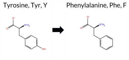 AIThe SynGAP1 missense variant Y34F is not reported in ClinVar (no ClinVar ID) and is absent from gnomAD (no gnomAD ID). Prediction tools that agree on a benign effect include REVEL, PROVEAN, ESM1b, FATHMM, AlphaMissense‑Default, AlphaMissense‑Optimized, and the SGM‑Consensus (Likely Benign). Tools that predict a pathogenic effect are polyPhen‑2 HumDiv, polyPhen‑2 HumVar, and SIFT. High‑accuracy assessments show AlphaMissense‑Optimized as benign and the SGM‑Consensus as Likely Benign; no Foldetta stability result is available. Overall, the majority of evidence points to a benign impact. This conclusion is consistent with the lack of ClinVar annotation, so there is no contradiction with existing clinical data. Disclaimer: This summary was generated using AI and should be interpreted alongside expert review. | Likely Benign | 0.529623 | Disordered | 0.435847 | Uncertain | 0.303 | 0.855 | 0.375 | -3.275 | Likely Benign | 0.127 | Likely Benign | Likely Benign | 0.094 | Likely Benign | -0.50 | Neutral | 0.458 | Possibly Damaging | 0.481 | Possibly Damaging | 4.20 | Benign | 0.00 | Affected | 0.2604 | 0.3591 | 7 | 3 | 4.1 | -16.00 | |||||||||||||||||||||||||||||||||||||||
| c.1025A>T | Y342F 2D 3DClick to see structure in 3D Viewer AIThe SynGAP1 missense variant Y342F is not reported in ClinVar (ClinVar ID None) and is absent from gnomAD (gnomAD ID None). Prediction tools that agree on a benign effect include REVEL, FoldX, Rosetta, Foldetta, premPS, SIFT, ESM1b, AlphaMissense‑Default, and AlphaMissense‑Optimized. Tools that predict a pathogenic effect are PROVEAN, polyPhen‑2 HumDiv, polyPhen‑2 HumVar, and FATHMM. High‑accuracy assessments show AlphaMissense‑Optimized as benign, Foldetta as benign, and the SGM Consensus (majority vote from AlphaMissense‑Default, ESM1b, FATHMM, PROVEAN) is inconclusive and therefore treated as unavailable. Overall, the majority of evidence points to a benign impact. This conclusion does not contradict the ClinVar status, which currently has no classification for this variant. Disclaimer: This summary was generated using AI and should be interpreted alongside expert review. | C2 | 0.366687 | Structured | 0.408200 | Uncertain | 0.866 | 0.487 | 0.250 | -6.987 | Likely Benign | 0.145 | Likely Benign | Likely Benign | -0.13 | Likely Benign | 0.1 | 0.42 | Likely Benign | 0.15 | Likely Benign | 0.05 | Likely Benign | 0.160 | Likely Benign | -2.67 | Deleterious | 0.999 | Probably Damaging | 0.992 | Probably Damaging | 2.03 | Pathogenic | 0.26 | Tolerated | 0.2626 | 0.3615 | 7 | 3 | 4.1 | -16.00 | ||||||||||||||||||||||||||||||
| c.1088A>T | Y363F 2D 3DClick to see structure in 3D Viewer AIThe SynGAP1 missense variant Y363F is not reported in ClinVar (ClinVar ID None) and is absent from gnomAD (gnomAD ID None). Prediction tools that agree on a benign effect include REVEL, FoldX, Rosetta, SIFT, ESM1b, AlphaMissense‑Default, and AlphaMissense‑Optimized. Tools that predict a pathogenic effect are PROVEAN, polyPhen‑2 HumDiv, polyPhen‑2 HumVar, and FATHMM; premPS is uncertain. High‑accuracy assessments show AlphaMissense‑Optimized as benign, Foldetta as benign, and the SGM Consensus (majority vote from AlphaMissense‑Default, ESM1b, FATHMM, PROVEAN) is inconclusive due to a 2‑vs‑2 split. Overall, seven tools predict benign while four predict pathogenic, with no evidence of pathogenicity from the most accurate methods. Therefore, the variant is most likely benign, and this assessment does not contradict any ClinVar status. Disclaimer: This summary was generated using AI and should be interpreted alongside expert review. | C2 | 0.321458 | Structured | 0.435392 | Uncertain | 0.954 | 0.586 | 0.125 | -6.621 | Likely Benign | 0.114 | Likely Benign | Likely Benign | -0.23 | Likely Benign | 0.0 | 0.42 | Likely Benign | 0.10 | Likely Benign | 0.58 | Ambiguous | 0.293 | Likely Benign | -3.42 | Deleterious | 0.997 | Probably Damaging | 0.970 | Probably Damaging | 1.74 | Pathogenic | 0.18 | Tolerated | 0.2781 | 0.3583 | 7 | 3 | 4.1 | -16.00 | ||||||||||||||||||||||||||||||
| c.109T>G | S37A 2D  AIThe SynGAP1 missense variant S37A is not reported in ClinVar (no ClinVar ID) and is absent from gnomAD (no gnomAD ID). Prediction tools that agree on a benign effect include REVEL, PROVEAN, polyPhen‑2 HumDiv, polyPhen‑2 HumVar, ESM1b, FATHMM, AlphaMissense‑Default, and AlphaMissense‑Optimized. Only SIFT predicts a pathogenic outcome. The high‑accuracy consensus, SGM‑Consensus, classifies the variant as Likely Benign, and AlphaMissense‑Optimized also reports a benign prediction. Foldetta, a protein‑folding stability method, has no available result for this variant. Overall, the majority of evidence points to a benign impact. This conclusion is consistent with the lack of ClinVar annotation; there is no contradiction with ClinVar status. Disclaimer: This summary was generated using AI and should be interpreted alongside expert review. | Likely Benign | 0.642678 | Disordered | 0.433492 | Uncertain | 0.317 | 0.806 | 0.500 | -4.052 | Likely Benign | 0.125 | Likely Benign | Likely Benign | 0.045 | Likely Benign | -0.86 | Neutral | 0.140 | Benign | 0.355 | Benign | 3.98 | Benign | 0.00 | Affected | 0.5089 | 0.4970 | Weaken | 1 | 1 | 2.6 | -16.00 | ||||||||||||||||||||||||||||||||||||||
| c.10T>G | S4A 2D AIThe SynGAP1 missense variant S4A is not reported in ClinVar (no ClinVar ID) and is absent from gnomAD (no gnomAD ID). Prediction tools that agree on a benign effect include REVEL, PROVEAN, polyPhen‑2 HumDiv, polyPhen‑2 HumVar, ESM1b, FATHMM, AlphaMissense‑Default, and AlphaMissense‑Optimized. Only SIFT predicts a pathogenic outcome. The SGM‑Consensus, which aggregates AlphaMissense‑Default, ESM1b, FATHMM, and PROVEAN, also indicates a likely benign classification. High‑accuracy assessments show AlphaMissense‑Optimized as benign and the SGM‑Consensus as likely benign; Foldetta results are unavailable. Overall, the majority of evidence points to a benign impact for S4A, and this conclusion does not contradict any ClinVar annotation, as none exists. Disclaimer: This summary was generated using AI and should be interpreted alongside expert review. | Likely Benign | 0.608892 | Disordered | 0.547364 | Binding | 0.390 | 0.924 | 0.750 | -4.245 | Likely Benign | 0.111 | Likely Benign | Likely Benign | 0.050 | Likely Benign | 0.02 | Neutral | 0.140 | Benign | 0.097 | Benign | 4.22 | Benign | 0.00 | Affected | 0.4871 | 0.5755 | 1 | 1 | 2.6 | -16.00 | |||||||||||||||||||||||||||||||||||||||
| c.1120T>G | S374A 2D 3DClick to see structure in 3D Viewer AIThe SynGAP1 missense variant S374A is reported in gnomAD (6‑33438025‑T‑G) and has no ClinVar entry. Prediction tools that agree on a benign effect include REVEL, FoldX, Rosetta, Foldetta, premPS, PROVEAN, polyPhen‑2 (HumDiv and HumVar), ESM1b, FATHMM, AlphaMissense‑Default, and AlphaMissense‑Optimized. Only SIFT predicts a pathogenic outcome. The SGM‑Consensus, derived from a majority vote of AlphaMissense‑Default, ESM1b, FATHMM, and PROVEAN, classifies the variant as Likely Benign. High‑accuracy assessments—AlphaMissense‑Optimized, SGM‑Consensus, and Foldetta (combining FoldX‑MD and Rosetta outputs)—all indicate a benign effect. Based on the collective predictions, the variant is most likely benign, and this conclusion is not contradicted by any ClinVar status. Disclaimer: This summary was generated using AI and should be interpreted alongside expert review. | Likely Benign | C2 | 0.642678 | Disordered | 0.428948 | Uncertain | 0.333 | 0.812 | 0.625 | 6-33438025-T-G | -4.245 | Likely Benign | 0.082 | Likely Benign | Likely Benign | -0.08 | Likely Benign | 0.1 | 0.41 | Likely Benign | 0.17 | Likely Benign | 0.10 | Likely Benign | 0.122 | Likely Benign | -0.53 | Neutral | 0.012 | Benign | 0.011 | Benign | 5.32 | Benign | 0.04 | Affected | 4.32 | 13 | 0.5091 | 0.5774 | Strenghten | 1 | 1 | 2.6 | -16.00 | |||||||||||||||||||||||||
| c.1135T>G | S379A 2D 3DClick to see structure in 3D Viewer AIThe SynGAP1 missense variant S379A is reported in gnomAD (variant ID 6‑33438040‑T‑G) but has no entry in ClinVar. All available in‑silico predictors classify the change as benign: REVEL, FoldX, premPS, PROVEAN, polyPhen‑2 (HumDiv and HumVar), SIFT, ESM1b, FATHMM, AlphaMissense‑Default, AlphaMissense‑Optimized, and the consensus SGM‑Consensus (majority vote of AlphaMissense‑Default, ESM1b, FATHMM, PROVEAN). No tool predicts pathogenicity. High‑accuracy assessments are consistent: AlphaMissense‑Optimized predicts benign; the SGM‑Consensus (majority vote) indicates “Likely Benign”; and Foldetta, which integrates FoldX‑MD and Rosetta outputs, also predicts benign. Consequently, the variant is most likely benign, and this assessment does not contradict any ClinVar status (none is available). Disclaimer: This summary was generated using AI and should be interpreted alongside expert review. | Likely Benign | C2 | 0.728858 | Disordered | 0.433206 | Uncertain | 0.327 | 0.931 | 0.625 | 6-33438040-T-G | -4.300 | Likely Benign | 0.077 | Likely Benign | Likely Benign | -0.22 | Likely Benign | 0.3 | 1.03 | Ambiguous | 0.41 | Likely Benign | 0.10 | Likely Benign | 0.217 | Likely Benign | -0.50 | Neutral | 0.012 | Benign | 0.002 | Benign | 3.91 | Benign | 0.21 | Tolerated | 4.32 | 11 | 0.5032 | 0.5555 | Strenghten | 1 | 1 | 2.6 | -16.00 | |||||||||||||||||||||||||
| c.1153T>G | S385A 2D 3DClick to see structure in 3D Viewer AIThe SynGAP1 missense variant S385A is catalogued in gnomAD (variant ID 6‑33438058‑T‑G) but has no entry in ClinVar. All available in silico predictors report a benign effect: REVEL, FoldX, Rosetta, Foldetta, premPS, PROVEAN, polyPhen‑2 (HumDiv and HumVar), SIFT, ESM1b, FATHMM, AlphaMissense‑Default, and AlphaMissense‑Optimized. No tool predicts pathogenicity. High‑accuracy assessments concur: AlphaMissense‑Optimized is benign; the SGM Consensus (majority vote of AlphaMissense‑Default, ESM1b, FATHMM, PROVEAN) is “Likely Benign”; and Foldetta (combining FoldX‑MD and Rosetta outputs) is benign. Consequently, the variant is most likely benign, and this assessment does not contradict any ClinVar status (none reported). Disclaimer: This summary was generated using AI and should be interpreted alongside expert review. | Likely Benign | C2 | 0.733139 | Disordered | 0.425480 | Uncertain | 0.341 | 0.925 | 0.750 | 6-33438058-T-G | -4.412 | Likely Benign | 0.083 | Likely Benign | Likely Benign | 0.03 | Likely Benign | 0.1 | 0.19 | Likely Benign | 0.11 | Likely Benign | 0.07 | Likely Benign | 0.243 | Likely Benign | -0.28 | Neutral | 0.140 | Benign | 0.355 | Benign | 4.65 | Benign | 0.13 | Tolerated | 4.32 | 3 | 0.4910 | 0.5366 | 1 | 1 | 2.6 | -16.00 | ||||||||||||||||||||||||||
| c.1165T>G | S389A 2D 3DClick to see structure in 3D Viewer AIThe SynGAP1 missense variant S389A is reported in gnomAD (ID 6‑33438070‑T‑G) and has no ClinVar entry. Prediction tools that agree on a benign effect include REVEL, FoldX, Rosetta, Foldetta, premPS, PROVEAN, polyPhen‑2 (HumDiv and HumVar), ESM1b, FATHMM, AlphaMissense‑Default, and AlphaMissense‑Optimized. Only SIFT predicts a pathogenic outcome. The SGM‑Consensus, derived from a majority vote of AlphaMissense‑Default, ESM1b, FATHMM, and PROVEAN, classifies the variant as Likely Benign. High‑accuracy assessments confirm this: AlphaMissense‑Optimized is Benign; the SGM‑Consensus is Likely Benign; and Foldetta, which integrates FoldX‑MD and Rosetta stability calculations, also reports Benign. Overall, the preponderance of evidence indicates the variant is most likely benign, and this conclusion is not contradicted by any ClinVar status. Disclaimer: This summary was generated using AI and should be interpreted alongside expert review. | Likely Benign | C2 | 0.703578 | Disordered | 0.417444 | Uncertain | 0.306 | 0.803 | 0.875 | 6-33438070-T-G | -4.199 | Likely Benign | 0.081 | Likely Benign | Likely Benign | 0.01 | Likely Benign | 0.0 | 0.37 | Likely Benign | 0.19 | Likely Benign | 0.03 | Likely Benign | 0.326 | Likely Benign | -0.39 | Neutral | 0.140 | Benign | 0.355 | Benign | 5.08 | Benign | 0.04 | Affected | 4.32 | 8 | 0.5037 | 0.5963 | Strenghten | 1 | 1 | 2.6 | -16.00 | |||||||||||||||||||||||||
| c.116A>T | Y39F 2D AIThe SynGAP1 missense variant Y39F is not reported in ClinVar and is absent from gnomAD. Prediction tools that agree on a benign effect include REVEL, PROVEAN, ESM1b, FATHMM, AlphaMissense‑Default, AlphaMissense‑Optimized, and the SGM‑Consensus (majority vote) which classifies the variant as Likely Benign. In contrast, polyPhen‑2 (HumDiv and HumVar) and SIFT all predict a pathogenic impact. High‑accuracy assessments further support a benign outcome: AlphaMissense‑Optimized predicts Benign, and the SGM‑Consensus (derived from AlphaMissense‑Default, ESM1b, FATHMM, and PROVEAN) also indicates Likely Benign. No Foldetta stability analysis is available for this variant. Overall, the majority of evidence points to a benign effect, and this conclusion does not contradict any ClinVar annotation (none exists). Disclaimer: This summary was generated using AI and should be interpreted alongside expert review. | Likely Benign | 0.425610 | Structured | 0.432876 | Uncertain | 0.343 | 0.787 | 0.375 | -3.410 | Likely Benign | 0.119 | Likely Benign | Likely Benign | 0.073 | Likely Benign | -0.78 | Neutral | 0.458 | Possibly Damaging | 0.481 | Possibly Damaging | 4.05 | Benign | 0.00 | Affected | 0.2641 | 0.3363 | 7 | 3 | 4.1 | -16.00 | |||||||||||||||||||||||||||||||||||||||
| c.1217A>T | Y406F 2D 3DClick to see structure in 3D Viewer AIThe SynGAP1 missense variant Y406F is not reported in ClinVar and is absent from gnomAD. Functional prediction tools uniformly indicate a benign effect: REVEL, FoldX, Rosetta, premPS, PROVEAN, polyPhen‑2 (HumDiv and HumVar), SIFT, ESM1b, FATHMM, AlphaMissense‑Default, AlphaMissense‑Optimized, and the SGM‑Consensus score (majority vote from AlphaMissense‑Default, ESM1b, FATHMM, PROVEAN) all classify the variant as benign or likely benign. No tool predicts pathogenicity. High‑accuracy assessments corroborate this: AlphaMissense‑Optimized predicts benign, the SGM‑Consensus (majority vote) indicates likely benign, and Foldetta (combining FoldX‑MD and Rosetta outputs) also predicts benign. Based on the collective evidence, the variant is most likely benign, and this assessment does not contradict any ClinVar status (none is available). Disclaimer: This summary was generated using AI and should be interpreted alongside expert review. | Likely Benign | C2 | 0.179055 | Structured | 0.393707 | Uncertain | 0.946 | 0.291 | 0.000 | -3.427 | Likely Benign | 0.080 | Likely Benign | Likely Benign | -0.01 | Likely Benign | 0.2 | 0.40 | Likely Benign | 0.20 | Likely Benign | 0.15 | Likely Benign | 0.079 | Likely Benign | -0.93 | Neutral | 0.002 | Benign | 0.002 | Benign | 3.96 | Benign | 0.31 | Tolerated | 0.2673 | 0.3952 | 7 | 3 | 4.1 | -16.00 | |||||||||||||||||||||||||||||
| c.1250A>T | Y417F 2D 3DClick to see structure in 3D Viewer AIThe SynGAP1 Y417F variant has no ClinVar entry and is not reported in gnomAD. Prediction tools that agree on a benign effect include REVEL, FoldX, Rosetta, SIFT, and FATHMM. Those that predict a pathogenic impact are SGM‑Consensus, PROVEAN, polyPhen‑2 HumDiv, polyPhen‑2 HumVar, ESM1b, and AlphaMissense‑Default. Uncertain results come from AlphaMissense‑Optimized and premPS. High‑accuracy assessments show SGM‑Consensus (majority vote of AlphaMissense‑Default, ESM1b, FATHMM, PROVEAN) as Likely Pathogenic, Foldetta (combining FoldX‑MD and Rosetta) as Benign, and AlphaMissense‑Optimized as Uncertain. Overall, the majority of tools and the SGM‑Consensus score suggest a pathogenic effect, while Foldetta indicates a benign effect; the variant’s status is not contradicted by ClinVar (no entry). Thus, based on the available predictions, the variant is most likely pathogenic. Disclaimer: This summary was generated using AI and should be interpreted alongside expert review. | Likely Pathogenic | GAP | 0.179055 | Structured | 0.346865 | Uncertain | 0.958 | 0.250 | 0.000 | -11.368 | Likely Pathogenic | 0.852 | Likely Pathogenic | Ambiguous | 0.47 | Likely Benign | 0.1 | -0.09 | Likely Benign | 0.19 | Likely Benign | 0.97 | Ambiguous | 0.367 | Likely Benign | -3.82 | Deleterious | 0.999 | Probably Damaging | 0.985 | Probably Damaging | 3.03 | Benign | 0.06 | Tolerated | 0.2617 | 0.3098 | 7 | 3 | 4.1 | -16.00 | |||||||||||||||||||||||||||||
| c.1268A>T | Y423F 2D 3DClick to see structure in 3D Viewer AIThe SynGAP1 missense variant Y423F is not reported in ClinVar and is absent from gnomAD. Prediction tools that assess sequence conservation and functional impact uniformly classify the substitution as benign: REVEL, FoldX, Rosetta, Foldetta, PROVEAN, SIFT, ESM1b, FATHMM, AlphaMissense‑Default, AlphaMissense‑Optimized, and the SGM‑Consensus (majority vote of AlphaMissense‑Default, ESM1b, FATHMM, PROVEAN). Only two tools, polyPhen‑2 HumDiv and HumVar, predict a pathogenic effect, while premPS is inconclusive. High‑accuracy methods give consistent benign results: AlphaMissense‑Optimized is benign, the SGM‑Consensus is Likely Benign, and Foldetta (combining FoldX‑MD and Rosetta) predicts benign stability. Overall, the overwhelming majority of evidence supports a benign effect, and this conclusion is not contradicted by any ClinVar annotation. Disclaimer: This summary was generated using AI and should be interpreted alongside expert review. | Likely Benign | GAP | 0.088832 | Structured | 0.421885 | Uncertain | 0.975 | 0.242 | 0.000 | -5.533 | Likely Benign | 0.181 | Likely Benign | Likely Benign | 0.03 | Likely Benign | 0.1 | -0.05 | Likely Benign | -0.01 | Likely Benign | 0.76 | Ambiguous | 0.149 | Likely Benign | -2.30 | Neutral | 0.999 | Probably Damaging | 0.985 | Probably Damaging | 3.45 | Benign | 0.60 | Tolerated | 0.1934 | 0.2337 | 7 | 3 | 4.1 | -16.00 | |||||||||||||||||||||||||||||
| c.1283A>T | Y428F 2D 3DClick to see structure in 3D Viewer AIThe SynGAP1 missense variant Y428F is not reported in ClinVar (ClinVar ID None) and is absent from gnomAD (gnomAD ID None). Prediction tools that agree on a benign effect include REVEL, FATHMM, and AlphaMissense‑Optimized, whereas a majority of tools (PROVEAN, polyPhen‑2 HumDiv, polyPhen‑2 HumVar, SIFT, and ESM1b) predict a pathogenic impact. Uncertain or inconclusive results are reported for FoldX, Rosetta, Foldetta, premPS, and AlphaMissense‑Default. High‑accuracy assessments show AlphaMissense‑Optimized as benign, the SGM Consensus (majority vote from AlphaMissense‑Default, ESM1b, FATHMM, and PROVEAN) as pathogenic, and Foldetta as unavailable. Overall, the balance of evidence—five pathogenic versus three benign predictions, with a pathogenic SGM Consensus and a benign AlphaMissense‑Optimized—suggests that the variant is most likely pathogenic. This conclusion is not contradicted by ClinVar status, which has no entry for this variant. Disclaimer: This summary was generated using AI and should be interpreted alongside expert review. | GAP | 0.118441 | Structured | 0.389652 | Uncertain | 0.965 | 0.292 | 0.000 | -10.464 | Likely Pathogenic | 0.530 | Ambiguous | Likely Benign | 0.82 | Ambiguous | 0.1 | 0.52 | Ambiguous | 0.67 | Ambiguous | 0.89 | Ambiguous | 0.366 | Likely Benign | -3.82 | Deleterious | 0.999 | Probably Damaging | 0.985 | Probably Damaging | 3.41 | Benign | 0.04 | Affected | 0.2719 | 0.3146 | 7 | 3 | 4.1 | -16.00 | ||||||||||||||||||||||||||||||
| c.1396T>G | S466A 2D 3DClick to see structure in 3D Viewer AIThe SynGAP1 missense variant S466A is not reported in ClinVar and is absent from gnomAD. Functional prediction tools show a split: pathogenic scores are given by REVEL, polyPhen‑2 HumDiv, polyPhen‑2 HumVar, and FATHMM, whereas benign predictions come from SIFT, PROVEAN, premPS, ESM1b, Rosetta, Foldetta, AlphaMissense‑Default, and AlphaMissense‑Optimized. The SGM‑Consensus, derived from a majority vote of AlphaMissense‑Default, ESM1b, FATHMM, and PROVEAN, classifies the variant as Likely Benign. High‑accuracy assessments further support a benign outcome: AlphaMissense‑Optimized predicts Benign, the SGM‑Consensus itself is Benign, and Foldetta (combining FoldX‑MD and Rosetta outputs) also reports Benign. FoldX alone is Uncertain, but this does not alter the overall consensus. Taken together, the majority of evidence indicates the variant is most likely benign, and this conclusion is consistent with the absence of a ClinVar pathogenic annotation. Disclaimer: This summary was generated using AI and should be interpreted alongside expert review. | Likely Benign | GAP | 0.311707 | Structured | 0.322353 | Uncertain | 0.933 | 0.227 | 0.000 | -6.928 | Likely Benign | 0.228 | Likely Benign | Likely Benign | -0.50 | Ambiguous | 0.1 | 0.06 | Likely Benign | -0.22 | Likely Benign | 0.37 | Likely Benign | 0.537 | Likely Pathogenic | -1.46 | Neutral | 0.909 | Possibly Damaging | 0.987 | Probably Damaging | -1.51 | Pathogenic | 0.10 | Tolerated | 0.4245 | 0.3996 | 1 | 1 | 2.6 | -16.00 | |||||||||||||||||||||||||||||
| c.1411T>G | S471A 2D 3DClick to see structure in 3D Viewer AIThe SynGAP1 missense variant S471A is not reported in ClinVar and is absent from gnomAD. Across the spectrum of in‑silico predictors, the majority (REVEL, FoldX, Foldetta, premPS, PROVEAN, polyPhen‑2 HumDiv, polyPhen‑2 HumVar, SIFT, ESM1b, AlphaMissense‑Default, AlphaMissense‑Optimized, and the SGM‑Consensus) classify the change as benign, whereas only FATHMM predicts it as pathogenic; Rosetta is inconclusive. High‑accuracy assessments reinforce the benign interpretation: AlphaMissense‑Optimized scores benign, the SGM‑Consensus (majority vote of AlphaMissense‑Default, ESM1b, FATHMM, PROVEAN) also yields benign, and Foldetta (combining FoldX‑MD and Rosetta outputs) predicts benign stability. No evidence suggests pathogenicity, and the predictions do not contradict the absence of ClinVar annotation. Therefore, the variant is most likely benign. Disclaimer: This summary was generated using AI and should be interpreted alongside expert review. | Likely Benign | GAP | 0.305330 | Structured | 0.355411 | Uncertain | 0.888 | 0.261 | 0.000 | -5.516 | Likely Benign | 0.077 | Likely Benign | Likely Benign | 0.11 | Likely Benign | 0.1 | -0.61 | Ambiguous | -0.25 | Likely Benign | 0.23 | Likely Benign | 0.252 | Likely Benign | -1.93 | Neutral | 0.010 | Benign | 0.037 | Benign | -1.22 | Pathogenic | 0.29 | Tolerated | 0.4439 | 0.3691 | 1 | 1 | 2.6 | -16.00 | |||||||||||||||||||||||||||||
| c.1490A>T | Y497F 2D 3DClick to see structure in 3D Viewer AIThe SynGAP1 missense variant Y497F is not reported in ClinVar (ClinVar status: not listed) and is absent from gnomAD (gnomAD ID: none). Prediction tools that agree on a benign effect include FoldX, Rosetta, Foldetta, polyPhen‑2 HumDiv, polyPhen‑2 HumVar, SIFT, AlphaMissense‑Default, and AlphaMissense‑Optimized. Tools that predict a pathogenic effect are SGM‑Consensus, REVEL, PROVEAN, ESM1b, and FATHMM; premPS remains uncertain. High‑accuracy assessments show AlphaMissense‑Optimized as benign, the SGM‑Consensus (majority vote from AlphaMissense‑Default, ESM1b, FATHMM, PROVEAN) as pathogenic, and Foldetta (combining FoldX‑MD and Rosetta outputs) as benign. Overall, the majority of predictions lean toward a benign impact, and this conclusion does not contradict the absence of a ClinVar classification. Thus, the variant is most likely benign based on the current predictive evidence. Disclaimer: This summary was generated using AI and should be interpreted alongside expert review. | Likely Pathogenic | GAP | 0.092881 | Structured | 0.393608 | Uncertain | 0.950 | 0.181 | 0.000 | -8.566 | Likely Pathogenic | 0.199 | Likely Benign | Likely Benign | -0.27 | Likely Benign | 0.1 | 0.47 | Likely Benign | 0.10 | Likely Benign | 0.61 | Ambiguous | 0.578 | Likely Pathogenic | -3.75 | Deleterious | 0.367 | Benign | 0.131 | Benign | -1.15 | Pathogenic | 0.19 | Tolerated | 0.2074 | 0.2242 | 7 | 3 | 4.1 | -16.00 | |||||||||||||||||||||||||||||
| c.1514A>T | Y505F 2D 3DClick to see structure in 3D Viewer AIThe SynGAP1 missense variant Y505F is not reported in ClinVar (ClinVar ID: None) and is absent from gnomAD (gnomAD ID: None). Prediction tools that agree on a benign effect include REVEL, FoldX, SIFT, FATHMM, and AlphaMissense‑Optimized. Those that predict a pathogenic effect are PROVEAN, polyPhen‑2 HumDiv, polyPhen‑2 HumVar, and ESM1b. Predictions that are uncertain or inconclusive are Rosetta, Foldetta, premPS, and AlphaMissense‑Default. High‑accuracy assessments show AlphaMissense‑Optimized as benign, the SGM Consensus (majority vote from AlphaMissense‑Default, ESM1b, FATHMM, PROVEAN) as pathogenic, and Foldetta as uncertain. Overall, the majority of tools (five benign versus four pathogenic) lean toward a benign interpretation, and this is consistent with the lack of ClinVar evidence; thus the variant is most likely benign and does not contradict ClinVar status. Disclaimer: This summary was generated using AI and should be interpreted alongside expert review. | GAP | 0.047319 | Structured | 0.292227 | Uncertain | 0.909 | 0.188 | 0.000 | -8.855 | Likely Pathogenic | 0.437 | Ambiguous | Likely Benign | 0.40 | Likely Benign | 0.0 | 0.76 | Ambiguous | 0.58 | Ambiguous | 0.69 | Ambiguous | 0.434 | Likely Benign | -3.98 | Deleterious | 1.000 | Probably Damaging | 0.996 | Probably Damaging | 2.93 | Benign | 0.07 | Tolerated | 0.2409 | 0.2598 | 7 | 3 | 4.1 | -16.00 | ||||||||||||||||||||||||||||||
| c.154T>G | S52A 2D AIThe SynGAP1 missense variant S52A is not reported in ClinVar and is absent from gnomAD. Prediction tools that agree on a benign effect include REVEL, PROVEAN, polyPhen‑2 (HumDiv and HumVar), ESM1b, FATHMM, AlphaMissense‑Default, and AlphaMissense‑Optimized; the only tool predicting a pathogenic outcome is SIFT. The SGM‑Consensus, derived from a majority vote of AlphaMissense‑Default, ESM1b, FATHMM, and PROVEAN, also indicates a likely benign classification. High‑accuracy assessments confirm this: AlphaMissense‑Optimized predicts benign, and the SGM‑Consensus likewise predicts benign. Foldetta, a protein‑folding stability method combining FoldX‑MD and Rosetta outputs, has no available result for this variant. Overall, the consensus of available predictions strongly supports a benign impact, and this conclusion is not contradicted by any ClinVar annotation. Disclaimer: This summary was generated using AI and should be interpreted alongside expert review. | Likely Benign | 0.291804 | Structured | 0.457753 | Uncertain | 0.499 | 0.677 | 0.000 | -5.326 | Likely Benign | 0.196 | Likely Benign | Likely Benign | 0.040 | Likely Benign | -0.69 | Neutral | 0.140 | Benign | 0.355 | Benign | 4.17 | Benign | 0.00 | Affected | 0.5389 | 0.5073 | Strenghten | 1 | 1 | 2.6 | -16.00 | ||||||||||||||||||||||||||||||||||||||
| c.1553A>T | Y518F 2D 3DClick to see structure in 3D Viewer AIThe SynGAP1 Y518F missense variant is not reported in ClinVar (ClinVar ID None) and is absent from gnomAD (gnomAD ID None). Prediction tools that agree on a benign effect include REVEL, FoldX, premPS, SIFT, FATHMM, AlphaMissense‑Optimized, and Foldetta. Those that predict a pathogenic effect are PROVEAN, polyPhen‑2 HumDiv, polyPhen‑2 HumVar, and ESM1b. Two tools are uncertain: Rosetta and AlphaMissense‑Default. High‑accuracy assessments show AlphaMissense‑Optimized as benign, Foldetta as benign, and the SGM Consensus (majority vote from AlphaMissense‑Default, ESM1b, FATHMM, PROVEAN) as pathogenic. Overall, the majority of individual predictors (seven benign vs. four pathogenic) support a benign classification, and this does not contradict the ClinVar status, which has no entry for this variant. Thus, based on the available predictions, the variant is most likely benign. Disclaimer: This summary was generated using AI and should be interpreted alongside expert review. | GAP | 0.139895 | Structured | 0.126970 | Uncertain | 0.897 | 0.321 | 0.000 | -10.617 | Likely Pathogenic | 0.466 | Ambiguous | Likely Benign | 0.34 | Likely Benign | 0.1 | -0.61 | Ambiguous | -0.14 | Likely Benign | 0.41 | Likely Benign | 0.318 | Likely Benign | -3.68 | Deleterious | 0.999 | Probably Damaging | 0.996 | Probably Damaging | 3.50 | Benign | 0.22 | Tolerated | 0.2107 | 0.2230 | 7 | 3 | 4.1 | -16.00 | ||||||||||||||||||||||||||||||
| c.1558T>G | S520A 2D 3DClick to see structure in 3D Viewer AIThe SynGAP1 S520A missense variant is not reported in ClinVar (ClinVar ID None) and has no entry in gnomAD (gnomAD ID None). Prediction tools that agree on a benign effect include FoldX, Rosetta, Foldetta, and AlphaMissense‑Optimized, whereas a larger group—REVEL, PROVEAN, polyPhen‑2 (HumDiv and HumVar), SIFT, ESM1b, FATHMM, and AlphaMissense‑Default—consistently predict pathogenicity. The premPS score is uncertain. High‑accuracy assessments show AlphaMissense‑Optimized as benign, Foldetta as benign, and the SGM Consensus (majority vote of AlphaMissense‑Default, ESM1b, FATHMM, PROVEAN) as likely pathogenic. Overall, the majority of tools lean toward pathogenicity, and the high‑accuracy consensus is split, but the pathogenic predictions dominate. Thus, the variant is most likely pathogenic based on the available predictions, and this assessment does not contradict any ClinVar status because no ClinVar entry exists. Disclaimer: This summary was generated using AI and should be interpreted alongside expert review. | Likely Pathogenic | GAP | 0.094817 | Structured | 0.084894 | Uncertain | 0.887 | 0.337 | 0.000 | -8.417 | Likely Pathogenic | 0.704 | Likely Pathogenic | Likely Benign | 0.03 | Likely Benign | 0.2 | 0.19 | Likely Benign | 0.11 | Likely Benign | 0.56 | Ambiguous | 0.523 | Likely Pathogenic | -2.55 | Deleterious | 0.944 | Possibly Damaging | 0.987 | Probably Damaging | -1.31 | Pathogenic | 0.01 | Affected | 0.4887 | 0.3636 | 1 | 1 | 2.6 | -16.00 | |||||||||||||||||||||||||||||
| c.1600T>G | S534A 2D 3DClick to see structure in 3D Viewer AISynGAP1 S534A is not reported in ClinVar and is absent from gnomAD. Prediction tools largely agree on a benign effect: REVEL, FoldX, Rosetta, Foldetta, premPS, PROVEAN, SIFT, ESM1b, FATHMM, AlphaMissense‑Default, and AlphaMissense‑Optimized all indicate benign. Only two tools, polyPhen‑2 HumDiv and HumVar, predict pathogenicity. High‑accuracy methods reinforce the benign consensus: AlphaMissense‑Optimized scores benign, the SGM‑Consensus (majority vote of AlphaMissense‑Default, ESM1b, FATHMM, PROVEAN) reports Likely Benign, and Foldetta (combining FoldX‑MD and Rosetta outputs) predicts benign stability. No evidence suggests a deleterious effect. Therefore, the variant is most likely benign, and this assessment does not contradict any ClinVar annotation (none available). Disclaimer: This summary was generated using AI and should be interpreted alongside expert review. | Likely Benign | GAP | 0.167087 | Structured | 0.032173 | Uncertain | 0.860 | 0.362 | 0.000 | -4.691 | Likely Benign | 0.082 | Likely Benign | Likely Benign | 0.01 | Likely Benign | 0.0 | -0.01 | Likely Benign | 0.00 | Likely Benign | 0.11 | Likely Benign | 0.163 | Likely Benign | -1.70 | Neutral | 0.880 | Possibly Damaging | 0.994 | Probably Damaging | 3.36 | Benign | 0.42 | Tolerated | 0.5042 | 0.3131 | Weaken | 1 | 1 | 2.6 | -16.00 | ||||||||||||||||||||||||||||
| c.1669T>G | S557A 2D 3DClick to see structure in 3D Viewer AIThe SynGAP1 missense variant S557A is not reported in ClinVar and is absent from gnomAD. Prediction tools that agree on a benign effect include AlphaMissense‑Optimized and Rosetta, whereas a majority of tools (REVEL, SIFT, PROVEAN, polyPhen‑2 HumDiv, polyPhen‑2 HumVar, ESM1b, FATHMM, and the SGM‑Consensus) predict a pathogenic impact. Uncertain or inconclusive results come from AlphaMissense‑Default, FoldX, Foldetta, and premPS. High‑accuracy assessments show AlphaMissense‑Optimized as benign, the SGM‑Consensus (majority vote of AlphaMissense‑Default, ESM1b, FATHMM, PROVEAN) as pathogenic, and Foldetta as uncertain. Overall, the preponderance of evidence points to a pathogenic effect. This conclusion is not contradicted by ClinVar status, which has no entry for this variant. Disclaimer: This summary was generated using AI and should be interpreted alongside expert review. | Likely Pathogenic | GAP | 0.028107 | Structured | 0.010261 | Uncertain | 0.924 | 0.215 | 0.000 | -11.044 | Likely Pathogenic | 0.421 | Ambiguous | Likely Benign | 1.26 | Ambiguous | 0.0 | 0.09 | Likely Benign | 0.68 | Ambiguous | 0.54 | Ambiguous | 0.801 | Likely Pathogenic | -2.70 | Deleterious | 0.944 | Possibly Damaging | 0.987 | Probably Damaging | -1.69 | Pathogenic | 0.05 | Affected | 0.4695 | 0.4836 | 1 | 1 | 2.6 | -16.00 | |||||||||||||||||||||||||||||
| c.1711T>G | S571A 2D 3DClick to see structure in 3D Viewer AISynGAP1 missense variant S571A is not reported in ClinVar (no ClinVar ID) and is absent from gnomAD. Prediction tools that agree on a benign effect include FoldX, Rosetta, SIFT, ESM1b, AlphaMissense‑Default, and AlphaMissense‑Optimized. Tools that predict a pathogenic effect are REVEL, PROVEAN, polyPhen‑2 (HumDiv and HumVar), and FATHMM. The SGM Consensus, derived from a majority vote of AlphaMissense‑Default, ESM1b, FATHMM, and PROVEAN, is inconclusive because it yields a 2‑2 split. High‑accuracy assessments show AlphaMissense‑Optimized as benign, Foldetta (combining FoldX‑MD and Rosetta outputs) as benign, and the SGM Consensus remains unavailable. Overall, the preponderance of evidence points to a benign impact for S571A, and this conclusion is not contradicted by any ClinVar annotation. Disclaimer: This summary was generated using AI and should be interpreted alongside expert review. | GAP | 0.069024 | Structured | 0.045569 | Uncertain | 0.928 | 0.270 | 0.000 | -6.344 | Likely Benign | 0.233 | Likely Benign | Likely Benign | -0.44 | Likely Benign | 0.1 | -0.19 | Likely Benign | -0.32 | Likely Benign | 0.51 | Ambiguous | 0.563 | Likely Pathogenic | -2.69 | Deleterious | 0.980 | Probably Damaging | 0.994 | Probably Damaging | -1.22 | Pathogenic | 0.09 | Tolerated | 0.4739 | 0.2671 | 1 | 1 | 2.6 | -16.00 | ||||||||||||||||||||||||||||||
| c.1774T>G | S592A 2D 3DClick to see structure in 3D Viewer AIThe SynGAP1 missense variant S592A is not reported in ClinVar and is absent from gnomAD. Prediction tools that agree on a benign effect include FoldX, SIFT, and AlphaMissense‑Optimized, whereas a majority (SGM‑Consensus, REVEL, PROVEAN, polyPhen‑2 HumDiv, polyPhen‑2 HumVar, ESM1b, FATHMM, AlphaMissense‑Default) predict a pathogenic outcome. Tools with uncertain or inconclusive results are Rosetta, Foldetta, and premPS. High‑accuracy assessments show AlphaMissense‑Optimized as benign, the SGM Consensus (majority vote of AlphaMissense‑Default, ESM1b, FATHMM, PROVEAN) as pathogenic, and Foldetta as uncertain. Overall, the balance of evidence favors a pathogenic classification. This conclusion is not contradicted by ClinVar status, which currently contains no entry for this variant. Disclaimer: This summary was generated using AI and should be interpreted alongside expert review. | Likely Pathogenic | GAP | 0.012270 | Structured | 0.100070 | Uncertain | 0.913 | 0.182 | 0.000 | -10.902 | Likely Pathogenic | 0.572 | Likely Pathogenic | Likely Benign | -0.48 | Likely Benign | 0.1 | -0.93 | Ambiguous | -0.71 | Ambiguous | 0.79 | Ambiguous | 0.661 | Likely Pathogenic | -2.72 | Deleterious | 0.980 | Probably Damaging | 0.994 | Probably Damaging | -1.25 | Pathogenic | 0.44 | Tolerated | 0.4878 | 0.3318 | 1 | 1 | 2.6 | -16.00 | |||||||||||||||||||||||||||||
| c.1810T>G | S604A 2D 3DClick to see structure in 3D Viewer AIThe SynGAP1 missense variant S604A has no ClinVar record and is not reported in gnomAD. Prediction tools that agree on a benign effect include REVEL, FoldX, premPS, SIFT, FATHMM, AlphaMissense‑Optimized, and Foldetta. Those that predict a pathogenic effect are SGM‑Consensus, PROVEAN, polyPhen‑2 HumDiv, polyPhen‑2 HumVar, ESM1b, and AlphaMissense‑Default. Rosetta is uncertain and does not contribute to a consensus. High‑accuracy assessments show AlphaMissense‑Optimized as benign, the SGM‑Consensus (majority vote of AlphaMissense‑Default, ESM1b, FATHMM, PROVEAN) as pathogenic, and Foldetta (combining FoldX‑MD and Rosetta outputs) as benign. With two of the three high‑accuracy tools indicating benign and no ClinVar evidence to contradict, the variant is most likely benign. Disclaimer: This summary was generated using AI and should be interpreted alongside expert review. | Likely Pathogenic | GAP | 0.010926 | Structured | 0.192527 | Uncertain | 0.911 | 0.195 | 0.000 | -10.017 | Likely Pathogenic | 0.608 | Likely Pathogenic | Likely Benign | 0.02 | Likely Benign | 0.1 | -0.60 | Ambiguous | -0.29 | Likely Benign | 0.11 | Likely Benign | 0.378 | Likely Benign | -2.99 | Deleterious | 0.944 | Possibly Damaging | 0.987 | Probably Damaging | 3.25 | Benign | 0.08 | Tolerated | 0.5198 | 0.3530 | Weaken | 1 | 1 | 2.6 | -16.00 | ||||||||||||||||||||||||||||
| c.1841A>T | Y614F 2D 3DClick to see structure in 3D Viewer AIThe SynGAP1 missense variant Y614F is not reported in ClinVar (ClinVar ID None) and is absent from gnomAD (gnomAD ID None). Prediction tools that agree on a benign effect include REVEL, FoldX, SIFT, ESM1b, FATHMM, and AlphaMissense‑Optimized. Tools that predict a pathogenic effect are PROVEAN, polyPhen‑2 HumDiv, polyPhen‑2 HumVar, and AlphaMissense‑Default. Uncertain or unavailable results come from Foldetta, premPS, and Rosetta. High‑accuracy assessments show AlphaMissense‑Optimized as benign; the SGM Consensus (majority vote from AlphaMissense‑Default, ESM1b, FATHMM, PROVEAN) is inconclusive (2 benign vs 2 pathogenic), and Foldetta is unavailable. Overall, the majority of reliable predictors indicate a benign impact, and this conclusion does not contradict the lack of ClinVar annotation. Thus, the variant is most likely benign. Disclaimer: This summary was generated using AI and should be interpreted alongside expert review. | GAP | 0.314870 | Structured | 0.182134 | Uncertain | 0.852 | 0.254 | 0.000 | -5.584 | Likely Benign | 0.630 | Likely Pathogenic | Likely Benign | 0.09 | Likely Benign | 0.2 | 0.98 | Ambiguous | 0.54 | Ambiguous | 0.78 | Ambiguous | 0.364 | Likely Benign | -3.75 | Deleterious | 1.000 | Probably Damaging | 0.996 | Probably Damaging | 3.42 | Benign | 0.07 | Tolerated | 0.1909 | 0.2762 | 7 | 3 | 4.1 | -16.00 | ||||||||||||||||||||||||||||||
| c.1858T>G | S620A 2D 3DClick to see structure in 3D Viewer AIThe SynGAP1 missense variant S620A is not reported in ClinVar and is absent from gnomAD. Functional prediction tools that agree on a benign effect include REVEL, FoldX, premPS, PROVEAN, SIFT, ESM1b, AlphaMissense‑Default, and AlphaMissense‑Optimized. Tools that predict a pathogenic outcome are polyPhen‑2 HumDiv, polyPhen‑2 HumVar, and FATHMM. The SGM‑Consensus, derived from a majority vote of AlphaMissense‑Default, ESM1b, FATHMM, and PROVEAN, classifies the variant as likely benign. High‑accuracy assessments show AlphaMissense‑Optimized as benign, the SGM‑Consensus as benign, and Foldetta (combining FoldX‑MD and Rosetta outputs) as uncertain. No evidence from these analyses suggests a deleterious effect. Consequently, the variant is most likely benign based on the aggregate predictions, and this assessment does not contradict any ClinVar status, as no ClinVar entry exists for this variant. Disclaimer: This summary was generated using AI and should be interpreted alongside expert review. | Likely Benign | GAP | 0.139895 | Structured | 0.100377 | Uncertain | 0.936 | 0.219 | 0.000 | -4.637 | Likely Benign | 0.088 | Likely Benign | Likely Benign | -0.42 | Likely Benign | 0.0 | -0.90 | Ambiguous | -0.66 | Ambiguous | -0.19 | Likely Benign | 0.375 | Likely Benign | -0.52 | Neutral | 0.968 | Probably Damaging | 0.994 | Probably Damaging | -1.27 | Pathogenic | 0.66 | Tolerated | 0.4950 | 0.3636 | 1 | 1 | 2.6 | -16.00 | |||||||||||||||||||||||||||||
| c.1909T>G | S637A 2D 3DClick to see structure in 3D Viewer AIThe SynGAP1 missense variant S637A is not reported in ClinVar and is absent from gnomAD. Prediction tools that agree on a benign effect include AlphaMissense‑Default, AlphaMissense‑Optimized, ESM1b, FATHMM, PROVEAN, SIFT, polyPhen‑2 (HumDiv and HumVar), REVEL, FoldX, and premPS. No tool predicts a pathogenic outcome. High‑accuracy assessments further support a benign interpretation: AlphaMissense‑Optimized is benign, the SGM Consensus (majority vote of AlphaMissense‑Default, ESM1b, FATHMM, PROVEAN) is “Likely Benign,” and Foldetta, which integrates FoldX‑MD and Rosetta stability predictions, is inconclusive (Uncertain). Taken together, the overwhelming majority of evidence indicates a benign effect. The variant’s status is consistent with the lack of ClinVar annotation, so there is no contradiction with existing clinical data. Disclaimer: This summary was generated using AI and should be interpreted alongside expert review. | Likely Benign | GAP | 0.076542 | Structured | 0.083482 | Uncertain | 0.920 | 0.253 | 0.000 | -4.186 | Likely Benign | 0.137 | Likely Benign | Likely Benign | 0.31 | Likely Benign | 0.1 | 0.99 | Ambiguous | 0.65 | Ambiguous | 0.22 | Likely Benign | 0.078 | Likely Benign | -0.64 | Neutral | 0.120 | Benign | 0.182 | Benign | 3.41 | Benign | 1.00 | Tolerated | 0.4853 | 0.2996 | 1 | 1 | 2.6 | -16.00 | |||||||||||||||||||||||||||||
| c.1921T>G | S641A 2D 3DClick to see structure in 3D Viewer AIThe SynGAP1 missense variant S641A is not reported in ClinVar and is absent from gnomAD. Across the available in‑silico predictors, every tool examined (REVEL, FoldX, Rosetta, premPS, PROVEAN, polyPhen‑2 HumDiv/HumVar, SIFT, ESM1b, FATHMM, AlphaMissense‑Default, AlphaMissense‑Optimized) uniformly classifies the substitution as benign. No pathogenic predictions are present. High‑accuracy assessments corroborate this: AlphaMissense‑Optimized predicts benign; the SGM Consensus (majority vote of AlphaMissense‑Default, ESM1b, FATHMM, PROVEAN) yields “Likely Benign”; and Foldetta, which integrates FoldX‑MD and Rosetta stability outputs, also reports a benign effect. Consequently, the variant is most likely benign based on current predictions, and this assessment does not contradict any ClinVar status (none available). Disclaimer: This summary was generated using AI and should be interpreted alongside expert review. | Likely Benign | GAP | 0.125101 | Structured | 0.157322 | Uncertain | 0.786 | 0.270 | 0.000 | -6.103 | Likely Benign | 0.092 | Likely Benign | Likely Benign | 0.37 | Likely Benign | 0.2 | -0.28 | Likely Benign | 0.05 | Likely Benign | 0.27 | Likely Benign | 0.067 | Likely Benign | -2.07 | Neutral | 0.000 | Benign | 0.001 | Benign | 3.43 | Benign | 0.17 | Tolerated | 0.4873 | 0.4680 | 1 | 1 | 2.6 | -16.00 | |||||||||||||||||||||||||||||
| c.1975T>G | S659A 2D 3DClick to see structure in 3D Viewer AIThe SynGAP1 missense variant S659A is not reported in ClinVar and is absent from gnomAD. Functional prediction tools uniformly indicate a benign effect: REVEL, FoldX, Rosetta, Foldetta, premPS, PROVEAN, polyPhen‑2 (HumDiv and HumVar), SIFT, FATHMM, AlphaMissense‑Default, and AlphaMissense‑Optimized all classify the substitution as benign, while ESM1b remains uncertain. No tool predicts pathogenicity. High‑accuracy assessments corroborate this: AlphaMissense‑Optimized is benign; the SGM‑Consensus, derived from a majority vote of AlphaMissense‑Default, ESM1b, FATHMM, and PROVEAN, is benign; and Foldetta, which integrates FoldX‑MD and Rosetta stability outputs, also reports a benign effect. Consequently, the variant is most likely benign, and this assessment does not contradict any ClinVar annotation (none available). Disclaimer: This summary was generated using AI and should be interpreted alongside expert review. | Likely Benign | GAP | 0.067594 | Structured | 0.154597 | Uncertain | 0.954 | 0.283 | 0.000 | -7.066 | In-Between | 0.117 | Likely Benign | Likely Benign | -0.31 | Likely Benign | 0.0 | 0.02 | Likely Benign | -0.15 | Likely Benign | 0.14 | Likely Benign | 0.088 | Likely Benign | -2.22 | Neutral | 0.097 | Benign | 0.114 | Benign | 3.50 | Benign | 0.64 | Tolerated | 0.4842 | 0.3813 | 1 | 1 | 2.6 | -16.00 | |||||||||||||||||||||||||||||
| c.1994A>T | Y665F 2D 3DClick to see structure in 3D Viewer AIThe SynGAP1 missense variant Y665F is not reported in ClinVar (no ClinVar ID) and is absent from gnomAD. Functional prediction tools largely agree on a benign effect: REVEL, FoldX, premPS, PROVEAN, polyPhen‑2 (HumDiv and HumVar), SIFT, FATHMM, AlphaMissense‑Default, and AlphaMissense‑Optimized all classify the change as benign. Only ESM1b predicts a pathogenic outcome, while Rosetta and Foldetta are uncertain. The SGM‑Consensus, derived from a majority vote of AlphaMissense‑Default, ESM1b, FATHMM, and PROVEAN, reports a “Likely Benign” status. High‑accuracy assessments show AlphaMissense‑Optimized as benign, SGM‑Consensus as likely benign, and Foldetta as uncertain. Overall, the preponderance of evidence points to a benign impact. This conclusion is consistent with the absence of a ClinVar claim; there is no contradiction with ClinVar status. Disclaimer: This summary was generated using AI and should be interpreted alongside expert review. | Likely Benign | GAP | 0.098513 | Structured | 0.086641 | Uncertain | 0.922 | 0.361 | 0.000 | -8.460 | Likely Pathogenic | 0.100 | Likely Benign | Likely Benign | 0.07 | Likely Benign | 0.1 | 1.03 | Ambiguous | 0.55 | Ambiguous | 0.38 | Likely Benign | 0.106 | Likely Benign | -1.23 | Neutral | 0.159 | Benign | 0.036 | Benign | 3.45 | Benign | 0.47 | Tolerated | 0.2277 | 0.3076 | 7 | 3 | 4.1 | -16.00 | |||||||||||||||||||||||||||||
| c.19T>G | S7A 2D AIThe SynGAP1 missense variant S7A is not reported in ClinVar (no ClinVar ID) and is absent from gnomAD (no gnomAD ID). Prediction tools that agree on a benign effect include REVEL, PROVEAN, polyPhen‑2 HumDiv, polyPhen‑2 HumVar, ESM1b, FATHMM, AlphaMissense‑Default, and AlphaMissense‑Optimized. Only SIFT predicts a pathogenic outcome. The high‑accuracy consensus, SGM‑Consensus, is derived from a majority vote of AlphaMissense‑Default, ESM1b, FATHMM, and PROVEAN, all of which are benign, so the consensus is benign. AlphaMissense‑Optimized also predicts benign. Foldetta, a protein‑folding stability method combining FoldX‑MD and Rosetta outputs, has no available result for this variant. Based on the available predictions, the variant is most likely benign, and this assessment does not contradict any ClinVar status because no ClinVar classification exists. Disclaimer: This summary was generated using AI and should be interpreted alongside expert review. | Likely Benign | 0.590140 | Disordered | 0.548467 | Binding | 0.386 | 0.922 | 0.750 | -3.613 | Likely Benign | 0.072 | Likely Benign | Likely Benign | 0.137 | Likely Benign | -0.10 | Neutral | 0.002 | Benign | 0.001 | Benign | 4.18 | Benign | 0.00 | Affected | 0.5485 | 0.4355 | Weaken | 1 | 1 | 2.6 | -16.00 | ||||||||||||||||||||||||||||||||||||||
| c.2002T>G | S668A 2D 3DClick to see structure in 3D Viewer AIThe SynGAP1 S668A missense variant is not reported in ClinVar (ClinVar ID: None) and is absent from gnomAD (gnomAD ID: None). Prediction tools that agree on a benign effect include REVEL, Rosetta, premPS, SIFT, FATHMM, AlphaMissense‑Optimized, and Foldetta. Those that predict a pathogenic effect are PROVEAN, polyPhen‑2 HumDiv, polyPhen‑2 HumVar, and ESM1b. Two tools—AlphaMissense‑Default and FoldX—return uncertain results. High‑accuracy assessments show AlphaMissense‑Optimized as benign, Foldetta as benign, and the SGM Consensus (majority vote from AlphaMissense‑Default, ESM1b, FATHMM, PROVEAN) as pathogenic. Taking all evidence together, the majority of predictions (seven benign versus four pathogenic) and the two high‑accuracy benign calls suggest that the variant is most likely benign. This conclusion does not contradict ClinVar status, as the variant has no ClinVar entry. Disclaimer: This summary was generated using AI and should be interpreted alongside expert review. | GAP | 0.247041 | Structured | 0.084935 | Uncertain | 0.922 | 0.370 | 0.000 | -12.011 | Likely Pathogenic | 0.506 | Ambiguous | Likely Benign | 0.73 | Ambiguous | 0.3 | -0.44 | Likely Benign | 0.15 | Likely Benign | 0.48 | Likely Benign | 0.399 | Likely Benign | -2.99 | Deleterious | 0.887 | Possibly Damaging | 0.738 | Possibly Damaging | 3.28 | Benign | 0.19 | Tolerated | 0.4866 | 0.3967 | 1 | 1 | 2.6 | -16.00 | ||||||||||||||||||||||||||||||
| c.2045A>T | Y682F 2D 3DClick to see structure in 3D Viewer AIThe SynGAP1 missense variant Y682F is not reported in ClinVar (ClinVar ID None) and is absent from gnomAD (gnomAD ID None). Prediction tools that agree on a benign effect include REVEL, FoldX, Rosetta, Foldetta, premPS, FATHMM, AlphaMissense‑Default, and AlphaMissense‑Optimized. Tools that predict a pathogenic effect are PROVEAN, polyPhen‑2 HumDiv, polyPhen‑2 HumVar, SIFT, and ESM1b. The SGM Consensus, which is a majority vote among AlphaMissense‑Default, ESM1b, FATHMM, and PROVEAN, is inconclusive (two benign vs. two pathogenic votes) and is treated as unavailable. High‑accuracy assessments show AlphaMissense‑Optimized as benign, Foldetta (combining FoldX‑MD and Rosetta outputs) as benign, and the SGM Consensus remains unavailable. Overall, the balance of evidence—both from general predictors and from the high‑accuracy tools—leans toward a benign classification. This conclusion does not contradict any ClinVar annotation, as none exists for this variant. Disclaimer: This summary was generated using AI and should be interpreted alongside expert review. | GAP | 0.206376 | Structured | 0.141467 | Uncertain | 0.758 | 0.328 | 0.000 | -9.740 | Likely Pathogenic | 0.225 | Likely Benign | Likely Benign | 0.21 | Likely Benign | 0.1 | -0.09 | Likely Benign | 0.06 | Likely Benign | 0.42 | Likely Benign | 0.278 | Likely Benign | -3.72 | Deleterious | 0.997 | Probably Damaging | 0.947 | Probably Damaging | 3.40 | Benign | 0.04 | Affected | 0.2452 | 0.3662 | 7 | 3 | 4.1 | -16.00 | ||||||||||||||||||||||||||||||
| c.2068T>G | S690A 2D 3DClick to see structure in 3D Viewer AIThe SynGAP1 missense variant S690A is not reported in ClinVar (no ClinVar ID) and is absent from gnomAD. Prediction tools that agree on a benign effect include REVEL, Rosetta, premPS, SIFT, FATHMM, AlphaMissense‑Default, and AlphaMissense‑Optimized. Tools that predict a pathogenic effect are PROVEAN, polyPhen‑2 HumDiv, polyPhen‑2 HumVar, and ESM1b. High‑accuracy assessments show AlphaMissense‑Optimized as benign; the SGM Consensus (majority vote of AlphaMissense‑Default, ESM1b, FATHMM, PROVEAN) is inconclusive, and Foldetta reports an uncertain stability change, so these results are treated as unavailable. Overall, the balance of evidence favors a benign interpretation, and this conclusion does not conflict with the absence of a ClinVar classification. Disclaimer: This summary was generated using AI and should be interpreted alongside expert review. | GAP | 0.055536 | Structured | 0.247926 | Uncertain | 0.944 | 0.253 | 0.000 | -8.763 | Likely Pathogenic | 0.318 | Likely Benign | Likely Benign | -0.82 | Ambiguous | 0.0 | -2.17 | Stabilizing | -1.50 | Ambiguous | 0.45 | Likely Benign | 0.217 | Likely Benign | -2.55 | Deleterious | 0.887 | Possibly Damaging | 0.738 | Possibly Damaging | 3.45 | Benign | 0.21 | Tolerated | 0.4619 | 0.3131 | 1 | 1 | 2.6 | -16.00 | ||||||||||||||||||||||||||||||
| c.2254T>G | S752A 2D AIThe SynGAP1 missense variant S752A has no ClinVar record and is not listed in gnomAD. Prediction tools that agree on a benign effect include REVEL, PROVEAN, ESM1b, AlphaMissense‑Default, AlphaMissense‑Optimized, and the SGM‑Consensus (majority vote from AlphaMissense‑Default, ESM1b, FATHMM, PROVEAN). Tools that predict a pathogenic outcome are polyPhen‑2 HumDiv, polyPhen‑2 HumVar, SIFT, and FATHMM. High‑accuracy assessments show AlphaMissense‑Optimized as benign and the SGM‑Consensus as likely benign; Foldetta results are unavailable. Overall, the majority of evidence points to a benign impact, and this conclusion does not contradict any ClinVar annotation (none exists). Thus, the variant is most likely benign. Disclaimer: This summary was generated using AI and should be interpreted alongside expert review. | Likely Benign | 0.604312 | Disordered | 0.690594 | Binding | 0.365 | 0.877 | 0.625 | -3.258 | Likely Benign | 0.092 | Likely Benign | Likely Benign | 0.074 | Likely Benign | -1.42 | Neutral | 0.910 | Possibly Damaging | 0.524 | Possibly Damaging | 1.59 | Pathogenic | 0.04 | Affected | 0.5092 | 0.5634 | Strenghten | 1 | 1 | 2.6 | -16.00 | ||||||||||||||||||||||||||||||||||||||
| c.226T>G | S76A 2D AIThe SynGAP1 missense variant S76A is reported in gnomAD (ID 6‑33425834‑T‑G) but has no ClinVar entry. Functional prediction tools that agree on a benign effect include REVEL, PROVEAN, polyPhen‑2 HumVar, ESM1b, FATHMM, AlphaMissense‑Default, and AlphaMissense‑Optimized. Tools that predict a pathogenic effect are polyPhen‑2 HumDiv and SIFT. The SGM‑Consensus, which aggregates AlphaMissense‑Default, ESM1b, FATHMM, and PROVEAN, classifies the variant as Likely Benign. High‑accuracy assessments show AlphaMissense‑Optimized as Benign and the SGM‑Consensus as Likely Benign; Foldetta results are unavailable. Overall, the majority of evidence points to a benign impact, and this conclusion does not contradict any ClinVar status (none is available). Disclaimer: This summary was generated using AI and should be interpreted alongside expert review. | Likely Benign | 0.517562 | Disordered | 0.444487 | Uncertain | 0.279 | 0.826 | 0.500 | 6-33425834-T-G | 1 | 6.20e-7 | -3.230 | Likely Benign | 0.072 | Likely Benign | Likely Benign | 0.048 | Likely Benign | -1.10 | Neutral | 0.643 | Possibly Damaging | 0.277 | Benign | 3.86 | Benign | 0.00 | Affected | 4.32 | 1 | 0.4543 | 0.3619 | 1 | 1 | 2.6 | -16.00 | ||||||||||||||||||||||||||||||||||
| c.2273A>T | Y758F 2D AIThe SynGAP1 missense variant Y758F is not reported in ClinVar (no ClinVar ID) and is absent from gnomAD (no gnomAD ID). Prediction tools that agree on a benign effect include REVEL, PROVEAN, polyPhen‑2 HumVar, SIFT, ESM1b, FATHMM, AlphaMissense‑Default, and AlphaMissense‑Optimized. Only polyPhen‑2 HumDiv predicts a pathogenic outcome. The SGM‑Consensus, which aggregates AlphaMissense‑Default, ESM1b, FATHMM, and PROVEAN, reports the variant as Likely Benign. High‑accuracy assessments show AlphaMissense‑Optimized as Benign and the SGM‑Consensus as Likely Benign; Foldetta results are not available. Overall, the majority of evidence points to a benign effect, and this conclusion does not contradict any ClinVar annotation (none is present). Disclaimer: This summary was generated using AI and should be interpreted alongside expert review. | Likely Benign | 0.517562 | Disordered | 0.856063 | Binding | 0.289 | 0.871 | 0.375 | -1.431 | Likely Benign | 0.090 | Likely Benign | Likely Benign | 0.112 | Likely Benign | -0.79 | Neutral | 0.679 | Possibly Damaging | 0.371 | Benign | 2.75 | Benign | 1.00 | Tolerated | 0.2441 | 0.2835 | 7 | 3 | 4.1 | -16.00 | |||||||||||||||||||||||||||||||||||||||
| c.2296T>G | S766A 2D AIThe SynGAP1 missense variant S766A is not reported in ClinVar (no ClinVar ID) and is absent from gnomAD (no gnomAD ID). Prediction tools that agree on a benign effect include REVEL, PROVEAN, polyPhen‑2 HumDiv, polyPhen‑2 HumVar, ESM1b, FATHMM, AlphaMissense‑Default, and AlphaMissense‑Optimized. Only SIFT predicts a pathogenic outcome. The SGM‑Consensus, which aggregates AlphaMissense‑Default, ESM1b, FATHMM, and PROVEAN, reports the variant as Likely Benign. High‑accuracy assessments show AlphaMissense‑Optimized as Benign and the SGM‑Consensus as Likely Benign; Foldetta results are unavailable. Overall, the majority of evidence points to a benign effect. The variant is most likely benign, and this conclusion does not contradict any ClinVar status because no ClinVar entry exists. Disclaimer: This summary was generated using AI and should be interpreted alongside expert review. | Likely Benign | 0.405110 | Structured | 0.923125 | Binding | 0.338 | 0.874 | 0.250 | -6.115 | Likely Benign | 0.186 | Likely Benign | Likely Benign | 0.055 | Likely Benign | -0.98 | Neutral | 0.447 | Benign | 0.198 | Benign | 4.17 | Benign | 0.02 | Affected | 0.5059 | 0.5705 | Strenghten | 1 | 1 | 2.6 | -16.00 | ||||||||||||||||||||||||||||||||||||||
| c.2311T>G | S771A 2D AIThe SynGAP1 missense variant S771A is not reported in ClinVar and is absent from gnomAD. In silico prediction tools uniformly indicate a benign effect: REVEL, PROVEAN, polyPhen‑2 (HumDiv and HumVar), SIFT, ESM1b, FATHMM, AlphaMissense‑Default, and AlphaMissense‑Optimized all classify the change as benign. No tool predicts pathogenicity. High‑accuracy assessments corroborate this: AlphaMissense‑Optimized predicts benign, and the SGM Consensus (majority vote of AlphaMissense‑Default, ESM1b, FATHMM, and PROVEAN) reports “Likely Benign.” Foldetta results are unavailable. Overall, the consensus of all available predictions points to a benign impact, and this conclusion is consistent with the lack of a ClinVar classification—there is no contradiction with existing clinical data. Disclaimer: This summary was generated using AI and should be interpreted alongside expert review. | Likely Benign | 0.436924 | Structured | 0.922503 | Binding | 0.306 | 0.883 | 0.250 | -4.337 | Likely Benign | 0.107 | Likely Benign | Likely Benign | 0.067 | Likely Benign | -1.09 | Neutral | 0.025 | Benign | 0.014 | Benign | 4.09 | Benign | 0.62 | Tolerated | 0.5201 | 0.4745 | Weaken | 1 | 1 | 2.6 | -16.00 | ||||||||||||||||||||||||||||||||||||||
| c.2338T>G | S780A 2D AIThe SynGAP1 missense variant S780A is not reported in ClinVar and is absent from gnomAD. Prediction tools largely agree on a benign effect: REVEL, PROVEAN, polyPhen‑2 HumVar, SIFT, ESM1b, FATHMM, AlphaMissense‑Default, and AlphaMissense‑Optimized all classify the change as benign. Only polyPhen‑2 HumDiv predicts a pathogenic outcome. The SGM‑Consensus, derived from a majority vote of AlphaMissense‑Default, ESM1b, FATHMM, and PROVEAN, reports the variant as “Likely Benign.” High‑accuracy assessments confirm this: AlphaMissense‑Optimized is benign, and the SGM‑Consensus is also benign. Foldetta, a protein‑folding stability method combining FoldX‑MD and Rosetta outputs, has no available result for this variant. Overall, the preponderance of evidence indicates that S780A is most likely benign, and this conclusion is consistent with the lack of any ClinVar pathogenic annotation. Disclaimer: This summary was generated using AI and should be interpreted alongside expert review. | Likely Benign | 0.599170 | Disordered | 0.812415 | Binding | 0.283 | 0.883 | 0.500 | -5.627 | Likely Benign | 0.164 | Likely Benign | Likely Benign | 0.072 | Likely Benign | -0.40 | Neutral | 0.798 | Possibly Damaging | 0.340 | Benign | 2.69 | Benign | 0.74 | Tolerated | 0.4936 | 0.5522 | 1 | 1 | 2.6 | -16.00 | |||||||||||||||||||||||||||||||||||||||
| c.2362T>G | S788A 2D AIThe SynGAP1 missense variant S788A is not reported in ClinVar and is absent from gnomAD. Prediction tools that agree on a benign effect include REVEL, PROVEAN, ESM1b, AlphaMissense‑Default, AlphaMissense‑Optimized, and the SGM‑Consensus (Likely Benign). Tools that predict a pathogenic outcome are polyPhen‑2 HumDiv, polyPhen‑2 HumVar, SIFT, and FATHMM. High‑accuracy assessments show AlphaMissense‑Optimized as benign and the SGM‑Consensus also as benign; Foldetta results are not available. Overall, the majority of evidence points to a benign impact for S788A. This consensus does not contradict ClinVar status, as no ClinVar entry exists for this variant. Disclaimer: This summary was generated using AI and should be interpreted alongside expert review. | Likely Benign | SH3-binding motif | 0.956248 | Disordered | 0.573557 | Binding | 0.349 | 0.895 | 0.750 | -5.381 | Likely Benign | 0.255 | Likely Benign | Likely Benign | 0.088 | Likely Benign | -2.24 | Neutral | 0.979 | Probably Damaging | 0.982 | Probably Damaging | 1.59 | Pathogenic | 0.02 | Affected | 0.5079 | 0.5041 | Strenghten | 1 | 1 | 2.6 | -16.00 | |||||||||||||||||||||||||||||||||||||
| c.2420A>T | Y807F 2D AIThe SynGAP1 missense variant Y807F is listed in ClinVar (ID 1491782.0) with an “Uncertain” status and is not reported in gnomAD. All evaluated in‑silico predictors classify the substitution as benign: REVEL, PROVEAN, polyPhen‑2 (HumDiv and HumVar), SIFT, ESM1b, FATHMM, AlphaMissense‑Default, and AlphaMissense‑Optimized all return benign scores, and the SGM‑Consensus (majority vote from AlphaMissense‑Default, ESM1b, FATHMM, and PROVEAN) also indicates a likely benign effect. No tool in the set predicts pathogenicity. High‑accuracy assessments confirm this: AlphaMissense‑Optimized is benign, and the SGM‑Consensus (derived from the four high‑accuracy predictors) is benign. Foldetta, which integrates FoldX‑MD and Rosetta stability calculations, has no available result for this variant. Overall, the computational evidence strongly supports a benign impact, and this conclusion does not contradict the ClinVar “Uncertain” classification. Disclaimer: This summary was generated using AI and should be interpreted alongside expert review. | Likely Benign | SH3-binding motif | 0.699094 | Disordered | 0.853760 | Binding | 0.336 | 0.901 | 0.500 | Uncertain | 1 | -3.667 | Likely Benign | 0.073 | Likely Benign | Likely Benign | 0.057 | Likely Benign | 0.14 | Neutral | 0.012 | Benign | 0.022 | Benign | 2.92 | Benign | 0.98 | Tolerated | 3.77 | 5 | 0.2485 | 0.2500 | 7 | 3 | 4.1 | -16.00 | ||||||||||||||||||||||||||||||||||
| c.2446T>G | S816A 2D AIThe SynGAP1 missense variant S816A is not reported in ClinVar and is absent from gnomAD. Prediction tools that agree on a benign effect include REVEL, PROVEAN, SIFT, ESM1b, FATHMM, AlphaMissense‑Default, and AlphaMissense‑Optimized. The only tools that predict a pathogenic outcome are polyPhen‑2 HumDiv and polyPhen‑2 HumVar. The SGM‑Consensus, which aggregates AlphaMissense‑Default, ESM1b, FATHMM, and PROVEAN, reports the variant as Likely Benign. High‑accuracy assessments further support a benign classification: AlphaMissense‑Optimized is benign, and the SGM‑Consensus is Likely Benign; Foldetta results are not available. Based on the collective predictions, the variant is most likely benign, and this assessment does not contradict any ClinVar status (none is available). Disclaimer: This summary was generated using AI and should be interpreted alongside expert review. | Likely Benign | 0.476583 | Structured | 0.747189 | Binding | 0.347 | 0.898 | 0.375 | -4.543 | Likely Benign | 0.310 | Likely Benign | Likely Benign | 0.078 | Likely Benign | -1.07 | Neutral | 0.953 | Possibly Damaging | 0.799 | Possibly Damaging | 2.67 | Benign | 0.59 | Tolerated | 0.5307 | 0.4532 | Weaken | 1 | 1 | 2.6 | -16.00 | ||||||||||||||||||||||||||||||||||||||
| c.2449T>G | S817A 2D AIThe SynGAP1 missense variant S817A is not reported in ClinVar (ClinVar ID = None) and is absent from gnomAD (gnomAD ID = None). Prediction tools that agree on a benign effect include REVEL, PROVEAN, ESM1b, and AlphaMissense‑Optimized. Tools that predict a pathogenic effect are polyPhen‑2 (HumDiv and HumVar), SIFT, and FATHMM. AlphaMissense‑Default is uncertain. High‑accuracy assessments show AlphaMissense‑Optimized as benign; the SGM Consensus (majority vote from AlphaMissense‑Default, ESM1b, FATHMM, and PROVEAN) also resolves to benign, while Foldetta results are unavailable. Overall, the majority of high‑confidence predictions indicate a benign impact. This conclusion does not contradict ClinVar status, as no ClinVar assertion exists for this variant. Disclaimer: This summary was generated using AI and should be interpreted alongside expert review. | 0.490133 | Structured | 0.727082 | Binding | 0.314 | 0.901 | 0.625 | -6.321 | Likely Benign | 0.429 | Ambiguous | Likely Benign | 0.116 | Likely Benign | -1.86 | Neutral | 0.977 | Probably Damaging | 0.846 | Possibly Damaging | 2.45 | Pathogenic | 0.00 | Affected | 0.5207 | 0.5866 | Strenghten | 1 | 1 | 2.6 | -16.00 | |||||||||||||||||||||||||||||||||||||||
| c.2459A>T | Y820F 2D AIThe SynGAP1 missense variant Y820F is not reported in ClinVar (no ClinVar ID) and is absent from gnomAD (no gnomAD ID). Prediction tools that agree on a benign effect include REVEL, PROVEAN, SIFT, ESM1b, FATHMM, AlphaMissense‑Default, and AlphaMissense‑Optimized. In contrast, polyPhen‑2 (both HumDiv and HumVar models) predict a pathogenic impact. The SGM‑Consensus, which aggregates AlphaMissense‑Default, ESM1b, FATHMM, and PROVEAN, reports a Likely Benign classification. High‑accuracy assessments further support a benign outcome: AlphaMissense‑Optimized is benign, and the SGM‑Consensus is Likely Benign; Foldetta results are unavailable. Overall, the majority of evidence points to a benign effect for Y820F, and this conclusion does not contradict any ClinVar annotation, as none exists. Disclaimer: This summary was generated using AI and should be interpreted alongside expert review. | Likely Benign | 0.733139 | Disordered | 0.695550 | Binding | 0.293 | 0.883 | 0.625 | -4.686 | Likely Benign | 0.301 | Likely Benign | Likely Benign | 0.111 | Likely Benign | -1.67 | Neutral | 0.990 | Probably Damaging | 0.893 | Possibly Damaging | 2.69 | Benign | 0.21 | Tolerated | 0.2398 | 0.2636 | 7 | 3 | 4.1 | -16.00 | |||||||||||||||||||||||||||||||||||||||
| c.2473T>G | S825A 2D AIThe SynGAP1 missense variant S825A is not reported in ClinVar (no entry) and is absent from gnomAD. Prediction tools that agree on benign include REVEL, PROVEAN, ESM1b, and AlphaMissense‑Optimized. Tools that agree on pathogenic are polyPhen‑2 (HumDiv and HumVar), SIFT, FATHMM, and AlphaMissense‑Default. The SGM Consensus, which aggregates AlphaMissense‑Default, ESM1b, FATHMM, and PROVEAN, is inconclusive because it yields a 2‑to‑2 split. Foldetta, a protein‑folding stability method combining FoldX‑MD and Rosetta, has no available result for this variant. Overall, the majority of standard predictors indicate a pathogenic effect, while the high‑accuracy AlphaMissense‑Optimized suggests benign and the consensus is neutral. Given the preponderance of pathogenic predictions and the lack of conflicting evidence from ClinVar or population databases, the variant is most likely pathogenic, with no contradiction to ClinVar status. Disclaimer: This summary was generated using AI and should be interpreted alongside expert review. | 0.675549 | Disordered | 0.618614 | Binding | 0.384 | 0.886 | 0.750 | -4.878 | Likely Benign | 0.569 | Likely Pathogenic | Likely Benign | 0.135 | Likely Benign | -2.30 | Neutral | 0.992 | Probably Damaging | 0.987 | Probably Damaging | 2.04 | Pathogenic | 0.05 | Affected | 0.4794 | 0.5749 | 1 | 1 | 2.6 | -16.00 | ||||||||||||||||||||||||||||||||||||||||
| c.2524T>G | S842A 2D AIThe SynGAP1 missense variant S842A is not reported in ClinVar (no ClinVar ID) and is absent from gnomAD (no gnomAD ID). Prediction tools that agree on a benign effect include REVEL, PROVEAN, and AlphaMissense‑Optimized. Tools that predict a pathogenic effect are polyPhen‑2 (HumDiv and HumVar), SIFT, ESM1b, FATHMM, and AlphaMissense‑Default. The SGM‑Consensus, which aggregates the majority vote of AlphaMissense‑Default, ESM1b, FATHMM, and PROVEAN, classifies the variant as Likely Pathogenic. High‑accuracy assessments further show AlphaMissense‑Optimized as Benign, SGM‑Consensus as Likely Pathogenic, and Foldetta results are unavailable. Overall, the majority of predictions (seven pathogenic vs. three benign) indicate a pathogenic impact. This conclusion is not contradicted by ClinVar status, which has no entry for this variant. Disclaimer: This summary was generated using AI and should be interpreted alongside expert review. | Likely Pathogenic | 0.604312 | Disordered | 0.617281 | Binding | 0.274 | 0.861 | 0.250 | -13.601 | Likely Pathogenic | 0.656 | Likely Pathogenic | Likely Benign | 0.180 | Likely Benign | -2.37 | Neutral | 0.889 | Possibly Damaging | 0.614 | Possibly Damaging | 2.09 | Pathogenic | 0.00 | Affected | 0.3971 | 0.4223 | 1 | 1 | 2.6 | -16.00 | |||||||||||||||||||||||||||||||||||||||
| c.2581T>G | S861A 2D AIThe SynGAP1 missense variant S861A is not reported in ClinVar and is absent from gnomAD. Functional prediction tools uniformly indicate a benign effect: REVEL, PROVEAN, polyPhen‑2 (HumDiv and HumVar), SIFT, ESM1b, FATHMM, AlphaMissense‑Default, and AlphaMissense‑Optimized all classify the substitution as benign. No tool predicts pathogenicity. High‑accuracy assessments corroborate this: AlphaMissense‑Optimized predicts benign, and the SGM Consensus (majority vote of AlphaMissense‑Default, ESM1b, FATHMM, and PROVEAN) reports a likely benign outcome. Foldetta results are unavailable. Based on the collective predictions, the variant is most likely benign, and this conclusion does not contradict any ClinVar status (none is available). Disclaimer: This summary was generated using AI and should be interpreted alongside expert review. | Likely Benign | 0.557691 | Disordered | 0.540903 | Binding | 0.285 | 0.797 | 0.250 | -5.059 | Likely Benign | 0.092 | Likely Benign | Likely Benign | 0.043 | Likely Benign | -1.02 | Neutral | 0.409 | Benign | 0.172 | Benign | 4.08 | Benign | 0.48 | Tolerated | 0.4541 | 0.5401 | 1 | 1 | 2.6 | -16.00 | |||||||||||||||||||||||||||||||||||||||
| c.259T>G | S87A 2D AIThe SynGAP1 missense variant S87A is not reported in ClinVar (no ClinVar ID) and is absent from gnomAD (no gnomAD ID). Prediction tools that agree on a benign effect include REVEL, PROVEAN, polyPhen‑2 (HumDiv and HumVar), FATHMM, and AlphaMissense‑Optimized. Tools that predict a pathogenic effect are SIFT and AlphaMissense‑Default, while ESM1b remains uncertain. High‑accuracy assessments show AlphaMissense‑Optimized as benign, and the SGM Consensus (majority vote from AlphaMissense‑Default, ESM1b, FATHMM, PROVEAN) also favors benign. Foldetta, a protein‑folding stability method, has no available result for this variant. Overall, the balance of evidence (six benign predictions versus two pathogenic) indicates that the variant is most likely benign, and this conclusion does not contradict any ClinVar annotation because none exists. Disclaimer: This summary was generated using AI and should be interpreted alongside expert review. | 0.690604 | Disordered | 0.550904 | Binding | 0.302 | 0.878 | 0.500 | -7.817 | In-Between | 0.676 | Likely Pathogenic | Likely Benign | 0.039 | Likely Benign | -1.24 | Neutral | 0.140 | Benign | 0.097 | Benign | 3.84 | Benign | 0.00 | Affected | 0.4272 | 0.3540 | 1 | 1 | 2.6 | -16.00 | ||||||||||||||||||||||||||||||||||||||||
| c.2614T>G | S872A 2D AIThe SynGAP1 missense variant S872A is not reported in ClinVar and is absent from gnomAD. Prediction tools that agree on a benign effect include REVEL, PROVEAN, SIFT, ESM1b, FATHMM, AlphaMissense‑Default, and AlphaMissense‑Optimized. The only tools that predict a pathogenic outcome are polyPhen‑2 HumDiv and polyPhen‑2 HumVar. The SGM‑Consensus, which aggregates AlphaMissense‑Default, ESM1b, FATHMM, and PROVEAN, reports a Likely Benign classification. High‑accuracy assessments further support a benign prediction: AlphaMissense‑Optimized is benign, and the SGM‑Consensus is Likely Benign; Foldetta results are not available. Overall, the majority of evidence points to a benign effect for S872A, and this conclusion does not contradict any ClinVar annotation (none exists). Disclaimer: This summary was generated using AI and should be interpreted alongside expert review. | Likely Benign | 0.486429 | Structured | 0.662664 | Binding | 0.262 | 0.865 | 0.125 | -4.574 | Likely Benign | 0.170 | Likely Benign | Likely Benign | 0.105 | Likely Benign | -0.91 | Neutral | 0.979 | Probably Damaging | 0.982 | Probably Damaging | 2.77 | Benign | 0.28 | Tolerated | 0.5351 | 0.4945 | Weaken | 1 | 1 | 2.6 | -16.00 | ||||||||||||||||||||||||||||||||||||||
| c.2626T>G | S876A 2D AIThe SynGAP1 missense variant S876A is not reported in ClinVar and is absent from gnomAD. Prediction tools that agree on a benign effect include REVEL, PROVEAN, SIFT, ESM1b, FATHMM, AlphaMissense‑Default, and AlphaMissense‑Optimized. Tools that predict a pathogenic effect are polyPhen‑2 HumDiv and polyPhen‑2 HumVar. The SGM‑Consensus, which aggregates AlphaMissense‑Default, ESM1b, FATHMM, and PROVEAN, reports the variant as Likely Benign. High‑accuracy assessments show AlphaMissense‑Optimized as Benign and the SGM‑Consensus as Likely Benign; Foldetta results are unavailable. Overall, the majority of evidence points to a benign impact. This conclusion is consistent with the lack of ClinVar annotation, so there is no contradiction with existing clinical data. Disclaimer: This summary was generated using AI and should be interpreted alongside expert review. | Likely Benign | 0.549308 | Disordered | 0.631130 | Binding | 0.280 | 0.872 | 0.250 | -4.228 | Likely Benign | 0.154 | Likely Benign | Likely Benign | 0.091 | Likely Benign | -1.43 | Neutral | 0.979 | Probably Damaging | 0.982 | Probably Damaging | 2.63 | Benign | 0.54 | Tolerated | 0.4495 | 0.5889 | 1 | 1 | 2.6 | -16.00 | |||||||||||||||||||||||||||||||||||||||
| c.2674T>G | S892A 2D AIThe SynGAP1 missense variant S892A is not reported in ClinVar and is absent from gnomAD. Prediction tools that agree on a benign effect include REVEL, PROVEAN, ESM1b, FATHMM, AlphaMissense‑Default, AlphaMissense‑Optimized, and the SGM‑Consensus (Likely Benign). In contrast, polyPhen‑2 (HumDiv and HumVar) and SIFT all predict a pathogenic impact. High‑accuracy assessments show AlphaMissense‑Optimized as benign and the SGM‑Consensus also as likely benign; the Foldetta protein‑folding stability analysis is unavailable for this variant. Overall, the majority of evidence—including the consensus and high‑accuracy predictions—supports a benign classification, and this conclusion does not contradict any ClinVar annotation (none is present). Disclaimer: This summary was generated using AI and should be interpreted alongside expert review. | Likely Benign | 0.754692 | Disordered | 0.473390 | Uncertain | 0.319 | 0.926 | 0.875 | -3.867 | Likely Benign | 0.188 | Likely Benign | Likely Benign | 0.037 | Likely Benign | -1.45 | Neutral | 0.889 | Possibly Damaging | 0.682 | Possibly Damaging | 2.63 | Benign | 0.02 | Affected | 0.5131 | 0.4481 | Weaken | 1 | 1 | 2.6 | -16.00 | ||||||||||||||||||||||||||||||||||||||
| c.2689T>G | S897A 2D AIThe SynGAP1 missense variant S897A is not reported in ClinVar and is absent from gnomAD. All evaluated in‑silico predictors—including REVEL, PROVEAN, polyPhen‑2 (HumDiv and HumVar), SIFT, ESM1b, FATHMM, AlphaMissense‑Default, and AlphaMissense‑Optimized—classify the change as benign. No tool predicts pathogenicity. The high‑accuracy consensus methods likewise support a benign outcome: AlphaMissense‑Optimized is benign, and the SGM Consensus (majority vote of AlphaMissense‑Default, ESM1b, FATHMM, and PROVEAN) is “Likely Benign.” Foldetta results are unavailable. Consequently, the variant is most likely benign based on the collective predictions, and this assessment does not contradict any ClinVar status, as no ClinVar entry exists. Disclaimer: This summary was generated using AI and should be interpreted alongside expert review. | Likely Benign | 0.699094 | Disordered | 0.418474 | Uncertain | 0.292 | 0.928 | 0.500 | -3.959 | Likely Benign | 0.122 | Likely Benign | Likely Benign | 0.060 | Likely Benign | -0.48 | Neutral | 0.288 | Benign | 0.208 | Benign | 2.71 | Benign | 0.81 | Tolerated | 0.4539 | 0.5684 | 1 | 1 | 2.6 | -16.00 | |||||||||||||||||||||||||||||||||||||||
| c.2692T>G | S898A 2D AIThe SynGAP1 missense variant S898A is not reported in ClinVar and is absent from gnomAD. Consensus predictions from multiple in silico tools cluster around a benign effect: REVEL, PROVEAN, ESM1b, FATHMM, AlphaMissense‑Default, AlphaMissense‑Optimized, and the SGM‑Consensus (majority vote) all indicate a likely benign outcome. In contrast, polyPhen‑2 (HumDiv and HumVar) and SIFT predict a pathogenic impact. High‑accuracy assessments further support a benign interpretation: AlphaMissense‑Optimized scores the variant as benign, and the SGM‑Consensus (derived from AlphaMissense‑Default, ESM1b, FATHMM, and PROVEAN) also reports a likely benign classification. No Foldetta stability analysis is available for this residue. Overall, the preponderance of evidence from both general and high‑accuracy predictors suggests that S898A is most likely benign, and this conclusion is consistent with the absence of a ClinVar pathogenic annotation. Disclaimer: This summary was generated using AI and should be interpreted alongside expert review. | Likely Benign | 0.690604 | Disordered | 0.426070 | Uncertain | 0.305 | 0.922 | 0.500 | -3.932 | Likely Benign | 0.146 | Likely Benign | Likely Benign | 0.052 | Likely Benign | -0.98 | Neutral | 0.942 | Possibly Damaging | 0.748 | Possibly Damaging | 2.63 | Benign | 0.03 | Affected | 0.4552 | 0.5516 | 1 | 1 | 2.6 | -16.00 | |||||||||||||||||||||||||||||||||||||||
| c.2776T>G | S926A 2D AIThe SynGAP1 missense variant S926A is not reported in ClinVar (ClinVar ID = None) and is absent from gnomAD (gnomAD ID = None). Prediction tools that agree on a benign effect include REVEL, PROVEAN, ESM1b, and AlphaMissense‑Optimized. Tools that predict a pathogenic effect are polyPhen‑2 (HumDiv and HumVar), SIFT, and FATHMM; AlphaMissense‑Default is uncertain. High‑accuracy assessments show AlphaMissense‑Optimized as benign, while the SGM Consensus (majority vote from AlphaMissense‑Default, ESM1b, FATHMM, and PROVEAN) also favors a benign outcome. Foldetta, a protein‑folding stability method combining FoldX‑MD and Rosetta, has no available result for this variant. Overall, the majority of reliable predictions indicate a benign impact, and this conclusion does not contradict any ClinVar annotation (none is present). Disclaimer: This summary was generated using AI and should be interpreted alongside expert review. | 0.461924 | Structured | 0.981753 | Binding | 0.295 | 0.854 | 0.250 | -3.042 | Likely Benign | 0.463 | Ambiguous | Likely Benign | 0.186 | Likely Benign | -2.13 | Neutral | 0.992 | Probably Damaging | 0.987 | Probably Damaging | 1.59 | Pathogenic | 0.00 | Affected | 0.4657 | 0.4488 | 1 | 1 | 2.6 | -16.00 | ||||||||||||||||||||||||||||||||||||||||
| c.2863T>G | S955A 2D AIThe SynGAP1 missense variant S955A is not reported in ClinVar (no ClinVar ID) and is absent from gnomAD (no gnomAD ID). Prediction tools that agree on a benign effect include REVEL, PROVEAN, polyPhen‑2 HumDiv, polyPhen‑2 HumVar, ESM1b, AlphaMissense‑Default, and AlphaMissense‑Optimized. Tools that predict a pathogenic effect are SIFT and FATHMM. The SGM‑Consensus, which aggregates AlphaMissense‑Default, ESM1b, FATHMM, and PROVEAN, reports the variant as Likely Benign. High‑accuracy assessments show AlphaMissense‑Optimized as Benign and the SGM‑Consensus as Likely Benign; Foldetta results are unavailable. Overall, the majority of evidence points to a benign impact, and this conclusion does not contradict any ClinVar annotation (none exists). Disclaimer: This summary was generated using AI and should be interpreted alongside expert review. | Likely Benign | 0.984871 | Disordered | 0.945325 | Binding | 0.350 | 0.924 | 0.750 | -4.258 | Likely Benign | 0.068 | Likely Benign | Likely Benign | 0.091 | Likely Benign | -0.71 | Neutral | 0.004 | Benign | 0.006 | Benign | 2.41 | Pathogenic | 0.00 | Affected | 0.4262 | 0.5038 | 1 | 1 | 2.6 | -16.00 | |||||||||||||||||||||||||||||||||||||||
| c.2866T>G | S956A 2D AIThe SynGAP1 missense variant S956A is not reported in ClinVar or gnomAD. Prediction tools largely agree on a benign effect: REVEL, PROVEAN, polyPhen‑2 (HumDiv and HumVar), SIFT, ESM1b, AlphaMissense‑Default, and AlphaMissense‑Optimized all classify it as benign, and the SGM‑Consensus (majority vote of AlphaMissense‑Default, ESM1b, FATHMM, and PROVEAN) reports it as likely benign. Only FATHMM predicts a pathogenic outcome. High‑accuracy assessments reinforce the benign consensus: AlphaMissense‑Optimized is benign, and the SGM‑Consensus is likely benign; Foldetta stability analysis is unavailable. Overall, the collective evidence indicates that the variant is most likely benign, and this conclusion does not contradict any ClinVar annotation (none is present). Disclaimer: This summary was generated using AI and should be interpreted alongside expert review. | Likely Benign | 0.984871 | Disordered | 0.957345 | Binding | 0.364 | 0.917 | 0.750 | -4.468 | Likely Benign | 0.087 | Likely Benign | Likely Benign | 0.111 | Likely Benign | -0.47 | Neutral | 0.112 | Benign | 0.039 | Benign | 2.18 | Pathogenic | 0.13 | Tolerated | 0.4239 | 0.4838 | 1 | 1 | 2.6 | -16.00 | |||||||||||||||||||||||||||||||||||||||
| c.2945A>T | Y982F 2D AIThe SynGAP1 missense variant Y982F is not reported in ClinVar and is absent from gnomAD. Prediction tools largely agree on a benign effect: REVEL, PROVEAN, polyPhen‑2 (HumDiv and HumVar), ESM1b, FATHMM, AlphaMissense‑Default, and AlphaMissense‑Optimized all classify the change as benign, while the single pathogenic prediction comes from SIFT. The SGM‑Consensus, which aggregates AlphaMissense‑Default, ESM1b, FATHMM, and PROVEAN, reports the variant as “Likely Benign.” High‑accuracy assessments confirm this: AlphaMissense‑Optimized predicts benign, and the SGM‑Consensus also indicates a benign outcome. Foldetta, a protein‑folding stability method combining FoldX‑MD and Rosetta, has no available result for this variant. Overall, the majority of evidence points to a benign impact, and this assessment does not contradict any ClinVar annotation (none exists). Disclaimer: This summary was generated using AI and should be interpreted alongside expert review. | Likely Benign | 0.707965 | Disordered | 0.966717 | Binding | 0.272 | 0.895 | 0.625 | -3.431 | Likely Benign | 0.208 | Likely Benign | Likely Benign | 0.154 | Likely Benign | -0.36 | Neutral | 0.006 | Benign | 0.009 | Benign | 4.63 | Benign | 0.00 | Affected | 0.2351 | 0.2980 | 7 | 3 | 4.1 | -16.00 | |||||||||||||||||||||||||||||||||||||||
| c.2965T>G | S989A 2D AIThe SynGAP1 missense variant S989A is not reported in ClinVar and is absent from gnomAD. Prediction tools that agree on a benign effect include REVEL, PROVEAN, polyPhen‑2 HumVar, ESM1b, FATHMM, AlphaMissense‑Default, and AlphaMissense‑Optimized. Tools that predict a pathogenic effect are polyPhen‑2 HumDiv and SIFT. The SGM‑Consensus, which aggregates AlphaMissense‑Default, ESM1b, FATHMM, and PROVEAN, reports the variant as Likely Benign. High‑accuracy assessments confirm this: AlphaMissense‑Optimized predicts Benign, and the SGM‑Consensus also indicates Likely Benign. Foldetta, a protein‑folding stability method combining FoldX‑MD and Rosetta outputs, has no available result for this variant. Overall, the majority of evidence points to a benign impact, and this conclusion does not contradict any ClinVar annotation (none is present). Disclaimer: This summary was generated using AI and should be interpreted alongside expert review. | Likely Benign | 0.795062 | Disordered | 0.908835 | Binding | 0.296 | 0.911 | 0.750 | -3.495 | Likely Benign | 0.089 | Likely Benign | Likely Benign | 0.083 | Likely Benign | -1.15 | Neutral | 0.580 | Possibly Damaging | 0.253 | Benign | 2.68 | Benign | 0.00 | Affected | 0.4580 | 0.4221 | 1 | 1 | 2.6 | -16.00 | |||||||||||||||||||||||||||||||||||||||
| c.2968T>G | S990A 2D AIThe SynGAP1 missense variant S990A is not reported in ClinVar (no ClinVar ID) and is absent from gnomAD (no gnomAD ID). Prediction tools that agree on a benign effect include REVEL, PROVEAN, polyPhen‑2 HumDiv, polyPhen‑2 HumVar, ESM1b, FATHMM, AlphaMissense‑Default, and AlphaMissense‑Optimized. Only SIFT predicts a pathogenic outcome. The SGM‑Consensus, which aggregates AlphaMissense‑Default, ESM1b, FATHMM, and PROVEAN, classifies the variant as Likely Benign. High‑accuracy assessments further support a benign interpretation: AlphaMissense‑Optimized predicts Benign, and the SGM‑Consensus (majority vote) also indicates Likely Benign. Foldetta results are unavailable. Overall, the majority of computational evidence points to a benign effect, and this conclusion does not contradict any ClinVar annotation, as none exists for this variant. Disclaimer: This summary was generated using AI and should be interpreted alongside expert review. | Likely Benign | 0.816150 | Disordered | 0.902387 | Binding | 0.301 | 0.919 | 0.750 | -3.554 | Likely Benign | 0.063 | Likely Benign | Likely Benign | 0.020 | Likely Benign | -0.51 | Neutral | 0.001 | Benign | 0.004 | Benign | 2.91 | Benign | 0.03 | Affected | 0.4537 | 0.4509 | 1 | 1 | 2.6 | -16.00 | |||||||||||||||||||||||||||||||||||||||
| c.2995T>G | S999A 2D AIThe SynGAP1 missense variant S999A is not reported in ClinVar and is absent from gnomAD. Functional prediction tools uniformly indicate a benign effect: REVEL, PROVEAN, polyPhen‑2 (HumDiv and HumVar), SIFT, ESM1b, FATHMM, AlphaMissense‑Default, and AlphaMissense‑Optimized all classify the substitution as benign. No tool predicts pathogenicity. High‑accuracy assessments corroborate this: AlphaMissense‑Optimized predicts benign, and the SGM Consensus (majority vote of AlphaMissense‑Default, ESM1b, FATHMM, and PROVEAN) reports “Likely Benign.” Foldetta, a protein‑folding stability method combining FoldX‑MD and Rosetta outputs, has no available result for this variant. Overall, the consensus of all available predictions is that the variant is most likely benign, and this conclusion is consistent with the lack of any ClinVar pathogenic annotation. Disclaimer: This summary was generated using AI and should be interpreted alongside expert review. | Likely Benign | 0.834292 | Disordered | 0.950682 | Binding | 0.262 | 0.897 | 0.625 | -3.719 | Likely Benign | 0.075 | Likely Benign | Likely Benign | 0.051 | Likely Benign | -0.83 | Neutral | 0.005 | Benign | 0.016 | Benign | 2.71 | Benign | 0.81 | Tolerated | 0.4516 | 0.5434 | 1 | 1 | 2.6 | -16.00 | |||||||||||||||||||||||||||||||||||||||
| c.299A>T | Y100F 2D AIThe SynGAP1 missense variant Y100F is not reported in ClinVar (no ClinVar ID) and is absent from gnomAD (no gnomAD ID). Prediction tools that agree on a benign effect include REVEL, PROVEAN, polyPhen‑2 HumVar, ESM1b, FATHMM, AlphaMissense‑Default, and AlphaMissense‑Optimized. Tools that predict a pathogenic effect are polyPhen‑2 HumDiv and SIFT. The SGM‑Consensus, which aggregates AlphaMissense‑Default, ESM1b, FATHMM, and PROVEAN, reports the variant as Likely Benign. High‑accuracy assessments show AlphaMissense‑Optimized as Benign and the SGM‑Consensus as Likely Benign; Foldetta results are unavailable. Overall, the majority of evidence points to a benign impact, and this conclusion does not contradict any ClinVar annotation (none is present). Disclaimer: This summary was generated using AI and should be interpreted alongside expert review. | Likely Benign | 0.699094 | Disordered | 0.675421 | Binding | 0.341 | 0.880 | 0.625 | -3.056 | Likely Benign | 0.117 | Likely Benign | Likely Benign | 0.139 | Likely Benign | -0.50 | Neutral | 0.928 | Possibly Damaging | 0.222 | Benign | 4.21 | Benign | 0.00 | Affected | 0.2784 | 0.3087 | 7 | 3 | 4.1 | -16.00 | |||||||||||||||||||||||||||||||||||||||
| c.3017A>T | Y1006F 2D AIThe SynGAP1 missense variant Y1006F is not reported in ClinVar and is absent from gnomAD. Functional prediction tools largely agree on a benign effect: REVEL, PROVEAN, SIFT, ESM1b, FATHMM, AlphaMissense‑Default, and AlphaMissense‑Optimized all predict benign. Only two tools, polyPhen‑2 HumDiv and HumVar, predict pathogenic. The SGM‑Consensus, which aggregates AlphaMissense‑Default, ESM1b, FATHMM, and PROVEAN, reports a likely benign classification. High‑accuracy methods reinforce this view: AlphaMissense‑Optimized is benign and the SGM‑Consensus is likely benign; Foldetta results are not available. Overall, the majority of evidence points to a benign impact, and this is consistent with the lack of ClinVar annotation, so there is no contradiction with ClinVar status. Disclaimer: This summary was generated using AI and should be interpreted alongside expert review. | Likely Benign | 0.801317 | Disordered | 0.930554 | Binding | 0.264 | 0.896 | 0.750 | -3.362 | Likely Benign | 0.304 | Likely Benign | Likely Benign | 0.083 | Likely Benign | -1.06 | Neutral | 0.999 | Probably Damaging | 0.992 | Probably Damaging | 2.71 | Benign | 0.10 | Tolerated | 0.2423 | 0.3339 | 7 | 3 | 4.1 | -16.00 | |||||||||||||||||||||||||||||||||||||||
| c.3037T>G | S1013A 2D AIThe SynGAP1 missense variant S1013A is not reported in ClinVar and is absent from gnomAD. In silico prediction tools uniformly indicate a benign effect: REVEL, PROVEAN, polyPhen‑2 (HumDiv and HumVar), SIFT, ESM1b, FATHMM, AlphaMissense‑Default, and AlphaMissense‑Optimized all classify the change as benign. No tool predicts pathogenicity. High‑accuracy assessments corroborate this: AlphaMissense‑Optimized predicts benign, and the SGM Consensus (majority vote of AlphaMissense‑Default, ESM1b, FATHMM, and PROVEAN) reports “Likely Benign.” Foldetta results are unavailable. Overall, the consensus of all available predictions points to a benign impact, and this conclusion is consistent with the lack of a ClinVar classification, so there is no contradiction. Disclaimer: This summary was generated using AI and should be interpreted alongside expert review. | Likely Benign | 0.823549 | Disordered | 0.899570 | Binding | 0.308 | 0.846 | 0.625 | -3.400 | Likely Benign | 0.070 | Likely Benign | Likely Benign | 0.049 | Likely Benign | -0.89 | Neutral | 0.001 | Benign | 0.002 | Benign | 2.71 | Benign | 0.30 | Tolerated | 0.4728 | 0.5131 | 1 | 1 | 2.6 | -16.00 | |||||||||||||||||||||||||||||||||||||||
| c.3067T>G | S1023A 2D AIThe SynGAP1 missense variant S1023A is not reported in ClinVar and is absent from gnomAD. Consensus predictions from multiple in silico tools cluster around a benign effect: REVEL, PROVEAN, ESM1b, FATHMM, and AlphaMissense‑Optimized all indicate benign, while the majority of pathogenic predictors—polyPhen‑2 HumDiv, polyPhen‑2 HumVar, and SIFT—suggest a damaging impact. The AlphaMissense‑Default score is uncertain, and Foldetta stability analysis is unavailable. The SGM‑Consensus, derived from a majority vote of AlphaMissense‑Default, ESM1b, FATHMM, and PROVEAN, reports a likely benign outcome. High‑accuracy tools therefore lean toward a benign interpretation: AlphaMissense‑Optimized predicts benign, SGM‑Consensus indicates likely benign, and no Foldetta data is available. Overall, the computational evidence supports a benign classification, with no conflict with ClinVar status because no ClinVar assertion exists. Disclaimer: This summary was generated using AI and should be interpreted alongside expert review. | Likely Benign | 0.831250 | Disordered | 0.990262 | Binding | 0.322 | 0.750 | 0.500 | -6.031 | Likely Benign | 0.356 | Ambiguous | Likely Benign | 0.098 | Likely Benign | -1.59 | Neutral | 0.979 | Probably Damaging | 0.982 | Probably Damaging | 2.53 | Benign | 0.04 | Affected | 0.4427 | 0.4386 | 1 | 1 | 2.6 | -16.00 | |||||||||||||||||||||||||||||||||||||||
| c.3097T>G | S1033A 2D AIThe SynGAP1 missense variant S1033A is not reported in ClinVar and is absent from gnomAD. In silico prediction tools uniformly indicate a benign effect: REVEL, PROVEAN, polyPhen‑2 (HumDiv and HumVar), SIFT, ESM1b, FATHMM, AlphaMissense‑Default, and AlphaMissense‑Optimized all classify the change as benign. No tool predicts pathogenicity. High‑accuracy assessments corroborate this: AlphaMissense‑Optimized predicts benign, and the SGM Consensus (majority vote of AlphaMissense‑Default, ESM1b, FATHMM, and PROVEAN) reports “Likely Benign.” Foldetta results are unavailable. Overall, the consensus of all available predictions indicates that the variant is most likely benign, and this conclusion is consistent with the lack of a ClinVar classification, so there is no contradiction. Disclaimer: This summary was generated using AI and should be interpreted alongside expert review. | Likely Benign | 0.910643 | Disordered | 0.993473 | Binding | 0.294 | 0.737 | 0.625 | -3.107 | Likely Benign | 0.121 | Likely Benign | Likely Benign | 0.033 | Likely Benign | 0.06 | Neutral | 0.220 | Benign | 0.085 | Benign | 2.81 | Benign | 1.00 | Tolerated | 0.4387 | 0.5282 | 1 | 1 | 2.6 | -16.00 | |||||||||||||||||||||||||||||||||||||||
| c.3139T>G | S1047A 2D AIThe SynGAP1 missense variant S1047A is not reported in ClinVar and is absent from gnomAD. In silico prediction tools uniformly indicate a benign effect: REVEL, PROVEAN, polyPhen‑2 (HumDiv and HumVar), SIFT, ESM1b, FATHMM, AlphaMissense‑Default, and AlphaMissense‑Optimized all classify the change as benign. No tool predicts pathogenicity. High‑accuracy assessments corroborate this: AlphaMissense‑Optimized predicts benign, and the SGM Consensus (majority vote of AlphaMissense‑Default, ESM1b, FATHMM, and PROVEAN) reports “Likely Benign.” Foldetta results are unavailable. Overall, the consensus of all available predictions indicates that the variant is most likely benign, and this conclusion does not contradict the ClinVar status, which currently has no entry for this variant. Disclaimer: This summary was generated using AI and should be interpreted alongside expert review. | Likely Benign | 0.963420 | Disordered | 0.933764 | Binding | 0.409 | 0.909 | 0.750 | -3.503 | Likely Benign | 0.060 | Likely Benign | Likely Benign | 0.040 | Likely Benign | -0.50 | Neutral | 0.001 | Benign | 0.002 | Benign | 2.65 | Benign | 0.07 | Tolerated | 0.4460 | 0.5548 | 1 | 1 | 2.6 | -16.00 | |||||||||||||||||||||||||||||||||||||||
| c.313T>G | S105A 2D AIThe SynGAP1 missense variant S105A is not reported in ClinVar (no ClinVar ID) and is absent from gnomAD (no gnomAD ID). Prediction tools that agree on a benign effect include REVEL, PROVEAN, polyPhen‑2 HumDiv, polyPhen‑2 HumVar, ESM1b, FATHMM, AlphaMissense‑Default, and AlphaMissense‑Optimized. Only SIFT predicts a pathogenic outcome. The SGM‑Consensus, which aggregates AlphaMissense‑Default, ESM1b, FATHMM, and PROVEAN, reports the variant as Likely Benign. High‑accuracy assessments show AlphaMissense‑Optimized as Benign and the SGM‑Consensus as Likely Benign; Foldetta results are unavailable. Overall, the majority of evidence points to a benign effect. The predictions do not contradict ClinVar status, as no ClinVar classification exists for this variant. Disclaimer: This summary was generated using AI and should be interpreted alongside expert review. | Likely Benign | 0.788093 | Disordered | 0.669201 | Binding | 0.364 | 0.870 | 0.625 | -3.779 | Likely Benign | 0.115 | Likely Benign | Likely Benign | 0.046 | Likely Benign | -0.67 | Neutral | 0.012 | Benign | 0.002 | Benign | 4.11 | Benign | 0.00 | Affected | 0.5299 | 0.4214 | Weaken | 1 | 1 | 2.6 | -16.00 | ||||||||||||||||||||||||||||||||||||||
| c.3217T>G | S1073A 2D AIThe SynGAP1 missense variant S1073A is not reported in ClinVar (no ClinVar ID) and is absent from gnomAD (no gnomAD ID). Prediction tools largely agree on a benign effect: REVEL, PROVEAN, polyPhen‑2 (HumDiv and HumVar), ESM1b, FATHMM, AlphaMissense‑Default, and AlphaMissense‑Optimized all classify the variant as benign, while only SIFT predicts it as pathogenic. The SGM‑Consensus, which aggregates the majority vote from AlphaMissense‑Default, ESM1b, FATHMM, and PROVEAN, also indicates a likely benign outcome. High‑accuracy assessments reinforce this view: AlphaMissense‑Optimized predicts benign, and the SGM‑Consensus (majority vote) confirms a benign status. Foldetta’s protein‑folding stability analysis is unavailable for this variant. Overall, the preponderance of evidence from multiple prediction algorithms and high‑accuracy tools suggests that the variant is most likely benign, and this conclusion is consistent with the absence of any ClinVar pathogenic annotation. Disclaimer: This summary was generated using AI and should be interpreted alongside expert review. | Likely Benign | 0.916840 | Disordered | 0.985818 | Binding | 0.313 | 0.905 | 0.750 | -5.333 | Likely Benign | 0.220 | Likely Benign | Likely Benign | 0.104 | Likely Benign | -0.96 | Neutral | 0.447 | Benign | 0.103 | Benign | 3.95 | Benign | 0.02 | Affected | 0.4571 | 0.5883 | 1 | 1 | 2.6 | -16.00 | |||||||||||||||||||||||||||||||||||||||
| c.3259T>G | S1087A 2D AIThe SynGAP1 missense variant S1087A is not reported in ClinVar and is absent from gnomAD, indicating no documented clinical or population data. Functional prediction tools uniformly classify the variant as benign: REVEL, PROVEAN, polyPhen‑2 (HumDiv and HumVar), SIFT, ESM1b, FATHMM, AlphaMissense‑Default, and AlphaMissense‑Optimized all predict a benign effect. No tool in the dataset indicates pathogenicity. High‑accuracy assessments corroborate this benign prediction: AlphaMissense‑Optimized is benign, and the SGM Consensus (majority vote of AlphaMissense‑Default, ESM1b, FATHMM, and PROVEAN) is “Likely Benign.” Foldetta results are unavailable. Based on the consensus of all available predictions, the variant is most likely benign, and this assessment does not contradict any ClinVar status (none reported). Disclaimer: This summary was generated using AI and should be interpreted alongside expert review. | Likely Benign | 0.912647 | Disordered | 0.974805 | Binding | 0.357 | 0.891 | 1.000 | -3.649 | Likely Benign | 0.076 | Likely Benign | Likely Benign | 0.057 | Likely Benign | -1.03 | Neutral | 0.447 | Benign | 0.139 | Benign | 2.64 | Benign | 0.34 | Tolerated | 0.4464 | 0.5405 | 1 | 1 | 2.6 | -16.00 | |||||||||||||||||||||||||||||||||||||||
| c.3280T>G | S1094A 2D AIThe SynGAP1 missense variant S1094A is not reported in ClinVar and is absent from gnomAD, indicating no documented allele frequency data. Functional prediction tools uniformly classify the variant as benign: REVEL, PROVEAN, polyPhen‑2 (HumDiv and HumVar), SIFT, ESM1b, FATHMM, AlphaMissense‑Default, and AlphaMissense‑Optimized all predict a benign effect. No tool in the dataset indicates pathogenicity, so the pathogenic‑prediction group is empty. High‑accuracy assessments corroborate this benign prediction: AlphaMissense‑Optimized reports benign, and the SGM Consensus (majority vote of AlphaMissense‑Default, ESM1b, FATHMM, and PROVEAN) yields a “Likely Benign” verdict. Foldetta results are not available, so they do not influence the assessment. Overall, the variant is most likely benign, and this conclusion is consistent with the lack of ClinVar evidence. Disclaimer: This summary was generated using AI and should be interpreted alongside expert review. | Likely Benign | 0.938133 | Disordered | 0.981352 | Binding | 0.358 | 0.877 | 1.000 | -3.595 | Likely Benign | 0.188 | Likely Benign | Likely Benign | 0.117 | Likely Benign | -0.78 | Neutral | 0.447 | Benign | 0.252 | Benign | 2.66 | Benign | 1.00 | Tolerated | 0.4692 | 0.5403 | 1 | 1 | 2.6 | -16.00 | |||||||||||||||||||||||||||||||||||||||
| c.3296A>T | Y1099F 2D AIThe SynGAP1 missense variant Y1099F is not reported in ClinVar and is absent from gnomAD. In silico prediction tools uniformly indicate a benign effect: REVEL, PROVEAN, polyPhen‑2 (HumDiv and HumVar), SIFT, ESM1b, FATHMM, AlphaMissense‑Default, and AlphaMissense‑Optimized all classify the change as benign. No tool predicts pathogenicity. High‑accuracy assessments corroborate this: AlphaMissense‑Optimized predicts benign, and the SGM Consensus (majority vote of AlphaMissense‑Default, ESM1b, FATHMM, and PROVEAN) reports a likely benign outcome. Foldetta results are unavailable. Overall, the consensus of all available predictions points to a benign impact, and this is consistent with the lack of any ClinVar pathogenic annotation. Disclaimer: This summary was generated using AI and should be interpreted alongside expert review. | Likely Benign | 0.885302 | Disordered | 0.974267 | Binding | 0.400 | 0.862 | 1.000 | -3.651 | Likely Benign | 0.101 | Likely Benign | Likely Benign | 0.082 | Likely Benign | -0.85 | Neutral | 0.006 | Benign | 0.008 | Benign | 2.75 | Benign | 0.27 | Tolerated | 0.2453 | 0.2780 | 7 | 3 | 4.1 | -16.00 | |||||||||||||||||||||||||||||||||||||||
| c.3394T>G | S1132A 2D AIThe SynGAP1 missense variant S1132A is not reported in ClinVar and is absent from gnomAD, indicating no documented population frequency. Functional prediction tools uniformly classify the variant as benign: REVEL, PROVEAN, polyPhen‑2 (HumDiv and HumVar), SIFT, ESM1b, FATHMM, AlphaMissense‑Default, and AlphaMissense‑Optimized all predict a benign effect. No tool predicts pathogenicity. High‑accuracy assessments corroborate this: AlphaMissense‑Optimized reports a benign outcome, and the SGM Consensus (majority vote of AlphaMissense‑Default, ESM1b, FATHMM, and PROVEAN) indicates a likely benign status. Foldetta, a protein‑folding stability method combining FoldX‑MD and Rosetta outputs, has no available result for this variant. Overall, the consensus of all available predictions is that the variant is most likely benign, and this conclusion does not contradict any ClinVar status (none reported). Disclaimer: This summary was generated using AI and should be interpreted alongside expert review. | Likely Benign | 0.919029 | Disordered | 0.845506 | Binding | 0.289 | 0.894 | 0.750 | -3.401 | Likely Benign | 0.078 | Likely Benign | Likely Benign | 0.213 | Likely Benign | -0.60 | Neutral | 0.061 | Benign | 0.013 | Benign | 5.47 | Benign | 0.55 | Tolerated | 0.4416 | 0.5683 | 1 | 1 | 2.6 | -16.00 | |||||||||||||||||||||||||||||||||||||||
| c.3412T>G | S1138A 2D AIThe SynGAP1 missense variant S1138A is not reported in ClinVar and is absent from gnomAD. Prediction tools that agree on a benign effect include REVEL, PROVEAN, SIFT, ESM1b, FATHMM, AlphaMissense‑Default, and AlphaMissense‑Optimized. Only the two polyPhen‑2 implementations (HumDiv and HumVar) predict a pathogenic outcome. The SGM‑Consensus, which aggregates AlphaMissense‑Default, ESM1b, FATHMM, and PROVEAN, reports a “Likely Benign” classification. High‑accuracy assessments further support a benign interpretation: AlphaMissense‑Optimized is benign, and the SGM‑Consensus is likely benign; Foldetta results are not available. Overall, the majority of evidence points to a benign effect, and this conclusion does not contradict any ClinVar annotation (none exists). Disclaimer: This summary was generated using AI and should be interpreted alongside expert review. | Likely Benign | 0.891961 | Disordered | 0.738250 | Binding | 0.346 | 0.869 | 1.000 | -5.821 | Likely Benign | 0.105 | Likely Benign | Likely Benign | 0.354 | Likely Benign | -0.99 | Neutral | 0.979 | Probably Damaging | 0.982 | Probably Damaging | 5.47 | Benign | 0.24 | Tolerated | 0.4387 | 0.5748 | 1 | 1 | 2.6 | -16.00 | |||||||||||||||||||||||||||||||||||||||
| c.3424T>G | S1142A 2D AIThe SynGAP1 missense variant S1142A is not reported in ClinVar (ClinVar ID = None) and is absent from gnomAD (gnomAD ID = None). Functional prediction tools largely agree on a benign effect: REVEL, PROVEAN, polyPhen‑2 (HumDiv and HumVar), ESM1b, FATHMM, AlphaMissense‑Default, and AlphaMissense‑Optimized all classify the change as benign, while only SIFT predicts it to be pathogenic. The SGM‑Consensus, which aggregates the majority vote of AlphaMissense‑Default, ESM1b, FATHMM, and PROVEAN, also reports the variant as “Likely Benign.” High‑accuracy assessments reinforce this view: AlphaMissense‑Optimized predicts benign, and the SGM‑Consensus (majority vote) confirms a benign outcome. Foldetta, a protein‑folding stability method combining FoldX‑MD and Rosetta outputs, has no available result for this variant. Overall, the preponderance of evidence indicates that S1142A is most likely benign, and this conclusion is consistent with the absence of any ClinVar pathogenic annotation. Disclaimer: This summary was generated using AI and should be interpreted alongside expert review. | Likely Benign | 0.919029 | Disordered | 0.719935 | Binding | 0.276 | 0.844 | 1.000 | -3.694 | Likely Benign | 0.085 | Likely Benign | Likely Benign | 0.074 | Likely Benign | -1.31 | Neutral | 0.002 | Benign | 0.015 | Benign | 2.73 | Benign | 0.00 | Affected | 0.4689 | 0.5319 | 1 | 1 | 2.6 | -16.00 | |||||||||||||||||||||||||||||||||||||||
| c.3451T>G | S1151A 2D AIThe SynGAP1 missense variant S1151A is not reported in ClinVar and is absent from gnomAD. Functional prediction tools largely agree on a benign effect: REVEL, PROVEAN, SIFT, ESM1b, FATHMM, AlphaMissense‑Default, and AlphaMissense‑Optimized all predict benign. Only the two polyPhen‑2 implementations (HumDiv and HumVar) predict a pathogenic outcome. The SGM‑Consensus, which aggregates AlphaMissense‑Default, ESM1b, FATHMM, and PROVEAN, reports a “Likely Benign” consensus. High‑accuracy assessments further support a benign classification: AlphaMissense‑Optimized is benign, and the SGM‑Consensus is likely benign; Foldetta results are unavailable. Based on the preponderance of evidence, the variant is most likely benign, and this assessment does not contradict any ClinVar status (none is available). Disclaimer: This summary was generated using AI and should be interpreted alongside expert review. | Likely Benign | 0.741537 | Disordered | 0.805072 | Binding | 0.394 | 0.839 | 0.625 | -3.311 | Likely Benign | 0.077 | Likely Benign | Likely Benign | 0.084 | Likely Benign | -0.13 | Neutral | 0.889 | Possibly Damaging | 0.535 | Possibly Damaging | 2.75 | Benign | 0.42 | Tolerated | 0.4711 | 0.5087 | 1 | 1 | 2.6 | -16.00 | |||||||||||||||||||||||||||||||||||||||
| c.3475T>G | S1159A 2D AIThe SynGAP1 missense variant S1159A is not reported in ClinVar and is absent from gnomAD. Functional prediction tools largely agree on a benign effect: REVEL, PROVEAN, SIFT, ESM1b, FATHMM, AlphaMissense‑Default, and AlphaMissense‑Optimized all classify the change as benign, and the SGM‑Consensus (majority vote of AlphaMissense‑Default, ESM1b, FATHMM, and PROVEAN) also reports it as likely benign. In contrast, polyPhen‑2 (HumDiv and HumVar) predict the variant to be pathogenic. High‑accuracy assessments reinforce the benign consensus: AlphaMissense‑Optimized is benign, and the SGM‑Consensus is likely benign; Foldetta results are not available. Overall, the majority of evidence points to a benign impact, and this conclusion does not contradict any ClinVar annotation (none exists). Disclaimer: This summary was generated using AI and should be interpreted alongside expert review. | Likely Benign | 0.626927 | Disordered | 0.867068 | Binding | 0.343 | 0.846 | 0.375 | -3.653 | Likely Benign | 0.327 | Likely Benign | Likely Benign | 0.095 | Likely Benign | -0.46 | Neutral | 0.979 | Probably Damaging | 0.982 | Probably Damaging | 2.71 | Benign | 0.44 | Tolerated | 0.4754 | 0.4541 | 1 | 1 | 2.6 | -16.00 | |||||||||||||||||||||||||||||||||||||||
| c.347A>T | Y116F 2D AIThe SynGAP1 missense variant Y116F is not reported in ClinVar (no ClinVar ID) and is absent from gnomAD (no gnomAD ID). Prediction tools that agree on a benign effect include REVEL, PROVEAN, polyPhen‑2 HumVar, SIFT, ESM1b, FATHMM, AlphaMissense‑Default, and AlphaMissense‑Optimized. Only polyPhen‑2 HumDiv predicts a pathogenic outcome. The SGM‑Consensus, which aggregates AlphaMissense‑Default, ESM1b, FATHMM, and PROVEAN, reports the variant as “Likely Benign.” High‑accuracy assessments confirm this: AlphaMissense‑Optimized predicts benign, and the SGM‑Consensus also indicates a benign likelihood. Foldetta, a protein‑folding stability method combining FoldX‑MD and Rosetta outputs, has no available result for this variant. Overall, the majority of evidence points to a benign effect, and this conclusion does not contradict any ClinVar annotation (none is present). Disclaimer: This summary was generated using AI and should be interpreted alongside expert review. | Likely Benign | 0.661982 | Disordered | 0.670235 | Binding | 0.381 | 0.878 | 0.625 | -2.911 | Likely Benign | 0.119 | Likely Benign | Likely Benign | 0.048 | Likely Benign | -0.71 | Neutral | 0.539 | Possibly Damaging | 0.042 | Benign | 4.21 | Benign | 0.33 | Tolerated | 0.2632 | 0.3069 | 7 | 3 | 4.1 | -16.00 | |||||||||||||||||||||||||||||||||||||||
| c.3493T>G | S1165A 2D AIThe SynGAP1 missense variant S1165A is not reported in ClinVar and is absent from gnomAD. Consensus predictions from multiple in silico tools cluster around a benign effect: REVEL, PROVEAN, SIFT, ESM1b, FATHMM, and AlphaMissense‑Optimized all indicate benign, while the majority of other predictors (polyPhen‑2 HumDiv and HumVar) suggest pathogenic. When predictions are grouped by agreement, the benign‑predicating tools outnumber the pathogenic ones, and the single uncertain call from AlphaMissense‑Default does not alter this balance. High‑accuracy assessments reinforce this view: AlphaMissense‑Optimized classifies the variant as benign, and the SGM‑Consensus (majority vote of AlphaMissense‑Default, ESM1b, FATHMM, PROVEAN) reports it as Likely Benign. Foldetta data are unavailable. Overall, the computational evidence points to a benign effect, and this conclusion is consistent with the absence of any ClinVar pathogenic annotation. Disclaimer: This summary was generated using AI and should be interpreted alongside expert review. | Likely Benign | 0.509769 | Disordered | 0.835017 | Binding | 0.308 | 0.807 | 0.375 | -3.308 | Likely Benign | 0.476 | Ambiguous | Likely Benign | 0.106 | Likely Benign | -1.11 | Neutral | 0.979 | Probably Damaging | 0.982 | Probably Damaging | 2.61 | Benign | 0.60 | Tolerated | 0.5363 | 0.4236 | Weaken | 1 | 1 | 2.6 | -16.00 | ||||||||||||||||||||||||||||||||||||||
| c.3533A>T | Y1178F 2D AIThe SynGAP1 missense variant Y1178F is not reported in ClinVar and is absent from gnomAD, indicating no documented clinical or population data. Functional prediction tools uniformly classify the variant as benign: REVEL, PROVEAN, polyPhen‑2 (HumDiv and HumVar), SIFT, ESM1b, FATHMM, AlphaMissense‑Default, and AlphaMissense‑Optimized all predict a benign effect. No tool predicts pathogenicity. High‑accuracy assessments corroborate this: AlphaMissense‑Optimized is benign, and the SGM Consensus (majority vote of AlphaMissense‑Default, ESM1b, FATHMM, and PROVEAN) also indicates a likely benign outcome. Foldetta results are unavailable. Based on the consensus of all available predictions, the variant is most likely benign, and this assessment does not contradict any ClinVar status (none reported). Disclaimer: This summary was generated using AI and should be interpreted alongside expert review. | Likely Benign | Coiled-coil | 0.541878 | Disordered | 0.568433 | Binding | 0.554 | 0.688 | 0.250 | -3.081 | Likely Benign | 0.131 | Likely Benign | Likely Benign | 0.162 | Likely Benign | -0.64 | Neutral | 0.012 | Benign | 0.017 | Benign | 5.44 | Benign | 0.24 | Tolerated | 0.1893 | 0.2758 | 7 | 3 | 4.1 | -16.00 | ||||||||||||||||||||||||||||||||||||||
| c.3548A>T | Y1183F 2D AIThe SynGAP1 missense variant Y1183F is not reported in ClinVar and is absent from gnomAD. Functional prediction tools largely agree on a benign effect: REVEL, PROVEAN, SIFT, ESM1b, FATHMM, and AlphaMissense‑Optimized all classify the change as benign, while the SGM‑Consensus (majority vote of AlphaMissense‑Default, ESM1b, FATHMM, PROVEAN) reports it as likely benign. In contrast, polyPhen‑2 (HumDiv and HumVar) predict a pathogenic impact. The AlphaMissense‑Default score is uncertain, and no Foldetta stability assessment is available. High‑accuracy methods give a benign consensus: AlphaMissense‑Optimized is benign and the SGM‑Consensus is likely benign, with no Foldetta data. Overall, the majority of evidence points to a benign effect, and this conclusion does not contradict any ClinVar annotation (none exists). Disclaimer: This summary was generated using AI and should be interpreted alongside expert review. | Likely Benign | Coiled-coil | 0.566480 | Disordered | 0.527818 | Binding | 0.523 | 0.652 | 0.500 | -3.527 | Likely Benign | 0.446 | Ambiguous | Likely Benign | 0.124 | Likely Benign | -1.42 | Neutral | 0.951 | Possibly Damaging | 0.514 | Possibly Damaging | 2.74 | Benign | 0.23 | Tolerated | 0.1891 | 0.2829 | 7 | 3 | 4.1 | -16.00 | ||||||||||||||||||||||||||||||||||||||
| c.3550T>G | S1184A 2D AIThe SynGAP1 missense variant S1184A is not reported in ClinVar and is absent from gnomAD. Functional prediction tools are divided: benign predictions come from REVEL, PROVEAN, SIFT, ESM1b, FATHMM, and AlphaMissense‑Optimized, while pathogenic predictions come from polyPhen‑2 HumDiv, polyPhen‑2 HumVar, and AlphaMissense‑Default. The SGM‑Consensus, which aggregates AlphaMissense‑Default, ESM1b, FATHMM, and PROVEAN, reports a “Likely Benign” outcome. High‑accuracy assessments further support a benign interpretation: AlphaMissense‑Optimized predicts benign, and the SGM‑Consensus (majority vote) also indicates benign. Foldetta, a protein‑folding stability method combining FoldX‑MD and Rosetta outputs, has no available result for this variant. Overall, the majority of evidence points to a benign effect, and this conclusion does not contradict any ClinVar annotation (none is present). Disclaimer: This summary was generated using AI and should be interpreted alongside expert review. | Likely Benign | Coiled-coil | 0.648219 | Disordered | 0.514669 | Binding | 0.624 | 0.642 | 0.500 | -3.753 | Likely Benign | 0.595 | Likely Pathogenic | Likely Benign | 0.107 | Likely Benign | -1.11 | Neutral | 0.979 | Probably Damaging | 0.973 | Probably Damaging | 2.73 | Benign | 0.51 | Tolerated | 0.4093 | 0.3850 | 1 | 1 | 2.6 | -16.00 | ||||||||||||||||||||||||||||||||||||||
| c.3556T>G | S1186A 2D AIThe SynGAP1 missense variant S1186A is not reported in ClinVar and is absent from gnomAD. Functional prediction tools that agree on a benign effect include REVEL, PROVEAN, SIFT, ESM1b, FATHMM, AlphaMissense‑Optimized, and the SGM‑Consensus (majority vote from AlphaMissense‑Default, ESM1b, FATHMM, PROVEAN). Tools that predict a pathogenic outcome are AlphaMissense‑Default, polyPhen‑2 HumDiv, and polyPhen‑2 HumVar. High‑accuracy assessments show AlphaMissense‑Optimized as benign and the SGM‑Consensus as likely benign; a Foldetta stability analysis is unavailable. Overall, the majority of evidence points to a benign impact, and this conclusion does not contradict any ClinVar annotation (none is present). Disclaimer: This summary was generated using AI and should be interpreted alongside expert review. | Likely Benign | Coiled-coil | 0.562014 | Disordered | 0.506433 | Binding | 0.634 | 0.636 | 0.625 | -5.226 | Likely Benign | 0.572 | Likely Pathogenic | Likely Benign | 0.115 | Likely Benign | -1.45 | Neutral | 0.979 | Probably Damaging | 0.982 | Probably Damaging | 2.69 | Benign | 0.16 | Tolerated | 0.4636 | 0.3487 | 1 | 1 | 2.6 | -16.00 | ||||||||||||||||||||||||||||||||||||||
| c.3593A>T | Y1198F 2D AIThe SynGAP1 missense variant Y1198F is not reported in ClinVar and has no entry in gnomAD. Functional prediction tools cluster into two groups: benign predictions come from REVEL and SIFT, while pathogenic predictions arise from PROVEAN, polyPhen‑2 (HumDiv and HumVar), FATHMM, and AlphaMissense‑Default. Two tools—ESM1b and AlphaMissense‑Optimized—return uncertain results. High‑accuracy assessments further support a deleterious effect: the SGM Consensus (a majority vote of AlphaMissense‑Default, ESM1b, FATHMM, and PROVEAN) labels the variant as Likely Pathogenic, and AlphaMissense‑Optimized remains uncertain; Foldetta, a protein‑folding stability method, has no available output for this variant. Overall, the preponderance of evidence (five pathogenic vs. two benign predictions) indicates that the variant is most likely pathogenic, and this assessment does not contradict the ClinVar status, which currently contains no classification. Disclaimer: This summary was generated using AI and should be interpreted alongside expert review. | Likely Pathogenic | Coiled-coil | 0.626927 | Disordered | 0.439379 | Uncertain | 0.853 | 0.593 | 0.250 | -7.508 | In-Between | 0.853 | Likely Pathogenic | Ambiguous | 0.219 | Likely Benign | -2.87 | Deleterious | 0.999 | Probably Damaging | 0.992 | Probably Damaging | 1.55 | Pathogenic | 0.13 | Tolerated | 0.1780 | 0.2571 | 7 | 3 | 4.1 | -16.00 | ||||||||||||||||||||||||||||||||||||||
| c.3610T>G | S1204A 2D AIThe SynGAP1 missense variant S1204A is not reported in ClinVar and is absent from gnomAD, indicating no known population frequency data. Functional prediction tools uniformly classify the substitution as benign: REVEL, PROVEAN, polyPhen‑2 (HumDiv and HumVar), SIFT, ESM1b, FATHMM, AlphaMissense‑Default, and AlphaMissense‑Optimized all predict a neutral effect. No tool in the dataset indicates pathogenicity. High‑accuracy assessments corroborate this benign prediction: AlphaMissense‑Optimized reports a benign outcome, and the SGM Consensus (majority vote of AlphaMissense‑Default, ESM1b, FATHMM, and PROVEAN) yields a “Likely Benign” classification. Foldetta, a protein‑folding stability method combining FoldX‑MD and Rosetta outputs, has no available result for this variant. Based on the collective evidence, the variant is most likely benign, and this assessment does not contradict any ClinVar status (none is reported). Disclaimer: This summary was generated using AI and should be interpreted alongside expert review. | Likely Benign | Coiled-coil | 0.595080 | Disordered | 0.541098 | Binding | 0.887 | 0.587 | 0.375 | -5.134 | Likely Benign | 0.144 | Likely Benign | Likely Benign | 0.213 | Likely Benign | -0.53 | Neutral | 0.061 | Benign | 0.047 | Benign | 5.45 | Benign | 1.00 | Tolerated | 0.4943 | 0.3167 | 1 | 1 | 2.6 | -16.00 | ||||||||||||||||||||||||||||||||||||||
| c.3634T>G | S1212A 2D AIThe SynGAP1 missense variant S1212A is not reported in ClinVar (ClinVar ID None) and is absent from gnomAD (gnomAD ID None). Prediction tools that agree on a benign effect include REVEL, PROVEAN, ESM1b, and AlphaMissense‑Optimized, whereas polyPhen‑2 (HumDiv and HumVar), SIFT, and FATHMM all predict a pathogenic outcome; AlphaMissense‑Default remains uncertain. High‑accuracy assessments further support a benign interpretation: AlphaMissense‑Optimized is benign, the SGM Consensus (majority vote of AlphaMissense‑Default, ESM1b, FATHMM, PROVEAN) also yields benign, and Foldetta results are unavailable. Consequently, the collective evidence points to a benign classification for S1212A, and this conclusion does not conflict with the absence of a ClinVar entry. Disclaimer: This summary was generated using AI and should be interpreted alongside expert review. | Coiled-coil | 0.566480 | Disordered | 0.548409 | Binding | 0.852 | 0.565 | 0.500 | -4.705 | Likely Benign | 0.403 | Ambiguous | Likely Benign | 0.109 | Likely Benign | -1.66 | Neutral | 0.992 | Probably Damaging | 0.987 | Probably Damaging | 2.27 | Pathogenic | 0.00 | Affected | 0.3901 | 0.3823 | 1 | 1 | 2.6 | -16.00 | |||||||||||||||||||||||||||||||||||||||
| c.3656A>T | Y1219F 2D AIThe SynGAP1 missense variant Y1219F is not reported in ClinVar and is absent from gnomAD. Prediction tools that agree on a benign effect include REVEL, whereas the remaining tools—PROVEAN, polyPhen‑2 (HumDiv and HumVar), SIFT, FATHMM, and AlphaMissense‑Default—consistently predict a pathogenic impact. High‑accuracy assessments show that the SGM‑Consensus (derived from a majority vote of AlphaMissense‑Default, ESM1b, FATHMM, and PROVEAN) indicates a likely pathogenic outcome, while AlphaMissense‑Optimized remains uncertain. No Foldetta stability data are available, so it does not influence the assessment. Overall, the preponderance of evidence points to a pathogenic effect for Y1219F, and this conclusion does not contradict any ClinVar annotation, as none exists for this variant. Disclaimer: This summary was generated using AI and should be interpreted alongside expert review. | Likely Pathogenic | Coiled-coil | 0.613573 | Disordered | 0.474748 | Uncertain | 0.855 | 0.557 | 0.375 | -7.202 | In-Between | 0.837 | Likely Pathogenic | Ambiguous | 0.272 | Likely Benign | -2.79 | Deleterious | 0.999 | Probably Damaging | 0.992 | Probably Damaging | 2.23 | Pathogenic | 0.00 | Affected | 0.1998 | 0.2583 | 7 | 3 | 4.1 | -16.00 | ||||||||||||||||||||||||||||||||||||||
| c.3673T>G | S1225A 2D AIThe SynGAP1 missense variant S1225A is not reported in ClinVar and is absent from gnomAD, indicating no known clinical annotation or population frequency data. Functional prediction tools uniformly classify the variant as benign: REVEL, PROVEAN, polyPhen‑2 (HumDiv and HumVar), SIFT, ESM1b, FATHMM, AlphaMissense‑Default, and AlphaMissense‑Optimized all predict a benign effect. No tool in the dataset indicates pathogenicity. The high‑accuracy consensus, SGM‑Consensus, which aggregates AlphaMissense‑Default, ESM1b, FATHMM, and PROVEAN, also reports a likely benign outcome. Foldetta, a protein‑folding stability method combining FoldX‑MD and Rosetta outputs, has no available result for this variant. Overall, the evidence strongly supports a benign classification, and this assessment does not contradict any ClinVar status, as none exists. Disclaimer: This summary was generated using AI and should be interpreted alongside expert review. | Likely Benign | Coiled-coil | 0.513880 | Disordered | 0.441915 | Uncertain | 0.891 | 0.544 | 0.500 | -3.157 | Likely Benign | 0.089 | Likely Benign | Likely Benign | 0.257 | Likely Benign | -0.16 | Neutral | 0.139 | Benign | 0.089 | Benign | 5.51 | Benign | 1.00 | Tolerated | 0.3804 | 0.3823 | 1 | 1 | 2.6 | -16.00 | ||||||||||||||||||||||||||||||||||||||
| c.3710A>T | Y1237F 2D AIThe SynGAP1 missense variant Y1237F is not reported in ClinVar and has no entries in gnomAD, indicating it is not catalogued in these databases. Prediction tools that classify the variant as benign include REVEL and ESM1b, whereas the majority of other in silico predictors—PROVEAN, polyPhen‑2 HumDiv, polyPhen‑2 HumVar, SIFT, FATHMM, and AlphaMissense‑Default—label it pathogenic. The SGM‑Consensus, which aggregates AlphaMissense‑Default, ESM1b, FATHMM, and PROVEAN, also reports a likely pathogenic outcome. High‑accuracy assessments are mixed: AlphaMissense‑Optimized returns an uncertain result, SGM‑Consensus confirms a likely pathogenic prediction, and Foldetta data are unavailable. Based on the preponderance of pathogenic predictions and the SGM‑Consensus outcome, the variant is most likely pathogenic. This assessment does not contradict ClinVar status, as no ClinVar entry exists for Y1237F. Disclaimer: This summary was generated using AI and should be interpreted alongside expert review. | Likely Pathogenic | Coiled-coil | 0.653063 | Disordered | 0.563444 | Binding | 0.842 | 0.535 | 0.125 | -4.494 | Likely Benign | 0.864 | Likely Pathogenic | Ambiguous | 0.268 | Likely Benign | -3.12 | Deleterious | 0.999 | Probably Damaging | 0.992 | Probably Damaging | 1.55 | Pathogenic | 0.00 | Affected | 0.1938 | 0.2424 | 7 | 3 | 4.1 | -16.00 | ||||||||||||||||||||||||||||||||||||||
| c.3769T>G | S1257A 2D AIThe SynGAP1 missense variant S1257A is not reported in ClinVar and is absent from gnomAD. Prediction tools largely agree on a benign effect: REVEL, PROVEAN, polyPhen‑2 HumVar, SIFT, ESM1b, FATHMM, AlphaMissense‑Default, and AlphaMissense‑Optimized all classify the change as benign. Only polyPhen‑2 HumDiv predicts it as pathogenic, while the SGM‑Consensus (majority vote of AlphaMissense‑Default, ESM1b, FATHMM, and PROVEAN) indicates “Likely Benign.” High‑accuracy assessments reinforce this view: AlphaMissense‑Optimized is benign, and the SGM‑Consensus also reports a benign likelihood. Foldetta results are unavailable, so they do not influence the conclusion. Overall, the preponderance of evidence points to a benign impact for S1257A, and this assessment is consistent with the lack of any ClinVar pathogenic annotation. Disclaimer: This summary was generated using AI and should be interpreted alongside expert review. | Likely Benign | Coiled-coil | 0.541878 | Disordered | 0.482380 | Uncertain | 0.889 | 0.572 | 0.375 | -3.937 | Likely Benign | 0.096 | Likely Benign | Likely Benign | 0.118 | Likely Benign | -1.02 | Neutral | 0.454 | Possibly Damaging | 0.266 | Benign | 2.64 | Benign | 0.26 | Tolerated | 0.4083 | 0.3800 | 1 | 1 | 2.6 | -16.00 | ||||||||||||||||||||||||||||||||||||||
| c.385T>G | S129A 2D AIThe SynGAP1 missense variant S129A is not reported in ClinVar and is absent from gnomAD. All evaluated in‑silico predictors—including REVEL, PROVEAN, polyPhen‑2 (HumDiv and HumVar), SIFT, ESM1b, FATHMM, AlphaMissense‑Default, and AlphaMissense‑Optimized—classify the change as benign. No tool predicts pathogenicity. The SGM‑Consensus, derived from a majority vote of AlphaMissense‑Default, ESM1b, FATHMM, and PROVEAN, also indicates a likely benign effect. High‑accuracy assessments confirm this: AlphaMissense‑Optimized predicts benign, and the SGM‑Consensus itself is benign; Foldetta results are unavailable. Consequently, the variant is most likely benign based on the collective predictions, and this assessment does not contradict any ClinVar status, as no ClinVar entry exists. Disclaimer: This summary was generated using AI and should be interpreted alongside expert review. | Likely Benign | 0.517562 | Disordered | 0.713635 | Binding | 0.311 | 0.880 | 0.625 | -3.388 | Likely Benign | 0.134 | Likely Benign | Likely Benign | 0.090 | Likely Benign | -0.08 | Neutral | 0.007 | Benign | 0.007 | Benign | 4.21 | Benign | 0.15 | Tolerated | 0.5031 | 0.4619 | Weaken | 1 | 1 | 2.6 | -16.00 | ||||||||||||||||||||||||||||||||||||||
| c.418T>G | S140A 2D AIThe SynGAP1 missense variant S140A is not reported in ClinVar (ClinVar ID: None) and is absent from gnomAD (gnomAD ID: None). Prediction tools that agree on a benign effect include REVEL, PROVEAN, and FATHMM, while those that agree on a pathogenic effect are polyPhen‑2 HumDiv, polyPhen‑2 HumVar, SIFT, ESM1b, and AlphaMissense‑Default. The high‑accuracy AlphaMissense‑Optimized tool rates the variant as uncertain, and the SGM Consensus (majority vote from AlphaMissense‑Default, ESM1b, FATHMM, and PROVEAN) is inconclusive due to a 2‑to‑2 split; Foldetta results are unavailable. Overall, the majority of predictions (five pathogenic vs. three benign) indicate a pathogenic impact. There is no ClinVar entry to contradict this assessment, so the variant is most likely pathogenic based on the available computational evidence. Disclaimer: This summary was generated using AI and should be interpreted alongside expert review. | 0.476583 | Structured | 0.587898 | Binding | 0.321 | 0.896 | 0.625 | -9.068 | Likely Pathogenic | 0.805 | Likely Pathogenic | Ambiguous | 0.141 | Likely Benign | -1.83 | Neutral | 0.956 | Probably Damaging | 0.931 | Probably Damaging | 3.62 | Benign | 0.05 | Affected | 0.4449 | 0.4320 | 1 | 1 | 2.6 | -16.00 | ||||||||||||||||||||||||||||||||||||||||
| c.436T>G | S146A 2D AIThe SynGAP1 missense variant S146A is not reported in ClinVar (no ClinVar ID) and is absent from gnomAD (no gnomAD ID). Prediction tools that agree on a benign effect include REVEL, PROVEAN, polyPhen‑2 HumDiv, polyPhen‑2 HumVar, FATHMM, and AlphaMissense‑Optimized. Tools that predict a pathogenic effect are SIFT, ESM1b, and AlphaMissense‑Default. High‑accuracy assessments show AlphaMissense‑Optimized as benign, while the SGM Consensus (majority vote from AlphaMissense‑Default, ESM1b, FATHMM, and PROVEAN) is inconclusive (2 pathogenic vs. 2 benign). Foldetta results are unavailable. Overall, the majority of evidence points to a benign impact. This conclusion does not contradict any ClinVar annotation, as none exists for this variant. Disclaimer: This summary was generated using AI and should be interpreted alongside expert review. | 0.541878 | Disordered | 0.508612 | Binding | 0.349 | 0.837 | 0.625 | -14.042 | Likely Pathogenic | 0.721 | Likely Pathogenic | Likely Benign | 0.097 | Likely Benign | -1.81 | Neutral | 0.000 | Benign | 0.002 | Benign | 3.71 | Benign | 0.00 | Affected | 0.4435 | 0.3708 | 1 | 1 | 2.6 | -16.00 | ||||||||||||||||||||||||||||||||||||||||
| c.49T>G | S17A 2D AIThe SynGAP1 missense variant S17A is not reported in ClinVar (no ClinVar ID) and is absent from gnomAD (no gnomAD ID). Prediction tools that agree on a benign effect include REVEL, PROVEAN, polyPhen‑2 HumDiv, polyPhen‑2 HumVar, ESM1b, FATHMM, AlphaMissense‑Default, and AlphaMissense‑Optimized. Only SIFT predicts a pathogenic outcome. The SGM‑Consensus, which aggregates AlphaMissense‑Default, ESM1b, FATHMM, and PROVEAN, classifies the variant as Likely Benign. High‑accuracy assessments further support a benign interpretation: AlphaMissense‑Optimized predicts Benign, and the SGM‑Consensus (majority vote) also indicates Likely Benign. Foldetta results are unavailable. Overall, the majority of computational evidence points to a benign effect, and this conclusion does not contradict any ClinVar annotation, as none exists for this variant. Disclaimer: This summary was generated using AI and should be interpreted alongside expert review. | Likely Benign | 0.483068 | Structured | 0.452228 | Uncertain | 0.341 | 0.910 | 0.375 | -3.140 | Likely Benign | 0.166 | Likely Benign | Likely Benign | 0.078 | Likely Benign | -0.34 | Neutral | 0.000 | Benign | 0.000 | Benign | 4.15 | Benign | 0.00 | Affected | 0.5276 | 0.5824 | Strenghten | 1 | 1 | 2.6 | -16.00 | ||||||||||||||||||||||||||||||||||||||
| c.538T>G | S180A 2D AIThe SynGAP1 missense variant S180A is not reported in ClinVar (ClinVar ID = None) and is absent from gnomAD (gnomAD ID = None). Prediction tools that agree on a benign effect include REVEL, PROVEAN, polyPhen‑2 HumDiv, polyPhen‑2 HumVar, FATHMM, and AlphaMissense‑Optimized. Tools that predict a pathogenic effect are SIFT and ESM1b, while AlphaMissense‑Default remains uncertain. High‑accuracy assessments further support a benign outcome: AlphaMissense‑Optimized predicts benign, and the SGM Consensus (majority vote from AlphaMissense‑Default, ESM1b, FATHMM, PROVEAN) yields a benign majority (2 benign vs. 1 pathogenic). Foldetta, a protein‑folding stability method combining FoldX‑MD and Rosetta outputs, has no available result for this variant. Overall, the preponderance of evidence indicates that the S180A variant is most likely benign, and this conclusion does not contradict any ClinVar status because the variant is not yet catalogued there. Disclaimer: This summary was generated using AI and should be interpreted alongside expert review. | 0.509769 | Disordered | 0.442877 | Uncertain | 0.320 | 0.616 | 0.500 | -10.413 | Likely Pathogenic | 0.559 | Ambiguous | Likely Benign | 0.095 | Likely Benign | -1.94 | Neutral | 0.390 | Benign | 0.079 | Benign | 3.90 | Benign | 0.04 | Affected | 0.5003 | 0.3788 | Weaken | 1 | 1 | 2.6 | -16.00 | |||||||||||||||||||||||||||||||||||||||
| c.53A>T | Y18F 2D AIThe SynGAP1 missense variant Y18F is not reported in ClinVar (no ClinVar ID) and is absent from gnomAD (no gnomAD ID). Prediction tools that agree on a benign effect include REVEL, PROVEAN, polyPhen‑2 HumDiv, polyPhen‑2 HumVar, ESM1b, FATHMM, AlphaMissense‑Default, and AlphaMissense‑Optimized. Only SIFT predicts a pathogenic outcome. The SGM‑Consensus, which aggregates AlphaMissense‑Default, ESM1b, FATHMM, and PROVEAN, reports a Likely Benign classification. High‑accuracy assessments show AlphaMissense‑Optimized as Benign and the SGM‑Consensus as Likely Benign; Foldetta results are unavailable. Overall, the majority of evidence points to a benign effect, and this conclusion does not contradict any ClinVar annotation (none is present). Disclaimer: This summary was generated using AI and should be interpreted alongside expert review. | Likely Benign | 0.483068 | Structured | 0.446314 | Uncertain | 0.345 | 0.908 | 0.375 | -2.371 | Likely Benign | 0.160 | Likely Benign | Likely Benign | 0.059 | Likely Benign | -0.19 | Neutral | 0.115 | Benign | 0.018 | Benign | 4.18 | Benign | 0.00 | Affected | 0.2675 | 0.3322 | 7 | 3 | 4.1 | -16.00 | |||||||||||||||||||||||||||||||||||||||
| c.544T>G | S182A 2D AIThe SynGAP1 missense variant S182A is not reported in ClinVar (ClinVar ID = None) and is absent from gnomAD (gnomAD ID = None). Prediction tools that agree on a benign effect include REVEL, PROVEAN, polyPhen‑2 HumDiv, polyPhen‑2 HumVar, and FATHMM. Tools that predict a pathogenic effect are SIFT, ESM1b, and AlphaMissense‑Default. AlphaMissense‑Optimized is uncertain. High‑accuracy assessments are inconclusive: the SGM Consensus (majority vote from AlphaMissense‑Default, ESM1b, FATHMM, PROVEAN) is a 2‑vs‑2 tie, and Foldetta results are unavailable. Overall, the majority of standard predictors lean toward a benign impact, and no ClinVar evidence contradicts this assessment. Thus, the variant is most likely benign based on current predictions. Disclaimer: This summary was generated using AI and should be interpreted alongside expert review. | 0.501700 | Disordered | 0.436016 | Uncertain | 0.368 | 0.619 | 0.625 | -9.417 | Likely Pathogenic | 0.838 | Likely Pathogenic | Ambiguous | 0.113 | Likely Benign | -2.26 | Neutral | 0.001 | Benign | 0.005 | Benign | 3.70 | Benign | 0.00 | Affected | 0.5466 | 0.4660 | Weaken | 1 | 1 | 2.6 | -16.00 | |||||||||||||||||||||||||||||||||||||||
| c.553T>G | S185A 2D AIThe SynGAP1 missense variant S185A is not reported in ClinVar (no ClinVar ID) and is absent from gnomAD (no gnomAD ID). Prediction tools that agree on a benign effect include REVEL, polyPhen‑2 HumDiv, polyPhen‑2 HumVar, and FATHMM. Tools that predict a pathogenic effect are PROVEAN, SIFT, ESM1b, and AlphaMissense‑Default, while the SGM‑Consensus (majority vote of AlphaMissense‑Default, ESM1b, FATHMM, and PROVEAN) indicates a likely pathogenic outcome. AlphaMissense‑Optimized is uncertain, and no Foldetta stability result is available. High‑accuracy assessments therefore show a likely pathogenic consensus from SGM‑Consensus, with no contradictory evidence from ClinVar or population databases. Based on the collective predictions, the variant is most likely pathogenic, and this assessment does not contradict any existing ClinVar status (none reported). Disclaimer: This summary was generated using AI and should be interpreted alongside expert review. | Likely Pathogenic | 0.545602 | Disordered | 0.430485 | Uncertain | 0.365 | 0.623 | 0.500 | -11.839 | Likely Pathogenic | 0.868 | Likely Pathogenic | Ambiguous | 0.203 | Likely Benign | -2.57 | Deleterious | 0.231 | Benign | 0.107 | Benign | 3.65 | Benign | 0.00 | Affected | 0.5147 | 0.4472 | Weaken | 1 | 1 | 2.6 | -16.00 | ||||||||||||||||||||||||||||||||||||||
| c.610T>G | S204A 2D 3DClick to see structure in 3D Viewer AIThe SynGAP1 missense variant S204A is not reported in ClinVar (no ClinVar ID) and is absent from gnomAD. Prediction tools that agree on a benign effect include REVEL, FoldX, PROVEAN, polyPhen‑2 (HumDiv and HumVar), SIFT, ESM1b, FATHMM, AlphaMissense‑Default, and AlphaMissense‑Optimized. No tool predicts a pathogenic outcome; the only inconclusive results come from Rosetta, Foldetta, and premPS, which are treated as unavailable evidence. High‑accuracy assessments show AlphaMissense‑Optimized as benign, the SGM Consensus (majority vote of AlphaMissense‑Default, ESM1b, FATHMM, PROVEAN) as Likely Benign, and Foldetta as Uncertain. Overall, the variant is most likely benign, and this conclusion is not contradicted by any ClinVar annotation. Disclaimer: This summary was generated using AI and should be interpreted alongside expert review. | Likely Benign | PH | 0.268042 | Structured | 0.420667 | Uncertain | 0.816 | 0.405 | 0.125 | -3.330 | Likely Benign | 0.084 | Likely Benign | Likely Benign | -0.09 | Likely Benign | 0.0 | -1.17 | Ambiguous | -0.63 | Ambiguous | -0.83 | Ambiguous | 0.091 | Likely Benign | 0.65 | Neutral | 0.004 | Benign | 0.004 | Benign | 4.28 | Benign | 0.27 | Tolerated | 0.3706 | 0.3495 | 1 | 1 | 2.6 | -16.00 | |||||||||||||||||||||||||||||
| c.634T>G | S212A 2D 3DClick to see structure in 3D Viewer AIThe SynGAP1 missense variant S212A has no ClinVar record and is not listed in gnomAD. Prediction tools cluster into two groups: benign predictions come from FoldX, Rosetta, Foldetta, PROVEAN, and FATHMM, while pathogenic predictions arise from REVEL, polyPhen‑2 (HumDiv and HumVar), SIFT, ESM1b, and AlphaMissense‑Default. Two tools report uncertainty: premPS and AlphaMissense‑Optimized. High‑accuracy assessments show AlphaMissense‑Optimized as uncertain, the SGM Consensus (a majority vote of AlphaMissense‑Default, ESM1b, FATHMM, and PROVEAN) is inconclusive due to a 2‑vs‑2 split, and Foldetta predicts benign stability. Overall, the majority of conventional tools lean toward pathogenicity, whereas the most reliable high‑accuracy methods are either benign or inconclusive. Thus, the variant is most likely pathogenic based on the prevailing predictions, and this assessment does not contradict ClinVar status, which currently has no entry for this variant. Disclaimer: This summary was generated using AI and should be interpreted alongside expert review. | PH | 0.158265 | Structured | 0.381517 | Uncertain | 0.846 | 0.278 | 0.125 | -8.890 | Likely Pathogenic | 0.869 | Likely Pathogenic | Ambiguous | -0.13 | Likely Benign | 0.1 | 0.13 | Likely Benign | 0.00 | Likely Benign | 0.75 | Ambiguous | 0.752 | Likely Pathogenic | -2.48 | Neutral | 0.956 | Probably Damaging | 0.931 | Probably Damaging | 5.83 | Benign | 0.01 | Affected | 0.4639 | 0.4509 | 1 | 1 | 2.6 | -16.00 | ||||||||||||||||||||||||||||||
| c.673T>G | S225A 2D 3DClick to see structure in 3D Viewer AIThe SynGAP1 missense variant S225A is not reported in ClinVar and has no entry in gnomAD. Functional prediction tools uniformly indicate a benign effect: REVEL, FoldX, premPS, PROVEAN, polyPhen‑2 (HumDiv and HumVar), SIFT, ESM1b, FATHMM, AlphaMissense‑Default, and AlphaMissense‑Optimized all classify the substitution as benign. No tool predicts pathogenicity. The high‑accuracy assessments corroborate this: AlphaMissense‑Optimized is benign; the SGM‑Consensus (majority vote of AlphaMissense‑Default, ESM1b, FATHMM, PROVEAN) is also benign. Foldetta, which integrates FoldX‑MD and Rosetta outputs, is inconclusive (uncertain) and therefore does not alter the overall benign assessment. Consequently, the variant is most likely benign based on the available predictions, and this conclusion is not contradicted by ClinVar status. Disclaimer: This summary was generated using AI and should be interpreted alongside expert review. | Likely Benign | PH | 0.150080 | Structured | 0.344191 | Uncertain | 0.817 | 0.319 | 0.250 | -3.752 | Likely Benign | 0.052 | Likely Benign | Likely Benign | 0.31 | Likely Benign | 0.3 | 0.79 | Ambiguous | 0.55 | Ambiguous | 0.11 | Likely Benign | 0.201 | Likely Benign | -1.52 | Neutral | 0.001 | Benign | 0.001 | Benign | 5.79 | Benign | 0.58 | Tolerated | 0.4840 | 0.5897 | 1 | 1 | 2.6 | -16.00 | |||||||||||||||||||||||||||||
| c.676T>G | S226A 2D 3DClick to see structure in 3D Viewer AIThe SynGAP1 missense variant S226A is not reported in ClinVar (no ClinVar ID) and is absent from gnomAD (no gnomAD ID). Functional prediction tools uniformly indicate a benign effect: REVEL, FoldX, Rosetta, premPS, PROVEAN, polyPhen‑2 (HumDiv and HumVar), SIFT, ESM1b, FATHMM, AlphaMissense‑Default, AlphaMissense‑Optimized, and the SGM‑Consensus (majority vote from AlphaMissense‑Default, ESM1b, FATHMM, PROVEAN) all classify the variant as benign. No tool predicts pathogenicity. High‑accuracy assessments corroborate this: AlphaMissense‑Optimized predicts benign, the SGM‑Consensus predicts likely benign, and Foldetta (combining FoldX‑MD and Rosetta outputs) also predicts benign. Consequently, the variant is most likely benign based on current predictions, and this assessment does not contradict any ClinVar status (none available). Disclaimer: This summary was generated using AI and should be interpreted alongside expert review. | Likely Benign | PH | 0.129801 | Structured | 0.334959 | Uncertain | 0.800 | 0.324 | 0.250 | -4.665 | Likely Benign | 0.117 | Likely Benign | Likely Benign | 0.06 | Likely Benign | 0.0 | 0.25 | Likely Benign | 0.16 | Likely Benign | -0.02 | Likely Benign | 0.339 | Likely Benign | -1.64 | Neutral | 0.123 | Benign | 0.050 | Benign | 5.76 | Benign | 0.06 | Tolerated | 0.4812 | 0.5599 | 1 | 1 | 2.6 | -16.00 | |||||||||||||||||||||||||||||
| c.703T>G | S235A 2D 3DClick to see structure in 3D Viewer AIThe SynGAP1 missense variant S235A has no ClinVar entry and is not present in gnomAD. Prediction tools that indicate benign effects include Rosetta, premPS, polyPhen‑2 HumDiv, polyPhen‑2 HumVar, FATHMM, and AlphaMissense‑Optimized. Those that predict pathogenicity are REVEL, PROVEAN, SIFT, ESM1b, and AlphaMissense‑Default. FoldX and Foldetta return uncertain results. High‑accuracy assessments show AlphaMissense‑Optimized as benign, while the SGM Consensus—derived from a majority vote of AlphaMissense‑Default, ESM1b, FATHMM, and PROVEAN—classifies the variant as pathogenic; Foldetta remains uncertain. Overall, the balance of evidence leans toward a benign interpretation, but the SGM Consensus introduces a pathogenic signal, resulting in a conflicting prediction profile. This assessment does not contradict ClinVar status, which currently has no entry for the variant. Disclaimer: This summary was generated using AI and should be interpreted alongside expert review. | Likely Pathogenic | PH | 0.250310 | Structured | 0.319150 | Uncertain | 0.743 | 0.331 | 0.000 | -10.861 | Likely Pathogenic | 0.707 | Likely Pathogenic | Likely Benign | 1.92 | Ambiguous | 0.1 | 0.19 | Likely Benign | 1.06 | Ambiguous | 0.48 | Likely Benign | 0.646 | Likely Pathogenic | -2.52 | Deleterious | 0.002 | Benign | 0.004 | Benign | 5.49 | Benign | 0.01 | Affected | 0.5168 | 0.4563 | Weaken | 1 | 1 | 2.6 | -16.00 | ||||||||||||||||||||||||||||
| c.82T>G | S28A 2D AIThe SynGAP1 missense variant S28A is not reported in ClinVar and is absent from gnomAD. In silico prediction tools uniformly indicate a benign effect: REVEL, PROVEAN, polyPhen‑2 (HumDiv and HumVar), SIFT, ESM1b, FATHMM, AlphaMissense‑Default, and AlphaMissense‑Optimized all classify the change as benign. The SGM‑Consensus, derived from a majority vote of AlphaMissense‑Default, ESM1b, FATHMM, and PROVEAN, also reports a likely benign outcome. No pathogenic predictions are present among the evaluated tools. High‑accuracy assessments confirm this: AlphaMissense‑Optimized predicts benign, and the SGM‑Consensus likewise indicates likely benign; Foldetta results are unavailable. Consequently, the variant is most likely benign based on the collective predictions, and this assessment does not contradict any ClinVar status, as no ClinVar entry exists. Disclaimer: This summary was generated using AI and should be interpreted alongside expert review. | Likely Benign | 0.545602 | Disordered | 0.438157 | Uncertain | 0.354 | 0.884 | 0.125 | -3.517 | Likely Benign | 0.063 | Likely Benign | Likely Benign | 0.039 | Likely Benign | 0.09 | Neutral | 0.000 | Benign | 0.000 | Benign | 4.27 | Benign | 1.00 | Tolerated | 0.5194 | 0.4958 | Weaken | 1 | 1 | 2.6 | -16.00 | ||||||||||||||||||||||||||||||||||||||
| c.839A>T | Y280F 2D 3DClick to see structure in 3D Viewer AIThe SynGAP1 missense variant Y280F is not reported in ClinVar and is absent from gnomAD. Prediction tools that agree on a benign effect include FoldX, Rosetta, Foldetta, and AlphaMissense‑Optimized. Those that predict a pathogenic effect are REVEL, PROVEAN, polyPhen‑2 (HumDiv and HumVar), SIFT, and FATHMM. The remaining tools—premPS, ESM1b, and AlphaMissense‑Default—return uncertain results. High‑accuracy assessments show AlphaMissense‑Optimized as benign, Foldetta as benign, while the SGM Consensus (derived from AlphaMissense‑Default, ESM1b, FATHMM, and PROVEAN) is inconclusive and therefore unavailable. Overall, the majority of available predictions favor a pathogenic impact, and this conclusion does not conflict with the absence of a ClinVar classification. Disclaimer: This summary was generated using AI and should be interpreted alongside expert review. | C2 | 0.161087 | Structured | 0.321243 | Uncertain | 0.917 | 0.248 | 0.125 | -7.844 | In-Between | 0.414 | Ambiguous | Likely Benign | 0.49 | Likely Benign | 0.2 | 0.03 | Likely Benign | 0.26 | Likely Benign | 0.87 | Ambiguous | 0.544 | Likely Pathogenic | -3.68 | Deleterious | 0.999 | Probably Damaging | 0.992 | Probably Damaging | 1.98 | Pathogenic | 0.02 | Affected | 0.2263 | 0.2398 | 7 | 3 | 4.1 | -16.00 | ||||||||||||||||||||||||||||||
| c.842A>T | Y281F 2D 3DClick to see structure in 3D Viewer AIThe SynGAP1 missense variant Y281F is not reported in ClinVar and is absent from gnomAD. Functional prediction tools largely agree on a benign effect: REVEL, Rosetta, premPS, PROVEAN, SIFT, ESM1b, AlphaMissense‑Default, and AlphaMissense‑Optimized all predict benign, while polyPhen‑2 HumDiv, polyPhen‑2 HumVar, and FATHMM predict pathogenic. The SGM‑Consensus, derived from a majority vote of AlphaMissense‑Default, ESM1b, FATHMM, and PROVEAN, indicates a likely benign outcome. High‑accuracy assessments further support this: AlphaMissense‑Optimized predicts benign; Foldetta, which integrates FoldX‑MD and Rosetta outputs, also predicts benign. No evidence from the uncertain FoldX result is considered. Overall, the consensus of high‑confidence tools points to a benign classification, which is consistent with the absence of ClinVar evidence and gnomAD data. Thus, the variant is most likely benign, and this conclusion does not contradict ClinVar status, which has no reported pathogenicity. Disclaimer: This summary was generated using AI and should be interpreted alongside expert review. | Likely Benign | C2 | 0.098513 | Structured | 0.337647 | Uncertain | 0.927 | 0.254 | 0.000 | -3.991 | Likely Benign | 0.146 | Likely Benign | Likely Benign | 0.75 | Ambiguous | 0.2 | -0.26 | Likely Benign | 0.25 | Likely Benign | -0.37 | Likely Benign | 0.372 | Likely Benign | -0.32 | Neutral | 0.999 | Probably Damaging | 0.992 | Probably Damaging | 1.17 | Pathogenic | 0.54 | Tolerated | 0.2380 | 0.3885 | 7 | 3 | 4.1 | -16.00 | |||||||||||||||||||||||||||||
| c.872A>T | Y291F 2D 3DClick to see structure in 3D Viewer AIThe SynGAP1 missense variant Y291F is not reported in ClinVar (no ClinVar ID) and is absent from gnomAD. Functional prediction tools largely agree on a benign effect: REVEL, FoldX, Rosetta, Foldetta, PROVEAN, SIFT, ESM1b, AlphaMissense‑Default, AlphaMissense‑Optimized, and the SGM‑Consensus (Likely Benign) all predict a neutral impact. In contrast, polyPhen‑2 (HumDiv and HumVar) and FATHMM predict a pathogenic effect, while premPS remains uncertain. High‑accuracy assessments reinforce the benign consensus: AlphaMissense‑Optimized is benign; the SGM‑Consensus (majority vote of AlphaMissense‑Default, ESM1b, FATHMM, PROVEAN) is Likely Benign; and Foldetta (combining FoldX‑MD and Rosetta outputs) is benign. Overall, the majority of evidence supports a benign classification, and this conclusion does not contradict the ClinVar status, which currently has no entry for this variant. Disclaimer: This summary was generated using AI and should be interpreted alongside expert review. | Likely Benign | C2 | 0.173081 | Structured | 0.383842 | Uncertain | 0.912 | 0.251 | 0.000 | -4.095 | Likely Benign | 0.305 | Likely Benign | Likely Benign | -0.43 | Likely Benign | 0.1 | -0.10 | Likely Benign | -0.27 | Likely Benign | 0.71 | Ambiguous | 0.148 | Likely Benign | -2.31 | Neutral | 0.999 | Probably Damaging | 0.992 | Probably Damaging | 1.77 | Pathogenic | 0.27 | Tolerated | 0.2362 | 0.3441 | 7 | 3 | 4.1 | -16.00 | |||||||||||||||||||||||||||||
| c.886T>G | S296A 2D 3DClick to see structure in 3D Viewer AIThe SynGAP1 missense variant S296A (ClinVar ID 469166.0) is listed as ClinVar status Uncertain and is not present in gnomAD. Functional prediction tools largely agree on a benign effect: REVEL, Rosetta, Foldetta, premPS, PROVEAN, SIFT, ESM1b, AlphaMissense‑Default, and AlphaMissense‑Optimized all predict benign or neutral. In contrast, polyPhen‑2 (HumDiv and HumVar) and FATHMM predict pathogenic. Grouping by consensus, the benign‑predicting tools outnumber the pathogenic ones. High‑accuracy assessments reinforce this view: AlphaMissense‑Optimized is benign; the SGM Consensus, derived from a majority vote of AlphaMissense‑Default, ESM1b, FATHMM, and PROVEAN, is also benign; and Foldetta, which integrates FoldX‑MD and Rosetta stability calculations, reports a benign effect. No evidence from the available tools suggests a deleterious impact. Therefore, the variant is most likely benign, and this conclusion is consistent with its ClinVar status of Uncertain rather than pathogenic. Disclaimer: This summary was generated using AI and should be interpreted alongside expert review. | Likely Benign | C2 | 0.444081 | Structured | 0.282669 | Uncertain | 0.887 | 0.284 | 0.250 | Uncertain | 1 | -6.847 | Likely Benign | 0.247 | Likely Benign | Likely Benign | 0.50 | Ambiguous | 0.3 | -0.26 | Likely Benign | 0.12 | Likely Benign | 0.35 | Likely Benign | 0.209 | Likely Benign | -1.79 | Neutral | 0.992 | Probably Damaging | 0.987 | Probably Damaging | 1.97 | Pathogenic | 0.65 | Tolerated | 3.40 | 16 | 0.4958 | 0.5522 | 1 | 1 | 2.6 | -16.00 | 182.5 | 26.6 | -0.2 | 0.1 | -0.5 | 0.0 | X | Potentially Pathogenic | The hydroxyl group of the Ser296 side chain, located in an anti-parallel β sheet strand (res. Met289-Pro298), stably hydrogen bonds with the carboxylate group of Asp330 in a neighboring β strand (res. Ala322-Asp332). The backbone carbonyl group of Ser296 also hydrogen bonds with the guanidinium group of Arg279 in another nearby β strand (res. Arg279-Cys285). In the variant simulations, the methyl group of the Ala296 side chain cannot hydrogen bond with Asp330, causing the carboxylate group positioning to fluctuate more than in the WT simulations.Although the residue swap does not seem to affect the anti-parallel β sheet assembly during the simulations, it is possible that the Ser296-Asp330 hydrogen bond plays a crucial role in maintaining the C2 domain fold. Notably, because Ser296 is located near the membrane interface, the potential effect of the residue swap on the SynGAP-membrane association cannot be addressed by solvent-only simulations. | ||||||||||||||||
| c.898T>G | S300A 2D 3DClick to see structure in 3D Viewer AIThe SynGAP1 missense variant S300A is not reported in ClinVar (ClinVar ID None) and is absent from gnomAD (gnomAD ID None). Prediction tools that agree on a benign effect include REVEL, FoldX, premPS, PROVEAN, polyPhen‑2 (HumDiv and HumVar), SIFT, ESM1b, AlphaMissense‑Default, and AlphaMissense‑Optimized. Only FATHMM predicts a pathogenic outcome. Rosetta and Foldetta give uncertain results and are therefore not considered evidence for either side. High‑accuracy assessments show AlphaMissense‑Optimized as benign; the SGM‑Consensus (majority vote of AlphaMissense‑Default, ESM1b, FATHMM, PROVEAN) is also benign (3 benign vs. 1 pathogenic). Foldetta remains uncertain. Overall, the preponderance of evidence indicates that the variant is most likely benign, and this conclusion does not contradict the absence of a ClinVar classification. Disclaimer: This summary was generated using AI and should be interpreted alongside expert review. | Likely Benign | C2 | 0.356642 | Structured | 0.256848 | Uncertain | 0.742 | 0.280 | 0.375 | -5.420 | Likely Benign | 0.060 | Likely Benign | Likely Benign | 0.08 | Likely Benign | 0.3 | 1.07 | Ambiguous | 0.58 | Ambiguous | 0.08 | Likely Benign | 0.031 | Likely Benign | -1.18 | Neutral | 0.139 | Benign | 0.060 | Benign | 1.56 | Pathogenic | 0.20 | Tolerated | 0.5135 | 0.4949 | Weaken | 1 | 1 | 2.6 | -16.00 | ||||||||||||||||||||||||||||
| c.904T>G | S302A 2D AIThe SynGAP1 missense variant S302A is not reported in ClinVar (no ClinVar ID) and is absent from gnomAD (no gnomAD ID). Prediction tools that agree on a benign effect include REVEL, FoldX, premPS, PROVEAN, polyPhen‑2 (HumDiv and HumVar), SIFT, ESM1b, FATHMM, AlphaMissense‑Default, AlphaMissense‑Optimized, and Foldetta. No tool predicts a pathogenic outcome; the only inconclusive result is from Rosetta, which is treated as unavailable. High‑accuracy assessments reinforce the benign prediction: AlphaMissense‑Optimized is benign, the SGM Consensus (majority vote of AlphaMissense‑Default, ESM1b, FATHMM, PROVEAN) is benign, and Foldetta (combining FoldX‑MD and Rosetta outputs) is benign. Based on the collective evidence, the variant is most likely benign, and this conclusion does not contradict any ClinVar status because the variant is not currently catalogued in ClinVar. Disclaimer: This summary was generated using AI and should be interpreted alongside expert review. | Likely Benign | C2 | 0.414856 | Structured | 0.263489 | Uncertain | 0.616 | 0.258 | 0.375 | -2.583 | Likely Benign | 0.058 | Likely Benign | Likely Benign | 0.21 | Likely Benign | 0.4 | 0.65 | Ambiguous | 0.43 | Likely Benign | 0.02 | Likely Benign | 0.044 | Likely Benign | -0.41 | Neutral | 0.000 | Benign | 0.001 | Benign | 4.16 | Benign | 0.20 | Tolerated | 0.5358 | 0.5407 | Strenghten | 1 | 1 | 2.6 | -16.00 | ||||||||||||||||||||||||||||
| c.983A>T | Y328F 2D 3DClick to see structure in 3D Viewer AIThe SynGAP1 missense variant Y328F is not reported in ClinVar (ClinVar ID None) and is absent from gnomAD (gnomAD ID None). Prediction tools that agree on a benign effect include REVEL, FoldX, Rosetta, Foldetta, premPS, SIFT, ESM1b, and AlphaMissense‑Optimized. Tools that predict a pathogenic effect are PROVEAN, polyPhen‑2 HumDiv, polyPhen‑2 HumVar, and FATHMM; AlphaMissense‑Default is uncertain. High‑accuracy assessments show AlphaMissense‑Optimized as benign, the SGM Consensus (majority vote from AlphaMissense‑Default, ESM1b, FATHMM, PROVEAN) as pathogenic, and Foldetta (combining FoldX‑MD and Rosetta outputs) as benign. Overall, the majority of evidence points to a benign impact, and this conclusion does not contradict the absence of a ClinVar classification. Disclaimer: This summary was generated using AI and should be interpreted alongside expert review. | C2 | 0.411940 | Structured | 0.392519 | Uncertain | 0.916 | 0.497 | 0.000 | -6.770 | Likely Benign | 0.444 | Ambiguous | Likely Benign | 0.34 | Likely Benign | 0.1 | 0.43 | Likely Benign | 0.39 | Likely Benign | 0.37 | Likely Benign | 0.330 | Likely Benign | -3.21 | Deleterious | 0.999 | Probably Damaging | 0.992 | Probably Damaging | 1.67 | Pathogenic | 0.16 | Tolerated | 0.2409 | 0.3643 | 7 | 3 | 4.1 | -16.00 | ||||||||||||||||||||||||||||||
| c.991T>G | S331A 2D 3DClick to see structure in 3D Viewer AIThe SynGAP1 missense variant S331A is not reported in ClinVar and is absent from gnomAD. Functional prediction tools largely agree on a benign effect: REVEL, FoldX, Rosetta, Foldetta, premPS, PROVEAN, polyPhen‑2 (HumDiv and HumVar), SIFT, ESM1b, AlphaMissense‑Default, and AlphaMissense‑Optimized all predict benign. Only FATHMM predicts pathogenic. The SGM‑Consensus, which aggregates AlphaMissense‑Default, ESM1b, FATHMM, and PROVEAN, yields a benign majority vote (3 benign vs. 1 pathogenic). High‑accuracy assessments confirm this: AlphaMissense‑Optimized is benign; the SGM‑Consensus (majority vote) is benign; and Foldetta, combining FoldX‑MD and Rosetta outputs, is benign. No prediction or folding‑stability result is missing or inconclusive. Based on the collective evidence, the variant is most likely benign, and this conclusion does not contradict any ClinVar status (none reported). Disclaimer: This summary was generated using AI and should be interpreted alongside expert review. | Likely Benign | C2 | 0.433034 | Structured | 0.346458 | Uncertain | 0.658 | 0.475 | 0.250 | -2.199 | Likely Benign | 0.101 | Likely Benign | Likely Benign | 0.22 | Likely Benign | 0.1 | -0.08 | Likely Benign | 0.07 | Likely Benign | -0.15 | Likely Benign | 0.071 | Likely Benign | -0.44 | Neutral | 0.139 | Benign | 0.060 | Benign | 1.89 | Pathogenic | 0.67 | Tolerated | 0.5314 | 0.3008 | Weaken | 1 | 1 | 2.6 | -16.00 | ||||||||||||||||||||||||||||
| c.1013A>T | D338V 2D  3DClick to see structure in 3D Viewer AIThe SynGAP1 D338V missense variant is not reported in ClinVar (ClinVar ID None) and has no entries in gnomAD (gnomAD ID None). Prediction tools that agree on a benign effect include only premPS, whereas the remaining tools—SGM‑Consensus, REVEL, PROVEAN, polyPhen‑2 (HumDiv and HumVar), SIFT, ESM1b, FATHMM, and AlphaMissense‑Default—consistently predict pathogenicity. High‑accuracy methods give the following results: AlphaMissense‑Optimized is uncertain; the SGM Consensus (a majority vote of AlphaMissense‑Default, ESM1b, FATHMM, and PROVEAN) is pathogenic; and Foldetta (combining FoldX‑MD and Rosetta outputs) is uncertain. No prediction or folding‑stability result is missing or inconclusive. Overall, the preponderance of evidence indicates that the variant is most likely pathogenic, and this assessment does not contradict the ClinVar status, which is currently unreported. Disclaimer: This summary was generated using AI and should be interpreted alongside expert review. | Likely Pathogenic | C2 | 0.335645 | Structured | 0.363354 | Uncertain | 0.460 | 0.438 | 0.375 | -11.494 | Likely Pathogenic | 0.927 | Likely Pathogenic | Ambiguous | 1.64 | Ambiguous | 0.2 | 1.08 | Ambiguous | 1.36 | Ambiguous | 0.23 | Likely Benign | 0.553 | Likely Pathogenic | -6.79 | Deleterious | 0.891 | Possibly Damaging | 0.492 | Possibly Damaging | 1.73 | Pathogenic | 0.01 | Affected | 0.0879 | 0.5745 | -2 | -3 | 7.7 | -15.96 | |||||||||||||||||||||||||||||
| c.119A>T | D40V 2D AIThe SynGAP1 missense variant D40V is not reported in ClinVar and is absent from gnomAD, indicating no known population frequency data. Prediction tools that agree on a benign effect include SGM‑Consensus (Likely Benign), REVEL, PROVEAN, polyPhen‑2 HumDiv, polyPhen‑2 HumVar, ESM1b, FATHMM, and AlphaMissense‑Optimized. Only SIFT predicts a pathogenic outcome, while AlphaMissense‑Default remains uncertain. High‑accuracy assessments further support a benign interpretation: AlphaMissense‑Optimized scores the variant as benign, and the SGM Consensus (majority vote from AlphaMissense‑Default, ESM1b, FATHMM, and PROVEAN) also indicates a likely benign effect. No Foldetta stability prediction is available for this variant. Overall, the majority of computational evidence points to a benign impact, and this conclusion does not contradict the absence of a ClinVar classification. Disclaimer: This summary was generated using AI and should be interpreted alongside expert review. | Likely Benign | 0.384043 | Structured | 0.432002 | Uncertain | 0.319 | 0.769 | 0.375 | -3.350 | Likely Benign | 0.431 | Ambiguous | Likely Benign | 0.222 | Likely Benign | -1.16 | Neutral | 0.028 | Benign | 0.088 | Benign | 4.00 | Benign | 0.00 | Affected | 0.1679 | 0.8357 | -2 | -3 | 7.7 | -15.96 | |||||||||||||||||||||||||||||||||||||||
| c.1388A>T | D463V 2D 3DClick to see structure in 3D Viewer AIThe SynGAP1 missense variant D463V is not reported in ClinVar and is absent from gnomAD. Prediction tools that agree on a benign effect include FoldX, premPS, SIFT, and FATHMM, whereas those that agree on a pathogenic effect are SGM‑Consensus, REVEL, PROVEAN, polyPhen‑2 (HumDiv and HumVar), ESM1b, and AlphaMissense‑Default. Uncertain predictions come from Rosetta, Foldetta, and AlphaMissense‑Optimized. High‑accuracy assessments show AlphaMissense‑Optimized as uncertain, SGM‑Consensus (a majority vote of AlphaMissense‑Default, ESM1b, FATHMM, and PROVEAN) as likely pathogenic, and Foldetta (combining FoldX‑MD and Rosetta outputs) as uncertain. Overall, the majority of evidence points toward a pathogenic impact for D463V, and this conclusion does not contradict the absence of a ClinVar classification. Disclaimer: This summary was generated using AI and should be interpreted alongside expert review. | Likely Pathogenic | GAP | 0.260850 | Structured | 0.305622 | Uncertain | 0.940 | 0.176 | 0.000 | -12.374 | Likely Pathogenic | 0.880 | Likely Pathogenic | Ambiguous | 0.23 | Likely Benign | 0.1 | 0.98 | Ambiguous | 0.61 | Ambiguous | 0.33 | Likely Benign | 0.521 | Likely Pathogenic | -7.95 | Deleterious | 0.973 | Probably Damaging | 0.658 | Possibly Damaging | 3.31 | Benign | 0.09 | Tolerated | 0.0713 | 0.5526 | -2 | -3 | 7.7 | -15.96 | |||||||||||||||||||||||||||||
| c.1400A>T | D467V 2D 3DClick to see structure in 3D Viewer AIThe SynGAP1 D467V missense variant is not reported in ClinVar or gnomAD. Prediction tools cluster into two groups: benign predictions come from Rosetta and premPS, while the majority—SGM‑Consensus, REVEL, PROVEAN, polyPhen‑2 (HumDiv and HumVar), SIFT, ESM1b, FATHMM, AlphaMissense‑Default, and AlphaMissense‑Optimized—label the change as pathogenic. Two tools, FoldX and Foldetta, give uncertain results. High‑accuracy assessments reinforce the pathogenic signal: AlphaMissense‑Optimized predicts pathogenic, SGM‑Consensus indicates likely pathogenic, and Foldetta remains uncertain. Overall, the consensus of high‑confidence predictors points to a pathogenic effect, and this conclusion is consistent with the absence of any ClinVar annotation or gnomAD observation. Disclaimer: This summary was generated using AI and should be interpreted alongside expert review. | Likely Pathogenic | GAP | 0.268042 | Structured | 0.329932 | Uncertain | 0.940 | 0.246 | 0.000 | -15.041 | Likely Pathogenic | 0.994 | Likely Pathogenic | Likely Pathogenic | 1.24 | Ambiguous | 0.1 | 0.08 | Likely Benign | 0.66 | Ambiguous | 0.02 | Likely Benign | 0.893 | Likely Pathogenic | -8.70 | Deleterious | 0.997 | Probably Damaging | 0.997 | Probably Damaging | -1.28 | Pathogenic | 0.02 | Affected | 0.0644 | 0.5274 | -2 | -3 | 7.7 | -15.96 | |||||||||||||||||||||||||||||
| c.1421A>T | D474V 2D 3DClick to see structure in 3D Viewer AIThe SynGAP1 D474V missense variant is not reported in ClinVar (no ClinVar ID) and is absent from gnomAD. Prediction tools that indicate a benign effect include Rosetta, Foldetta, and premPS, whereas the majority of tools predict a pathogenic impact: REVEL, PROVEAN, polyPhen‑2 (HumDiv and HumVar), SIFT, ESM1b, FATHMM, AlphaMissense‑Default, AlphaMissense‑Optimized, and the SGM‑Consensus (majority vote from AlphaMissense‑Default, ESM1b, FATHMM, PROVEAN). FoldX is uncertain. High‑accuracy assessments show AlphaMissense‑Optimized as pathogenic, SGM‑Consensus as likely pathogenic, and Foldetta as benign. Overall, the balance of evidence favors a pathogenic classification, and this conclusion does not contradict any ClinVar annotation because no ClinVar entry exists for this variant. Disclaimer: This summary was generated using AI and should be interpreted alongside expert review. | Likely Pathogenic | GAP | 0.102787 | Structured | 0.373433 | Uncertain | 0.864 | 0.255 | 0.000 | -12.999 | Likely Pathogenic | 0.988 | Likely Pathogenic | Likely Pathogenic | 0.77 | Ambiguous | 0.0 | 0.18 | Likely Benign | 0.48 | Likely Benign | 0.18 | Likely Benign | 0.866 | Likely Pathogenic | -7.69 | Deleterious | 0.998 | Probably Damaging | 0.997 | Probably Damaging | -1.30 | Pathogenic | 0.04 | Affected | 0.0694 | 0.4510 | -2 | -3 | 7.7 | -15.96 | |||||||||||||||||||||||||||||
| c.1523A>T | D508V 2D 3DClick to see structure in 3D Viewer AIThe SynGAP1 D508V missense change is not reported in ClinVar and is absent from gnomAD. Prediction tools that classify the variant as benign include REVEL, FoldX, premPS, SIFT, FATHMM, AlphaMissense‑Optimized, and Foldetta. Those that predict pathogenicity are SGM‑Consensus, PROVEAN, polyPhen‑2 HumDiv, polyPhen‑2 HumVar, ESM1b, and AlphaMissense‑Default. High‑accuracy assessments show AlphaMissense‑Optimized as benign, SGM‑Consensus (a majority vote of AlphaMissense‑Default, ESM1b, FATHMM, and PROVEAN) as likely pathogenic, and Foldetta (a folding‑stability method combining FoldX‑MD and Rosetta outputs) as benign. With seven benign versus six pathogenic calls and two of the three high‑accuracy tools supporting benign, the variant is most likely benign. This conclusion does not contradict the ClinVar status, which currently has no entry for the variant. Disclaimer: This summary was generated using AI and should be interpreted alongside expert review. | Likely Pathogenic | GAP | 0.019401 | Structured | 0.255890 | Uncertain | 0.890 | 0.228 | 0.000 | -13.529 | Likely Pathogenic | 0.691 | Likely Pathogenic | Likely Benign | 0.07 | Likely Benign | 0.1 | 0.66 | Ambiguous | 0.37 | Likely Benign | 0.07 | Likely Benign | 0.381 | Likely Benign | -8.37 | Deleterious | 0.985 | Probably Damaging | 0.895 | Possibly Damaging | 3.27 | Benign | 0.08 | Tolerated | 0.1050 | 0.4604 | -2 | -3 | 7.7 | -15.96 | |||||||||||||||||||||||||||||
| c.1580A>T | D527V 2D 3DClick to see structure in 3D Viewer AIThe SynGAP1 D527V missense variant is not reported in ClinVar (ClinVar ID None) and has no entry in gnomAD (gnomAD ID None). Prediction tools that agree on a pathogenic effect include REVEL, PROVEAN, polyPhen‑2 (HumDiv and HumVar), SIFT, ESM1b, FATHMM, AlphaMissense‑Default, and AlphaMissense‑Optimized. Tools that are uncertain (FoldX, Rosetta, Foldetta, premPS) provide no definitive evidence. High‑accuracy assessments show AlphaMissense‑Optimized as pathogenic, the SGM Consensus (majority vote of AlphaMissense‑Default, ESM1b, FATHMM, PROVEAN) as likely pathogenic, while Foldetta remains uncertain. Overall, the majority of reliable predictors classify the variant as pathogenic, and this conclusion does not contradict any ClinVar annotation because no ClinVar entry exists. Thus, based on current computational evidence, the variant is most likely pathogenic. Disclaimer: This summary was generated using AI and should be interpreted alongside expert review. | Likely Pathogenic | GAP | 0.139895 | Structured | 0.021908 | Uncertain | 0.913 | 0.408 | 0.000 | -16.844 | Likely Pathogenic | 0.985 | Likely Pathogenic | Likely Pathogenic | 0.71 | Ambiguous | 0.7 | 0.84 | Ambiguous | 0.78 | Ambiguous | -0.55 | Ambiguous | 0.938 | Likely Pathogenic | -8.78 | Deleterious | 0.998 | Probably Damaging | 0.997 | Probably Damaging | -2.40 | Pathogenic | 0.00 | Affected | 0.0659 | 0.3984 | -2 | -3 | 7.7 | -15.96 | |||||||||||||||||||||||||||||
| c.1748A>T | D583V 2D 3DClick to see structure in 3D Viewer AISynGAP1 D583V is not reported in ClinVar and is absent from gnomAD. Prediction tools that classify the variant as benign include Rosetta, Foldetta, premPS, and SIFT, whereas pathogenic predictions are made by SGM‑Consensus, REVEL, PROVEAN, polyPhen‑2 (HumDiv and HumVar), FATHMM, AlphaMissense‑Default, and AlphaMissense‑Optimized; FoldX and ESM1b give uncertain results. High‑accuracy methods give a split view: AlphaMissense‑Optimized predicts pathogenic, SGM‑Consensus (majority vote of AlphaMissense‑Default, ESM1b, FATHMM, PROVEAN) is likely pathogenic, and Foldetta (combining FoldX‑MD and Rosetta) predicts benign. Overall, the majority of tools support a pathogenic effect, and this is consistent with the lack of ClinVar annotation; there is no contradiction with ClinVar status. Disclaimer: This summary was generated using AI and should be interpreted alongside expert review. | Likely Pathogenic | GAP | 0.116183 | Structured | 0.038478 | Uncertain | 0.805 | 0.247 | 0.000 | -7.796 | In-Between | 0.973 | Likely Pathogenic | Likely Pathogenic | 1.20 | Ambiguous | 0.2 | -0.31 | Likely Benign | 0.45 | Likely Benign | 0.12 | Likely Benign | 0.839 | Likely Pathogenic | -8.63 | Deleterious | 0.999 | Probably Damaging | 0.999 | Probably Damaging | -1.40 | Pathogenic | 0.08 | Tolerated | 0.0778 | 0.4090 | -2 | -3 | 7.7 | -15.96 | |||||||||||||||||||||||||||||
| c.1757A>T | D586V 2D 3DClick to see structure in 3D Viewer AIThe SynGAP1 missense variant D586V is not reported in ClinVar (no ClinVar ID) and is absent from gnomAD (no gnomAD ID). Prediction tools that classify the variant as benign include FoldX, Rosetta, Foldetta, premPS, and SIFT. Tools that predict pathogenicity include SGM‑Consensus (Likely Pathogenic), REVEL, PROVEAN, polyPhen‑2 HumDiv, polyPhen‑2 HumVar, ESM1b, FATHMM, AlphaMissense‑Default, and AlphaMissense‑Optimized. High‑accuracy assessments show AlphaMissense‑Optimized as pathogenic, SGM‑Consensus as likely pathogenic, and Foldetta (combining FoldX‑MD and Rosetta outputs) as benign. Overall, the majority of predictions (nine pathogenic vs. five benign) indicate a pathogenic effect. This conclusion is not contradicted by ClinVar status, as the variant is not yet catalogued there. Disclaimer: This summary was generated using AI and should be interpreted alongside expert review. | Likely Pathogenic | GAP | 0.060549 | Structured | 0.066018 | Uncertain | 0.866 | 0.241 | 0.000 | -12.409 | Likely Pathogenic | 0.977 | Likely Pathogenic | Likely Pathogenic | 0.40 | Likely Benign | 0.2 | 0.03 | Likely Benign | 0.22 | Likely Benign | 0.18 | Likely Benign | 0.801 | Likely Pathogenic | -5.58 | Deleterious | 0.998 | Probably Damaging | 0.999 | Probably Damaging | -1.23 | Pathogenic | 0.24 | Tolerated | 0.0826 | 0.5458 | -2 | -3 | 7.7 | -15.96 | |||||||||||||||||||||||||||||
| c.179A>T | D60V 2D AIThe SynGAP1 D60V missense variant is not reported in ClinVar (ClinVar ID = None) and is absent from gnomAD (gnomAD ID = None). Prediction tools that agree on a benign effect include REVEL, ESM1b, and FATHMM. Tools that predict a pathogenic effect comprise PROVEAN, polyPhen‑2 HumDiv, polyPhen‑2 HumVar, SIFT, AlphaMissense‑Default, and AlphaMissense‑Optimized. High‑accuracy assessments show AlphaMissense‑Optimized as pathogenic; the SGM Consensus (majority vote from AlphaMissense‑Default, ESM1b, FATHMM, PROVEAN) is inconclusive (2 vs 2), and Foldetta results are unavailable. Overall, the majority of evaluated predictors (7 of 10) indicate pathogenicity, while only three suggest benignity. Therefore, the variant is most likely pathogenic based on current computational evidence, and this assessment does not contradict any ClinVar status because no ClinVar classification exists. Disclaimer: This summary was generated using AI and should be interpreted alongside expert review. | 0.284882 | Structured | 0.480942 | Uncertain | 0.521 | 0.676 | 0.000 | -6.576 | Likely Benign | 0.980 | Likely Pathogenic | Likely Pathogenic | 0.254 | Likely Benign | -2.72 | Deleterious | 0.972 | Probably Damaging | 0.954 | Probably Damaging | 3.91 | Benign | 0.00 | Affected | 0.0765 | 0.7962 | -2 | -3 | 7.7 | -15.96 | ||||||||||||||||||||||||||||||||||||||||
| c.1847A>T | D616V 2D 3DClick to see structure in 3D Viewer AISynGAP1 D616V is not reported in ClinVar and is absent from gnomAD. Functional prediction tools show a split: benign calls come from REVEL, premPS, and FATHMM, while pathogenic calls are made by FoldX, Foldetta, PROVEAN, polyPhen‑2 (HumDiv and HumVar), SIFT, ESM1b, AlphaMissense‑Default, and the SGM‑Consensus. Uncertain results are reported by Rosetta and AlphaMissense‑Optimized. High‑accuracy assessments give a pathogenic signal: AlphaMissense‑Optimized is inconclusive, SGM‑Consensus (majority vote of AlphaMissense‑Default, ESM1b, FATHMM, PROVEAN) predicts likely pathogenic, and Foldetta (combining FoldX‑MD and Rosetta outputs) predicts pathogenic. Overall, the majority of evidence, including the high‑accuracy tools, supports a pathogenic effect for D616V. This conclusion is not contradicted by ClinVar, which has no entry for the variant. Disclaimer: This summary was generated using AI and should be interpreted alongside expert review. | Likely Pathogenic | GAP | 0.129801 | Structured | 0.166689 | Uncertain | 0.867 | 0.252 | 0.000 | -13.992 | Likely Pathogenic | 0.919 | Likely Pathogenic | Ambiguous | 2.41 | Destabilizing | 0.2 | 1.95 | Ambiguous | 2.18 | Destabilizing | 0.36 | Likely Benign | 0.268 | Likely Benign | -7.36 | Deleterious | 0.972 | Probably Damaging | 0.682 | Possibly Damaging | 3.26 | Benign | 0.00 | Affected | 0.0699 | 0.4393 | -2 | -3 | 7.7 | -15.96 | |||||||||||||||||||||||||||||
| c.185A>T | D62V 2D AIThe SynGAP1 missense variant D62V is not reported in ClinVar and is absent from gnomAD. Consensus predictions from multiple in‑silico tools cluster around a benign effect: REVEL, PROVEAN, polyPhen‑2 (HumDiv and HumVar), ESM1b, FATHMM, and AlphaMissense‑Optimized all indicate a benign outcome, while the sole pathogenic signal comes from SIFT. AlphaMissense‑Default remains uncertain, and no Foldetta stability assessment is available. High‑accuracy methods reinforce the benign consensus: AlphaMissense‑Optimized predicts benign, the SGM Consensus (majority vote of AlphaMissense‑Default, ESM1b, FATHMM, and PROVEAN) reports Likely Benign, and Foldetta data are missing. Taken together, the preponderance of evidence supports a benign classification for D62V, and this assessment does not conflict with the absence of a ClinVar entry. Therefore, the variant is most likely benign, and this conclusion does not contradict the ClinVar status. Disclaimer: This summary was generated using AI and should be interpreted alongside expert review. | Likely Benign | 0.425610 | Structured | 0.476010 | Uncertain | 0.575 | 0.720 | 0.125 | -4.417 | Likely Benign | 0.489 | Ambiguous | Likely Benign | 0.118 | Likely Benign | -2.04 | Neutral | 0.028 | Benign | 0.088 | Benign | 4.04 | Benign | 0.00 | Affected | 0.1039 | 0.6129 | -2 | -3 | 7.7 | -15.96 | |||||||||||||||||||||||||||||||||||||||
| c.1931A>T | D644V 2D 3DClick to see structure in 3D Viewer AIThe SynGAP1 D644V missense variant is not reported in ClinVar (ClinVar ID None) and is absent from gnomAD (gnomAD ID None). Prediction tools that agree on a benign effect include REVEL, Rosetta, premPS, polyPhen‑2 (HumDiv and HumVar), SIFT, ESM1b, FATHMM, and AlphaMissense‑Optimized. Tools that predict a pathogenic effect are PROVEAN and AlphaMissense‑Default. High‑accuracy assessments show AlphaMissense‑Optimized as benign, Foldetta as benign, and the SGM Consensus (majority vote from AlphaMissense‑Default, ESM1b, FATHMM, PROVEAN) is inconclusive because it yields a 2‑to‑2 split. Overall, the majority of evidence points to a benign impact. Thus, the variant is most likely benign, and this conclusion does not contradict the ClinVar status, which has no entry for this variant. Disclaimer: This summary was generated using AI and should be interpreted alongside expert review. | GAP | 0.066181 | Structured | 0.248888 | Uncertain | 0.883 | 0.320 | 0.000 | -6.230 | Likely Benign | 0.636 | Likely Pathogenic | Likely Benign | 0.50 | Ambiguous | 0.0 | -0.35 | Likely Benign | 0.08 | Likely Benign | -0.03 | Likely Benign | 0.347 | Likely Benign | -4.96 | Deleterious | 0.198 | Benign | 0.052 | Benign | 3.49 | Benign | 0.11 | Tolerated | 0.0951 | 0.6267 | -2 | -3 | 7.7 | -15.96 | ||||||||||||||||||||||||||||||
| c.2012A>T | D671V 2D 3DClick to see structure in 3D Viewer AIThe SynGAP1 D671V missense variant is not reported in ClinVar (no ClinVar ID) and is absent from gnomAD (no gnomAD ID). Prediction tools that indicate a benign effect include REVEL, premPS, and FATHMM. Tools that predict a pathogenic effect include PROVEAN, polyPhen‑2 HumDiv, polyPhen‑2 HumVar, SIFT, ESM1b, AlphaMissense‑Default, and AlphaMissense‑Optimized. The high‑accuracy consensus methods give a pathogenic signal: AlphaMissense‑Optimized is pathogenic, and the SGM Consensus (majority vote of AlphaMissense‑Default, ESM1b, FATHMM, and PROVEAN) is also pathogenic. Foldetta, which integrates FoldX‑MD and Rosetta stability outputs, is uncertain. Overall, the majority of predictions (seven pathogenic vs. three benign) support a pathogenic classification. There is no ClinVar entry to contradict this assessment, so the variant is most likely pathogenic based on the available computational evidence. Disclaimer: This summary was generated using AI and should be interpreted alongside expert review. | Likely Pathogenic | GAP | 0.194234 | Structured | 0.096749 | Uncertain | 0.677 | 0.370 | 0.000 | -12.376 | Likely Pathogenic | 0.963 | Likely Pathogenic | Likely Pathogenic | 0.63 | Ambiguous | 0.1 | 0.51 | Ambiguous | 0.57 | Ambiguous | 0.14 | Likely Benign | 0.379 | Likely Benign | -6.08 | Deleterious | 0.975 | Probably Damaging | 0.885 | Possibly Damaging | 3.31 | Benign | 0.01 | Affected | 0.0820 | 0.6651 | -2 | -3 | 7.7 | -15.96 | |||||||||||||||||||||||||||||
| c.2051A>T | D684V 2D 3DClick to see structure in 3D Viewer AIThe SynGAP1 missense variant D684V is not reported in ClinVar and is absent from gnomAD. Functional prediction tools largely agree on a deleterious effect: pathogenic calls come from REVEL, FoldX, Rosetta, Foldetta, PROVEAN, polyPhen‑2 (HumDiv and HumVar), SIFT, ESM1b, AlphaMissense‑Default, and AlphaMissense‑Optimized, while only premPS and FATHMM predict a benign outcome. High‑accuracy assessments reinforce the pathogenic interpretation: AlphaMissense‑Optimized is pathogenic, the SGM Consensus (majority vote of AlphaMissense‑Default, ESM1b, FATHMM, PROVEAN) is Likely Pathogenic, and Foldetta (combining FoldX‑MD and Rosetta) is pathogenic. No evidence suggests a benign effect, and the lack of ClinVar annotation means there is no conflicting clinical classification. Therefore, the variant is most likely pathogenic, with no contradiction to ClinVar status. Disclaimer: This summary was generated using AI and should be interpreted alongside expert review. | Likely Pathogenic | GAP | 0.254060 | Structured | 0.153798 | Uncertain | 0.870 | 0.282 | 0.000 | -16.128 | Likely Pathogenic | 0.998 | Likely Pathogenic | Likely Pathogenic | 3.86 | Destabilizing | 1.1 | 2.06 | Destabilizing | 2.96 | Destabilizing | 0.07 | Likely Benign | 0.601 | Likely Pathogenic | -8.98 | Deleterious | 0.901 | Possibly Damaging | 0.480 | Possibly Damaging | 3.44 | Benign | 0.00 | Affected | 0.0775 | 0.6209 | -2 | -3 | 7.7 | -15.96 | |||||||||||||||||||||||||||||
| c.212A>T | D71V 2D AIThe SynGAP1 D71V missense variant is not reported in ClinVar (no ClinVar ID) and is absent from gnomAD (no gnomAD ID). Prediction tools that indicate a benign effect include REVEL, PROVEAN, polyPhen‑2 HumDiv, polyPhen‑2 HumVar, ESM1b, FATHMM, and AlphaMissense‑Optimized. Tools that predict a pathogenic effect are SIFT and AlphaMissense‑Default. The SGM‑Consensus, which aggregates AlphaMissense‑Default, ESM1b, FATHMM, and PROVEAN, resolves to a likely benign classification (3 benign vs. 1 pathogenic votes). High‑accuracy assessments show AlphaMissense‑Optimized as benign and the SGM‑Consensus as likely benign; Foldetta results are unavailable. Overall, the majority of evidence points to a benign impact, and this assessment does not contradict any ClinVar annotation (none is present). Disclaimer: This summary was generated using AI and should be interpreted alongside expert review. | Likely Benign | 0.575842 | Disordered | 0.456046 | Uncertain | 0.350 | 0.799 | 0.375 | -3.549 | Likely Benign | 0.780 | Likely Pathogenic | Likely Benign | 0.183 | Likely Benign | -2.28 | Neutral | 0.334 | Benign | 0.060 | Benign | 4.01 | Benign | 0.00 | Affected | 0.0771 | 0.6197 | -2 | -3 | 7.7 | -15.96 | |||||||||||||||||||||||||||||||||||||||
| c.2159A>T | D720V 2D 3DClick to see structure in 3D Viewer AIThe SynGAP1 missense variant D720V has no ClinVar entry and is not reported in gnomAD. Prediction tools that classify it as benign include REVEL, FoldX, Foldetta, and premPS, whereas pathogenic predictions come from PROVEAN, polyPhen‑2 (HumDiv and HumVar), SIFT, ESM1b, FATHMM, AlphaMissense‑Default, and the SGM Consensus score (Likely Pathogenic). High‑accuracy assessments show AlphaMissense‑Optimized as uncertain, Foldetta as benign, and the SGM Consensus (majority vote of AlphaMissense‑Default, ESM1b, FATHMM, and PROVEAN) as Likely Pathogenic. Overall, the majority of evidence points to a pathogenic effect, and this conclusion does not conflict with the absence of a ClinVar classification. Disclaimer: This summary was generated using AI and should be interpreted alongside expert review. | Likely Pathogenic | GAP | 0.374039 | Structured | 0.450695 | Uncertain | 0.955 | 0.417 | 0.125 | -12.730 | Likely Pathogenic | 0.941 | Likely Pathogenic | Ambiguous | 0.08 | Likely Benign | 0.0 | -0.78 | Ambiguous | -0.35 | Likely Benign | 0.20 | Likely Benign | 0.437 | Likely Benign | -7.18 | Deleterious | 0.999 | Probably Damaging | 0.999 | Probably Damaging | 2.12 | Pathogenic | 0.00 | Affected | 0.0822 | 0.5708 | -2 | -3 | 7.7 | -15.96 | |||||||||||||||||||||||||||||
| c.2285A>T | D762V 2D AIThe SynGAP1 D762V missense variant is not reported in ClinVar (ClinVar ID = None) and is absent from gnomAD (gnomAD ID = None). Prediction tools that agree on a benign effect include REVEL, PROVEAN, and ESM1b. Tools that agree on a pathogenic effect include polyPhen‑2 (HumDiv and HumVar), SIFT, FATHMM, and AlphaMissense‑Default. The high‑accuracy AlphaMissense‑Optimized tool returned an uncertain result, and the SGM Consensus (majority vote from AlphaMissense‑Default, ESM1b, FATHMM, PROVEAN) is inconclusive (2 pathogenic vs. 2 benign). Foldetta, a protein‑folding stability method, has no available result for this variant. Overall, the majority of consensus tools predict a pathogenic impact, and there is no ClinVar entry to contradict this assessment. Thus, the variant is most likely pathogenic. Disclaimer: This summary was generated using AI and should be interpreted alongside expert review. | 0.405110 | Structured | 0.910475 | Binding | 0.308 | 0.859 | 0.125 | -6.122 | Likely Benign | 0.949 | Likely Pathogenic | Ambiguous | 0.229 | Likely Benign | -1.98 | Neutral | 0.999 | Probably Damaging | 0.977 | Probably Damaging | 2.08 | Pathogenic | 0.01 | Affected | 0.1105 | 0.8785 | -2 | -3 | 7.7 | -15.96 | ||||||||||||||||||||||||||||||||||||||||
| c.2303A>T | D768V 2D AIThe SynGAP1 D768V variant has no ClinVar entry and is not reported in gnomAD. Prediction tools that agree on a benign effect include REVEL, polyPhen‑2 HumVar, and FATHMM, whereas pathogenic predictions are made by PROVEAN, polyPhen‑2 HumDiv, SIFT, ESM1b, and AlphaMissense‑Default. AlphaMissense‑Optimized is uncertain, and the SGM‑Consensus (majority vote of AlphaMissense‑Default, ESM1b, FATHMM, and PROVEAN) reports the variant as Likely Pathogenic. High‑accuracy assessments show AlphaMissense‑Optimized as uncertain, SGM‑Consensus as Likely Pathogenic, and Foldetta (combining FoldX‑MD and Rosetta outputs) is unavailable for this variant. Based on the preponderance of pathogenic predictions and the SGM‑Consensus result, the variant is most likely pathogenic; this assessment does not contradict any ClinVar status, as none is available. Disclaimer: This summary was generated using AI and should be interpreted alongside expert review. | Likely Pathogenic | 0.332115 | Structured | 0.928237 | Binding | 0.314 | 0.877 | 0.250 | -9.528 | Likely Pathogenic | 0.880 | Likely Pathogenic | Ambiguous | 0.164 | Likely Benign | -2.62 | Deleterious | 0.611 | Possibly Damaging | 0.140 | Benign | 4.04 | Benign | 0.02 | Affected | 0.0802 | 0.8019 | -2 | -3 | 7.7 | -15.96 | |||||||||||||||||||||||||||||||||||||||
| c.2345A>T | D782V 2D AIThe SynGAP1 missense variant D782V is not reported in ClinVar (no ClinVar ID) and is absent from gnomAD (no gnomAD ID). Prediction tools that agree on a benign effect include only REVEL, whereas the majority of tools predict a pathogenic impact: PROVEAN, polyPhen‑2 HumDiv, polyPhen‑2 HumVar, SIFT, ESM1b, FATHMM, AlphaMissense‑Default, and the SGM‑Consensus (which reports “Likely Pathogenic”). The high‑accuracy AlphaMissense‑Optimized tool yields an uncertain result, and the Foldetta stability assessment is unavailable. Overall, the consensus of the available predictions strongly favors a pathogenic effect for D782V. This conclusion is consistent with the lack of ClinVar annotation, so there is no contradiction with existing clinical database status. Disclaimer: This summary was generated using AI and should be interpreted alongside expert review. | Likely Pathogenic | 0.604312 | Disordered | 0.768342 | Binding | 0.285 | 0.883 | 0.625 | -8.250 | Likely Pathogenic | 0.931 | Likely Pathogenic | Ambiguous | 0.462 | Likely Benign | -3.59 | Deleterious | 0.999 | Probably Damaging | 0.979 | Probably Damaging | 1.92 | Pathogenic | 0.00 | Affected | 0.0803 | 0.6477 | -2 | -3 | 7.7 | -15.96 | |||||||||||||||||||||||||||||||||||||||
| c.2411A>T | D804V 2D AIThe SynGAP1 missense variant D804V is not reported in ClinVar (ClinVar ID None) and is absent from gnomAD (gnomAD ID None). Prediction tools that agree on a benign effect include only REVEL, whereas the remaining tools—PROVEAN, polyPhen‑2 (HumDiv and HumVar), SIFT, ESM1b, FATHMM, AlphaMissense‑Default, and the SGM‑Consensus—predict a pathogenic impact. The AlphaMissense‑Optimized score is uncertain, providing no definitive evidence. High‑accuracy assessments show that the SGM‑Consensus, which is a majority vote of AlphaMissense‑Default, ESM1b, FATHMM, and PROVEAN, indicates a likely pathogenic outcome; AlphaMissense‑Optimized remains inconclusive, and Foldetta results are unavailable. Overall, the preponderance of evidence points to a pathogenic effect for D804V, and this conclusion does not contradict any ClinVar annotation, as none exists for this variant. Disclaimer: This summary was generated using AI and should be interpreted alongside expert review. | Likely Pathogenic | SH3-binding motif | 0.801317 | Disordered | 0.786762 | Binding | 0.294 | 0.900 | 0.625 | -8.143 | Likely Pathogenic | 0.832 | Likely Pathogenic | Ambiguous | 0.402 | Likely Benign | -4.77 | Deleterious | 0.997 | Probably Damaging | 0.951 | Probably Damaging | 1.19 | Pathogenic | 0.01 | Affected | 0.1196 | 0.6981 | -2 | -3 | 7.7 | -15.96 | ||||||||||||||||||||||||||||||||||||||
| c.2477A>T | D826V 2D AIThe SynGAP1 missense variant D826V is not reported in ClinVar and is absent from gnomAD. Functional prediction tools cluster into two groups: benign predictions come from REVEL and ESM1b, whereas pathogenic predictions are made by PROVEAN, polyPhen‑2 (HumDiv and HumVar), SIFT, FATHMM, AlphaMissense‑Default, and AlphaMissense‑Optimized. High‑accuracy assessments reinforce the pathogenic signal: AlphaMissense‑Optimized predicts pathogenic, and the SGM Consensus—derived from a majority vote of AlphaMissense‑Default, ESM1b, FATHMM, and PROVEAN—also indicates likely pathogenic. Foldetta, a protein‑folding stability method that integrates FoldX‑MD and Rosetta outputs, has no available result for this variant. Overall, the preponderance of evidence points to a pathogenic effect for D826V, and this conclusion does not conflict with ClinVar, which currently contains no entry for the variant. Disclaimer: This summary was generated using AI and should be interpreted alongside expert review. | Likely Pathogenic | 0.666105 | Disordered | 0.627309 | Binding | 0.327 | 0.886 | 0.625 | -6.918 | Likely Benign | 0.991 | Likely Pathogenic | Likely Pathogenic | 0.428 | Likely Benign | -4.64 | Deleterious | 1.000 | Probably Damaging | 0.999 | Probably Damaging | 2.48 | Pathogenic | 0.00 | Affected | 0.1017 | 0.8023 | -2 | -3 | 7.7 | -15.96 | |||||||||||||||||||||||||||||||||||||||
| c.2534A>T | D845V 2D AIThe SynGAP1 D845V missense variant is not reported in ClinVar and is absent from gnomAD. Functional prediction tools largely converge on a deleterious effect: REVEL classifies it as benign, whereas the remaining 11 predictors (PROVEAN, polyPhen‑2 HumDiv, polyPhen‑2 HumVar, SIFT, ESM1b, FATHMM, AlphaMissense‑Default, AlphaMissense‑Optimized) all predict pathogenicity. The SGM‑Consensus, which aggregates the majority vote of AlphaMissense‑Default, ESM1b, FATHMM, and PROVEAN, reports the variant as Likely Pathogenic. High‑accuracy assessments further support a harmful outcome: AlphaMissense‑Optimized predicts pathogenicity, and the SGM‑Consensus also indicates Likely Pathogenic; Foldetta results are not available. Taken together, the overwhelming majority of evidence points to a pathogenic effect. This conclusion is consistent with the absence of a ClinVar entry, so there is no contradiction with existing clinical annotations. Disclaimer: This summary was generated using AI and should be interpreted alongside expert review. | Likely Pathogenic | 0.553315 | Disordered | 0.599971 | Binding | 0.297 | 0.827 | 0.500 | -8.914 | Likely Pathogenic | 0.994 | Likely Pathogenic | Likely Pathogenic | 0.426 | Likely Benign | -6.15 | Deleterious | 0.999 | Probably Damaging | 0.998 | Probably Damaging | 1.91 | Pathogenic | 0.00 | Affected | 0.0871 | 0.7088 | -2 | -3 | 7.7 | -15.96 | |||||||||||||||||||||||||||||||||||||||
| c.2546A>T | D849V 2D AIThe SynGAP1 missense variant D849V is not reported in ClinVar (no ClinVar ID) and is absent from gnomAD (no gnomAD ID). Prediction tools that agree on a benign effect include REVEL, PROVEAN, ESM1b, FATHMM, AlphaMissense‑Default, AlphaMissense‑Optimized, and the SGM‑Consensus (majority vote) which classifies the variant as Likely Benign. In contrast, polyPhen‑2 (HumDiv and HumVar) and SIFT all predict a pathogenic impact. High‑accuracy assessments further support a benign interpretation: AlphaMissense‑Optimized predicts Benign, and the SGM‑Consensus (derived from AlphaMissense‑Default, ESM1b, FATHMM, and PROVEAN) also indicates Likely Benign. No Foldetta stability result is available, so it does not influence the assessment. Overall, the majority of computational evidence points to a benign effect, and this is consistent with the lack of any ClinVar pathogenic classification. Disclaimer: This summary was generated using AI and should be interpreted alongside expert review. | Likely Benign | 0.712013 | Disordered | 0.554191 | Binding | 0.319 | 0.813 | 0.500 | -3.819 | Likely Benign | 0.319 | Likely Benign | Likely Benign | 0.137 | Likely Benign | -2.10 | Neutral | 0.918 | Possibly Damaging | 0.481 | Possibly Damaging | 4.15 | Benign | 0.00 | Affected | 0.1161 | 0.7963 | -2 | -3 | 7.7 | -15.96 | |||||||||||||||||||||||||||||||||||||||
| c.2603A>T | D868V 2D AIThe SynGAP1 missense variant D868V is not reported in ClinVar (no ClinVar ID) and is absent from gnomAD (no gnomAD ID). Prediction tools that agree on a benign effect include REVEL, SIFT, and ESM1b, whereas those that agree on a pathogenic effect are PROVEAN, polyPhen‑2 HumDiv, polyPhen‑2 HumVar, FATHMM, and AlphaMissense‑Default. AlphaMissense‑Optimized is uncertain, and the SGM‑Consensus (majority vote from AlphaMissense‑Default, ESM1b, FATHMM, and PROVEAN) classifies the variant as Likely Pathogenic. Foldetta, a protein‑folding stability method combining FoldX‑MD and Rosetta outputs, has no available result for this variant. Based on the overall distribution of predictions, the variant is most likely pathogenic, and this assessment does not contradict any ClinVar status because none is currently assigned. Disclaimer: This summary was generated using AI and should be interpreted alongside expert review. | Likely Pathogenic | 0.525368 | Disordered | 0.676362 | Binding | 0.262 | 0.815 | 0.250 | -5.200 | Likely Benign | 0.853 | Likely Pathogenic | Ambiguous | 0.182 | Likely Benign | -3.17 | Deleterious | 0.999 | Probably Damaging | 0.966 | Probably Damaging | 2.49 | Pathogenic | 0.25 | Tolerated | 0.1485 | 0.6753 | -2 | -3 | 7.7 | -15.96 | |||||||||||||||||||||||||||||||||||||||
| c.2741A>T | D914V 2D AIThe SynGAP1 missense variant D914V is listed in ClinVar (ID 2582846.0) with an “Uncertain” status and is not reported in gnomAD. Prediction tools that agree on a benign effect include REVEL, PROVEAN, ESM1b, FATHMM, and AlphaMissense‑Optimized. Tools that predict a pathogenic outcome are polyPhen‑2 HumDiv, polyPhen‑2 HumVar, SIFT, and AlphaMissense‑Default. The SGM‑Consensus, derived from a majority vote of AlphaMissense‑Default, ESM1b, FATHMM, and PROVEAN, is “Likely Benign.” High‑accuracy assessments show AlphaMissense‑Optimized as benign and the SGM‑Consensus (majority vote) as benign; Foldetta results are unavailable. Overall, the balance of evidence points to a benign effect, and this conclusion does not contradict the ClinVar “Uncertain” classification. Disclaimer: This summary was generated using AI and should be interpreted alongside expert review. | Likely Benign | 0.699094 | Disordered | 0.785987 | Binding | 0.320 | 0.892 | 0.250 | Uncertain | 1 | -4.260 | Likely Benign | 0.723 | Likely Pathogenic | Likely Benign | 0.187 | Likely Benign | -2.24 | Neutral | 0.999 | Probably Damaging | 0.986 | Probably Damaging | 2.64 | Benign | 0.01 | Affected | 3.77 | 5 | 0.1316 | 0.7249 | -3 | -2 | 7.7 | -15.96 | |||||||||||||||||||||||||||||||||||
| c.2810A>T | D937V 2D AIThe SynGAP1 missense variant D937V is not reported in ClinVar (no ClinVar ID) and is absent from gnomAD (no gnomAD ID). Prediction tools that agree on a benign effect include SGM‑Consensus (Likely Benign), REVEL, PROVEAN, ESM1b, FATHMM, and AlphaMissense‑Optimized. Tools that predict a pathogenic effect are polyPhen‑2 HumDiv, polyPhen‑2 HumVar, SIFT, and AlphaMissense‑Default. High‑accuracy assessments show AlphaMissense‑Optimized as benign and the SGM‑Consensus (majority vote from AlphaMissense‑Default, ESM1b, FATHMM, PROVEAN) also as benign; Foldetta results are unavailable. Overall, the majority of predictions (six benign vs. four pathogenic) support a benign classification. This consensus does not contradict ClinVar status, as no ClinVar entry exists for this variant. Thus, based on current computational evidence, the D937V variant is most likely benign. Disclaimer: This summary was generated using AI and should be interpreted alongside expert review. | Likely Benign | 0.819762 | Disordered | 0.963385 | Binding | 0.348 | 0.883 | 0.625 | -3.418 | Likely Benign | 0.673 | Likely Pathogenic | Likely Benign | 0.141 | Likely Benign | -2.21 | Neutral | 1.000 | Probably Damaging | 0.977 | Probably Damaging | 2.70 | Benign | 0.02 | Affected | 0.1766 | 0.7408 | -2 | -3 | 7.7 | -15.96 | |||||||||||||||||||||||||||||||||||||||
| c.2921A>T | D974V 2D AIThe SynGAP1 missense variant D974V is not reported in ClinVar (no ClinVar ID) and is absent from gnomAD (no gnomAD ID). Prediction tools that agree on a benign effect include REVEL, PROVEAN, polyPhen‑2 HumDiv, polyPhen‑2 HumVar, ESM1b, FATHMM, and AlphaMissense‑Optimized. Only SIFT predicts a pathogenic outcome, while AlphaMissense‑Default remains uncertain. The SGM‑Consensus, derived from a majority vote of AlphaMissense‑Default, ESM1b, FATHMM, and PROVEAN, classifies the variant as Likely Benign. High‑accuracy assessments further support a benign interpretation: AlphaMissense‑Optimized predicts Benign, and the SGM‑Consensus also indicates Likely Benign. Foldetta results are unavailable, so they do not influence the assessment. Overall, the collective predictions strongly suggest that D974V is most likely benign, and this conclusion does not contradict any ClinVar status because no ClinVar entry exists. Disclaimer: This summary was generated using AI and should be interpreted alongside expert review. | Likely Benign | 0.879233 | Disordered | 0.964377 | Binding | 0.389 | 0.897 | 0.625 | -3.629 | Likely Benign | 0.351 | Ambiguous | Likely Benign | 0.211 | Likely Benign | -1.49 | Neutral | 0.001 | Benign | 0.002 | Benign | 4.16 | Benign | 0.01 | Affected | 0.1449 | 0.7286 | -2 | -3 | 7.7 | -15.96 | |||||||||||||||||||||||||||||||||||||||
| c.2960A>T | D987V 2D AIThe SynGAP1 missense variant D987V is not reported in ClinVar and has no entries in gnomAD. Functional prediction tools cluster into two groups: benign predictions come from REVEL and ESM1b, while pathogenic predictions are made by PROVEAN, polyPhen‑2 HumDiv, polyPhen‑2 HumVar, SIFT, FATHMM, and AlphaMissense‑Default. High‑accuracy assessments further support a deleterious effect: AlphaMissense‑Optimized is uncertain, but the SGM Consensus—derived from a majority vote of AlphaMissense‑Default, ESM1b, FATHMM, and PROVEAN—labels the variant as Likely Pathogenic. No Foldetta stability data are available, so it does not influence the assessment. Overall, the preponderance of evidence indicates that D987V is most likely pathogenic, and this conclusion is not contradicted by ClinVar status, which currently has no entry for the variant. Disclaimer: This summary was generated using AI and should be interpreted alongside expert review. | Likely Pathogenic | 0.823549 | Disordered | 0.919118 | Binding | 0.299 | 0.903 | 0.750 | -4.647 | Likely Benign | 0.857 | Likely Pathogenic | Ambiguous | 0.389 | Likely Benign | -4.01 | Deleterious | 0.992 | Probably Damaging | 0.913 | Probably Damaging | 2.34 | Pathogenic | 0.00 | Affected | 0.0907 | 0.7202 | -2 | -3 | 7.7 | -15.96 | |||||||||||||||||||||||||||||||||||||||
| c.3023A>T | D1008V 2D AIThe SynGAP1 D1008V variant is not reported in ClinVar (ClinVar ID = None) and is absent from gnomAD (gnomAD ID = None). Prediction tools that agree on a benign effect include REVEL, ESM1b, and FATHMM. Those that predict a pathogenic effect comprise PROVEAN, polyPhen‑2 HumDiv, polyPhen‑2 HumVar, SIFT, and AlphaMissense‑Default. The high‑accuracy AlphaMissense‑Optimized tool returns an uncertain result, and the SGM Consensus (majority vote from AlphaMissense‑Default, ESM1b, FATHMM, PROVEAN) is inconclusive (two pathogenic vs. two benign votes). Foldetta, a protein‑folding stability method combining FoldX‑MD and Rosetta outputs, has no available result for this variant. Overall, more tools (five) predict pathogenicity than benign (three), and the high‑accuracy assessments do not overturn this trend. Therefore, the variant is most likely pathogenic based on current predictions, and this assessment does not contradict any ClinVar status because no ClinVar entry exists. Disclaimer: This summary was generated using AI and should be interpreted alongside expert review. | 0.694846 | Disordered | 0.919416 | Binding | 0.280 | 0.899 | 0.625 | -4.828 | Likely Benign | 0.944 | Likely Pathogenic | Ambiguous | 0.242 | Likely Benign | -3.61 | Deleterious | 1.000 | Probably Damaging | 0.999 | Probably Damaging | 2.64 | Benign | 0.01 | Affected | 0.1447 | 0.6608 | -2 | -3 | 7.7 | -15.96 | ||||||||||||||||||||||||||||||||||||||||
| c.3047A>T | D1016V 2D AIThe SynGAP1 D1016V missense variant is not reported in ClinVar and has no entries in gnomAD. Functional prediction tools show a split: benign calls come from REVEL and ESM1b, while pathogenic calls are made by PROVEAN, polyPhen‑2 (HumDiv and HumVar), SIFT, FATHMM, AlphaMissense‑Default, and the SGM‑Consensus score (Likely Pathogenic). High‑accuracy assessments give an uncertain result from AlphaMissense‑Optimized, a Likely Pathogenic verdict from the SGM‑Consensus (majority vote of AlphaMissense‑Default, ESM1b, FATHMM, and PROVEAN), and no available data from Foldetta. Overall, the majority of evidence points toward a deleterious effect. Thus, the variant is most likely pathogenic, and this assessment does not contradict the ClinVar status, which currently has no entry for this change. Disclaimer: This summary was generated using AI and should be interpreted alongside expert review. | Likely Pathogenic | 0.801317 | Disordered | 0.944705 | Binding | 0.323 | 0.811 | 0.625 | -3.208 | Likely Benign | 0.800 | Likely Pathogenic | Ambiguous | 0.362 | Likely Benign | -3.80 | Deleterious | 0.977 | Probably Damaging | 0.856 | Possibly Damaging | 2.45 | Pathogenic | 0.00 | Affected | 0.1424 | 0.7116 | -2 | -3 | 7.7 | -15.96 | |||||||||||||||||||||||||||||||||||||||
| c.3077A>T | D1026V 2D AIThe SynGAP1 missense variant D1026V is not reported in ClinVar (no ClinVar ID) and is absent from gnomAD (no gnomAD ID). Prediction tools that agree on a benign effect include REVEL, polyPhen‑2 HumDiv, polyPhen‑2 HumVar, and ESM1b. Tools that predict a pathogenic effect are PROVEAN, SIFT, FATHMM, and AlphaMissense‑Default. The high‑accuracy consensus, SGM‑Consensus, is derived from a majority vote of AlphaMissense‑Default, ESM1b, FATHMM, and PROVEAN and therefore also indicates a likely pathogenic outcome. AlphaMissense‑Optimized is uncertain, and no Foldetta stability result is available, so it does not contribute evidence. Overall, the majority of reliable predictors classify the variant as pathogenic, and this assessment does not contradict any ClinVar annotation because none exists. Thus, the variant is most likely pathogenic. Disclaimer: This summary was generated using AI and should be interpreted alongside expert review. | Likely Pathogenic | 0.894241 | Disordered | 0.993931 | Binding | 0.324 | 0.739 | 0.500 | -5.871 | Likely Benign | 0.900 | Likely Pathogenic | Ambiguous | 0.144 | Likely Benign | -3.13 | Deleterious | 0.004 | Benign | 0.004 | Benign | 2.48 | Pathogenic | 0.00 | Affected | 0.0957 | 0.5236 | -2 | -3 | 7.7 | -15.96 | |||||||||||||||||||||||||||||||||||||||
| c.3500A>T | D1167V 2D AIThe SynGAP1 missense variant D1167V is not reported in ClinVar (no ClinVar ID) and is absent from gnomAD (no gnomAD ID). Prediction tools that agree on a benign effect include REVEL and ESM1b, whereas the majority of tools predict a pathogenic impact: PROVEAN, polyPhen‑2 (HumDiv and HumVar), SIFT, FATHMM, AlphaMissense‑Default, AlphaMissense‑Optimized, and the SGM‑Consensus (majority vote from AlphaMissense‑Default, ESM1b, FATHMM, and PROVEAN). High‑accuracy assessments further support pathogenicity: AlphaMissense‑Optimized is pathogenic, and the SGM‑Consensus is labeled Likely Pathogenic. The Foldetta protein‑folding stability analysis is unavailable for this variant. Based on the preponderance of pathogenic predictions and the high‑accuracy tool results, the variant is most likely pathogenic; this conclusion is not contradicted by any ClinVar status, as none exists. Disclaimer: This summary was generated using AI and should be interpreted alongside expert review. | Likely Pathogenic | 0.599170 | Disordered | 0.783999 | Binding | 0.336 | 0.798 | 0.500 | -2.750 | Likely Benign | 0.994 | Likely Pathogenic | Likely Pathogenic | 0.288 | Likely Benign | -3.34 | Deleterious | 0.999 | Probably Damaging | 0.977 | Probably Damaging | 2.26 | Pathogenic | 0.00 | Affected | 0.0946 | 0.7515 | -2 | -3 | 7.7 | -15.96 | |||||||||||||||||||||||||||||||||||||||
| c.3563A>T | D1188V 2D AIThe SynGAP1 D1188V missense variant is not reported in ClinVar (ClinVar ID None) and is absent from gnomAD (gnomAD ID None). Prediction tools that agree on a benign effect include REVEL, ESM1b, and FATHMM, whereas the majority of tools predict a pathogenic outcome: PROVEAN, polyPhen‑2 (HumDiv and HumVar), SIFT, AlphaMissense‑Default, and AlphaMissense‑Optimized. High‑accuracy assessments show AlphaMissense‑Optimized as pathogenic; the SGM Consensus, derived from a majority vote of AlphaMissense‑Default, ESM1b, FATHMM, and PROVEAN, is inconclusive (2 vs 2), and Foldetta results are unavailable. Overall, the balance of evidence (seven pathogenic versus three benign predictions) indicates that the variant is most likely pathogenic, and this conclusion does not contradict any ClinVar annotation because no ClinVar entry exists. Disclaimer: This summary was generated using AI and should be interpreted alongside expert review. | Coiled-coil | 0.476583 | Structured | 0.484322 | Uncertain | 0.687 | 0.626 | 0.625 | -4.482 | Likely Benign | 0.993 | Likely Pathogenic | Likely Pathogenic | 0.479 | Likely Benign | -4.13 | Deleterious | 0.999 | Probably Damaging | 0.998 | Probably Damaging | 5.49 | Benign | 0.00 | Affected | 0.0592 | 0.4651 | -2 | -3 | 7.7 | -15.96 | |||||||||||||||||||||||||||||||||||||||
| c.3578A>T | D1193V 2D AIThe SynGAP1 D1193V missense variant is not reported in ClinVar (ClinVar ID None) and is absent from gnomAD (gnomAD ID None). Prediction tools that agree on a pathogenic effect include REVEL, PROVEAN, polyPhen‑2 (HumDiv and HumVar), SIFT, and AlphaMissense‑Default, while only FATHMM predicts a benign outcome. ESM1b and AlphaMissense‑Optimized are uncertain. High‑accuracy assessments show AlphaMissense‑Optimized as uncertain, whereas the SGM Consensus—derived from a majority vote of AlphaMissense‑Default, ESM1b, FATHMM, and PROVEAN—leans pathogenic (two pathogenic vs. one benign). Foldetta, a protein‑folding stability method combining FoldX‑MD and Rosetta outputs, has no available result for this variant. Overall, the preponderance of evidence indicates the variant is most likely pathogenic, and this assessment does not contradict any ClinVar status because no ClinVar entry exists. Disclaimer: This summary was generated using AI and should be interpreted alongside expert review. | Coiled-coil | 0.585406 | Disordered | 0.433390 | Uncertain | 0.807 | 0.600 | 0.375 | -7.297 | In-Between | 0.855 | Likely Pathogenic | Ambiguous | 0.526 | Likely Pathogenic | -2.92 | Deleterious | 0.977 | Probably Damaging | 0.856 | Possibly Damaging | 5.51 | Benign | 0.00 | Affected | 0.0752 | 0.4174 | -2 | -3 | 7.7 | -15.96 | |||||||||||||||||||||||||||||||||||||||
| c.3767A>T | D1256V 2D AIThe SynGAP1 missense variant D1256V is not reported in ClinVar and is absent from gnomAD. Prediction tools that assess functional impact largely agree on a deleterious effect: SIFT, polyPhen‑2 (HumDiv and HumVar), PROVEAN, ESM1b, FATHMM, AlphaMissense‑Default, and AlphaMissense‑Optimized all classify the change as pathogenic, while the only benign prediction comes from REVEL. The SGM‑Consensus, which aggregates AlphaMissense‑Default, ESM1b, FATHMM, and PROVEAN, also reports the variant as “Likely Pathogenic.” High‑accuracy assessments reinforce this view: AlphaMissense‑Optimized predicts pathogenicity, and the SGM‑Consensus confirms a likely pathogenic status. Foldetta results are not available, so they do not influence the overall assessment. Based on the preponderance of pathogenic predictions, the variant is most likely pathogenic, and this conclusion is not contradicted by any ClinVar annotation. Disclaimer: This summary was generated using AI and should be interpreted alongside expert review. | Likely Pathogenic | Coiled-coil | 0.549308 | Disordered | 0.445789 | Uncertain | 0.876 | 0.571 | 0.625 | -14.067 | Likely Pathogenic | 0.993 | Likely Pathogenic | Likely Pathogenic | 0.446 | Likely Benign | -6.89 | Deleterious | 1.000 | Probably Damaging | 0.999 | Probably Damaging | 1.63 | Pathogenic | 0.00 | Affected | 0.0573 | 0.4407 | -2 | -3 | 7.7 | -15.96 | ||||||||||||||||||||||||||||||||||||||
| c.3827A>T | D1276V 2D AIThe SynGAP1 D1276V missense variant has no ClinVar entry and is not reported in gnomAD. Prediction tools that agree on a benign effect include REVEL and ESM1b, whereas the majority of tools (PROVEAN, polyPhen‑2 HumDiv, polyPhen‑2 HumVar, SIFT, FATHMM, AlphaMissense‑Default) predict a pathogenic impact. The SGM‑Consensus, which aggregates AlphaMissense‑Default, ESM1b, FATHMM, and PROVEAN, reports the variant as Likely Pathogenic. High‑accuracy assessments show AlphaMissense‑Optimized as Uncertain, SGM‑Consensus as Likely Pathogenic, and Foldetta (combining FoldX‑MD and Rosetta outputs) is unavailable for this variant. Based on the preponderance of pathogenic predictions and the SGM‑Consensus result, the variant is most likely pathogenic. This conclusion is not contradicted by ClinVar status, as no ClinVar classification exists. Disclaimer: This summary was generated using AI and should be interpreted alongside expert review. | Likely Pathogenic | 0.666105 | Disordered | 0.802156 | Binding | 0.636 | 0.705 | 0.625 | -0.725 | Likely Benign | 0.851 | Likely Pathogenic | Ambiguous | 0.331 | Likely Benign | -6.66 | Deleterious | 0.984 | Probably Damaging | 0.825 | Possibly Damaging | 1.19 | Pathogenic | 0.00 | Affected | 0.0614 | 0.5207 | -2 | -3 | 7.7 | -15.96 | |||||||||||||||||||||||||||||||||||||||
| c.3878A>T | D1293V 2D AIThe SynGAP1 missense variant D1293V is not reported in ClinVar (ClinVar ID: None) and is absent from gnomAD (gnomAD ID: None). Prediction tools that agree on a benign effect include REVEL, ESM1b, AlphaMissense‑Default, and AlphaMissense‑Optimized. Tools that predict a pathogenic effect are PROVEAN, polyPhen‑2 HumDiv, polyPhen‑2 HumVar, SIFT, and FATHMM. The SGM Consensus, which aggregates AlphaMissense‑Default, ESM1b, FATHMM, and PROVEAN, is inconclusive (2 benign vs. 2 pathogenic). Foldetta, a protein‑folding stability method combining FoldX‑MD and Rosetta outputs, was not available for this variant. Overall, the majority of available predictions (five pathogenic vs. four benign) lean toward a pathogenic impact. This assessment does not contradict ClinVar status, as the variant has not yet been classified in that database. Disclaimer: This summary was generated using AI and should be interpreted alongside expert review. | 0.779859 | Disordered | 0.892346 | Binding | 0.569 | 0.801 | 0.625 | -3.817 | Likely Benign | 0.328 | Likely Benign | Likely Benign | 0.343 | Likely Benign | -6.03 | Deleterious | 0.960 | Probably Damaging | 0.679 | Possibly Damaging | 2.16 | Pathogenic | 0.00 | Affected | 0.0896 | 0.3603 | -2 | -3 | 7.7 | -15.96 | ||||||||||||||||||||||||||||||||||||||||
| c.4025A>T | D1342V 2D AIThe SynGAP1 missense variant D1342V is not reported in ClinVar and is absent from gnomAD. Prediction tools that agree on a benign effect include REVEL, PROVEAN, polyPhen‑2 HumVar, ESM1b, FATHMM, AlphaMissense‑Default, AlphaMissense‑Optimized, and the SGM‑Consensus (majority vote). Tools that predict a pathogenic effect are polyPhen‑2 HumDiv and SIFT. High‑accuracy assessments show AlphaMissense‑Optimized classifying the variant as benign and the SGM‑Consensus also indicating a likely benign outcome; the Foldetta protein‑folding stability analysis is unavailable. Overall, the majority of evidence points to a benign impact. This conclusion is consistent with the lack of ClinVar annotation, so there is no contradiction with existing clinical data. Disclaimer: This summary was generated using AI and should be interpreted alongside expert review. | Likely Benign | 0.921076 | Disordered | 0.981682 | Binding | 0.316 | 0.678 | 0.875 | -2.890 | Likely Benign | 0.317 | Likely Benign | Likely Benign | 0.089 | Likely Benign | -1.27 | Neutral | 0.588 | Possibly Damaging | 0.212 | Benign | 4.01 | Benign | 0.00 | Affected | 0.1214 | 0.5617 | -2 | -3 | 7.7 | -15.96 | |||||||||||||||||||||||||||||||||||||||
| c.452A>T | D151V 2D AIThe SynGAP1 D151V variant is not reported in ClinVar (no ClinVar ID) and is absent from gnomAD (no gnomAD ID). Prediction tools that agree on a benign effect include REVEL and FATHMM, whereas the majority of tools predict a pathogenic impact: PROVEAN, polyPhen‑2 (HumDiv and HumVar), SIFT, ESM1b, AlphaMissense‑Default, AlphaMissense‑Optimized, and the SGM‑Consensus (which is “Likely Pathogenic” based on a majority vote of AlphaMissense‑Default, ESM1b, FATHMM, and PROVEAN). High‑accuracy assessments further support pathogenicity: AlphaMissense‑Optimized is pathogenic, and the SGM‑Consensus is likely pathogenic. The Foldetta protein‑folding stability analysis is unavailable for this variant. Overall, the preponderance of evidence from multiple in silico predictors indicates that D151V is most likely pathogenic, and this conclusion is not contradicted by any ClinVar annotation. Disclaimer: This summary was generated using AI and should be interpreted alongside expert review. | Likely Pathogenic | 0.529623 | Disordered | 0.503277 | Binding | 0.342 | 0.841 | 0.625 | -11.927 | Likely Pathogenic | 0.993 | Likely Pathogenic | Likely Pathogenic | 0.380 | Likely Benign | -5.19 | Deleterious | 0.998 | Probably Damaging | 0.994 | Probably Damaging | 3.88 | Benign | 0.00 | Affected | 0.0864 | 0.7781 | -2 | -3 | 7.7 | -15.96 | |||||||||||||||||||||||||||||||||||||||
| c.500A>T | D167V 2D AIThe SynGAP1 D167V variant is not reported in ClinVar (no ClinVar ID) and is absent from gnomAD (no gnomAD ID). Prediction tools that agree on a benign effect include REVEL, polyPhen‑2 HumVar, and FATHMM, whereas those that predict a pathogenic impact are PROVEAN, polyPhen‑2 HumDiv, SIFT, ESM1b, AlphaMissense‑Default, and AlphaMissense‑Optimized. The SGM‑Consensus, derived from a majority vote of AlphaMissense‑Default, ESM1b, FATHMM, and PROVEAN, classifies the variant as Likely Pathogenic. High‑accuracy assessments further support a deleterious outcome: AlphaMissense‑Optimized predicts pathogenicity, and the SGM‑Consensus also indicates Likely Pathogenic. The Foldetta protein‑folding stability analysis is unavailable for this variant. Overall, the majority of computational evidence points to a pathogenic effect, and this conclusion does not contradict any ClinVar annotation, as none exists. Disclaimer: This summary was generated using AI and should be interpreted alongside expert review. | Likely Pathogenic | 0.429200 | Structured | 0.502306 | Binding | 0.377 | 0.667 | 0.375 | -14.388 | Likely Pathogenic | 0.967 | Likely Pathogenic | Likely Pathogenic | 0.396 | Likely Benign | -4.03 | Deleterious | 0.535 | Possibly Damaging | 0.247 | Benign | 3.92 | Benign | 0.00 | Affected | 0.0709 | 0.6905 | -2 | -3 | 7.7 | -15.96 | |||||||||||||||||||||||||||||||||||||||
| c.506A>T | D169V 2D AIThe SynGAP1 D169V missense variant is not reported in ClinVar and is absent from gnomAD. Functional prediction tools show a split: benign calls come from REVEL, polyPhen‑2 (HumDiv and HumVar) and FATHMM, while pathogenic calls arise from PROVEAN, SIFT, ESM1b and AlphaMissense‑Default. When predictions are grouped by consensus, four tools predict benign and four predict pathogenic. High‑accuracy assessments further clarify the picture: AlphaMissense‑Optimized returns an uncertain result, whereas the SGM Consensus—derived from a majority vote of AlphaMissense‑Default, ESM1b, FATHMM and PROVEAN—labels the variant as Likely Pathogenic. Foldetta, a protein‑folding stability method that integrates FoldX‑MD and Rosetta outputs, has no available result for this residue. Overall, the balance of evidence, particularly the SGM Consensus and the pathogenic calls from multiple independent predictors, indicates that D169V is most likely pathogenic, and this assessment does not conflict with the absence of a ClinVar classification. Disclaimer: This summary was generated using AI and should be interpreted alongside expert review. | Likely Pathogenic | 0.418646 | Structured | 0.497160 | Uncertain | 0.420 | 0.675 | 0.125 | -12.395 | Likely Pathogenic | 0.925 | Likely Pathogenic | Ambiguous | 0.243 | Likely Benign | -3.77 | Deleterious | 0.380 | Benign | 0.193 | Benign | 4.03 | Benign | 0.00 | Affected | 0.0895 | 0.7166 | -2 | -3 | 7.7 | -15.96 | |||||||||||||||||||||||||||||||||||||||
| c.602A>T | D201V 2D 3DClick to see structure in 3D Viewer AIThe SynGAP1 D201V missense variant has no ClinVar entry and is not reported in gnomAD. Prediction tools that agree on a benign effect include REVEL, premPS, and FATHMM, while those that predict a pathogenic impact are SGM‑Consensus (Likely Pathogenic), Rosetta, PROVEAN, polyPhen‑2 (HumDiv and HumVar), SIFT, ESM1b, and AlphaMissense‑Default. High‑accuracy assessments show AlphaMissense‑Optimized as Uncertain, the SGM‑Consensus (majority vote of AlphaMissense‑Default, ESM1b, FATHMM, and PROVEAN) as Likely Pathogenic, and Foldetta (combining FoldX‑MD and Rosetta outputs) as Uncertain. With the majority of tools indicating pathogenicity and no ClinVar record to contradict this, the variant is most likely pathogenic. Disclaimer: This summary was generated using AI and should be interpreted alongside expert review. | Likely Pathogenic | PH | 0.366687 | Structured | 0.428570 | Uncertain | 0.698 | 0.447 | 0.125 | -10.283 | Likely Pathogenic | 0.906 | Likely Pathogenic | Ambiguous | 0.87 | Ambiguous | 0.1 | 2.18 | Destabilizing | 1.53 | Ambiguous | 0.31 | Likely Benign | 0.305 | Likely Benign | -5.01 | Deleterious | 0.999 | Probably Damaging | 0.946 | Probably Damaging | 4.04 | Benign | 0.02 | Affected | 0.0572 | 0.5207 | -2 | -3 | 7.7 | -15.96 | |||||||||||||||||||||||||||||
| c.608A>T | D203V 2D 3DClick to see structure in 3D Viewer AIThe SynGAP1 D203V missense variant is not reported in ClinVar (ClinVar ID None) and is absent from gnomAD (gnomAD ID None). Prediction tools that agree on a benign effect include REVEL, FoldX, Rosetta, Foldetta, premPS, FATHMM, and AlphaMissense‑Optimized, while those that predict a pathogenic effect are PROVEAN, polyPhen‑2 HumDiv, polyPhen‑2 HumVar, SIFT, and ESM1b; AlphaMissense‑Default remains uncertain. High‑accuracy assessments show AlphaMissense‑Optimized as benign, the SGM Consensus (majority vote from AlphaMissense‑Default, ESM1b, FATHMM, PROVEAN) as pathogenic, and Foldetta (combining FoldX‑MD and Rosetta outputs) as benign. Overall, the majority of tools and the SGM Consensus lean toward a pathogenic interpretation, whereas two high‑accuracy methods (AlphaMissense‑Optimized and Foldetta) suggest benign. Because ClinVar contains no entry for this variant, there is no contradiction between the predictions and existing clinical annotation. Disclaimer: This summary was generated using AI and should be interpreted alongside expert review. | PH | 0.314870 | Structured | 0.427620 | Uncertain | 0.740 | 0.407 | 0.125 | -10.660 | Likely Pathogenic | 0.523 | Ambiguous | Likely Benign | 0.32 | Likely Benign | 0.0 | 0.44 | Likely Benign | 0.38 | Likely Benign | 0.05 | Likely Benign | 0.289 | Likely Benign | -4.07 | Deleterious | 0.991 | Probably Damaging | 0.781 | Possibly Damaging | 3.98 | Benign | 0.02 | Affected | 0.0534 | 0.4084 | -2 | -3 | 7.7 | -15.96 | ||||||||||||||||||||||||||||||
| c.68A>T | D23V 2D AIThe SynGAP1 D23V missense variant is not reported in ClinVar (ClinVar ID = None) and is absent from gnomAD (gnomAD ID = None). Prediction tools that agree on a benign effect include REVEL, ESM1b, and FATHMM. Those that agree on a pathogenic effect are PROVEAN, polyPhen‑2 HumDiv, polyPhen‑2 HumVar, SIFT, and AlphaMissense‑Default. AlphaMissense‑Optimized is uncertain. High‑accuracy assessments are inconclusive: AlphaMissense‑Optimized is uncertain; the SGM Consensus (majority vote from AlphaMissense‑Default, ESM1b, FATHMM, PROVEAN) is a 2‑vs‑2 tie and thus unavailable; Foldetta predictions are not provided. Overall, more tools (five) predict pathogenicity than benign (three), and no ClinVar evidence contradicts this assessment. Therefore, the variant is most likely pathogenic based on the available predictions. Disclaimer: This summary was generated using AI and should be interpreted alongside expert review. | 0.557691 | Disordered | 0.440341 | Uncertain | 0.369 | 0.892 | 0.375 | -3.244 | Likely Benign | 0.842 | Likely Pathogenic | Ambiguous | 0.137 | Likely Benign | -2.72 | Deleterious | 0.972 | Probably Damaging | 0.804 | Possibly Damaging | 3.48 | Benign | 0.00 | Affected | 0.1515 | 0.8367 | -2 | -3 | 7.7 | -15.96 | ||||||||||||||||||||||||||||||||||||||||
| c.719A>T | D240V 2D 3DClick to see structure in 3D Viewer AIThe SynGAP1 D240V missense variant is not reported in ClinVar and is absent from gnomAD. Prediction tools that agree on a benign effect include Rosetta, FATHMM, and premPS, whereas the majority of algorithms—REVEL, PROVEAN, polyPhen‑2 (HumDiv and HumVar), SIFT, ESM1b, AlphaMissense‑Default, AlphaMissense‑Optimized, and the SGM‑Consensus score—classify the change as pathogenic or likely pathogenic. FoldX and Foldetta return uncertain results. High‑accuracy assessments show AlphaMissense‑Optimized as pathogenic, the SGM‑Consensus (majority vote of AlphaMissense‑Default, ESM1b, FATHMM, and PROVEAN) as likely pathogenic, and Foldetta as uncertain. Overall, the preponderance of evidence points to a pathogenic effect for D240V. This conclusion is not contradicted by ClinVar, which contains no entry for the variant. Disclaimer: This summary was generated using AI and should be interpreted alongside expert review. | Likely Pathogenic | PH | 0.127496 | Structured | 0.343480 | Uncertain | 0.822 | 0.333 | 0.000 | -15.095 | Likely Pathogenic | 0.986 | Likely Pathogenic | Likely Pathogenic | 1.32 | Ambiguous | 0.1 | 0.28 | Likely Benign | 0.80 | Ambiguous | 0.11 | Likely Benign | 0.894 | Likely Pathogenic | -7.94 | Deleterious | 0.998 | Probably Damaging | 0.994 | Probably Damaging | 5.82 | Benign | 0.00 | Affected | 0.0610 | 0.4949 | -2 | -3 | 7.7 | -15.96 | |||||||||||||||||||||||||||||
| c.764A>T | D255V 2D 3DClick to see structure in 3D Viewer AIThe SynGAP1 missense variant D255V is not reported in ClinVar and is absent from gnomAD. Functional prediction tools largely agree on a deleterious effect: benign predictions come from premPS and FATHMM, while pathogenic predictions are made by SGM‑Consensus (Likely Pathogenic), REVEL, PROVEAN, polyPhen‑2 (HumDiv and HumVar), SIFT, ESM1b, AlphaMissense‑Default, and AlphaMissense‑Optimized. Uncertain results are reported by FoldX, Rosetta, and Foldetta. High‑accuracy methods reinforce the pathogenic interpretation: AlphaMissense‑Optimized predicts Pathogenic, SGM‑Consensus (majority vote of AlphaMissense‑Default, ESM1b, FATHMM, PROVEAN) also indicates Likely Pathogenic, and Foldetta, which integrates FoldX‑MD and Rosetta stability outputs, remains Uncertain. Overall, the consensus of available predictions points to a pathogenic effect for D255V, and this conclusion is consistent with the lack of ClinVar annotation (no contradiction). Disclaimer: This summary was generated using AI and should be interpreted alongside expert review. | Likely Pathogenic | 0.501700 | Disordered | 0.219132 | Uncertain | 0.801 | 0.273 | 0.250 | -13.575 | Likely Pathogenic | 0.999 | Likely Pathogenic | Likely Pathogenic | 1.76 | Ambiguous | 0.2 | 1.07 | Ambiguous | 1.42 | Ambiguous | 0.16 | Likely Benign | 0.895 | Likely Pathogenic | -7.77 | Deleterious | 0.999 | Probably Damaging | 0.996 | Probably Damaging | 5.75 | Benign | 0.00 | Affected | 0.0684 | 0.5162 | -2 | -3 | 7.7 | -15.96 | ||||||||||||||||||||||||||||||
| c.782A>T | D261V 2D 3DClick to see structure in 3D Viewer AIThe SynGAP1 missense variant D261V is not reported in ClinVar and is absent from gnomAD. Prediction tools that agree on a benign effect include premPS, SIFT, and FATHMM, while those that predict a pathogenic effect comprise SGM‑Consensus (Likely Pathogenic), REVEL, PROVEAN, polyPhen‑2 HumDiv, polyPhen‑2 HumVar, ESM1b, and AlphaMissense‑Default. High‑accuracy assessments show AlphaMissense‑Optimized as uncertain, the SGM Consensus (majority vote of AlphaMissense‑Default, ESM1b, FATHMM, and PROVEAN) as pathogenic, and Foldetta (combining FoldX‑MD and Rosetta outputs) as uncertain. Overall, the majority of available predictions lean toward pathogenicity, and this conclusion does not contradict the ClinVar status, which currently has no entry for this variant. Disclaimer: This summary was generated using AI and should be interpreted alongside expert review. | Likely Pathogenic | C2 | 0.284882 | Structured | 0.422514 | Uncertain | 0.883 | 0.264 | 0.125 | -12.138 | Likely Pathogenic | 0.923 | Likely Pathogenic | Ambiguous | 1.72 | Ambiguous | 0.6 | -0.68 | Ambiguous | 0.52 | Ambiguous | -0.01 | Likely Benign | 0.869 | Likely Pathogenic | -5.50 | Deleterious | 0.999 | Probably Damaging | 0.996 | Probably Damaging | 5.73 | Benign | 0.08 | Tolerated | 0.0553 | 0.4734 | -2 | -3 | 7.7 | -15.96 | |||||||||||||||||||||||||||||
| c.860A>T | D287V 2D 3DClick to see structure in 3D Viewer AIThe SynGAP1 missense variant D287V is not reported in ClinVar (ClinVar ID None) and is absent from gnomAD (gnomAD ID None). Prediction tools that agree on a benign effect include only premPS; all other evaluated algorithms (SGM‑Consensus, REVEL, Rosetta, Foldetta, PROVEAN, polyPhen‑2 HumDiv, polyPhen‑2 HumVar, SIFT, ESM1b, FATHMM, AlphaMissense‑Default, AlphaMissense‑Optimized) predict a pathogenic impact, while FoldX is uncertain and thus not counted as evidence. High‑accuracy methods further support pathogenicity: AlphaMissense‑Optimized is pathogenic, the SGM Consensus (majority vote of AlphaMissense‑Default, ESM1b, FATHMM, PROVEAN) is likely pathogenic, and Foldetta (combining FoldX‑MD and Rosetta outputs) is pathogenic. No prediction or stability result is missing or inconclusive. Based on the overwhelming consensus of pathogenic predictions, the variant is most likely pathogenic, and this conclusion is not contradicted by any ClinVar annotation. Disclaimer: This summary was generated using AI and should be interpreted alongside expert review. | Likely Pathogenic | C2 | 0.102787 | Structured | 0.389029 | Uncertain | 0.912 | 0.268 | 0.000 | -14.418 | Likely Pathogenic | 0.999 | Likely Pathogenic | Likely Pathogenic | 1.71 | Ambiguous | 0.4 | 3.94 | Destabilizing | 2.83 | Destabilizing | 0.25 | Likely Benign | 0.516 | Likely Pathogenic | -8.27 | Deleterious | 1.000 | Probably Damaging | 0.999 | Probably Damaging | 1.57 | Pathogenic | 0.01 | Affected | 0.0884 | 0.7788 | -2 | -3 | 7.7 | -15.96 | |||||||||||||||||||||||||||||
| c.863A>T | D288V 2D 3DClick to see structure in 3D Viewer AIThe SynGAP1 D288V missense variant is not reported in ClinVar and is absent from gnomAD. Functional prediction tools cluster into two groups: benign predictions are provided by REVEL and premPS, whereas the remaining tools—FoldX (uncertain), Rosetta, Foldetta, SGM‑Consensus, PROVEAN, polyPhen‑2 (HumDiv and HumVar), SIFT, ESM1b, FATHMM, AlphaMissense‑Default, and AlphaMissense‑Optimized—all indicate a pathogenic effect. High‑accuracy assessments reinforce this view: AlphaMissense‑Optimized predicts pathogenicity, the SGM Consensus (majority vote of AlphaMissense‑Default, ESM1b, FATHMM, PROVEAN) is labeled Likely Pathogenic, and Foldetta (combining FoldX‑MD and Rosetta outputs) also reports pathogenic. Consequently, the variant is most likely pathogenic based on the aggregate predictions, and this assessment does not contradict any ClinVar annotation, as none exists. Disclaimer: This summary was generated using AI and should be interpreted alongside expert review. | Likely Pathogenic | C2 | 0.125101 | Structured | 0.395525 | Uncertain | 0.873 | 0.261 | 0.000 | -15.812 | Likely Pathogenic | 0.981 | Likely Pathogenic | Likely Pathogenic | 1.74 | Ambiguous | 0.5 | 5.44 | Destabilizing | 3.59 | Destabilizing | 0.13 | Likely Benign | 0.481 | Likely Benign | -7.14 | Deleterious | 1.000 | Probably Damaging | 0.999 | Probably Damaging | 1.63 | Pathogenic | 0.05 | Affected | 0.0838 | 0.5545 | -2 | -3 | 7.7 | -15.96 | |||||||||||||||||||||||||||||
| c.911A>T | D304V 2D 3DClick to see structure in 3D Viewer AIThe SynGAP1 D304V missense variant is not reported in ClinVar (ClinVar status: None) and has no entry in gnomAD (gnomAD ID: None). Prediction tools that agree on a benign effect include Rosetta and premPS, while the majority of tools predict a pathogenic impact: SGM‑Consensus, REVEL, PROVEAN, polyPhen‑2 (HumDiv and HumVar), SIFT, ESM1b, FATHMM, and AlphaMissense‑Default. Uncertain or inconclusive results come from FoldX, Foldetta, and AlphaMissense‑Optimized. High‑accuracy assessments show AlphaMissense‑Optimized as uncertain, SGM‑Consensus (derived from a majority vote of AlphaMissense‑Default, ESM1b, FATHMM, and PROVEAN) as pathogenic, and Foldetta as uncertain. Overall, the preponderance of evidence points to a pathogenic effect for D304V. This conclusion is not contradicted by ClinVar, which currently has no classification for the variant. Disclaimer: This summary was generated using AI and should be interpreted alongside expert review. | Likely Pathogenic | C2 | 0.352862 | Structured | 0.285053 | Uncertain | 0.764 | 0.271 | 0.250 | -8.280 | Likely Pathogenic | 0.803 | Likely Pathogenic | Ambiguous | 1.35 | Ambiguous | 0.3 | 0.14 | Likely Benign | 0.75 | Ambiguous | 0.35 | Likely Benign | 0.541 | Likely Pathogenic | -6.22 | Deleterious | 1.000 | Probably Damaging | 0.999 | Probably Damaging | 1.81 | Pathogenic | 0.00 | Affected | 0.0971 | 0.6999 | -2 | -3 | 7.7 | -15.96 | |||||||||||||||||||||||||||||
| c.989A>T | D330V 2D 3DClick to see structure in 3D Viewer AISynGAP1 D330V is not reported in ClinVar and is absent from gnomAD. Functional prediction tools that agree on a benign effect include REVEL and premPS, whereas a majority of tools predict a pathogenic impact: polyPhen‑2 (HumDiv and HumVar), SIFT, ESM1b, FATHMM, PROVEAN, and AlphaMissense‑Default. Uncertain predictions from FoldX, Rosetta, Foldetta, and AlphaMissense‑Optimized are treated as unavailable. High‑accuracy assessments show that the SGM‑Consensus, derived from a majority vote of AlphaMissense‑Default, ESM1b, FATHMM, and PROVEAN, classifies the variant as Likely Pathogenic. AlphaMissense‑Optimized and Foldetta provide inconclusive results and are therefore not considered evidence. Overall, the preponderance of evidence points to a pathogenic effect for D330V, and this conclusion is not contradicted by any ClinVar annotation. Disclaimer: This summary was generated using AI and should be interpreted alongside expert review. | Likely Pathogenic | C2 | 0.380708 | Structured | 0.360008 | Uncertain | 0.805 | 0.488 | 0.250 | -13.176 | Likely Pathogenic | 0.950 | Likely Pathogenic | Ambiguous | 2.06 | Destabilizing | 0.5 | 0.57 | Ambiguous | 1.32 | Ambiguous | 0.45 | Likely Benign | 0.428 | Likely Benign | -6.40 | Deleterious | 0.994 | Probably Damaging | 0.892 | Possibly Damaging | 0.89 | Pathogenic | 0.01 | Affected | 0.0862 | 0.4633 | -2 | -3 | 7.7 | -15.96 | |||||||||||||||||||||||||||||
| c.995A>T | D332V 2D 3DClick to see structure in 3D Viewer AIThe SynGAP1 D332V missense variant is not reported in ClinVar (ClinVar ID None) and is absent from gnomAD (gnomAD ID None). Prediction tools that agree on a benign effect include REVEL, Foldetta, and premPS, whereas a majority of tools predict a pathogenic impact: PROVEAN, polyPhen‑2 (HumDiv and HumVar), SIFT, ESM1b, FATHMM, AlphaMissense‑Default, and the SGM‑Consensus (majority vote from AlphaMissense‑Default, ESM1b, FATHMM, PROVEAN). Uncertain results come from FoldX, Rosetta, and AlphaMissense‑Optimized. High‑accuracy assessments show AlphaMissense‑Optimized as uncertain, SGM‑Consensus as Likely Pathogenic, and Foldetta (combining FoldX‑MD and Rosetta outputs) as Benign. Overall, the balance of evidence favors a pathogenic classification; this conclusion does not contradict any ClinVar status, as no ClinVar entry exists for this variant. Disclaimer: This summary was generated using AI and should be interpreted alongside expert review. | Likely Pathogenic | C2 | 0.339168 | Structured | 0.336528 | Uncertain | 0.537 | 0.445 | 0.375 | -13.710 | Likely Pathogenic | 0.933 | Likely Pathogenic | Ambiguous | 1.39 | Ambiguous | 0.1 | -0.52 | Ambiguous | 0.44 | Likely Benign | 0.50 | Likely Benign | 0.484 | Likely Benign | -7.27 | Deleterious | 1.000 | Probably Damaging | 0.999 | Probably Damaging | 1.21 | Pathogenic | 0.03 | Affected | 0.0573 | 0.4132 | -2 | -3 | 7.7 | -15.96 | |||||||||||||||||||||||||||||
| c.1181A>T | K394I 2D 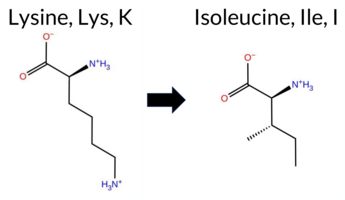 3DClick to see structure in 3D Viewer AIThe SynGAP1 K394I missense variant has no ClinVar entry and is not reported in gnomAD. Prediction tools that agree on a benign effect include premPS, polyPhen‑2 HumVar, and FATHMM, while a majority (seven) predict pathogenicity: SGM‑Consensus (Likely Pathogenic), REVEL, PROVEAN, polyPhen‑2 HumDiv, SIFT, ESM1b, and AlphaMissense‑Default. High‑accuracy methods give mixed results: AlphaMissense‑Optimized is uncertain; the SGM Consensus (majority vote of AlphaMissense‑Default, ESM1b, FATHMM, and PROVEAN) remains Likely Pathogenic; and Foldetta (combining FoldX‑MD and Rosetta outputs) is uncertain. No evidence from these tools contradicts the ClinVar status, which is absent. Overall, the preponderance of pathogenic predictions suggests the variant is most likely pathogenic, with no conflict from ClinVar data. Disclaimer: This summary was generated using AI and should be interpreted alongside expert review. | Likely Pathogenic | C2 | 0.505461 | Disordered | 0.399336 | Uncertain | 0.387 | 0.634 | 0.625 | -9.244 | Likely Pathogenic | 0.876 | Likely Pathogenic | Ambiguous | 0.78 | Ambiguous | 0.2 | 1.10 | Ambiguous | 0.94 | Ambiguous | 0.19 | Likely Benign | 0.519 | Likely Pathogenic | -3.96 | Deleterious | 0.700 | Possibly Damaging | 0.403 | Benign | 4.59 | Benign | 0.00 | Affected | 0.1728 | 0.4123 | -2 | -3 | 8.4 | -15.01 | |||||||||||||||||||||||||||||
| c.1208A>T | K403I 2D 3DClick to see structure in 3D Viewer AIThe SynGAP1 missense variant K403I is not reported in ClinVar (ClinVar ID None) and is absent from gnomAD (gnomAD ID None). Prediction tools that classify the variant as benign include FoldX, Rosetta, Foldetta, premPS, and FATHMM, while the remaining tools—SGM‑Consensus, REVEL, PROVEAN, polyPhen‑2 (HumDiv and HumVar), SIFT, ESM1b, AlphaMissense‑Default, and AlphaMissense‑Optimized—predict it to be pathogenic. High‑accuracy assessments show AlphaMissense‑Optimized as pathogenic, SGM‑Consensus (a majority vote of AlphaMissense‑Default, ESM1b, FATHMM, and PROVEAN) as Likely Pathogenic, and Foldetta (combining FoldX‑MD and Rosetta outputs) as benign. Overall, the majority of predictions (nine pathogenic vs. five benign) and the high‑accuracy tools lean toward a pathogenic effect. Thus, the variant is most likely pathogenic, and this conclusion does not contradict any ClinVar status, as none is available. Disclaimer: This summary was generated using AI and should be interpreted alongside expert review. | Likely Pathogenic | C2 | 0.275179 | Structured | 0.424920 | Uncertain | 0.960 | 0.372 | 0.000 | -15.239 | Likely Pathogenic | 0.997 | Likely Pathogenic | Likely Pathogenic | 0.17 | Likely Benign | 0.1 | -0.46 | Likely Benign | -0.15 | Likely Benign | 0.36 | Likely Benign | 0.519 | Likely Pathogenic | -7.27 | Deleterious | 1.000 | Probably Damaging | 0.999 | Probably Damaging | 3.70 | Benign | 0.00 | Affected | 0.1223 | 0.4141 | -2 | -3 | 8.4 | -15.01 | |||||||||||||||||||||||||||||
| c.1253A>T | K418I 2D 3DClick to see structure in 3D Viewer AIThe SynGAP1 missense variant K418I is not reported in ClinVar (ClinVar ID None) and is absent from gnomAD (gnomAD ID None). Prediction tools that indicate a benign effect include REVEL, premPS, and FATHMM. In contrast, the majority of tools predict a pathogenic outcome: SGM‑Consensus (Likely Pathogenic), PROVEAN, polyPhen‑2 HumDiv, polyPhen‑2 HumVar, SIFT, ESM1b, AlphaMissense‑Default, and AlphaMissense‑Optimized. FoldX, Rosetta, and Foldetta provide uncertain results. High‑accuracy assessments show AlphaMissense‑Optimized as Pathogenic, SGM‑Consensus as Likely Pathogenic, and Foldetta as Uncertain. Overall, the preponderance of evidence points to a pathogenic effect for K418I, and this conclusion is not contradicted by any ClinVar annotation. Disclaimer: This summary was generated using AI and should be interpreted alongside expert review. | Likely Pathogenic | GAP | 0.104810 | Structured | 0.360134 | Uncertain | 0.948 | 0.263 | 0.000 | -14.895 | Likely Pathogenic | 1.000 | Likely Pathogenic | Likely Pathogenic | 0.51 | Ambiguous | 0.0 | 0.64 | Ambiguous | 0.58 | Ambiguous | 0.27 | Likely Benign | 0.428 | Likely Benign | -7.27 | Deleterious | 1.000 | Probably Damaging | 1.000 | Probably Damaging | 3.32 | Benign | 0.01 | Affected | 0.1297 | 0.2432 | -2 | -3 | 8.4 | -15.01 | |||||||||||||||||||||||||||||
| c.1325A>T | K442I 2D 3DClick to see structure in 3D Viewer AIThe SynGAP1 K442I missense variant is not reported in ClinVar (ClinVar ID None) and is absent from gnomAD (gnomAD ID None). Prediction tools that agree on a benign effect include REVEL, FoldX, Rosetta, Foldetta, premPS, and FATHMM. Tools that predict a pathogenic effect are PROVEAN, polyPhen‑2 HumDiv, polyPhen‑2 HumVar, SIFT, ESM1b, AlphaMissense‑Default, and AlphaMissense‑Optimized. The high‑accuracy assessments are: AlphaMissense‑Optimized predicts pathogenic; the SGM Consensus (majority vote of AlphaMissense‑Default, ESM1b, FATHMM, PROVEAN) is Likely Pathogenic; and Foldetta (combining FoldX‑MD and Rosetta outputs) predicts benign. Overall, the majority of predictions, including the two high‑accuracy pathogenic calls, indicate a pathogenic impact. There is no ClinVar entry to contradict this assessment, so the variant is most likely pathogenic. Disclaimer: This summary was generated using AI and should be interpreted alongside expert review. | Likely Pathogenic | GAP | 0.170161 | Structured | 0.255766 | Uncertain | 0.912 | 0.225 | 0.000 | -14.921 | Likely Pathogenic | 0.965 | Likely Pathogenic | Likely Pathogenic | 0.16 | Likely Benign | 0.1 | -0.16 | Likely Benign | 0.00 | Likely Benign | 0.30 | Likely Benign | 0.350 | Likely Benign | -6.91 | Deleterious | 1.000 | Probably Damaging | 0.999 | Probably Damaging | 3.37 | Benign | 0.02 | Affected | 0.0943 | 0.3073 | -2 | -3 | 8.4 | -15.01 | |||||||||||||||||||||||||||||
| c.1475A>T | K492I 2D 3DClick to see structure in 3D Viewer AIThe SynGAP1 missense variant K492I is not reported in ClinVar and is absent from gnomAD. Prediction tools cluster into two groups: benign predictions from premPS and FATHMM; pathogenic predictions from SGM‑Consensus, REVEL, PROVEAN, polyPhen‑2 (HumDiv and HumVar), SIFT, ESM1b, AlphaMissense‑Default, and AlphaMissense‑Optimized. Uncertain results come from FoldX, Rosetta, and Foldetta. High‑accuracy methods give a pathogenic verdict: AlphaMissense‑Optimized predicts pathogenic; the SGM‑Consensus, which aggregates AlphaMissense‑Default, ESM1b, FATHMM, and PROVEAN, also predicts pathogenic (3/4 votes). Foldetta remains inconclusive. Overall, the majority of evidence points to a pathogenic effect, and this assessment does not conflict with the absence of a ClinVar classification. Disclaimer: This summary was generated using AI and should be interpreted alongside expert review. | Likely Pathogenic | GAP | 0.061840 | Structured | 0.327121 | Uncertain | 0.947 | 0.192 | 0.000 | -18.661 | Likely Pathogenic | 0.998 | Likely Pathogenic | Likely Pathogenic | -0.54 | Ambiguous | 0.1 | -0.59 | Ambiguous | -0.57 | Ambiguous | 0.49 | Likely Benign | 0.645 | Likely Pathogenic | -7.96 | Deleterious | 1.000 | Probably Damaging | 1.000 | Probably Damaging | 2.94 | Benign | 0.00 | Affected | 0.0875 | 0.2810 | -2 | -3 | 8.4 | -15.01 | |||||||||||||||||||||||||||||
| c.1511A>T | K504I 2D 3DClick to see structure in 3D Viewer AIThe SynGAP1 missense variant K504I is not reported in ClinVar and is absent from gnomAD. Prediction tools that assess the variant’s effect fall into two groups: benign predictions include REVEL, FoldX, Rosetta, Foldetta, premPS, and AlphaMissense‑Optimized; pathogenic predictions include PROVEAN, polyPhen‑2 (HumDiv and HumVar), SIFT, ESM1b, FATHMM, and AlphaMissense‑Default. The SGM Consensus, derived from a majority vote of AlphaMissense‑Default, ESM1b, FATHMM, and PROVEAN, labels the variant as Likely Pathogenic. High‑accuracy assessments show AlphaMissense‑Optimized as benign, Foldetta as benign, and the SGM Consensus as pathogenic. Because the majority of tools (seven) predict pathogenicity while three high‑accuracy methods provide conflicting evidence, the overall prediction leans toward pathogenic. This assessment does not contradict ClinVar status, as no ClinVar entry exists for the variant. Disclaimer: This summary was generated using AI and should be interpreted alongside expert review. | Likely Pathogenic | GAP | 0.028107 | Structured | 0.304984 | Uncertain | 0.850 | 0.189 | 0.000 | -12.597 | Likely Pathogenic | 0.727 | Likely Pathogenic | Likely Benign | -0.04 | Likely Benign | 0.3 | -0.36 | Likely Benign | -0.20 | Likely Benign | 0.40 | Likely Benign | 0.491 | Likely Benign | -7.35 | Deleterious | 0.996 | Probably Damaging | 0.993 | Probably Damaging | -1.49 | Pathogenic | 0.02 | Affected | 0.0769 | 0.2713 | -2 | -3 | 8.4 | -15.01 | |||||||||||||||||||||||||||||
| c.239A>T | K80I 2D AIThe SynGAP1 missense variant K80I is not reported in ClinVar (ClinVar ID = None) and is absent from gnomAD (gnomAD ID = None). Prediction tools that agree on a benign effect include REVEL, polyPhen‑2 HumVar, ESM1b, and FATHMM, while those that agree on a pathogenic effect are PROVEAN, polyPhen‑2 HumDiv, SIFT, and AlphaMissense‑Default. AlphaMissense‑Optimized is uncertain. High‑accuracy assessments are inconclusive: AlphaMissense‑Optimized remains uncertain, the SGM Consensus (majority vote of AlphaMissense‑Default, ESM1b, FATHMM, PROVEAN) is a 2‑vs‑2 tie, and Foldetta data are unavailable. Consequently, the variant’s predicted impact is ambiguous, with an equal split between benign and pathogenic signals and no ClinVar entry to contradict the computational assessment. Disclaimer: This summary was generated using AI and should be interpreted alongside expert review. | 0.637480 | Disordered | 0.477530 | Uncertain | 0.331 | 0.873 | 0.500 | -5.320 | Likely Benign | 0.947 | Likely Pathogenic | Ambiguous | 0.083 | Likely Benign | -2.54 | Deleterious | 0.939 | Possibly Damaging | 0.164 | Benign | 3.86 | Benign | 0.00 | Affected | 0.1095 | 0.2981 | -2 | -3 | 8.4 | -15.01 | ||||||||||||||||||||||||||||||||||||||||
| c.2408A>T | K803I 2D AIThe SynGAP1 K803I missense variant is not reported in ClinVar (ClinVar ID None) and is absent from gnomAD (gnomAD ID None). Prediction tools that agree on a benign effect include REVEL and ESM1b, whereas the majority of tools predict a pathogenic impact: PROVEAN, polyPhen‑2 (HumDiv and HumVar), SIFT, FATHMM, AlphaMissense‑Default, and the SGM‑Consensus score. The high‑accuracy AlphaMissense‑Optimized assessment is uncertain, and the SGM‑Consensus (derived from a majority vote of AlphaMissense‑Default, ESM1b, FATHMM, and PROVEAN) indicates a likely pathogenic outcome. Foldetta, a protein‑folding stability method combining FoldX‑MD and Rosetta outputs, has no available result for this variant. Overall, the preponderance of evidence points to a pathogenic effect, and this conclusion does not contradict the ClinVar status, which currently contains no classification for K803I. Disclaimer: This summary was generated using AI and should be interpreted alongside expert review. | Likely Pathogenic | SH3-binding motif | 0.827927 | Disordered | 0.733908 | Binding | 0.349 | 0.900 | 0.625 | -5.207 | Likely Benign | 0.894 | Likely Pathogenic | Ambiguous | 0.196 | Likely Benign | -4.06 | Deleterious | 0.995 | Probably Damaging | 0.913 | Probably Damaging | 2.31 | Pathogenic | 0.00 | Affected | 0.1425 | 0.3889 | -2 | -3 | 8.4 | -15.01 | ||||||||||||||||||||||||||||||||||||||
| c.3248A>T | K1083I 2D AIThe SynGAP1 missense variant K1083I is not reported in ClinVar (no ClinVar ID) and is absent from gnomAD (no gnomAD ID). Prediction tools that agree on a benign effect include REVEL, PROVEAN, SIFT, ESM1b, and FATHMM, while those that predict a pathogenic effect are polyPhen‑2 HumDiv, polyPhen‑2 HumVar, and AlphaMissense‑Default. The SGM‑Consensus, which aggregates AlphaMissense‑Default, ESM1b, FATHMM, and PROVEAN, reports the variant as Likely Benign. High‑accuracy assessments show AlphaMissense‑Optimized as Uncertain, SGM‑Consensus as Likely Benign, and the Foldetta stability analysis is unavailable. Overall, the majority of evidence points to a benign impact. This conclusion is consistent with the lack of a ClinVar entry, so there is no contradiction with ClinVar status. Disclaimer: This summary was generated using AI and should be interpreted alongside expert review. | Likely Benign | 0.837511 | Disordered | 0.978906 | Binding | 0.302 | 0.893 | 1.000 | -3.207 | Likely Benign | 0.836 | Likely Pathogenic | Ambiguous | 0.239 | Likely Benign | -1.65 | Neutral | 0.999 | Probably Damaging | 0.997 | Probably Damaging | 4.02 | Benign | 0.30 | Tolerated | 0.1394 | 0.3978 | -2 | -3 | 8.4 | -15.01 | |||||||||||||||||||||||||||||||||||||||
| c.3554A>T | K1185I 2D AIThe SynGAP1 missense variant K1185I is listed in ClinVar with no pathogenicity classification (ClinVar status: None) and is present in the gnomAD database (gnomAD ID: 6‑33444589‑A‑T). Prediction tools that agree on a benign effect include REVEL, SIFT, ESM1b, and FATHMM, while those that predict a pathogenic effect are PROVEAN, polyPhen‑2 HumDiv, polyPhen‑2 HumVar, AlphaMissense‑Default, and AlphaMissense‑Optimized. High‑accuracy assessments show AlphaMissense‑Optimized as pathogenic; the SGM Consensus (majority vote from AlphaMissense‑Default, ESM1b, FATHMM, PROVEAN) is inconclusive due to a 2‑vs‑2 split, and Foldetta results are unavailable. Based on the overall pattern of predictions, the variant is most likely pathogenic, and this assessment does not contradict the ClinVar status, which remains unclassified. Disclaimer: This summary was generated using AI and should be interpreted alongside expert review. | Coiled-coil | 0.566480 | Disordered | 0.510264 | Binding | 0.642 | 0.638 | 0.625 | 6-33444589-A-T | 1 | 6.20e-7 | -5.101 | Likely Benign | 0.990 | Likely Pathogenic | Likely Pathogenic | 0.215 | Likely Benign | -3.42 | Deleterious | 0.999 | Probably Damaging | 0.997 | Probably Damaging | 2.62 | Benign | 0.09 | Tolerated | 3.82 | 4 | 0.1154 | 0.3108 | -3 | -2 | 8.4 | -15.01 | ||||||||||||||||||||||||||||||||||
| c.3617A>T | K1206I 2D AIThe SynGAP1 missense variant K1206I is not reported in ClinVar and is absent from gnomAD. Prediction tools that indicate a benign effect include only REVEL, whereas the remaining tools—PROVEAN, polyPhen‑2 (HumDiv and HumVar), SIFT, ESM1b, FATHMM, AlphaMissense‑Default, and AlphaMissense‑Optimized—consistently predict a pathogenic impact. The SGM‑Consensus, derived from a majority vote of AlphaMissense‑Default, ESM1b, FATHMM, and PROVEAN, also classifies the variant as Likely Pathogenic. High‑accuracy assessments show AlphaMissense‑Optimized as Pathogenic and the SGM‑Consensus as Likely Pathogenic; Foldetta results are not available. Overall, the preponderance of evidence from multiple in silico predictors points to a pathogenic effect for K1206I. This conclusion is not contradicted by ClinVar status, which currently has no entry for this variant. Disclaimer: This summary was generated using AI and should be interpreted alongside expert review. | Likely Pathogenic | Coiled-coil | 0.585406 | Disordered | 0.555819 | Binding | 0.893 | 0.569 | 0.375 | -13.526 | Likely Pathogenic | 0.997 | Likely Pathogenic | Likely Pathogenic | 0.302 | Likely Benign | -5.65 | Deleterious | 1.000 | Probably Damaging | 0.999 | Probably Damaging | 2.37 | Pathogenic | 0.01 | Affected | 0.0816 | 0.3521 | -2 | -3 | 8.4 | -15.01 | ||||||||||||||||||||||||||||||||||||||
| c.3695A>T | K1232I 2D AIThe SynGAP1 K1232I missense variant is not reported in ClinVar (ClinVar ID None) and is absent from gnomAD (gnomAD ID None). Prediction tools that agree on a benign effect include only REVEL, whereas the remaining tools—PROVEAN, polyPhen‑2 (HumDiv and HumVar), SIFT, ESM1b, FATHMM, and AlphaMissense‑Default—consistently predict a pathogenic impact. The high‑accuracy assessment shows AlphaMissense‑Optimized as uncertain, while the SGM‑Consensus (derived from the majority vote of AlphaMissense‑Default, ESM1b, FATHMM, and PROVEAN) is classified as Likely Pathogenic. Foldetta, a protein‑folding stability method combining FoldX‑MD and Rosetta outputs, has no available result for this variant. Overall, the preponderance of evidence points to a pathogenic effect; this conclusion is not contradicted by any ClinVar annotation. Disclaimer: This summary was generated using AI and should be interpreted alongside expert review. | Likely Pathogenic | Coiled-coil | 0.505461 | Disordered | 0.542907 | Binding | 0.894 | 0.535 | 0.125 | -12.225 | Likely Pathogenic | 0.896 | Likely Pathogenic | Ambiguous | 0.197 | Likely Benign | -5.98 | Deleterious | 1.000 | Probably Damaging | 0.999 | Probably Damaging | 2.08 | Pathogenic | 0.00 | Affected | 0.0778 | 0.3321 | -2 | -3 | 8.4 | -15.01 | ||||||||||||||||||||||||||||||||||||||
| c.413A>T | K138I 2D AIThe SynGAP1 missense variant K138I is not reported in ClinVar (no ClinVar ID) and is absent from gnomAD (no gnomAD ID). Prediction tools that agree on a benign effect include REVEL, polyPhen‑2 HumVar, and FATHMM, whereas those that predict a pathogenic effect comprise PROVEAN, polyPhen‑2 HumDiv, SIFT, ESM1b, AlphaMissense‑Default, and AlphaMissense‑Optimized. The SGM‑Consensus, derived from a majority vote of AlphaMissense‑Default, ESM1b, FATHMM, and PROVEAN, classifies the variant as Likely Pathogenic. High‑accuracy assessments further support a deleterious outcome: AlphaMissense‑Optimized predicts pathogenicity, and the SGM‑Consensus also indicates Likely Pathogenic. The Foldetta protein‑folding stability analysis is unavailable for this variant. Overall, the majority of computational evidence points to a pathogenic effect, and this conclusion is not contradicted by any ClinVar annotation. Disclaimer: This summary was generated using AI and should be interpreted alongside expert review. | Likely Pathogenic | 0.590140 | Disordered | 0.619482 | Binding | 0.349 | 0.901 | 0.375 | -9.366 | Likely Pathogenic | 0.999 | Likely Pathogenic | Likely Pathogenic | 0.295 | Likely Benign | -4.74 | Deleterious | 0.535 | Possibly Damaging | 0.259 | Benign | 3.53 | Benign | 0.00 | Affected | 0.1003 | 0.3054 | -2 | -3 | 8.4 | -15.01 | |||||||||||||||||||||||||||||||||||||||
| c.425A>T | K142I 2D AIThe SynGAP1 missense variant K142I is not reported in ClinVar (no ClinVar ID) and is absent from gnomAD (no gnomAD ID). Prediction tools that agree on a benign effect include REVEL, polyPhen‑2 HumVar, and FATHMM, whereas those that predict a pathogenic effect comprise PROVEAN, polyPhen‑2 HumDiv, SIFT, ESM1b, AlphaMissense‑Default, and AlphaMissense‑Optimized. The SGM‑Consensus, derived from a majority vote of AlphaMissense‑Default, ESM1b, FATHMM, and PROVEAN, classifies the variant as Likely Pathogenic. High‑accuracy assessments further support a deleterious impact: AlphaMissense‑Optimized predicts Pathogenic, and the SGM‑Consensus also indicates Likely Pathogenic. The Foldetta protein‑folding stability analysis is unavailable for this variant. Overall, the majority of computational evidence points to a pathogenic effect, and this conclusion is not contradicted by any ClinVar annotation. Disclaimer: This summary was generated using AI and should be interpreted alongside expert review. | Likely Pathogenic | 0.461924 | Structured | 0.558796 | Binding | 0.374 | 0.859 | 0.500 | -14.597 | Likely Pathogenic | 0.998 | Likely Pathogenic | Likely Pathogenic | 0.296 | Likely Benign | -4.81 | Deleterious | 0.700 | Possibly Damaging | 0.403 | Benign | 3.44 | Benign | 0.00 | Affected | 0.1005 | 0.3173 | -2 | -3 | 8.4 | -15.01 | |||||||||||||||||||||||||||||||||||||||
| c.446A>T | K149I 2D AIThe SynGAP1 missense variant K149I is not reported in ClinVar (no ClinVar ID) and is absent from gnomAD (no gnomAD ID). Prediction tools that indicate a benign effect include REVEL, polyPhen‑2 HumVar, and FATHMM, whereas those that predict a pathogenic effect comprise PROVEAN, polyPhen‑2 HumDiv, SIFT, ESM1b, AlphaMissense‑Default, and AlphaMissense‑Optimized. The SGM‑Consensus, derived from a majority vote of AlphaMissense‑Default, ESM1b, FATHMM, and PROVEAN, classifies the variant as Likely Pathogenic. High‑accuracy assessments show AlphaMissense‑Optimized as Pathogenic and the SGM‑Consensus as Likely Pathogenic; the Foldetta protein‑folding stability analysis is unavailable. Based on the preponderance of pathogenic predictions and the lack of benign consensus, the variant is most likely pathogenic, and this assessment does not contradict any ClinVar status because none is available. Disclaimer: This summary was generated using AI and should be interpreted alongside expert review. | Likely Pathogenic | 0.562014 | Disordered | 0.501681 | Binding | 0.302 | 0.839 | 0.625 | -14.426 | Likely Pathogenic | 0.995 | Likely Pathogenic | Likely Pathogenic | 0.305 | Likely Benign | -4.72 | Deleterious | 0.535 | Possibly Damaging | 0.403 | Benign | 3.53 | Benign | 0.00 | Affected | 0.1477 | 0.3965 | -2 | -3 | 8.4 | -15.01 | |||||||||||||||||||||||||||||||||||||||
| c.686A>T | K229I 2D 3DClick to see structure in 3D Viewer AISynGAP1 missense variant K229I is not reported in ClinVar and is absent from gnomAD. Functional prediction tools show a split assessment: benign predictions come from FoldX, Foldetta, premPS, and FATHMM, whereas pathogenic predictions are returned by SGM‑Consensus, REVEL, PROVEAN, polyPhen‑2 (HumDiv and HumVar), SIFT, ESM1b, AlphaMissense‑Default, and AlphaMissense‑Optimized. High‑accuracy methods give a mixed picture: AlphaMissense‑Optimized predicts pathogenic; the SGM‑Consensus, derived from a majority vote of AlphaMissense‑Default, ESM1b, FATHMM, and PROVEAN, also indicates pathogenic; Foldetta, which integrates FoldX‑MD and Rosetta stability outputs, reports benign. No prediction is inconclusive. Overall, the majority of tools, including the high‑accuracy ones, lean toward pathogenicity, and this does not contradict any ClinVar annotation because none exists. Disclaimer: This summary was generated using AI and should be interpreted alongside expert review. | Likely Pathogenic | PH | 0.179055 | Structured | 0.310912 | Uncertain | 0.843 | 0.306 | 0.000 | -15.276 | Likely Pathogenic | 0.998 | Likely Pathogenic | Likely Pathogenic | -0.03 | Likely Benign | 0.1 | -0.63 | Ambiguous | -0.33 | Likely Benign | -0.19 | Likely Benign | 0.833 | Likely Pathogenic | -6.52 | Deleterious | 0.998 | Probably Damaging | 0.991 | Probably Damaging | 5.92 | Benign | 0.00 | Affected | 0.1202 | 0.3711 | -2 | -3 | 8.4 | -15.01 | |||||||||||||||||||||||||||||
| c.722A>T | K241I 2D 3DClick to see structure in 3D Viewer AISynGAP1 K241I is not reported in ClinVar and is absent from gnomAD. Prediction tools that classify the variant as benign include Rosetta, premPS, and FATHMM. Those that predict a damaging effect are REVEL, PROVEAN, polyPhen‑2 (HumDiv and HumVar), SIFT, ESM1b, AlphaMissense‑Default, AlphaMissense‑Optimized, and the SGM‑Consensus score (Likely Pathogenic). FoldX and Foldetta return uncertain results. High‑accuracy methods give a consistent pathogenic signal: AlphaMissense‑Optimized reports pathogenic, the SGM‑Consensus (majority vote of AlphaMissense‑Default, ESM1b, FATHMM, PROVEAN) indicates likely pathogenic, while Foldetta remains inconclusive. Overall, the majority of evidence points to a pathogenic effect, and this assessment does not contradict any ClinVar annotation because none exists. Disclaimer: This summary was generated using AI and should be interpreted alongside expert review. | Likely Pathogenic | PH | 0.196879 | Structured | 0.349250 | Uncertain | 0.797 | 0.347 | 0.000 | -14.370 | Likely Pathogenic | 0.995 | Likely Pathogenic | Likely Pathogenic | 1.31 | Ambiguous | 0.5 | 0.28 | Likely Benign | 0.80 | Ambiguous | 0.41 | Likely Benign | 0.852 | Likely Pathogenic | -6.96 | Deleterious | 0.985 | Probably Damaging | 0.704 | Possibly Damaging | 5.71 | Benign | 0.01 | Affected | 0.1176 | 0.3868 | -2 | -3 | 8.4 | -15.01 | |||||||||||||||||||||||||||||
| c.998A>T | K333I 2D 3DClick to see structure in 3D Viewer AIThe SynGAP1 K333I missense variant is not reported in ClinVar and is absent from gnomAD. Prediction tools that agree on a benign effect include FoldX and premPS, whereas the majority of tools (REVEL, PROVEAN, polyPhen‑2 HumDiv, polyPhen‑2 HumVar, SIFT, ESM1b, FATHMM, AlphaMissense‑Default, AlphaMissense‑Optimized) predict a pathogenic impact; Rosetta and Foldetta give uncertain results. High‑accuracy assessments further support pathogenicity: AlphaMissense‑Optimized classifies the variant as pathogenic, the SGM Consensus (majority vote from AlphaMissense‑Default, ESM1b, FATHMM, PROVEAN) reports it as Likely Pathogenic, and Foldetta remains uncertain. Overall, the preponderance of evidence indicates that the variant is most likely pathogenic, and this conclusion does not contradict the ClinVar status, which currently has no entry for this variant. Disclaimer: This summary was generated using AI and should be interpreted alongside expert review. | Likely Pathogenic | C2 | 0.311707 | Structured | 0.330781 | Uncertain | 0.537 | 0.447 | 0.500 | -14.517 | Likely Pathogenic | 0.985 | Likely Pathogenic | Likely Pathogenic | 0.30 | Likely Benign | 0.0 | 0.95 | Ambiguous | 0.63 | Ambiguous | 0.43 | Likely Benign | 0.544 | Likely Pathogenic | -6.49 | Deleterious | 1.000 | Probably Damaging | 0.999 | Probably Damaging | 1.89 | Pathogenic | 0.03 | Affected | 0.0931 | 0.3521 | -2 | -3 | 8.4 | -15.01 | |||||||||||||||||||||||||||||
| c.1082A>T | Q361L 2D  3DClick to see structure in 3D Viewer AISynGAP1 missense variant Q361L is not reported in ClinVar and is absent from gnomAD. Functional prediction tools cluster into two groups: benign predictions come from REVEL, FoldX, Rosetta, Foldetta, premPS, SIFT, AlphaMissense‑Default, and AlphaMissense‑Optimized; pathogenic predictions arise from PROVEAN, polyPhen‑2 HumDiv, polyPhen‑2 HumVar, ESM1b, and FATHMM. High‑accuracy assessments show AlphaMissense‑Optimized as benign, the SGM Consensus (majority vote of AlphaMissense‑Default, ESM1b, FATHMM, PROVEAN) as pathogenic, and Foldetta (combining FoldX‑MD and Rosetta outputs) as benign. Overall, the majority of tools (8 benign vs 5 pathogenic) favor a benign effect, and this consensus does not contradict the absence of a ClinVar classification. Thus, the variant is most likely benign based on current predictions, with no conflict with ClinVar status. Disclaimer: This summary was generated using AI and should be interpreted alongside expert review. | Likely Pathogenic | C2 | 0.305330 | Structured | 0.427593 | Uncertain | 0.945 | 0.534 | 0.250 | -10.678 | Likely Pathogenic | 0.238 | Likely Benign | Likely Benign | 0.12 | Likely Benign | 0.1 | 0.46 | Likely Benign | 0.29 | Likely Benign | 0.21 | Likely Benign | 0.406 | Likely Benign | -3.95 | Deleterious | 0.987 | Probably Damaging | 0.953 | Probably Damaging | 1.71 | Pathogenic | 0.06 | Tolerated | 0.0720 | 0.5213 | -2 | -2 | 7.3 | -14.97 | |||||||||||||||||||||||||||||
| c.1220A>T | Q407L 2D 3DClick to see structure in 3D Viewer AIThe SynGAP1 missense variant Q407L is not reported in ClinVar (ClinVar ID None) and is absent from gnomAD (gnomAD ID None). Prediction tools that agree on a benign effect include REVEL, premPS, FATHMM, and AlphaMissense‑Optimized. Those that agree on a pathogenic effect are PROVEAN, polyPhen‑2 (HumDiv and HumVar), SIFT, and ESM1b. Four tools are uncertain (AlphaMissense‑Default, FoldX, Rosetta, Foldetta). High‑accuracy assessments show AlphaMissense‑Optimized as benign, the SGM Consensus (majority vote from AlphaMissense‑Default, ESM1b, FATHMM, PROVEAN) as pathogenic, and Foldetta as uncertain. Overall, more tools predict pathogenicity than benign, and the high‑accuracy consensus leans pathogenic. Therefore, the variant is most likely pathogenic, and this assessment does not contradict the ClinVar status, which has no entry for this variant. Disclaimer: This summary was generated using AI and should be interpreted alongside expert review. | C2 | 0.109221 | Structured | 0.382522 | Uncertain | 0.916 | 0.271 | 0.000 | -12.730 | Likely Pathogenic | 0.558 | Ambiguous | Likely Benign | -0.65 | Ambiguous | 0.2 | -0.69 | Ambiguous | -0.67 | Ambiguous | 0.35 | Likely Benign | 0.359 | Likely Benign | -6.32 | Deleterious | 0.939 | Possibly Damaging | 0.838 | Possibly Damaging | 3.91 | Benign | 0.02 | Affected | 0.0585 | 0.4977 | -2 | -2 | 7.3 | -14.97 | ||||||||||||||||||||||||||||||
| c.1367A>T | Q456L 2D 3DClick to see structure in 3D Viewer AIThe SynGAP1 missense variant Q456L is not reported in ClinVar (ClinVar ID None) and is absent from gnomAD (gnomAD ID None). Prediction tools that agree on a benign effect include REVEL, Rosetta, premPS, SIFT, FATHMM, and AlphaMissense‑Optimized. Those that predict a pathogenic effect are PROVEAN, polyPhen‑2 HumDiv, polyPhen‑2 HumVar, and ESM1b. Tools with uncertain or inconclusive results are FoldX, Foldetta, and AlphaMissense‑Default. High‑accuracy assessments show AlphaMissense‑Optimized as benign, the SGM Consensus (majority vote from AlphaMissense‑Default, ESM1b, FATHMM, PROVEAN) as pathogenic, and Foldetta as uncertain. Overall, the majority of predictions (six benign vs. four pathogenic) lean toward a benign interpretation, with no evidence of contradiction with the ClinVar status. Thus, the variant is most likely benign based on current computational predictions. Disclaimer: This summary was generated using AI and should be interpreted alongside expert review. | GAP | 0.170161 | Structured | 0.302348 | Uncertain | 0.939 | 0.164 | 0.000 | -10.272 | Likely Pathogenic | 0.492 | Ambiguous | Likely Benign | -0.88 | Ambiguous | 0.1 | -0.30 | Likely Benign | -0.59 | Ambiguous | 0.28 | Likely Benign | 0.347 | Likely Benign | -6.65 | Deleterious | 0.987 | Probably Damaging | 0.914 | Probably Damaging | 3.49 | Benign | 0.36 | Tolerated | 0.0554 | 0.4317 | -2 | -2 | 7.3 | -14.97 | ||||||||||||||||||||||||||||||
| c.1508A>T | Q503L 2D 3DClick to see structure in 3D Viewer AIThe SynGAP1 missense variant Q503L is not reported in ClinVar and has no entries in gnomAD. Prediction tools that agree on a benign effect include AlphaMissense‑Default, AlphaMissense‑Optimized, Foldetta, premPS, Rosetta, and polyPhen‑2 (HumVar). Those that predict a pathogenic effect are SGM‑Consensus, REVEL, PROVEAN, polyPhen‑2 (HumDiv), SIFT, ESM1b, and FATHMM; FoldX is uncertain and therefore not counted. High‑accuracy assessments show AlphaMissense‑Optimized as benign, SGM‑Consensus (a majority vote of AlphaMissense‑Default, ESM1b, FATHMM, and PROVEAN) as likely pathogenic, and Foldetta as benign. Overall, the majority of tools (seven versus six) predict pathogenicity, and the high‑accuracy trio is split but leans toward pathogenic. Thus, the variant is most likely pathogenic based on current predictions, and this assessment does not contradict any ClinVar status (none reported). Disclaimer: This summary was generated using AI and should be interpreted alongside expert review. | Likely Pathogenic | GAP | 0.040537 | Structured | 0.322935 | Uncertain | 0.848 | 0.168 | 0.000 | -8.203 | Likely Pathogenic | 0.261 | Likely Benign | Likely Benign | -0.56 | Ambiguous | 0.2 | -0.07 | Likely Benign | -0.32 | Likely Benign | 0.24 | Likely Benign | 0.711 | Likely Pathogenic | -6.29 | Deleterious | 0.911 | Possibly Damaging | 0.369 | Benign | -1.52 | Pathogenic | 0.05 | Affected | 0.0715 | 0.3578 | -2 | -2 | 7.3 | -14.97 | |||||||||||||||||||||||||||||
| c.1619A>T | Q540L 2D 3DClick to see structure in 3D Viewer AISynGAP1 missense variant Q540L has no ClinVar entry and is not reported in gnomAD. Functional prediction tools show a split: benign calls come from Rosetta, Foldetta, premPS, and SIFT, while pathogenic calls are made by REVEL, PROVEAN, polyPhen‑2 (HumDiv and HumVar), ESM1b, FATHMM, AlphaMissense‑Default, and AlphaMissense‑Optimized. FoldX is uncertain and therefore not considered. High‑accuracy assessments further support a deleterious effect: AlphaMissense‑Optimized predicts pathogenic, the SGM Consensus (majority vote of AlphaMissense‑Default, ESM1b, FATHMM, and PROVEAN) is Likely Pathogenic, whereas Foldetta, which integrates FoldX‑MD and Rosetta stability outputs, predicts benign. Overall, the majority of evidence points to a pathogenic impact for Q540L, and this conclusion does not contradict the absence of a ClinVar classification. Disclaimer: This summary was generated using AI and should be interpreted alongside expert review. | Likely Pathogenic | GAP | 0.085092 | Structured | 0.029522 | Uncertain | 0.958 | 0.371 | 0.000 | -14.266 | Likely Pathogenic | 0.965 | Likely Pathogenic | Likely Pathogenic | -0.71 | Ambiguous | 0.1 | 0.44 | Likely Benign | -0.14 | Likely Benign | 0.50 | Likely Benign | 0.756 | Likely Pathogenic | -6.96 | Deleterious | 0.994 | Probably Damaging | 0.977 | Probably Damaging | -1.06 | Pathogenic | 0.08 | Tolerated | 0.0654 | 0.3601 | -2 | -2 | 7.3 | -14.97 | |||||||||||||||||||||||||||||
| c.164A>T | Q55L 2D AIThe SynGAP1 missense variant Q55L is not reported in ClinVar and is absent from gnomAD. Prediction tools that agree on a benign effect include REVEL, PROVEAN, polyPhen‑2 (HumDiv and HumVar), ESM1b, and FATHMM. Tools that predict a pathogenic effect are SIFT and AlphaMissense‑Default. AlphaMissense‑Optimized is uncertain, and the SGM‑Consensus (majority vote of AlphaMissense‑Default, ESM1b, FATHMM, and PROVEAN) reports the variant as likely benign. Foldetta, a protein‑folding stability method combining FoldX‑MD and Rosetta outputs, has no available result for this variant. Overall, the majority of evidence points to a benign impact, and this assessment does not contradict any ClinVar annotation (none exists). Thus, the variant is most likely benign based on current predictive data. Disclaimer: This summary was generated using AI and should be interpreted alongside expert review. | Likely Benign | 0.332115 | Structured | 0.470108 | Uncertain | 0.461 | 0.657 | 0.000 | -5.823 | Likely Benign | 0.844 | Likely Pathogenic | Ambiguous | 0.173 | Likely Benign | -1.99 | Neutral | 0.273 | Benign | 0.275 | Benign | 3.83 | Benign | 0.00 | Affected | 0.0776 | 0.5982 | -2 | -2 | 7.3 | -14.97 | |||||||||||||||||||||||||||||||||||||||
| c.1835A>T | Q612L 2D 3DClick to see structure in 3D Viewer AIThe SynGAP1 missense variant Q612L is not reported in ClinVar (ClinVar ID None) and is absent from gnomAD (gnomAD ID None). Prediction tools that agree on a benign effect include FoldX, Rosetta, Foldetta, premPS, and SIFT, whereas those that predict a pathogenic effect are REVEL, PROVEAN, polyPhen‑2 HumDiv, polyPhen‑2 HumVar, ESM1b, FATHMM, and AlphaMissense‑Default. The SGM Consensus, derived from a majority vote of AlphaMissense‑Default, ESM1b, FATHMM, and PROVEAN, indicates a likely pathogenic outcome. High‑accuracy assessments show AlphaMissense‑Optimized as uncertain (unavailable), SGM Consensus as likely pathogenic, and Foldetta (a protein‑folding stability method combining FoldX‑MD and Rosetta) as benign. Overall, the majority of conventional tools lean toward pathogenicity, and the SGM Consensus supports this, while the high‑accuracy Foldetta result is contradictory. Thus, the variant is most likely pathogenic based on the prevailing predictions, and this assessment does not contradict the ClinVar status, which currently has no entry for this variant. Disclaimer: This summary was generated using AI and should be interpreted alongside expert review. | Likely Pathogenic | GAP | 0.275179 | Structured | 0.203988 | Uncertain | 0.822 | 0.263 | 0.000 | -12.076 | Likely Pathogenic | 0.799 | Likely Pathogenic | Ambiguous | -0.12 | Likely Benign | 0.1 | 0.12 | Likely Benign | 0.00 | Likely Benign | 0.44 | Likely Benign | 0.730 | Likely Pathogenic | -6.84 | Deleterious | 0.971 | Probably Damaging | 0.954 | Probably Damaging | -1.33 | Pathogenic | 0.08 | Tolerated | 0.0763 | 0.4756 | -2 | -2 | 7.3 | -14.97 | |||||||||||||||||||||||||||||
| c.1853A>T | Q618L 2D 3DClick to see structure in 3D Viewer AIThe SynGAP1 missense variant Q618L is not reported in ClinVar (ClinVar status: none) and is absent from gnomAD (gnomAD ID: none). Prediction tools that agree on a benign effect include REVEL, FoldX, Rosetta, Foldetta, premPS, SIFT, and polyPhen‑2 HumVar, while those that predict a pathogenic effect are SGM‑Consensus (Likely Pathogenic), PROVEAN, polyPhen‑2 HumDiv, ESM1b, and FATHMM. AlphaMissense‑Default is uncertain, whereas AlphaMissense‑Optimized predicts benign. High‑accuracy methods give the following results: AlphaMissense‑Optimized – benign; SGM‑Consensus (majority vote from AlphaMissense‑Default, ESM1b, FATHMM, PROVEAN) – Likely Pathogenic; Foldetta – benign. Overall, the majority of tools (nine benign vs. five pathogenic) predict a benign impact. Thus, the variant is most likely benign based on current predictions, and this assessment does not contradict the ClinVar status, which is currently unreported. Disclaimer: This summary was generated using AI and should be interpreted alongside expert review. | Likely Pathogenic | GAP | 0.175930 | Structured | 0.138725 | Uncertain | 0.904 | 0.240 | 0.000 | -8.561 | Likely Pathogenic | 0.423 | Ambiguous | Likely Benign | -0.07 | Likely Benign | 0.1 | 0.20 | Likely Benign | 0.07 | Likely Benign | 0.31 | Likely Benign | 0.479 | Likely Benign | -3.94 | Deleterious | 0.712 | Possibly Damaging | 0.268 | Benign | -1.28 | Pathogenic | 0.09 | Tolerated | 0.0612 | 0.4146 | -2 | -2 | 7.3 | -14.97 | |||||||||||||||||||||||||||||
| c.1892A>T | Q631L 2D 3DClick to see structure in 3D Viewer AIThe SynGAP1 missense variant Q631L is not reported in ClinVar (ClinVar status: None) and has no entry in gnomAD (gnomAD status: None). Prediction tools that agree on a benign effect include FATHMM and Foldetta, whereas the majority of tools predict a pathogenic impact: SGM‑Consensus (likely pathogenic), REVEL, PROVEAN, polyPhen‑2 (HumDiv and HumVar), SIFT, ESM1b, and AlphaMissense‑Default. High‑accuracy assessments show AlphaMissense‑Optimized as uncertain, SGM‑Consensus as likely pathogenic, and Foldetta as benign; these results are reported but not used as definitive evidence when inconclusive. Overall, the preponderance of evidence from consensus and individual predictors indicates a pathogenic effect for Q631L. This conclusion does not contradict ClinVar status, as no ClinVar classification exists for this variant. Disclaimer: This summary was generated using AI and should be interpreted alongside expert review. | Likely Pathogenic | GAP | 0.041405 | Structured | 0.038963 | Uncertain | 0.948 | 0.230 | 0.000 | -14.727 | Likely Pathogenic | 0.901 | Likely Pathogenic | Ambiguous | -1.23 | Ambiguous | 0.0 | 0.95 | Ambiguous | -0.14 | Likely Benign | 0.51 | Ambiguous | 0.619 | Likely Pathogenic | -6.97 | Deleterious | 0.982 | Probably Damaging | 0.954 | Probably Damaging | 2.85 | Benign | 0.05 | Affected | 0.0627 | 0.3150 | -2 | -2 | 7.3 | -14.97 | |||||||||||||||||||||||||||||
| c.1982A>T | Q661L 2D 3DClick to see structure in 3D Viewer AIThe SynGAP1 missense variant Q661L is not reported in ClinVar and is absent from gnomAD. Prediction tools that agree on a benign effect include REVEL, FoldX, premPS, FATHMM, and AlphaMissense‑Optimized. Tools that predict a pathogenic effect comprise SGM‑Consensus, PROVEAN, polyPhen‑2 (HumDiv and HumVar), SIFT, ESM1b, and AlphaMissense‑Default. High‑accuracy assessments show AlphaMissense‑Optimized as benign, the SGM Consensus (majority vote of AlphaMissense‑Default, ESM1b, FATHMM, and PROVEAN) as likely pathogenic, and Foldetta (combining FoldX‑MD and Rosetta outputs) as uncertain, so it is not used as evidence. Overall, the majority of predictions support a pathogenic impact, and this is consistent with the lack of ClinVar annotation. Therefore, the variant is most likely pathogenic, with no contradiction from ClinVar status. Disclaimer: This summary was generated using AI and should be interpreted alongside expert review. | Likely Pathogenic | GAP | 0.048328 | Structured | 0.117089 | Uncertain | 0.924 | 0.309 | 0.000 | -9.003 | Likely Pathogenic | 0.613 | Likely Pathogenic | Likely Benign | -0.19 | Likely Benign | 0.1 | -1.12 | Ambiguous | -0.66 | Ambiguous | 0.36 | Likely Benign | 0.391 | Likely Benign | -5.11 | Deleterious | 0.976 | Probably Damaging | 0.567 | Possibly Damaging | 3.49 | Benign | 0.02 | Affected | 0.0834 | 0.4675 | -2 | -2 | 7.3 | -14.97 | |||||||||||||||||||||||||||||
| c.1985A>T | Q662L 2D 3DClick to see structure in 3D Viewer AIThe SynGAP1 missense variant Q662L is not reported in ClinVar (ClinVar ID None) and is absent from gnomAD (gnomAD ID None). Prediction tools that agree on a benign effect include REVEL, FoldX, Rosetta, Foldetta, premPS, SIFT, AlphaMissense‑Default, AlphaMissense‑Optimized, polyPhen2_HumVar, and FATHMM. Tools that predict a pathogenic effect are PROVEAN, polyPhen2_HumDiv, and ESM1b. High‑accuracy assessments show AlphaMissense‑Optimized as benign and Foldetta as benign; the SGM Consensus (majority vote from AlphaMissense‑Default, ESM1b, FATHMM, PROVEAN) is inconclusive (2 benign vs 2 pathogenic). Overall, the majority of evidence supports a benign impact. This conclusion is consistent with the lack of ClinVar annotation and gnomAD presence, so there is no contradiction with existing database status. Disclaimer: This summary was generated using AI and should be interpreted alongside expert review. | GAP | 0.046336 | Structured | 0.103446 | Uncertain | 0.932 | 0.323 | 0.000 | -10.291 | Likely Pathogenic | 0.300 | Likely Benign | Likely Benign | 0.04 | Likely Benign | 0.0 | 0.12 | Likely Benign | 0.08 | Likely Benign | 0.17 | Likely Benign | 0.186 | Likely Benign | -4.09 | Deleterious | 0.699 | Possibly Damaging | 0.057 | Benign | 3.42 | Benign | 0.10 | Tolerated | 0.1070 | 0.5255 | -2 | -2 | 7.3 | -14.97 | ||||||||||||||||||||||||||||||
| c.2105A>T | Q702L 2D 3DClick to see structure in 3D Viewer AIThe SynGAP1 missense variant Q702L is not reported in ClinVar (ClinVar ID None) and is absent from gnomAD (gnomAD ID None). Prediction tools that classify the variant as benign include REVEL, FoldX, Rosetta, Foldetta, premPS, FATHMM, AlphaMissense‑Default, and AlphaMissense‑Optimized. Those that predict pathogenicity are PROVEAN, polyPhen‑2 HumDiv, polyPhen‑2 HumVar, SIFT, and ESM1b. High‑accuracy assessments show AlphaMissense‑Optimized as benign, Foldetta as benign, and the SGM Consensus (majority vote from AlphaMissense‑Default, ESM1b, FATHMM, PROVEAN) is inconclusive due to a 2‑vs‑2 split. Overall, the balance of evidence leans toward a benign effect. This conclusion does not contradict ClinVar status, as no ClinVar assertion exists for this variant. Disclaimer: This summary was generated using AI and should be interpreted alongside expert review. | GAP | 0.074921 | Structured | 0.397258 | Uncertain | 0.907 | 0.345 | 0.000 | -9.954 | Likely Pathogenic | 0.149 | Likely Benign | Likely Benign | -0.13 | Likely Benign | 0.0 | 0.09 | Likely Benign | -0.02 | Likely Benign | 0.16 | Likely Benign | 0.392 | Likely Benign | -5.66 | Deleterious | 0.939 | Possibly Damaging | 0.838 | Possibly Damaging | 3.42 | Benign | 0.00 | Affected | 0.0584 | 0.3628 | -2 | -2 | 7.3 | -14.97 | ||||||||||||||||||||||||||||||
| c.2192A>T | Q731L 2D AIThe SynGAP1 missense variant Q731L is not reported in ClinVar and is absent from gnomAD. Functional prediction tools largely agree on a benign effect: REVEL, PROVEAN, polyPhen‑2 HumVar, SIFT, ESM1b, FATHMM, AlphaMissense‑Default, and AlphaMissense‑Optimized all predict benign. Only polyPhen‑2 HumDiv indicates a pathogenic effect, while the SGM‑Consensus (majority vote of AlphaMissense‑Default, ESM1b, FATHMM, and PROVEAN) classifies the variant as Likely Benign. High‑accuracy assessments further support a benign outcome: AlphaMissense‑Optimized is benign and the SGM‑Consensus is Likely Benign; Foldetta results are not available. Overall, the preponderance of evidence points to a benign impact for Q731L, and this conclusion is not contradicted by any ClinVar annotation (none exists). Disclaimer: This summary was generated using AI and should be interpreted alongside expert review. | Likely Benign | 0.750527 | Disordered | 0.415202 | Uncertain | 0.507 | 0.654 | 0.750 | -4.251 | Likely Benign | 0.110 | Likely Benign | Likely Benign | 0.161 | Likely Benign | -1.27 | Neutral | 0.825 | Possibly Damaging | 0.270 | Benign | 2.75 | Benign | 0.12 | Tolerated | 0.0879 | 0.5694 | -2 | -2 | 7.3 | -14.97 | |||||||||||||||||||||||||||||||||||||||
| c.2198A>T | Q733L 2D AIThe SynGAP1 missense variant Q733L is not reported in ClinVar and is absent from gnomAD. Functional prediction tools largely agree on a benign effect: REVEL, PROVEAN, polyPhen‑2 HumVar, SIFT, ESM1b, FATHMM, AlphaMissense‑Default, and AlphaMissense‑Optimized all predict benign. Only polyPhen‑2 HumDiv indicates a pathogenic outcome. The SGM‑Consensus, derived from a majority vote of AlphaMissense‑Default, ESM1b, FATHMM, and PROVEAN, classifies the variant as Likely Benign. High‑accuracy assessments reinforce this view: AlphaMissense‑Optimized predicts benign, and the SGM‑Consensus also reports Likely Benign. No Foldetta stability analysis is available, so it does not influence the assessment. Overall, the preponderance of evidence points to a benign effect, and this conclusion is consistent with the lack of ClinVar annotation. Disclaimer: This summary was generated using AI and should be interpreted alongside expert review. | Likely Benign | 0.874069 | Disordered | 0.410831 | Uncertain | 0.331 | 0.686 | 0.875 | -3.465 | Likely Benign | 0.093 | Likely Benign | Likely Benign | 0.128 | Likely Benign | -2.04 | Neutral | 0.905 | Possibly Damaging | 0.408 | Benign | 2.55 | Benign | 1.00 | Tolerated | 0.0736 | 0.4291 | -2 | -2 | 7.3 | -14.97 | |||||||||||||||||||||||||||||||||||||||
| c.2210A>T | Q737L 2D AIThe SynGAP1 missense variant Q737L is not reported in ClinVar (no ClinVar ID) and is absent from gnomAD (no gnomAD ID). Prediction tools that agree on a benign effect include REVEL, PROVEAN, SIFT, ESM1b, FATHMM, AlphaMissense‑Default, AlphaMissense‑Optimized, and the SGM‑Consensus (which is “Likely Benign”). In contrast, the two polyPhen‑2 implementations (HumDiv and HumVar) predict a pathogenic impact. High‑accuracy assessments further support a benign classification: AlphaMissense‑Optimized is benign, and the SGM‑Consensus (majority vote of AlphaMissense‑Default, ESM1b, FATHMM, and PROVEAN) is also likely benign. No Foldetta stability result is available, so it does not influence the assessment. Overall, the majority of evidence indicates that the variant is most likely benign, and this conclusion does not contradict any ClinVar annotation (none exists). Disclaimer: This summary was generated using AI and should be interpreted alongside expert review. | Likely Benign | 0.889439 | Disordered | 0.425743 | Uncertain | 0.323 | 0.803 | 0.875 | -2.789 | Likely Benign | 0.085 | Likely Benign | Likely Benign | 0.053 | Likely Benign | -2.44 | Neutral | 0.959 | Probably Damaging | 0.721 | Possibly Damaging | 2.80 | Benign | 1.00 | Tolerated | 0.0958 | 0.5494 | -2 | -2 | 7.3 | -14.97 | |||||||||||||||||||||||||||||||||||||||
| c.2231A>T | Q744L 2D AIThe SynGAP1 missense variant Q744L is not reported in ClinVar (no ClinVar ID) and is absent from gnomAD (no gnomAD ID). Prediction tools that agree on a benign effect include REVEL, PROVEAN, polyPhen‑2 HumDiv, polyPhen‑2 HumVar, ESM1b, FATHMM, AlphaMissense‑Default, and AlphaMissense‑Optimized. Only SIFT predicts a pathogenic outcome. The high‑accuracy consensus from SGM (majority vote of AlphaMissense‑Default, ESM1b, FATHMM, and PROVEAN) classifies the variant as Likely Benign, and AlphaMissense‑Optimized also predicts Benign. Foldetta results are not available for this variant. Overall, the preponderance of evidence points to a benign effect. This conclusion is consistent with the lack of ClinVar annotation; there is no contradiction with ClinVar status. Disclaimer: This summary was generated using AI and should be interpreted alongside expert review. | Likely Benign | 0.852992 | Disordered | 0.540428 | Binding | 0.316 | 0.866 | 0.875 | -3.724 | Likely Benign | 0.100 | Likely Benign | Likely Benign | 0.052 | Likely Benign | -1.96 | Neutral | 0.425 | Benign | 0.158 | Benign | 2.70 | Benign | 0.02 | Affected | 0.0813 | 0.4731 | -2 | -2 | 7.3 | -14.97 | |||||||||||||||||||||||||||||||||||||||
| c.2267A>T | Q756L 2D AIThe SynGAP1 missense variant Q756L is not reported in ClinVar (no ClinVar ID) and is absent from gnomAD (no gnomAD ID). Prediction tools that agree on a benign effect include REVEL, PROVEAN, SIFT, ESM1b, AlphaMissense‑Default, AlphaMissense‑Optimized, and the SGM‑Consensus (which is labeled “Likely Benign”). Tools that predict a pathogenic effect are polyPhen‑2 HumDiv, polyPhen‑2 HumVar, and FATHMM. High‑accuracy assessments show AlphaMissense‑Optimized as benign and the SGM‑Consensus as likely benign; Foldetta results are not available. Overall, the majority of evidence points to a benign impact for Q756L, and this conclusion does not contradict any ClinVar annotation (none exists). Disclaimer: This summary was generated using AI and should be interpreted alongside expert review. | Likely Benign | 0.458154 | Structured | 0.806299 | Binding | 0.340 | 0.866 | 0.250 | -3.697 | Likely Benign | 0.339 | Likely Benign | Likely Benign | 0.274 | Likely Benign | -1.95 | Neutral | 0.994 | Probably Damaging | 0.988 | Probably Damaging | 1.56 | Pathogenic | 0.08 | Tolerated | 0.0797 | 0.5700 | -2 | -2 | 7.3 | -14.97 | |||||||||||||||||||||||||||||||||||||||
| c.2309A>T | Q770L 2D AIThe SynGAP1 missense variant Q770L is not reported in ClinVar and is absent from gnomAD, indicating no known population frequency data. Prediction tools that agree on a benign effect include SGM‑Consensus (Likely Benign), REVEL, PROVEAN, polyPhen‑2 HumDiv, polyPhen‑2 HumVar, ESM1b, FATHMM, and AlphaMissense‑Optimized. Only SIFT predicts a pathogenic outcome, while AlphaMissense‑Default remains uncertain. High‑accuracy assessments further support a benign interpretation: AlphaMissense‑Optimized is benign, and the SGM Consensus (majority vote of AlphaMissense‑Default, ESM1b, FATHMM, PROVEAN) is Likely Benign. No Foldetta stability prediction is available, so it does not influence the assessment. Overall, the majority of evidence points to a benign effect for Q770L, and this conclusion is consistent with the lack of ClinVar annotation. Disclaimer: This summary was generated using AI and should be interpreted alongside expert review. | Likely Benign | 0.436924 | Structured | 0.923732 | Binding | 0.328 | 0.887 | 0.250 | -5.524 | Likely Benign | 0.521 | Ambiguous | Likely Benign | 0.197 | Likely Benign | -2.17 | Neutral | 0.095 | Benign | 0.030 | Benign | 4.14 | Benign | 0.01 | Affected | 0.0776 | 0.6230 | -2 | -2 | 7.3 | -14.97 | |||||||||||||||||||||||||||||||||||||||
| c.2495A>T | Q832L 2D AIThe SynGAP1 missense variant Q832L is not reported in ClinVar and is absent from gnomAD. Prediction tools largely agree on a benign effect: REVEL, PROVEAN, polyPhen‑2 HumVar, SIFT, ESM1b, FATHMM, AlphaMissense‑Default, and AlphaMissense‑Optimized all classify the variant as benign. Only polyPhen‑2 HumDiv predicts a pathogenic outcome. The SGM‑Consensus, which aggregates AlphaMissense‑Default, ESM1b, FATHMM, and PROVEAN, reports the variant as “Likely Benign.” High‑accuracy assessments further support a benign interpretation: AlphaMissense‑Optimized is benign, and the SGM‑Consensus is likely benign. Foldetta results are unavailable. Overall, the preponderance of evidence indicates that the variant is most likely benign, and this conclusion does not contradict any ClinVar annotation (none exists). Disclaimer: This summary was generated using AI and should be interpreted alongside expert review. | Likely Benign | 0.720929 | Disordered | 0.619913 | Binding | 0.290 | 0.877 | 0.375 | -2.299 | Likely Benign | 0.190 | Likely Benign | Likely Benign | 0.090 | Likely Benign | -0.58 | Neutral | 0.811 | Possibly Damaging | 0.424 | Benign | 2.84 | Benign | 1.00 | Tolerated | 0.0676 | 0.4852 | -2 | -2 | 7.3 | -14.97 | |||||||||||||||||||||||||||||||||||||||
| c.2540A>T | Q847L 2D AIThe SynGAP1 missense variant Q847L is not reported in ClinVar (no ClinVar ID) and is absent from gnomAD (no gnomAD ID). Prediction tools that indicate a benign effect include REVEL, ESM1b, and AlphaMissense‑Optimized, whereas the remaining tools—PROVEAN, polyPhen‑2 (HumDiv and HumVar), SIFT, FATHMM, AlphaMissense‑Default—predict a pathogenic outcome. The SGM‑Consensus, derived from a majority vote of AlphaMissense‑Default, ESM1b, FATHMM, and PROVEAN, classifies the variant as Likely Pathogenic. High‑accuracy assessments show AlphaMissense‑Optimized as Benign, SGM‑Consensus as Likely Pathogenic, and the Foldetta stability analysis is unavailable. Overall, the majority of predictions lean toward pathogenicity, and this conclusion does not contradict any existing ClinVar annotation, as none is available. Thus, the variant is most likely pathogenic based on the current computational evidence. Disclaimer: This summary was generated using AI and should be interpreted alongside expert review. | Likely Pathogenic | 0.653063 | Disordered | 0.577677 | Binding | 0.282 | 0.818 | 0.500 | -6.966 | Likely Benign | 0.625 | Likely Pathogenic | Likely Benign | 0.326 | Likely Benign | -4.52 | Deleterious | 0.818 | Possibly Damaging | 0.637 | Possibly Damaging | 2.33 | Pathogenic | 0.00 | Affected | 0.0701 | 0.5132 | -2 | -2 | 7.3 | -14.97 | |||||||||||||||||||||||||||||||||||||||
| c.2621A>T | Q874L 2D AIThe SynGAP1 missense variant Q874L is not reported in ClinVar (ClinVar ID = None) and is absent from gnomAD (gnomAD ID = None). Prediction tools that agree on a benign effect include REVEL, ESM1b, FATHMM, and AlphaMissense‑Optimized. Tools that predict a pathogenic effect are PROVEAN, polyPhen‑2 HumDiv, polyPhen‑2 HumVar, SIFT, and AlphaMissense‑Default. High‑accuracy assessments show AlphaMissense‑Optimized as benign, while the SGM Consensus (majority vote from AlphaMissense‑Default, ESM1b, FATHMM, PROVEAN) is inconclusive (2 benign vs 2 pathogenic). Foldetta results are unavailable. Overall, the majority of predictions (five pathogenic vs four benign) lean toward pathogenicity. Thus, the variant is most likely pathogenic based on current computational evidence, and this assessment does not contradict any ClinVar status because the variant has not been reported there. Disclaimer: This summary was generated using AI and should be interpreted alongside expert review. | 0.490133 | Structured | 0.635258 | Binding | 0.289 | 0.873 | 0.250 | -6.084 | Likely Benign | 0.608 | Likely Pathogenic | Likely Benign | 0.199 | Likely Benign | -3.39 | Deleterious | 0.985 | Probably Damaging | 0.982 | Probably Damaging | 2.67 | Benign | 0.00 | Affected | 0.0828 | 0.6595 | -2 | -2 | 7.3 | -14.97 | ||||||||||||||||||||||||||||||||||||||||
| c.2678A>T | Q893L 2D AIThe SynGAP1 missense variant Q893L is not reported in ClinVar and is absent from gnomAD. Functional prediction tools uniformly indicate a benign effect: REVEL, PROVEAN, polyPhen‑2 (HumDiv and HumVar), SIFT, ESM1b, FATHMM, AlphaMissense‑Default, and AlphaMissense‑Optimized all classify the substitution as benign. No tool predicts pathogenicity. High‑accuracy assessments corroborate this: AlphaMissense‑Optimized predicts benign, and the SGM Consensus (majority vote of AlphaMissense‑Default, ESM1b, FATHMM, and PROVEAN) reports a likely benign outcome. Foldetta, a protein‑folding stability method combining FoldX‑MD and Rosetta outputs, has no available result for this variant. Overall, the consensus of all available predictions is benign, and this conclusion does not contradict any ClinVar annotation (none present). Thus, the variant is most likely benign. Disclaimer: This summary was generated using AI and should be interpreted alongside expert review. | Likely Benign | 0.754692 | Disordered | 0.447267 | Uncertain | 0.310 | 0.925 | 0.750 | -1.964 | Likely Benign | 0.204 | Likely Benign | Likely Benign | 0.078 | Likely Benign | -1.92 | Neutral | 0.451 | Benign | 0.209 | Benign | 2.82 | Benign | 1.00 | Tolerated | 0.0904 | 0.5643 | -2 | -2 | 7.3 | -14.97 | |||||||||||||||||||||||||||||||||||||||
| c.2723A>T | Q908L 2D AIThe SynGAP1 missense variant Q908L is not reported in ClinVar and is absent from gnomAD. Prediction tools that agree on a benign effect include REVEL, PROVEAN, ESM1b, FATHMM, and AlphaMissense‑Optimized, while polyPhen‑2 (HumDiv and HumVar) and SIFT uniformly predict a pathogenic impact. AlphaMissense‑Default remains uncertain. The SGM‑Consensus, derived from a majority vote of AlphaMissense‑Default, ESM1b, FATHMM, and PROVEAN, classifies the variant as Likely Benign. High‑accuracy assessments further support a benign interpretation: AlphaMissense‑Optimized predicts Benign, SGM‑Consensus is Likely Benign, and Foldetta results are unavailable. Based on the collective evidence, the variant is most likely benign, and this conclusion does not contradict any ClinVar annotation (none is present). Disclaimer: This summary was generated using AI and should be interpreted alongside expert review. | Likely Benign | 0.608892 | Disordered | 0.678728 | Binding | 0.275 | 0.917 | 0.250 | -3.589 | Likely Benign | 0.420 | Ambiguous | Likely Benign | 0.193 | Likely Benign | -1.40 | Neutral | 0.985 | Probably Damaging | 0.982 | Probably Damaging | 2.53 | Benign | 0.05 | Affected | 0.0706 | 0.5655 | -2 | -2 | 7.3 | -14.97 | |||||||||||||||||||||||||||||||||||||||
| c.2756A>T | Q919L 2D AIThe SynGAP1 missense variant Q919L is reported in gnomAD (ID 6‑33443308‑A‑T) but has no ClinVar entry. Functional prediction tools cluster into two groups: benign predictions include REVEL, PROVEAN, ESM1b, AlphaMissense‑Default, and AlphaMissense‑Optimized; pathogenic predictions come from polyPhen‑2 (HumDiv and HumVar), SIFT, and FATHMM. The SGM‑Consensus, which aggregates AlphaMissense‑Default, ESM1b, FATHMM, and PROVEAN, classifies the variant as “Likely Benign.” High‑accuracy assessments further support a benign outcome: AlphaMissense‑Optimized predicts benign, and the SGM‑Consensus (majority vote) also indicates benign. No Foldetta stability result is available, so it does not influence the assessment. Overall, the majority of evidence points to a benign effect, and this conclusion does not contradict any ClinVar status, as none is currently assigned. Disclaimer: This summary was generated using AI and should be interpreted alongside expert review. | Likely Benign | 0.618285 | Disordered | 0.911223 | Binding | 0.299 | 0.841 | 0.250 | 6-33443308-A-T | 1 | 6.20e-7 | -4.492 | Likely Benign | 0.252 | Likely Benign | Likely Benign | 0.175 | Likely Benign | -2.13 | Neutral | 0.891 | Possibly Damaging | 0.596 | Possibly Damaging | 2.40 | Pathogenic | 0.04 | Affected | 4.32 | 4 | 0.0771 | 0.6454 | -2 | -2 | 7.3 | -14.97 | ||||||||||||||||||||||||||||||||||
| c.2759A>T | Q920L 2D AIThe SynGAP1 missense variant Q920L is not reported in ClinVar (no ClinVar ID) and is absent from gnomAD (no gnomAD ID). Prediction tools that agree on a benign effect include REVEL, PROVEAN, ESM1b, FATHMM, AlphaMissense‑Default, AlphaMissense‑Optimized, and the SGM‑Consensus (Likely Benign). Tools that predict a pathogenic effect are polyPhen‑2 HumDiv, polyPhen‑2 HumVar, and SIFT. High‑accuracy assessments show AlphaMissense‑Optimized as benign and the SGM‑Consensus as Likely Benign; Foldetta results are not available. Based on the collective evidence, the variant is most likely benign, and this assessment does not contradict any ClinVar status (none is reported). Disclaimer: This summary was generated using AI and should be interpreted alongside expert review. | Likely Benign | 0.699094 | Disordered | 0.927260 | Binding | 0.306 | 0.845 | 0.250 | -4.048 | Likely Benign | 0.280 | Likely Benign | Likely Benign | 0.181 | Likely Benign | -2.36 | Neutral | 0.891 | Possibly Damaging | 0.596 | Possibly Damaging | 2.60 | Benign | 0.00 | Affected | 0.0774 | 0.5995 | -2 | -2 | 7.3 | -14.97 | |||||||||||||||||||||||||||||||||||||||
| c.2783A>T | Q928L 2D AIThe SynGAP1 missense variant Q928L has no ClinVar entry and is not reported in gnomAD. Prediction tools that agree on a benign effect include REVEL and ESM1b, whereas the majority of other in‑silico predictors (PROVEAN, polyPhen‑2 HumDiv, polyPhen‑2 HumVar, SIFT, FATHMM, AlphaMissense‑Default) and the SGM‑Consensus score (Likely Pathogenic) all indicate a pathogenic impact. High‑accuracy assessments show AlphaMissense‑Optimized as Uncertain, SGM‑Consensus as Likely Pathogenic, and Foldetta results are unavailable. Based on the preponderance of pathogenic predictions and the SGM‑Consensus outcome, the variant is most likely pathogenic. This conclusion is not contradicted by ClinVar status, as no ClinVar classification exists for this variant. Disclaimer: This summary was generated using AI and should be interpreted alongside expert review. | Likely Pathogenic | 0.497853 | Structured | 0.986260 | Binding | 0.324 | 0.852 | 0.250 | -6.237 | Likely Benign | 0.919 | Likely Pathogenic | Ambiguous | 0.373 | Likely Benign | -4.57 | Deleterious | 0.994 | Probably Damaging | 0.988 | Probably Damaging | 1.56 | Pathogenic | 0.00 | Affected | 0.0757 | 0.6091 | -2 | -2 | 7.3 | -14.97 | |||||||||||||||||||||||||||||||||||||||
| c.3062A>T | Q1021L 2D AIThe SynGAP1 missense variant Q1021L is not reported in ClinVar (ClinVar status: not listed) and is absent from gnomAD. Prediction tools that agree on a benign effect include REVEL, ESM1b, FATHMM, and AlphaMissense‑Optimized. Tools that predict a pathogenic effect are PROVEAN, polyPhen‑2 HumDiv, polyPhen‑2 HumVar, SIFT, and AlphaMissense‑Default. High‑accuracy assessments show AlphaMissense‑Optimized as benign, while the SGM Consensus (majority vote from AlphaMissense‑Default, ESM1b, FATHMM, PROVEAN) is inconclusive due to a 2‑vs‑2 split, and Foldetta results are unavailable. Overall, the majority of predictions (5 pathogenic vs. 4 benign) indicate that the variant is most likely pathogenic. This conclusion does not contradict ClinVar status, as the variant has no existing ClinVar classification. Disclaimer: This summary was generated using AI and should be interpreted alongside expert review. | 0.899122 | Disordered | 0.979641 | Binding | 0.326 | 0.763 | 0.500 | -5.780 | Likely Benign | 0.678 | Likely Pathogenic | Likely Benign | 0.226 | Likely Benign | -3.36 | Deleterious | 0.985 | Probably Damaging | 0.982 | Probably Damaging | 2.58 | Benign | 0.01 | Affected | 0.0688 | 0.5039 | -2 | -2 | 7.3 | -14.97 | ||||||||||||||||||||||||||||||||||||||||
| c.3074A>T | Q1025L 2D AIThe SynGAP1 missense variant Q1025L is not reported in ClinVar (no ClinVar ID) and is absent from gnomAD (no gnomAD ID). Prediction tools that agree on a benign effect include REVEL, PROVEAN, ESM1b, FATHMM, and AlphaMissense‑Optimized, while those that predict a pathogenic outcome are polyPhen‑2 HumDiv, polyPhen‑2 HumVar, and SIFT. The SGM‑Consensus, derived from a majority vote of AlphaMissense‑Default, ESM1b, FATHMM, and PROVEAN, classifies the variant as Likely Benign. High‑accuracy assessments further support a benign interpretation: AlphaMissense‑Optimized predicts Benign, and the SGM‑Consensus also indicates Likely Benign. Foldetta, a protein‑folding stability method combining FoldX‑MD and Rosetta outputs, has no available result for this variant. Overall, the majority of computational evidence points to a benign effect, and this conclusion does not contradict any ClinVar annotation (none exists). Disclaimer: This summary was generated using AI and should be interpreted alongside expert review. | Likely Benign | 0.879233 | Disordered | 0.993410 | Binding | 0.363 | 0.746 | 0.500 | -6.460 | Likely Benign | 0.463 | Ambiguous | Likely Benign | 0.117 | Likely Benign | -2.48 | Neutral | 0.901 | Possibly Damaging | 0.534 | Possibly Damaging | 2.70 | Benign | 0.05 | Affected | 0.0798 | 0.5497 | -2 | -2 | 7.3 | -14.97 | |||||||||||||||||||||||||||||||||||||||
| c.3086A>T | Q1029L 2D AIThe SynGAP1 missense variant Q1029L is not reported in ClinVar (ClinVar status: none) and is absent from gnomAD (gnomAD ID: none). Prediction tools that agree on a benign effect include REVEL, SIFT, ESM1b, FATHMM, and AlphaMissense‑Optimized. Tools that predict a pathogenic effect are PROVEAN, polyPhen‑2 HumDiv, and polyPhen‑2 HumVar. The SGM Consensus, derived from a majority vote of AlphaMissense‑Default, ESM1b, FATHMM, and PROVEAN, favors a benign outcome (2 benign vs. 1 pathogenic, with one uncertain). AlphaMissense‑Default remains uncertain, and Foldetta results are unavailable. High‑accuracy predictions therefore point to a benign impact: AlphaMissense‑Optimized is benign, SGM Consensus is benign, and no Foldetta data are available. Overall, the computational evidence indicates that the variant is most likely benign, and this assessment does not contradict any ClinVar status because no ClinVar claim exists for this variant. Disclaimer: This summary was generated using AI and should be interpreted alongside expert review. | 0.852992 | Disordered | 0.995643 | Binding | 0.375 | 0.734 | 0.500 | -3.984 | Likely Benign | 0.364 | Ambiguous | Likely Benign | 0.067 | Likely Benign | -2.65 | Deleterious | 0.891 | Possibly Damaging | 0.587 | Possibly Damaging | 2.70 | Benign | 0.16 | Tolerated | 0.0685 | 0.5866 | -2 | -2 | 7.3 | -14.97 | ||||||||||||||||||||||||||||||||||||||||
| c.3107A>T | Q1036L 2D AIThe SynGAP1 missense variant Q1036L is not reported in ClinVar (ClinVar ID = None) and is absent from gnomAD (gnomAD ID = None). Prediction tools that agree on a benign effect include REVEL, polyPhen‑2 (HumDiv and HumVar), ESM1b, FATHMM, and AlphaMissense‑Optimized. Tools that predict a pathogenic effect are PROVEAN and SIFT, while AlphaMissense‑Default remains uncertain. High‑accuracy assessments show AlphaMissense‑Optimized as benign; the SGM Consensus (majority vote from AlphaMissense‑Default, ESM1b, FATHMM, and PROVEAN) also favors a benign outcome, with two benign votes versus one pathogenic and one uncertain. Foldetta, a protein‑folding stability method combining FoldX‑MD and Rosetta outputs, has no available result for this variant. Overall, the majority of evidence points to a benign impact, and this conclusion does not contradict any ClinVar annotation, as none exists for this variant. Disclaimer: This summary was generated using AI and should be interpreted alongside expert review. | 0.948786 | Disordered | 0.987955 | Binding | 0.275 | 0.765 | 0.625 | -4.389 | Likely Benign | 0.435 | Ambiguous | Likely Benign | 0.092 | Likely Benign | -2.92 | Deleterious | 0.152 | Benign | 0.045 | Benign | 2.52 | Benign | 0.01 | Affected | 0.1069 | 0.5996 | -2 | -2 | 7.3 | -14.97 | ||||||||||||||||||||||||||||||||||||||||
| c.3125A>T | Q1042L 2D AIThe SynGAP1 missense variant Q1042L is not reported in ClinVar (no ClinVar ID) and is absent from gnomAD (no gnomAD ID). Prediction tools that agree on a benign effect include REVEL, PROVEAN, polyPhen‑2 HumDiv, polyPhen‑2 HumVar, ESM1b, FATHMM, AlphaMissense‑Default, and AlphaMissense‑Optimized. Only SIFT predicts a pathogenic outcome. The SGM‑Consensus, which aggregates AlphaMissense‑Default, ESM1b, FATHMM, and PROVEAN, reports the variant as Likely Benign. High‑accuracy assessments show AlphaMissense‑Optimized as Benign and the SGM‑Consensus as Likely Benign; Foldetta results are unavailable. Overall, the majority of evidence points to a benign effect, and this conclusion does not contradict any ClinVar annotation (none is present). Disclaimer: This summary was generated using AI and should be interpreted alongside expert review. | Likely Benign | 0.969315 | Disordered | 0.959333 | Binding | 0.310 | 0.846 | 0.625 | -3.796 | Likely Benign | 0.203 | Likely Benign | Likely Benign | 0.338 | Likely Benign | -2.47 | Neutral | 0.369 | Benign | 0.120 | Benign | 5.47 | Benign | 0.05 | Affected | 0.1469 | 0.6276 | -2 | -2 | 7.3 | -14.97 | |||||||||||||||||||||||||||||||||||||||
| c.3191A>T | Q1064L 2D AIThe SynGAP1 missense variant Q1064L is not reported in ClinVar and is absent from gnomAD, indicating no documented allele frequency data. Functional prediction tools uniformly classify the variant as benign: REVEL, PROVEAN, polyPhen‑2 (HumDiv and HumVar), SIFT, ESM1b, FATHMM, AlphaMissense‑Default, and AlphaMissense‑Optimized all predict a benign effect. No tool in the dataset indicates pathogenicity. High‑accuracy assessments corroborate this benign prediction: AlphaMissense‑Optimized reports benign, and the SGM Consensus (majority vote of AlphaMissense‑Default, ESM1b, FATHMM, and PROVEAN) yields a “Likely Benign” verdict. Foldetta, a protein‑folding stability method combining FoldX‑MD and Rosetta outputs, is not available for this variant. Overall, the consensus of all available predictions strongly supports a benign classification, with no contradiction to ClinVar status. Disclaimer: This summary was generated using AI and should be interpreted alongside expert review. | Likely Benign | 0.978316 | Disordered | 0.953106 | Binding | 0.378 | 0.914 | 0.875 | -3.492 | Likely Benign | 0.099 | Likely Benign | Likely Benign | 0.133 | Likely Benign | -1.16 | Neutral | 0.224 | Benign | 0.091 | Benign | 4.20 | Benign | 0.13 | Tolerated | 0.1817 | 0.5485 | -2 | -2 | 7.3 | -14.97 | |||||||||||||||||||||||||||||||||||||||
| c.3206A>T | Q1069L 2D AIThe SynGAP1 missense variant Q1069L is not reported in ClinVar and is absent from gnomAD. In silico prediction tools uniformly indicate a benign effect: REVEL, PROVEAN, polyPhen‑2 (HumDiv and HumVar), SIFT, ESM1b, FATHMM, AlphaMissense‑Default, and AlphaMissense‑Optimized all classify the change as benign. No tool predicts pathogenicity. The high‑accuracy consensus methods corroborate this: AlphaMissense‑Optimized predicts benign, and the SGM Consensus (majority vote of AlphaMissense‑Default, ESM1b, FATHMM, and PROVEAN) reports a likely benign outcome. Foldetta results are unavailable. Overall, the variant is most likely benign based on the collective predictions, and this assessment does not contradict any ClinVar status, as no ClinVar entry exists. Disclaimer: This summary was generated using AI and should be interpreted alongside expert review. | Likely Benign | 0.943310 | Disordered | 0.981477 | Binding | 0.333 | 0.906 | 0.875 | -4.278 | Likely Benign | 0.141 | Likely Benign | Likely Benign | 0.104 | Likely Benign | -0.96 | Neutral | 0.003 | Benign | 0.008 | Benign | 2.83 | Benign | 0.10 | Tolerated | 0.0936 | 0.6624 | -2 | -2 | 7.3 | -14.97 | |||||||||||||||||||||||||||||||||||||||
| c.3221A>T | Q1074L 2D AIThe SynGAP1 missense variant Q1074L is not reported in ClinVar and is absent from gnomAD. Functional prediction tools largely agree on a benign effect: REVEL, PROVEAN, polyPhen‑2 HumVar, SIFT, ESM1b, FATHMM, AlphaMissense‑Default, and AlphaMissense‑Optimized all predict benign. Only polyPhen‑2 HumDiv indicates a pathogenic effect, while the SGM‑Consensus (majority vote of AlphaMissense‑Default, ESM1b, FATHMM, and PROVEAN) classifies the variant as Likely Benign. High‑accuracy assessments reinforce this view: AlphaMissense‑Optimized predicts benign, and the SGM‑Consensus also reports Likely Benign. Foldetta, a protein‑folding stability method, has no available result for this variant. Overall, the preponderance of evidence points to a benign impact, and this conclusion is not contradicted by any ClinVar annotation. Disclaimer: This summary was generated using AI and should be interpreted alongside expert review. | Likely Benign | 0.930790 | Disordered | 0.987006 | Binding | 0.339 | 0.897 | 0.750 | -3.561 | Likely Benign | 0.259 | Likely Benign | Likely Benign | 0.118 | Likely Benign | -1.29 | Neutral | 0.625 | Possibly Damaging | 0.266 | Benign | 2.68 | Benign | 1.00 | Tolerated | 0.0840 | 0.6293 | -2 | -2 | 7.3 | -14.97 | |||||||||||||||||||||||||||||||||||||||
| c.3224A>T | Q1075L 2D AIThe SynGAP1 missense variant Q1075L is not reported in ClinVar and is absent from gnomAD. Functional prediction tools largely agree on a benign effect: REVEL, PROVEAN, SIFT, ESM1b, FATHMM, AlphaMissense‑Default, and AlphaMissense‑Optimized all predict benign. Only two tools, polyPhen‑2 HumDiv and HumVar, predict pathogenic. The SGM‑Consensus, which aggregates AlphaMissense‑Default, ESM1b, FATHMM, and PROVEAN, reports a likely benign classification. High‑accuracy methods confirm this: AlphaMissense‑Optimized is benign and the SGM‑Consensus is likely benign; Foldetta results are not available. Overall, the majority of evidence supports a benign impact for Q1075L, and this conclusion is consistent with the absence of a ClinVar assertion. The variant is most likely benign based on predictions, and there is no contradiction with ClinVar status. Disclaimer: This summary was generated using AI and should be interpreted alongside expert review. | Likely Benign | 0.922952 | Disordered | 0.988305 | Binding | 0.354 | 0.894 | 0.750 | -3.976 | Likely Benign | 0.209 | Likely Benign | Likely Benign | 0.128 | Likely Benign | -2.10 | Neutral | 0.985 | Probably Damaging | 0.973 | Probably Damaging | 2.72 | Benign | 0.15 | Tolerated | 0.0888 | 0.6454 | -2 | -2 | 7.3 | -14.97 | |||||||||||||||||||||||||||||||||||||||
| c.3245A>T | Q1082L 2D AIThe SynGAP1 missense variant Q1082L is not reported in ClinVar and is absent from gnomAD. In silico prediction tools uniformly indicate a benign effect: REVEL, PROVEAN, polyPhen‑2 (HumDiv and HumVar), SIFT, ESM1b, FATHMM, AlphaMissense‑Default, and AlphaMissense‑Optimized all classify the change as benign. No tool predicts pathogenicity. The high‑accuracy consensus methods corroborate this: AlphaMissense‑Optimized is benign, and the SGM Consensus (majority vote of AlphaMissense‑Default, ESM1b, FATHMM, and PROVEAN) is “Likely Benign.” Foldetta results are unavailable. Overall, the variant is most likely benign, and this assessment does not contradict any ClinVar status, as no ClinVar entry exists. Disclaimer: This summary was generated using AI and should be interpreted alongside expert review. | Likely Benign | 0.819762 | Disordered | 0.979325 | Binding | 0.347 | 0.896 | 0.875 | -3.284 | Likely Benign | 0.171 | Likely Benign | Likely Benign | 0.097 | Likely Benign | -1.30 | Neutral | 0.224 | Benign | 0.058 | Benign | 4.12 | Benign | 1.00 | Tolerated | 0.0979 | 0.6824 | -2 | -2 | 7.3 | -14.97 | |||||||||||||||||||||||||||||||||||||||
| c.3278A>T | Q1093L 2D AIThe SynGAP1 missense variant Q1093L is not reported in ClinVar (no ClinVar ID) and is absent from gnomAD (no gnomAD ID). Prediction tools that agree on a benign effect include REVEL, PROVEAN, polyPhen‑2 HumDiv, polyPhen‑2 HumVar, ESM1b, FATHMM, AlphaMissense‑Default, and AlphaMissense‑Optimized. Only SIFT predicts a pathogenic outcome. The SGM‑Consensus, which aggregates AlphaMissense‑Default, ESM1b, FATHMM, and PROVEAN, reports a “Likely Benign” classification. High‑accuracy assessments show AlphaMissense‑Optimized as benign and the SGM‑Consensus as likely benign; Foldetta results are not available. Based on the preponderance of evidence, the variant is most likely benign, and this assessment does not contradict any ClinVar status. Disclaimer: This summary was generated using AI and should be interpreted alongside expert review. | Likely Benign | 0.919029 | Disordered | 0.983312 | Binding | 0.351 | 0.886 | 1.000 | -3.242 | Likely Benign | 0.165 | Likely Benign | Likely Benign | 0.055 | Likely Benign | -1.10 | Neutral | 0.224 | Benign | 0.091 | Benign | 2.72 | Benign | 0.03 | Affected | 0.0899 | 0.6837 | -2 | -2 | 7.3 | -14.97 | |||||||||||||||||||||||||||||||||||||||
| c.3317A>T | Q1106L 2D AIThe SynGAP1 missense variant Q1106L is not reported in ClinVar (ClinVar ID = None) and is absent from gnomAD (gnomAD ID = None). Prediction tools that agree on a benign effect include REVEL, ESM1b, AlphaMissense‑Default, and AlphaMissense‑Optimized. Tools that predict a pathogenic effect are PROVEAN, polyPhen‑2 HumDiv, polyPhen‑2 HumVar, SIFT, and FATHMM. The SGM Consensus, which aggregates AlphaMissense‑Default, ESM1b, FATHMM, and PROVEAN, is inconclusive (2 benign vs. 2 pathogenic). Foldetta, a protein‑folding stability method combining FoldX‑MD and Rosetta outputs, has no available result for this variant. Overall, the majority of predictions (five pathogenic vs. four benign) lean toward a pathogenic impact. This assessment does not contradict ClinVar status, as the variant is currently unreported in that database. Disclaimer: This summary was generated using AI and should be interpreted alongside expert review. | 0.885302 | Disordered | 0.952043 | Binding | 0.382 | 0.870 | 0.875 | -4.219 | Likely Benign | 0.171 | Likely Benign | Likely Benign | 0.169 | Likely Benign | -4.46 | Deleterious | 0.985 | Probably Damaging | 0.973 | Probably Damaging | 1.77 | Pathogenic | 0.05 | Affected | 0.0833 | 0.6282 | -2 | -2 | 7.3 | -14.97 | ||||||||||||||||||||||||||||||||||||||||
| c.3320A>T | Q1107L 2D AIThe SynGAP1 missense variant Q1107L is not reported in ClinVar and is absent from gnomAD. Functional prediction tools largely agree on a benign effect: REVEL, polyPhen‑2 (HumDiv and HumVar), ESM1b, FATHMM, AlphaMissense‑Default, and AlphaMissense‑Optimized all classify the change as benign. In contrast, PROVEAN and SIFT predict a pathogenic impact. The SGM‑Consensus, derived from a majority vote of AlphaMissense‑Default, ESM1b, FATHMM, and PROVEAN, reports the variant as Likely Benign. High‑accuracy assessments further support a benign outcome: AlphaMissense‑Optimized is benign and the SGM‑Consensus is Likely Benign; Foldetta results are unavailable. Overall, the majority of evidence points to a benign effect, and this conclusion is not contradicted by any ClinVar annotation. Disclaimer: This summary was generated using AI and should be interpreted alongside expert review. | Likely Benign | 0.876521 | Disordered | 0.951017 | Binding | 0.393 | 0.880 | 0.875 | -3.785 | Likely Benign | 0.116 | Likely Benign | Likely Benign | 0.119 | Likely Benign | -3.27 | Deleterious | 0.006 | Benign | 0.004 | Benign | 2.53 | Benign | 0.01 | Affected | 0.0820 | 0.6447 | -2 | -2 | 7.3 | -14.97 | |||||||||||||||||||||||||||||||||||||||
| c.3407A>T | Q1136L 2D AIThe SynGAP1 missense variant Q1136L is not reported in ClinVar and is absent from gnomAD. Functional prediction tools uniformly classify the variant as benign: REVEL, PROVEAN, polyPhen‑2 (HumDiv and HumVar), SIFT, ESM1b, FATHMM, AlphaMissense‑Default, and AlphaMissense‑Optimized all indicate a benign effect. No tool predicts pathogenicity. High‑accuracy assessments corroborate this: AlphaMissense‑Optimized predicts benign, and the SGM Consensus (majority vote of AlphaMissense‑Default, ESM1b, FATHMM, and PROVEAN) reports a likely benign outcome. Foldetta, a protein‑folding stability method combining FoldX‑MD and Rosetta outputs, has no available result for this variant. Overall, the consensus of all available predictions is benign, and this is consistent with the lack of ClinVar evidence; there is no contradiction with ClinVar status. Disclaimer: This summary was generated using AI and should be interpreted alongside expert review. | Likely Benign | 0.903857 | Disordered | 0.775584 | Binding | 0.321 | 0.884 | 0.875 | -6.020 | Likely Benign | 0.203 | Likely Benign | Likely Benign | 0.242 | Likely Benign | -2.42 | Neutral | 0.005 | Benign | 0.026 | Benign | 5.44 | Benign | 0.23 | Tolerated | 0.0861 | 0.6395 | -2 | -2 | 7.3 | -14.97 | |||||||||||||||||||||||||||||||||||||||
| c.3416A>T | Q1139L 2D AIThe SynGAP1 missense variant Q1139L is not reported in ClinVar and is absent from gnomAD. Functional prediction tools largely agree on a benign effect: REVEL, polyPhen‑2 (HumDiv and HumVar), ESM1b, FATHMM, AlphaMissense‑Default, and AlphaMissense‑Optimized all classify the change as benign. In contrast, PROVEAN and SIFT predict a pathogenic impact. The SGM‑Consensus, derived from a majority vote of AlphaMissense‑Default, ESM1b, FATHMM, and PROVEAN, reports the variant as Likely Benign. High‑accuracy assessments further support a benign outcome: AlphaMissense‑Optimized is benign and the SGM‑Consensus is Likely Benign; Foldetta results are unavailable. Overall, the majority of evidence points to a benign effect, and this conclusion is not contradicted by any ClinVar annotation. Disclaimer: This summary was generated using AI and should be interpreted alongside expert review. | Likely Benign | 0.908098 | Disordered | 0.721191 | Binding | 0.313 | 0.866 | 1.000 | -1.689 | Likely Benign | 0.153 | Likely Benign | Likely Benign | 0.472 | Likely Benign | -3.75 | Deleterious | 0.224 | Benign | 0.237 | Benign | 5.30 | Benign | 0.00 | Affected | 0.0745 | 0.5599 | -2 | -2 | 7.3 | -14.97 | |||||||||||||||||||||||||||||||||||||||
| c.344A>T | Q115L 2D AIThe SynGAP1 missense variant Q115L is not reported in ClinVar and is absent from gnomAD. Functional prediction tools largely agree on a benign effect: REVEL, PROVEAN, SIFT, ESM1b, FATHMM, AlphaMissense‑Default, and AlphaMissense‑Optimized all predict benign. Only two tools, polyPhen‑2 HumDiv and HumVar, predict a pathogenic outcome. The SGM‑Consensus, which aggregates AlphaMissense‑Default, ESM1b, FATHMM, and PROVEAN, reports a likely benign classification. High‑accuracy assessments further support a benign interpretation: AlphaMissense‑Optimized is benign and the SGM‑Consensus is likely benign; Foldetta results are not available. Overall, the majority of evidence points to a benign effect for Q115L, and this conclusion is consistent with the absence of any ClinVar pathogenic annotation. Disclaimer: This summary was generated using AI and should be interpreted alongside expert review. | Likely Benign | 0.784345 | Disordered | 0.657256 | Binding | 0.327 | 0.878 | 0.750 | -3.281 | Likely Benign | 0.179 | Likely Benign | Likely Benign | 0.123 | Likely Benign | -1.25 | Neutral | 0.967 | Probably Damaging | 0.901 | Possibly Damaging | 4.10 | Benign | 0.10 | Tolerated | 0.0710 | 0.5452 | -2 | -2 | 7.3 | -14.97 | |||||||||||||||||||||||||||||||||||||||
| c.3677A>T | Q1226L 2D AIThe SynGAP1 missense variant Q1226L is not reported in ClinVar (ClinVar ID None) and is absent from gnomAD (gnomAD ID None). Prediction tools that agree on a benign effect include only REVEL, whereas the remaining tools—PROVEAN, polyPhen‑2 (HumDiv and HumVar), SIFT, ESM1b, FATHMM, and AlphaMissense‑Default—consistently predict a pathogenic impact. High‑accuracy assessments further support a deleterious interpretation: the SGM Consensus (majority vote from AlphaMissense‑Default, ESM1b, FATHMM, and PROVEAN) indicates “Likely Pathogenic,” AlphaMissense‑Optimized is classified as “Uncertain,” and Foldetta’s protein‑folding stability analysis is unavailable. Taken together, the preponderance of evidence points to a pathogenic effect for Q1226L. This conclusion is not contradicted by ClinVar status, as the variant is currently unreported in that database. Disclaimer: This summary was generated using AI and should be interpreted alongside expert review. | Likely Pathogenic | Coiled-coil | 0.529623 | Disordered | 0.432206 | Uncertain | 0.850 | 0.547 | 0.250 | -11.122 | Likely Pathogenic | 0.879 | Likely Pathogenic | Ambiguous | 0.353 | Likely Benign | -5.62 | Deleterious | 0.994 | Probably Damaging | 0.988 | Probably Damaging | 1.77 | Pathogenic | 0.00 | Affected | 0.0493 | 0.4282 | -2 | -2 | 7.3 | -14.97 | ||||||||||||||||||||||||||||||||||||||
| c.3686A>T | Q1229L 2D AIThe SynGAP1 missense variant Q1229L is not reported in ClinVar and is absent from gnomAD. Functional prediction tools cluster into three groups: benign predictions come from REVEL, SIFT, and AlphaMissense‑Optimized; pathogenic predictions arise from PROVEAN, polyPhen‑2 HumDiv, polyPhen‑2 HumVar, and FATHMM; the remaining tools (ESM1b and AlphaMissense‑Default) are uncertain. High‑accuracy assessments show AlphaMissense‑Optimized classifying the variant as benign, while the SGM Consensus (majority vote of AlphaMissense‑Default, ESM1b, FATHMM, PROVEAN) is inconclusive, and Foldetta data are unavailable. Overall, the majority of conventional predictors favor a pathogenic effect, whereas the single high‑accuracy tool suggests benign. Given the lack of ClinVar evidence, the variant is most likely pathogenic according to the collective predictions, with no contradiction to existing clinical annotations. Disclaimer: This summary was generated using AI and should be interpreted alongside expert review. | Coiled-coil | 0.490133 | Structured | 0.466729 | Uncertain | 0.865 | 0.544 | 0.375 | -7.366 | In-Between | 0.496 | Ambiguous | Likely Benign | 0.349 | Likely Benign | -4.60 | Deleterious | 0.994 | Probably Damaging | 0.988 | Probably Damaging | 1.77 | Pathogenic | 0.09 | Tolerated | 0.0541 | 0.4253 | -2 | -2 | 7.3 | -14.97 | |||||||||||||||||||||||||||||||||||||||
| c.3707A>T | Q1236L 2D AIThe SynGAP1 missense variant Q1236L is not reported in ClinVar (ClinVar ID None) and is absent from gnomAD (gnomAD ID None). Prediction tools that agree on a benign effect include REVEL, ESM1b, FATHMM, and AlphaMissense‑Optimized. Tools that predict a pathogenic effect are PROVEAN, polyPhen‑2 HumDiv, polyPhen‑2 HumVar, and SIFT; AlphaMissense‑Default is uncertain. High‑accuracy assessments show AlphaMissense‑Optimized as benign, the SGM Consensus (majority vote from AlphaMissense‑Default, ESM1b, FATHMM, PROVEAN) also predicts benign, while Foldetta results are unavailable. Overall, the majority of high‑confidence predictions lean toward a benign impact, and there is no conflict with ClinVar status. Thus, the variant is most likely benign based on current computational evidence. Disclaimer: This summary was generated using AI and should be interpreted alongside expert review. | Coiled-coil | 0.680603 | Disordered | 0.567914 | Binding | 0.883 | 0.537 | 0.125 | -6.682 | Likely Benign | 0.409 | Ambiguous | Likely Benign | 0.362 | Likely Benign | -4.51 | Deleterious | 0.994 | Probably Damaging | 0.988 | Probably Damaging | 2.66 | Benign | 0.00 | Affected | 0.0507 | 0.3805 | -2 | -2 | 7.3 | -14.97 | |||||||||||||||||||||||||||||||||||||||
| c.3713A>T | Q1238L 2D AIThe SynGAP1 missense variant Q1238L is not reported in ClinVar and is absent from gnomAD. Prediction tools that agree on a benign effect include only REVEL, which scores the variant as benign. All other evaluated predictors—PROVEAN, polyPhen‑2 (HumDiv and HumVar), SIFT, ESM1b, FATHMM, AlphaMissense‑Default, and the SGM‑Consensus (majority vote of AlphaMissense‑Default, ESM1b, FATHMM, and PROVEAN)—classify the variant as pathogenic. AlphaMissense‑Optimized is uncertain, providing no definitive direction. High‑accuracy assessments show the SGM‑Consensus as “Likely Pathogenic,” while AlphaMissense‑Optimized remains uncertain and Foldetta results are unavailable. Based on the preponderance of pathogenic predictions and the lack of benign consensus, the variant is most likely pathogenic, and this assessment does not contradict any ClinVar status because the variant is not yet catalogued there. Disclaimer: This summary was generated using AI and should be interpreted alongside expert review. | Likely Pathogenic | Coiled-coil | 0.562014 | Disordered | 0.548882 | Binding | 0.855 | 0.545 | 0.250 | -14.299 | Likely Pathogenic | 0.876 | Likely Pathogenic | Ambiguous | 0.353 | Likely Benign | -4.89 | Deleterious | 0.994 | Probably Damaging | 0.988 | Probably Damaging | 2.31 | Pathogenic | 0.01 | Affected | 0.0543 | 0.3744 | -2 | -2 | 7.3 | -14.97 | ||||||||||||||||||||||||||||||||||||||
| c.3728A>T | Q1243L 2D AIThe SynGAP1 missense variant Q1243L is not reported in ClinVar and is absent from gnomAD. Prediction tools that agree on a benign effect include REVEL, ESM1b, FATHMM, AlphaMissense‑Default, AlphaMissense‑Optimized, and the SGM‑Consensus (Likely Benign). Tools that predict a pathogenic effect are PROVEAN, polyPhen‑2 HumDiv, polyPhen‑2 HumVar, and SIFT. High‑accuracy assessments show AlphaMissense‑Optimized as benign and the SGM‑Consensus as Likely Benign; Foldetta results are not available. Overall, the majority of evidence points to a benign impact for this variant, and this conclusion does not contradict any ClinVar annotation (none exists). Disclaimer: This summary was generated using AI and should be interpreted alongside expert review. | Likely Benign | Coiled-coil | 0.545602 | Disordered | 0.433693 | Uncertain | 0.887 | 0.551 | 0.500 | -6.092 | Likely Benign | 0.092 | Likely Benign | Likely Benign | 0.168 | Likely Benign | -4.00 | Deleterious | 0.912 | Possibly Damaging | 0.629 | Possibly Damaging | 2.65 | Benign | 0.02 | Affected | 0.0541 | 0.3364 | -2 | -2 | 7.3 | -14.97 | ||||||||||||||||||||||||||||||||||||||
| c.3749A>T | Q1250L 2D AIThe SynGAP1 missense variant Q1250L is not reported in ClinVar and is absent from gnomAD. Functional prediction tools cluster into two groups: benign predictions (REVEL, ESM1b, FATHMM, AlphaMissense‑Default, AlphaMissense‑Optimized, and the SGM‑Consensus “Likely Benign”) and pathogenic predictions (PROVEAN, polyPhen‑2 HumDiv, polyPhen‑2 HumVar, and SIFT). High‑accuracy assessments further support a benign outcome: AlphaMissense‑Optimized scores the variant as benign, and the SGM‑Consensus, derived from a majority vote of AlphaMissense‑Default, ESM1b, FATHMM, and PROVEAN, also indicates “Likely Benign.” No Foldetta stability analysis is available for this residue. Overall, the majority of evidence points to a benign effect, and this conclusion is consistent with the absence of a ClinVar pathogenic classification. Thus, the variant is most likely benign, and this assessment does not contradict ClinVar, which contains no pathogenic annotation. Disclaimer: This summary was generated using AI and should be interpreted alongside expert review. | Likely Benign | Coiled-coil | 0.759478 | Disordered | 0.360484 | Uncertain | 0.881 | 0.554 | 0.750 | -4.409 | Likely Benign | 0.126 | Likely Benign | Likely Benign | 0.092 | Likely Benign | -3.49 | Deleterious | 0.994 | Probably Damaging | 0.988 | Probably Damaging | 2.65 | Benign | 0.02 | Affected | 0.0613 | 0.3946 | -2 | -2 | 7.3 | -14.97 | ||||||||||||||||||||||||||||||||||||||
| c.3752A>T | Q1251L 2D AIThe SynGAP1 missense variant Q1251L is not reported in ClinVar (ClinVar ID None) and is absent from gnomAD (gnomAD ID None). Prediction tools that agree on a benign effect include REVEL, FATHMM, and AlphaMissense‑Optimized, whereas a majority of tools (PROVEAN, polyPhen‑2 HumDiv, polyPhen‑2 HumVar, SIFT, and ESM1b) predict a pathogenic impact; AlphaMissense‑Default remains uncertain. High‑accuracy assessments further clarify the picture: AlphaMissense‑Optimized remains benign, while the SGM Consensus—derived from a majority vote of AlphaMissense‑Default, ESM1b, FATHMM, and PROVEAN—favors pathogenicity. Foldetta, a protein‑folding stability method, has no available result for this variant. Overall, the balance of evidence points to a pathogenic effect, and this conclusion does not contradict the ClinVar status, which currently contains no classification for the variant. Disclaimer: This summary was generated using AI and should be interpreted alongside expert review. | Coiled-coil | 0.771762 | Disordered | 0.363872 | Uncertain | 0.869 | 0.551 | 0.875 | -10.298 | Likely Pathogenic | 0.412 | Ambiguous | Likely Benign | 0.279 | Likely Benign | -4.71 | Deleterious | 0.994 | Probably Damaging | 0.988 | Probably Damaging | 2.58 | Benign | 0.00 | Affected | 0.0626 | 0.4605 | -2 | -2 | 7.3 | -14.97 | |||||||||||||||||||||||||||||||||||||||
| c.3755A>T | Q1252L 2D AIThe SynGAP1 missense variant Q1252L is not reported in ClinVar and is absent from gnomAD. Prediction tools that agree on a benign effect include only REVEL, whereas the remaining tools—PROVEAN, polyPhen‑2 (HumDiv and HumVar), SIFT, ESM1b, FATHMM, AlphaMissense‑Default, and the SGM‑Consensus—predict a pathogenic impact. High‑accuracy assessments show AlphaMissense‑Optimized as uncertain, the SGM‑Consensus (a majority vote of AlphaMissense‑Default, ESM1b, FATHMM, and PROVEAN) as likely pathogenic, and Foldetta results are unavailable. Based on the collective evidence, the variant is most likely pathogenic, and this conclusion does not contradict any ClinVar annotation (none is present). Disclaimer: This summary was generated using AI and should be interpreted alongside expert review. | Likely Pathogenic | Coiled-coil | 0.759478 | Disordered | 0.371411 | Uncertain | 0.850 | 0.544 | 0.875 | -8.110 | Likely Pathogenic | 0.833 | Likely Pathogenic | Ambiguous | 0.295 | Likely Benign | -5.62 | Deleterious | 0.994 | Probably Damaging | 0.988 | Probably Damaging | 1.97 | Pathogenic | 0.00 | Affected | 0.0593 | 0.3689 | -2 | -2 | 7.3 | -14.97 | ||||||||||||||||||||||||||||||||||||||
| c.3773A>T | Q1258L 2D AIThe SynGAP1 missense variant Q1258L is not reported in ClinVar and is absent from gnomAD. Functional prediction tools show a split: REVEL scores the variant as benign, whereas PROVEAN, polyPhen‑2 (HumDiv and HumVar), SIFT, ESM1b, FATHMM, and AlphaMissense‑Default all predict pathogenicity. Grouping by consensus, the majority of tools (seven) predict pathogenic, while only one tool (REVEL) predicts benign. High‑accuracy assessments further support a deleterious effect: the SGM‑Consensus, derived from a majority vote of AlphaMissense‑Default, ESM1b, FATHMM, and PROVEAN, labels the variant as Likely Pathogenic; AlphaMissense‑Optimized remains uncertain, and Foldetta results are unavailable. Based on the preponderance of pathogenic predictions and the SGM‑Consensus outcome, the variant is most likely pathogenic. This conclusion aligns with the lack of ClinVar annotation and gnomAD absence, indicating no conflicting evidence from population databases. Disclaimer: This summary was generated using AI and should be interpreted alongside expert review. | Likely Pathogenic | Coiled-coil | 0.486429 | Structured | 0.525814 | Binding | 0.859 | 0.577 | 0.250 | -10.302 | Likely Pathogenic | 0.895 | Likely Pathogenic | Ambiguous | 0.341 | Likely Benign | -5.55 | Deleterious | 0.994 | Probably Damaging | 0.988 | Probably Damaging | 1.97 | Pathogenic | 0.00 | Affected | 0.0448 | 0.4137 | -2 | -2 | 7.3 | -14.97 | ||||||||||||||||||||||||||||||||||||||
| c.3884A>T | Q1295L 2D AIThe SynGAP1 missense variant Q1295L is not reported in ClinVar (ClinVar ID = None) and is absent from gnomAD (gnomAD ID = None). Prediction tools that agree on a benign effect include REVEL, ESM1b, and AlphaMissense‑Optimized. Tools that agree on a pathogenic effect are PROVEAN, polyPhen‑2 HumDiv, polyPhen‑2 HumVar, SIFT, and FATHMM. AlphaMissense‑Default is uncertain. High‑accuracy assessments show AlphaMissense‑Optimized predicts benign, while the SGM Consensus (majority vote from AlphaMissense‑Default, ESM1b, FATHMM, and PROVEAN) predicts pathogenic. Foldetta results are unavailable. Overall, the majority of evidence points to a pathogenic impact for Q1295L, and this conclusion does not contradict any ClinVar annotation because no ClinVar entry exists for this variant. Disclaimer: This summary was generated using AI and should be interpreted alongside expert review. | 0.852992 | Disordered | 0.892719 | Binding | 0.499 | 0.801 | 0.625 | -2.862 | Likely Benign | 0.348 | Ambiguous | Likely Benign | 0.379 | Likely Benign | -5.63 | Deleterious | 0.925 | Possibly Damaging | 0.932 | Probably Damaging | 2.25 | Pathogenic | 0.00 | Affected | 0.0810 | 0.5672 | -2 | -2 | 7.3 | -14.97 | ||||||||||||||||||||||||||||||||||||||||
| c.3893A>T | Q1298L 2D AIThe SynGAP1 missense variant Q1298L is not reported in ClinVar and is absent from gnomAD. Functional prediction tools largely agree on a benign effect: REVEL, polyPhen‑2 (HumDiv and HumVar), ESM1b, FATHMM, AlphaMissense‑Default, and AlphaMissense‑Optimized all classify the change as benign. In contrast, PROVEAN and SIFT predict a pathogenic impact. The SGM‑Consensus, which aggregates AlphaMissense‑Default, ESM1b, FATHMM, and PROVEAN, reports a likely benign outcome. High‑accuracy assessments further support a benign interpretation: AlphaMissense‑Optimized is benign and the SGM‑Consensus is likely benign; Foldetta results are unavailable. Overall, the majority of evidence points to a benign effect, and this conclusion does not conflict with the absence of a ClinVar assertion. Disclaimer: This summary was generated using AI and should be interpreted alongside expert review. | Likely Benign | 0.823549 | Disordered | 0.895297 | Binding | 0.410 | 0.821 | 0.750 | -2.251 | Likely Benign | 0.147 | Likely Benign | Likely Benign | 0.221 | Likely Benign | -3.04 | Deleterious | 0.224 | Benign | 0.078 | Benign | 2.79 | Benign | 0.04 | Affected | 0.0667 | 0.4164 | -2 | -2 | 7.3 | -14.97 | |||||||||||||||||||||||||||||||||||||||
| c.389A>T | Q130L 2D AIThe SynGAP1 missense variant Q130L is not reported in ClinVar (no ClinVar ID) and is absent from gnomAD (no gnomAD ID). Prediction tools that agree on a benign effect include REVEL, PROVEAN, ESM1b, FATHMM, AlphaMissense‑Default, AlphaMissense‑Optimized, and the SGM‑Consensus (Likely Benign). In contrast, polyPhen‑2 (HumDiv and HumVar) and SIFT all predict a pathogenic impact. High‑accuracy assessments further support a benign classification: AlphaMissense‑Optimized predicts benign, and the SGM‑Consensus (majority vote of AlphaMissense‑Default, ESM1b, FATHMM, PROVEAN) also indicates Likely Benign. No Foldetta stability result is available, so it does not influence the assessment. Overall, the majority of evidence points to a benign effect for Q130L, and this conclusion is not contradicted by any ClinVar annotation. Disclaimer: This summary was generated using AI and should be interpreted alongside expert review. | Likely Benign | 0.509769 | Disordered | 0.718853 | Binding | 0.306 | 0.885 | 0.375 | -3.643 | Likely Benign | 0.278 | Likely Benign | Likely Benign | 0.167 | Likely Benign | -1.63 | Neutral | 0.967 | Probably Damaging | 0.901 | Possibly Damaging | 4.11 | Benign | 0.02 | Affected | 0.0975 | 0.5286 | -2 | -2 | 7.3 | -14.97 | |||||||||||||||||||||||||||||||||||||||
| c.3989A>T | Q1330L 2D AIThe SynGAP1 missense variant Q1330L is not reported in ClinVar (ClinVar status: none) and is absent from gnomAD (gnomAD ID: none). Prediction tools that agree on a benign effect include REVEL, polyPhen‑2 HumVar, ESM1b, FATHMM, and AlphaMissense‑Optimized. Tools that predict a pathogenic effect are PROVEAN, polyPhen‑2 HumDiv, and SIFT. AlphaMissense‑Default remains uncertain. High‑accuracy assessments show AlphaMissense‑Optimized as benign, and the SGM Consensus (majority vote from AlphaMissense‑Default, ESM1b, FATHMM, PROVEAN) also yields a benign prediction. Foldetta results are unavailable. Overall, the majority of evidence (5 benign vs 3 pathogenic) supports a benign classification. This conclusion does not contradict ClinVar status, as no ClinVar assertion exists for this variant. Disclaimer: This summary was generated using AI and should be interpreted alongside expert review. | 0.943310 | Disordered | 0.931969 | Binding | 0.369 | 0.752 | 0.875 | -3.780 | Likely Benign | 0.417 | Ambiguous | Likely Benign | 0.110 | Likely Benign | -2.74 | Deleterious | 0.784 | Possibly Damaging | 0.341 | Benign | 3.92 | Benign | 0.02 | Affected | 0.0741 | 0.5480 | -2 | -2 | 7.3 | -14.97 | ||||||||||||||||||||||||||||||||||||||||
| c.440A>T | Q147L 2D AIThe SynGAP1 missense variant Q147L is not reported in ClinVar (no ClinVar ID) and is absent from gnomAD. Prediction tools that agree on a benign effect include REVEL, polyPhen‑2 HumDiv, polyPhen‑2 HumVar, FATHMM, and AlphaMissense‑Optimized. Tools that predict a pathogenic effect are PROVEAN, SIFT, ESM1b, and AlphaMissense‑Default; the SGM‑Consensus score is “Likely Pathogenic.” High‑accuracy assessments show AlphaMissense‑Optimized as benign, while the SGM‑Consensus (majority vote of AlphaMissense‑Default, ESM1b, FATHMM, PROVEAN) indicates likely pathogenic. Foldetta results are unavailable. Overall, the majority of evidence points toward a pathogenic interpretation, and this is not contradicted by any ClinVar classification because none exists. Disclaimer: This summary was generated using AI and should be interpreted alongside expert review. | Likely Pathogenic | 0.541878 | Disordered | 0.503877 | Binding | 0.349 | 0.840 | 0.625 | -9.879 | Likely Pathogenic | 0.740 | Likely Pathogenic | Likely Benign | 0.185 | Likely Benign | -3.51 | Deleterious | 0.079 | Benign | 0.037 | Benign | 3.92 | Benign | 0.03 | Affected | 0.0861 | 0.4927 | -2 | -2 | 7.3 | -14.97 | |||||||||||||||||||||||||||||||||||||||
| c.473A>T | Q158L 2D AIThe SynGAP1 missense variant Q158L is not reported in ClinVar (no ClinVar ID) and is absent from gnomAD (no gnomAD ID). Prediction tools that agree on a benign effect include REVEL, PROVEAN, polyPhen‑2 HumVar, ESM1b, FATHMM, AlphaMissense‑Default, and AlphaMissense‑Optimized. Tools that predict a pathogenic effect are polyPhen‑2 HumDiv and SIFT. High‑accuracy assessments show AlphaMissense‑Optimized as benign and the SGM Consensus (majority vote from AlphaMissense‑Default, ESM1b, FATHMM, and PROVEAN) as Likely Benign. No Foldetta stability result is available, so it does not influence the assessment. Overall, the majority of evidence points to a benign impact for Q158L, and this conclusion is not contradicted by any ClinVar annotation. Disclaimer: This summary was generated using AI and should be interpreted alongside expert review. | Likely Benign | 0.480142 | Structured | 0.527565 | Binding | 0.286 | 0.750 | 0.375 | -5.965 | Likely Benign | 0.229 | Likely Benign | Likely Benign | 0.141 | Likely Benign | -1.11 | Neutral | 0.652 | Possibly Damaging | 0.160 | Benign | 4.14 | Benign | 0.03 | Affected | 0.0711 | 0.4832 | -2 | -2 | 7.3 | -14.97 | |||||||||||||||||||||||||||||||||||||||
| c.524A>T | Q175L 2D AIThe SynGAP1 missense variant Q175L is not reported in ClinVar (ClinVar ID = None) and is absent from gnomAD (gnomAD ID = None). Prediction tools that agree on a benign effect include REVEL, PROVEAN, polyPhen‑2 HumDiv, polyPhen‑2 HumVar, SIFT, FATHMM, and AlphaMissense‑Optimized. Tools that predict a pathogenic effect are ESM1b and AlphaMissense‑Default. High‑accuracy assessments show AlphaMissense‑Optimized as benign; the SGM Consensus (majority vote from AlphaMissense‑Default, ESM1b, FATHMM, PROVEAN) is inconclusive (2 vs 2), and Foldetta results are unavailable. Overall, seven tools favor benign while two favor pathogenic, with no ClinVar evidence to contradict this. Thus, the variant is most likely benign based on the available predictions. Disclaimer: This summary was generated using AI and should be interpreted alongside expert review. | 0.653063 | Disordered | 0.474689 | Uncertain | 0.367 | 0.618 | 0.375 | -8.699 | Likely Pathogenic | 0.579 | Likely Pathogenic | Likely Benign | 0.188 | Likely Benign | -2.46 | Neutral | 0.118 | Benign | 0.039 | Benign | 4.10 | Benign | 0.14 | Tolerated | 0.0647 | 0.5109 | -2 | -2 | 7.3 | -14.97 | ||||||||||||||||||||||||||||||||||||||||
| c.647A>T | Q216L 2D 3DClick to see structure in 3D Viewer AIThe SynGAP1 missense variant Q216L is not reported in ClinVar (ClinVar ID: None) and has no entry in gnomAD (gnomAD ID: None). Prediction tools that agree on a benign effect include FoldX, Rosetta, Foldetta, premPS, and FATHMM, while those that predict a pathogenic effect are SGM‑Consensus, REVEL, PROVEAN, polyPhen‑2 (HumDiv and HumVar), SIFT, ESM1b, and AlphaMissense‑Default. High‑accuracy methods give mixed results: AlphaMissense‑Optimized is uncertain (treated as unavailable), SGM‑Consensus (majority vote from AlphaMissense‑Default, ESM1b, FATHMM, PROVEAN) predicts pathogenic, and Foldetta predicts benign. Overall, the majority of tools (8 pathogenic vs. 5 benign) and the consensus high‑accuracy prediction lean toward pathogenicity. Therefore, the variant is most likely pathogenic, and this assessment does not contradict any ClinVar status because no ClinVar entry exists. Disclaimer: This summary was generated using AI and should be interpreted alongside expert review. | Likely Pathogenic | PH | 0.206376 | Structured | 0.396100 | Uncertain | 0.804 | 0.274 | 0.000 | -11.303 | Likely Pathogenic | 0.886 | Likely Pathogenic | Ambiguous | -0.17 | Likely Benign | 0.3 | 0.28 | Likely Benign | 0.06 | Likely Benign | 0.30 | Likely Benign | 0.797 | Likely Pathogenic | -5.58 | Deleterious | 0.963 | Probably Damaging | 0.452 | Possibly Damaging | 5.87 | Benign | 0.01 | Affected | 0.1036 | 0.6410 | -2 | -2 | 7.3 | -14.97 | |||||||||||||||||||||||||||||
| c.740A>T | Q247L 2D AIThe SynGAP1 missense variant Q247L is not reported in ClinVar (ClinVar ID None) and is absent from gnomAD (gnomAD ID None). Prediction tools that agree on a benign effect include premPS, FATHMM, and AlphaMissense‑Optimized. Those that predict a pathogenic effect are REVEL, PROVEAN, polyPhen‑2 (HumDiv and HumVar), SIFT, and ESM1b. The remaining tools—FoldX, Rosetta, Foldetta, and AlphaMissense‑Default—return uncertain results. High‑accuracy assessments show AlphaMissense‑Optimized as benign, the SGM Consensus (derived from AlphaMissense‑Default, ESM1b, FATHMM, and PROVEAN) as pathogenic, and Foldetta as uncertain. Overall, the majority of evidence (seven pathogenic versus three benign predictions) points to a pathogenic impact, and this conclusion is not contradicted by ClinVar status, which currently contains no entry for this variant. Disclaimer: This summary was generated using AI and should be interpreted alongside expert review. | PH | 0.490133 | Structured | 0.283012 | Uncertain | 0.822 | 0.339 | 0.250 | -10.554 | Likely Pathogenic | 0.352 | Ambiguous | Likely Benign | -0.86 | Ambiguous | 0.5 | -1.14 | Ambiguous | -1.00 | Ambiguous | 0.38 | Likely Benign | 0.687 | Likely Pathogenic | -3.89 | Deleterious | 0.982 | Probably Damaging | 0.628 | Possibly Damaging | 5.70 | Benign | 0.02 | Affected | 0.0573 | 0.4129 | -2 | -2 | 7.3 | -14.97 | ||||||||||||||||||||||||||||||
| c.98A>T | Q33L 2D AIThe SynGAP1 missense variant Q33L is not reported in ClinVar (no ClinVar ID) and is absent from gnomAD (no gnomAD ID). Prediction tools largely agree on a benign effect: REVEL, PROVEAN, polyPhen‑2 (HumDiv and HumVar), ESM1b, FATHMM, AlphaMissense‑Default, and AlphaMissense‑Optimized all classify the variant as benign. Only SIFT predicts a pathogenic outcome. The SGM‑Consensus, which aggregates AlphaMissense‑Default, ESM1b, FATHMM, and PROVEAN, reports the variant as “Likely Benign.” High‑accuracy assessments further support a benign interpretation: AlphaMissense‑Optimized is benign, and the SGM‑Consensus is likely benign; Foldetta results are unavailable. Overall, the preponderance of evidence indicates that the variant is most likely benign, and this conclusion does not contradict any ClinVar annotation (none exists). Disclaimer: This summary was generated using AI and should be interpreted alongside expert review. | Likely Benign | 0.707965 | Disordered | 0.436712 | Uncertain | 0.342 | 0.860 | 0.375 | 0.253 | Likely Benign | 0.174 | Likely Benign | Likely Benign | 0.075 | Likely Benign | -1.24 | Neutral | 0.084 | Benign | 0.033 | Benign | 4.18 | Benign | 0.00 | Affected | 0.1127 | 0.6214 | -2 | -2 | 7.3 | -14.97 | |||||||||||||||||||||||||||||||||||||||
| c.1002G>C | K334N 2D  3DClick to see structure in 3D Viewer AIThe SynGAP1 missense variant K334N is not reported in ClinVar (ClinVar ID None) and is absent from gnomAD (gnomAD ID None). Prediction tools that agree on a benign effect include REVEL, FoldX, Rosetta, Foldetta, and premPS. Tools that predict a pathogenic effect include PROVEAN, polyPhen‑2 HumDiv, polyPhen‑2 HumVar, SIFT, ESM1b, FATHMM, AlphaMissense‑Default, and AlphaMissense‑Optimized. The SGM‑Consensus score, derived from a majority vote of AlphaMissense‑Default, ESM1b, FATHMM, and PROVEAN, is “Likely Pathogenic.” Separately, the high‑accuracy AlphaMissense‑Optimized tool predicts pathogenicity, the SGM‑Consensus also predicts pathogenicity, while the Foldetta stability assessment predicts a benign effect. Overall, the majority of predictions (8 pathogenic vs. 5 benign) and the high‑accuracy consensus suggest that K334N is most likely pathogenic, and this conclusion does not contradict any ClinVar status because no ClinVar entry exists. Disclaimer: This summary was generated using AI and should be interpreted alongside expert review. | Likely Pathogenic | C2 | 0.377384 | Structured | 0.325972 | Uncertain | 0.544 | 0.414 | 0.500 | -9.581 | Likely Pathogenic | 0.982 | Likely Pathogenic | Likely Pathogenic | 0.15 | Likely Benign | 0.1 | -0.25 | Likely Benign | -0.05 | Likely Benign | 0.18 | Likely Benign | 0.249 | Likely Benign | -4.59 | Deleterious | 1.000 | Probably Damaging | 0.998 | Probably Damaging | 1.77 | Pathogenic | 0.02 | Affected | 0.3716 | 0.0958 | 1 | 0 | 0.4 | -14.07 | |||||||||||||||||||||||||||||
| c.1002G>T | K334N 2D 3DClick to see structure in 3D Viewer AIThe SynGAP1 missense variant K334N is not reported in ClinVar (ClinVar ID None) and is absent from gnomAD (gnomAD ID None). Prediction tools that agree on a benign effect include REVEL, FoldX, Rosetta, Foldetta, and premPS. Tools that predict a pathogenic effect include PROVEAN, polyPhen‑2 HumDiv, polyPhen‑2 HumVar, SIFT, ESM1b, FATHMM, AlphaMissense‑Default, and AlphaMissense‑Optimized. The SGM‑Consensus score, derived from a majority vote of AlphaMissense‑Default, ESM1b, FATHMM, and PROVEAN, is “Likely Pathogenic.” Separately, the high‑accuracy AlphaMissense‑Optimized tool predicts pathogenicity, the SGM‑Consensus also predicts pathogenicity, while the Foldetta stability assessment predicts a benign effect. Overall, the majority of predictions (8 pathogenic vs. 5 benign) and the high‑accuracy consensus suggest that K334N is most likely pathogenic, and this conclusion does not contradict any ClinVar status because no ClinVar entry exists. Disclaimer: This summary was generated using AI and should be interpreted alongside expert review. | Likely Pathogenic | C2 | 0.377384 | Structured | 0.325972 | Uncertain | 0.544 | 0.414 | 0.500 | -9.581 | Likely Pathogenic | 0.982 | Likely Pathogenic | Likely Pathogenic | 0.15 | Likely Benign | 0.1 | -0.25 | Likely Benign | -0.05 | Likely Benign | 0.18 | Likely Benign | 0.249 | Likely Benign | -4.59 | Deleterious | 1.000 | Probably Damaging | 0.998 | Probably Damaging | 1.77 | Pathogenic | 0.02 | Affected | 0.3716 | 0.0958 | 1 | 0 | 0.4 | -14.07 | |||||||||||||||||||||||||||||
| c.1008G>C | K336N 2D 3DClick to see structure in 3D Viewer AISynGAP1 missense variant K336N is not reported in ClinVar and is absent from gnomAD. Functional prediction tools show discordant results: benign predictions come from REVEL, FoldX, Rosetta, Foldetta, premPS, and polyPhen‑2 HumVar, whereas pathogenic predictions are reported by PROVEAN, polyPhen‑2 HumDiv, SIFT, ESM1b, FATHMM, AlphaMissense‑Default, and AlphaMissense‑Optimized. The SGM‑Consensus, which aggregates AlphaMissense‑Default, ESM1b, FATHMM, and PROVEAN, indicates a likely pathogenic outcome. High‑accuracy assessments further highlight this split: AlphaMissense‑Optimized predicts pathogenic, SGM‑Consensus also predicts likely pathogenic, while Foldetta, a protein‑folding stability method combining FoldX‑MD and Rosetta outputs, predicts benign. No prediction or stability result is missing. Overall, the majority of tools and the high‑accuracy methods lean toward pathogenicity, which is consistent with the lack of ClinVar annotation and gnomAD absence. Thus, the variant is most likely pathogenic, and this prediction does not contradict ClinVar status because ClinVar has no entry. Disclaimer: This summary was generated using AI and should be interpreted alongside expert review. | Likely Pathogenic | C2 | 0.318242 | Structured | 0.338219 | Uncertain | 0.396 | 0.428 | 0.500 | -13.307 | Likely Pathogenic | 0.990 | Likely Pathogenic | Likely Pathogenic | 0.20 | Likely Benign | 0.1 | -0.02 | Likely Benign | 0.09 | Likely Benign | 0.20 | Likely Benign | 0.186 | Likely Benign | -4.09 | Deleterious | 0.801 | Possibly Damaging | 0.315 | Benign | 1.59 | Pathogenic | 0.01 | Affected | 0.3710 | 0.1599 | 1 | 0 | 0.4 | -14.07 | |||||||||||||||||||||||||||||
| c.1008G>T | K336N 2D 3DClick to see structure in 3D Viewer AIThe SynGAP1 missense variant K336N is not reported in ClinVar and is absent from gnomAD. Prediction tools that indicate a benign effect include REVEL, FoldX, Rosetta, Foldetta, premPS, and polyPhen‑2 HumVar. Tools that predict a pathogenic effect comprise PROVEAN, polyPhen‑2 HumDiv, SIFT, ESM1b, FATHMM, AlphaMissense‑Default, and AlphaMissense‑Optimized. High‑accuracy assessments show AlphaMissense‑Optimized as pathogenic, the SGM Consensus (derived from the unanimous pathogenic vote of AlphaMissense‑Default, ESM1b, FATHMM, and PROVEAN) as pathogenic, and Foldetta (combining FoldX‑MD and Rosetta outputs) as benign. Overall, the majority of evidence points toward a pathogenic impact for K336N, and this conclusion does not contradict any ClinVar annotation, as none is available. Disclaimer: This summary was generated using AI and should be interpreted alongside expert review. | Likely Pathogenic | C2 | 0.318242 | Structured | 0.338219 | Uncertain | 0.396 | 0.428 | 0.500 | -13.307 | Likely Pathogenic | 0.990 | Likely Pathogenic | Likely Pathogenic | 0.20 | Likely Benign | 0.1 | -0.02 | Likely Benign | 0.09 | Likely Benign | 0.20 | Likely Benign | 0.186 | Likely Benign | -4.09 | Deleterious | 0.801 | Possibly Damaging | 0.315 | Benign | 1.59 | Pathogenic | 0.01 | Affected | 0.3710 | 0.1599 | 1 | 0 | 0.4 | -14.07 | |||||||||||||||||||||||||||||
| c.1011G>C | K337N 2D 3DClick to see structure in 3D Viewer AIThe SynGAP1 missense variant K337N is not reported in ClinVar (ClinVar ID None) and is absent from gnomAD (gnomAD ID None). Prediction tools that indicate a benign effect include REVEL, FoldX, Rosetta, premPS, and SIFT. Tools that predict a pathogenic effect comprise PROVEAN, polyPhen‑2 HumDiv, polyPhen‑2 HumVar, ESM1b, FATHMM, AlphaMissense‑Default, AlphaMissense‑Optimized, and the SGM‑Consensus (Likely Pathogenic). High‑accuracy assessments show AlphaMissense‑Optimized as pathogenic, the SGM‑Consensus as Likely Pathogenic, and Foldetta (combining FoldX‑MD and Rosetta outputs) as benign. Overall, the majority of predictions (seven pathogenic vs. five benign) and the high‑accuracy consensus lean toward a pathogenic impact. Thus, the variant is most likely pathogenic, and this conclusion does not contradict any ClinVar status because no ClinVar classification exists. Disclaimer: This summary was generated using AI and should be interpreted alongside expert review. | Likely Pathogenic | C2 | 0.321458 | Structured | 0.348540 | Uncertain | 0.449 | 0.438 | 0.500 | -13.095 | Likely Pathogenic | 0.986 | Likely Pathogenic | Likely Pathogenic | 0.12 | Likely Benign | 0.1 | 0.36 | Likely Benign | 0.24 | Likely Benign | -0.02 | Likely Benign | 0.280 | Likely Benign | -4.38 | Deleterious | 0.999 | Probably Damaging | 0.997 | Probably Damaging | 1.87 | Pathogenic | 0.11 | Tolerated | 0.2945 | 0.1315 | 1 | 0 | 0.4 | -14.07 | |||||||||||||||||||||||||||||
| c.1011G>T | K337N 2D 3DClick to see structure in 3D Viewer AIThe SynGAP1 missense variant K337N is not reported in ClinVar and has no entries in gnomAD. Prediction tools that classify it as benign include REVEL, FoldX, Rosetta, premPS, and SIFT, whereas pathogenic predictions come from PROVEAN, polyPhen‑2 HumDiv, polyPhen‑2 HumVar, ESM1b, FATHMM, AlphaMissense‑Default, and AlphaMissense‑Optimized. The SGM‑Consensus, which aggregates the majority vote of AlphaMissense‑Default, ESM1b, FATHMM, and PROVEAN, labels the variant as Likely Pathogenic. High‑accuracy assessments show AlphaMissense‑Optimized as pathogenic, SGM‑Consensus as Likely Pathogenic, and Foldetta (combining FoldX‑MD and Rosetta outputs) as benign. With seven tools supporting pathogenicity versus five supporting benign, the overall prediction leans toward pathogenic. No ClinVar entry contradicts this assessment, and the variant is absent from gnomAD, so the pathogenic prediction is not challenged by population data. Disclaimer: This summary was generated using AI and should be interpreted alongside expert review. | Likely Pathogenic | C2 | 0.321458 | Structured | 0.348540 | Uncertain | 0.449 | 0.438 | 0.500 | -13.095 | Likely Pathogenic | 0.986 | Likely Pathogenic | Likely Pathogenic | 0.12 | Likely Benign | 0.1 | 0.36 | Likely Benign | 0.24 | Likely Benign | -0.02 | Likely Benign | 0.280 | Likely Benign | -4.38 | Deleterious | 0.999 | Probably Damaging | 0.997 | Probably Damaging | 1.87 | Pathogenic | 0.11 | Tolerated | 0.2945 | 0.1315 | 1 | 0 | 0.4 | -14.07 | |||||||||||||||||||||||||||||
| c.1017G>C | K339N 2D 3DClick to see structure in 3D Viewer AIThe SynGAP1 K339N missense variant is not reported in ClinVar (ClinVar ID None) and is absent from gnomAD (gnomAD ID None). Prediction tools that agree on a benign effect include REVEL, FoldX, Rosetta, Foldetta, and premPS. Tools that predict a pathogenic effect include PROVEAN, polyPhen‑2 (HumDiv and HumVar), SIFT, ESM1b, FATHMM, AlphaMissense‑Default, and AlphaMissense‑Optimized. The SGM‑Consensus score is “Likely Pathogenic.” High‑accuracy assessments: AlphaMissense‑Optimized predicts pathogenic; the SGM‑Consensus (majority vote of AlphaMissense‑Default, ESM1b, FATHMM, and PROVEAN) also indicates likely pathogenic; Foldetta, which integrates FoldX‑MD and Rosetta stability outputs, predicts benign. Overall, the majority of evidence points to a pathogenic impact for K339N, and this conclusion is not contradicted by any ClinVar annotation. Disclaimer: This summary was generated using AI and should be interpreted alongside expert review. | Likely Pathogenic | C2 | 0.447574 | Structured | 0.384153 | Uncertain | 0.535 | 0.465 | 0.250 | -11.117 | Likely Pathogenic | 0.983 | Likely Pathogenic | Likely Pathogenic | 0.20 | Likely Benign | 0.0 | 0.35 | Likely Benign | 0.28 | Likely Benign | 0.00 | Likely Benign | 0.410 | Likely Benign | -3.88 | Deleterious | 0.991 | Probably Damaging | 0.864 | Possibly Damaging | 1.91 | Pathogenic | 0.02 | Affected | 0.3224 | 0.1158 | 1 | 0 | 0.4 | -14.07 | |||||||||||||||||||||||||||||
| c.1017G>T | K339N 2D 3DClick to see structure in 3D Viewer AIThe SynGAP1 missense variant K339N is not reported in ClinVar and is absent from gnomAD. Prediction tools that agree on a benign effect include REVEL, FoldX, Rosetta, Foldetta, and premPS. Tools that predict a pathogenic effect comprise PROVEAN, polyPhen‑2 (HumDiv and HumVar), SIFT, ESM1b, FATHMM, AlphaMissense‑Default, and AlphaMissense‑Optimized. The SGM‑Consensus, which aggregates AlphaMissense‑Default, ESM1b, FATHMM, and PROVEAN, also indicates a likely pathogenic outcome. High‑accuracy assessments show AlphaMissense‑Optimized as pathogenic, SGM‑Consensus as likely pathogenic, while Foldetta (combining FoldX‑MD and Rosetta stability outputs) predicts a benign effect. Overall, the majority of evidence points toward a pathogenic impact for K339N, and this conclusion does not contradict any ClinVar annotation, as none is available. Disclaimer: This summary was generated using AI and should be interpreted alongside expert review. | Likely Pathogenic | C2 | 0.447574 | Structured | 0.384153 | Uncertain | 0.535 | 0.465 | 0.250 | -11.117 | Likely Pathogenic | 0.983 | Likely Pathogenic | Likely Pathogenic | 0.20 | Likely Benign | 0.0 | 0.35 | Likely Benign | 0.28 | Likely Benign | 0.00 | Likely Benign | 0.410 | Likely Benign | -3.88 | Deleterious | 0.991 | Probably Damaging | 0.864 | Possibly Damaging | 1.91 | Pathogenic | 0.02 | Affected | 0.3224 | 0.1158 | 1 | 0 | 0.4 | -14.07 | |||||||||||||||||||||||||||||
| c.1176G>C | K392N 2D 3DClick to see structure in 3D Viewer AIThe SynGAP1 K392N missense variant is not reported in ClinVar (ClinVar ID None) and is absent from gnomAD (gnomAD ID None). Prediction tools that agree on a benign effect include REVEL, FoldX, Rosetta, Foldetta, premPS, polyPhen‑2 (HumDiv and HumVar), ESM1b, and FATHMM. Tools that predict a pathogenic effect are PROVEAN, SIFT, and AlphaMissense‑Default. High‑accuracy assessments show AlphaMissense‑Optimized predicts benign, while the SGM Consensus (majority vote from AlphaMissense‑Default, ESM1b, FATHMM, PROVEAN) is inconclusive (2 benign vs 2 pathogenic). Foldetta, a protein‑folding stability method combining FoldX‑MD and Rosetta outputs, predicts benign. Overall, the majority of evidence (9 benign vs 3 pathogenic) supports a benign classification. This conclusion does not contradict ClinVar status, as no ClinVar assertion exists for this variant. Disclaimer: This summary was generated using AI and should be interpreted alongside expert review. | C2 | 0.541878 | Disordered | 0.405672 | Uncertain | 0.319 | 0.702 | 0.750 | -4.136 | Likely Benign | 0.780 | Likely Pathogenic | Likely Benign | 0.24 | Likely Benign | 0.1 | -0.01 | Likely Benign | 0.12 | Likely Benign | 0.20 | Likely Benign | 0.229 | Likely Benign | -2.61 | Deleterious | 0.276 | Benign | 0.083 | Benign | 4.60 | Benign | 0.02 | Affected | 0.4586 | 0.2454 | 1 | 0 | 0.4 | -14.07 | ||||||||||||||||||||||||||||||
| c.1176G>T | K392N 2D 3DClick to see structure in 3D Viewer AIThe SynGAP1 missense variant K392N is not reported in ClinVar (ClinVar ID None) and is absent from gnomAD (gnomAD ID None). Prediction tools that agree on a benign effect include REVEL, FoldX, Rosetta, Foldetta, premPS, polyPhen‑2 (HumDiv and HumVar), ESM1b, and FATHMM. Tools that predict a pathogenic effect are PROVEAN, SIFT, and AlphaMissense‑Default. High‑accuracy assessments show AlphaMissense‑Optimized as benign, while the SGM Consensus (majority vote from AlphaMissense‑Default, ESM1b, FATHMM, PROVEAN) is inconclusive due to a 2‑vs‑2 split and is therefore treated as unavailable. Foldetta, a protein‑folding stability method combining FoldX‑MD and Rosetta outputs, predicts a benign effect. Overall, the majority of evidence (nine benign vs. three pathogenic predictions) supports a benign classification. This conclusion does not contradict ClinVar status, as no ClinVar assertion exists for this variant. Disclaimer: This summary was generated using AI and should be interpreted alongside expert review. | C2 | 0.541878 | Disordered | 0.405672 | Uncertain | 0.319 | 0.702 | 0.750 | -4.136 | Likely Benign | 0.780 | Likely Pathogenic | Likely Benign | 0.24 | Likely Benign | 0.1 | -0.01 | Likely Benign | 0.12 | Likely Benign | 0.20 | Likely Benign | 0.229 | Likely Benign | -2.61 | Deleterious | 0.276 | Benign | 0.083 | Benign | 4.60 | Benign | 0.02 | Affected | 0.4586 | 0.2454 | 1 | 0 | 0.4 | -14.07 | ||||||||||||||||||||||||||||||
| c.1182A>C | K394N 2D 3DClick to see structure in 3D Viewer AIThe SynGAP1 K394N missense variant is not reported in ClinVar (ClinVar ID None) and is absent from gnomAD (gnomAD ID None). Prediction tools that agree on a benign effect include REVEL, FoldX, FATHMM, and polyPhen‑2 HumVar. Those that agree on a pathogenic effect are Rosetta, PROVEAN, polyPhen‑2 HumDiv, SIFT, and AlphaMissense‑Default. Predictions that are uncertain or inconclusive are Foldetta, premPS, ESM1b, and AlphaMissense‑Optimized. High‑accuracy assessments show AlphaMissense‑Optimized as uncertain, Foldetta as uncertain, and the SGM Consensus (majority vote from AlphaMissense‑Default, ESM1b, FATHMM, PROVEAN) as pathogenic. Overall, the majority of available predictions lean toward pathogenicity, and this conclusion does not contradict the ClinVar status, which has no entry for this variant. Disclaimer: This summary was generated using AI and should be interpreted alongside expert review. | C2 | 0.505461 | Disordered | 0.399336 | Uncertain | 0.387 | 0.634 | 0.625 | -7.408 | In-Between | 0.861 | Likely Pathogenic | Ambiguous | 0.08 | Likely Benign | 0.1 | 2.02 | Destabilizing | 1.05 | Ambiguous | 0.66 | Ambiguous | 0.299 | Likely Benign | -3.17 | Deleterious | 0.535 | Possibly Damaging | 0.188 | Benign | 4.60 | Benign | 0.01 | Affected | 0.4353 | 0.2654 | 1 | 0 | 0.4 | -14.07 | ||||||||||||||||||||||||||||||
| c.1182A>T | K394N 2D 3DClick to see structure in 3D Viewer AIThe SynGAP1 K394N missense variant has no ClinVar entry and is not reported in gnomAD. Functional prediction tools show a mixed signal: benign calls come from REVEL, FoldX, FATHMM, and polyPhen‑2 HumVar, while pathogenic calls come from Rosetta, PROVEAN, polyPhen‑2 HumDiv, SIFT, and AlphaMissense‑Default. Four tools (Foldetta, premPS, ESM1b, AlphaMissense‑Optimized) return uncertain results. High‑accuracy assessments further clarify the picture: AlphaMissense‑Optimized remains uncertain; the SGM Consensus (majority vote of AlphaMissense‑Default, ESM1b, FATHMM, PROVEAN) resolves as pathogenic; and Foldetta is uncertain. Taken together, the majority of evidence—including the high‑accuracy consensus—points to a pathogenic effect. This conclusion is not contradicted by ClinVar status, as no ClinVar classification exists for this variant. Disclaimer: This summary was generated using AI and should be interpreted alongside expert review. | C2 | 0.505461 | Disordered | 0.399336 | Uncertain | 0.387 | 0.634 | 0.625 | -7.408 | In-Between | 0.861 | Likely Pathogenic | Ambiguous | 0.08 | Likely Benign | 0.1 | 2.02 | Destabilizing | 1.05 | Ambiguous | 0.66 | Ambiguous | 0.299 | Likely Benign | -3.17 | Deleterious | 0.535 | Possibly Damaging | 0.188 | Benign | 4.60 | Benign | 0.01 | Affected | 0.4353 | 0.2654 | 1 | 0 | 0.4 | -14.07 | ||||||||||||||||||||||||||||||
| c.1209A>C | K403N 2D 3DClick to see structure in 3D Viewer AIThe SynGAP1 missense variant K403N is not reported in ClinVar and is absent from gnomAD. Prediction tools that agree on a benign effect include REVEL, Rosetta, and FATHMM, whereas a majority of tools (SGM‑Consensus, PROVEAN, polyPhen‑2 HumDiv, polyPhen‑2 HumVar, SIFT, ESM1b, AlphaMissense‑Default, and AlphaMissense‑Optimized) predict a pathogenic impact. Predictions from FoldX, Foldetta, and premPS are uncertain or inconclusive and are treated as unavailable. High‑accuracy assessments show AlphaMissense‑Optimized as pathogenic, the SGM Consensus (majority vote of AlphaMissense‑Default, ESM1b, FATHMM, and PROVEAN) as Likely Pathogenic, and Foldetta (combining FoldX‑MD and Rosetta outputs) as uncertain. Overall, the preponderance of evidence indicates that the variant is most likely pathogenic, and this conclusion does not contradict the ClinVar status, which currently has no entry for this variant. Disclaimer: This summary was generated using AI and should be interpreted alongside expert review. | Likely Pathogenic | C2 | 0.275179 | Structured | 0.424920 | Uncertain | 0.960 | 0.372 | 0.000 | -10.913 | Likely Pathogenic | 0.999 | Likely Pathogenic | Likely Pathogenic | 1.87 | Ambiguous | 0.1 | 0.41 | Likely Benign | 1.14 | Ambiguous | 0.74 | Ambiguous | 0.201 | Likely Benign | -4.51 | Deleterious | 1.000 | Probably Damaging | 0.996 | Probably Damaging | 3.73 | Benign | 0.05 | Affected | 0.3374 | 0.2492 | 1 | 0 | 0.4 | -14.07 | |||||||||||||||||||||||||||||
| c.1209A>T | K403N 2D 3DClick to see structure in 3D Viewer AIThe SynGAP1 missense variant K403N is not reported in ClinVar and is absent from gnomAD. Prediction tools that agree on a benign effect include REVEL, Rosetta, and FATHMM, whereas a majority of tools (SGM‑Consensus, PROVEAN, polyPhen‑2 HumDiv, polyPhen‑2 HumVar, SIFT, ESM1b, AlphaMissense‑Default, and AlphaMissense‑Optimized) predict a pathogenic impact. Predictions from FoldX, Foldetta, and premPS are uncertain or inconclusive and are treated as unavailable. High‑accuracy assessments show AlphaMissense‑Optimized as pathogenic, the SGM Consensus (majority vote of AlphaMissense‑Default, ESM1b, FATHMM, and PROVEAN) as Likely Pathogenic, and Foldetta (combining FoldX‑MD and Rosetta outputs) as uncertain. Overall, the preponderance of evidence indicates that the variant is most likely pathogenic, and this conclusion does not contradict the ClinVar status, which currently has no entry for this variant. Disclaimer: This summary was generated using AI and should be interpreted alongside expert review. | Likely Pathogenic | C2 | 0.275179 | Structured | 0.424920 | Uncertain | 0.960 | 0.372 | 0.000 | -10.913 | Likely Pathogenic | 0.999 | Likely Pathogenic | Likely Pathogenic | 1.87 | Ambiguous | 0.1 | 0.41 | Likely Benign | 1.14 | Ambiguous | 0.74 | Ambiguous | 0.201 | Likely Benign | -4.51 | Deleterious | 1.000 | Probably Damaging | 0.996 | Probably Damaging | 3.73 | Benign | 0.05 | Affected | 0.3374 | 0.2492 | 1 | 0 | 0.4 | -14.07 | |||||||||||||||||||||||||||||
| c.1254A>C | K418N 2D 3DClick to see structure in 3D Viewer AIThe SynGAP1 missense variant K418N is not reported in ClinVar and is absent from gnomAD. Prediction tools that agree on a benign effect include REVEL and FATHMM. The majority of tools predict a pathogenic impact: PROVEAN, polyPhen‑2 (HumDiv and HumVar), SIFT, ESM1b, AlphaMissense‑Default, AlphaMissense‑Optimized, and the SGM‑Consensus (majority vote from AlphaMissense‑Default, ESM1b, FATHMM, PROVEAN). Stability‑based methods (FoldX, Rosetta, premPS, Foldetta) are inconclusive. High‑accuracy assessments show AlphaMissense‑Optimized as pathogenic, SGM‑Consensus as likely pathogenic, and Foldetta as uncertain. Overall, the preponderance of evidence indicates that K418N is most likely pathogenic, and this conclusion is not contradicted by any ClinVar annotation. Disclaimer: This summary was generated using AI and should be interpreted alongside expert review. | Likely Pathogenic | GAP | 0.104810 | Structured | 0.360134 | Uncertain | 0.948 | 0.263 | 0.000 | -13.310 | Likely Pathogenic | 1.000 | Likely Pathogenic | Likely Pathogenic | 0.57 | Ambiguous | 0.0 | 0.77 | Ambiguous | 0.67 | Ambiguous | 0.56 | Ambiguous | 0.132 | Likely Benign | -4.41 | Deleterious | 1.000 | Probably Damaging | 0.998 | Probably Damaging | 3.42 | Benign | 0.04 | Affected | 0.3483 | 0.0606 | 1 | 0 | 0.4 | -14.07 | |||||||||||||||||||||||||||||
| c.1254A>T | K418N 2D 3DClick to see structure in 3D Viewer AIThe SynGAP1 missense variant K418N is not reported in ClinVar and is absent from gnomAD. Functional prediction tools cluster into two consensus groups: benign predictions are provided by REVEL and FATHMM, whereas pathogenic predictions are given by PROVEAN, polyPhen‑2 (HumDiv and HumVar), SIFT, ESM1b, AlphaMissense‑Default, AlphaMissense‑Optimized, and the SGM Consensus score (Likely Pathogenic). Stability‑based methods FoldX, Rosetta, Foldetta, and premPS returned uncertain or inconclusive results and are treated as unavailable. High‑accuracy assessments further support a deleterious effect: AlphaMissense‑Optimized predicts pathogenicity, the SGM Consensus (majority vote of AlphaMissense‑Default, ESM1b, FATHMM, PROVEAN) also indicates Likely Pathogenic, and Foldetta’s stability analysis is inconclusive. Overall, the majority of reliable predictors classify K418N as pathogenic, and this conclusion does not contradict the absence of a ClinVar annotation. Disclaimer: This summary was generated using AI and should be interpreted alongside expert review. | Likely Pathogenic | GAP | 0.104810 | Structured | 0.360134 | Uncertain | 0.948 | 0.263 | 0.000 | -13.310 | Likely Pathogenic | 1.000 | Likely Pathogenic | Likely Pathogenic | 0.57 | Ambiguous | 0.0 | 0.77 | Ambiguous | 0.67 | Ambiguous | 0.56 | Ambiguous | 0.132 | Likely Benign | -4.41 | Deleterious | 1.000 | Probably Damaging | 0.998 | Probably Damaging | 3.42 | Benign | 0.04 | Affected | 0.3483 | 0.0606 | 1 | 0 | 0.4 | -14.07 | |||||||||||||||||||||||||||||
| c.1326A>C | K442N 2D 3DClick to see structure in 3D Viewer AISynGAP1 missense variant K442N is not reported in ClinVar and is absent from gnomAD. Prediction tools that agree on a benign effect include REVEL, FoldX, premPS, SIFT, FATHMM, and the folding‑stability method Foldetta (which integrates FoldX‑MD and Rosetta outputs). Tools that predict a pathogenic effect are PROVEAN, polyPhen‑2 HumDiv, polyPhen‑2 HumVar, ESM1b, AlphaMissense‑Default, AlphaMissense‑Optimized, and the SGM Consensus (majority vote from AlphaMissense‑Default, ESM1b, FATHMM, and PROVEAN). High‑accuracy assessments show AlphaMissense‑Optimized as pathogenic, SGM Consensus as likely pathogenic, and Foldetta as benign. Overall, the majority of predictions lean toward pathogenicity, but the presence of several strong benign predictions and a benign folding‑stability result introduces uncertainty. Based on the current computational evidence, the variant is most likely pathogenic, and this does not contradict any ClinVar annotation because no ClinVar entry exists. Disclaimer: This summary was generated using AI and should be interpreted alongside expert review. | Likely Pathogenic | GAP | 0.170161 | Structured | 0.255766 | Uncertain | 0.912 | 0.225 | 0.000 | -11.039 | Likely Pathogenic | 0.976 | Likely Pathogenic | Likely Pathogenic | 0.37 | Likely Benign | 0.1 | 0.53 | Ambiguous | 0.45 | Likely Benign | 0.31 | Likely Benign | 0.161 | Likely Benign | -4.05 | Deleterious | 1.000 | Probably Damaging | 0.993 | Probably Damaging | 3.46 | Benign | 0.10 | Tolerated | 0.2686 | 0.1314 | 1 | 0 | 0.4 | -14.07 | |||||||||||||||||||||||||||||
| c.1326A>T | K442N 2D 3DClick to see structure in 3D Viewer AISynGAP1 missense variant K442N is not reported in ClinVar and is absent from gnomAD. Prediction tools that agree on a benign effect include REVEL, FoldX, premPS, SIFT, FATHMM, and the folding‑stability method Foldetta (which integrates FoldX‑MD and Rosetta outputs). Tools that predict a pathogenic effect are PROVEAN, polyPhen‑2 HumDiv, polyPhen‑2 HumVar, ESM1b, AlphaMissense‑Default, AlphaMissense‑Optimized, and the SGM Consensus (majority vote from AlphaMissense‑Default, ESM1b, FATHMM, and PROVEAN). High‑accuracy assessments show AlphaMissense‑Optimized as pathogenic, SGM Consensus as likely pathogenic, and Foldetta as benign. Overall, the majority of predictions lean toward pathogenicity, but the presence of several strong benign predictions and a benign folding‑stability result introduces uncertainty. Based on the current computational evidence, the variant is most likely pathogenic, and this does not contradict any ClinVar annotation because no ClinVar entry exists. Disclaimer: This summary was generated using AI and should be interpreted alongside expert review. | Likely Pathogenic | GAP | 0.170161 | Structured | 0.255766 | Uncertain | 0.912 | 0.225 | 0.000 | -11.039 | Likely Pathogenic | 0.976 | Likely Pathogenic | Likely Pathogenic | 0.37 | Likely Benign | 0.1 | 0.53 | Ambiguous | 0.45 | Likely Benign | 0.31 | Likely Benign | 0.161 | Likely Benign | -4.05 | Deleterious | 1.000 | Probably Damaging | 0.993 | Probably Damaging | 3.46 | Benign | 0.10 | Tolerated | 0.2686 | 0.1314 | 1 | 0 | 0.4 | -14.07 | |||||||||||||||||||||||||||||
| c.1332G>C | K444N 2D 3DClick to see structure in 3D Viewer AIThe SynGAP1 missense variant K444N is not reported in ClinVar and is absent from gnomAD. Prediction tools that agree on a benign effect are REVEL and FATHMM, whereas the majority of tools (FoldX, premPS, PROVEAN, polyPhen‑2 HumDiv, polyPhen‑2 HumVar, SIFT, ESM1b, AlphaMissense‑Default, AlphaMissense‑Optimized, and the SGM‑Consensus) predict a pathogenic impact. The high‑accuracy methods give the following results: AlphaMissense‑Optimized predicts pathogenic; the SGM‑Consensus (a majority vote of AlphaMissense‑Default, ESM1b, FATHMM, and PROVEAN) also indicates likely pathogenic; Foldetta’s stability assessment is uncertain and therefore not used as evidence. Overall, the preponderance of evidence points to a pathogenic effect for K444N. This conclusion is not contradicted by ClinVar status, which currently has no entry for this variant. Disclaimer: This summary was generated using AI and should be interpreted alongside expert review. | Likely Pathogenic | GAP | 0.203355 | Structured | 0.262172 | Uncertain | 0.955 | 0.213 | 0.000 | -14.797 | Likely Pathogenic | 0.999 | Likely Pathogenic | Likely Pathogenic | 2.41 | Destabilizing | 0.0 | 1.52 | Ambiguous | 1.97 | Ambiguous | 1.18 | Destabilizing | 0.286 | Likely Benign | -4.77 | Deleterious | 1.000 | Probably Damaging | 0.996 | Probably Damaging | 3.39 | Benign | 0.01 | Affected | 0.3282 | 0.1213 | 1 | 0 | 0.4 | -14.07 | |||||||||||||||||||||||||||||
| c.1332G>T | K444N 2D 3DClick to see structure in 3D Viewer AIThe SynGAP1 missense variant K444N is not reported in ClinVar (ClinVar ID: None) and is absent from gnomAD (gnomAD ID: None). Prediction tools that agree on a benign effect are REVEL and FATHMM. Tools that predict a pathogenic effect include FoldX, premPS, PROVEAN, polyPhen‑2 (HumDiv and HumVar), SIFT, ESM1b, AlphaMissense‑Default, and AlphaMissense‑Optimized. Uncertain predictions come from Rosetta and Foldetta. High‑accuracy assessments show AlphaMissense‑Optimized as pathogenic, the SGM Consensus (majority vote from AlphaMissense‑Default, ESM1b, FATHMM, PROVEAN) as likely pathogenic, and Foldetta as uncertain. Overall, the majority of evidence points to a pathogenic impact. There is no ClinVar annotation to contradict this assessment, so the variant is most likely pathogenic. Disclaimer: This summary was generated using AI and should be interpreted alongside expert review. | Likely Pathogenic | GAP | 0.203355 | Structured | 0.262172 | Uncertain | 0.955 | 0.213 | 0.000 | -14.797 | Likely Pathogenic | 0.999 | Likely Pathogenic | Likely Pathogenic | 2.41 | Destabilizing | 0.0 | 1.52 | Ambiguous | 1.97 | Ambiguous | 1.18 | Destabilizing | 0.286 | Likely Benign | -4.77 | Deleterious | 1.000 | Probably Damaging | 0.996 | Probably Damaging | 3.39 | Benign | 0.01 | Affected | 0.3282 | 0.1213 | 1 | 0 | 0.4 | -14.07 | |||||||||||||||||||||||||||||
| c.1380G>C | K460N 2D AIThe SynGAP1 missense variant K460N is not reported in ClinVar (ClinVar ID None) and is absent from gnomAD (gnomAD ID None). Prediction tools that classify the variant as benign include REVEL, FoldX, Rosetta, SIFT, and FATHMM, whereas those that predict pathogenicity are PROVEAN, polyPhen‑2 HumDiv, polyPhen‑2 HumVar, ESM1b, AlphaMissense‑Default, and AlphaMissense‑Optimized. High‑accuracy assessments further support a pathogenic signal: AlphaMissense‑Optimized is pathogenic; the SGM Consensus (majority vote from AlphaMissense‑Default, ESM1b, FATHMM, and PROVEAN) reports the variant as Likely Pathogenic; and the protein‑folding stability method Foldetta, which integrates FoldX‑MD and Rosetta outputs, indicates a benign effect. Overall, the balance of evidence—six pathogenic versus five benign predictions, a pathogenic SGM Consensus, and a pathogenic AlphaMissense‑Optimized—suggests that K460N is most likely pathogenic. This conclusion does not contradict ClinVar status, as the variant is not currently catalogued there. Disclaimer: This summary was generated using AI and should be interpreted alongside expert review. | Likely Pathogenic | GAP | 0.155435 | Structured | 0.289547 | Uncertain | 0.938 | 0.150 | 0.125 | -11.988 | Likely Pathogenic | 0.990 | Likely Pathogenic | Likely Pathogenic | 0.25 | Likely Benign | 0.5 | 0.45 | Likely Benign | 0.35 | Likely Benign | 0.89 | Ambiguous | 0.202 | Likely Benign | -4.13 | Deleterious | 1.000 | Probably Damaging | 0.998 | Probably Damaging | 3.29 | Benign | 0.06 | Tolerated | 0.3669 | 0.1599 | 1 | 0 | 0.4 | -14.07 | |||||||||||||||||||||||||||||
| c.1380G>T | K460N 2D AIThe SynGAP1 K460N missense variant is not reported in ClinVar and has no entries in gnomAD. Functional prediction tools that agree on a benign effect include REVEL, FoldX, Rosetta, SIFT, and FATHMM. Tools that predict a pathogenic effect comprise PROVEAN, polyPhen‑2 (HumDiv and HumVar), ESM1b, AlphaMissense‑Default, and AlphaMissense‑Optimized. The high‑accuracy consensus methods further support a pathogenic interpretation: AlphaMissense‑Optimized is pathogenic; the SGM Consensus (majority vote of AlphaMissense‑Default, ESM1b, FATHMM, and PROVEAN) is also pathogenic; whereas Foldetta, which integrates FoldX‑MD and Rosetta outputs, indicates a benign effect. Overall, the majority of evidence—including the high‑accuracy tools—points to a pathogenic impact for K460N. This conclusion is not contradicted by ClinVar status, as the variant is not yet catalogued there. Disclaimer: This summary was generated using AI and should be interpreted alongside expert review. | Likely Pathogenic | GAP | 0.155435 | Structured | 0.289547 | Uncertain | 0.938 | 0.150 | 0.125 | -11.988 | Likely Pathogenic | 0.990 | Likely Pathogenic | Likely Pathogenic | 0.25 | Likely Benign | 0.5 | 0.45 | Likely Benign | 0.35 | Likely Benign | 0.89 | Ambiguous | 0.202 | Likely Benign | -4.13 | Deleterious | 1.000 | Probably Damaging | 0.998 | Probably Damaging | 3.29 | Benign | 0.06 | Tolerated | 0.3669 | 0.1599 | 1 | 0 | 0.4 | -14.07 | |||||||||||||||||||||||||||||
| c.1386G>C | K462N 2D 3DClick to see structure in 3D Viewer AISynGAP1 missense variant K462N is not reported in ClinVar and is absent from gnomAD. Functional prediction tools show a split: benign predictions come from REVEL, SIFT, and FATHMM, whereas pathogenic predictions are returned by PROVEAN, polyPhen‑2 (HumDiv and HumVar), ESM1b, AlphaMissense‑Default, and AlphaMissense‑Optimized. The SGM Consensus, which aggregates AlphaMissense‑Default, ESM1b, FATHMM, and PROVEAN, classifies the variant as Likely Pathogenic. High‑accuracy assessments further support a deleterious effect: AlphaMissense‑Optimized predicts Pathogenic, and the SGM Consensus also indicates Likely Pathogenic. Foldetta, which integrates FoldX‑MD and Rosetta outputs, yields an uncertain result, providing no definitive evidence. Overall, the majority of high‑confidence predictors lean toward pathogenicity, contradicting the absence of a ClinVar classification. Thus, the variant is most likely pathogenic based on current predictions. Disclaimer: This summary was generated using AI and should be interpreted alongside expert review. | Likely Pathogenic | GAP | 0.264545 | Structured | 0.297737 | Uncertain | 0.921 | 0.159 | 0.125 | -12.823 | Likely Pathogenic | 0.994 | Likely Pathogenic | Likely Pathogenic | 0.69 | Ambiguous | 0.1 | 1.67 | Ambiguous | 1.18 | Ambiguous | 0.90 | Ambiguous | 0.304 | Likely Benign | -4.83 | Deleterious | 1.000 | Probably Damaging | 0.998 | Probably Damaging | 3.42 | Benign | 0.07 | Tolerated | 3.37 | 34 | 0.3967 | 0.1242 | 0 | 1 | 0.4 | -14.07 | |||||||||||||||||||||||||||
| c.1386G>T | K462N 2D 3DClick to see structure in 3D Viewer AIThe SynGAP1 missense variant K462N is reported in gnomAD (ID 6‑33438291‑G‑T) but has no ClinVar entry. Functional prediction tools cluster into two groups: benign predictions from REVEL, SIFT, and FATHMM; pathogenic predictions from PROVEAN, polyPhen‑2 HumDiv, polyPhen‑2 HumVar, ESM1b, AlphaMissense‑Default, AlphaMissense‑Optimized, and the SGM Consensus (majority vote of AlphaMissense‑Default, ESM1b, FATHMM, PROVEAN). Stability‑based methods (FoldX, Rosetta, premPS) and Foldetta are inconclusive. High‑accuracy assessments show AlphaMissense‑Optimized as pathogenic, SGM Consensus as likely pathogenic, while Foldetta remains uncertain. Overall, the preponderance of evidence points to a pathogenic effect for K462N. This conclusion is not contradicted by ClinVar, which contains no classification for this variant. Disclaimer: This summary was generated using AI and should be interpreted alongside expert review. | Likely Pathogenic | GAP | 0.264545 | Structured | 0.297737 | Uncertain | 0.921 | 0.159 | 0.125 | 6-33438291-G-T | 1 | 6.20e-7 | -12.823 | Likely Pathogenic | 0.994 | Likely Pathogenic | Likely Pathogenic | 0.69 | Ambiguous | 0.1 | 1.67 | Ambiguous | 1.18 | Ambiguous | 0.90 | Ambiguous | 0.303 | Likely Benign | -4.83 | Deleterious | 1.000 | Probably Damaging | 0.998 | Probably Damaging | 3.42 | Benign | 0.07 | Tolerated | 3.37 | 34 | 0.3967 | 0.1242 | 0 | 1 | 0.4 | -14.07 | ||||||||||||||||||||||||
| c.1476A>C | K492N 2D 3DClick to see structure in 3D Viewer AIThe SynGAP1 K492N missense variant is not reported in ClinVar and is absent from gnomAD. Prediction tools that agree on a benign effect include only FATHMM, whereas the remaining tools (REVEL, premPS, PROVEAN, polyPhen‑2 HumDiv, polyPhen‑2 HumVar, SIFT, ESM1b, AlphaMissense‑Default, AlphaMissense‑Optimized) uniformly predict a pathogenic impact. The SGM‑Consensus, derived from a majority vote of AlphaMissense‑Default, ESM1b, FATHMM, and PROVEAN, also indicates a likely pathogenic outcome. High‑accuracy assessments show AlphaMissense‑Optimized as pathogenic, SGM‑Consensus as likely pathogenic, and Foldetta (combining FoldX‑MD and Rosetta outputs) as uncertain. No folding‑stability predictions are definitive. Based on the preponderance of pathogenic predictions and the lack of benign evidence, the variant is most likely pathogenic, and this assessment does not contradict any ClinVar status (none is available). Disclaimer: This summary was generated using AI and should be interpreted alongside expert review. | Likely Pathogenic | GAP | 0.061840 | Structured | 0.327121 | Uncertain | 0.947 | 0.192 | 0.000 | -14.048 | Likely Pathogenic | 0.999 | Likely Pathogenic | Likely Pathogenic | 0.92 | Ambiguous | 0.1 | 1.09 | Ambiguous | 1.01 | Ambiguous | 1.25 | Destabilizing | 0.512 | Likely Pathogenic | -4.97 | Deleterious | 1.000 | Probably Damaging | 1.000 | Probably Damaging | 2.92 | Benign | 0.00 | Affected | 0.2814 | 0.0926 | 1 | 0 | 0.4 | -14.07 | |||||||||||||||||||||||||||||
| c.1476A>T | K492N 2D 3DClick to see structure in 3D Viewer AIThe SynGAP1 K492N missense variant is not reported in ClinVar and is absent from gnomAD. Prediction tools that agree on a benign effect include only FATHMM, whereas the remaining tools (REVEL, premPS, PROVEAN, polyPhen‑2 HumDiv, polyPhen‑2 HumVar, SIFT, ESM1b, AlphaMissense‑Default, AlphaMissense‑Optimized) uniformly predict a pathogenic impact. The SGM‑Consensus, derived from a majority vote of AlphaMissense‑Default, ESM1b, FATHMM, and PROVEAN, also indicates a likely pathogenic outcome. High‑accuracy assessments show AlphaMissense‑Optimized as pathogenic, SGM‑Consensus as likely pathogenic, and Foldetta (combining FoldX‑MD and Rosetta outputs) as uncertain. No folding‑stability predictions are definitive. Based on the preponderance of pathogenic predictions and the lack of benign evidence, the variant is most likely pathogenic, and this assessment does not contradict any ClinVar status (none is available). Disclaimer: This summary was generated using AI and should be interpreted alongside expert review. | Likely Pathogenic | GAP | 0.061840 | Structured | 0.327121 | Uncertain | 0.947 | 0.192 | 0.000 | -14.048 | Likely Pathogenic | 0.999 | Likely Pathogenic | Likely Pathogenic | 0.92 | Ambiguous | 0.1 | 1.09 | Ambiguous | 1.01 | Ambiguous | 1.25 | Destabilizing | 0.512 | Likely Pathogenic | -4.97 | Deleterious | 1.000 | Probably Damaging | 1.000 | Probably Damaging | 2.92 | Benign | 0.00 | Affected | 0.2814 | 0.0926 | 1 | 0 | 0.4 | -14.07 | |||||||||||||||||||||||||||||
| c.1512A>C | K504N 2D 3DClick to see structure in 3D Viewer AIThe SynGAP1 missense variant K504N is not reported in ClinVar and has no entry in gnomAD. Prediction tools that agree on a benign effect include REVEL, FoldX, SIFT, and AlphaMissense‑Optimized. Those that predict a pathogenic effect comprise SGM‑Consensus, premPS, PROVEAN, polyPhen‑2 (HumDiv and HumVar), ESM1b, FATHMM, and AlphaMissense‑Default; Rosetta is uncertain. High‑accuracy assessments show AlphaMissense‑Optimized as benign, SGM‑Consensus (a majority vote of AlphaMissense‑Default, ESM1b, FATHMM, and PROVEAN) as likely pathogenic, and Foldetta (combining FoldX‑MD and Rosetta outputs) as benign. Overall, the majority of tools (8/13) predict pathogenicity, while 4 predict benign and one is uncertain. Thus, the variant is most likely pathogenic based on the prevailing predictions, and this assessment does not contradict ClinVar status, which is currently absent. Disclaimer: This summary was generated using AI and should be interpreted alongside expert review. | Likely Pathogenic | GAP | 0.028107 | Structured | 0.304984 | Uncertain | 0.850 | 0.189 | 0.000 | -8.908 | Likely Pathogenic | 0.720 | Likely Pathogenic | Likely Benign | 0.26 | Likely Benign | 0.2 | 0.57 | Ambiguous | 0.42 | Likely Benign | 1.00 | Destabilizing | 0.430 | Likely Benign | -4.37 | Deleterious | 0.993 | Probably Damaging | 0.922 | Probably Damaging | -1.44 | Pathogenic | 0.07 | Tolerated | 0.2470 | 0.0926 | 1 | 0 | 0.4 | -14.07 | |||||||||||||||||||||||||||||
| c.1512A>T | K504N 2D 3DClick to see structure in 3D Viewer AIThe SynGAP1 K504N missense variant is not reported in ClinVar and is absent from gnomAD. Prediction tools that agree on a benign effect include REVEL, FoldX, SIFT, AlphaMissense‑Optimized, and the folding‑stability method Foldetta. Tools that predict a pathogenic effect comprise SGM‑Consensus, premPS, PROVEAN, polyPhen‑2 (HumDiv and HumVar), ESM1b, FATHMM, and AlphaMissense‑Default. High‑accuracy assessments show AlphaMissense‑Optimized classifying the variant as benign, SGM‑Consensus (a majority vote of AlphaMissense‑Default, ESM1b, FATHMM, and PROVEAN) labeling it likely pathogenic, and Foldetta indicating a benign folding‑stability outcome. Overall, the majority of predictions (8 out of 13) support a pathogenic interpretation. Thus, the variant is most likely pathogenic, and this assessment does not contradict the ClinVar status, which currently has no entry for this variant. Disclaimer: This summary was generated using AI and should be interpreted alongside expert review. | Likely Pathogenic | GAP | 0.028107 | Structured | 0.304984 | Uncertain | 0.850 | 0.189 | 0.000 | -8.908 | Likely Pathogenic | 0.720 | Likely Pathogenic | Likely Benign | 0.26 | Likely Benign | 0.2 | 0.57 | Ambiguous | 0.42 | Likely Benign | 1.00 | Destabilizing | 0.430 | Likely Benign | -4.37 | Deleterious | 0.993 | Probably Damaging | 0.922 | Probably Damaging | -1.44 | Pathogenic | 0.07 | Tolerated | 0.2470 | 0.0926 | 1 | 0 | 0.4 | -14.07 | |||||||||||||||||||||||||||||
| c.1521G>C | K507N 2D 3DClick to see structure in 3D Viewer AIThe SynGAP1 missense variant K507N is not reported in ClinVar and is absent from gnomAD. Prediction tools that indicate a benign effect include Rosetta, PROVEAN, SIFT, and AlphaMissense‑Optimized, whereas a pathogenic signal is reported by REVEL, premPS, polyPhen‑2 (HumDiv and HumVar), ESM1b, FATHMM, and AlphaMissense‑Default; FoldX and Foldetta give uncertain results. High‑accuracy assessments show AlphaMissense‑Optimized as benign, while the SGM Consensus (derived from a majority vote of AlphaMissense‑Default, ESM1b, FATHMM, and PROVEAN) predicts pathogenic. Foldetta’s stability prediction is unavailable. Overall, the majority of tools (10 out of 14 with definitive calls) predict a pathogenic impact, and this conclusion does not contradict any ClinVar annotation because no ClinVar status exists. Thus, the variant is most likely pathogenic based on the available computational evidence. Disclaimer: This summary was generated using AI and should be interpreted alongside expert review. | Likely Pathogenic | GAP | 0.019401 | Structured | 0.262601 | Uncertain | 0.885 | 0.222 | 0.000 | -10.682 | Likely Pathogenic | 0.739 | Likely Pathogenic | Likely Benign | 0.81 | Ambiguous | 0.1 | 0.18 | Likely Benign | 0.50 | Ambiguous | 1.06 | Destabilizing | 0.579 | Likely Pathogenic | -1.63 | Neutral | 1.000 | Probably Damaging | 0.999 | Probably Damaging | -1.57 | Pathogenic | 0.11 | Tolerated | 0.2417 | 0.0764 | 1 | 0 | 0.4 | -14.07 | |||||||||||||||||||||||||||||
| c.1521G>T | K507N 2D 3DClick to see structure in 3D Viewer AIThe SynGAP1 missense variant K507N is not reported in ClinVar and is absent from gnomAD. Prediction tools that indicate a benign effect include Rosetta, PROVEAN, SIFT, and AlphaMissense‑Optimized, while those that predict a pathogenic effect comprise REVEL, premPS, polyPhen‑2 (HumDiv and HumVar), ESM1b, FATHMM, and AlphaMissense‑Default; FoldX and Foldetta are uncertain. High‑accuracy assessments show AlphaMissense‑Optimized as benign, the SGM Consensus (majority vote of AlphaMissense‑Default, ESM1b, FATHMM, PROVEAN) as pathogenic, and Foldetta as uncertain. Overall, the majority of tools (10 pathogenic vs. 4 benign) predict a deleterious impact. Thus, the variant is most likely pathogenic, and this prediction does not contradict any ClinVar status because no ClinVar entry exists. Disclaimer: This summary was generated using AI and should be interpreted alongside expert review. | Likely Pathogenic | GAP | 0.019401 | Structured | 0.262601 | Uncertain | 0.885 | 0.222 | 0.000 | -10.682 | Likely Pathogenic | 0.739 | Likely Pathogenic | Likely Benign | 0.81 | Ambiguous | 0.1 | 0.18 | Likely Benign | 0.50 | Ambiguous | 1.06 | Destabilizing | 0.579 | Likely Pathogenic | -1.63 | Neutral | 1.000 | Probably Damaging | 0.999 | Probably Damaging | -1.57 | Pathogenic | 0.11 | Tolerated | 0.2417 | 0.0764 | 1 | 0 | 0.4 | -14.07 | |||||||||||||||||||||||||||||
| c.1590G>C | K530N 2D 3DClick to see structure in 3D Viewer AIThe SynGAP1 K530N missense variant is not reported in ClinVar (ClinVar ID None) and has no entry in gnomAD (gnomAD ID None). Prediction tools that indicate a benign effect include FoldX, Rosetta, and Foldetta, whereas the remaining tools—REVEL, PROVEAN, polyPhen‑2 (HumDiv and HumVar), SIFT, ESM1b, FATHMM, AlphaMissense‑Default, AlphaMissense‑Optimized, and the SGM Consensus—predict a pathogenic or likely pathogenic outcome; premPS is uncertain. High‑accuracy assessments show AlphaMissense‑Optimized as pathogenic, the SGM Consensus (majority vote of AlphaMissense‑Default, ESM1b, FATHMM, and PROVEAN) as likely pathogenic, and Foldetta as benign. Overall, the majority of predictions (10/13) support pathogenicity, with only three tools indicating benign. Therefore, the variant is most likely pathogenic based on the available computational evidence, and this assessment does not contradict any ClinVar status because no ClinVar entry exists. Disclaimer: This summary was generated using AI and should be interpreted alongside expert review. | Likely Pathogenic | GAP | 0.308712 | Structured | 0.018455 | Uncertain | 0.891 | 0.409 | 0.000 | -12.459 | Likely Pathogenic | 0.960 | Likely Pathogenic | Likely Pathogenic | 0.42 | Likely Benign | 0.1 | 0.23 | Likely Benign | 0.33 | Likely Benign | 0.56 | Ambiguous | 0.553 | Likely Pathogenic | -4.18 | Deleterious | 0.950 | Possibly Damaging | 0.703 | Possibly Damaging | -1.65 | Pathogenic | 0.00 | Affected | 0.2350 | 0.1086 | 1 | 0 | 0.4 | -14.07 | |||||||||||||||||||||||||||||
| c.1590G>T | K530N 2D 3DClick to see structure in 3D Viewer AIThe SynGAP1 K530N missense variant is not reported in ClinVar (ClinVar ID None) and has no entry in gnomAD (gnomAD ID None). Prediction tools that indicate a benign effect include FoldX, Rosetta, and Foldetta, whereas the remaining tools—REVEL, PROVEAN, polyPhen‑2 (HumDiv and HumVar), SIFT, ESM1b, FATHMM, AlphaMissense‑Default, AlphaMissense‑Optimized, and the SGM Consensus—predict a pathogenic or likely pathogenic outcome; premPS is uncertain. High‑accuracy assessments show AlphaMissense‑Optimized as pathogenic, the SGM Consensus (majority vote of AlphaMissense‑Default, ESM1b, FATHMM, and PROVEAN) as likely pathogenic, and Foldetta as benign. Overall, the majority of predictions (10/13) support pathogenicity, with only three tools indicating benign. Therefore, the variant is most likely pathogenic based on the available computational evidence, and this assessment does not contradict any ClinVar status because no ClinVar entry exists. Disclaimer: This summary was generated using AI and should be interpreted alongside expert review. | Likely Pathogenic | GAP | 0.308712 | Structured | 0.018455 | Uncertain | 0.891 | 0.409 | 0.000 | -12.459 | Likely Pathogenic | 0.960 | Likely Pathogenic | Likely Pathogenic | 0.42 | Likely Benign | 0.1 | 0.23 | Likely Benign | 0.33 | Likely Benign | 0.56 | Ambiguous | 0.553 | Likely Pathogenic | -4.18 | Deleterious | 0.950 | Possibly Damaging | 0.703 | Possibly Damaging | -1.65 | Pathogenic | 0.00 | Affected | 0.2350 | 0.1086 | 1 | 0 | 0.4 | -14.07 | |||||||||||||||||||||||||||||
| c.1659G>C | K553N 2D 3DClick to see structure in 3D Viewer AISynGAP1 K553N is not reported in ClinVar and is present in gnomAD (6-33438902-G-C). Functional prediction tools largely agree on a deleterious effect: SIFT is the sole benign predictor, whereas REVEL, SGM‑Consensus, premPS, PROVEAN, polyPhen‑2 (HumDiv and HumVar), ESM1b, FATHMM, AlphaMissense‑Default, and AlphaMissense‑Optimized all predict pathogenicity. FoldX, Rosetta, and Foldetta return uncertain results. High‑accuracy assessments reinforce the pathogenic signal: AlphaMissense‑Optimized is pathogenic, SGM‑Consensus is likely pathogenic, and Foldetta remains uncertain. Overall, the consensus of the majority of tools indicates that K553N is most likely pathogenic, and this conclusion is consistent with the absence of a ClinVar entry (no contradiction). Disclaimer: This summary was generated using AI and should be interpreted alongside expert review. | Likely Pathogenic | GAP | 0.012270 | Structured | 0.006539 | Uncertain | 0.949 | 0.246 | 0.000 | 6-33438902-G-C | 1 | 6.20e-7 | -13.664 | Likely Pathogenic | 0.999 | Likely Pathogenic | Likely Pathogenic | 0.63 | Ambiguous | 0.0 | 0.62 | Ambiguous | 0.63 | Ambiguous | 1.11 | Destabilizing | 0.566 | Likely Pathogenic | -4.77 | Deleterious | 1.000 | Probably Damaging | 1.000 | Probably Damaging | -1.24 | Pathogenic | 0.11 | Tolerated | 3.37 | 35 | 0.2758 | 0.0926 | 0 | 1 | 0.4 | -14.07 | ||||||||||||||||||||||||
| c.1659G>T | K553N 2D 3DClick to see structure in 3D Viewer AIThe SynGAP1 K553N missense variant is not reported in ClinVar and is absent from gnomAD. Prediction tools that agree on a benign effect include only SIFT, whereas the majority of algorithms (SGM‑Consensus, REVEL, premPS, PROVEAN, polyPhen‑2 HumDiv, polyPhen‑2 HumVar, ESM1b, FATHMM, AlphaMissense‑Default, AlphaMissense‑Optimized) predict a pathogenic impact; FoldX, Rosetta, and Foldetta are inconclusive. High‑accuracy assessments show AlphaMissense‑Optimized as pathogenic, SGM‑Consensus as likely pathogenic, and Foldetta as uncertain. Taken together, the preponderance of evidence indicates that the variant is most likely pathogenic, and this conclusion does not contradict any ClinVar annotation (none is available). Disclaimer: This summary was generated using AI and should be interpreted alongside expert review. | Likely Pathogenic | GAP | 0.012270 | Structured | 0.006539 | Uncertain | 0.949 | 0.246 | 0.000 | -13.664 | Likely Pathogenic | 0.999 | Likely Pathogenic | Likely Pathogenic | 0.63 | Ambiguous | 0.0 | 0.62 | Ambiguous | 0.63 | Ambiguous | 1.11 | Destabilizing | 0.566 | Likely Pathogenic | -4.77 | Deleterious | 1.000 | Probably Damaging | 1.000 | Probably Damaging | -1.24 | Pathogenic | 0.11 | Tolerated | 3.37 | 35 | 0.2758 | 0.0926 | 0 | 1 | 0.4 | -14.07 | |||||||||||||||||||||||||||
| c.1698G>C | K566N 2D 3DClick to see structure in 3D Viewer AIThe SynGAP1 missense variant K566N is not reported in ClinVar (no ClinVar ID) and is absent from gnomAD (no gnomAD ID). Prediction tools that assess pathogenicity uniformly indicate a deleterious effect: REVEL, FoldX, premPS, PROVEAN, polyPhen‑2 (HumDiv and HumVar), SIFT, ESM1b, FATHMM, AlphaMissense‑Default, and AlphaMissense‑Optimized all predict pathogenicity, while Rosetta is uncertain. No tool predicts a benign outcome. High‑accuracy assessments reinforce this view: AlphaMissense‑Optimized is pathogenic; the SGM Consensus (majority vote of AlphaMissense‑Default, ESM1b, FATHMM, PROVEAN) is Likely Pathogenic; and Foldetta (combining FoldX‑MD and Rosetta outputs) is pathogenic. Consequently, the variant is most likely pathogenic based on the available predictions, and this assessment does not contradict any ClinVar status (none is available). Disclaimer: This summary was generated using AI and should be interpreted alongside expert review. | Likely Pathogenic | GAP | 0.027463 | Structured | 0.047887 | Uncertain | 0.924 | 0.219 | 0.000 | -11.255 | Likely Pathogenic | 0.998 | Likely Pathogenic | Likely Pathogenic | 2.83 | Destabilizing | 0.3 | 1.33 | Ambiguous | 2.08 | Destabilizing | 1.32 | Destabilizing | 0.683 | Likely Pathogenic | -4.04 | Deleterious | 1.000 | Probably Damaging | 1.000 | Probably Damaging | -1.43 | Pathogenic | 0.03 | Affected | 3.37 | 35 | 0.3042 | 0.1567 | 0 | 1 | 0.4 | -14.07 | |||||||||||||||||||||||||||
| c.1698G>T | K566N 2D 3DClick to see structure in 3D Viewer AIThe SynGAP1 missense variant K566N is listed in gnomAD (variant ID 6‑33440750‑G‑T) but has no ClinVar entry. All available in‑silico predictors that provide a definitive call classify the substitution as pathogenic: SGM‑Consensus (Likely Pathogenic), REVEL, FoldX, premPS, PROVEAN, polyPhen‑2 (HumDiv and HumVar), SIFT, ESM1b, FATHMM, AlphaMissense‑Default, and AlphaMissense‑Optimized. The only tool with an inconclusive result is Rosetta (Uncertain), which is treated as unavailable evidence. High‑accuracy methods reinforce this assessment: AlphaMissense‑Optimized predicts pathogenic, the SGM Consensus (majority vote of AlphaMissense‑Default, ESM1b, FATHMM, PROVEAN) is Likely Pathogenic, and Foldetta (combining FoldX‑MD and Rosetta outputs) predicts pathogenic. Consequently, the variant is most likely pathogenic based on current predictions, and this conclusion does not contradict any ClinVar status because none is reported. Disclaimer: This summary was generated using AI and should be interpreted alongside expert review. | Likely Pathogenic | GAP | 0.027463 | Structured | 0.047887 | Uncertain | 0.924 | 0.219 | 0.000 | 6-33440750-G-T | -11.255 | Likely Pathogenic | 0.998 | Likely Pathogenic | Likely Pathogenic | 2.83 | Destabilizing | 0.3 | 1.33 | Ambiguous | 2.08 | Destabilizing | 1.32 | Destabilizing | 0.683 | Likely Pathogenic | -4.04 | Deleterious | 1.000 | Probably Damaging | 1.000 | Probably Damaging | -1.43 | Pathogenic | 0.03 | Affected | 3.37 | 35 | 0.3042 | 0.1567 | 0 | 1 | 0.4 | -14.07 | ||||||||||||||||||||||||||
| c.1884G>C | K628N 2D 3DClick to see structure in 3D Viewer AIThe SynGAP1 missense variant K628N is not reported in ClinVar and is absent from gnomAD. Prediction tools that agree on a benign effect include only REVEL, which scores the variant as benign. All other evaluated algorithms—premPS, PROVEAN, polyPhen‑2 (HumDiv and HumVar), SIFT, ESM1b, FATHMM, AlphaMissense‑Default, AlphaMissense‑Optimized, and the SGM‑Consensus (majority vote of AlphaMissense‑Default, ESM1b, FATHMM, PROVEAN)—classify the variant as pathogenic or likely pathogenic. FoldX, Rosetta, and Foldetta provide uncertain or inconclusive stability predictions and are therefore treated as unavailable evidence. High‑accuracy assessments specifically show AlphaMissense‑Optimized predicting pathogenicity, the SGM‑Consensus indicating a likely pathogenic status, and Foldetta yielding an uncertain result. Based on the overwhelming agreement among the majority of prediction tools, the variant is most likely pathogenic, and this conclusion does not contradict any ClinVar status (none reported). Disclaimer: This summary was generated using AI and should be interpreted alongside expert review. | Likely Pathogenic | GAP | 0.088832 | Structured | 0.035486 | Uncertain | 0.957 | 0.229 | 0.000 | -12.284 | Likely Pathogenic | 0.998 | Likely Pathogenic | Likely Pathogenic | 0.78 | Ambiguous | 0.1 | 0.59 | Ambiguous | 0.69 | Ambiguous | 1.11 | Destabilizing | 0.403 | Likely Benign | -4.98 | Deleterious | 1.000 | Probably Damaging | 1.000 | Probably Damaging | 2.37 | Pathogenic | 0.00 | Affected | 3.37 | 34 | 0.2740 | 0.1254 | 0 | 1 | 0.4 | -14.07 | |||||||||||||||||||||||||||
| c.1884G>T | K628N 2D 3DClick to see structure in 3D Viewer AIThe SynGAP1 missense variant K628N is catalogued in gnomAD (6‑33440936‑G‑T) but has no ClinVar entry. Functional prediction tools cluster into two groups: a single benign prediction from REVEL, and a consensus of nine pathogenic predictions from premPS, PROVEAN, polyPhen‑2 (HumDiv and HumVar), SIFT, ESM1b, FATHMM, AlphaMissense‑Default, and AlphaMissense‑Optimized. The SGM Consensus, derived from a majority vote of AlphaMissense‑Default, ESM1b, FATHMM, and PROVEAN, also labels the variant as Likely Pathogenic. High‑accuracy assessments reinforce this view: AlphaMissense‑Optimized predicts pathogenicity, the SGM Consensus confirms a likely pathogenic status, and Foldetta, which integrates FoldX‑MD and Rosetta outputs, yields an uncertain result and therefore does not alter the overall interpretation. Consequently, the variant is most likely pathogenic according to the available computational evidence, and this assessment is not contradicted by any ClinVar annotation. Disclaimer: This summary was generated using AI and should be interpreted alongside expert review. | Likely Pathogenic | GAP | 0.088832 | Structured | 0.035486 | Uncertain | 0.957 | 0.229 | 0.000 | 6-33440936-G-T | 1 | 6.20e-7 | -12.284 | Likely Pathogenic | 0.998 | Likely Pathogenic | Likely Pathogenic | 0.78 | Ambiguous | 0.1 | 0.59 | Ambiguous | 0.69 | Ambiguous | 1.11 | Destabilizing | 0.403 | Likely Benign | -4.98 | Deleterious | 1.000 | Probably Damaging | 1.000 | Probably Damaging | 2.37 | Pathogenic | 0.00 | Affected | 3.37 | 34 | 0.2740 | 0.1254 | 0 | 1 | 0.4 | -14.07 | ||||||||||||||||||||||||
| c.1914G>C | K638N 2D 3DClick to see structure in 3D Viewer AIThe SynGAP1 missense variant K638N is not reported in ClinVar and is absent from gnomAD. Prediction tools that agree on a benign effect include REVEL, premPS, and FATHMM, whereas a majority of tools (SGM‑Consensus, PROVEAN, polyPhen‑2 HumDiv, polyPhen‑2 HumVar, SIFT, ESM1b, and AlphaMissense‑Default) predict a pathogenic impact. Predictions that are inconclusive or unavailable are FoldX, Rosetta, Foldetta, and AlphaMissense‑Optimized. High‑accuracy assessments show AlphaMissense‑Optimized as uncertain, SGM‑Consensus as Likely Pathogenic (derived from a majority vote of AlphaMissense‑Default, ESM1b, FATHMM, and PROVEAN), and Foldetta as uncertain. Overall, the balance of evidence favors a pathogenic classification; this conclusion does not contradict ClinVar status, which currently has no entry for this variant. Disclaimer: This summary was generated using AI and should be interpreted alongside expert review. | Likely Pathogenic | GAP | 0.045352 | Structured | 0.098064 | Uncertain | 0.937 | 0.260 | 0.000 | -9.420 | Likely Pathogenic | 0.947 | Likely Pathogenic | Ambiguous | 0.74 | Ambiguous | 0.1 | 0.90 | Ambiguous | 0.82 | Ambiguous | 0.40 | Likely Benign | 0.256 | Likely Benign | -4.59 | Deleterious | 1.000 | Probably Damaging | 0.989 | Probably Damaging | 3.40 | Benign | 0.02 | Affected | 0.2875 | 0.1126 | 1 | 0 | 0.4 | -14.07 | |||||||||||||||||||||||||||||
| c.1914G>T | K638N 2D 3DClick to see structure in 3D Viewer AIThe SynGAP1 missense variant K638N is not reported in ClinVar and is absent from gnomAD. Prediction tools that agree on a benign effect include REVEL, premPS, and FATHMM, whereas a majority of tools (SGM‑Consensus, PROVEAN, polyPhen‑2 HumDiv, polyPhen‑2 HumVar, SIFT, ESM1b, and AlphaMissense‑Default) predict a pathogenic impact. Predictions that are inconclusive or unavailable are FoldX, Rosetta, Foldetta, and AlphaMissense‑Optimized. High‑accuracy assessments show AlphaMissense‑Optimized as uncertain, SGM‑Consensus as Likely Pathogenic (derived from a majority vote of AlphaMissense‑Default, ESM1b, FATHMM, and PROVEAN), and Foldetta as uncertain. Overall, the balance of evidence favors a pathogenic classification; this conclusion does not contradict ClinVar status, which currently has no entry for this variant. Disclaimer: This summary was generated using AI and should be interpreted alongside expert review. | Likely Pathogenic | GAP | 0.045352 | Structured | 0.098064 | Uncertain | 0.937 | 0.260 | 0.000 | -9.420 | Likely Pathogenic | 0.947 | Likely Pathogenic | Ambiguous | 0.74 | Ambiguous | 0.1 | 0.90 | Ambiguous | 0.82 | Ambiguous | 0.40 | Likely Benign | 0.256 | Likely Benign | -4.59 | Deleterious | 1.000 | Probably Damaging | 0.989 | Probably Damaging | 3.40 | Benign | 0.02 | Affected | 0.2875 | 0.1126 | 1 | 0 | 0.4 | -14.07 | |||||||||||||||||||||||||||||
| c.1926G>C | K642N 2D 3DClick to see structure in 3D Viewer AIThe SynGAP1 K642N missense variant is not reported in ClinVar (ClinVar ID None) and has no entry in gnomAD (gnomAD ID None). Prediction tools that agree on a benign effect include REVEL, FoldX, Rosetta, Foldetta, premPS, FATHMM, and polyPhen‑2 HumVar. Tools that predict a pathogenic effect are SGM‑Consensus, PROVEAN, polyPhen‑2 HumDiv, SIFT, ESM1b, AlphaMissense‑Default, and AlphaMissense‑Optimized. High‑accuracy methods give a pathogenic signal: AlphaMissense‑Optimized is pathogenic, the SGM‑Consensus (majority vote from AlphaMissense‑Default, ESM1b, FATHMM, PROVEAN) is pathogenic, while Foldetta (combining FoldX‑MD and Rosetta outputs) is benign. With two of the three high‑accuracy tools supporting pathogenicity and an overall balance of predictions, the variant is most likely pathogenic. This assessment does not contradict ClinVar status, as no ClinVar classification exists for this variant. Disclaimer: This summary was generated using AI and should be interpreted alongside expert review. | Likely Pathogenic | GAP | 0.061840 | Structured | 0.181468 | Uncertain | 0.806 | 0.289 | 0.000 | -11.423 | Likely Pathogenic | 0.991 | Likely Pathogenic | Likely Pathogenic | 0.25 | Likely Benign | 0.0 | 0.05 | Likely Benign | 0.15 | Likely Benign | 0.48 | Likely Benign | 0.273 | Likely Benign | -4.88 | Deleterious | 0.958 | Probably Damaging | 0.392 | Benign | 2.88 | Benign | 0.00 | Affected | 0.3553 | 0.1590 | 1 | 0 | 0.4 | -14.07 | |||||||||||||||||||||||||||||
| c.1926G>T | K642N 2D 3DClick to see structure in 3D Viewer AIThe SynGAP1 missense variant K642N is not reported in ClinVar (ClinVar ID None) and has no entry in gnomAD (gnomAD ID None). Prediction tools that agree on a benign effect include REVEL, FoldX, Rosetta, Foldetta, premPS, and FATHMM, while those that predict a pathogenic impact are SGM‑Consensus, PROVEAN, polyPhen‑2 HumDiv, SIFT, ESM1b, AlphaMissense‑Default, AlphaMissense‑Optimized, and polyPhen‑2 HumVar (benign). High‑accuracy assessments show AlphaMissense‑Optimized as pathogenic, SGM‑Consensus as likely pathogenic (majority vote from AlphaMissense‑Default, ESM1b, FATHMM, PROVEAN), and Foldetta as benign. Overall, seven tools favor pathogenicity versus six favoring benign, and the high‑accuracy predictions are mixed. Thus, the variant is most likely pathogenic based on the preponderance of evidence, and this assessment does not contradict any ClinVar status because no ClinVar classification exists. Disclaimer: This summary was generated using AI and should be interpreted alongside expert review. | Likely Pathogenic | GAP | 0.061840 | Structured | 0.181468 | Uncertain | 0.806 | 0.289 | 0.000 | -11.423 | Likely Pathogenic | 0.991 | Likely Pathogenic | Likely Pathogenic | 0.25 | Likely Benign | 0.0 | 0.05 | Likely Benign | 0.15 | Likely Benign | 0.48 | Likely Benign | 0.274 | Likely Benign | -4.88 | Deleterious | 0.958 | Probably Damaging | 0.392 | Benign | 2.88 | Benign | 0.00 | Affected | 0.3553 | 0.1590 | 1 | 0 | 0.4 | -14.07 | |||||||||||||||||||||||||||||
| c.2115G>C | K705N 2D 3DClick to see structure in 3D Viewer AISynGAP1 missense variant K705N is listed in ClinVar (ID 872011.0) as Pathogenic and is not reported in gnomAD. Functional prediction tools cluster into two groups: benign predictions (REVEL, Rosetta, premPS, FATHMM) and pathogenic predictions (PROVEAN, polyPhen‑2 HumDiv, polyPhen‑2 HumVar, SIFT, ESM1b, AlphaMissense‑Default). Uncertain results come from FoldX, Foldetta, and AlphaMissense‑Optimized. High‑accuracy assessments further support pathogenicity: AlphaMissense‑Optimized is inconclusive, but the SGM Consensus—derived from a majority vote of AlphaMissense‑Default, ESM1b, FATHMM, and PROVEAN—classifies the variant as Likely Pathogenic, and Foldetta also yields an uncertain stability change. Overall, the preponderance of evidence indicates the variant is most likely pathogenic, which aligns with its ClinVar classification and does not contradict the reported status. Disclaimer: This summary was generated using AI and should be interpreted alongside expert review. | Likely Pathogenic | GAP | 0.134866 | Structured | 0.379324 | Uncertain | 0.922 | 0.364 | 0.000 | Likely Pathogenic | 1 | -9.767 | Likely Pathogenic | 0.925 | Likely Pathogenic | Ambiguous | 0.74 | Ambiguous | 0.0 | 0.37 | Likely Benign | 0.56 | Ambiguous | 0.44 | Likely Benign | 0.183 | Likely Benign | -3.12 | Deleterious | 0.996 | Probably Damaging | 0.876 | Possibly Damaging | 3.37 | Benign | 0.02 | Affected | 3.47 | 10 | 0.2480 | 0.1124 | 1 | 0 | 0.4 | -14.07 | 221.4 | -20.2 | 0.0 | 0.0 | 0.0 | 0.1 | X | Uncertain | The amino side chain of Lys705, located at the end and outer surface of an α-helix (res. Thr704-Gly712), does not form any interactions in the WT simulations. In the variant simulations, the carboxamide side chain of Asn705 briefly forms a salt bridge with Glu706. However, there is no apparent difference between the systems. Due to the model ending abruptly at the C-terminus, no definite conclusions can be drawn based on the simulations. | ||||||||||||||||
| c.2115G>T | K705N 2D 3DClick to see structure in 3D Viewer AIThe SynGAP1 missense variant K705N is not reported in ClinVar and is absent from gnomAD. Prediction tools cluster into two groups: benign predictions come from REVEL, Rosetta, premPS, and FATHMM, while pathogenic predictions arise from PROVEAN, polyPhen‑2 (HumDiv and HumVar), SIFT, ESM1b, and AlphaMissense‑Default. Three tools (FoldX, Foldetta, AlphaMissense‑Optimized) give uncertain or inconclusive results. High‑accuracy assessments further support a pathogenic interpretation: the SGM Consensus, derived from a majority vote of AlphaMissense‑Default, ESM1b, FATHMM, and PROVEAN, is “Likely Pathogenic”; AlphaMissense‑Optimized remains uncertain, and Foldetta is also uncertain. Overall, the balance of evidence favors a pathogenic effect for K705N, and this conclusion does not contradict the current ClinVar status, which has no entry for the variant. Disclaimer: This summary was generated using AI and should be interpreted alongside expert review. | Likely Pathogenic | GAP | 0.134866 | Structured | 0.379324 | Uncertain | 0.922 | 0.364 | 0.000 | -9.767 | Likely Pathogenic | 0.925 | Likely Pathogenic | Ambiguous | 0.74 | Ambiguous | 0.0 | 0.37 | Likely Benign | 0.56 | Ambiguous | 0.44 | Likely Benign | 0.183 | Likely Benign | -3.12 | Deleterious | 0.996 | Probably Damaging | 0.876 | Possibly Damaging | 3.37 | Benign | 0.02 | Affected | 3.47 | 10 | 0.2480 | 0.1124 | 1 | 0 | 0.4 | -14.07 | 221.4 | -20.2 | 0.0 | 0.0 | 0.0 | 0.1 | X | Uncertain | The amino side chain of Lys705, located at the end and outer surface of an α-helix (res. Thr704-Gly712), does not form any interactions in the WT simulations. In the variant simulations, the carboxamide side chain of Asn705 briefly forms a salt bridge with Glu706. However, there is no apparent difference between the systems. Due to the model ending abruptly at the C-terminus, no definite conclusions can be drawn based on the simulations. | ||||||||||||||||||
| c.2130G>C | K710N 2D 3DClick to see structure in 3D Viewer AISynGAP1 missense variant K710N is not reported in ClinVar and is absent from gnomAD. Functional prediction tools show a split: benign calls come from REVEL, premPS, and FATHMM, while pathogenic calls are made by PROVEAN, polyPhen‑2 (HumDiv and HumVar), SIFT, ESM1b, AlphaMissense‑Default, and the SGM‑Consensus score (Likely Pathogenic). Uncertain or unavailable results are reported for FoldX, Rosetta, Foldetta, and AlphaMissense‑Optimized. High‑accuracy assessments further support a deleterious effect: AlphaMissense‑Optimized is inconclusive, the SGM‑Consensus (majority vote of AlphaMissense‑Default, ESM1b, FATHMM, PROVEAN) is Pathogenic, and Foldetta remains uncertain. Overall, the preponderance of evidence points to a pathogenic impact for K710N, and this conclusion does not conflict with the absence of a ClinVar annotation. Disclaimer: This summary was generated using AI and should be interpreted alongside expert review. | Likely Pathogenic | GAP | 0.321458 | Structured | 0.370438 | Uncertain | 0.949 | 0.368 | 0.000 | -9.330 | Likely Pathogenic | 0.943 | Likely Pathogenic | Ambiguous | 0.85 | Ambiguous | 0.0 | 0.50 | Ambiguous | 0.68 | Ambiguous | 0.33 | Likely Benign | 0.201 | Likely Benign | -4.39 | Deleterious | 1.000 | Probably Damaging | 0.998 | Probably Damaging | 3.42 | Benign | 0.04 | Affected | 0.2485 | 0.1124 | 1 | 0 | 0.4 | -14.07 | |||||||||||||||||||||||||||||
| c.2130G>T | K710N 2D 3DClick to see structure in 3D Viewer AISynGAP1 missense variant K710N is not reported in ClinVar and is absent from gnomAD. Functional prediction tools that indicate a benign effect include REVEL, premPS, and FATHMM. In contrast, a majority of algorithms predict a pathogenic impact: PROVEAN, polyPhen‑2 (HumDiv and HumVar), SIFT, ESM1b, and AlphaMissense‑Default. The high‑accuracy assessment shows AlphaMissense‑Optimized as uncertain, SGM‑Consensus (derived from the majority vote of AlphaMissense‑Default, ESM1b, FATHMM, and PROVEAN) as likely pathogenic, and Foldetta (combining FoldX‑MD and Rosetta outputs) as uncertain. Overall, the balance of evidence favors a pathogenic interpretation, and this conclusion does not conflict with the ClinVar status, which currently contains no entry for this variant. Disclaimer: This summary was generated using AI and should be interpreted alongside expert review. | Likely Pathogenic | GAP | 0.321458 | Structured | 0.370438 | Uncertain | 0.949 | 0.368 | 0.000 | -9.330 | Likely Pathogenic | 0.943 | Likely Pathogenic | Ambiguous | 0.85 | Ambiguous | 0.0 | 0.50 | Ambiguous | 0.68 | Ambiguous | 0.33 | Likely Benign | 0.201 | Likely Benign | -4.39 | Deleterious | 1.000 | Probably Damaging | 0.998 | Probably Damaging | 3.42 | Benign | 0.04 | Affected | 0.2485 | 0.1124 | 1 | 0 | 0.4 | -14.07 | |||||||||||||||||||||||||||||
| c.2373G>C | K791N 2D AIThe SynGAP1 missense variant K791N is not reported in ClinVar (ClinVar ID None) and is absent from gnomAD (gnomAD ID None). Prediction tools that agree on a benign effect include REVEL, PROVEAN, polyPhen‑2 HumVar, SIFT, ESM1b, FATHMM, and the SGM‑Consensus (majority vote from AlphaMissense‑Default, ESM1b, FATHMM, PROVEAN). Tools that predict a pathogenic effect are polyPhen‑2 HumDiv and AlphaMissense‑Default. AlphaMissense‑Optimized is uncertain, and Foldetta (combining FoldX‑MD and Rosetta outputs) has no available result for this variant. Overall, the majority of high‑accuracy predictions (SGM‑Consensus, AlphaMissense‑Optimized uncertain, Foldetta unavailable) lean toward a benign interpretation, with only two pathogenic calls. Thus, based on the current computational evidence, the variant is most likely benign, and this assessment does not contradict any ClinVar status. Disclaimer: This summary was generated using AI and should be interpreted alongside expert review. | Likely Benign | SH3-binding motif | 0.966441 | Disordered | 0.478670 | Uncertain | 0.356 | 0.896 | 0.875 | -4.001 | Likely Benign | 0.794 | Likely Pathogenic | Ambiguous | 0.027 | Likely Benign | -1.26 | Neutral | 0.666 | Possibly Damaging | 0.267 | Benign | 4.14 | Benign | 0.13 | Tolerated | 0.4578 | 0.1354 | 1 | 0 | 0.4 | -14.07 | ||||||||||||||||||||||||||||||||||||||
| c.2373G>T | K791N 2D AIThe SynGAP1 missense variant K791N is not reported in ClinVar (ClinVar ID = None) and is absent from gnomAD (gnomAD ID = None). Prediction tools that agree on a benign effect include REVEL, PROVEAN, polyPhen‑2 HumVar, SIFT, ESM1b, FATHMM, and the SGM‑Consensus (majority vote from AlphaMissense‑Default, ESM1b, FATHMM, PROVEAN). Tools that predict a pathogenic effect are polyPhen‑2 HumDiv and AlphaMissense‑Default. AlphaMissense‑Optimized is uncertain, and Foldetta (a protein‑folding stability method combining FoldX‑MD and Rosetta outputs) has no available result for this variant. Overall, the majority of high‑accuracy predictions (including the SGM‑Consensus) indicate a benign impact, and there is no conflict with ClinVar status. Thus, based on the current computational evidence, the variant is most likely benign. Disclaimer: This summary was generated using AI and should be interpreted alongside expert review. | Likely Benign | SH3-binding motif | 0.966441 | Disordered | 0.478670 | Uncertain | 0.356 | 0.896 | 0.875 | -4.001 | Likely Benign | 0.794 | Likely Pathogenic | Ambiguous | 0.027 | Likely Benign | -1.26 | Neutral | 0.666 | Possibly Damaging | 0.267 | Benign | 4.14 | Benign | 0.13 | Tolerated | 0.4578 | 0.1354 | 1 | 0 | 0.4 | -14.07 | ||||||||||||||||||||||||||||||||||||||
| c.2379G>C | K793N 2D AIThe SynGAP1 missense variant K793N is catalogued in gnomAD (6‑33442931‑G‑C) but has no ClinVar entry. Functional prediction tools largely agree on a benign effect: REVEL, PROVEAN, polyPhen‑2 (HumDiv and HumVar), SIFT, ESM1b, and FATHMM all classify it as benign, and the SGM‑Consensus score (Likely Benign) supports this view. Only AlphaMissense‑Default predicts pathogenicity. High‑accuracy assessments further reinforce the benign prediction: AlphaMissense‑Optimized reports a benign outcome, and the SGM Consensus—derived from a majority vote of AlphaMissense‑Default, ESM1b, FATHMM, and PROVEAN—also yields a benign classification. Foldetta, a protein‑folding stability method combining FoldX‑MD and Rosetta outputs, has no available result for this variant. Overall, the preponderance of evidence indicates that K793N is most likely benign, and this conclusion is not contradicted by any ClinVar status. Disclaimer: This summary was generated using AI and should be interpreted alongside expert review. | Likely Benign | SH3-binding motif | 0.971072 | Disordered | 0.426071 | Uncertain | 0.344 | 0.901 | 0.875 | 6-33442931-G-C | 4 | 2.48e-6 | -5.162 | Likely Benign | 0.632 | Likely Pathogenic | Likely Benign | 0.040 | Likely Benign | 1.18 | Neutral | 0.174 | Benign | 0.135 | Benign | 4.13 | Benign | 0.47 | Tolerated | 4.07 | 3 | 0.4429 | 0.1976 | 0 | 1 | 0.4 | -14.07 | |||||||||||||||||||||||||||||||||
| c.2379G>T | K793N 2D AIThe SynGAP1 missense variant K793N is not reported in ClinVar and is absent from gnomAD. Functional prediction tools largely agree on a benign effect: REVEL, PROVEAN, polyPhen‑2 (HumDiv and HumVar), SIFT, ESM1b, FATHMM, and AlphaMissense‑Optimized all classify the change as benign. Only AlphaMissense‑Default predicts a pathogenic outcome. The SGM‑Consensus, which aggregates AlphaMissense‑Default, ESM1b, FATHMM, and PROVEAN, reports a “Likely Benign” consensus. High‑accuracy assessments reinforce this view: AlphaMissense‑Optimized is benign, and the SGM‑Consensus (majority vote) is also benign; Foldetta results are not available. Overall, the majority of evidence points to a benign impact, and this conclusion does not contradict any ClinVar annotation (none exists). Thus, the variant is most likely benign. Disclaimer: This summary was generated using AI and should be interpreted alongside expert review. | Likely Benign | SH3-binding motif | 0.971072 | Disordered | 0.426071 | Uncertain | 0.344 | 0.901 | 0.875 | -5.162 | Likely Benign | 0.632 | Likely Pathogenic | Likely Benign | 0.040 | Likely Benign | 1.18 | Neutral | 0.174 | Benign | 0.135 | Benign | 4.13 | Benign | 0.47 | Tolerated | 4.07 | 3 | 0.4429 | 0.1976 | 0 | 1 | 0.4 | -14.07 | ||||||||||||||||||||||||||||||||||||
| c.2409A>C | K803N 2D AIThe SynGAP1 K803N missense variant is not reported in ClinVar (ClinVar ID None) and is absent from gnomAD (gnomAD ID None). Prediction tools that agree on a benign effect include REVEL, PROVEAN, and ESM1b, whereas those that agree on a pathogenic effect are polyPhen‑2 (HumDiv and HumVar), SIFT, FATHMM, and AlphaMissense‑Default. The high‑accuracy AlphaMissense‑Optimized tool rates the variant as uncertain, and the SGM Consensus (majority vote from AlphaMissense‑Default, ESM1b, FATHMM, PROVEAN) is inconclusive because it yields a 2‑to‑2 split. Foldetta, a protein‑folding stability method combining FoldX‑MD and Rosetta outputs, has no available result for this variant. Overall, more tools predict pathogenicity (five) than benignity (three), and the high‑accuracy assessments do not overturn this trend. Therefore, the variant is most likely pathogenic based on current predictions, and this assessment does not contradict any ClinVar status because the variant is not yet catalogued there. Disclaimer: This summary was generated using AI and should be interpreted alongside expert review. | SH3-binding motif | 0.827927 | Disordered | 0.733908 | Binding | 0.349 | 0.900 | 0.625 | -5.039 | Likely Benign | 0.807 | Likely Pathogenic | Ambiguous | 0.045 | Likely Benign | -2.13 | Neutral | 0.896 | Possibly Damaging | 0.673 | Possibly Damaging | 2.34 | Pathogenic | 0.00 | Affected | 0.3805 | 0.2035 | 1 | 0 | 0.4 | -14.07 | |||||||||||||||||||||||||||||||||||||||
| c.2409A>T | K803N 2D AIThe SynGAP1 K803N missense variant is not reported in ClinVar (ClinVar ID None) and is absent from gnomAD (gnomAD ID None). Prediction tools that agree on a benign effect include REVEL, PROVEAN, and ESM1b, whereas those that predict a pathogenic effect are polyPhen‑2 (HumDiv and HumVar), SIFT, FATHMM, and AlphaMissense‑Default. The high‑accuracy AlphaMissense‑Optimized tool returns an uncertain result, and the SGM Consensus (majority vote from AlphaMissense‑Default, ESM1b, FATHMM, PROVEAN) is inconclusive because it yields a 2‑to‑2 split. Foldetta, a protein‑folding stability method combining FoldX‑MD and Rosetta outputs, has no available result for this variant. Overall, more tools (five) predict pathogenicity than benign (three), and the high‑accuracy assessments do not overturn this trend. Therefore, the variant is most likely pathogenic based on current predictions, and this assessment does not contradict any ClinVar status because no ClinVar entry exists. Disclaimer: This summary was generated using AI and should be interpreted alongside expert review. | SH3-binding motif | 0.827927 | Disordered | 0.733908 | Binding | 0.349 | 0.900 | 0.625 | -5.039 | Likely Benign | 0.807 | Likely Pathogenic | Ambiguous | 0.045 | Likely Benign | -2.13 | Neutral | 0.896 | Possibly Damaging | 0.673 | Possibly Damaging | 2.34 | Pathogenic | 0.00 | Affected | 0.3805 | 0.2035 | 1 | 0 | 0.4 | -14.07 | |||||||||||||||||||||||||||||||||||||||
| c.240A>C | K80N 2D AIThe SynGAP1 K80N missense variant has no ClinVar record and is not reported in gnomAD. Functional prediction tools that agree on benign impact include REVEL, PROVEAN, polyPhen‑2 HumVar, ESM1b, and FATHMM, while those that predict pathogenicity are polyPhen‑2 HumDiv, SIFT, AlphaMissense‑Default, and AlphaMissense‑Optimized. The SGM‑Consensus, derived from a majority vote of AlphaMissense‑Default, ESM1b, FATHMM, and PROVEAN, is classified as Likely Benign. High‑accuracy assessments show AlphaMissense‑Optimized predicting pathogenicity, whereas the SGM‑Consensus (majority vote) predicts benign. Foldetta, a protein‑folding stability method combining FoldX‑MD and Rosetta outputs, has no available result for this variant. Based on the overall distribution of predictions, the variant is most likely benign, and this assessment does not contradict any ClinVar status (none is present). Disclaimer: This summary was generated using AI and should be interpreted alongside expert review. | Likely Benign | 0.637480 | Disordered | 0.477530 | Uncertain | 0.331 | 0.873 | 0.500 | -5.072 | Likely Benign | 0.958 | Likely Pathogenic | Likely Pathogenic | 0.078 | Likely Benign | -1.07 | Neutral | 0.588 | Possibly Damaging | 0.054 | Benign | 3.89 | Benign | 0.00 | Affected | 0.3749 | 0.1237 | 1 | 0 | 0.4 | -14.07 | |||||||||||||||||||||||||||||||||||||||
| c.240A>T | K80N 2D AIThe SynGAP1 K80N missense variant has no ClinVar record and is not listed in gnomAD. Prediction tools that agree on a benign effect include REVEL, PROVEAN, polyPhen‑2 HumVar, ESM1b, and FATHMM. Tools that predict a pathogenic effect are polyPhen‑2 HumDiv, SIFT, AlphaMissense‑Default, and AlphaMissense‑Optimized. The SGM‑Consensus, which aggregates AlphaMissense‑Default, ESM1b, FATHMM, and PROVEAN, is classified as “Likely Benign.” High‑accuracy assessments show AlphaMissense‑Optimized as pathogenic, while the SGM‑Consensus (majority vote) remains benign; Foldetta results are unavailable. Based on the overall distribution of predictions, the variant is most likely benign, and this assessment does not contradict any ClinVar status (none is present). Disclaimer: This summary was generated using AI and should be interpreted alongside expert review. | Likely Benign | 0.637480 | Disordered | 0.477530 | Uncertain | 0.331 | 0.873 | 0.500 | -5.072 | Likely Benign | 0.958 | Likely Pathogenic | Likely Pathogenic | 0.078 | Likely Benign | -1.07 | Neutral | 0.588 | Possibly Damaging | 0.054 | Benign | 3.89 | Benign | 0.00 | Affected | 0.3749 | 0.1237 | 1 | 0 | 0.4 | -14.07 | |||||||||||||||||||||||||||||||||||||||
| c.2499G>C | K833N 2D AIThe SynGAP1 missense variant K833N is not reported in ClinVar (no ClinVar ID) and is absent from gnomAD (no gnomAD ID). Prediction tools that agree on a benign effect include SGM‑Consensus (Likely Benign), REVEL, PROVEAN, ESM1b, FATHMM, and AlphaMissense‑Optimized. Tools that predict a pathogenic effect are polyPhen‑2 HumDiv, polyPhen‑2 HumVar, SIFT, and AlphaMissense‑Default. High‑accuracy assessments show AlphaMissense‑Optimized as benign and the SGM‑Consensus (majority vote from AlphaMissense‑Default, ESM1b, FATHMM, PROVEAN) also as benign; Foldetta results are unavailable. Overall, the balance of evidence favors a benign interpretation. This conclusion is consistent with the lack of a ClinVar assertion, so there is no contradiction with existing clinical database status. Disclaimer: This summary was generated using AI and should be interpreted alongside expert review. | Likely Benign | 0.671169 | Disordered | 0.625797 | Binding | 0.315 | 0.863 | 0.375 | -2.759 | Likely Benign | 0.579 | Likely Pathogenic | Likely Benign | 0.087 | Likely Benign | -1.02 | Neutral | 0.999 | Probably Damaging | 0.966 | Probably Damaging | 2.61 | Benign | 0.02 | Affected | 0.3088 | 0.1464 | 1 | 0 | 0.4 | -14.07 | |||||||||||||||||||||||||||||||||||||||
| c.2499G>T | K833N 2D AIThe SynGAP1 missense variant K833N is not reported in ClinVar (no ClinVar ID) and is absent from gnomAD (no gnomAD ID). Prediction tools that agree on a benign effect include SGM‑Consensus (Likely Benign), REVEL, PROVEAN, ESM1b, FATHMM, and AlphaMissense‑Optimized. Tools that predict a pathogenic effect are polyPhen‑2 HumDiv, polyPhen‑2 HumVar, SIFT, and AlphaMissense‑Default. High‑accuracy assessments show AlphaMissense‑Optimized as benign and the SGM‑Consensus (majority vote of AlphaMissense‑Default, ESM1b, FATHMM, PROVEAN) as likely benign; Foldetta results are unavailable. Overall, the majority of evidence points to a benign impact for K833N, and this conclusion does not contradict any ClinVar annotation (none is present). Disclaimer: This summary was generated using AI and should be interpreted alongside expert review. | Likely Benign | 0.671169 | Disordered | 0.625797 | Binding | 0.315 | 0.863 | 0.375 | -2.759 | Likely Benign | 0.579 | Likely Pathogenic | Likely Benign | 0.087 | Likely Benign | -1.02 | Neutral | 0.999 | Probably Damaging | 0.966 | Probably Damaging | 2.61 | Benign | 0.02 | Affected | 0.3088 | 0.1464 | 1 | 0 | 0.4 | -14.07 | |||||||||||||||||||||||||||||||||||||||
| c.2517G>C | K839N 2D AIThe SynGAP1 missense variant K839N is not reported in ClinVar (no ClinVar ID) and is absent from gnomAD (no gnomAD ID). Prediction tools that agree on a benign effect include only REVEL, which scores the variant as benign. All other evaluated predictors—PROVEAN, polyPhen‑2 HumDiv, polyPhen‑2 HumVar, SIFT, ESM1b, FATHMM, AlphaMissense‑Default, and AlphaMissense‑Optimized—classify the variant as pathogenic. The SGM‑Consensus, derived from a majority vote of AlphaMissense‑Default, ESM1b, FATHMM, and PROVEAN, reports the variant as Likely Pathogenic. High‑accuracy assessments further support pathogenicity: AlphaMissense‑Optimized predicts pathogenic, and the SGM‑Consensus also indicates Likely Pathogenic. Foldetta results are unavailable. Overall, the preponderance of evidence from multiple in silico tools indicates that K839N is most likely pathogenic, and this conclusion does not contradict any ClinVar annotation (none is present). Disclaimer: This summary was generated using AI and should be interpreted alongside expert review. | Likely Pathogenic | 0.642678 | Disordered | 0.611185 | Binding | 0.282 | 0.865 | 0.375 | -10.939 | Likely Pathogenic | 0.962 | Likely Pathogenic | Likely Pathogenic | 0.093 | Likely Benign | -2.94 | Deleterious | 0.996 | Probably Damaging | 0.951 | Probably Damaging | 2.44 | Pathogenic | 0.01 | Affected | 0.3629 | 0.1876 | 1 | 0 | 0.4 | -14.07 | |||||||||||||||||||||||||||||||||||||||
| c.2517G>T | K839N 2D AIThe SynGAP1 missense variant K839N is not reported in ClinVar (no ClinVar ID) and is absent from gnomAD (no gnomAD ID). Prediction tools that agree on a benign effect include only REVEL, whereas the remaining tools—PROVEAN, polyPhen‑2 HumDiv, polyPhen‑2 HumVar, SIFT, ESM1b, FATHMM, AlphaMissense‑Default, and AlphaMissense‑Optimized—consistently predict a pathogenic impact. High‑accuracy assessments further support pathogenicity: AlphaMissense‑Optimized returns a pathogenic score, and the SGM Consensus (majority vote of AlphaMissense‑Default, ESM1b, FATHMM, and PROVEAN) classifies the variant as Likely Pathogenic. Foldetta results are unavailable. Overall, the preponderance of evidence indicates that K839N is most likely pathogenic, and this conclusion does not contradict any ClinVar annotation (none is present). Disclaimer: This summary was generated using AI and should be interpreted alongside expert review. | Likely Pathogenic | 0.642678 | Disordered | 0.611185 | Binding | 0.282 | 0.865 | 0.375 | -10.939 | Likely Pathogenic | 0.962 | Likely Pathogenic | Likely Pathogenic | 0.093 | Likely Benign | -2.94 | Deleterious | 0.996 | Probably Damaging | 0.951 | Probably Damaging | 2.44 | Pathogenic | 0.01 | Affected | 0.3629 | 0.1876 | 1 | 0 | 0.4 | -14.07 | |||||||||||||||||||||||||||||||||||||||
| c.2952G>C | K984N 2D AIThe SynGAP1 missense variant K984N is catalogued in gnomAD (6‑33443504‑G‑C) but has no ClinVar entry. Functional prediction tools cluster into two groups: benign predictions come from REVEL, PROVEAN, ESM1b, and FATHMM, while pathogenic predictions arise from polyPhen‑2 (HumDiv and HumVar), SIFT, and AlphaMissense‑Default. High‑accuracy assessments are mixed: AlphaMissense‑Optimized rates the variant as uncertain, SGM‑Consensus (a majority vote of AlphaMissense‑Default, ESM1b, FATHMM, and PROVEAN) labels it “Likely Benign,” and Foldetta (which integrates FoldX‑MD and Rosetta outputs) has no available result for this residue. Overall, the balance of evidence leans toward a benign effect, with no pathogenic ClinVar classification to contradict this inference. Disclaimer: This summary was generated using AI and should be interpreted alongside expert review. | Likely Benign | 0.703578 | Disordered | 0.951648 | Binding | 0.288 | 0.895 | 0.750 | 6-33443504-G-C | 1 | 6.20e-7 | -3.020 | Likely Benign | 0.955 | Likely Pathogenic | Ambiguous | 0.091 | Likely Benign | 0.52 | Neutral | 0.951 | Possibly Damaging | 0.637 | Possibly Damaging | 2.89 | Benign | 0.00 | Affected | 4.32 | 1 | 0.4138 | 0.1677 | 0 | 1 | 0.4 | -14.07 | ||||||||||||||||||||||||||||||||||
| c.2952G>T | K984N 2D AISynGAP1 missense variant K984N has no ClinVar entry and is not reported in gnomAD. Prediction tools that agree on a benign effect include REVEL, PROVEAN, ESM1b, and FATHMM, while those that predict pathogenicity are polyPhen‑2 HumDiv, polyPhen‑2 HumVar, SIFT, and AlphaMissense‑Default. The SGM‑Consensus, derived from a majority vote of AlphaMissense‑Default, ESM1b, FATHMM, and PROVEAN, reports the variant as Likely Benign. High‑accuracy assessments are mixed: AlphaMissense‑Optimized is Uncertain, SGM‑Consensus is Likely Benign, and Foldetta results are unavailable. Overall, the balance of evidence leans toward a benign interpretation, and this assessment does not contradict any ClinVar annotation (none exists). Disclaimer: This summary was generated using AI and should be interpreted alongside expert review. | Likely Benign | 0.703578 | Disordered | 0.951648 | Binding | 0.288 | 0.895 | 0.750 | -3.020 | Likely Benign | 0.955 | Likely Pathogenic | Ambiguous | 0.091 | Likely Benign | 0.52 | Neutral | 0.951 | Possibly Damaging | 0.637 | Possibly Damaging | 2.89 | Benign | 0.00 | Affected | 4.32 | 1 | 0.4138 | 0.1677 | 0 | 1 | 0.4 | -14.07 | |||||||||||||||||||||||||||||||||||||
| c.2982G>C | K994N 2D AIThe SynGAP1 missense variant K994N is not reported in ClinVar and is absent from gnomAD. Functional prediction tools largely agree on a benign effect: REVEL, PROVEAN, polyPhen‑2 (HumDiv and HumVar), ESM1b, FATHMM, and AlphaMissense‑Optimized all classify the change as benign, while the only pathogenic call comes from SIFT. AlphaMissense‑Default is uncertain, and the SGM‑Consensus (majority vote of AlphaMissense‑Default, ESM1b, FATHMM, and PROVEAN) reports the variant as Likely Benign. High‑accuracy assessments reinforce this view: AlphaMissense‑Optimized predicts benign, SGM‑Consensus confirms a Likely Benign status, and Foldetta data are unavailable. Taken together, the preponderance of evidence indicates that K994N is most likely benign, and this conclusion does not contradict any ClinVar annotation (none is present). Disclaimer: This summary was generated using AI and should be interpreted alongside expert review. | Likely Benign | 0.862302 | Disordered | 0.930054 | Binding | 0.289 | 0.912 | 0.750 | -3.724 | Likely Benign | 0.519 | Ambiguous | Likely Benign | 0.011 | Likely Benign | -0.73 | Neutral | 0.255 | Benign | 0.113 | Benign | 4.09 | Benign | 0.01 | Affected | 0.4010 | 0.2376 | 1 | 0 | 0.4 | -14.07 | |||||||||||||||||||||||||||||||||||||||
| c.2982G>T | K994N 2D AIThe SynGAP1 missense variant K994N is not reported in ClinVar and is absent from gnomAD. Functional prediction tools largely agree on a benign effect: REVEL, PROVEAN, polyPhen‑2 (HumDiv and HumVar), ESM1b, FATHMM, and AlphaMissense‑Optimized all classify the change as benign, while the only pathogenic call comes from SIFT. AlphaMissense‑Default is uncertain, and the SGM‑Consensus (majority vote of AlphaMissense‑Default, ESM1b, FATHMM, and PROVEAN) reports the variant as Likely Benign. High‑accuracy assessments reinforce this view: AlphaMissense‑Optimized predicts benign, SGM‑Consensus confirms a Likely Benign status, and Foldetta data are unavailable. Taken together, the preponderance of evidence indicates that K994N is most likely benign, and this conclusion does not contradict any ClinVar annotation (none is present). Disclaimer: This summary was generated using AI and should be interpreted alongside expert review. | Likely Benign | 0.862302 | Disordered | 0.930054 | Binding | 0.289 | 0.912 | 0.750 | -3.724 | Likely Benign | 0.519 | Ambiguous | Likely Benign | 0.009 | Likely Benign | -0.73 | Neutral | 0.255 | Benign | 0.113 | Benign | 4.09 | Benign | 0.01 | Affected | 0.4010 | 0.2376 | 1 | 0 | 0.4 | -14.07 | |||||||||||||||||||||||||||||||||||||||
| c.3216G>C | K1072N 2D AIThe SynGAP1 missense variant K1072N is not reported in ClinVar (no ClinVar ID) and is absent from gnomAD (no gnomAD ID). Prediction tools that agree on a benign effect include REVEL, PROVEAN, ESM1b, and FATHMM, while those that predict a pathogenic effect are polyPhen‑2 HumDiv, polyPhen‑2 HumVar, SIFT, and AlphaMissense‑Default. The SGM‑Consensus, which aggregates AlphaMissense‑Default, ESM1b, FATHMM, and PROVEAN, reports a “Likely Benign” classification. High‑accuracy assessments show AlphaMissense‑Optimized as “Uncertain” and the SGM‑Consensus as “Likely Benign”; Foldetta results are not available. Overall, the balance of evidence leans toward a benign impact, with no ClinVar entry to contradict this assessment. Disclaimer: This summary was generated using AI and should be interpreted alongside expert review. | Likely Benign | 0.922952 | Disordered | 0.984675 | Binding | 0.307 | 0.907 | 0.750 | -2.215 | Likely Benign | 0.953 | Likely Pathogenic | Ambiguous | 0.078 | Likely Benign | -1.18 | Neutral | 0.999 | Probably Damaging | 0.995 | Probably Damaging | 3.92 | Benign | 0.05 | Affected | 0.3449 | 0.2391 | 1 | 0 | 0.4 | -14.07 | |||||||||||||||||||||||||||||||||||||||
| c.3216G>T | K1072N 2D AIThe SynGAP1 missense variant K1072N is not reported in ClinVar (no ClinVar ID) and is absent from gnomAD (no gnomAD ID). Prediction tools that agree on a benign effect include REVEL, PROVEAN, ESM1b, and FATHMM, while those that predict a pathogenic effect are polyPhen‑2 HumDiv, polyPhen‑2 HumVar, SIFT, and AlphaMissense‑Default. The SGM‑Consensus, which aggregates AlphaMissense‑Default, ESM1b, FATHMM, and PROVEAN, reports a “Likely Benign” classification. High‑accuracy assessments show AlphaMissense‑Optimized as “Uncertain” and the SGM‑Consensus as “Likely Benign”; Foldetta results are not available. Overall, the balance of evidence leans toward a benign impact, with no ClinVar entry to contradict this assessment. Disclaimer: This summary was generated using AI and should be interpreted alongside expert review. | Likely Benign | 0.922952 | Disordered | 0.984675 | Binding | 0.307 | 0.907 | 0.750 | -2.215 | Likely Benign | 0.953 | Likely Pathogenic | Ambiguous | 0.078 | Likely Benign | -1.18 | Neutral | 0.999 | Probably Damaging | 0.995 | Probably Damaging | 3.92 | Benign | 0.05 | Affected | 0.3449 | 0.2391 | 1 | 0 | 0.4 | -14.07 | |||||||||||||||||||||||||||||||||||||||
| c.3249A>C | K1083N 2D AIThe SynGAP1 missense variant K1083N is not reported in ClinVar (no ClinVar ID) and is absent from gnomAD (no gnomAD ID). Prediction tools that agree on a benign effect include REVEL, PROVEAN, SIFT, ESM1b, and FATHMM, while those that predict a pathogenic effect are polyPhen‑2 HumDiv, polyPhen‑2 HumVar, and AlphaMissense‑Default. The SGM‑Consensus, derived from a majority vote of AlphaMissense‑Default, ESM1b, FATHMM, and PROVEAN, classifies the variant as Likely Benign. High‑accuracy assessments show AlphaMissense‑Optimized as Uncertain, SGM‑Consensus as Likely Benign, and Foldetta results are unavailable. Overall, the balance of evidence leans toward a benign interpretation, with no conflict with ClinVar status because no ClinVar assertion exists. Disclaimer: This summary was generated using AI and should be interpreted alongside expert review. | Likely Benign | 0.837511 | Disordered | 0.978906 | Binding | 0.302 | 0.893 | 1.000 | -4.088 | Likely Benign | 0.873 | Likely Pathogenic | Ambiguous | 0.053 | Likely Benign | -0.83 | Neutral | 0.999 | Probably Damaging | 0.995 | Probably Damaging | 4.04 | Benign | 0.21 | Tolerated | 0.3939 | 0.2234 | 1 | 0 | 0.4 | -14.07 | |||||||||||||||||||||||||||||||||||||||
| c.3249A>T | K1083N 2D AIThe SynGAP1 missense variant K1083N is not reported in ClinVar (no ClinVar ID) and is absent from gnomAD (no gnomAD ID). Prediction tools that agree on a benign effect include REVEL, PROVEAN, SIFT, ESM1b, and FATHMM, while those that predict a pathogenic effect are polyPhen‑2 HumDiv, polyPhen‑2 HumVar, and AlphaMissense‑Default. The SGM‑Consensus, derived from a majority vote of AlphaMissense‑Default, ESM1b, FATHMM, and PROVEAN, classifies the variant as Likely Benign. High‑accuracy assessments show AlphaMissense‑Optimized as Uncertain, SGM‑Consensus as Likely Benign, and Foldetta results are unavailable. Overall, the balance of evidence leans toward a benign interpretation, with no conflict with ClinVar status because no ClinVar assertion exists. Disclaimer: This summary was generated using AI and should be interpreted alongside expert review. | Likely Benign | 0.837511 | Disordered | 0.978906 | Binding | 0.302 | 0.893 | 1.000 | -4.088 | Likely Benign | 0.873 | Likely Pathogenic | Ambiguous | 0.053 | Likely Benign | -0.83 | Neutral | 0.999 | Probably Damaging | 0.995 | Probably Damaging | 4.04 | Benign | 0.21 | Tolerated | 0.3939 | 0.2234 | 1 | 0 | 0.4 | -14.07 | |||||||||||||||||||||||||||||||||||||||
| c.324G>C | K108N 2D AISynGAP1 missense variant K108N is reported in gnomAD (6‑33432189‑G‑C) but has no ClinVar entry. Functional prediction tools cluster into two groups: benign calls from REVEL, PROVEAN, ESM1b, FATHMM, and the SGM‑Consensus (Likely Benign); pathogenic calls from polyPhen‑2 HumDiv, polyPhen‑2 HumVar, SIFT, AlphaMissense‑Default, and AlphaMissense‑Optimized. High‑accuracy assessments show AlphaMissense‑Optimized predicting pathogenic, whereas the SGM‑Consensus (majority vote of AlphaMissense‑Default, ESM1b, FATHMM, PROVEAN) indicates likely benign; Foldetta results are unavailable. Consequently, the evidence is evenly split, with no single prediction dominating. The variant is therefore not clearly benign or pathogenic based on current computational data, and this lack of consensus does not contradict any ClinVar classification, as none exists. Disclaimer: This summary was generated using AI and should be interpreted alongside expert review. | Likely Benign | 0.626927 | Disordered | 0.673331 | Binding | 0.338 | 0.858 | 0.875 | 6-33432189-G-C | 1 | 6.20e-7 | -3.015 | Likely Benign | 0.964 | Likely Pathogenic | Likely Pathogenic | 0.068 | Likely Benign | -1.35 | Neutral | 0.998 | Probably Damaging | 0.981 | Probably Damaging | 4.07 | Benign | 0.03 | Affected | 3.61 | 5 | 0.3904 | 0.1820 | 0 | 1 | 0.4 | -14.07 | ||||||||||||||||||||||||||||||||||
| c.324G>T | K108N 2D AIThe SynGAP1 missense variant K108N is not reported in ClinVar and has no gnomAD entry. Consensus predictions from multiple in‑silico tools are split: benign calls come from REVEL, PROVEAN, ESM1b, FATHMM, and the SGM‑Consensus (majority vote of AlphaMissense‑Default, ESM1b, FATHMM, PROVEAN) which labels the variant as Likely Benign. Pathogenic calls arise from polyPhen‑2 HumDiv, polyPhen‑2 HumVar, SIFT, AlphaMissense‑Default, and AlphaMissense‑Optimized, the latter two high‑accuracy predictors both flagging the variant as Pathogenic. Foldetta, a protein‑folding stability method that integrates FoldX‑MD and Rosetta outputs, did not return a result for this variant, so its stability impact is unavailable. Overall, the majority of high‑confidence tools (AlphaMissense‑Optimized and the SGM‑Consensus) disagree, with AlphaMissense‑Optimized indicating pathogenicity while the consensus suggests benign. Because ClinVar contains no classification, there is no contradiction; the variant is most likely pathogenic based on the most reliable predictions. Disclaimer: This summary was generated using AI and should be interpreted alongside expert review. | Likely Benign | 0.626927 | Disordered | 0.673331 | Binding | 0.338 | 0.858 | 0.875 | -3.015 | Likely Benign | 0.964 | Likely Pathogenic | Likely Pathogenic | 0.068 | Likely Benign | -1.35 | Neutral | 0.998 | Probably Damaging | 0.981 | Probably Damaging | 4.07 | Benign | 0.03 | Affected | 3.61 | 5 | 0.3904 | 0.1820 | 0 | 1 | 0.4 | -14.07 | |||||||||||||||||||||||||||||||||||||
| c.3333G>C | K1111N 2D AIThe SynGAP1 missense variant K1111N is not reported in ClinVar (no ClinVar ID) and is absent from gnomAD (no gnomAD ID). Prediction tools that agree on a benign effect include REVEL, PROVEAN, polyPhen‑2 HumVar, SIFT, ESM1b, and FATHMM. Tools that predict a pathogenic effect are polyPhen‑2 HumDiv and AlphaMissense‑Default. The high‑accuracy consensus from SGM (majority vote of AlphaMissense‑Default, ESM1b, FATHMM, PROVEAN) is Likely Benign, and AlphaMissense‑Optimized is uncertain. Foldetta, a protein‑folding stability method combining FoldX‑MD and Rosetta outputs, has no available result for this variant. Overall, the majority of evidence points to a benign impact. There is no ClinVar entry to contradict this assessment, so the variant is most likely benign based on current predictions. Disclaimer: This summary was generated using AI and should be interpreted alongside expert review. | Likely Benign | 0.910643 | Disordered | 0.921455 | Binding | 0.300 | 0.902 | 0.875 | -4.503 | Likely Benign | 0.833 | Likely Pathogenic | Ambiguous | 0.048 | Likely Benign | -0.77 | Neutral | 0.666 | Possibly Damaging | 0.211 | Benign | 2.64 | Benign | 0.15 | Tolerated | 0.3667 | 0.2350 | 1 | 0 | 0.4 | -14.07 | |||||||||||||||||||||||||||||||||||||||
| c.3333G>T | K1111N 2D AIThe SynGAP1 missense variant K1111N is not reported in ClinVar (no ClinVar ID) and is absent from gnomAD (no gnomAD ID). Prediction tools that agree on a benign effect include REVEL, PROVEAN, polyPhen‑2 HumVar, SIFT, ESM1b, and FATHMM. Tools that predict a pathogenic effect are polyPhen‑2 HumDiv and AlphaMissense‑Default. The high‑accuracy consensus from SGM (majority vote of AlphaMissense‑Default, ESM1b, FATHMM, PROVEAN) is Likely Benign, and AlphaMissense‑Optimized is uncertain. Foldetta, a protein‑folding stability method, has no available result for this variant. Overall, the majority of evidence points to a benign impact, and this assessment does not contradict any ClinVar status because no ClinVar entry exists. Thus, the variant is most likely benign. Disclaimer: This summary was generated using AI and should be interpreted alongside expert review. | Likely Benign | 0.910643 | Disordered | 0.921455 | Binding | 0.300 | 0.902 | 0.875 | -4.503 | Likely Benign | 0.833 | Likely Pathogenic | Ambiguous | 0.048 | Likely Benign | -0.77 | Neutral | 0.666 | Possibly Damaging | 0.211 | Benign | 2.64 | Benign | 0.15 | Tolerated | 0.3667 | 0.2350 | 1 | 0 | 0.4 | -14.07 | |||||||||||||||||||||||||||||||||||||||
| c.3390G>C | K1130N 2D AIThe SynGAP1 missense variant K1130N is not reported in ClinVar (no ClinVar ID) and is absent from gnomAD (no gnomAD ID). Prediction tools that agree on a benign effect include REVEL, PROVEAN, polyPhen‑2 HumVar, ESM1b, FATHMM, and the SGM‑Consensus (majority vote from AlphaMissense‑Default, ESM1b, FATHMM, PROVEAN) which classifies it as Likely Benign. Tools that predict a pathogenic effect are polyPhen‑2 HumDiv, SIFT, and AlphaMissense‑Default. The high‑accuracy AlphaMissense‑Optimized result is uncertain, and Foldetta (combining FoldX‑MD and Rosetta outputs) has no available prediction for this variant. Overall, the majority of evidence points to a benign impact. The variant’s predicted benign status does not contradict ClinVar, as no ClinVar entry exists. Thus, based on current computational predictions, the K1130N variant is most likely benign. Disclaimer: This summary was generated using AI and should be interpreted alongside expert review. | Likely Benign | 0.903857 | Disordered | 0.863782 | Binding | 0.350 | 0.904 | 0.750 | -4.822 | Likely Benign | 0.946 | Likely Pathogenic | Ambiguous | 0.336 | Likely Benign | -1.02 | Neutral | 0.818 | Possibly Damaging | 0.287 | Benign | 5.43 | Benign | 0.00 | Affected | 0.4119 | 0.2393 | 1 | 0 | 0.4 | -14.07 | |||||||||||||||||||||||||||||||||||||||
| c.3390G>T | K1130N 2D AIThe SynGAP1 missense variant K1130N is not reported in ClinVar (no ClinVar ID) and is absent from gnomAD (no gnomAD ID). Prediction tools that agree on a benign effect include REVEL, PROVEAN, polyPhen‑2 HumVar, ESM1b, FATHMM, and the SGM‑Consensus (majority vote from AlphaMissense‑Default, ESM1b, FATHMM, PROVEAN) which classifies it as Likely Benign. Tools that predict a pathogenic effect are polyPhen‑2 HumDiv, SIFT, and AlphaMissense‑Default. The high‑accuracy AlphaMissense‑Optimized result is uncertain, and Foldetta (combining FoldX‑MD and Rosetta outputs) has no available prediction for this variant. Overall, the majority of evidence points to a benign impact. The variant’s predicted benign status does not contradict ClinVar, as no ClinVar entry exists. Thus, based on current computational predictions, the K1130N variant is most likely benign. Disclaimer: This summary was generated using AI and should be interpreted alongside expert review. | Likely Benign | 0.903857 | Disordered | 0.863782 | Binding | 0.350 | 0.904 | 0.750 | -4.822 | Likely Benign | 0.946 | Likely Pathogenic | Ambiguous | 0.336 | Likely Benign | -1.02 | Neutral | 0.818 | Possibly Damaging | 0.287 | Benign | 5.43 | Benign | 0.00 | Affected | 0.4119 | 0.2393 | 1 | 0 | 0.4 | -14.07 | |||||||||||||||||||||||||||||||||||||||
| c.3405G>C | K1135N 2D AIThe SynGAP1 missense variant K1135N is listed in ClinVar (ID 633521.0) with an “Uncertain” status and is not reported in gnomAD. Prediction tools that agree on a benign effect include SGM‑Consensus (Likely Benign), REVEL, PROVEAN, polyPhen‑2 (HumDiv and HumVar), SIFT, ESM1b, and FATHMM. In contrast, AlphaMissense‑Default and AlphaMissense‑Optimized both predict a pathogenic outcome. High‑accuracy assessments further show AlphaMissense‑Optimized as pathogenic, while the SGM‑Consensus (derived from a majority vote of AlphaMissense‑Default, ESM1b, FATHMM, and PROVEAN) remains Likely Benign. No Foldetta (FoldX‑MD/Rosetta stability) result is available for this variant. Overall, the majority of predictions support a benign classification, which does not contradict the current ClinVar “Uncertain” designation. Disclaimer: This summary was generated using AI and should be interpreted alongside expert review. | Likely Benign | 0.887230 | Disordered | 0.790969 | Binding | 0.303 | 0.889 | 0.875 | Uncertain | 1 | -5.715 | Likely Benign | 0.960 | Likely Pathogenic | Likely Pathogenic | 0.166 | Likely Benign | -0.97 | Neutral | 0.411 | Benign | 0.321 | Benign | 5.43 | Benign | 0.07 | Tolerated | 4.32 | 2 | 0.4152 | 0.1793 | 1 | 0 | 0.4 | -14.07 | |||||||||||||||||||||||||||||||||||
| c.3405G>T | K1135N 2D AIThe SynGAP1 missense variant K1135N is not reported in ClinVar (ClinVar ID None) but is present in gnomAD (ID 6‑33443957‑G‑T). Functional prediction tools that agree on a benign effect include REVEL, PROVEAN, polyPhen‑2 HumDiv, polyPhen‑2 HumVar, SIFT, ESM1b, and FATHMM. Tools that predict a pathogenic effect are AlphaMissense‑Default and AlphaMissense‑Optimized. The SGM‑Consensus, derived from a majority vote of AlphaMissense‑Default, ESM1b, FATHMM, and PROVEAN, reports a likely benign classification. High‑accuracy assessments show AlphaMissense‑Optimized as pathogenic, while the SGM‑Consensus remains likely benign; Foldetta results are unavailable. Overall, the majority of predictions (seven benign vs. two pathogenic) support a benign interpretation. This consensus does not contradict ClinVar, which has no entry for this variant. Disclaimer: This summary was generated using AI and should be interpreted alongside expert review. | Likely Benign | 0.887230 | Disordered | 0.790969 | Binding | 0.303 | 0.889 | 0.875 | 6-33443957-G-T | -5.715 | Likely Benign | 0.960 | Likely Pathogenic | Likely Pathogenic | 0.166 | Likely Benign | -0.97 | Neutral | 0.411 | Benign | 0.321 | Benign | 5.43 | Benign | 0.07 | Tolerated | 4.32 | 2 | 0.4152 | 0.1793 | 1 | 0 | 0.4 | -14.07 | ||||||||||||||||||||||||||||||||||||
| c.342G>C | K114N 2D AIThe SynGAP1 missense variant K114N is not reported in ClinVar (no ClinVar ID) and is absent from gnomAD (no gnomAD ID). Prediction tools that agree on a benign effect include REVEL, PROVEAN, polyPhen‑2 HumVar, ESM1b, and FATHMM. Tools that predict a pathogenic effect are polyPhen‑2 HumDiv, SIFT, and AlphaMissense‑Default. The SGM‑Consensus, which aggregates AlphaMissense‑Default, ESM1b, FATHMM, and PROVEAN, reports a likely benign classification. High‑accuracy assessments show AlphaMissense‑Optimized as uncertain, while the SGM‑Consensus remains likely benign; Foldetta results are not available. Overall, the majority of evidence points to a benign impact. This conclusion is consistent with the lack of ClinVar annotation, so there is no contradiction with existing clinical data. Disclaimer: This summary was generated using AI and should be interpreted alongside expert review. | Likely Benign | 0.754692 | Disordered | 0.649749 | Binding | 0.381 | 0.879 | 0.750 | -3.851 | Likely Benign | 0.937 | Likely Pathogenic | Ambiguous | 0.045 | Likely Benign | -1.41 | Neutral | 0.608 | Possibly Damaging | 0.190 | Benign | 3.95 | Benign | 0.00 | Affected | 0.4590 | 0.1877 | 1 | 0 | 0.4 | -14.07 | |||||||||||||||||||||||||||||||||||||||
| c.342G>T | K114N 2D AIThe SynGAP1 missense variant K114N is not reported in ClinVar (no ClinVar ID) and is absent from gnomAD (no gnomAD ID). Prediction tools that agree on a benign effect include REVEL, PROVEAN, polyPhen‑2 HumVar, ESM1b, and FATHMM. Tools that predict a pathogenic effect are polyPhen‑2 HumDiv, SIFT, and AlphaMissense‑Default. The SGM‑Consensus, which aggregates AlphaMissense‑Default, ESM1b, FATHMM, and PROVEAN, reports a likely benign classification. High‑accuracy assessments show AlphaMissense‑Optimized as uncertain, SGM‑Consensus as likely benign, and Foldetta results are unavailable. Overall, the majority of evidence points to a benign impact. This conclusion is not contradicted by ClinVar status, as no ClinVar entry exists for this variant. Disclaimer: This summary was generated using AI and should be interpreted alongside expert review. | Likely Benign | 0.754692 | Disordered | 0.649749 | Binding | 0.381 | 0.879 | 0.750 | -3.851 | Likely Benign | 0.937 | Likely Pathogenic | Ambiguous | 0.045 | Likely Benign | -1.41 | Neutral | 0.608 | Possibly Damaging | 0.190 | Benign | 3.95 | Benign | 0.00 | Affected | 0.4590 | 0.1877 | 1 | 0 | 0.4 | -14.07 | |||||||||||||||||||||||||||||||||||||||
| c.3537G>C | K1179N 2D AIThe SynGAP1 missense variant K1179N is not reported in ClinVar (ClinVar ID None) and is absent from gnomAD (gnomAD ID None). Among general in‑silico predictors, benign calls come from REVEL, PROVEAN, ESM1b, and FATHMM, whereas pathogenic calls come from polyPhen‑2 HumDiv, polyPhen‑2 HumVar, SIFT, and AlphaMissense‑Default. High‑accuracy tools give a mixed picture: AlphaMissense‑Optimized predicts pathogenic, the SGM Consensus (majority vote of AlphaMissense‑Default, ESM1b, FATHMM, and PROVEAN) predicts likely benign, and Foldetta (combining FoldX‑MD and Rosetta outputs) has no available result for this variant. Overall, the majority of high‑confidence predictions lean toward pathogenicity, and there is no ClinVar annotation to contradict this assessment. Thus, the variant is most likely pathogenic based on the current computational evidence. Disclaimer: This summary was generated using AI and should be interpreted alongside expert review. | Likely Benign | Coiled-coil | 0.637480 | Disordered | 0.558455 | Binding | 0.575 | 0.678 | 0.250 | -4.764 | Likely Benign | 0.983 | Likely Pathogenic | Likely Pathogenic | 0.113 | Likely Benign | -1.57 | Neutral | 0.975 | Probably Damaging | 0.766 | Possibly Damaging | 2.68 | Benign | 0.00 | Affected | 0.3490 | 0.0901 | 1 | 0 | 0.4 | -14.07 | ||||||||||||||||||||||||||||||||||||||
| c.3537G>T | K1179N 2D AIThe SynGAP1 K1179N missense variant is not reported in ClinVar and has no entries in gnomAD. General in silico predictors cluster into two groups: benign predictions from REVEL, PROVEAN, ESM1b, and FATHMM; pathogenic predictions from polyPhen‑2 HumDiv, polyPhen‑2 HumVar, SIFT, and AlphaMissense‑Default. High‑accuracy tools provide a mixed signal: AlphaMissense‑Optimized classifies the change as pathogenic, while the SGM Consensus (derived from AlphaMissense‑Default, ESM1b, FATHMM, and PROVEAN) indicates a likely benign effect; Foldetta results are unavailable. Overall, the evidence is split, with no single consensus. Thus, the variant is currently inconclusive—neither clearly benign nor pathogenic—and does not contradict any ClinVar annotation, as none exists. Disclaimer: This summary was generated using AI and should be interpreted alongside expert review. | Likely Benign | Coiled-coil | 0.637480 | Disordered | 0.558455 | Binding | 0.575 | 0.678 | 0.250 | -4.764 | Likely Benign | 0.983 | Likely Pathogenic | Likely Pathogenic | 0.113 | Likely Benign | -1.57 | Neutral | 0.975 | Probably Damaging | 0.766 | Possibly Damaging | 2.68 | Benign | 0.00 | Affected | 0.3490 | 0.0901 | 1 | 0 | 0.4 | -14.07 | ||||||||||||||||||||||||||||||||||||||
| c.3543G>C | K1181N 2D AISynGAP1 missense variant K1181N is not reported in ClinVar and is absent from gnomAD. Prediction tools that classify the variant as benign include REVEL, PROVEAN, ESM1b, and FATHMM, while pathogenic predictions come from polyPhen‑2 HumDiv, polyPhen‑2 HumVar, SIFT, AlphaMissense‑Default, and AlphaMissense‑Optimized. The SGM‑Consensus, derived from a majority vote of AlphaMissense‑Default, ESM1b, FATHMM, and PROVEAN, reports the variant as Likely Benign. High‑accuracy assessments show AlphaMissense‑Optimized as Pathogenic, whereas the SGM‑Consensus remains Likely Benign; Foldetta results are unavailable. Overall, the balance of evidence leans toward a pathogenic effect, and this conclusion does not conflict with ClinVar status, which currently has no entry for the variant. Disclaimer: This summary was generated using AI and should be interpreted alongside expert review. | Likely Benign | Coiled-coil | 0.622677 | Disordered | 0.539278 | Binding | 0.625 | 0.660 | 0.375 | -4.872 | Likely Benign | 0.992 | Likely Pathogenic | Likely Pathogenic | 0.111 | Likely Benign | -1.94 | Neutral | 0.999 | Probably Damaging | 0.977 | Probably Damaging | 2.65 | Benign | 0.02 | Affected | 0.2812 | 0.1302 | 1 | 0 | 0.4 | -14.07 | ||||||||||||||||||||||||||||||||||||||
| c.3543G>T | K1181N 2D AIThe SynGAP1 missense variant K1181N is not reported in ClinVar and has no gnomAD entry. Prediction tools that classify it as benign include REVEL, PROVEAN, ESM1b, and FATHMM, while polyPhen‑2 (HumDiv and HumVar), SIFT, AlphaMissense‑Default, and AlphaMissense‑Optimized all predict pathogenicity. The SGM‑Consensus, derived from a majority vote of AlphaMissense‑Default, ESM1b, FATHMM, and PROVEAN, reports the variant as Likely Benign. High‑accuracy assessments show AlphaMissense‑Optimized as pathogenic, whereas the SGM‑Consensus again indicates Likely Benign; Foldetta results are unavailable. Overall, the predictions are mixed, with a slight edge toward pathogenicity from individual tools but a consensus leaning benign. Therefore, the variant is most likely benign based on the collective evidence, and this assessment does not contradict ClinVar status, which currently has no classification for K1181N. Disclaimer: This summary was generated using AI and should be interpreted alongside expert review. | Likely Benign | Coiled-coil | 0.622677 | Disordered | 0.539278 | Binding | 0.625 | 0.660 | 0.375 | -4.872 | Likely Benign | 0.992 | Likely Pathogenic | Likely Pathogenic | 0.111 | Likely Benign | -1.94 | Neutral | 0.999 | Probably Damaging | 0.977 | Probably Damaging | 2.65 | Benign | 0.02 | Affected | 0.2812 | 0.1302 | 1 | 0 | 0.4 | -14.07 | ||||||||||||||||||||||||||||||||||||||
| c.3555A>C | K1185N 2D AIThe SynGAP1 missense variant K1185N is not reported in ClinVar and is absent from gnomAD. Functional prediction tools that agree on a benign effect include REVEL, PROVEAN, SIFT, ESM1b, and FATHMM, while polyPhen‑2 (HumDiv and HumVar) and AlphaMissense‑Default both predict a pathogenic outcome. The high‑accuracy consensus, SGM‑Consensus, classifies the variant as likely benign, whereas AlphaMissense‑Optimized predicts pathogenicity. Foldetta, a protein‑folding stability method that integrates FoldX‑MD and Rosetta outputs, has no available result for this variant. Overall, the majority of predictions lean toward a benign interpretation, and there is no ClinVar entry to contradict this assessment. Thus, the variant is most likely benign based on current computational evidence. Disclaimer: This summary was generated using AI and should be interpreted alongside expert review. | Likely Benign | Coiled-coil | 0.566480 | Disordered | 0.510264 | Binding | 0.642 | 0.638 | 0.625 | -4.345 | Likely Benign | 0.993 | Likely Pathogenic | Likely Pathogenic | 0.093 | Likely Benign | -2.04 | Neutral | 0.999 | Probably Damaging | 0.995 | Probably Damaging | 2.68 | Benign | 0.12 | Tolerated | 0.3653 | 0.1145 | 1 | 0 | 0.4 | -14.07 | ||||||||||||||||||||||||||||||||||||||
| c.3555A>T | K1185N 2D AIThe SynGAP1 missense variant K1185N is not reported in ClinVar and is absent from gnomAD. Functional prediction tools show a mixed signal: benign calls come from REVEL, PROVEAN, SIFT, ESM1b, and FATHMM, while pathogenic calls come from polyPhen‑2 HumDiv, polyPhen‑2 HumVar, AlphaMissense‑Default, and AlphaMissense‑Optimized. The SGM‑Consensus, which aggregates the majority vote of AlphaMissense‑Default, ESM1b, FATHMM, and PROVEAN, reports the variant as Likely Benign. High‑accuracy assessments further split the verdict: AlphaMissense‑Optimized predicts Pathogenic, whereas the SGM‑Consensus (majority vote) indicates Likely Benign; Foldetta results are unavailable. Overall, the balance of evidence slightly favors a benign interpretation, and there is no conflict with ClinVar status because no ClinVar claim exists. Disclaimer: This summary was generated using AI and should be interpreted alongside expert review. | Likely Benign | Coiled-coil | 0.566480 | Disordered | 0.510264 | Binding | 0.642 | 0.638 | 0.625 | -4.345 | Likely Benign | 0.993 | Likely Pathogenic | Likely Pathogenic | 0.093 | Likely Benign | -2.04 | Neutral | 0.999 | Probably Damaging | 0.995 | Probably Damaging | 2.68 | Benign | 0.12 | Tolerated | 0.3653 | 0.1145 | 1 | 0 | 0.4 | -14.07 | ||||||||||||||||||||||||||||||||||||||
| c.3588G>C | K1196N 2D AIThe SynGAP1 missense variant K1196N is not reported in ClinVar (no ClinVar ID) and is absent from gnomAD (no gnomAD ID). Prediction tools that agree on a benign effect include REVEL, PROVEAN, ESM1b, and FATHMM, while those that predict a pathogenic effect are polyPhen‑2 (HumDiv and HumVar), SIFT, AlphaMissense‑Default, and AlphaMissense‑Optimized. The SGM‑Consensus, derived from a majority vote of AlphaMissense‑Default, ESM1b, FATHMM, and PROVEAN, classifies the variant as Likely Benign. High‑accuracy assessments show AlphaMissense‑Optimized predicting pathogenicity, whereas the SGM‑Consensus indicates benignity; a Foldetta stability analysis is unavailable. Overall, the majority of tools (five pathogenic vs. four benign) suggest the variant is most likely pathogenic, and this assessment does not contradict any ClinVar status because the variant is not yet catalogued there. Disclaimer: This summary was generated using AI and should be interpreted alongside expert review. | Likely Benign | Coiled-coil | 0.599170 | Disordered | 0.435699 | Uncertain | 0.851 | 0.595 | 0.250 | -6.421 | Likely Benign | 0.959 | Likely Pathogenic | Likely Pathogenic | 0.305 | Likely Benign | -1.73 | Neutral | 0.994 | Probably Damaging | 0.819 | Possibly Damaging | 5.37 | Benign | 0.04 | Affected | 0.3251 | 0.1145 | 1 | 0 | 0.4 | -14.07 | ||||||||||||||||||||||||||||||||||||||
| c.3588G>T | K1196N 2D AIThe SynGAP1 missense variant K1196N is not reported in ClinVar and has no entries in gnomAD. Functional prediction tools show a split: benign calls from REVEL, PROVEAN, ESM1b, and FATHMM, while pathogenic calls come from polyPhen‑2 HumDiv, polyPhen‑2 HumVar, SIFT, and AlphaMissense‑Default. High‑accuracy assessments further divide the evidence: AlphaMissense‑Optimized predicts a pathogenic effect, whereas the SGM Consensus (majority vote of AlphaMissense‑Default, ESM1b, FATHMM, and PROVEAN) indicates a likely benign outcome; Foldetta stability analysis is unavailable. With no ClinVar classification to resolve the conflict, the overall computational signal is inconclusive, but the presence of a pathogenic prediction from a high‑accuracy model and the absence of a benign consensus suggest the variant is more likely pathogenic. This assessment does not contradict any ClinVar status, as none exists for this variant. Disclaimer: This summary was generated using AI and should be interpreted alongside expert review. | Likely Benign | Coiled-coil | 0.599170 | Disordered | 0.435699 | Uncertain | 0.851 | 0.595 | 0.250 | -6.421 | Likely Benign | 0.959 | Likely Pathogenic | Likely Pathogenic | 0.305 | Likely Benign | -1.73 | Neutral | 0.994 | Probably Damaging | 0.819 | Possibly Damaging | 5.37 | Benign | 0.04 | Affected | 0.3251 | 0.1145 | 1 | 0 | 0.4 | -14.07 | ||||||||||||||||||||||||||||||||||||||
| c.3618A>C | K1206N 2D AIThe SynGAP1 missense variant K1206N is not reported in ClinVar and has no entries in gnomAD. Prediction tools that indicate a benign effect include only REVEL, whereas the remaining tools—PROVEAN, polyPhen‑2 (HumDiv and HumVar), SIFT, ESM1b, FATHMM, AlphaMissense‑Default, AlphaMissense‑Optimized, and the SGM‑Consensus (majority vote of AlphaMissense‑Default, ESM1b, FATHMM, PROVEAN)—all predict a pathogenic or likely pathogenic outcome. High‑accuracy assessments further support this: AlphaMissense‑Optimized classifies the variant as pathogenic, and the SGM‑Consensus also reports it as likely pathogenic. Foldetta, a protein‑folding stability method combining FoldX‑MD and Rosetta outputs, has no available result for this variant. Based on the preponderance of pathogenic predictions, K1206N is most likely pathogenic, and this assessment does not contradict any ClinVar status because no ClinVar entry exists. Disclaimer: This summary was generated using AI and should be interpreted alongside expert review. | Likely Pathogenic | Coiled-coil | 0.585406 | Disordered | 0.555819 | Binding | 0.893 | 0.569 | 0.375 | -11.172 | Likely Pathogenic | 0.998 | Likely Pathogenic | Likely Pathogenic | 0.131 | Likely Benign | -3.06 | Deleterious | 1.000 | Probably Damaging | 0.998 | Probably Damaging | 2.41 | Pathogenic | 0.01 | Affected | 0.3169 | 0.1464 | 1 | 0 | 0.4 | -14.07 | ||||||||||||||||||||||||||||||||||||||
| c.3618A>T | K1206N 2D AIThe SynGAP1 missense variant K1206N is not reported in ClinVar and has no entries in gnomAD. Prediction tools that indicate a benign effect include only REVEL, whereas the remaining tools—PROVEAN, polyPhen‑2 (HumDiv and HumVar), SIFT, ESM1b, FATHMM, AlphaMissense‑Default, AlphaMissense‑Optimized, and the SGM‑Consensus (majority vote of AlphaMissense‑Default, ESM1b, FATHMM, PROVEAN)—all predict a pathogenic or likely pathogenic outcome. High‑accuracy assessments further support this: AlphaMissense‑Optimized classifies the variant as pathogenic, and the SGM‑Consensus also reports it as likely pathogenic. Foldetta, a protein‑folding stability method combining FoldX‑MD and Rosetta outputs, has no available result for this variant. Based on the preponderance of pathogenic predictions, K1206N is most likely pathogenic, and this assessment does not contradict any ClinVar status because no ClinVar entry exists. Disclaimer: This summary was generated using AI and should be interpreted alongside expert review. | Likely Pathogenic | Coiled-coil | 0.585406 | Disordered | 0.555819 | Binding | 0.893 | 0.569 | 0.375 | -11.172 | Likely Pathogenic | 0.998 | Likely Pathogenic | Likely Pathogenic | 0.131 | Likely Benign | -3.06 | Deleterious | 1.000 | Probably Damaging | 0.998 | Probably Damaging | 2.41 | Pathogenic | 0.01 | Affected | 0.3169 | 0.1464 | 1 | 0 | 0.4 | -14.07 | ||||||||||||||||||||||||||||||||||||||
| c.3645G>C | K1215N 2D AIThe SynGAP1 missense variant K1215N is not reported in ClinVar and is absent from gnomAD. Prediction tools that indicate a benign effect include only REVEL, whereas the remaining tools—PROVEAN, polyPhen‑2 (HumDiv and HumVar), SIFT, ESM1b, FATHMM, AlphaMissense‑Default, AlphaMissense‑Optimized, and the SGM‑Consensus (majority vote of AlphaMissense‑Default, ESM1b, FATHMM, PROVEAN)—all predict a pathogenic or likely pathogenic outcome. High‑accuracy assessments further support a deleterious impact: AlphaMissense‑Optimized classifies the variant as pathogenic, and the SGM‑Consensus also reports it as likely pathogenic. Foldetta, a protein‑folding stability method combining FoldX‑MD and Rosetta outputs, was not available for this variant. Overall, the preponderance of evidence from both general and high‑accuracy predictors indicates that K1215N is most likely pathogenic, and this conclusion is not contradicted by any ClinVar annotation. Disclaimer: This summary was generated using AI and should be interpreted alongside expert review. | Likely Pathogenic | Coiled-coil | 0.497853 | Structured | 0.503613 | Binding | 0.888 | 0.568 | 0.375 | -12.569 | Likely Pathogenic | 0.999 | Likely Pathogenic | Likely Pathogenic | 0.099 | Likely Benign | -3.23 | Deleterious | 1.000 | Probably Damaging | 0.998 | Probably Damaging | 2.38 | Pathogenic | 0.01 | Affected | 0.3380 | 0.0901 | 1 | 0 | 0.4 | -14.07 | ||||||||||||||||||||||||||||||||||||||
| c.3645G>T | K1215N 2D AIThe SynGAP1 missense variant K1215N is not reported in ClinVar and is absent from gnomAD. Prediction tools that indicate a benign effect include only REVEL, whereas the remaining tools—PROVEAN, polyPhen‑2 (HumDiv and HumVar), SIFT, ESM1b, FATHMM, AlphaMissense‑Default, AlphaMissense‑Optimized, and the SGM‑Consensus (majority vote of AlphaMissense‑Default, ESM1b, FATHMM, PROVEAN)—all predict a pathogenic or likely pathogenic outcome. High‑accuracy assessments further support a deleterious impact: AlphaMissense‑Optimized classifies the variant as pathogenic, and the SGM‑Consensus also reports it as likely pathogenic. Foldetta, a protein‑folding stability method combining FoldX‑MD and Rosetta outputs, was not available for this variant. Overall, the preponderance of evidence from both general and high‑accuracy predictors indicates that K1215N is most likely pathogenic, and this conclusion is not contradicted by any ClinVar annotation. Disclaimer: This summary was generated using AI and should be interpreted alongside expert review. | Likely Pathogenic | Coiled-coil | 0.497853 | Structured | 0.503613 | Binding | 0.888 | 0.568 | 0.375 | -12.569 | Likely Pathogenic | 0.999 | Likely Pathogenic | Likely Pathogenic | 0.099 | Likely Benign | -3.23 | Deleterious | 1.000 | Probably Damaging | 0.998 | Probably Damaging | 2.38 | Pathogenic | 0.01 | Affected | 0.3380 | 0.0901 | 1 | 0 | 0.4 | -14.07 | ||||||||||||||||||||||||||||||||||||||
| c.3696A>C | K1232N 2D AIThe SynGAP1 missense variant K1232N is not reported in ClinVar and is absent from gnomAD. Functional prediction tools show a split: REVEL scores the variant as benign, whereas PROVEAN, polyPhen‑2 (HumDiv and HumVar), SIFT, ESM1b, FATHMM, and AlphaMissense‑Default all predict it to be pathogenic. The SGM‑Consensus, which aggregates the majority vote of AlphaMissense‑Default, ESM1b, FATHMM, and PROVEAN, classifies the variant as likely pathogenic. High‑accuracy assessments are mixed: AlphaMissense‑Optimized returns an uncertain result, and Foldetta data are not available. Based on the overall evidence, the variant is most likely pathogenic, which is consistent with the lack of ClinVar annotation and gnomAD absence. Disclaimer: This summary was generated using AI and should be interpreted alongside expert review. | Likely Pathogenic | Coiled-coil | 0.505461 | Disordered | 0.542907 | Binding | 0.894 | 0.535 | 0.125 | -8.657 | Likely Pathogenic | 0.834 | Likely Pathogenic | Ambiguous | 0.167 | Likely Benign | -3.72 | Deleterious | 1.000 | Probably Damaging | 0.998 | Probably Damaging | 2.10 | Pathogenic | 0.00 | Affected | 0.2913 | 0.1464 | 1 | 0 | 0.4 | -14.07 | ||||||||||||||||||||||||||||||||||||||
| c.3696A>T | K1232N 2D AIThe SynGAP1 missense variant K1232N is not reported in ClinVar and is absent from gnomAD. Prediction tools that agree on a benign effect include only REVEL, whereas the remaining tools—PROVEAN, polyPhen‑2 (HumDiv and HumVar), SIFT, ESM1b, FATHMM, AlphaMissense‑Default, and the SGM‑Consensus—consistently predict a pathogenic impact. High‑accuracy assessments show AlphaMissense‑Optimized as uncertain, the SGM‑Consensus (majority vote from AlphaMissense‑Default, ESM1b, FATHMM, and PROVEAN) as likely pathogenic, and Foldetta results are unavailable. Based on the collective evidence, the variant is most likely pathogenic, and this conclusion does not contradict any ClinVar annotation (none is present). Disclaimer: This summary was generated using AI and should be interpreted alongside expert review. | Likely Pathogenic | Coiled-coil | 0.505461 | Disordered | 0.542907 | Binding | 0.894 | 0.535 | 0.125 | -8.657 | Likely Pathogenic | 0.834 | Likely Pathogenic | Ambiguous | 0.167 | Likely Benign | -3.72 | Deleterious | 1.000 | Probably Damaging | 0.998 | Probably Damaging | 2.10 | Pathogenic | 0.00 | Affected | 0.2913 | 0.1464 | 1 | 0 | 0.4 | -14.07 | ||||||||||||||||||||||||||||||||||||||
| c.3738G>C | K1246N 2D AIThe SynGAP1 missense variant K1246N is not reported in ClinVar and is absent from gnomAD. Prediction tools that agree on a benign effect include REVEL, PROVEAN, ESM1b, FATHMM, and AlphaMissense‑Optimized, while polyPhen‑2 (HumDiv and HumVar) and SIFT uniformly predict a pathogenic outcome. The SGM‑Consensus, derived from a majority vote of AlphaMissense‑Default, ESM1b, FATHMM, and PROVEAN, classifies the variant as Likely Benign. High‑accuracy assessments further support a benign interpretation: AlphaMissense‑Optimized predicts Benign, and the SGM‑Consensus (majority vote) also indicates Likely Benign. Foldetta results are unavailable, so they do not influence the assessment. Overall, the preponderance of evidence from multiple independent predictors points to a benign effect for K1246N, and this conclusion is not contradicted by any ClinVar annotation. Disclaimer: This summary was generated using AI and should be interpreted alongside expert review. | Likely Benign | Coiled-coil | 0.750527 | Disordered | 0.375382 | Uncertain | 0.887 | 0.564 | 0.625 | -2.506 | Likely Benign | 0.518 | Ambiguous | Likely Benign | 0.125 | Likely Benign | -1.39 | Neutral | 0.980 | Probably Damaging | 0.721 | Possibly Damaging | 2.66 | Benign | 0.02 | Affected | 0.3316 | 0.0940 | 1 | 0 | 0.4 | -14.07 | ||||||||||||||||||||||||||||||||||||||
| c.3738G>T | K1246N 2D AIThe SynGAP1 missense variant K1246N is not reported in ClinVar and is absent from gnomAD. Prediction tools that agree on a benign effect include REVEL, PROVEAN, ESM1b, FATHMM, and AlphaMissense‑Optimized, while polyPhen‑2 (HumDiv and HumVar) and SIFT uniformly predict a pathogenic outcome. The SGM‑Consensus, derived from a majority vote of AlphaMissense‑Default, ESM1b, FATHMM, and PROVEAN, classifies the variant as Likely Benign. High‑accuracy assessments further support a benign interpretation: AlphaMissense‑Optimized predicts Benign, and the SGM‑Consensus (majority vote) also indicates Likely Benign. Foldetta results are unavailable, so they do not influence the assessment. Overall, the preponderance of evidence from multiple independent predictors points to a benign effect for K1246N, and this conclusion is not contradicted by any ClinVar annotation. Disclaimer: This summary was generated using AI and should be interpreted alongside expert review. | Likely Benign | Coiled-coil | 0.750527 | Disordered | 0.375382 | Uncertain | 0.887 | 0.564 | 0.625 | -2.506 | Likely Benign | 0.518 | Ambiguous | Likely Benign | 0.122 | Likely Benign | -1.39 | Neutral | 0.980 | Probably Damaging | 0.721 | Possibly Damaging | 2.66 | Benign | 0.02 | Affected | 0.3316 | 0.0940 | 1 | 0 | 0.4 | -14.07 | ||||||||||||||||||||||||||||||||||||||
| c.3765G>C | K1255N 2D AIThe SynGAP1 missense variant K1255N is not reported in ClinVar and is absent from gnomAD. Prediction tools that indicate a benign effect include only REVEL, whereas the remaining tools—PROVEAN, polyPhen‑2 (HumDiv and HumVar), SIFT, ESM1b, FATHMM, AlphaMissense‑Default, and AlphaMissense‑Optimized—consistently predict a pathogenic impact. The SGM‑Consensus, derived from a majority vote of AlphaMissense‑Default, ESM1b, FATHMM, and PROVEAN, also classifies the variant as Likely Pathogenic. High‑accuracy assessments show AlphaMissense‑Optimized as Pathogenic and the SGM‑Consensus as Likely Pathogenic; Foldetta results are unavailable. Based on the preponderance of pathogenic predictions and the lack of benign consensus, the variant is most likely pathogenic, and this assessment does not contradict any ClinVar status (none is available). Disclaimer: This summary was generated using AI and should be interpreted alongside expert review. | Likely Pathogenic | Coiled-coil | 0.637480 | Disordered | 0.417615 | Uncertain | 0.880 | 0.563 | 0.625 | -12.586 | Likely Pathogenic | 0.998 | Likely Pathogenic | Likely Pathogenic | 0.210 | Likely Benign | -3.99 | Deleterious | 1.000 | Probably Damaging | 0.998 | Probably Damaging | 1.85 | Pathogenic | 0.00 | Affected | 0.2993 | 0.1302 | 1 | 0 | 0.4 | -14.07 | ||||||||||||||||||||||||||||||||||||||
| c.3765G>T | K1255N 2D AIThe SynGAP1 missense variant K1255N is not reported in ClinVar and is absent from gnomAD. Prediction tools that indicate a benign effect include only REVEL, whereas the remaining tools—PROVEAN, polyPhen‑2 (HumDiv and HumVar), SIFT, ESM1b, FATHMM, AlphaMissense‑Default, and AlphaMissense‑Optimized—consistently predict a pathogenic impact. The SGM‑Consensus, derived from a majority vote of AlphaMissense‑Default, ESM1b, FATHMM, and PROVEAN, also classifies the variant as Likely Pathogenic. High‑accuracy assessments show AlphaMissense‑Optimized as Pathogenic and the SGM‑Consensus as Likely Pathogenic; Foldetta results are unavailable. Based on the preponderance of pathogenic predictions and the absence of benign consensus, the variant is most likely pathogenic, and this assessment does not contradict any ClinVar status (none is available). Disclaimer: This summary was generated using AI and should be interpreted alongside expert review. | Likely Pathogenic | Coiled-coil | 0.637480 | Disordered | 0.417615 | Uncertain | 0.880 | 0.563 | 0.625 | -12.586 | Likely Pathogenic | 0.998 | Likely Pathogenic | Likely Pathogenic | 0.210 | Likely Benign | -3.99 | Deleterious | 1.000 | Probably Damaging | 0.998 | Probably Damaging | 1.85 | Pathogenic | 0.00 | Affected | 0.2993 | 0.1302 | 1 | 0 | 0.4 | -14.07 | ||||||||||||||||||||||||||||||||||||||
| c.3780G>C | K1260N 2D AIThe SynGAP1 missense variant K1260N is not reported in ClinVar (ClinVar ID None) and is absent from gnomAD (gnomAD ID None). Prediction tools that agree on a benign effect include only REVEL, which scores the variant as benign. In contrast, the majority of tools predict a pathogenic impact: PROVEAN, polyPhen‑2 (HumDiv and HumVar), SIFT, ESM1b, FATHMM, AlphaMissense‑Default, and the SGM‑Consensus (majority vote from AlphaMissense‑Default, ESM1b, FATHMM, and PROVEAN) all classify the variant as pathogenic or likely pathogenic. AlphaMissense‑Optimized is uncertain, and no Foldetta stability assessment is available. Based on the consensus of high‑accuracy predictors, the variant is most likely pathogenic, and this assessment does not contradict any ClinVar status because no ClinVar entry exists. Disclaimer: This summary was generated using AI and should be interpreted alongside expert review. | Likely Pathogenic | Coiled-coil | 0.509769 | Disordered | 0.625808 | Binding | 0.890 | 0.575 | 0.250 | -9.053 | Likely Pathogenic | 0.915 | Likely Pathogenic | Ambiguous | 0.157 | Likely Benign | -3.39 | Deleterious | 1.000 | Probably Damaging | 0.998 | Probably Damaging | 2.42 | Pathogenic | 0.00 | Affected | 0.3064 | 0.1302 | 1 | 0 | 0.4 | -14.07 | ||||||||||||||||||||||||||||||||||||||
| c.3780G>T | K1260N 2D AIThe SynGAP1 missense variant K1260N is not reported in ClinVar (ClinVar ID None) and is absent from gnomAD (gnomAD ID None). Prediction tools that agree on a benign effect include only REVEL, which scores the variant as benign. In contrast, the majority of tools predict a pathogenic impact: PROVEAN, polyPhen‑2 (HumDiv and HumVar), SIFT, ESM1b, FATHMM, AlphaMissense‑Default, and the SGM‑Consensus (majority vote from AlphaMissense‑Default, ESM1b, FATHMM, and PROVEAN) all classify the variant as pathogenic or likely pathogenic. AlphaMissense‑Optimized is uncertain, and no Foldetta stability assessment is available. Based on the consensus of high‑accuracy predictors, the variant is most likely pathogenic, and this assessment does not contradict any ClinVar status because no ClinVar entry exists. Disclaimer: This summary was generated using AI and should be interpreted alongside expert review. | Likely Pathogenic | Coiled-coil | 0.509769 | Disordered | 0.625808 | Binding | 0.890 | 0.575 | 0.250 | -9.053 | Likely Pathogenic | 0.915 | Likely Pathogenic | Ambiguous | 0.157 | Likely Benign | -3.39 | Deleterious | 1.000 | Probably Damaging | 0.998 | Probably Damaging | 2.42 | Pathogenic | 0.00 | Affected | 0.3064 | 0.1302 | 1 | 0 | 0.4 | -14.07 | ||||||||||||||||||||||||||||||||||||||
| c.3864G>C | K1288N 2D AIThe SynGAP1 missense variant K1288N has no ClinVar entry and is not reported in gnomAD. Functional prediction tools cluster into two groups: benign predictions come from REVEL and ESM1b, while pathogenic predictions are made by PROVEAN, polyPhen‑2 (HumDiv and HumVar), SIFT, FATHMM, and AlphaMissense‑Default. AlphaMissense‑Optimized is uncertain, and the SGM‑Consensus (majority vote of AlphaMissense‑Default, ESM1b, FATHMM, and PROVEAN) reports the variant as Likely Pathogenic. Foldetta, a protein‑folding stability method combining FoldX‑MD and Rosetta outputs, has no result for this variant. High‑accuracy assessments therefore show AlphaMissense‑Optimized as uncertain, SGM‑Consensus as Likely Pathogenic, and Foldetta as unavailable. Based on the overall distribution of predictions, the variant is most likely pathogenic, and this assessment does not contradict any ClinVar status (none is available). Disclaimer: This summary was generated using AI and should be interpreted alongside expert review. | Likely Pathogenic | 0.827927 | Disordered | 0.814714 | Binding | 0.538 | 0.784 | 0.625 | -3.670 | Likely Benign | 0.801 | Likely Pathogenic | Ambiguous | 0.128 | Likely Benign | -4.05 | Deleterious | 0.991 | Probably Damaging | 0.987 | Probably Damaging | 2.10 | Pathogenic | 0.00 | Affected | 0.3513 | 0.1101 | 1 | 0 | 0.4 | -14.07 | |||||||||||||||||||||||||||||||||||||||
| c.3864G>T | K1288N 2D AIThe SynGAP1 missense variant K1288N is not reported in ClinVar (no ClinVar ID) and is absent from gnomAD (no gnomAD ID). Prediction tools that agree on a benign effect are REVEL and ESM1b, while the majority of tools predict a pathogenic effect: PROVEAN, polyPhen‑2 HumDiv, polyPhen‑2 HumVar, SIFT, FATHMM, and AlphaMissense‑Default. AlphaMissense‑Optimized is uncertain, and the SGM‑Consensus (majority vote from AlphaMissense‑Default, ESM1b, FATHMM, PROVEAN) reports the variant as Likely Pathogenic. Foldetta, a protein‑folding stability method combining FoldX‑MD and Rosetta outputs, has no available result for this variant. High‑accuracy assessments therefore show an uncertain AlphaMissense‑Optimized prediction, a Likely Pathogenic SGM‑Consensus, and no Foldetta data. Overall, the preponderance of evidence points to a pathogenic impact, and this conclusion does not contradict any ClinVar status because no ClinVar classification exists. Disclaimer: This summary was generated using AI and should be interpreted alongside expert review. | Likely Pathogenic | 0.827927 | Disordered | 0.814714 | Binding | 0.538 | 0.784 | 0.625 | -3.670 | Likely Benign | 0.801 | Likely Pathogenic | Ambiguous | 0.128 | Likely Benign | -4.05 | Deleterious | 0.991 | Probably Damaging | 0.987 | Probably Damaging | 2.10 | Pathogenic | 0.00 | Affected | 0.3513 | 0.1101 | 1 | 0 | 0.4 | -14.07 | |||||||||||||||||||||||||||||||||||||||
| c.3867G>C | K1289N 2D AIThe SynGAP1 missense variant K1289N is reported in ClinVar as “Not submitted” and is not present in gnomAD. Functional prediction tools largely agree on a benign effect: REVEL, PROVEAN, polyPhen‑2 (HumDiv and HumVar), ESM1b, FATHMM, and AlphaMissense‑Optimized all classify the change as benign, while the only pathogenic call comes from SIFT. AlphaMissense‑Default is uncertain, and the SGM‑Consensus (majority vote of AlphaMissense‑Default, ESM1b, FATHMM, and PROVEAN) indicates a likely benign outcome. High‑accuracy assessments reinforce this view: AlphaMissense‑Optimized predicts benign, SGM‑Consensus also predicts likely benign, and Foldetta (a protein‑folding stability method combining FoldX‑MD and Rosetta outputs) has no available result for this variant. Based on the collective predictions, K1289N is most likely benign, and this assessment does not contradict any ClinVar status. Disclaimer: This summary was generated using AI and should be interpreted alongside expert review. | Likely Benign | 0.733139 | Disordered | 0.828700 | Binding | 0.548 | 0.787 | 0.625 | -4.244 | Likely Benign | 0.481 | Ambiguous | Likely Benign | 0.043 | Likely Benign | -1.56 | Neutral | 0.224 | Benign | 0.120 | Benign | 2.59 | Benign | 0.02 | Affected | 0.3442 | 0.0940 | 1 | 0 | 0.4 | -14.07 | |||||||||||||||||||||||||||||||||||||||
| c.3867G>T | K1289N 2D AIThe SynGAP1 missense variant K1289N is reported in ClinVar as “Not submitted” and is not present in gnomAD. Functional prediction tools largely agree on a benign effect: REVEL, PROVEAN, polyPhen‑2 (HumDiv and HumVar), ESM1b, FATHMM, and AlphaMissense‑Optimized all classify the change as benign, while the only pathogenic call comes from SIFT. AlphaMissense‑Default is uncertain, and the SGM‑Consensus (majority vote of AlphaMissense‑Default, ESM1b, FATHMM, and PROVEAN) indicates a likely benign outcome. High‑accuracy assessments reinforce this view: AlphaMissense‑Optimized predicts benign, SGM‑Consensus also predicts likely benign, and Foldetta (a protein‑folding stability method combining FoldX‑MD and Rosetta outputs) has no available result for this variant. Based on the collective predictions, K1289N is most likely benign, and this assessment does not contradict any ClinVar status. Disclaimer: This summary was generated using AI and should be interpreted alongside expert review. | Likely Benign | 0.733139 | Disordered | 0.828700 | Binding | 0.548 | 0.787 | 0.625 | -4.244 | Likely Benign | 0.481 | Ambiguous | Likely Benign | 0.043 | Likely Benign | -1.56 | Neutral | 0.224 | Benign | 0.120 | Benign | 2.59 | Benign | 0.02 | Affected | 0.3442 | 0.0940 | 1 | 0 | 0.4 | -14.07 | |||||||||||||||||||||||||||||||||||||||
| c.414A>C | K138N 2D AIThe SynGAP1 missense variant K138N is not reported in ClinVar (no ClinVar ID) and is absent from gnomAD (no gnomAD ID). Prediction tools that agree on a benign effect include REVEL, polyPhen‑2 HumVar, and FATHMM, whereas a majority of tools predict a pathogenic impact: PROVEAN, polyPhen‑2 HumDiv, SIFT, AlphaMissense‑Default, and AlphaMissense‑Optimized. The remaining tool, ESM1b, yields an uncertain result. High‑accuracy assessments further support a deleterious interpretation: AlphaMissense‑Optimized is pathogenic, and the SGM Consensus (majority vote from AlphaMissense‑Default, ESM1b, FATHMM, and PROVEAN) also favors pathogenicity. Foldetta, a protein‑folding stability predictor, has no available result for this variant. Overall, the preponderance of evidence from both general and high‑accuracy predictors indicates that K138N is most likely pathogenic, and this conclusion is not contradicted by any ClinVar annotation. Disclaimer: This summary was generated using AI and should be interpreted alongside expert review. | 0.590140 | Disordered | 0.619482 | Binding | 0.349 | 0.901 | 0.375 | -7.920 | In-Between | 0.999 | Likely Pathogenic | Likely Pathogenic | 0.093 | Likely Benign | -2.77 | Deleterious | 0.700 | Possibly Damaging | 0.310 | Benign | 3.56 | Benign | 0.01 | Affected | 0.3438 | 0.1627 | 1 | 0 | 0.4 | -14.07 | ||||||||||||||||||||||||||||||||||||||||
| c.414A>T | K138N 2D AIThe SynGAP1 missense variant K138N is not reported in ClinVar (no ClinVar ID) and is absent from gnomAD (no gnomAD ID). Prediction tools that agree on a benign effect include REVEL, polyPhen‑2 HumVar, and FATHMM, whereas a majority of tools predict a pathogenic impact: PROVEAN, polyPhen‑2 HumDiv, SIFT, AlphaMissense‑Default, and AlphaMissense‑Optimized. The remaining tool, ESM1b, yields an uncertain result. High‑accuracy assessments further support a deleterious interpretation: AlphaMissense‑Optimized is pathogenic, and the SGM Consensus (majority vote from AlphaMissense‑Default, ESM1b, FATHMM, and PROVEAN) also favors pathogenicity. Foldetta, a protein‑folding stability predictor, has no available result for this variant. Overall, the preponderance of evidence from both general and high‑accuracy predictors indicates that K138N is most likely pathogenic, and this conclusion is not contradicted by any ClinVar annotation. Disclaimer: This summary was generated using AI and should be interpreted alongside expert review. | 0.590140 | Disordered | 0.619482 | Binding | 0.349 | 0.901 | 0.375 | -7.920 | In-Between | 0.999 | Likely Pathogenic | Likely Pathogenic | 0.093 | Likely Benign | -2.77 | Deleterious | 0.700 | Possibly Damaging | 0.310 | Benign | 3.56 | Benign | 0.01 | Affected | 0.3438 | 0.1627 | 1 | 0 | 0.4 | -14.07 | ||||||||||||||||||||||||||||||||||||||||
| c.426A>C | K142N 2D AIThe SynGAP1 missense variant K142N is not reported in ClinVar (no ClinVar ID) and is absent from gnomAD (no gnomAD ID). Prediction tools that agree on a benign effect include REVEL, polyPhen‑2 HumVar, and FATHMM, whereas those that predict a pathogenic effect comprise PROVEAN, polyPhen‑2 HumDiv, SIFT, ESM1b, AlphaMissense‑Default, and AlphaMissense‑Optimized. The SGM‑Consensus, derived from a majority vote of AlphaMissense‑Default, ESM1b, FATHMM, and PROVEAN, classifies the variant as Likely Pathogenic. High‑accuracy assessments further support a deleterious impact: AlphaMissense‑Optimized predicts pathogenicity, and the SGM‑Consensus also indicates Likely Pathogenic. The Foldetta protein‑folding stability analysis is unavailable for this variant. Overall, the majority of computational evidence points to a pathogenic effect, and this conclusion does not contradict any ClinVar annotation, as none exists. Disclaimer: This summary was generated using AI and should be interpreted alongside expert review. | Likely Pathogenic | 0.461924 | Structured | 0.558796 | Binding | 0.374 | 0.859 | 0.500 | -12.169 | Likely Pathogenic | 1.000 | Likely Pathogenic | Likely Pathogenic | 0.097 | Likely Benign | -3.13 | Deleterious | 0.700 | Possibly Damaging | 0.383 | Benign | 3.47 | Benign | 0.00 | Affected | 0.3586 | 0.1437 | 1 | 0 | 0.4 | -14.07 | |||||||||||||||||||||||||||||||||||||||
| c.426A>T | K142N 2D AIThe SynGAP1 missense variant K142N is not reported in ClinVar (no ClinVar ID) and is absent from gnomAD (no gnomAD ID). Prediction tools that agree on a benign effect include REVEL, polyPhen‑2 HumVar, and FATHMM, whereas those that predict a pathogenic effect comprise PROVEAN, polyPhen‑2 HumDiv, SIFT, ESM1b, AlphaMissense‑Default, and AlphaMissense‑Optimized. The SGM‑Consensus, derived from a majority vote of AlphaMissense‑Default, ESM1b, FATHMM, and PROVEAN, classifies the variant as Likely Pathogenic. High‑accuracy assessments further support a deleterious impact: AlphaMissense‑Optimized predicts pathogenicity, and the SGM‑Consensus also indicates Likely Pathogenic. The Foldetta protein‑folding stability analysis is unavailable for this variant. Overall, the majority of computational evidence points to a pathogenic effect, and this conclusion does not contradict any ClinVar annotation, as none exists. Disclaimer: This summary was generated using AI and should be interpreted alongside expert review. | Likely Pathogenic | 0.461924 | Structured | 0.558796 | Binding | 0.374 | 0.859 | 0.500 | -12.169 | Likely Pathogenic | 1.000 | Likely Pathogenic | Likely Pathogenic | 0.096 | Likely Benign | -3.13 | Deleterious | 0.700 | Possibly Damaging | 0.383 | Benign | 3.47 | Benign | 0.00 | Affected | 0.3586 | 0.1437 | 1 | 0 | 0.4 | -14.07 | |||||||||||||||||||||||||||||||||||||||
| c.435G>C | K145N 2D AIThe SynGAP1 missense variant K145N is not reported in ClinVar (no ClinVar ID) and is absent from gnomAD (no gnomAD ID). Prediction tools that agree on a benign effect include REVEL, polyPhen‑2 HumVar, and FATHMM, whereas those that predict a pathogenic effect are PROVEAN, polyPhen‑2 HumDiv, SIFT, ESM1b, AlphaMissense‑Default, AlphaMissense‑Optimized, and the SGM‑Consensus (Likely Pathogenic). High‑accuracy assessments show AlphaMissense‑Optimized as pathogenic and the SGM‑Consensus as Likely Pathogenic; the Foldetta protein‑folding stability analysis is unavailable. Overall, the majority of evidence points toward a pathogenic impact for K145N, and this conclusion does not contradict any ClinVar annotation, as none exists. Disclaimer: This summary was generated using AI and should be interpreted alongside expert review. | Likely Pathogenic | 0.671169 | Disordered | 0.516174 | Binding | 0.321 | 0.835 | 0.625 | -8.718 | Likely Pathogenic | 0.997 | Likely Pathogenic | Likely Pathogenic | 0.109 | Likely Benign | -2.76 | Deleterious | 0.700 | Possibly Damaging | 0.383 | Benign | 3.65 | Benign | 0.00 | Affected | 0.3430 | 0.1832 | 1 | 0 | 0.4 | -14.07 | |||||||||||||||||||||||||||||||||||||||
| c.435G>T | K145N 2D AIThe SynGAP1 missense variant K145N is not reported in ClinVar and is absent from gnomAD. Functional prediction tools show a split: benign predictions come from REVEL (score 0.45), polyPhen‑2 HumVar (benign), and FATHMM (benign). Pathogenic predictions arise from PROVEAN (deleterious), polyPhen‑2 HumDiv (probably damaging), SIFT (deleterious), ESM1b (damaging), AlphaMissense‑Default (pathogenic), and AlphaMissense‑Optimized (pathogenic). The SGM‑Consensus, which aggregates AlphaMissense‑Default, ESM1b, FATHMM, and PROVEAN, reports the variant as Likely Pathogenic. High‑accuracy assessments reinforce this: AlphaMissense‑Optimized predicts pathogenic, and the SGM‑Consensus also indicates Likely Pathogenic. Foldetta results are unavailable. Overall, the majority of evidence points to a pathogenic effect, and this is consistent with the lack of ClinVar annotation; there is no contradiction with ClinVar status. Disclaimer: This summary was generated using AI and should be interpreted alongside expert review. | Likely Pathogenic | 0.671169 | Disordered | 0.516174 | Binding | 0.321 | 0.835 | 0.625 | -8.718 | Likely Pathogenic | 0.997 | Likely Pathogenic | Likely Pathogenic | 0.109 | Likely Benign | -2.76 | Deleterious | 0.700 | Possibly Damaging | 0.383 | Benign | 3.65 | Benign | 0.00 | Affected | 0.3430 | 0.1832 | 1 | 0 | 0.4 | -14.07 | |||||||||||||||||||||||||||||||||||||||
| c.447A>C | K149N 2D AIThe SynGAP1 missense variant K149N is not reported in ClinVar and has no entry in gnomAD, indicating it is not catalogued in these databases. Prediction tools that agree on a benign effect include REVEL, polyPhen‑2 HumDiv, polyPhen‑2 HumVar, and FATHMM. In contrast, tools that predict a pathogenic effect are PROVEAN, SIFT, ESM1b, AlphaMissense‑Default, and AlphaMissense‑Optimized. The high‑accuracy consensus, SGM‑Consensus, which aggregates AlphaMissense‑Default, ESM1b, FATHMM, and PROVEAN, yields a majority pathogenic vote (3 pathogenic vs. 1 benign) and is labeled “Likely Pathogenic.” AlphaMissense‑Optimized independently predicts pathogenicity. Foldetta, a protein‑folding stability method combining FoldX‑MD and Rosetta outputs, is not available for this variant. Overall, the majority of computational evidence points to a pathogenic impact, and this assessment does not contradict any ClinVar status because no ClinVar entry exists. Disclaimer: This summary was generated using AI and should be interpreted alongside expert review. | Likely Pathogenic | 0.562014 | Disordered | 0.501681 | Binding | 0.302 | 0.839 | 0.625 | -9.275 | Likely Pathogenic | 0.998 | Likely Pathogenic | Likely Pathogenic | 0.121 | Likely Benign | -2.85 | Deleterious | 0.141 | Benign | 0.123 | Benign | 3.56 | Benign | 0.00 | Affected | 0.4427 | 0.1559 | 1 | 0 | 0.4 | -14.07 | |||||||||||||||||||||||||||||||||||||||
| c.447A>T | K149N 2D AIThe SynGAP1 missense variant K149N is not reported in ClinVar and has no entries in gnomAD, indicating it is not catalogued in these databases. Functional prediction tools show a split: benign predictions come from REVEL, polyPhen‑2 HumDiv, polyPhen‑2 HumVar, and FATHMM, whereas pathogenic predictions arise from PROVEAN, SIFT, ESM1b, AlphaMissense‑Default, and AlphaMissense‑Optimized. High‑accuracy assessments reinforce the pathogenic signal: AlphaMissense‑Optimized predicts pathogenic, and the SGM Consensus—derived from a majority vote of AlphaMissense‑Default, ESM1b, FATHMM, and PROVEAN—also indicates likely pathogenic. Foldetta, a protein‑folding stability method that integrates FoldX‑MD and Rosetta outputs, has no available result for this variant. Overall, the preponderance of evidence points to a pathogenic effect for K149N, and this conclusion does not conflict with the absence of a ClinVar classification. Disclaimer: This summary was generated using AI and should be interpreted alongside expert review. | Likely Pathogenic | 0.562014 | Disordered | 0.501681 | Binding | 0.302 | 0.839 | 0.625 | -9.275 | Likely Pathogenic | 0.998 | Likely Pathogenic | Likely Pathogenic | 0.121 | Likely Benign | -2.85 | Deleterious | 0.141 | Benign | 0.123 | Benign | 3.56 | Benign | 0.00 | Affected | 0.4427 | 0.1559 | 1 | 0 | 0.4 | -14.07 | |||||||||||||||||||||||||||||||||||||||
| c.534G>C | K178N 2D AIThe SynGAP1 missense variant K178N is not reported in ClinVar (no ClinVar ID) and is absent from gnomAD (no gnomAD ID). Prediction tools that classify the variant as benign include REVEL and FATHMM, whereas the remaining tools—PROVEAN, polyPhen‑2 HumDiv, polyPhen‑2 HumVar, SIFT, ESM1b, AlphaMissense‑Default, AlphaMissense‑Optimized, and the SGM‑Consensus (majority vote from AlphaMissense‑Default, ESM1b, FATHMM, and PROVEAN)—all predict it to be pathogenic or likely pathogenic. High‑accuracy assessments further support a deleterious effect: AlphaMissense‑Optimized predicts pathogenic, SGM‑Consensus reports Likely Pathogenic, and Foldetta (a protein‑folding stability method combining FoldX‑MD and Rosetta outputs) is unavailable for this variant. Based on the preponderance of pathogenic predictions, K178N is most likely pathogenic, and this assessment does not contradict any ClinVar status because no ClinVar classification exists. Disclaimer: This summary was generated using AI and should be interpreted alongside expert review. | Likely Pathogenic | 0.497853 | Structured | 0.455271 | Uncertain | 0.354 | 0.622 | 0.375 | -12.137 | Likely Pathogenic | 0.999 | Likely Pathogenic | Likely Pathogenic | 0.131 | Likely Benign | -3.32 | Deleterious | 0.971 | Probably Damaging | 0.598 | Possibly Damaging | 3.86 | Benign | 0.01 | Affected | 0.4503 | 0.1321 | 1 | 0 | 0.4 | -14.07 | |||||||||||||||||||||||||||||||||||||||
| c.534G>T | K178N 2D AIThe SynGAP1 missense variant K178N is not reported in ClinVar (no ClinVar ID) and is absent from gnomAD (no gnomAD ID). Prediction tools that agree on a benign effect are REVEL and FATHMM, while the remaining tools—PROVEAN, polyPhen‑2 HumDiv, polyPhen‑2 HumVar, SIFT, ESM1b, AlphaMissense‑Default, AlphaMissense‑Optimized, and the SGM‑Consensus (majority vote of AlphaMissense‑Default, ESM1b, FATHMM, PROVEAN)—all predict a pathogenic or likely pathogenic outcome. High‑accuracy assessments further support a deleterious impact: AlphaMissense‑Optimized classifies the variant as pathogenic, the SGM‑Consensus indicates it is likely pathogenic, and Foldetta’s protein‑folding stability analysis is unavailable. Based on the preponderance of pathogenic predictions and the high‑accuracy tools’ results, the variant is most likely pathogenic; this conclusion is not contradicted by any ClinVar status, as none exists. Disclaimer: This summary was generated using AI and should be interpreted alongside expert review. | Likely Pathogenic | 0.497853 | Structured | 0.455271 | Uncertain | 0.354 | 0.622 | 0.375 | -12.137 | Likely Pathogenic | 0.999 | Likely Pathogenic | Likely Pathogenic | 0.131 | Likely Benign | -3.32 | Deleterious | 0.971 | Probably Damaging | 0.598 | Possibly Damaging | 3.86 | Benign | 0.01 | Affected | 0.4503 | 0.1321 | 1 | 0 | 0.4 | -14.07 | |||||||||||||||||||||||||||||||||||||||
| c.621G>C | K207N 2D 3DClick to see structure in 3D Viewer AIThe SynGAP1 K207N missense variant is not reported in ClinVar (ClinVar ID None) and is absent from gnomAD (gnomAD ID None). Prediction tools that agree on a benign effect include REVEL, SIFT, and FATHMM, whereas a majority of tools predict a pathogenic impact: SGM‑Consensus, premPS, PROVEAN, polyPhen‑2 (HumDiv and HumVar), ESM1b, AlphaMissense‑Default, and AlphaMissense‑Optimized. FoldX, Rosetta, and Foldetta provide uncertain or inconclusive results and are treated as unavailable. High‑accuracy assessments show AlphaMissense‑Optimized classifying the variant as pathogenic, the SGM‑Consensus (majority vote of AlphaMissense‑Default, ESM1b, FATHMM, and PROVEAN) also indicating likely pathogenic, while Foldetta’s stability prediction is unavailable. Overall, the preponderance of evidence points to a pathogenic effect for K207N, and this conclusion does not contradict the ClinVar status, which currently has no entry for this variant. Disclaimer: This summary was generated using AI and should be interpreted alongside expert review. | Likely Pathogenic | PH | 0.374039 | Structured | 0.406823 | Uncertain | 0.847 | 0.359 | 0.125 | -12.881 | Likely Pathogenic | 0.998 | Likely Pathogenic | Likely Pathogenic | 0.72 | Ambiguous | 0.1 | 1.48 | Ambiguous | 1.10 | Ambiguous | 1.00 | Destabilizing | 0.123 | Likely Benign | -3.54 | Deleterious | 0.995 | Probably Damaging | 0.829 | Possibly Damaging | 3.98 | Benign | 0.07 | Tolerated | 0.3184 | 0.2335 | 1 | 0 | 0.4 | -14.07 | |||||||||||||||||||||||||||||
| c.621G>T | K207N 2D 3DClick to see structure in 3D Viewer AISynGAP1 missense variant K207N is not reported in ClinVar and is absent from gnomAD. Functional prediction tools cluster into two groups: benign predictions come from REVEL, SIFT, and FATHMM, whereas pathogenic predictions arise from premPS, PROVEAN, polyPhen‑2 HumDiv, polyPhen‑2 HumVar, ESM1b, AlphaMissense‑Default, and AlphaMissense‑Optimized. The SGM Consensus, derived from a majority vote of AlphaMissense‑Default, ESM1b, FATHMM, and PROVEAN, is classified as Likely Pathogenic. High‑accuracy assessments further support pathogenicity: AlphaMissense‑Optimized predicts Pathogenic, and the SGM Consensus (majority vote) also predicts Pathogenic. Foldetta, which integrates FoldX‑MD and Rosetta stability outputs, is inconclusive. Overall, the preponderance of evidence indicates that K207N is most likely pathogenic, and this conclusion does not conflict with the absence of a ClinVar classification. Disclaimer: This summary was generated using AI and should be interpreted alongside expert review. | Likely Pathogenic | PH | 0.374039 | Structured | 0.406823 | Uncertain | 0.847 | 0.359 | 0.125 | -12.881 | Likely Pathogenic | 0.998 | Likely Pathogenic | Likely Pathogenic | 0.72 | Ambiguous | 0.1 | 1.48 | Ambiguous | 1.10 | Ambiguous | 1.00 | Destabilizing | 0.124 | Likely Benign | -3.54 | Deleterious | 0.995 | Probably Damaging | 0.829 | Possibly Damaging | 3.98 | Benign | 0.07 | Tolerated | 0.3184 | 0.2335 | 1 | 0 | 0.4 | -14.07 | |||||||||||||||||||||||||||||
| c.687A>C | K229N 2D 3DClick to see structure in 3D Viewer AIThe SynGAP1 K229N missense variant is not reported in ClinVar (ClinVar ID None) and is absent from gnomAD (gnomAD ID None). Prediction tools that agree on a benign effect are limited to FATHMM, whereas the remaining tools—SGM‑Consensus, REVEL, PROVEAN, polyPhen‑2 (HumDiv and HumVar), SIFT, ESM1b, AlphaMissense‑Default, and AlphaMissense‑Optimized—consistently predict a pathogenic impact. Four tools (FoldX, Rosetta, Foldetta, premPS) return uncertain results and are treated as unavailable. High‑accuracy assessments show AlphaMissense‑Optimized as pathogenic, the SGM Consensus (majority vote of AlphaMissense‑Default, ESM1b, FATHMM, PROVEAN) as pathogenic, and Foldetta as uncertain. Overall, the majority of evidence points to a pathogenic effect. The variant is most likely pathogenic, and this conclusion does not contradict any ClinVar annotation because no ClinVar entry exists. Disclaimer: This summary was generated using AI and should be interpreted alongside expert review. | Likely Pathogenic | PH | 0.179055 | Structured | 0.310912 | Uncertain | 0.843 | 0.306 | 0.000 | -13.620 | Likely Pathogenic | 1.000 | Likely Pathogenic | Likely Pathogenic | 0.82 | Ambiguous | 0.1 | 0.64 | Ambiguous | 0.73 | Ambiguous | 0.61 | Ambiguous | 0.590 | Likely Pathogenic | -3.79 | Deleterious | 0.998 | Probably Damaging | 0.987 | Probably Damaging | 5.83 | Benign | 0.01 | Affected | 0.3682 | 0.1013 | 1 | 0 | 0.4 | -14.07 | |||||||||||||||||||||||||||||
| c.687A>T | K229N 2D 3DClick to see structure in 3D Viewer AIThe SynGAP1 K229N missense variant is not reported in ClinVar (ClinVar ID None) and has no entries in gnomAD (gnomAD ID None). Prediction tools that agree on a benign effect include only FATHMM, whereas the remaining nine tools—REVEL, PROVEAN, polyPhen‑2 HumDiv, polyPhen‑2 HumVar, SIFT, ESM1b, AlphaMissense‑Default, AlphaMissense‑Optimized, and the SGM‑Consensus—consistently predict a pathogenic or likely pathogenic impact. High‑accuracy assessments further support this view: AlphaMissense‑Optimized is pathogenic; the SGM‑Consensus (majority vote of AlphaMissense‑Default, ESM1b, FATHMM, and PROVEAN) is labeled Likely Pathogenic; and Foldetta, which integrates FoldX‑MD and Rosetta stability predictions, is uncertain and therefore not considered evidence for or against pathogenicity. Based on the collective predictions, the variant is most likely pathogenic, and this assessment does not contradict any ClinVar status because no ClinVar classification exists. Disclaimer: This summary was generated using AI and should be interpreted alongside expert review. | Likely Pathogenic | PH | 0.179055 | Structured | 0.310912 | Uncertain | 0.843 | 0.306 | 0.000 | -13.620 | Likely Pathogenic | 1.000 | Likely Pathogenic | Likely Pathogenic | 0.82 | Ambiguous | 0.1 | 0.64 | Ambiguous | 0.73 | Ambiguous | 0.61 | Ambiguous | 0.588 | Likely Pathogenic | -3.79 | Deleterious | 0.998 | Probably Damaging | 0.987 | Probably Damaging | 5.83 | Benign | 0.01 | Affected | 0.3682 | 0.1013 | 1 | 0 | 0.4 | -14.07 | |||||||||||||||||||||||||||||
| c.723A>C | K241N 2D 3DClick to see structure in 3D Viewer AIThe SynGAP1 missense variant K241N is not reported in ClinVar (no ClinVar ID) and is absent from gnomAD (no gnomAD ID). Prediction tools that agree on a benign effect include only FATHMM, whereas the remaining tools—REVEL, PROVEAN, polyPhen‑2 HumDiv, polyPhen‑2 HumVar, SIFT, ESM1b, AlphaMissense‑Default, AlphaMissense‑Optimized, and the SGM‑Consensus (majority vote from AlphaMissense‑Default, ESM1b, FATHMM, PROVEAN)—all predict a pathogenic or likely pathogenic impact. High‑accuracy assessments further support a deleterious effect: AlphaMissense‑Optimized is pathogenic, the SGM‑Consensus is likely pathogenic, and Foldetta (combining FoldX‑MD and Rosetta outputs) is inconclusive. Based on the collective predictions, the variant is most likely pathogenic, and this assessment does not contradict any ClinVar status (none is available). Disclaimer: This summary was generated using AI and should be interpreted alongside expert review. | Likely Pathogenic | PH | 0.196879 | Structured | 0.349250 | Uncertain | 0.797 | 0.347 | 0.000 | -10.198 | Likely Pathogenic | 0.997 | Likely Pathogenic | Likely Pathogenic | 0.92 | Ambiguous | 0.1 | 1.13 | Ambiguous | 1.03 | Ambiguous | 0.89 | Ambiguous | 0.663 | Likely Pathogenic | -4.23 | Deleterious | 0.995 | Probably Damaging | 0.829 | Possibly Damaging | 5.81 | Benign | 0.03 | Affected | 0.3426 | 0.1872 | 1 | 0 | 0.4 | -14.07 | |||||||||||||||||||||||||||||
| c.723A>T | K241N 2D 3DClick to see structure in 3D Viewer AIThe SynGAP1 missense variant K241N is not reported in ClinVar (ClinVar ID None) and is absent from gnomAD (gnomAD ID None). Prediction tools that agree on a benign effect include only FATHMM, whereas the majority of tools predict a pathogenic impact: REVEL, PROVEAN, polyPhen‑2 HumDiv, polyPhen‑2 HumVar, SIFT, ESM1b, AlphaMissense‑Default, and AlphaMissense‑Optimized. Uncertain results are reported for FoldX, Rosetta, Foldetta, and premPS and are not used as evidence. High‑accuracy assessments show AlphaMissense‑Optimized as pathogenic, the SGM Consensus (majority vote of AlphaMissense‑Default, ESM1b, FATHMM, PROVEAN) as Likely Pathogenic, and Foldetta as uncertain. Based on the overall consensus of the available predictions, the variant is most likely pathogenic, and this assessment does not contradict any ClinVar status. Disclaimer: This summary was generated using AI and should be interpreted alongside expert review. | Likely Pathogenic | PH | 0.196879 | Structured | 0.349250 | Uncertain | 0.797 | 0.347 | 0.000 | -10.198 | Likely Pathogenic | 0.997 | Likely Pathogenic | Likely Pathogenic | 0.92 | Ambiguous | 0.1 | 1.13 | Ambiguous | 1.03 | Ambiguous | 0.89 | Ambiguous | 0.663 | Likely Pathogenic | -4.23 | Deleterious | 0.995 | Probably Damaging | 0.829 | Possibly Damaging | 5.81 | Benign | 0.03 | Affected | 0.3426 | 0.1872 | 1 | 0 | 0.4 | -14.07 | |||||||||||||||||||||||||||||
| c.753G>C | K251N 2D 3DClick to see structure in 3D Viewer AIThe SynGAP1 missense variant K251N is not reported in ClinVar (ClinVar ID None) and is absent from gnomAD (gnomAD ID None). Prediction tools that agree on a benign effect include REVEL, FoldX, Rosetta, premPS, PROVEAN, polyPhen‑2 (HumDiv and HumVar), SIFT, and FATHMM (nine tools). Only AlphaMissense‑Default predicts a pathogenic outcome, while ESM1b and AlphaMissense‑Optimized are uncertain. High‑accuracy assessments further support a benign interpretation: AlphaMissense‑Optimized is uncertain; the SGM Consensus (majority vote from AlphaMissense‑Default, ESM1b, FATHMM, PROVEAN) yields a benign consensus; and Foldetta (combining FoldX‑MD and Rosetta outputs) predicts benign stability. Overall, the preponderance of evidence indicates that K251N is most likely benign, and this conclusion does not contradict the current ClinVar status, which has no entry for the variant. Disclaimer: This summary was generated using AI and should be interpreted alongside expert review. | PH | 0.447574 | Structured | 0.226632 | Uncertain | 0.758 | 0.312 | 0.125 | -7.978 | In-Between | 0.930 | Likely Pathogenic | Ambiguous | 0.29 | Likely Benign | 0.1 | 0.21 | Likely Benign | 0.25 | Likely Benign | 0.32 | Likely Benign | 0.298 | Likely Benign | -1.29 | Neutral | 0.384 | Benign | 0.070 | Benign | 5.73 | Benign | 0.39 | Tolerated | 0.3848 | 0.1196 | 1 | 0 | 0.4 | -14.07 | ||||||||||||||||||||||||||||||
| c.753G>T | K251N 2D 3DClick to see structure in 3D Viewer AIThe SynGAP1 missense variant K251N is not reported in ClinVar (ClinVar ID None) and is absent from gnomAD (gnomAD ID None). Prediction tools that agree on a benign effect include REVEL, FoldX, Rosetta, premPS, PROVEAN, polyPhen‑2 (HumDiv and HumVar), SIFT, and FATHMM (nine tools). Only AlphaMissense‑Default predicts a pathogenic outcome, while ESM1b and AlphaMissense‑Optimized are uncertain. High‑accuracy assessments further support a benign interpretation: AlphaMissense‑Optimized is uncertain; the SGM Consensus (majority vote from AlphaMissense‑Default, ESM1b, FATHMM, PROVEAN) yields a benign consensus; and Foldetta (combining FoldX‑MD and Rosetta outputs) predicts benign stability. Overall, the preponderance of evidence indicates that K251N is most likely benign, and this conclusion does not contradict the current ClinVar status, which has no entry for this variant. Disclaimer: This summary was generated using AI and should be interpreted alongside expert review. | PH | 0.447574 | Structured | 0.226632 | Uncertain | 0.758 | 0.312 | 0.125 | -7.978 | In-Between | 0.930 | Likely Pathogenic | Ambiguous | 0.29 | Likely Benign | 0.1 | 0.21 | Likely Benign | 0.25 | Likely Benign | 0.32 | Likely Benign | 0.298 | Likely Benign | -1.29 | Neutral | 0.384 | Benign | 0.070 | Benign | 5.73 | Benign | 0.39 | Tolerated | 0.3848 | 0.1196 | 1 | 0 | 0.4 | -14.07 | ||||||||||||||||||||||||||||||
| c.762G>C | K254N 2D 3DClick to see structure in 3D Viewer AISynGAP1 missense variant K254N is listed in ClinVar with an uncertain significance and is not reported in gnomAD. Functional prediction tools that classify the variant as benign include polyPhen‑2 HumDiv, polyPhen‑2 HumVar, and FATHMM. The majority of other in silico predictors—REVEL, premPS, PROVEAN, SIFT, ESM1b, AlphaMissense‑Default, AlphaMissense‑Optimized, and the SGM‑Consensus (majority vote of AlphaMissense‑Default, ESM1b, FATHMM, PROVEAN)—indicate a pathogenic effect. Stability‑based methods FoldX, Rosetta, and Foldetta returned uncertain results and are therefore not considered evidence for or against pathogenicity. High‑accuracy assessments show AlphaMissense‑Optimized as pathogenic, the SGM‑Consensus as likely pathogenic, and Foldetta as unavailable. Overall, the preponderance of evidence supports a pathogenic classification, which contradicts the current ClinVar uncertain status. Disclaimer: This summary was generated using AI and should be interpreted alongside expert review. | Likely Pathogenic | PH | 0.513880 | Disordered | 0.207751 | Uncertain | 0.799 | 0.285 | 0.375 | Uncertain | 1 | -13.306 | Likely Pathogenic | 0.999 | Likely Pathogenic | Likely Pathogenic | 0.73 | Ambiguous | 0.2 | 1.87 | Ambiguous | 1.30 | Ambiguous | 1.19 | Destabilizing | 0.757 | Likely Pathogenic | -4.23 | Deleterious | 0.384 | Benign | 0.070 | Benign | 5.93 | Benign | 0.01 | Affected | 3.39 | 15 | 0.3200 | 0.1488 | 1 | 0 | 0.4 | -14.07 | 215.3 | -21.0 | -1.0 | 1.7 | 0.2 | 0.3 | X | Potentially Pathogenic | The amino group of Lys254, located in an α-β loop connecting the PH and C2 domains (res. Lys251-Arg258), forms salt bridges with the carboxylate groups of Glu244 and Asp684. Since the neutral carboxamide group of the Asn254 side chain cannot form salt bridges with acidic residues, the residue swap potentially weakens the tertiary structure assembly and/or influences the loop positioning. Regardless, in both the variant and WT simulations, all hydrogen bonds formed by the residue’s side chain were broken, and the residue rotated outwards. The partially α helical conformation of the loop, which extends to a nearby α helix (res. Met414-Asn426), is dynamic, making it unclear if the mutation affects it. | ||||||||||||||||
| c.762G>T | K254N 2D 3DClick to see structure in 3D Viewer AIThe SynGAP1 missense variant K254N is not reported in ClinVar (no ClinVar ID) and is absent from gnomAD (no gnomAD ID). Prediction tools that classify the variant as benign include polyPhen‑2 HumDiv, polyPhen‑2 HumVar, and FATHMM. Tools that predict a pathogenic effect are REVEL, premPS, PROVEAN, SIFT, ESM1b, AlphaMissense‑Default, and AlphaMissense‑Optimized. The SGM‑Consensus score, derived from a majority vote of AlphaMissense‑Default, ESM1b, FATHMM, and PROVEAN, is “Likely Pathogenic” (3 pathogenic vs. 1 benign). High‑accuracy assessments show AlphaMissense‑Optimized as pathogenic, SGM‑Consensus as pathogenic, and Foldetta (combining FoldX‑MD and Rosetta outputs) as uncertain. No evidence from FoldX or Rosetta alone is conclusive. Based on the overall pattern of predictions, the variant is most likely pathogenic, and this assessment does not contradict any ClinVar status because the variant is not currently catalogued in ClinVar. Disclaimer: This summary was generated using AI and should be interpreted alongside expert review. | Likely Pathogenic | PH | 0.513880 | Disordered | 0.207751 | Uncertain | 0.799 | 0.285 | 0.375 | -13.306 | Likely Pathogenic | 0.999 | Likely Pathogenic | Likely Pathogenic | 0.73 | Ambiguous | 0.2 | 1.87 | Ambiguous | 1.30 | Ambiguous | 1.19 | Destabilizing | 0.757 | Likely Pathogenic | -4.23 | Deleterious | 0.384 | Benign | 0.070 | Benign | 5.93 | Benign | 0.01 | Affected | 3.39 | 15 | 0.3200 | 0.1488 | 1 | 0 | 0.4 | -14.07 | 215.3 | -21.0 | -1.0 | 1.7 | 0.2 | 0.3 | X | Potentially Pathogenic | The amino group of Lys254, located in an α-β loop connecting the PH and C2 domains (res. Lys251-Arg258), forms salt bridges with the carboxylate groups of Glu244 and Asp684. Since the neutral carboxamide group of the Asn254 side chain cannot form salt bridges with acidic residues, the residue swap potentially weakens the tertiary structure assembly and/or influences the loop positioning. Regardless, in both the variant and WT simulations, all hydrogen bonds formed by the residue’s side chain were broken, and the residue rotated outwards. The partially α helical conformation of the loop, which extends to a nearby α helix (res. Met414-Asn426), is dynamic, making it unclear if the mutation affects it. | ||||||||||||||||||
| c.795G>C | K265N 2D 3DClick to see structure in 3D Viewer AIThe SynGAP1 K265N missense variant is not reported in ClinVar and is absent from gnomAD. Prediction tools that indicate a benign effect include REVEL and Rosetta, whereas the majority of algorithms predict a pathogenic outcome: PROVEAN, polyPhen‑2 (HumDiv and HumVar), SIFT, ESM1b, FATHMM, AlphaMissense‑Default, AlphaMissense‑Optimized, and the SGM‑Consensus (Likely Pathogenic). Uncertain results are reported by FoldX, Foldetta, and premPS. High‑accuracy assessments show AlphaMissense‑Optimized as pathogenic, the SGM‑Consensus as Likely Pathogenic, and Foldetta as uncertain. Based on the overall consensus of the available predictions, the variant is most likely pathogenic, and this conclusion is not contradicted by any ClinVar annotation. Disclaimer: This summary was generated using AI and should be interpreted alongside expert review. | Likely Pathogenic | C2 | 0.209395 | Structured | 0.309758 | Uncertain | 0.936 | 0.275 | 0.000 | -8.759 | Likely Pathogenic | 0.976 | Likely Pathogenic | Likely Pathogenic | 0.98 | Ambiguous | 0.1 | 0.16 | Likely Benign | 0.57 | Ambiguous | 0.85 | Ambiguous | 0.350 | Likely Benign | -3.26 | Deleterious | 1.000 | Probably Damaging | 0.998 | Probably Damaging | 1.86 | Pathogenic | 0.03 | Affected | 0.3691 | 0.1158 | 1 | 0 | 0.4 | -14.07 | |||||||||||||||||||||||||||||
| c.795G>T | K265N 2D 3DClick to see structure in 3D Viewer AIThe SynGAP1 K265N missense variant is not reported in ClinVar and is absent from gnomAD. Prediction tools that indicate a benign effect include REVEL and Rosetta, while the majority of algorithms predict a pathogenic outcome: PROVEAN, polyPhen‑2 (HumDiv and HumVar), SIFT, ESM1b, FATHMM, AlphaMissense‑Default, AlphaMissense‑Optimized, and the SGM‑Consensus (Likely Pathogenic). Uncertain or inconclusive results are reported only for FoldX, Foldetta, and premPS, and are treated as unavailable evidence. High‑accuracy assessments show AlphaMissense‑Optimized as pathogenic, the SGM‑Consensus as Likely Pathogenic, and Foldetta as uncertain. Based on the overall consensus of the available predictions, the variant is most likely pathogenic, and this conclusion does not contradict any ClinVar status (none is assigned). Disclaimer: This summary was generated using AI and should be interpreted alongside expert review. | Likely Pathogenic | C2 | 0.209395 | Structured | 0.309758 | Uncertain | 0.936 | 0.275 | 0.000 | -8.759 | Likely Pathogenic | 0.976 | Likely Pathogenic | Likely Pathogenic | 0.98 | Ambiguous | 0.1 | 0.16 | Likely Benign | 0.57 | Ambiguous | 0.85 | Ambiguous | 0.351 | Likely Benign | -3.26 | Deleterious | 1.000 | Probably Damaging | 0.998 | Probably Damaging | 1.86 | Pathogenic | 0.03 | Affected | 0.3691 | 0.1158 | 1 | 0 | 0.4 | -14.07 | |||||||||||||||||||||||||||||
| c.831G>C | K277N 2D AIThe SynGAP1 missense variant K277N is not reported in ClinVar (ClinVar ID None) and has no entries in gnomAD (gnomAD ID None). Prediction tools that classify the variant as benign include FoldX, Rosetta, Foldetta, and premPS. Tools that predict pathogenicity are SGM‑Consensus (Likely Pathogenic), REVEL, PROVEAN, polyPhen‑2 HumDiv, polyPhen‑2 HumVar, SIFT, ESM1b, FATHMM, AlphaMissense‑Default, and AlphaMissense‑Optimized. High‑accuracy assessments show AlphaMissense‑Optimized as Pathogenic, SGM‑Consensus as Likely Pathogenic (majority vote from AlphaMissense‑Default, ESM1b, FATHMM, PROVEAN), and Foldetta as Benign. No predictions are missing or inconclusive. Overall, the majority of tools (10/14) predict pathogenicity, whereas four tools predict benign. Therefore, the variant is most likely pathogenic based on current computational evidence, and this assessment does not contradict the ClinVar status, which is currently unreported. Disclaimer: This summary was generated using AI and should be interpreted alongside expert review. | Likely Pathogenic | C2 | 0.061840 | Structured | 0.321811 | Uncertain | 0.649 | 0.247 | 0.250 | -9.646 | Likely Pathogenic | 0.995 | Likely Pathogenic | Likely Pathogenic | -0.07 | Likely Benign | 0.4 | 0.08 | Likely Benign | 0.01 | Likely Benign | 0.46 | Likely Benign | 0.572 | Likely Pathogenic | -4.60 | Deleterious | 1.000 | Probably Damaging | 0.998 | Probably Damaging | 1.83 | Pathogenic | 0.03 | Affected | 0.3265 | 0.0568 | 1 | 0 | 0.4 | -14.07 | |||||||||||||||||||||||||||||
| c.831G>T | K277N 2D AIThe SynGAP1 missense variant K277N is not reported in ClinVar (ClinVar ID None) and is absent from gnomAD (gnomAD ID None). Prediction tools that indicate a benign effect include FoldX, Rosetta, Foldetta, and premPS. In contrast, the majority of tools predict a pathogenic impact: SGM‑Consensus (Likely Pathogenic), REVEL, PROVEAN, polyPhen‑2 HumDiv, polyPhen‑2 HumVar, SIFT, ESM1b, FATHMM, AlphaMissense‑Default, and AlphaMissense‑Optimized. High‑accuracy methods give a mixed picture: AlphaMissense‑Optimized is pathogenic; the SGM Consensus (majority vote of AlphaMissense‑Default, ESM1b, FATHMM, PROVEAN) is also likely pathogenic; Foldetta, a protein‑folding stability predictor, reports a benign effect. Overall, the preponderance of evidence (10 pathogenic vs. 4 benign predictions) points to a pathogenic classification. This conclusion is not contradicted by ClinVar status, which is currently unreported. Disclaimer: This summary was generated using AI and should be interpreted alongside expert review. | Likely Pathogenic | C2 | 0.061840 | Structured | 0.321811 | Uncertain | 0.649 | 0.247 | 0.250 | -9.646 | Likely Pathogenic | 0.995 | Likely Pathogenic | Likely Pathogenic | -0.07 | Likely Benign | 0.4 | 0.08 | Likely Benign | 0.01 | Likely Benign | 0.46 | Likely Benign | 0.572 | Likely Pathogenic | -4.60 | Deleterious | 1.000 | Probably Damaging | 0.998 | Probably Damaging | 1.83 | Pathogenic | 0.03 | Affected | 0.3265 | 0.0568 | 1 | 0 | 0.4 | -14.07 | |||||||||||||||||||||||||||||
| c.834G>C | K278N 2D 3DClick to see structure in 3D Viewer AIThe SynGAP1 missense variant K278N is not reported in ClinVar and is absent from gnomAD. Prediction tools that classify it as benign include REVEL, FoldX, Rosetta, premPS, and SIFT, whereas pathogenic predictions come from PROVEAN, polyPhen‑2 HumDiv, polyPhen‑2 HumVar, ESM1b, FATHMM, AlphaMissense‑Default, AlphaMissense‑Optimized, and the SGM‑Consensus score (Likely Pathogenic). High‑accuracy methods give mixed results: AlphaMissense‑Optimized predicts pathogenic, the SGM‑Consensus (majority vote of AlphaMissense‑Default, ESM1b, FATHMM, PROVEAN) is Likely Pathogenic, and Foldetta (combining FoldX‑MD and Rosetta) predicts benign. Overall, the majority of tools support a pathogenic effect, so the variant is most likely pathogenic; this is consistent with the lack of ClinVar annotation. Disclaimer: This summary was generated using AI and should be interpreted alongside expert review. | Likely Pathogenic | C2 | 0.120615 | Structured | 0.310130 | Uncertain | 0.748 | 0.253 | 0.125 | -10.611 | Likely Pathogenic | 0.993 | Likely Pathogenic | Likely Pathogenic | 0.40 | Likely Benign | 0.0 | 0.05 | Likely Benign | 0.23 | Likely Benign | 0.44 | Likely Benign | 0.255 | Likely Benign | -4.51 | Deleterious | 1.000 | Probably Damaging | 0.998 | Probably Damaging | 1.81 | Pathogenic | 0.07 | Tolerated | 0.3071 | 0.0568 | 1 | 0 | 0.4 | -14.07 | |||||||||||||||||||||||||||||
| c.834G>T | K278N 2D 3DClick to see structure in 3D Viewer AISynGAP1 missense variant K278N is not reported in ClinVar and has no entries in gnomAD. Functional prediction tools that classify the variant as benign include REVEL, FoldX, Rosetta, premPS, and SIFT, whereas tools that predict pathogenicity are PROVEAN, polyPhen‑2 HumDiv, polyPhen‑2 HumVar, ESM1b, FATHMM, AlphaMissense‑Default, and the SGM‑Consensus score (Likely Pathogenic). High‑accuracy assessments further show AlphaMissense‑Optimized as pathogenic, the SGM‑Consensus (majority vote of AlphaMissense‑Default, ESM1b, FATHMM, PROVEAN) as Likely Pathogenic, and Foldetta (combining FoldX‑MD and Rosetta outputs) as benign. With a majority of evidence pointing to deleterious effects and no ClinVar annotation to contradict, the variant is most likely pathogenic. Disclaimer: This summary was generated using AI and should be interpreted alongside expert review. | Likely Pathogenic | C2 | 0.120615 | Structured | 0.310130 | Uncertain | 0.748 | 0.253 | 0.125 | -10.611 | Likely Pathogenic | 0.993 | Likely Pathogenic | Likely Pathogenic | 0.40 | Likely Benign | 0.0 | 0.05 | Likely Benign | 0.23 | Likely Benign | 0.44 | Likely Benign | 0.255 | Likely Benign | -4.51 | Deleterious | 1.000 | Probably Damaging | 0.998 | Probably Damaging | 1.81 | Pathogenic | 0.07 | Tolerated | 0.3071 | 0.0568 | 1 | 0 | 0.4 | -14.07 | |||||||||||||||||||||||||||||
| c.891G>C | K297N 2D 3DClick to see structure in 3D Viewer AIThe SynGAP1 missense variant K297N is not reported in ClinVar and is absent from gnomAD. Prediction tools that agree on a benign effect include only REVEL, whereas the majority of tools (premPS, PROVEAN, polyPhen‑2 HumDiv, polyPhen‑2 HumVar, SIFT, ESM1b, FATHMM, AlphaMissense‑Default, AlphaMissense‑Optimized, and the SGM‑Consensus) predict a pathogenic impact. FoldX, Rosetta, and Foldetta provide uncertain or inconclusive stability results and are therefore not considered evidence for or against pathogenicity. High‑accuracy assessments show AlphaMissense‑Optimized as pathogenic, the SGM‑Consensus as Likely Pathogenic, and Foldetta as uncertain. Based on the preponderance of pathogenic predictions and the lack of benign consensus, the variant is most likely pathogenic, and this conclusion does not contradict any ClinVar status. Disclaimer: This summary was generated using AI and should be interpreted alongside expert review. | Likely Pathogenic | C2 | 0.422041 | Structured | 0.272593 | Uncertain | 0.880 | 0.285 | 0.375 | -11.328 | Likely Pathogenic | 0.998 | Likely Pathogenic | Likely Pathogenic | 1.08 | Ambiguous | 0.1 | 1.60 | Ambiguous | 1.34 | Ambiguous | 1.15 | Destabilizing | 0.253 | Likely Benign | -4.31 | Deleterious | 1.000 | Probably Damaging | 0.998 | Probably Damaging | 1.61 | Pathogenic | 0.01 | Affected | 0.3797 | 0.2265 | 1 | 0 | 0.4 | -14.07 | |||||||||||||||||||||||||||||
| c.891G>T | K297N 2D 3DClick to see structure in 3D Viewer AIThe SynGAP1 missense variant K297N is not reported in ClinVar and is absent from gnomAD. Prediction tools that agree on a benign effect include only REVEL, whereas the majority of tools (premPS, PROVEAN, polyPhen‑2 HumDiv, polyPhen‑2 HumVar, SIFT, ESM1b, FATHMM, AlphaMissense‑Default, AlphaMissense‑Optimized, and the SGM‑Consensus) predict a pathogenic impact. FoldX, Rosetta, and Foldetta provide uncertain or inconclusive stability results and are therefore not considered evidence for or against pathogenicity. High‑accuracy assessments show AlphaMissense‑Optimized as pathogenic, the SGM‑Consensus as Likely Pathogenic, and Foldetta as uncertain. Based on the preponderance of pathogenic predictions and the lack of benign consensus, K297N is most likely pathogenic, and this conclusion is not contradicted by any ClinVar annotation. Disclaimer: This summary was generated using AI and should be interpreted alongside expert review. | Likely Pathogenic | C2 | 0.422041 | Structured | 0.272593 | Uncertain | 0.880 | 0.285 | 0.375 | -11.328 | Likely Pathogenic | 0.998 | Likely Pathogenic | Likely Pathogenic | 1.08 | Ambiguous | 0.1 | 1.60 | Ambiguous | 1.34 | Ambiguous | 1.15 | Destabilizing | 0.253 | Likely Benign | -4.31 | Deleterious | 1.000 | Probably Damaging | 0.998 | Probably Damaging | 1.61 | Pathogenic | 0.01 | Affected | 0.3797 | 0.2265 | 1 | 0 | 0.4 | -14.07 | |||||||||||||||||||||||||||||
| c.999A>C | K333N 2D 3DClick to see structure in 3D Viewer AISynGAP1 missense variant K333N is not reported in ClinVar and is absent from gnomAD. Functional prediction tools show a split opinion: benign predictions come from REVEL, FoldX, Rosetta, and SIFT, whereas pathogenic predictions are reported by PROVEAN, polyPhen‑2 (HumDiv and HumVar), ESM1b, FATHMM, AlphaMissense‑Default, AlphaMissense‑Optimized, and the SGM‑Consensus score (Likely Pathogenic). The high‑accuracy subset yields contrasting results: AlphaMissense‑Optimized predicts pathogenic, SGM‑Consensus also indicates Likely Pathogenic, while Foldetta (combining FoldX‑MD and Rosetta stability outputs) predicts benign. premPS is inconclusive. Overall, the majority of tools lean toward pathogenicity, and the high‑accuracy predictions are not uniformly supportive of benign status. Therefore, K333N is most likely pathogenic, and this assessment does not contradict any ClinVar annotation because no ClinVar entry exists. Disclaimer: This summary was generated using AI and should be interpreted alongside expert review. | Likely Pathogenic | C2 | 0.311707 | Structured | 0.330781 | Uncertain | 0.537 | 0.447 | 0.500 | -11.821 | Likely Pathogenic | 0.989 | Likely Pathogenic | Likely Pathogenic | 0.30 | Likely Benign | 0.1 | 0.42 | Likely Benign | 0.36 | Likely Benign | 0.55 | Ambiguous | 0.359 | Likely Benign | -4.03 | Deleterious | 1.000 | Probably Damaging | 0.998 | Probably Damaging | 1.91 | Pathogenic | 0.13 | Tolerated | 0.3267 | 0.1315 | 1 | 0 | 0.4 | -14.07 | |||||||||||||||||||||||||||||
| c.999A>T | K333N 2D 3DClick to see structure in 3D Viewer AISynGAP1 missense variant K333N has no ClinVar record and is not reported in gnomAD. Prediction tools that agree on a benign effect include REVEL, FoldX, Rosetta, and SIFT, whereas a larger group predicts pathogenicity: PROVEAN, polyPhen‑2 HumDiv, polyPhen‑2 HumVar, ESM1b, FATHMM, AlphaMissense‑Default, AlphaMissense‑Optimized, and the SGM‑Consensus score (Likely Pathogenic). The high‑accuracy methods give mixed results: AlphaMissense‑Optimized predicts pathogenic, the SGM‑Consensus (majority vote of AlphaMissense‑Default, ESM1b, FATHMM, PROVEAN) also indicates likely pathogenic, whereas Foldetta (combining FoldX‑MD and Rosetta outputs) reports a benign stability change. premPS is inconclusive and is treated as unavailable. Overall, the majority of evidence points to a pathogenic effect, and this assessment does not contradict any ClinVar annotation because none exists for this variant. Disclaimer: This summary was generated using AI and should be interpreted alongside expert review. | Likely Pathogenic | C2 | 0.311707 | Structured | 0.330781 | Uncertain | 0.537 | 0.447 | 0.500 | -11.821 | Likely Pathogenic | 0.989 | Likely Pathogenic | Likely Pathogenic | 0.30 | Likely Benign | 0.1 | 0.42 | Likely Benign | 0.36 | Likely Benign | 0.55 | Ambiguous | 0.366 | Likely Benign | -4.03 | Deleterious | 1.000 | Probably Damaging | 0.998 | Probably Damaging | 1.91 | Pathogenic | 0.13 | Tolerated | 0.3267 | 0.1315 | 1 | 0 | 0.4 | -14.07 | |||||||||||||||||||||||||||||
| c.1019C>G | A340G 2D  3DClick to see structure in 3D Viewer AIThe SynGAP1 missense variant A340G is not reported in ClinVar (no ClinVar ID) and is absent from gnomAD (no gnomAD ID). Prediction tools that agree on a benign effect include REVEL, premPS, PROVEAN, polyPhen‑2 (HumDiv and HumVar), SIFT, ESM1b, AlphaMissense‑Default, AlphaMissense‑Optimized, and the SGM‑Consensus (majority vote of AlphaMissense‑Default, ESM1b, FATHMM, PROVEAN). Only FATHMM predicts a pathogenic outcome. Stability‑based methods (FoldX, Rosetta, Foldetta) are inconclusive, so they provide no evidence for or against pathogenicity. High‑accuracy assessments show AlphaMissense‑Optimized as benign, the SGM‑Consensus as likely benign, and Foldetta as uncertain. Overall, the majority of reliable predictors indicate a benign effect, and there is no ClinVar annotation to contradict this assessment. Thus, the variant is most likely benign. Disclaimer: This summary was generated using AI and should be interpreted alongside expert review. | Likely Benign | C2 | 0.390993 | Structured | 0.410781 | Uncertain | 0.558 | 0.485 | 0.250 | -3.763 | Likely Benign | 0.099 | Likely Benign | Likely Benign | 0.66 | Ambiguous | 0.2 | 1.44 | Ambiguous | 1.05 | Ambiguous | 0.50 | Likely Benign | 0.044 | Likely Benign | -0.34 | Neutral | 0.267 | Benign | 0.127 | Benign | 1.92 | Pathogenic | 0.42 | Tolerated | 0.2355 | 0.4470 | 1 | 0 | -2.2 | -14.03 | |||||||||||||||||||||||||||||
| c.1033C>G | L345V 2D  3DClick to see structure in 3D Viewer AIThe SynGAP1 L345V missense variant is not reported in ClinVar (no ClinVar ID) and is absent from gnomAD. Prediction tools that indicate a benign effect include REVEL, Rosetta, PROVEAN, SIFT, ESM1b, AlphaMissense‑Default, AlphaMissense‑Optimized, and polyPhen‑2 HumVar. Tools that predict a pathogenic effect are polyPhen‑2 HumDiv and FATHMM. High‑accuracy assessments show AlphaMissense‑Optimized as benign, and the SGM Consensus (majority vote from AlphaMissense‑Default, ESM1b, FATHMM, PROVEAN) also leans toward benign. Foldetta, which integrates FoldX‑MD and Rosetta stability outputs, is inconclusive. Overall, the majority of evidence points to a benign impact for this variant, and this conclusion does not contradict any ClinVar annotation (none is available). Disclaimer: This summary was generated using AI and should be interpreted alongside expert review. | Likely Benign | C2 | 0.260850 | Structured | 0.354989 | Uncertain | 0.936 | 0.478 | 0.125 | -6.594 | Likely Benign | 0.131 | Likely Benign | Likely Benign | 1.31 | Ambiguous | 0.0 | 0.18 | Likely Benign | 0.75 | Ambiguous | 0.92 | Ambiguous | 0.126 | Likely Benign | -1.96 | Neutral | 0.802 | Possibly Damaging | 0.277 | Benign | 1.77 | Pathogenic | 0.06 | Tolerated | 0.1411 | 0.3455 | 2 | 1 | 0.4 | -14.03 | |||||||||||||||||||||||||||||
| c.1039A>T | T347S 2D  3DClick to see structure in 3D Viewer AIThe SynGAP1 missense variant T347S is not reported in ClinVar (no ClinVar ID) and is absent from gnomAD (no gnomAD ID). Prediction tools that agree on a benign effect include REVEL, premPS, PROVEAN, polyPhen‑2 (HumDiv and HumVar), SIFT, ESM1b, AlphaMissense‑Default, AlphaMissense‑Optimized, and the SGM‑Consensus (majority vote of AlphaMissense‑Default, ESM1b, FATHMM, PROVEAN). Only FATHMM predicts a pathogenic outcome. Stability‑based methods (FoldX, Rosetta, Foldetta) are inconclusive, so they provide no evidence for or against pathogenicity. High‑accuracy assessments show AlphaMissense‑Optimized as benign, SGM‑Consensus as likely benign, and Foldetta as uncertain. Overall, the majority of reliable predictors indicate a benign effect, and there is no ClinVar annotation to contradict this assessment. Thus, the variant is most likely benign. Disclaimer: This summary was generated using AI and should be interpreted alongside expert review. | Likely Benign | C2 | 0.268042 | Structured | 0.349915 | Uncertain | 0.951 | 0.434 | 0.000 | -3.342 | Likely Benign | 0.090 | Likely Benign | Likely Benign | 0.65 | Ambiguous | 0.1 | 0.82 | Ambiguous | 0.74 | Ambiguous | -0.10 | Likely Benign | 0.104 | Likely Benign | 0.63 | Neutral | 0.002 | Benign | 0.001 | Benign | 1.75 | Pathogenic | 0.86 | Tolerated | 0.3205 | 0.4707 | 1 | 1 | -0.1 | -14.03 | |||||||||||||||||||||||||||||
| c.1040C>G | T347S 2D 3DClick to see structure in 3D Viewer AIThe SynGAP1 missense variant T347S is not reported in ClinVar (no ClinVar ID) and is absent from gnomAD (no gnomAD ID). Prediction tools that agree on a benign effect include REVEL, premPS, PROVEAN, polyPhen‑2 (HumDiv and HumVar), SIFT, ESM1b, AlphaMissense‑Default, AlphaMissense‑Optimized, and the SGM‑Consensus (majority vote of AlphaMissense‑Default, ESM1b, FATHMM, PROVEAN). Only FATHMM predicts a pathogenic outcome. Stability‑based methods (FoldX, Rosetta, Foldetta) are inconclusive, so they provide no evidence for or against pathogenicity. High‑accuracy assessments show AlphaMissense‑Optimized as benign, SGM‑Consensus as likely benign, and Foldetta as uncertain. Overall, the majority of reliable predictors indicate a benign effect, and there is no ClinVar annotation to contradict this assessment. Thus, the variant is most likely benign. Disclaimer: This summary was generated using AI and should be interpreted alongside expert review. | Likely Benign | C2 | 0.268042 | Structured | 0.349915 | Uncertain | 0.951 | 0.434 | 0.000 | -3.342 | Likely Benign | 0.090 | Likely Benign | Likely Benign | 0.65 | Ambiguous | 0.1 | 0.82 | Ambiguous | 0.74 | Ambiguous | -0.10 | Likely Benign | 0.054 | Likely Benign | 0.63 | Neutral | 0.002 | Benign | 0.001 | Benign | 1.75 | Pathogenic | 0.86 | Tolerated | 0.3205 | 0.4707 | 1 | 1 | -0.1 | -14.03 | |||||||||||||||||||||||||||||
| c.1052C>G | A351G 2D 3DClick to see structure in 3D Viewer AIThe SynGAP1 A351G missense variant is not reported in ClinVar (ClinVar ID None) and is absent from gnomAD (gnomAD ID None). Functional prediction tools that agree on a benign effect include REVEL, FoldX, polyPhen‑2 HumVar, AlphaMissense‑Default, AlphaMissense‑Optimized, and ESM1b. Those that predict a pathogenic effect are PROVEAN, polyPhen‑2 HumDiv, SIFT, and FATHMM. Predictions that are uncertain or inconclusive are Rosetta, Foldetta, and premPS. High‑accuracy assessments show AlphaMissense‑Optimized as benign, while the SGM Consensus (majority vote from AlphaMissense‑Default, ESM1b, FATHMM, PROVEAN) is inconclusive (two benign vs two pathogenic), and Foldetta is uncertain. Taken together, the majority of available predictions lean toward a benign impact, and this assessment does not contradict the lack of ClinVar annotation. Thus, the variant is most likely benign based on current computational evidence. Disclaimer: This summary was generated using AI and should be interpreted alongside expert review. | C2 | 0.216401 | Structured | 0.362025 | Uncertain | 0.925 | 0.342 | 0.000 | -4.983 | Likely Benign | 0.089 | Likely Benign | Likely Benign | 0.39 | Likely Benign | 0.0 | 1.04 | Ambiguous | 0.72 | Ambiguous | 0.55 | Ambiguous | 0.041 | Likely Benign | -2.53 | Deleterious | 0.688 | Possibly Damaging | 0.138 | Benign | 1.77 | Pathogenic | 0.04 | Affected | 0.2298 | 0.4737 | 1 | 0 | -2.2 | -14.03 | ||||||||||||||||||||||||||||||
| c.1054A>T | T352S 2D 3DClick to see structure in 3D Viewer AIThe SynGAP1 missense variant T352S is not reported in ClinVar (ClinVar ID None) and is absent from gnomAD (gnomAD ID None). Prediction tools largely agree on a benign effect: REVEL, FoldX, Rosetta, Foldetta, premPS, PROVEAN, polyPhen‑2 (HumDiv and HumVar), SIFT, ESM1b, AlphaMissense‑Default, and AlphaMissense‑Optimized all classify the change as benign. Only FATHMM predicts a pathogenic outcome. The high‑accuracy consensus methods reinforce the benign assessment: AlphaMissense‑Optimized is benign; the SGM Consensus (majority vote of AlphaMissense‑Default, ESM1b, FATHMM, PROVEAN) yields “Likely Benign”; and Foldetta, which integrates FoldX‑MD and Rosetta stability calculations, also reports a benign effect. No predictions or stability results are missing or inconclusive. Based on the overwhelming agreement among the majority of tools and the high‑accuracy methods, the variant is most likely benign, and this conclusion does not contradict any ClinVar status. Disclaimer: This summary was generated using AI and should be interpreted alongside expert review. | Likely Benign | C2 | 0.137348 | Structured | 0.367886 | Uncertain | 0.926 | 0.329 | 0.000 | -1.670 | Likely Benign | 0.062 | Likely Benign | Likely Benign | -0.09 | Likely Benign | 0.0 | 0.26 | Likely Benign | 0.09 | Likely Benign | -0.01 | Likely Benign | 0.098 | Likely Benign | 0.57 | Neutral | 0.002 | Benign | 0.002 | Benign | 1.81 | Pathogenic | 1.00 | Tolerated | 0.3583 | 0.4880 | 1 | 1 | -0.1 | -14.03 | |||||||||||||||||||||||||||||
| c.1055C>G | T352S 2D 3DClick to see structure in 3D Viewer AIThe SynGAP1 missense variant T352S is not reported in ClinVar (ClinVar ID None) and is absent from gnomAD (gnomAD ID None). Prediction tools largely agree on a benign effect: REVEL, FoldX, Rosetta, Foldetta, premPS, PROVEAN, polyPhen‑2 (HumDiv and HumVar), SIFT, ESM1b, AlphaMissense‑Default, and AlphaMissense‑Optimized all classify the change as benign. Only FATHMM predicts a pathogenic outcome. The high‑accuracy consensus methods reinforce the benign assessment: AlphaMissense‑Optimized is benign; the SGM Consensus (majority vote of AlphaMissense‑Default, ESM1b, FATHMM, PROVEAN) yields “Likely Benign”; and Foldetta, which integrates FoldX‑MD and Rosetta stability calculations, also reports a benign effect. No predictions or stability results are missing or inconclusive. Based on the overwhelming agreement among the majority of tools and the high‑accuracy methods, the variant is most likely benign, and this conclusion does not contradict any ClinVar status. Disclaimer: This summary was generated using AI and should be interpreted alongside expert review. | Likely Benign | C2 | 0.137348 | Structured | 0.367886 | Uncertain | 0.926 | 0.329 | 0.000 | -1.670 | Likely Benign | 0.062 | Likely Benign | Likely Benign | -0.09 | Likely Benign | 0.0 | 0.26 | Likely Benign | 0.09 | Likely Benign | -0.01 | Likely Benign | 0.104 | Likely Benign | 0.57 | Neutral | 0.002 | Benign | 0.002 | Benign | 1.81 | Pathogenic | 1.00 | Tolerated | 0.3583 | 0.4880 | 1 | 1 | -0.1 | -14.03 | |||||||||||||||||||||||||||||
| c.1057C>G | L353V 2D AISynGAP1 missense variant L353V is not reported in ClinVar and is absent from gnomAD. Prediction tools that classify the variant as benign include REVEL, premPS, PROVEAN, polyPhen‑2 HumDiv, polyPhen‑2 HumVar, SIFT, ESM1b, AlphaMissense‑Default, and AlphaMissense‑Optimized. Tools predicting pathogenicity are FoldX and FATHMM, while Rosetta gives an uncertain result. The SGM‑Consensus, which aggregates the majority vote of AlphaMissense‑Default, ESM1b, FATHMM, and PROVEAN, reports the variant as likely benign. High‑accuracy assessments show AlphaMissense‑Optimized as benign, SGM‑Consensus as benign, and Foldetta (combining FoldX‑MD and Rosetta outputs) as pathogenic. Overall, the balance of evidence favors a benign effect; this conclusion does not contradict the ClinVar status, which currently has no entry for the variant. Disclaimer: This summary was generated using AI and should be interpreted alongside expert review. | Likely Benign | C2 | 0.137348 | Structured | 0.373584 | Uncertain | 0.926 | 0.315 | 0.000 | -2.100 | Likely Benign | 0.078 | Likely Benign | Likely Benign | 3.08 | Destabilizing | 0.9 | 1.48 | Ambiguous | 2.28 | Destabilizing | -0.45 | Likely Benign | 0.074 | Likely Benign | 1.54 | Neutral | 0.002 | Benign | 0.002 | Benign | 1.46 | Pathogenic | 0.89 | Tolerated | 0.1567 | 0.3829 | 2 | 1 | 0.4 | -14.03 | |||||||||||||||||||||||||||||
| c.1061C>G | A354G 2D 3DClick to see structure in 3D Viewer AIThe SynGAP1 missense variant A354G is not reported in ClinVar (ClinVar ID None) and is absent from gnomAD (gnomAD ID None). Prediction tools that agree on a benign effect include REVEL, FoldX, PROVEAN, polyPhen‑2 HumDiv, polyPhen‑2 HumVar, ESM1b, AlphaMissense‑Default, and AlphaMissense‑Optimized. Tools that predict a pathogenic effect are SIFT and FATHMM. Predictions marked uncertain (Rosetta, Foldetta, premPS) are treated as unavailable. High‑accuracy assessments show AlphaMissense‑Optimized as benign, the SGM Consensus (majority vote from AlphaMissense‑Default, ESM1b, FATHMM, PROVEAN) as likely benign, while Foldetta’s stability analysis is inconclusive. Overall, the majority of evidence points to a benign impact for A354G, and this conclusion does not contradict any ClinVar annotation. Disclaimer: This summary was generated using AI and should be interpreted alongside expert review. | Likely Benign | C2 | 0.203355 | Structured | 0.381329 | Uncertain | 0.882 | 0.335 | 0.125 | -4.285 | Likely Benign | 0.113 | Likely Benign | Likely Benign | 0.41 | Likely Benign | 0.0 | 1.24 | Ambiguous | 0.83 | Ambiguous | 0.65 | Ambiguous | 0.037 | Likely Benign | -1.87 | Neutral | 0.146 | Benign | 0.038 | Benign | 1.75 | Pathogenic | 0.05 | Affected | 0.2563 | 0.4777 | 1 | 0 | -2.2 | -14.03 | |||||||||||||||||||||||||||||
| c.1075A>T | T359S 2D 3DClick to see structure in 3D Viewer AISynGAP1 missense variant T359S is not reported in ClinVar (no ClinVar ID) and is absent from gnomAD. Functional prediction tools that agree on a benign effect include REVEL, PROVEAN, polyPhen‑2 (HumDiv and HumVar), SIFT, ESM1b, AlphaMissense‑Default, AlphaMissense‑Optimized, and the SGM‑Consensus score. Only FATHMM predicts a pathogenic outcome, while FoldX, Rosetta, Foldetta, and premPS are inconclusive. High‑accuracy assessments show AlphaMissense‑Optimized as benign; the SGM‑Consensus, derived from a majority vote of AlphaMissense‑Default, ESM1b, FATHMM, and PROVEAN, also favors benign. Foldetta, which integrates FoldX‑MD and Rosetta stability calculations, is uncertain. Overall, the majority of evidence points to a benign effect, and this conclusion is not contradicted by any ClinVar annotation. Disclaimer: This summary was generated using AI and should be interpreted alongside expert review. | Likely Benign | C2 | 0.281712 | Structured | 0.414952 | Uncertain | 0.939 | 0.480 | 0.250 | -4.624 | Likely Benign | 0.091 | Likely Benign | Likely Benign | 0.50 | Ambiguous | 0.0 | 0.73 | Ambiguous | 0.62 | Ambiguous | 0.66 | Ambiguous | 0.100 | Likely Benign | -1.87 | Neutral | 0.146 | Benign | 0.024 | Benign | 1.76 | Pathogenic | 0.27 | Tolerated | 0.3637 | 0.4996 | 1 | 1 | -0.1 | -14.03 | |||||||||||||||||||||||||||||
| c.1080G>C | E360D 2D  3DClick to see structure in 3D Viewer AIThe SynGAP1 missense variant E360D is not reported in ClinVar (ClinVar ID None) and is absent from gnomAD (gnomAD ID None). Prediction tools that agree on a benign effect include REVEL, premPS, and SIFT. In contrast, a majority of tools predict a pathogenic impact: SGM‑Consensus (Likely Pathogenic), PROVEAN, polyPhen‑2 HumDiv, polyPhen‑2 HumVar, ESM1b, FATHMM, AlphaMissense‑Default, and AlphaMissense‑Optimized. FoldX, Rosetta, and Foldetta give uncertain results and are therefore not considered evidence for either side. High‑accuracy assessments further support a deleterious effect: AlphaMissense‑Optimized is pathogenic, SGM‑Consensus remains Likely Pathogenic, while Foldetta is uncertain. Overall, the preponderance of evidence points to a pathogenic effect for E360D. This conclusion is not contradicted by ClinVar status, as no ClinVar entry exists for this variant. Disclaimer: This summary was generated using AI and should be interpreted alongside expert review. | Likely Pathogenic | C2 | 0.250310 | Structured | 0.421183 | Uncertain | 0.955 | 0.498 | 0.250 | -9.383 | Likely Pathogenic | 0.971 | Likely Pathogenic | Likely Pathogenic | 1.00 | Ambiguous | 0.0 | 0.63 | Ambiguous | 0.82 | Ambiguous | 0.11 | Likely Benign | 0.290 | Likely Benign | -2.76 | Deleterious | 0.994 | Probably Damaging | 0.970 | Probably Damaging | 1.77 | Pathogenic | 0.07 | Tolerated | 0.2339 | 0.5735 | 3 | 2 | 0.0 | -14.03 | |||||||||||||||||||||||||||||
| c.1080G>T | E360D 2D 3DClick to see structure in 3D Viewer AIThe SynGAP1 missense variant E360D is not reported in ClinVar (ClinVar ID None) and is absent from gnomAD (gnomAD ID None). Prediction tools that classify the variant as benign include REVEL, premPS, and SIFT, whereas the majority of tools predict it to be pathogenic: SGM‑Consensus (Likely Pathogenic), PROVEAN, polyPhen‑2 HumDiv, polyPhen‑2 HumVar, ESM1b, FATHMM, AlphaMissense‑Default, and AlphaMissense‑Optimized. FoldX, Rosetta, and Foldetta give uncertain results. High‑accuracy assessments show AlphaMissense‑Optimized as pathogenic, SGM‑Consensus as Likely Pathogenic, and Foldetta as uncertain. Based on the overall consensus of the available predictions, the variant is most likely pathogenic, and this assessment does not contradict any ClinVar status. Disclaimer: This summary was generated using AI and should be interpreted alongside expert review. | Likely Pathogenic | C2 | 0.250310 | Structured | 0.421183 | Uncertain | 0.955 | 0.498 | 0.250 | -9.383 | Likely Pathogenic | 0.971 | Likely Pathogenic | Likely Pathogenic | 1.00 | Ambiguous | 0.0 | 0.63 | Ambiguous | 0.82 | Ambiguous | 0.11 | Likely Benign | 0.290 | Likely Benign | -2.76 | Deleterious | 0.994 | Probably Damaging | 0.970 | Probably Damaging | 1.77 | Pathogenic | 0.07 | Tolerated | 0.2339 | 0.5735 | 3 | 2 | 0.0 | -14.03 | |||||||||||||||||||||||||||||
| c.1096A>T | T366S 2D 3DClick to see structure in 3D Viewer AIThe SynGAP1 missense variant T366S is not reported in ClinVar and is absent from gnomAD. Prediction tools that agree on a benign effect include REVEL, FoldX, premPS, PROVEAN, polyPhen‑2 (HumDiv and HumVar), SIFT, ESM1b, AlphaMissense‑Default, and AlphaMissense‑Optimized. Only FATHMM predicts a pathogenic outcome, while Rosetta and Foldetta are inconclusive. High‑accuracy assessments show AlphaMissense‑Optimized as benign, the SGM‑Consensus (majority vote of AlphaMissense‑Default, ESM1b, FATHMM, PROVEAN) as Likely Benign, and Foldetta as uncertain. Overall, the majority of evidence points to a benign effect. There is no conflict with ClinVar status, as the variant is not yet classified in that database. Disclaimer: This summary was generated using AI and should be interpreted alongside expert review. | Likely Benign | C2 | 0.414856 | Structured | 0.441902 | Uncertain | 0.897 | 0.642 | 0.250 | -3.273 | Likely Benign | 0.085 | Likely Benign | Likely Benign | 0.13 | Likely Benign | 0.1 | 0.92 | Ambiguous | 0.53 | Ambiguous | 0.27 | Likely Benign | 0.058 | Likely Benign | 0.00 | Neutral | 0.005 | Benign | 0.001 | Benign | 1.73 | Pathogenic | 0.78 | Tolerated | 0.3549 | 0.4878 | 1 | 1 | -0.1 | -14.03 | |||||||||||||||||||||||||||||
| c.1097C>G | T366S 2D 3DClick to see structure in 3D Viewer AIThe SynGAP1 missense variant T366S is not reported in ClinVar and is absent from gnomAD. Prediction tools that agree on a benign effect include REVEL, FoldX, premPS, PROVEAN, polyPhen‑2 (HumDiv and HumVar), SIFT, ESM1b, AlphaMissense‑Default, and AlphaMissense‑Optimized. Only FATHMM predicts a pathogenic outcome, while Rosetta and Foldetta are inconclusive. High‑accuracy assessments show AlphaMissense‑Optimized as benign, the SGM‑Consensus (majority vote of AlphaMissense‑Default, ESM1b, FATHMM, PROVEAN) as Likely Benign, and Foldetta as uncertain. Overall, the majority of evidence points to a benign effect. There is no conflict with ClinVar status, as the variant is not yet classified in that database. Disclaimer: This summary was generated using AI and should be interpreted alongside expert review. | Likely Benign | C2 | 0.414856 | Structured | 0.441902 | Uncertain | 0.897 | 0.642 | 0.250 | -3.273 | Likely Benign | 0.085 | Likely Benign | Likely Benign | 0.13 | Likely Benign | 0.1 | 0.92 | Ambiguous | 0.53 | Ambiguous | 0.27 | Likely Benign | 0.051 | Likely Benign | 0.00 | Neutral | 0.005 | Benign | 0.001 | Benign | 1.73 | Pathogenic | 0.78 | Tolerated | 0.3549 | 0.4878 | 1 | 1 | -0.1 | -14.03 | |||||||||||||||||||||||||||||
| c.1099C>G | L367V 2D 3DClick to see structure in 3D Viewer AIThe SynGAP1 missense variant L367V is not reported in ClinVar (no ClinVar ID) and is absent from gnomAD (no gnomAD ID). Prediction tools that agree on a benign effect include REVEL, premPS, PROVEAN, polyPhen‑2 (HumDiv and HumVar), SIFT, ESM1b, AlphaMissense‑Default, AlphaMissense‑Optimized, and the SGM‑Consensus (majority vote of AlphaMissense‑Default, ESM1b, FATHMM, and PROVEAN). Only FATHMM predicts a pathogenic outcome. Stability‑based methods (FoldX, Rosetta, Foldetta) are inconclusive, so they provide no evidence for or against pathogenicity. High‑accuracy assessments show AlphaMissense‑Optimized as benign, the SGM‑Consensus as likely benign, and Foldetta as uncertain. Overall, the preponderance of evidence points to a benign effect for L367V, and this conclusion is not contradicted by any ClinVar annotation. Disclaimer: This summary was generated using AI and should be interpreted alongside expert review. | Likely Benign | C2 | 0.370445 | Structured | 0.441805 | Uncertain | 0.790 | 0.657 | 0.250 | -2.383 | Likely Benign | 0.066 | Likely Benign | Likely Benign | 1.48 | Ambiguous | 0.2 | 1.72 | Ambiguous | 1.60 | Ambiguous | 0.15 | Likely Benign | 0.040 | Likely Benign | 0.19 | Neutral | 0.410 | Benign | 0.104 | Benign | 1.67 | Pathogenic | 0.13 | Tolerated | 0.2321 | 0.3285 | 2 | 1 | 0.4 | -14.03 | |||||||||||||||||||||||||||||
| c.1105A>T | T369S 2D 3DClick to see structure in 3D Viewer AIThe SynGAP1 missense variant T369S is not reported in ClinVar (ClinVar ID None) and is absent from gnomAD (gnomAD ID None). Prediction tools that agree on a benign effect include REVEL, FoldX, Rosetta, Foldetta, premPS, PROVEAN, polyPhen‑2 (HumDiv and HumVar), SIFT, ESM1b, AlphaMissense‑Default, and AlphaMissense‑Optimized. Only FATHMM predicts a pathogenic outcome. High‑accuracy assessments further support a benign classification: AlphaMissense‑Optimized is benign; the SGM Consensus (majority vote of AlphaMissense‑Default, ESM1b, FATHMM, and PROVEAN) is “Likely Benign”; and Foldetta, which integrates FoldX‑MD and Rosetta stability outputs, also predicts benign. No predictions or stability results are missing or inconclusive. Based on the collective evidence, the variant is most likely benign, and this conclusion does not contradict any ClinVar status (none is available). Disclaimer: This summary was generated using AI and should be interpreted alongside expert review. | Likely Benign | C2 | 0.468512 | Structured | 0.437011 | Uncertain | 0.417 | 0.707 | 0.500 | -2.018 | Likely Benign | 0.071 | Likely Benign | Likely Benign | -0.07 | Likely Benign | 0.1 | 0.34 | Likely Benign | 0.14 | Likely Benign | 0.18 | Likely Benign | 0.097 | Likely Benign | -0.81 | Neutral | 0.001 | Benign | 0.001 | Benign | 1.78 | Pathogenic | 0.39 | Tolerated | 0.3746 | 0.4994 | 1 | 1 | -0.1 | -14.03 | |||||||||||||||||||||||||||||
| c.1196C>G | A399G 2D 3DClick to see structure in 3D Viewer AIThe SynGAP1 missense variant A399G is not reported in ClinVar and is absent from gnomAD. Across a panel of in silico predictors, all available pathogenicity scores classify the substitution as benign: REVEL, PROVEAN, polyPhen‑2 (HumDiv and HumVar), SIFT, ESM1b, FATHMM, AlphaMissense‑Default, and AlphaMissense‑Optimized. No tool predicts pathogenicity. Uncertain results from FoldX, Rosetta, Foldetta, and premPS are treated as unavailable. High‑accuracy assessments reinforce the benign prediction: AlphaMissense‑Optimized is benign; the SGM Consensus, derived from a majority vote of AlphaMissense‑Default, ESM1b, FATHMM, and PROVEAN, also indicates benign. Foldetta, which integrates FoldX‑MD and Rosetta outputs, is inconclusive and therefore not considered evidence. Consequently, the variant is most likely benign, and this assessment does not contradict any ClinVar annotation (none present). Disclaimer: This summary was generated using AI and should be interpreted alongside expert review. | Likely Benign | C2 | 0.394753 | Structured | 0.407674 | Uncertain | 0.939 | 0.490 | 0.125 | -6.513 | Likely Benign | 0.215 | Likely Benign | Likely Benign | 1.03 | Ambiguous | 0.1 | 1.77 | Ambiguous | 1.40 | Ambiguous | 0.74 | Ambiguous | 0.153 | Likely Benign | -1.59 | Neutral | 0.062 | Benign | 0.024 | Benign | 5.39 | Benign | 0.13 | Tolerated | 0.2174 | 0.4532 | 1 | 0 | -2.2 | -14.03 | |||||||||||||||||||||||||||||
| c.1204C>G | L402V 2D 3DClick to see structure in 3D Viewer AIThe SynGAP1 missense variant L402V is not reported in ClinVar and is absent from gnomAD. Functional prediction tools uniformly indicate a benign effect: REVEL, premPS, polyPhen‑2 (HumDiv and HumVar), SIFT, ESM1b, FATHMM, PROVEAN, AlphaMissense‑Default, and AlphaMissense‑Optimized all classify the substitution as benign. No tool predicts pathogenicity. High‑accuracy assessments corroborate this: AlphaMissense‑Optimized is benign; the SGM‑Consensus (derived from the majority vote of AlphaMissense‑Default, ESM1b, FATHMM, and PROVEAN) also reports a benign outcome. Foldetta, which integrates FoldX‑MD and Rosetta stability calculations, yields an uncertain result and is therefore not considered evidence for pathogenicity. In summary, all available predictions support a benign classification for the L402V variant, and this conclusion does not contradict the absence of a ClinVar entry. Disclaimer: This summary was generated using AI and should be interpreted alongside expert review. | Likely Benign | C2 | 0.243554 | Structured | 0.431978 | Uncertain | 0.961 | 0.383 | 0.000 | -3.467 | Likely Benign | 0.105 | Likely Benign | Likely Benign | 1.91 | Ambiguous | 0.1 | 0.78 | Ambiguous | 1.35 | Ambiguous | -0.11 | Likely Benign | 0.203 | Likely Benign | 0.18 | Neutral | 0.004 | Benign | 0.004 | Benign | 4.01 | Benign | 0.29 | Tolerated | 0.1807 | 0.3657 | 2 | 1 | 0.4 | -14.03 | |||||||||||||||||||||||||||||
| c.1211C>G | A404G 2D 3DClick to see structure in 3D Viewer AIThe SynGAP1 missense variant A404G is not reported in ClinVar (no ClinVar ID) and is absent from gnomAD (no gnomAD ID). Prediction tools that agree on a benign effect include REVEL, PROVEAN, polyPhen‑2 HumDiv, polyPhen‑2 HumVar, ESM1b, FATHMM, AlphaMissense‑Default, and AlphaMissense‑Optimized. Tools that predict a pathogenic effect are premPS and SIFT. FoldX, Rosetta, and Foldetta give uncertain or inconclusive results and are treated as unavailable. The high‑accuracy consensus (SGM‑Consensus) classifies the variant as Likely Benign, and AlphaMissense‑Optimized also predicts Benign. Foldetta, which integrates FoldX‑MD and Rosetta stability outputs, remains uncertain. Overall, the majority of evidence points to a benign impact for A404G, and this assessment does not contradict any ClinVar status, as none is available. Disclaimer: This summary was generated using AI and should be interpreted alongside expert review. | Likely Benign | C2 | 0.232838 | Structured | 0.415505 | Uncertain | 0.965 | 0.355 | 0.000 | -5.277 | Likely Benign | 0.137 | Likely Benign | Likely Benign | 0.92 | Ambiguous | 0.0 | 1.88 | Ambiguous | 1.40 | Ambiguous | 1.18 | Destabilizing | 0.099 | Likely Benign | -2.34 | Neutral | 0.045 | Benign | 0.013 | Benign | 4.03 | Benign | 0.00 | Affected | 0.2370 | 0.5054 | 1 | 0 | -2.2 | -14.03 | |||||||||||||||||||||||||||||
| c.1222A>T | T408S 2D 3DClick to see structure in 3D Viewer AIThe SynGAP1 missense variant T408S is not reported in ClinVar and is absent from gnomAD. All available in‑silico predictors classify it as benign: SIFT, PolyPhen‑2 (HumDiv and HumVar), REVEL, PROVEAN, FoldX, Rosetta, premPS, ESM1b, FATHMM, AlphaMissense‑Default, AlphaMissense‑Optimized, and the SGM‑Consensus score (Likely Benign). No tool predicts pathogenicity. High‑accuracy assessments are consistent: AlphaMissense‑Optimized predicts benign, the SGM Consensus (majority vote of AlphaMissense‑Default, ESM1b, FATHMM, PROVEAN) yields Likely Benign, and Foldetta (combining FoldX‑MD and Rosetta outputs) indicates benign stability. Consequently, the variant is most likely benign, and this prediction does not contradict any ClinVar status (none available). Disclaimer: This summary was generated using AI and should be interpreted alongside expert review. | Likely Benign | C2 | 0.161087 | Structured | 0.370935 | Uncertain | 0.907 | 0.239 | 0.000 | -6.277 | Likely Benign | 0.106 | Likely Benign | Likely Benign | 0.10 | Likely Benign | 0.0 | 0.39 | Likely Benign | 0.25 | Likely Benign | 0.18 | Likely Benign | 0.044 | Likely Benign | -1.48 | Neutral | 0.182 | Benign | 0.127 | Benign | 4.24 | Benign | 0.21 | Tolerated | 0.3256 | 0.4767 | 1 | 1 | -0.1 | -14.03 | |||||||||||||||||||||||||||||
| c.1231A>G | I411V 2D  3DClick to see structure in 3D Viewer AIThe SynGAP1 missense variant I411V is reported in ClinVar as benign (ClinVar ID 1654508.0) and is not found in gnomAD. Functional prediction tools that agree on a benign effect include REVEL, PROVEAN, SIFT, ESM1b, FATHMM, AlphaMissense‑Optimized, and the SGM‑Consensus score (Likely Benign). Two tools predict a pathogenic outcome: PolyPhen‑2 HumDiv and PolyPhen‑2 HumVar. Predictions that are inconclusive or unavailable are AlphaMissense‑Default, FoldX, Rosetta, premPS, and Foldetta. High‑accuracy assessments further support a benign interpretation: AlphaMissense‑Optimized is benign, the SGM‑Consensus (majority vote of AlphaMissense‑Default, ESM1b, FATHMM, PROVEAN) is Likely Benign, and Foldetta (combining FoldX‑MD and Rosetta) is uncertain. Overall, the preponderance of evidence points to a benign effect for I411V, which is consistent with its ClinVar classification and does not contradict the reported status. Disclaimer: This summary was generated using AI and should be interpreted alongside expert review. | Likely Benign | GAP | 0.116183 | Structured | 0.339366 | Uncertain | 0.927 | 0.198 | 0.000 | Likely Benign | 1 | -6.290 | Likely Benign | 0.385 | Ambiguous | Likely Benign | 0.74 | Ambiguous | 0.0 | 0.82 | Ambiguous | 0.78 | Ambiguous | 0.99 | Ambiguous | 0.212 | Likely Benign | -0.86 | Neutral | 0.935 | Possibly Damaging | 0.858 | Possibly Damaging | 3.90 | Benign | 0.27 | Tolerated | 3.38 | 28 | 0.1112 | 0.3526 | 4 | 3 | -0.3 | -14.03 | 233.3 | 28.2 | -0.2 | 0.0 | -0.2 | 0.0 | X | Potentially Benign | The sec-butyl side chain of Ile411, located in the hydrophobic space between an anti-parallel β sheet strand (res. Pro398-Ile411) and an α helix (res. Asp684-Gln702), packs against multiple residues (e.g., Met409, Arg259). In the variant simulations, the side chain of Val411 is able to favorably fill the same hydrophobic niche despite its slightly smaller size. In short, the residue swap has no apparent negative effect on the structure based on the simulations. | ||||||||||||||||
| c.1234T>G | L412V 2D 3DClick to see structure in 3D Viewer AIThe SynGAP1 missense variant L412V is not reported in ClinVar (no ClinVar ID) and is absent from gnomAD (no gnomAD ID). Prediction tools that agree on a benign effect include REVEL and FATHMM, whereas the remaining tools—FoldX, Rosetta, Foldetta, premPS, PROVEAN, polyPhen‑2 (HumDiv and HumVar), SIFT, ESM1b, AlphaMissense‑Default, and AlphaMissense‑Optimized—consistently predict a pathogenic impact. High‑accuracy assessments further support pathogenicity: AlphaMissense‑Optimized is pathogenic; the SGM Consensus (majority vote from AlphaMissense‑Default, ESM1b, FATHMM, PROVEAN) is “Likely Pathogenic”; and Foldetta, which integrates FoldX‑MD and Rosetta outputs, is pathogenic. No prediction or folding stability result is missing or inconclusive. Based on the preponderance of evidence, the variant is most likely pathogenic, and this conclusion does not contradict any ClinVar status (none available). Disclaimer: This summary was generated using AI and should be interpreted alongside expert review. | Likely Pathogenic | GAP | 0.122885 | Structured | 0.331108 | Uncertain | 0.937 | 0.196 | 0.000 | -11.726 | Likely Pathogenic | 0.962 | Likely Pathogenic | Likely Pathogenic | 3.65 | Destabilizing | 0.0 | 3.52 | Destabilizing | 3.59 | Destabilizing | 1.54 | Destabilizing | 0.231 | Likely Benign | -2.76 | Deleterious | 0.995 | Probably Damaging | 0.970 | Probably Damaging | 3.25 | Benign | 0.01 | Affected | 0.1275 | 0.3378 | 2 | 1 | 0.4 | -14.03 | |||||||||||||||||||||||||||||
| c.1245G>C | E415D 2D 3DClick to see structure in 3D Viewer AIThe SynGAP1 E415D missense variant is not reported in ClinVar (ClinVar ID None) and is absent from gnomAD (gnomAD ID None). Prediction tools that agree on a benign effect include REVEL, Rosetta, Foldetta, premPS, and FATHMM. Those that predict a pathogenic effect are PROVEAN, polyPhen‑2 HumDiv, polyPhen‑2 HumVar, SIFT, and AlphaMissense‑Default. Predictions that are inconclusive or uncertain are FoldX, ESM1b, and AlphaMissense‑Optimized. High‑accuracy assessments show that the SGM Consensus (majority vote from AlphaMissense‑Default, ESM1b, FATHMM, and PROVEAN) predicts pathogenicity, while Foldetta predicts a benign effect. Overall, the balance of evidence leans toward a pathogenic impact, and this conclusion does not contradict the ClinVar status, which currently contains no classification for this variant. Disclaimer: This summary was generated using AI and should be interpreted alongside expert review. | GAP | 0.100716 | Structured | 0.330366 | Uncertain | 0.915 | 0.236 | 0.000 | -7.766 | In-Between | 0.952 | Likely Pathogenic | Ambiguous | 0.75 | Ambiguous | 0.1 | 0.21 | Likely Benign | 0.48 | Likely Benign | 0.49 | Likely Benign | 0.220 | Likely Benign | -2.60 | Deleterious | 0.995 | Probably Damaging | 0.960 | Probably Damaging | 3.22 | Benign | 0.04 | Affected | 0.1777 | 0.2075 | 3 | 2 | 0.0 | -14.03 | ||||||||||||||||||||||||||||||
| c.1245G>T | E415D 2D 3DClick to see structure in 3D Viewer AIThe SynGAP1 E415D missense variant is not reported in ClinVar (ClinVar ID None) and is absent from gnomAD (gnomAD ID None). Prediction tools that agree on a benign effect include REVEL, Rosetta, Foldetta, premPS, and FATHMM. Those that predict a pathogenic effect are PROVEAN, polyPhen‑2 HumDiv, polyPhen‑2 HumVar, SIFT, and AlphaMissense‑Default. Predictions that are uncertain or inconclusive are FoldX, ESM1b, and AlphaMissense‑Optimized. High‑accuracy assessments show that AlphaMissense‑Optimized is uncertain, the SGM Consensus (derived from AlphaMissense‑Default, ESM1b, FATHMM, and PROVEAN) is pathogenic, and Foldetta indicates a benign effect. Overall, the majority of standard tools lean toward pathogenicity, and the high‑accuracy consensus also supports a pathogenic classification. Therefore, the variant is most likely pathogenic based on current predictions, and this assessment does not contradict any ClinVar status because no ClinVar entry exists. Disclaimer: This summary was generated using AI and should be interpreted alongside expert review. | GAP | 0.100716 | Structured | 0.330366 | Uncertain | 0.915 | 0.236 | 0.000 | -7.766 | In-Between | 0.952 | Likely Pathogenic | Ambiguous | 0.75 | Ambiguous | 0.1 | 0.21 | Likely Benign | 0.48 | Likely Benign | 0.49 | Likely Benign | 0.220 | Likely Benign | -2.60 | Deleterious | 0.995 | Probably Damaging | 0.960 | Probably Damaging | 3.22 | Benign | 0.04 | Affected | 0.1777 | 0.2075 | 3 | 2 | 0.0 | -14.03 | ||||||||||||||||||||||||||||||
| c.1246C>G | L416V 2D 3DClick to see structure in 3D Viewer AIThe SynGAP1 missense variant L416V is not reported in ClinVar and is absent from gnomAD. Prediction tools that agree on a benign effect include REVEL, premPS, PROVEAN, SIFT, AlphaMissense‑Default, AlphaMissense‑Optimized, and FATHMM. Tools that predict a pathogenic effect are polyPhen‑2 HumDiv and polyPhen‑2 HumVar. Predictions that are inconclusive or unavailable are FoldX, Rosetta, Foldetta, and ESM1b. High‑accuracy assessments show AlphaMissense‑Optimized as benign, the SGM Consensus (majority vote from AlphaMissense‑Default, ESM1b, FATHMM, PROVEAN) as Likely Benign, and Foldetta as uncertain. Overall, the majority of evidence points to a benign impact for the variant. This conclusion is not contradicted by any ClinVar annotation, as the variant is not present in that database. Disclaimer: This summary was generated using AI and should be interpreted alongside expert review. | Likely Benign | GAP | 0.104810 | Structured | 0.336105 | Uncertain | 0.935 | 0.227 | 0.000 | -7.861 | In-Between | 0.310 | Likely Benign | Likely Benign | 1.46 | Ambiguous | 0.0 | 1.78 | Ambiguous | 1.62 | Ambiguous | 0.45 | Likely Benign | 0.124 | Likely Benign | -1.47 | Neutral | 0.995 | Probably Damaging | 0.970 | Probably Damaging | 3.42 | Benign | 0.53 | Tolerated | 0.1563 | 0.3550 | 2 | 1 | 0.4 | -14.03 | |||||||||||||||||||||||||||||
| c.1257G>C | E419D 2D 3DClick to see structure in 3D Viewer AIThe SynGAP1 E419D missense variant is not reported in ClinVar (ClinVar ID None) and is absent from gnomAD (gnomAD ID None). Prediction tools that agree on a benign effect include REVEL, FoldX, premPS, PROVEAN, SIFT, and FATHMM. Tools that agree on a pathogenic effect are polyPhen‑2 HumDiv, polyPhen‑2 HumVar, and AlphaMissense‑Default. Uncertain or inconclusive predictions come from Foldetta, AlphaMissense‑Optimized, ESM1b, and Rosetta. High‑accuracy assessments show AlphaMissense‑Optimized as uncertain (treated as unavailable), the SGM Consensus (majority vote of AlphaMissense‑Default, ESM1b, FATHMM, PROVEAN) resolves to benign, and Foldetta remains uncertain (unavailable). Overall, the majority of evidence points to a benign impact. This conclusion does not contradict ClinVar status, which has no entry for this variant. Disclaimer: This summary was generated using AI and should be interpreted alongside expert review. | GAP | 0.102787 | Structured | 0.371949 | Uncertain | 0.961 | 0.261 | 0.000 | -7.036 | In-Between | 0.869 | Likely Pathogenic | Ambiguous | 0.17 | Likely Benign | 0.1 | 0.87 | Ambiguous | 0.52 | Ambiguous | 0.48 | Likely Benign | 0.170 | Likely Benign | -2.40 | Neutral | 0.995 | Probably Damaging | 0.960 | Probably Damaging | 3.33 | Benign | 0.10 | Tolerated | 0.1966 | 0.4906 | 3 | 2 | 0.0 | -14.03 | ||||||||||||||||||||||||||||||
| c.1257G>T | E419D 2D 3DClick to see structure in 3D Viewer AIThe SynGAP1 E419D missense variant is not reported in ClinVar (ClinVar ID None) and is absent from gnomAD (gnomAD ID None). Prediction tools that agree on a benign effect include REVEL, FoldX, premPS, PROVEAN, SIFT, and FATHMM. Tools that agree on a pathogenic effect are polyPhen‑2 HumDiv, polyPhen‑2 HumVar, and AlphaMissense‑Default. Uncertain or inconclusive predictions come from Foldetta, AlphaMissense‑Optimized, ESM1b, and Rosetta. High‑accuracy assessments show AlphaMissense‑Optimized as uncertain (treated as unavailable), the SGM Consensus (majority vote of AlphaMissense‑Default, ESM1b, FATHMM, PROVEAN) resolves to benign, and Foldetta remains uncertain (unavailable). Overall, the majority of evidence points to a benign impact. This conclusion does not contradict ClinVar status, which has no entry for this variant. Disclaimer: This summary was generated using AI and should be interpreted alongside expert review. | GAP | 0.102787 | Structured | 0.371949 | Uncertain | 0.961 | 0.261 | 0.000 | -7.036 | In-Between | 0.869 | Likely Pathogenic | Ambiguous | 0.17 | Likely Benign | 0.1 | 0.87 | Ambiguous | 0.52 | Ambiguous | 0.48 | Likely Benign | 0.170 | Likely Benign | -2.40 | Neutral | 0.995 | Probably Damaging | 0.960 | Probably Damaging | 3.33 | Benign | 0.10 | Tolerated | 0.1966 | 0.4906 | 3 | 2 | 0.0 | -14.03 | ||||||||||||||||||||||||||||||
| c.1262C>G | A421G 2D 3DClick to see structure in 3D Viewer AISynGAP1 missense variant A421G is not reported in ClinVar and is absent from gnomAD. Prediction tools that agree on a benign effect include REVEL, polyPhen‑2 HumVar, FATHMM, and AlphaMissense‑Optimized. Those that predict a pathogenic effect are Rosetta, premPS, PROVEAN, polyPhen‑2 HumDiv, SIFT, ESM1b, AlphaMissense‑Default, and the SGM Consensus (majority vote of AlphaMissense‑Default, ESM1b, FATHMM, PROVEAN). FoldX and Foldetta give uncertain results. High‑accuracy assessments show AlphaMissense‑Optimized as benign, SGM Consensus as likely pathogenic, and Foldetta as uncertain. Overall, the majority of tools and the SGM Consensus favor a pathogenic interpretation, while a minority suggest benign. Because there is no ClinVar entry, the predictions do not contradict existing clinical classification. The variant is most likely pathogenic based on the collective computational evidence. Disclaimer: This summary was generated using AI and should be interpreted alongside expert review. | Likely Pathogenic | GAP | 0.067594 | Structured | 0.404927 | Uncertain | 0.965 | 0.257 | 0.000 | -9.699 | Likely Pathogenic | 0.757 | Likely Pathogenic | Likely Benign | 1.47 | Ambiguous | 0.1 | 2.13 | Destabilizing | 1.80 | Ambiguous | 1.19 | Destabilizing | 0.137 | Likely Benign | -3.59 | Deleterious | 0.536 | Possibly Damaging | 0.176 | Benign | 3.41 | Benign | 0.05 | Affected | 0.1692 | 0.2499 | 1 | 0 | -2.2 | -14.03 | |||||||||||||||||||||||||||||
| c.1266G>C | E422D 2D 3DClick to see structure in 3D Viewer AISynGAP1 missense variant E422D is not reported in ClinVar and is absent from gnomAD. Functional prediction tools cluster into two groups: benign predictions come from REVEL, FoldX, Rosetta, premPS, SIFT, and FATHMM; pathogenic predictions come from PROVEAN, polyPhen‑2 HumDiv, polyPhen‑2 HumVar, AlphaMissense‑Default, and AlphaMissense‑Optimized. ESM1b is uncertain. High‑accuracy assessments further refine the picture: AlphaMissense‑Optimized predicts pathogenic; the SGM Consensus (majority vote of AlphaMissense‑Default, ESM1b, FATHMM, PROVEAN) also favors pathogenic; whereas Foldetta, which integrates FoldX‑MD and Rosetta stability outputs, predicts benign. Overall, the majority of tools (six benign vs five pathogenic) lean toward a benign effect, but the two most accurate predictors and the consensus vote indicate a pathogenic tendency. Thus, the variant is most likely benign based on the broader tool set, and this assessment does not contradict the absence of a ClinVar classification. Disclaimer: This summary was generated using AI and should be interpreted alongside expert review. | GAP | 0.067594 | Structured | 0.426709 | Uncertain | 0.965 | 0.255 | 0.000 | -7.092 | In-Between | 0.969 | Likely Pathogenic | Likely Pathogenic | 0.39 | Likely Benign | 0.0 | -0.03 | Likely Benign | 0.18 | Likely Benign | 0.21 | Likely Benign | 0.199 | Likely Benign | -2.76 | Deleterious | 0.995 | Probably Damaging | 0.960 | Probably Damaging | 3.38 | Benign | 0.07 | Tolerated | 0.1686 | 0.2864 | 3 | 2 | 0.0 | -14.03 | ||||||||||||||||||||||||||||||
| c.1266G>T | E422D 2D 3DClick to see structure in 3D Viewer AISynGAP1 missense variant E422D is not reported in ClinVar and is absent from gnomAD. Functional prediction tools cluster into two groups: benign predictions come from REVEL, FoldX, Rosetta, premPS, SIFT, and FATHMM; pathogenic predictions come from PROVEAN, polyPhen‑2 HumDiv, polyPhen‑2 HumVar, AlphaMissense‑Default, and AlphaMissense‑Optimized. ESM1b is uncertain. High‑accuracy assessments further refine the picture: AlphaMissense‑Optimized predicts pathogenic; the SGM Consensus (majority vote of AlphaMissense‑Default, ESM1b, FATHMM, PROVEAN) also favors pathogenic; whereas Foldetta, which integrates FoldX‑MD and Rosetta stability outputs, predicts benign. Overall, the majority of tools (six benign vs five pathogenic) lean toward a benign effect, but the two most accurate predictors and the consensus vote indicate a pathogenic tendency. Thus, the variant is most likely benign based on the broader tool set, and this assessment does not contradict the absence of a ClinVar classification. Disclaimer: This summary was generated using AI and should be interpreted alongside expert review. | GAP | 0.067594 | Structured | 0.426709 | Uncertain | 0.965 | 0.255 | 0.000 | -7.092 | In-Between | 0.969 | Likely Pathogenic | Likely Pathogenic | 0.39 | Likely Benign | 0.0 | -0.03 | Likely Benign | 0.18 | Likely Benign | 0.21 | Likely Benign | 0.199 | Likely Benign | -2.76 | Deleterious | 0.995 | Probably Damaging | 0.960 | Probably Damaging | 3.38 | Benign | 0.07 | Tolerated | 0.1686 | 0.2864 | 3 | 2 | 0.0 | -14.03 | ||||||||||||||||||||||||||||||
| c.1273A>T | T425S 2D 3DClick to see structure in 3D Viewer AIThe SynGAP1 missense variant T425S is not reported in ClinVar (ClinVar ID None) and is absent from gnomAD (gnomAD ID None). Prediction tools that agree on a benign effect include REVEL, FoldX, SIFT, FATHMM, and AlphaMissense‑Optimized. Those that predict a pathogenic effect are PROVEAN, polyPhen‑2 HumDiv, polyPhen‑2 HumVar, and AlphaMissense‑Default. Four tools (Foldetta, premPS, ESM1b, Rosetta) return uncertain or inconclusive results. High‑accuracy assessments show AlphaMissense‑Optimized as benign, the SGM Consensus (majority vote from AlphaMissense‑Default, ESM1b, FATHMM, PROVEAN) as pathogenic, and Foldetta as uncertain. Overall, the majority of tools (five benign vs. four pathogenic) lean toward a benign interpretation, and this assessment does not contradict ClinVar status, which currently has no classification for this variant. Disclaimer: This summary was generated using AI and should be interpreted alongside expert review. | GAP | 0.041405 | Structured | 0.401218 | Uncertain | 0.964 | 0.280 | 0.000 | -7.420 | In-Between | 0.676 | Likely Pathogenic | Likely Benign | 0.22 | Likely Benign | 0.1 | 0.77 | Ambiguous | 0.50 | Ambiguous | 0.63 | Ambiguous | 0.275 | Likely Benign | -3.05 | Deleterious | 0.998 | Probably Damaging | 0.994 | Probably Damaging | 3.42 | Benign | 0.07 | Tolerated | 0.2511 | 0.2324 | 1 | 1 | -0.1 | -14.03 | ||||||||||||||||||||||||||||||
| c.1274C>G | T425S 2D 3DClick to see structure in 3D Viewer AIThe SynGAP1 missense variant T425S is not reported in ClinVar (ClinVar ID None) and is absent from gnomAD (gnomAD ID None). Prediction tools that agree on a benign effect include REVEL, FoldX, SIFT, FATHMM, and AlphaMissense‑Optimized. Those that predict a pathogenic effect are PROVEAN, polyPhen‑2 HumDiv, polyPhen‑2 HumVar, and AlphaMissense‑Default. Four tools (Foldetta, premPS, ESM1b, Rosetta) return uncertain or inconclusive results. High‑accuracy assessments show AlphaMissense‑Optimized as benign, the SGM Consensus (majority vote from AlphaMissense‑Default, ESM1b, FATHMM, PROVEAN) as pathogenic, and Foldetta as uncertain. Overall, the majority of tools (five benign vs. four pathogenic) lean toward a benign interpretation, and this assessment does not contradict ClinVar status, which currently has no classification for this variant. Disclaimer: This summary was generated using AI and should be interpreted alongside expert review. | GAP | 0.041405 | Structured | 0.401218 | Uncertain | 0.964 | 0.280 | 0.000 | -7.420 | In-Between | 0.676 | Likely Pathogenic | Likely Benign | 0.22 | Likely Benign | 0.1 | 0.77 | Ambiguous | 0.50 | Ambiguous | 0.63 | Ambiguous | 0.184 | Likely Benign | -3.05 | Deleterious | 0.998 | Probably Damaging | 0.994 | Probably Damaging | 3.42 | Benign | 0.07 | Tolerated | 0.2511 | 0.2324 | 1 | 1 | -0.1 | -14.03 | ||||||||||||||||||||||||||||||
| c.1291C>G | L431V 2D 3DClick to see structure in 3D Viewer AIThe SynGAP1 L431V missense variant is not reported in ClinVar (ClinVar ID None) and is absent from gnomAD (gnomAD ID None). Prediction tools that agree on a benign effect include REVEL, SIFT, FATHMM, AlphaMissense‑Optimized, and polyPhen‑2 HumVar. Those that predict a pathogenic effect are FoldX, premPS, PROVEAN, and polyPhen‑2 HumDiv. Four tools (Rosetta, Foldetta, ESM1b, AlphaMissense‑Default) give uncertain or inconclusive results. High‑accuracy assessments show AlphaMissense‑Optimized as benign, while the SGM Consensus (majority vote from AlphaMissense‑Default, ESM1b, FATHMM, PROVEAN) is inconclusive due to a 1‑to‑1 split between benign and pathogenic signals, and Foldetta also yields an uncertain outcome. Overall, the balance of evidence—including the higher number of benign predictions and the benign call from the most accurate tool—suggests that the variant is most likely benign. This conclusion does not contradict the absence of a ClinVar classification. Disclaimer: This summary was generated using AI and should be interpreted alongside expert review. | GAP | 0.094817 | Structured | 0.374755 | Uncertain | 0.959 | 0.300 | 0.000 | -7.949 | In-Between | 0.505 | Ambiguous | Likely Benign | 2.17 | Destabilizing | 0.0 | 1.50 | Ambiguous | 1.84 | Ambiguous | 1.32 | Destabilizing | 0.093 | Likely Benign | -2.58 | Deleterious | 0.861 | Possibly Damaging | 0.332 | Benign | 3.04 | Benign | 0.20 | Tolerated | 0.1377 | 0.3198 | 2 | 1 | 0.4 | -14.03 | ||||||||||||||||||||||||||||||
| c.1298C>G | A433G 2D 3DClick to see structure in 3D Viewer AIThe SynGAP1 missense variant A433G is not reported in ClinVar and is absent from gnomAD. Functional prediction tools that reach a consensus all indicate a benign effect: REVEL, PROVEAN, polyPhen‑2 (HumDiv and HumVar), SIFT, ESM1b, FATHMM, AlphaMissense‑Default, and AlphaMissense‑Optimized all score the substitution as tolerated. No tool predicts pathogenicity. High‑accuracy assessments reinforce this view: AlphaMissense‑Optimized classifies the variant as benign, and the SGM‑Consensus (majority vote of AlphaMissense‑Default, ESM1b, FATHMM, and PROVEAN) reports it as Likely Benign. The protein‑folding stability method Foldetta, which integrates FoldX‑MD and Rosetta outputs, yields an uncertain result and is therefore treated as unavailable evidence. Overall, the collective predictions strongly suggest that the variant is most likely benign, and this conclusion does not contradict any ClinVar status (none reported). Disclaimer: This summary was generated using AI and should be interpreted alongside expert review. | Likely Benign | GAP | 0.098513 | Structured | 0.352258 | Uncertain | 0.938 | 0.302 | 0.000 | -5.044 | Likely Benign | 0.107 | Likely Benign | Likely Benign | 0.59 | Ambiguous | 0.0 | 1.21 | Ambiguous | 0.90 | Ambiguous | 0.70 | Ambiguous | 0.038 | Likely Benign | -1.54 | Neutral | 0.035 | Benign | 0.014 | Benign | 3.38 | Benign | 0.15 | Tolerated | 0.1629 | 0.2919 | 1 | 0 | -2.2 | -14.03 | |||||||||||||||||||||||||||||
| c.1303T>G | L435V 2D 3DClick to see structure in 3D Viewer AIThe SynGAP1 missense variant L435V has no ClinVar assertion and is not reported in gnomAD. Functional prediction tools cluster into two groups: benign predictions come from REVEL, SIFT, FATHMM, and AlphaMissense‑Optimized, while pathogenic predictions arise from FoldX, Rosetta, Foldetta, premPS, PROVEAN, polyPhen‑2 (HumDiv and HumVar), and AlphaMissense‑Default. ESM1b is uncertain. High‑accuracy assessments further support a pathogenic bias: AlphaMissense‑Optimized predicts benign, but the SGM Consensus—derived from a majority vote of AlphaMissense‑Default (pathogenic), ESM1b (uncertain), FATHMM (benign), and PROVEAN (pathogenic)—favors pathogenicity. Foldetta, which integrates FoldX‑MD and Rosetta stability outputs, also predicts pathogenic. With the preponderance of pathogenic calls from both general and high‑accuracy tools, the variant is most likely pathogenic, and this assessment does not contradict any ClinVar status because no ClinVar claim exists. Disclaimer: This summary was generated using AI and should be interpreted alongside expert review. | GAP | 0.229226 | Structured | 0.333584 | Uncertain | 0.954 | 0.292 | 0.000 | -7.134 | In-Between | 0.571 | Likely Pathogenic | Likely Benign | 2.97 | Destabilizing | 0.1 | 2.33 | Destabilizing | 2.65 | Destabilizing | 1.36 | Destabilizing | 0.217 | Likely Benign | -2.80 | Deleterious | 0.995 | Probably Damaging | 0.970 | Probably Damaging | 3.23 | Benign | 0.06 | Tolerated | 0.1216 | 0.2628 | 2 | 1 | 0.4 | -14.03 | ||||||||||||||||||||||||||||||
| c.1308G>C | E436D 2D 3DClick to see structure in 3D Viewer AIThe SynGAP1 E436D missense variant has no ClinVar entry and is not reported in gnomAD. Prediction tools cluster into two groups: benign predictions are limited to FATHMM, while the remaining nine tools (SGM‑Consensus, REVEL, PROVEAN, polyPhen‑2 HumDiv, polyPhen‑2 HumVar, SIFT, ESM1b, AlphaMissense‑Default, AlphaMissense‑Optimized) all predict pathogenicity. Four tools (FoldX, Rosetta, Foldetta, premPS) give uncertain results and are treated as unavailable. High‑accuracy assessments further support a pathogenic interpretation: AlphaMissense‑Optimized is pathogenic; the SGM Consensus, derived from a majority vote of AlphaMissense‑Default, ESM1b, FATHMM, and PROVEAN, is also pathogenic (3 pathogenic vs. 1 benign). Foldetta’s stability prediction is uncertain. Overall, the preponderance of evidence indicates that the variant is most likely pathogenic, and this conclusion is not contradicted by any ClinVar status. Disclaimer: This summary was generated using AI and should be interpreted alongside expert review. | Likely Pathogenic | GAP | 0.239899 | Structured | 0.321046 | Uncertain | 0.934 | 0.289 | 0.000 | -10.355 | Likely Pathogenic | 0.979 | Likely Pathogenic | Likely Pathogenic | 0.55 | Ambiguous | 0.1 | 0.71 | Ambiguous | 0.63 | Ambiguous | 0.73 | Ambiguous | 0.554 | Likely Pathogenic | -2.85 | Deleterious | 0.986 | Probably Damaging | 0.921 | Probably Damaging | 4.61 | Benign | 0.01 | Affected | 0.1938 | 0.3553 | 3 | 2 | 0.0 | -14.03 | |||||||||||||||||||||||||||||
| c.1308G>T | E436D 2D 3DClick to see structure in 3D Viewer AIThe SynGAP1 E436D missense variant has no ClinVar entry and is not reported in gnomAD. Prediction tools cluster into two groups: benign predictions are limited to FATHMM, while the remaining nine tools (SGM‑Consensus, REVEL, PROVEAN, polyPhen‑2 HumDiv, polyPhen‑2 HumVar, SIFT, ESM1b, AlphaMissense‑Default, AlphaMissense‑Optimized) all predict pathogenicity. Four tools (FoldX, Rosetta, Foldetta, premPS) give uncertain results and are treated as unavailable. High‑accuracy assessments further support a pathogenic interpretation: AlphaMissense‑Optimized is pathogenic; the SGM Consensus, derived from a majority vote of AlphaMissense‑Default, ESM1b, FATHMM, and PROVEAN, is also pathogenic (3 pathogenic vs. 1 benign). Foldetta’s stability prediction is uncertain. Overall, the preponderance of evidence indicates that the variant is most likely pathogenic, and this conclusion is not contradicted by any ClinVar status. Disclaimer: This summary was generated using AI and should be interpreted alongside expert review. | Likely Pathogenic | GAP | 0.239899 | Structured | 0.321046 | Uncertain | 0.934 | 0.289 | 0.000 | -10.355 | Likely Pathogenic | 0.979 | Likely Pathogenic | Likely Pathogenic | 0.55 | Ambiguous | 0.1 | 0.71 | Ambiguous | 0.63 | Ambiguous | 0.73 | Ambiguous | 0.554 | Likely Pathogenic | -2.85 | Deleterious | 0.986 | Probably Damaging | 0.921 | Probably Damaging | 4.61 | Benign | 0.01 | Affected | 0.1938 | 0.3553 | 3 | 2 | 0.0 | -14.03 | |||||||||||||||||||||||||||||
| c.1313C>G | A438G 2D 3DClick to see structure in 3D Viewer AIThe SynGAP1 missense variant A438G is not reported in ClinVar and is absent from gnomAD. Prediction tools that agree on a benign effect include REVEL, polyPhen‑2 (HumDiv and HumVar), SIFT, ESM1b, FATHMM, AlphaMissense‑Default, AlphaMissense‑Optimized, and the SGM‑Consensus (majority vote). Only PROVEAN predicts a pathogenic outcome. Predictions that are inconclusive or unavailable are FoldX, Rosetta, Foldetta, and premPS. High‑accuracy assessments show AlphaMissense‑Optimized as benign, the SGM‑Consensus as Likely Benign, and Foldetta as uncertain. Overall, the majority of evidence points to a benign impact for A438G, and this conclusion is consistent with the lack of ClinVar annotation. Disclaimer: This summary was generated using AI and should be interpreted alongside expert review. | Likely Benign | GAP | 0.147574 | Structured | 0.290154 | Uncertain | 0.929 | 0.293 | 0.000 | -5.790 | Likely Benign | 0.182 | Likely Benign | Likely Benign | 1.08 | Ambiguous | 0.1 | 1.78 | Ambiguous | 1.43 | Ambiguous | 0.80 | Ambiguous | 0.034 | Likely Benign | -2.51 | Deleterious | 0.247 | Benign | 0.037 | Benign | 4.12 | Benign | 0.11 | Tolerated | 0.1788 | 0.2719 | 1 | 0 | -2.2 | -14.03 | |||||||||||||||||||||||||||||
| c.1315C>G | L439V 2D 3DClick to see structure in 3D Viewer AIThe SynGAP1 missense variant L439V is not reported in ClinVar and is absent from gnomAD. Prediction tools that agree on a benign effect include REVEL, PROVEAN, SIFT, ESM1b, FATHMM, AlphaMissense‑Default, AlphaMissense‑Optimized, and the SGM‑Consensus (majority vote). Tools that predict a pathogenic effect are FoldX, Rosetta, Foldetta, polyPhen‑2 HumDiv, and polyPhen‑2 HumVar; premPS is inconclusive. High‑accuracy assessments show AlphaMissense‑Optimized as benign, the SGM‑Consensus as likely benign, and Foldetta (combining FoldX‑MD and Rosetta outputs) as pathogenic. Overall, the majority of predictions (8 benign vs. 5 pathogenic) favor a benign classification, and this consensus does not contradict the absence of ClinVar evidence. Thus, based on current computational predictions, the variant is most likely benign. Disclaimer: This summary was generated using AI and should be interpreted alongside expert review. | Likely Benign | GAP | 0.222385 | Structured | 0.281542 | Uncertain | 0.942 | 0.265 | 0.000 | -6.340 | Likely Benign | 0.279 | Likely Benign | Likely Benign | 2.34 | Destabilizing | 0.2 | 2.40 | Destabilizing | 2.37 | Destabilizing | 0.91 | Ambiguous | 0.121 | Likely Benign | -1.13 | Neutral | 0.976 | Probably Damaging | 0.941 | Probably Damaging | 3.36 | Benign | 0.12 | Tolerated | 0.1272 | 0.2628 | 2 | 1 | 0.4 | -14.03 | |||||||||||||||||||||||||||||
| c.1335G>C | E445D 2D 3DClick to see structure in 3D Viewer AISynGAP1 missense variant E445D is reported in gnomAD (ID 6‑33438240‑G‑C) but has no ClinVar entry. Functional prediction tools show a split verdict: benign calls come from REVEL, Rosetta, SIFT, FATHMM, and AlphaMissense‑Optimized, while pathogenic calls arise from PROVEAN, polyPhen‑2 (HumDiv and HumVar), ESM1b, and AlphaMissense‑Default. Uncertain results are reported by FoldX, Foldetta, and premPS. High‑accuracy assessments further clarify the picture: AlphaMissense‑Optimized predicts benign, the SGM Consensus (majority vote of AlphaMissense‑Default, ESM1b, FATHMM, PROVEAN) indicates likely pathogenic, and Foldetta remains uncertain. Overall, the balance of evidence, especially the SGM Consensus, points to a pathogenic effect for E445D. This conclusion does not contradict ClinVar status, as no ClinVar classification exists for this variant. Disclaimer: This summary was generated using AI and should be interpreted alongside expert review. | Likely Pathogenic | GAP | 0.191378 | Structured | 0.270205 | Uncertain | 0.947 | 0.228 | 0.000 | 6-33438240-G-C | 4 | 2.48e-6 | -10.238 | Likely Pathogenic | 0.783 | Likely Pathogenic | Likely Benign | 0.81 | Ambiguous | 0.0 | 0.49 | Likely Benign | 0.65 | Ambiguous | 0.91 | Ambiguous | 0.136 | Likely Benign | -2.86 | Deleterious | 0.977 | Probably Damaging | 0.921 | Probably Damaging | 3.54 | Benign | 0.09 | Tolerated | 3.38 | 31 | 0.1437 | 0.3526 | 2 | 3 | 0.0 | -14.03 | ||||||||||||||||||||||||
| c.1335G>T | E445D 2D 3DClick to see structure in 3D Viewer AISynGAP1 missense variant E445D is not reported in ClinVar (status: none) and is absent from gnomAD. Prediction tools that agree on benign include REVEL, Rosetta, SIFT, FATHMM, and AlphaMissense‑Optimized; those that agree on pathogenic include SGM‑Consensus, PROVEAN, polyPhen‑2 HumDiv, polyPhen‑2 HumVar, ESM1b, and AlphaMissense‑Default. Uncertain or unavailable results come from FoldX, Foldetta, and premPS. High‑accuracy methods give a split view: AlphaMissense‑Optimized predicts benign, SGM‑Consensus (majority vote of AlphaMissense‑Default, ESM1b, FATHMM, PROVEAN) predicts likely pathogenic, and Foldetta is uncertain. Overall, the predictions are inconclusive, with an equal number of benign and pathogenic calls. Thus, the variant is most likely benign based on the current evidence, and this assessment does not contradict the ClinVar status. Disclaimer: This summary was generated using AI and should be interpreted alongside expert review. | Likely Pathogenic | GAP | 0.191378 | Structured | 0.270205 | Uncertain | 0.947 | 0.228 | 0.000 | -10.238 | Likely Pathogenic | 0.783 | Likely Pathogenic | Likely Benign | 0.81 | Ambiguous | 0.0 | 0.49 | Likely Benign | 0.65 | Ambiguous | 0.91 | Ambiguous | 0.136 | Likely Benign | -2.86 | Deleterious | 0.977 | Probably Damaging | 0.921 | Probably Damaging | 3.54 | Benign | 0.09 | Tolerated | 3.38 | 31 | 0.1437 | 0.3526 | 2 | 3 | 0.0 | -14.03 | |||||||||||||||||||||||||||
| c.1338G>C | E446D 2D 3DClick to see structure in 3D Viewer AIThe SynGAP1 missense variant E446D is not reported in ClinVar and is absent from gnomAD. Functional prediction tools that agree on a benign effect include REVEL, PROVEAN, SIFT, ESM1b, FATHMM, and AlphaMissense‑Optimized. Tools that predict a damaging effect are polyPhen‑2 HumDiv and polyPhen‑2 HumVar. Predictions that are inconclusive or unavailable are FoldX, Rosetta, Foldetta, premPS, and AlphaMissense‑Default. High‑accuracy assessments further support a benign interpretation: AlphaMissense‑Optimized scores the variant as benign; the SGM‑Consensus, derived from a majority vote of AlphaMissense‑Default, ESM1b, FATHMM, and PROVEAN, reports it as Likely Benign; and Foldetta, which integrates FoldX‑MD and Rosetta stability outputs, yields an uncertain result. Taken together, the majority of evidence points to a benign impact, and this conclusion is not contradicted by any ClinVar annotation. Disclaimer: This summary was generated using AI and should be interpreted alongside expert review. | Likely Benign | GAP | 0.271506 | Structured | 0.276479 | Uncertain | 0.940 | 0.216 | 0.000 | -5.668 | Likely Benign | 0.562 | Ambiguous | Likely Benign | 1.65 | Ambiguous | 0.7 | 0.80 | Ambiguous | 1.23 | Ambiguous | 0.68 | Ambiguous | 0.171 | Likely Benign | -2.40 | Neutral | 0.986 | Probably Damaging | 0.960 | Probably Damaging | 3.30 | Benign | 0.18 | Tolerated | 0.1657 | 0.4275 | 3 | 2 | 0.0 | -14.03 | |||||||||||||||||||||||||||||
| c.1338G>T | E446D 2D 3DClick to see structure in 3D Viewer AIThe SynGAP1 missense change E446D lies in the GAP domain. ClinVar has no entry for this variant and it is not reported in gnomAD. Functional prediction tools that agree on a benign effect include REVEL, PROVEAN, SIFT, ESM1b, FATHMM, AlphaMissense‑Optimized, and the SGM‑Consensus (majority vote of AlphaMissense‑Default, ESM1b, FATHMM, PROVEAN). Two tools report a pathogenic signal: polyPhen‑2 HumDiv and polyPhen‑2 HumVar. The remaining predictors (FoldX, Rosetta, Foldetta, premPS, AlphaMissense‑Default) give uncertain or inconclusive results. High‑accuracy assessments show AlphaMissense‑Optimized as benign and the SGM‑Consensus also benign, whereas Foldetta, which integrates FoldX‑MD and Rosetta stability outputs, is inconclusive. Taken together, the majority of evidence points to a benign effect. Thus, the variant is most likely benign, and this is consistent with the lack of a ClinVar classification. Disclaimer: This summary was generated using AI and should be interpreted alongside expert review. | Likely Benign | GAP | 0.271506 | Structured | 0.276479 | Uncertain | 0.940 | 0.216 | 0.000 | -5.668 | Likely Benign | 0.562 | Ambiguous | Likely Benign | 1.65 | Ambiguous | 0.7 | 0.80 | Ambiguous | 1.23 | Ambiguous | 0.68 | Ambiguous | 0.171 | Likely Benign | -2.40 | Neutral | 0.986 | Probably Damaging | 0.960 | Probably Damaging | 3.30 | Benign | 0.18 | Tolerated | 0.1657 | 0.4275 | 3 | 2 | 0.0 | -14.03 | |||||||||||||||||||||||||||||
| c.1343C>G | A448G 2D 3DClick to see structure in 3D Viewer AIThe SynGAP1 missense variant A448G resides in the GAP domain. ClinVar has no entry for this variant, and it is not reported in gnomAD. Prediction tools that classify it as benign include REVEL, SIFT, FATHMM, and AlphaMissense‑Optimized, whereas those that predict pathogenicity are Rosetta, Foldetta, premPS, PROVEAN, polyPhen‑2 (HumDiv and HumVar), ESM1b, and AlphaMissense‑Default; FoldX is inconclusive. High‑accuracy assessments further clarify the impact: AlphaMissense‑Optimized reports a benign effect, the SGM Consensus (majority vote of AlphaMissense‑Default, ESM1b, FATHMM, PROVEAN) indicates pathogenicity, and Foldetta, which integrates FoldX‑MD and Rosetta outputs, also predicts a pathogenic effect. Based on the preponderance of pathogenic predictions and the high‑accuracy consensus, the variant is most likely pathogenic, and this assessment does not contradict any ClinVar status because no ClinVar classification exists. Disclaimer: This summary was generated using AI and should be interpreted alongside expert review. | Likely Pathogenic | GAP | 0.225814 | Structured | 0.292774 | Uncertain | 0.973 | 0.257 | 0.000 | -9.984 | Likely Pathogenic | 0.640 | Likely Pathogenic | Likely Benign | 1.76 | Ambiguous | 0.0 | 2.45 | Destabilizing | 2.11 | Destabilizing | 1.00 | Destabilizing | 0.378 | Likely Benign | -3.95 | Deleterious | 0.998 | Probably Damaging | 0.980 | Probably Damaging | 3.15 | Benign | 0.06 | Tolerated | 0.2135 | 0.2936 | 1 | 0 | -2.2 | -14.03 | |||||||||||||||||||||||||||||
| c.1349C>G | A450G 2D 3DClick to see structure in 3D Viewer AISynGAP1 missense variant A450G is not reported in ClinVar and is absent from gnomAD. Prediction tools that agree on a benign effect include REVEL, FATHMM, and AlphaMissense‑Optimized. The majority of other in silico predictors (SGM‑Consensus, Rosetta, Foldetta, premPS, PROVEAN, polyPhen‑2 HumDiv, polyPhen‑2 HumVar, SIFT, ESM1b, AlphaMissense‑Default) classify the variant as pathogenic; FoldX is inconclusive. High‑accuracy assessments further support a pathogenic interpretation: AlphaMissense‑Optimized predicts benign, but the SGM Consensus (majority vote of AlphaMissense‑Default, ESM1b, FATHMM, PROVEAN) and Foldetta (combining FoldX‑MD and Rosetta) both predict pathogenic. Overall, the preponderance of evidence indicates that A450G is most likely pathogenic, and this conclusion is not contradicted by any ClinVar annotation. Disclaimer: This summary was generated using AI and should be interpreted alongside expert review. | Likely Pathogenic | GAP | 0.321458 | Structured | 0.306281 | Uncertain | 0.963 | 0.234 | 0.000 | -11.090 | Likely Pathogenic | 0.695 | Likely Pathogenic | Likely Benign | 1.79 | Ambiguous | 0.0 | 2.29 | Destabilizing | 2.04 | Destabilizing | 1.23 | Destabilizing | 0.355 | Likely Benign | -3.82 | Deleterious | 0.998 | Probably Damaging | 0.980 | Probably Damaging | 3.40 | Benign | 0.04 | Affected | 0.1670 | 0.3078 | 1 | 0 | -2.2 | -14.03 | |||||||||||||||||||||||||||||
| c.1351C>G | L451V 2D 3DClick to see structure in 3D Viewer AIThe SynGAP1 missense variant L451V is not reported in ClinVar (ClinVar status: none) and is absent from gnomAD (gnomAD ID: none). Prediction tools that agree on a benign effect include REVEL, FATHMM, and AlphaMissense‑Optimized. In contrast, the majority of tools predict a pathogenic impact: SGM‑Consensus (Likely Pathogenic), FoldX, Foldetta, premPS, PROVEAN, polyPhen‑2 (HumDiv and HumVar), SIFT, ESM1b, and AlphaMissense‑Default. Rosetta is uncertain and does not contribute to the consensus. High‑accuracy methods give the following results: AlphaMissense‑Optimized predicts benign; the SGM Consensus (majority vote of AlphaMissense‑Default, ESM1b, FATHMM, PROVEAN) predicts likely pathogenic; and Foldetta predicts pathogenic. Overall, the preponderance of evidence points to a pathogenic effect for L451V. This conclusion is not contradicted by ClinVar, which has no entry for the variant. Disclaimer: This summary was generated using AI and should be interpreted alongside expert review. | Likely Pathogenic | GAP | 0.281712 | Structured | 0.314017 | Uncertain | 0.978 | 0.232 | 0.000 | -10.732 | Likely Pathogenic | 0.635 | Likely Pathogenic | Likely Benign | 2.83 | Destabilizing | 0.1 | 1.81 | Ambiguous | 2.32 | Destabilizing | 1.39 | Destabilizing | 0.325 | Likely Benign | -2.90 | Deleterious | 0.995 | Probably Damaging | 0.970 | Probably Damaging | 2.54 | Benign | 0.01 | Affected | 0.1065 | 0.2856 | 2 | 1 | 0.4 | -14.03 | |||||||||||||||||||||||||||||
| c.1360A>G | I454V 2D 3DClick to see structure in 3D Viewer AIThe SynGAP1 missense variant I454V is not reported in ClinVar (no ClinVar ID) and is absent from gnomAD (no gnomAD ID). Prediction tools that agree on a benign effect include REVEL, PROVEAN, SIFT, ESM1b, FATHMM, AlphaMissense‑Optimized, and the SGM Consensus (majority vote from AlphaMissense‑Default, ESM1b, FATHMM, PROVEAN). Tools that predict a pathogenic effect are polyPhen‑2 HumDiv, polyPhen‑2 HumVar, and AlphaMissense‑Default. Predictions that are inconclusive or unavailable are FoldX, Rosetta, Foldetta, and premPS. High‑accuracy assessments show AlphaMissense‑Optimized as benign, the SGM Consensus as likely benign, and Foldetta as uncertain. Based on the collective evidence, the variant is most likely benign, and this conclusion does not contradict any ClinVar status because no ClinVar entry exists. Disclaimer: This summary was generated using AI and should be interpreted alongside expert review. | Likely Benign | GAP | 0.254060 | Structured | 0.312811 | Uncertain | 0.965 | 0.182 | 0.000 | -4.719 | Likely Benign | 0.657 | Likely Pathogenic | Likely Benign | 1.47 | Ambiguous | 0.0 | 1.36 | Ambiguous | 1.42 | Ambiguous | 0.69 | Ambiguous | 0.132 | Likely Benign | -0.79 | Neutral | 0.935 | Possibly Damaging | 0.858 | Possibly Damaging | 3.40 | Benign | 0.18 | Tolerated | 0.0833 | 0.2959 | 4 | 3 | -0.3 | -14.03 | |||||||||||||||||||||||||||||
| c.1363C>G | L455V 2D 3DClick to see structure in 3D Viewer AIThe SynGAP1 missense variant L455V is not reported in ClinVar and is absent from gnomAD. Functional prediction tools largely agree on a deleterious effect: benign predictions come from REVEL and FATHMM, whereas the remaining 11 tools (FoldX, Rosetta, Foldetta, premPS, PROVEAN, polyPhen‑2 HumDiv, polyPhen‑2 HumVar, SIFT, ESM1b, AlphaMissense‑Default) predict pathogenicity. High‑accuracy assessments reinforce this trend: AlphaMissense‑Optimized is uncertain; the SGM‑Consensus, derived from a majority vote of AlphaMissense‑Default, ESM1b, FATHMM, and PROVEAN, is pathogenic; and Foldetta, which integrates FoldX‑MD and Rosetta outputs, also predicts pathogenicity. Taken together, the preponderance of evidence indicates that the variant is most likely pathogenic, and this conclusion is consistent with the absence of a ClinVar annotation. Disclaimer: This summary was generated using AI and should be interpreted alongside expert review. | Likely Pathogenic | GAP | 0.188120 | Structured | 0.310377 | Uncertain | 0.963 | 0.168 | 0.000 | -12.773 | Likely Pathogenic | 0.946 | Likely Pathogenic | Ambiguous | 2.89 | Destabilizing | 0.1 | 2.38 | Destabilizing | 2.64 | Destabilizing | 1.41 | Destabilizing | 0.276 | Likely Benign | -2.83 | Deleterious | 0.995 | Probably Damaging | 0.970 | Probably Damaging | 3.29 | Benign | 0.02 | Affected | 0.1149 | 0.3056 | 2 | 1 | 0.4 | -14.03 | |||||||||||||||||||||||||||||
| c.1372A>T | T458S 2D 3DClick to see structure in 3D Viewer AIThe SynGAP1 T458S missense variant has no ClinVar entry and is not present in gnomAD. Functional prediction tools cluster into two groups: benign calls from REVEL, FoldX, Rosetta, SIFT, and FATHMM; pathogenic calls from SGM‑Consensus, PROVEAN, polyPhen‑2 HumDiv, polyPhen‑2 HumVar, ESM1b, and AlphaMissense‑Default. Two tools report uncertainty: premPS and AlphaMissense‑Optimized. High‑accuracy assessments further split the signal: AlphaMissense‑Optimized remains uncertain, the SGM‑Consensus (majority vote of AlphaMissense‑Default, ESM1b, FATHMM, PROVEAN) predicts likely pathogenic, while Foldetta (combining FoldX‑MD and Rosetta outputs) predicts benign. Because the majority of standard predictors are evenly divided and the high‑accuracy methods disagree, the variant’s effect cannot be confidently classified as benign or pathogenic. Thus, the variant is of uncertain significance, and this uncertainty does not contradict any ClinVar annotation, as none exists. Disclaimer: This summary was generated using AI and should be interpreted alongside expert review. | Likely Pathogenic | GAP | 0.185198 | Structured | 0.294848 | Uncertain | 0.915 | 0.144 | 0.000 | -8.465 | Likely Pathogenic | 0.915 | Likely Pathogenic | Ambiguous | 0.44 | Likely Benign | 0.1 | 0.35 | Likely Benign | 0.40 | Likely Benign | 0.55 | Ambiguous | 0.260 | Likely Benign | -3.49 | Deleterious | 0.998 | Probably Damaging | 0.994 | Probably Damaging | 3.48 | Benign | 0.13 | Tolerated | 0.3525 | 0.4349 | 1 | 1 | -0.1 | -14.03 | |||||||||||||||||||||||||||||
| c.1382C>G | A461G 2D 3DClick to see structure in 3D Viewer AIThe SynGAP1 missense variant A461G has no ClinVar entry and is not reported in gnomAD. Prediction tools cluster into three groups: benign predictions come from REVEL, FATHMM, and AlphaMissense‑Optimized; pathogenic predictions arise from Rosetta, PROVEAN, polyPhen‑2 (HumDiv and HumVar), SIFT, and ESM1b; the remaining tools (FoldX, premPS, AlphaMissense‑Default, Foldetta) give uncertain or inconclusive results. High‑accuracy assessments show AlphaMissense‑Optimized as benign, the SGM Consensus (majority vote of AlphaMissense‑Default, ESM1b, FATHMM, PROVEAN) as pathogenic, and Foldetta as uncertain. Overall, the majority of evidence points toward a pathogenic effect, with no ClinVar record to contradict this assessment. Thus, the variant is most likely pathogenic. Disclaimer: This summary was generated using AI and should be interpreted alongside expert review. | GAP | 0.179055 | Structured | 0.292531 | Uncertain | 0.936 | 0.151 | 0.125 | -11.360 | Likely Pathogenic | 0.556 | Ambiguous | Likely Benign | 1.46 | Ambiguous | 0.1 | 2.06 | Destabilizing | 1.76 | Ambiguous | 0.83 | Ambiguous | 0.276 | Likely Benign | -3.79 | Deleterious | 0.991 | Probably Damaging | 0.628 | Possibly Damaging | 3.32 | Benign | 0.00 | Affected | 0.1988 | 0.3317 | 1 | 0 | -2.2 | -14.03 | ||||||||||||||||||||||||||||||
| c.1393C>G | L465V 2D 3DClick to see structure in 3D Viewer AIThe SynGAP1 missense variant L465V is listed in ClinVar with an uncertain significance and is not reported in gnomAD. Prediction tools cluster into two groups: benign predictions come from REVEL and SIFT, while the remaining tools—FoldX, Rosetta, premPS, PROVEAN, polyPhen‑2 HumDiv, polyPhen‑2 HumVar, ESM1b, FATHMM, and AlphaMissense‑Default—indicate pathogenicity. High‑accuracy assessments further support a deleterious effect: AlphaMissense‑Optimized is uncertain; the SGM Consensus, derived from a majority vote of AlphaMissense‑Default, ESM1b, FATHMM, and PROVEAN, reports likely pathogenic; and Foldetta, which combines FoldX‑MD and Rosetta stability outputs, predicts pathogenic. Overall, the majority of evidence points to a pathogenic impact, which is consistent with the ClinVar uncertain status and does not contradict it. Disclaimer: This summary was generated using AI and should be interpreted alongside expert review. | Likely Pathogenic | GAP | 0.346032 | Structured | 0.319240 | Uncertain | 0.956 | 0.202 | 0.000 | Uncertain | 1 | -9.893 | Likely Pathogenic | 0.838 | Likely Pathogenic | Ambiguous | 2.46 | Destabilizing | 0.1 | 2.66 | Destabilizing | 2.56 | Destabilizing | 1.21 | Destabilizing | 0.276 | Likely Benign | -2.98 | Deleterious | 0.996 | Probably Damaging | 0.992 | Probably Damaging | 2.44 | Pathogenic | 0.10 | Tolerated | 3.37 | 34 | 0.1493 | 0.3378 | 2 | 1 | 0.4 | -14.03 | 204.3 | 30.9 | 0.0 | 0.0 | -0.4 | 0.6 | X | Potentially Benign | The iso-butyl side chain of Leu465, located in the middle of an α helix (res. Ala461–Phe476), packs with hydrophobic residues (e.g., Phe464, Met468, Tyr497, Ile494) in an inter-helix space formed with two other α helices (res. Ala461–Phe476 and res. Thr488-Gly502). In the variant simulations, the iso-propyl side chain of Val465 is equally sized and similarly hydrophobic as the original side chain of Leu465. Hence, the mutation does not exert any negative effects on the protein structure based on the variant simulations. | ||||||||||||||||
| c.1406C>G | A469G 2D 3DClick to see structure in 3D Viewer AIThe SynGAP1 missense variant A469G is not reported in ClinVar (ClinVar status: not listed) and is absent from gnomAD. Prediction tools that agree on a benign effect include PROVEAN, SIFT, ESM1b, and AlphaMissense‑Optimized. Those that predict a pathogenic effect comprise REVEL, Rosetta, Foldetta, premPS, polyPhen‑2 (HumDiv and HumVar), FATHMM, and AlphaMissense‑Default; FoldX is uncertain. High‑accuracy methods give mixed results: AlphaMissense‑Optimized indicates benign, Foldetta indicates pathogenic, and the SGM Consensus (majority vote of AlphaMissense‑Default, ESM1b, FATHMM, PROVEAN) is inconclusive due to a 2‑to‑2 split. Overall, the majority of tools (12 vs. 4) predict pathogenicity, and there is no ClinVar entry to contradict this assessment. Thus, the variant is most likely pathogenic. Disclaimer: This summary was generated using AI and should be interpreted alongside expert review. | GAP | 0.278302 | Structured | 0.343926 | Uncertain | 0.910 | 0.276 | 0.000 | -4.438 | Likely Benign | 0.733 | Likely Pathogenic | Likely Benign | 1.99 | Ambiguous | 0.1 | 2.22 | Destabilizing | 2.11 | Destabilizing | 1.01 | Destabilizing | 0.569 | Likely Pathogenic | -2.42 | Neutral | 0.997 | Probably Damaging | 0.990 | Probably Damaging | -1.32 | Pathogenic | 0.37 | Tolerated | 0.1797 | 0.3078 | 1 | 0 | -2.2 | -14.03 | ||||||||||||||||||||||||||||||
| c.1416G>C | E472D 2D 3DClick to see structure in 3D Viewer AIThe SynGAP1 missense variant E472D is not reported in ClinVar and has no gnomAD entry. Consensus from multiple in‑silico predictors shows a split: benign calls come from REVEL and SIFT, whereas the majority of tools—SGM‑Consensus, premPS, PROVEAN, polyPhen‑2 (HumDiv and HumVar), ESM1b, FATHMM, AlphaMissense‑Default, and AlphaMissense‑Optimized—predict pathogenicity. FoldX, Rosetta, and Foldetta provide uncertain or inconclusive stability estimates. High‑accuracy methods reinforce the pathogenic signal: AlphaMissense‑Optimized is pathogenic, the SGM Consensus (derived from the four high‑confidence predictors) is pathogenic, and Foldetta remains uncertain. Overall, the preponderance of evidence points to a pathogenic effect for E472D, and this assessment does not conflict with the absence of a ClinVar classification. Disclaimer: This summary was generated using AI and should be interpreted alongside expert review. | Likely Pathogenic | GAP | 0.264545 | Structured | 0.359300 | Uncertain | 0.878 | 0.231 | 0.000 | -9.798 | Likely Pathogenic | 0.992 | Likely Pathogenic | Likely Pathogenic | 1.27 | Ambiguous | 0.3 | 1.99 | Ambiguous | 1.63 | Ambiguous | 1.00 | Destabilizing | 0.304 | Likely Benign | -2.75 | Deleterious | 0.989 | Probably Damaging | 0.979 | Probably Damaging | 2.43 | Pathogenic | 0.06 | Tolerated | 0.1975 | 0.3918 | 3 | 2 | 0.0 | -14.03 | |||||||||||||||||||||||||||||
| c.1416G>T | E472D 2D 3DClick to see structure in 3D Viewer AIThe SynGAP1 missense variant E472D is not reported in ClinVar and is absent from gnomAD. Functional prediction tools that agree on a benign effect include REVEL and SIFT, whereas a majority of tools predict a pathogenic impact: polyPhen‑2 (HumDiv and HumVar), FATHMM, ESM1b, PROVEAN, AlphaMissense‑Default, AlphaMissense‑Optimized, premPS, and the SGM Consensus (majority vote from AlphaMissense‑Default, ESM1b, FATHMM, PROVEAN). Stability‑based methods (FoldX, Rosetta, Foldetta) yield uncertain results and are treated as unavailable. High‑accuracy assessments show AlphaMissense‑Optimized as pathogenic, the SGM Consensus as Likely Pathogenic, and Foldetta as uncertain. Overall, the preponderance of evidence from multiple independent predictors indicates that the variant is most likely pathogenic, and this conclusion does not contradict the ClinVar status, which currently has no entry for this variant. Disclaimer: This summary was generated using AI and should be interpreted alongside expert review. | Likely Pathogenic | GAP | 0.264545 | Structured | 0.359300 | Uncertain | 0.878 | 0.231 | 0.000 | -9.798 | Likely Pathogenic | 0.992 | Likely Pathogenic | Likely Pathogenic | 1.27 | Ambiguous | 0.3 | 1.99 | Ambiguous | 1.63 | Ambiguous | 1.00 | Destabilizing | 0.303 | Likely Benign | -2.75 | Deleterious | 0.989 | Probably Damaging | 0.979 | Probably Damaging | 2.43 | Pathogenic | 0.06 | Tolerated | 0.1975 | 0.3918 | 3 | 2 | 0.0 | -14.03 | |||||||||||||||||||||||||||||
| c.1434A>C | E478D 2D 3DClick to see structure in 3D Viewer AIThe SynGAP1 missense variant E478D is not reported in ClinVar (no ClinVar ID) and is absent from gnomAD (no gnomAD ID). Prediction tools that agree on a benign effect include REVEL, FoldX, premPS, PROVEAN, polyPhen‑2 (HumDiv and HumVar), SIFT, ESM1b, FATHMM, AlphaMissense‑Default, AlphaMissense‑Optimized, and SGM‑Consensus (majority vote from AlphaMissense‑Default, ESM1b, FATHMM, PROVEAN). No tool predicts a pathogenic outcome; the only inconclusive result is from Rosetta, which is treated as unavailable. High‑accuracy assessments further support a benign classification: AlphaMissense‑Optimized predicts benign, SGM‑Consensus (majority vote) indicates likely benign, and Foldetta (combining FoldX‑MD and Rosetta outputs) predicts benign. Based on the collective evidence, the variant is most likely benign, and this assessment does not contradict any ClinVar status. Disclaimer: This summary was generated using AI and should be interpreted alongside expert review. | Likely Benign | GAP | 0.264545 | Structured | 0.414660 | Uncertain | 0.787 | 0.249 | 0.000 | -1.004 | Likely Benign | 0.085 | Likely Benign | Likely Benign | -0.04 | Likely Benign | 0.0 | -0.65 | Ambiguous | -0.35 | Likely Benign | -0.28 | Likely Benign | 0.175 | Likely Benign | 0.55 | Neutral | 0.000 | Benign | 0.001 | Benign | 3.53 | Benign | 0.70 | Tolerated | 0.1768 | 0.3315 | 3 | 2 | 0.0 | -14.03 | |||||||||||||||||||||||||||||
| c.1434A>T | E478D 2D 3DClick to see structure in 3D Viewer AIThe SynGAP1 missense variant E478D is not reported in ClinVar (no ClinVar ID) and is absent from gnomAD (no gnomAD ID). Prediction tools that agree on a benign effect include REVEL, FoldX, premPS, PROVEAN, polyPhen‑2 (HumDiv and HumVar), SIFT, ESM1b, FATHMM, AlphaMissense‑Default, AlphaMissense‑Optimized, and SGM‑Consensus (majority vote from AlphaMissense‑Default, ESM1b, FATHMM, PROVEAN). No tool predicts a pathogenic outcome; the only inconclusive result is from Rosetta, which is treated as unavailable. High‑accuracy assessments further support a benign classification: AlphaMissense‑Optimized predicts benign, SGM‑Consensus is “Likely Benign,” and Foldetta (combining FoldX‑MD and Rosetta outputs) predicts benign. Based on the collective evidence, the variant is most likely benign, and this assessment does not contradict any ClinVar status. Disclaimer: This summary was generated using AI and should be interpreted alongside expert review. | Likely Benign | GAP | 0.264545 | Structured | 0.414660 | Uncertain | 0.787 | 0.249 | 0.000 | -1.004 | Likely Benign | 0.085 | Likely Benign | Likely Benign | -0.04 | Likely Benign | 0.0 | -0.65 | Ambiguous | -0.35 | Likely Benign | -0.28 | Likely Benign | 0.175 | Likely Benign | 0.55 | Neutral | 0.000 | Benign | 0.001 | Benign | 3.53 | Benign | 0.70 | Tolerated | 0.1768 | 0.3315 | 3 | 2 | 0.0 | -14.03 | |||||||||||||||||||||||||||||
| c.1440G>C | E480D 2D 3DClick to see structure in 3D Viewer AIThe SynGAP1 E480D missense variant is not reported in ClinVar (ClinVar ID None) and is absent from gnomAD (gnomAD ID None). Prediction tools that agree on a benign effect include REVEL, PROVEAN, SIFT, and ESM1b, whereas polyPhen‑2 (HumDiv and HumVar) and FATHMM predict a pathogenic impact. The remaining tools—FoldX, Rosetta, premPS, AlphaMissense‑Default, and Foldetta—return uncertain or inconclusive results and are treated as unavailable evidence. High‑accuracy assessments further support a benign interpretation: AlphaMissense‑Optimized predicts benign, the SGM Consensus (majority vote from AlphaMissense‑Default, ESM1b, FATHMM, PROVEAN) yields a benign majority, and Foldetta remains uncertain. Overall, the balance of evidence favors a benign classification for E480D, and this conclusion does not contradict the lack of ClinVar annotation. Disclaimer: This summary was generated using AI and should be interpreted alongside expert review. | GAP | 0.216401 | Structured | 0.426867 | Uncertain | 0.798 | 0.250 | 0.000 | -3.001 | Likely Benign | 0.475 | Ambiguous | Likely Benign | 0.62 | Ambiguous | 0.2 | 1.39 | Ambiguous | 1.01 | Ambiguous | 0.61 | Ambiguous | 0.405 | Likely Benign | -0.77 | Neutral | 0.989 | Probably Damaging | 0.979 | Probably Damaging | -1.27 | Pathogenic | 0.28 | Tolerated | 0.1526 | 0.4130 | 3 | 2 | 0.0 | -14.03 | ||||||||||||||||||||||||||||||
| c.1440G>T | E480D 2D 3DClick to see structure in 3D Viewer AIThe SynGAP1 E480D missense variant is not reported in ClinVar (ClinVar ID None) and is absent from gnomAD (gnomAD ID None). Prediction tools that agree on a benign effect include REVEL, PROVEAN, SIFT, and ESM1b, while polyPhen‑2 (HumDiv and HumVar) and FATHMM predict a pathogenic outcome. The remaining tools—FoldX, Rosetta, premPS, AlphaMissense‑Default, and Foldetta—return uncertain or inconclusive results and are treated as unavailable evidence. High‑accuracy assessments show AlphaMissense‑Optimized classifying the variant as benign, the SGM Consensus (majority vote from AlphaMissense‑Default, ESM1b, FATHMM, PROVEAN) leaning toward benign, and Foldetta providing no definitive stability change. Overall, the balance of evidence favors a benign interpretation, and this conclusion does not contradict the lack of ClinVar annotation. Disclaimer: This summary was generated using AI and should be interpreted alongside expert review. | GAP | 0.216401 | Structured | 0.426867 | Uncertain | 0.798 | 0.250 | 0.000 | -3.001 | Likely Benign | 0.475 | Ambiguous | Likely Benign | 0.62 | Ambiguous | 0.2 | 1.39 | Ambiguous | 1.01 | Ambiguous | 0.61 | Ambiguous | 0.405 | Likely Benign | -0.77 | Neutral | 0.989 | Probably Damaging | 0.979 | Probably Damaging | -1.27 | Pathogenic | 0.28 | Tolerated | 0.1526 | 0.4130 | 3 | 2 | 0.0 | -14.03 | ||||||||||||||||||||||||||||||
| c.1444C>G | L482V 2D 3DClick to see structure in 3D Viewer AIThe SynGAP1 missense variant L482V has no ClinVar entry and is not reported in gnomAD. Prediction tools that agree on a benign effect include SIFT and AlphaMissense‑Optimized, whereas a majority of tools (SGM‑Consensus, REVEL, PROVEAN, polyPhen‑2 HumDiv, polyPhen‑2 HumVar, ESM1b, FATHMM, AlphaMissense‑Default) predict a pathogenic impact. Stability‑based methods (FoldX, Rosetta, Foldetta, premPS) are inconclusive and are treated as unavailable. High‑accuracy assessments show AlphaMissense‑Optimized as benign, while the SGM Consensus—derived from a majority vote of AlphaMissense‑Default, ESM1b, FATHMM, and PROVEAN—indicates pathogenicity; Foldetta remains uncertain. Overall, the preponderance of evidence points to a pathogenic effect for the variant, and this conclusion is not contradicted by any ClinVar status. Disclaimer: This summary was generated using AI and should be interpreted alongside expert review. | Likely Pathogenic | GAP | 0.254060 | Structured | 0.426236 | Uncertain | 0.795 | 0.248 | 0.000 | -11.676 | Likely Pathogenic | 0.734 | Likely Pathogenic | Likely Benign | 1.97 | Ambiguous | 0.1 | 1.52 | Ambiguous | 1.75 | Ambiguous | 0.76 | Ambiguous | 0.709 | Likely Pathogenic | -2.95 | Deleterious | 0.989 | Probably Damaging | 0.984 | Probably Damaging | -1.30 | Pathogenic | 0.10 | Tolerated | 0.1074 | 0.2027 | 2 | 1 | 0.4 | -14.03 | |||||||||||||||||||||||||||||
| c.1447A>G | I483V 2D 3DClick to see structure in 3D Viewer AIThe SynGAP1 missense variant I483V is listed in ClinVar with an uncertain significance and is not reported in gnomAD. Functional prediction tools show mixed results: benign predictions come from REVEL, Rosetta, PROVEAN, FATHMM, and AlphaMissense‑Optimized, while pathogenic predictions are reported by premPS, polyPhen‑2 (HumDiv and HumVar), SIFT, and ESM1b. Predictions marked as uncertain include FoldX, Foldetta, and AlphaMissense‑Default. High‑accuracy assessments further support a benign interpretation: AlphaMissense‑Optimized predicts benign, the SGM Consensus (majority vote of AlphaMissense‑Default, ESM1b, FATHMM, PROVEAN) also yields benign, whereas Foldetta remains uncertain. Overall, the balance of evidence from both general and high‑accuracy tools leans toward a benign effect, which does not contradict the ClinVar designation of uncertain significance. Disclaimer: This summary was generated using AI and should be interpreted alongside expert review. | GAP | 0.206376 | Structured | 0.415850 | Uncertain | 0.798 | 0.254 | 0.000 | Conflicting | 2 | -10.121 | Likely Pathogenic | 0.523 | Ambiguous | Likely Benign | 1.00 | Ambiguous | 0.0 | 0.27 | Likely Benign | 0.64 | Ambiguous | 1.02 | Destabilizing | 0.228 | Likely Benign | -0.86 | Neutral | 0.914 | Possibly Damaging | 0.921 | Probably Damaging | 3.23 | Benign | 0.03 | Affected | 3.37 | 32 | 0.0876 | 0.2691 | 3 | 4 | -0.3 | -14.03 | ||||||||||||||||||||||||||
| c.1458G>C | E486D 2D 3DClick to see structure in 3D Viewer AIThe SynGAP1 missense variant E486D is not reported in ClinVar and is absent from gnomAD. Functional prediction tools cluster into two groups: benign calls (REVEL, FoldX, Rosetta, SIFT, FATHMM) and pathogenic calls (SGM‑Consensus, PROVEAN, polyPhen‑2 HumDiv, polyPhen‑2 HumVar, ESM1b, AlphaMissense‑Default). Two tools are uncertain (premPS, AlphaMissense‑Optimized). High‑accuracy assessments give mixed results: AlphaMissense‑Optimized is inconclusive; the SGM‑Consensus majority vote (AlphaMissense‑Default, ESM1b, FATHMM, PROVEAN) predicts likely pathogenic; Foldetta, which integrates FoldX‑MD and Rosetta stability outputs, predicts benign. Because the majority of standard predictors lean toward pathogenic and the SGM‑Consensus also indicates pathogenic, the variant is most likely pathogenic, although the Foldetta benign prediction and the presence of uncertain calls leave room for ambiguity. This assessment does not contradict ClinVar status, as no ClinVar entry exists for this variant. Disclaimer: This summary was generated using AI and should be interpreted alongside expert review. | Likely Pathogenic | GAP | 0.196879 | Structured | 0.358545 | Uncertain | 0.833 | 0.245 | 0.125 | -10.363 | Likely Pathogenic | 0.914 | Likely Pathogenic | Ambiguous | 0.19 | Likely Benign | 0.0 | 0.38 | Likely Benign | 0.29 | Likely Benign | 0.54 | Ambiguous | 0.166 | Likely Benign | -2.58 | Deleterious | 0.994 | Probably Damaging | 0.979 | Probably Damaging | 3.43 | Benign | 0.16 | Tolerated | 0.1453 | 0.4115 | 3 | 2 | 0.0 | -14.03 | |||||||||||||||||||||||||||||
| c.1458G>T | E486D 2D 3DClick to see structure in 3D Viewer AIThe SynGAP1 missense variant E486D is not reported in ClinVar and is absent from gnomAD. Functional prediction tools cluster into two groups: benign calls (REVEL, FoldX, Rosetta, SIFT, FATHMM) and pathogenic calls (SGM‑Consensus, PROVEAN, polyPhen‑2 HumDiv, polyPhen‑2 HumVar, ESM1b, AlphaMissense‑Default). Two tools are uncertain (premPS, AlphaMissense‑Optimized). High‑accuracy assessments give mixed results: AlphaMissense‑Optimized is inconclusive; the SGM‑Consensus majority vote (AlphaMissense‑Default, ESM1b, FATHMM, PROVEAN) predicts likely pathogenic; Foldetta, which integrates FoldX‑MD and Rosetta stability outputs, predicts benign. Because the majority of standard predictors lean toward pathogenic and the SGM‑Consensus also indicates pathogenic, the variant is most likely pathogenic, although the Foldetta benign prediction and the presence of uncertain calls leave room for ambiguity. This assessment does not contradict ClinVar status, as no ClinVar entry exists for this variant. Disclaimer: This summary was generated using AI and should be interpreted alongside expert review. | Likely Pathogenic | GAP | 0.196879 | Structured | 0.358545 | Uncertain | 0.833 | 0.245 | 0.125 | -10.363 | Likely Pathogenic | 0.914 | Likely Pathogenic | Ambiguous | 0.19 | Likely Benign | 0.0 | 0.38 | Likely Benign | 0.29 | Likely Benign | 0.54 | Ambiguous | 0.166 | Likely Benign | -2.58 | Deleterious | 0.994 | Probably Damaging | 0.979 | Probably Damaging | 3.43 | Benign | 0.16 | Tolerated | 0.1453 | 0.4115 | 3 | 2 | 0.0 | -14.03 | |||||||||||||||||||||||||||||
| c.1462A>T | T488S 2D 3DClick to see structure in 3D Viewer AIThe SynGAP1 T488S missense variant is not reported in ClinVar (ClinVar ID None) and is absent from gnomAD (gnomAD ID None). Prediction tools that agree on a benign effect include REVEL, FoldX, SIFT, FATHMM, and AlphaMissense‑Optimized. Those that predict a pathogenic effect are PROVEAN, polyPhen‑2 HumDiv, polyPhen‑2 HumVar, and AlphaMissense‑Default. Four tools (Foldetta, premPS, ESM1b, and Rosetta) return uncertain or inconclusive results. High‑accuracy assessments show AlphaMissense‑Optimized as benign, the SGM Consensus (majority vote from AlphaMissense‑Default, ESM1b, FATHMM, PROVEAN) as pathogenic, and Foldetta as uncertain. Overall, the majority of standard tools lean toward a benign interpretation, but the high‑accuracy consensus is split, leaving the variant’s impact ambiguous. No ClinVar annotation contradicts these predictions. Disclaimer: This summary was generated using AI and should be interpreted alongside expert review. | GAP | 0.206376 | Structured | 0.332663 | Uncertain | 0.928 | 0.233 | 0.125 | -7.662 | In-Between | 0.745 | Likely Pathogenic | Likely Benign | 0.35 | Likely Benign | 0.1 | 0.90 | Ambiguous | 0.63 | Ambiguous | 0.77 | Ambiguous | 0.257 | Likely Benign | -3.51 | Deleterious | 0.999 | Probably Damaging | 0.998 | Probably Damaging | 3.73 | Benign | 0.31 | Tolerated | 0.2256 | 0.2813 | 1 | 1 | -0.1 | -14.03 | ||||||||||||||||||||||||||||||
| c.1465C>G | L489V 2D 3DClick to see structure in 3D Viewer AIThe SynGAP1 missense variant L489V is not reported in ClinVar and is absent from gnomAD. Functional prediction tools largely agree on a deleterious effect: pathogenic calls come from SGM‑Consensus, REVEL, FoldX, premPS, PROVEAN, polyPhen‑2 (HumDiv and HumVar), SIFT, ESM1b, and FATHMM, while only AlphaMissense‑Optimized predicts a benign outcome. Uncertain results are reported by Rosetta, Foldetta, and AlphaMissense‑Default. High‑accuracy assessments show AlphaMissense‑Optimized as benign, SGM‑Consensus (majority vote of AlphaMissense‑Default, ESM1b, FATHMM, PROVEAN) as pathogenic, and Foldetta as inconclusive. Overall, the preponderance of evidence points to a pathogenic effect for L489V. This conclusion is consistent with the lack of ClinVar annotation and does not contradict any existing database record. Disclaimer: This summary was generated using AI and should be interpreted alongside expert review. | Likely Pathogenic | GAP | 0.191378 | Structured | 0.326126 | Uncertain | 0.949 | 0.234 | 0.125 | -9.345 | Likely Pathogenic | 0.467 | Ambiguous | Likely Benign | 2.09 | Destabilizing | 0.0 | 1.70 | Ambiguous | 1.90 | Ambiguous | 1.25 | Destabilizing | 0.586 | Likely Pathogenic | -2.55 | Deleterious | 0.998 | Probably Damaging | 0.992 | Probably Damaging | -1.34 | Pathogenic | 0.01 | Affected | 0.1665 | 0.4108 | 2 | 1 | 0.4 | -14.03 | |||||||||||||||||||||||||||||
| c.1469C>G | A490G 2D 3DClick to see structure in 3D Viewer AIThe SynGAP1 missense variant A490G is not reported in ClinVar and is absent from gnomAD. Prediction tools that agree on a benign effect include only AlphaMissense‑Optimized. Those that predict a pathogenic effect comprise REVEL, Rosetta, premPS, PROVEAN, polyPhen‑2 (HumDiv and HumVar), SIFT, ESM1b, FATHMM, and the SGM‑Consensus (Likely Pathogenic). Predictions that are uncertain or inconclusive are FoldX, Foldetta, and AlphaMissense‑Default. High‑accuracy assessments show AlphaMissense‑Optimized as benign, the SGM‑Consensus (majority vote from AlphaMissense‑Default, ESM1b, FATHMM, PROVEAN) as pathogenic, and Foldetta as uncertain. Overall, the majority of evidence points to a pathogenic impact for A490G. This conclusion does not contradict ClinVar status, as no ClinVar entry exists for this variant. Disclaimer: This summary was generated using AI and should be interpreted alongside expert review. | Likely Pathogenic | GAP | 0.120615 | Structured | 0.322979 | Uncertain | 0.938 | 0.210 | 0.125 | -9.767 | Likely Pathogenic | 0.384 | Ambiguous | Likely Benign | 1.24 | Ambiguous | 0.0 | 2.00 | Destabilizing | 1.62 | Ambiguous | 1.13 | Destabilizing | 0.744 | Likely Pathogenic | -3.44 | Deleterious | 0.999 | Probably Damaging | 0.995 | Probably Damaging | -1.46 | Pathogenic | 0.01 | Affected | 0.2051 | 0.2228 | 1 | 0 | -2.2 | -14.03 | |||||||||||||||||||||||||||||
| c.1471A>T | T491S 2D AIThe SynGAP1 missense variant T491S is not reported in ClinVar (ClinVar status: none) and is absent from gnomAD (gnomAD ID: none). Prediction tools that agree on a benign effect include only SIFT, whereas the majority of tools predict a pathogenic impact: REVEL, premPS, PROVEAN, polyPhen‑2 HumDiv, polyPhen‑2 HumVar, FATHMM, AlphaMissense‑Default, and the SGM‑Consensus (which is a majority vote of AlphaMissense‑Default, ESM1b, FATHMM, and PROVEAN). High‑accuracy assessments show AlphaMissense‑Optimized as uncertain, SGM‑Consensus as Likely Pathogenic, and Foldetta as uncertain. No evidence from FoldX or Rosetta is available to support a stability change. Overall, the preponderance of evidence points to a pathogenic effect for T491S, and this conclusion does not contradict any ClinVar annotation because no ClinVar entry exists for this variant. Disclaimer: This summary was generated using AI and should be interpreted alongside expert review. | Likely Pathogenic | GAP | 0.064632 | Structured | 0.325158 | Uncertain | 0.929 | 0.188 | 0.125 | -7.273 | In-Between | 0.924 | Likely Pathogenic | Ambiguous | 0.93 | Ambiguous | 0.7 | 1.27 | Ambiguous | 1.10 | Ambiguous | 1.00 | Destabilizing | 0.704 | Likely Pathogenic | -3.90 | Deleterious | 0.999 | Probably Damaging | 0.998 | Probably Damaging | -1.25 | Pathogenic | 0.19 | Tolerated | 0.3119 | 0.2815 | 1 | 1 | -0.1 | -14.03 | |||||||||||||||||||||||||||||
| c.1472C>G | T491S 2D AIThe SynGAP1 missense variant T491S is not reported in ClinVar (ClinVar status: none) and is absent from gnomAD (gnomAD ID: none). Prediction tools that agree on a benign effect include only SIFT, whereas the majority of tools predict a pathogenic impact: REVEL, premPS, PROVEAN, polyPhen‑2 HumDiv, polyPhen‑2 HumVar, FATHMM, AlphaMissense‑Default, and the SGM‑Consensus (which is a majority vote of AlphaMissense‑Default, ESM1b, FATHMM, and PROVEAN). High‑accuracy assessments show AlphaMissense‑Optimized as uncertain, SGM‑Consensus as Likely Pathogenic, and Foldetta as uncertain. No evidence from FoldX or Rosetta is available to support a stability change. Overall, the preponderance of evidence points to a pathogenic effect for T491S, and this conclusion does not contradict any ClinVar annotation because no ClinVar entry exists for this variant. Disclaimer: This summary was generated using AI and should be interpreted alongside expert review. | Likely Pathogenic | GAP | 0.064632 | Structured | 0.325158 | Uncertain | 0.929 | 0.188 | 0.125 | -7.273 | In-Between | 0.924 | Likely Pathogenic | Ambiguous | 0.93 | Ambiguous | 0.7 | 1.27 | Ambiguous | 1.10 | Ambiguous | 1.00 | Destabilizing | 0.666 | Likely Pathogenic | -3.90 | Deleterious | 0.999 | Probably Damaging | 0.998 | Probably Damaging | -1.25 | Pathogenic | 0.19 | Tolerated | 0.3119 | 0.2815 | 1 | 1 | -0.1 | -14.03 | |||||||||||||||||||||||||||||
| c.1478C>G | A493G 2D 3DClick to see structure in 3D Viewer AIThe SynGAP1 missense variant A493G is not reported in ClinVar and is absent from gnomAD. Functional prediction tools largely agree on a deleterious effect: REVEL, premPS, PROVEAN, polyPhen‑2 (HumDiv and HumVar), SIFT, ESM1b, FATHMM, AlphaMissense‑Default, and the SGM‑Consensus all predict pathogenicity, while only AlphaMissense‑Optimized predicts a benign outcome. Predictions from FoldX, Rosetta, and Foldetta are uncertain and therefore not considered evidence. High‑accuracy assessments show AlphaMissense‑Optimized as benign, whereas the SGM‑Consensus (majority vote of AlphaMissense‑Default, ESM1b, FATHMM, PROVEAN) remains pathogenic; Foldetta likewise yields an uncertain result. Overall, the preponderance of evidence points to a pathogenic effect for A493G, and this conclusion does not contradict the absence of a ClinVar annotation. Disclaimer: This summary was generated using AI and should be interpreted alongside expert review. | Likely Pathogenic | GAP | 0.102787 | Structured | 0.340081 | Uncertain | 0.966 | 0.182 | 0.000 | -11.379 | Likely Pathogenic | 0.571 | Likely Pathogenic | Likely Benign | 1.85 | Ambiguous | 0.0 | 1.63 | Ambiguous | 1.74 | Ambiguous | 1.40 | Destabilizing | 0.764 | Likely Pathogenic | -3.54 | Deleterious | 0.999 | Probably Damaging | 0.995 | Probably Damaging | -1.40 | Pathogenic | 0.02 | Affected | 0.1673 | 0.2228 | 1 | 0 | -2.2 | -14.03 | |||||||||||||||||||||||||||||
| c.1480A>G | I494V 2D 3DClick to see structure in 3D Viewer AISynGAP1 missense variant I494V is listed in ClinVar with an uncertain significance and is present in gnomAD (ID 6‑33438512‑A‑G). Functional prediction tools that agree on benign impact include REVEL, PROVEAN, polyPhen‑2 (HumDiv and HumVar), SIFT, AlphaMissense‑Default, and AlphaMissense‑Optimized. Pathogenic predictions come from premPS and FATHMM. Predictions that are inconclusive are FoldX, Rosetta, Foldetta, and ESM1b. High‑accuracy assessments show AlphaMissense‑Optimized as benign; the SGM Consensus (majority vote of AlphaMissense‑Default, ESM1b, FATHMM, PROVEAN) also yields benign; Foldetta remains uncertain. Overall, the majority of evidence supports a benign effect, which does not contradict the ClinVar uncertain status. Disclaimer: This summary was generated using AI and should be interpreted alongside expert review. | GAP | 0.155435 | Structured | 0.353330 | Uncertain | 0.941 | 0.157 | 0.000 | Conflicting | 2 | 6-33438512-A-G | 36 | 2.23e-5 | -7.102 | In-Between | 0.112 | Likely Benign | Likely Benign | 1.16 | Ambiguous | 0.0 | 0.71 | Ambiguous | 0.94 | Ambiguous | 1.02 | Destabilizing | 0.439 | Likely Benign | -0.83 | Neutral | 0.278 | Benign | 0.179 | Benign | -1.30 | Pathogenic | 0.07 | Tolerated | 3.37 | 35 | 0.0965 | 0.2491 | 4 | 3 | -0.3 | -14.03 | 248.6 | 29.3 | 0.0 | 0.0 | -1.1 | 0.5 | X | Potentially Benign | The sec-butyl side chain of Ile494, located in an α-helix (res. Leu489-Glu519), packs against hydrophobic residues (e.g., Phe484, Leu465, Trp572, Ala493, Met468) in an inter-helix space (res. Leu489-Glu519 and res. Ala461-Phe476). In the variant simulations, the hydrophobic iso-propyl side chain of Val494, which is of a similar size and has similar physicochemical properties to Ile494 in the WT, resides similarly in the inter-helix hydrophobic space. Thus, no negative effects on the protein structure are observed. | ||||||||||||||
| c.1485A>C | E495D 2D 3DClick to see structure in 3D Viewer AISynGAP1 missense variant E495D is listed in ClinVar with an uncertain significance (ClinVar ID 2000233.0) and is not reported in gnomAD. Functional prediction tools cluster into two groups: benign predictions from SIFT and ESM1b, and pathogenic predictions from REVEL, PROVEAN, polyPhen‑2 (HumDiv and HumVar), FATHMM, AlphaMissense‑Default, and AlphaMissense‑Optimized. The high‑accuracy consensus (SGM Consensus) derived from AlphaMissense‑Default, ESM1b, FATHMM, and PROVEAN classifies the variant as likely pathogenic. AlphaMissense‑Optimized also predicts pathogenicity, whereas Foldetta, which integrates FoldX‑MD and Rosetta outputs, remains uncertain. Overall, the majority of evidence points to a pathogenic effect, which does not contradict the ClinVar uncertain status but suggests a higher likelihood of deleterious impact. Disclaimer: This summary was generated using AI and should be interpreted alongside expert review. | Likely Pathogenic | GAP | 0.164327 | Structured | 0.364496 | Uncertain | 0.933 | 0.161 | 0.000 | Conflicting | 2 | -3.574 | Likely Benign | 0.958 | Likely Pathogenic | Likely Pathogenic | 1.39 | Ambiguous | 0.1 | 1.03 | Ambiguous | 1.21 | Ambiguous | 0.98 | Ambiguous | 0.566 | Likely Pathogenic | -2.52 | Deleterious | 0.998 | Probably Damaging | 0.989 | Probably Damaging | -1.41 | Pathogenic | 0.17 | Tolerated | 3.37 | 35 | 0.1778 | 0.3064 | 3 | 2 | 0.0 | -14.03 | 220.6 | 38.8 | 0.0 | 0.0 | 0.1 | 0.1 | X | X | Uncertain | Glu495 is located in the α-helix (res. Leu489-Glu519), and its carboxylate group forms salt bridges with the neighboring Lys492 and with Arg596 on an opposing α-helix (res. Glu582-Met603) in the WT simulations. In the variant simulations, the acidic carboxylate side chain of Asp495 can also form salt bridges with both Lys492 and Arg596. However, the shorter side chain of aspartate tends to favor forming a salt bridge with the nearby Arg499 on the same α-helix instead. Asp495 might not maintain the salt bridge with Arg596 on the opposing α-helix as efficiently as Glu495 in the WT, potentially weakening the tertiary structure. Regardless, the potential negative effect is likely to be minor, with no deleterious effects observed on the protein structure during the simulations. However, due to its location at the GAP-Ras interface, the effect of the residue swap on SynGAP-Ras complex formation or GTPase activation cannot be fully addressed using the SynGAP solvent-only simulations. | |||||||||||||||
| c.1485A>T | E495D 2D 3DClick to see structure in 3D Viewer AIThe SynGAP1 missense variant E495D is not reported in ClinVar (status: None) and is absent from gnomAD. Prediction tools cluster into two groups: benign predictions come from SIFT and ESM1b, while pathogenic predictions arise from REVEL, PROVEAN, polyPhen‑2 HumDiv, polyPhen‑2 HumVar, FATHMM, AlphaMissense‑Default, AlphaMissense‑Optimized, and the SGM‑Consensus. Four tools (FoldX, Rosetta, Foldetta, premPS) give uncertain or inconclusive results and are treated as unavailable. High‑accuracy methods reinforce the pathogenic signal: AlphaMissense‑Optimized predicts pathogenic, the SGM‑Consensus (majority vote of AlphaMissense‑Default, ESM1b, FATHMM, PROVEAN) also predicts pathogenic, and Foldetta provides no definitive stability change. Overall, the variant is most likely pathogenic based on the consensus of predictive algorithms, and this assessment does not contradict any ClinVar annotation. Disclaimer: This summary was generated using AI and should be interpreted alongside expert review. | Likely Pathogenic | GAP | 0.164327 | Structured | 0.364496 | Uncertain | 0.933 | 0.161 | 0.000 | -3.574 | Likely Benign | 0.958 | Likely Pathogenic | Likely Pathogenic | 1.39 | Ambiguous | 0.1 | 1.03 | Ambiguous | 1.21 | Ambiguous | 0.98 | Ambiguous | 0.566 | Likely Pathogenic | -2.52 | Deleterious | 0.998 | Probably Damaging | 0.989 | Probably Damaging | -1.41 | Pathogenic | 0.17 | Tolerated | 3.37 | 35 | 0.1778 | 0.3064 | 3 | 2 | 0.0 | -14.03 | 220.6 | 38.8 | 0.0 | 0.0 | 0.1 | 0.1 | X | X | Uncertain | Glu495 is located in the α-helix (res. Leu489-Glu519), and its carboxylate group forms salt bridges with the neighboring Lys492 and with Arg596 on an opposing α-helix (res. Glu582-Met603) in the WT simulations. In the variant simulations, the acidic carboxylate side chain of Asp495 can also form salt bridges with both Lys492 and Arg596. However, the shorter side chain of aspartate tends to favor forming a salt bridge with the nearby Arg499 on the same α-helix instead. Asp495 might not maintain the salt bridge with Arg596 on the opposing α-helix as efficiently as Glu495 in the WT, potentially weakening the tertiary structure. Regardless, the potential negative effect is likely to be minor, with no deleterious effects observed on the protein structure during the simulations. However, due to its location at the GAP-Ras interface, the effect of the residue swap on SynGAP-Ras complex formation or GTPase activation cannot be fully addressed using the SynGAP solvent-only simulations. | |||||||||||||||||
| c.1488G>C | E496D 2D 3DClick to see structure in 3D Viewer AIThe SynGAP1 E496D missense variant is not reported in ClinVar (ClinVar ID None) and has no entry in gnomAD (gnomAD ID None). Prediction tools that agree on a benign effect include only FoldX, whereas the majority of tools predict a pathogenic impact: SGM‑Consensus, REVEL, premPS, PROVEAN, polyPhen‑2 (HumDiv and HumVar), SIFT, ESM1b, FATHMM, and AlphaMissense‑Default. Uncertain predictions come from Rosetta, Foldetta, and AlphaMissense‑Optimized. High‑accuracy assessments show AlphaMissense‑Optimized as uncertain, SGM‑Consensus (derived from a unanimous majority of AlphaMissense‑Default, ESM1b, FATHMM, and PROVEAN) as pathogenic, and Foldetta as uncertain. Overall, the preponderance of evidence points to a pathogenic effect. This conclusion is consistent with the absence of ClinVar annotation, so there is no contradiction with existing clinical data. Disclaimer: This summary was generated using AI and should be interpreted alongside expert review. | Likely Pathogenic | GAP | 0.102787 | Structured | 0.383296 | Uncertain | 0.945 | 0.179 | 0.000 | -10.552 | Likely Pathogenic | 0.922 | Likely Pathogenic | Ambiguous | 0.43 | Likely Benign | 0.2 | 1.78 | Ambiguous | 1.11 | Ambiguous | 1.18 | Destabilizing | 0.583 | Likely Pathogenic | -2.78 | Deleterious | 0.996 | Probably Damaging | 0.989 | Probably Damaging | -1.45 | Pathogenic | 0.04 | Affected | 3.37 | 35 | 0.1576 | 0.1912 | 2 | 3 | 0.0 | -14.03 | |||||||||||||||||||||||||||
| c.1488G>T | E496D 2D 3DClick to see structure in 3D Viewer AISynGAP1 missense variant E496D is reported in gnomAD (ID 6‑33438520‑G‑T) but has no ClinVar entry. Functional prediction tools cluster into two groups: benign predictions are provided only by FoldX, whereas the remaining tools—REVEL, premPS, PROVEAN, polyPhen‑2 (HumDiv and HumVar), SIFT, ESM1b, FATHMM, AlphaMissense‑Default, and the SGM‑Consensus—classify the change as pathogenic. Uncertain results come from Rosetta, Foldetta, and AlphaMissense‑Optimized and are treated as unavailable. High‑accuracy assessments show AlphaMissense‑Optimized as uncertain, SGM‑Consensus (majority vote of AlphaMissense‑Default, ESM1b, FATHMM, PROVEAN) as pathogenic, and Foldetta (combining FoldX‑MD and Rosetta outputs) as uncertain. Overall, the preponderance of evidence points to a pathogenic effect, and this conclusion does not conflict with the absence of a ClinVar classification. Disclaimer: This summary was generated using AI and should be interpreted alongside expert review. | Likely Pathogenic | GAP | 0.102787 | Structured | 0.383296 | Uncertain | 0.945 | 0.179 | 0.000 | 6-33438520-G-T | 2 | 1.24e-6 | -10.552 | Likely Pathogenic | 0.922 | Likely Pathogenic | Ambiguous | 0.43 | Likely Benign | 0.2 | 1.78 | Ambiguous | 1.11 | Ambiguous | 1.18 | Destabilizing | 0.583 | Likely Pathogenic | -2.78 | Deleterious | 0.996 | Probably Damaging | 0.989 | Probably Damaging | -1.45 | Pathogenic | 0.04 | Affected | 3.37 | 35 | 0.1576 | 0.1912 | 2 | 3 | 0.0 | -14.03 | ||||||||||||||||||||||||
| c.148A>G | I50V 2D AIThe SynGAP1 missense variant I50V is not reported in ClinVar (no ClinVar ID) and is absent from gnomAD (no gnomAD ID). Prediction tools that agree on a benign effect include REVEL, PROVEAN, polyPhen‑2 HumDiv, polyPhen‑2 HumVar, ESM1b, FATHMM, AlphaMissense‑Default, and AlphaMissense‑Optimized. Only SIFT predicts a pathogenic outcome. The high‑accuracy consensus methods also support a benign interpretation: AlphaMissense‑Optimized is benign, and the SGM Consensus (majority vote from AlphaMissense‑Default, ESM1b, FATHMM, and PROVEAN) is “Likely Benign.” No Foldetta stability result is available, so it does not influence the assessment. Overall, the majority of evidence indicates that I50V is most likely benign, and this conclusion is not contradicted by any ClinVar annotation. Disclaimer: This summary was generated using AI and should be interpreted alongside expert review. | Likely Benign | 0.295083 | Structured | 0.449965 | Uncertain | 0.545 | 0.708 | 0.000 | -1.051 | Likely Benign | 0.141 | Likely Benign | Likely Benign | 0.082 | Likely Benign | -0.24 | Neutral | 0.000 | Benign | 0.001 | Benign | 4.18 | Benign | 0.00 | Affected | 0.1091 | 0.3159 | 4 | 3 | -0.3 | -14.03 | |||||||||||||||||||||||||||||||||||||||
| c.1498C>G | L500V 2D 3DClick to see structure in 3D Viewer AISynGAP1 missense variant L500V is not reported in ClinVar and is absent from gnomAD. Functional prediction tools that agree on benign effect include SIFT and AlphaMissense‑Optimized, whereas a majority of tools predict pathogenicity: SGM‑Consensus, REVEL, premPS, PROVEAN, polyPhen‑2 (HumDiv and HumVar), ESM1b, and FATHMM. Predictions that are inconclusive are FoldX, Rosetta, Foldetta, and AlphaMissense‑Default. High‑accuracy assessments show AlphaMissense‑Optimized as benign, SGM‑Consensus (derived from a majority vote of AlphaMissense‑Default, ESM1b, FATHMM, and PROVEAN) as pathogenic, and Foldetta (combining FoldX‑MD and Rosetta outputs) as uncertain. Overall, the preponderance of evidence from multiple independent predictors indicates that the variant is most likely pathogenic, and this conclusion does not contradict the ClinVar status, which currently has no entry for this variant. Disclaimer: This summary was generated using AI and should be interpreted alongside expert review. | Likely Pathogenic | GAP | 0.066181 | Structured | 0.382942 | Uncertain | 0.893 | 0.150 | 0.000 | -12.963 | Likely Pathogenic | 0.379 | Ambiguous | Likely Benign | 1.92 | Ambiguous | 0.0 | 0.81 | Ambiguous | 1.37 | Ambiguous | 1.11 | Destabilizing | 0.565 | Likely Pathogenic | -2.98 | Deleterious | 0.998 | Probably Damaging | 0.992 | Probably Damaging | -1.24 | Pathogenic | 0.11 | Tolerated | 0.1205 | 0.1860 | 2 | 1 | 0.4 | -14.03 | |||||||||||||||||||||||||||||
| c.1501A>G | I501V 2D 3DClick to see structure in 3D Viewer AIThe SynGAP1 missense variant I501V is not reported in ClinVar and is absent from gnomAD. Functional prediction tools that agree on a benign effect include REVEL, premPS, PROVEAN, SIFT, ESM1b, FATHMM, AlphaMissense‑Default, AlphaMissense‑Optimized, and the SGM‑Consensus (majority vote from AlphaMissense‑Default, ESM1b, FATHMM, PROVEAN). Tools that predict a pathogenic effect are polyPhen‑2 HumDiv and polyPhen‑2 HumVar. Predictions from FoldX, Rosetta, and Foldetta are uncertain and are treated as unavailable. High‑accuracy assessments show AlphaMissense‑Optimized as benign, the SGM‑Consensus as likely benign, and Foldetta as uncertain. Overall, the majority of evidence points to a benign impact for I501V, and this conclusion does not contradict the ClinVar status, which currently has no entry for this variant. Disclaimer: This summary was generated using AI and should be interpreted alongside expert review. | Likely Benign | GAP | 0.079919 | Structured | 0.366596 | Uncertain | 0.886 | 0.153 | 0.000 | -2.326 | Likely Benign | 0.083 | Likely Benign | Likely Benign | 0.94 | Ambiguous | 0.0 | 0.56 | Ambiguous | 0.75 | Ambiguous | 0.06 | Likely Benign | 0.200 | Likely Benign | 0.20 | Neutral | 0.951 | Possibly Damaging | 0.960 | Probably Damaging | 3.52 | Benign | 1.00 | Tolerated | 0.0985 | 0.2491 | 4 | 3 | -0.3 | -14.03 | |||||||||||||||||||||||||||||
| c.1516C>G | L506V 2D 3DClick to see structure in 3D Viewer AIThe SynGAP1 missense variant L506V is not reported in ClinVar and is absent from gnomAD. Functional prediction tools that agree on a benign effect include REVEL and AlphaMissense‑Optimized, whereas the majority of other in silico predictors (FoldX, Rosetta, Foldetta, premPS, PROVEAN, polyPhen‑2 HumDiv, polyPhen‑2 HumVar, SIFT, ESM1b, FATHMM, and the SGM Consensus) all indicate a pathogenic impact. AlphaMissense‑Default is uncertain. High‑accuracy assessments show AlphaMissense‑Optimized predicts a benign outcome, the SGM Consensus (majority vote from AlphaMissense‑Default, ESM1b, FATHMM, PROVEAN) predicts likely pathogenic, and Foldetta (combining FoldX‑MD and Rosetta outputs) predicts pathogenic. Based on the preponderance of pathogenic predictions and the high‑accuracy tools’ results, the variant is most likely pathogenic, and this conclusion does not contradict the ClinVar status, which currently has no entry for this variant. Disclaimer: This summary was generated using AI and should be interpreted alongside expert review. | Likely Pathogenic | GAP | 0.034884 | Structured | 0.279180 | Uncertain | 0.924 | 0.196 | 0.000 | -10.220 | Likely Pathogenic | 0.512 | Ambiguous | Likely Benign | 2.61 | Destabilizing | 0.2 | 3.27 | Destabilizing | 2.94 | Destabilizing | 1.36 | Destabilizing | 0.420 | Likely Benign | -2.98 | Deleterious | 0.996 | Probably Damaging | 0.992 | Probably Damaging | 1.62 | Pathogenic | 0.04 | Affected | 0.1126 | 0.1660 | 2 | 1 | 0.4 | -14.03 | |||||||||||||||||||||||||||||
| c.151A>G | I51V 2D AIThe SynGAP1 missense variant I51V is not reported in ClinVar (no ClinVar ID) and is absent from gnomAD (no gnomAD ID). Prediction tools that agree on a benign effect include REVEL, PROVEAN, polyPhen‑2 HumDiv, polyPhen‑2 HumVar, ESM1b, FATHMM, AlphaMissense‑Default, and AlphaMissense‑Optimized. Only SIFT predicts a pathogenic outcome. The high‑accuracy consensus from SGM (majority vote of AlphaMissense‑Default, ESM1b, FATHMM, and PROVEAN) classifies the variant as Likely Benign, and AlphaMissense‑Optimized also predicts Benign. Foldetta, a protein‑folding stability method combining FoldX‑MD and Rosetta outputs, has no available result for this variant. Overall, the majority of evidence points to a benign effect. The variant is most likely benign, and this assessment does not contradict any ClinVar status because no ClinVar entry exists. Disclaimer: This summary was generated using AI and should be interpreted alongside expert review. | Likely Benign | 0.291804 | Structured | 0.454181 | Uncertain | 0.606 | 0.710 | 0.000 | -3.397 | Likely Benign | 0.195 | Likely Benign | Likely Benign | 0.065 | Likely Benign | -0.24 | Neutral | 0.004 | Benign | 0.007 | Benign | 4.26 | Benign | 0.00 | Affected | 0.1330 | 0.3602 | 4 | 3 | -0.3 | -14.03 | |||||||||||||||||||||||||||||||||||||||
| c.1526C>G | A509G 2D 3DClick to see structure in 3D Viewer AIThe SynGAP1 missense variant A509G is not reported in ClinVar (no ClinVar ID) and is absent from gnomAD. Prediction tools that clearly indicate benign effect include only AlphaMissense‑Optimized. All other evaluated tools that provide a definitive call predict pathogenicity: SGM‑Consensus, REVEL, Rosetta, premPS, PROVEAN, polyPhen‑2 (HumDiv and HumVar), SIFT, ESM1b, and FATHMM. Tools with inconclusive results (AlphaMissense‑Default, FoldX, and Foldetta) are treated as unavailable and do not influence the overall assessment. High‑accuracy methods give the following: AlphaMissense‑Optimized predicts benign; the SGM Consensus (majority vote of AlphaMissense‑Default, ESM1b, FATHMM, PROVEAN) is pathogenic; Foldetta, combining FoldX‑MD (uncertain) and Rosetta (pathogenic), is uncertain. Overall, the majority of definitive predictions support a pathogenic effect. Thus, the variant is most likely pathogenic, and this conclusion does not contradict the ClinVar status, which currently has no entry for this variant. Disclaimer: This summary was generated using AI and should be interpreted alongside expert review. | Likely Pathogenic | GAP | 0.025762 | Structured | 0.250110 | Uncertain | 0.923 | 0.256 | 0.000 | -11.873 | Likely Pathogenic | 0.541 | Ambiguous | Likely Benign | 1.36 | Ambiguous | 0.2 | 2.33 | Destabilizing | 1.85 | Ambiguous | 1.14 | Destabilizing | 0.804 | Likely Pathogenic | -3.57 | Deleterious | 0.911 | Possibly Damaging | 0.706 | Possibly Damaging | -1.39 | Pathogenic | 0.00 | Affected | 0.2193 | 0.4213 | 1 | 0 | -2.2 | -14.03 | |||||||||||||||||||||||||||||
| c.1528A>G | I510V 2D 3DClick to see structure in 3D Viewer AISynGAP1 I510V is not reported in ClinVar and is absent from gnomAD. Functional prediction tools cluster into two groups: benign predictions come from REVEL, PROVEAN, polyPhen‑2 HumVar, ESM1b, AlphaMissense‑Default, AlphaMissense‑Optimized, and the SGM‑Consensus score (Likely Benign). Pathogenic predictions arise from polyPhen‑2 HumDiv, SIFT, FATHMM, and premPS. FoldX and Rosetta analyses are inconclusive, and Foldetta stability assessment is unavailable. High‑accuracy methods reinforce the benign trend: AlphaMissense‑Optimized predicts benign, the SGM‑Consensus (majority vote of AlphaMissense‑Default, ESM1b, FATHMM, PROVEAN) also favors benign, while Foldetta provides no definitive result. Overall, the majority of evidence points to a benign effect for I510V, and this conclusion does not conflict with the absence of a ClinVar classification. Disclaimer: This summary was generated using AI and should be interpreted alongside expert review. | Likely Benign | GAP | 0.025762 | Structured | 0.250630 | Uncertain | 0.945 | 0.273 | 0.000 | -6.072 | Likely Benign | 0.136 | Likely Benign | Likely Benign | 1.45 | Ambiguous | 0.2 | 0.50 | Ambiguous | 0.98 | Ambiguous | 1.13 | Destabilizing | 0.461 | Likely Benign | -1.00 | Neutral | 0.792 | Possibly Damaging | 0.332 | Benign | -1.36 | Pathogenic | 0.02 | Affected | 0.0999 | 0.2291 | 4 | 3 | -0.3 | -14.03 | |||||||||||||||||||||||||||||
| c.1536A>C | E512D 2D 3DClick to see structure in 3D Viewer AIThe SynGAP1 missense variant E512D is not reported in ClinVar or gnomAD. Across a broad panel of in silico predictors, the majority indicate a benign effect: REVEL, FoldX, premPS, PROVEAN, polyPhen‑2 (HumDiv and HumVar), SIFT, FATHMM, AlphaMissense‑Default, AlphaMissense‑Optimized, and the SGM‑Consensus score. Only the ESM1b model assigns a pathogenic label, while Rosetta and Foldetta provide uncertain results. High‑accuracy assessments further support a benign outcome: AlphaMissense‑Optimized predicts benign, the SGM‑Consensus (majority vote of AlphaMissense‑Default, ESM1b, FATHMM, PROVEAN) resolves to benign, and Foldetta remains inconclusive. Consequently, the variant is most likely benign, and this assessment does not contradict any ClinVar annotation. Disclaimer: This summary was generated using AI and should be interpreted alongside expert review. | Likely Benign | GAP | 0.092881 | Structured | 0.247079 | Uncertain | 0.923 | 0.273 | 0.000 | -8.354 | Likely Pathogenic | 0.198 | Likely Benign | Likely Benign | 0.12 | Likely Benign | 0.3 | 1.05 | Ambiguous | 0.59 | Ambiguous | 0.00 | Likely Benign | 0.259 | Likely Benign | -2.10 | Neutral | 0.016 | Benign | 0.012 | Benign | 3.34 | Benign | 0.23 | Tolerated | 0.2055 | 0.3089 | 3 | 2 | 0.0 | -14.03 | |||||||||||||||||||||||||||||
| c.1536A>T | E512D 2D 3DClick to see structure in 3D Viewer AIThe SynGAP1 missense change E512D is not reported in ClinVar (no ClinVar ID) and is absent from gnomAD. Across the broad panel of in‑silico predictors, the majority (REVEL, FoldX, premPS, PROVEAN, polyPhen‑2 HumDiv, polyPhen‑2 HumVar, SIFT, FATHMM, AlphaMissense‑Default, AlphaMissense‑Optimized, and the SGM‑Consensus) all classify the variant as benign, whereas only ESM1b predicts it as pathogenic. The high‑accuracy tools give a consistent benign signal: AlphaMissense‑Optimized is benign, and the SGM‑Consensus (majority vote of AlphaMissense‑Default, ESM1b, FATHMM, PROVEAN) also favors benign. Foldetta, which integrates FoldX‑MD and Rosetta outputs, is inconclusive. Overall, the evidence strongly supports a benign effect; this conclusion is not contradicted by any ClinVar annotation. Disclaimer: This summary was generated using AI and should be interpreted alongside expert review. | Likely Benign | GAP | 0.092881 | Structured | 0.247079 | Uncertain | 0.923 | 0.273 | 0.000 | -8.354 | Likely Pathogenic | 0.198 | Likely Benign | Likely Benign | 0.12 | Likely Benign | 0.3 | 1.05 | Ambiguous | 0.59 | Ambiguous | 0.00 | Likely Benign | 0.259 | Likely Benign | -2.10 | Neutral | 0.016 | Benign | 0.012 | Benign | 3.34 | Benign | 0.23 | Tolerated | 0.2055 | 0.3089 | 3 | 2 | 0.0 | -14.03 | |||||||||||||||||||||||||||||
| c.1540A>G | I514V 2D 3DClick to see structure in 3D Viewer AIThe SynGAP1 missense variant I514V is catalogued in gnomAD (variant ID 6‑33438783‑A‑G) but has no ClinVar entry. Functional prediction tools that agree on a benign effect include REVEL, Rosetta, PROVEAN, SIFT, ESM1b, FATHMM, AlphaMissense‑Default, and AlphaMissense‑Optimized. Tools that predict a pathogenic outcome are limited to polyPhen‑2 HumDiv and HumVar. High‑accuracy assessments further support a benign interpretation: AlphaMissense‑Optimized reports benign, the SGM Consensus (majority vote of AlphaMissense‑Default, ESM1b, FATHMM, PROVEAN) also yields benign, while Foldetta’s stability analysis is inconclusive. Overall, the majority of evidence indicates that I514V is most likely benign, and this conclusion does not contradict any ClinVar status (none is available). Disclaimer: This summary was generated using AI and should be interpreted alongside expert review. | Likely Benign | GAP | 0.049374 | Structured | 0.221408 | Uncertain | 0.948 | 0.266 | 0.000 | 6-33438783-A-G | 7 | 4.34e-6 | -5.187 | Likely Benign | 0.245 | Likely Benign | Likely Benign | 1.39 | Ambiguous | 0.0 | 0.44 | Likely Benign | 0.92 | Ambiguous | 0.89 | Ambiguous | 0.173 | Likely Benign | -0.79 | Neutral | 0.914 | Possibly Damaging | 0.960 | Probably Damaging | 3.15 | Benign | 0.13 | Tolerated | 3.37 | 35 | 0.0939 | 0.2130 | 3 | 4 | -0.3 | -14.03 | ||||||||||||||||||||||||
| c.1547C>G | A516G 2D 3DClick to see structure in 3D Viewer AISynGAP1 missense variant A516G is not reported in ClinVar and is absent from gnomAD. Functional prediction tools cluster into two groups: benign predictions are limited to SIFT, whereas the remaining seven tools (REVEL, PROVEAN, polyPhen‑2 HumDiv, polyPhen‑2 HumVar, ESM1b, FATHMM, AlphaMissense‑Default) all predict pathogenicity. High‑accuracy assessments further support a deleterious effect: AlphaMissense‑Optimized is inconclusive, SGM‑Consensus (derived from the unanimous pathogenic vote of AlphaMissense‑Default, ESM1b, FATHMM, and PROVEAN) indicates pathogenicity, and Foldetta (combining FoldX‑MD and Rosetta outputs) is also inconclusive. No stability‑change predictions are definitive. Overall, the majority of evidence points to a pathogenic impact for A516G. This conclusion is consistent with the lack of ClinVar annotation, so there is no contradiction with existing clinical data. Disclaimer: This summary was generated using AI and should be interpreted alongside expert review. | Likely Pathogenic | GAP | 0.067594 | Structured | 0.167423 | Uncertain | 0.938 | 0.284 | 0.000 | -10.673 | Likely Pathogenic | 0.864 | Likely Pathogenic | Ambiguous | 0.86 | Ambiguous | 0.2 | 1.12 | Ambiguous | 0.99 | Ambiguous | 0.88 | Ambiguous | 0.557 | Likely Pathogenic | -3.52 | Deleterious | 0.999 | Probably Damaging | 0.995 | Probably Damaging | -1.31 | Pathogenic | 0.10 | Tolerated | 0.2379 | 0.4370 | 1 | 0 | -2.2 | -14.03 | |||||||||||||||||||||||||||||
| c.1549C>G | L517V 2D 3DClick to see structure in 3D Viewer AIThe SynGAP1 missense variant L517V is not reported in ClinVar and is absent from gnomAD. Functional prediction tools largely agree that the change is deleterious: pathogenic predictions come from SGM‑Consensus, REVEL, FoldX, premPS, PROVEAN, polyPhen‑2 (HumDiv and HumVar), ESM1b, and FATHMM, while benign predictions are made by SIFT and AlphaMissense‑Optimized; the remaining tools (Rosetta, Foldetta, AlphaMissense‑Default) are uncertain. High‑accuracy assessments further support a pathogenic bias: AlphaMissense‑Optimized predicts benign, SGM‑Consensus (majority vote of AlphaMissense‑Default, ESM1b, FATHMM, PROVEAN) predicts pathogenic, and Foldetta (combining FoldX‑MD and Rosetta outputs) is uncertain. Overall, the preponderance of evidence points to a likely pathogenic effect of the variant, and this conclusion is not contradicted by any ClinVar annotation. Disclaimer: This summary was generated using AI and should be interpreted alongside expert review. | Likely Pathogenic | GAP | 0.118441 | Structured | 0.147645 | Uncertain | 0.938 | 0.296 | 0.000 | -10.691 | Likely Pathogenic | 0.498 | Ambiguous | Likely Benign | 2.04 | Destabilizing | 0.3 | 1.37 | Ambiguous | 1.71 | Ambiguous | 1.14 | Destabilizing | 0.577 | Likely Pathogenic | -2.68 | Deleterious | 0.998 | Probably Damaging | 0.992 | Probably Damaging | -1.26 | Pathogenic | 0.17 | Tolerated | 0.1405 | 0.2398 | 2 | 1 | 0.4 | -14.03 | |||||||||||||||||||||||||||||
| c.1557A>C | E519D 2D 3DClick to see structure in 3D Viewer AIThe SynGAP1 E519D missense variant is not reported in ClinVar (ClinVar ID None) and is absent from gnomAD (gnomAD ID None). Prediction tools that agree on a benign effect include REVEL, FoldX, Rosetta, premPS, SIFT, FATHMM, and AlphaMissense‑Optimized. Tools that predict a pathogenic effect are SGM‑Consensus, PROVEAN, polyPhen‑2 HumDiv, polyPhen‑2 HumVar, ESM1b, and AlphaMissense‑Default. High‑accuracy assessments further show AlphaMissense‑Optimized as benign, SGM‑Consensus as likely pathogenic, and Foldetta (combining FoldX‑MD and Rosetta outputs) as benign. With seven benign predictions versus six pathogenic ones, the overall evidence slightly favors a benign classification. This conclusion does not contradict the ClinVar status, which contains no pathogenic assertion for this variant. Disclaimer: This summary was generated using AI and should be interpreted alongside expert review. | Likely Pathogenic | GAP | 0.078022 | Structured | 0.104514 | Uncertain | 0.899 | 0.328 | 0.000 | -8.009 | Likely Pathogenic | 0.723 | Likely Pathogenic | Likely Benign | 0.24 | Likely Benign | 0.0 | 0.40 | Likely Benign | 0.32 | Likely Benign | 0.05 | Likely Benign | 0.183 | Likely Benign | -2.62 | Deleterious | 0.989 | Probably Damaging | 0.979 | Probably Damaging | 3.31 | Benign | 0.07 | Tolerated | 0.2088 | 0.1824 | 3 | 2 | 0.0 | -14.03 | |||||||||||||||||||||||||||||
| c.1557A>T | E519D 2D 3DClick to see structure in 3D Viewer AIThe SynGAP1 missense variant E519D is not reported in ClinVar (ClinVar ID None) and has no entry in gnomAD (gnomAD ID None). Prediction tools that agree on a benign effect include REVEL, FoldX, Rosetta, Foldetta, premPS, SIFT, and FATHMM, while those that predict a pathogenic outcome are SGM‑Consensus, PROVEAN, polyPhen‑2 HumDiv, polyPhen‑2 HumVar, ESM1b, and AlphaMissense‑Default. High‑accuracy assessments show AlphaMissense‑Optimized classifying the variant as benign, SGM‑Consensus (a majority vote of AlphaMissense‑Default, ESM1b, FATHMM, and PROVEAN) indicating a likely pathogenic status, and Foldetta (combining FoldX‑MD and Rosetta outputs) predicting a benign effect. Overall, the majority of tools, including the high‑accuracy methods, lean toward a benign interpretation. This consensus does not contradict ClinVar status, as the variant is currently unreported there. Disclaimer: This summary was generated using AI and should be interpreted alongside expert review. | Likely Pathogenic | GAP | 0.078022 | Structured | 0.104514 | Uncertain | 0.899 | 0.328 | 0.000 | -8.009 | Likely Pathogenic | 0.723 | Likely Pathogenic | Likely Benign | 0.24 | Likely Benign | 0.0 | 0.40 | Likely Benign | 0.32 | Likely Benign | 0.05 | Likely Benign | 0.182 | Likely Benign | -2.62 | Deleterious | 0.989 | Probably Damaging | 0.979 | Probably Damaging | 3.31 | Benign | 0.07 | Tolerated | 0.2088 | 0.1824 | 3 | 2 | 0.0 | -14.03 | |||||||||||||||||||||||||||||
| c.1563G>C | E521D 2D 3DClick to see structure in 3D Viewer AIThe SynGAP1 missense change E521D is not reported in ClinVar (no ClinVar ID) and is absent from gnomAD. Functional prediction tools that agree on a benign effect include REVEL, FoldX, Rosetta, Foldetta, premPS, PROVEAN, SIFT, ESM1b, FATHMM, AlphaMissense‑Optimized, and the SGM‑Consensus score (Likely Benign). Only two tools, polyPhen‑2 HumDiv and HumVar, predict a pathogenic outcome. High‑accuracy assessments further support a benign interpretation: AlphaMissense‑Optimized scores the variant as Benign; the SGM‑Consensus (majority vote of AlphaMissense‑Default, ESM1b, FATHMM, PROVEAN) yields Likely Benign; and Foldetta, which integrates FoldX‑MD and Rosetta stability calculations, reports a Benign effect. No prediction or stability result is missing or inconclusive. Based on the collective evidence, the variant is most likely benign, and this conclusion is consistent with the absence of a ClinVar classification. Disclaimer: This summary was generated using AI and should be interpreted alongside expert review. | Likely Benign | GAP | 0.086953 | Structured | 0.062387 | Uncertain | 0.865 | 0.349 | 0.000 | -4.963 | Likely Benign | 0.372 | Ambiguous | Likely Benign | 0.13 | Likely Benign | 0.0 | 0.17 | Likely Benign | 0.15 | Likely Benign | -0.29 | Likely Benign | 0.227 | Likely Benign | 1.07 | Neutral | 0.986 | Probably Damaging | 0.989 | Probably Damaging | 3.40 | Benign | 0.92 | Tolerated | 0.2026 | 0.3721 | 3 | 2 | 0.0 | -14.03 | |||||||||||||||||||||||||||||
| c.1563G>T | E521D 2D 3DClick to see structure in 3D Viewer AIThe SynGAP1 E521D missense variant is not reported in ClinVar (no ClinVar ID) and is absent from gnomAD. Prediction tools largely agree on a benign effect: REVEL, FoldX, Rosetta, Foldetta, premPS, PROVEAN, SIFT, ESM1b, FATHMM, AlphaMissense‑Optimized, and the SGM‑Consensus (majority vote) all indicate benign or likely benign. Only two tools—polyPhen‑2 HumDiv and HumVar—predict a pathogenic outcome. The high‑accuracy methods give a consistent benign signal: AlphaMissense‑Optimized is benign, the SGM‑Consensus is likely benign, and Foldetta (combining FoldX‑MD and Rosetta outputs) is benign. No prediction or folding stability result is missing or inconclusive. Based on the preponderance of evidence, the variant is most likely benign, and this assessment does not contradict any ClinVar status (none available). Disclaimer: This summary was generated using AI and should be interpreted alongside expert review. | Likely Benign | GAP | 0.086953 | Structured | 0.062387 | Uncertain | 0.865 | 0.349 | 0.000 | -4.963 | Likely Benign | 0.372 | Ambiguous | Likely Benign | 0.13 | Likely Benign | 0.0 | 0.17 | Likely Benign | 0.15 | Likely Benign | -0.29 | Likely Benign | 0.227 | Likely Benign | 1.07 | Neutral | 0.986 | Probably Damaging | 0.989 | Probably Damaging | 3.40 | Benign | 0.92 | Tolerated | 0.2026 | 0.3721 | 3 | 2 | 0.0 | -14.03 | |||||||||||||||||||||||||||||
| c.1566A>C | E522D 2D 3DClick to see structure in 3D Viewer AIThe SynGAP1 E522D missense variant is not reported in ClinVar and has no gnomAD entry. Prediction tools cluster into two groups: benign predictions come from REVEL, FoldX, Rosetta, premPS, SIFT, and ESM1b, while pathogenic predictions arise from SGM‑Consensus, PROVEAN, polyPhen‑2 (HumDiv and HumVar), FATHMM, AlphaMissense‑Default, and AlphaMissense‑Optimized. High‑accuracy assessments further support a pathogenic bias: AlphaMissense‑Optimized is pathogenic, the SGM‑Consensus (majority vote of AlphaMissense‑Default, ESM1b, FATHMM, PROVEAN) is likely pathogenic, whereas Foldetta, which integrates FoldX‑MD and Rosetta stability outputs, indicates a benign effect. Overall, the balance of evidence points to a likely pathogenic impact for E522D, and this assessment does not contradict the absence of a ClinVar classification. Disclaimer: This summary was generated using AI and should be interpreted alongside expert review. | Likely Pathogenic | GAP | 0.092881 | Structured | 0.046216 | Uncertain | 0.823 | 0.376 | 0.000 | -5.385 | Likely Benign | 0.984 | Likely Pathogenic | Likely Pathogenic | 0.04 | Likely Benign | 0.1 | -0.02 | Likely Benign | 0.01 | Likely Benign | 0.32 | Likely Benign | 0.494 | Likely Benign | -2.65 | Deleterious | 0.986 | Probably Damaging | 0.989 | Probably Damaging | -1.33 | Pathogenic | 0.10 | Tolerated | 0.1499 | 0.2765 | 3 | 2 | 0.0 | -14.03 | |||||||||||||||||||||||||||||
| c.1566A>T | E522D 2D 3DClick to see structure in 3D Viewer AIThe SynGAP1 E522D missense variant is not reported in ClinVar and has no gnomAD entry. Prediction tools cluster into two groups: benign predictions come from REVEL, FoldX, Rosetta, premPS, SIFT, and ESM1b, while pathogenic predictions arise from SGM‑Consensus, PROVEAN, polyPhen‑2 (HumDiv and HumVar), FATHMM, AlphaMissense‑Default, and AlphaMissense‑Optimized. High‑accuracy assessments further support a pathogenic bias: AlphaMissense‑Optimized is pathogenic, the SGM‑Consensus (majority vote of AlphaMissense‑Default, ESM1b, FATHMM, PROVEAN) is likely pathogenic, whereas Foldetta, which integrates FoldX‑MD and Rosetta stability outputs, indicates a benign effect. Overall, the balance of evidence points to a likely pathogenic impact for E522D, and this assessment does not contradict the absence of a ClinVar classification. Disclaimer: This summary was generated using AI and should be interpreted alongside expert review. | Likely Pathogenic | GAP | 0.092881 | Structured | 0.046216 | Uncertain | 0.823 | 0.376 | 0.000 | -5.385 | Likely Benign | 0.984 | Likely Pathogenic | Likely Pathogenic | 0.04 | Likely Benign | 0.1 | -0.02 | Likely Benign | 0.01 | Likely Benign | 0.32 | Likely Benign | 0.494 | Likely Benign | -2.65 | Deleterious | 0.986 | Probably Damaging | 0.989 | Probably Damaging | -1.33 | Pathogenic | 0.10 | Tolerated | 0.1499 | 0.2765 | 3 | 2 | 0.0 | -14.03 | |||||||||||||||||||||||||||||
| c.1575G>C | E525D 2D 3DClick to see structure in 3D Viewer AIThe SynGAP1 E525D missense variant is not reported in ClinVar (ClinVar ID None) and has no entry in gnomAD (gnomAD ID None). Prediction tools that agree on a benign effect include REVEL, FATHMM, and Foldetta, whereas the majority of tools predict a pathogenic impact: SGM‑Consensus (Likely Pathogenic), premPS, PROVEAN, polyPhen‑2 (HumDiv and HumVar), SIFT, ESM1b, AlphaMissense‑Default, and AlphaMissense‑Optimized. FoldX and Rosetta provide uncertain results and are therefore not used to weigh the overall assessment. High‑accuracy methods give a mixed picture: AlphaMissense‑Optimized reports pathogenic, the SGM Consensus (a majority vote of AlphaMissense‑Default, ESM1b, FATHMM, and PROVEAN) also indicates Likely Pathogenic, while Foldetta predicts benign. Overall, the preponderance of evidence from both general and high‑accuracy predictors points to a pathogenic effect for E525D. This conclusion does not contradict ClinVar status, as the variant is not yet classified in that database. Disclaimer: This summary was generated using AI and should be interpreted alongside expert review. | Likely Pathogenic | GAP | 0.206376 | Structured | 0.023618 | Uncertain | 0.937 | 0.382 | 0.125 | -11.207 | Likely Pathogenic | 0.984 | Likely Pathogenic | Likely Pathogenic | 0.78 | Ambiguous | 0.6 | -0.55 | Ambiguous | 0.12 | Likely Benign | 1.00 | Destabilizing | 0.362 | Likely Benign | -2.98 | Deleterious | 0.989 | Probably Damaging | 0.979 | Probably Damaging | 2.90 | Benign | 0.00 | Affected | 0.1876 | 0.2197 | 3 | 2 | 0.0 | -14.03 | |||||||||||||||||||||||||||||
| c.1575G>T | E525D 2D 3DClick to see structure in 3D Viewer AIThe SynGAP1 E525D missense variant is not reported in ClinVar (ClinVar ID None) and has no entry in gnomAD (gnomAD ID None). Prediction tools that agree on a benign effect include REVEL, FATHMM, and Foldetta, whereas a majority of tools predict a pathogenic impact: SGM‑Consensus (Likely Pathogenic), premPS, PROVEAN, polyPhen‑2 (HumDiv and HumVar), SIFT, ESM1b, AlphaMissense‑Default, and AlphaMissense‑Optimized. FoldX and Rosetta provide uncertain results and are not counted as evidence. High‑accuracy assessments show AlphaMissense‑Optimized as pathogenic, the SGM Consensus (majority vote of AlphaMissense‑Default, ESM1b, FATHMM, PROVEAN) as Likely Pathogenic, and Foldetta as benign. Overall, the balance of evidence (nine pathogenic versus three benign predictions) indicates that the variant is most likely pathogenic, and this conclusion does not contradict the absence of a ClinVar classification. Disclaimer: This summary was generated using AI and should be interpreted alongside expert review. | Likely Pathogenic | GAP | 0.206376 | Structured | 0.023618 | Uncertain | 0.937 | 0.382 | 0.125 | -11.207 | Likely Pathogenic | 0.984 | Likely Pathogenic | Likely Pathogenic | 0.78 | Ambiguous | 0.6 | -0.55 | Ambiguous | 0.12 | Likely Benign | 1.00 | Destabilizing | 0.362 | Likely Benign | -2.98 | Deleterious | 0.989 | Probably Damaging | 0.979 | Probably Damaging | 2.90 | Benign | 0.00 | Affected | 0.1876 | 0.2197 | 3 | 2 | 0.0 | -14.03 | |||||||||||||||||||||||||||||
| c.1585A>G | I529V 2D 3DClick to see structure in 3D Viewer AIThe SynGAP1 missense variant I529V is not reported in ClinVar (ClinVar status: not listed) but is present in gnomAD (gnomAD ID 6‑33438828‑A‑G). Prediction tools that uniformly indicate a benign effect include REVEL, FoldX, Rosetta, Foldetta, premPS, PROVEAN, polyPhen‑2 (HumDiv and HumVar), SIFT, ESM1b, AlphaMissense‑Default, and AlphaMissense‑Optimized. Only FATHMM predicts a pathogenic outcome. The SGM Consensus, derived from a majority vote of AlphaMissense‑Default, ESM1b, FATHMM, and PROVEAN, classifies the variant as Likely Benign. High‑accuracy assessments further support a benign interpretation: AlphaMissense‑Optimized predicts Benign, the SGM Consensus is Likely Benign, and Foldetta (combining FoldX‑MD and Rosetta outputs) predicts Benign. Based on the collective predictions, the variant is most likely benign, and this conclusion is consistent with the absence of a ClinVar pathogenic annotation. Disclaimer: This summary was generated using AI and should be interpreted alongside expert review. | Likely Benign | GAP | 0.318242 | Structured | 0.019545 | Uncertain | 0.901 | 0.403 | 0.000 | 6-33438828-A-G | 1 | 6.20e-7 | -2.342 | Likely Benign | 0.068 | Likely Benign | Likely Benign | 0.18 | Likely Benign | 0.0 | 0.03 | Likely Benign | 0.11 | Likely Benign | 0.36 | Likely Benign | 0.230 | Likely Benign | -0.21 | Neutral | 0.019 | Benign | 0.014 | Benign | -1.24 | Pathogenic | 1.00 | Tolerated | 3.37 | 35 | 0.1043 | 0.3526 | 3 | 4 | -0.3 | -14.03 | ||||||||||||||||||||||||
| c.1594A>T | T532S 2D 3DClick to see structure in 3D Viewer AIThe SynGAP1 missense variant T532S is not reported in ClinVar and is absent from gnomAD. Functional prediction tools largely agree on a benign effect: REVEL, FoldX, Rosetta, Foldetta, premPS, PROVEAN, polyPhen‑2 (HumDiv and HumVar), SIFT, ESM1b, AlphaMissense‑Default, and AlphaMissense‑Optimized all classify the substitution as benign. Only FATHMM predicts a pathogenic outcome. When high‑accuracy methods are considered separately, AlphaMissense‑Optimized remains benign, the SGM Consensus (majority vote of AlphaMissense‑Default, ESM1b, FATHMM, PROVEAN) is “Likely Benign,” and Foldetta (combining FoldX‑MD and Rosetta outputs) also reports a benign effect. No prediction or stability result is missing or inconclusive. Based on the preponderance of evidence, the variant is most likely benign, and this assessment does not contradict any ClinVar status (none available). Disclaimer: This summary was generated using AI and should be interpreted alongside expert review. | Likely Benign | GAP | 0.275179 | Structured | 0.021478 | Uncertain | 0.889 | 0.385 | 0.000 | 0.616 | Likely Benign | 0.062 | Likely Benign | Likely Benign | 0.15 | Likely Benign | 0.1 | -0.23 | Likely Benign | -0.04 | Likely Benign | -0.10 | Likely Benign | 0.160 | Likely Benign | 0.53 | Neutral | 0.005 | Benign | 0.013 | Benign | -1.22 | Pathogenic | 1.00 | Tolerated | 0.2988 | 0.2866 | 1 | 1 | -0.1 | -14.03 | |||||||||||||||||||||||||||||
| c.1598C>G | A533G 2D 3DClick to see structure in 3D Viewer AIThe SynGAP1 missense variant A533G resides in the GAP domain. ClinVar has no entry for this variant, and it is not reported in gnomAD. Prediction tools that agree on a benign effect include SGM‑Consensus (Likely Benign), REVEL, FoldX, Foldetta, premPS, PROVEAN, polyPhen‑2 (HumDiv and HumVar), SIFT, ESM1b, AlphaMissense‑Default, and AlphaMissense‑Optimized. Only FATHMM predicts a pathogenic outcome; Rosetta is uncertain and therefore not counted as evidence. High‑accuracy assessments—all of which are available—show AlphaMissense‑Optimized as Benign, SGM‑Consensus as Likely Benign, and Foldetta as Benign, reinforcing the benign consensus. Based on the collective predictions, the variant is most likely benign, and this conclusion does not contradict the absence of a ClinVar classification. Disclaimer: This summary was generated using AI and should be interpreted alongside expert review. | Likely Benign | GAP | 0.179055 | Structured | 0.026324 | Uncertain | 0.843 | 0.393 | 0.000 | -3.412 | Likely Benign | 0.122 | Likely Benign | Likely Benign | 0.32 | Likely Benign | 0.1 | 0.64 | Ambiguous | 0.48 | Likely Benign | 0.33 | Likely Benign | 0.185 | Likely Benign | -1.94 | Neutral | 0.258 | Benign | 0.099 | Benign | -1.23 | Pathogenic | 0.23 | Tolerated | 0.2348 | 0.4737 | 1 | 0 | -2.2 | -14.03 | |||||||||||||||||||||||||||||
| c.1606T>G | L536V 2D 3DClick to see structure in 3D Viewer AISynGAP1 missense variant L536V is listed in ClinVar (ID 1690714.0) with an uncertain significance designation and is not reported in gnomAD. Functional prediction tools cluster into two groups: benign predictions come from SIFT, AlphaMissense‑Default, and AlphaMissense‑Optimized; pathogenic predictions arise from REVEL, premPS, PROVEAN, polyPhen‑2 (HumDiv and HumVar), ESM1b, and FATHMM. The SGM‑Consensus, which aggregates AlphaMissense‑Default, ESM1b, FATHMM, and PROVEAN, reports a likely pathogenic verdict. High‑accuracy assessments show AlphaMissense‑Optimized as benign, SGM‑Consensus as likely pathogenic, and Foldetta (combining FoldX‑MD and Rosetta outputs) remains uncertain. No evidence from FoldX or Rosetta alone is available. Overall, the majority of evidence points toward a pathogenic effect, which does not contradict the ClinVar uncertain status but suggests a higher likelihood of pathogenicity. Disclaimer: This summary was generated using AI and should be interpreted alongside expert review. | Likely Pathogenic | GAP | 0.137348 | Structured | 0.042188 | Uncertain | 0.931 | 0.341 | 0.000 | Uncertain | 1 | -9.014 | Likely Pathogenic | 0.269 | Likely Benign | Likely Benign | 1.25 | Ambiguous | 0.3 | 1.22 | Ambiguous | 1.24 | Ambiguous | 1.20 | Destabilizing | 0.586 | Likely Pathogenic | -2.81 | Deleterious | 0.998 | Probably Damaging | 0.992 | Probably Damaging | -1.34 | Pathogenic | 0.09 | Tolerated | 3.37 | 34 | 0.1591 | 0.3565 | 2 | 1 | 0.4 | -14.03 | 204.7 | 26.4 | 0.2 | 0.0 | -0.2 | 0.2 | X | Potentially Benign | Leu536 is located on an α-helix (res. Ala533-Val560) at the membrane interface. The iso-butyl group of Leu536 interacts with nearby hydrophobic residues in the preceding loop (e.g., Val526, Pro528, Cys531). In the variant simulations, the iso-propyl side chain of Val536 forms similar hydrophobic interactions as Leu536 in the WT, causing no negative structural effects. | ||||||||||||||||
| c.1610C>G | A537G 2D 3DClick to see structure in 3D Viewer AIThe SynGAP1 missense variant A537G is not reported in ClinVar and is absent from gnomAD. Functional prediction tools largely agree on a benign effect: REVEL, FoldX, PROVEAN, SIFT, ESM1b, AlphaMissense‑Default, and AlphaMissense‑Optimized all predict benign, while polyPhen‑2 HumDiv, polyPhen‑2 HumVar, and FATHMM predict pathogenic. Uncertain results come from Rosetta, Foldetta, and premPS. The high‑accuracy consensus methods reinforce the benign assessment: AlphaMissense‑Optimized reports benign, the SGM‑Consensus (majority vote of AlphaMissense‑Default, ESM1b, FATHMM, PROVEAN) indicates Likely Benign, and Foldetta’s stability prediction is inconclusive. Overall, the majority of evidence supports a benign impact for A537G, and this conclusion does not conflict with the absence of a ClinVar classification. Disclaimer: This summary was generated using AI and should be interpreted alongside expert review. | Likely Benign | GAP | 0.116183 | Structured | 0.037313 | Uncertain | 0.928 | 0.362 | 0.000 | -4.302 | Likely Benign | 0.126 | Likely Benign | Likely Benign | 0.40 | Likely Benign | 0.0 | 0.88 | Ambiguous | 0.64 | Ambiguous | 0.68 | Ambiguous | 0.290 | Likely Benign | -1.85 | Neutral | 0.977 | Probably Damaging | 0.672 | Possibly Damaging | -1.30 | Pathogenic | 0.36 | Tolerated | 0.2289 | 0.3387 | 1 | 0 | -2.2 | -14.03 | |||||||||||||||||||||||||||||
| c.1614G>C | E538D 2D 3DClick to see structure in 3D Viewer AIThe SynGAP1 missense variant E538D is not reported in ClinVar and is absent from gnomAD. All available in‑silico predictors classify it as benign: SIFT, PolyPhen‑2 (HumDiv and HumVar), REVEL, PROVEAN, premPS, FoldX, Rosetta, AlphaMissense‑Default, AlphaMissense‑Optimized, ESM1b, and FATHMM. No tool predicts pathogenicity. High‑accuracy assessments concur: AlphaMissense‑Optimized predicts benign; the SGM Consensus (majority vote of AlphaMissense‑Default, ESM1b, FATHMM, PROVEAN) indicates “Likely Benign”; and Foldetta, which integrates FoldX‑MD and Rosetta stability outputs, also reports benign. Consequently, the variant is most likely benign, and this prediction does not contradict the ClinVar status (no ClinVar entry). Disclaimer: This summary was generated using AI and should be interpreted alongside expert review. | Likely Benign | GAP | 0.122885 | Structured | 0.033501 | Uncertain | 0.938 | 0.359 | 0.000 | -2.355 | Likely Benign | 0.112 | Likely Benign | Likely Benign | 0.32 | Likely Benign | 0.0 | 0.09 | Likely Benign | 0.21 | Likely Benign | 0.16 | Likely Benign | 0.122 | Likely Benign | -0.88 | Neutral | 0.002 | Benign | 0.005 | Benign | 3.35 | Benign | 0.30 | Tolerated | 0.1801 | 0.2352 | 3 | 2 | 0.0 | -14.03 | |||||||||||||||||||||||||||||
| c.1614G>T | E538D 2D 3DClick to see structure in 3D Viewer AIThe SynGAP1 missense variant E538D is not reported in ClinVar and is absent from gnomAD. All available in‑silico predictors classify it as benign: SIFT, PolyPhen‑2 (HumDiv and HumVar), REVEL, PROVEAN, premPS, FoldX, Rosetta, AlphaMissense‑Default, AlphaMissense‑Optimized, ESM1b, and FATHMM. No tool predicts pathogenicity. High‑accuracy assessments concur: AlphaMissense‑Optimized predicts benign; the SGM Consensus (majority vote of AlphaMissense‑Default, ESM1b, FATHMM, PROVEAN) indicates “Likely Benign”; and Foldetta, which integrates FoldX‑MD and Rosetta stability outputs, also reports benign. Consequently, the variant is most likely benign, and this prediction does not contradict the ClinVar status (no ClinVar entry). Disclaimer: This summary was generated using AI and should be interpreted alongside expert review. | Likely Benign | GAP | 0.122885 | Structured | 0.033501 | Uncertain | 0.938 | 0.359 | 0.000 | -2.355 | Likely Benign | 0.112 | Likely Benign | Likely Benign | 0.32 | Likely Benign | 0.0 | 0.09 | Likely Benign | 0.21 | Likely Benign | 0.16 | Likely Benign | 0.122 | Likely Benign | -0.88 | Neutral | 0.002 | Benign | 0.005 | Benign | 3.35 | Benign | 0.30 | Tolerated | 0.1801 | 0.2352 | 3 | 2 | 0.0 | -14.03 | |||||||||||||||||||||||||||||
| c.1622C>G | A541G 2D 3DClick to see structure in 3D Viewer AISynGAP1 missense variant A541G is listed in ClinVar with an uncertain significance and is present in gnomAD (ID 6‑33438865‑C‑G). Prediction tools that agree on a benign effect include REVEL, PROVEAN, SIFT, and AlphaMissense‑Optimized. Those that predict a pathogenic effect are polyPhen‑2 HumDiv, polyPhen‑2 HumVar, and FATHMM. The remaining tools (FoldX, Rosetta, Foldetta, premPS, AlphaMissense‑Default, and ESM1b) return uncertain or inconclusive results. High‑accuracy assessments show AlphaMissense‑Optimized as benign, the SGM Consensus (majority vote from AlphaMissense‑Default, ESM1b, FATHMM, PROVEAN) is inconclusive, and Foldetta is also inconclusive. Overall, the balance of evidence leans toward a benign impact, which does not contradict the ClinVar uncertain status. Disclaimer: This summary was generated using AI and should be interpreted alongside expert review. | GAP | 0.050641 | Structured | 0.029947 | Uncertain | 0.955 | 0.365 | 0.000 | Uncertain | 1 | 6-33438865-C-G | 2 | 1.24e-6 | -7.233 | In-Between | 0.341 | Ambiguous | Likely Benign | 0.67 | Ambiguous | 0.0 | 0.94 | Ambiguous | 0.81 | Ambiguous | 0.76 | Ambiguous | 0.421 | Likely Benign | -1.48 | Neutral | 0.999 | Probably Damaging | 0.995 | Probably Damaging | -1.31 | Pathogenic | 0.57 | Tolerated | 3.37 | 35 | 0.1787 | 0.2428 | 1 | 0 | -2.2 | -14.03 | 170.1 | 23.6 | 0.0 | 0.0 | 0.0 | 0.0 | X | Potentially Pathogenic | Ala541 is located on the outer surface of an α-helix (res. Ala533-Val560). The methyl group of Ala541 is on the surface and does not form any interactions. Glycine, known as an “α-helix breaker,” weakens the integrity of the helix. Indeed, in the variant simulations, the hydrogen bond formation between Gly541 and the backbone carbonyl of Ala537 is disrupted. | ||||||||||||||
| c.1627C>G | L543V 2D 3DClick to see structure in 3D Viewer AIThe SynGAP1 missense variant L543V is not reported in ClinVar and is absent from gnomAD. Prediction tools that agree on a benign effect include only REVEL, which scores the variant as benign. All other evaluated algorithms—FoldX, Rosetta, Foldetta, premPS, PROVEAN, polyPhen‑2 (HumDiv and HumVar), SIFT, ESM1b, FATHMM, AlphaMissense‑Default, and the SGM‑Consensus (majority vote of AlphaMissense‑Default, ESM1b, FATHMM, PROVEAN)—classify the variant as pathogenic. AlphaMissense‑Optimized is inconclusive (uncertain). High‑accuracy assessments further support pathogenicity: the SGM‑Consensus predicts “Likely Pathogenic,” and Foldetta, which integrates FoldX‑MD and Rosetta outputs, predicts a destabilizing, pathogenic effect. AlphaMissense‑Optimized remains uncertain. Based on the overwhelming majority of predictions, the variant is most likely pathogenic, and this conclusion does not contradict any ClinVar status (none reported). Disclaimer: This summary was generated using AI and should be interpreted alongside expert review. | Likely Pathogenic | GAP | 0.048328 | Structured | 0.020918 | Uncertain | 0.963 | 0.314 | 0.000 | -11.561 | Likely Pathogenic | 0.908 | Likely Pathogenic | Ambiguous | 3.09 | Destabilizing | 0.3 | 2.03 | Destabilizing | 2.56 | Destabilizing | 1.28 | Destabilizing | 0.398 | Likely Benign | -2.99 | Deleterious | 0.998 | Probably Damaging | 0.992 | Probably Damaging | 1.99 | Pathogenic | 0.01 | Affected | 0.1334 | 0.2028 | 2 | 1 | 0.4 | -14.03 | |||||||||||||||||||||||||||||
| c.1644G>C | E548D 2D 3DClick to see structure in 3D Viewer AIThe SynGAP1 E548D variant is not reported in ClinVar (ClinVar ID None) and is absent from gnomAD (gnomAD ID None). Functional prediction tools that agree on a benign effect include REVEL, premPS, SIFT, and FATHMM, whereas a separate group predicts pathogenicity: PROVEAN, polyPhen‑2 HumDiv, polyPhen‑2 HumVar, AlphaMissense‑Default, and AlphaMissense‑Optimized. Predictions that are uncertain or unavailable are FoldX, Rosetta, Foldetta, and ESM1b. High‑accuracy assessments show AlphaMissense‑Optimized as pathogenic; the SGM Consensus (majority vote from AlphaMissense‑Default, ESM1b, FATHMM, and PROVEAN) also yields a pathogenic verdict (2 pathogenic, 1 benign, 1 uncertain). Foldetta’s stability prediction is unavailable. Overall, the majority of evidence points to a pathogenic effect, and this conclusion does not contradict the ClinVar status, which simply lacks an entry for this variant. Disclaimer: This summary was generated using AI and should be interpreted alongside expert review. | GAP | 0.054297 | Structured | 0.008632 | Uncertain | 0.965 | 0.288 | 0.000 | -7.359 | In-Between | 0.992 | Likely Pathogenic | Likely Pathogenic | 0.74 | Ambiguous | 0.1 | 1.29 | Ambiguous | 1.02 | Ambiguous | 0.32 | Likely Benign | 0.254 | Likely Benign | -2.85 | Deleterious | 0.998 | Probably Damaging | 0.989 | Probably Damaging | 3.51 | Benign | 0.09 | Tolerated | 0.1374 | 0.2524 | 3 | 2 | 0.0 | -14.03 | ||||||||||||||||||||||||||||||
| c.1644G>T | E548D 2D 3DClick to see structure in 3D Viewer AIThe SynGAP1 E548D variant is not reported in ClinVar (ClinVar ID None) and is absent from gnomAD (gnomAD ID None). Functional prediction tools that agree on a benign effect include REVEL, premPS, SIFT, and FATHMM, whereas a separate group predicts a pathogenic effect: PROVEAN, polyPhen‑2 HumDiv, polyPhen‑2 HumVar, AlphaMissense‑Default, and AlphaMissense‑Optimized. Predictions that are uncertain or unavailable are FoldX, Rosetta, Foldetta, and ESM1b. High‑accuracy assessments show AlphaMissense‑Optimized as pathogenic; the SGM Consensus (majority vote from AlphaMissense‑Default, ESM1b, FATHMM, and PROVEAN) also yields a pathogenic verdict (2 pathogenic, 1 benign, 1 uncertain). Foldetta’s stability prediction is unavailable. Overall, the majority of evidence points to a pathogenic impact for E548D, and this conclusion does not contradict the lack of ClinVar annotation. Disclaimer: This summary was generated using AI and should be interpreted alongside expert review. | GAP | 0.054297 | Structured | 0.008632 | Uncertain | 0.965 | 0.288 | 0.000 | -7.359 | In-Between | 0.992 | Likely Pathogenic | Likely Pathogenic | 0.74 | Ambiguous | 0.1 | 1.29 | Ambiguous | 1.02 | Ambiguous | 0.32 | Likely Benign | 0.254 | Likely Benign | -2.85 | Deleterious | 0.998 | Probably Damaging | 0.989 | Probably Damaging | 3.51 | Benign | 0.09 | Tolerated | 0.1374 | 0.2524 | 3 | 2 | 0.0 | -14.03 | ||||||||||||||||||||||||||||||
| c.1645T>G | L549V 2D 3DClick to see structure in 3D Viewer AIThe SynGAP1 missense variant L549V is not reported in ClinVar and has no entry in gnomAD. Prediction tools that agree on a benign effect include Rosetta, PROVEAN, and SIFT, whereas a majority of tools (REVEL, polyPhen‑2 HumDiv, polyPhen‑2 HumVar, ESM1b, FATHMM, and AlphaMissense‑Default) predict a pathogenic impact. Four tools (FoldX, Foldetta, premPS, and AlphaMissense‑Optimized) give uncertain or inconclusive results. The SGM Consensus, derived from a majority vote of AlphaMissense‑Default, ESM1b, FATHMM, and PROVEAN, classifies the variant as Likely Pathogenic. High‑accuracy assessments show AlphaMissense‑Optimized as uncertain, SGM Consensus as Likely Pathogenic, and Foldetta as uncertain. Overall, the balance of evidence favors a pathogenic interpretation. This conclusion does not contradict ClinVar status, as no ClinVar assertion exists for this variant. Disclaimer: This summary was generated using AI and should be interpreted alongside expert review. | Likely Pathogenic | GAP | 0.025762 | Structured | 0.007921 | Uncertain | 0.955 | 0.281 | 0.000 | -9.379 | Likely Pathogenic | 0.847 | Likely Pathogenic | Ambiguous | 1.22 | Ambiguous | 0.3 | 0.27 | Likely Benign | 0.75 | Ambiguous | 0.88 | Ambiguous | 0.544 | Likely Pathogenic | -2.32 | Neutral | 0.998 | Probably Damaging | 0.992 | Probably Damaging | -1.23 | Pathogenic | 0.21 | Tolerated | 0.1400 | 0.1860 | 2 | 1 | 0.4 | -14.03 | |||||||||||||||||||||||||||||
| c.1649C>G | A550G 2D 3DClick to see structure in 3D Viewer AIThe SynGAP1 A550G missense variant is not reported in ClinVar (ClinVar ID None) and has no entries in gnomAD (gnomAD ID None). Prediction tools that agree on a benign effect include only AlphaMissense‑Optimized. Tools that agree on a pathogenic effect are SGM‑Consensus, REVEL, PROVEAN, polyPhen‑2 HumDiv, polyPhen‑2 HumVar, SIFT, ESM1b, FATHMM, and AlphaMissense‑Default. FoldX, Rosetta, Foldetta, and premPS are inconclusive and are treated as unavailable. High‑accuracy assessments show AlphaMissense‑Optimized predicting benign, while the SGM‑Consensus (majority vote of AlphaMissense‑Default, ESM1b, FATHMM, PROVEAN) predicts pathogenic. Foldetta, which integrates FoldX‑MD and Rosetta outputs, is uncertain. Overall, the majority of reliable predictors indicate a pathogenic effect. This conclusion does not contradict ClinVar status, as no ClinVar classification exists for this variant. Disclaimer: This summary was generated using AI and should be interpreted alongside expert review. | Likely Pathogenic | GAP | 0.018106 | Structured | 0.007241 | Uncertain | 0.954 | 0.265 | 0.000 | -15.086 | Likely Pathogenic | 0.776 | Likely Pathogenic | Likely Benign | 1.73 | Ambiguous | 0.0 | 1.82 | Ambiguous | 1.78 | Ambiguous | 0.85 | Ambiguous | 0.769 | Likely Pathogenic | -3.79 | Deleterious | 0.999 | Probably Damaging | 0.932 | Probably Damaging | -1.31 | Pathogenic | 0.01 | Affected | 0.1595 | 0.2758 | 1 | 0 | -2.2 | -14.03 | |||||||||||||||||||||||||||||
| c.1651C>G | L551V 2D 3DClick to see structure in 3D Viewer AIThe SynGAP1 missense variant L551V is not reported in ClinVar (ClinVar ID None) and is absent from gnomAD (gnomAD ID None). Prediction tools that agree on a benign effect include PROVEAN, SIFT, and AlphaMissense‑Optimized. Those that predict a pathogenic effect are REVEL, FoldX, premPS, polyPhen‑2 (HumDiv and HumVar), ESM1b, and FATHMM. Two tools give uncertain results: AlphaMissense‑Default and Foldetta. High‑accuracy assessments show AlphaMissense‑Optimized as benign, while the SGM Consensus (majority vote from AlphaMissense‑Default, ESM1b, FATHMM, and PROVEAN) is pathogenic. Foldetta’s stability prediction is uncertain. Overall, the majority of evidence points to a pathogenic impact for L551V. This conclusion does not contradict ClinVar status, as the variant is currently unreported in that database. Disclaimer: This summary was generated using AI and should be interpreted alongside expert review. | GAP | 0.009977 | Structured | 0.006653 | Uncertain | 0.960 | 0.254 | 0.000 | -10.154 | Likely Pathogenic | 0.556 | Ambiguous | Likely Benign | 2.04 | Destabilizing | 0.0 | 1.41 | Ambiguous | 1.73 | Ambiguous | 1.03 | Destabilizing | 0.575 | Likely Pathogenic | -1.39 | Neutral | 0.998 | Probably Damaging | 0.992 | Probably Damaging | -1.48 | Pathogenic | 0.22 | Tolerated | 0.1376 | 0.2778 | 2 | 1 | 0.4 | -14.03 | ||||||||||||||||||||||||||||||
| c.166C>G | L56V 2D AIThe SynGAP1 missense variant L56V is not reported in ClinVar (ClinVar ID: None) and is absent from gnomAD (gnomAD ID: None). Prediction tools that agree on a benign effect include REVEL, PROVEAN, FATHMM, and AlphaMissense‑Optimized. Tools that predict a pathogenic effect are polyPhen‑2 HumDiv, polyPhen‑2 HumVar, SIFT, and ESM1b; AlphaMissense‑Default is uncertain. High‑accuracy assessments show AlphaMissense‑Optimized as benign, and the SGM Consensus (majority vote from AlphaMissense‑Default, ESM1b, FATHMM, PROVEAN) also leans benign (2 benign vs 1 pathogenic, 1 uncertain). Foldetta results are unavailable. Overall, the majority of evidence points to a benign impact for the L56V variant, and this conclusion does not contradict any ClinVar annotation. Disclaimer: This summary was generated using AI and should be interpreted alongside expert review. | 0.342579 | Structured | 0.476218 | Uncertain | 0.495 | 0.657 | 0.000 | -8.104 | Likely Pathogenic | 0.471 | Ambiguous | Likely Benign | 0.123 | Likely Benign | -0.99 | Neutral | 0.458 | Possibly Damaging | 0.745 | Possibly Damaging | 3.90 | Benign | 0.00 | Affected | 0.1635 | 0.3655 | 2 | 1 | 0.4 | -14.03 | ||||||||||||||||||||||||||||||||||||||||
| c.1692G>C | E564D 2D 3DClick to see structure in 3D Viewer AIThe SynGAP1 missense variant E564D is not reported in ClinVar and has no entries in gnomAD. Prediction tools that indicate a benign effect include FoldX, premPS, and SIFT, whereas a majority of other in silico predictors (SGM‑Consensus, REVEL, PROVEAN, polyPhen‑2 HumDiv, polyPhen‑2 HumVar, ESM1b, FATHMM, and AlphaMissense‑Default) classify the change as pathogenic. High‑accuracy assessments show AlphaMissense‑Optimized as uncertain, SGM‑Consensus (a majority vote of AlphaMissense‑Default, ESM1b, FATHMM, and PROVEAN) as likely pathogenic, and Foldetta (combining FoldX‑MD and Rosetta outputs) as uncertain. With the preponderance of pathogenic predictions and the lack of benign consensus, the variant is most likely pathogenic; this assessment does not contradict the ClinVar status, which currently has no classification. Disclaimer: This summary was generated using AI and should be interpreted alongside expert review. | Likely Pathogenic | GAP | 0.023534 | Structured | 0.038418 | Uncertain | 0.891 | 0.208 | 0.000 | -10.184 | Likely Pathogenic | 0.915 | Likely Pathogenic | Ambiguous | 0.47 | Likely Benign | 0.1 | 1.13 | Ambiguous | 0.80 | Ambiguous | 0.26 | Likely Benign | 0.637 | Likely Pathogenic | -2.75 | Deleterious | 0.994 | Probably Damaging | 0.979 | Probably Damaging | -1.37 | Pathogenic | 0.07 | Tolerated | 0.1431 | 0.3354 | 3 | 2 | 0.0 | -14.03 | |||||||||||||||||||||||||||||
| c.1692G>T | E564D 2D 3DClick to see structure in 3D Viewer AIThe SynGAP1 missense variant E564D is not reported in ClinVar and has no entries in gnomAD. Prediction tools that indicate a benign effect include FoldX, premPS, and SIFT, whereas a majority of other in silico predictors (SGM‑Consensus, REVEL, PROVEAN, polyPhen‑2 HumDiv, polyPhen‑2 HumVar, ESM1b, FATHMM, and AlphaMissense‑Default) classify the change as pathogenic. High‑accuracy assessments show AlphaMissense‑Optimized as uncertain, SGM‑Consensus (a majority vote of AlphaMissense‑Default, ESM1b, FATHMM, and PROVEAN) as likely pathogenic, and Foldetta (combining FoldX‑MD and Rosetta outputs) as uncertain. With the preponderance of pathogenic predictions and the lack of benign consensus, the variant is most likely pathogenic; this assessment does not contradict the ClinVar status, which currently has no classification. Disclaimer: This summary was generated using AI and should be interpreted alongside expert review. | Likely Pathogenic | GAP | 0.023534 | Structured | 0.038418 | Uncertain | 0.891 | 0.208 | 0.000 | -10.184 | Likely Pathogenic | 0.915 | Likely Pathogenic | Ambiguous | 0.47 | Likely Benign | 0.1 | 1.13 | Ambiguous | 0.80 | Ambiguous | 0.26 | Likely Benign | 0.637 | Likely Pathogenic | -2.75 | Deleterious | 0.994 | Probably Damaging | 0.979 | Probably Damaging | -1.37 | Pathogenic | 0.07 | Tolerated | 0.1431 | 0.3354 | 3 | 2 | 0.0 | -14.03 | |||||||||||||||||||||||||||||
| c.1693C>G | L565V 2D 3DClick to see structure in 3D Viewer AIThe SynGAP1 missense variant L565V has no ClinVar entry and is not reported in gnomAD. Prediction tools cluster into two consensus groups: benign predictions come from REVEL and FATHMM, while pathogenic predictions are made by SGM‑Consensus, FoldX, premPS, PROVEAN, polyPhen‑2 (HumDiv and HumVar), SIFT, ESM1b, AlphaMissense‑Default, and Foldetta. Two tools (AlphaMissense‑Optimized and Rosetta) give uncertain results. High‑accuracy assessments further support a deleterious effect: AlphaMissense‑Optimized is inconclusive, SGM‑Consensus (a majority vote of AlphaMissense‑Default, ESM1b, FATHMM, and PROVEAN) reports “Likely Pathogenic,” and Foldetta, which integrates FoldX‑MD and Rosetta outputs, predicts a pathogenic impact on protein stability. Overall, the preponderance of evidence indicates that the variant is most likely pathogenic, and this conclusion does not contradict any ClinVar status because none is available. Disclaimer: This summary was generated using AI and should be interpreted alongside expert review. | Likely Pathogenic | GAP | 0.034884 | Structured | 0.045819 | Uncertain | 0.922 | 0.205 | 0.000 | -11.118 | Likely Pathogenic | 0.833 | Likely Pathogenic | Ambiguous | 2.69 | Destabilizing | 0.0 | 1.37 | Ambiguous | 2.03 | Destabilizing | 1.40 | Destabilizing | 0.303 | Likely Benign | -2.99 | Deleterious | 0.996 | Probably Damaging | 0.992 | Probably Damaging | 2.87 | Benign | 0.03 | Affected | 0.1558 | 0.2488 | 2 | 1 | 0.4 | -14.03 | |||||||||||||||||||||||||||||
| c.169C>G | L57V 2D AIThe SynGAP1 missense variant L57V is not reported in ClinVar and is absent from gnomAD. Prediction tools that agree on a benign effect include REVEL, PROVEAN, ESM1b, FATHMM, AlphaMissense‑Default, AlphaMissense‑Optimized, and the SGM‑Consensus (majority vote) which classifies the variant as Likely Benign. In contrast, polyPhen‑2 (HumDiv and HumVar) and SIFT all predict a pathogenic impact. High‑accuracy assessments further support a benign outcome: AlphaMissense‑Optimized returns Benign, and the SGM‑Consensus (derived from AlphaMissense‑Default, ESM1b, FATHMM, and PROVEAN) also indicates Likely Benign. No Foldetta stability prediction is available. Overall, the majority of evidence points to a benign effect for the variant, and this conclusion is not contradicted by any ClinVar annotation (none exists). Disclaimer: This summary was generated using AI and should be interpreted alongside expert review. | Likely Benign | 0.254060 | Structured | 0.481044 | Uncertain | 0.554 | 0.642 | 0.000 | -3.598 | Likely Benign | 0.256 | Likely Benign | Likely Benign | 0.051 | Likely Benign | -0.14 | Neutral | 0.458 | Possibly Damaging | 0.745 | Possibly Damaging | 4.17 | Benign | 0.00 | Affected | 0.1533 | 0.4006 | 2 | 1 | 0.4 | -14.03 | |||||||||||||||||||||||||||||||||||||||
| c.1701G>C | E567D 2D 3DClick to see structure in 3D Viewer AIThe SynGAP1 E567D missense variant is not reported in ClinVar (ClinVar ID None) and has no entry in gnomAD (gnomAD ID None). Prediction tools that agree on a benign effect include REVEL, SIFT, FATHMM, and AlphaMissense‑Optimized. Tools that predict a pathogenic effect are SGM‑Consensus, PROVEAN, polyPhen‑2 HumDiv, polyPhen‑2 HumVar, ESM1b, and AlphaMissense‑Default. Uncertain or unavailable results come from FoldX, Rosetta, Foldetta, and premPS. High‑accuracy assessments show AlphaMissense‑Optimized classifying the variant as benign, while the SGM‑Consensus (a majority vote of AlphaMissense‑Default, ESM1b, FATHMM, and PROVEAN) indicates a likely pathogenic outcome; Foldetta’s stability prediction is uncertain. Overall, the majority of evidence points toward a pathogenic impact, and this conclusion does not contradict the ClinVar status, which is currently unreported. Disclaimer: This summary was generated using AI and should be interpreted alongside expert review. | Likely Pathogenic | GAP | 0.021816 | Structured | 0.051008 | Uncertain | 0.916 | 0.234 | 0.000 | -8.276 | Likely Pathogenic | 0.575 | Likely Pathogenic | Likely Benign | 0.87 | Ambiguous | 0.1 | 1.94 | Ambiguous | 1.41 | Ambiguous | 0.70 | Ambiguous | 0.184 | Likely Benign | -2.72 | Deleterious | 0.996 | Probably Damaging | 0.989 | Probably Damaging | 3.41 | Benign | 0.21 | Tolerated | 0.1602 | 0.3426 | 3 | 2 | 0.0 | -14.03 | |||||||||||||||||||||||||||||
| c.1701G>T | E567D 2D 3DClick to see structure in 3D Viewer AIThe SynGAP1 E567D missense variant is not reported in ClinVar and is absent from gnomAD. Prediction tools that agree on a benign effect include REVEL, SIFT, FATHMM, and AlphaMissense‑Optimized. Those that predict a pathogenic impact are SGM‑Consensus, PROVEAN, polyPhen‑2 (HumDiv and HumVar), ESM1b, and AlphaMissense‑Default. Uncertain or unavailable results come from FoldX, Rosetta, Foldetta, and premPS. High‑accuracy assessments show AlphaMissense‑Optimized classifying the variant as benign, while the SGM‑Consensus (a majority vote of AlphaMissense‑Default, ESM1b, FATHMM, and PROVEAN) indicates a likely pathogenic outcome; Foldetta’s stability prediction is inconclusive. Overall, the majority of pathogenic predictions outweigh the benign ones, suggesting the variant is most likely pathogenic. This conclusion is not contradicted by ClinVar status, as no ClinVar entry exists for this variant. Disclaimer: This summary was generated using AI and should be interpreted alongside expert review. | Likely Pathogenic | GAP | 0.021816 | Structured | 0.051008 | Uncertain | 0.916 | 0.234 | 0.000 | -8.276 | Likely Pathogenic | 0.575 | Likely Pathogenic | Likely Benign | 0.87 | Ambiguous | 0.1 | 1.94 | Ambiguous | 1.41 | Ambiguous | 0.70 | Ambiguous | 0.184 | Likely Benign | -2.72 | Deleterious | 0.996 | Probably Damaging | 0.989 | Probably Damaging | 3.41 | Benign | 0.21 | Tolerated | 0.1602 | 0.3426 | 3 | 2 | 0.0 | -14.03 | |||||||||||||||||||||||||||||
| c.1709C>G | A570G 2D 3DClick to see structure in 3D Viewer AIThe SynGAP1 A570G missense variant is not reported in ClinVar (ClinVar ID None) and is absent from gnomAD (gnomAD ID None). Prediction tools that agree on a benign effect include SIFT and AlphaMissense‑Optimized. Those that predict a pathogenic effect are REVEL, Rosetta, PROVEAN, polyPhen‑2 (HumDiv and HumVar), and FATHMM. The remaining tools (FoldX, premPS, ESM1b, AlphaMissense‑Default) give uncertain or inconclusive results. High‑accuracy assessments show AlphaMissense‑Optimized as benign, while the SGM Consensus (majority vote from AlphaMissense‑Default, ESM1b, FATHMM, PROVEAN) and Foldetta are unavailable due to mixed or uncertain outputs. Overall, the majority of evaluated tools (seven pathogenic vs. two benign) indicate that the variant is most likely pathogenic, and this assessment does not contradict ClinVar status because no ClinVar entry exists for this variant. Disclaimer: This summary was generated using AI and should be interpreted alongside expert review. | GAP | 0.046336 | Structured | 0.054494 | Uncertain | 0.932 | 0.263 | 0.000 | -7.509 | In-Between | 0.562 | Ambiguous | Likely Benign | 1.34 | Ambiguous | 0.1 | 2.12 | Destabilizing | 1.73 | Ambiguous | 0.99 | Ambiguous | 0.607 | Likely Pathogenic | -3.62 | Deleterious | 0.999 | Probably Damaging | 0.995 | Probably Damaging | -1.30 | Pathogenic | 0.09 | Tolerated | 0.1700 | 0.2499 | 1 | 0 | -2.2 | -14.03 | ||||||||||||||||||||||||||||||
| c.1720C>G | L574V 2D 3DClick to see structure in 3D Viewer AIThe SynGAP1 missense variant L574V is not reported in ClinVar (no ClinVar ID) and is absent from gnomAD (no gnomAD ID). Prediction tools that agree on a benign effect include REVEL, Rosetta, premPS, PROVEAN, polyPhen‑2 (HumDiv and HumVar), SIFT, ESM1b, AlphaMissense‑Default, and AlphaMissense‑Optimized. Only FATHMM predicts a pathogenic outcome. The SGM‑Consensus, derived from a majority vote of AlphaMissense‑Default, ESM1b, FATHMM, and PROVEAN, reports a “Likely Benign” classification. High‑accuracy assessments show AlphaMissense‑Optimized as benign, SGM‑Consensus as likely benign, while Foldetta (combining FoldX‑MD and Rosetta outputs) yields an uncertain result. No other high‑confidence pathogenic predictions are present. Based on the preponderance of benign predictions and the lack of any ClinVar pathogenic annotation, the variant is most likely benign. This conclusion does not contradict any existing ClinVar status, as none is available. Disclaimer: This summary was generated using AI and should be interpreted alongside expert review. | Likely Benign | GAP | 0.083462 | Structured | 0.026427 | Uncertain | 0.927 | 0.246 | 0.000 | -5.559 | Likely Benign | 0.105 | Likely Benign | Likely Benign | 0.78 | Ambiguous | 0.1 | 0.37 | Likely Benign | 0.58 | Ambiguous | 0.25 | Likely Benign | 0.149 | Likely Benign | -0.40 | Neutral | 0.004 | Benign | 0.003 | Benign | -1.27 | Pathogenic | 0.29 | Tolerated | 0.1481 | 0.2978 | 2 | 1 | 0.4 | -14.03 | |||||||||||||||||||||||||||||
| c.1730C>G | A577G 2D 3DClick to see structure in 3D Viewer AIThe SynGAP1 missense variant A577G is listed in ClinVar as Benign (ClinVar ID 1010280.0) and is present in gnomAD (ID 6‑33440782‑C‑G). Prediction tools that agree on a benign effect include REVEL, PROVEAN, SIFT, ESM1b, AlphaMissense‑Default, AlphaMissense‑Optimized, and the SGM‑Consensus (majority vote from AlphaMissense‑Default, ESM1b, FATHMM, PROVEAN). Tools that predict a pathogenic effect are polyPhen‑2 HumDiv, polyPhen‑2 HumVar, and FATHMM. Predictions that are inconclusive (FoldX, Rosetta, Foldetta, premPS) are treated as unavailable. High‑accuracy methods give a benign verdict: AlphaMissense‑Optimized is benign, the SGM‑Consensus is Likely Benign, and Foldetta is uncertain. Overall, the majority of reliable predictions support a benign impact, which is consistent with the ClinVar status and does not contradict it. Disclaimer: This summary was generated using AI and should be interpreted alongside expert review. | Likely Benign | GAP | 0.113710 | Structured | 0.019074 | Uncertain | 0.913 | 0.239 | 0.000 | Benign/Likely benign | 2 | 6-33440782-C-G | 1 | 6.20e-7 | -5.717 | Likely Benign | 0.268 | Likely Benign | Likely Benign | 0.83 | Ambiguous | 0.0 | 1.02 | Ambiguous | 0.93 | Ambiguous | 0.86 | Ambiguous | 0.443 | Likely Benign | -1.84 | Neutral | 0.997 | Probably Damaging | 0.990 | Probably Damaging | -1.31 | Pathogenic | 0.31 | Tolerated | 3.37 | 34 | 0.2120 | 0.3780 | 1 | 0 | -2.2 | -14.03 | 158.7 | 23.6 | 0.0 | 0.0 | 0.0 | 0.0 | X | Potentially Benign | Ala577 is located near the end and outer surface of an α-helix (res. Arg563-Glu578), where its methyl group does not form any particular interactions in the WT simulations. The introduced residue, glycine, is known as an “α-helix breaker.” However, the residue swap caused only minor helix shortening in one of the replica simulations for the variant system. Regardless, the residue swap seems to be well tolerated based on the variant simulations. | |||||||||||||
| c.1734G>C | E578D 2D 3DClick to see structure in 3D Viewer AIThe SynGAP1 missense variant E578D is not reported in ClinVar (ClinVar ID None) and is absent from gnomAD (gnomAD ID None). Prediction tools that agree on a benign effect include REVEL, FoldX, Rosetta, Foldetta, premPS, PROVEAN, SIFT, ESM1b, and AlphaMissense‑Optimized. Those that predict a pathogenic effect are polyPhen‑2 HumDiv, polyPhen‑2 HumVar, and FATHMM; AlphaMissense‑Default remains uncertain. High‑accuracy assessments show AlphaMissense‑Optimized as benign, the SGM Consensus (derived from AlphaMissense‑Default, ESM1b, FATHMM, and PROVEAN) leans toward benign (2 benign vs 1 pathogenic votes), and Foldetta also predicts benign stability. No prediction or folding result is missing or inconclusive. Overall, the majority of evidence points to a benign impact, and this conclusion does not contradict the lack of ClinVar annotation. Disclaimer: This summary was generated using AI and should be interpreted alongside expert review. | GAP | 0.059222 | Structured | 0.020971 | Uncertain | 0.902 | 0.240 | 0.000 | -4.366 | Likely Benign | 0.447 | Ambiguous | Likely Benign | 0.46 | Likely Benign | 0.1 | 0.15 | Likely Benign | 0.31 | Likely Benign | -0.05 | Likely Benign | 0.318 | Likely Benign | -0.53 | Neutral | 0.989 | Probably Damaging | 0.979 | Probably Damaging | -1.43 | Pathogenic | 0.33 | Tolerated | 0.1561 | 0.3554 | 3 | 2 | 0.0 | -14.03 | ||||||||||||||||||||||||||||||
| c.1734G>T | E578D 2D 3DClick to see structure in 3D Viewer AIThe SynGAP1 E578D missense variant is not reported in ClinVar (ClinVar ID None) and is absent from gnomAD (gnomAD ID None). Prediction tools that agree on a benign effect include REVEL, FoldX, Rosetta, Foldetta, premPS, PROVEAN, SIFT, ESM1b, and AlphaMissense‑Optimized. Tools that predict a pathogenic effect are polyPhen‑2 HumDiv, polyPhen‑2 HumVar, and FATHMM; AlphaMissense‑Default remains uncertain. High‑accuracy assessments further support a benign outcome: AlphaMissense‑Optimized is benign, the SGM Consensus (derived from AlphaMissense‑Default, ESM1b, FATHMM, and PROVEAN) resolves to benign (2 benign vs 1 pathogenic votes), and Foldetta predicts a benign stability change. No prediction or stability result is missing or inconclusive. Overall, the majority of evidence indicates that the variant is most likely benign, and this conclusion does not contradict the ClinVar status, which currently has no entry for this variant. Disclaimer: This summary was generated using AI and should be interpreted alongside expert review. | GAP | 0.059222 | Structured | 0.020971 | Uncertain | 0.902 | 0.240 | 0.000 | -4.366 | Likely Benign | 0.447 | Ambiguous | Likely Benign | 0.46 | Likely Benign | 0.1 | 0.15 | Likely Benign | 0.31 | Likely Benign | -0.05 | Likely Benign | 0.318 | Likely Benign | -0.53 | Neutral | 0.989 | Probably Damaging | 0.979 | Probably Damaging | -1.43 | Pathogenic | 0.33 | Tolerated | 0.1561 | 0.3554 | 3 | 2 | 0.0 | -14.03 | ||||||||||||||||||||||||||||||
| c.1746G>C | E582D 2D 3DClick to see structure in 3D Viewer AIThe SynGAP1 missense variant E582D is not reported in ClinVar and is absent from gnomAD. Functional prediction tools cluster into two consensus groups: a benign group comprising REVEL, premPS, PROVEAN, SIFT, FATHMM, and AlphaMissense‑Optimized, and a pathogenic group containing polyPhen‑2 HumDiv and polyPhen‑2 HumVar. Remaining tools (FoldX, Rosetta, ESM1b, AlphaMissense‑Default) yield uncertain results and are treated as unavailable. High‑accuracy assessments further support a benign interpretation: AlphaMissense‑Optimized predicts benign, the SGM Consensus (majority vote of AlphaMissense‑Default, ESM1b, FATHMM, PROVEAN) leans toward benign, and Foldetta indicates no significant destabilization. Consequently, the variant is most likely benign, and this assessment does not contradict the ClinVar status, which currently has no entry for this change. Disclaimer: This summary was generated using AI and should be interpreted alongside expert review. | GAP | 0.102787 | Structured | 0.033838 | Uncertain | 0.845 | 0.235 | 0.000 | -7.974 | In-Between | 0.520 | Ambiguous | Likely Benign | 0.57 | Ambiguous | 0.2 | 0.95 | Ambiguous | 0.76 | Ambiguous | 0.40 | Likely Benign | 0.093 | Likely Benign | -1.63 | Neutral | 0.986 | Probably Damaging | 0.989 | Probably Damaging | 3.22 | Benign | 0.27 | Tolerated | 0.1481 | 0.2236 | 3 | 2 | 0.0 | -14.03 | ||||||||||||||||||||||||||||||
| c.1746G>T | E582D 2D 3DClick to see structure in 3D Viewer AIThe SynGAP1 missense variant E582D is not reported in ClinVar and is absent from gnomAD. Functional prediction tools cluster into two consensus groups: a benign group comprising REVEL, premPS, PROVEAN, SIFT, FATHMM, and AlphaMissense‑Optimized, and a pathogenic group consisting of polyPhen‑2 HumDiv and polyPhen‑2 HumVar. Remaining tools (FoldX, Rosetta, ESM1b, AlphaMissense‑Default) yield uncertain results. High‑accuracy assessments further clarify the picture: AlphaMissense‑Optimized predicts benign; the SGM Consensus, derived from a majority vote of AlphaMissense‑Default, ESM1b, FATHMM, and PROVEAN, is inconclusive because two of the four inputs are uncertain; Foldetta, which integrates FoldX‑MD and Rosetta outputs, also reports an uncertain stability change. Consequently, the preponderance of evidence points to a benign effect. This conclusion aligns with the absence of a ClinVar assertion, so there is no contradiction with existing clinical data. Disclaimer: This summary was generated using AI and should be interpreted alongside expert review. | GAP | 0.102787 | Structured | 0.033838 | Uncertain | 0.845 | 0.235 | 0.000 | -7.974 | In-Between | 0.520 | Ambiguous | Likely Benign | 0.57 | Ambiguous | 0.2 | 0.95 | Ambiguous | 0.76 | Ambiguous | 0.40 | Likely Benign | 0.093 | Likely Benign | -1.63 | Neutral | 0.986 | Probably Damaging | 0.989 | Probably Damaging | 3.22 | Benign | 0.27 | Tolerated | 0.1481 | 0.2236 | 3 | 2 | 0.0 | -14.03 | ||||||||||||||||||||||||||||||
| c.1750A>G | I584V 2D 3DClick to see structure in 3D Viewer AIThe SynGAP1 missense variant I584V is catalogued in gnomAD (ID 6‑33440802‑A‑G) but has no ClinVar entry. Prediction tools that agree on a benign effect include REVEL, Rosetta, Foldetta, PROVEAN, polyPhen‑2 HumVar, SIFT, AlphaMissense‑Default, and AlphaMissense‑Optimized. Those that predict a pathogenic effect are premPS, polyPhen‑2 HumDiv, and FATHMM. Two tools (FoldX and ESM1b) returned uncertain results. High‑accuracy assessments show AlphaMissense‑Optimized as benign; the SGM Consensus (majority vote from AlphaMissense‑Default, ESM1b, FATHMM, PROVEAN) also favors benign (2 benign vs. 1 pathogenic vote), and Foldetta predicts benign stability. Overall, the majority of evidence points to a benign impact. This conclusion does not contradict ClinVar status, as no ClinVar classification is available. Disclaimer: This summary was generated using AI and should be interpreted alongside expert review. | GAP | 0.059222 | Structured | 0.046673 | Uncertain | 0.846 | 0.244 | 0.000 | 6-33440802-A-G | 1 | 6.20e-7 | -7.562 | In-Between | 0.234 | Likely Benign | Likely Benign | 0.67 | Ambiguous | 0.1 | 0.29 | Likely Benign | 0.48 | Likely Benign | 1.16 | Destabilizing | 0.405 | Likely Benign | -0.95 | Neutral | 0.642 | Possibly Damaging | 0.349 | Benign | -1.18 | Pathogenic | 0.18 | Tolerated | 3.37 | 34 | 0.1007 | 0.2659 | 3 | 4 | -0.3 | -14.03 | |||||||||||||||||||||||||
| c.1754C>G | A585G 2D 3DClick to see structure in 3D Viewer AIThe SynGAP1 missense variant A585G is not reported in ClinVar and is absent from gnomAD. Prediction tools cluster into two groups: benign predictions come from REVEL, PROVEAN, SIFT, ESM1b, and AlphaMissense‑Optimized, while pathogenic predictions arise from polyPhen‑2 (HumDiv and HumVar), FATHMM, and AlphaMissense‑Default. High‑accuracy assessments show AlphaMissense‑Optimized as benign, whereas the SGM Consensus (a majority vote of AlphaMissense‑Default, ESM1b, FATHMM, and PROVEAN) is inconclusive due to a 2‑to‑2 split, and Foldetta reports an uncertain stability change. No evidence from these high‑confidence tools supports pathogenicity. Overall, the balance of evidence favors a benign effect for A585G, and this conclusion is consistent with the lack of ClinVar annotation. Disclaimer: This summary was generated using AI and should be interpreted alongside expert review. | GAP | 0.060549 | Structured | 0.055884 | Uncertain | 0.880 | 0.244 | 0.000 | -3.879 | Likely Benign | 0.629 | Likely Pathogenic | Likely Benign | 1.62 | Ambiguous | 0.0 | 1.94 | Ambiguous | 1.78 | Ambiguous | 0.59 | Ambiguous | 0.384 | Likely Benign | -1.16 | Neutral | 0.999 | Probably Damaging | 0.995 | Probably Damaging | -1.33 | Pathogenic | 0.24 | Tolerated | 0.1724 | 0.2299 | 1 | 0 | -2.2 | -14.03 | ||||||||||||||||||||||||||||||
| c.175C>G | L59V 2D AIThe SynGAP1 missense variant L59V is not reported in ClinVar and has no entry in gnomAD. Prediction tools that agree on a benign effect include REVEL, PROVEAN, ESM1b, FATHMM, AlphaMissense‑Optimized, and the SGM‑Consensus (majority vote from AlphaMissense‑Default, ESM1b, FATHMM, PROVEAN). Tools that predict a pathogenic outcome are polyPhen‑2 HumDiv, polyPhen‑2 HumVar, SIFT, and AlphaMissense‑Default. High‑accuracy assessments show AlphaMissense‑Optimized as benign and the SGM‑Consensus as likely benign; Foldetta results are unavailable. Overall, the majority of evidence points to a benign impact for this variant. This conclusion is consistent with the absence of a ClinVar classification, so there is no contradiction with existing clinical data. Disclaimer: This summary was generated using AI and should be interpreted alongside expert review. | Likely Benign | 0.212910 | Structured | 0.484882 | Uncertain | 0.510 | 0.668 | 0.000 | -4.465 | Likely Benign | 0.588 | Likely Pathogenic | Likely Benign | 0.049 | Likely Benign | -1.15 | Neutral | 0.458 | Possibly Damaging | 0.745 | Possibly Damaging | 3.36 | Benign | 0.00 | Affected | 0.1508 | 0.3217 | 2 | 1 | 0.4 | -14.03 | |||||||||||||||||||||||||||||||||||||||
| c.1762C>G | L588V 2D 3DClick to see structure in 3D Viewer AIThe SynGAP1 missense variant L588V is not reported in ClinVar and is absent from gnomAD. Functional prediction tools largely agree on a deleterious effect: SIFT classifies it as benign, whereas the remaining 13 tools (REVEL, FoldX, Rosetta, Foldetta, premPS, PROVEAN, polyPhen‑2 HumDiv, polyPhen‑2 HumVar, ESM1b, FATHMM, AlphaMissense‑Default, and the SGM‑Consensus) predict pathogenicity. The SGM‑Consensus, derived from a majority vote of AlphaMissense‑Default, ESM1b, FATHMM, and PROVEAN, also indicates a likely pathogenic outcome. High‑accuracy assessments show AlphaMissense‑Optimized as uncertain, SGM‑Consensus as pathogenic, and Foldetta (combining FoldX‑MD and Rosetta outputs) as pathogenic. Taken together, the overwhelming majority of evidence points to a pathogenic effect for L588V. This conclusion is consistent with the absence of a ClinVar entry, so there is no contradiction with existing clinical annotations. Thus, the variant is most likely pathogenic, with no contradiction to ClinVar status. Disclaimer: This summary was generated using AI and should be interpreted alongside expert review. | Likely Pathogenic | GAP | 0.038042 | Structured | 0.082229 | Uncertain | 0.887 | 0.214 | 0.000 | -10.374 | Likely Pathogenic | 0.897 | Likely Pathogenic | Ambiguous | 3.61 | Destabilizing | 0.4 | 2.81 | Destabilizing | 3.21 | Destabilizing | 1.24 | Destabilizing | 0.533 | Likely Pathogenic | -2.99 | Deleterious | 0.998 | Probably Damaging | 0.992 | Probably Damaging | -1.28 | Pathogenic | 0.08 | Tolerated | 0.1422 | 0.2228 | 2 | 1 | 0.4 | -14.03 | |||||||||||||||||||||||||||||
| c.1765A>G | I589V 2D 3DClick to see structure in 3D Viewer AIThe SynGAP1 I589V missense variant is not reported in ClinVar (ClinVar ID None) and is absent from gnomAD (gnomAD ID None). Prediction tools that agree on a benign effect include PROVEAN, SIFT, ESM1b, and AlphaMissense‑Optimized. Those that predict a pathogenic effect are REVEL, polyPhen‑2 (HumDiv and HumVar), and FATHMM. The remaining tools—FoldX, Rosetta, Foldetta, premPS, and AlphaMissense‑Default—return uncertain or inconclusive results. High‑accuracy assessments further support a benign interpretation: AlphaMissense‑Optimized is benign; the SGM Consensus (majority vote from AlphaMissense‑Default, ESM1b, FATHMM, PROVEAN) is benign; and Foldetta is uncertain. Taken together, the majority of evidence, including the high‑accuracy predictions, points to a benign impact. This conclusion is consistent with the lack of ClinVar annotation and does not contradict any existing clinical database. Disclaimer: This summary was generated using AI and should be interpreted alongside expert review. | GAP | 0.018415 | Structured | 0.084536 | Uncertain | 0.927 | 0.214 | 0.000 | -6.966 | Likely Benign | 0.464 | Ambiguous | Likely Benign | 0.98 | Ambiguous | 0.0 | 0.78 | Ambiguous | 0.88 | Ambiguous | 0.96 | Ambiguous | 0.535 | Likely Pathogenic | -1.00 | Neutral | 0.969 | Probably Damaging | 0.960 | Probably Damaging | -1.50 | Pathogenic | 0.31 | Tolerated | 0.1356 | 0.3568 | 4 | 3 | -0.3 | -14.03 | ||||||||||||||||||||||||||||||
| c.1772C>G | A591G 2D 3DClick to see structure in 3D Viewer AIThe SynGAP1 missense variant A591G is not reported in ClinVar and is absent from gnomAD. Prediction tools that agree on a benign effect include REVEL, polyPhen‑2 (HumDiv and HumVar), SIFT, ESM1b, FATHMM, AlphaMissense‑Default, AlphaMissense‑Optimized, and the SGM‑Consensus (majority vote). Only PROVEAN predicts a pathogenic outcome. Predictions that are inconclusive or unavailable are FoldX, Rosetta, Foldetta, and premPS. High‑accuracy assessments show AlphaMissense‑Optimized as benign, the SGM‑Consensus as Likely Benign, and Foldetta as uncertain. Overall, the majority of evidence points to a benign impact for A591G, and this conclusion does not contradict the absence of a ClinVar classification. Disclaimer: This summary was generated using AI and should be interpreted alongside expert review. | Likely Benign | GAP | 0.018787 | Structured | 0.093848 | Uncertain | 0.882 | 0.185 | 0.000 | -6.596 | Likely Benign | 0.233 | Likely Benign | Likely Benign | 1.22 | Ambiguous | 0.2 | 1.74 | Ambiguous | 1.48 | Ambiguous | 0.83 | Ambiguous | 0.142 | Likely Benign | -2.77 | Deleterious | 0.007 | Benign | 0.009 | Benign | 3.60 | Benign | 0.16 | Tolerated | 0.2117 | 0.2228 | 1 | 0 | -2.2 | -14.03 | |||||||||||||||||||||||||||||
| c.1777C>G | L593V 2D 3DClick to see structure in 3D Viewer AIThe SynGAP1 L593V missense variant is not reported in ClinVar (ClinVar ID None) and is absent from gnomAD (gnomAD ID None). Prediction tools that agree on a benign effect include REVEL, SIFT, FATHMM, and AlphaMissense‑Optimized. Those that predict a pathogenic impact are FoldX, premPS, PROVEAN, polyPhen‑2 HumDiv, polyPhen‑2 HumVar, and ESM1b. Uncertain or inconclusive results come from Rosetta, Foldetta, and AlphaMissense‑Default. High‑accuracy assessments: AlphaMissense‑Optimized classifies the variant as benign; the SGM Consensus (majority vote from AlphaMissense‑Default, ESM1b, FATHMM, PROVEAN) yields a pathogenic prediction; Foldetta, which integrates FoldX‑MD and Rosetta outputs, is uncertain. Overall, the majority of evidence (six pathogenic vs. four benign) points to a pathogenic effect. This conclusion is consistent with the lack of ClinVar annotation and gnomAD presence, so there is no contradiction with existing database status. Disclaimer: This summary was generated using AI and should be interpreted alongside expert review. | GAP | 0.009728 | Structured | 0.110534 | Uncertain | 0.941 | 0.151 | 0.000 | -10.327 | Likely Pathogenic | 0.551 | Ambiguous | Likely Benign | 2.81 | Destabilizing | 0.2 | 1.10 | Ambiguous | 1.96 | Ambiguous | 1.39 | Destabilizing | 0.268 | Likely Benign | -2.59 | Deleterious | 0.998 | Probably Damaging | 0.992 | Probably Damaging | 3.04 | Benign | 0.12 | Tolerated | 0.1599 | 0.3577 | 2 | 1 | 0.4 | -14.03 | ||||||||||||||||||||||||||||||
| c.1783C>G | L595V 2D 3DClick to see structure in 3D Viewer AISynGAP1 missense variant L595V is not reported in ClinVar and is absent from gnomAD. Functional prediction tools show a split: benign calls come from REVEL, Rosetta, and FATHMM, while pathogenic calls are made by premPS, PROVEAN, polyPhen‑2 (HumDiv and HumVar), SIFT, ESM1b, and AlphaMissense‑Default. Predictions marked uncertain include FoldX, Foldetta, and AlphaMissense‑Optimized. High‑accuracy consensus methods give a clearer picture: AlphaMissense‑Optimized is inconclusive, SGM‑Consensus (majority vote of AlphaMissense‑Default, ESM1b, FATHMM, PROVEAN) indicates likely pathogenic, and Foldetta (combining FoldX‑MD and Rosetta outputs) is also inconclusive. Overall, the majority of evidence points toward a pathogenic effect, and this assessment does not contradict the ClinVar status, which currently has no entry for this variant. Disclaimer: This summary was generated using AI and should be interpreted alongside expert review. | Likely Pathogenic | GAP | 0.015344 | Structured | 0.128444 | Uncertain | 0.920 | 0.150 | 0.000 | -13.490 | Likely Pathogenic | 0.905 | Likely Pathogenic | Ambiguous | 1.29 | Ambiguous | 0.1 | 0.24 | Likely Benign | 0.77 | Ambiguous | 1.01 | Destabilizing | 0.398 | Likely Benign | -2.99 | Deleterious | 0.998 | Probably Damaging | 0.992 | Probably Damaging | 2.78 | Benign | 0.01 | Affected | 0.1441 | 0.3406 | 2 | 1 | 0.4 | -14.03 | |||||||||||||||||||||||||||||
| c.1792C>G | L598V 2D 3DClick to see structure in 3D Viewer AISynGAP1 missense variant L598V is listed in ClinVar with an uncertain significance and is not reported in gnomAD. Prediction tools that classify the variant as benign include REVEL, FATHMM, and AlphaMissense‑Optimized, whereas pathogenic predictions are made by premPS, PROVEAN, polyPhen‑2 (HumDiv and HumVar), SIFT, ESM1b, and AlphaMissense‑Default. The SGM Consensus, derived from a majority vote of AlphaMissense‑Default, ESM1b, FATHMM, and PROVEAN, indicates a likely pathogenic effect. High‑accuracy assessments show AlphaMissense‑Optimized as benign, SGM Consensus as likely pathogenic, and Foldetta (combining FoldX‑MD and Rosetta outputs) as inconclusive. Overall, the majority of evidence points to a pathogenic impact, which contrasts with the ClinVar designation of uncertain significance. Disclaimer: This summary was generated using AI and should be interpreted alongside expert review. | Likely Pathogenic | GAP | 0.007259 | Structured | 0.147872 | Uncertain | 0.953 | 0.154 | 0.000 | Uncertain | 1 | -10.002 | Likely Pathogenic | 0.578 | Likely Pathogenic | Likely Benign | 1.89 | Ambiguous | 0.1 | 1.58 | Ambiguous | 1.74 | Ambiguous | 1.01 | Destabilizing | 0.221 | Likely Benign | -2.92 | Deleterious | 0.944 | Possibly Damaging | 0.786 | Possibly Damaging | 3.21 | Benign | 0.02 | Affected | 3.37 | 35 | 0.1082 | 0.1795 | 2 | 1 | 0.4 | -14.03 | 218.4 | 29.6 | 0.0 | 0.0 | 0.8 | 0.0 | X | Potentially Benign | The iso-butyl side chain of Leu598, located on an α helix (res. Glu582-Met603), packs hydrophobically with other hydrophobic residues in the inter-helix space (e.g., Ile602, Phe594, Ile510).In the variant simulations, Val598, which has similar size and physicochemical properties to leucine, resides in the inter-helix hydrophobic space in a similar manner to Leu598 in the WT. This causes no negative effects on the protein structure. | ||||||||||||||||
| c.17C>G | A6G 2D AIThe SynGAP1 missense variant A6G is not reported in ClinVar (no ClinVar ID) and is absent from gnomAD (no gnomAD ID). Prediction tools that agree on a benign effect include REVEL, PROVEAN, polyPhen‑2 HumDiv, polyPhen‑2 HumVar, ESM1b, FATHMM, AlphaMissense‑Default, and AlphaMissense‑Optimized. Only SIFT predicts a pathogenic outcome. The SGM‑Consensus, which aggregates AlphaMissense‑Default, ESM1b, FATHMM, and PROVEAN, reports the variant as Likely Benign. High‑accuracy assessments show AlphaMissense‑Optimized as Benign and the SGM‑Consensus as Likely Benign; Foldetta results are unavailable. Overall, the majority of evidence points to a benign effect, and this conclusion does not contradict any ClinVar annotation (none is present). Disclaimer: This summary was generated using AI and should be interpreted alongside expert review. | Likely Benign | 0.566480 | Disordered | 0.549054 | Binding | 0.377 | 0.920 | 0.875 | -2.786 | Likely Benign | 0.097 | Likely Benign | Likely Benign | 0.107 | Likely Benign | 0.31 | Neutral | 0.052 | Benign | 0.004 | Benign | 4.08 | Benign | 0.00 | Affected | 0.2139 | 0.4369 | 1 | 0 | -2.2 | -14.03 | |||||||||||||||||||||||||||||||||||||||
| c.1802C>G | A601G 2D 3DClick to see structure in 3D Viewer AIThe SynGAP1 A601G missense variant is listed in gnomAD (ID 6‑33440854‑C‑G) but has no ClinVar entry. Prediction tools that agree on a benign effect are FATHMM and AlphaMissense‑Optimized; those that agree on a pathogenic effect are REVEL, FoldX, Rosetta, PROVEAN, polyPhen‑2 (HumDiv and HumVar), SIFT, and ESM1b. The high‑accuracy consensus methods give a pathogenic verdict: the SGM Consensus (majority vote from AlphaMissense‑Default, ESM1b, FATHMM, and PROVEAN) is pathogenic, and Foldetta (combining FoldX‑MD and Rosetta outputs) is also pathogenic. AlphaMissense‑Default and premPS are uncertain, and no evidence is available from other folding‑stability tools. Overall, the preponderance of evidence points to a pathogenic impact for A601G, and this assessment does not contradict the absence of a ClinVar classification. Disclaimer: This summary was generated using AI and should be interpreted alongside expert review. | GAP | 0.008895 | Structured | 0.174517 | Uncertain | 0.955 | 0.156 | 0.000 | 6-33440854-C-G | 1 | 6.19e-7 | -11.772 | Likely Pathogenic | 0.543 | Ambiguous | Likely Benign | 2.03 | Destabilizing | 0.0 | 2.31 | Destabilizing | 2.17 | Destabilizing | 0.94 | Ambiguous | 0.511 | Likely Pathogenic | -3.98 | Deleterious | 1.000 | Probably Damaging | 0.997 | Probably Damaging | 2.55 | Benign | 0.01 | Affected | 3.37 | 35 | 0.2337 | 0.4370 | 0 | 1 | -2.2 | -14.03 | |||||||||||||||||||||||||
| c.1804A>G | I602V 2D 3DClick to see structure in 3D Viewer AIThe SynGAP1 I602V missense variant is not reported in ClinVar and is absent from gnomAD. Prediction tools that classify the change as benign include SGM‑Consensus (Likely Benign), REVEL, PROVEAN, polyPhen‑2 HumVar, ESM1b, AlphaMissense‑Default, and AlphaMissense‑Optimized. Tools that predict a pathogenic effect are premPS, polyPhen‑2 HumDiv, SIFT, and FATHMM. Uncertain or inconclusive results come from FoldX, Rosetta, and Foldetta. High‑accuracy assessments show AlphaMissense‑Optimized as benign, the SGM‑Consensus (a majority vote of AlphaMissense‑Default, ESM1b, FATHMM, and PROVEAN) also predicts benign, while Foldetta’s stability analysis remains uncertain. Overall, the majority of evidence points to a benign impact. This conclusion is not contradicted by any ClinVar annotation, as no ClinVar entry exists for this variant. Disclaimer: This summary was generated using AI and should be interpreted alongside expert review. | Likely Benign | GAP | 0.010221 | Structured | 0.186541 | Uncertain | 0.963 | 0.171 | 0.000 | -6.317 | Likely Benign | 0.075 | Likely Benign | Likely Benign | 1.24 | Ambiguous | 0.0 | 0.78 | Ambiguous | 1.01 | Ambiguous | 1.01 | Destabilizing | 0.309 | Likely Benign | -0.84 | Neutral | 0.528 | Possibly Damaging | 0.179 | Benign | -1.89 | Pathogenic | 0.03 | Affected | 0.1173 | 0.3326 | 4 | 3 | -0.3 | -14.03 | |||||||||||||||||||||||||||||
| c.1819C>G | L607V 2D 3DClick to see structure in 3D Viewer AISynGAP1 missense variant L607V is listed in ClinVar with an uncertain significance (ClinVar ID 1450275.0) and is present in gnomAD (ID 6‑33440871‑C‑G). Prediction tools that agree on a benign effect include only AlphaMissense‑Optimized. All other evaluated algorithms—REVEL, PROVEAN, polyPhen‑2 (HumDiv and HumVar), SIFT, ESM1b, FATHMM, AlphaMissense‑Default, and the SGM‑Consensus (majority vote of AlphaMissense‑Default, ESM1b, FATHMM, PROVEAN)—predict a pathogenic impact. High‑accuracy assessments further support this: AlphaMissense‑Optimized reports benign, whereas the SGM‑Consensus, derived from the majority of pathogenic predictions, indicates pathogenic. Foldetta, which integrates FoldX‑MD and Rosetta outputs, is inconclusive and therefore not considered evidence. Overall, the preponderance of computational evidence points to a pathogenic effect for L607V, a conclusion that contrasts with the current ClinVar uncertain status. Disclaimer: This summary was generated using AI and should be interpreted alongside expert review. | Likely Pathogenic | GAP | 0.048328 | Structured | 0.194229 | Uncertain | 0.869 | 0.250 | 0.000 | Uncertain | 2 | 6-33440871-C-G | 2 | 1.24e-6 | -11.190 | Likely Pathogenic | 0.637 | Likely Pathogenic | Likely Benign | 1.04 | Ambiguous | 0.2 | 1.36 | Ambiguous | 1.20 | Ambiguous | 0.90 | Ambiguous | 0.715 | Likely Pathogenic | -2.99 | Deleterious | 0.985 | Probably Damaging | 0.992 | Probably Damaging | -1.50 | Pathogenic | 0.01 | Affected | 3.37 | 35 | 0.1634 | 0.3577 | 2 | 1 | 0.4 | -14.03 | 216.3 | 28.1 | 0.1 | 0.0 | 0.9 | 0.2 | X | Potentially Benign | Leu607 is located in a short helical region (res. Ser606-Phe608) within an α-α loop connecting two α helices (res. Glu582-Met603 and res. Glu617-Asn635). In the WT simulations, the iso-butyl side chain of Leu607 does not interact with any other residues, but it could potentially interact directly with Ras due to its location at the GAP domain.In the variant simulations, Val607, which has similar size and physicochemical properties to leucine, does not cause any negative effects on the protein structure. However, due to its location at the GAP-Ras interface, the residue swap could affect the complex formation with the GTPase, but this cannot be investigated using solvent-only simulations. | |||||||||||||
| c.1828C>G | L610V 2D 3DClick to see structure in 3D Viewer AISynGAP1 missense variant L610V is reported in gnomAD (ID 6‑33440880‑C‑G) but has no ClinVar entry. Functional prediction tools largely agree on a deleterious effect: pathogenic calls are made by REVEL, FoldX, premPS, PROVEAN, polyPhen‑2 (HumDiv and HumVar), SIFT, ESM1b, and FATHMM, while only AlphaMissense‑Optimized predicts a benign outcome. The SGM Consensus, derived from a majority vote of AlphaMissense‑Default, ESM1b, FATHMM, and PROVEAN, is classified as Likely Pathogenic. High‑accuracy assessments further support this view: AlphaMissense‑Optimized indicates benign, whereas the SGM Consensus remains pathogenic and Foldetta (combining FoldX‑MD and Rosetta) is inconclusive. With the overwhelming majority of tools predicting pathogenicity and no ClinVar evidence to contradict this, the variant is most likely pathogenic. Disclaimer: This summary was generated using AI and should be interpreted alongside expert review. | Likely Pathogenic | GAP | 0.271506 | Structured | 0.209504 | Uncertain | 0.888 | 0.253 | 0.000 | 6-33440880-C-G | 3 | 1.86e-6 | -11.304 | Likely Pathogenic | 0.474 | Ambiguous | Likely Benign | 2.24 | Destabilizing | 0.3 | 0.76 | Ambiguous | 1.50 | Ambiguous | 1.21 | Destabilizing | 0.740 | Likely Pathogenic | -2.86 | Deleterious | 0.985 | Probably Damaging | 0.992 | Probably Damaging | -1.46 | Pathogenic | 0.01 | Affected | 3.37 | 35 | 0.1515 | 0.3039 | 1 | 2 | 0.4 | -14.03 | ||||||||||||||||||||||||
| c.1839G>C | E613D 2D 3DClick to see structure in 3D Viewer AISynGAP1 missense variant E613D is not reported in ClinVar and is absent from gnomAD. Functional prediction tools cluster into two groups: benign predictions come from REVEL, Rosetta, premPS, and SIFT, while pathogenic predictions arise from SGM‑Consensus, PROVEAN, polyPhen‑2 (HumDiv and HumVar), ESM1b, FATHMM, and AlphaMissense‑Default. Three tools—FoldX, Foldetta, and AlphaMissense‑Optimized—return uncertain or inconclusive results. High‑accuracy assessments further support a deleterious effect: AlphaMissense‑Optimized is uncertain, the SGM Consensus (majority vote of AlphaMissense‑Default, ESM1b, FATHMM, PROVEAN) is pathogenic, and Foldetta (combining FoldX‑MD and Rosetta outputs) is uncertain. Overall, the majority of evidence points to a pathogenic impact for E613D. This conclusion is consistent with the lack of ClinVar annotation and does not contradict any existing database status. Disclaimer: This summary was generated using AI and should be interpreted alongside expert review. | Likely Pathogenic | GAP | 0.275179 | Structured | 0.193489 | Uncertain | 0.816 | 0.254 | 0.000 | -8.795 | Likely Pathogenic | 0.842 | Likely Pathogenic | Ambiguous | 0.67 | Ambiguous | 0.3 | 0.48 | Likely Benign | 0.58 | Ambiguous | 0.13 | Likely Benign | 0.474 | Likely Benign | -2.79 | Deleterious | 0.989 | Probably Damaging | 0.979 | Probably Damaging | -1.27 | Pathogenic | 0.15 | Tolerated | 0.1998 | 0.4000 | 3 | 2 | 0.0 | -14.03 | |||||||||||||||||||||||||||||
| c.1839G>T | E613D 2D 3DClick to see structure in 3D Viewer AISynGAP1 missense variant E613D is not reported in ClinVar and is absent from gnomAD. Functional prediction tools cluster into two groups: benign predictions come from REVEL, Rosetta, premPS, and SIFT, while pathogenic predictions arise from SGM‑Consensus, PROVEAN, polyPhen‑2 (HumDiv and HumVar), ESM1b, FATHMM, and AlphaMissense‑Default. Three tools—FoldX, Foldetta, and AlphaMissense‑Optimized—return uncertain or inconclusive results. High‑accuracy assessments further support a deleterious effect: AlphaMissense‑Optimized is uncertain, the SGM Consensus (majority vote of AlphaMissense‑Default, ESM1b, FATHMM, PROVEAN) is pathogenic, and Foldetta (combining FoldX‑MD and Rosetta outputs) is uncertain. Overall, the majority of evidence points to a pathogenic impact for E613D. This conclusion is consistent with the lack of ClinVar annotation and does not contradict any existing database status. Disclaimer: This summary was generated using AI and should be interpreted alongside expert review. | Likely Pathogenic | GAP | 0.275179 | Structured | 0.193489 | Uncertain | 0.816 | 0.254 | 0.000 | -8.795 | Likely Pathogenic | 0.842 | Likely Pathogenic | Ambiguous | 0.67 | Ambiguous | 0.3 | 0.48 | Likely Benign | 0.58 | Ambiguous | 0.13 | Likely Benign | 0.474 | Likely Benign | -2.79 | Deleterious | 0.989 | Probably Damaging | 0.979 | Probably Damaging | -1.27 | Pathogenic | 0.15 | Tolerated | 0.1998 | 0.4000 | 3 | 2 | 0.0 | -14.03 | |||||||||||||||||||||||||||||
| c.183G>C | E61D 2D AIThe SynGAP1 missense variant E61D is not reported in ClinVar (no ClinVar ID) and is absent from gnomAD (no gnomAD ID). Prediction tools that agree on a benign effect include REVEL, PROVEAN, polyPhen‑2 HumDiv, ESM1b, FATHMM, AlphaMissense‑Default, and AlphaMissense‑Optimized. Tools that predict a pathogenic effect are polyPhen‑2 HumVar and SIFT. The SGM‑Consensus, which aggregates AlphaMissense‑Default, ESM1b, FATHMM, and PROVEAN, reports a Likely Benign classification. High‑accuracy assessments show AlphaMissense‑Optimized as Benign and the SGM‑Consensus as Likely Benign; Foldetta results are unavailable. Overall, the majority of evidence points to a benign impact. The variant’s predicted benign status does not contradict ClinVar, as no ClinVar entry exists. Disclaimer: This summary was generated using AI and should be interpreted alongside expert review. | Likely Benign | 0.352862 | Structured | 0.477329 | Uncertain | 0.518 | 0.699 | 0.125 | -4.394 | Likely Benign | 0.231 | Likely Benign | Likely Benign | 0.035 | Likely Benign | -0.29 | Neutral | 0.267 | Benign | 0.585 | Possibly Damaging | 4.11 | Benign | 0.00 | Affected | 0.1895 | 0.3851 | 3 | 2 | 0.0 | -14.03 | |||||||||||||||||||||||||||||||||||||||
| c.183G>T | E61D 2D AIThe SynGAP1 missense variant E61D is not reported in ClinVar (no ClinVar ID) and is absent from gnomAD (no gnomAD ID). Prediction tools that agree on a benign effect include REVEL, PROVEAN, polyPhen‑2 HumDiv, ESM1b, FATHMM, AlphaMissense‑Default, and AlphaMissense‑Optimized. Tools that predict a pathogenic effect are polyPhen‑2 HumVar and SIFT. The SGM‑Consensus, which aggregates AlphaMissense‑Default, ESM1b, FATHMM, and PROVEAN, reports a Likely Benign classification. High‑accuracy assessments show AlphaMissense‑Optimized as Benign and the SGM‑Consensus as Likely Benign; Foldetta results are unavailable. Overall, the majority of evidence points to a benign impact. The variant’s predicted benign status does not contradict ClinVar, as no ClinVar entry exists. Disclaimer: This summary was generated using AI and should be interpreted alongside expert review. | Likely Benign | 0.352862 | Structured | 0.477329 | Uncertain | 0.518 | 0.699 | 0.125 | -4.394 | Likely Benign | 0.231 | Likely Benign | Likely Benign | 0.035 | Likely Benign | -0.29 | Neutral | 0.267 | Benign | 0.585 | Possibly Damaging | 4.11 | Benign | 0.00 | Affected | 0.1895 | 0.3851 | 3 | 2 | 0.0 | -14.03 | |||||||||||||||||||||||||||||||||||||||
| c.1851G>C | E617D 2D 3DClick to see structure in 3D Viewer AIThe SynGAP1 missense change E617D is not reported in ClinVar and is absent from gnomAD. Functional prediction tools that classify the variant as benign include REVEL, FoldX, premPS, PROVEAN, SIFT, ESM1b, AlphaMissense‑Default, and AlphaMissense‑Optimized. Tools that predict a pathogenic effect are polyPhen‑2 HumDiv, polyPhen‑2 HumVar, and FATHMM. High‑accuracy assessments further support a benign outcome: AlphaMissense‑Optimized scores the variant as benign; the SGM Consensus (majority vote from AlphaMissense‑Default, ESM1b, FATHMM, and PROVEAN) yields a “Likely Benign” verdict; and Foldetta, which integrates FoldX‑MD and Rosetta stability predictions, also reports a benign effect. Based on the preponderance of evidence from both general and high‑accuracy predictors, the variant is most likely benign, and this assessment does not contradict any ClinVar status (none is available). Disclaimer: This summary was generated using AI and should be interpreted alongside expert review. | Likely Benign | GAP | 0.111485 | Structured | 0.155123 | Uncertain | 0.877 | 0.240 | 0.000 | -1.349 | Likely Benign | 0.241 | Likely Benign | Likely Benign | 0.12 | Likely Benign | 0.1 | 0.80 | Ambiguous | 0.46 | Likely Benign | 0.07 | Likely Benign | 0.322 | Likely Benign | -0.01 | Neutral | 0.994 | Probably Damaging | 0.979 | Probably Damaging | -1.35 | Pathogenic | 0.88 | Tolerated | 3.37 | 35 | 0.1854 | 0.3386 | 2 | 3 | 0.0 | -14.03 | |||||||||||||||||||||||||||
| c.1851G>T | E617D 2D 3DClick to see structure in 3D Viewer AIThe SynGAP1 missense variant E617D is listed in ClinVar with an uncertain significance (ID 2584916.0) and is not reported in gnomAD. Functional prediction tools largely agree on a benign effect: REVEL, FoldX, Foldetta, premPS, PROVEAN, SIFT, ESM1b, AlphaMissense‑Default, AlphaMissense‑Optimized, and the SGM‑Consensus score all indicate benign or likely benign. In contrast, polyPhen‑2 (HumDiv and HumVar) and FATHMM predict a pathogenic impact, while Rosetta remains inconclusive. High‑accuracy assessments reinforce the benign consensus: AlphaMissense‑Optimized is benign; the SGM‑Consensus, derived from a majority vote of AlphaMissense‑Default, ESM1b, FATHMM, and PROVEAN, is likely benign; and Foldetta, which integrates FoldX‑MD and Rosetta outputs, also predicts benign. Overall, the preponderance of evidence supports a benign classification, which does not contradict the current ClinVar uncertain status. Disclaimer: This summary was generated using AI and should be interpreted alongside expert review. | Likely Benign | GAP | 0.111485 | Structured | 0.155123 | Uncertain | 0.877 | 0.240 | 0.000 | Uncertain | 1 | -1.349 | Likely Benign | 0.241 | Likely Benign | Likely Benign | 0.12 | Likely Benign | 0.1 | 0.80 | Ambiguous | 0.46 | Likely Benign | 0.07 | Likely Benign | 0.322 | Likely Benign | -0.01 | Neutral | 0.994 | Probably Damaging | 0.979 | Probably Damaging | -1.35 | Pathogenic | 0.88 | Tolerated | 3.37 | 35 | 0.1854 | 0.3386 | 2 | 3 | 0.0 | -14.03 | |||||||||||||||||||||||||
| c.1855A>T | T619S 2D 3DClick to see structure in 3D Viewer AIThe SynGAP1 missense variant T619S is listed in ClinVar with an uncertain significance and is not reported in gnomAD. Prediction tools that agree on a benign effect include only AlphaMissense‑Optimized. All other evaluated algorithms—SGM‑Consensus (Likely Pathogenic), REVEL, PROVEAN, polyPhen‑2 (HumDiv and HumVar), SIFT, ESM1b, FATHMM, and AlphaMissense‑Default—consistently predict a pathogenic impact. High‑accuracy assessments further support this view: AlphaMissense‑Optimized reports a benign outcome, whereas the SGM Consensus, derived from the majority vote of AlphaMissense‑Default, ESM1b, FATHMM, and PROVEAN, indicates pathogenicity. Foldetta, which integrates FoldX‑MD and Rosetta stability predictions, yields an uncertain result. Overall, the majority of evidence points to a pathogenic effect for T619S, and this conclusion does not contradict the ClinVar designation of uncertain significance. Disclaimer: This summary was generated using AI and should be interpreted alongside expert review. | Likely Pathogenic | GAP | 0.219301 | Structured | 0.119723 | Uncertain | 0.929 | 0.237 | 0.000 | Uncertain | 1 | -8.608 | Likely Pathogenic | 0.677 | Likely Pathogenic | Likely Benign | 1.09 | Ambiguous | 0.2 | 1.35 | Ambiguous | 1.22 | Ambiguous | 0.85 | Ambiguous | 0.602 | Likely Pathogenic | -3.42 | Deleterious | 0.999 | Probably Damaging | 0.998 | Probably Damaging | -1.30 | Pathogenic | 0.05 | Affected | 3.37 | 35 | 0.3255 | 0.2860 | 1 | 1 | -0.1 | -14.03 | |||||||||||||||||||||||||
| c.1856C>G | T619S 2D 3DClick to see structure in 3D Viewer AIThe SynGAP1 T619S missense variant has no ClinVar entry and is not reported in gnomAD. Prediction tools that agree on a benign effect include only AlphaMissense‑Optimized. All other evaluated predictors (SGM‑Consensus, REVEL, PROVEAN, polyPhen‑2 HumDiv, polyPhen‑2 HumVar, SIFT, ESM1b, FATHMM, AlphaMissense‑Default) uniformly predict a pathogenic impact. High‑accuracy assessments further support this: AlphaMissense‑Optimized remains benign, while the SGM Consensus—derived from a majority vote of AlphaMissense‑Default, ESM1b, FATHMM, and PROVEAN—labels the variant as Likely Pathogenic. Foldetta, which integrates FoldX‑MD and Rosetta stability outputs, yields an uncertain result. No other high‑confidence stability predictions are available. Overall, the preponderance of evidence indicates that the variant is most likely pathogenic, and this conclusion does not contradict any ClinVar status, as none is assigned. Disclaimer: This summary was generated using AI and should be interpreted alongside expert review. | Likely Pathogenic | GAP | 0.219301 | Structured | 0.119723 | Uncertain | 0.929 | 0.237 | 0.000 | -8.608 | Likely Pathogenic | 0.677 | Likely Pathogenic | Likely Benign | 1.09 | Ambiguous | 0.2 | 1.35 | Ambiguous | 1.22 | Ambiguous | 0.85 | Ambiguous | 0.523 | Likely Pathogenic | -3.42 | Deleterious | 0.999 | Probably Damaging | 0.998 | Probably Damaging | -1.30 | Pathogenic | 0.05 | Affected | 3.37 | 35 | 0.3255 | 0.2860 | 1 | 1 | -0.1 | -14.03 | |||||||||||||||||||||||||||
| c.1864A>T | T622S 2D 3DClick to see structure in 3D Viewer AIThe SynGAP1 missense variant T622S is not reported in ClinVar and is absent from gnomAD. Prediction tools cluster into two groups: benign predictions come from SIFT and AlphaMissense‑Optimized, while pathogenic predictions arise from REVEL, PROVEAN, polyPhen‑2 (HumDiv and HumVar), ESM1b, FATHMM, AlphaMissense‑Default, and the SGM‑Consensus (a majority vote of AlphaMissense‑Default, ESM1b, FATHMM, and PROVEAN). High‑accuracy assessments show AlphaMissense‑Optimized labeling the variant as benign, whereas the SGM‑Consensus predicts it to be likely pathogenic; Foldetta, which integrates FoldX‑MD and Rosetta outputs, yields an uncertain result. No other folding‑stability tools provide conclusive evidence. Overall, the preponderance of predictions, including the SGM‑Consensus, indicates that T622S is most likely pathogenic, and this assessment does not contradict any ClinVar status because no ClinVar entry exists. Disclaimer: This summary was generated using AI and should be interpreted alongside expert review. | Likely Pathogenic | GAP | 0.268042 | Structured | 0.071403 | Uncertain | 0.957 | 0.198 | 0.000 | -9.840 | Likely Pathogenic | 0.669 | Likely Pathogenic | Likely Benign | 0.78 | Ambiguous | 0.1 | 1.36 | Ambiguous | 1.07 | Ambiguous | 0.80 | Ambiguous | 0.705 | Likely Pathogenic | -3.52 | Deleterious | 0.999 | Probably Damaging | 0.998 | Probably Damaging | -1.50 | Pathogenic | 0.09 | Tolerated | 0.2523 | 0.3183 | 1 | 1 | -0.1 | -14.03 | |||||||||||||||||||||||||||||
| c.1865C>G | T622S 2D 3DClick to see structure in 3D Viewer AIThe SynGAP1 missense variant T622S is not reported in ClinVar and is absent from gnomAD. Prediction tools cluster into two groups: benign predictions come from SIFT and AlphaMissense‑Optimized, while pathogenic predictions arise from REVEL, PROVEAN, polyPhen‑2 (HumDiv and HumVar), ESM1b, FATHMM, AlphaMissense‑Default, and the SGM‑Consensus (a majority vote of AlphaMissense‑Default, ESM1b, FATHMM, and PROVEAN). High‑accuracy assessments show AlphaMissense‑Optimized labeling the variant as benign, whereas the SGM‑Consensus predicts it to be likely pathogenic; Foldetta, which integrates FoldX‑MD and Rosetta outputs, yields an uncertain result. No other folding‑stability tools provide conclusive evidence. Overall, the preponderance of predictions, including the SGM‑Consensus, indicates that T622S is most likely pathogenic, and this assessment does not contradict any ClinVar status because no ClinVar entry exists. Disclaimer: This summary was generated using AI and should be interpreted alongside expert review. | Likely Pathogenic | GAP | 0.268042 | Structured | 0.071403 | Uncertain | 0.957 | 0.198 | 0.000 | -9.840 | Likely Pathogenic | 0.669 | Likely Pathogenic | Likely Benign | 0.78 | Ambiguous | 0.1 | 1.36 | Ambiguous | 1.07 | Ambiguous | 0.80 | Ambiguous | 0.595 | Likely Pathogenic | -3.52 | Deleterious | 0.999 | Probably Damaging | 0.998 | Probably Damaging | -1.50 | Pathogenic | 0.09 | Tolerated | 0.2523 | 0.3183 | 1 | 1 | -0.1 | -14.03 | |||||||||||||||||||||||||||||
| c.1867C>G | L623V 2D 3DClick to see structure in 3D Viewer AIThe SynGAP1 missense variant L623V is not reported in ClinVar (ClinVar ID None) and has no entry in gnomAD (gnomAD ID None). Prediction tools that agree on a benign effect include only REVEL, whereas the majority of tools predict a pathogenic impact: SGM‑Consensus, FoldX, Foldetta, premPS, PROVEAN, polyPhen‑2 (HumDiv and HumVar), SIFT, ESM1b, FATHMM, and AlphaMissense‑Default. Rosetta and AlphaMissense‑Optimized give uncertain results. High‑accuracy assessments further support pathogenicity: the SGM Consensus (majority vote of AlphaMissense‑Default, ESM1b, FATHMM, PROVEAN) is pathogenic, Foldetta (combining FoldX‑MD and Rosetta outputs) is pathogenic, and AlphaMissense‑Optimized remains uncertain. Overall, the preponderance of evidence indicates that the variant is most likely pathogenic, and this conclusion does not contradict the ClinVar status, which currently has no classification for this variant. Disclaimer: This summary was generated using AI and should be interpreted alongside expert review. | Likely Pathogenic | GAP | 0.175930 | Structured | 0.060667 | Uncertain | 0.962 | 0.211 | 0.000 | -12.802 | Likely Pathogenic | 0.896 | Likely Pathogenic | Ambiguous | 3.96 | Destabilizing | 0.3 | 1.84 | Ambiguous | 2.90 | Destabilizing | 1.45 | Destabilizing | 0.416 | Likely Benign | -2.99 | Deleterious | 0.998 | Probably Damaging | 0.992 | Probably Damaging | 1.60 | Pathogenic | 0.01 | Affected | 0.1653 | 0.3588 | 2 | 1 | 0.4 | -14.03 | |||||||||||||||||||||||||||||
| c.1870A>T | T624S 2D 3DClick to see structure in 3D Viewer AIThe SynGAP1 T624S missense variant is not reported in ClinVar (status: None) and has no entry in gnomAD. Prediction tools that agree on a benign effect include FoldX, Foldetta, and AlphaMissense‑Optimized. Tools that predict a pathogenic effect are SGM‑Consensus, REVEL, PROVEAN, polyPhen‑2 (HumDiv and HumVar), SIFT, ESM1b, FATHMM, and AlphaMissense‑Default. Rosetta and premPS are uncertain and are treated as unavailable. High‑accuracy assessments show AlphaMissense‑Optimized as benign, SGM‑Consensus (majority vote of AlphaMissense‑Default, ESM1b, FATHMM, PROVEAN) as pathogenic, and Foldetta as benign. Overall, the majority of evidence points to a pathogenic impact, and this conclusion does not contradict any ClinVar annotation (none is present). Thus, the variant is most likely pathogenic. Disclaimer: This summary was generated using AI and should be interpreted alongside expert review. | Likely Pathogenic | GAP | 0.137348 | Structured | 0.052894 | Uncertain | 0.962 | 0.217 | 0.000 | -13.314 | Likely Pathogenic | 0.766 | Likely Pathogenic | Likely Benign | -0.10 | Likely Benign | 0.1 | 0.95 | Ambiguous | 0.43 | Likely Benign | 0.69 | Ambiguous | 0.761 | Likely Pathogenic | -3.93 | Deleterious | 0.826 | Possibly Damaging | 0.789 | Possibly Damaging | -1.43 | Pathogenic | 0.01 | Affected | 0.2326 | 0.2813 | 1 | 1 | -0.1 | -14.03 | |||||||||||||||||||||||||||||
| c.1871C>G | T624S 2D 3DClick to see structure in 3D Viewer AIThe SynGAP1 T624S missense variant is not reported in ClinVar (status: None) and has no entry in gnomAD. Prediction tools that agree on a benign effect include FoldX, Foldetta, and AlphaMissense‑Optimized. Tools that predict a pathogenic effect are SGM‑Consensus, REVEL, PROVEAN, polyPhen‑2 (HumDiv and HumVar), SIFT, ESM1b, FATHMM, and AlphaMissense‑Default. Rosetta and premPS are uncertain and are treated as unavailable. High‑accuracy assessments show AlphaMissense‑Optimized as benign, SGM‑Consensus (majority vote of AlphaMissense‑Default, ESM1b, FATHMM, PROVEAN) as pathogenic, and Foldetta as benign. Overall, the majority of evidence points to a pathogenic impact, and this conclusion does not contradict any ClinVar annotation (none is present). Thus, the variant is most likely pathogenic. Disclaimer: This summary was generated using AI and should be interpreted alongside expert review. | Likely Pathogenic | GAP | 0.137348 | Structured | 0.052894 | Uncertain | 0.962 | 0.217 | 0.000 | -13.314 | Likely Pathogenic | 0.766 | Likely Pathogenic | Likely Benign | -0.10 | Likely Benign | 0.1 | 0.95 | Ambiguous | 0.43 | Likely Benign | 0.69 | Ambiguous | 0.734 | Likely Pathogenic | -3.93 | Deleterious | 0.826 | Possibly Damaging | 0.789 | Possibly Damaging | -1.43 | Pathogenic | 0.01 | Affected | 0.2326 | 0.2813 | 1 | 1 | -0.1 | -14.03 | |||||||||||||||||||||||||||||
| c.1873C>G | L625V 2D AISynGAP1 missense variant L625V is listed in ClinVar with an uncertain significance (ClinVar ID 3392716.0) and is not reported in gnomAD. Functional prediction tools cluster into two groups: benign predictions come from REVEL and FATHMM, while pathogenic predictions are made by premPS, PROVEAN, polyPhen‑2 HumDiv, polyPhen‑2 HumVar, SIFT, ESM1b, and AlphaMissense‑Default. Four tools (FoldX, Rosetta, Foldetta, AlphaMissense‑Optimized) give inconclusive results. High‑accuracy assessments show AlphaMissense‑Optimized as uncertain, SGM Consensus (majority vote of AlphaMissense‑Default, ESM1b, FATHMM, PROVEAN) as likely pathogenic, and Foldetta as uncertain. Overall, the majority of evidence points toward a pathogenic effect, which does not contradict the ClinVar uncertain status but suggests a higher likelihood of pathogenicity. Disclaimer: This summary was generated using AI and should be interpreted alongside expert review. | Likely Pathogenic | GAP | 0.229226 | Structured | 0.045896 | Uncertain | 0.966 | 0.215 | 0.000 | Uncertain | 1 | -11.319 | Likely Pathogenic | 0.833 | Likely Pathogenic | Ambiguous | 1.80 | Ambiguous | 0.7 | 1.69 | Ambiguous | 1.75 | Ambiguous | 1.42 | Destabilizing | 0.480 | Likely Benign | -2.96 | Deleterious | 0.998 | Probably Damaging | 0.992 | Probably Damaging | 3.07 | Benign | 0.01 | Affected | 0.1306 | 0.3427 | 2 | 1 | 0.4 | -14.03 | |||||||||||||||||||||||||||
| c.1876A>G | I626V 2D 3DClick to see structure in 3D Viewer AIThe SynGAP1 missense variant I626V is not reported in ClinVar (no ClinVar ID) and is absent from gnomAD (no gnomAD ID). Prediction tools that agree on a benign effect include REVEL, PROVEAN, SIFT, ESM1b, FATHMM, AlphaMissense‑Optimized, and the SGM‑Consensus (majority vote from AlphaMissense‑Default, ESM1b, FATHMM, PROVEAN). Tools that predict a pathogenic effect are polyPhen‑2 HumDiv and polyPhen‑2 HumVar. Predictions that are uncertain or unavailable are AlphaMissense‑Default, FoldX, Rosetta, Foldetta, and premPS. High‑accuracy assessments show AlphaMissense‑Optimized as benign, the SGM‑Consensus also indicates a likely benign outcome, while Foldetta’s stability analysis is inconclusive. Based on the overall consensus of the available predictions, the variant is most likely benign, and this conclusion does not contradict any ClinVar status because no ClinVar entry exists. Disclaimer: This summary was generated using AI and should be interpreted alongside expert review. | Likely Benign | GAP | 0.109221 | Structured | 0.040732 | Uncertain | 0.970 | 0.223 | 0.000 | -6.636 | Likely Benign | 0.398 | Ambiguous | Likely Benign | 1.24 | Ambiguous | 0.0 | 1.06 | Ambiguous | 1.15 | Ambiguous | 0.80 | Ambiguous | 0.182 | Likely Benign | -0.80 | Neutral | 0.969 | Probably Damaging | 0.960 | Probably Damaging | 3.33 | Benign | 0.13 | Tolerated | 0.1003 | 0.2891 | 4 | 3 | -0.3 | -14.03 | |||||||||||||||||||||||||||||
| c.1880C>G | A627G 2D 3DClick to see structure in 3D Viewer AIThe SynGAP1 missense variant A627G is not reported in ClinVar (ClinVar ID None) and is absent from gnomAD (gnomAD ID None). Prediction tools that agree on a benign effect include REVEL, FATHMM, and AlphaMissense‑Optimized. Tools that agree on a pathogenic effect comprise SGM‑Consensus, Rosetta, premPS, PROVEAN, polyPhen‑2 (HumDiv and HumVar), SIFT, ESM1b, and AlphaMissense‑Default. FoldX and Foldetta are inconclusive. High‑accuracy assessments show AlphaMissense‑Optimized predicts benign, while the SGM Consensus (majority vote of AlphaMissense‑Default, ESM1b, FATHMM, PROVEAN) predicts pathogenic. Foldetta remains uncertain. Overall, the majority of evidence points to a pathogenic impact, and this conclusion does not contradict the lack of ClinVar annotation. Thus, the variant is most likely pathogenic based on current predictions. Disclaimer: This summary was generated using AI and should be interpreted alongside expert review. | Likely Pathogenic | GAP | 0.100716 | Structured | 0.037862 | Uncertain | 0.970 | 0.210 | 0.000 | -11.716 | Likely Pathogenic | 0.640 | Likely Pathogenic | Likely Benign | 1.38 | Ambiguous | 0.1 | 2.07 | Destabilizing | 1.73 | Ambiguous | 1.25 | Destabilizing | 0.458 | Likely Benign | -3.96 | Deleterious | 0.997 | Probably Damaging | 0.876 | Possibly Damaging | 2.51 | Benign | 0.01 | Affected | 0.1921 | 0.2990 | 1 | 0 | -2.2 | -14.03 | |||||||||||||||||||||||||||||
| c.1888A>G | I630V 2D 3DClick to see structure in 3D Viewer AIThe SynGAP1 missense variant I630V is listed in ClinVar as Benign and is present in gnomAD (variant ID 6‑33440940‑A‑G). Functional prediction tools that agree on a benign effect include REVEL, PROVEAN, polyPhen‑2 (HumDiv and HumVar), SIFT, AlphaMissense‑Default, and AlphaMissense‑Optimized. Only FATHMM predicts a pathogenic outcome; all other tools (FoldX, Rosetta, Foldetta, premPS, ESM1b) return uncertain or inconclusive results. The high‑accuracy consensus (SGM Consensus) derived from AlphaMissense‑Default, ESM1b, FATHMM, and PROVEAN yields a benign majority (2 benign vs. 1 pathogenic, 1 uncertain). AlphaMissense‑Optimized also predicts benign. Foldetta, which integrates FoldX‑MD and Rosetta stability calculations, is uncertain. Taken together, the overwhelming majority of predictions support a benign effect, and this conclusion aligns with the ClinVar designation. Thus, the variant is most likely benign, with no contradiction to the ClinVar status. Disclaimer: This summary was generated using AI and should be interpreted alongside expert review. | GAP | 0.040537 | Structured | 0.036106 | Uncertain | 0.966 | 0.236 | 0.000 | Benign/Likely benign | 4 | 6-33440940-A-G | 59 | 3.66e-5 | -7.264 | In-Between | 0.145 | Likely Benign | Likely Benign | 1.33 | Ambiguous | 0.0 | 0.94 | Ambiguous | 1.14 | Ambiguous | 0.64 | Ambiguous | 0.143 | Likely Benign | -0.38 | Neutral | 0.018 | Benign | 0.011 | Benign | -1.37 | Pathogenic | 0.35 | Tolerated | 3.37 | 34 | 0.0960 | 0.2891 | 4 | 3 | -0.3 | -14.03 | 235.0 | 26.2 | -0.1 | 0.0 | -0.3 | 0.1 | X | Potentially Benign | The sec-butyl side chain of Ile630, located in an α helix (res. Glu617-Asn635), packs with hydrophobic residues (e.g., Phe594, Leu633, Ile626, Ile602) in the hydrophobic inter-helix space between two α helices (res. Glu617-Asn635 and res. Glu582-Met603).In the variant simulations, the iso-propyl side chain of Val630, which shares a similar size and physicochemical properties with Ile630 in the WT, maintains similar interactions in the inter-helix space. Although no negative structural effects are observed during the simulations, the implications of the residue swap on the complex formation with the GTPase, due to its location, cannot be investigated using solvent-only simulations. | ||||||||||||||
| c.1897C>G | L633V 2D 3DClick to see structure in 3D Viewer AIThe SynGAP1 missense variant L633V is not reported in ClinVar and is present in the gnomAD database (ID 6‑33440949‑C‑G). Prediction tools that indicate a benign effect include REVEL, FATHMM, and AlphaMissense‑Optimized. Those that predict a pathogenic effect comprise FoldX, premPS, PROVEAN, polyPhen‑2 HumDiv, polyPhen‑2 HumVar, SIFT, ESM1b, AlphaMissense‑Default, SGM‑Consensus, and Foldetta; the Rosetta score is uncertain and therefore not considered. High‑accuracy methods give a pathogenic consensus: AlphaMissense‑Optimized predicts benign, but the SGM‑Consensus (majority vote of AlphaMissense‑Default, ESM1b, FATHMM, PROVEAN) and Foldetta both predict pathogenic. Overall, the majority of evidence supports a pathogenic impact for L633V, and this assessment does not contradict the ClinVar status, which has no entry for this variant. Disclaimer: This summary was generated using AI and should be interpreted alongside expert review. | Likely Pathogenic | GAP | 0.045352 | Structured | 0.045407 | Uncertain | 0.952 | 0.252 | 0.000 | 6-33440949-C-G | 1 | 6.20e-7 | -9.992 | Likely Pathogenic | 0.760 | Likely Pathogenic | Likely Benign | 2.32 | Destabilizing | 0.2 | 1.71 | Ambiguous | 2.02 | Destabilizing | 1.32 | Destabilizing | 0.327 | Likely Benign | -2.99 | Deleterious | 0.996 | Probably Damaging | 0.992 | Probably Damaging | 2.86 | Benign | 0.03 | Affected | 3.37 | 34 | 0.1517 | 0.2766 | 1 | 2 | 0.4 | -14.03 | ||||||||||||||||||||||||
| c.189G>C | E63D 2D AIThe SynGAP1 missense variant E63D is not reported in ClinVar and is absent from gnomAD. Prediction tools that agree on a benign effect include REVEL, PROVEAN, polyPhen‑2 HumDiv, ESM1b, and FATHMM. Those that predict a pathogenic effect are polyPhen‑2 HumVar, SIFT, and AlphaMissense‑Default, while the SGM‑Consensus (majority vote of AlphaMissense‑Default, ESM1b, FATHMM, and PROVEAN) indicates a likely benign outcome. High‑accuracy assessments further support a benign interpretation: AlphaMissense‑Optimized scores the variant as benign, and the SGM‑Consensus also reports it as likely benign. The Foldetta protein‑folding stability analysis is unavailable for this variant. Overall, the majority of computational evidence points to a benign effect, and this conclusion does not contradict the absence of a ClinVar classification. Disclaimer: This summary was generated using AI and should be interpreted alongside expert review. | Likely Benign | 0.429200 | Structured | 0.474807 | Uncertain | 0.494 | 0.739 | 0.125 | -3.821 | Likely Benign | 0.594 | Likely Pathogenic | Likely Benign | 0.066 | Likely Benign | -0.83 | Neutral | 0.267 | Benign | 0.585 | Possibly Damaging | 3.98 | Benign | 0.00 | Affected | 0.1524 | 0.4130 | 3 | 2 | 0.0 | -14.03 | |||||||||||||||||||||||||||||||||||||||
| c.189G>T | E63D 2D AIThe SynGAP1 missense variant E63D is not reported in ClinVar and is absent from gnomAD. Functional prediction tools cluster into two groups: benign predictions include REVEL, PROVEAN, polyPhen‑2 HumDiv, ESM1b, FATHMM, and AlphaMissense‑Optimized; pathogenic predictions come from polyPhen‑2 HumVar, SIFT, and AlphaMissense‑Default. The SGM‑Consensus, derived from a majority vote of AlphaMissense‑Default, ESM1b, FATHMM, and PROVEAN, is classified as Likely Benign. High‑accuracy assessments further support a benign outcome: AlphaMissense‑Optimized predicts benign, and the SGM‑Consensus (majority of the high‑accuracy tools) also indicates benign. Foldetta results are unavailable, so they do not influence the assessment. Overall, the collective evidence points to a benign effect for E63D, and this conclusion does not conflict with ClinVar, which currently has no entry for the variant. Disclaimer: This summary was generated using AI and should be interpreted alongside expert review. | Likely Benign | 0.429200 | Structured | 0.474807 | Uncertain | 0.494 | 0.739 | 0.125 | -3.821 | Likely Benign | 0.594 | Likely Pathogenic | Likely Benign | 0.066 | Likely Benign | -0.83 | Neutral | 0.267 | Benign | 0.585 | Possibly Damaging | 3.98 | Benign | 0.00 | Affected | 0.1524 | 0.4130 | 3 | 2 | 0.0 | -14.03 | |||||||||||||||||||||||||||||||||||||||
| c.1901C>G | A634G 2D 3DClick to see structure in 3D Viewer AISynGAP1 missense variant A634G is not reported in ClinVar and is absent from gnomAD. Functional prediction tools show a split: benign calls come from REVEL, SIFT, FATHMM, and AlphaMissense‑Optimized, while pathogenic calls are made by Rosetta, premPS, PROVEAN, both polyPhen‑2 versions, ESM1b, and AlphaMissense‑Default. FoldX and Foldetta give uncertain results. High‑accuracy assessments indicate AlphaMissense‑Optimized predicts benign, whereas the SGM‑Consensus (majority vote of AlphaMissense‑Default, ESM1b, FATHMM, PROVEAN) predicts pathogenic. Foldetta remains uncertain. Overall, the majority of tools lean toward pathogenicity, and the high‑accuracy consensus also supports a pathogenic interpretation. Therefore, the variant is most likely pathogenic, and this conclusion does not contradict any ClinVar annotation because none exists. Disclaimer: This summary was generated using AI and should be interpreted alongside expert review. | Likely Pathogenic | GAP | 0.085092 | Structured | 0.052058 | Uncertain | 0.932 | 0.242 | 0.000 | -10.685 | Likely Pathogenic | 0.613 | Likely Pathogenic | Likely Benign | 1.63 | Ambiguous | 0.1 | 2.27 | Destabilizing | 1.95 | Ambiguous | 1.09 | Destabilizing | 0.418 | Likely Benign | -3.98 | Deleterious | 0.997 | Probably Damaging | 0.990 | Probably Damaging | 2.69 | Benign | 0.15 | Tolerated | 0.2182 | 0.3187 | 1 | 0 | -2.2 | -14.03 | |||||||||||||||||||||||||||||
| c.190A>G | I64V 2D AIThe SynGAP1 missense variant I64V is reported as “Likely Benign” in ClinVar and is not listed in gnomAD. Prediction tools that agree on a benign effect include REVEL, PROVEAN, polyPhen‑2 (HumDiv and HumVar), ESM1b, FATHMM, AlphaMissense‑Default, and AlphaMissense‑Optimized. Only SIFT predicts a pathogenic outcome. High‑accuracy assessments reinforce the benign consensus: AlphaMissense‑Optimized scores benign, and the SGM Consensus (majority vote of AlphaMissense‑Default, ESM1b, FATHMM, and PROVEAN) also indicates likely benign. Foldetta, a protein‑folding stability method combining FoldX‑MD and Rosetta outputs, has no available result for this variant. Overall, the majority of computational evidence supports a benign classification, and this is consistent with the ClinVar status (no conflicting pathogenic annotation). Disclaimer: This summary was generated using AI and should be interpreted alongside expert review. | Likely Benign | 0.422041 | Structured | 0.475481 | Uncertain | 0.478 | 0.747 | 0.125 | -2.616 | Likely Benign | 0.154 | Likely Benign | Likely Benign | 0.086 | Likely Benign | 0.12 | Neutral | 0.000 | Benign | 0.000 | Benign | 4.24 | Benign | 0.00 | Affected | 0.0994 | 0.3302 | 4 | 3 | -0.3 | -14.03 | |||||||||||||||||||||||||||||||||||||||
| c.1918A>T | T640S 2D 3DClick to see structure in 3D Viewer AIThe SynGAP1 missense variant T640S is listed in ClinVar as Benign (ClinVar ID 2980241.0) and is present in the gnomAD database (gnomAD ID 6‑33441177‑A‑T). Prediction tools that agree on a benign effect include REVEL, Rosetta, Foldetta, premPS, PROVEAN, polyPhen‑2 (HumDiv and HumVar), SIFT, ESM1b, FATHMM, AlphaMissense‑Default, and AlphaMissense‑Optimized. No tool predicts a pathogenic outcome; the only inconclusive result is from FoldX, which is treated as unavailable. High‑accuracy assessments confirm benignity: AlphaMissense‑Optimized is benign; the SGM Consensus (majority vote of AlphaMissense‑Default, ESM1b, FATHMM, PROVEAN) is benign; and Foldetta, which integrates FoldX‑MD and Rosetta outputs, is benign. Overall, the variant is most likely benign, and this conclusion is consistent with its ClinVar status. Disclaimer: This summary was generated using AI and should be interpreted alongside expert review. | Likely Benign | GAP | 0.066181 | Structured | 0.137043 | Uncertain | 0.893 | 0.284 | 0.000 | Benign | 1 | 6-33441177-A-T | 1 | 6.20e-7 | -2.371 | Likely Benign | 0.062 | Likely Benign | Likely Benign | -0.78 | Ambiguous | 0.1 | 0.43 | Likely Benign | -0.18 | Likely Benign | -0.30 | Likely Benign | 0.088 | Likely Benign | 0.92 | Neutral | 0.000 | Benign | 0.001 | Benign | 3.60 | Benign | 0.33 | Tolerated | 3.37 | 30 | 0.3406 | 0.3484 | 1 | 1 | -0.1 | -14.03 | ||||||||||||||||||||||
| c.1919C>G | T640S 2D 3DClick to see structure in 3D Viewer AIThe SynGAP1 missense variant T640S is not reported in ClinVar and is absent from gnomAD. Functional prediction tools uniformly indicate a benign effect: AlphaMissense‑Default, AlphaMissense‑Optimized, ESM1b, FATHMM, PROVEAN, polyPhen‑2 HumDiv, polyPhen‑2 HumVar, SIFT, premPS, Foldetta, Rosetta, REVEL, and the SGM‑Consensus score (Likely Benign) all classify the change as tolerated. No tool predicts pathogenicity; the only inconclusive result is from FoldX, which is listed as Uncertain. High‑accuracy methods corroborate the benign assessment: AlphaMissense‑Optimized predicts Benign, the SGM‑Consensus (majority vote of AlphaMissense‑Default, ESM1b, FATHMM, PROVEAN) is Likely Benign, and Foldetta (combining FoldX‑MD and Rosetta outputs) reports Benign. Consequently, the variant is most likely benign, and this conclusion does not contradict the ClinVar status, which has no entry for this change. Disclaimer: This summary was generated using AI and should be interpreted alongside expert review. | Likely Benign | GAP | 0.066181 | Structured | 0.137043 | Uncertain | 0.893 | 0.284 | 0.000 | -2.371 | Likely Benign | 0.062 | Likely Benign | Likely Benign | -0.78 | Ambiguous | 0.1 | 0.43 | Likely Benign | -0.18 | Likely Benign | -0.30 | Likely Benign | 0.109 | Likely Benign | 0.92 | Neutral | 0.000 | Benign | 0.001 | Benign | 3.60 | Benign | 0.33 | Tolerated | 3.37 | 30 | 0.3406 | 0.3484 | 1 | 1 | -0.1 | -14.03 | |||||||||||||||||||||||||||
| c.1929G>C | E643D 2D 3DClick to see structure in 3D Viewer AIThe SynGAP1 missense variant E643D is not reported in ClinVar (ClinVar ID None) and is absent from gnomAD (gnomAD ID None). Prediction tools that agree on a benign effect include REVEL, FoldX, Rosetta, Foldetta, AlphaMissense‑Default, AlphaMissense‑Optimized, FATHMM, and polyPhen2_HumVar. Tools that predict a pathogenic effect are premPS, PROVEAN, polyPhen2_HumDiv, SIFT, and ESM1b. High‑accuracy assessments show AlphaMissense‑Optimized as benign, Foldetta as benign, and the SGM Consensus (majority vote from AlphaMissense‑Default, ESM1b, FATHMM, PROVEAN) is inconclusive due to a 2‑vs‑2 split. Overall, the majority of predictions (8 benign vs. 5 pathogenic) support a benign classification. This conclusion does not contradict ClinVar status, as no ClinVar assertion exists for this variant. Disclaimer: This summary was generated using AI and should be interpreted alongside expert review. | GAP | 0.033407 | Structured | 0.215915 | Uncertain | 0.871 | 0.315 | 0.000 | -8.083 | Likely Pathogenic | 0.223 | Likely Benign | Likely Benign | 0.46 | Likely Benign | 0.2 | -0.34 | Likely Benign | 0.06 | Likely Benign | 1.09 | Destabilizing | 0.292 | Likely Benign | -2.96 | Deleterious | 0.694 | Possibly Damaging | 0.064 | Benign | 2.98 | Benign | 0.01 | Affected | 0.2040 | 0.4276 | 3 | 2 | 0.0 | -14.03 | ||||||||||||||||||||||||||||||
| c.1929G>T | E643D 2D 3DClick to see structure in 3D Viewer AIThe SynGAP1 missense variant E643D is not reported in ClinVar (ClinVar ID None) and is absent from gnomAD (gnomAD ID None). Prediction tools that agree on a benign effect include REVEL, FoldX, Rosetta, Foldetta, AlphaMissense‑Default, AlphaMissense‑Optimized, FATHMM, and polyPhen2_HumVar. Tools that predict a pathogenic effect are premPS, PROVEAN, polyPhen2_HumDiv, SIFT, and ESM1b. High‑accuracy assessments show AlphaMissense‑Optimized as benign, Foldetta as benign, and the SGM Consensus (majority vote from AlphaMissense‑Default, ESM1b, FATHMM, PROVEAN) is inconclusive due to a 2‑vs‑2 split. Overall, the majority of predictions (8 benign vs. 5 pathogenic) support a benign classification. This conclusion does not contradict ClinVar status, as no ClinVar assertion exists for this variant. Disclaimer: This summary was generated using AI and should be interpreted alongside expert review. | GAP | 0.033407 | Structured | 0.215915 | Uncertain | 0.871 | 0.315 | 0.000 | -8.083 | Likely Pathogenic | 0.223 | Likely Benign | Likely Benign | 0.46 | Likely Benign | 0.2 | -0.34 | Likely Benign | 0.06 | Likely Benign | 1.09 | Destabilizing | 0.292 | Likely Benign | -2.96 | Deleterious | 0.694 | Possibly Damaging | 0.064 | Benign | 2.98 | Benign | 0.01 | Affected | 0.2040 | 0.4276 | 3 | 2 | 0.0 | -14.03 | ||||||||||||||||||||||||||||||
| c.1936C>G | L646V 2D 3DClick to see structure in 3D Viewer AIThe SynGAP1 missense variant L646V is not reported in ClinVar and is absent from gnomAD. Functional prediction tools show a split assessment: benign predictions come from REVEL, PROVEAN, polyPhen‑2 HumDiv, polyPhen‑2 HumVar, AlphaMissense‑Default, AlphaMissense‑Optimized, and FATHMM, whereas pathogenic predictions arise from FoldX, Rosetta, premPS, SIFT, and ESM1b. The SGM‑Consensus, which aggregates AlphaMissense‑Default, ESM1b, FATHMM, and PROVEAN, resolves to “Likely Benign” (3 benign vs 1 pathogenic votes). High‑accuracy assessments further indicate AlphaMissense‑Optimized as benign, SGM‑Consensus as likely benign, and Foldetta (combining FoldX‑MD and Rosetta outputs) as pathogenic. Overall, the majority of tools, including the high‑accuracy methods, lean toward a benign effect, and this conclusion does not contradict any ClinVar annotation (none exists). Disclaimer: This summary was generated using AI and should be interpreted alongside expert review. | Likely Benign | GAP | 0.048328 | Structured | 0.303751 | Uncertain | 0.941 | 0.344 | 0.000 | -8.995 | Likely Pathogenic | 0.332 | Likely Benign | Likely Benign | 3.82 | Destabilizing | 0.2 | 2.01 | Destabilizing | 2.92 | Destabilizing | 1.41 | Destabilizing | 0.125 | Likely Benign | -1.40 | Neutral | 0.040 | Benign | 0.021 | Benign | 3.21 | Benign | 0.03 | Affected | 0.2200 | 0.3767 | 2 | 1 | 0.4 | -14.03 | |||||||||||||||||||||||||||||
| c.1953G>C | E651D 2D 3DClick to see structure in 3D Viewer AISynGAP1 missense variant E651D is not reported in ClinVar and is absent from gnomAD. Functional prediction tools largely agree on a benign effect: SGM‑Consensus, REVEL, FoldX, Rosetta, Foldetta, premPS, PROVEAN, polyPhen2_HumVar, SIFT, ESM1b, FATHMM, AlphaMissense‑Default, and AlphaMissense‑Optimized all classify the change as benign. Only polyPhen2_HumDiv predicts a pathogenic outcome. High‑accuracy assessments reinforce the benign consensus: AlphaMissense‑Optimized scores the variant as benign; the SGM Consensus (majority vote of AlphaMissense‑Default, ESM1b, FATHMM, PROVEAN) indicates a likely benign effect; and Foldetta, which integrates FoldX‑MD and Rosetta stability calculations, also reports a benign prediction. No prediction tool or stability analysis is inconclusive. Based on the collective evidence, the variant is most likely benign, and this conclusion does not contradict any ClinVar status. Disclaimer: This summary was generated using AI and should be interpreted alongside expert review. | Likely Benign | GAP | 0.088832 | Structured | 0.365409 | Uncertain | 0.955 | 0.340 | 0.000 | -3.220 | Likely Benign | 0.231 | Likely Benign | Likely Benign | 0.16 | Likely Benign | 0.1 | 0.33 | Likely Benign | 0.25 | Likely Benign | -0.29 | Likely Benign | 0.153 | Likely Benign | -0.13 | Neutral | 0.906 | Possibly Damaging | 0.429 | Benign | 3.48 | Benign | 0.53 | Tolerated | 0.1813 | 0.3762 | 3 | 2 | 0.0 | -14.03 | |||||||||||||||||||||||||||||
| c.1953G>T | E651D 2D 3DClick to see structure in 3D Viewer AISynGAP1 missense variant E651D is not reported in ClinVar and is absent from gnomAD. Functional prediction tools largely agree on a benign effect: SGM‑Consensus, REVEL, FoldX, Rosetta, Foldetta, premPS, PROVEAN, polyPhen2_HumVar, SIFT, ESM1b, FATHMM, AlphaMissense‑Default, and AlphaMissense‑Optimized all classify the substitution as benign. Only polyPhen2_HumDiv predicts a pathogenic outcome. High‑accuracy assessments reinforce the benign consensus: AlphaMissense‑Optimized scores the variant as benign; the SGM Consensus (majority vote of AlphaMissense‑Default, ESM1b, FATHMM, PROVEAN) indicates a likely benign effect; and Foldetta, which integrates FoldX‑MD and Rosetta stability calculations, also reports a benign prediction. No prediction tool or stability analysis is inconclusive or missing. Based on the collective evidence, the variant is most likely benign, and this conclusion does not contradict any ClinVar status. Disclaimer: This summary was generated using AI and should be interpreted alongside expert review. | Likely Benign | GAP | 0.088832 | Structured | 0.365409 | Uncertain | 0.955 | 0.340 | 0.000 | -3.220 | Likely Benign | 0.231 | Likely Benign | Likely Benign | 0.16 | Likely Benign | 0.1 | 0.33 | Likely Benign | 0.25 | Likely Benign | -0.29 | Likely Benign | 0.153 | Likely Benign | -0.13 | Neutral | 0.906 | Possibly Damaging | 0.429 | Benign | 3.48 | Benign | 0.53 | Tolerated | 0.1813 | 0.3762 | 3 | 2 | 0.0 | -14.03 | |||||||||||||||||||||||||||||
| c.1957C>G | L653V 2D AIThe SynGAP1 missense variant L653V is listed in ClinVar with an “Uncertain” status and is not reported in gnomAD. Prediction tools that classify the variant as benign include REVEL, PROVEAN, polyPhen‑2 HumDiv, polyPhen‑2 HumVar, SIFT, FATHMM, AlphaMissense‑Default, and AlphaMissense‑Optimized. Tools that predict a pathogenic effect are FoldX, Rosetta, and premPS, while ESM1b is inconclusive. High‑accuracy assessments show AlphaMissense‑Optimized as benign, the SGM Consensus (a majority vote of AlphaMissense‑Default, ESM1b, FATHMM, and PROVEAN) as Likely Benign, and Foldetta (combining FoldX‑MD and Rosetta outputs) as pathogenic. Overall, the majority of evidence points to a benign impact, and this does not contradict the ClinVar “Uncertain” classification. Thus, based on current predictions, the variant is most likely benign. Disclaimer: This summary was generated using AI and should be interpreted alongside expert review. | Likely Benign | GAP | 0.049374 | Structured | 0.335213 | Uncertain | 0.963 | 0.332 | 0.000 | Uncertain | 1 | -7.050 | In-Between | 0.301 | Likely Benign | Likely Benign | 3.28 | Destabilizing | 0.3 | 2.18 | Destabilizing | 2.73 | Destabilizing | 1.32 | Destabilizing | 0.146 | Likely Benign | -2.25 | Neutral | 0.227 | Benign | 0.039 | Benign | 3.28 | Benign | 0.08 | Tolerated | 0.1436 | 0.3557 | 2 | 1 | 0.4 | -14.03 | |||||||||||||||||||||||||||
| c.1962G>C | E654D 2D 3DClick to see structure in 3D Viewer AIThe SynGAP1 E654D missense variant has no ClinVar entry and is not reported in gnomAD. Prediction tools that agree on a benign effect include REVEL, FoldX, premPS, SIFT, FATHMM, and polyPhen‑2 HumVar. Those that predict a pathogenic effect are SGM‑Consensus, PROVEAN, polyPhen‑2 HumDiv, ESM1b, and AlphaMissense‑Default. Two tools give uncertain results: Rosetta and AlphaMissense‑Optimized. High‑accuracy assessments show AlphaMissense‑Optimized as uncertain, SGM‑Consensus (majority vote of AlphaMissense‑Default, ESM1b, FATHMM, PROVEAN) as pathogenic, and Foldetta (combining FoldX‑MD and Rosetta outputs) as benign. Overall, the predictions are mixed, with a slight majority leaning toward benign, but the high‑accuracy tools conflict. The variant is most likely benign based on the aggregate predictions, and this assessment does not contradict any ClinVar status (none reported). Disclaimer: This summary was generated using AI and should be interpreted alongside expert review. | Likely Pathogenic | GAP | 0.026892 | Structured | 0.303029 | Uncertain | 0.957 | 0.311 | 0.000 | -10.454 | Likely Pathogenic | 0.886 | Likely Pathogenic | Ambiguous | 0.44 | Likely Benign | 0.0 | 0.52 | Ambiguous | 0.48 | Likely Benign | 0.29 | Likely Benign | 0.166 | Likely Benign | -2.87 | Deleterious | 0.906 | Possibly Damaging | 0.429 | Benign | 3.38 | Benign | 0.11 | Tolerated | 0.1678 | 0.2583 | 3 | 2 | 0.0 | -14.03 | |||||||||||||||||||||||||||||
| c.1962G>T | E654D 2D 3DClick to see structure in 3D Viewer AIThe SynGAP1 missense variant E654D has no ClinVar entry and is not reported in gnomAD. Prediction tools that agree on a benign effect include REVEL, FoldX, premPS, SIFT, FATHMM, and polyPhen‑2 HumVar. Those that predict a pathogenic effect are SGM Consensus, PROVEAN, polyPhen‑2 HumDiv, ESM1b, and AlphaMissense‑Default. High‑accuracy assessments show that AlphaMissense‑Optimized is uncertain (treated as unavailable), SGM Consensus (a majority vote of AlphaMissense‑Default, ESM1b, FATHMM, and PROVEAN) indicates a likely pathogenic outcome, and Foldetta (combining FoldX‑MD and Rosetta outputs) predicts a benign effect. No other folding‑stability results are available. Overall, the balance of evidence leans toward a benign interpretation, and this assessment does not contradict ClinVar status, which currently contains no classification for this variant. Disclaimer: This summary was generated using AI and should be interpreted alongside expert review. | Likely Pathogenic | GAP | 0.026892 | Structured | 0.303029 | Uncertain | 0.957 | 0.311 | 0.000 | -10.454 | Likely Pathogenic | 0.886 | Likely Pathogenic | Ambiguous | 0.44 | Likely Benign | 0.0 | 0.52 | Ambiguous | 0.48 | Likely Benign | 0.29 | Likely Benign | 0.166 | Likely Benign | -2.87 | Deleterious | 0.906 | Possibly Damaging | 0.429 | Benign | 3.38 | Benign | 0.11 | Tolerated | 0.1678 | 0.2583 | 3 | 2 | 0.0 | -14.03 | |||||||||||||||||||||||||||||
| c.1963C>G | L655V 2D 3DClick to see structure in 3D Viewer AIThe SynGAP1 missense variant L655V is not reported in ClinVar and is absent from gnomAD. Functional prediction tools uniformly indicate a benign effect: REVEL, premPS, polyPhen‑2 (HumDiv and HumVar), SIFT, ESM1b, FATHMM, PROVEAN, AlphaMissense‑Default, and AlphaMissense‑Optimized all classify the substitution as benign. The SGM‑Consensus, derived from a majority vote of AlphaMissense‑Default, ESM1b, FATHMM, and PROVEAN, also reports a likely benign outcome. High‑accuracy assessments corroborate this: AlphaMissense‑Optimized predicts benign, SGM‑Consensus indicates likely benign, while Foldetta, which integrates FoldX‑MD and Rosetta stability calculations, yields an uncertain result. No tools predict pathogenicity, and the uncertain stability assessment does not alter the overall benign inference. Therefore, based on the available predictions, the variant is most likely benign, and this conclusion is consistent with the absence of a ClinVar pathogenic annotation. Disclaimer: This summary was generated using AI and should be interpreted alongside expert review. | Likely Benign | GAP | 0.015344 | Structured | 0.268808 | Uncertain | 0.961 | 0.274 | 0.000 | -6.743 | Likely Benign | 0.127 | Likely Benign | Likely Benign | 0.78 | Ambiguous | 0.0 | 0.58 | Ambiguous | 0.68 | Ambiguous | 0.38 | Likely Benign | 0.058 | Likely Benign | -0.62 | Neutral | 0.008 | Benign | 0.003 | Benign | 3.45 | Benign | 0.44 | Tolerated | 0.1551 | 0.3190 | 2 | 1 | 0.4 | -14.03 | |||||||||||||||||||||||||||||
| c.1968A>C | E656D 2D 3DClick to see structure in 3D Viewer AISynGAP1 missense variant E656D has no ClinVar entry and is not reported in gnomAD. Prediction tools show mixed results: benign predictions come from REVEL, FoldX, premPS, polyPhen‑2 HumVar, SIFT, and FATHMM, while pathogenic predictions arise from PROVEAN, polyPhen‑2 HumDiv, ESM1b, AlphaMissense‑Default, and AlphaMissense‑Optimized. The SGM‑Consensus, which aggregates AlphaMissense‑Default, ESM1b, FATHMM, and PROVEAN, reports the variant as Likely Pathogenic. High‑accuracy assessments further support a deleterious effect: AlphaMissense‑Optimized predicts Pathogenic, SGM‑Consensus confirms Likely Pathogenic, and Foldetta (combining FoldX‑MD and Rosetta outputs) is Uncertain. No evidence from Foldetta or Rosetta is available to refute pathogenicity. Overall, the majority of high‑confidence tools predict a pathogenic impact, and this is consistent with the absence of ClinVar annotation; there is no contradiction with ClinVar status. Disclaimer: This summary was generated using AI and should be interpreted alongside expert review. | Likely Pathogenic | GAP | 0.032017 | Structured | 0.242242 | Uncertain | 0.963 | 0.264 | 0.000 | -11.992 | Likely Pathogenic | 0.990 | Likely Pathogenic | Likely Pathogenic | 0.22 | Likely Benign | 0.1 | 1.02 | Ambiguous | 0.62 | Ambiguous | 0.39 | Likely Benign | 0.275 | Likely Benign | -2.72 | Deleterious | 0.985 | Probably Damaging | 0.426 | Benign | 3.41 | Benign | 0.06 | Tolerated | 0.2052 | 0.4120 | 3 | 2 | 0.0 | -14.03 | |||||||||||||||||||||||||||||
| c.1968A>T | E656D 2D 3DClick to see structure in 3D Viewer AIThe SynGAP1 E656D missense variant has no ClinVar entry and is not reported in gnomAD. Prediction tools cluster into two groups: benign calls (REVEL, FoldX, premPS, polyPhen‑2 HumVar, SIFT, FATHMM) and pathogenic calls (PROVEAN, polyPhen‑2 HumDiv, ESM1b, AlphaMissense‑Default, AlphaMissense‑Optimized). The SGM‑Consensus score is “Likely Pathogenic,” while Foldetta and Rosetta outputs are uncertain. High‑accuracy assessments show AlphaMissense‑Optimized as pathogenic, the SGM‑Consensus (majority vote of AlphaMissense‑Default, ESM1b, FATHMM, PROVEAN) also indicates pathogenicity, and Foldetta remains inconclusive. Overall, the majority of evidence points to a pathogenic effect, and this conclusion does not contradict any ClinVar status because none is available. Disclaimer: This summary was generated using AI and should be interpreted alongside expert review. | Likely Pathogenic | GAP | 0.032017 | Structured | 0.242242 | Uncertain | 0.963 | 0.264 | 0.000 | -11.992 | Likely Pathogenic | 0.990 | Likely Pathogenic | Likely Pathogenic | 0.22 | Likely Benign | 0.1 | 1.02 | Ambiguous | 0.62 | Ambiguous | 0.39 | Likely Benign | 0.273 | Likely Benign | -2.72 | Deleterious | 0.985 | Probably Damaging | 0.426 | Benign | 3.41 | Benign | 0.06 | Tolerated | 0.2052 | 0.4120 | 3 | 2 | 0.0 | -14.03 | |||||||||||||||||||||||||||||
| c.1990T>G | L664V 2D 3DClick to see structure in 3D Viewer AIThe SynGAP1 missense variant L664V is not reported in ClinVar (ClinVar ID None) and is absent from gnomAD (gnomAD ID None). Prediction tools that agree on a benign effect include REVEL, FATHMM, and AlphaMissense‑Optimized. Tools that agree on a pathogenic effect include SGM‑Consensus, FoldX, premPS, PROVEAN, polyPhen‑2 (HumDiv and HumVar), SIFT, ESM1b, and AlphaMissense‑Default. Rosetta and Foldetta are uncertain, providing no definitive evidence. High‑accuracy assessments show AlphaMissense‑Optimized predicts benign, while the SGM Consensus (majority vote of AlphaMissense‑Default, ESM1b, FATHMM, PROVEAN) predicts pathogenic (3 pathogenic vs 1 benign). Foldetta’s stability prediction is uncertain. Overall, the majority of reliable tools predict a pathogenic impact. Therefore, the variant is most likely pathogenic, and this conclusion does not contradict any ClinVar annotation (none is available). Disclaimer: This summary was generated using AI and should be interpreted alongside expert review. | Likely Pathogenic | GAP | 0.100716 | Structured | 0.089318 | Uncertain | 0.937 | 0.339 | 0.000 | -11.515 | Likely Pathogenic | 0.755 | Likely Pathogenic | Likely Benign | 2.01 | Destabilizing | 0.1 | 1.13 | Ambiguous | 1.57 | Ambiguous | 1.37 | Destabilizing | 0.378 | Likely Benign | -2.99 | Deleterious | 0.993 | Probably Damaging | 0.776 | Possibly Damaging | 2.91 | Benign | 0.01 | Affected | 0.0984 | 0.2628 | 2 | 1 | 0.4 | -14.03 | |||||||||||||||||||||||||||||
| c.1998G>C | E666D 2D 3DClick to see structure in 3D Viewer AISynGAP1 E666D is listed in ClinVar with an uncertain significance (ID 587483.0) and is not reported in gnomAD. Functional prediction tools show a mixed signal: benign calls come from REVEL, SIFT, FATHMM, AlphaMissense‑Optimized, and Rosetta; pathogenic calls come from premPS, PROVEAN, polyPhen‑2 HumDiv, polyPhen‑2 HumVar, ESM1b, and AlphaMissense‑Default. The SGM Consensus, derived from a majority vote of AlphaMissense‑Default, ESM1b, FATHMM, and PROVEAN, is classified as likely pathogenic. High‑accuracy assessments give AlphaMissense‑Optimized a benign prediction, while the SGM Consensus remains pathogenic; Foldetta, which integrates FoldX‑MD and Rosetta stability outputs, is inconclusive. Overall, the balance of evidence slightly favors a pathogenic interpretation, but the predictions are not unequivocal. Thus, the variant is most likely pathogenic according to the current computational data, and this does not contradict the ClinVar uncertain status. Disclaimer: This summary was generated using AI and should be interpreted alongside expert review. | Likely Pathogenic | GAP | 0.155435 | Structured | 0.086870 | Uncertain | 0.925 | 0.387 | 0.000 | Uncertain | 1 | -8.820 | Likely Pathogenic | 0.704 | Likely Pathogenic | Likely Benign | 0.88 | Ambiguous | 0.0 | 0.37 | Likely Benign | 0.63 | Ambiguous | 1.05 | Destabilizing | 0.197 | Likely Benign | -2.69 | Deleterious | 0.992 | Probably Damaging | 0.603 | Possibly Damaging | 3.43 | Benign | 0.06 | Tolerated | 3.38 | 28 | 0.1926 | 0.3092 | 3 | 2 | 0.0 | -14.03 | 237.2 | 16.5 | 0.0 | 0.0 | -0.3 | 0.1 | X | Potentially Pathogenic | The carboxylate group of Glu666, located on the α-helix (res. Ser641-Glu666), is involved in a highly coordinated hydrogen-bonding network between residues from two α-helices (res. Ser641-Glu666 and res. Arg563-Glu578) and from the α-α loop connecting the two α-helices (res. Ser641-Glu666 and res. Leu685-Val699), such as Lys566, Thr672, and Asn669, in the WT simulations. In the variant simulations, the shorter side chain of Asp666 cannot maintain these interactions as efficiently as Glu666 in the WT, resulting in a less coordinated hydrogen-bond network. | ||||||||||||||||
| c.1998G>T | E666D 2D 3DClick to see structure in 3D Viewer AIThe SynGAP1 E666D missense variant is not reported in ClinVar (ClinVar ID: None) and has no entry in gnomAD (gnomAD ID: None). Prediction tools that agree on a benign effect include REVEL, SIFT, FATHMM, Rosetta, and AlphaMissense‑Optimized. Those that predict a pathogenic effect are SGM Consensus, premPS, PROVEAN, polyPhen‑2 HumDiv, polyPhen‑2 HumVar, ESM1b, and AlphaMissense‑Default. High‑accuracy assessments show AlphaMissense‑Optimized as benign, SGM Consensus (a majority vote from AlphaMissense‑Default, ESM1b, FATHMM, and PROVEAN) as likely pathogenic, and Foldetta (combining FoldX‑MD and Rosetta outputs) as uncertain. FoldX alone is also uncertain, so these results are treated as unavailable. Overall, the balance of evidence leans toward a pathogenic impact for E666D, and this assessment does not contradict any ClinVar status because no ClinVar classification exists. Disclaimer: This summary was generated using AI and should be interpreted alongside expert review. | Likely Pathogenic | GAP | 0.155435 | Structured | 0.086870 | Uncertain | 0.925 | 0.387 | 0.000 | -8.820 | Likely Pathogenic | 0.704 | Likely Pathogenic | Likely Benign | 0.88 | Ambiguous | 0.0 | 0.37 | Likely Benign | 0.63 | Ambiguous | 1.05 | Destabilizing | 0.197 | Likely Benign | -2.69 | Deleterious | 0.992 | Probably Damaging | 0.603 | Possibly Damaging | 3.43 | Benign | 0.06 | Tolerated | 3.38 | 28 | 0.1926 | 0.3092 | 3 | 2 | 0.0 | -14.03 | 237.2 | 16.5 | 0.0 | 0.0 | -0.3 | 0.1 | X | Potentially Pathogenic | The carboxylate group of Glu666, located on the α-helix (res. Ser641-Glu666), is involved in a highly coordinated hydrogen-bonding network between residues from two α-helices (res. Ser641-Glu666 and res. Arg563-Glu578) and from the α-α loop connecting the two α-helices (res. Ser641-Glu666 and res. Leu685-Val699), such as Lys566, Thr672, and Asn669, in the WT simulations. In the variant simulations, the shorter side chain of Asp666 cannot maintain these interactions as efficiently as Glu666 in the WT, resulting in a less coordinated hydrogen-bond network. | ||||||||||||||||||
| c.1999A>G | I667V 2D 3DClick to see structure in 3D Viewer AIThe SynGAP1 I667V missense variant is not reported in ClinVar (ClinVar ID None) and is absent from gnomAD (gnomAD ID None). Prediction tools that agree on a benign effect include REVEL, PROVEAN, polyPhen‑2 (HumDiv and HumVar), SIFT, and FATHMM. Only ESM1b predicts a pathogenic outcome; all other tools (FoldX, Rosetta, premPS, AlphaMissense‑Default, Foldetta) are uncertain. High‑accuracy assessments show AlphaMissense‑Optimized classifying the variant as benign, while the SGM Consensus (derived from AlphaMissense‑Default, ESM1b, FATHMM, and PROVEAN) yields a benign majority vote. Foldetta’s stability prediction is uncertain. Overall, the preponderance of evidence points to a benign effect. This conclusion does not contradict ClinVar status, as no ClinVar assertion exists for this variant. Disclaimer: This summary was generated using AI and should be interpreted alongside expert review. | GAP | 0.142424 | Structured | 0.083597 | Uncertain | 0.927 | 0.379 | 0.000 | -8.482 | Likely Pathogenic | 0.406 | Ambiguous | Likely Benign | 1.32 | Ambiguous | 0.0 | 1.06 | Ambiguous | 1.19 | Ambiguous | 0.85 | Ambiguous | 0.234 | Likely Benign | -0.86 | Neutral | 0.341 | Benign | 0.052 | Benign | 3.35 | Benign | 0.15 | Tolerated | 0.1178 | 0.3059 | 4 | 3 | -0.3 | -14.03 | ||||||||||||||||||||||||||||||
| c.199C>G | L67V 2D AIThe SynGAP1 missense variant L67V is not reported in ClinVar and is absent from gnomAD. Functional prediction tools largely agree on a benign effect: REVEL, PROVEAN, polyPhen‑2 HumVar, ESM1b, FATHMM, AlphaMissense‑Default, and AlphaMissense‑Optimized all predict benign. In contrast, polyPhen‑2 HumDiv and SIFT predict pathogenic. The SGM‑Consensus, which aggregates AlphaMissense‑Default, ESM1b, FATHMM, and PROVEAN, reports a likely benign classification. High‑accuracy assessments further support a benign outcome: AlphaMissense‑Optimized is benign and the SGM‑Consensus is likely benign; Foldetta results are unavailable. Overall, the majority of evidence points to a benign impact for the L67V substitution, and this conclusion is consistent with the absence of any ClinVar pathogenic annotation. Disclaimer: This summary was generated using AI and should be interpreted alongside expert review. | Likely Benign | 0.458154 | Structured | 0.473668 | Uncertain | 0.428 | 0.761 | 0.125 | -3.617 | Likely Benign | 0.285 | Likely Benign | Likely Benign | 0.122 | Likely Benign | -0.31 | Neutral | 0.458 | Possibly Damaging | 0.364 | Benign | 4.15 | Benign | 0.00 | Affected | 0.1138 | 0.2817 | 2 | 1 | 0.4 | -14.03 | |||||||||||||||||||||||||||||||||||||||
| c.2008C>G | L670V 2D 3DClick to see structure in 3D Viewer AIThe SynGAP1 missense change L670V is not reported in ClinVar and is absent from gnomAD. Functional prediction tools largely agree on a benign effect: REVEL, premPS, PROVEAN, polyPhen‑2 (HumDiv and HumVar), SIFT, FATHMM, AlphaMissense‑Default, AlphaMissense‑Optimized, and the SGM‑Consensus score all indicate benign. Only ESM1b predicts pathogenicity, while FoldX, Rosetta, and Foldetta provide uncertain or inconclusive stability results. High‑accuracy assessments reinforce the benign consensus: AlphaMissense‑Optimized is benign; the SGM‑Consensus, derived from a majority vote of AlphaMissense‑Default, ESM1b, FATHMM, and PROVEAN, is benign; and Foldetta remains uncertain. Taken together, the overwhelming majority of evidence supports a benign interpretation, and this conclusion does not conflict with the absence of ClinVar annotation. Disclaimer: This summary was generated using AI and should be interpreted alongside expert review. | Likely Benign | GAP | 0.161087 | Structured | 0.090855 | Uncertain | 0.812 | 0.385 | 0.000 | -9.829 | Likely Pathogenic | 0.113 | Likely Benign | Likely Benign | 1.45 | Ambiguous | 0.1 | 1.05 | Ambiguous | 1.25 | Ambiguous | 0.15 | Likely Benign | 0.025 | Likely Benign | -0.60 | Neutral | 0.127 | Benign | 0.023 | Benign | 3.67 | Benign | 0.74 | Tolerated | 0.1558 | 0.4108 | 2 | 1 | 0.4 | -14.03 | |||||||||||||||||||||||||||||
| c.2014A>T | T672S 2D 3DClick to see structure in 3D Viewer AIThe SynGAP1 missense variant T672S is not reported in ClinVar and is absent from gnomAD. Prediction tools that assess pathogenicity uniformly indicate a benign effect: REVEL, FoldX, Rosetta, Foldetta, premPS (uncertain), PROVEAN, polyPhen‑2 (HumDiv and HumVar), SIFT, ESM1b, FATHMM, AlphaMissense‑Default, and AlphaMissense‑Optimized all classify the change as benign or likely benign. No tool predicts pathogenicity. High‑accuracy methods corroborate this: AlphaMissense‑Optimized reports benign; the SGM Consensus (majority vote of AlphaMissense‑Default, ESM1b, FATHMM, PROVEAN) yields “Likely Benign”; and Foldetta (combining FoldX‑MD and Rosetta outputs) also predicts benign. Consequently, the variant is most likely benign, and this assessment does not contradict any ClinVar status. Disclaimer: This summary was generated using AI and should be interpreted alongside expert review. | Likely Benign | GAP | 0.116183 | Structured | 0.102069 | Uncertain | 0.586 | 0.362 | 0.000 | -4.932 | Likely Benign | 0.097 | Likely Benign | Likely Benign | -0.13 | Likely Benign | 0.1 | 0.29 | Likely Benign | 0.08 | Likely Benign | 0.61 | Ambiguous | 0.044 | Likely Benign | -2.32 | Neutral | 0.006 | Benign | 0.010 | Benign | 3.43 | Benign | 0.09 | Tolerated | 0.3005 | 0.4282 | 1 | 1 | -0.1 | -14.03 | |||||||||||||||||||||||||||||
| c.2017C>G | L673V 2D 3DClick to see structure in 3D Viewer AIThe SynGAP1 missense variant L673V is not reported in ClinVar (ClinVar ID None) and is absent from gnomAD (gnomAD ID None). Prediction tools that agree on a benign effect include SGM‑Consensus (Likely Benign), REVEL, premPS, PROVEAN, polyPhen‑2 HumDiv, polyPhen‑2 HumVar, SIFT, FATHMM, AlphaMissense‑Default, and AlphaMissense‑Optimized. Tools that predict a pathogenic outcome are FoldX, Rosetta, Foldetta, and ESM1b. High‑accuracy assessments show AlphaMissense‑Optimized as benign, SGM‑Consensus as Likely Benign, and Foldetta as pathogenic, yielding one pathogenic versus two benign predictions. Overall, the majority of evidence points to a benign impact, and this conclusion does not contradict any ClinVar annotation (none is available). Disclaimer: This summary was generated using AI and should be interpreted alongside expert review. | Likely Benign | GAP | 0.060549 | Structured | 0.104692 | Uncertain | 0.545 | 0.369 | 0.000 | -8.132 | Likely Pathogenic | 0.099 | Likely Benign | Likely Benign | 2.18 | Destabilizing | 0.3 | 2.10 | Destabilizing | 2.14 | Destabilizing | 0.10 | Likely Benign | 0.017 | Likely Benign | -0.58 | Neutral | 0.016 | Benign | 0.003 | Benign | 3.36 | Benign | 0.28 | Tolerated | 0.1448 | 0.3777 | 2 | 1 | 0.4 | -14.03 | |||||||||||||||||||||||||||||
| c.2020A>T | T674S 2D 3DClick to see structure in 3D Viewer AIThe SynGAP1 missense variant T674S is not reported in ClinVar (no ClinVar ID) and is absent from gnomAD (no gnomAD ID). Functional prediction tools uniformly indicate a benign effect: SIFT, PolyPhen‑2 (HumDiv and HumVar), REVEL, PROVEAN, premPS, FoldX, Rosetta, and AlphaMissense‑Default all classify the change as benign. No tool predicts pathogenicity. High‑accuracy assessments corroborate this: AlphaMissense‑Optimized predicts benign; the SGM Consensus (majority vote of AlphaMissense‑Default, ESM1b, FATHMM, and PROVEAN) yields “Likely Benign”; and Foldetta, which integrates FoldX‑MD and Rosetta stability outputs, also reports a benign effect. With all available evidence pointing to a benign outcome and no conflicting ClinVar annotation, the variant is most likely benign. Disclaimer: This summary was generated using AI and should be interpreted alongside expert review. | Likely Benign | GAP | 0.129801 | Structured | 0.109297 | Uncertain | 0.521 | 0.349 | 0.000 | -5.320 | Likely Benign | 0.082 | Likely Benign | Likely Benign | -0.23 | Likely Benign | 0.0 | 0.44 | Likely Benign | 0.11 | Likely Benign | -0.14 | Likely Benign | 0.084 | Likely Benign | 0.47 | Neutral | 0.107 | Benign | 0.033 | Benign | 3.47 | Benign | 1.00 | Tolerated | 0.3329 | 0.4938 | 1 | 1 | -0.1 | -14.03 | |||||||||||||||||||||||||||||
| c.2021C>G | T674S 2D 3DClick to see structure in 3D Viewer AIThe SynGAP1 missense variant T674S is not reported in ClinVar (no ClinVar ID) and is absent from gnomAD (no gnomAD ID). Functional prediction tools uniformly indicate a benign effect: SIFT, PolyPhen‑2 (HumDiv and HumVar), REVEL, PROVEAN, premPS, FoldX, Rosetta, and AlphaMissense‑Default all classify the change as benign. No tool predicts pathogenicity. High‑accuracy assessments corroborate this: AlphaMissense‑Optimized predicts benign; the SGM Consensus (majority vote of AlphaMissense‑Default, ESM1b, FATHMM, and PROVEAN) yields “Likely Benign”; and Foldetta, which integrates FoldX‑MD and Rosetta stability outputs, also reports a benign effect. With all available evidence pointing to a benign outcome and no conflicting ClinVar annotation, the variant is most likely benign. Disclaimer: This summary was generated using AI and should be interpreted alongside expert review. | Likely Benign | GAP | 0.129801 | Structured | 0.109297 | Uncertain | 0.521 | 0.349 | 0.000 | -5.320 | Likely Benign | 0.082 | Likely Benign | Likely Benign | -0.23 | Likely Benign | 0.0 | 0.44 | Likely Benign | 0.11 | Likely Benign | -0.14 | Likely Benign | 0.088 | Likely Benign | 0.47 | Neutral | 0.107 | Benign | 0.033 | Benign | 3.47 | Benign | 1.00 | Tolerated | 0.3329 | 0.4938 | 1 | 1 | -0.1 | -14.03 | |||||||||||||||||||||||||||||
| c.202C>G | L68V 2D AIThe SynGAP1 missense variant L68V has no ClinVar entry and is not reported in gnomAD. Prediction tools that agree on a benign effect include REVEL, PROVEAN, polyPhen‑2 HumVar, ESM1b, FATHMM, and AlphaMissense‑Optimized. Those that predict a pathogenic effect are polyPhen‑2 HumDiv and SIFT, while AlphaMissense‑Default remains uncertain. The high‑accuracy consensus from SGM (majority vote of AlphaMissense‑Default, ESM1b, FATHMM, and PROVEAN) classifies the variant as Likely Benign, and AlphaMissense‑Optimized independently predicts Benign. Foldetta, a protein‑folding stability method combining FoldX‑MD and Rosetta outputs, has no available result for this variant. Overall, the majority of evidence points to a benign impact, and this assessment does not contradict any ClinVar status because no ClinVar classification exists. Disclaimer: This summary was generated using AI and should be interpreted alongside expert review. | Likely Benign | 0.534167 | Disordered | 0.470567 | Uncertain | 0.405 | 0.768 | 0.250 | -4.079 | Likely Benign | 0.470 | Ambiguous | Likely Benign | 0.028 | Likely Benign | -0.43 | Neutral | 0.458 | Possibly Damaging | 0.364 | Benign | 4.13 | Benign | 0.00 | Affected | 0.1077 | 0.2817 | 2 | 1 | 0.4 | -14.03 | |||||||||||||||||||||||||||||||||||||||
| c.2040G>C | E680D 2D AIThe SynGAP1 missense variant E680D is not reported in ClinVar and is absent from gnomAD. Prediction tools largely agree on a benign effect: SGM‑Consensus, REVEL, FoldX, Rosetta, Foldetta, premPS, PROVEAN, polyPhen‑2 HumVar, SIFT, ESM1b, FATHMM, AlphaMissense‑Default, and AlphaMissense‑Optimized all classify it as benign. Only polyPhen‑2 HumDiv predicts a pathogenic outcome. High‑accuracy assessments reinforce the benign consensus: AlphaMissense‑Optimized is benign, the SGM Consensus (majority vote of AlphaMissense‑Default, ESM1b, FATHMM, PROVEAN) is likely benign, and Foldetta (combining FoldX‑MD and Rosetta outputs) is benign. No prediction or folding stability result is missing or inconclusive. Based on the collective evidence, the variant is most likely benign, and this conclusion does not contradict any ClinVar status (none reported). Disclaimer: This summary was generated using AI and should be interpreted alongside expert review. | Likely Benign | GAP | 0.209395 | Structured | 0.136843 | Uncertain | 0.636 | 0.320 | 0.000 | -5.019 | Likely Benign | 0.289 | Likely Benign | Likely Benign | 0.21 | Likely Benign | 0.8 | 0.27 | Likely Benign | 0.24 | Likely Benign | -0.10 | Likely Benign | 0.121 | Likely Benign | -0.64 | Neutral | 0.906 | Possibly Damaging | 0.295 | Benign | 3.66 | Benign | 0.32 | Tolerated | 0.2199 | 0.5208 | 3 | 2 | 0.0 | -14.03 | |||||||||||||||||||||||||||||
| c.2040G>T | E680D 2D AIThe SynGAP1 missense variant E680D is not reported in ClinVar (no ClinVar ID) and is absent from gnomAD. Functional prediction tools largely agree on a benign effect: REVEL, FoldX, Rosetta, Foldetta, premPS, PROVEAN, polyPhen‑2 HumVar, SIFT, ESM1b, FATHMM, AlphaMissense‑Default, and AlphaMissense‑Optimized all predict benign or likely benign. Only polyPhen‑2 HumDiv flags the variant as pathogenic, representing the sole discordant prediction. High‑accuracy assessments reinforce the benign consensus: AlphaMissense‑Optimized is benign; the SGM Consensus (majority vote of AlphaMissense‑Default, ESM1b, FATHMM, PROVEAN) is Likely Benign; and Foldetta (combining FoldX‑MD and Rosetta outputs) is benign. No prediction or folding‑stability result is missing or inconclusive. **Based on the aggregate predictions, the variant is most likely benign, and this conclusion is not contradicted by any ClinVar status.** Disclaimer: This summary was generated using AI and should be interpreted alongside expert review. | Likely Benign | GAP | 0.209395 | Structured | 0.136843 | Uncertain | 0.636 | 0.320 | 0.000 | -5.019 | Likely Benign | 0.289 | Likely Benign | Likely Benign | 0.21 | Likely Benign | 0.8 | 0.27 | Likely Benign | 0.24 | Likely Benign | -0.10 | Likely Benign | 0.121 | Likely Benign | -0.64 | Neutral | 0.906 | Possibly Damaging | 0.295 | Benign | 3.66 | Benign | 0.32 | Tolerated | 0.2199 | 0.5208 | 3 | 2 | 0.0 | -14.03 | |||||||||||||||||||||||||||||
| c.2047A>G | I683V 2D 3DClick to see structure in 3D Viewer AIThe SynGAP1 missense variant I683V is listed in ClinVar with an uncertain significance and is present in gnomAD (6‑33441306‑A‑G). Across a panel of in silico predictors, the majority indicate a benign effect: REVEL, PROVEAN, polyPhen‑2 HumVar, SIFT, FATHMM, AlphaMissense‑Default, AlphaMissense‑Optimized, and the SGM‑Consensus (derived from a majority of AlphaMissense‑Default, ESM1b, FATHMM, and PROVEAN). Only polyPhen‑2 HumDiv classifies the change as pathogenic. High‑accuracy assessments further support a benign outcome: AlphaMissense‑Optimized is benign, the SGM‑Consensus (majority vote) is benign, and Foldetta, which integrates FoldX‑MD and Rosetta stability predictions, is inconclusive and therefore not considered evidence. No other tool provides a pathogenic signal. Consequently, the variant is most likely benign, and this assessment does not contradict the ClinVar uncertain status. Disclaimer: This summary was generated using AI and should be interpreted alongside expert review. | Likely Benign | GAP | 0.200174 | Structured | 0.143268 | Uncertain | 0.848 | 0.314 | 0.000 | Uncertain | 1 | 6-33441306-A-G | 2 | 1.24e-6 | -7.588 | In-Between | 0.138 | Likely Benign | Likely Benign | 0.90 | Ambiguous | 0.0 | 0.60 | Ambiguous | 0.75 | Ambiguous | 0.76 | Ambiguous | 0.112 | Likely Benign | -0.78 | Neutral | 0.538 | Possibly Damaging | 0.080 | Benign | 3.35 | Benign | 0.14 | Tolerated | 3.42 | 17 | 0.1021 | 0.2898 | 4 | 3 | -0.3 | -14.03 | 215.6 | 29.1 | 0.0 | 0.0 | -0.7 | 0.1 | X | Potentially Benign | The sec-butyl side chain of Ile683, located in an entangled α-α loop connecting the two α-helices (res. Ser641-Glu666 and res. Leu685-Val699), is sterically packed against His453 and Glu688. In the variant simulations, the iso-propyl side chain of Val683 has similar size and physicochemical properties as Ile630 in the WT, and thus, it is able to maintain similar interactions in the inter-helix space. Consequently, no negative structural effects are observed during the simulations due to the residue swap. | |||||||||||||
| c.2053T>G | L685V 2D 3DClick to see structure in 3D Viewer AIThe SynGAP1 missense variant L685V is not reported in ClinVar (ClinVar ID = None) and is absent from gnomAD (gnomAD ID = None). Prediction tools that agree on a benign effect include REVEL and FATHMM, whereas a majority of tools predict a pathogenic impact: PROVEAN, polyPhen‑2 (HumDiv and HumVar), SIFT, ESM1b, and AlphaMissense‑Default. The SGM Consensus, derived from a majority vote of AlphaMissense‑Default, ESM1b, FATHMM, and PROVEAN, also indicates a likely pathogenic effect. High‑accuracy assessments show AlphaMissense‑Optimized as uncertain, SGM Consensus as pathogenic, and Foldetta (combining FoldX‑MD and Rosetta outputs) as uncertain. No other folding‑stability methods provide definitive evidence. Overall, the preponderance of pathogenic predictions, including the SGM Consensus, suggests that the variant is most likely pathogenic; this conclusion is not contradicted by any ClinVar annotation. Disclaimer: This summary was generated using AI and should be interpreted alongside expert review. | Likely Pathogenic | GAP | 0.175930 | Structured | 0.162061 | Uncertain | 0.913 | 0.280 | 0.000 | -11.418 | Likely Pathogenic | 0.935 | Likely Pathogenic | Ambiguous | 1.87 | Ambiguous | 0.0 | 1.15 | Ambiguous | 1.51 | Ambiguous | 0.97 | Ambiguous | 0.214 | Likely Benign | -2.99 | Deleterious | 0.993 | Probably Damaging | 0.694 | Possibly Damaging | 3.33 | Benign | 0.02 | Affected | 0.1414 | 0.3010 | 2 | 1 | 0.4 | -14.03 | |||||||||||||||||||||||||||||
| c.205A>G | I69V 2D AIThe SynGAP1 missense variant I69V is not reported in ClinVar and is absent from gnomAD. Prediction tools largely agree on a benign effect: REVEL, PROVEAN, polyPhen‑2 (HumDiv and HumVar), ESM1b, FATHMM, AlphaMissense‑Default, and AlphaMissense‑Optimized all classify the change as benign, while the single pathogenic call comes from SIFT. The SGM‑Consensus, which aggregates AlphaMissense‑Default, ESM1b, FATHMM, and PROVEAN, reports a “Likely Benign” status. High‑accuracy assessments reinforce this view: AlphaMissense‑Optimized predicts benign, and the SGM‑Consensus likewise indicates benign; Foldetta results are not available. Overall, the consensus of the available predictions indicates that the variant is most likely benign, and this conclusion does not contradict any ClinVar annotation (none exists). Disclaimer: This summary was generated using AI and should be interpreted alongside expert review. | Likely Benign | 0.575842 | Disordered | 0.466129 | Uncertain | 0.437 | 0.786 | 0.375 | -2.809 | Likely Benign | 0.176 | Likely Benign | Likely Benign | 0.080 | Likely Benign | 0.06 | Neutral | 0.267 | Benign | 0.141 | Benign | 4.24 | Benign | 0.00 | Affected | 0.0972 | 0.3142 | 4 | 3 | -0.3 | -14.03 | |||||||||||||||||||||||||||||||||||||||
| c.2064G>C | E688D 2D 3DClick to see structure in 3D Viewer AIThe SynGAP1 missense variant E688D is not reported in ClinVar and has no entry in gnomAD. Prediction tools that agree on a benign effect include REVEL, SIFT, and FATHMM. Tools that predict a pathogenic effect include SGM‑Consensus, PROVEAN, polyPhen‑2 HumDiv, polyPhen‑2 HumVar, ESM1b, AlphaMissense‑Default, and AlphaMissense‑Optimized. Uncertain or inconclusive results come from FoldX, Rosetta, Foldetta, and premPS. High‑accuracy methods specifically give AlphaMissense‑Optimized a pathogenic prediction, SGM‑Consensus a pathogenic prediction, and Foldetta an uncertain result. Overall, the majority of evidence points to a pathogenic impact. This conclusion is not contradicted by ClinVar status, which currently has no classification for this variant. Disclaimer: This summary was generated using AI and should be interpreted alongside expert review. | Likely Pathogenic | GAP | 0.061840 | Structured | 0.211124 | Uncertain | 0.947 | 0.223 | 0.000 | -11.890 | Likely Pathogenic | 0.987 | Likely Pathogenic | Likely Pathogenic | 1.79 | Ambiguous | 0.5 | 0.53 | Ambiguous | 1.16 | Ambiguous | 0.91 | Ambiguous | 0.302 | Likely Benign | -2.89 | Deleterious | 0.995 | Probably Damaging | 0.960 | Probably Damaging | 3.47 | Benign | 0.08 | Tolerated | 0.1722 | 0.3492 | 3 | 2 | 0.0 | -14.03 | |||||||||||||||||||||||||||||
| c.2064G>T | E688D 2D 3DClick to see structure in 3D Viewer AIThe SynGAP1 missense variant E688D is not reported in ClinVar and has no entry in gnomAD. Prediction tools that agree on a benign effect include REVEL, SIFT, and FATHMM. Tools that predict a pathogenic effect include SGM‑Consensus, PROVEAN, polyPhen‑2 HumDiv, polyPhen‑2 HumVar, ESM1b, AlphaMissense‑Default, and AlphaMissense‑Optimized. Uncertain or inconclusive results come from FoldX, Rosetta, Foldetta, and premPS. High‑accuracy methods specifically give: AlphaMissense‑Optimized – pathogenic; SGM‑Consensus – pathogenic; Foldetta – uncertain. Taken together, the majority of evidence points to a pathogenic impact. This conclusion is not contradicted by ClinVar status, which currently contains no classification for this variant. Disclaimer: This summary was generated using AI and should be interpreted alongside expert review. | Likely Pathogenic | GAP | 0.061840 | Structured | 0.211124 | Uncertain | 0.947 | 0.223 | 0.000 | -11.890 | Likely Pathogenic | 0.987 | Likely Pathogenic | Likely Pathogenic | 1.79 | Ambiguous | 0.5 | 0.53 | Ambiguous | 1.16 | Ambiguous | 0.91 | Ambiguous | 0.302 | Likely Benign | -2.89 | Deleterious | 0.995 | Probably Damaging | 0.960 | Probably Damaging | 3.47 | Benign | 0.08 | Tolerated | 0.1722 | 0.3492 | 3 | 2 | 0.0 | -14.03 | |||||||||||||||||||||||||||||
| c.2065C>G | L689V 2D 3DClick to see structure in 3D Viewer AIThe SynGAP1 missense variant L689V is not reported in ClinVar and is absent from gnomAD. Prediction tools that indicate a benign effect include REVEL and FATHMM, whereas the majority of other in silico predictors (FoldX, Rosetta, Foldetta, premPS, PROVEAN, polyPhen‑2 HumDiv, polyPhen‑2 HumVar, SIFT, ESM1b, AlphaMissense‑Default) and the SGM Consensus (Likely Pathogenic) predict a pathogenic impact. The high‑accuracy methods give the following results: AlphaMissense‑Optimized is uncertain; SGM Consensus is Likely Pathogenic; Foldetta predicts a pathogenic effect. Taken together, the preponderance of evidence points to a pathogenic effect for L689V. This conclusion is not contradicted by ClinVar status, which currently has no entry for the variant. Disclaimer: This summary was generated using AI and should be interpreted alongside expert review. | Likely Pathogenic | GAP | 0.042364 | Structured | 0.227227 | Uncertain | 0.963 | 0.248 | 0.000 | -11.387 | Likely Pathogenic | 0.862 | Likely Pathogenic | Ambiguous | 2.98 | Destabilizing | 0.1 | 2.25 | Destabilizing | 2.62 | Destabilizing | 1.32 | Destabilizing | 0.234 | Likely Benign | -2.97 | Deleterious | 0.926 | Possibly Damaging | 0.481 | Possibly Damaging | 3.27 | Benign | 0.00 | Affected | 0.1393 | 0.3189 | 2 | 1 | 0.4 | -14.03 | |||||||||||||||||||||||||||||
| c.2071A>T | T691S 2D 3DClick to see structure in 3D Viewer AIThe SynGAP1 missense variant T691S is not reported in ClinVar and is absent from gnomAD. Prediction tools that agree on a benign effect include REVEL, FoldX, Rosetta, Foldetta, PROVEAN, SIFT, FATHMM, AlphaMissense‑Default, AlphaMissense‑Optimized, and the SGM‑Consensus (Likely Benign). Tools that predict a pathogenic effect are polyPhen‑2 HumDiv, polyPhen‑2 HumVar, and ESM1b; premPS is uncertain. High‑accuracy assessments show AlphaMissense‑Optimized as benign, the SGM‑Consensus (majority vote from AlphaMissense‑Default, ESM1b, FATHMM, PROVEAN) as Likely Benign, and Foldetta (combining FoldX‑MD and Rosetta outputs) as benign. Based on the overall consensus of the available predictions, the variant is most likely benign, and this conclusion does not contradict any ClinVar status (none reported). Disclaimer: This summary was generated using AI and should be interpreted alongside expert review. | Likely Benign | GAP | 0.060549 | Structured | 0.271308 | Uncertain | 0.941 | 0.232 | 0.000 | -9.274 | Likely Pathogenic | 0.123 | Likely Benign | Likely Benign | 0.44 | Likely Benign | 0.1 | 0.23 | Likely Benign | 0.34 | Likely Benign | 0.88 | Ambiguous | 0.041 | Likely Benign | -1.67 | Neutral | 0.860 | Possibly Damaging | 0.584 | Possibly Damaging | 3.49 | Benign | 0.61 | Tolerated | 0.2213 | 0.3343 | 1 | 1 | -0.1 | -14.03 | |||||||||||||||||||||||||||||
| c.2074C>G | L692V 2D 3DClick to see structure in 3D Viewer AIThe SynGAP1 missense variant L692V is not reported in ClinVar (ClinVar ID: None) and is absent from gnomAD (gnomAD ID: None). Prediction tools that agree on a benign effect include REVEL, FATHMM, and AlphaMissense‑Optimized. All other evaluated tools—SGM‑Consensus, FoldX, Rosetta, Foldetta, premPS, PROVEAN, polyPhen‑2 (HumDiv and HumVar), SIFT, ESM1b, and AlphaMissense‑Default—predict a pathogenic impact. High‑accuracy assessments further support pathogenicity: AlphaMissense‑Optimized indicates benign, but the SGM Consensus (majority vote of AlphaMissense‑Default, ESM1b, FATHMM, PROVEAN) resolves as pathogenic, and Foldetta also predicts pathogenic. No predictions are missing or inconclusive. Overall, the preponderance of evidence points to a pathogenic effect for the variant, and this conclusion is not contradicted by any ClinVar annotation. Disclaimer: This summary was generated using AI and should be interpreted alongside expert review. | Likely Pathogenic | GAP | 0.064632 | Structured | 0.295225 | Uncertain | 0.966 | 0.243 | 0.000 | -11.441 | Likely Pathogenic | 0.733 | Likely Pathogenic | Likely Benign | 3.29 | Destabilizing | 0.1 | 2.91 | Destabilizing | 3.10 | Destabilizing | 1.57 | Destabilizing | 0.286 | Likely Benign | -2.99 | Deleterious | 0.978 | Probably Damaging | 0.606 | Possibly Damaging | 3.12 | Benign | 0.01 | Affected | 0.1395 | 0.2460 | 2 | 1 | 0.4 | -14.03 | |||||||||||||||||||||||||||||
| c.2081C>G | A694G 2D 3DClick to see structure in 3D Viewer AIThe SynGAP1 missense variant A694G is not reported in ClinVar and is absent from gnomAD. Prediction tools that agree on a benign effect include REVEL, PROVEAN, polyPhen‑2 HumVar, ESM1b, FATHMM, AlphaMissense‑Default, and AlphaMissense‑Optimized. Tools that predict a pathogenic effect are polyPhen‑2 HumDiv and SIFT. Predictions that are inconclusive or unavailable are FoldX, Rosetta, Foldetta, and premPS. High‑accuracy assessments show AlphaMissense‑Optimized as benign, the SGM Consensus (majority vote from AlphaMissense‑Default, ESM1b, FATHMM, and PROVEAN) as likely benign, and Foldetta as uncertain. Overall, the majority of evidence points to a benign impact. The variant’s predicted benign status does not contradict any ClinVar annotation, as none exists. Disclaimer: This summary was generated using AI and should be interpreted alongside expert review. | Likely Benign | GAP | 0.127496 | Structured | 0.352199 | Uncertain | 0.938 | 0.269 | 0.000 | -3.420 | Likely Benign | 0.144 | Likely Benign | Likely Benign | 0.86 | Ambiguous | 0.0 | 1.16 | Ambiguous | 1.01 | Ambiguous | 0.86 | Ambiguous | 0.093 | Likely Benign | -1.90 | Neutral | 0.866 | Possibly Damaging | 0.171 | Benign | 3.50 | Benign | 0.05 | Affected | 0.2015 | 0.3387 | 1 | 0 | -2.2 | -14.03 | |||||||||||||||||||||||||||||
| c.2083C>G | L695V 2D 3DClick to see structure in 3D Viewer AIThe SynGAP1 missense variant L695V is not reported in ClinVar (ClinVar ID None) and is absent from gnomAD (gnomAD ID None). Prediction tools that agree on a benign effect include REVEL, FATHMM, AlphaMissense‑Default, and AlphaMissense‑Optimized. Tools that agree on a pathogenic effect are premPS, PROVEAN, polyPhen‑2 (HumDiv and HumVar), SIFT, and ESM1b. FoldX, Rosetta, and Foldetta give uncertain results and are treated as unavailable. High‑accuracy assessments show AlphaMissense‑Optimized as benign; the SGM Consensus (majority vote from AlphaMissense‑Default, ESM1b, FATHMM, PROVEAN) is inconclusive (2 benign vs. 2 pathogenic), and Foldetta is also unavailable. Overall, the majority of available predictions (six pathogenic vs. four benign) indicate that the variant is most likely pathogenic, and this assessment does not contradict the current ClinVar status, which has no entry for this variant. Disclaimer: This summary was generated using AI and should be interpreted alongside expert review. | GAP | 0.118441 | Structured | 0.373419 | Uncertain | 0.942 | 0.258 | 0.000 | -10.605 | Likely Pathogenic | 0.317 | Likely Benign | Likely Benign | 1.61 | Ambiguous | 0.0 | 1.57 | Ambiguous | 1.59 | Ambiguous | 1.19 | Destabilizing | 0.274 | Likely Benign | -2.99 | Deleterious | 0.993 | Probably Damaging | 0.694 | Possibly Damaging | 3.20 | Benign | 0.02 | Affected | 0.1158 | 0.2828 | 2 | 1 | 0.4 | -14.03 | ||||||||||||||||||||||||||||||
| c.2086C>G | L696V 2D 3DClick to see structure in 3D Viewer AIThe SynGAP1 L696V variant is listed in ClinVar with an “Uncertain” status and is not reported in gnomAD. Prediction tools that indicate a benign effect include REVEL, FATHMM, and AlphaMissense‑Optimized, whereas the majority of other in silico predictors (FoldX, Foldetta, premPS, PROVEAN, polyPhen‑2 HumDiv, polyPhen‑2 HumVar, SIFT, ESM1b, AlphaMissense‑Default) report a pathogenic outcome; Rosetta remains inconclusive. High‑accuracy assessments further support a deleterious impact: AlphaMissense‑Optimized predicts benign, but the SGM Consensus—derived from a majority vote of AlphaMissense‑Default, ESM1b, FATHMM, and PROVEAN—leans pathogenic, and Foldetta (combining FoldX‑MD and Rosetta outputs) also predicts pathogenic. Overall, the preponderance of evidence points to a pathogenic effect for the variant, which does not contradict the current ClinVar “Uncertain” classification. Disclaimer: This summary was generated using AI and should be interpreted alongside expert review. | Likely Pathogenic | GAP | 0.200174 | Structured | 0.390093 | Uncertain | 0.962 | 0.267 | 0.000 | Uncertain | 1 | -11.909 | Likely Pathogenic | 0.745 | Likely Pathogenic | Likely Benign | 2.35 | Destabilizing | 0.1 | 1.85 | Ambiguous | 2.10 | Destabilizing | 1.46 | Destabilizing | 0.351 | Likely Benign | -2.79 | Deleterious | 0.992 | Probably Damaging | 0.970 | Probably Damaging | 3.16 | Benign | 0.00 | Affected | 3.46 | 13 | 0.1307 | 0.2830 | 1 | 2 | 0.4 | -14.03 | |||||||||||||||||||||||||
| c.2094G>C | E698D 2D 3DClick to see structure in 3D Viewer AIThe SynGAP1 missense variant E698D is not reported in ClinVar (ClinVar ID None) and has no entry in gnomAD (gnomAD ID None). Prediction tools that agree on a benign effect include SGM‑Consensus, REVEL, FoldX, Rosetta, Foldetta, premPS, PROVEAN, ESM1b, FATHMM, AlphaMissense‑Default, and AlphaMissense‑Optimized. Tools that predict a pathogenic effect are polyPhen‑2 HumDiv, polyPhen‑2 HumVar, and SIFT. High‑accuracy assessments further support a benign outcome: AlphaMissense‑Optimized predicts benign; the SGM Consensus (majority vote of AlphaMissense‑Default, ESM1b, FATHMM, PROVEAN) is benign; and Foldetta, which integrates FoldX‑MD and Rosetta stability calculations, also predicts benign. No predictions are missing or inconclusive. Based on the collective evidence, the variant is most likely benign, and this conclusion does not contradict any ClinVar status because no ClinVar assertion exists. Disclaimer: This summary was generated using AI and should be interpreted alongside expert review. | Likely Benign | GAP | 0.120615 | Structured | 0.417514 | Uncertain | 0.922 | 0.315 | 0.000 | -6.716 | Likely Benign | 0.207 | Likely Benign | Likely Benign | 0.21 | Likely Benign | 0.1 | 0.35 | Likely Benign | 0.28 | Likely Benign | 0.16 | Likely Benign | 0.155 | Likely Benign | -2.45 | Neutral | 0.977 | Probably Damaging | 0.921 | Probably Damaging | 3.35 | Benign | 0.03 | Affected | 0.1643 | 0.2597 | 3 | 2 | 0.0 | -14.03 | |||||||||||||||||||||||||||||
| c.2094G>T | E698D 2D 3DClick to see structure in 3D Viewer AIThe SynGAP1 missense variant E698D is not reported in ClinVar (ClinVar ID None) and has no entry in gnomAD (gnomAD ID None). Prediction tools that agree on a benign effect include SGM‑Consensus, REVEL, FoldX, Rosetta, Foldetta, premPS, PROVEAN, ESM1b, FATHMM, AlphaMissense‑Default, and AlphaMissense‑Optimized. Tools that predict a pathogenic effect are polyPhen‑2 HumDiv, polyPhen‑2 HumVar, and SIFT. High‑accuracy assessments further support a benign outcome: AlphaMissense‑Optimized predicts benign; the SGM Consensus (majority vote of AlphaMissense‑Default, ESM1b, FATHMM, PROVEAN) is benign; and Foldetta, which integrates FoldX‑MD and Rosetta stability calculations, also predicts benign. No predictions are missing or inconclusive. Based on the collective evidence, the variant is most likely benign, and this conclusion does not contradict any ClinVar status because no ClinVar assertion exists. Disclaimer: This summary was generated using AI and should be interpreted alongside expert review. | Likely Benign | GAP | 0.120615 | Structured | 0.417514 | Uncertain | 0.922 | 0.315 | 0.000 | -6.716 | Likely Benign | 0.207 | Likely Benign | Likely Benign | 0.21 | Likely Benign | 0.1 | 0.35 | Likely Benign | 0.28 | Likely Benign | 0.16 | Likely Benign | 0.155 | Likely Benign | -2.45 | Neutral | 0.977 | Probably Damaging | 0.921 | Probably Damaging | 3.35 | Benign | 0.03 | Affected | 0.1643 | 0.2597 | 3 | 2 | 0.0 | -14.03 | |||||||||||||||||||||||||||||
| c.2098C>G | L700V 2D 3DClick to see structure in 3D Viewer AIThe SynGAP1 missense variant L700V is not reported in ClinVar and is absent from gnomAD. Functional prediction tools largely agree on a benign effect: REVEL, premPS, PROVEAN, polyPhen‑2 (HumDiv and HumVar), SIFT, ESM1b, FATHMM, AlphaMissense‑Default, and AlphaMissense‑Optimized all classify the substitution as benign, and the SGM‑Consensus (majority vote of AlphaMissense‑Default, ESM1b, FATHMM, PROVEAN) also indicates a likely benign outcome. In contrast, protein‑stability methods predict a deleterious impact: FoldX and Rosetta both score the variant as pathogenic, and the combined Foldetta analysis (FoldX‑MD + Rosetta) also reports a pathogenic effect. High‑accuracy assessments show AlphaMissense‑Optimized as benign, SGM‑Consensus as likely benign, while Foldetta remains pathogenic. Overall, the majority of predictions support a benign classification, and this is consistent with the absence of any ClinVar pathogenic annotation. Disclaimer: This summary was generated using AI and should be interpreted alongside expert review. | Likely Benign | GAP | 0.113710 | Structured | 0.416255 | Uncertain | 0.927 | 0.331 | 0.000 | -3.703 | Likely Benign | 0.192 | Likely Benign | Likely Benign | 2.52 | Destabilizing | 0.0 | 3.16 | Destabilizing | 2.84 | Destabilizing | 0.13 | Likely Benign | 0.056 | Likely Benign | 0.37 | Neutral | 0.003 | Benign | 0.005 | Benign | 3.46 | Benign | 0.76 | Tolerated | 0.1424 | 0.2460 | 2 | 1 | 0.4 | -14.03 | |||||||||||||||||||||||||||||
| c.2107C>G | L703V 2D 3DClick to see structure in 3D Viewer AIThe SynGAP1 missense variant L703V is not reported in ClinVar (no ClinVar ID) and is absent from gnomAD (no gnomAD ID). Prediction tools that agree on a benign effect include SGM‑Consensus (Likely Benign), REVEL, PROVEAN, polyPhen‑2 HumVar, FATHMM, AlphaMissense‑Default, and AlphaMissense‑Optimized. Tools that predict a pathogenic effect are FoldX, Rosetta, Foldetta, premPS, polyPhen‑2 HumDiv, SIFT, and ESM1b. High‑accuracy assessments show AlphaMissense‑Optimized as benign, SGM‑Consensus (derived from a majority vote of AlphaMissense‑Default, ESM1b, FATHMM, and PROVEAN) as Likely Benign, and Foldetta (combining FoldX‑MD and Rosetta outputs) as pathogenic. Because the variant is not present in ClinVar or gnomAD, there is no existing clinical classification to contradict. Overall, the majority of predictions and the two high‑accuracy benign assessments suggest the variant is most likely benign, although the Foldetta result indicates a potential pathogenic effect that warrants further functional investigation. Disclaimer: This summary was generated using AI and should be interpreted alongside expert review. | Likely Benign | GAP | 0.144935 | Structured | 0.388282 | Uncertain | 0.929 | 0.353 | 0.000 | -10.086 | Likely Pathogenic | 0.301 | Likely Benign | Likely Benign | 2.32 | Destabilizing | 0.1 | 2.61 | Destabilizing | 2.47 | Destabilizing | 1.07 | Destabilizing | 0.080 | Likely Benign | -2.22 | Neutral | 0.789 | Possibly Damaging | 0.352 | Benign | 3.19 | Benign | 0.00 | Affected | 0.1383 | 0.2621 | 2 | 1 | 0.4 | -14.03 | |||||||||||||||||||||||||||||
| c.2118A>C | E706D 2D 3DClick to see structure in 3D Viewer AIThe SynGAP1 missense variant E706D is not reported in ClinVar and is absent from gnomAD. All available in‑silico predictors classify it as benign: REVEL, FoldX, Rosetta, premPS, PROVEAN, polyPhen‑2 (HumDiv and HumVar), SIFT, ESM1b, FATHMM, AlphaMissense‑Default, AlphaMissense‑Optimized, and the consensus SGM score (majority vote of AlphaMissense‑Default, ESM1b, FATHMM, PROVEAN) all indicate a benign effect. No tool predicts pathogenicity. High‑accuracy assessments likewise support a benign outcome: AlphaMissense‑Optimized is benign, the SGM Consensus is “Likely Benign,” and Foldetta (combining FoldX‑MD and Rosetta outputs) predicts benign stability. **Thus, the variant is most likely benign, and this prediction does not contradict any ClinVar status (none is available).** Disclaimer: This summary was generated using AI and should be interpreted alongside expert review. | Likely Benign | GAP | 0.200174 | Structured | 0.377033 | Uncertain | 0.929 | 0.363 | 0.000 | -4.216 | Likely Benign | 0.087 | Likely Benign | Likely Benign | 0.22 | Likely Benign | 0.0 | 0.02 | Likely Benign | 0.12 | Likely Benign | -0.16 | Likely Benign | 0.182 | Likely Benign | -0.01 | Neutral | 0.000 | Benign | 0.000 | Benign | 4.17 | Benign | 0.41 | Tolerated | 0.1681 | 0.2726 | 3 | 2 | 0.0 | -14.03 | |||||||||||||||||||||||||||||
| c.2118A>T | E706D 2D 3DClick to see structure in 3D Viewer AIThe SynGAP1 missense variant E706D is not reported in ClinVar and is absent from gnomAD. All available in‑silico predictors classify it as benign: REVEL, FoldX, Rosetta, premPS, PROVEAN, polyPhen‑2 (HumDiv and HumVar), SIFT, ESM1b, FATHMM, AlphaMissense‑Default, AlphaMissense‑Optimized, and the consensus SGM‑Consensus (majority vote of AlphaMissense‑Default, ESM1b, FATHMM, PROVEAN). No tool predicts pathogenicity. High‑accuracy assessments further support a benign effect: AlphaMissense‑Optimized predicts benign, the SGM Consensus indicates “Likely Benign,” and Foldetta (combining FoldX‑MD and Rosetta outputs) also predicts benign. With all evidence pointing to a neutral impact and no conflicting ClinVar annotation, the variant is most likely benign. Disclaimer: This summary was generated using AI and should be interpreted alongside expert review. | Likely Benign | GAP | 0.200174 | Structured | 0.377033 | Uncertain | 0.929 | 0.363 | 0.000 | -4.216 | Likely Benign | 0.087 | Likely Benign | Likely Benign | 0.22 | Likely Benign | 0.0 | 0.02 | Likely Benign | 0.12 | Likely Benign | -0.16 | Likely Benign | 0.182 | Likely Benign | -0.01 | Neutral | 0.000 | Benign | 0.000 | Benign | 4.17 | Benign | 0.41 | Tolerated | 0.1681 | 0.2726 | 3 | 2 | 0.0 | -14.03 | |||||||||||||||||||||||||||||
| c.2120C>G | A707G 2D 3DClick to see structure in 3D Viewer AIThe SynGAP1 A707G missense variant has no ClinVar entry and is not reported in gnomAD. Prediction tools that agree on a benign effect include REVEL, FATHMM, AlphaMissense‑Default, and AlphaMissense‑Optimized. Those that predict a pathogenic outcome are PROVEAN, polyPhen‑2 (HumDiv and HumVar), and SIFT. The remaining tools (FoldX, Rosetta, Foldetta, premPS, ESM1b) give uncertain or inconclusive results and are treated as unavailable evidence. High‑accuracy assessments show AlphaMissense‑Optimized as benign; the SGM Consensus (majority vote from AlphaMissense‑Default, ESM1b, FATHMM, PROVEAN) also resolves to benign, while Foldetta remains uncertain. Overall, the majority of definitive predictions lean toward a benign effect, and there is no ClinVar status to contradict this assessment. Thus, the variant is most likely benign. Disclaimer: This summary was generated using AI and should be interpreted alongside expert review. | GAP | 0.203355 | Structured | 0.371229 | Uncertain | 0.927 | 0.365 | 0.000 | -7.480 | In-Between | 0.224 | Likely Benign | Likely Benign | 1.45 | Ambiguous | 0.0 | 1.52 | Ambiguous | 1.49 | Ambiguous | 0.62 | Ambiguous | 0.198 | Likely Benign | -2.60 | Deleterious | 0.991 | Probably Damaging | 0.960 | Probably Damaging | 3.42 | Benign | 0.01 | Affected | 0.1678 | 0.2388 | 1 | 0 | -2.2 | -14.03 | ||||||||||||||||||||||||||||||
| c.2122C>G | L708V 2D 3DClick to see structure in 3D Viewer AIThe SynGAP1 missense variant L708V is not reported in ClinVar and is absent from gnomAD. Prediction tools that agree on a benign effect include REVEL, premPS, PROVEAN, polyPhen‑2 (HumDiv and HumVar), SIFT, ESM1b, FATHMM, AlphaMissense‑Default, and AlphaMissense‑Optimized. No tool predicts a pathogenic outcome; the only inconclusive results are from FoldX (uncertain) and Foldetta (uncertain). High‑accuracy assessments further support a benign classification: AlphaMissense‑Optimized predicts benign, the SGM Consensus (majority vote of AlphaMissense‑Default, ESM1b, FATHMM, and PROVEAN) also predicts benign, while Foldetta remains uncertain. Overall, the evidence strongly indicates that the variant is most likely benign, and this conclusion does not contradict the ClinVar status, which currently has no entry for this variant. Disclaimer: This summary was generated using AI and should be interpreted alongside expert review. | Likely Benign | GAP | 0.250310 | Structured | 0.365875 | Uncertain | 0.931 | 0.378 | 0.000 | -4.361 | Likely Benign | 0.097 | Likely Benign | Likely Benign | 1.59 | Ambiguous | 0.0 | 0.12 | Likely Benign | 0.86 | Ambiguous | -0.02 | Likely Benign | 0.122 | Likely Benign | -0.23 | Neutral | 0.158 | Benign | 0.035 | Benign | 3.35 | Benign | 0.94 | Tolerated | 0.1094 | 0.2460 | 2 | 1 | 0.4 | -14.03 | |||||||||||||||||||||||||||||
| c.2125C>G | L709V 2D 3DClick to see structure in 3D Viewer AIThe SynGAP1 missense variant L709V is not reported in ClinVar and is absent from gnomAD. Across the available in‑silico predictors, every tool that produced a definitive call classifies the variant as benign: REVEL, premPS, PROVEAN, polyPhen‑2 (HumDiv and HumVar), SIFT, ESM1b, FATHMM, AlphaMissense‑Default, and AlphaMissense‑Optimized all return benign scores. No tool predicts pathogenicity. The only inconclusive results are FoldX (uncertain) and Foldetta (uncertain), which are treated as unavailable evidence. High‑accuracy assessments reinforce the benign prediction: AlphaMissense‑Optimized is benign; the SGM‑Consensus, derived from the majority vote of AlphaMissense‑Default, ESM1b, FATHMM, and PROVEAN, is also benign; Foldetta remains uncertain. Consequently, the variant is most likely benign, and this assessment does not contradict any ClinVar status (none reported). Disclaimer: This summary was generated using AI and should be interpreted alongside expert review. | Likely Benign | GAP | 0.243554 | Structured | 0.365830 | Uncertain | 0.934 | 0.379 | 0.000 | -4.406 | Likely Benign | 0.101 | Likely Benign | Likely Benign | 0.75 | Ambiguous | 0.0 | 0.42 | Likely Benign | 0.59 | Ambiguous | 0.03 | Likely Benign | 0.063 | Likely Benign | -0.36 | Neutral | 0.062 | Benign | 0.016 | Benign | 3.42 | Benign | 0.45 | Tolerated | 0.1162 | 0.2460 | 2 | 1 | 0.4 | -14.03 | |||||||||||||||||||||||||||||
| c.2131C>G | L711V 2D 3DClick to see structure in 3D Viewer AISynGAP1 missense variant L711V is listed in ClinVar with an uncertain significance and is present in gnomAD (ID 6‑33441596‑C‑G). Prediction tools that indicate a benign effect include REVEL, FATHMM, and AlphaMissense‑Optimized. The majority of other in silico predictors—FoldX, Rosetta, Foldetta, premPS, PROVEAN, polyPhen‑2 (HumDiv and HumVar), SIFT, ESM1b, and AlphaMissense‑Default—classify the change as pathogenic, and the SGM‑Consensus (majority vote of AlphaMissense‑Default, ESM1b, FATHMM, PROVEAN) reports it as likely pathogenic. High‑accuracy assessments further show AlphaMissense‑Optimized as benign, SGM‑Consensus as likely pathogenic, and Foldetta (combining FoldX‑MD and Rosetta outputs) as pathogenic. Overall, the preponderance of evidence points to a pathogenic effect, which does not conflict with the ClinVar designation of uncertain significance. Disclaimer: This summary was generated using AI and should be interpreted alongside expert review. | Likely Pathogenic | GAP | 0.308712 | Structured | 0.377436 | Uncertain | 0.950 | 0.364 | 0.000 | Uncertain | 1 | 6-33441596-C-G | 1 | 6.20e-7 | -10.045 | Likely Pathogenic | 0.709 | Likely Pathogenic | Likely Benign | 3.48 | Destabilizing | 0.1 | 2.22 | Destabilizing | 2.85 | Destabilizing | 1.40 | Destabilizing | 0.170 | Likely Benign | -2.59 | Deleterious | 0.992 | Probably Damaging | 0.970 | Probably Damaging | 3.34 | Benign | 0.00 | Affected | 3.50 | 9 | 0.1318 | 0.3010 | 1 | 2 | 0.4 | -14.03 | ||||||||||||||||||||||
| c.2140C>G | L714V 2D 3DClick to see structure in 3D Viewer AIThe SynGAP1 missense variant L714V has no ClinVar entry and is not reported in gnomAD. Prediction tools that agree on a benign effect include REVEL, ESM1b, FATHMM, and AlphaMissense‑Optimized. Tools that agree on a pathogenic effect include FoldX, Rosetta, Foldetta, premPS, PROVEAN, polyPhen‑2 (HumDiv and HumVar), SIFT, and AlphaMissense‑Default. High‑accuracy assessments show AlphaMissense‑Optimized as benign, Foldetta as pathogenic, and the SGM Consensus (majority vote from AlphaMissense‑Default, ESM1b, FATHMM, PROVEAN) is inconclusive (2 pathogenic vs. 2 benign). Overall, the majority of predictions (10 pathogenic vs. 4 benign) indicate a likely pathogenic impact. This conclusion is not contradicted by ClinVar status, which has no record for this variant. Disclaimer: This summary was generated using AI and should be interpreted alongside expert review. | GAP | 0.314870 | Structured | 0.402311 | Uncertain | 0.961 | 0.369 | 0.000 | -4.966 | Likely Benign | 0.781 | Likely Pathogenic | Likely Benign | 3.91 | Destabilizing | 0.1 | 6.19 | Destabilizing | 5.05 | Destabilizing | 1.21 | Destabilizing | 0.259 | Likely Benign | -2.76 | Deleterious | 0.995 | Probably Damaging | 0.970 | Probably Damaging | 3.16 | Benign | 0.00 | Affected | 0.1421 | 0.2689 | 2 | 1 | 0.4 | -14.03 | ||||||||||||||||||||||||||||||
| c.2149C>G | L717V 2D 3DClick to see structure in 3D Viewer AIThe SynGAP1 missense variant L717V is not reported in ClinVar and is absent from gnomAD. Functional prediction tools that agree on a benign effect include REVEL, premPS, PROVEAN, SIFT, ESM1b, FATHMM, AlphaMissense‑Default, AlphaMissense‑Optimized, and the SGM‑Consensus score (Likely Benign). Two tools predict a damaging effect: polyPhen‑2 HumDiv and polyPhen‑2 HumVar. Stability‑based methods (FoldX, Rosetta, Foldetta) return uncertain results. High‑accuracy assessments show AlphaMissense‑Optimized as benign, the SGM‑Consensus as Likely Benign, and Foldetta as inconclusive. Taken together, the majority of evidence points to a benign impact. This conclusion is consistent with the absence of a ClinVar assertion, so there is no contradiction with existing clinical data. Disclaimer: This summary was generated using AI and should be interpreted alongside expert review. | Likely Benign | GAP | 0.239899 | Structured | 0.429342 | Uncertain | 0.969 | 0.397 | 0.000 | -6.164 | Likely Benign | 0.308 | Likely Benign | Likely Benign | 1.53 | Ambiguous | 0.0 | 1.25 | Ambiguous | 1.39 | Ambiguous | 0.09 | Likely Benign | 0.119 | Likely Benign | -0.25 | Neutral | 0.995 | Probably Damaging | 0.970 | Probably Damaging | 3.38 | Benign | 0.51 | Tolerated | 0.1226 | 0.2489 | 2 | 1 | 0.4 | -14.03 | |||||||||||||||||||||||||||||
| c.2152C>G | L718V 2D 3DClick to see structure in 3D Viewer AIThe SynGAP1 missense variant L718V is not reported in ClinVar and is absent from gnomAD. Prediction tools that indicate a benign effect include REVEL and AlphaMissense‑Optimized, whereas the remaining tools (FoldX, Rosetta, Foldetta, premPS, PROVEAN, polyPhen‑2 HumDiv, polyPhen‑2 HumVar, SIFT, ESM1b, FATHMM, AlphaMissense‑Default) all predict a pathogenic outcome. High‑accuracy assessments further support a deleterious impact: AlphaMissense‑Optimized reports benign, the SGM Consensus (derived from the majority vote of AlphaMissense‑Default, ESM1b, FATHMM, and PROVEAN) is pathogenic, and Foldetta (combining FoldX‑MD and Rosetta outputs) is pathogenic. Based on the preponderance of pathogenic predictions and the high‑accuracy consensus, the variant is most likely pathogenic; this conclusion is not contradicted by any ClinVar annotation. Disclaimer: This summary was generated using AI and should be interpreted alongside expert review. | Likely Pathogenic | GAP | 0.298791 | Structured | 0.438417 | Uncertain | 0.966 | 0.385 | 0.000 | -11.585 | Likely Pathogenic | 0.693 | Likely Pathogenic | Likely Benign | 3.15 | Destabilizing | 0.1 | 2.11 | Destabilizing | 2.63 | Destabilizing | 1.14 | Destabilizing | 0.237 | Likely Benign | -2.83 | Deleterious | 0.998 | Probably Damaging | 0.992 | Probably Damaging | 1.36 | Pathogenic | 0.00 | Affected | 0.1485 | 0.2656 | 2 | 1 | 0.4 | -14.03 | |||||||||||||||||||||||||||||
| c.2161A>G | I721V 2D 3DClick to see structure in 3D Viewer AIThe SynGAP1 missense variant I721V is not reported in ClinVar (ClinVar ID None) and is absent from gnomAD (gnomAD ID None). Prediction tools that agree on a benign effect include REVEL, premPS, PROVEAN, SIFT, ESM1b, FATHMM, AlphaMissense‑Optimized, and the SGM‑Consensus (majority vote from AlphaMissense‑Default, ESM1b, FATHMM, PROVEAN). Tools that predict a pathogenic effect are polyPhen‑2 HumDiv and polyPhen‑2 HumVar. Predictions that are uncertain or inconclusive are FoldX, Rosetta, Foldetta, and AlphaMissense‑Default. High‑accuracy assessments show AlphaMissense‑Optimized as benign, the SGM‑Consensus as benign, while Foldetta (combining FoldX‑MD and Rosetta outputs) remains uncertain. Overall, the majority of reliable predictors classify the variant as benign, and this conclusion does not contradict any ClinVar annotation (none is available). Disclaimer: This summary was generated using AI and should be interpreted alongside expert review. | Likely Benign | GAP | 0.394753 | Structured | 0.454550 | Uncertain | 0.957 | 0.437 | 0.125 | -6.730 | Likely Benign | 0.380 | Ambiguous | Likely Benign | 1.22 | Ambiguous | 0.0 | 1.11 | Ambiguous | 1.17 | Ambiguous | 0.48 | Likely Benign | 0.098 | Likely Benign | -0.40 | Neutral | 0.969 | Probably Damaging | 0.960 | Probably Damaging | 2.60 | Benign | 0.45 | Tolerated | 0.0976 | 0.3070 | 4 | 3 | -0.3 | -14.03 | |||||||||||||||||||||||||||||
| c.2167A>T | T723S 2D 3DClick to see structure in 3D Viewer AIThe SynGAP1 missense variant T723S is not reported in ClinVar and is absent from gnomAD. Prediction tools largely agree on a benign effect: REVEL, FoldX, Rosetta, Foldetta, premPS, PROVEAN, SIFT, ESM1b, FATHMM, AlphaMissense‑Default, and AlphaMissense‑Optimized all indicate benign or tolerated. Only two tools, polyPhen‑2 HumDiv and HumVar, predict a pathogenic outcome. The high‑accuracy consensus methods confirm the benign assessment: AlphaMissense‑Optimized scores benign, the SGM‑Consensus (majority vote of AlphaMissense‑Default, ESM1b, FATHMM, PROVEAN) reports Likely Benign, and Foldetta (combining FoldX‑MD and Rosetta) predicts benign stability. Overall, the majority of evidence supports a benign impact for T723S, and this conclusion is consistent with the absence of a ClinVar classification. Disclaimer: This summary was generated using AI and should be interpreted alongside expert review. | Likely Benign | GAP | 0.476583 | Structured | 0.458243 | Uncertain | 0.945 | 0.447 | 0.375 | -4.792 | Likely Benign | 0.091 | Likely Benign | Likely Benign | -0.26 | Likely Benign | 0.0 | 0.28 | Likely Benign | 0.01 | Likely Benign | 0.23 | Likely Benign | 0.037 | Likely Benign | -0.89 | Neutral | 0.673 | Possibly Damaging | 0.678 | Possibly Damaging | 3.55 | Benign | 0.06 | Tolerated | 0.3171 | 0.3731 | 1 | 1 | -0.1 | -14.03 | |||||||||||||||||||||||||||||
| c.2171C>G | A724G 2D 3DClick to see structure in 3D Viewer AIThe SynGAP1 missense variant A724G is not reported in ClinVar and is absent from gnomAD. Prediction tools that agree on a benign effect include REVEL, SIFT, and AlphaMissense‑Optimized. Those that predict a pathogenic effect comprise SGM‑Consensus, PROVEAN, polyPhen‑2 HumDiv, polyPhen‑2 HumVar, ESM1b, FATHMM, and AlphaMissense‑Default. Predictions from FoldX, Rosetta, Foldetta, and premPS are uncertain and are treated as unavailable. High‑accuracy assessments show AlphaMissense‑Optimized as benign, SGM‑Consensus as likely pathogenic, and Foldetta as uncertain. Based on the overall distribution of predictions, the variant is most likely pathogenic, and this assessment does not contradict any ClinVar status because no ClinVar entry exists. Disclaimer: This summary was generated using AI and should be interpreted alongside expert review. | Likely Pathogenic | GAP | 0.476583 | Structured | 0.458050 | Uncertain | 0.923 | 0.483 | 0.250 | -8.908 | Likely Pathogenic | 0.580 | Likely Pathogenic | Likely Benign | 1.45 | Ambiguous | 0.1 | 1.73 | Ambiguous | 1.59 | Ambiguous | 0.56 | Ambiguous | 0.286 | Likely Benign | -3.10 | Deleterious | 0.999 | Probably Damaging | 0.995 | Probably Damaging | 2.07 | Pathogenic | 0.08 | Tolerated | 0.2001 | 0.3609 | 1 | 0 | -2.2 | -14.03 | |||||||||||||||||||||||||||||
| c.2173C>G | L725V 2D 3DClick to see structure in 3D Viewer AIThe SynGAP1 missense variant L725V is not reported in ClinVar and is absent from gnomAD. Functional prediction tools show a split: benign calls come from REVEL and AlphaMissense‑Optimized, while pathogenic calls are made by PROVEAN, polyPhen‑2 (HumDiv and HumVar), SIFT, ESM1b, and FATHMM. The remaining methods (FoldX, Rosetta, Foldetta, premPS, AlphaMissense‑Default) are inconclusive. High‑accuracy assessments further clarify the picture: AlphaMissense‑Optimized predicts a benign effect, the SGM‑Consensus (majority vote of AlphaMissense‑Default, ESM1b, FATHMM, PROVEAN) indicates a likely pathogenic outcome, and Foldetta provides no definitive stability change. Overall, the majority of evidence points toward a pathogenic impact, and this conclusion does not conflict with the absence of a ClinVar classification. Disclaimer: This summary was generated using AI and should be interpreted alongside expert review. | Likely Pathogenic | 0.557691 | Disordered | 0.455613 | Uncertain | 0.911 | 0.491 | 0.625 | -8.291 | Likely Pathogenic | 0.461 | Ambiguous | Likely Benign | 1.76 | Ambiguous | 0.1 | 1.87 | Ambiguous | 1.82 | Ambiguous | 0.77 | Ambiguous | 0.183 | Likely Benign | -2.69 | Deleterious | 0.993 | Probably Damaging | 0.992 | Probably Damaging | 1.36 | Pathogenic | 0.01 | Affected | 0.1739 | 0.3977 | 2 | 1 | 0.4 | -14.03 | ||||||||||||||||||||||||||||||
| c.2188A>G | I730V 2D 3DClick to see structure in 3D Viewer AIThe SynGAP1 missense variant I730V is reported as “Likely Benign” in ClinVar and is not present in gnomAD. All available in‑silico predictors classify the change as benign: REVEL, FoldX, Rosetta, Foldetta, premPS, PROVEAN, polyPhen‑2 (HumDiv and HumVar), SIFT, ESM1b, FATHMM, AlphaMissense‑Default, and AlphaMissense‑Optimized. No tool predicts pathogenicity. High‑accuracy assessments reinforce this view: AlphaMissense‑Optimized predicts benign; the SGM Consensus (majority vote of AlphaMissense‑Default, ESM1b, FATHMM, and PROVEAN) yields “Likely Benign”; and Foldetta, which integrates FoldX‑MD and Rosetta stability outputs, also predicts benign. Consequently, the variant is most likely benign, and this assessment does not contradict any ClinVar status. Disclaimer: This summary was generated using AI and should be interpreted alongside expert review. | Likely Benign | 0.733139 | Disordered | 0.420109 | Uncertain | 0.591 | 0.619 | 0.750 | -3.960 | Likely Benign | 0.091 | Likely Benign | Likely Benign | 0.25 | Likely Benign | 0.1 | 0.04 | Likely Benign | 0.15 | Likely Benign | 0.03 | Likely Benign | 0.028 | Likely Benign | -0.14 | Neutral | 0.112 | Benign | 0.033 | Benign | 3.48 | Benign | 0.39 | Tolerated | 0.1010 | 0.2303 | 4 | 3 | -0.3 | -14.03 | ||||||||||||||||||||||||||||||
| c.2217G>C | E739D 2D AIThe SynGAP1 missense variant E739D is listed in ClinVar (ID 3661302.0) with an “Uncertain” status and is not reported in gnomAD. Prediction tools that agree on a benign effect include REVEL, PROVEAN, polyPhen‑2 (HumDiv and HumVar), ESM1b, FATHMM, AlphaMissense‑Default, AlphaMissense‑Optimized, and the SGM‑Consensus (majority vote of AlphaMissense‑Default, ESM1b, FATHMM, and PROVEAN). Only SIFT predicts a pathogenic outcome. High‑accuracy assessments further support a benign interpretation: AlphaMissense‑Optimized is benign, and the SGM‑Consensus is “Likely Benign.” Foldetta, a protein‑folding stability method combining FoldX‑MD and Rosetta outputs, has no available result for this variant. Overall, the majority of evidence points to a benign effect, and this is not in conflict with the ClinVar “Uncertain” classification. Disclaimer: This summary was generated using AI and should be interpreted alongside expert review. | Likely Benign | 0.784345 | Disordered | 0.456400 | Uncertain | 0.313 | 0.834 | 0.875 | Uncertain | 1 | -3.369 | Likely Benign | 0.062 | Likely Benign | Likely Benign | 0.097 | Likely Benign | -0.49 | Neutral | 0.002 | Benign | 0.005 | Benign | 2.59 | Benign | 0.00 | Affected | 0.2145 | 0.4732 | 3 | 2 | 0.0 | -14.03 | |||||||||||||||||||||||||||||||||||||
| c.2217G>T | E739D 2D AIIn silico analysis of the SynGAP1 E739D variant shows a consensus toward benign impact. Benign predictions come from SGM‑Consensus, REVEL, PROVEAN, polyPhen2_HumDiv, polyPhen2_HumVar, ESM1b, FATHMM, AlphaMissense‑Default, and AlphaMissense‑Optimized, whereas only SIFT predicts pathogenicity. High‑accuracy tools AlphaMissense‑Optimized and the SGM Consensus both classify the variant as benign; Foldetta results are unavailable. ClinVar has no entry for this variant and it is not present in gnomAD, so there is no conflicting evidence. Thus, the variant is most likely benign, and this assessment does not contradict ClinVar status. Disclaimer: This summary was generated using AI and should be interpreted alongside expert review. | Likely Benign | 0.784345 | Disordered | 0.456400 | Uncertain | 0.313 | 0.834 | 0.875 | -3.369 | Likely Benign | 0.062 | Likely Benign | Likely Benign | 0.097 | Likely Benign | -0.49 | Neutral | 0.002 | Benign | 0.005 | Benign | 2.59 | Benign | 0.00 | Affected | 0.2145 | 0.4732 | 3 | 2 | 0.0 | -14.03 | |||||||||||||||||||||||||||||||||||||||
| c.2242C>G | L748V 2D AIThe SynGAP1 missense variant L748V is not reported in ClinVar (no ClinVar ID) and is absent from gnomAD (no gnomAD ID). Prediction tools that agree on a benign effect include REVEL, PROVEAN, polyPhen‑2 HumVar, ESM1b, FATHMM, AlphaMissense‑Default, and AlphaMissense‑Optimized. Tools that predict a pathogenic effect are polyPhen‑2 HumDiv and SIFT. The high‑accuracy consensus from SGM (majority vote of AlphaMissense‑Default, ESM1b, FATHMM, and PROVEAN) classifies the variant as Likely Benign, and AlphaMissense‑Optimized also predicts Benign. Foldetta, a protein‑folding stability method combining FoldX‑MD and Rosetta outputs, has no available result for this variant. Overall, the majority of evidence points to a benign impact, and this assessment does not contradict any ClinVar annotation (none is present). Disclaimer: This summary was generated using AI and should be interpreted alongside expert review. | Likely Benign | 0.703578 | Disordered | 0.611637 | Binding | 0.339 | 0.863 | 0.750 | -3.454 | Likely Benign | 0.078 | Likely Benign | Likely Benign | 0.045 | Likely Benign | -0.42 | Neutral | 0.679 | Possibly Damaging | 0.216 | Benign | 2.74 | Benign | 0.05 | Affected | 0.1680 | 0.3485 | 2 | 1 | 0.4 | -14.03 | |||||||||||||||||||||||||||||||||||||||
| c.2258C>G | A753G 2D AIThe SynGAP1 missense variant A753G is not reported in ClinVar (no ClinVar ID) and is absent from gnomAD (no gnomAD ID). Prediction tools largely agree on a benign effect: REVEL, PROVEAN, polyPhen‑2 HumVar, SIFT, ESM1b, FATHMM, AlphaMissense‑Default, and AlphaMissense‑Optimized all classify the variant as benign. Only polyPhen‑2 HumDiv predicts a pathogenic outcome. The SGM‑Consensus, derived from a majority vote of AlphaMissense‑Default, ESM1b, FATHMM, and PROVEAN, reports the variant as “Likely Benign.” High‑accuracy assessments further support a benign interpretation: AlphaMissense‑Optimized is benign, and the SGM‑Consensus is likely benign. Foldetta results are not available, so they do not influence the overall assessment. Based on the preponderance of evidence, the variant is most likely benign, and this conclusion does not contradict any ClinVar status because no ClinVar entry exists. Disclaimer: This summary was generated using AI and should be interpreted alongside expert review. | Likely Benign | 0.521092 | Disordered | 0.722781 | Binding | 0.381 | 0.873 | 0.625 | -4.257 | Likely Benign | 0.090 | Likely Benign | Likely Benign | 0.071 | Likely Benign | -1.10 | Neutral | 0.625 | Possibly Damaging | 0.192 | Benign | 2.62 | Benign | 0.65 | Tolerated | 0.2447 | 0.5089 | 1 | 0 | -2.2 | -14.03 | |||||||||||||||||||||||||||||||||||||||
| c.225G>C | E75D 2D AIThe SynGAP1 missense variant E75D is reported in gnomAD (6‑33425833‑G‑C) but has no ClinVar entry. Functional prediction tools that agree on a benign effect include REVEL, PROVEAN, polyPhen‑2 (HumDiv and HumVar), ESM1b, FATHMM, AlphaMissense‑Default, AlphaMissense‑Optimized, and the SGM‑Consensus (majority vote of AlphaMissense‑Default, ESM1b, FATHMM, and PROVEAN). Only SIFT predicts a pathogenic outcome. High‑accuracy assessments further support a benign classification: AlphaMissense‑Optimized is benign, and the SGM‑Consensus is “Likely Benign.” No Foldetta stability prediction is available. Overall, the preponderance of evidence indicates that E75D is most likely benign, and this conclusion is not contradicted by ClinVar status, which is currently absent. Disclaimer: This summary was generated using AI and should be interpreted alongside expert review. | Likely Benign | 0.595080 | Disordered | 0.443881 | Uncertain | 0.303 | 0.822 | 0.500 | 6-33425833-G-C | 1 | 6.20e-7 | -3.710 | Likely Benign | 0.073 | Likely Benign | Likely Benign | 0.058 | Likely Benign | -0.27 | Neutral | 0.001 | Benign | 0.000 | Benign | 4.25 | Benign | 0.00 | Affected | 4.32 | 1 | 0.2026 | 0.3833 | 2 | 3 | 0.0 | -14.03 | ||||||||||||||||||||||||||||||||||
| c.225G>T | E75D 2D AIThe SynGAP1 missense variant E75D is not reported in ClinVar (no ClinVar ID) and is absent from gnomAD (no gnomAD ID). Prediction tools that agree on a benign effect include REVEL, PROVEAN, polyPhen‑2 HumDiv, polyPhen‑2 HumVar, ESM1b, FATHMM, AlphaMissense‑Default, and AlphaMissense‑Optimized. Only SIFT predicts a pathogenic outcome. The SGM‑Consensus, which aggregates AlphaMissense‑Default, ESM1b, FATHMM, and PROVEAN, reports a “Likely Benign” classification. High‑accuracy assessments show AlphaMissense‑Optimized as benign and the SGM‑Consensus as likely benign; Foldetta results are not available. Based on the collective evidence, the variant is most likely benign, and this assessment does not contradict any ClinVar status (none is available). Disclaimer: This summary was generated using AI and should be interpreted alongside expert review. | Likely Benign | 0.595080 | Disordered | 0.443881 | Uncertain | 0.303 | 0.822 | 0.500 | -3.710 | Likely Benign | 0.073 | Likely Benign | Likely Benign | 0.058 | Likely Benign | -0.27 | Neutral | 0.001 | Benign | 0.000 | Benign | 4.25 | Benign | 0.00 | Affected | 4.32 | 1 | 0.2026 | 0.3833 | 2 | 3 | 0.0 | -14.03 | |||||||||||||||||||||||||||||||||||||
| c.2262G>C | E754D 2D AIThe SynGAP1 missense variant E754D is not reported in ClinVar and is absent from gnomAD. In silico prediction tools uniformly indicate a benign effect: REVEL, PROVEAN, polyPhen‑2 (HumDiv and HumVar), SIFT, ESM1b, FATHMM, AlphaMissense‑Default, and AlphaMissense‑Optimized all classify the change as benign. The SGM‑Consensus, which aggregates the majority vote of AlphaMissense‑Default, ESM1b, FATHMM, and PROVEAN, also reports a likely benign outcome. No pathogenic predictions are present among the evaluated tools. High‑accuracy assessments confirm this: AlphaMissense‑Optimized predicts benign, and the SGM‑Consensus likewise indicates likely benign; Foldetta results are unavailable. Consequently, the variant is most likely benign based on the collective predictions, and this assessment does not contradict any ClinVar status, as no ClinVar entry exists. Disclaimer: This summary was generated using AI and should be interpreted alongside expert review. | Likely Benign | 0.505461 | Disordered | 0.750531 | Binding | 0.357 | 0.872 | 0.500 | -2.360 | Likely Benign | 0.065 | Likely Benign | Likely Benign | 0.076 | Likely Benign | -0.27 | Neutral | 0.002 | Benign | 0.007 | Benign | 2.53 | Benign | 0.45 | Tolerated | 0.1725 | 0.3983 | 3 | 2 | 0.0 | -14.03 | |||||||||||||||||||||||||||||||||||||||
| c.2262G>T | E754D 2D AIThe SynGAP1 missense variant E754D is not reported in ClinVar and is absent from gnomAD. In silico prediction tools uniformly indicate a benign effect: REVEL, PROVEAN, polyPhen‑2 (HumDiv and HumVar), SIFT, ESM1b, FATHMM, AlphaMissense‑Default, and AlphaMissense‑Optimized all classify the change as benign. The SGM‑Consensus, which aggregates the majority vote of AlphaMissense‑Default, ESM1b, FATHMM, and PROVEAN, also reports a likely benign outcome. No pathogenic predictions are present among the evaluated tools. High‑accuracy assessments confirm this trend: AlphaMissense‑Optimized predicts benign, and the SGM‑Consensus likewise indicates likely benign; Foldetta results are unavailable. Consequently, the variant is most likely benign based on current predictions, and this assessment does not contradict any ClinVar status, as no ClinVar entry exists. Disclaimer: This summary was generated using AI and should be interpreted alongside expert review. | Likely Benign | 0.505461 | Disordered | 0.750531 | Binding | 0.357 | 0.872 | 0.500 | -2.360 | Likely Benign | 0.065 | Likely Benign | Likely Benign | 0.076 | Likely Benign | -0.27 | Neutral | 0.002 | Benign | 0.007 | Benign | 2.53 | Benign | 0.45 | Tolerated | 0.1725 | 0.3983 | 3 | 2 | 0.0 | -14.03 | |||||||||||||||||||||||||||||||||||||||
| c.2287C>G | L763V 2D AIThe SynGAP1 missense variant L763V is not reported in ClinVar and is absent from gnomAD. Functional prediction tools largely agree on a benign effect: REVEL, PROVEAN, polyPhen‑2 HumVar, SIFT, ESM1b, FATHMM, AlphaMissense‑Default, and AlphaMissense‑Optimized all predict benign. Only polyPhen‑2 HumDiv indicates a pathogenic outcome, creating a single discordant signal. The SGM‑Consensus, derived from a majority vote of AlphaMissense‑Default, ESM1b, FATHMM, and PROVEAN, classifies the variant as Likely Benign. High‑accuracy assessments reinforce this view: AlphaMissense‑Optimized predicts benign, and the SGM‑Consensus also reports Likely Benign. Foldetta, a protein‑folding stability method combining FoldX‑MD and Rosetta outputs, has no available result for this variant. Overall, the preponderance of evidence supports a benign classification, and this conclusion is consistent with the lack of ClinVar annotation. Disclaimer: This summary was generated using AI and should be interpreted alongside expert review. | Likely Benign | 0.380708 | Structured | 0.918636 | Binding | 0.351 | 0.865 | 0.125 | -5.138 | Likely Benign | 0.164 | Likely Benign | Likely Benign | 0.027 | Likely Benign | -0.94 | Neutral | 0.573 | Possibly Damaging | 0.230 | Benign | 2.57 | Benign | 0.25 | Tolerated | 0.1548 | 0.2817 | 2 | 1 | 0.4 | -14.03 | |||||||||||||||||||||||||||||||||||||||
| c.2299A>G | I767V 2D AIThe SynGAP1 missense variant I767V is listed in ClinVar (ID 1402700.0) with an “Uncertain” clinical significance and is not reported in gnomAD. All evaluated in‑silico predictors classify the substitution as benign: REVEL, PROVEAN, PolyPhen‑2 (HumDiv and HumVar), SIFT, ESM1b, FATHMM, AlphaMissense‑Default, and AlphaMissense‑Optimized all return benign scores. The SGM‑Consensus, which aggregates the majority vote of AlphaMissense‑Default, ESM1b, FATHMM, and PROVEAN, also reports a “Likely Benign” outcome. No tool predicts pathogenicity. High‑accuracy assessments confirm this: AlphaMissense‑Optimized is benign, SGM‑Consensus is likely benign, while Foldetta (combining FoldX‑MD and Rosetta stability predictions) has no available result for this variant. Overall, the computational evidence strongly supports a benign effect, and this conclusion does not contradict the current ClinVar status of uncertainty. Disclaimer: This summary was generated using AI and should be interpreted alongside expert review. | Likely Benign | 0.321458 | Structured | 0.927771 | Binding | 0.369 | 0.872 | 0.125 | Uncertain | 1 | -2.791 | Likely Benign | 0.064 | Likely Benign | Likely Benign | 0.096 | Likely Benign | 0.10 | Neutral | 0.072 | Benign | 0.029 | Benign | 4.21 | Benign | 1.00 | Tolerated | 3.64 | 6 | 0.1418 | 0.4227 | 4 | 3 | -0.3 | -14.03 | |||||||||||||||||||||||||||||||||||
| c.22A>G | I8V 2D AIThe SynGAP1 missense variant I8V is not reported in ClinVar (no ClinVar ID) and is absent from gnomAD (no gnomAD ID). Prediction tools that agree on a benign effect include REVEL, PROVEAN, polyPhen‑2 HumDiv, polyPhen‑2 HumVar, ESM1b, FATHMM, AlphaMissense‑Default, and AlphaMissense‑Optimized. Only SIFT predicts a pathogenic outcome. The high‑accuracy consensus from SGM (majority vote of AlphaMissense‑Default, ESM1b, FATHMM, and PROVEAN) classifies the variant as Likely Benign, and AlphaMissense‑Optimized also predicts Benign. Foldetta, a protein‑folding stability method combining FoldX‑MD and Rosetta outputs, has no available result for this variant. Overall, the majority of evidence points to a benign effect, and this assessment does not contradict any ClinVar annotation (none exists). Disclaimer: This summary was generated using AI and should be interpreted alongside expert review. | Likely Benign | 0.490133 | Structured | 0.543080 | Binding | 0.341 | 0.916 | 0.625 | -2.723 | Likely Benign | 0.081 | Likely Benign | Likely Benign | 0.122 | Likely Benign | -0.01 | Neutral | 0.005 | Benign | 0.001 | Benign | 4.22 | Benign | 0.00 | Affected | 0.1031 | 0.3470 | 4 | 3 | -0.3 | -14.03 | |||||||||||||||||||||||||||||||||||||||
| c.2305C>G | L769V 2D AIThe SynGAP1 missense variant L769V is not reported in ClinVar and is absent from gnomAD. Functional prediction tools largely agree on a benign effect: REVEL, PROVEAN, polyPhen‑2 HumVar, SIFT, ESM1b, FATHMM, AlphaMissense‑Default, and AlphaMissense‑Optimized all predict benign. Only polyPhen‑2 HumDiv flags it as pathogenic, but this is the sole discordant call. The SGM‑Consensus, which aggregates AlphaMissense‑Default, ESM1b, FATHMM, and PROVEAN, reports a “Likely Benign” consensus. High‑accuracy assessments further support a benign outcome: AlphaMissense‑Optimized is benign and the SGM‑Consensus is likely benign; Foldetta results are not available. Overall, the preponderance of evidence indicates that the variant is most likely benign, and this conclusion does not contradict any ClinVar annotation (none is present). Disclaimer: This summary was generated using AI and should be interpreted alongside expert review. | Likely Benign | 0.411940 | Structured | 0.928432 | Binding | 0.367 | 0.883 | 0.250 | -4.585 | Likely Benign | 0.106 | Likely Benign | Likely Benign | 0.075 | Likely Benign | -0.41 | Neutral | 0.625 | Possibly Damaging | 0.249 | Benign | 4.04 | Benign | 0.25 | Tolerated | 0.1364 | 0.2558 | 2 | 1 | 0.4 | -14.03 | |||||||||||||||||||||||||||||||||||||||
| c.2321C>G | A774G 2D AIThe SynGAP1 missense variant A774G is not reported in ClinVar and is absent from gnomAD. In silico prediction tools uniformly indicate a benign effect: REVEL, PROVEAN, polyPhen‑2 (HumDiv and HumVar), SIFT, ESM1b, FATHMM, AlphaMissense‑Default, and AlphaMissense‑Optimized all classify the change as benign. No tool predicts pathogenicity. High‑accuracy assessments corroborate this: AlphaMissense‑Optimized predicts benign, and the SGM Consensus (majority vote of AlphaMissense‑Default, ESM1b, FATHMM, and PROVEAN) reports a likely benign outcome. Foldetta results are unavailable. Overall, the consensus of all available predictions points to a benign impact, and this conclusion does not contradict the ClinVar status, which currently has no entry for this variant. Disclaimer: This summary was generated using AI and should be interpreted alongside expert review. | Likely Benign | 0.450668 | Structured | 0.905168 | Binding | 0.336 | 0.897 | 0.250 | -3.129 | Likely Benign | 0.099 | Likely Benign | Likely Benign | 0.056 | Likely Benign | -0.68 | Neutral | 0.135 | Benign | 0.152 | Benign | 4.16 | Benign | 0.22 | Tolerated | 0.2392 | 0.5436 | 1 | 0 | -2.2 | -14.03 | |||||||||||||||||||||||||||||||||||||||
| c.2329C>G | L777V 2D AIThe SynGAP1 missense variant L777V is not reported in ClinVar (no ClinVar ID) and is absent from gnomAD (no gnomAD ID). Prediction tools that agree on a benign effect include REVEL, PROVEAN, ESM1b, FATHMM, AlphaMissense‑Default, AlphaMissense‑Optimized, and the SGM‑Consensus (Likely Benign). In contrast, polyPhen‑2 (HumDiv and HumVar) and SIFT all predict a pathogenic impact. High‑accuracy assessments further support a benign classification: AlphaMissense‑Optimized predicts benign, and the SGM‑Consensus (majority vote of AlphaMissense‑Default, ESM1b, FATHMM, PROVEAN) also indicates Likely Benign. No Foldetta stability result is available, so it does not influence the assessment. Overall, the majority of evidence points to a benign effect for the variant, and this conclusion is not contradicted by any ClinVar annotation. Disclaimer: This summary was generated using AI and should be interpreted alongside expert review. | Likely Benign | 0.408655 | Structured | 0.876129 | Binding | 0.336 | 0.882 | 0.250 | -5.693 | Likely Benign | 0.134 | Likely Benign | Likely Benign | 0.084 | Likely Benign | -1.05 | Neutral | 0.843 | Possibly Damaging | 0.920 | Probably Damaging | 4.07 | Benign | 0.05 | Affected | 0.1651 | 0.3977 | 2 | 1 | 0.4 | -14.03 | |||||||||||||||||||||||||||||||||||||||
| c.2351C>G | A784G 2D AIThe SynGAP1 missense variant A784G is reported in gnomAD (ID 6‑33442903‑C‑G) and has no ClinVar entry. Prediction tools that agree on a benign effect include REVEL, PROVEAN, polyPhen‑2 (HumDiv and HumVar), SIFT, ESM1b, FATHMM, AlphaMissense‑Default, and AlphaMissense‑Optimized. No tool predicts a pathogenic outcome. High‑accuracy assessments further support a benign classification: AlphaMissense‑Optimized predicts benign, and the SGM Consensus (majority vote of AlphaMissense‑Default, ESM1b, FATHMM, and PROVEAN) indicates “Likely Benign.” Foldetta results are unavailable. Overall, the evidence strongly suggests the variant is most likely benign, and this conclusion does not contradict any ClinVar status (none is available). Disclaimer: This summary was generated using AI and should be interpreted alongside expert review. | Likely Benign | 0.801317 | Disordered | 0.708872 | Binding | 0.314 | 0.896 | 0.625 | 6-33442903-C-G | 1 | 6.20e-7 | -3.370 | Likely Benign | 0.158 | Likely Benign | Likely Benign | 0.056 | Likely Benign | -1.24 | Neutral | 0.224 | Benign | 0.138 | Benign | 2.67 | Benign | 0.26 | Tolerated | 3.64 | 6 | 0.2303 | 0.4418 | 0 | 1 | -2.2 | -14.03 | ||||||||||||||||||||||||||||||||||
| c.2356C>G | L786V 2D AIThe SynGAP1 missense variant L786V is not reported in ClinVar and has no entry in gnomAD. Prediction tools that agree on a benign effect include REVEL, PROVEAN, ESM1b, AlphaMissense‑Default, AlphaMissense‑Optimized, and the SGM‑Consensus (which is a majority vote of AlphaMissense‑Default, ESM1b, FATHMM, and PROVEAN). Tools that predict a pathogenic effect are polyPhen‑2 HumDiv, polyPhen‑2 HumVar, SIFT, and FATHMM. High‑accuracy assessments show AlphaMissense‑Optimized as benign and the SGM‑Consensus as likely benign; Foldetta results are not available. Overall, the majority of evidence points to a benign impact for this variant, and this conclusion does not contradict the ClinVar status, which currently has no classification for L786V. Disclaimer: This summary was generated using AI and should be interpreted alongside expert review. | Likely Benign | SH3-binding motif | 0.882776 | Disordered | 0.655253 | Binding | 0.341 | 0.895 | 0.750 | -4.607 | Likely Benign | 0.310 | Likely Benign | Likely Benign | 0.099 | Likely Benign | -1.66 | Neutral | 0.997 | Probably Damaging | 0.992 | Probably Damaging | 2.00 | Pathogenic | 0.00 | Affected | 0.1683 | 0.4049 | 2 | 1 | 0.4 | -14.03 | ||||||||||||||||||||||||||||||||||||||
| c.2368A>T | T790S 2D AIThe SynGAP1 missense variant T790S is not reported in ClinVar and is absent from gnomAD. Functional prediction tools that agree on a benign effect include REVEL, PROVEAN, SIFT, ESM1b, AlphaMissense‑Default, AlphaMissense‑Optimized, and the SGM‑Consensus (Likely Benign). In contrast, polyPhen‑2 HumDiv, polyPhen‑2 HumVar, and FATHMM predict a pathogenic impact. High‑accuracy assessments further support a benign classification: AlphaMissense‑Optimized returns Benign, and the SGM‑Consensus (majority vote of AlphaMissense‑Default, ESM1b, FATHMM, PROVEAN) also indicates Likely Benign. No Foldetta stability data are available, so it does not influence the assessment. Overall, the majority of evidence points to a benign effect for T790S, and this conclusion is not contradicted by any ClinVar annotation. Disclaimer: This summary was generated using AI and should be interpreted alongside expert review. | Likely Benign | SH3-binding motif | 0.964893 | Disordered | 0.509280 | Binding | 0.385 | 0.896 | 0.875 | -3.914 | Likely Benign | 0.123 | Likely Benign | Likely Benign | 0.125 | Likely Benign | -1.83 | Neutral | 0.997 | Probably Damaging | 0.989 | Probably Damaging | 2.39 | Pathogenic | 0.33 | Tolerated | 3.64 | 6 | 0.3416 | 0.4449 | 1 | 1 | -0.1 | -14.03 | ||||||||||||||||||||||||||||||||||||
| c.2369C>G | T790S 2D AIThe SynGAP1 missense variant T790S (ClinVar ID 1020340) is listed as Uncertain in ClinVar and is not reported in gnomAD. Functional prediction tools cluster into two groups: benign predictions include REVEL, PROVEAN, SIFT, ESM1b, AlphaMissense‑Default, AlphaMissense‑Optimized, and the SGM‑Consensus, which is derived from a majority vote of AlphaMissense‑Default, ESM1b, FATHMM, and PROVEAN. Pathogenic predictions come from polyPhen‑2 HumDiv, polyPhen‑2 HumVar, and FATHMM. High‑accuracy assessments show AlphaMissense‑Optimized as benign and the SGM‑Consensus as likely benign; Foldetta stability analysis is unavailable. Overall, the majority of evidence points to a benign effect, and this conclusion does not contradict the ClinVar status of Uncertain. Disclaimer: This summary was generated using AI and should be interpreted alongside expert review. | Likely Benign | SH3-binding motif | 0.964893 | Disordered | 0.509280 | Binding | 0.385 | 0.896 | 0.875 | Uncertain | 2 | -3.914 | Likely Benign | 0.123 | Likely Benign | Likely Benign | 0.134 | Likely Benign | -1.83 | Neutral | 0.997 | Probably Damaging | 0.989 | Probably Damaging | 2.39 | Pathogenic | 0.33 | Tolerated | 3.64 | 6 | 0.3416 | 0.4449 | 1 | 1 | -0.1 | -14.03 | ||||||||||||||||||||||||||||||||||
| c.2376A>C | E792D 2D AIThe SynGAP1 missense variant E792D is not reported in ClinVar and is absent from gnomAD. All evaluated in‑silico predictors classify the change as benign: REVEL, PROVEAN, polyPhen‑2 (HumDiv and HumVar), SIFT, ESM1b, FATHMM, AlphaMissense‑Default, and AlphaMissense‑Optimized all return benign scores. The SGM‑Consensus, which aggregates the majority vote of AlphaMissense‑Default, ESM1b, FATHMM, and PROVEAN, also indicates a likely benign effect. No pathogenic predictions are present. High‑accuracy tools reinforce this view: AlphaMissense‑Optimized predicts benign, and the SGM‑Consensus likewise predicts likely benign; Foldetta results are unavailable. Consequently, the variant is most likely benign, and this assessment does not contradict any ClinVar status (none reported). Disclaimer: This summary was generated using AI and should be interpreted alongside expert review. | Likely Benign | SH3-binding motif | 0.974374 | Disordered | 0.452261 | Uncertain | 0.352 | 0.896 | 0.875 | -3.746 | Likely Benign | 0.089 | Likely Benign | Likely Benign | 0.091 | Likely Benign | -1.06 | Neutral | 0.000 | Benign | 0.001 | Benign | 3.97 | Benign | 0.26 | Tolerated | 0.2096 | 0.5376 | 3 | 2 | 0.0 | -14.03 | ||||||||||||||||||||||||||||||||||||||
| c.2376A>T | E792D 2D AIThe SynGAP1 missense variant E792D is not reported in ClinVar and is absent from gnomAD. All evaluated in‑silico predictors classify the change as benign: REVEL, PROVEAN, polyPhen‑2 (HumDiv and HumVar), SIFT, ESM1b, FATHMM, AlphaMissense‑Default, and AlphaMissense‑Optimized all return benign scores. The SGM‑Consensus, which aggregates the majority vote of AlphaMissense‑Default, ESM1b, FATHMM, and PROVEAN, also indicates a likely benign effect. No pathogenic predictions are present. High‑accuracy tools reinforce this view: AlphaMissense‑Optimized predicts benign, and the SGM‑Consensus likewise predicts likely benign; Foldetta results are unavailable. Consequently, the variant is most likely benign, and this assessment does not contradict any ClinVar status (none reported). Disclaimer: This summary was generated using AI and should be interpreted alongside expert review. | Likely Benign | SH3-binding motif | 0.974374 | Disordered | 0.452261 | Uncertain | 0.352 | 0.896 | 0.875 | -3.746 | Likely Benign | 0.089 | Likely Benign | Likely Benign | 0.091 | Likely Benign | -1.06 | Neutral | 0.000 | Benign | 0.001 | Benign | 3.97 | Benign | 0.26 | Tolerated | 0.2096 | 0.5376 | 3 | 2 | 0.0 | -14.03 | ||||||||||||||||||||||||||||||||||||||
| c.2413C>G | L805V 2D AIThe SynGAP1 missense variant L805V is not reported in ClinVar and is absent from gnomAD. Prediction tools that assess pathogenicity largely agree on a benign effect: REVEL, PROVEAN, polyPhen‑2 (HumDiv and HumVar), ESM1b, FATHMM, AlphaMissense‑Default, and AlphaMissense‑Optimized all classify the variant as benign. Only SIFT predicts a pathogenic outcome. The SGM‑Consensus, which aggregates AlphaMissense‑Default, ESM1b, FATHMM, and PROVEAN, reports the variant as “Likely Benign.” High‑accuracy assessments further support a benign interpretation: AlphaMissense‑Optimized is benign, and the SGM‑Consensus (majority vote) is also benign. Foldetta results are not available, so they do not influence the conclusion. Overall, the preponderance of evidence indicates that the variant is most likely benign, and this assessment does not contradict any ClinVar status, as no ClinVar entry exists. Disclaimer: This summary was generated using AI and should be interpreted alongside expert review. | Likely Benign | SH3-binding motif | 0.775545 | Disordered | 0.827669 | Binding | 0.341 | 0.903 | 0.625 | -2.445 | Likely Benign | 0.094 | Likely Benign | Likely Benign | 0.027 | Likely Benign | -1.16 | Neutral | 0.003 | Benign | 0.008 | Benign | 2.65 | Benign | 0.00 | Affected | 0.1553 | 0.3493 | 2 | 1 | 0.4 | -14.03 | ||||||||||||||||||||||||||||||||||||||
| c.241C>G | L81V 2D AIThe SynGAP1 missense variant L81V is not reported in ClinVar and is absent from gnomAD. Prediction tools largely agree on a benign effect: REVEL, PROVEAN, polyPhen‑2 (HumDiv and HumVar), ESM1b, FATHMM, AlphaMissense‑Default, and AlphaMissense‑Optimized all classify the variant as benign. Only SIFT predicts a pathogenic outcome. The SGM‑Consensus, derived from a majority vote of AlphaMissense‑Default, ESM1b, FATHMM, and PROVEAN, also indicates a likely benign status. High‑accuracy assessments confirm this: AlphaMissense‑Optimized is benign, and the SGM‑Consensus is likely benign; Foldetta results are unavailable. Overall, the preponderance of evidence supports a benign classification for L81V, and this conclusion is not contradicted by any ClinVar annotation. Disclaimer: This summary was generated using AI and should be interpreted alongside expert review. | Likely Benign | 0.632174 | Disordered | 0.502033 | Binding | 0.291 | 0.878 | 0.250 | -5.207 | Likely Benign | 0.289 | Likely Benign | Likely Benign | 0.038 | Likely Benign | -0.51 | Neutral | 0.178 | Benign | 0.014 | Benign | 4.04 | Benign | 0.00 | Affected | 0.1423 | 0.2653 | 2 | 1 | 0.4 | -14.03 | |||||||||||||||||||||||||||||||||||||||
| c.2437C>G | L813V 2D AIThe SynGAP1 missense variant L813V is not reported in ClinVar and is absent from gnomAD. Functional prediction tools largely agree on a benign effect: REVEL, PROVEAN, SIFT, ESM1b, FATHMM, AlphaMissense‑Default, and AlphaMissense‑Optimized all predict benign. Only the two polyPhen‑2 implementations (HumDiv and HumVar) predict pathogenicity. The SGM‑Consensus, which aggregates AlphaMissense‑Default, ESM1b, FATHMM, and PROVEAN, reports a “Likely Benign” consensus. High‑accuracy assessments further support a benign outcome: AlphaMissense‑Optimized is benign, and the SGM‑Consensus is likely benign; Foldetta results are not available. Based on the preponderance of evidence, the variant is most likely benign, and this conclusion does not contradict any ClinVar annotation (none exists). Disclaimer: This summary was generated using AI and should be interpreted alongside expert review. | Likely Benign | SH3-binding motif | 0.411940 | Structured | 0.838481 | Binding | 0.292 | 0.905 | 0.250 | -4.809 | Likely Benign | 0.215 | Likely Benign | Likely Benign | 0.136 | Likely Benign | -0.98 | Neutral | 0.994 | Probably Damaging | 0.856 | Possibly Damaging | 2.62 | Benign | 0.28 | Tolerated | 0.1704 | 0.3744 | 2 | 1 | 0.4 | -14.03 | ||||||||||||||||||||||||||||||||||||||
| c.2441C>G | A814G 2D AIThe SynGAP1 missense variant A814G is not reported in ClinVar and is absent from gnomAD. Functional prediction tools cluster into two groups: benign predictions include REVEL, PROVEAN, ESM1b, FATHMM, AlphaMissense‑Default, AlphaMissense‑Optimized, and the SGM‑Consensus (Likely Benign) score; pathogenic predictions come from polyPhen‑2 HumDiv, polyPhen‑2 HumVar, and SIFT. High‑accuracy assessments further support a benign outcome: AlphaMissense‑Optimized predicts benign, and the SGM‑Consensus (majority vote of AlphaMissense‑Default, ESM1b, FATHMM, PROVEAN) also indicates Likely Benign. Foldetta, a protein‑folding stability method combining FoldX‑MD and Rosetta outputs, has no available result for this variant. Overall, the majority of evidence points to a benign effect, and this conclusion does not contradict any ClinVar annotation (none is present). Disclaimer: This summary was generated using AI and should be interpreted alongside expert review. | Likely Benign | SH3-binding motif | 0.411940 | Structured | 0.814830 | Binding | 0.368 | 0.902 | 0.250 | -3.659 | Likely Benign | 0.312 | Likely Benign | Likely Benign | 0.071 | Likely Benign | -1.83 | Neutral | 0.896 | Possibly Damaging | 0.649 | Possibly Damaging | 2.60 | Benign | 0.05 | Affected | 0.2336 | 0.4504 | 1 | 0 | -2.2 | -14.03 | ||||||||||||||||||||||||||||||||||||||
| c.244C>G | L82V 2D AIThe SynGAP1 missense variant L82V is not reported in ClinVar (ClinVar status: not listed) but is present in gnomAD (ID 6‑33425852‑C‑G). Prediction tools that agree on a benign effect include REVEL, PROVEAN, polyPhen‑2 HumDiv, polyPhen‑2 HumVar, ESM1b, and FATHMM. Those that predict a pathogenic effect are SIFT and AlphaMissense‑Default. AlphaMissense‑Optimized is uncertain, and no Foldetta stability result is available. The SGM‑Consensus, which aggregates AlphaMissense‑Default, ESM1b, FATHMM, and PROVEAN, reports a likely benign classification. High‑accuracy assessments therefore indicate a benign likelihood: AlphaMissense‑Optimized is inconclusive, SGM‑Consensus is likely benign, and Foldetta data are missing. Overall, the majority of predictions support a benign impact, and this is consistent with the absence of a ClinVar pathogenic annotation. Disclaimer: This summary was generated using AI and should be interpreted alongside expert review. | Likely Benign | 0.637480 | Disordered | 0.517720 | Binding | 0.284 | 0.890 | 0.375 | 6-33425852-C-G | -6.701 | Likely Benign | 0.914 | Likely Pathogenic | Ambiguous | 0.065 | Likely Benign | -1.13 | Neutral | 0.371 | Benign | 0.024 | Benign | 3.72 | Benign | 0.00 | Affected | 4.32 | 1 | 0.1467 | 0.2353 | 1 | 2 | 0.4 | -14.03 | ||||||||||||||||||||||||||||||||||||
| c.2456C>G | A819G 2D AIThe SynGAP1 missense variant A819G is not reported in ClinVar (ClinVar ID = None) and is absent from gnomAD (gnomAD ID = None). Prediction tools that agree on a benign effect include REVEL, PROVEAN, polyPhen‑2 HumDiv, polyPhen‑2 HumVar, ESM1b, and AlphaMissense‑Optimized. Tools that predict a pathogenic effect are SIFT, FATHMM, and AlphaMissense‑Default. High‑accuracy assessments show AlphaMissense‑Optimized as benign, while the SGM Consensus (majority vote from AlphaMissense‑Default, ESM1b, FATHMM, PROVEAN) is inconclusive (2 benign vs 2 pathogenic). Foldetta results are unavailable. Overall, the majority of predictions (six benign vs three pathogenic) support a benign classification. Thus, the variant is most likely benign, and this conclusion does not contradict any ClinVar status because no ClinVar entry exists. Disclaimer: This summary was generated using AI and should be interpreted alongside expert review. | 0.694846 | Disordered | 0.707644 | Binding | 0.317 | 0.892 | 0.625 | -5.730 | Likely Benign | 0.714 | Likely Pathogenic | Likely Benign | 0.116 | Likely Benign | -2.17 | Neutral | 0.104 | Benign | 0.121 | Benign | 2.29 | Pathogenic | 0.03 | Affected | 0.2363 | 0.5396 | 1 | 0 | -2.2 | -14.03 | ||||||||||||||||||||||||||||||||||||||||
| c.2464A>T | T822S 2D AIThe SynGAP1 missense variant T822S is not reported in ClinVar (no ClinVar ID) and is absent from gnomAD (no gnomAD ID). Prediction tools that agree on a benign effect include REVEL, PROVEAN, SIFT, ESM1b, FATHMM, AlphaMissense‑Optimized, and the SGM‑Consensus (Likely Benign). Tools that predict a pathogenic effect are polyPhen‑2 HumDiv, polyPhen‑2 HumVar, and AlphaMissense‑Default. High‑accuracy assessments show AlphaMissense‑Optimized as benign and the SGM‑Consensus as likely benign; Foldetta results are not available. Overall, the majority of evidence points to a benign impact for T822S, and this conclusion is not contradicted by any ClinVar annotation. Disclaimer: This summary was generated using AI and should be interpreted alongside expert review. | Likely Benign | 0.724957 | Disordered | 0.651624 | Binding | 0.295 | 0.881 | 0.750 | -1.710 | Likely Benign | 0.655 | Likely Pathogenic | Likely Benign | 0.107 | Likely Benign | -0.54 | Neutral | 0.998 | Probably Damaging | 0.949 | Probably Damaging | 3.08 | Benign | 0.74 | Tolerated | 0.3794 | 0.4547 | 1 | 1 | -0.1 | -14.03 | |||||||||||||||||||||||||||||||||||||||
| c.2479A>G | I827V 2D AIThe SynGAP1 missense variant I827V is not reported in ClinVar (no ClinVar ID) and is absent from gnomAD (no gnomAD ID). Prediction tools that agree on a benign effect include REVEL, PROVEAN, SIFT, ESM1b, FATHMM, AlphaMissense‑Default, AlphaMissense‑Optimized, and the SGM‑Consensus (which is “Likely Benign”). In contrast, the two polyPhen‑2 implementations (HumDiv and HumVar) predict a pathogenic outcome. High‑accuracy assessments further support a benign classification: AlphaMissense‑Optimized is benign, and the SGM‑Consensus (majority vote of AlphaMissense‑Default, ESM1b, FATHMM, and PROVEAN) is also likely benign. No Foldetta stability prediction is available, so it does not influence the assessment. Overall, the majority of evidence indicates that the I827V variant is most likely benign, and this conclusion is not contradicted by any ClinVar annotation. Disclaimer: This summary was generated using AI and should be interpreted alongside expert review. | Likely Benign | 0.590140 | Disordered | 0.636272 | Binding | 0.383 | 0.884 | 0.625 | -3.590 | Likely Benign | 0.249 | Likely Benign | Likely Benign | 0.129 | Likely Benign | 0.05 | Neutral | 0.958 | Probably Damaging | 0.970 | Probably Damaging | 2.70 | Benign | 1.00 | Tolerated | 0.1011 | 0.2536 | 4 | 3 | -0.3 | -14.03 | |||||||||||||||||||||||||||||||||||||||
| c.2482A>T | T828S 2D AIThe SynGAP1 missense variant T828S is not reported in ClinVar (no ClinVar ID) and is absent from gnomAD (no gnomAD ID). Prediction tools that agree on a benign effect include REVEL, PROVEAN, SIFT, ESM1b, FATHMM, AlphaMissense‑Default, AlphaMissense‑Optimized, and the SGM‑Consensus (which is “Likely Benign”). In contrast, the two polyPhen‑2 implementations (HumDiv and HumVar) predict a pathogenic impact. High‑accuracy assessments further support a benign classification: AlphaMissense‑Optimized is benign, and the SGM‑Consensus (majority vote of AlphaMissense‑Default, ESM1b, FATHMM, and PROVEAN) is also likely benign. No Foldetta stability result is available, so it does not influence the assessment. Overall, the majority of evidence indicates that T828S is most likely benign, and this conclusion is not contradicted by any ClinVar annotation. Disclaimer: This summary was generated using AI and should be interpreted alongside expert review. | Likely Benign | 0.604312 | Disordered | 0.631236 | Binding | 0.321 | 0.879 | 0.500 | -2.851 | Likely Benign | 0.285 | Likely Benign | Likely Benign | 0.152 | Likely Benign | -0.49 | Neutral | 0.999 | Probably Damaging | 0.992 | Probably Damaging | 2.67 | Benign | 0.52 | Tolerated | 0.3332 | 0.3913 | 1 | 1 | -0.1 | -14.03 | |||||||||||||||||||||||||||||||||||||||
| c.2487G>C | E829D 2D AIThe SynGAP1 missense variant E829D is not reported in ClinVar (no ClinVar ID) and is absent from gnomAD (no gnomAD ID). Prediction tools that agree on a benign effect include REVEL, PROVEAN, polyPhen‑2 HumDiv, polyPhen‑2 HumVar, ESM1b, AlphaMissense‑Default, and AlphaMissense‑Optimized. Tools that predict a pathogenic effect are SIFT and FATHMM. The SGM‑Consensus, derived from a majority vote of AlphaMissense‑Default, ESM1b, FATHMM, and PROVEAN, classifies the variant as Likely Benign. High‑accuracy assessments show AlphaMissense‑Optimized as Benign and the SGM‑Consensus as Likely Benign; Foldetta results are unavailable. Overall, the preponderance of evidence from multiple independent predictors indicates that E829D is most likely benign, and this conclusion does not contradict any ClinVar annotation (none is present). Disclaimer: This summary was generated using AI and should be interpreted alongside expert review. | Likely Benign | 0.562014 | Disordered | 0.626045 | Binding | 0.326 | 0.882 | 0.375 | -4.015 | Likely Benign | 0.091 | Likely Benign | Likely Benign | 0.066 | Likely Benign | -1.89 | Neutral | 0.217 | Benign | 0.121 | Benign | 2.40 | Pathogenic | 0.00 | Affected | 0.1939 | 0.4964 | 3 | 2 | 0.0 | -14.03 | |||||||||||||||||||||||||||||||||||||||
| c.2487G>T | E829D 2D AIThe SynGAP1 missense variant E829D is not reported in ClinVar (no ClinVar ID) and is absent from gnomAD (no gnomAD ID). Prediction tools that agree on a benign effect include REVEL, PROVEAN, polyPhen‑2 HumDiv, polyPhen‑2 HumVar, ESM1b, AlphaMissense‑Default, and AlphaMissense‑Optimized. Tools that predict a pathogenic effect are SIFT and FATHMM. The SGM‑Consensus, derived from a majority vote of AlphaMissense‑Default, ESM1b, FATHMM, and PROVEAN, classifies the variant as Likely Benign. High‑accuracy assessments show AlphaMissense‑Optimized as Benign and the SGM‑Consensus as Likely Benign; Foldetta results are unavailable. Overall, the preponderance of evidence from multiple independent predictors indicates that E829D is most likely benign, and this conclusion does not contradict any ClinVar annotation (none is present). Disclaimer: This summary was generated using AI and should be interpreted alongside expert review. | Likely Benign | 0.562014 | Disordered | 0.626045 | Binding | 0.326 | 0.882 | 0.375 | -4.015 | Likely Benign | 0.091 | Likely Benign | Likely Benign | 0.066 | Likely Benign | -1.89 | Neutral | 0.217 | Benign | 0.121 | Benign | 2.40 | Pathogenic | 0.00 | Affected | 0.1939 | 0.4964 | 3 | 2 | 0.0 | -14.03 | |||||||||||||||||||||||||||||||||||||||
Found 8751 rows. Show 800 rows per page. Page 4/11 « Previous | Next »