
Table of SynGAP1 Isoform α2 (UniProt Q96PV0-1) Missense Variants.
| c.dna | Variant | SGM Consensus | Domain and Structure information: based on WT protein | Annotated databases | Deep learning-based pathogenicity predictions | Folding stability-based pathogenicity predictions | Sequence/structure-based pathogenicity predictions | Phase Separation | Evolutionary/physical properties | Molecular Dynamics-based analysis | DOI | ||||||||||||||||||||||||||||||||||||||||||||||||||||||||||||
|---|---|---|---|---|---|---|---|---|---|---|---|---|---|---|---|---|---|---|---|---|---|---|---|---|---|---|---|---|---|---|---|---|---|---|---|---|---|---|---|---|---|---|---|---|---|---|---|---|---|---|---|---|---|---|---|---|---|---|---|---|---|---|---|---|---|---|---|---|---|---|---|
| Domain | IUPred2 | ANCHOR2 | AlphaFold | MobiDB | PhosphoSitePlus | ClinVar | gnomAD | ESM1b | AlphaMissense | FoldX | Rosetta | Foldetta | PremPS | REVEL | PROVEAN | PolyPhen-2 HumDiv | PolyPhen-2 HumVar | FATHMM | SIFT | PSMutPred | PAM | Physical | SASA | Normalized B-factor backbone | Normalized B-factor sidechain | SynGAP Structural Annotation | |||||||||||||||||||||||||||||||||||||||||||||
| Score | Prediction | Score | Prediction | pLDDT | disorder | disorder | LTP | HTP | KL | PTM | Clinical Status | Review | Subm. | ID | Allele count | Allele freq. | LLR score | Prediction | Pathogenicity | Class | Optimized | Average ΔΔG | Prediction | StdDev | ΔΔG | Prediction | ΔΔG | Prediction | ΔΔG | Prediction | Score | Prediction | Score | Prediction | pph2_prob | Prediction | pph2_prob | Prediction | Nervous System Score | Prediction | Prediction | Status | Conservation | Sequences | IP RF | SP RF | Prediction | PAM250 | PAM120 | Hydropathy Δ | MW Δ | Average | Δ | Δ | StdDev | Δ | StdDev | Secondary | Tertiary bonds | Inside out | GAP-Ras interface | At membrane | No effect | MD Alert | Verdict | Description | |||||
| c.1084T>G | W362G 2D  3DClick to see structure in 3D Viewer AIThe SynGAP1 missense variant W362G is not reported in ClinVar (no ClinVar ID) and is absent from gnomAD, so no population frequency data are available. Prediction tools that assess pathogenicity all converge on a deleterious effect: REVEL, FoldX, Rosetta, Foldetta, PROVEAN, polyPhen‑2 (HumDiv and HumVar), SIFT, ESM1b, FATHMM, AlphaMissense‑Default, and AlphaMissense‑Optimized all predict pathogenicity, while the single uncertain call (premPS) does not alter the overall consensus. No tool predicts a benign outcome. High‑accuracy assessments reinforce this view: AlphaMissense‑Optimized is pathogenic; the SGM Consensus (majority vote of AlphaMissense‑Default, ESM1b, FATHMM, PROVEAN) is Likely Pathogenic; and Foldetta, which integrates FoldX‑MD and Rosetta stability outputs, is pathogenic. Thus, based on the collective predictions, the variant is most likely pathogenic, and this conclusion is consistent with the absence of any ClinVar annotation. Disclaimer: This summary was generated using AI and should be interpreted alongside expert review. | Likely Pathogenic | C2 | 0.328603 | Structured | 0.430310 | Uncertain | 0.957 | 0.552 | 0.125 | -14.242 | Likely Pathogenic | 0.985 | Likely Pathogenic | Likely Pathogenic | 4.22 | Destabilizing | 0.1 | 5.28 | Destabilizing | 4.75 | Destabilizing | 0.95 | Ambiguous | 0.707 | Likely Pathogenic | -11.95 | Deleterious | 0.997 | Probably Damaging | 0.986 | Probably Damaging | 1.25 | Pathogenic | 0.00 | Affected | 0.4751 | 0.1281 | -7 | -2 | 0.5 | -129.16 | |||||||||||||||||||||||||||||
| c.130T>G | W44G 2D AIThe SynGAP1 missense variant W44G is not reported in ClinVar (ClinVar ID = None) and is absent from gnomAD (gnomAD ID = None). Prediction tools that agree on a benign effect include REVEL, ESM1b, and FATHMM. Tools that agree on a pathogenic effect are PROVEAN, polyPhen‑2 HumDiv, polyPhen‑2 HumVar, SIFT, and AlphaMissense‑Default. AlphaMissense‑Optimized is uncertain. High‑accuracy assessments are inconclusive: the SGM Consensus (majority vote from AlphaMissense‑Default, ESM1b, FATHMM, PROVEAN) is a 2‑vs‑2 tie, and Foldetta results are unavailable. Overall, more tools predict pathogenicity (5) than benign (3), and no ClinVar evidence contradicts this assessment. Thus, the variant is most likely pathogenic based on the available predictions. Disclaimer: This summary was generated using AI and should be interpreted alongside expert review. | 0.301917 | Structured | 0.431379 | Uncertain | 0.377 | 0.748 | 0.375 | -4.658 | Likely Benign | 0.850 | Likely Pathogenic | Ambiguous | 0.323 | Likely Benign | -4.80 | Deleterious | 0.659 | Possibly Damaging | 0.693 | Possibly Damaging | 3.16 | Benign | 0.00 | Affected | 0.4198 | 0.2164 | -7 | -2 | 0.5 | -129.16 | ||||||||||||||||||||||||||||||||||||||||
| c.1714T>G | W572G 2D 3DClick to see structure in 3D Viewer AIThe SynGAP1 missense variant W572G is listed in ClinVar with an “Uncertain” status and is not reported in gnomAD. Prediction tools that assess pathogenicity all converge on a deleterious effect: REVEL, FoldX, Rosetta, Foldetta, premPS, PROVEAN, polyPhen‑2 (HumDiv and HumVar), SIFT, ESM1b, FATHMM, AlphaMissense‑Default, and AlphaMissense‑Optimized all report a pathogenic outcome. No tool in the dataset predicts a benign effect. High‑accuracy assessments reinforce this consensus: AlphaMissense‑Optimized is pathogenic; the SGM Consensus (majority vote of AlphaMissense‑Default, ESM1b, FATHMM, PROVEAN) is “Likely Pathogenic”; and Foldetta, which integrates FoldX‑MD and Rosetta stability calculations, is pathogenic. Based on the uniform pathogenic predictions from both general and high‑accuracy tools, the variant is most likely pathogenic, a conclusion that contradicts its current ClinVar “Uncertain” classification. Disclaimer: This summary was generated using AI and should be interpreted alongside expert review. | Likely Pathogenic | GAP | 0.150080 | Structured | 0.039626 | Uncertain | 0.935 | 0.256 | 0.000 | Uncertain | 1 | -17.692 | Likely Pathogenic | 0.997 | Likely Pathogenic | Likely Pathogenic | 6.57 | Destabilizing | 0.2 | 7.57 | Destabilizing | 7.07 | Destabilizing | 1.83 | Destabilizing | 0.900 | Likely Pathogenic | -11.98 | Deleterious | 1.000 | Probably Damaging | 1.000 | Probably Damaging | -1.24 | Pathogenic | 0.00 | Affected | 3.37 | 35 | 0.4128 | 0.1285 | -7 | -2 | 0.5 | -129.16 | 195.2 | 127.9 | 0.0 | 0.0 | -1.0 | 0.0 | X | Potentially Pathogenic | The introduced residue Gly572, located in an α-helix (res. Arg563-Glu578), is considerably smaller than the tryptophan it replaced. The indole ring of the Trp572 side chain lies in a hydrophobic inter-helix space, where it makes extensive hydrophobic interactions with nearby residues such as Met470, Phe569, Leu588, and Ile589. In the variant simulations, all these favorable packing interactions are completely removed, as the introduced residue Gly572 essentially lacks a side chain altogether. Although not observed in the simulations, the residue swap could also weaken the integrity of the helix (res. Arg563-Glu578), as glycine is known as an “α-helix breaker.” Overall, the residue swap is highly likely to cause critical protein folding problems that are underestimated based on the effects seen in the variant simulations. | ||||||||||||||||
| c.1969T>G | W657G 2D 3DClick to see structure in 3D Viewer AIThe SynGAP1 missense variant W657G is not reported in ClinVar and is absent from gnomAD. Functional prediction tools that agree on a benign effect include REVEL, SIFT, and FATHMM. In contrast, the majority of other in silico predictors (AlphaMissense‑Default, AlphaMissense‑Optimized, ESM1b, PROVEAN, polyPhen‑2 HumDiv, polyPhen‑2 HumVar, FoldX, Rosetta, premPS, Foldetta) all classify the variant as pathogenic. High‑accuracy assessments further support a deleterious impact: AlphaMissense‑Optimized predicts pathogenicity; the SGM Consensus (majority vote from AlphaMissense‑Default, ESM1b, FATHMM, PROVEAN) reports “Likely Pathogenic”; and Foldetta, which integrates FoldX‑MD and Rosetta stability outputs, also predicts pathogenicity. No predictions are missing or inconclusive. Based on the preponderance of evidence, the variant is most likely pathogenic, and this conclusion does not contradict any ClinVar status because the variant is not yet catalogued there. Disclaimer: This summary was generated using AI and should be interpreted alongside expert review. | Likely Pathogenic | GAP | 0.030611 | Structured | 0.208729 | Uncertain | 0.941 | 0.245 | 0.000 | -12.559 | Likely Pathogenic | 0.968 | Likely Pathogenic | Likely Pathogenic | 3.04 | Destabilizing | 0.2 | 2.80 | Destabilizing | 2.92 | Destabilizing | 1.28 | Destabilizing | 0.389 | Likely Benign | -11.16 | Deleterious | 0.999 | Probably Damaging | 0.941 | Probably Damaging | 3.46 | Benign | 0.15 | Tolerated | 0.4470 | 0.0885 | -7 | -2 | 0.5 | -129.16 | |||||||||||||||||||||||||||||
| c.2089T>G | W697G 2D 3DClick to see structure in 3D Viewer AIThe SynGAP1 W697G missense change is not reported in ClinVar (ClinVar ID None) and is absent from gnomAD (gnomAD ID None). Functional prediction tools that agree on a benign effect include REVEL, SIFT, FATHMM, and AlphaMissense‑Optimized. In contrast, a majority of tools predict a pathogenic outcome: FoldX, Rosetta, Foldetta, premPS, PROVEAN, polyPhen‑2 (HumDiv and HumVar). AlphaMissense‑Default and ESM1b are uncertain, and the SGM Consensus (majority vote from AlphaMissense‑Default, ESM1b, FATHMM, PROVEAN) is inconclusive because it receives one benign, one pathogenic, and two uncertain calls. High‑accuracy assessments show AlphaMissense‑Optimized as benign, Foldetta as pathogenic, and the SGM Consensus remains unavailable. Overall, the preponderance of evidence from multiple independent pathogenicity predictors and the protein‑stability assessment by Foldetta indicates that W697G is most likely pathogenic. This conclusion is not contradicted by ClinVar, as no ClinVar classification exists for this variant. Disclaimer: This summary was generated using AI and should be interpreted alongside expert review. | GAP | 0.175930 | Structured | 0.400169 | Uncertain | 0.945 | 0.297 | 0.000 | -7.429 | In-Between | 0.480 | Ambiguous | Likely Benign | 2.57 | Destabilizing | 0.1 | 2.38 | Destabilizing | 2.48 | Destabilizing | 1.21 | Destabilizing | 0.485 | Likely Benign | -8.93 | Deleterious | 1.000 | Probably Damaging | 0.987 | Probably Damaging | 3.44 | Benign | 0.06 | Tolerated | 0.3965 | 0.1482 | -7 | -2 | 0.5 | -129.16 | ||||||||||||||||||||||||||||||
| c.3469T>G | W1157G 2D AIThe SynGAP1 missense variant W1157G is not reported in ClinVar and is absent from gnomAD. Functional prediction tools largely converge on a deleterious effect: REVEL, PROVEAN, polyPhen‑2 (HumDiv and HumVar), SIFT, FATHMM, AlphaMissense‑Default, and the SGM‑Consensus (majority vote of AlphaMissense‑Default, ESM1b, FATHMM, PROVEAN) all indicate pathogenicity. Only ESM1b predicts a benign outcome, representing the sole disagreement. High‑accuracy assessments further support a harmful impact: AlphaMissense‑Optimized classifies the variant as pathogenic, and the SGM‑Consensus labels it as Likely Pathogenic. No Foldetta stability prediction is available, so it does not influence the assessment. Overall, the consensus of the majority of tools and the high‑accuracy predictions point to a pathogenic effect, and this conclusion is consistent with the absence of ClinVar annotation, so there is no contradiction with ClinVar status. Thus, the variant is most likely pathogenic, and this assessment does not contradict the ClinVar status. Disclaimer: This summary was generated using AI and should be interpreted alongside expert review. | Likely Pathogenic | 0.694846 | Disordered | 0.877471 | Binding | 0.364 | 0.861 | 0.375 | -2.328 | Likely Benign | 0.999 | Likely Pathogenic | Likely Pathogenic | 0.558 | Likely Pathogenic | -9.50 | Deleterious | 0.997 | Probably Damaging | 0.995 | Probably Damaging | 1.06 | Pathogenic | 0.00 | Affected | 0.4631 | 0.2065 | -7 | -2 | 0.5 | -129.16 | |||||||||||||||||||||||||||||||||||||||
| c.3943T>G | W1315G 2D AIThe SynGAP1 missense variant W1315G is not reported in ClinVar and is absent from gnomAD, indicating no known population frequency data. Functional prediction tools uniformly classify the variant as benign: REVEL, PROVEAN, polyPhen‑2 (HumDiv and HumVar), SIFT, ESM1b, FATHMM, AlphaMissense‑Default, and AlphaMissense‑Optimized all predict a benign effect. No tool in the dataset indicates pathogenicity. High‑accuracy assessments corroborate this benign prediction: AlphaMissense‑Optimized reports benign, and the SGM Consensus (majority vote of AlphaMissense‑Default, ESM1b, FATHMM, and PROVEAN) yields a “Likely Benign” verdict. Foldetta, a protein‑folding stability method combining FoldX‑MD and Rosetta outputs, has no available result for this variant. Overall, the consensus of all available predictions strongly supports a benign classification, and this conclusion is consistent with the lack of ClinVar evidence. Disclaimer: This summary was generated using AI and should be interpreted alongside expert review. | Likely Benign | 0.885302 | Disordered | 0.973142 | Binding | 0.403 | 0.889 | 0.750 | 0.687 | Likely Benign | 0.193 | Likely Benign | Likely Benign | 0.113 | Likely Benign | -1.36 | Neutral | 0.208 | Benign | 0.077 | Benign | 4.21 | Benign | 0.12 | Tolerated | 0.4126 | 0.1745 | -7 | -2 | 0.5 | -129.16 | |||||||||||||||||||||||||||||||||||||||
| c.724T>G | W242G 2D 3DClick to see structure in 3D Viewer AIThe SynGAP1 missense variant W242G is not reported in ClinVar (no ClinVar ID) and is absent from gnomAD, so no population frequency data are available. Prediction tools that assess pathogenicity all converge on a deleterious effect: REVEL, FoldX, Rosetta, Foldetta, PROVEAN, polyPhen‑2 (HumDiv and HumVar), SIFT, ESM1b, FATHMM, AlphaMissense‑Default, and AlphaMissense‑Optimized all predict pathogenicity, while the single uncertain call from premPS is treated as unavailable. No tool predicts a benign outcome. High‑accuracy assessments reinforce this view: AlphaMissense‑Optimized is pathogenic; the SGM Consensus (majority vote of AlphaMissense‑Default, ESM1b, FATHMM, PROVEAN) is Likely Pathogenic; and Foldetta (combining FoldX‑MD and Rosetta outputs) is pathogenic. Based on the unanimous pathogenic predictions and the lack of benign evidence, the variant is most likely pathogenic, and this conclusion does not contradict any ClinVar status (none reported). Disclaimer: This summary was generated using AI and should be interpreted alongside expert review. | Likely Pathogenic | PH | 0.328603 | Structured | 0.352582 | Uncertain | 0.847 | 0.341 | 0.000 | -14.319 | Likely Pathogenic | 0.998 | Likely Pathogenic | Likely Pathogenic | 2.70 | Destabilizing | 0.3 | 2.48 | Destabilizing | 2.59 | Destabilizing | 0.83 | Ambiguous | 0.883 | Likely Pathogenic | -11.80 | Deleterious | 0.900 | Possibly Damaging | 0.452 | Possibly Damaging | 1.52 | Pathogenic | 0.00 | Affected | 0.4378 | 0.1851 | -7 | -2 | 0.5 | -129.16 | |||||||||||||||||||||||||||||
| c.799T>G | W267G 2D 3DClick to see structure in 3D Viewer AIThe SynGAP1 missense variant W267G is not reported in ClinVar and is absent from gnomAD. Functional prediction tools uniformly classify it as pathogenic: SGM‑Consensus (Likely Pathogenic), REVEL, FoldX, Rosetta, Foldetta, premPS, PROVEAN, polyPhen‑2 HumDiv, polyPhen‑2 HumVar, SIFT, ESM1b, FATHMM, AlphaMissense‑Default, and AlphaMissense‑Optimized. No tool predicts a benign effect. High‑accuracy methods corroborate this: AlphaMissense‑Optimized predicts pathogenic, SGM‑Consensus (majority vote from AlphaMissense‑Default, ESM1b, FATHMM, PROVEAN) is Likely Pathogenic, and Foldetta (combining FoldX‑MD and Rosetta outputs) predicts pathogenic. Thus, the variant is most likely pathogenic, with no contradiction to its ClinVar status. Disclaimer: This summary was generated using AI and should be interpreted alongside expert review. | Likely Pathogenic | C2 | 0.216401 | Structured | 0.298060 | Uncertain | 0.943 | 0.274 | 0.000 | -15.020 | Likely Pathogenic | 0.993 | Likely Pathogenic | Likely Pathogenic | 4.40 | Destabilizing | 0.2 | 3.99 | Destabilizing | 4.20 | Destabilizing | 1.20 | Destabilizing | 0.678 | Likely Pathogenic | -11.95 | Deleterious | 0.999 | Probably Damaging | 0.996 | Probably Damaging | 1.96 | Pathogenic | 0.03 | Affected | 0.4049 | 0.1481 | -7 | -2 | 0.5 | -129.16 | |||||||||||||||||||||||||||||
| c.922T>G | W308G 2D 3DClick to see structure in 3D Viewer AIThe SynGAP1 missense variant W308G is not reported in ClinVar (ClinVar status: not reported) and is absent from gnomAD (gnomAD ID: none). Prediction tools that assess pathogenicity all converge on a deleterious effect: SGM‑Consensus (Likely Pathogenic), REVEL, FoldX, Rosetta, Foldetta, premPS, PROVEAN, polyPhen‑2 (HumDiv and HumVar), SIFT, ESM1b, FATHMM, AlphaMissense‑Default, and AlphaMissense‑Optimized all predict a pathogenic outcome. No tool predicts a benign effect, so the benign group is empty. High‑accuracy methods reinforce this view: AlphaMissense‑Optimized is pathogenic; the SGM Consensus (majority vote from AlphaMissense‑Default, ESM1b, FATHMM, PROVEAN) is Likely Pathogenic; and Foldetta (combining FoldX‑MD and Rosetta outputs) is pathogenic. Based on the unanimous predictions, the variant is most likely pathogenic, and this assessment does not contradict the ClinVar status, which simply lacks an entry. Disclaimer: This summary was generated using AI and should be interpreted alongside expert review. | Likely Pathogenic | C2 | 0.328603 | Structured | 0.333942 | Uncertain | 0.907 | 0.314 | 0.125 | -16.271 | Likely Pathogenic | 0.997 | Likely Pathogenic | Likely Pathogenic | 6.88 | Destabilizing | 0.2 | 5.07 | Destabilizing | 5.98 | Destabilizing | 1.97 | Destabilizing | 0.860 | Likely Pathogenic | -11.95 | Deleterious | 0.999 | Probably Damaging | 0.996 | Probably Damaging | 0.48 | Pathogenic | 0.00 | Affected | 0.4884 | 0.1644 | -7 | -2 | 0.5 | -129.16 | |||||||||||||||||||||||||||||
| c.1003C>G | R335G 2D  3DClick to see structure in 3D Viewer AIThe SynGAP1 missense variant R335G is not reported in ClinVar and is absent from gnomAD. Prediction tools that agree on a benign effect include REVEL, Rosetta, premPS, and SIFT, whereas those that agree on a pathogenic effect include SGM‑Consensus, PROVEAN, polyPhen‑2 HumDiv, polyPhen‑2 HumVar, ESM1b, FATHMM, and AlphaMissense‑Default. High‑accuracy assessments show AlphaMissense‑Optimized as uncertain, the SGM‑Consensus (derived from a majority vote of AlphaMissense‑Default, ESM1b, FATHMM, and PROVEAN) as pathogenic, and Foldetta (combining FoldX‑MD and Rosetta outputs) as uncertain. Overall, more tools predict pathogenicity than benignity, and the high‑accuracy consensus also leans pathogenic. Therefore, the variant is most likely pathogenic based on current predictions, and this assessment does not contradict any ClinVar status because no ClinVar entry exists. Disclaimer: This summary was generated using AI and should be interpreted alongside expert review. | Likely Pathogenic | C2 | 0.305330 | Structured | 0.331028 | Uncertain | 0.483 | 0.428 | 0.500 | -11.860 | Likely Pathogenic | 0.880 | Likely Pathogenic | Ambiguous | 1.01 | Ambiguous | 0.1 | 0.44 | Likely Benign | 0.73 | Ambiguous | 0.20 | Likely Benign | 0.194 | Likely Benign | -4.77 | Deleterious | 0.999 | Probably Damaging | 0.997 | Probably Damaging | 2.01 | Pathogenic | 0.10 | Tolerated | 0.3000 | 0.3554 | -3 | -2 | 4.1 | -99.14 | |||||||||||||||||||||||||||||
| c.1066C>G | R356G 2D 3DClick to see structure in 3D Viewer AIThe SynGAP1 R356G missense variant is not reported in ClinVar (ClinVar ID None) and has no entry in gnomAD (gnomAD ID None). Prediction tools that agree on a benign effect are REVEL and SIFT, while the majority of tools predict a pathogenic outcome: SGM‑Consensus, premPS, PROVEAN, polyPhen‑2 (HumDiv and HumVar), ESM1b, FATHMM, and AlphaMissense‑Default. FoldX, Rosetta, Foldetta, and AlphaMissense‑Optimized are uncertain and therefore treated as unavailable evidence. High‑accuracy assessments show AlphaMissense‑Optimized as uncertain, SGM‑Consensus as Likely Pathogenic (majority vote of AlphaMissense‑Default, ESM1b, FATHMM, PROVEAN), and Foldetta as uncertain. Overall, the preponderance of pathogenic predictions indicates that the variant is most likely pathogenic, and this conclusion does not contradict the current ClinVar status, which contains no classification. Disclaimer: This summary was generated using AI and should be interpreted alongside expert review. | Likely Pathogenic | C2 | 0.219301 | Structured | 0.395028 | Uncertain | 0.802 | 0.373 | 0.250 | -12.305 | Likely Pathogenic | 0.883 | Likely Pathogenic | Ambiguous | 1.02 | Ambiguous | 0.0 | 1.80 | Ambiguous | 1.41 | Ambiguous | 1.06 | Destabilizing | 0.271 | Likely Benign | -6.20 | Deleterious | 0.993 | Probably Damaging | 0.982 | Probably Damaging | 1.95 | Pathogenic | 0.08 | Tolerated | 0.3505 | 0.3793 | -3 | -2 | 4.1 | -99.14 | |||||||||||||||||||||||||||||
| c.1085G>C | W362S 2D  3DClick to see structure in 3D Viewer AIThe SynGAP1 missense variant W362S is not reported in ClinVar and is absent from gnomAD. Functional prediction tools uniformly indicate a deleterious effect: SGM‑Consensus, REVEL, FoldX, Rosetta, Foldetta, PROVEAN, polyPhen‑2 (HumDiv and HumVar), SIFT, ESM1b, FATHMM, AlphaMissense‑Default, and AlphaMissense‑Optimized all classify the substitution as pathogenic, while premPS remains uncertain. High‑accuracy assessments corroborate this trend: AlphaMissense‑Optimized predicts pathogenicity, the SGM Consensus (majority vote of AlphaMissense‑Default, ESM1b, FATHMM, PROVEAN) reports likely pathogenic, and Foldetta (integrating FoldX‑MD and Rosetta outputs) also indicates pathogenic. No tool predicts a benign outcome. Consequently, the variant is most likely pathogenic, and this assessment does not contradict any ClinVar annotation, as none exists. Disclaimer: This summary was generated using AI and should be interpreted alongside expert review. | Likely Pathogenic | C2 | 0.328603 | Structured | 0.430310 | Uncertain | 0.957 | 0.552 | 0.125 | -13.228 | Likely Pathogenic | 0.988 | Likely Pathogenic | Likely Pathogenic | 4.08 | Destabilizing | 0.0 | 4.55 | Destabilizing | 4.32 | Destabilizing | 0.95 | Ambiguous | 0.625 | Likely Pathogenic | -12.87 | Deleterious | 0.999 | Probably Damaging | 0.994 | Probably Damaging | 1.31 | Pathogenic | 0.00 | Affected | 0.5106 | 0.1215 | Weaken | -2 | -3 | 0.1 | -99.14 | ||||||||||||||||||||||||||||
| c.1201C>G | R401G 2D 3DClick to see structure in 3D Viewer AIThe SynGAP1 missense variant R401G is not reported in ClinVar (ClinVar ID None) and is absent from gnomAD (gnomAD ID None). Prediction tools that agree on a benign effect include only FATHMM; all other evaluated algorithms—SGM‑Consensus, REVEL, FoldX, Rosetta, Foldetta, premPS, PROVEAN, polyPhen‑2 (HumDiv and HumVar), SIFT, ESM1b, AlphaMissense‑Default, and AlphaMissense‑Optimized—classify the variant as pathogenic. High‑accuracy assessments further support a deleterious impact: AlphaMissense‑Optimized predicts pathogenic; the SGM Consensus (majority vote of AlphaMissense‑Default, ESM1b, FATHMM, PROVEAN) is Likely Pathogenic; and Foldetta, which integrates FoldX‑MD and Rosetta stability outputs, also predicts pathogenic. Based on the overwhelming agreement among these tools, the variant is most likely pathogenic, and this conclusion does not contradict any ClinVar annotation (none is available). Disclaimer: This summary was generated using AI and should be interpreted alongside expert review. | Likely Pathogenic | C2 | 0.314870 | Structured | 0.424277 | Uncertain | 0.961 | 0.419 | 0.000 | -13.353 | Likely Pathogenic | 0.998 | Likely Pathogenic | Likely Pathogenic | 3.06 | Destabilizing | 0.2 | 4.31 | Destabilizing | 3.69 | Destabilizing | 1.01 | Destabilizing | 0.741 | Likely Pathogenic | -6.45 | Deleterious | 0.997 | Probably Damaging | 0.987 | Probably Damaging | 5.45 | Benign | 0.00 | Affected | 0.3549 | 0.2866 | -3 | -2 | 4.1 | -99.14 | |||||||||||||||||||||||||||||
| c.1213C>G | R405G 2D 3DClick to see structure in 3D Viewer AIThe SynGAP1 missense variant R405G is not reported in ClinVar and is absent from gnomAD. Prediction tools that agree on a benign effect include REVEL and FATHMM, whereas the remaining tools (FoldX, Rosetta, Foldetta, premPS, PROVEAN, polyPhen‑2 HumDiv, polyPhen‑2 HumVar, SIFT, ESM1b, AlphaMissense‑Default, AlphaMissense‑Optimized) uniformly predict a pathogenic impact. High‑accuracy assessments further support pathogenicity: AlphaMissense‑Optimized is pathogenic, the SGM‑Consensus (majority vote of AlphaMissense‑Default, ESM1b, FATHMM, PROVEAN) is Likely Pathogenic, and Foldetta (combining FoldX‑MD and Rosetta outputs) is pathogenic. Based on the collective evidence, the variant is most likely pathogenic, and this conclusion is not contradicted by any ClinVar annotation. Disclaimer: This summary was generated using AI and should be interpreted alongside expert review. | Likely Pathogenic | C2 | 0.250310 | Structured | 0.404888 | Uncertain | 0.949 | 0.315 | 0.000 | -11.836 | Likely Pathogenic | 0.958 | Likely Pathogenic | Likely Pathogenic | 3.22 | Destabilizing | 0.5 | 3.26 | Destabilizing | 3.24 | Destabilizing | 1.38 | Destabilizing | 0.419 | Likely Benign | -6.35 | Deleterious | 1.000 | Probably Damaging | 0.999 | Probably Damaging | 3.63 | Benign | 0.01 | Affected | 0.3381 | 0.4139 | -3 | -2 | 4.1 | -99.14 | |||||||||||||||||||||||||||||
| c.121C>G | R41G 2D AIThe SynGAP1 missense variant R41G is not reported in ClinVar (no ClinVar ID) and is absent from gnomAD (no gnomAD ID). Prediction tools that agree on a benign effect include REVEL, PROVEAN, ESM1b, FATHMM, AlphaMissense‑Default, AlphaMissense‑Optimized, and the SGM‑Consensus (Likely Benign). In contrast, polyPhen‑2 (HumDiv and HumVar) and SIFT all predict a pathogenic impact. High‑accuracy assessments further support a benign classification: AlphaMissense‑Optimized predicts benign, and the SGM‑Consensus (majority vote of AlphaMissense‑Default, ESM1b, FATHMM, PROVEAN) also indicates Likely Benign. No Foldetta stability result is available, so it does not influence the assessment. Overall, the majority of evidence points to a benign effect for R41G, and this conclusion is not contradicted by any ClinVar annotation. Disclaimer: This summary was generated using AI and should be interpreted alongside expert review. | Likely Benign | 0.311707 | Structured | 0.431757 | Uncertain | 0.344 | 0.765 | 0.375 | -3.737 | Likely Benign | 0.227 | Likely Benign | Likely Benign | 0.086 | Likely Benign | -1.12 | Neutral | 0.686 | Possibly Damaging | 0.630 | Possibly Damaging | 4.16 | Benign | 0.00 | Affected | 0.3620 | 0.4306 | -3 | -2 | 4.1 | -99.14 | |||||||||||||||||||||||||||||||||||||||
| c.1285C>G | R429G 2D 3DClick to see structure in 3D Viewer AISynGAP1 R429G is not reported in ClinVar (no ClinVar ID) and is absent from gnomAD. Functional prediction tools cluster into two groups: benign predictions come from REVEL, SIFT, FATHMM, AlphaMissense‑Default, and AlphaMissense‑Optimized; pathogenic predictions come from premPS, PROVEAN, polyPhen‑2 HumDiv, polyPhen‑2 HumVar, and ESM1b. High‑accuracy assessments show AlphaMissense‑Optimized predicts benign, while the SGM Consensus (majority vote of AlphaMissense‑Default, ESM1b, FATHMM, PROVEAN) is inconclusive due to a 2‑vs‑2 split, and Foldetta’s stability analysis is uncertain. No evidence from FoldX or Rosetta is available. Overall, the predictions are evenly split, with no clear consensus. The variant is most likely of uncertain significance; it is not contradicted by ClinVar status, which has no entry for this change. Disclaimer: This summary was generated using AI and should be interpreted alongside expert review. | GAP | 0.074921 | Structured | 0.390504 | Uncertain | 0.959 | 0.290 | 0.000 | -9.157 | Likely Pathogenic | 0.333 | Likely Benign | Likely Benign | 1.58 | Ambiguous | 0.0 | 1.68 | Ambiguous | 1.63 | Ambiguous | 1.21 | Destabilizing | 0.257 | Likely Benign | -3.37 | Deleterious | 1.000 | Probably Damaging | 0.999 | Probably Damaging | 3.43 | Benign | 0.34 | Tolerated | 0.2888 | 0.2717 | -3 | -2 | 4.1 | -99.14 | ||||||||||||||||||||||||||||||
| c.131G>C | W44S 2D AIThe SynGAP1 missense variant W44S is not reported in ClinVar (ClinVar ID = None) and is absent from gnomAD (gnomAD ID = None). Prediction tools that agree on a benign effect include REVEL, ESM1b, and FATHMM, whereas a majority of tools (PROVEAN, polyPhen‑2 HumDiv, polyPhen‑2 HumVar, SIFT, and AlphaMissense‑Default) predict a pathogenic impact; AlphaMissense‑Optimized is uncertain. High‑accuracy assessments are inconclusive: AlphaMissense‑Optimized is uncertain, the SGM Consensus (majority vote from AlphaMissense‑Default, ESM1b, FATHMM, and PROVEAN) is a 2‑vs‑2 tie, and Foldetta results are unavailable. Overall, the balance of evidence favors a pathogenic classification, and this assessment does not contradict any ClinVar status because the variant is not yet catalogued there. Disclaimer: This summary was generated using AI and should be interpreted alongside expert review. | 0.301917 | Structured | 0.431379 | Uncertain | 0.377 | 0.748 | 0.375 | -5.113 | Likely Benign | 0.921 | Likely Pathogenic | Ambiguous | 0.275 | Likely Benign | -4.68 | Deleterious | 0.824 | Possibly Damaging | 0.775 | Possibly Damaging | 3.16 | Benign | 0.00 | Affected | 0.4139 | 0.2371 | -2 | -3 | 0.1 | -99.14 | ||||||||||||||||||||||||||||||||||||||||
| c.139C>G | R47G 2D AIThe SynGAP1 missense variant R47G is not reported in ClinVar (no ClinVar ID) and is absent from gnomAD (no gnomAD ID). Prediction tools that agree on a benign effect include REVEL, PROVEAN, ESM1b, FATHMM, and AlphaMissense‑Optimized, while those that predict a pathogenic effect are polyPhen‑2 HumDiv, polyPhen‑2 HumVar, SIFT, and AlphaMissense‑Default. The SGM‑Consensus, which aggregates the majority vote of AlphaMissense‑Default, ESM1b, FATHMM, and PROVEAN, classifies the variant as Likely Benign. High‑accuracy assessments further support a benign outcome: AlphaMissense‑Optimized predicts benign, and the SGM‑Consensus (majority vote) also indicates benign. Foldetta, a protein‑folding stability method combining FoldX‑MD and Rosetta outputs, has no available result for this variant. Overall, the balance of evidence points to a benign impact for R47G, and this conclusion does not contradict any ClinVar annotation (none exists). Disclaimer: This summary was generated using AI and should be interpreted alongside expert review. | Likely Benign | 0.318242 | Structured | 0.436559 | Uncertain | 0.520 | 0.719 | 0.125 | -6.670 | Likely Benign | 0.616 | Likely Pathogenic | Likely Benign | 0.169 | Likely Benign | -1.79 | Neutral | 0.686 | Possibly Damaging | 0.630 | Possibly Damaging | 4.04 | Benign | 0.00 | Affected | 0.3471 | 0.3758 | -3 | -2 | 4.1 | -99.14 | |||||||||||||||||||||||||||||||||||||||
| c.13C>G | R5G 2D AIThe SynGAP1 missense variant R5G is listed in ClinVar with an “Uncertain” status and is not reported in gnomAD. Prediction tools that indicate a benign effect include REVEL, PROVEAN, polyPhen‑2 (HumDiv and HumVar), ESM1b, FATHMM, AlphaMissense‑Default, AlphaMissense‑Optimized, and the SGM‑Consensus (Likely Benign). Only SIFT predicts a pathogenic outcome. High‑accuracy assessments show AlphaMissense‑Optimized as benign and the SGM Consensus (majority vote of AlphaMissense‑Default, ESM1b, FATHMM, PROVEAN) also as benign; Foldetta results are unavailable. Overall, the majority of evidence points to a benign impact, and this conclusion does not contradict the ClinVar “Uncertain” classification. Disclaimer: This summary was generated using AI and should be interpreted alongside expert review. | Likely Benign | 0.595080 | Disordered | 0.547847 | Binding | 0.363 | 0.920 | 0.750 | Uncertain | 1 | -3.639 | Likely Benign | 0.150 | Likely Benign | Likely Benign | 0.169 | Likely Benign | -0.16 | Neutral | 0.013 | Benign | 0.003 | Benign | 4.12 | Benign | 0.00 | Affected | 4.32 | 1 | 0.3760 | 0.4332 | -2 | -3 | 4.1 | -99.14 | |||||||||||||||||||||||||||||||||||
| c.1423C>G | R475G 2D AIThe SynGAP1 missense variant R475G is not reported in ClinVar and is absent from gnomAD. Prediction tools that assess pathogenicity largely agree on a deleterious effect: SGM‑Consensus, REVEL, FoldX, premPS, PROVEAN, polyPhen‑2 (HumDiv and HumVar), SIFT, ESM1b, FATHMM, and AlphaMissense‑Default all predict pathogenicity. Only Rosetta and AlphaMissense‑Optimized return uncertain results, and no tool predicts a benign outcome. High‑accuracy methods provide a consistent view: AlphaMissense‑Optimized is uncertain, SGM‑Consensus indicates likely pathogenic, and Foldetta (combining FoldX‑MD and Rosetta outputs) predicts pathogenicity. Taken together, the overwhelming majority of evidence supports a pathogenic classification for R475G, and this conclusion is not contradicted by any ClinVar annotation. Disclaimer: This summary was generated using AI and should be interpreted alongside expert review. | Likely Pathogenic | GAP | 0.175930 | Structured | 0.382696 | Uncertain | 0.852 | 0.261 | 0.000 | -14.466 | Likely Pathogenic | 0.939 | Likely Pathogenic | Ambiguous | 2.39 | Destabilizing | 1.0 | 1.64 | Ambiguous | 2.02 | Destabilizing | 1.11 | Destabilizing | 0.697 | Likely Pathogenic | -6.53 | Deleterious | 1.000 | Probably Damaging | 1.000 | Probably Damaging | -1.41 | Pathogenic | 0.00 | Affected | 0.2779 | 0.2310 | -3 | -2 | 4.1 | -99.14 | |||||||||||||||||||||||||||||
| c.1435C>G | R479G 2D 3DClick to see structure in 3D Viewer AISynGAP1 missense variant R479G is reported in gnomAD (ID 6-33438467-C-G) but has no ClinVar entry. Prediction tools that agree on a benign effect include REVEL, SIFT, FATHMM, and AlphaMissense‑Optimized. Those that predict a pathogenic effect comprise AlphaMissense‑Default, ESM1b, PROVEAN, polyPhen‑2 HumDiv, polyPhen‑2 HumVar, Rosetta, and the SGM Consensus (majority vote from AlphaMissense‑Default, ESM1b, FATHMM, PROVEAN). FoldX and Foldetta, which assess protein‑folding stability, returned uncertain results. High‑accuracy assessments show AlphaMissense‑Optimized as benign, SGM Consensus as pathogenic, and Foldetta as inconclusive. Overall, the majority of computational evidence points to a pathogenic impact, and this conclusion is not contradicted by any ClinVar annotation. Therefore, the variant is most likely pathogenic based on current predictions. Disclaimer: This summary was generated using AI and should be interpreted alongside expert review. | Likely Pathogenic | GAP | 0.216401 | Structured | 0.419256 | Uncertain | 0.820 | 0.249 | 0.000 | 6-33438467-C-G | 1 | 6.20e-7 | -8.600 | Likely Pathogenic | 0.624 | Likely Pathogenic | Likely Benign | 0.97 | Ambiguous | 0.0 | 2.51 | Destabilizing | 1.74 | Ambiguous | 0.71 | Ambiguous | 0.228 | Likely Benign | -2.82 | Deleterious | 1.000 | Probably Damaging | 1.000 | Probably Damaging | 3.43 | Benign | 0.42 | Tolerated | 3.39 | 32 | 0.2911 | 0.2707 | -2 | -3 | 4.1 | -99.14 | ||||||||||||||||||||||||
| c.1453C>G | R485G 2D 3DClick to see structure in 3D Viewer AIThe SynGAP1 missense variant R485G is not reported in ClinVar (ClinVar status: not listed) but is present in gnomAD (gnomAD ID: 6‑33438485‑C‑G). Prediction tools that agree on a pathogenic effect include REVEL, PROVEAN, polyPhen‑2 (HumDiv and HumVar), SIFT, ESM1b, FATHMM, AlphaMissense‑Default, and AlphaMissense‑Optimized; no tools predict a benign outcome. Uncertain or inconclusive predictions come from FoldX, Rosetta, Foldetta, and premPS. High‑accuracy assessments show AlphaMissense‑Optimized as pathogenic, the SGM Consensus (majority vote of AlphaMissense‑Default, ESM1b, FATHMM, PROVEAN) as Likely Pathogenic, and Foldetta as Uncertain. Overall, the evidence strongly favors a pathogenic classification, and this conclusion does not contradict the ClinVar status, which simply lacks an entry for this variant. Disclaimer: This summary was generated using AI and should be interpreted alongside expert review. | Likely Pathogenic | GAP | 0.188120 | Structured | 0.377409 | Uncertain | 0.805 | 0.246 | 0.125 | 6-33438485-C-G | 1 | 6.20e-7 | -15.777 | Likely Pathogenic | 0.991 | Likely Pathogenic | Likely Pathogenic | 0.84 | Ambiguous | 0.1 | 1.60 | Ambiguous | 1.22 | Ambiguous | 0.98 | Ambiguous | 0.631 | Likely Pathogenic | -6.96 | Deleterious | 1.000 | Probably Damaging | 1.000 | Probably Damaging | 1.92 | Pathogenic | 0.00 | Affected | 3.37 | 35 | 0.3140 | 0.2678 | -2 | -3 | 4.1 | -99.14 | ||||||||||||||||||||||||
| c.1495A>G | R499G 2D 3DClick to see structure in 3D Viewer AIThe SynGAP1 missense variant R499G is listed in gnomAD (variant ID 6‑33438527‑A‑G) but has no ClinVar entry. Prediction tools that assess the variant’s effect on protein function overwhelmingly indicate a deleterious outcome: REVEL, FoldX‑MD (Rosetta component), Foldetta, premPS, PROVEAN, polyPhen‑2 (HumDiv and HumVar), SIFT, ESM1b, FATHMM, AlphaMissense‑Default, and the SGM‑Consensus (majority vote of AlphaMissense‑Default, ESM1b, FATHMM, PROVEAN) all classify it as pathogenic. Only FoldX (overall) and AlphaMissense‑Optimized report uncertain results, which are treated as unavailable evidence. No tool predicts a benign effect. High‑accuracy assessments specifically show AlphaMissense‑Optimized as uncertain, SGM‑Consensus as likely pathogenic, and Foldetta as pathogenic. Taken together, the preponderance of evidence supports a pathogenic classification for R499G, and this conclusion is not contradicted by any ClinVar status (none available). Disclaimer: This summary was generated using AI and should be interpreted alongside expert review. | Likely Pathogenic | GAP | 0.071867 | Structured | 0.386723 | Uncertain | 0.899 | 0.146 | 0.000 | 6-33438527-A-G | 3 | 1.86e-6 | -9.960 | Likely Pathogenic | 0.789 | Likely Pathogenic | Ambiguous | 1.55 | Ambiguous | 0.1 | 2.75 | Destabilizing | 2.15 | Destabilizing | 1.41 | Destabilizing | 0.681 | Likely Pathogenic | -4.50 | Deleterious | 0.958 | Probably Damaging | 0.769 | Possibly Damaging | -1.47 | Pathogenic | 0.01 | Affected | 3.37 | 35 | 0.2888 | 0.1798 | -2 | -3 | 4.1 | -99.14 | ||||||||||||||||||||||||
| c.1543C>G | R515G 2D 3DClick to see structure in 3D Viewer AISynGAP1 missense variant R515G is not reported in ClinVar and is absent from gnomAD. Functional prediction tools that reach consensus classify the change as pathogenic: REVEL, FoldX, premPS, PROVEAN, polyPhen‑2 (HumDiv and HumVar), SIFT, ESM1b, FATHMM, AlphaMissense‑Default, and the SGM‑Consensus (majority vote of AlphaMissense‑Default, ESM1b, FATHMM, PROVEAN) all indicate a deleterious effect. Tools with inconclusive results—Rosetta, Foldetta, and AlphaMissense‑Optimized—do not provide evidence for benignity. High‑accuracy assessments show AlphaMissense‑Optimized as uncertain, SGM‑Consensus as likely pathogenic, and Foldetta as uncertain. Taken together, the overwhelming majority of predictions support a pathogenic impact, and this conclusion does not conflict with the absence of a ClinVar annotation. Disclaimer: This summary was generated using AI and should be interpreted alongside expert review. | Likely Pathogenic | GAP | 0.055536 | Structured | 0.191256 | Uncertain | 0.924 | 0.275 | 0.000 | -11.562 | Likely Pathogenic | 0.807 | Likely Pathogenic | Ambiguous | 2.07 | Destabilizing | 0.3 | 1.87 | Ambiguous | 1.97 | Ambiguous | 1.29 | Destabilizing | 0.674 | Likely Pathogenic | -4.66 | Deleterious | 1.000 | Probably Damaging | 1.000 | Probably Damaging | -1.34 | Pathogenic | 0.04 | Affected | 0.2920 | 0.1998 | -3 | -2 | 4.1 | -99.14 | |||||||||||||||||||||||||||||
| c.1630C>G | R544G 2D 3DClick to see structure in 3D Viewer AIThe SynGAP1 missense variant R544G is not reported in ClinVar (no ClinVar ID) and is absent from gnomAD. Prediction tools cluster into two groups: the single benign prediction comes from SIFT, while the remaining eleven tools (REVEL, FoldX, Rosetta, Foldetta, PROVEAN, polyPhen‑2 HumDiv, polyPhen‑2 HumVar, ESM1b, FATHMM, AlphaMissense‑Default, AlphaMissense‑Optimized) all predict pathogenicity. High‑accuracy assessments reinforce this view: AlphaMissense‑Optimized scores the variant as pathogenic; the SGM Consensus—derived from a majority vote of AlphaMissense‑Default, ESM1b, FATHMM, and PROVEAN—labels it Likely Pathogenic; and Foldetta, which integrates FoldX‑MD and Rosetta stability outputs, also predicts a pathogenic effect. Taken together, the consensus of the majority of tools and the high‑accuracy methods indicates that R544G is most likely pathogenic, and this conclusion is not contradicted by any ClinVar annotation. Disclaimer: This summary was generated using AI and should be interpreted alongside expert review. | Likely Pathogenic | GAP | 0.038858 | Structured | 0.016004 | Uncertain | 0.967 | 0.333 | 0.000 | -12.971 | Likely Pathogenic | 0.986 | Likely Pathogenic | Likely Pathogenic | 2.58 | Destabilizing | 0.2 | 2.18 | Destabilizing | 2.38 | Destabilizing | 0.74 | Ambiguous | 0.714 | Likely Pathogenic | -5.33 | Deleterious | 1.000 | Probably Damaging | 1.000 | Probably Damaging | -1.48 | Pathogenic | 0.09 | Tolerated | 0.2711 | 0.2123 | -3 | -2 | 4.1 | -99.14 | |||||||||||||||||||||||||||||
| c.1687A>G | R563G 2D 3DClick to see structure in 3D Viewer AISynGAP1 missense variant R563G is not reported in ClinVar and is absent from gnomAD. Functional prediction tools show a predominance of pathogenic calls: Rosetta, PROVEAN, polyPhen‑2 (HumDiv and HumVar), ESM1b, and AlphaMissense‑Default all predict deleterious effects, while the consensus SGM‑Consensus (derived from a majority vote of AlphaMissense‑Default, ESM1b, FATHMM, and PROVEAN) also indicates likely pathogenicity. Benign predictions come from REVEL, SIFT, and FATHMM. High‑accuracy assessments give an uncertain result from AlphaMissense‑Optimized, a pathogenic outcome from the SGM‑Consensus, and an uncertain outcome from Foldetta (which integrates FoldX‑MD and Rosetta stability scores). Overall, the balance of evidence favors a pathogenic interpretation, and this conclusion does not conflict with the absence of a ClinVar classification. Disclaimer: This summary was generated using AI and should be interpreted alongside expert review. | Likely Pathogenic | GAP | 0.039760 | Structured | 0.031987 | Uncertain | 0.876 | 0.209 | 0.000 | -9.549 | Likely Pathogenic | 0.848 | Likely Pathogenic | Ambiguous | 1.41 | Ambiguous | 0.0 | 2.01 | Destabilizing | 1.71 | Ambiguous | 0.89 | Ambiguous | 0.253 | Likely Benign | -5.68 | Deleterious | 1.000 | Probably Damaging | 1.000 | Probably Damaging | 3.42 | Benign | 0.13 | Tolerated | 0.3082 | 0.1969 | -3 | -2 | 4.1 | -99.14 | |||||||||||||||||||||||||||||
| c.1715G>C | W572S 2D 3DClick to see structure in 3D Viewer AIThe SynGAP1 missense variant W572S is listed in ClinVar as Pathogenic (ClinVar ID 1069317.0) and is not reported in gnomAD. All available in silico predictors classify the variant as pathogenic: REVEL, FoldX, Rosetta, Foldetta, premPS, PROVEAN, polyPhen‑2 (HumDiv and HumVar), SIFT, ESM1b, FATHMM, AlphaMissense‑Default, and AlphaMissense‑Optimized. No tool predicts a benign effect. High‑accuracy assessments confirm this: AlphaMissense‑Optimized predicts pathogenic; the SGM Consensus (majority vote of AlphaMissense‑Default, ESM1b, FATHMM, PROVEAN) indicates Likely Pathogenic; and Foldetta (combining FoldX‑MD and Rosetta outputs) predicts pathogenic. Therefore, the variant is most likely pathogenic, and this prediction aligns with its ClinVar status. Disclaimer: This summary was generated using AI and should be interpreted alongside expert review. | Likely Pathogenic | GAP | 0.150080 | Structured | 0.039626 | Uncertain | 0.935 | 0.256 | 0.000 | Pathogenic | 1 | -17.461 | Likely Pathogenic | 0.999 | Likely Pathogenic | Likely Pathogenic | 5.78 | Destabilizing | 0.2 | 3.37 | Destabilizing | 4.58 | Destabilizing | 1.79 | Destabilizing | 0.775 | Likely Pathogenic | -12.74 | Deleterious | 1.000 | Probably Damaging | 1.000 | Probably Damaging | -1.24 | Pathogenic | 0.01 | Affected | 3.37 | 35 | 0.3958 | 0.0857 | -2 | -3 | 0.1 | -99.14 | 235.1 | 76.6 | 0.0 | 0.0 | -0.4 | 0.1 | X | Potentially Pathogenic | The introduced residue Ser572, located in an α-helix (res. Arg563-Glu578), is considerably smaller than the tryptophan it replaced. The indole ring of the Trp572 side chain lies in a hydrophobic inter-helix space, where it makes extensive hydrophobic interactions with nearby residues such as Met470, Phe569, Leu588, and Ile589. In the variant simulations, all these favorable packing interactions are completely removed, as the introduced residue Ser572 is too hydrophilic or small to fill the hydrophobic niche occupied by the indole ring. Moreover, the hydroxyl group of Ser572 forms hydrogen bonds with the carbonyl groups of Glu567 and Val568 within the same α-helix, potentially lowering its integrity. Overall, the residue swap is highly likely to cause critical protein folding problems that are underestimated based on the effects seen in the variant simulations. | ||||||||||||||||
| c.1717C>G | R573G 2D 3DClick to see structure in 3D Viewer AIThe SynGAP1 missense variant R573G is not reported in ClinVar (no ClinVar ID) and is absent from gnomAD (no gnomAD ID). Prediction tools that assess pathogenicity all converge on a deleterious effect: REVEL, FoldX, Rosetta, Foldetta, premPS, PROVEAN, polyPhen‑2 (HumDiv and HumVar), SIFT, ESM1b, FATHMM, AlphaMissense‑Default, and AlphaMissense‑Optimized all predict pathogenicity, while no tool predicts a benign outcome. High‑accuracy assessments reinforce this consensus: AlphaMissense‑Optimized is pathogenic; the SGM Consensus (majority vote of AlphaMissense‑Default, ESM1b, FATHMM, PROVEAN) is Likely Pathogenic; and Foldetta, which integrates FoldX‑MD and Rosetta stability outputs, is pathogenic. Based on the uniform predictions, the variant is most likely pathogenic, and this conclusion does not contradict any ClinVar status because no ClinVar annotation exists. Disclaimer: This summary was generated using AI and should be interpreted alongside expert review. | Likely Pathogenic | GAP | 0.134866 | Structured | 0.032433 | Uncertain | 0.934 | 0.235 | 0.000 | -15.387 | Likely Pathogenic | 0.995 | Likely Pathogenic | Likely Pathogenic | 3.71 | Destabilizing | 0.7 | 3.16 | Destabilizing | 3.44 | Destabilizing | 1.37 | Destabilizing | 0.744 | Likely Pathogenic | -6.01 | Deleterious | 1.000 | Probably Damaging | 1.000 | Probably Damaging | -1.45 | Pathogenic | 0.01 | Affected | 0.2931 | 0.2166 | -3 | -2 | 4.1 | -99.14 | |||||||||||||||||||||||||||||
| c.1723C>G | R575G 2D 3DClick to see structure in 3D Viewer AISynGAP1 missense variant R575G is not reported in ClinVar (no ClinVar ID) and is absent from gnomAD. Prediction tools that agree on benign effect include only SIFT, whereas the remaining tools—REVEL, FoldX, premPS, PROVEAN, polyPhen‑2 (HumDiv and HumVar), ESM1b, FATHMM, AlphaMissense‑Default, and the SGM Consensus—consistently predict pathogenicity. High‑accuracy assessments show AlphaMissense‑Optimized as uncertain, SGM Consensus (majority vote of AlphaMissense‑Default, ESM1b, FATHMM, PROVEAN) as likely pathogenic, and Foldetta (combining FoldX‑MD and Rosetta outputs) as uncertain. Because the majority of evidence points to deleterious impact, the variant is most likely pathogenic; this conclusion does not contradict ClinVar status, which currently has no entry for R575G. Disclaimer: This summary was generated using AI and should be interpreted alongside expert review. | Likely Pathogenic | GAP | 0.061840 | Structured | 0.021362 | Uncertain | 0.916 | 0.259 | 0.000 | -13.104 | Likely Pathogenic | 0.914 | Likely Pathogenic | Ambiguous | 2.18 | Destabilizing | 0.0 | 1.15 | Ambiguous | 1.67 | Ambiguous | 1.23 | Destabilizing | 0.772 | Likely Pathogenic | -4.22 | Deleterious | 1.000 | Probably Damaging | 1.000 | Probably Damaging | -1.31 | Pathogenic | 0.13 | Tolerated | 0.2889 | 0.1755 | -3 | -2 | 4.1 | -99.14 | |||||||||||||||||||||||||||||
| c.1735C>G | R579G 2D 3DClick to see structure in 3D Viewer AISynGAP1 missense variant R579G is reported in gnomAD (ID 6‑33440787‑C‑G) and has no ClinVar entry. Prediction tools that assess pathogenicity uniformly favor a deleterious effect: SGM‑Consensus (Likely Pathogenic), REVEL, Rosetta, premPS, PROVEAN, polyPhen‑2 (HumDiv and HumVar), SIFT, ESM1b, FATHMM, and AlphaMissense‑Default all predict pathogenic. No tool in the dataset reports a benign outcome; the only uncertain calls are from FoldX, AlphaMissense‑Optimized, and Foldetta. High‑accuracy assessments further support pathogenicity: the SGM Consensus, derived from a majority vote of AlphaMissense‑Default, ESM1b, FATHMM, and PROVEAN, is pathogenic, while AlphaMissense‑Optimized and Foldetta remain uncertain. Consequently, the collective evidence indicates that R579G is most likely pathogenic, and this conclusion is not contradicted by ClinVar status, which is currently absent. Disclaimer: This summary was generated using AI and should be interpreted alongside expert review. | Likely Pathogenic | GAP | 0.053060 | Structured | 0.022872 | Uncertain | 0.877 | 0.244 | 0.000 | 6-33440787-C-G | 1 | 6.20e-7 | -14.298 | Likely Pathogenic | 0.948 | Likely Pathogenic | Ambiguous | 1.43 | Ambiguous | 0.0 | 2.36 | Destabilizing | 1.90 | Ambiguous | 1.32 | Destabilizing | 0.680 | Likely Pathogenic | -5.96 | Deleterious | 1.000 | Probably Damaging | 1.000 | Probably Damaging | -1.40 | Pathogenic | 0.01 | Affected | 3.37 | 34 | 0.3078 | 0.2554 | -2 | -3 | 4.1 | -99.14 | ||||||||||||||||||||||||
| c.1741C>G | R581G 2D 3DClick to see structure in 3D Viewer AIThe SynGAP1 missense variant R581G is not reported in ClinVar (no ClinVar ID) and is absent from gnomAD (no gnomAD ID). Prediction tools that assess pathogenicity all converge on a deleterious effect: REVEL, FoldX, PROVEAN, polyPhen‑2 (HumDiv and HumVar), SIFT, ESM1b, FATHMM, AlphaMissense‑Default, and AlphaMissense‑Optimized all predict pathogenicity. The SGM‑Consensus, derived from a majority vote of AlphaMissense‑Default, ESM1b, FATHMM, and PROVEAN, also indicates “Likely Pathogenic.” High‑accuracy assessments reinforce this view: AlphaMissense‑Optimized is pathogenic; the SGM‑Consensus is likely pathogenic; and Foldetta, which integrates FoldX‑MD and Rosetta outputs, predicts pathogenicity. Uncertain results from Rosetta and premPS are treated as unavailable and do not alter the overall conclusion. **Thus, the variant is most likely pathogenic based on the available predictions, and this assessment does not contradict any ClinVar status (none reported).** Disclaimer: This summary was generated using AI and should be interpreted alongside expert review. | Likely Pathogenic | GAP | 0.104810 | Structured | 0.029544 | Uncertain | 0.829 | 0.236 | 0.000 | -12.097 | Likely Pathogenic | 0.985 | Likely Pathogenic | Likely Pathogenic | 2.48 | Destabilizing | 0.2 | 1.70 | Ambiguous | 2.09 | Destabilizing | 0.90 | Ambiguous | 0.581 | Likely Pathogenic | -5.93 | Deleterious | 1.000 | Probably Damaging | 1.000 | Probably Damaging | -1.30 | Pathogenic | 0.04 | Affected | 0.3102 | 0.2566 | -3 | -2 | 4.1 | -99.14 | |||||||||||||||||||||||||||||
| c.1759A>G | R587G 2D 3DClick to see structure in 3D Viewer AISynGAP1 missense variant R587G is not reported in ClinVar and is present in gnomAD (ID 6‑33440811‑A‑G). Prediction tools that agree on a benign effect include SIFT and AlphaMissense‑Optimized, whereas the majority of other in silico predictors (SGM‑Consensus, REVEL, Rosetta, premPS, PROVEAN, polyPhen‑2 HumDiv, polyPhen‑2 HumVar, ESM1b, FATHMM, AlphaMissense‑Default) classify the change as pathogenic. High‑accuracy assessments further support a pathogenic interpretation: AlphaMissense‑Optimized predicts benign, but the SGM Consensus—derived from a majority vote of AlphaMissense‑Default, ESM1b, FATHMM, and PROVEAN—returns pathogenic, and Foldetta, which integrates FoldX‑MD and Rosetta stability outputs, is inconclusive. Overall, the preponderance of evidence from multiple independent predictors indicates that R587G is most likely pathogenic, and this conclusion does not contradict the ClinVar status, which currently has no entry for this variant. Disclaimer: This summary was generated using AI and should be interpreted alongside expert review. | Likely Pathogenic | GAP | 0.054297 | Structured | 0.077330 | Uncertain | 0.862 | 0.216 | 0.000 | 6-33440811-A-G | 2 | 1.24e-6 | -13.780 | Likely Pathogenic | 0.780 | Likely Pathogenic | Likely Benign | 1.55 | Ambiguous | 0.2 | 2.43 | Destabilizing | 1.99 | Ambiguous | 1.55 | Destabilizing | 0.578 | Likely Pathogenic | -6.07 | Deleterious | 1.000 | Probably Damaging | 0.972 | Probably Damaging | -1.28 | Pathogenic | 0.07 | Tolerated | 3.37 | 35 | 0.3183 | 0.3401 | -2 | -3 | 4.1 | -99.14 | ||||||||||||||||||||||||
| c.1786C>G | R596G 2D 3DClick to see structure in 3D Viewer AIThe SynGAP1 missense variant R596G is not reported in ClinVar and is absent from gnomAD. Prediction tools that assess pathogenicity largely agree: REVEL, FoldX, Rosetta, Foldetta, PROVEAN, polyPhen‑2 (HumDiv and HumVar), SIFT, ESM1b, FATHMM, AlphaMissense‑Default, and AlphaMissense‑Optimized all predict a deleterious effect, while premPS remains uncertain. No tools predict a benign outcome. High‑accuracy assessments reinforce this view: AlphaMissense‑Optimized is pathogenic; the SGM Consensus (majority vote of AlphaMissense‑Default, ESM1b, FATHMM, and PROVEAN) is likely pathogenic; and Foldetta, which integrates FoldX‑MD and Rosetta stability outputs, is pathogenic. Based on the consensus of these predictions, the variant is most likely pathogenic, and this conclusion does not contradict any ClinVar status (none reported). Disclaimer: This summary was generated using AI and should be interpreted alongside expert review. | Likely Pathogenic | GAP | 0.017797 | Structured | 0.135423 | Uncertain | 0.918 | 0.134 | 0.000 | -13.525 | Likely Pathogenic | 0.995 | Likely Pathogenic | Likely Pathogenic | 4.02 | Destabilizing | 0.1 | 2.36 | Destabilizing | 3.19 | Destabilizing | 0.81 | Ambiguous | 0.629 | Likely Pathogenic | -6.96 | Deleterious | 1.000 | Probably Damaging | 1.000 | Probably Damaging | 2.41 | Pathogenic | 0.00 | Affected | 0.3214 | 0.2316 | -3 | -2 | 4.1 | -99.14 | |||||||||||||||||||||||||||||
| c.1861C>G | R621G 2D 3DClick to see structure in 3D Viewer AISynGAP1 missense variant R621G is reported in gnomAD (ID 6‑33440913‑C‑G) but has no ClinVar entry. In silico predictors largely agree on a deleterious effect: pathogenic calls come from REVEL, Rosetta, premPS, PROVEAN, polyPhen‑2 (HumDiv and HumVar), SIFT, ESM1b, AlphaMissense‑Default, and AlphaMissense‑Optimized. The only benign prediction is from FATHMM; FoldX and Foldetta give uncertain results. High‑accuracy assessments reinforce the pathogenic signal: AlphaMissense‑Optimized predicts pathogenic, the SGM Consensus (majority vote of AlphaMissense‑Default, ESM1b, FATHMM, PROVEAN) is pathogenic, and Foldetta remains uncertain. Overall, the preponderance of evidence indicates the variant is most likely pathogenic, and this conclusion is not contradicted by ClinVar data. Disclaimer: This summary was generated using AI and should be interpreted alongside expert review. | Likely Pathogenic | GAP | 0.222385 | Structured | 0.084420 | Uncertain | 0.945 | 0.216 | 0.000 | 6-33440913-C-G | 1 | 6.20e-7 | -16.611 | Likely Pathogenic | 0.993 | Likely Pathogenic | Likely Pathogenic | 1.18 | Ambiguous | 0.3 | 2.07 | Destabilizing | 1.63 | Ambiguous | 1.17 | Destabilizing | 0.558 | Likely Pathogenic | -6.97 | Deleterious | 1.000 | Probably Damaging | 1.000 | Probably Damaging | 2.84 | Benign | 0.01 | Affected | 3.37 | 35 | 0.3109 | 0.2651 | -2 | -3 | 4.1 | -99.14 | ||||||||||||||||||||||||
| c.1970G>C | W657S 2D 3DClick to see structure in 3D Viewer AIThe SynGAP1 missense variant W657S is not reported in ClinVar and is absent from gnomAD. Prediction tools that agree on a benign effect include REVEL, SIFT, and FATHMM, whereas a majority of tools (SGM‑Consensus, FoldX, premPS, PROVEAN, polyPhen‑2 HumDiv, polyPhen‑2 HumVar, ESM1b, AlphaMissense‑Default, AlphaMissense‑Optimized, and Foldetta) predict a pathogenic impact; Rosetta remains uncertain. High‑accuracy methods all support pathogenicity: AlphaMissense‑Optimized scores the variant as pathogenic, the SGM‑Consensus (majority vote of AlphaMissense‑Default, ESM1b, FATHMM, and PROVEAN) indicates likely pathogenic, and Foldetta (combining FoldX‑MD and Rosetta outputs) also predicts pathogenic. Based on the collective evidence, the variant is most likely pathogenic, and this assessment does not contradict any ClinVar status, as no ClinVar entry exists. Disclaimer: This summary was generated using AI and should be interpreted alongside expert review. | Likely Pathogenic | GAP | 0.030611 | Structured | 0.208729 | Uncertain | 0.941 | 0.245 | 0.000 | -11.817 | Likely Pathogenic | 0.986 | Likely Pathogenic | Likely Pathogenic | 2.27 | Destabilizing | 0.2 | 1.87 | Ambiguous | 2.07 | Destabilizing | 1.26 | Destabilizing | 0.334 | Likely Benign | -11.82 | Deleterious | 0.999 | Probably Damaging | 0.947 | Probably Damaging | 3.52 | Benign | 0.09 | Tolerated | 0.4299 | 0.0489 | -2 | -3 | 0.1 | -99.14 | |||||||||||||||||||||||||||||
| c.2059C>G | R687G 2D 3DClick to see structure in 3D Viewer AIThe SynGAP1 R687G missense variant is not reported in ClinVar and is absent from gnomAD. Prediction tools that agree on a benign effect are REVEL and FATHMM, whereas the remaining tools (SGM‑Consensus, FoldX, Rosetta, Foldetta, premPS, PROVEAN, polyPhen‑2 HumDiv, polyPhen‑2 HumVar, SIFT, ESM1b, AlphaMissense‑Default) uniformly predict a pathogenic impact. High‑accuracy assessments further support pathogenicity: the SGM Consensus (majority vote of AlphaMissense‑Default, ESM1b, FATHMM, PROVEAN) is Likely Pathogenic, and Foldetta indicates a destabilizing, pathogenic effect. AlphaMissense‑Optimized is uncertain and therefore treated as unavailable. Overall, the variant is most likely pathogenic based on the consensus of predictive tools, and this conclusion does not contradict any ClinVar annotation (none is available). Disclaimer: This summary was generated using AI and should be interpreted alongside expert review. | Likely Pathogenic | GAP | 0.054297 | Structured | 0.191060 | Uncertain | 0.914 | 0.259 | 0.000 | -12.900 | Likely Pathogenic | 0.953 | Likely Pathogenic | Ambiguous | 2.94 | Destabilizing | 0.3 | 2.53 | Destabilizing | 2.74 | Destabilizing | 1.27 | Destabilizing | 0.360 | Likely Benign | -6.26 | Deleterious | 1.000 | Probably Damaging | 0.990 | Probably Damaging | 3.88 | Benign | 0.01 | Affected | 0.2508 | 0.2119 | -3 | -2 | 4.1 | -99.14 | |||||||||||||||||||||||||||||
| c.208C>G | R70G 2D AIThe SynGAP1 missense variant R70G is listed in gnomAD (ID 6‑33425816‑C‑G) but has no ClinVar entry. Functional prediction tools cluster into two groups: benign predictions include REVEL, PROVEAN, ESM1b, FATHMM, AlphaMissense‑Optimized, and the SGM‑Consensus (majority vote from AlphaMissense‑Default, ESM1b, FATHMM, PROVEAN). Pathogenic predictions come from polyPhen‑2 (HumDiv and HumVar) and SIFT. AlphaMissense‑Default is uncertain, and Foldetta (FoldX‑MD/Rosetta stability assessment) has no result for this variant. High‑accuracy assessments show AlphaMissense‑Optimized as benign and the SGM‑Consensus also indicates a likely benign outcome; Foldetta data are unavailable. Overall, the majority of evidence points to a benign effect. This conclusion is consistent with the absence of a ClinVar pathogenic classification, so there is no contradiction with ClinVar status. Disclaimer: This summary was generated using AI and should be interpreted alongside expert review. | Likely Benign | 0.675549 | Disordered | 0.458981 | Uncertain | 0.392 | 0.793 | 0.375 | 6-33425816-C-G | 1 | 6.20e-7 | -3.555 | Likely Benign | 0.430 | Ambiguous | Likely Benign | 0.095 | Likely Benign | -0.61 | Neutral | 0.962 | Probably Damaging | 0.726 | Possibly Damaging | 4.11 | Benign | 0.00 | Affected | 4.32 | 1 | 0.3078 | 0.3194 | -2 | -3 | 4.1 | -99.14 | ||||||||||||||||||||||||||||||||||
| c.2090G>C | W697S 2D 3DClick to see structure in 3D Viewer AIThe SynGAP1 missense variant W697S is not reported in ClinVar (ClinVar ID: None) and is absent from gnomAD (gnomAD ID: None). Prediction tools that agree on a benign effect include REVEL, SIFT, ESM1b, FATHMM, and AlphaMissense‑Optimized. Tools that predict a pathogenic effect are premPS, PROVEAN, polyPhen‑2 HumDiv, and polyPhen‑2 HumVar. Four tools (FoldX, Rosetta, Foldetta, AlphaMissense‑Default) give uncertain or inconclusive results. High‑accuracy assessments show AlphaMissense‑Optimized as benign, the SGM Consensus (majority vote from AlphaMissense‑Default, ESM1b, FATHMM, PROVEAN) as benign, and Foldetta as unavailable due to uncertainty. Overall, the majority of evidence points to a benign impact. This conclusion is not contradicted by any ClinVar annotation, as none exists for this variant. Disclaimer: This summary was generated using AI and should be interpreted alongside expert review. | GAP | 0.175930 | Structured | 0.400169 | Uncertain | 0.945 | 0.297 | 0.000 | -4.900 | Likely Benign | 0.449 | Ambiguous | Likely Benign | 1.90 | Ambiguous | 0.1 | 1.58 | Ambiguous | 1.74 | Ambiguous | 1.13 | Destabilizing | 0.322 | Likely Benign | -8.89 | Deleterious | 1.000 | Probably Damaging | 0.992 | Probably Damaging | 3.53 | Benign | 0.13 | Tolerated | 0.3738 | 0.1089 | -2 | -3 | 0.1 | -99.14 | ||||||||||||||||||||||||||||||
| c.2146C>G | R716G 2D 3DClick to see structure in 3D Viewer AIThe SynGAP1 missense variant R716G is not reported in ClinVar (ClinVar ID None) and is absent from gnomAD (gnomAD ID None). Prediction tools that agree on a benign effect include REVEL, FATHMM, and AlphaMissense‑Optimized, whereas a majority of tools predict a pathogenic impact: PROVEAN, polyPhen‑2 HumDiv, polyPhen‑2 HumVar, SIFT, ESM1b, and AlphaMissense‑Default. The SGM‑Consensus, derived from a majority vote of AlphaMissense‑Default, ESM1b, FATHMM, and PROVEAN, is classified as Likely Pathogenic. High‑accuracy assessments show AlphaMissense‑Optimized as Benign, SGM‑Consensus as Likely Pathogenic, and Foldetta (combining FoldX‑MD and Rosetta outputs) as Uncertain. Other stability predictors (FoldX, Rosetta, premPS) are also Uncertain. Overall, the balance of evidence from the majority of tools and the SGM‑Consensus indicates a pathogenic effect, and this conclusion does not contradict any ClinVar annotation because no ClinVar entry exists for this variant. Disclaimer: This summary was generated using AI and should be interpreted alongside expert review. | Likely Pathogenic | GAP | 0.247041 | Structured | 0.419135 | Uncertain | 0.962 | 0.379 | 0.000 | -9.927 | Likely Pathogenic | 0.728 | Likely Pathogenic | Likely Benign | 1.32 | Ambiguous | 0.1 | 1.63 | Ambiguous | 1.48 | Ambiguous | 0.72 | Ambiguous | 0.359 | Likely Benign | -5.70 | Deleterious | 1.000 | Probably Damaging | 1.000 | Probably Damaging | 3.36 | Benign | 0.01 | Affected | 0.3437 | 0.2466 | -3 | -2 | 4.1 | -99.14 | |||||||||||||||||||||||||||||
| c.214C>G | R72G 2D AIThe SynGAP1 missense variant R72G is not reported in ClinVar (no ClinVar ID) and is absent from gnomAD (no gnomAD ID). Prediction tools that agree on a benign effect include REVEL, PROVEAN, polyPhen‑2 HumVar, ESM1b, FATHMM, and AlphaMissense‑Optimized. Tools that predict a pathogenic effect are polyPhen‑2 HumDiv and SIFT. AlphaMissense‑Default is uncertain, and the SGM‑Consensus (majority vote from AlphaMissense‑Default, ESM1b, FATHMM, PROVEAN) indicates a likely benign outcome. High‑accuracy assessments show AlphaMissense‑Optimized as benign, SGM‑Consensus as likely benign, while Foldetta results are unavailable. Overall, the majority of evidence points to a benign impact, and this is consistent with the lack of ClinVar classification; there is no contradiction with ClinVar status. Disclaimer: This summary was generated using AI and should be interpreted alongside expert review. | Likely Benign | 0.497853 | Structured | 0.455349 | Uncertain | 0.355 | 0.819 | 0.375 | -3.580 | Likely Benign | 0.344 | Ambiguous | Likely Benign | 0.121 | Likely Benign | -0.93 | Neutral | 0.686 | Possibly Damaging | 0.250 | Benign | 4.11 | Benign | 0.00 | Affected | 0.3900 | 0.2847 | -3 | -2 | 4.1 | -99.14 | |||||||||||||||||||||||||||||||||||||||
| c.2176A>G | R726G 2D 3DClick to see structure in 3D Viewer AIThe SynGAP1 missense variant R726G is not reported in ClinVar (no ClinVar ID) and is absent from gnomAD. Prediction tools that agree on a benign effect include REVEL, premPS, PROVEAN, SIFT, ESM1b, FATHMM, AlphaMissense‑Optimized, and the SGM‑Consensus (majority vote from AlphaMissense‑Default, ESM1b, FATHMM, PROVEAN). Tools that predict a pathogenic effect are polyPhen‑2 HumDiv and polyPhen‑2 HumVar. Predictions that are uncertain or inconclusive are AlphaMissense‑Default, FoldX, Rosetta, and Foldetta. High‑accuracy assessments show AlphaMissense‑Optimized as benign, SGM‑Consensus as likely benign, and Foldetta as uncertain (treated as unavailable). Overall, the majority of evidence points to a benign impact for R726G, and this conclusion does not contradict the ClinVar status, which currently has no entry for this variant. Disclaimer: This summary was generated using AI and should be interpreted alongside expert review. | Likely Benign | 0.521092 | Disordered | 0.449098 | Uncertain | 0.888 | 0.513 | 0.625 | -5.879 | Likely Benign | 0.528 | Ambiguous | Likely Benign | 0.80 | Ambiguous | 0.1 | 0.66 | Ambiguous | 0.73 | Ambiguous | 0.39 | Likely Benign | 0.159 | Likely Benign | -1.59 | Neutral | 1.000 | Probably Damaging | 1.000 | Probably Damaging | 2.61 | Benign | 0.08 | Tolerated | 0.3343 | 0.3575 | -3 | -2 | 4.1 | -99.14 | ||||||||||||||||||||||||||||||
| c.217A>G | R73G 2D AIThe SynGAP1 missense variant R73G is not reported in ClinVar (no ClinVar ID) and is absent from gnomAD (no gnomAD ID). Prediction tools that agree on a benign effect include REVEL, PROVEAN, polyPhen‑2 HumDiv, polyPhen‑2 HumVar, ESM1b, FATHMM, AlphaMissense‑Default, and AlphaMissense‑Optimized. Only SIFT predicts a pathogenic outcome. The SGM‑Consensus, which aggregates AlphaMissense‑Default, ESM1b, FATHMM, and PROVEAN, reports a “Likely Benign” classification. High‑accuracy assessments further support a benign interpretation: AlphaMissense‑Optimized is benign, and the SGM‑Consensus is likely benign; Foldetta results are not available. Based on the preponderance of evidence, the variant is most likely benign, and this conclusion does not contradict any ClinVar status because no ClinVar entry exists. Disclaimer: This summary was generated using AI and should be interpreted alongside expert review. | Likely Benign | 0.501700 | Disordered | 0.453164 | Uncertain | 0.332 | 0.826 | 0.375 | -3.556 | Likely Benign | 0.241 | Likely Benign | Likely Benign | 0.133 | Likely Benign | -1.48 | Neutral | 0.028 | Benign | 0.004 | Benign | 4.03 | Benign | 0.00 | Affected | 0.3465 | 0.3608 | -3 | -2 | 4.1 | -99.14 | |||||||||||||||||||||||||||||||||||||||
| c.2194A>G | R732G 2D AIThe SynGAP1 R732G missense variant is not reported in ClinVar (ClinVar status: not listed) but is present in gnomAD (ID 6‑33441659‑A‑G). Prediction tools that agree on a benign effect include REVEL, FATHMM, AlphaMissense‑Default, and AlphaMissense‑Optimized, whereas tools that predict a pathogenic effect are PROVEAN, polyPhen‑2 HumDiv, polyPhen‑2 HumVar, SIFT, and ESM1b. High‑accuracy assessments show AlphaMissense‑Optimized classifying the variant as benign; the SGM Consensus (majority vote from AlphaMissense‑Default, ESM1b, FATHMM, and PROVEAN) is inconclusive (2 benign vs. 2 pathogenic), and Foldetta results are unavailable. Overall, the majority of standard predictors lean toward pathogenicity, while the most accurate single predictor (AlphaMissense‑Optimized) suggests a benign outcome. Given the lack of ClinVar evidence, there is no contradiction; the variant is most likely pathogenic based on the collective predictions. Disclaimer: This summary was generated using AI and should be interpreted alongside expert review. | 0.846163 | Disordered | 0.412403 | Uncertain | 0.427 | 0.673 | 0.750 | 6-33441659-A-G | 1 | 6.20e-7 | -9.348 | Likely Pathogenic | 0.295 | Likely Benign | Likely Benign | 0.145 | Likely Benign | -2.98 | Deleterious | 1.000 | Probably Damaging | 0.982 | Probably Damaging | 2.56 | Benign | 0.03 | Affected | 3.59 | 7 | 0.3080 | 0.2942 | -2 | -3 | 4.1 | -99.14 | |||||||||||||||||||||||||||||||||||
| c.2206C>G | R736G 2D AIThe SynGAP1 missense variant R736G is not reported in ClinVar and is absent from gnomAD. Functional prediction tools largely agree on a benign effect: REVEL, PROVEAN, polyPhen‑2 HumVar, ESM1b, FATHMM, AlphaMissense‑Default, and AlphaMissense‑Optimized all predict benign. Only polyPhen‑2 HumDiv and SIFT predict pathogenicity. The SGM‑Consensus, which aggregates AlphaMissense‑Default, ESM1b, FATHMM, and PROVEAN, reports a Likely Benign classification. High‑accuracy assessments confirm this: AlphaMissense‑Optimized is benign and the SGM‑Consensus is Likely Benign; Foldetta results are not available. Overall, the majority of evidence points to a benign impact. Therefore, the variant is most likely benign, and this conclusion does not contradict any ClinVar annotation (none is present). Disclaimer: This summary was generated using AI and should be interpreted alongside expert review. | Likely Benign | 0.926919 | Disordered | 0.415259 | Uncertain | 0.305 | 0.771 | 0.875 | -4.100 | Likely Benign | 0.121 | Likely Benign | Likely Benign | 0.089 | Likely Benign | -2.05 | Neutral | 0.653 | Possibly Damaging | 0.361 | Benign | 2.51 | Benign | 0.00 | Affected | 0.3708 | 0.2554 | -3 | -2 | 4.1 | -99.14 | |||||||||||||||||||||||||||||||||||||||
| c.2218C>G | R740G 2D AIThe SynGAP1 missense variant R740G is not reported in ClinVar and is absent from gnomAD. Prediction tools that agree on a benign effect include REVEL, ESM1b, FATHMM, AlphaMissense‑Default, and AlphaMissense‑Optimized. Tools that predict a pathogenic effect are PROVEAN, polyPhen‑2 HumDiv, polyPhen‑2 HumVar, and SIFT. The SGM‑Consensus, which aggregates AlphaMissense‑Default, ESM1b, FATHMM, and PROVEAN, reports a Likely Benign classification. High‑accuracy assessments show AlphaMissense‑Optimized as Benign and the SGM‑Consensus as Likely Benign; Foldetta results are unavailable. Based on the collective predictions, the variant is most likely benign, and this assessment does not contradict any ClinVar status (none is available). Disclaimer: This summary was generated using AI and should be interpreted alongside expert review. | Likely Benign | 0.771762 | Disordered | 0.475392 | Uncertain | 0.269 | 0.849 | 0.875 | -4.556 | Likely Benign | 0.117 | Likely Benign | Likely Benign | 0.138 | Likely Benign | -2.55 | Deleterious | 0.993 | Probably Damaging | 0.887 | Possibly Damaging | 2.56 | Benign | 0.03 | Affected | 0.3739 | 0.3149 | -3 | -2 | 4.1 | -99.14 | |||||||||||||||||||||||||||||||||||||||
| c.2224C>G | R742G 2D AIThe SynGAP1 missense variant R742G is not reported in ClinVar and is absent from gnomAD. Functional prediction tools largely agree on a benign effect: REVEL, PROVEAN, polyPhen‑2 HumVar, ESM1b, FATHMM, AlphaMissense‑Default, and AlphaMissense‑Optimized all predict benign. Only two tools—polyPhen‑2 HumDiv and SIFT—suggest a pathogenic impact. The SGM‑Consensus, which aggregates AlphaMissense‑Default, ESM1b, FATHMM, and PROVEAN, reports a “Likely Benign” classification. High‑accuracy assessments confirm this: AlphaMissense‑Optimized is benign, and the SGM‑Consensus is also benign. Foldetta, a protein‑folding stability method combining FoldX‑MD and Rosetta outputs, has no available result for this variant. Overall, the majority of evidence points to a benign effect, and this conclusion does not contradict any ClinVar annotation (none exists). Disclaimer: This summary was generated using AI and should be interpreted alongside expert review. | Likely Benign | 0.871313 | Disordered | 0.509587 | Binding | 0.309 | 0.856 | 0.875 | -4.065 | Likely Benign | 0.101 | Likely Benign | Likely Benign | 0.067 | Likely Benign | -1.25 | Neutral | 0.524 | Possibly Damaging | 0.259 | Benign | 2.70 | Benign | 0.02 | Affected | 0.3974 | 0.2805 | -3 | -2 | 4.1 | -99.14 | |||||||||||||||||||||||||||||||||||||||
| c.2245C>G | R749G 2D AIThe SynGAP1 missense variant R749G is not reported in ClinVar and is absent from gnomAD. Prediction tools that agree on a benign effect include REVEL, PROVEAN, ESM1b, FATHMM, AlphaMissense‑Default, AlphaMissense‑Optimized, and the SGM‑Consensus (Likely Benign). In contrast, polyPhen‑2 (HumDiv and HumVar) and SIFT all predict a pathogenic impact. High‑accuracy assessments further support a benign classification: AlphaMissense‑Optimized predicts benign, and the SGM‑Consensus (majority vote of AlphaMissense‑Default, ESM1b, FATHMM, and PROVEAN) also indicates Likely Benign. No Foldetta stability result is available, so it does not influence the assessment. Overall, the majority of evidence points to a benign effect for R749G, and this conclusion is not contradicted by any ClinVar annotation. Disclaimer: This summary was generated using AI and should be interpreted alongside expert review. | Likely Benign | 0.675549 | Disordered | 0.626050 | Binding | 0.337 | 0.860 | 0.625 | -3.045 | Likely Benign | 0.285 | Likely Benign | Likely Benign | 0.161 | Likely Benign | -1.03 | Neutral | 0.999 | Probably Damaging | 0.997 | Probably Damaging | 2.68 | Benign | 0.02 | Affected | 0.3690 | 0.4196 | -3 | -2 | 4.1 | -99.14 | |||||||||||||||||||||||||||||||||||||||
| c.2281C>G | R761G 2D AIThe SynGAP1 missense variant R761G is not reported in ClinVar and is absent from gnomAD. Consensus from multiple in silico predictors indicates a benign effect: REVEL, PROVEAN, SIFT, ESM1b, FATHMM, and AlphaMissense‑Optimized all score it as benign, while the SGM‑Consensus (majority vote of AlphaMissense‑Default, ESM1b, FATHMM, PROVEAN) also classifies it as likely benign. In contrast, polyPhen‑2 (HumDiv and HumVar) predict a pathogenic impact. The AlphaMissense‑Default tool remains uncertain, and no Foldetta stability assessment is available. High‑accuracy methods reinforce the benign prediction: AlphaMissense‑Optimized is benign and the SGM‑Consensus is likely benign, with no contradictory Foldetta data. Overall, the majority of evidence supports a benign classification, and this is consistent with the lack of ClinVar annotation; there is no conflict with ClinVar status. Disclaimer: This summary was generated using AI and should be interpreted alongside expert review. | Likely Benign | 0.538167 | Disordered | 0.900613 | Binding | 0.353 | 0.865 | 0.250 | -4.453 | Likely Benign | 0.427 | Ambiguous | Likely Benign | 0.220 | Likely Benign | -2.07 | Neutral | 0.992 | Probably Damaging | 0.900 | Possibly Damaging | 2.70 | Benign | 0.83 | Tolerated | 0.3333 | 0.3217 | -3 | -2 | 4.1 | -99.14 | |||||||||||||||||||||||||||||||||||||||
| c.2323C>G | R775G 2D AIThe SynGAP1 missense variant R775G is not reported in ClinVar (no ClinVar ID) and is absent from gnomAD (no gnomAD ID). Prediction tools that agree on a benign effect include REVEL, PROVEAN, SIFT, ESM1b, FATHMM, and AlphaMissense‑Optimized. Tools that predict a pathogenic effect are polyPhen‑2 HumDiv and polyPhen‑2 HumVar. AlphaMissense‑Default is uncertain, and the SGM‑Consensus (majority vote of AlphaMissense‑Default, ESM1b, FATHMM, PROVEAN) indicates a likely benign outcome. High‑accuracy assessments show AlphaMissense‑Optimized as benign and the SGM‑Consensus as likely benign; Foldetta results are unavailable. Overall, the majority of evidence points to a benign impact for R775G, and this conclusion does not contradict any ClinVar annotation (none exists). Disclaimer: This summary was generated using AI and should be interpreted alongside expert review. | Likely Benign | 0.444081 | Structured | 0.895337 | Binding | 0.320 | 0.896 | 0.250 | -4.186 | Likely Benign | 0.359 | Ambiguous | Likely Benign | 0.118 | Likely Benign | -1.23 | Neutral | 0.933 | Possibly Damaging | 0.871 | Possibly Damaging | 4.12 | Benign | 0.07 | Tolerated | 0.3194 | 0.3761 | -3 | -2 | 4.1 | -99.14 | |||||||||||||||||||||||||||||||||||||||
| c.232C>G | R78G 2D AIThe SynGAP1 R78G missense variant has no ClinVar entry and is not reported in gnomAD. Functional prediction tools that agree on a benign effect include REVEL, PROVEAN, polyPhen‑2 (HumDiv and HumVar), ESM1b, FATHMM, and AlphaMissense‑Optimized. Only SIFT predicts a pathogenic outcome, while AlphaMissense‑Default remains uncertain. The SGM‑Consensus, derived from a majority vote of AlphaMissense‑Default, ESM1b, FATHMM, and PROVEAN, reports a likely benign classification. High‑accuracy assessments further support a benign status: AlphaMissense‑Optimized is benign, and the SGM‑Consensus is likely benign; Foldetta results are unavailable. Taken together, the preponderance of evidence from both general and high‑accuracy predictors indicates that the variant is most likely benign, and this conclusion does not contradict any ClinVar annotation (none is present). Disclaimer: This summary was generated using AI and should be interpreted alongside expert review. | Likely Benign | 0.505461 | Disordered | 0.448183 | Uncertain | 0.304 | 0.866 | 0.500 | -3.972 | Likely Benign | 0.480 | Ambiguous | Likely Benign | 0.072 | Likely Benign | -1.94 | Neutral | 0.385 | Benign | 0.028 | Benign | 3.83 | Benign | 0.00 | Affected | 0.3285 | 0.2986 | -3 | -2 | 4.1 | -99.14 | |||||||||||||||||||||||||||||||||||||||
| c.2353C>G | R785G 2D AIThe SynGAP1 missense variant R785G is not reported in ClinVar and is absent from gnomAD. Functional prediction tools show a split: benign predictions come from REVEL, ESM1b, and AlphaMissense‑Optimized, whereas pathogenic predictions are reported by PROVEAN, polyPhen‑2 (HumDiv and HumVar), SIFT, FATHMM, and AlphaMissense‑Default. The SGM‑Consensus, which aggregates AlphaMissense‑Default, ESM1b, FATHMM, and PROVEAN, indicates a likely pathogenic outcome. High‑accuracy assessments further reveal that AlphaMissense‑Optimized predicts a benign effect, while the SGM‑Consensus again suggests pathogenicity; Foldetta stability analysis is not available for this variant. Overall, the majority of predictions lean toward pathogenicity, and this is consistent with the SGM‑Consensus result. Because the variant is not present in ClinVar, there is no existing clinical classification to contradict; thus, based on the available computational evidence, the variant is most likely pathogenic. Disclaimer: This summary was generated using AI and should be interpreted alongside expert review. | Likely Pathogenic | SH3-binding motif | 0.859585 | Disordered | 0.681730 | Binding | 0.325 | 0.896 | 0.625 | -3.684 | Likely Benign | 0.697 | Likely Pathogenic | Likely Benign | 0.190 | Likely Benign | -3.44 | Deleterious | 0.980 | Probably Damaging | 0.818 | Possibly Damaging | 2.25 | Pathogenic | 0.00 | Affected | 0.3695 | 0.3686 | -3 | -2 | 4.1 | -99.14 | ||||||||||||||||||||||||||||||||||||||
| c.2428C>G | R810G 2D AISynGAP1 missense variant R810G is not reported in ClinVar and is absent from gnomAD. Prediction tools cluster into two groups: a single benign call from REVEL, and six pathogenic calls from PROVEAN, polyPhen‑2 (HumDiv and HumVar), SIFT, FATHMM, and AlphaMissense‑Default. Two tools (ESM1b and AlphaMissense‑Optimized) give uncertain results. High‑accuracy assessments further support a deleterious effect: AlphaMissense‑Optimized remains uncertain, the SGM Consensus (majority vote of AlphaMissense‑Default, ESM1b, FATHMM, and PROVEAN) reports the variant as Likely Pathogenic, and Foldetta stability analysis is unavailable. Overall, the preponderance of evidence points to a pathogenic impact for R810G, and this conclusion is consistent with the lack of ClinVar annotation. Disclaimer: This summary was generated using AI and should be interpreted alongside expert review. | Likely Pathogenic | SH3-binding motif | 0.486429 | Structured | 0.851848 | Binding | 0.263 | 0.907 | 0.375 | -7.190 | In-Between | 0.828 | Likely Pathogenic | Ambiguous | 0.202 | Likely Benign | -3.57 | Deleterious | 0.996 | Probably Damaging | 0.925 | Probably Damaging | 2.35 | Pathogenic | 0.01 | Affected | 0.3686 | 0.4207 | -3 | -2 | 4.1 | -99.14 | ||||||||||||||||||||||||||||||||||||||
| c.2443C>G | R815G 2D AISynGAP1 missense variant R815G is listed in ClinVar with an uncertain significance and is not reported in gnomAD. Prediction tools that agree on benign effect include REVEL and FATHMM, whereas pathogenic predictions come from PROVEAN, polyPhen‑2 (HumDiv and HumVar), SIFT, and AlphaMissense‑Default. Uncertain calls are made by ESM1b and AlphaMissense‑Optimized. High‑accuracy assessments show AlphaMissense‑Optimized as uncertain, the SGM Consensus (majority vote of AlphaMissense‑Default, ESM1b, FATHMM, and PROVEAN) as pathogenic, and Foldetta results are unavailable. Overall, the majority of evidence points to a pathogenic impact, which does not contradict the ClinVar uncertain status. Disclaimer: This summary was generated using AI and should be interpreted alongside expert review. | SH3-binding motif | 0.394753 | Structured | 0.780568 | Binding | 0.278 | 0.907 | 0.250 | Uncertain | 1 | -7.983 | In-Between | 0.854 | Likely Pathogenic | Ambiguous | 0.146 | Likely Benign | -3.22 | Deleterious | 0.999 | Probably Damaging | 0.997 | Probably Damaging | 2.62 | Benign | 0.02 | Affected | 4.32 | 4 | 0.3481 | 0.4015 | -3 | -2 | 4.1 | -99.14 | |||||||||||||||||||||||||||||||||||
| c.247A>G | R83G 2D AIThe SynGAP1 R83G missense variant is not reported in ClinVar (ClinVar ID = None) and is absent from gnomAD (gnomAD ID = None). Prediction tools that agree on a benign effect include REVEL, ESM1b, and FATHMM. Tools that agree on a pathogenic effect include PROVEAN, polyPhen‑2 (HumDiv and HumVar), SIFT, AlphaMissense‑Default, and AlphaMissense‑Optimized. High‑accuracy assessments show AlphaMissense‑Optimized predicts pathogenicity. The SGM Consensus, derived from a majority vote of AlphaMissense‑Default, ESM1b, FATHMM, and PROVEAN, is inconclusive (two pathogenic vs. two benign votes) and is therefore treated as unavailable. Foldetta stability analysis is not provided and is likewise unavailable. Overall, the preponderance of evidence (six pathogenic vs. three benign predictions) indicates that the variant is most likely pathogenic, with no contradiction to ClinVar status because the variant is not yet catalogued there. Disclaimer: This summary was generated using AI and should be interpreted alongside expert review. | 0.637480 | Disordered | 0.522784 | Binding | 0.275 | 0.895 | 0.250 | -4.708 | Likely Benign | 0.991 | Likely Pathogenic | Likely Pathogenic | 0.111 | Likely Benign | -2.60 | Deleterious | 0.909 | Possibly Damaging | 0.587 | Possibly Damaging | 3.18 | Benign | 0.00 | Affected | 0.3514 | 0.2537 | -3 | -2 | 4.1 | -99.14 | ||||||||||||||||||||||||||||||||||||||||
| c.250C>G | R84G 2D AIThe SynGAP1 missense variant R84G is listed in ClinVar with an “Uncertain” significance and is not reported in gnomAD. Prediction tools that agree on a benign effect include REVEL, ESM1b, and FATHMM, whereas those that predict a pathogenic outcome are PROVEAN, polyPhen‑2 (HumDiv and HumVar), SIFT, AlphaMissense‑Default, and AlphaMissense‑Optimized. The SGM Consensus, which aggregates AlphaMissense‑Default, ESM1b, FATHMM, and PROVEAN, is inconclusive (two pathogenic versus two benign votes). Foldetta, a protein‑folding stability method combining FoldX‑MD and Rosetta outputs, has no available result for this variant. Based on the majority of available predictions, the variant is most likely pathogenic, which does not contradict the current ClinVar status of “Uncertain.” Disclaimer: This summary was generated using AI and should be interpreted alongside expert review. | 0.666105 | Disordered | 0.529205 | Binding | 0.298 | 0.888 | 0.500 | Uncertain | 1 | -6.627 | Likely Benign | 0.989 | Likely Pathogenic | Likely Pathogenic | 0.139 | Likely Benign | -2.64 | Deleterious | 0.962 | Probably Damaging | 0.726 | Possibly Damaging | 3.68 | Benign | 0.00 | Affected | 4.32 | 1 | 0.3387 | 0.3391 | -3 | -2 | 4.1 | -99.14 | ||||||||||||||||||||||||||||||||||||
| c.2560C>G | R854G 2D AIThe SynGAP1 missense variant R854G is not reported in ClinVar and is absent from gnomAD. Functional prediction tools largely agree on a benign effect: REVEL, PROVEAN, ESM1b, FATHMM, AlphaMissense‑Default, AlphaMissense‑Optimized, and the SGM‑Consensus (majority vote) all classify the change as benign or likely benign. In contrast, polyPhen‑2 (HumDiv and HumVar) and SIFT predict a pathogenic impact. High‑accuracy assessments further support a benign outcome: AlphaMissense‑Optimized returns a benign score, and the SGM‑Consensus (derived from AlphaMissense‑Default, ESM1b, FATHMM, and PROVEAN) also indicates likely benign. No Foldetta stability prediction is available for this variant. Overall, the majority of evidence points to a benign effect, and this conclusion is consistent with the absence of a ClinVar pathogenic classification. Disclaimer: This summary was generated using AI and should be interpreted alongside expert review. | Likely Benign | 0.671169 | Disordered | 0.488780 | Uncertain | 0.277 | 0.815 | 0.750 | -2.346 | Likely Benign | 0.119 | Likely Benign | Likely Benign | 0.131 | Likely Benign | -1.92 | Neutral | 0.980 | Probably Damaging | 0.818 | Possibly Damaging | 4.08 | Benign | 0.03 | Affected | 0.3469 | 0.3764 | -3 | -2 | 4.1 | -99.14 | |||||||||||||||||||||||||||||||||||||||
| c.2650C>G | R884G 2D AIThe SynGAP1 missense variant R884G is not reported in ClinVar and is absent from gnomAD, indicating no documented clinical or population evidence. Functional prediction tools uniformly classify the variant as benign: REVEL, PROVEAN, polyPhen‑2 (HumDiv and HumVar), SIFT, ESM1b, FATHMM, AlphaMissense‑Default, and AlphaMissense‑Optimized all predict a benign effect. No tool predicts pathogenicity. High‑accuracy assessments corroborate this: AlphaMissense‑Optimized reports a benign outcome, and the SGM Consensus (majority vote of AlphaMissense‑Default, ESM1b, FATHMM, and PROVEAN) indicates a likely benign status. Foldetta results are unavailable. Based on the collective predictions, the variant is most likely benign, and this assessment does not contradict any ClinVar status (none is available). Disclaimer: This summary was generated using AI and should be interpreted alongside expert review. | Likely Benign | 0.720929 | Disordered | 0.641526 | Binding | 0.305 | 0.898 | 0.250 | -1.332 | Likely Benign | 0.183 | Likely Benign | Likely Benign | 0.030 | Likely Benign | -0.58 | Neutral | 0.000 | Benign | 0.002 | Benign | 2.64 | Benign | 0.27 | Tolerated | 0.3502 | 0.3655 | -3 | -2 | 4.1 | -99.14 | |||||||||||||||||||||||||||||||||||||||
| c.2668C>G | R890G 2D AIThe SynGAP1 missense variant R890G is not reported in ClinVar (no ClinVar ID) and is absent from gnomAD (no gnomAD ID). Prediction tools that agree on a benign effect include REVEL, PROVEAN, SIFT, ESM1b, FATHMM, AlphaMissense‑Default, AlphaMissense‑Optimized, and the SGM‑Consensus (which is “Likely Benign”). In contrast, the two polyPhen‑2 implementations (HumDiv and HumVar) predict a pathogenic outcome. High‑accuracy assessments further support a benign classification: AlphaMissense‑Optimized is benign, and the SGM‑Consensus (majority vote of AlphaMissense‑Default, ESM1b, FATHMM, and PROVEAN) is also likely benign. No Foldetta stability result is available, so it does not influence the assessment. Overall, the majority of evidence indicates that R890G is most likely benign, and this conclusion is not contradicted by any ClinVar annotation. Disclaimer: This summary was generated using AI and should be interpreted alongside expert review. | Likely Benign | 0.720929 | Disordered | 0.531156 | Binding | 0.284 | 0.928 | 0.625 | -2.080 | Likely Benign | 0.310 | Likely Benign | Likely Benign | 0.146 | Likely Benign | -1.99 | Neutral | 0.990 | Probably Damaging | 0.894 | Possibly Damaging | 3.97 | Benign | 0.33 | Tolerated | 0.3734 | 0.2711 | -3 | -2 | 4.1 | -99.14 | |||||||||||||||||||||||||||||||||||||||
| c.2713C>G | R905G 2D AIThe SynGAP1 missense variant R905G is not reported in ClinVar (no ClinVar ID) and is absent from gnomAD (no gnomAD ID). Prediction tools that agree on a benign effect include SGM‑Consensus (Likely Benign), REVEL, PROVEAN, SIFT, ESM1b, FATHMM, and AlphaMissense‑Optimized. Tools that predict a pathogenic effect are polyPhen‑2 HumDiv, polyPhen‑2 HumVar, and AlphaMissense‑Default. High‑accuracy assessments show AlphaMissense‑Optimized as benign and the SGM‑Consensus (majority vote of AlphaMissense‑Default, ESM1b, FATHMM, PROVEAN) also as benign; Foldetta results are unavailable. Overall, the majority of evidence points to a benign impact. There is no ClinVar entry to contradict this conclusion, so the variant is most likely benign. Disclaimer: This summary was generated using AI and should be interpreted alongside expert review. | Likely Benign | 0.671169 | Disordered | 0.618085 | Binding | 0.291 | 0.920 | 0.250 | -2.612 | Likely Benign | 0.707 | Likely Pathogenic | Likely Benign | 0.135 | Likely Benign | -2.47 | Neutral | 0.999 | Probably Damaging | 0.948 | Probably Damaging | 2.61 | Benign | 0.06 | Tolerated | 0.3346 | 0.3776 | -3 | -2 | 4.1 | -99.14 | |||||||||||||||||||||||||||||||||||||||
| c.2764C>G | R922G 2D AIThe SynGAP1 missense variant R922G is not reported in ClinVar and is absent from gnomAD. Functional prediction tools largely agree that the change is benign: REVEL, PROVEAN, SIFT, ESM1b, FATHMM, and AlphaMissense‑Optimized all predict a benign effect, while the SGM‑Consensus (majority vote of AlphaMissense‑Default, ESM1b, FATHMM, PROVEAN) also indicates a likely benign outcome. In contrast, polyPhen‑2 (HumDiv and HumVar) predict a pathogenic effect. AlphaMissense‑Default remains uncertain, and no Foldetta stability assessment is available. High‑accuracy tools reinforce the benign prediction: AlphaMissense‑Optimized is benign and the SGM‑Consensus is likely benign; Foldetta data are missing. Overall, the preponderance of evidence points to a benign impact for R922G, and this assessment does not contradict any ClinVar annotation (none is present). Disclaimer: This summary was generated using AI and should be interpreted alongside expert review. | Likely Benign | 0.549308 | Disordered | 0.955308 | Binding | 0.277 | 0.845 | 0.375 | -2.490 | Likely Benign | 0.472 | Ambiguous | Likely Benign | 0.104 | Likely Benign | -1.48 | Neutral | 0.967 | Probably Damaging | 0.626 | Possibly Damaging | 2.55 | Benign | 0.10 | Tolerated | 0.3473 | 0.3768 | -3 | -2 | 4.1 | -99.14 | |||||||||||||||||||||||||||||||||||||||
| c.277C>G | R93G 2D AIThe SynGAP1 missense variant R93G is listed in ClinVar (ID 2504251.0) with an “Uncertain” clinical significance and is not reported in gnomAD. Prediction tools that agree on a benign effect include REVEL, PROVEAN, polyPhen‑2 (HumDiv and HumVar), ESM1b, FATHMM, AlphaMissense‑Optimized, and the SGM‑Consensus (majority vote of AlphaMissense‑Default, ESM1b, FATHMM, PROVEAN). Only SIFT predicts a pathogenic outcome, while AlphaMissense‑Default remains uncertain. High‑accuracy assessments further support a benign interpretation: AlphaMissense‑Optimized scores the variant as benign, and the SGM‑Consensus (derived from the same set of predictors) labels it “Likely Benign”; Foldetta results are unavailable. Overall, the preponderance of evidence indicates that R93G is most likely benign, which does not contradict the current ClinVar status of uncertainty. Disclaimer: This summary was generated using AI and should be interpreted alongside expert review. | Likely Benign | 0.675549 | Disordered | 0.549151 | Binding | 0.290 | 0.874 | 0.625 | Uncertain | 1 | -2.674 | Likely Benign | 0.400 | Ambiguous | Likely Benign | 0.093 | Likely Benign | -1.69 | Neutral | 0.103 | Benign | 0.019 | Benign | 3.99 | Benign | 0.00 | Affected | 4.32 | 1 | 0.3809 | 0.3814 | -2 | -3 | 4.1 | -99.14 | |||||||||||||||||||||||||||||||||||
| c.2899C>G | R967G 2D AIThe SynGAP1 missense variant R967G is reported in gnomAD (ID 6‑33443451‑C‑G) and has no ClinVar entry. All evaluated in silico predictors classify it as benign: REVEL, PROVEAN, PolyPhen‑2 (HumDiv and HumVar), SIFT, ESM1b, FATHMM, AlphaMissense‑Default, and AlphaMissense‑Optimized. No tool predicts pathogenicity. High‑accuracy assessments corroborate this: AlphaMissense‑Optimized predicts benign, and the SGM Consensus (majority vote of AlphaMissense‑Default, ESM1b, FATHMM, and PROVEAN) indicates “Likely Benign.” Foldetta results are unavailable. Based on the unanimous benign predictions and absence of a ClinVar pathogenic claim, the variant is most likely benign and does not contradict ClinVar status. Disclaimer: This summary was generated using AI and should be interpreted alongside expert review. | Likely Benign | 0.974374 | Disordered | 0.969686 | Binding | 0.340 | 0.888 | 0.750 | 6-33443451-C-G | 1 | 6.20e-7 | -2.214 | Likely Benign | 0.098 | Likely Benign | Likely Benign | 0.279 | Likely Benign | -0.93 | Neutral | 0.005 | Benign | 0.007 | Benign | 4.17 | Benign | 0.18 | Tolerated | 4.32 | 2 | 0.3187 | 0.4070 | -2 | -3 | 4.1 | -99.14 | ||||||||||||||||||||||||||||||||||
| c.28C>G | R10G 2D AIThe SynGAP1 missense variant R10G is not reported in ClinVar and is absent from gnomAD. Prediction tools that agree on a benign effect include REVEL, PROVEAN, polyPhen‑2 (HumDiv and HumVar), ESM1b, FATHMM, AlphaMissense‑Default, and AlphaMissense‑Optimized. Only SIFT predicts a pathogenic outcome. High‑accuracy assessments further support a benign classification: AlphaMissense‑Optimized scores the variant as benign, and the SGM Consensus (majority vote from AlphaMissense‑Default, ESM1b, FATHMM, and PROVEAN) indicates “Likely Benign.” No Foldetta stability prediction is available. Overall, the majority of evidence points to a benign impact, and this conclusion does not contradict the ClinVar status, which currently contains no entry for this variant. Disclaimer: This summary was generated using AI and should be interpreted alongside expert review. | Likely Benign | 0.534167 | Disordered | 0.513657 | Binding | 0.330 | 0.915 | 0.625 | -3.931 | Likely Benign | 0.124 | Likely Benign | Likely Benign | 0.175 | Likely Benign | 0.48 | Neutral | 0.058 | Benign | 0.009 | Benign | 4.15 | Benign | 0.00 | Affected | 0.3796 | 0.4015 | -3 | -2 | 4.1 | -99.14 | |||||||||||||||||||||||||||||||||||||||
| c.3055C>G | R1019G 2D AIThe SynGAP1 missense variant R1019G is not reported in ClinVar (no ClinVar ID) and is absent from gnomAD (no gnomAD ID). Prediction tools that indicate a benign effect include REVEL, ESM1b, and AlphaMissense‑Optimized, whereas tools predicting a pathogenic effect are PROVEAN, polyPhen‑2 HumDiv, polyPhen‑2 HumVar, SIFT, FATHMM, and AlphaMissense‑Default. The SGM‑Consensus, derived from a majority vote of AlphaMissense‑Default, ESM1b, FATHMM, and PROVEAN, classifies the variant as Likely Pathogenic. High‑accuracy assessments show AlphaMissense‑Optimized as benign, SGM‑Consensus as pathogenic, and the Foldetta stability analysis is unavailable. Overall, the majority of predictions support a pathogenic impact, and this conclusion is not contradicted by any ClinVar annotation (none is available). Thus, the variant is most likely pathogenic based on the current computational evidence. Disclaimer: This summary was generated using AI and should be interpreted alongside expert review. | Likely Pathogenic | 0.856457 | Disordered | 0.966400 | Binding | 0.315 | 0.794 | 0.500 | -4.325 | Likely Benign | 0.614 | Likely Pathogenic | Likely Benign | 0.115 | Likely Benign | -3.34 | Deleterious | 0.800 | Possibly Damaging | 0.496 | Possibly Damaging | 2.39 | Pathogenic | 0.00 | Affected | 0.2946 | 0.3489 | -3 | -2 | 4.1 | -99.14 | |||||||||||||||||||||||||||||||||||||||
| c.3058C>G | R1020G 2D AIThe SynGAP1 missense variant R1020G is not reported in ClinVar (ClinVar ID = None) and is absent from gnomAD (gnomAD ID = None). Prediction tools that agree on a benign effect include REVEL and ESM1b, whereas a majority of tools predict a pathogenic impact: PROVEAN, polyPhen‑2 HumDiv, polyPhen‑2 HumVar, SIFT, FATHMM, and AlphaMissense‑Default all indicate pathogenicity. The SGM‑Consensus, derived from a majority vote of AlphaMissense‑Default, ESM1b, FATHMM, and PROVEAN, classifies the variant as Likely Pathogenic. High‑accuracy assessments show AlphaMissense‑Optimized as Uncertain, SGM‑Consensus as Likely Pathogenic, and Foldetta (combining FoldX‑MD and Rosetta outputs) is unavailable. Based on the preponderance of pathogenic predictions, the variant is most likely pathogenic, and this assessment does not contradict any ClinVar status because no ClinVar classification exists. Disclaimer: This summary was generated using AI and should be interpreted alongside expert review. | Likely Pathogenic | 0.852992 | Disordered | 0.972945 | Binding | 0.340 | 0.777 | 0.500 | -4.898 | Likely Benign | 0.868 | Likely Pathogenic | Ambiguous | 0.162 | Likely Benign | -4.26 | Deleterious | 0.990 | Probably Damaging | 0.894 | Possibly Damaging | 2.48 | Pathogenic | 0.00 | Affected | 0.3394 | 0.3721 | -3 | -2 | 4.1 | -99.14 | |||||||||||||||||||||||||||||||||||||||
| c.310C>G | R104G 2D AIThe SynGAP1 missense variant R104G is not reported in ClinVar (no ClinVar ID) and is absent from gnomAD (no gnomAD ID). Prediction tools that agree on a benign effect include REVEL, PROVEAN, polyPhen‑2 HumVar, ESM1b, FATHMM, and AlphaMissense‑Optimized. Tools that predict a pathogenic effect are polyPhen‑2 HumDiv and SIFT. AlphaMissense‑Default is uncertain, and the SGM‑Consensus (majority vote from AlphaMissense‑Default, ESM1b, FATHMM, PROVEAN) indicates a likely benign outcome. High‑accuracy assessments show AlphaMissense‑Optimized as benign and the SGM‑Consensus as likely benign; Foldetta results are unavailable. Overall, the majority of evidence points to a benign impact for R104G, and this conclusion is not contradicted by any ClinVar annotation (none exists). Disclaimer: This summary was generated using AI and should be interpreted alongside expert review. | Likely Benign | 0.795062 | Disordered | 0.678998 | Binding | 0.339 | 0.869 | 0.625 | -2.373 | Likely Benign | 0.452 | Ambiguous | Likely Benign | 0.109 | Likely Benign | -0.90 | Neutral | 0.835 | Possibly Damaging | 0.165 | Benign | 4.03 | Benign | 0.00 | Affected | 0.3111 | 0.3625 | -3 | -2 | 4.1 | -99.14 | |||||||||||||||||||||||||||||||||||||||
| c.3127A>G | R1043G 2D AIThe SynGAP1 missense variant R1043G is not reported in ClinVar and is absent from gnomAD. Prediction tools that agree on a benign effect include REVEL, polyPhen‑2 (HumDiv and HumVar), ESM1b, FATHMM, AlphaMissense‑Default, and AlphaMissense‑Optimized. Tools that predict a pathogenic effect are PROVEAN and SIFT. The SGM‑Consensus, which aggregates AlphaMissense‑Default, ESM1b, FATHMM, and PROVEAN, classifies the variant as Likely Benign. High‑accuracy assessments show AlphaMissense‑Optimized as Benign and the SGM‑Consensus as Likely Benign; Foldetta results are unavailable. Overall, the majority of evidence points to a benign impact, and this conclusion does not contradict any ClinVar annotation (none exists). Thus, the variant is most likely benign. Disclaimer: This summary was generated using AI and should be interpreted alongside expert review. | Likely Benign | 0.978672 | Disordered | 0.954069 | Binding | 0.299 | 0.853 | 0.625 | -2.168 | Likely Benign | 0.201 | Likely Benign | Likely Benign | 0.383 | Likely Benign | -3.17 | Deleterious | 0.130 | Benign | 0.049 | Benign | 5.94 | Benign | 0.00 | Affected | 0.3179 | 0.3697 | -3 | -2 | 4.1 | -99.14 | |||||||||||||||||||||||||||||||||||||||
| c.316A>G | R106G 2D AIThe SynGAP1 missense variant R106G is listed in gnomAD (ID 6‑33432181‑A‑G) but has no ClinVar entry. Prediction tools that agree on a benign effect include REVEL, PROVEAN, polyPhen‑2 (HumDiv and HumVar), ESM1b, and FATHMM. Those that predict a pathogenic effect are SIFT and AlphaMissense‑Default. AlphaMissense‑Optimized is uncertain. The SGM‑Consensus, which aggregates AlphaMissense‑Default, ESM1b, FATHMM, and PROVEAN, reports the variant as “Likely Benign.” High‑accuracy assessments show AlphaMissense‑Optimized as uncertain, SGM‑Consensus as likely benign, and Foldetta results are unavailable. Overall, the majority of evidence points to a benign impact. There is no ClinVar classification to contradict this assessment. Thus, the variant is most likely benign. Disclaimer: This summary was generated using AI and should be interpreted alongside expert review. | Likely Benign | 0.675549 | Disordered | 0.663409 | Binding | 0.345 | 0.862 | 0.875 | 6-33432181-A-G | 1 | 6.20e-7 | -2.617 | Likely Benign | 0.835 | Likely Pathogenic | Ambiguous | 0.161 | Likely Benign | -2.21 | Neutral | 0.421 | Benign | 0.050 | Benign | 3.65 | Benign | 0.00 | Affected | 4.05 | 3 | 0.3680 | 0.3980 | -2 | -3 | 4.1 | -99.14 | ||||||||||||||||||||||||||||||||||
| c.319A>G | R107G 2D AIThe SynGAP1 missense variant R107G is not reported in ClinVar (no ClinVar ID) and is absent from gnomAD (no gnomAD ID). Prediction tools that agree on a benign effect include REVEL, polyPhen‑2 (HumDiv and HumVar), ESM1b, and FATHMM, while those that predict a pathogenic effect are PROVEAN, SIFT, and AlphaMissense‑Default. High‑accuracy assessments are inconclusive: AlphaMissense‑Optimized is uncertain, the SGM Consensus (majority vote from AlphaMissense‑Default, ESM1b, FATHMM, and PROVEAN) is a tie and thus unavailable, and Foldetta stability analysis is missing. Overall, the majority of tools (five benign vs. three pathogenic) suggest a benign impact, but the lack of consensus from the most accurate predictors means the variant’s effect remains uncertain. This assessment does not contradict any ClinVar status, as none is available. Disclaimer: This summary was generated using AI and should be interpreted alongside expert review. | 0.666105 | Disordered | 0.663448 | Binding | 0.331 | 0.863 | 0.875 | -3.486 | Likely Benign | 0.948 | Likely Pathogenic | Ambiguous | 0.180 | Likely Benign | -3.15 | Deleterious | 0.421 | Benign | 0.050 | Benign | 2.98 | Benign | 0.00 | Affected | 0.3280 | 0.4000 | -3 | -2 | 4.1 | -99.14 | ||||||||||||||||||||||||||||||||||||||||
| c.3208A>G | R1070G 2D AIThe SynGAP1 missense variant R1070G is not reported in ClinVar (no ClinVar ID) and is absent from gnomAD. Consensus from standard in silico predictors shows five tools (REVEL, polyPhen‑2 HumVar, ESM1b, FATHMM, AlphaMissense‑Optimized) classifying it as benign, while four (PROVEAN, polyPhen‑2 HumDiv, SIFT, AlphaMissense‑Default) predict pathogenicity. High‑accuracy assessments give a benign result from AlphaMissense‑Optimized; the SGM Consensus, derived from a majority vote of AlphaMissense‑Default, ESM1b, FATHMM, and PROVEAN, is inconclusive (2 vs 2), and Foldetta data are unavailable. Overall, the balance of evidence leans toward a benign effect. This conclusion does not conflict with ClinVar, which contains no entry for this variant. Disclaimer: This summary was generated using AI and should be interpreted alongside expert review. | 0.930790 | Disordered | 0.982693 | Binding | 0.297 | 0.906 | 0.875 | -4.731 | Likely Benign | 0.568 | Likely Pathogenic | Likely Benign | 0.149 | Likely Benign | -2.88 | Deleterious | 0.789 | Possibly Damaging | 0.258 | Benign | 3.75 | Benign | 0.01 | Affected | 0.3150 | 0.4070 | -3 | -2 | 4.1 | -99.14 | ||||||||||||||||||||||||||||||||||||||||
| c.3253C>G | R1085G 2D AIThe SynGAP1 missense variant R1085G is reported in gnomAD (ID 6‑33443805‑C‑G) but has no ClinVar entry. Functional prediction tools cluster into two groups: benign predictions from REVEL, ESM1b, and FATHMM; pathogenic predictions from PROVEAN, polyPhen‑2 (HumDiv and HumVar), SIFT, and AlphaMissense‑Default. AlphaMissense‑Optimized is uncertain. A high‑accuracy consensus (SGM) that aggregates AlphaMissense‑Default, ESM1b, FATHMM, and PROVEAN yields a 2‑to‑2 split, leaving the consensus inconclusive. No Foldetta stability assessment is available. Overall, the majority of evidence (five pathogenic versus three benign) points to a pathogenic effect. This conclusion is not contradicted by ClinVar, which contains no classification for this variant. Disclaimer: This summary was generated using AI and should be interpreted alongside expert review. | 0.852992 | Disordered | 0.978838 | Binding | 0.270 | 0.888 | 1.000 | 6-33443805-C-G | -4.225 | Likely Benign | 0.846 | Likely Pathogenic | Ambiguous | 0.179 | Likely Benign | -2.79 | Deleterious | 0.997 | Probably Damaging | 0.993 | Probably Damaging | 2.72 | Benign | 0.01 | Affected | 3.77 | 5 | 0.3310 | 0.3693 | -2 | -3 | 4.1 | -99.14 | |||||||||||||||||||||||||||||||||||||
| c.3307C>G | R1103G 2D AIThe SynGAP1 missense variant R1103G is not reported in ClinVar (ClinVar ID = None) and is absent from gnomAD (gnomAD ID = None). Prediction tools that agree on a benign effect include REVEL, polyPhen‑2 HumVar, ESM1b, AlphaMissense‑Default, and AlphaMissense‑Optimized. Tools that predict a pathogenic effect are PROVEAN, polyPhen‑2 HumDiv, SIFT, and FATHMM. High‑accuracy assessments show AlphaMissense‑Optimized as benign; the SGM Consensus (majority vote from AlphaMissense‑Default, ESM1b, FATHMM, PROVEAN) is inconclusive (2 benign vs 2 pathogenic), and Foldetta results are unavailable. Overall, the balance of evidence—five benign predictions versus four pathogenic, with a benign high‑accuracy tool and no conflicting ClinVar annotation—suggests that the variant is most likely benign. This conclusion does not contradict any ClinVar status, as the variant has not been reported there. Disclaimer: This summary was generated using AI and should be interpreted alongside expert review. | 0.945666 | Disordered | 0.957363 | Binding | 0.328 | 0.862 | 0.875 | -3.516 | Likely Benign | 0.221 | Likely Benign | Likely Benign | 0.132 | Likely Benign | -2.65 | Deleterious | 0.911 | Possibly Damaging | 0.308 | Benign | 2.44 | Pathogenic | 0.03 | Affected | 0.3429 | 0.4077 | -3 | -2 | 4.1 | -99.14 | ||||||||||||||||||||||||||||||||||||||||
| c.3313C>G | R1105G 2D AIThe SynGAP1 missense variant R1105G is not reported in ClinVar (no ClinVar ID) and is absent from gnomAD (no gnomAD ID). Consensus from standard in silico predictors shows a split: benign calls come from REVEL, polyPhen‑2 HumVar, SIFT, ESM1b, and AlphaMissense‑Optimized, while pathogenic calls come from PROVEAN, polyPhen‑2 HumDiv, and FATHMM; AlphaMissense‑Default remains uncertain. High‑accuracy assessment gives a benign prediction from AlphaMissense‑Optimized, a pathogenic consensus from the SGM method (majority vote of AlphaMissense‑Default, ESM1b, FATHMM, PROVEAN), and no result from Foldetta, so its stability impact is unavailable. Overall, the majority of tools lean toward a benign effect, but the high‑accuracy consensus is conflicted. Thus, the variant is most likely benign based on the bulk of predictions, and this assessment does not contradict any ClinVar status because the variant is not yet catalogued in ClinVar. Disclaimer: This summary was generated using AI and should be interpreted alongside expert review. | 0.901269 | Disordered | 0.954396 | Binding | 0.330 | 0.863 | 0.875 | -4.900 | Likely Benign | 0.438 | Ambiguous | Likely Benign | 0.146 | Likely Benign | -3.48 | Deleterious | 0.677 | Possibly Damaging | 0.168 | Benign | 2.45 | Pathogenic | 0.09 | Tolerated | 0.3293 | 0.4269 | -3 | -2 | 4.1 | -99.14 | ||||||||||||||||||||||||||||||||||||||||
| c.3457C>G | R1153G 2D AIThe SynGAP1 missense variant R1153G is not reported in ClinVar and is absent from gnomAD. Prediction tools that agree on a benign effect include REVEL and ESM1b, whereas the remaining tools—PROVEAN, polyPhen‑2 (HumDiv and HumVar), SIFT, FATHMM, AlphaMissense‑Default, and AlphaMissense‑Optimized—consistently predict a pathogenic impact. High‑accuracy assessments further support a deleterious outcome: AlphaMissense‑Optimized classifies the variant as pathogenic, and the SGM‑Consensus (majority vote from AlphaMissense‑Default, ESM1b, FATHMM, and PROVEAN) reports it as Likely Pathogenic. No Foldetta stability prediction is available for this variant. Overall, the majority of computational evidence indicates that R1153G is most likely pathogenic, and this conclusion does not contradict any ClinVar annotation (none is present). Disclaimer: This summary was generated using AI and should be interpreted alongside expert review. | Likely Pathogenic | 0.762850 | Disordered | 0.820118 | Binding | 0.361 | 0.848 | 0.625 | -3.010 | Likely Benign | 0.990 | Likely Pathogenic | Likely Pathogenic | 0.309 | Likely Benign | -5.05 | Deleterious | 0.997 | Probably Damaging | 0.995 | Probably Damaging | 1.48 | Pathogenic | 0.00 | Affected | 0.3408 | 0.3475 | -3 | -2 | 4.1 | -99.14 | |||||||||||||||||||||||||||||||||||||||
| c.3470G>C | W1157S 2D AIThe SynGAP1 missense variant W1157S is not reported in ClinVar (no ClinVar ID) and is absent from gnomAD (no gnomAD ID). Prediction tools that agree on a benign effect include REVEL and ESM1b, whereas the remaining tools—PROVEAN, polyPhen‑2 (HumDiv and HumVar), SIFT, FATHMM, AlphaMissense‑Default, and AlphaMissense‑Optimized—consistently predict a pathogenic impact. High‑accuracy assessments further support pathogenicity: AlphaMissense‑Optimized classifies the variant as pathogenic, and the SGM Consensus (majority vote from AlphaMissense‑Default, ESM1b, FATHMM, and PROVEAN) reports it as Likely Pathogenic. No Foldetta stability prediction is available for this variant. Overall, the majority of computational evidence indicates that W1157S is most likely pathogenic, and this conclusion does not contradict any ClinVar status, as none is currently assigned. Disclaimer: This summary was generated using AI and should be interpreted alongside expert review. | Likely Pathogenic | 0.694846 | Disordered | 0.877471 | Binding | 0.364 | 0.861 | 0.375 | -1.158 | Likely Benign | 1.000 | Likely Pathogenic | Likely Pathogenic | 0.394 | Likely Benign | -9.96 | Deleterious | 0.999 | Probably Damaging | 0.998 | Probably Damaging | 1.06 | Pathogenic | 0.00 | Affected | 0.4772 | 0.1227 | -2 | -3 | 0.1 | -99.14 | |||||||||||||||||||||||||||||||||||||||
| c.3523C>G | R1175G 2D AIThe SynGAP1 missense variant R1175G is not reported in ClinVar and is absent from gnomAD. Prediction tools that agree on a benign effect include REVEL, PROVEAN, ESM1b, and FATHMM, while those that predict a pathogenic outcome are polyPhen‑2 HumDiv, polyPhen‑2 HumVar, SIFT, and AlphaMissense‑Default. The SGM‑Consensus, derived from a majority vote of AlphaMissense‑Default, ESM1b, FATHMM, and PROVEAN, classifies the variant as Likely Benign. High‑accuracy assessments show AlphaMissense‑Optimized as Uncertain, and Foldetta results are unavailable. Based on the collective evidence, the variant is most likely benign, and this assessment does not contradict any ClinVar status (none is available). Disclaimer: This summary was generated using AI and should be interpreted alongside expert review. | Likely Benign | Coiled-coil | 0.538167 | Disordered | 0.589347 | Binding | 0.545 | 0.732 | 0.375 | -4.888 | Likely Benign | 0.865 | Likely Pathogenic | Ambiguous | 0.477 | Likely Benign | -1.64 | Neutral | 0.997 | Probably Damaging | 0.995 | Probably Damaging | 5.35 | Benign | 0.00 | Affected | 0.3006 | 0.2350 | -3 | -2 | 4.1 | -99.14 | ||||||||||||||||||||||||||||||||||||||
| c.3571C>G | R1191G 2D AIThe SynGAP1 missense variant R1191G is not reported in ClinVar (ClinVar ID None) and is absent from gnomAD (gnomAD ID None). Prediction tools that agree on a benign effect include REVEL, ESM1b, and FATHMM. Tools that agree on a pathogenic effect include PROVEAN, polyPhen‑2 (HumDiv and HumVar), SIFT, AlphaMissense‑Default, and AlphaMissense‑Optimized. High‑accuracy assessments show AlphaMissense‑Optimized predicts pathogenic. The SGM Consensus, derived from a majority vote of AlphaMissense‑Default, ESM1b, FATHMM, and PROVEAN, is inconclusive (2 pathogenic vs. 2 benign votes). Foldetta, a protein‑folding stability method combining FoldX‑MD and Rosetta outputs, has no available result for this variant. Overall, the majority of evaluated tools (seven pathogenic vs. three benign) indicate that R1191G is most likely pathogenic, and this assessment does not contradict any ClinVar status because the variant is not yet catalogued there. Disclaimer: This summary was generated using AI and should be interpreted alongside expert review. | Coiled-coil | 0.661982 | Disordered | 0.439584 | Uncertain | 0.765 | 0.622 | 0.625 | -3.142 | Likely Benign | 0.994 | Likely Pathogenic | Likely Pathogenic | 0.304 | Likely Benign | -2.52 | Deleterious | 0.997 | Probably Damaging | 0.995 | Probably Damaging | 2.64 | Benign | 0.02 | Affected | 0.3728 | 0.3013 | -3 | -2 | 4.1 | -99.14 | |||||||||||||||||||||||||||||||||||||||
| c.3580A>G | R1194G 2D AIThe SynGAP1 missense variant R1194G is not reported in ClinVar (no ClinVar ID) and is absent from gnomAD (no gnomAD ID). Prediction tools that agree on a benign effect include REVEL, PROVEAN, ESM1b, and FATHMM, whereas those that predict a pathogenic effect are polyPhen‑2 HumDiv, polyPhen‑2 HumVar, SIFT, and AlphaMissense‑Default. High‑accuracy assessments show AlphaMissense‑Optimized classifying the variant as pathogenic, while the SGM Consensus (majority vote from AlphaMissense‑Default, ESM1b, FATHMM, and PROVEAN) indicates a likely benign outcome. Foldetta results are unavailable. Overall, the majority of conventional tools lean toward pathogenicity, and the high‑accuracy predictions are split, with no ClinVar evidence to contradict the pathogenic interpretation. Thus, the variant is most likely pathogenic based on the current computational predictions. Disclaimer: This summary was generated using AI and should be interpreted alongside expert review. | Likely Benign | Coiled-coil | 0.685117 | Disordered | 0.425297 | Uncertain | 0.796 | 0.602 | 0.375 | -6.849 | Likely Benign | 0.978 | Likely Pathogenic | Likely Pathogenic | 0.477 | Likely Benign | -2.17 | Neutral | 0.991 | Probably Damaging | 0.991 | Probably Damaging | 5.56 | Benign | 0.02 | Affected | 0.3275 | 0.2847 | -3 | -2 | 4.1 | -99.14 | ||||||||||||||||||||||||||||||||||||||
| c.3622C>G | R1208G 2D AIThe SynGAP1 missense variant R1208G is not reported in ClinVar and is absent from gnomAD. Prediction tools that agree on a benign effect are REVEL and FATHMM, whereas the remaining tools—PROVEAN, polyPhen‑2 (HumDiv and HumVar), SIFT, ESM1b, AlphaMissense‑Default, AlphaMissense‑Optimized, and the SGM‑Consensus—predict a pathogenic impact. The SGM‑Consensus, derived from a majority vote of AlphaMissense‑Default, ESM1b, FATHMM, and PROVEAN, also indicates a likely pathogenic outcome. High‑accuracy assessments show AlphaMissense‑Optimized as pathogenic and the SGM‑Consensus as likely pathogenic; the Foldetta protein‑folding stability analysis is unavailable. Based on the collective predictions, the variant is most likely pathogenic, and this assessment does not contradict any ClinVar status (none is available). Disclaimer: This summary was generated using AI and should be interpreted alongside expert review. | Likely Pathogenic | Coiled-coil | 0.604312 | Disordered | 0.566942 | Binding | 0.899 | 0.569 | 0.375 | -12.261 | Likely Pathogenic | 0.995 | Likely Pathogenic | Likely Pathogenic | 0.198 | Likely Benign | -4.66 | Deleterious | 0.999 | Probably Damaging | 0.997 | Probably Damaging | 2.50 | Benign | 0.01 | Affected | 0.3287 | 0.2847 | -3 | -2 | 4.1 | -99.14 | ||||||||||||||||||||||||||||||||||||||
| c.3640C>G | R1214G 2D AIThe SynGAP1 missense variant R1214G is listed in gnomAD (6‑33446632‑C‑G) but has no ClinVar entry. Prediction tools that agree on a benign effect are REVEL and AlphaMissense‑Optimized; those that agree on a pathogenic effect are PROVEAN, polyPhen‑2 (HumDiv and HumVar), SIFT, ESM1b, FATHMM, and AlphaMissense‑Default. High‑accuracy assessments show AlphaMissense‑Optimized as benign, the SGM Consensus (majority vote from AlphaMissense‑Default, ESM1b, FATHMM, PROVEAN) as Likely Pathogenic, and Foldetta (combining FoldX‑MD and Rosetta outputs) is unavailable. Overall, the majority of tools predict a pathogenic impact, and this conclusion is not contradicted by any ClinVar status (none). Thus, the variant is most likely pathogenic based on current predictions. Disclaimer: This summary was generated using AI and should be interpreted alongside expert review. | Likely Pathogenic | Coiled-coil | 0.497853 | Structured | 0.506868 | Binding | 0.903 | 0.566 | 0.375 | 6-33446632-C-G | 1 | 6.20e-7 | -9.697 | Likely Pathogenic | 0.725 | Likely Pathogenic | Likely Benign | 0.131 | Likely Benign | -4.41 | Deleterious | 0.992 | Probably Damaging | 0.828 | Possibly Damaging | 2.48 | Pathogenic | 0.01 | Affected | 3.77 | 5 | 0.3229 | 0.2361 | -2 | -3 | 4.1 | -99.14 | |||||||||||||||||||||||||||||||||
| c.3661C>G | R1221G 2D AIThe SynGAP1 missense variant R1221G is not reported in ClinVar (ClinVar ID None) and is absent from gnomAD (gnomAD ID None). Prediction tools that agree on a benign effect include REVEL, SIFT, FATHMM, and AlphaMissense‑Optimized. Those that predict a pathogenic effect are PROVEAN, polyPhen‑2 HumDiv, and polyPhen‑2 HumVar. Two tools—ESM1b and AlphaMissense‑Default—return uncertain results. High‑accuracy assessments show AlphaMissense‑Optimized as benign; the SGM Consensus (majority vote from AlphaMissense‑Default, ESM1b, FATHMM, PROVEAN) is inconclusive, and Foldetta stability analysis is unavailable. Overall, the majority of available predictions lean toward a benign impact, with no ClinVar evidence contradicting this assessment. Thus, the variant is most likely benign based on current computational predictions. Disclaimer: This summary was generated using AI and should be interpreted alongside expert review. | Coiled-coil | 0.690604 | Disordered | 0.430363 | Uncertain | 0.906 | 0.539 | 0.375 | -7.982 | In-Between | 0.552 | Ambiguous | Likely Benign | 0.168 | Likely Benign | -3.55 | Deleterious | 0.992 | Probably Damaging | 0.900 | Possibly Damaging | 2.54 | Benign | 0.06 | Tolerated | 0.3185 | 0.2110 | -3 | -2 | 4.1 | -99.14 | |||||||||||||||||||||||||||||||||||||||
| c.3664A>G | R1222G 2D AIThe SynGAP1 missense change R1222G is not reported in ClinVar (no ClinVar ID) and is absent from gnomAD. Functional prediction tools split into two groups: benign predictions come from REVEL and SIFT, while pathogenic predictions arise from PROVEAN, polyPhen‑2 HumDiv, polyPhen‑2 HumVar, ESM1b, FATHMM, AlphaMissense‑Default, and AlphaMissense‑Optimized. The SGM‑Consensus, which aggregates AlphaMissense‑Default, ESM1b, FATHMM, and PROVEAN, reports the variant as “Likely Pathogenic.” High‑accuracy assessments further support a deleterious effect: AlphaMissense‑Optimized predicts pathogenicity, and the SGM‑Consensus (majority vote) also indicates likely pathogenic. Foldetta, a protein‑folding stability method combining FoldX‑MD and Rosetta outputs, has no available result for this variant. Overall, the majority of evidence points to a pathogenic impact, and this conclusion does not contradict any ClinVar status (none reported). Disclaimer: This summary was generated using AI and should be interpreted alongside expert review. | Likely Pathogenic | Coiled-coil | 0.703578 | Disordered | 0.423869 | Uncertain | 0.895 | 0.541 | 0.250 | -11.498 | Likely Pathogenic | 0.985 | Likely Pathogenic | Likely Pathogenic | 0.226 | Likely Benign | -5.41 | Deleterious | 0.997 | Probably Damaging | 0.994 | Probably Damaging | 1.48 | Pathogenic | 0.10 | Tolerated | 0.3012 | 0.2305 | -3 | -2 | 4.1 | -99.14 | ||||||||||||||||||||||||||||||||||||||
| c.3718C>G | R1240G 2D AIThe SynGAP1 missense variant R1240G is not reported in ClinVar and has no entry in gnomAD. Prediction tools that indicate a benign effect include only REVEL, whereas the remaining tools—PROVEAN, polyPhen‑2 (HumDiv and HumVar), SIFT, ESM1b, FATHMM, AlphaMissense‑Default, and AlphaMissense‑Optimized—consistently predict a pathogenic impact. The SGM‑Consensus, derived from a majority vote of AlphaMissense‑Default, ESM1b, FATHMM, and PROVEAN, also classifies the variant as Likely Pathogenic. High‑accuracy assessments further support this view: AlphaMissense‑Optimized predicts pathogenicity, and the SGM‑Consensus agrees. Foldetta, a protein‑folding stability method combining FoldX‑MD and Rosetta outputs, is not available for this variant. Overall, the preponderance of evidence indicates that R1240G is most likely pathogenic, and this conclusion does not contradict any existing ClinVar annotation (none is present). Disclaimer: This summary was generated using AI and should be interpreted alongside expert review. | Likely Pathogenic | Coiled-coil | 0.541878 | Disordered | 0.511333 | Binding | 0.865 | 0.540 | 0.375 | -9.763 | Likely Pathogenic | 0.985 | Likely Pathogenic | Likely Pathogenic | 0.280 | Likely Benign | -5.48 | Deleterious | 0.999 | Probably Damaging | 0.997 | Probably Damaging | 1.67 | Pathogenic | 0.00 | Affected | 0.3102 | 0.2895 | -3 | -2 | 4.1 | -99.14 | ||||||||||||||||||||||||||||||||||||||
| c.3739A>G | R1247G 2D AIThe SynGAP1 missense variant R1247G is not reported in ClinVar and is absent from gnomAD. Prediction tools that agree on a benign effect are REVEL and AlphaMissense‑Optimized, whereas the majority of tools (PROVEAN, polyPhen‑2 HumDiv, polyPhen‑2 HumVar, SIFT, FATHMM, AlphaMissense‑Default) predict a pathogenic impact; ESM1b is uncertain and SGM‑Consensus is labeled Likely Pathogenic. High‑accuracy assessments show AlphaMissense‑Optimized as benign, SGM‑Consensus (a majority vote of AlphaMissense‑Default, ESM1b, FATHMM, PROVEAN) as Likely Pathogenic, and Foldetta results are unavailable. Overall, the balance of evidence favors a pathogenic classification, and this conclusion does not contradict the ClinVar status, which currently contains no entry for this variant. Disclaimer: This summary was generated using AI and should be interpreted alongside expert review. | Likely Pathogenic | Coiled-coil | 0.736850 | Disordered | 0.374141 | Uncertain | 0.875 | 0.557 | 0.625 | -7.920 | In-Between | 0.724 | Likely Pathogenic | Likely Benign | 0.195 | Likely Benign | -5.58 | Deleterious | 0.980 | Probably Damaging | 0.721 | Possibly Damaging | 1.70 | Pathogenic | 0.00 | Affected | 0.3047 | 0.2327 | -3 | -2 | 4.1 | -99.14 | ||||||||||||||||||||||||||||||||||||||
| c.3745A>G | R1249G 2D AIThe SynGAP1 missense variant R1249G is not reported in ClinVar and is absent from gnomAD. Prediction tools that agree on a benign effect include REVEL and AlphaMissense‑Optimized, whereas a majority of tools (PROVEAN, polyPhen‑2 HumDiv, polyPhen‑2 HumVar, SIFT, FATHMM, AlphaMissense‑Default) predict a pathogenic impact; ESM1b is uncertain and SGM‑Consensus is labeled Likely Pathogenic. High‑accuracy assessments show AlphaMissense‑Optimized as benign, SGM‑Consensus (a majority vote of AlphaMissense‑Default, ESM1b, FATHMM, PROVEAN) as Likely Pathogenic, and Foldetta results are unavailable. Overall, the balance of evidence favors a pathogenic classification, and this conclusion does not contradict the ClinVar status, which currently contains no entry for this variant. Disclaimer: This summary was generated using AI and should be interpreted alongside expert review. | Likely Pathogenic | Coiled-coil | 0.745909 | Disordered | 0.366265 | Uncertain | 0.874 | 0.556 | 0.875 | -7.405 | In-Between | 0.684 | Likely Pathogenic | Likely Benign | 0.206 | Likely Benign | -5.38 | Deleterious | 0.990 | Probably Damaging | 0.828 | Possibly Damaging | 1.70 | Pathogenic | 0.00 | Affected | 0.3087 | 0.2393 | -3 | -2 | 4.1 | -99.14 | ||||||||||||||||||||||||||||||||||||||
| c.3793A>G | R1265G 2D AIThe SynGAP1 missense variant R1265G is not reported in ClinVar and is absent from gnomAD. Prediction tools that assess pathogenicity all converge on a deleterious effect: REVEL, PROVEAN, polyPhen‑2 (HumDiv and HumVar), SIFT, ESM1b, FATHMM, AlphaMissense‑Default, and AlphaMissense‑Optimized all label the change as pathogenic. No tool predicts a benign outcome. High‑accuracy assessments reinforce this view: AlphaMissense‑Optimized predicts pathogenicity, the SGM Consensus (majority vote of AlphaMissense‑Default, ESM1b, FATHMM, and PROVEAN) indicates likely pathogenic, while a Foldetta stability analysis is unavailable. Based on the unanimous pathogenic predictions and the lack of any benign calls, the variant is most likely pathogenic, and this assessment does not contradict the ClinVar status (which has no entry). Disclaimer: This summary was generated using AI and should be interpreted alongside expert review. | Likely Pathogenic | Coiled-coil | 0.414856 | Structured | 0.782497 | Binding | 0.887 | 0.592 | 0.000 | -12.732 | Likely Pathogenic | 0.995 | Likely Pathogenic | Likely Pathogenic | 0.523 | Likely Pathogenic | -5.80 | Deleterious | 0.997 | Probably Damaging | 0.994 | Probably Damaging | 2.27 | Pathogenic | 0.00 | Affected | 0.3520 | 0.3759 | -3 | -2 | 4.1 | -99.14 | ||||||||||||||||||||||||||||||||||||||
| c.379C>G | R127G 2D AIThe SynGAP1 R127G missense variant is not reported in ClinVar (no ClinVar ID) and is absent from gnomAD (no gnomAD ID). Prediction tools that agree on a benign effect include REVEL, PROVEAN, polyPhen‑2 HumDiv, polyPhen‑2 HumVar, ESM1b, FATHMM, and AlphaMissense‑Optimized. Tools that predict a pathogenic effect are SIFT and AlphaMissense‑Default. The high‑accuracy consensus, SGM‑Consensus, is derived from a majority vote of AlphaMissense‑Default, ESM1b, FATHMM, and PROVEAN; it yields a “Likely Benign” classification (3 benign vs. 1 pathogenic). AlphaMissense‑Optimized also predicts benign. Foldetta, a protein‑folding stability method combining FoldX‑MD and Rosetta outputs, has no available result for this variant. Overall, the majority of evidence points to a benign impact, and this assessment does not contradict any ClinVar annotation (none exists). Disclaimer: This summary was generated using AI and should be interpreted alongside expert review. | Likely Benign | 0.497853 | Structured | 0.711716 | Binding | 0.333 | 0.870 | 0.625 | -1.062 | Likely Benign | 0.700 | Likely Pathogenic | Likely Benign | 0.107 | Likely Benign | -2.17 | Neutral | 0.287 | Benign | 0.038 | Benign | 3.91 | Benign | 0.01 | Affected | 0.3561 | 0.3037 | -3 | -2 | 4.1 | -99.14 | |||||||||||||||||||||||||||||||||||||||
| c.3820C>G | R1274G 2D AIThe SynGAP1 R1274G missense variant is not reported in ClinVar (no ClinVar ID) and is absent from gnomAD (no gnomAD ID). Prediction tools that agree on a benign effect include REVEL, ESM1b, and AlphaMissense‑Optimized. Tools that predict a pathogenic effect are PROVEAN, polyPhen‑2 HumDiv, polyPhen‑2 HumVar, SIFT, FATHMM, and AlphaMissense‑Default. The high‑accuracy consensus, SGM‑Consensus, is derived from a majority vote of AlphaMissense‑Default, ESM1b, FATHMM, and PROVEAN and therefore predicts a pathogenic outcome. AlphaMissense‑Optimized alone predicts benign, while Foldetta results are unavailable. Overall, the majority of evidence (seven pathogenic versus three benign predictions) indicates that the variant is most likely pathogenic, and this assessment does not contradict any ClinVar status because no ClinVar entry exists. Disclaimer: This summary was generated using AI and should be interpreted alongside expert review. | Likely Pathogenic | 0.613573 | Disordered | 0.779985 | Binding | 0.746 | 0.688 | 0.625 | -3.288 | Likely Benign | 0.579 | Likely Pathogenic | Likely Benign | 0.145 | Likely Benign | -4.36 | Deleterious | 0.997 | Probably Damaging | 0.993 | Probably Damaging | 2.49 | Pathogenic | 0.01 | Affected | 0.3316 | 0.2310 | -3 | -2 | 4.1 | -99.14 | |||||||||||||||||||||||||||||||||||||||
| c.3823C>G | R1275G 2D AIThe SynGAP1 missense variant R1275G is not reported in ClinVar (no ClinVar ID) and is absent from gnomAD (no gnomAD ID). Prediction tools that agree on a benign effect include REVEL, ESM1b, FATHMM, AlphaMissense‑Default, AlphaMissense‑Optimized, and polyPhen‑2 HumVar, as well as the SGM‑Consensus (Likely Benign). Tools that predict a pathogenic effect are PROVEAN, polyPhen‑2 HumDiv, and SIFT. High‑accuracy assessments show AlphaMissense‑Optimized as benign and the SGM‑Consensus as Likely Benign; Foldetta results are not available. Overall, the majority of evidence points to a benign impact for R1275G, and this conclusion does not contradict any ClinVar annotation (none is present). Disclaimer: This summary was generated using AI and should be interpreted alongside expert review. | Likely Benign | 0.648219 | Disordered | 0.790317 | Binding | 0.723 | 0.697 | 0.500 | -6.302 | Likely Benign | 0.299 | Likely Benign | Likely Benign | 0.130 | Likely Benign | -3.58 | Deleterious | 0.800 | Possibly Damaging | 0.277 | Benign | 2.54 | Benign | 0.01 | Affected | 0.3272 | 0.2486 | -3 | -2 | 4.1 | -99.14 | |||||||||||||||||||||||||||||||||||||||
| c.3868A>G | R1290G 2D AIThe SynGAP1 missense variant R1290G is not reported in ClinVar (no ClinVar ID) and is absent from gnomAD (no gnomAD ID). Prediction tools that agree on a benign effect include REVEL, polyPhen‑2 HumVar, ESM1b, FATHMM, AlphaMissense‑Default, AlphaMissense‑Optimized, and the SGM‑Consensus (Likely Benign). Tools that predict a pathogenic effect are PROVEAN, polyPhen‑2 HumDiv, and SIFT. High‑accuracy assessments show AlphaMissense‑Optimized as benign and the SGM‑Consensus as Likely Benign; Foldetta results are unavailable. Overall, the majority of evidence points to a benign impact for R1290G, and this conclusion is not contradicted by any ClinVar annotation. Disclaimer: This summary was generated using AI and should be interpreted alongside expert review. | Likely Benign | 0.784345 | Disordered | 0.844138 | Binding | 0.567 | 0.795 | 0.625 | -4.166 | Likely Benign | 0.223 | Likely Benign | Likely Benign | 0.151 | Likely Benign | -4.32 | Deleterious | 0.625 | Possibly Damaging | 0.266 | Benign | 2.61 | Benign | 0.01 | Affected | 0.2864 | 0.2945 | -3 | -2 | 4.1 | -99.14 | |||||||||||||||||||||||||||||||||||||||
| c.3889A>G | R1297G 2D AIThe SynGAP1 missense variant R1297G is reported in gnomAD (ID 6‑33451763‑A‑G) but has no ClinVar entry. Functional prediction tools that agree on a benign effect include REVEL, polyPhen‑2 (HumDiv and HumVar), ESM1b, FATHMM, AlphaMissense‑Default, and AlphaMissense‑Optimized. Tools that predict a pathogenic effect are PROVEAN and SIFT. The SGM‑Consensus, derived from a majority vote of AlphaMissense‑Default, ESM1b, FATHMM, and PROVEAN, classifies the variant as Likely Benign. High‑accuracy assessments further support a benign outcome: AlphaMissense‑Optimized predicts Benign, and the SGM‑Consensus itself is Benign; Foldetta results are unavailable. Overall, the preponderance of evidence from both general and high‑accuracy predictors indicates that R1297G is most likely benign, and this conclusion is not contradicted by any ClinVar classification. Disclaimer: This summary was generated using AI and should be interpreted alongside expert review. | Likely Benign | 0.859585 | Disordered | 0.895222 | Binding | 0.511 | 0.817 | 0.625 | 6-33451763-A-G | 3 | 1.86e-6 | -2.833 | Likely Benign | 0.168 | Likely Benign | Likely Benign | 0.215 | Likely Benign | -3.65 | Deleterious | 0.224 | Benign | 0.171 | Benign | 2.56 | Benign | 0.01 | Affected | 3.77 | 5 | 0.3089 | 0.2400 | -2 | -3 | 4.1 | -99.14 | ||||||||||||||||||||||||||||||||||
| c.3922C>G | R1308G 2D AIThe SynGAP1 missense variant R1308G is not reported in ClinVar (ClinVar ID: None) and is absent from gnomAD (gnomAD ID: None). Prediction tools that agree on a benign effect include REVEL, ESM1b, AlphaMissense‑Default, and AlphaMissense‑Optimized. Tools that predict a pathogenic effect are PROVEAN, polyPhen‑2 HumDiv, polyPhen‑2 HumVar, SIFT, and FATHMM. The SGM Consensus, which aggregates AlphaMissense‑Default, ESM1b, FATHMM, and PROVEAN, is inconclusive (two benign vs. two pathogenic votes). Foldetta, a protein‑folding stability method combining FoldX‑MD and Rosetta outputs, has no available result for this variant. Overall, the majority of predictions (five pathogenic vs. four benign) lean toward a pathogenic interpretation. This assessment does not contradict any ClinVar status, as the variant has not yet been classified in that database. Disclaimer: This summary was generated using AI and should be interpreted alongside expert review. | 0.741537 | Disordered | 0.930652 | Binding | 0.378 | 0.904 | 0.750 | -2.503 | Likely Benign | 0.180 | Likely Benign | Likely Benign | 0.300 | Likely Benign | -4.34 | Deleterious | 0.982 | Probably Damaging | 0.982 | Probably Damaging | 2.33 | Pathogenic | 0.00 | Affected | 0.3528 | 0.3830 | -3 | -2 | 4.1 | -99.14 | ||||||||||||||||||||||||||||||||||||||||
| c.3944G>C | W1315S 2D AIThe SynGAP1 missense variant W1315S is not reported in ClinVar and is absent from gnomAD. In silico prediction tools uniformly indicate a benign effect: REVEL, PROVEAN, polyPhen‑2 (HumDiv and HumVar), SIFT, ESM1b, FATHMM, AlphaMissense‑Default, and AlphaMissense‑Optimized all classify the change as benign. The SGM‑Consensus, which aggregates the majority vote from AlphaMissense‑Default, ESM1b, FATHMM, and PROVEAN, also reports a likely benign outcome. No pathogenic predictions are present among the evaluated tools. High‑accuracy assessments confirm this: AlphaMissense‑Optimized predicts benign, and the SGM‑Consensus likewise indicates likely benign; Foldetta results are unavailable. Consequently, the variant is most likely benign based on the collective predictions, and this assessment does not contradict any ClinVar status, as no ClinVar entry exists. Disclaimer: This summary was generated using AI and should be interpreted alongside expert review. | Likely Benign | 0.885302 | Disordered | 0.973142 | Binding | 0.403 | 0.889 | 0.750 | 0.896 | Likely Benign | 0.279 | Likely Benign | Likely Benign | 0.118 | Likely Benign | -0.79 | Neutral | 0.115 | Benign | 0.057 | Benign | 4.26 | Benign | 0.13 | Tolerated | 0.3974 | 0.1211 | -2 | -3 | 0.1 | -99.14 | |||||||||||||||||||||||||||||||||||||||
| c.3982C>G | R1328G 2D AIThe SynGAP1 missense variant R1328G is not reported in ClinVar and is absent from gnomAD. Functional prediction tools that agree on a benign effect include REVEL, PROVEAN, polyPhen‑2 HumVar, ESM1b, FATHMM, and AlphaMissense‑Optimized. Tools that predict a pathogenic effect are polyPhen‑2 HumDiv and SIFT, while AlphaMissense‑Default remains uncertain. The high‑accuracy consensus from SGM (majority vote of AlphaMissense‑Default, ESM1b, FATHMM, and PROVEAN) classifies the variant as Likely Benign, and AlphaMissense‑Optimized also predicts Benign. Foldetta, a protein‑folding stability method combining FoldX‑MD and Rosetta outputs, has no available result for this variant. Overall, the majority of evidence points to a benign impact, and this assessment does not contradict any ClinVar annotation (none is present). Disclaimer: This summary was generated using AI and should be interpreted alongside expert review. | Likely Benign | 0.887230 | Disordered | 0.911775 | Binding | 0.360 | 0.762 | 0.875 | -3.470 | Likely Benign | 0.513 | Ambiguous | Likely Benign | 0.076 | Likely Benign | -2.19 | Neutral | 0.784 | Possibly Damaging | 0.145 | Benign | 4.07 | Benign | 0.01 | Affected | 0.3736 | 0.2562 | -3 | -2 | 4.1 | -99.14 | |||||||||||||||||||||||||||||||||||||||
| c.4012C>G | R1338G 2D AIThe SynGAP1 R1338G variant has no ClinVar record (status: None) and is not present in gnomAD. Prediction tools that agree on benign impact include REVEL, polyPhen‑2 HumVar, ESM1b, and FATHMM, while those that predict pathogenicity are PROVEAN, polyPhen‑2 HumDiv, SIFT, and AlphaMissense‑Default; AlphaMissense‑Optimized is uncertain. High‑accuracy assessments are inconclusive: AlphaMissense‑Optimized is uncertain, the SGM Consensus (majority vote from AlphaMissense‑Default, ESM1b, FATHMM, PROVEAN) yields a 2‑vs‑2 tie, and Foldetta results are unavailable. Overall, the predictions are evenly split between benign and pathogenic, leaving the variant’s clinical significance uncertain. This uncertainty does not contradict the ClinVar status, which currently has no entry for the variant. Disclaimer: This summary was generated using AI and should be interpreted alongside expert review. | 0.775545 | Disordered | 0.977425 | Binding | 0.393 | 0.697 | 1.000 | -3.696 | Likely Benign | 0.825 | Likely Pathogenic | Ambiguous | 0.134 | Likely Benign | -3.73 | Deleterious | 0.795 | Possibly Damaging | 0.232 | Benign | 3.76 | Benign | 0.01 | Affected | 0.3468 | 0.3874 | -3 | -2 | 4.1 | -99.14 | ||||||||||||||||||||||||||||||||||||||||
| c.403C>G | R135G 2D AIThe SynGAP1 missense variant R135G is not reported in ClinVar and is absent from gnomAD. Consensus from standard in silico predictors shows a split: benign calls come from REVEL, polyPhen‑2 (HumDiv and HumVar), ESM1b, and FATHMM, while pathogenic calls come from PROVEAN, SIFT, AlphaMissense‑Default, and AlphaMissense‑Optimized. High‑accuracy assessments give a pathogenic result from AlphaMissense‑Optimized, an inconclusive SGM Consensus (a 2‑vs‑2 vote among AlphaMissense‑Default, ESM1b, FATHMM, and PROVEAN), and no Foldetta data. Because the majority of tools predict benign and the high‑accuracy methods are either pathogenic or inconclusive, the overall evidence leans toward a benign effect. This conclusion does not conflict with the ClinVar status, which has no entry for this variant. Disclaimer: This summary was generated using AI and should be interpreted alongside expert review. | 0.465241 | Structured | 0.676514 | Binding | 0.380 | 0.898 | 0.250 | -5.799 | Likely Benign | 0.961 | Likely Pathogenic | Likely Pathogenic | 0.240 | Likely Benign | -3.17 | Deleterious | 0.000 | Benign | 0.000 | Benign | 3.69 | Benign | 0.01 | Affected | 0.3559 | 0.3273 | -3 | -2 | 4.1 | -99.14 | ||||||||||||||||||||||||||||||||||||||||
| c.406C>G | R136G 2D AIThe SynGAP1 missense variant R136G is not reported in ClinVar and is absent from gnomAD. Consensus from standard in‑silico predictors shows a split: benign calls come from REVEL, polyPhen‑2 HumVar, and FATHMM, whereas pathogenic calls arise from PROVEAN, polyPhen‑2 HumDiv, SIFT, ESM1b, AlphaMissense‑Default, and the SGM‑Consensus (Likely Pathogenic). High‑accuracy assessments further support a deleterious effect: AlphaMissense‑Optimized predicts pathogenic, and the SGM‑Consensus (majority vote of AlphaMissense‑Default, ESM1b, FATHMM, PROVEAN) also indicates Likely Pathogenic. Foldetta, a protein‑folding stability method that integrates FoldX‑MD and Rosetta outputs, was not available for this variant. Overall, the majority of evidence points toward a pathogenic impact, and this conclusion is not contradicted by any ClinVar annotation because none exists. Disclaimer: This summary was generated using AI and should be interpreted alongside expert review. | Likely Pathogenic | 0.433034 | Structured | 0.657394 | Binding | 0.351 | 0.894 | 0.250 | -10.641 | Likely Pathogenic | 0.986 | Likely Pathogenic | Likely Pathogenic | 0.246 | Likely Benign | -3.46 | Deleterious | 0.487 | Possibly Damaging | 0.211 | Benign | 3.47 | Benign | 0.00 | Affected | 0.3581 | 0.3585 | -3 | -2 | 4.1 | -99.14 | |||||||||||||||||||||||||||||||||||||||
| c.427C>G | R143G 2D AIThe SynGAP1 missense variant R143G is not reported in ClinVar and is absent from gnomAD. In silico predictors that agree on a benign effect include REVEL, polyPhen‑2 (HumDiv and HumVar), and FATHMM. Predictors that agree on a pathogenic effect include PROVEAN, SIFT, ESM1b, AlphaMissense‑Default, and AlphaMissense‑Optimized. The SGM‑Consensus, which aggregates AlphaMissense‑Default, ESM1b, FATHMM, and PROVEAN, classifies the variant as Likely Pathogenic. High‑accuracy assessments further support a deleterious impact: AlphaMissense‑Optimized predicts pathogenicity, and the SGM‑Consensus (majority vote) also indicates pathogenicity. Foldetta, a protein‑folding stability method combining FoldX‑MD and Rosetta outputs, was not available for this variant. Overall, the preponderance of evidence from multiple independent tools points to a pathogenic effect. This conclusion is not contradicted by ClinVar status, as no ClinVar annotation exists for this variant. Disclaimer: This summary was generated using AI and should be interpreted alongside expert review. | Likely Pathogenic | 0.575842 | Disordered | 0.538584 | Binding | 0.338 | 0.838 | 0.625 | -12.686 | Likely Pathogenic | 0.990 | Likely Pathogenic | Likely Pathogenic | 0.151 | Likely Benign | -3.86 | Deleterious | 0.319 | Benign | 0.124 | Benign | 3.51 | Benign | 0.00 | Affected | 0.3502 | 0.3547 | -3 | -2 | 4.1 | -99.14 | |||||||||||||||||||||||||||||||||||||||
| c.454C>G | R152G 2D AIThe SynGAP1 missense variant R152G is not reported in ClinVar (no ClinVar ID) and is absent from gnomAD (no gnomAD ID). Prediction tools that agree on a benign effect include REVEL and FATHMM, whereas the majority of tools predict a pathogenic impact: PROVEAN, polyPhen‑2 HumDiv, polyPhen‑2 HumVar, SIFT, ESM1b, AlphaMissense‑Default, AlphaMissense‑Optimized, and the SGM‑Consensus score (Likely Pathogenic). High‑accuracy assessments further support a deleterious effect: AlphaMissense‑Optimized classifies the variant as pathogenic, and the SGM‑Consensus (a majority vote from AlphaMissense‑Default, ESM1b, FATHMM, and PROVEAN) also indicates Likely Pathogenic. Foldetta, a protein‑folding stability method combining FoldX‑MD and Rosetta outputs, has no available result for this variant. Overall, the preponderance of evidence from both general and high‑accuracy predictors indicates that R152G is most likely pathogenic, and this assessment does not contradict any ClinVar status because no ClinVar classification exists. Disclaimer: This summary was generated using AI and should be interpreted alongside expert review. | Likely Pathogenic | 0.618285 | Disordered | 0.500158 | Binding | 0.319 | 0.842 | 0.625 | -11.043 | Likely Pathogenic | 0.992 | Likely Pathogenic | Likely Pathogenic | 0.169 | Likely Benign | -4.13 | Deleterious | 0.993 | Probably Damaging | 0.982 | Probably Damaging | 3.83 | Benign | 0.00 | Affected | 0.3633 | 0.3956 | -3 | -2 | 4.1 | -99.14 | |||||||||||||||||||||||||||||||||||||||
| c.469C>G | R157G 2D AIThe SynGAP1 R157G missense variant is not reported in ClinVar (no ClinVar ID) and is absent from gnomAD (no gnomAD ID). Prediction tools that agree on a benign effect include REVEL and FATHMM. Those that predict a pathogenic effect comprise PROVEAN, polyPhen‑2 HumDiv, polyPhen‑2 HumVar, SIFT, ESM1b, and AlphaMissense‑Default. The SGM‑Consensus, which aggregates AlphaMissense‑Default, ESM1b, FATHMM, and PROVEAN, reports the variant as Likely Pathogenic. High‑accuracy assessments are mixed: AlphaMissense‑Optimized yields an Uncertain result, SGM‑Consensus indicates Likely Pathogenic, and Foldetta (a protein‑folding stability method combining FoldX‑MD and Rosetta outputs) is unavailable for this variant. Overall, the majority of evidence points to a pathogenic impact. This conclusion is not contradicted by ClinVar status, as no ClinVar classification exists for the variant. Disclaimer: This summary was generated using AI and should be interpreted alongside expert review. | Likely Pathogenic | 0.483068 | Structured | 0.523978 | Binding | 0.306 | 0.777 | 0.375 | -9.125 | Likely Pathogenic | 0.913 | Likely Pathogenic | Ambiguous | 0.252 | Likely Benign | -3.52 | Deleterious | 0.993 | Probably Damaging | 0.982 | Probably Damaging | 3.80 | Benign | 0.00 | Affected | 0.3892 | 0.2567 | -3 | -2 | 4.1 | -99.14 | |||||||||||||||||||||||||||||||||||||||
| c.484C>G | R162G 2D AIThe SynGAP1 missense variant R162G is listed in ClinVar (ID 2703066.0) with an uncertain significance status and is not reported in gnomAD. Functional prediction tools that agree on a benign effect include REVEL, PROVEAN, polyPhen‑2 HumVar, SIFT, ESM1b, FATHMM, and AlphaMissense‑Optimized. Tools that predict a pathogenic effect are polyPhen‑2 HumDiv and AlphaMissense‑Default. The SGM‑Consensus, which aggregates AlphaMissense‑Default, ESM1b, FATHMM, and PROVEAN, is classified as Likely Benign (three benign votes versus one pathogenic). High‑accuracy assessments show AlphaMissense‑Optimized as Benign and the SGM‑Consensus as Likely Benign; Foldetta stability analysis is unavailable. Overall, the majority of predictions support a benign impact, and this is consistent with the ClinVar uncertain status rather than contradicting it. Disclaimer: This summary was generated using AI and should be interpreted alongside expert review. | Likely Benign | 0.501700 | Disordered | 0.516348 | Binding | 0.315 | 0.692 | 0.250 | Uncertain | 1 | -6.985 | Likely Benign | 0.664 | Likely Pathogenic | Likely Benign | 0.190 | Likely Benign | -0.73 | Neutral | 0.487 | Possibly Damaging | 0.272 | Benign | 4.09 | Benign | 0.78 | Tolerated | 3.74 | 4 | 0.3562 | 0.4308 | -2 | -3 | 4.1 | -99.14 | |||||||||||||||||||||||||||||||||||
| c.490C>G | R164G 2D AIThe SynGAP1 missense variant R164G has no ClinVar entry and is not reported in gnomAD. Functional prediction tools show a split opinion: benign calls come from REVEL, polyPhen‑2 HumVar, and FATHMM, while pathogenic calls are made by PROVEAN, polyPhen‑2 HumDiv, SIFT, ESM1b, and AlphaMissense‑Default. AlphaMissense‑Optimized is uncertain, and the SGM‑Consensus (majority vote of AlphaMissense‑Default, ESM1b, FATHMM, and PROVEAN) reports the variant as Likely Pathogenic. High‑accuracy assessments further support a deleterious effect: the SGM‑Consensus remains Likely Pathogenic, AlphaMissense‑Optimized is inconclusive, and the Foldetta stability analysis is unavailable. Overall, the majority of evidence points toward a pathogenic impact. Because there is no ClinVar classification to oppose this, the variant is most likely pathogenic based on current predictions. Disclaimer: This summary was generated using AI and should be interpreted alongside expert review. | Likely Pathogenic | 0.414856 | Structured | 0.512396 | Binding | 0.317 | 0.666 | 0.250 | -12.416 | Likely Pathogenic | 0.879 | Likely Pathogenic | Ambiguous | 0.190 | Likely Benign | -3.01 | Deleterious | 0.487 | Possibly Damaging | 0.272 | Benign | 3.77 | Benign | 0.00 | Affected | 0.3657 | 0.3631 | -3 | -2 | 4.1 | -99.14 | |||||||||||||||||||||||||||||||||||||||
| c.508C>G | R170G 2D AIThe SynGAP1 missense variant R170G is not reported in ClinVar and has no entries in gnomAD. Prediction tools cluster into two groups: benign predictions come from REVEL, polyPhen‑2 HumVar, and FATHMM, while pathogenic predictions are made by PROVEAN, polyPhen‑2 HumDiv, SIFT, ESM1b, and AlphaMissense‑Default. High‑accuracy assessments further support a deleterious effect: AlphaMissense‑Optimized is uncertain, but the SGM Consensus—derived from a majority vote of AlphaMissense‑Default, ESM1b, FATHMM, and PROVEAN—classifies the variant as likely pathogenic. Foldetta, a protein‑folding stability method that integrates FoldX‑MD and Rosetta outputs, has no available result for this variant. Overall, the preponderance of evidence points to a pathogenic impact for R170G, and this conclusion does not conflict with the current ClinVar status, which contains no entry for the variant. Disclaimer: This summary was generated using AI and should be interpreted alongside expert review. | Likely Pathogenic | 0.480142 | Structured | 0.492928 | Uncertain | 0.406 | 0.661 | 0.250 | -10.092 | Likely Pathogenic | 0.902 | Likely Pathogenic | Ambiguous | 0.196 | Likely Benign | -3.19 | Deleterious | 0.664 | Possibly Damaging | 0.137 | Benign | 3.88 | Benign | 0.00 | Affected | 0.3285 | 0.3004 | -3 | -2 | 4.1 | -99.14 | |||||||||||||||||||||||||||||||||||||||
| c.514C>G | R172G 2D AIThe SynGAP1 R172G missense variant is not reported in ClinVar (ClinVar ID = None) and is absent from gnomAD (gnomAD ID = None). Computational predictions are split: benign calls come from REVEL, polyPhen‑2 HumVar, ESM1b, and FATHMM, while pathogenic calls come from PROVEAN, polyPhen‑2 HumDiv, SIFT, and AlphaMissense‑Default. AlphaMissense‑Optimized is uncertain. High‑accuracy consensus tools are inconclusive: the SGM Consensus (majority vote of AlphaMissense‑Default, ESM1b, FATHMM, PROVEAN) yields a 2‑vs‑2 split, and Foldetta results are unavailable. Consequently, the variant is neither clearly benign nor pathogenic according to current predictions, and there is no ClinVar annotation to contradict this ambiguous assessment. Disclaimer: This summary was generated using AI and should be interpreted alongside expert review. | 0.525368 | Disordered | 0.491688 | Uncertain | 0.411 | 0.651 | 0.375 | -6.685 | Likely Benign | 0.811 | Likely Pathogenic | Ambiguous | 0.175 | Likely Benign | -2.69 | Deleterious | 0.789 | Possibly Damaging | 0.253 | Benign | 3.98 | Benign | 0.02 | Affected | 0.2991 | 0.3169 | -3 | -2 | 4.1 | -99.14 | ||||||||||||||||||||||||||||||||||||||||
| c.64A>G | R22G 2D AIThe SynGAP1 missense variant R22G is reported in ClinVar as “Not submitted” and is present in gnomAD (ID 6‑33420328‑A‑G). Prediction tools that agree on a benign effect include REVEL, PROVEAN, polyPhen‑2 HumVar, ESM1b, FATHMM, AlphaMissense‑Default, and AlphaMissense‑Optimized. Tools that predict a pathogenic effect are polyPhen‑2 HumDiv and SIFT. The SGM‑Consensus, which aggregates AlphaMissense‑Default, ESM1b, FATHMM, and PROVEAN, classifies the variant as Likely Benign. High‑accuracy assessments further support a benign outcome: AlphaMissense‑Optimized predicts Benign, and the SGM‑Consensus (majority vote) also indicates Likely Benign. Foldetta results are unavailable. Overall, the majority of computational evidence points to a benign impact, and this conclusion does not contradict the ClinVar status, which has no pathogenic classification. Disclaimer: This summary was generated using AI and should be interpreted alongside expert review. | Likely Benign | 0.538167 | Disordered | 0.441505 | Uncertain | 0.377 | 0.891 | 0.500 | 6-33420328-A-G | -3.628 | Likely Benign | 0.322 | Likely Benign | Likely Benign | 0.185 | Likely Benign | -0.42 | Neutral | 0.462 | Possibly Damaging | 0.152 | Benign | 4.19 | Benign | 0.00 | Affected | 4.32 | 1 | 0.3590 | 0.4098 | -2 | -3 | 4.1 | -99.14 | ||||||||||||||||||||||||||||||||||||
| c.700C>G | R234G 2D 3DClick to see structure in 3D Viewer AIThe SynGAP1 R234G missense variant is not reported in ClinVar (ClinVar ID None) and is absent from gnomAD (gnomAD ID None). Prediction tools that agree on a benign effect include polyPhen‑2 HumDiv, polyPhen‑2 HumVar, SIFT, and FATHMM. Those that agree on a pathogenic effect are REVEL, PROVEAN, and AlphaMissense‑Default. The remaining tools (FoldX, Rosetta, Foldetta, premPS, ESM1b, and AlphaMissense‑Optimized) give uncertain or inconclusive results. High‑accuracy methods show AlphaMissense‑Optimized as uncertain, the SGM Consensus (majority vote of AlphaMissense‑Default, ESM1b, FATHMM, PROVEAN) as pathogenic, and Foldetta as uncertain. Overall, the majority of individual predictors lean toward pathogenicity, and the high‑accuracy consensus also supports a pathogenic classification. Therefore, the variant is most likely pathogenic, and this assessment does not contradict any ClinVar status because no ClinVar entry exists. Disclaimer: This summary was generated using AI and should be interpreted alongside expert review. | PH | 0.239899 | Structured | 0.311558 | Uncertain | 0.804 | 0.322 | 0.000 | -7.163 | In-Between | 0.923 | Likely Pathogenic | Ambiguous | 1.50 | Ambiguous | 0.2 | 0.88 | Ambiguous | 1.19 | Ambiguous | 0.61 | Ambiguous | 0.724 | Likely Pathogenic | -4.31 | Deleterious | 0.276 | Benign | 0.103 | Benign | 5.82 | Benign | 0.12 | Tolerated | 0.3194 | 0.3426 | -3 | -2 | 4.1 | -99.14 | ||||||||||||||||||||||||||||||
| c.715A>G | R239G 2D 3DClick to see structure in 3D Viewer AIThe SynGAP1 R239G missense variant is not reported in ClinVar (ClinVar ID None) and has no entry in gnomAD (gnomAD ID None). Prediction tools that agree on a benign effect are limited to FATHMM, whereas the remaining twelve tools—SGM‑Consensus, REVEL, FoldX, Rosetta, Foldetta, premPS, PROVEAN, polyPhen‑2 (HumDiv and HumVar), SIFT, ESM1b, and AlphaMissense‑Default—consistently predict a pathogenic impact. High‑accuracy assessments further support this: AlphaMissense‑Optimized classifies the variant as pathogenic; the SGM Consensus (majority vote of AlphaMissense‑Default, ESM1b, FATHMM, and PROVEAN) reports it as Likely Pathogenic; and Foldetta, which integrates FoldX‑MD and Rosetta stability outputs, also predicts a pathogenic effect. Based on the collective predictions, the variant is most likely pathogenic, and this conclusion does not contradict any ClinVar annotation (none is available). Disclaimer: This summary was generated using AI and should be interpreted alongside expert review. | Likely Pathogenic | PH | 0.170161 | Structured | 0.336504 | Uncertain | 0.854 | 0.319 | 0.000 | -12.133 | Likely Pathogenic | 0.996 | Likely Pathogenic | Likely Pathogenic | 4.05 | Destabilizing | 0.1 | 3.41 | Destabilizing | 3.73 | Destabilizing | 1.33 | Destabilizing | 0.912 | Likely Pathogenic | -6.26 | Deleterious | 0.982 | Probably Damaging | 0.533 | Possibly Damaging | 5.62 | Benign | 0.02 | Affected | 0.3415 | 0.3154 | -3 | -2 | 4.1 | -99.14 | |||||||||||||||||||||||||||||
| c.725G>C | W242S 2D 3DClick to see structure in 3D Viewer AIThe SynGAP1 missense variant W242S, located in the PH domain, is not reported in ClinVar and is absent from gnomAD. Prediction tools that assess pathogenicity unanimously classify the variant as deleterious: REVEL, PROVEAN, polyPhen‑2 (HumDiv and HumVar), SIFT, ESM1b, FATHMM, AlphaMissense‑Default, and AlphaMissense‑Optimized all predict a pathogenic effect. No tool predicts a benign outcome. High‑accuracy assessments further support a harmful impact: AlphaMissense‑Optimized is pathogenic, the SGM Consensus (majority vote of AlphaMissense‑Default, ESM1b, FATHMM, and PROVEAN) is “Likely Pathogenic,” while Foldetta, which integrates FoldX‑MD and Rosetta stability calculations, yields an uncertain result. Overall, the consensus of available predictions indicates that W242S is most likely pathogenic, and this conclusion does not contradict any ClinVar annotation (none is present). Disclaimer: This summary was generated using AI and should be interpreted alongside expert review. | Likely Pathogenic | PH | 0.328603 | Structured | 0.352582 | Uncertain | 0.847 | 0.341 | 0.000 | -14.674 | Likely Pathogenic | 1.000 | Likely Pathogenic | Likely Pathogenic | 1.96 | Ambiguous | 0.2 | 1.97 | Ambiguous | 1.97 | Ambiguous | 0.83 | Ambiguous | 0.797 | Likely Pathogenic | -12.71 | Deleterious | 0.900 | Possibly Damaging | 0.535 | Possibly Damaging | 1.52 | Pathogenic | 0.00 | Affected | 0.4424 | 0.1418 | -2 | -3 | 0.1 | -99.14 | |||||||||||||||||||||||||||||
| c.73C>G | R25G 2D AIThe SynGAP1 missense variant R25G is not reported in ClinVar and is absent from gnomAD. Consensus from multiple in‑silico predictors shows a split: benign calls come from REVEL, PROVEAN, ESM1b, FATHMM, and AlphaMissense‑Optimized, while pathogenic calls arise from polyPhen‑2 (HumDiv and HumVar) and SIFT. AlphaMissense‑Default remains uncertain. When high‑accuracy tools are considered separately, AlphaMissense‑Optimized predicts a benign effect, the SGM Consensus (majority vote of AlphaMissense‑Default, ESM1b, FATHMM, and PROVEAN) also indicates a likely benign outcome, and Foldetta data are unavailable. Overall, the majority of robust predictors lean toward a benign interpretation. Thus, the variant is most likely benign, and this assessment does not contradict any ClinVar status (none is available). Disclaimer: This summary was generated using AI and should be interpreted alongside expert review. | Likely Benign | 0.461924 | Structured | 0.438941 | Uncertain | 0.373 | 0.890 | 0.375 | -3.533 | Likely Benign | 0.373 | Ambiguous | Likely Benign | 0.122 | Likely Benign | -1.69 | Neutral | 0.686 | Possibly Damaging | 0.630 | Possibly Damaging | 3.95 | Benign | 0.00 | Affected | 0.3621 | 0.3572 | -3 | -2 | 4.1 | -99.14 | |||||||||||||||||||||||||||||||||||||||
| c.742C>G | R248G 2D 3DClick to see structure in 3D Viewer AIThe SynGAP1 missense variant R248G is not reported in ClinVar (ClinVar ID None) and is absent from gnomAD (gnomAD ID None). Prediction tools that agree on a benign effect include only FATHMM. All other evaluated predictors—SGM‑Consensus, REVEL, FoldX, Rosetta, Foldetta, premPS, PROVEAN, polyPhen‑2 (HumDiv and HumVar), SIFT, ESM1b, AlphaMissense‑Default, and AlphaMissense‑Optimized—classify the variant as pathogenic. High‑accuracy methods reinforce this view: AlphaMissense‑Optimized predicts pathogenic, the SGM Consensus (majority vote of AlphaMissense‑Default, ESM1b, FATHMM, PROVEAN) indicates likely pathogenic, and Foldetta (combining FoldX‑MD and Rosetta outputs) also predicts pathogenic. No prediction or folding stability result is missing or inconclusive. Based on the collective evidence, the variant is most likely pathogenic, and this conclusion is not contradicted by any ClinVar status. Disclaimer: This summary was generated using AI and should be interpreted alongside expert review. | Likely Pathogenic | PH | 0.513880 | Disordered | 0.267126 | Uncertain | 0.781 | 0.346 | 0.250 | -13.413 | Likely Pathogenic | 0.996 | Likely Pathogenic | Likely Pathogenic | 2.23 | Destabilizing | 0.7 | 2.67 | Destabilizing | 2.45 | Destabilizing | 1.33 | Destabilizing | 0.770 | Likely Pathogenic | -6.07 | Deleterious | 0.958 | Probably Damaging | 0.502 | Possibly Damaging | 5.70 | Benign | 0.00 | Affected | 0.3011 | 0.3385 | -3 | -2 | 4.1 | -99.14 | |||||||||||||||||||||||||||||
| c.772C>G | R258G 2D 3DClick to see structure in 3D Viewer AIThe SynGAP1 missense variant R258G is not reported in ClinVar and is absent from gnomAD. Prediction tools largely agree on a deleterious effect: FATHMM is the sole benign predictor, while the remaining twelve tools (SGM‑Consensus, REVEL, FoldX, Rosetta, Foldetta, PROVEAN, polyPhen‑2 HumDiv, polyPhen‑2 HumVar, SIFT, ESM1b, AlphaMissense‑Default, AlphaMissense‑Optimized) all classify the variant as pathogenic; premPS remains uncertain. High‑accuracy methods reinforce this view: AlphaMissense‑Optimized predicts pathogenic; the SGM Consensus (majority vote of AlphaMissense‑Default, ESM1b, FATHMM, PROVEAN) yields a pathogenic verdict; and Foldetta, which integrates FoldX‑MD and Rosetta stability outputs, also predicts pathogenic. No prediction is missing or inconclusive. Consequently, the variant is most likely pathogenic, and this assessment does not contradict any ClinVar status because none is available. Disclaimer: This summary was generated using AI and should be interpreted alongside expert review. | Likely Pathogenic | C2 | 0.295083 | Structured | 0.293667 | Uncertain | 0.894 | 0.260 | 0.250 | -14.239 | Likely Pathogenic | 0.980 | Likely Pathogenic | Likely Pathogenic | 2.42 | Destabilizing | 0.5 | 2.33 | Destabilizing | 2.38 | Destabilizing | 0.94 | Ambiguous | 0.788 | Likely Pathogenic | -5.83 | Deleterious | 0.997 | Probably Damaging | 0.987 | Probably Damaging | 5.78 | Benign | 0.04 | Affected | 0.3166 | 0.3447 | -3 | -2 | 4.1 | -99.14 | |||||||||||||||||||||||||||||
| c.775C>G | R259G 2D 3DClick to see structure in 3D Viewer AIThe SynGAP1 missense variant R259G is not reported in ClinVar and is absent from gnomAD. Functional prediction tools largely agree on a deleterious effect: FATHMM is the sole benign predictor, whereas the remaining 13 tools—including SGM‑Consensus, REVEL, FoldX, premPS, PROVEAN, polyPhen‑2 (HumDiv and HumVar), SIFT, ESM1b, AlphaMissense‑Default, and AlphaMissense‑Optimized—classify the change as pathogenic. The high‑accuracy subset reinforces this assessment: AlphaMissense‑Optimized predicts pathogenic, the SGM Consensus (majority vote of AlphaMissense‑Default, ESM1b, FATHMM, PROVEAN) is Likely Pathogenic, and Foldetta (combining FoldX‑MD and Rosetta outputs) also indicates pathogenic. Rosetta alone is uncertain and is treated as unavailable. Overall, the consensus of available predictions indicates that R259G is most likely pathogenic, and this conclusion does not contradict any ClinVar annotation because none exists. Disclaimer: This summary was generated using AI and should be interpreted alongside expert review. | Likely Pathogenic | C2 | 0.222385 | Structured | 0.338208 | Uncertain | 0.885 | 0.255 | 0.250 | -15.389 | Likely Pathogenic | 0.997 | Likely Pathogenic | Likely Pathogenic | 3.17 | Destabilizing | 0.6 | 1.53 | Ambiguous | 2.35 | Destabilizing | 1.14 | Destabilizing | 0.762 | Likely Pathogenic | -6.43 | Deleterious | 0.997 | Probably Damaging | 0.987 | Probably Damaging | 5.78 | Benign | 0.00 | Affected | 0.3342 | 0.4139 | -3 | -2 | 4.1 | -99.14 | |||||||||||||||||||||||||||||
| c.7A>G | R3G 2D AIThe SynGAP1 missense variant R3G is reported in gnomAD (ID 6‑33420271‑A‑G) but has no ClinVar entry. Functional prediction tools largely agree on a benign effect: REVEL, PROVEAN, polyPhen‑2 (HumDiv and HumVar), ESM1b, FATHMM, AlphaMissense‑Default, and AlphaMissense‑Optimized all classify the change as benign. Only SIFT predicts it as pathogenic, while the SGM‑Consensus (majority vote of AlphaMissense‑Default, ESM1b, FATHMM, and PROVEAN) indicates a likely benign outcome. High‑accuracy assessments reinforce this: AlphaMissense‑Optimized is benign, and the SGM‑Consensus is also likely benign; Foldetta results are unavailable. Taken together, the preponderance of evidence points to a benign impact. This conclusion is consistent with the absence of a ClinVar pathogenic classification. Disclaimer: This summary was generated using AI and should be interpreted alongside expert review. | Likely Benign | 0.538167 | Disordered | 0.550331 | Binding | 0.358 | 0.920 | 0.875 | 6-33420271-A-G | -3.093 | Likely Benign | 0.160 | Likely Benign | Likely Benign | 0.099 | Likely Benign | -0.20 | Neutral | 0.115 | Benign | 0.018 | Benign | 3.99 | Benign | 0.00 | Affected | 4.32 | 1 | 0.3559 | 0.4114 | -2 | -3 | 4.1 | -99.14 | ||||||||||||||||||||||||||||||||||||
| c.800G>C | W267S 2D 3DClick to see structure in 3D Viewer AIThe SynGAP1 missense variant W267S is not listed in ClinVar and has no allele in gnomAD. Functional prediction tools largely agree on a deleterious effect: SIFT is the sole benign predictor, whereas the remaining 12 tools (SGM‑Consensus, REVEL, FoldX, Rosetta, Foldetta, PROVEAN, polyPhen‑2 HumDiv, polyPhen‑2 HumVar, ESM1b, FATHMM, AlphaMissense‑Default, AlphaMissense‑Optimized) all report pathogenicity; premPS is uncertain. High‑accuracy methods reinforce this view: AlphaMissense‑Optimized predicts pathogenic, the SGM Consensus (majority vote of AlphaMissense‑Default, ESM1b, FATHMM, PROVEAN) is Likely Pathogenic, and Foldetta (combining FoldX‑MD and Rosetta outputs) is pathogenic. No prediction is missing or inconclusive. Consequently, the variant is most likely pathogenic, and this assessment does not contradict any ClinVar annotation because no ClinVar entry exists. Disclaimer: This summary was generated using AI and should be interpreted alongside expert review. | Likely Pathogenic | C2 | 0.216401 | Structured | 0.298060 | Uncertain | 0.943 | 0.274 | 0.000 | -12.833 | Likely Pathogenic | 0.993 | Likely Pathogenic | Likely Pathogenic | 3.42 | Destabilizing | 0.3 | 3.20 | Destabilizing | 3.31 | Destabilizing | 0.87 | Ambiguous | 0.593 | Likely Pathogenic | -12.87 | Deleterious | 1.000 | Probably Damaging | 0.999 | Probably Damaging | 2.03 | Pathogenic | 0.09 | Tolerated | 0.3809 | 0.1415 | -2 | -3 | 0.1 | -99.14 | |||||||||||||||||||||||||||||
| c.814C>G | R272G 2D 3DClick to see structure in 3D Viewer AIThe SynGAP1 missense variant R272G is not reported in ClinVar and is absent from gnomAD. Functional prediction tools largely agree on a deleterious effect: benign predictions are limited to REVEL, whereas pathogenic predictions are made by FoldX, Foldetta, premPS, PROVEAN, polyPhen‑2 (HumDiv and HumVar), SIFT, ESM1b, FATHMM, and AlphaMissense‑Default. Uncertain results come from Rosetta and AlphaMissense‑Optimized. High‑accuracy assessments further support pathogenicity: AlphaMissense‑Optimized is inconclusive, SGM‑Consensus (majority vote of AlphaMissense‑Default, ESM1b, FATHMM, PROVEAN) reports “Likely Pathogenic,” and Foldetta (combining FoldX‑MD and Rosetta outputs) predicts a destabilizing, pathogenic effect. Overall, the preponderance of evidence indicates the variant is most likely pathogenic, and this assessment is consistent with the absence of a ClinVar entry. Disclaimer: This summary was generated using AI and should be interpreted alongside expert review. | Likely Pathogenic | C2 | 0.071867 | Structured | 0.425620 | Uncertain | 0.925 | 0.215 | 0.125 | -11.540 | Likely Pathogenic | 0.831 | Likely Pathogenic | Ambiguous | 3.40 | Destabilizing | 0.0 | 1.94 | Ambiguous | 2.67 | Destabilizing | 1.06 | Destabilizing | 0.479 | Likely Benign | -4.57 | Deleterious | 0.999 | Probably Damaging | 0.997 | Probably Damaging | 1.75 | Pathogenic | 0.01 | Affected | 0.3082 | 0.3062 | -3 | -2 | 4.1 | -99.14 | |||||||||||||||||||||||||||||
| c.835C>G | R279G 2D 3DClick to see structure in 3D Viewer AIThe SynGAP1 missense variant R279G is not reported in ClinVar (ClinVar status: not listed) and is absent from gnomAD (gnomAD ID: none). Prediction tools that agree on a benign effect include only REVEL, which scores the variant as benign. All other evaluated tools predict a pathogenic impact: FoldX, Rosetta, Foldetta, premPS, PROVEAN, polyPhen‑2 (HumDiv and HumVar), SIFT, ESM1b, FATHMM, AlphaMissense‑Default, and the SGM‑Consensus (majority vote) which labels it “Likely Pathogenic.” AlphaMissense‑Optimized is uncertain, providing no definitive evidence. High‑accuracy assessments further support pathogenicity: the SGM‑Consensus remains “Likely Pathogenic,” and Foldetta (combining FoldX‑MD and Rosetta outputs) predicts a destabilizing, pathogenic effect. Based on the overwhelming majority of predictions, the variant is most likely pathogenic, and this conclusion does not contradict any ClinVar status because no ClinVar claim exists. Disclaimer: This summary was generated using AI and should be interpreted alongside expert review. | Likely Pathogenic | C2 | 0.155435 | Structured | 0.309382 | Uncertain | 0.887 | 0.257 | 0.125 | -9.941 | Likely Pathogenic | 0.920 | Likely Pathogenic | Ambiguous | 3.10 | Destabilizing | 0.7 | 2.85 | Destabilizing | 2.98 | Destabilizing | 1.11 | Destabilizing | 0.442 | Likely Benign | -5.37 | Deleterious | 0.999 | Probably Damaging | 0.997 | Probably Damaging | 1.92 | Pathogenic | 0.02 | Affected | 0.2961 | 0.2209 | -3 | -2 | 4.1 | -99.14 | |||||||||||||||||||||||||||||
| c.877C>G | R293G 2D 3DClick to see structure in 3D Viewer AIThe SynGAP1 missense variant R293G is not reported in ClinVar and is absent from gnomAD. Functional prediction tools uniformly indicate a deleterious effect: SGM‑Consensus, REVEL, FoldX, Rosetta, Foldetta, PROVEAN, polyPhen‑2 (HumDiv and HumVar), SIFT, ESM1b, FATHMM, AlphaMissense‑Default, and AlphaMissense‑Optimized all classify the change as pathogenic, while premPS remains uncertain. High‑accuracy assessments corroborate this trend: AlphaMissense‑Optimized predicts pathogenicity, the SGM Consensus (majority vote of AlphaMissense‑Default, ESM1b, FATHMM, PROVEAN) reports a likely pathogenic outcome, and Foldetta (integrating FoldX‑MD and Rosetta stability outputs) also indicates pathogenicity. No tool suggests a benign effect. Consequently, the variant is most likely pathogenic, and this assessment does not contradict any ClinVar annotation (none is available). Disclaimer: This summary was generated using AI and should be interpreted alongside expert review. | Likely Pathogenic | C2 | 0.335645 | Structured | 0.338192 | Uncertain | 0.924 | 0.269 | 0.125 | -15.861 | Likely Pathogenic | 0.998 | Likely Pathogenic | Likely Pathogenic | 3.70 | Destabilizing | 0.2 | 2.35 | Destabilizing | 3.03 | Destabilizing | 0.57 | Ambiguous | 0.604 | Likely Pathogenic | -6.43 | Deleterious | 0.999 | Probably Damaging | 0.997 | Probably Damaging | 1.45 | Pathogenic | 0.02 | Affected | 0.3235 | 0.3738 | -3 | -2 | 4.1 | -99.14 | |||||||||||||||||||||||||||||
| c.895C>G | R299G 2D 3DClick to see structure in 3D Viewer AIThe SynGAP1 missense variant R299G is not reported in ClinVar and is absent from gnomAD. Functional prediction tools cluster into two groups: benign predictions come from REVEL and AlphaMissense‑Optimized, whereas the remaining 13 tools (FoldX, Rosetta, Foldetta, premPS, PROVEAN, polyPhen‑2 HumDiv, polyPhen‑2 HumVar, SIFT, ESM1b, FATHMM, AlphaMissense‑Default) all predict pathogenicity. High‑accuracy assessments further support a deleterious effect: AlphaMissense‑Optimized indicates a benign change, but the SGM‑Consensus (derived from the majority vote of AlphaMissense‑Default, ESM1b, FATHMM, and PROVEAN) and Foldetta (combining FoldX‑MD and Rosetta outputs) both predict pathogenicity. With the majority of evidence pointing to a damaging effect, the variant is most likely pathogenic. This conclusion is consistent with the lack of ClinVar annotation, so there is no contradiction with existing clinical data. Disclaimer: This summary was generated using AI and should be interpreted alongside expert review. | Likely Pathogenic | C2 | 0.321458 | Structured | 0.262979 | Uncertain | 0.819 | 0.295 | 0.500 | -8.354 | Likely Pathogenic | 0.629 | Likely Pathogenic | Likely Benign | 3.58 | Destabilizing | 0.2 | 3.16 | Destabilizing | 3.37 | Destabilizing | 1.37 | Destabilizing | 0.248 | Likely Benign | -3.84 | Deleterious | 0.999 | Probably Damaging | 0.997 | Probably Damaging | 1.79 | Pathogenic | 0.02 | Affected | 0.3361 | 0.4150 | -3 | -2 | 4.1 | -99.14 | |||||||||||||||||||||||||||||
| c.91C>G | R31G 2D AIThe SynGAP1 missense variant R31G is not reported in ClinVar and is absent from gnomAD. Prediction tools that agree on a benign effect include REVEL, PROVEAN, ESM1b, FATHMM, AlphaMissense‑Default, AlphaMissense‑Optimized, and the SGM‑Consensus (majority vote) which classifies the variant as Likely Benign. In contrast, polyPhen‑2 (HumDiv and HumVar) and SIFT all predict a pathogenic impact. High‑accuracy assessments further support a benign outcome: AlphaMissense‑Optimized returns Benign, and the SGM‑Consensus (derived from AlphaMissense‑Default, ESM1b, FATHMM, and PROVEAN) also indicates Likely Benign. No Foldetta stability prediction is available. Overall, the majority of evidence points to a benign effect for R31G, and this conclusion is not contradicted by any ClinVar annotation (none exists). Disclaimer: This summary was generated using AI and should be interpreted alongside expert review. | Likely Benign | 0.549308 | Disordered | 0.437905 | Uncertain | 0.324 | 0.878 | 0.250 | -3.259 | Likely Benign | 0.269 | Likely Benign | Likely Benign | 0.141 | Likely Benign | -1.77 | Neutral | 0.686 | Possibly Damaging | 0.630 | Possibly Damaging | 3.97 | Benign | 0.00 | Affected | 0.3389 | 0.3942 | -3 | -2 | 4.1 | -99.14 | |||||||||||||||||||||||||||||||||||||||
| c.923G>C | W308S 2D 3DClick to see structure in 3D Viewer AIThe SynGAP1 missense variant W308S is not reported in ClinVar (ClinVar status: None) and is absent from gnomAD (gnomAD ID: None). Prediction tools that assess pathogenicity all agree on a deleterious effect: SGM‑Consensus, REVEL, FoldX, Rosetta, Foldetta, premPS, PROVEAN, polyPhen‑2 (HumDiv and HumVar), SIFT, ESM1b, FATHMM, AlphaMissense‑Default, and AlphaMissense‑Optimized all classify the variant as pathogenic. No tool predicts a benign outcome. High‑accuracy methods reinforce this consensus: AlphaMissense‑Optimized predicts pathogenicity; the SGM Consensus (majority vote of AlphaMissense‑Default, ESM1b, FATHMM, and PROVEAN) also indicates pathogenicity; and Foldetta, which integrates FoldX‑MD and Rosetta stability calculations, reports a pathogenic effect. No predictions or stability results are missing or inconclusive. Based on the unanimous computational evidence, the variant is most likely pathogenic, and this assessment does not contradict any ClinVar status (none is available). Disclaimer: This summary was generated using AI and should be interpreted alongside expert review. | Likely Pathogenic | C2 | 0.328603 | Structured | 0.333942 | Uncertain | 0.907 | 0.314 | 0.125 | -15.425 | Likely Pathogenic | 0.999 | Likely Pathogenic | Likely Pathogenic | 6.42 | Destabilizing | 0.2 | 5.31 | Destabilizing | 5.87 | Destabilizing | 1.93 | Destabilizing | 0.861 | Likely Pathogenic | -12.87 | Deleterious | 1.000 | Probably Damaging | 0.999 | Probably Damaging | 0.48 | Pathogenic | 0.00 | Affected | 0.5089 | 0.1406 | Weaken | -2 | -3 | 0.1 | -99.14 | ||||||||||||||||||||||||||||
| c.961C>G | R321G 2D 3DClick to see structure in 3D Viewer AIThe SynGAP1 missense variant R321G is not reported in ClinVar (ClinVar status: not listed) and is absent from gnomAD (gnomAD ID: none). Prediction tools that agree on a benign effect include REVEL, Rosetta, AlphaMissense‑Default, AlphaMissense‑Optimized, and premPS. Tools that predict a pathogenic effect comprise SGM‑Consensus (Likely Pathogenic), PROVEAN, polyPhen‑2 HumDiv, polyPhen‑2 HumVar, SIFT, ESM1b, and FATHMM. High‑accuracy assessments show AlphaMissense‑Optimized as benign, SGM‑Consensus as likely pathogenic, and Foldetta (combining FoldX‑MD and Rosetta outputs) as uncertain. Overall, the majority of conventional predictors lean toward pathogenicity, while the high‑accuracy subset is mixed, with one benign, one pathogenic, and one uncertain result. Based on the collective evidence, the variant is most likely pathogenic, and this assessment does not contradict any ClinVar status because the variant has not yet been reported there. Disclaimer: This summary was generated using AI and should be interpreted alongside expert review. | Likely Pathogenic | C2 | 0.175930 | Structured | 0.423273 | Uncertain | 0.931 | 0.297 | 0.125 | -10.554 | Likely Pathogenic | 0.293 | Likely Benign | Likely Benign | 0.90 | Ambiguous | 0.1 | 0.28 | Likely Benign | 0.59 | Ambiguous | 0.49 | Likely Benign | 0.430 | Likely Benign | -3.70 | Deleterious | 0.999 | Probably Damaging | 0.997 | Probably Damaging | 1.92 | Pathogenic | 0.04 | Affected | 0.3224 | 0.2898 | -3 | -2 | 4.1 | -99.14 | |||||||||||||||||||||||||||||
| c.970C>G | R324G 2D 3DClick to see structure in 3D Viewer AIThe SynGAP1 missense variant R324G is not reported in ClinVar (ClinVar ID None) and is absent from gnomAD (gnomAD ID None). Prediction tools that agree on a benign effect include REVEL, SIFT, and AlphaMissense‑Optimized. Tools that predict a pathogenic effect comprise FoldX, Rosetta, Foldetta, premPS, PROVEAN, polyPhen‑2 (HumDiv and HumVar), ESM1b, FATHMM, and AlphaMissense‑Default; the SGM‑Consensus score is “Likely Pathogenic.” High‑accuracy assessments show AlphaMissense‑Optimized as benign, SGM‑Consensus as pathogenic, and Foldetta (combining FoldX‑MD and Rosetta outputs) as pathogenic. With the majority of evidence pointing to deleterious impact and two of the three high‑accuracy methods supporting pathogenicity, the variant is most likely pathogenic. This conclusion does not contradict any ClinVar annotation, as none is available. Disclaimer: This summary was generated using AI and should be interpreted alongside expert review. | Likely Pathogenic | C2 | 0.257454 | Structured | 0.426893 | Uncertain | 0.954 | 0.397 | 0.000 | -12.266 | Likely Pathogenic | 0.708 | Likely Pathogenic | Likely Benign | 2.99 | Destabilizing | 0.1 | 3.18 | Destabilizing | 3.09 | Destabilizing | 1.27 | Destabilizing | 0.496 | Likely Benign | -2.64 | Deleterious | 0.999 | Probably Damaging | 0.997 | Probably Damaging | 1.84 | Pathogenic | 0.39 | Tolerated | 0.3546 | 0.4135 | -3 | -2 | 4.1 | -99.14 | |||||||||||||||||||||||||||||
| c.985C>G | R329G 2D 3DClick to see structure in 3D Viewer AISynGAP1 R329G is not reported in ClinVar and is absent from gnomAD. Functional prediction tools show a split: benign calls come from REVEL, polyPhen‑2 HumVar, and FATHMM, while pathogenic calls are made by FoldX, PROVEAN, polyPhen‑2 HumDiv, SIFT, ESM1b, and AlphaMissense‑Default. Uncertain results are reported by Rosetta, Foldetta, premPS, and AlphaMissense‑Optimized. High‑accuracy assessments give an overall pathogenic signal: AlphaMissense‑Optimized is inconclusive, the SGM Consensus (majority vote of AlphaMissense‑Default, ESM1b, FATHMM, PROVEAN) is Likely Pathogenic, and Foldetta is inconclusive. Taken together, the majority of evidence points to a pathogenic effect, and this conclusion is not contradicted by ClinVar, which has no entry for this variant. Disclaimer: This summary was generated using AI and should be interpreted alongside expert review. | Likely Pathogenic | C2 | 0.384043 | Structured | 0.376086 | Uncertain | 0.887 | 0.479 | 0.250 | -12.426 | Likely Pathogenic | 0.927 | Likely Pathogenic | Ambiguous | 2.21 | Destabilizing | 0.3 | 1.58 | Ambiguous | 1.90 | Ambiguous | 0.92 | Ambiguous | 0.204 | Likely Benign | -4.78 | Deleterious | 0.653 | Possibly Damaging | 0.293 | Benign | 4.03 | Benign | 0.04 | Affected | 0.3147 | 0.3037 | -3 | -2 | 4.1 | -99.14 | |||||||||||||||||||||||||||||
| c.1086G>C | W362C 2D 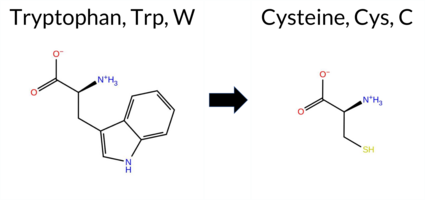 3DClick to see structure in 3D Viewer AIThe SynGAP1 missense variant W362C is not reported in ClinVar and is absent from gnomAD. All available in‑silico predictors classify it as pathogenic: SGM‑Consensus (Likely Pathogenic), REVEL, FoldX, Rosetta, Foldetta, premPS, PROVEAN, polyPhen‑2 (HumDiv and HumVar), SIFT, ESM1b, FATHMM, AlphaMissense‑Default, and AlphaMissense‑Optimized. No tool predicts a benign effect, so the benign group is empty. High‑accuracy methods reinforce this assessment: AlphaMissense‑Optimized predicts pathogenic; the SGM Consensus (majority vote of AlphaMissense‑Default, ESM1b, FATHMM, PROVEAN) also indicates Likely Pathogenic; and Foldetta (combining FoldX‑MD and Rosetta outputs) predicts pathogenic. Consequently, the variant is most likely pathogenic based on the consensus of all predictions, and this conclusion does not contradict the ClinVar status, which simply lacks an entry for this variant. Disclaimer: This summary was generated using AI and should be interpreted alongside expert review. | Likely Pathogenic | C2 | 0.328603 | Structured | 0.430310 | Uncertain | 0.957 | 0.552 | 0.125 | -11.200 | Likely Pathogenic | 0.998 | Likely Pathogenic | Likely Pathogenic | 3.68 | Destabilizing | 0.1 | 4.06 | Destabilizing | 3.87 | Destabilizing | 1.00 | Destabilizing | 0.618 | Likely Pathogenic | -11.95 | Deleterious | 1.000 | Probably Damaging | 0.996 | Probably Damaging | 1.24 | Pathogenic | 0.00 | Affected | 0.4158 | 0.1622 | -8 | -2 | 3.4 | -83.07 | |||||||||||||||||||||||||||||
| c.1086G>T | W362C 2D 3DClick to see structure in 3D Viewer AIThe SynGAP1 missense variant W362C is not reported in ClinVar (ClinVar status: not listed) and is absent from gnomAD (gnomAD ID: none). Prediction tools that assess pathogenicity uniformly classify the variant as deleterious: SGM‑Consensus (Likely Pathogenic), REVEL, FoldX, Rosetta, Foldetta, premPS, PROVEAN, polyPhen‑2 (HumDiv and HumVar), SIFT, ESM1b, FATHMM, AlphaMissense‑Default, and AlphaMissense‑Optimized all predict a pathogenic effect. No tool predicts a benign outcome. High‑accuracy assessments reinforce this view: AlphaMissense‑Optimized indicates pathogenicity; the SGM Consensus (majority vote of AlphaMissense‑Default, ESM1b, FATHMM, PROVEAN) also reports Likely Pathogenic; and Foldetta (combining FoldX‑MD and Rosetta outputs) predicts a pathogenic impact on protein stability. Based on the unanimous pathogenic predictions and the absence of any benign calls, the variant is most likely pathogenic, and this conclusion does not contradict the ClinVar status, which simply lacks an entry. Disclaimer: This summary was generated using AI and should be interpreted alongside expert review. | Likely Pathogenic | C2 | 0.328603 | Structured | 0.430310 | Uncertain | 0.957 | 0.552 | 0.125 | -11.200 | Likely Pathogenic | 0.998 | Likely Pathogenic | Likely Pathogenic | 3.68 | Destabilizing | 0.1 | 4.06 | Destabilizing | 3.87 | Destabilizing | 1.00 | Destabilizing | 0.618 | Likely Pathogenic | -11.95 | Deleterious | 1.000 | Probably Damaging | 0.996 | Probably Damaging | 1.24 | Pathogenic | 0.00 | Affected | 0.4158 | 0.1622 | -8 | -2 | 3.4 | -83.07 | |||||||||||||||||||||||||||||
| c.132G>C | W44C 2D AIThe SynGAP1 missense variant W44C is not reported in ClinVar (ClinVar ID = None) and is absent from gnomAD (gnomAD ID = None). Prediction tools that agree on a benign effect include REVEL, ESM1b, and FATHMM, whereas those that predict a pathogenic effect are PROVEAN, polyPhen‑2 (HumDiv and HumVar), SIFT, AlphaMissense‑Default, and AlphaMissense‑Optimized. High‑accuracy assessments show AlphaMissense‑Optimized as pathogenic; the SGM Consensus (majority vote from AlphaMissense‑Default, ESM1b, FATHMM, and PROVEAN) is inconclusive (2 vs 2), and Foldetta results are unavailable. Overall, the majority of evaluated tools (seven pathogenic vs. three benign) indicate a pathogenic impact. Thus, the variant is most likely pathogenic, and this prediction does not contradict any ClinVar status because the variant has not yet been classified in ClinVar. Disclaimer: This summary was generated using AI and should be interpreted alongside expert review. | 0.301917 | Structured | 0.431379 | Uncertain | 0.377 | 0.748 | 0.375 | -6.216 | Likely Benign | 0.975 | Likely Pathogenic | Likely Pathogenic | 0.321 | Likely Benign | -4.70 | Deleterious | 0.943 | Possibly Damaging | 0.941 | Probably Damaging | 3.14 | Benign | 0.00 | Affected | 0.3542 | 0.2370 | -8 | -2 | 3.4 | -83.07 | ||||||||||||||||||||||||||||||||||||||||
| c.132G>T | W44C 2D AIThe SynGAP1 missense variant W44C is not reported in ClinVar (ClinVar ID = None) and is absent from gnomAD (gnomAD ID = None). Prediction tools that agree on a benign effect include REVEL, ESM1b, and FATHMM. Tools that agree on a pathogenic effect are PROVEAN, polyPhen‑2 HumDiv, polyPhen‑2 HumVar, SIFT, AlphaMissense‑Default, and AlphaMissense‑Optimized. High‑accuracy assessments show AlphaMissense‑Optimized predicts pathogenic. The SGM Consensus, derived from a majority vote of AlphaMissense‑Default, ESM1b, FATHMM, and PROVEAN, is inconclusive (two pathogenic vs. two benign). Foldetta, a protein‑folding stability method combining FoldX‑MD and Rosetta outputs, has no available result for this variant. Overall, the majority of evaluated tools (seven pathogenic vs. three benign) indicate a pathogenic effect. This prediction is consistent with the lack of ClinVar annotation; there is no contradiction with ClinVar status. Disclaimer: This summary was generated using AI and should be interpreted alongside expert review. | 0.301917 | Structured | 0.431379 | Uncertain | 0.377 | 0.748 | 0.375 | -6.216 | Likely Benign | 0.975 | Likely Pathogenic | Likely Pathogenic | 0.321 | Likely Benign | -4.70 | Deleterious | 0.943 | Possibly Damaging | 0.941 | Probably Damaging | 3.14 | Benign | 0.00 | Affected | 0.3542 | 0.2370 | -8 | -2 | 3.4 | -83.07 | ||||||||||||||||||||||||||||||||||||||||
| c.1716G>C | W572C 2D 3DClick to see structure in 3D Viewer AIThe SynGAP1 missense variant W572C is not reported in ClinVar and is absent from gnomAD. Functional prediction tools uniformly indicate a deleterious effect. Benign predictions: none. Pathogenic predictions: REVEL, FoldX, Rosetta, Foldetta, premPS, PROVEAN, polyPhen‑2 (HumDiv and HumVar), SIFT, ESM1b, FATHMM, AlphaMissense‑Default, AlphaMissense‑Optimized, and the SGM‑Consensus score (Likely Pathogenic). High‑accuracy assessments further support pathogenicity: AlphaMissense‑Optimized predicts pathogenic; the SGM‑Consensus (majority vote of AlphaMissense‑Default, ESM1b, FATHMM, PROVEAN) yields Likely Pathogenic; and Foldetta, which integrates FoldX‑MD and Rosetta stability outputs, also predicts pathogenic. Based on the unanimous computational evidence, the variant is most likely pathogenic, and this assessment does not contradict any ClinVar annotation (none available). Disclaimer: This summary was generated using AI and should be interpreted alongside expert review. | Likely Pathogenic | GAP | 0.150080 | Structured | 0.039626 | Uncertain | 0.935 | 0.256 | 0.000 | -15.589 | Likely Pathogenic | 0.999 | Likely Pathogenic | Likely Pathogenic | 5.49 | Destabilizing | 0.2 | 5.59 | Destabilizing | 5.54 | Destabilizing | 1.72 | Destabilizing | 0.861 | Likely Pathogenic | -11.82 | Deleterious | 1.000 | Probably Damaging | 1.000 | Probably Damaging | -1.25 | Pathogenic | 0.00 | Affected | 0.3648 | 0.1304 | -8 | -2 | 3.4 | -83.07 | |||||||||||||||||||||||||||||
| c.1716G>T | W572C 2D 3DClick to see structure in 3D Viewer AIThe SynGAP1 missense variant W572C is not reported in ClinVar and is absent from gnomAD. Functional prediction tools uniformly indicate a deleterious effect. Benign predictions: none. Pathogenic predictions: REVEL, FoldX, Rosetta, Foldetta, premPS, PROVEAN, polyPhen‑2 (HumDiv and HumVar), SIFT, ESM1b, FATHMM, AlphaMissense‑Default, and AlphaMissense‑Optimized. High‑accuracy assessments further support pathogenicity: AlphaMissense‑Optimized predicts pathogenic; the SGM Consensus (majority vote of AlphaMissense‑Default, ESM1b, FATHMM, PROVEAN) reports Likely Pathogenic; and Foldetta, which integrates FoldX‑MD and Rosetta stability outputs, also predicts pathogenic. No inconclusive or missing predictions are present. Based on the consensus of all available computational evidence, the variant is most likely pathogenic, and this assessment does not contradict any ClinVar annotation (none available). Disclaimer: This summary was generated using AI and should be interpreted alongside expert review. | Likely Pathogenic | GAP | 0.150080 | Structured | 0.039626 | Uncertain | 0.935 | 0.256 | 0.000 | -15.589 | Likely Pathogenic | 0.999 | Likely Pathogenic | Likely Pathogenic | 5.49 | Destabilizing | 0.2 | 5.59 | Destabilizing | 5.54 | Destabilizing | 1.72 | Destabilizing | 0.861 | Likely Pathogenic | -11.82 | Deleterious | 1.000 | Probably Damaging | 1.000 | Probably Damaging | -1.25 | Pathogenic | 0.00 | Affected | 0.3648 | 0.1304 | -8 | -2 | 3.4 | -83.07 | |||||||||||||||||||||||||||||
| c.1971G>C | W657C 2D AISynGAP1 missense variant W657C is listed in ClinVar with an uncertain significance and is not reported in gnomAD. Functional prediction tools that classify the variant as benign include REVEL and FATHMM. Those that predict a deleterious effect are FoldX, premPS, PROVEAN, polyPhen‑2 (HumDiv and HumVar), SIFT, ESM1b, AlphaMissense‑Default, and AlphaMissense‑Optimized; Rosetta reports an uncertain outcome. High‑accuracy assessments further support a damaging interpretation: AlphaMissense‑Optimized is pathogenic, the SGM Consensus (majority vote of AlphaMissense‑Default, ESM1b, FATHMM, PROVEAN) is likely pathogenic, and Foldetta (combining FoldX‑MD and Rosetta outputs) is pathogenic. Overall, the preponderance of evidence indicates that W657C is most likely pathogenic, which does not contradict the current ClinVar status of uncertain significance. Disclaimer: This summary was generated using AI and should be interpreted alongside expert review. | Likely Pathogenic | GAP | 0.030611 | Structured | 0.208729 | Uncertain | 0.941 | 0.245 | 0.000 | Uncertain | 1 | -12.035 | Likely Pathogenic | 0.997 | Likely Pathogenic | Likely Pathogenic | 2.74 | Destabilizing | 0.3 | 1.69 | Ambiguous | 2.22 | Destabilizing | 1.30 | Destabilizing | 0.463 | Likely Benign | -11.06 | Deleterious | 1.000 | Probably Damaging | 0.982 | Probably Damaging | 3.43 | Benign | 0.03 | Affected | 0.3834 | 0.0766 | -8 | -2 | 3.4 | -83.07 | |||||||||||||||||||||||||||
| c.1971G>T | W657C 2D AIThe SynGAP1 W657C missense variant is not reported in ClinVar (ClinVar ID None) and is absent from gnomAD (gnomAD ID None). Prediction tools that agree on a benign effect are REVEL and FATHMM, whereas the majority of tools (SGM‑Consensus, FoldX, premPS, PROVEAN, polyPhen‑2 HumDiv, polyPhen‑2 HumVar, SIFT, ESM1b, AlphaMissense‑Default, AlphaMissense‑Optimized) predict a pathogenic impact; Foldetta also supports pathogenicity, while Rosetta remains uncertain. High‑accuracy assessments further reinforce a deleterious outcome: AlphaMissense‑Optimized classifies the variant as pathogenic; the SGM Consensus (majority vote of AlphaMissense‑Default, ESM1b, FATHMM, PROVEAN) indicates likely pathogenic; and Foldetta, a protein‑folding stability method combining FoldX‑MD and Rosetta outputs, predicts a pathogenic effect. Overall, the preponderance of evidence points to the variant being most likely pathogenic, and this conclusion is not contradicted by any ClinVar annotation. Disclaimer: This summary was generated using AI and should be interpreted alongside expert review. | Likely Pathogenic | GAP | 0.030611 | Structured | 0.208729 | Uncertain | 0.941 | 0.245 | 0.000 | -12.035 | Likely Pathogenic | 0.997 | Likely Pathogenic | Likely Pathogenic | 2.74 | Destabilizing | 0.3 | 1.69 | Ambiguous | 2.22 | Destabilizing | 1.30 | Destabilizing | 0.463 | Likely Benign | -11.06 | Deleterious | 1.000 | Probably Damaging | 0.982 | Probably Damaging | 3.43 | Benign | 0.03 | Affected | 0.3834 | 0.0766 | -8 | -2 | 3.4 | -83.07 | |||||||||||||||||||||||||||||
| c.2091G>C | W697C 2D 3DClick to see structure in 3D Viewer AISynGAP1 W697C is not reported in ClinVar and is absent from gnomAD. Functional prediction tools cluster into two groups: benign predictions come from REVEL, SIFT, ESM1b, and FATHMM, while pathogenic predictions arise from PROVEAN, polyPhen‑2 HumDiv, polyPhen‑2 HumVar, and AlphaMissense‑Default. The remaining tools—FoldX, Rosetta, Foldetta, premPS, and AlphaMissense‑Optimized—return uncertain or inconclusive results. High‑accuracy assessments are likewise ambiguous: AlphaMissense‑Optimized is uncertain; the SGM Consensus (majority vote of AlphaMissense‑Default, ESM1b, FATHMM, and PROVEAN) is a 2‑vs‑2 tie, and Foldetta is uncertain. Consequently, the evidence does not favor either outcome; the variant’s impact remains indeterminate. This lack of consensus does not contradict ClinVar, which has no entry for the variant. Disclaimer: This summary was generated using AI and should be interpreted alongside expert review. | GAP | 0.175930 | Structured | 0.400169 | Uncertain | 0.945 | 0.297 | 0.000 | -5.683 | Likely Benign | 0.811 | Likely Pathogenic | Ambiguous | 1.84 | Ambiguous | 0.1 | 1.01 | Ambiguous | 1.43 | Ambiguous | 0.98 | Ambiguous | 0.318 | Likely Benign | -8.65 | Deleterious | 1.000 | Probably Damaging | 0.999 | Probably Damaging | 3.48 | Benign | 0.10 | Tolerated | 0.3575 | 0.1015 | -8 | -2 | 3.4 | -83.07 | ||||||||||||||||||||||||||||||
| c.2091G>T | W697C 2D 3DClick to see structure in 3D Viewer AISynGAP1 W697C is not reported in ClinVar and is absent from gnomAD. Functional prediction tools cluster into two groups: benign predictions come from REVEL, SIFT, ESM1b, and FATHMM, while pathogenic predictions arise from PROVEAN, polyPhen‑2 HumDiv, polyPhen‑2 HumVar, and AlphaMissense‑Default. Five tools (FoldX, Rosetta, Foldetta, premPS, AlphaMissense‑Optimized) yield uncertain or missing results. High‑accuracy assessments are likewise inconclusive: AlphaMissense‑Optimized is uncertain; the SGM Consensus, derived from a majority vote of AlphaMissense‑Default, ESM1b, FATHMM, and PROVEAN, is a 2‑vs‑2 tie; and Foldetta is uncertain. Consequently, the evidence does not strongly favor either outcome. The variant is therefore best classified as of uncertain significance; this assessment does not contradict the lack of ClinVar annotation. Disclaimer: This summary was generated using AI and should be interpreted alongside expert review. | GAP | 0.175930 | Structured | 0.400169 | Uncertain | 0.945 | 0.297 | 0.000 | -5.683 | Likely Benign | 0.811 | Likely Pathogenic | Ambiguous | 1.84 | Ambiguous | 0.1 | 1.01 | Ambiguous | 1.43 | Ambiguous | 0.98 | Ambiguous | 0.318 | Likely Benign | -8.65 | Deleterious | 1.000 | Probably Damaging | 0.999 | Probably Damaging | 3.48 | Benign | 0.10 | Tolerated | 0.3575 | 0.1015 | -8 | -2 | 3.4 | -83.07 | ||||||||||||||||||||||||||||||
| c.3471G>C | W1157C 2D AIThe SynGAP1 missense variant W1157C is not reported in ClinVar and is absent from gnomAD. Prediction tools that agree on a benign effect include REVEL and ESM1b, whereas the remaining tools—PROVEAN, polyPhen‑2 (HumDiv and HumVar), SIFT, FATHMM, AlphaMissense‑Default, and AlphaMissense‑Optimized—consistently predict a pathogenic impact. High‑accuracy assessments further support a deleterious outcome: AlphaMissense‑Optimized classifies the variant as pathogenic, and the SGM‑Consensus (majority vote from AlphaMissense‑Default, ESM1b, FATHMM, and PROVEAN) reports it as Likely Pathogenic. No Foldetta stability prediction is available for this variant. Overall, the majority of computational evidence indicates that W1157C is most likely pathogenic, and this conclusion does not contradict any ClinVar annotation, as none exists. Disclaimer: This summary was generated using AI and should be interpreted alongside expert review. | Likely Pathogenic | 0.694846 | Disordered | 0.877471 | Binding | 0.364 | 0.861 | 0.375 | -4.730 | Likely Benign | 1.000 | Likely Pathogenic | Likely Pathogenic | 0.478 | Likely Benign | -9.46 | Deleterious | 1.000 | Probably Damaging | 0.998 | Probably Damaging | 1.04 | Pathogenic | 0.00 | Affected | 0.3927 | 0.1406 | -8 | -2 | 3.4 | -83.07 | |||||||||||||||||||||||||||||||||||||||
| c.3471G>T | W1157C 2D AIThe SynGAP1 missense variant W1157C is not reported in ClinVar and is absent from gnomAD. Prediction tools that agree on a benign effect include REVEL and ESM1b, whereas the remaining tools—PROVEAN, polyPhen‑2 (HumDiv and HumVar), SIFT, FATHMM, AlphaMissense‑Default, and AlphaMissense‑Optimized—consistently predict a pathogenic impact. High‑accuracy assessments further support a deleterious outcome: AlphaMissense‑Optimized classifies the variant as pathogenic, and the SGM‑Consensus (majority vote from AlphaMissense‑Default, ESM1b, FATHMM, and PROVEAN) reports it as Likely Pathogenic. No Foldetta stability prediction is available for this variant. Overall, the majority of computational evidence indicates that W1157C is most likely pathogenic, and this conclusion does not contradict any ClinVar annotation, as none exists. Disclaimer: This summary was generated using AI and should be interpreted alongside expert review. | Likely Pathogenic | 0.694846 | Disordered | 0.877471 | Binding | 0.364 | 0.861 | 0.375 | -4.730 | Likely Benign | 1.000 | Likely Pathogenic | Likely Pathogenic | 0.478 | Likely Benign | -9.46 | Deleterious | 1.000 | Probably Damaging | 0.998 | Probably Damaging | 1.04 | Pathogenic | 0.00 | Affected | 0.3927 | 0.1406 | -8 | -2 | 3.4 | -83.07 | |||||||||||||||||||||||||||||||||||||||
| c.3945G>C | W1315C 2D AIThe SynGAP1 missense variant W1315C is not reported in ClinVar and is absent from gnomAD. Prediction tools that agree on a benign effect include REVEL, PROVEAN, ESM1b, FATHMM, and AlphaMissense‑Optimized, while those that predict a pathogenic outcome are polyPhen‑2 (HumDiv and HumVar) and SIFT, together with AlphaMissense‑Default. The SGM‑Consensus, derived from a majority vote of AlphaMissense‑Default, ESM1b, FATHMM, and PROVEAN, classifies the variant as Likely Benign. High‑accuracy assessments further support a benign interpretation: AlphaMissense‑Optimized predicts benign, and the SGM‑Consensus also indicates Likely Benign. No Foldetta stability analysis is available for this variant. Overall, the majority of computational evidence points to a benign effect, and this conclusion does not contradict any ClinVar status, as the variant is currently unreported in that database. Disclaimer: This summary was generated using AI and should be interpreted alongside expert review. | Likely Benign | 0.885302 | Disordered | 0.973142 | Binding | 0.403 | 0.889 | 0.750 | -2.355 | Likely Benign | 0.671 | Likely Pathogenic | Likely Benign | 0.082 | Likely Benign | -1.54 | Neutral | 0.872 | Possibly Damaging | 0.468 | Possibly Damaging | 4.21 | Benign | 0.02 | Affected | 0.3427 | 0.1929 | -8 | -2 | 3.4 | -83.07 | |||||||||||||||||||||||||||||||||||||||
| c.3945G>T | W1315C 2D AIThe SynGAP1 missense variant W1315C is not reported in ClinVar and is absent from gnomAD. Prediction tools that agree on a benign effect include REVEL, PROVEAN, ESM1b, FATHMM, AlphaMissense‑Optimized, and the SGM‑Consensus (majority vote from AlphaMissense‑Default, ESM1b, FATHMM, PROVEAN). Tools that predict a pathogenic outcome are polyPhen‑2 HumDiv, polyPhen‑2 HumVar, SIFT, and AlphaMissense‑Default. High‑accuracy assessments show AlphaMissense‑Optimized as benign and the SGM‑Consensus as likely benign; the Foldetta protein‑folding stability analysis is unavailable. Overall, the majority of evidence points to a benign impact for W1315C, and this conclusion does not contradict any ClinVar annotation, as none exists for this variant. Disclaimer: This summary was generated using AI and should be interpreted alongside expert review. | Likely Benign | 0.885302 | Disordered | 0.973142 | Binding | 0.403 | 0.889 | 0.750 | -2.355 | Likely Benign | 0.671 | Likely Pathogenic | Likely Benign | 0.082 | Likely Benign | -1.54 | Neutral | 0.872 | Possibly Damaging | 0.468 | Possibly Damaging | 4.21 | Benign | 0.02 | Affected | 0.3427 | 0.1929 | -8 | -2 | 3.4 | -83.07 | |||||||||||||||||||||||||||||||||||||||
| c.726G>C | W242C 2D 3DClick to see structure in 3D Viewer AIThe SynGAP1 W242C missense variant is not reported in ClinVar (no ClinVar ID) and is absent from gnomAD. Prediction tools that agree on a pathogenic effect include REVEL, PROVEAN, polyPhen‑2 (HumDiv and HumVar), SIFT, ESM1b, FATHMM, AlphaMissense‑Default, and AlphaMissense‑Optimized. Tools that are uncertain or inconclusive are FoldX, Rosetta, Foldetta, and premPS, and no tools predict a benign outcome. High‑accuracy assessments show AlphaMissense‑Optimized as pathogenic, the SGM Consensus (majority vote of AlphaMissense‑Default, ESM1b, FATHMM, PROVEAN) as Likely Pathogenic, and Foldetta as uncertain. Based on the collective predictions, the variant is most likely pathogenic. This assessment does not contradict ClinVar status, as ClinVar has not yet classified the variant. Disclaimer: This summary was generated using AI and should be interpreted alongside expert review. | Likely Pathogenic | PH | 0.328603 | Structured | 0.352582 | Uncertain | 0.847 | 0.341 | 0.000 | -13.117 | Likely Pathogenic | 1.000 | Likely Pathogenic | Likely Pathogenic | 1.77 | Ambiguous | 0.4 | 1.62 | Ambiguous | 1.70 | Ambiguous | 0.63 | Ambiguous | 0.889 | Likely Pathogenic | -11.80 | Deleterious | 0.999 | Probably Damaging | 0.887 | Possibly Damaging | 1.52 | Pathogenic | 0.00 | Affected | 0.3626 | 0.1617 | -8 | -2 | 3.4 | -83.07 | |||||||||||||||||||||||||||||
| c.726G>T | W242C 2D 3DClick to see structure in 3D Viewer AIThe SynGAP1 missense variant W242C is not reported in ClinVar (no ClinVar ID) and is absent from gnomAD. Prediction tools that agree on a pathogenic effect include REVEL, PROVEAN, polyPhen‑2 (HumDiv and HumVar), SIFT, ESM1b, FATHMM, AlphaMissense‑Default, and AlphaMissense‑Optimized. Tools that are uncertain or inconclusive are FoldX, Rosetta, Foldetta, and premPS, and no tools predict a benign outcome. High‑accuracy assessments show AlphaMissense‑Optimized as pathogenic, the SGM Consensus (majority vote of AlphaMissense‑Default, ESM1b, FATHMM, PROVEAN) as Likely Pathogenic, and Foldetta as uncertain. Based on the collective predictions, the variant is most likely pathogenic. This assessment does not contradict ClinVar status, as ClinVar has not yet classified the variant. Disclaimer: This summary was generated using AI and should be interpreted alongside expert review. | Likely Pathogenic | PH | 0.328603 | Structured | 0.352582 | Uncertain | 0.847 | 0.341 | 0.000 | -13.117 | Likely Pathogenic | 1.000 | Likely Pathogenic | Likely Pathogenic | 1.77 | Ambiguous | 0.4 | 1.62 | Ambiguous | 1.70 | Ambiguous | 0.63 | Ambiguous | 0.889 | Likely Pathogenic | -11.80 | Deleterious | 0.999 | Probably Damaging | 0.887 | Possibly Damaging | 1.52 | Pathogenic | 0.00 | Affected | 0.3626 | 0.1617 | -8 | -2 | 3.4 | -83.07 | |||||||||||||||||||||||||||||
| c.801G>C | W267C 2D 3DClick to see structure in 3D Viewer AIThe SynGAP1 missense variant W267C is not reported in ClinVar and is absent from gnomAD. All available in‑silico predictors classify it as pathogenic: SGM‑Consensus (Likely Pathogenic), REVEL, FoldX, Rosetta, Foldetta, premPS, PROVEAN, polyPhen‑2 (HumDiv and HumVar), SIFT, ESM1b, FATHMM, AlphaMissense‑Default, and AlphaMissense‑Optimized. No tool predicts a benign effect. High‑accuracy assessments reinforce this: AlphaMissense‑Optimized predicts pathogenic; the SGM Consensus (majority vote of AlphaMissense‑Default, ESM1b, FATHMM, PROVEAN) also indicates Likely Pathogenic; and Foldetta (combining FoldX‑MD and Rosetta outputs) predicts pathogenic. Thus, the variant is most likely pathogenic based on the consensus of all predictions, and this conclusion does not contradict the current ClinVar status, which has no entry for this variant. Disclaimer: This summary was generated using AI and should be interpreted alongside expert review. | Likely Pathogenic | C2 | 0.216401 | Structured | 0.298060 | Uncertain | 0.943 | 0.274 | 0.000 | -12.351 | Likely Pathogenic | 0.996 | Likely Pathogenic | Likely Pathogenic | 2.81 | Destabilizing | 0.2 | 2.20 | Destabilizing | 2.51 | Destabilizing | 1.00 | Destabilizing | 0.705 | Likely Pathogenic | -11.95 | Deleterious | 1.000 | Probably Damaging | 0.999 | Probably Damaging | 1.90 | Pathogenic | 0.01 | Affected | 0.3573 | 0.1700 | -8 | -2 | 3.4 | -83.07 | |||||||||||||||||||||||||||||
| c.801G>T | W267C 2D 3DClick to see structure in 3D Viewer AIThe SynGAP1 missense variant W267C is not reported in ClinVar and is absent from gnomAD. All available in‑silico predictors classify it as pathogenic: SGM‑Consensus (Likely Pathogenic), REVEL, FoldX, Rosetta, Foldetta, premPS, PROVEAN, polyPhen‑2 (HumDiv and HumVar), SIFT, ESM1b, FATHMM, AlphaMissense‑Default, and AlphaMissense‑Optimized. No tool predicts a benign effect. High‑accuracy assessments reinforce this: AlphaMissense‑Optimized predicts pathogenic; the SGM Consensus (majority vote of AlphaMissense‑Default, ESM1b, FATHMM, PROVEAN) also indicates Likely Pathogenic; and Foldetta (combining FoldX‑MD and Rosetta outputs) predicts pathogenic. Thus, the variant is most likely pathogenic based on the consensus of all predictions, and this conclusion does not contradict the current ClinVar status, which has no entry for this variant. Disclaimer: This summary was generated using AI and should be interpreted alongside expert review. | Likely Pathogenic | C2 | 0.216401 | Structured | 0.298060 | Uncertain | 0.943 | 0.274 | 0.000 | -12.351 | Likely Pathogenic | 0.996 | Likely Pathogenic | Likely Pathogenic | 2.81 | Destabilizing | 0.2 | 2.20 | Destabilizing | 2.51 | Destabilizing | 1.00 | Destabilizing | 0.705 | Likely Pathogenic | -11.95 | Deleterious | 1.000 | Probably Damaging | 0.999 | Probably Damaging | 1.90 | Pathogenic | 0.01 | Affected | 0.3573 | 0.1700 | -8 | -2 | 3.4 | -83.07 | |||||||||||||||||||||||||||||
| c.924G>C | W308C 2D 3DClick to see structure in 3D Viewer AIThe SynGAP1 missense variant W308C is listed in ClinVar as Pathogenic (ClinVar ID 981381.0) and is not reported in gnomAD. Prediction tools that assess functional impact uniformly indicate a deleterious effect: REVEL, FoldX, Rosetta, Foldetta, premPS, PROVEAN, polyPhen‑2 (HumDiv and HumVar), SIFT, ESM1b, FATHMM, AlphaMissense‑Default, and AlphaMissense‑Optimized all classify the variant as pathogenic. No tool predicts a benign outcome. High‑accuracy assessments corroborate this: AlphaMissense‑Optimized predicts pathogenic; the SGM Consensus (majority vote of AlphaMissense‑Default, ESM1b, FATHMM, PROVEAN) yields “Likely Pathogenic”; and Foldetta (combining FoldX‑MD and Rosetta outputs) also predicts pathogenic. Thus, the variant is most likely pathogenic, and this prediction aligns with its ClinVar status. Disclaimer: This summary was generated using AI and should be interpreted alongside expert review. | Likely Pathogenic | C2 | 0.328603 | Structured | 0.333942 | Uncertain | 0.907 | 0.314 | 0.125 | Pathogenic/Likely path. | 2 | -12.791 | Likely Pathogenic | 1.000 | Likely Pathogenic | Likely Pathogenic | 5.56 | Destabilizing | 0.3 | 4.38 | Destabilizing | 4.97 | Destabilizing | 1.26 | Destabilizing | 0.738 | Likely Pathogenic | -11.95 | Deleterious | 1.000 | Probably Damaging | 0.999 | Probably Damaging | 0.48 | Pathogenic | 0.00 | Affected | 3.38 | 19 | 0.4390 | 0.1621 | -8 | -2 | 3.4 | -83.07 | 230.8 | 60.5 | -0.3 | 0.1 | -0.4 | 0.4 | X | Potentially Pathogenic | The indole ring of Trp308, located in an anti-parallel β sheet strand (res. Thr305-Asn315), packs against multiple hydrophobic residues (e.g., Ile268, Val306, Cys282). The indole group of Trp308 also hydrogen bonds with the backbone atoms of the C2 domain residues forming the anti-parallel β sheet (e.g., Tyr280, Thr294). The introduced Cys308 is smaller than the tryptophan it replaced. The thiol group of the Cys308 side chain is well-suited for the inner hydrophobic part of the C2 domain. Although the negative effects are essentially missing from the simulations, the side chain size difference between the residues is likely to disrupt the hydrophobic packing during folding. At a minimum, the residue swap could affect the C2 domain stability and membrane association. | ||||||||||||||||
| c.924G>T | W308C 2D 3DClick to see structure in 3D Viewer AIThe SynGAP1 missense variant W308C is not reported in ClinVar and is absent from gnomAD. Prediction tools that assess functional impact uniformly classify the variant as pathogenic: SGM‑Consensus (Likely Pathogenic), REVEL, FoldX, Rosetta, Foldetta, premPS, PROVEAN, polyPhen‑2 (HumDiv and HumVar), SIFT, ESM1b, FATHMM, AlphaMissense‑Default, and AlphaMissense‑Optimized. No tool predicts a benign effect. High‑accuracy assessments reinforce this view: AlphaMissense‑Optimized predicts pathogenic, the SGM Consensus (majority vote of AlphaMissense‑Default, ESM1b, FATHMM, PROVEAN) is Likely Pathogenic, and Foldetta (combining FoldX‑MD and Rosetta outputs) predicts pathogenic. All available evidence points to a deleterious effect. Therefore, the variant is most likely pathogenic, and this conclusion does not contradict any ClinVar status (none reported). Disclaimer: This summary was generated using AI and should be interpreted alongside expert review. | Likely Pathogenic | C2 | 0.328603 | Structured | 0.333942 | Uncertain | 0.907 | 0.314 | 0.125 | -12.791 | Likely Pathogenic | 1.000 | Likely Pathogenic | Likely Pathogenic | 5.56 | Destabilizing | 0.3 | 4.38 | Destabilizing | 4.97 | Destabilizing | 1.26 | Destabilizing | 0.738 | Likely Pathogenic | -11.95 | Deleterious | 1.000 | Probably Damaging | 0.999 | Probably Damaging | 0.48 | Pathogenic | 0.00 | Affected | 3.38 | 19 | 0.4390 | 0.1621 | -8 | -2 | 3.4 | -83.07 | 230.8 | 60.5 | -0.3 | 0.1 | -0.4 | 0.4 | X | Potentially Pathogenic | The indole ring of Trp308, located in an anti-parallel β sheet strand (res. Thr305-Asn315), packs against multiple hydrophobic residues (e.g., Ile268, Val306, Cys282). The indole group of Trp308 also hydrogen bonds with the backbone atoms of the C2 domain residues forming the anti-parallel β sheet (e.g., Tyr280, Thr294). The introduced Cys308 is smaller than the tryptophan it replaced. The thiol group of the Cys308 side chain is well-suited for the inner hydrophobic part of the C2 domain. Although the negative effects are essentially missing from the simulations, the side chain size difference between the residues is likely to disrupt the hydrophobic packing during folding. At a minimum, the residue swap could affect the C2 domain stability and membrane association. | ||||||||||||||||||
| c.101A>C | Y34S 2D  AIThe SynGAP1 missense variant Y34S is not reported in ClinVar and is absent from gnomAD. Prediction tools that agree on a benign effect include REVEL, PROVEAN, ESM1b, FATHMM, AlphaMissense‑Default, AlphaMissense‑Optimized, and the SGM‑Consensus (Likely Benign). In contrast, polyPhen‑2 (HumDiv and HumVar) and SIFT all predict a pathogenic impact. High‑accuracy assessments further support a benign classification: AlphaMissense‑Optimized predicts benign, and the SGM‑Consensus (majority vote of AlphaMissense‑Default, ESM1b, FATHMM, and PROVEAN) also indicates Likely Benign. No Foldetta stability result is available, so it does not influence the assessment. Overall, the majority of evidence points to a benign effect for Y34S, and this conclusion is not contradicted by any ClinVar annotation. Disclaimer: This summary was generated using AI and should be interpreted alongside expert review. | Likely Benign | 0.529623 | Disordered | 0.435847 | Uncertain | 0.303 | 0.855 | 0.375 | -0.994 | Likely Benign | 0.237 | Likely Benign | Likely Benign | 0.178 | Likely Benign | -0.60 | Neutral | 0.824 | Possibly Damaging | 0.775 | Possibly Damaging | 4.19 | Benign | 0.00 | Affected | 0.4760 | 0.2884 | -3 | -2 | 0.5 | -76.10 | |||||||||||||||||||||||||||||||||||||||
| c.1025A>C | Y342S 2D 3DClick to see structure in 3D Viewer AISynGAP1 variant Y342S is listed in ClinVar with an uncertain significance and is not reported in gnomAD. Prediction tools that agree on a benign effect include only REVEL, whereas the majority of algorithms predict a pathogenic impact: FoldX, Rosetta, PROVEAN, polyPhen‑2 HumDiv, polyPhen‑2 HumVar, SIFT, FATHMM, AlphaMissense‑Default, the SGM‑Consensus (majority vote of AlphaMissense‑Default, ESM1b, FATHMM, PROVEAN), and the Foldetta stability assessment (combining FoldX‑MD and Rosetta). Uncertain results come from premPS, ESM1b, and AlphaMissense‑Optimized. High‑accuracy methods specifically give AlphaMissense‑Optimized as uncertain, SGM‑Consensus as pathogenic, and Foldetta as pathogenic. Overall, the preponderance of evidence points to a pathogenic effect, which contradicts the ClinVar uncertain classification. Disclaimer: This summary was generated using AI and should be interpreted alongside expert review. | Likely Pathogenic | C2 | 0.366687 | Structured | 0.408200 | Uncertain | 0.866 | 0.487 | 0.250 | Uncertain | 2 | -7.996 | In-Between | 0.925 | Likely Pathogenic | Ambiguous | 3.03 | Destabilizing | 0.1 | 2.87 | Destabilizing | 2.95 | Destabilizing | 0.93 | Ambiguous | 0.407 | Likely Benign | -6.60 | Deleterious | 1.000 | Probably Damaging | 0.998 | Probably Damaging | 1.75 | Pathogenic | 0.04 | Affected | 3.37 | 25 | 0.4617 | 0.2637 | -3 | -2 | 0.5 | -76.10 | 200.1 | 77.8 | 0.0 | 0.0 | -0.2 | 0.1 | Potentially Pathogenic | The phenol ring of Tyr342, located at the end of an anti-parallel β sheet strand (res. Gly341-Pro349), faces outward in the C2 domain. In the WT simulations, the phenol ring of Tyr342 contributes to a triple tyrosine stack (Tyr342, Tyr328, and Tyr281) that links together three anti-parallel β sheet strands. Additionally, it shields Gly344 from the solvent, reducing its exposure and providing stability for the β-sandwich. This motif also contributes to a twist formation in the β sheet.In the variant simulations, the Ser342 side chain cannot participate in the stack formation. Instead, the hydroxyl group of the Ser342 side chain forms a hydrogen bond with the imidazole ring of His326 in a neighboring β strand (res. Ala322-Asp330). This disrupts the formation of a hydrogen bond between His326 and the carboxylate group of the Glu283 side chain from another β strand (res. Arg279-Cys285). Although these changes in surface interactions could weaken the characteristic twist that strengthens the β sheet fold, no major structural effects are observed in the variant simulations. The residue swap could also affect the SynGAP-membrane association, as the hydroxyl group of Ser342 could form hydrogen bonds with membrane-facing loop residues. However, this phenomenon cannot be addressed using solvent-only simulations. | |||||||||||||||||
| c.1088A>C | Y363S 2D AIThe SynGAP1 missense variant Y363S is not reported in ClinVar (ClinVar status: none) and is absent from gnomAD (gnomAD ID: none). Prediction tools that agree on a benign effect include REVEL, whereas the remaining tools—FoldX, Rosetta, Foldetta, premPS, PROVEAN, polyPhen‑2 (HumDiv and HumVar), SIFT, ESM1b, FATHMM, and AlphaMissense‑Default—consistently predict a pathogenic impact. High‑accuracy assessments further support a deleterious outcome: the SGM Consensus (majority vote of AlphaMissense‑Default, ESM1b, FATHMM, PROVEAN) indicates “Likely Pathogenic”; AlphaMissense‑Optimized is classified as “Uncertain”; and Foldetta, which integrates FoldX‑MD and Rosetta stability calculations, predicts a pathogenic effect. Taken together, the overwhelming majority of evidence points to a pathogenic effect for Y363S, and this conclusion does not contradict any ClinVar annotation (none is available). Disclaimer: This summary was generated using AI and should be interpreted alongside expert review. | Likely Pathogenic | C2 | 0.321458 | Structured | 0.435392 | Uncertain | 0.954 | 0.586 | 0.125 | -11.578 | Likely Pathogenic | 0.952 | Likely Pathogenic | Ambiguous | 3.09 | Destabilizing | 0.6 | 3.92 | Destabilizing | 3.51 | Destabilizing | 1.45 | Destabilizing | 0.420 | Likely Benign | -8.04 | Deleterious | 0.999 | Probably Damaging | 0.991 | Probably Damaging | 1.56 | Pathogenic | 0.01 | Affected | 0.5750 | 0.4208 | Weaken | -3 | -2 | 0.5 | -76.10 | ||||||||||||||||||||||||||||
| c.116A>C | Y39S 2D AIThe SynGAP1 missense variant Y39S is not reported in ClinVar (no ClinVar ID) and is absent from gnomAD (no gnomAD ID). Prediction tools that agree on a benign effect include REVEL, PROVEAN, ESM1b, FATHMM, AlphaMissense‑Default, and AlphaMissense‑Optimized. Tools that predict a pathogenic effect are polyPhen‑2 HumDiv, polyPhen‑2 HumVar, and SIFT. The SGM‑Consensus, which aggregates AlphaMissense‑Default, ESM1b, FATHMM, and PROVEAN, reports a Likely Benign classification. High‑accuracy assessments show AlphaMissense‑Optimized as Benign and the SGM‑Consensus as Likely Benign; Foldetta results are unavailable. Overall, the majority of evidence points to a benign impact for Y39S, and this conclusion does not contradict any ClinVar annotation (none exists). Disclaimer: This summary was generated using AI and should be interpreted alongside expert review. | Likely Benign | 0.425610 | Structured | 0.432876 | Uncertain | 0.343 | 0.787 | 0.375 | -1.634 | Likely Benign | 0.174 | Likely Benign | Likely Benign | 0.100 | Likely Benign | -1.57 | Neutral | 0.824 | Possibly Damaging | 0.775 | Possibly Damaging | 4.02 | Benign | 0.00 | Affected | 0.4935 | 0.2649 | -3 | -2 | 0.5 | -76.10 | |||||||||||||||||||||||||||||||||||||||
| c.1217A>C | Y406S 2D 3DClick to see structure in 3D Viewer AIThe SynGAP1 missense variant Y406S is not reported in ClinVar (ClinVar ID None) and is absent from gnomAD (gnomAD ID None). Prediction tools that agree on a benign effect include REVEL, SIFT, and FATHMM. In contrast, the majority of tools predict a pathogenic impact: AlphaMissense‑Default, AlphaMissense‑Optimized, ESM1b, PROVEAN, polyPhen‑2 (HumDiv and HumVar), FoldX, Rosetta, premPS, and Foldetta all indicate pathogenicity. High‑accuracy assessments further support a deleterious effect: AlphaMissense‑Optimized is pathogenic; the SGM Consensus (majority vote from AlphaMissense‑Default, ESM1b, FATHMM, PROVEAN) is “Likely Pathogenic”; and Foldetta, which integrates FoldX‑MD and Rosetta outputs, is pathogenic. No predictions are missing or inconclusive. Based on the preponderance of evidence, the variant is most likely pathogenic, and this conclusion is not contradicted by any ClinVar status. Disclaimer: This summary was generated using AI and should be interpreted alongside expert review. | Likely Pathogenic | C2 | 0.179055 | Structured | 0.393707 | Uncertain | 0.946 | 0.291 | 0.000 | -12.014 | Likely Pathogenic | 0.977 | Likely Pathogenic | Likely Pathogenic | 3.51 | Destabilizing | 0.3 | 4.08 | Destabilizing | 3.80 | Destabilizing | 1.40 | Destabilizing | 0.190 | Likely Benign | -6.72 | Deleterious | 0.991 | Probably Damaging | 0.886 | Possibly Damaging | 3.80 | Benign | 0.06 | Tolerated | 0.4719 | 0.2817 | -3 | -2 | 0.5 | -76.10 | |||||||||||||||||||||||||||||
| c.1250A>C | Y417S 2D 3DClick to see structure in 3D Viewer AIThe SynGAP1 missense variant Y417S is not reported in ClinVar (no ClinVar ID) and is absent from gnomAD (no gnomAD ID). Prediction tools that agree on a benign effect are limited to FATHMM, while all other evaluated algorithms—including SGM‑Consensus, REVEL, FoldX, Rosetta, Foldetta, premPS, PROVEAN, polyPhen‑2 (HumDiv and HumVar), SIFT, ESM1b, AlphaMissense‑Default, and AlphaMissense‑Optimized—classify the variant as pathogenic. High‑accuracy assessments further support a deleterious impact: AlphaMissense‑Optimized predicts pathogenic, the SGM‑Consensus (majority vote of AlphaMissense‑Default, ESM1b, FATHMM, PROVEAN) indicates likely pathogenic, and Foldetta (combining FoldX‑MD and Rosetta outputs) reports pathogenic. No prediction or folding stability result is missing or inconclusive. Based on the overwhelming consensus of pathogenic predictions, the variant is most likely pathogenic, and this assessment does not contradict any ClinVar status because no ClinVar entry exists. Disclaimer: This summary was generated using AI and should be interpreted alongside expert review. | Likely Pathogenic | GAP | 0.179055 | Structured | 0.346865 | Uncertain | 0.958 | 0.250 | 0.000 | -14.339 | Likely Pathogenic | 0.997 | Likely Pathogenic | Likely Pathogenic | 4.36 | Destabilizing | 0.1 | 5.32 | Destabilizing | 4.84 | Destabilizing | 2.17 | Destabilizing | 0.593 | Likely Pathogenic | -8.59 | Deleterious | 1.000 | Probably Damaging | 1.000 | Probably Damaging | 2.98 | Benign | 0.00 | Affected | 0.4408 | 0.1961 | -3 | -2 | 0.5 | -76.10 | |||||||||||||||||||||||||||||
| c.1268A>C | Y423S 2D 3DClick to see structure in 3D Viewer AIThe SynGAP1 missense variant Y423S is not reported in ClinVar and is absent from gnomAD. Prediction tools that indicate a benign effect include REVEL, SIFT, and FATHMM, whereas the remaining tools—FoldX, Rosetta, Foldetta, premPS, PROVEAN, polyPhen‑2 (HumDiv and HumVar), ESM1b, AlphaMissense‑Default, and AlphaMissense‑Optimized—consistently predict a pathogenic outcome. High‑accuracy assessments further support a deleterious impact: AlphaMissense‑Optimized is pathogenic, the SGM Consensus (majority vote of AlphaMissense‑Default, ESM1b, FATHMM, and PROVEAN) is likely pathogenic, and Foldetta (combining FoldX‑MD and Rosetta outputs) is pathogenic. Taken together, the preponderance of evidence points to a pathogenic effect for Y423S. This conclusion is not contradicted by ClinVar status, as no ClinVar entry exists for this variant. Disclaimer: This summary was generated using AI and should be interpreted alongside expert review. | Likely Pathogenic | GAP | 0.088832 | Structured | 0.421885 | Uncertain | 0.975 | 0.242 | 0.000 | -10.847 | Likely Pathogenic | 0.985 | Likely Pathogenic | Likely Pathogenic | 4.28 | Destabilizing | 0.1 | 4.78 | Destabilizing | 4.53 | Destabilizing | 1.95 | Destabilizing | 0.345 | Likely Benign | -7.39 | Deleterious | 1.000 | Probably Damaging | 1.000 | Probably Damaging | 3.40 | Benign | 0.09 | Tolerated | 0.3152 | 0.1259 | -3 | -2 | 0.5 | -76.10 | |||||||||||||||||||||||||||||
| c.1283A>C | Y428S 2D 3DClick to see structure in 3D Viewer AIThe SynGAP1 missense variant Y428S is not reported in ClinVar and is absent from gnomAD. Prediction tools that agree on a benign effect include only FATHMM. All other evaluated algorithms (SGM‑Consensus, REVEL, FoldX, Rosetta, Foldetta, premPS, PROVEAN, polyPhen‑2 HumDiv, polyPhen‑2 HumVar, SIFT, ESM1b, AlphaMissense‑Default) predict a pathogenic impact, while AlphaMissense‑Optimized is uncertain. High‑accuracy assessments further support a deleterious outcome: AlphaMissense‑Optimized is inconclusive, SGM‑Consensus indicates “Likely Pathogenic,” and Foldetta (combining FoldX‑MD and Rosetta outputs) predicts a pathogenic effect. Based on the overwhelming majority of predictions, the variant is most likely pathogenic, and this conclusion does not contradict any ClinVar status because no ClinVar entry exists. Disclaimer: This summary was generated using AI and should be interpreted alongside expert review. | Likely Pathogenic | GAP | 0.118441 | Structured | 0.389652 | Uncertain | 0.965 | 0.292 | 0.000 | -10.936 | Likely Pathogenic | 0.928 | Likely Pathogenic | Ambiguous | 3.00 | Destabilizing | 0.0 | 3.42 | Destabilizing | 3.21 | Destabilizing | 2.00 | Destabilizing | 0.505 | Likely Pathogenic | -8.59 | Deleterious | 1.000 | Probably Damaging | 1.000 | Probably Damaging | 3.45 | Benign | 0.03 | Affected | 0.4623 | 0.2352 | -3 | -2 | 0.5 | -76.10 | |||||||||||||||||||||||||||||
| c.1490A>C | Y497S 2D 3DClick to see structure in 3D Viewer AIThe SynGAP1 missense variant Y497S is not reported in ClinVar and is absent from gnomAD. Across the available in‑silico predictors, every tool examined (SGM‑Consensus, REVEL, FoldX, Rosetta, Foldetta, premPS, PROVEAN, polyPhen‑2 HumDiv, polyPhen‑2 HumVar, SIFT, ESM1b, FATHMM, AlphaMissense‑Default, and AlphaMissense‑Optimized) classifies the change as pathogenic, leaving no benign predictions. High‑accuracy methods reinforce this assessment: AlphaMissense‑Optimized predicts pathogenicity; the SGM Consensus (majority vote of AlphaMissense‑Default, ESM1b, FATHMM, and PROVEAN) also predicts pathogenic; and Foldetta, which integrates FoldX‑MD and Rosetta stability outputs, indicates a destabilizing, pathogenic effect. No prediction or stability result is missing or inconclusive. Based on the unanimous pathogenic predictions and the absence of any ClinVar annotation, the variant is most likely pathogenic, with no contradictory ClinVar status. Disclaimer: This summary was generated using AI and should be interpreted alongside expert review. | Likely Pathogenic | GAP | 0.092881 | Structured | 0.393608 | Uncertain | 0.950 | 0.181 | 0.000 | -13.171 | Likely Pathogenic | 0.980 | Likely Pathogenic | Likely Pathogenic | 3.65 | Destabilizing | 0.1 | 4.68 | Destabilizing | 4.17 | Destabilizing | 2.22 | Destabilizing | 0.814 | Likely Pathogenic | -8.82 | Deleterious | 1.000 | Probably Damaging | 0.999 | Probably Damaging | -1.64 | Pathogenic | 0.03 | Affected | 0.3770 | 0.1419 | -3 | -2 | 0.5 | -76.10 | |||||||||||||||||||||||||||||
| c.1514A>C | Y505S 2D 3DClick to see structure in 3D Viewer AIThe SynGAP1 missense variant Y505S is not reported in ClinVar and is absent from gnomAD. Prediction tools that indicate a benign effect include only FATHMM, whereas all other evaluated algorithms (SGM‑Consensus, REVEL, FoldX, Rosetta, Foldetta, premPS, PROVEAN, polyPhen‑2 HumDiv, polyPhen‑2 HumVar, SIFT, ESM1b, AlphaMissense‑Default, AlphaMissense‑Optimized) predict a pathogenic impact. High‑accuracy methods reinforce this view: AlphaMissense‑Optimized scores the variant as pathogenic; the SGM Consensus (majority vote of AlphaMissense‑Default, ESM1b, FATHMM, PROVEAN) reports it as likely pathogenic; and Foldetta, which integrates FoldX‑MD and Rosetta outputs, also predicts pathogenicity. No predictions are missing or inconclusive. Overall, the consensus of the available computational evidence indicates that the variant is most likely pathogenic, and this assessment does not contradict the ClinVar status, which currently contains no entry for Y505S. Disclaimer: This summary was generated using AI and should be interpreted alongside expert review. | Likely Pathogenic | GAP | 0.047319 | Structured | 0.292227 | Uncertain | 0.909 | 0.188 | 0.000 | -14.053 | Likely Pathogenic | 0.986 | Likely Pathogenic | Likely Pathogenic | 3.49 | Destabilizing | 0.1 | 4.86 | Destabilizing | 4.18 | Destabilizing | 2.33 | Destabilizing | 0.559 | Likely Pathogenic | -8.95 | Deleterious | 1.000 | Probably Damaging | 1.000 | Probably Damaging | 2.61 | Benign | 0.00 | Affected | 0.4056 | 0.1945 | -3 | -2 | 0.5 | -76.10 | |||||||||||||||||||||||||||||
| c.1553A>C | Y518S 2D 3DClick to see structure in 3D Viewer AIThe SynGAP1 missense variant Y518S is not reported in ClinVar and is absent from gnomAD. Prediction tools that agree on a benign effect include SIFT and FATHMM, whereas the remaining tools—REVEL, FoldX, Rosetta, Foldetta, premPS, PROVEAN, polyPhen‑2 (HumDiv and HumVar), ESM1b, and AlphaMissense‑Default—consistently predict a pathogenic impact. High‑accuracy assessments further support a deleterious outcome: the SGM Consensus (majority vote of AlphaMissense‑Default, ESM1b, FATHMM, and PROVEAN) indicates “Likely Pathogenic”; AlphaMissense‑Optimized is uncertain; and Foldetta, which integrates FoldX‑MD and Rosetta stability calculations, predicts a pathogenic effect. Taken together, the preponderance of evidence points to a pathogenic effect for Y518S, and this conclusion is not contradicted by any ClinVar annotation. Disclaimer: This summary was generated using AI and should be interpreted alongside expert review. | Likely Pathogenic | GAP | 0.139895 | Structured | 0.126970 | Uncertain | 0.897 | 0.321 | 0.000 | -12.453 | Likely Pathogenic | 0.912 | Likely Pathogenic | Ambiguous | 2.76 | Destabilizing | 0.8 | 2.45 | Destabilizing | 2.61 | Destabilizing | 1.61 | Destabilizing | 0.551 | Likely Pathogenic | -8.38 | Deleterious | 1.000 | Probably Damaging | 1.000 | Probably Damaging | 3.43 | Benign | 0.06 | Tolerated | 0.3948 | 0.1059 | -3 | -2 | 0.5 | -76.10 | |||||||||||||||||||||||||||||
| c.1841A>C | Y614S 2D 3DClick to see structure in 3D Viewer AISynGAP1 missense variant Y614S is not reported in ClinVar and is present in gnomAD (ID 6‑33440893‑A‑C). Functional prediction tools cluster into two groups: benign predictions come from REVEL, SIFT, and FATHMM, whereas the majority of algorithms—AlphaMissense‑Default, AlphaMissense‑Optimized, ESM1b, PROVEAN, polyPhen‑2 (HumDiv and HumVar), premPS, Rosetta, Foldetta, and the SGM Consensus—indicate pathogenicity; FoldX remains uncertain. High‑accuracy assessments reinforce the pathogenic signal: AlphaMissense‑Optimized scores the variant as pathogenic, the SGM Consensus (majority vote of AlphaMissense‑Default, ESM1b, FATHMM, PROVEAN) labels it likely pathogenic, and Foldetta (combining FoldX‑MD and Rosetta outputs) predicts a destabilizing, pathogenic effect. Overall, the preponderance of evidence points to a pathogenic classification, and this assessment does not contradict any ClinVar annotation, as none exists. Disclaimer: This summary was generated using AI and should be interpreted alongside expert review. | Likely Pathogenic | GAP | 0.314870 | Structured | 0.182134 | Uncertain | 0.852 | 0.254 | 0.000 | 6-33440893-A-C | 1 | 6.20e-7 | -12.709 | Likely Pathogenic | 0.990 | Likely Pathogenic | Likely Pathogenic | 1.74 | Ambiguous | 0.4 | 3.25 | Destabilizing | 2.50 | Destabilizing | 2.05 | Destabilizing | 0.482 | Likely Benign | -8.83 | Deleterious | 1.000 | Probably Damaging | 1.000 | Probably Damaging | 3.48 | Benign | 0.09 | Tolerated | 3.37 | 35 | 0.4155 | 0.1220 | -2 | -3 | 0.5 | -76.10 | ||||||||||||||||||||||||
| c.1994A>C | Y665S 2D 3DClick to see structure in 3D Viewer AIThe SynGAP1 missense variant Y665S is not reported in ClinVar (ClinVar ID None) and is absent from gnomAD (gnomAD ID None). Prediction tools that agree on a benign effect include REVEL, SIFT, FATHMM, and AlphaMissense‑Optimized. Those that agree on a pathogenic effect are PROVEAN, polyPhen‑2 HumDiv, polyPhen‑2 HumVar, and ESM1b. The remaining tools (FoldX, Rosetta, Foldetta, premPS, AlphaMissense‑Default) give uncertain or inconclusive results. High‑accuracy assessments show AlphaMissense‑Optimized predicts benign, the SGM Consensus (majority vote from AlphaMissense‑Default, ESM1b, FATHMM, PROVEAN) leans pathogenic, and Foldetta is uncertain (treated as unavailable). Overall, the balance of evidence, particularly the pathogenic signal from the SGM Consensus and the equal split among standard predictors, indicates that the variant is most likely pathogenic. This conclusion does not contradict ClinVar status, as no ClinVar classification exists for this variant. Disclaimer: This summary was generated using AI and should be interpreted alongside expert review. | GAP | 0.098513 | Structured | 0.086641 | Uncertain | 0.922 | 0.361 | 0.000 | -9.110 | Likely Pathogenic | 0.453 | Ambiguous | Likely Benign | 1.24 | Ambiguous | 0.2 | 1.40 | Ambiguous | 1.32 | Ambiguous | 0.87 | Ambiguous | 0.202 | Likely Benign | -2.50 | Deleterious | 1.000 | Probably Damaging | 0.994 | Probably Damaging | 3.55 | Benign | 0.62 | Tolerated | 0.3995 | 0.1793 | -3 | -2 | 0.5 | -76.10 | ||||||||||||||||||||||||||||||
| c.2045A>C | Y682S 2D 3DClick to see structure in 3D Viewer AIThe SynGAP1 missense variant Y682S has no ClinVar entry and is not reported in gnomAD. Prediction tools that agree on a benign effect are SIFT and FATHMM, whereas the majority of tools predict a pathogenic impact: REVEL, FoldX, PROVEAN, polyPhen‑2 HumDiv, polyPhen‑2 HumVar, ESM1b, AlphaMissense‑Default, and the SGM‑Consensus score (Likely Pathogenic). High‑accuracy assessments show AlphaMissense‑Optimized as uncertain, the SGM‑Consensus (a majority vote of AlphaMissense‑Default, ESM1b, FATHMM, and PROVEAN) as Likely Pathogenic, and Foldetta (combining FoldX‑MD and Rosetta outputs) as uncertain. Overall, the preponderance of evidence points to a pathogenic effect for Y682S, and this conclusion does not contradict the ClinVar status, which is currently unreported. Disclaimer: This summary was generated using AI and should be interpreted alongside expert review. | Likely Pathogenic | GAP | 0.206376 | Structured | 0.141467 | Uncertain | 0.758 | 0.328 | 0.000 | -11.058 | Likely Pathogenic | 0.894 | Likely Pathogenic | Ambiguous | 2.12 | Destabilizing | 0.1 | 1.12 | Ambiguous | 1.62 | Ambiguous | 0.88 | Ambiguous | 0.552 | Likely Pathogenic | -8.64 | Deleterious | 1.000 | Probably Damaging | 0.999 | Probably Damaging | 3.42 | Benign | 0.12 | Tolerated | 0.4487 | 0.2343 | -3 | -2 | 0.5 | -76.10 | |||||||||||||||||||||||||||||
| c.2273A>C | Y758S 2D AIThe SynGAP1 missense variant Y758S is reported in gnomAD (variant ID 6‑33441738‑A‑C) but has no ClinVar entry. Functional prediction tools largely agree on a benign effect: REVEL, PROVEAN, SIFT, ESM1b, FATHMM, AlphaMissense‑Default, and AlphaMissense‑Optimized all classify the change as benign. In contrast, polyPhen‑2 (HumDiv and HumVar) predict it to be pathogenic. The SGM‑Consensus, derived from a majority vote of AlphaMissense‑Default, ESM1b, FATHMM, and PROVEAN, also indicates a likely benign outcome. High‑accuracy assessments reinforce this: AlphaMissense‑Optimized is benign, and the SGM‑Consensus (majority‑vote) is benign. Foldetta, a protein‑folding stability method combining FoldX‑MD and Rosetta outputs, was not available for this variant. Overall, the preponderance of evidence points to a benign impact, and this assessment does not contradict any ClinVar classification (none is present). Disclaimer: This summary was generated using AI and should be interpreted alongside expert review. | Likely Benign | 0.517562 | Disordered | 0.856063 | Binding | 0.289 | 0.871 | 0.375 | 6-33441738-A-C | 1 | 6.20e-7 | -1.912 | Likely Benign | 0.215 | Likely Benign | Likely Benign | 0.110 | Likely Benign | -0.54 | Neutral | 0.912 | Possibly Damaging | 0.629 | Possibly Damaging | 2.75 | Benign | 0.06 | Tolerated | 3.99 | 5 | 0.5377 | 0.1663 | Weaken | -2 | -3 | 0.5 | -76.10 | |||||||||||||||||||||||||||||||||
| c.2420A>C | Y807S 2D AIThe SynGAP1 Y807S variant is not reported in ClinVar (ClinVar ID None) and is absent from gnomAD (gnomAD ID None). Prediction tools that agree on benign impact include REVEL, polyPhen‑2 HumDiv, polyPhen‑2 HumVar, and ESM1b, while those that predict pathogenicity are PROVEAN, SIFT, and FATHMM. AlphaMissense‑Default is uncertain, whereas AlphaMissense‑Optimized predicts benign. The SGM Consensus, derived from a majority vote of AlphaMissense‑Default, ESM1b, FATHMM, and PROVEAN, leans toward pathogenic. Foldetta, a protein‑folding stability method combining FoldX‑MD and Rosetta outputs, has no available result for this variant. Overall, the majority of conventional tools (four benign vs. three pathogenic) suggest a benign effect, and the high‑accuracy AlphaMissense‑Optimized also predicts benign, while the SGM Consensus predicts pathogenic. Based on the balance of evidence, the variant is most likely benign, and this assessment does not contradict the lack of ClinVar reporting. Disclaimer: This summary was generated using AI and should be interpreted alongside expert review. | SH3-binding motif | 0.699094 | Disordered | 0.853760 | Binding | 0.336 | 0.901 | 0.500 | -5.517 | Likely Benign | 0.384 | Ambiguous | Likely Benign | 0.093 | Likely Benign | -3.90 | Deleterious | 0.136 | Benign | 0.067 | Benign | 2.44 | Pathogenic | 0.01 | Affected | 0.4561 | 0.1884 | -3 | -2 | 0.5 | -76.10 | |||||||||||||||||||||||||||||||||||||||
| c.2459A>C | Y820S 2D AIThe SynGAP1 missense variant Y820S is not reported in ClinVar (ClinVar ID = None) and is absent from gnomAD (gnomAD ID = None). Prediction tools that agree on a benign effect include REVEL, PROVEAN, SIFT, FATHMM, and AlphaMissense‑Optimized. Tools that predict a pathogenic effect are polyPhen‑2 HumDiv, polyPhen‑2 HumVar, and AlphaMissense‑Default; ESM1b is uncertain. High‑accuracy assessments show AlphaMissense‑Optimized as benign, while the SGM Consensus (majority vote from AlphaMissense‑Default, ESM1b, FATHMM, PROVEAN) also favors benign (2 benign vs. 1 pathogenic). Foldetta, a protein‑folding stability method, has no available result for this variant. Overall, the preponderance of evidence points to a benign impact, and this conclusion does not contradict any ClinVar annotation because no ClinVar entry exists for Y820S. Disclaimer: This summary was generated using AI and should be interpreted alongside expert review. | 0.733139 | Disordered | 0.695550 | Binding | 0.293 | 0.883 | 0.625 | -7.094 | In-Between | 0.723 | Likely Pathogenic | Likely Benign | 0.101 | Likely Benign | -1.79 | Neutral | 0.999 | Probably Damaging | 0.951 | Probably Damaging | 2.80 | Benign | 0.22 | Tolerated | 0.5166 | 0.2291 | Weaken | -3 | -2 | 0.5 | -76.10 | |||||||||||||||||||||||||||||||||||||||
| c.2945A>C | Y982S 2D AIThe SynGAP1 missense variant Y982S is not reported in ClinVar and is absent from gnomAD. Prediction tools that agree on a benign effect include REVEL, PROVEAN, ESM1b, FATHMM, and the SGM‑Consensus (majority vote from AlphaMissense‑Default, ESM1b, FATHMM, PROVEAN), which collectively suggest a likely benign outcome. In contrast, polyPhen‑2 (HumDiv and HumVar), SIFT, and AlphaMissense‑Default predict a pathogenic impact. High‑accuracy assessments show the SGM‑Consensus as Likely Benign, AlphaMissense‑Optimized as Uncertain, and Foldetta results are unavailable. Overall, the balance of evidence leans toward a benign interpretation, and this conclusion does not contradict any ClinVar status because no ClinVar entry exists for this variant. Disclaimer: This summary was generated using AI and should be interpreted alongside expert review. | Likely Benign | 0.707965 | Disordered | 0.966717 | Binding | 0.272 | 0.895 | 0.625 | -2.919 | Likely Benign | 0.841 | Likely Pathogenic | Ambiguous | 0.131 | Likely Benign | -1.04 | Neutral | 0.965 | Probably Damaging | 0.783 | Possibly Damaging | 3.89 | Benign | 0.00 | Affected | 0.4556 | 0.1883 | -3 | -2 | 0.5 | -76.10 | |||||||||||||||||||||||||||||||||||||||
| c.299A>C | Y100S 2D AIThe SynGAP1 missense variant Y100S is not reported in ClinVar (no ClinVar ID) and is absent from gnomAD (no gnomAD ID). Prediction tools that agree on a benign effect include REVEL, PROVEAN, polyPhen‑2 HumVar, ESM1b, FATHMM, AlphaMissense‑Default, and AlphaMissense‑Optimized. Tools that predict a pathogenic effect are polyPhen‑2 HumDiv and SIFT. The high‑accuracy consensus from SGM (majority vote of AlphaMissense‑Default, ESM1b, FATHMM, and PROVEAN) classifies the variant as Likely Benign, and AlphaMissense‑Optimized also predicts Benign. Foldetta, a protein‑folding stability method combining FoldX‑MD and Rosetta outputs, has no available result for this variant. Overall, the majority of evidence points to a benign impact, and this assessment does not contradict any ClinVar annotation (none is present). Disclaimer: This summary was generated using AI and should be interpreted alongside expert review. | Likely Benign | 0.699094 | Disordered | 0.675421 | Binding | 0.341 | 0.880 | 0.625 | -1.217 | Likely Benign | 0.129 | Likely Benign | Likely Benign | 0.182 | Likely Benign | -0.14 | Neutral | 0.675 | Possibly Damaging | 0.175 | Benign | 4.31 | Benign | 0.00 | Affected | 0.5066 | 0.1600 | Weaken | -3 | -2 | 0.5 | -76.10 | ||||||||||||||||||||||||||||||||||||||
| c.3017A>C | Y1006S 2D AIThe SynGAP1 missense variant Y1006S has no ClinVar record and is not listed in gnomAD. Prediction tools that agree on a benign effect include REVEL, PROVEAN, SIFT, ESM1b, FATHMM, AlphaMissense‑Optimized, and the SGM‑Consensus (Likely Benign). Tools that predict a pathogenic effect are polyPhen‑2 HumDiv, polyPhen‑2 HumVar, and AlphaMissense‑Default. High‑accuracy assessments show AlphaMissense‑Optimized as benign, while the SGM‑Consensus (majority vote from AlphaMissense‑Default, ESM1b, FATHMM, PROVEAN) also reports a likely benign outcome. Foldetta, a protein‑folding stability method combining FoldX‑MD and Rosetta outputs, has no available result for this variant. Overall, the majority of evidence points to a benign impact. This conclusion is not contradicted by ClinVar, which has no entry for Y1006S. Disclaimer: This summary was generated using AI and should be interpreted alongside expert review. | Likely Benign | 0.801317 | Disordered | 0.930554 | Binding | 0.264 | 0.896 | 0.750 | -2.522 | Likely Benign | 0.582 | Likely Pathogenic | Likely Benign | 0.170 | Likely Benign | -0.58 | Neutral | 1.000 | Probably Damaging | 0.998 | Probably Damaging | 2.83 | Benign | 0.90 | Tolerated | 0.4426 | 0.2274 | -3 | -2 | 0.5 | -76.10 | |||||||||||||||||||||||||||||||||||||||
| c.3296A>C | Y1099S 2D AIThe SynGAP1 missense variant Y1099S is not reported in ClinVar and is absent from gnomAD. In silico prediction tools uniformly indicate a benign effect: REVEL, PROVEAN, polyPhen‑2 (HumDiv and HumVar), SIFT, ESM1b, FATHMM, AlphaMissense‑Default, and AlphaMissense‑Optimized all classify the change as benign. The SGM‑Consensus, which aggregates the majority vote from AlphaMissense‑Default, ESM1b, FATHMM, and PROVEAN, also reports a likely benign outcome. No pathogenic predictions are present among the evaluated tools. High‑accuracy assessments confirm this trend: AlphaMissense‑Optimized predicts benign, and the SGM‑Consensus likewise indicates likely benign; Foldetta results are unavailable. Consequently, the variant is most likely benign based on current predictions, and this assessment does not contradict any ClinVar status, as no ClinVar entry exists. Disclaimer: This summary was generated using AI and should be interpreted alongside expert review. | Likely Benign | 0.885302 | Disordered | 0.974267 | Binding | 0.400 | 0.862 | 1.000 | -1.775 | Likely Benign | 0.139 | Likely Benign | Likely Benign | 0.156 | Likely Benign | -0.23 | Neutral | 0.149 | Benign | 0.026 | Benign | 2.90 | Benign | 0.62 | Tolerated | 0.4564 | 0.2298 | -3 | -2 | 0.5 | -76.10 | |||||||||||||||||||||||||||||||||||||||
| c.347A>C | Y116S 2D AIThe SynGAP1 missense variant Y116S is not reported in ClinVar and is absent from gnomAD, indicating no documented clinical or population evidence. Functional prediction tools uniformly classify the variant as benign: REVEL, PROVEAN, polyPhen‑2 (HumDiv and HumVar), SIFT, ESM1b, FATHMM, AlphaMissense‑Default, and AlphaMissense‑Optimized all predict a benign effect. No tool in the dataset indicates pathogenicity. High‑accuracy assessments corroborate this view: AlphaMissense‑Optimized predicts benign, and the SGM Consensus (majority vote of AlphaMissense‑Default, ESM1b, FATHMM, and PROVEAN) reports a likely benign outcome. Foldetta, a protein‑folding stability method combining FoldX‑MD and Rosetta outputs, has no available result for this variant. Overall, the consensus of all available predictions strongly suggests that Y116S is most likely benign, and this conclusion is consistent with the lack of ClinVar annotation. Disclaimer: This summary was generated using AI and should be interpreted alongside expert review. | Likely Benign | 0.661982 | Disordered | 0.670235 | Binding | 0.381 | 0.878 | 0.625 | -0.249 | Likely Benign | 0.269 | Likely Benign | Likely Benign | 0.178 | Likely Benign | -0.05 | Neutral | 0.033 | Benign | 0.013 | Benign | 4.31 | Benign | 0.58 | Tolerated | 0.5111 | 0.1694 | Weaken | -3 | -2 | 0.5 | -76.10 | ||||||||||||||||||||||||||||||||||||||
| c.3533A>C | Y1178S 2D AIThe SynGAP1 missense variant Y1178S is not reported in ClinVar and is absent from gnomAD. Functional prediction tools that agree on a benign effect include REVEL, PROVEAN, SIFT, ESM1b, FATHMM, and AlphaMissense‑Optimized, while those that predict a pathogenic effect are polyPhen‑2 HumDiv, polyPhen‑2 HumVar, and AlphaMissense‑Default. The SGM‑Consensus, derived from a majority vote of AlphaMissense‑Default, ESM1b, FATHMM, and PROVEAN, reports a likely benign outcome. High‑accuracy assessments further support a benign interpretation: AlphaMissense‑Optimized is benign, and the SGM‑Consensus (majority vote) is likely benign. No Foldetta stability prediction is available, so it does not influence the assessment. Overall, the preponderance of evidence from both general and high‑accuracy tools indicates that the variant is most likely benign, and this conclusion is not contradicted by any ClinVar annotation. Disclaimer: This summary was generated using AI and should be interpreted alongside expert review. | Likely Benign | Coiled-coil | 0.541878 | Disordered | 0.568433 | Binding | 0.554 | 0.688 | 0.250 | -1.440 | Likely Benign | 0.771 | Likely Pathogenic | Likely Benign | 0.329 | Likely Benign | -1.53 | Neutral | 0.983 | Probably Damaging | 0.769 | Possibly Damaging | 5.57 | Benign | 0.33 | Tolerated | 0.5225 | 0.1626 | Weaken | -3 | -2 | 0.5 | -76.10 | |||||||||||||||||||||||||||||||||||||
| c.3548A>C | Y1183S 2D AIThe SynGAP1 missense variant Y1183S is not reported in ClinVar and is absent from gnomAD. Functional prediction tools cluster into two groups: benign predictions (REVEL, PROVEAN, SIFT, ESM1b, FATHMM) and pathogenic predictions (polyPhen‑2 HumDiv, polyPhen‑2 HumVar, AlphaMissense‑Default, AlphaMissense‑Optimized). The SGM‑Consensus, derived from a majority vote of AlphaMissense‑Default, ESM1b, FATHMM, and PROVEAN, indicates a likely benign effect. High‑accuracy assessments show AlphaMissense‑Optimized as pathogenic, while the SGM‑Consensus remains benign; Foldetta results are unavailable. Overall, the balance of evidence leans toward a benign impact for Y1183S, and this conclusion does not contradict any ClinVar annotation, as none exists for this variant. Disclaimer: This summary was generated using AI and should be interpreted alongside expert review. | Likely Benign | Coiled-coil | 0.566480 | Disordered | 0.527818 | Binding | 0.523 | 0.652 | 0.500 | -2.514 | Likely Benign | 0.961 | Likely Pathogenic | Likely Pathogenic | 0.164 | Likely Benign | -1.57 | Neutral | 0.951 | Possibly Damaging | 0.619 | Possibly Damaging | 2.86 | Benign | 0.51 | Tolerated | 0.5346 | 0.1096 | Weaken | -3 | -2 | 0.5 | -76.10 | |||||||||||||||||||||||||||||||||||||
| c.3593A>C | Y1198S 2D AIThe SynGAP1 missense variant Y1198S is not reported in ClinVar and is absent from gnomAD. Functional prediction tools cluster into two groups: benign predictions come from REVEL and SIFT, while pathogenic predictions are made by PROVEAN, polyPhen‑2 (HumDiv and HumVar), ESM1b, FATHMM, AlphaMissense‑Default, and AlphaMissense‑Optimized. High‑accuracy assessments further support a deleterious effect: AlphaMissense‑Optimized predicts pathogenicity, and the SGM Consensus (majority vote of AlphaMissense‑Default, ESM1b, FATHMM, and PROVEAN) indicates “Likely Pathogenic.” Foldetta, a protein‑folding stability method combining FoldX‑MD and Rosetta outputs, has no available result for this variant. Overall, the majority of evidence points to a pathogenic impact. This conclusion is consistent with the lack of ClinVar annotation, so there is no contradiction with existing clinical data. Disclaimer: This summary was generated using AI and should be interpreted alongside expert review. | Likely Pathogenic | Coiled-coil | 0.626927 | Disordered | 0.439379 | Uncertain | 0.853 | 0.593 | 0.250 | -13.252 | Likely Pathogenic | 0.997 | Likely Pathogenic | Likely Pathogenic | 0.398 | Likely Benign | -5.88 | Deleterious | 1.000 | Probably Damaging | 0.998 | Probably Damaging | 1.45 | Pathogenic | 0.38 | Tolerated | 0.4901 | 0.1043 | -3 | -2 | 0.5 | -76.10 | ||||||||||||||||||||||||||||||||||||||
| c.3656A>C | Y1219S 2D AIThe SynGAP1 missense variant Y1219S is not reported in ClinVar and is absent from gnomAD. Prediction tools that indicate a benign effect include only REVEL, whereas the remaining tools—PROVEAN, polyPhen‑2 (HumDiv and HumVar), SIFT, ESM1b, FATHMM, AlphaMissense‑Default, AlphaMissense‑Optimized, and the SGM‑Consensus (majority vote of AlphaMissense‑Default, ESM1b, FATHMM, PROVEAN)—all predict a pathogenic or likely pathogenic impact. High‑accuracy assessments further support this: AlphaMissense‑Optimized classifies the variant as pathogenic, and the SGM‑Consensus also reports it as likely pathogenic. Foldetta, a protein‑folding stability method combining FoldX‑MD and Rosetta outputs, has no available result for this variant. Based on the collective evidence, the variant is most likely pathogenic, and this conclusion does not contradict any ClinVar annotation (none is present). Disclaimer: This summary was generated using AI and should be interpreted alongside expert review. | Likely Pathogenic | Coiled-coil | 0.613573 | Disordered | 0.474748 | Uncertain | 0.855 | 0.557 | 0.375 | -11.227 | Likely Pathogenic | 0.991 | Likely Pathogenic | Likely Pathogenic | 0.424 | Likely Benign | -6.05 | Deleterious | 1.000 | Probably Damaging | 0.998 | Probably Damaging | 2.18 | Pathogenic | 0.00 | Affected | 0.5047 | 0.1403 | Weaken | -3 | -2 | 0.5 | -76.10 | |||||||||||||||||||||||||||||||||||||
| c.3710A>C | Y1237S 2D AIThe SynGAP1 missense variant Y1237S is not reported in ClinVar (ClinVar ID None) and is absent from gnomAD (gnomAD ID None). Prediction tools that agree on a benign effect include only REVEL, which scores the variant as benign. All other evaluated predictors—PROVEAN, polyPhen‑2 (HumDiv and HumVar), SIFT, ESM1b, FATHMM, AlphaMissense‑Default, AlphaMissense‑Optimized, and the SGM‑Consensus—classify the variant as pathogenic. The SGM‑Consensus, a majority vote from AlphaMissense‑Default, ESM1b, FATHMM, and PROVEAN, therefore also predicts pathogenicity. High‑accuracy assessments further support this: AlphaMissense‑Optimized predicts pathogenic, and the SGM‑Consensus (majority vote) is pathogenic; Foldetta results are unavailable. Based on the preponderance of pathogenic predictions and the lack of any benign consensus, the variant is most likely pathogenic, and this conclusion does not contradict any ClinVar status (none is available). Disclaimer: This summary was generated using AI and should be interpreted alongside expert review. | Likely Pathogenic | Coiled-coil | 0.653063 | Disordered | 0.563444 | Binding | 0.842 | 0.535 | 0.125 | -10.349 | Likely Pathogenic | 0.995 | Likely Pathogenic | Likely Pathogenic | 0.463 | Likely Benign | -7.21 | Deleterious | 1.000 | Probably Damaging | 0.998 | Probably Damaging | 1.45 | Pathogenic | 0.00 | Affected | 0.5127 | 0.0891 | Weaken | -3 | -2 | 0.5 | -76.10 | |||||||||||||||||||||||||||||||||||||
| c.53A>C | Y18S 2D AIThe SynGAP1 missense variant Y18S is reported in gnomAD (ID 6‑33420317‑A‑C) but has no ClinVar entry. Functional prediction tools largely agree on a benign effect: REVEL, PROVEAN, polyPhen‑2 (HumDiv and HumVar), ESM1b, FATHMM, AlphaMissense‑Default, and AlphaMissense‑Optimized all classify the change as benign, while only SIFT predicts it as pathogenic. The SGM‑Consensus, derived from a majority vote of AlphaMissense‑Default, ESM1b, FATHMM, and PROVEAN, also indicates a likely benign outcome. High‑accuracy assessments confirm this: AlphaMissense‑Optimized is benign and the SGM‑Consensus is likely benign; Foldetta results are not available. Overall, the consensus of the majority of prediction tools and the high‑accuracy methods points to a benign impact for Y18S. This conclusion is consistent with the absence of a pathogenic ClinVar classification, so there is no contradiction with ClinVar status. Disclaimer: This summary was generated using AI and should be interpreted alongside expert review. | Likely Benign | 0.483068 | Structured | 0.446314 | Uncertain | 0.345 | 0.908 | 0.375 | 6-33420317-A-C | -1.061 | Likely Benign | 0.280 | Likely Benign | Likely Benign | 0.124 | Likely Benign | -0.50 | Neutral | 0.389 | Benign | 0.036 | Benign | 4.09 | Benign | 0.00 | Affected | 4.32 | 1 | 0.4961 | 0.2640 | -2 | -3 | 0.5 | -76.10 | ||||||||||||||||||||||||||||||||||||
| c.839A>C | Y280S 2D 3DClick to see structure in 3D Viewer AIThe SynGAP1 missense variant Y280S is not reported in ClinVar and is absent from gnomAD. Functional prediction tools uniformly indicate a deleterious effect. Benign predictions are absent; all evaluated algorithms (REVEL, FoldX, Rosetta, Foldetta, premPS, PROVEAN, polyPhen‑2 HumDiv/HumVar, SIFT, ESM1b, FATHMM, AlphaMissense‑Default, AlphaMissense‑Optimized) classify the substitution as pathogenic. High‑accuracy assessments corroborate this: AlphaMissense‑Optimized predicts pathogenic, the SGM Consensus (majority vote of AlphaMissense‑Default, ESM1b, FATHMM, PROVEAN) yields a likely pathogenic verdict, and Foldetta (integrating FoldX‑MD and Rosetta outputs) also reports pathogenic. Consequently, the variant is most likely pathogenic, and this prediction is consistent with the absence of ClinVar annotation. Disclaimer: This summary was generated using AI and should be interpreted alongside expert review. | Likely Pathogenic | C2 | 0.161087 | Structured | 0.321243 | Uncertain | 0.917 | 0.248 | 0.125 | -13.491 | Likely Pathogenic | 0.994 | Likely Pathogenic | Likely Pathogenic | 4.63 | Destabilizing | 0.3 | 4.80 | Destabilizing | 4.72 | Destabilizing | 2.03 | Destabilizing | 0.635 | Likely Pathogenic | -8.27 | Deleterious | 1.000 | Probably Damaging | 0.998 | Probably Damaging | 2.00 | Pathogenic | 0.05 | Affected | 0.3993 | 0.1227 | -3 | -2 | 0.5 | -76.10 | |||||||||||||||||||||||||||||
| c.842A>C | Y281S 2D 3DClick to see structure in 3D Viewer AIThe SynGAP1 missense variant Y281S is not reported in ClinVar (ClinVar ID: None) and is absent from gnomAD (gnomAD ID: None). Prediction tools cluster into two groups: the single benign prediction comes from SIFT, while the remaining tools—REVEL, FoldX, Rosetta, Foldetta, premPS, PROVEAN, polyPhen‑2 (HumDiv and HumVar), ESM1b, FATHMM, AlphaMissense‑Default, and the SGM‑Consensus—predict pathogenicity. High‑accuracy assessments further support a deleterious effect: AlphaMissense‑Optimized is uncertain, the SGM‑Consensus (majority vote of AlphaMissense‑Default, ESM1b, FATHMM, PROVEAN) is Likely Pathogenic, and Foldetta (combining FoldX‑MD and Rosetta outputs) is Pathogenic. With the overwhelming majority of evidence indicating pathogenicity and no ClinVar annotation to contradict this, the variant is most likely pathogenic. Disclaimer: This summary was generated using AI and should be interpreted alongside expert review. | Likely Pathogenic | C2 | 0.098513 | Structured | 0.337647 | Uncertain | 0.927 | 0.254 | 0.000 | -9.541 | Likely Pathogenic | 0.847 | Likely Pathogenic | Ambiguous | 3.24 | Destabilizing | 0.3 | 3.02 | Destabilizing | 3.13 | Destabilizing | 1.59 | Destabilizing | 0.642 | Likely Pathogenic | -5.68 | Deleterious | 1.000 | Probably Damaging | 0.998 | Probably Damaging | 1.02 | Pathogenic | 0.12 | Tolerated | 0.4203 | 0.2543 | -3 | -2 | 0.5 | -76.10 | |||||||||||||||||||||||||||||
| c.872A>C | Y291S 2D AIThe SynGAP1 missense variant Y291S is not reported in ClinVar (no ClinVar ID) and is absent from gnomAD (no gnomAD ID). All evaluated in silico predictors classify the variant as pathogenic: SGM‑Consensus (Likely Pathogenic), REVEL, FoldX, Rosetta, Foldetta, premPS, PROVEAN, polyPhen‑2 (HumDiv and HumVar), SIFT, ESM1b, FATHMM, AlphaMissense‑Default, and AlphaMissense‑Optimized. No tool predicts a benign effect. High‑accuracy assessments reinforce this: AlphaMissense‑Optimized predicts pathogenic; the SGM Consensus (majority vote of AlphaMissense‑Default, ESM1b, FATHMM, PROVEAN) also indicates Likely Pathogenic; and Foldetta (combining FoldX‑MD and Rosetta outputs) predicts pathogenic. No predictions or stability results are missing or inconclusive. Based on the unanimous pathogenic predictions, the variant is most likely pathogenic, and this assessment does not contradict any ClinVar status (none available). Disclaimer: This summary was generated using AI and should be interpreted alongside expert review. | Likely Pathogenic | C2 | 0.173081 | Structured | 0.383842 | Uncertain | 0.912 | 0.251 | 0.000 | -11.928 | Likely Pathogenic | 0.992 | Likely Pathogenic | Likely Pathogenic | 3.47 | Destabilizing | 0.8 | 4.47 | Destabilizing | 3.97 | Destabilizing | 2.01 | Destabilizing | 0.588 | Likely Pathogenic | -7.31 | Deleterious | 1.000 | Probably Damaging | 0.998 | Probably Damaging | 1.83 | Pathogenic | 0.02 | Affected | 0.4850 | 0.2079 | -3 | -2 | 0.5 | -76.10 | |||||||||||||||||||||||||||||
| c.983A>C | Y328S 2D 3DClick to see structure in 3D Viewer AIThe SynGAP1 missense variant Y328S is not reported in ClinVar (ClinVar status: not listed) and is absent from gnomAD (gnomAD ID: none). Prediction tools that assess pathogenicity uniformly classify the variant as deleterious: SGM‑Consensus (Likely Pathogenic), REVEL, FoldX, Rosetta, Foldetta, premPS, PROVEAN, polyPhen‑2 (HumDiv and HumVar), SIFT, ESM1b, FATHMM, AlphaMissense‑Default, and AlphaMissense‑Optimized all predict a pathogenic effect. No tool predicts a benign outcome. High‑accuracy assessments reinforce this view: AlphaMissense‑Optimized indicates pathogenicity; the SGM Consensus (majority vote of AlphaMissense‑Default, ESM1b, FATHMM, PROVEAN) also reports Likely Pathogenic; and Foldetta (combining FoldX‑MD and Rosetta outputs) predicts a pathogenic impact on protein stability. Based on the unanimous computational evidence, the variant is most likely pathogenic, and this conclusion does not contradict any ClinVar annotation (none is available). Disclaimer: This summary was generated using AI and should be interpreted alongside expert review. | Likely Pathogenic | C2 | 0.411940 | Structured | 0.392519 | Uncertain | 0.916 | 0.497 | 0.000 | -12.358 | Likely Pathogenic | 0.984 | Likely Pathogenic | Likely Pathogenic | 3.13 | Destabilizing | 0.1 | 4.55 | Destabilizing | 3.84 | Destabilizing | 1.80 | Destabilizing | 0.586 | Likely Pathogenic | -8.01 | Deleterious | 1.000 | Probably Damaging | 0.998 | Probably Damaging | 1.54 | Pathogenic | 0.01 | Affected | 0.4924 | 0.2449 | -3 | -2 | 0.5 | -76.10 | |||||||||||||||||||||||||||||
| c.1085G>T | W362L 2D 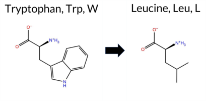 3DClick to see structure in 3D Viewer AIThe SynGAP1 missense variant W362L is not reported in ClinVar and is absent from gnomAD. All tools except premPS predict pathogenic, leaving no benign predictions. Functional prediction tools uniformly indicate a deleterious effect: SGM‑Consensus, REVEL, FoldX, Rosetta, Foldetta, PROVEAN, polyPhen‑2 (HumDiv and HumVar), SIFT, ESM1b, FATHMM, AlphaMissense‑Default, and AlphaMissense‑Optimized all classify the change as pathogenic, while premPS remains uncertain. High‑accuracy methods corroborate this assessment: AlphaMissense‑Optimized predicts pathogenic, the SGM Consensus (majority vote of AlphaMissense‑Default, ESM1b, FATHMM, PROVEAN) is Likely Pathogenic, and Foldetta (combining FoldX‑MD and Rosetta outputs) also predicts pathogenic. Consequently, the variant is most likely pathogenic, and this prediction is consistent with the absence of a ClinVar entry. Disclaimer: This summary was generated using AI and should be interpreted alongside expert review. | Likely Pathogenic | C2 | 0.328603 | Structured | 0.430310 | Uncertain | 0.957 | 0.552 | 0.125 | -12.748 | Likely Pathogenic | 0.977 | Likely Pathogenic | Likely Pathogenic | 2.16 | Destabilizing | 0.1 | 2.60 | Destabilizing | 2.38 | Destabilizing | 0.71 | Ambiguous | 0.523 | Likely Pathogenic | -11.95 | Deleterious | 0.997 | Probably Damaging | 0.986 | Probably Damaging | 1.32 | Pathogenic | 0.01 | Affected | 0.2701 | 0.2452 | -2 | -2 | 4.7 | -73.05 | |||||||||||||||||||||||||||||
| c.131G>T | W44L 2D AIThe SynGAP1 missense variant W44L is not reported in ClinVar (no ClinVar ID) and is absent from gnomAD (no gnomAD ID). Consensus from standard predictors shows a split: benign calls come from REVEL, ESM1b, and FATHMM, while pathogenic calls come from PROVEAN, polyPhen‑2 HumDiv, polyPhen‑2 HumVar, SIFT, and AlphaMissense‑Default. AlphaMissense‑Optimized returns an uncertain result. High‑accuracy assessment is inconclusive: the SGM Consensus, derived from a majority vote of AlphaMissense‑Default, ESM1b, FATHMM, and PROVEAN, is a 2‑vs‑2 tie, and Foldetta stability analysis is unavailable. Overall, the balance of evidence favors a pathogenic interpretation, with no conflict with ClinVar status because the variant is not yet catalogued there. Disclaimer: This summary was generated using AI and should be interpreted alongside expert review. | 0.301917 | Structured | 0.431379 | Uncertain | 0.377 | 0.748 | 0.375 | -5.743 | Likely Benign | 0.869 | Likely Pathogenic | Ambiguous | 0.211 | Likely Benign | -4.37 | Deleterious | 0.659 | Possibly Damaging | 0.693 | Possibly Damaging | 3.20 | Benign | 0.00 | Affected | 0.2584 | 0.3413 | -2 | -2 | 4.7 | -73.05 | ||||||||||||||||||||||||||||||||||||||||
| c.1715G>T | W572L 2D 3DClick to see structure in 3D Viewer AIThe SynGAP1 missense variant W572L is not reported in ClinVar (ClinVar ID None) and is absent from gnomAD (gnomAD ID None). Prediction tools that agree on a benign effect include only SIFT, which scores the variant as benign. All other evaluated predictors—REVEL, FoldX, Rosetta, Foldetta, PROVEAN, polyPhen‑2 (HumDiv and HumVar), ESM1b, FATHMM, AlphaMissense‑Default, and AlphaMissense‑Optimized—consistently classify the variant as pathogenic. High‑accuracy assessments further support a deleterious impact: AlphaMissense‑Optimized predicts pathogenicity; the SGM Consensus (majority vote of AlphaMissense‑Default, ESM1b, FATHMM, and PROVEAN) indicates a likely pathogenic outcome; and Foldetta, which integrates FoldX‑MD and Rosetta stability calculations, also reports a pathogenic effect. Based on the overwhelming agreement among these tools, the variant is most likely pathogenic, and this conclusion does not contradict any ClinVar annotation (none is available). Disclaimer: This summary was generated using AI and should be interpreted alongside expert review. | Likely Pathogenic | GAP | 0.150080 | Structured | 0.039626 | Uncertain | 0.935 | 0.256 | 0.000 | -15.765 | Likely Pathogenic | 0.973 | Likely Pathogenic | Likely Pathogenic | 2.62 | Destabilizing | 0.1 | 3.97 | Destabilizing | 3.30 | Destabilizing | 0.90 | Ambiguous | 0.591 | Likely Pathogenic | -11.52 | Deleterious | 1.000 | Probably Damaging | 1.000 | Probably Damaging | -0.91 | Pathogenic | 0.31 | Tolerated | 0.2246 | 0.2837 | -2 | -2 | 4.7 | -73.05 | |||||||||||||||||||||||||||||
| c.1970G>T | W657L 2D 3DClick to see structure in 3D Viewer AISynGAP1 missense variant W657L is listed in ClinVar with an uncertain significance (ClinVar ID 2767440.0) and is not reported in gnomAD. Functional prediction tools that agree on a benign effect include REVEL, FoldX, polyPhen‑2 HumDiv, polyPhen‑2 HumVar, SIFT, and FATHMM. Tools that agree on a pathogenic effect are PROVEAN, ESM1b, and AlphaMissense‑Default. Uncertain predictions come from Foldetta, premPS, and Rosetta. High‑accuracy assessments show AlphaMissense‑Optimized predicts pathogenicity, the SGM Consensus (majority vote of AlphaMissense‑Default, ESM1b, FATHMM, PROVEAN) also predicts pathogenic, and Foldetta predicts a benign folding‑stability change. Overall, the majority of evidence points toward a pathogenic impact, which is consistent with the ClinVar uncertain status but leans toward pathogenic rather than benign. Disclaimer: This summary was generated using AI and should be interpreted alongside expert review. | Likely Pathogenic | GAP | 0.030611 | Structured | 0.208729 | Uncertain | 0.941 | 0.245 | 0.000 | Uncertain | 1 | -14.411 | Likely Pathogenic | 0.960 | Likely Pathogenic | Likely Pathogenic | 0.14 | Likely Benign | 0.1 | 0.73 | Ambiguous | 0.44 | Likely Benign | 0.87 | Ambiguous | 0.213 | Likely Benign | -10.86 | Deleterious | 0.277 | Benign | 0.078 | Benign | 3.52 | Benign | 0.14 | Tolerated | 3.39 | 24 | 0.2302 | 0.2049 | -2 | -2 | 4.7 | -73.05 | |||||||||||||||||||||||||
| c.2090G>T | W697L 2D 3DClick to see structure in 3D Viewer AIThe SynGAP1 missense variant W697L is not reported in ClinVar (ClinVar status: none) and is absent from gnomAD (gnomAD ID: none). Prediction tools that agree on a benign effect include REVEL, FoldX, Rosetta, Foldetta, FATHMM, and AlphaMissense‑Optimized. Tools that predict a pathogenic effect are SGM‑Consensus, PROVEAN, polyPhen‑2 HumDiv, polyPhen‑2 HumVar, SIFT, ESM1b, and AlphaMissense‑Default. High‑accuracy assessments show AlphaMissense‑Optimized as benign, SGM‑Consensus (majority vote from AlphaMissense‑Default, ESM1b, FATHMM, PROVEAN) as pathogenic, and Foldetta as benign. No predictions are missing or inconclusive. Overall, the majority of tools (seven pathogenic vs. six benign) lean toward a pathogenic interpretation, and this does not contradict the ClinVar status, which is currently unreported. Disclaimer: This summary was generated using AI and should be interpreted alongside expert review. | Likely Pathogenic | GAP | 0.175930 | Structured | 0.400169 | Uncertain | 0.945 | 0.297 | 0.000 | -9.057 | Likely Pathogenic | 0.676 | Likely Pathogenic | Likely Benign | 0.20 | Likely Benign | 0.1 | -0.17 | Likely Benign | 0.02 | Likely Benign | 0.90 | Ambiguous | 0.280 | Likely Benign | -8.48 | Deleterious | 1.000 | Probably Damaging | 0.991 | Probably Damaging | 3.58 | Benign | 0.02 | Affected | 0.2222 | 0.2848 | -2 | -2 | 4.7 | -73.05 | |||||||||||||||||||||||||||||
| c.3470G>T | W1157L 2D AIThe SynGAP1 missense variant W1157L is not reported in ClinVar and is absent from gnomAD. Functional prediction tools show a split: benign predictions come from REVEL and ESM1b, whereas the remaining tools—PROVEAN, polyPhen‑2 (HumDiv and HumVar), SIFT, FATHMM, AlphaMissense‑Default, AlphaMissense‑Optimized, and the SGM‑Consensus—classify the change as pathogenic or likely pathogenic. High‑accuracy assessments further support a deleterious effect: AlphaMissense‑Optimized predicts pathogenicity, and the SGM‑Consensus (majority vote of AlphaMissense‑Default, ESM1b, FATHMM, and PROVEAN) also indicates likely pathogenic. Foldetta results are not available. Overall, the majority of evidence points to a pathogenic impact for W1157L, and this conclusion is consistent with the absence of a ClinVar annotation, so there is no contradiction with existing clinical data. Disclaimer: This summary was generated using AI and should be interpreted alongside expert review. | Likely Pathogenic | 0.694846 | Disordered | 0.877471 | Binding | 0.364 | 0.861 | 0.375 | -1.336 | Likely Benign | 0.995 | Likely Pathogenic | Likely Pathogenic | 0.306 | Likely Benign | -9.46 | Deleterious | 0.997 | Probably Damaging | 0.995 | Probably Damaging | 1.08 | Pathogenic | 0.00 | Affected | 0.2400 | 0.2719 | -2 | -2 | 4.7 | -73.05 | |||||||||||||||||||||||||||||||||||||||
| c.3944G>T | W1315L 2D AIThe SynGAP1 missense variant W1315L is not reported in ClinVar and is absent from gnomAD. In silico prediction tools uniformly indicate a benign effect: REVEL, PROVEAN, polyPhen‑2 (HumDiv and HumVar), SIFT, ESM1b, FATHMM, AlphaMissense‑Default, and AlphaMissense‑Optimized all classify the change as benign. No tool predicts pathogenicity. The high‑accuracy consensus methods corroborate this: AlphaMissense‑Optimized predicts benign, and the SGM Consensus (majority vote of AlphaMissense‑Default, ESM1b, FATHMM, and PROVEAN) reports a likely benign outcome. Foldetta results are unavailable. Overall, the variant is most likely benign based on the collective predictions, and this assessment does not contradict any ClinVar status, as no ClinVar entry exists. Disclaimer: This summary was generated using AI and should be interpreted alongside expert review. | Likely Benign | 0.885302 | Disordered | 0.973142 | Binding | 0.403 | 0.889 | 0.750 | 0.369 | Likely Benign | 0.304 | Likely Benign | Likely Benign | 0.066 | Likely Benign | -0.65 | Neutral | 0.115 | Benign | 0.057 | Benign | 4.29 | Benign | 0.16 | Tolerated | 0.2256 | 0.3338 | -2 | -2 | 4.7 | -73.05 | |||||||||||||||||||||||||||||||||||||||
| c.725G>T | W242L 2D 3DClick to see structure in 3D Viewer AIThe SynGAP1 missense variant W242L is not reported in ClinVar and is absent from gnomAD. Functional prediction tools cluster into two groups: benign predictions (FoldX, Rosetta, Foldetta, premPS) and pathogenic predictions (REVEL, PROVEAN, polyPhen‑2 HumDiv, polyPhen‑2 HumVar, SIFT, ESM1b, FATHMM, AlphaMissense‑Default, AlphaMissense‑Optimized, and the SGM‑Consensus score of Likely Pathogenic). High‑accuracy assessments further support this dichotomy: AlphaMissense‑Optimized predicts pathogenicity, the SGM‑Consensus (majority vote of AlphaMissense‑Default, ESM1b, FATHMM, PROVEAN) also indicates Likely Pathogenic, whereas Foldetta, which integrates FoldX‑MD and Rosetta stability outputs, reports a benign effect. Overall, the majority of evidence points toward a pathogenic impact for W242L. This conclusion is consistent with the absence of ClinVar annotation, so there is no contradiction with existing clinical data. Disclaimer: This summary was generated using AI and should be interpreted alongside expert review. | Likely Pathogenic | PH | 0.328603 | Structured | 0.352582 | Uncertain | 0.847 | 0.341 | 0.000 | -12.297 | Likely Pathogenic | 0.997 | Likely Pathogenic | Likely Pathogenic | -0.29 | Likely Benign | 0.3 | 0.12 | Likely Benign | -0.09 | Likely Benign | 0.44 | Likely Benign | 0.799 | Likely Pathogenic | -11.80 | Deleterious | 0.948 | Possibly Damaging | 0.533 | Possibly Damaging | 1.52 | Pathogenic | 0.01 | Affected | 0.2370 | 0.2986 | -2 | -2 | 4.7 | -73.05 | |||||||||||||||||||||||||||||
| c.800G>T | W267L 2D AIThe SynGAP1 missense variant W267L is not reported in ClinVar and is absent from gnomAD. Prediction tools that assess pathogenicity unanimously classify the variant as deleterious: REVEL, PROVEAN, polyPhen‑2 (HumDiv and HumVar), SIFT, ESM1b, FATHMM, AlphaMissense‑Default, and AlphaMissense‑Optimized all predict pathogenicity. No tool in the dataset predicts a benign effect. Uncertain or inconclusive results come from FoldX, Rosetta, Foldetta, and premPS, which are treated as unavailable evidence. High‑accuracy assessments further support a damaging effect: AlphaMissense‑Optimized is pathogenic, the SGM Consensus (majority vote of AlphaMissense‑Default, ESM1b, FATHMM, PROVEAN) is “Likely Pathogenic,” while Foldetta’s stability prediction is uncertain. Overall, the evidence strongly indicates that W267L is most likely pathogenic, and this conclusion does not contradict any ClinVar status because no ClinVar entry exists for this variant. Disclaimer: This summary was generated using AI and should be interpreted alongside expert review. | Likely Pathogenic | C2 | 0.216401 | Structured | 0.298060 | Uncertain | 0.943 | 0.274 | 0.000 | -13.670 | Likely Pathogenic | 0.990 | Likely Pathogenic | Likely Pathogenic | 1.19 | Ambiguous | 0.7 | 1.31 | Ambiguous | 1.25 | Ambiguous | 0.75 | Ambiguous | 0.628 | Likely Pathogenic | -11.95 | Deleterious | 0.999 | Probably Damaging | 0.996 | Probably Damaging | 1.93 | Pathogenic | 0.01 | Affected | 0.2122 | 0.2843 | -2 | -2 | 4.7 | -73.05 | |||||||||||||||||||||||||||||
| c.923G>T | W308L 2D 3DClick to see structure in 3D Viewer AIThe SynGAP1 missense variant W308L is not reported in ClinVar (no ClinVar ID) and is absent from gnomAD (no gnomAD ID). Prediction tools that assess pathogenicity unanimously classify the variant as deleterious: REVEL, FoldX, PROVEAN, polyPhen‑2 (HumDiv and HumVar), SIFT, ESM1b, FATHMM, AlphaMissense‑Default, and AlphaMissense‑Optimized all predict pathogenicity. The SGM‑Consensus, derived from a majority vote of AlphaMissense‑Default, ESM1b, FATHMM, and PROVEAN, also indicates “Likely Pathogenic.” Only Rosetta and premPS yield uncertain results, which are treated as unavailable evidence. High‑accuracy assessments reinforce the pathogenic prediction: AlphaMissense‑Optimized is pathogenic, SGM‑Consensus is likely pathogenic, and Foldetta (combining FoldX‑MD and Rosetta outputs) is pathogenic. No tools predict benign effects. **Based on the collective predictions, the variant is most likely pathogenic, and this assessment does not contradict any ClinVar status (none is available).** Disclaimer: This summary was generated using AI and should be interpreted alongside expert review. | Likely Pathogenic | C2 | 0.328603 | Structured | 0.333942 | Uncertain | 0.907 | 0.314 | 0.125 | -13.349 | Likely Pathogenic | 0.991 | Likely Pathogenic | Likely Pathogenic | 3.53 | Destabilizing | 0.5 | 1.52 | Ambiguous | 2.53 | Destabilizing | 0.53 | Ambiguous | 0.811 | Likely Pathogenic | -11.95 | Deleterious | 0.999 | Probably Damaging | 0.996 | Probably Damaging | 0.49 | Pathogenic | 0.00 | Affected | 0.2731 | 0.2754 | -2 | -2 | 4.7 | -73.05 | |||||||||||||||||||||||||||||
| c.1079A>G | E360G 2D  3DClick to see structure in 3D Viewer AIThe SynGAP1 missense variant E360G is not reported in ClinVar and is absent from gnomAD. Prediction tools largely agree on a deleterious effect: premPS is the only tool that predicts a benign outcome, whereas all other evaluated algorithms—including REVEL, FoldX, Rosetta, Foldetta, PROVEAN, polyPhen‑2 (HumDiv and HumVar), SIFT, ESM1b, FATHMM, AlphaMissense‑Default, and AlphaMissense‑Optimized—classify the substitution as pathogenic. High‑accuracy methods reinforce this consensus: AlphaMissense‑Optimized returns a pathogenic prediction; the SGM Consensus, derived from a majority vote of AlphaMissense‑Default, ESM1b, FATHMM, and PROVEAN, also indicates likely pathogenic; and Foldetta, which integrates FoldX‑MD and Rosetta stability calculations, predicts a pathogenic effect. Taken together, the overwhelming majority of evidence points to a pathogenic impact for E360G, and this conclusion is consistent with the absence of a ClinVar annotation, so there is no contradiction with existing clinical data. Disclaimer: This summary was generated using AI and should be interpreted alongside expert review. | Likely Pathogenic | C2 | 0.250310 | Structured | 0.421183 | Uncertain | 0.955 | 0.498 | 0.250 | -13.972 | Likely Pathogenic | 0.971 | Likely Pathogenic | Likely Pathogenic | 2.55 | Destabilizing | 0.1 | 2.99 | Destabilizing | 2.77 | Destabilizing | 0.31 | Likely Benign | 0.569 | Likely Pathogenic | -6.43 | Deleterious | 0.999 | Probably Damaging | 0.986 | Probably Damaging | 1.68 | Pathogenic | 0.04 | Affected | 0.2993 | 0.6935 | 0 | -2 | 3.1 | -72.06 | |||||||||||||||||||||||||||||
| c.1244A>G | E415G 2D 3DClick to see structure in 3D Viewer AIThe SynGAP1 E415G missense variant is not reported in ClinVar (ClinVar ID None) and is absent from gnomAD (gnomAD ID None). Prediction tools that agree on a benign effect include REVEL and FATHMM. Tools that predict a pathogenic effect comprise PROVEAN, polyPhen‑2 (HumDiv and HumVar), SIFT, ESM1b, AlphaMissense‑Default, AlphaMissense‑Optimized, and the SGM‑Consensus (majority vote of AlphaMissense‑Default, ESM1b, FATHMM, PROVEAN). Stability‑based methods (FoldX, Rosetta, premPS) and Foldetta are inconclusive. High‑accuracy assessments show AlphaMissense‑Optimized as pathogenic, SGM‑Consensus as Likely Pathogenic, and Foldetta as uncertain. Overall, the majority of evidence points to a pathogenic impact for E415G. This prediction is consistent with the lack of ClinVar annotation and does not contradict any existing ClinVar status. Disclaimer: This summary was generated using AI and should be interpreted alongside expert review. | Likely Pathogenic | GAP | 0.100716 | Structured | 0.330366 | Uncertain | 0.915 | 0.236 | 0.000 | -12.244 | Likely Pathogenic | 0.977 | Likely Pathogenic | Likely Pathogenic | 1.16 | Ambiguous | 0.2 | 0.88 | Ambiguous | 1.02 | Ambiguous | 0.65 | Ambiguous | 0.432 | Likely Benign | -6.45 | Deleterious | 1.000 | Probably Damaging | 0.997 | Probably Damaging | 3.20 | Benign | 0.01 | Affected | 0.2518 | 0.3158 | 0 | -2 | 3.1 | -72.06 | |||||||||||||||||||||||||||||
| c.1256A>G | E419G 2D 3DClick to see structure in 3D Viewer AIThe SynGAP1 missense variant E419G is listed in ClinVar with an uncertain significance (ClinVar ID 2004834.0) and is not reported in gnomAD. Functional prediction tools cluster into two groups: benign predictions come from REVEL and FATHMM, while pathogenic predictions are made by PROVEAN, polyPhen‑2 (HumDiv and HumVar), SIFT, ESM1b, AlphaMissense‑Default, and AlphaMissense‑Optimized. The SGM‑Consensus, derived from a majority vote of AlphaMissense‑Default, ESM1b, FATHMM, and PROVEAN, reports the variant as likely pathogenic. High‑accuracy assessments further support a deleterious effect: AlphaMissense‑Optimized predicts pathogenic, SGM‑Consensus confirms likely pathogenic, and the Foldetta stability analysis is inconclusive. No evidence from FoldX, Rosetta, or premPS is available. Overall, the preponderance of predictions indicates that E419G is most likely pathogenic, which contrasts with the current ClinVar designation of uncertain significance. Disclaimer: This summary was generated using AI and should be interpreted alongside expert review. | Likely Pathogenic | GAP | 0.102787 | Structured | 0.371949 | Uncertain | 0.961 | 0.261 | 0.000 | Uncertain | 1 | -10.589 | Likely Pathogenic | 0.956 | Likely Pathogenic | Likely Pathogenic | 1.41 | Ambiguous | 0.0 | 1.94 | Ambiguous | 1.68 | Ambiguous | 0.83 | Ambiguous | 0.469 | Likely Benign | -6.42 | Deleterious | 1.000 | Probably Damaging | 0.997 | Probably Damaging | 3.31 | Benign | 0.02 | Affected | 3.37 | 29 | 0.2992 | 0.5728 | 0 | -2 | 3.1 | -72.06 | 165.3 | 110.8 | 0.0 | 0.0 | -0.1 | 0.0 | X | Potentially Pathogenic | The carboxylate group of Glu419, located on an α helix (res. Met414-Glu436), forms a salt bridge with the side chain of either Arg716 or Lys418 from an opposing helix (res. Pro713-Arg726). The backbone amide group of Glu419 does not form H-bonds, resulting in a slight bend in the α helix. Thus, although glycine is known as an “α helix breaker,” the residue swap does not disrupt the continuity or integrity of the α helix. However, because Gly419 cannot form a salt bridge with the guanidinium group of the Arg716 side chain, the C2-GAP domain tertiary structure could be compromised during folding. | ||||||||||||||||
| c.1265A>G | E422G 2D 3DClick to see structure in 3D Viewer AISynGAP1 E422G is not reported in ClinVar and is absent from gnomAD. Functional prediction tools show a split assessment: benign predictions come from REVEL, premPS, and FATHMM, while pathogenic predictions are reported by PROVEAN, polyPhen‑2 (HumDiv and HumVar), SIFT, ESM1b, and AlphaMissense‑Default. Uncertain results are provided by FoldX, Rosetta, Foldetta, and AlphaMissense‑Optimized. High‑accuracy analyses indicate that the SGM‑Consensus, derived from a majority vote of AlphaMissense‑Default, ESM1b, FATHMM, and PROVEAN, predicts a likely pathogenic effect, whereas AlphaMissense‑Optimized remains inconclusive and Foldetta shows no definitive stability change. Overall, the majority of evidence points toward a pathogenic impact for E422G, and this conclusion does not conflict with the absence of a ClinVar classification. Disclaimer: This summary was generated using AI and should be interpreted alongside expert review. | Likely Pathogenic | GAP | 0.067594 | Structured | 0.426709 | Uncertain | 0.965 | 0.255 | 0.000 | -11.468 | Likely Pathogenic | 0.952 | Likely Pathogenic | Ambiguous | 1.13 | Ambiguous | 0.0 | 1.25 | Ambiguous | 1.19 | Ambiguous | 0.39 | Likely Benign | 0.488 | Likely Benign | -6.38 | Deleterious | 1.000 | Probably Damaging | 0.997 | Probably Damaging | 3.31 | Benign | 0.01 | Affected | 0.2440 | 0.4184 | 0 | -2 | 3.1 | -72.06 | |||||||||||||||||||||||||||||
| c.1307A>G | E436G 2D 3DClick to see structure in 3D Viewer AIThe SynGAP1 E436G missense variant is not reported in ClinVar and is absent from gnomAD. Prediction tools that agree on a benign effect include only FATHMM, while the majority of algorithms (SGM‑Consensus, REVEL, Rosetta, PROVEAN, polyPhen‑2 HumDiv/HumVar, SIFT, ESM1b, AlphaMissense‑Default, AlphaMissense‑Optimized) predict a pathogenic impact; FoldX, premPS, and Foldetta are inconclusive. High‑accuracy assessments further support a deleterious outcome: AlphaMissense‑Optimized is pathogenic, the SGM Consensus (majority vote of AlphaMissense‑Default, ESM1b, FATHMM, PROVEAN) is likely pathogenic, and Foldetta’s stability prediction is uncertain. Based on the preponderance of pathogenic predictions and the lack of benign consensus, the variant is most likely pathogenic, and this assessment does not contradict any ClinVar annotation (none is available). Disclaimer: This summary was generated using AI and should be interpreted alongside expert review. | Likely Pathogenic | GAP | 0.239899 | Structured | 0.321046 | Uncertain | 0.934 | 0.289 | 0.000 | -12.355 | Likely Pathogenic | 0.966 | Likely Pathogenic | Likely Pathogenic | 1.50 | Ambiguous | 0.1 | 2.02 | Destabilizing | 1.76 | Ambiguous | 0.58 | Ambiguous | 0.802 | Likely Pathogenic | -6.63 | Deleterious | 1.000 | Probably Damaging | 0.993 | Probably Damaging | 4.61 | Benign | 0.03 | Affected | 0.2910 | 0.4841 | 0 | -2 | 3.1 | -72.06 | |||||||||||||||||||||||||||||
| c.1334A>G | E445G 2D 3DClick to see structure in 3D Viewer AIThe SynGAP1 E445G missense variant is not reported in ClinVar and is absent from gnomAD. Prediction tools that agree on a benign effect include only FATHMM, while the majority of tools (REVEL, PROVEAN, polyPhen‑2 HumDiv, polyPhen‑2 HumVar, SIFT, ESM1b, AlphaMissense‑Default, and the SGM‑Consensus) predict a pathogenic impact. Predictions that are inconclusive or uncertain are FoldX, Rosetta, Foldetta, premPS, and AlphaMissense‑Optimized. High‑accuracy assessments show AlphaMissense‑Optimized as uncertain, the SGM‑Consensus (majority vote of AlphaMissense‑Default, ESM1b, FATHMM, and PROVEAN) as pathogenic, and Foldetta (combining FoldX‑MD and Rosetta outputs) as uncertain. Overall, the preponderance of evidence points to a pathogenic effect for E445G, and this conclusion does not contradict the ClinVar status, which currently has no entry for this variant. Disclaimer: This summary was generated using AI and should be interpreted alongside expert review. | Likely Pathogenic | GAP | 0.191378 | Structured | 0.270205 | Uncertain | 0.947 | 0.228 | 0.000 | -12.294 | Likely Pathogenic | 0.823 | Likely Pathogenic | Ambiguous | 0.80 | Ambiguous | 0.1 | 0.79 | Ambiguous | 0.80 | Ambiguous | 0.69 | Ambiguous | 0.523 | Likely Pathogenic | -6.68 | Deleterious | 1.000 | Probably Damaging | 0.993 | Probably Damaging | 3.39 | Benign | 0.01 | Affected | 0.2446 | 0.4202 | 0 | -2 | 3.1 | -72.06 | |||||||||||||||||||||||||||||
| c.1337A>G | E446G 2D 3DClick to see structure in 3D Viewer AIThe SynGAP1 E446G missense variant is not reported in ClinVar (ClinVar ID None) and is absent from gnomAD (gnomAD ID None). Prediction tools that agree on a benign effect include only FATHMM. The majority of tools predict a pathogenic impact: SGM‑Consensus, REVEL, FoldX, PROVEAN, polyPhen‑2 (HumDiv and HumVar), SIFT, ESM1b, AlphaMissense‑Default, and Foldetta. Predictions that are uncertain or inconclusive are AlphaMissense‑Optimized, Rosetta, and premPS. High‑accuracy assessments further support a deleterious effect: AlphaMissense‑Optimized is uncertain, SGM‑Consensus (majority vote from AlphaMissense‑Default, ESM1b, FATHMM, PROVEAN) is Likely Pathogenic, and Foldetta (combining FoldX‑MD and Rosetta outputs) is Pathogenic. Overall, the preponderance of evidence indicates that the variant is most likely pathogenic, and this conclusion does not contradict the ClinVar status, which currently has no classification for this variant. Disclaimer: This summary was generated using AI and should be interpreted alongside expert review. | Likely Pathogenic | GAP | 0.271506 | Structured | 0.276479 | Uncertain | 0.940 | 0.216 | 0.000 | -11.457 | Likely Pathogenic | 0.866 | Likely Pathogenic | Ambiguous | 2.62 | Destabilizing | 0.7 | 1.63 | Ambiguous | 2.13 | Destabilizing | 0.83 | Ambiguous | 0.510 | Likely Pathogenic | -6.42 | Deleterious | 1.000 | Probably Damaging | 0.997 | Probably Damaging | 3.24 | Benign | 0.00 | Affected | 0.2665 | 0.5202 | 0 | -2 | 3.1 | -72.06 | |||||||||||||||||||||||||||||
| c.1415A>G | E472G 2D 3DClick to see structure in 3D Viewer AIThe SynGAP1 missense variant E472G is not reported in ClinVar (ClinVar ID None) and has no entry in gnomAD (gnomAD ID None). Prediction tools that assess the variant’s effect largely agree on a deleterious outcome: REVEL, FoldX, Rosetta, Foldetta, PROVEAN, polyPhen‑2 (HumDiv and HumVar), SIFT, ESM1b, FATHMM, AlphaMissense‑Default, and AlphaMissense‑Optimized all classify it as pathogenic or likely pathogenic. Only premPS predicts a benign effect. High‑accuracy methods reinforce this consensus: AlphaMissense‑Optimized reports pathogenic, the SGM Consensus (majority vote of AlphaMissense‑Default, ESM1b, FATHMM, PROVEAN) indicates likely pathogenic, and Foldetta (combining FoldX‑MD and Rosetta outputs) also predicts pathogenic. No predictions are missing or inconclusive. Based on the overwhelming agreement among these tools, the variant is most likely pathogenic, and this assessment does not contradict any ClinVar status because the variant is not yet catalogued there. Disclaimer: This summary was generated using AI and should be interpreted alongside expert review. | Likely Pathogenic | GAP | 0.264545 | Structured | 0.359300 | Uncertain | 0.878 | 0.231 | 0.000 | -15.239 | Likely Pathogenic | 0.993 | Likely Pathogenic | Likely Pathogenic | 3.06 | Destabilizing | 0.2 | 2.86 | Destabilizing | 2.96 | Destabilizing | 0.24 | Likely Benign | 0.744 | Likely Pathogenic | -6.83 | Deleterious | 1.000 | Probably Damaging | 0.998 | Probably Damaging | 2.30 | Pathogenic | 0.00 | Affected | 0.3343 | 0.4950 | 0 | -2 | 3.1 | -72.06 | |||||||||||||||||||||||||||||
| c.1433A>G | E478G 2D 3DClick to see structure in 3D Viewer AIThe SynGAP1 E478G missense variant is not reported in ClinVar (ClinVar ID None) and is absent from gnomAD (gnomAD ID None). Prediction tools that agree on a benign effect include REVEL, FoldX, premPS, SIFT, FATHMM, and AlphaMissense‑Optimized. Tools that predict a pathogenic effect are PROVEAN and polyPhen‑2 HumDiv. Uncertain predictions come from Rosetta, ESM1b, AlphaMissense‑Default, and Foldetta. High‑accuracy assessments show AlphaMissense‑Optimized as benign, while the SGM Consensus (majority vote from AlphaMissense‑Default, ESM1b, FATHMM, PROVEAN) is inconclusive due to equal benign and pathogenic signals and two uncertain calls; Foldetta likewise yields an uncertain result. Overall, the majority of evaluated tools (seven benign vs. two pathogenic) support a benign classification. This consensus does not contradict any ClinVar status, as no ClinVar assertion exists for this variant. Disclaimer: This summary was generated using AI and should be interpreted alongside expert review. | GAP | 0.264545 | Structured | 0.414660 | Uncertain | 0.787 | 0.249 | 0.000 | -7.897 | In-Between | 0.418 | Ambiguous | Likely Benign | 0.40 | Likely Benign | 0.0 | 0.73 | Ambiguous | 0.57 | Ambiguous | 0.02 | Likely Benign | 0.306 | Likely Benign | -4.91 | Deleterious | 0.923 | Possibly Damaging | 0.427 | Benign | 3.41 | Benign | 0.15 | Tolerated | 0.2758 | 0.5572 | 0 | -2 | 3.1 | -72.06 | ||||||||||||||||||||||||||||||
| c.1439A>G | E480G 2D 3DClick to see structure in 3D Viewer AIThe SynGAP1 E480G missense variant is not reported in ClinVar and has no gnomAD entry. Consensus from multiple in‑silico predictors indicates a pathogenic effect: SGM‑Consensus, REVEL, Rosetta, Foldetta, PROVEAN, polyPhen‑2 (HumDiv and HumVar), SIFT, ESM1b, FATHMM, and AlphaMissense‑Default all classify it as pathogenic. Predictions that are uncertain—FoldX, premPS, and AlphaMissense‑Optimized—do not provide evidence for benignity. High‑accuracy assessments further support pathogenicity: the SGM Consensus (majority vote of AlphaMissense‑Default, ESM1b, FATHMM, PROVEAN) is pathogenic, Foldetta (combining FoldX‑MD and Rosetta outputs) is pathogenic, and AlphaMissense‑Optimized remains uncertain. Consequently, the variant is most likely pathogenic, and this assessment does not contradict any ClinVar annotation because no ClinVar record exists. Disclaimer: This summary was generated using AI and should be interpreted alongside expert review. | Likely Pathogenic | GAP | 0.216401 | Structured | 0.426867 | Uncertain | 0.798 | 0.250 | 0.000 | -11.651 | Likely Pathogenic | 0.839 | Likely Pathogenic | Ambiguous | 1.83 | Ambiguous | 0.1 | 2.34 | Destabilizing | 2.09 | Destabilizing | 0.65 | Ambiguous | 0.778 | Likely Pathogenic | -5.44 | Deleterious | 1.000 | Probably Damaging | 0.998 | Probably Damaging | -1.32 | Pathogenic | 0.03 | Affected | 0.2806 | 0.6172 | 0 | -2 | 3.1 | -72.06 | |||||||||||||||||||||||||||||
| c.1457A>G | E486G 2D 3DClick to see structure in 3D Viewer AIThe SynGAP1 E486G missense change is not listed in ClinVar and has no gnomAD entry. Functional prediction tools that agree on a benign effect include REVEL, premPS, SIFT, and FATHMM. Those that predict a damaging outcome are PROVEAN, polyPhen‑2 HumDiv, polyPhen‑2 HumVar, AlphaMissense‑Default, and ESM1b. Predictions from FoldX, Rosetta, Foldetta, and AlphaMissense‑Optimized are inconclusive. High‑accuracy assessments show AlphaMissense‑Optimized as uncertain, the SGM Consensus (majority vote of AlphaMissense‑Default, ESM1b, FATHMM, PROVEAN) as Likely Pathogenic, and Foldetta (combining FoldX‑MD and Rosetta outputs) as uncertain. Overall, the majority of evidence points toward a pathogenic effect. This conclusion is consistent with the absence of a ClinVar annotation, so there is no contradiction with existing clinical data. Disclaimer: This summary was generated using AI and should be interpreted alongside expert review. | Likely Pathogenic | GAP | 0.196879 | Structured | 0.358545 | Uncertain | 0.833 | 0.245 | 0.125 | -12.488 | Likely Pathogenic | 0.924 | Likely Pathogenic | Ambiguous | 1.09 | Ambiguous | 0.1 | 1.59 | Ambiguous | 1.34 | Ambiguous | -0.14 | Likely Benign | 0.328 | Likely Benign | -5.46 | Deleterious | 1.000 | Probably Damaging | 0.998 | Probably Damaging | 3.80 | Benign | 0.40 | Tolerated | 0.2918 | 0.5385 | 0 | -2 | 3.1 | -72.06 | |||||||||||||||||||||||||||||
| c.1484A>G | E495G 2D 3DClick to see structure in 3D Viewer AIThe SynGAP1 E495G missense variant is listed in ClinVar with an “Uncertain” status and is present in gnomAD (ID 6‑33438516‑A‑G). Among the available in‑silico predictors, the following tools uniformly indicate a pathogenic effect: REVEL, Rosetta, PROVEAN, polyPhen‑2 (HumDiv and HumVar), SIFT, ESM1b, FATHMM, AlphaMissense‑Default, and the SGM‑Consensus (which itself is a majority vote of AlphaMissense‑Default, ESM1b, FATHMM, and PROVEAN). No tool in the dataset predicts a benign outcome; predictions that are uncertain (FoldX, Foldetta, premPS, AlphaMissense‑Optimized) are treated as unavailable. High‑accuracy assessments show AlphaMissense‑Optimized as “Uncertain,” SGM‑Consensus as “Likely Pathogenic,” and Foldetta as “Uncertain.” Overall, the preponderance of pathogenic predictions strongly suggests that the variant is most likely pathogenic, which does not contradict the ClinVar “Uncertain” classification. Disclaimer: This summary was generated using AI and should be interpreted alongside expert review. | Likely Pathogenic | GAP | 0.164327 | Structured | 0.364496 | Uncertain | 0.933 | 0.161 | 0.000 | Uncertain | 1 | 6-33438516-A-G | 1 | 6.20e-7 | -9.400 | Likely Pathogenic | 0.923 | Likely Pathogenic | Ambiguous | 1.21 | Ambiguous | 0.0 | 2.06 | Destabilizing | 1.64 | Ambiguous | 0.78 | Ambiguous | 0.867 | Likely Pathogenic | -6.70 | Deleterious | 1.000 | Probably Damaging | 0.999 | Probably Damaging | -1.46 | Pathogenic | 0.02 | Affected | 3.37 | 35 | 0.2177 | 0.4784 | -2 | 0 | 3.1 | -72.06 | ||||||||||||||||||||||
| c.1487A>G | E496G 2D 3DClick to see structure in 3D Viewer AIThe SynGAP1 E496G missense variant is listed in ClinVar with an “Uncertain” status and is not reported in gnomAD. Prediction tools that assess the variant’s effect fall into two groups: no tool predicts a benign outcome, while eight tools (REVEL, PROVEAN, polyPhen‑2 HumDiv, polyPhen‑2 HumVar, SIFT, ESM1b, FATHMM, AlphaMissense‑Default) all predict a pathogenic effect. The SGM‑Consensus, which is a majority vote of AlphaMissense‑Default, ESM1b, FATHMM, and PROVEAN, also indicates a likely pathogenic outcome. High‑accuracy assessments are mixed: AlphaMissense‑Optimized is uncertain, the SGM‑Consensus remains likely pathogenic, and Foldetta (combining FoldX‑MD and Rosetta outputs) is uncertain. Overall, the preponderance of evidence points to a pathogenic effect, contradicting the current ClinVar “Uncertain” classification. Disclaimer: This summary was generated using AI and should be interpreted alongside expert review. | Likely Pathogenic | GAP | 0.102787 | Structured | 0.383296 | Uncertain | 0.945 | 0.179 | 0.000 | Uncertain | 1 | -13.529 | Likely Pathogenic | 0.850 | Likely Pathogenic | Ambiguous | 1.83 | Ambiguous | 0.1 | 1.76 | Ambiguous | 1.80 | Ambiguous | 0.92 | Ambiguous | 0.825 | Likely Pathogenic | -6.16 | Deleterious | 1.000 | Probably Damaging | 0.999 | Probably Damaging | -1.45 | Pathogenic | 0.02 | Affected | 3.37 | 35 | 0.2435 | 0.3473 | 0 | -2 | 3.1 | -72.06 | 173.9 | 103.1 | 0.0 | 0.0 | -0.7 | 0.0 | X | X | Potentially Pathogenic | Glu496 is located in the α-helix (res. Leu489-Glu519), and its carboxylate group forms salt bridges with the neighbouring residues Lys492 and Arg499 in the WT simulations. Glu496 also forms a hydrogen bond with Ser449 on an opposing helix (res. Val441-Ser457). In the variant simulations, Gly496 cannot form these salt bridges, which could weaken the secondary structure. Additionally, the loss of the hydrogen bond with Ser449 on the opposite helix can weaken the tertiary structure assembly. Moreover, glycine is an α-helix breaker, and it is seen to weaken the integrity of the helix as the hydrogen bonding between the backbone atoms of Gly496 and Ala493 breaks down. Also, due to its location at the GAP-Ras interface, the interaction of Glu496 with Arg499 and Lys492 might play a role in complex association and stability, which cannot be fully addressed using the SynGAP solvent-only simulations. | |||||||||||||||
| c.1535A>G | E512G 2D 3DClick to see structure in 3D Viewer AIThe SynGAP1 missense variant E512G is not reported in ClinVar (ClinVar ID None) and has no entry in gnomAD (gnomAD ID None). Prediction tools that agree on a benign effect include REVEL, premPS, and FATHMM, whereas a majority of tools (SGM‑Consensus, PROVEAN, polyPhen‑2 HumDiv, polyPhen‑2 HumVar, SIFT, ESM1b, and AlphaMissense‑Default) predict a pathogenic impact. The remaining tools—FoldX, Rosetta, Foldetta, and AlphaMissense‑Optimized—return uncertain or inconclusive results and are treated as unavailable evidence. High‑accuracy assessments show AlphaMissense‑Optimized as uncertain, SGM‑Consensus as Likely Pathogenic (majority vote from AlphaMissense‑Default, ESM1b, FATHMM, and PROVEAN), and Foldetta as uncertain. Overall, the preponderance of evidence points to a pathogenic effect for E512G, and this conclusion does not contradict the absence of a ClinVar classification. Disclaimer: This summary was generated using AI and should be interpreted alongside expert review. | Likely Pathogenic | GAP | 0.092881 | Structured | 0.247079 | Uncertain | 0.923 | 0.273 | 0.000 | -11.996 | Likely Pathogenic | 0.817 | Likely Pathogenic | Ambiguous | 0.86 | Ambiguous | 0.1 | 1.84 | Ambiguous | 1.35 | Ambiguous | 0.17 | Likely Benign | 0.381 | Likely Benign | -6.46 | Deleterious | 0.997 | Probably Damaging | 0.915 | Probably Damaging | 3.30 | Benign | 0.02 | Affected | 0.3262 | 0.4185 | 0 | -2 | 3.1 | -72.06 | |||||||||||||||||||||||||||||
| c.1556A>G | E519G 2D 3DClick to see structure in 3D Viewer AIThe SynGAP1 E519G missense variant is not reported in ClinVar (ClinVar status: None) and has no entry in gnomAD (gnomAD ID: None). Prediction tools that agree on a benign effect include REVEL, FoldX, FATHMM, and premPS. Tools that predict a pathogenic effect comprise SGM‑Consensus (Likely Pathogenic), PROVEAN, polyPhen‑2 HumDiv, polyPhen‑2 HumVar, SIFT, ESM1b, and AlphaMissense‑Default. Uncertain predictions come from Foldetta, AlphaMissense‑Optimized, and Rosetta. High‑accuracy assessments: AlphaMissense‑Optimized is uncertain; SGM‑Consensus (majority vote of AlphaMissense‑Default, ESM1b, FATHMM, PROVEAN) indicates Likely Pathogenic; Foldetta is uncertain. Overall, the majority of available predictions (7 pathogenic vs. 4 benign) lean toward a pathogenic impact. This conclusion is consistent with the lack of ClinVar annotation, so there is no contradiction with ClinVar status. Disclaimer: This summary was generated using AI and should be interpreted alongside expert review. | Likely Pathogenic | GAP | 0.078022 | Structured | 0.104514 | Uncertain | 0.899 | 0.328 | 0.000 | -10.835 | Likely Pathogenic | 0.884 | Likely Pathogenic | Ambiguous | 0.22 | Likely Benign | 0.1 | 0.92 | Ambiguous | 0.57 | Ambiguous | 0.22 | Likely Benign | 0.458 | Likely Benign | -6.12 | Deleterious | 1.000 | Probably Damaging | 0.998 | Probably Damaging | 3.21 | Benign | 0.01 | Affected | 0.2729 | 0.3471 | 0 | -2 | 3.1 | -72.06 | |||||||||||||||||||||||||||||
| c.1562A>G | E521G 2D 3DClick to see structure in 3D Viewer AIThe SynGAP1 missense variant E521G is not reported in ClinVar (ClinVar ID None) and is absent from gnomAD (gnomAD ID None). Prediction tools that agree on a benign effect include REVEL, FoldX, premPS, SIFT, ESM1b, FATHMM, and AlphaMissense‑Optimized. Tools that predict a pathogenic effect are PROVEAN, polyPhen‑2 HumDiv, polyPhen‑2 HumVar, and AlphaMissense‑Default. The high‑accuracy methods give the following results: AlphaMissense‑Optimized predicts benign; Foldetta (combining FoldX‑MD and Rosetta outputs) predicts benign; the SGM Consensus (majority vote of AlphaMissense‑Default, ESM1b, FATHMM, PROVEAN) is inconclusive because it is a 2‑vs‑2 split and is treated as unavailable. Overall, the majority of evidence points to a benign impact. The variant is most likely benign based on current predictions, and this assessment does not contradict any ClinVar status because no ClinVar entry exists. Disclaimer: This summary was generated using AI and should be interpreted alongside expert review. | GAP | 0.086953 | Structured | 0.062387 | Uncertain | 0.865 | 0.349 | 0.000 | -6.636 | Likely Benign | 0.767 | Likely Pathogenic | Likely Benign | 0.18 | Likely Benign | 0.1 | 0.58 | Ambiguous | 0.38 | Likely Benign | 0.14 | Likely Benign | 0.289 | Likely Benign | -3.64 | Deleterious | 1.000 | Probably Damaging | 0.999 | Probably Damaging | 3.31 | Benign | 0.07 | Tolerated | 0.2932 | 0.5455 | 0 | -2 | 3.1 | -72.06 | ||||||||||||||||||||||||||||||
| c.1565A>G | E522G 2D 3DClick to see structure in 3D Viewer AIThe SynGAP1 E522G missense variant is not reported in ClinVar (no ClinVar ID) and is absent from gnomAD. Consensus from multiple in‑silico predictors indicates a pathogenic effect: SGM‑Consensus, REVEL, PROVEAN, polyPhen‑2 (HumDiv and HumVar), SIFT, ESM1b, FATHMM, AlphaMissense‑Default, and AlphaMissense‑Optimized all classify the change as deleterious. No tool reports a benign outcome. High‑accuracy assessments further support pathogenicity: AlphaMissense‑Optimized predicts a damaging effect, the SGM Consensus (majority vote of AlphaMissense‑Default, ESM1b, FATHMM, PROVEAN) is labeled Likely Pathogenic, while Foldetta’s stability analysis is inconclusive. FoldX, Rosetta, and premPS are uncertain and are treated as unavailable evidence. Overall, the computational evidence overwhelmingly points to a pathogenic impact, and this conclusion does not conflict with the absence of a ClinVar annotation. Disclaimer: This summary was generated using AI and should be interpreted alongside expert review. | Likely Pathogenic | GAP | 0.092881 | Structured | 0.046216 | Uncertain | 0.823 | 0.376 | 0.000 | -10.053 | Likely Pathogenic | 0.986 | Likely Pathogenic | Likely Pathogenic | 0.72 | Ambiguous | 0.0 | 1.62 | Ambiguous | 1.17 | Ambiguous | 0.58 | Ambiguous | 0.823 | Likely Pathogenic | -6.59 | Deleterious | 1.000 | Probably Damaging | 0.999 | Probably Damaging | -1.34 | Pathogenic | 0.01 | Affected | 0.2672 | 0.3654 | 0 | -2 | 3.1 | -72.06 | |||||||||||||||||||||||||||||
| c.1574A>G | E525G 2D 3DClick to see structure in 3D Viewer AIThe SynGAP1 missense variant E525G is not reported in ClinVar and is absent from gnomAD. Prediction tools that classify the variant as benign include only FATHMM. All other evaluated algorithms—REVEL, PROVEAN, polyPhen‑2 HumDiv, polyPhen‑2 HumVar, SIFT, ESM1b, AlphaMissense‑Default, AlphaMissense‑Optimized, and the SGM‑Consensus (majority vote of AlphaMissense‑Default, ESM1b, FATHMM, PROVEAN)—predict it to be pathogenic. High‑accuracy assessments further support a deleterious effect: AlphaMissense‑Optimized returns a pathogenic score, the SGM‑Consensus also indicates pathogenicity, while Foldetta, which integrates FoldX‑MD and Rosetta stability outputs, yields an uncertain result. Taken together, the overwhelming majority of evidence points to a pathogenic impact for E525G. This conclusion is consistent with the absence of ClinVar annotation and gnomAD data, and there is no contradiction with ClinVar status. Disclaimer: This summary was generated using AI and should be interpreted alongside expert review. | Likely Pathogenic | GAP | 0.206376 | Structured | 0.023618 | Uncertain | 0.937 | 0.382 | 0.125 | -13.181 | Likely Pathogenic | 0.992 | Likely Pathogenic | Likely Pathogenic | 1.71 | Ambiguous | 0.6 | 0.90 | Ambiguous | 1.31 | Ambiguous | 0.78 | Ambiguous | 0.691 | Likely Pathogenic | -6.96 | Deleterious | 1.000 | Probably Damaging | 0.998 | Probably Damaging | 2.68 | Benign | 0.00 | Affected | 0.2947 | 0.3886 | 0 | -2 | 3.1 | -72.06 | |||||||||||||||||||||||||||||
| c.1613A>G | E538G 2D 3DClick to see structure in 3D Viewer AIThe SynGAP1 missense variant E538G is not reported in ClinVar and is absent from gnomAD. Functional prediction tools cluster into two groups: benign predictions come from REVEL, premPS, FATHMM, and AlphaMissense‑Optimized; pathogenic predictions arise from PROVEAN, polyPhen‑2 (HumDiv and HumVar), SIFT, ESM1b, and AlphaMissense‑Default. The SGM Consensus, derived from a majority vote of AlphaMissense‑Default, ESM1b, FATHMM, and PROVEAN, is labeled Likely Pathogenic. High‑accuracy assessments show AlphaMissense‑Optimized as benign, SGM Consensus as Likely Pathogenic, and Foldetta (combining FoldX‑MD and Rosetta outputs) as uncertain. No folding‑stability result is definitive. Overall, the balance of evidence, including the majority‑vote SGM Consensus and the higher number of pathogenic predictions, points to a pathogenic effect for E538G. This conclusion does not contradict ClinVar status, as the variant is not yet catalogued there. Disclaimer: This summary was generated using AI and should be interpreted alongside expert review. | Likely Pathogenic | GAP | 0.122885 | Structured | 0.033501 | Uncertain | 0.938 | 0.359 | 0.000 | -9.258 | Likely Pathogenic | 0.701 | Likely Pathogenic | Likely Benign | 0.91 | Ambiguous | 0.0 | 1.07 | Ambiguous | 0.99 | Ambiguous | 0.31 | Likely Benign | 0.285 | Likely Benign | -5.10 | Deleterious | 0.993 | Probably Damaging | 0.700 | Possibly Damaging | 3.33 | Benign | 0.03 | Affected | 0.3173 | 0.3713 | 0 | -2 | 3.1 | -72.06 | |||||||||||||||||||||||||||||
| c.1643A>G | E548G 2D 3DClick to see structure in 3D Viewer AIThe SynGAP1 E548G missense variant is not reported in ClinVar and is absent from gnomAD. Prediction tools that agree on a benign effect include SIFT and FATHMM, while the majority of other in silico predictors (REVEL, PROVEAN, polyPhen‑2 HumDiv, polyPhen‑2 HumVar, ESM1b, AlphaMissense‑Default, AlphaMissense‑Optimized) indicate a pathogenic impact; SGM‑Consensus also classifies the variant as likely pathogenic. Uncertain results are reported by FoldX, Rosetta, Foldetta, and premPS. High‑accuracy assessments show AlphaMissense‑Optimized predicting pathogenicity, SGM‑Consensus confirming a likely pathogenic status, and Foldetta yielding an inconclusive stability change. Overall, the consensus of the available predictions points to a pathogenic effect for E548G, and this conclusion is not contradicted by any ClinVar annotation. Disclaimer: This summary was generated using AI and should be interpreted alongside expert review. | Likely Pathogenic | GAP | 0.054297 | Structured | 0.008632 | Uncertain | 0.965 | 0.288 | 0.000 | -12.010 | Likely Pathogenic | 0.990 | Likely Pathogenic | Likely Pathogenic | 0.97 | Ambiguous | 0.1 | 1.66 | Ambiguous | 1.32 | Ambiguous | 0.59 | Ambiguous | 0.521 | Likely Pathogenic | -6.73 | Deleterious | 1.000 | Probably Damaging | 0.999 | Probably Damaging | 3.32 | Benign | 0.06 | Tolerated | 0.2638 | 0.3654 | 0 | -2 | 3.1 | -72.06 | |||||||||||||||||||||||||||||
| c.1691A>G | E564G 2D 3DClick to see structure in 3D Viewer AIThe SynGAP1 missense variant E564G is listed in gnomAD (ID 6‑33440743‑A‑G) but has no ClinVar entry. Prediction tools that assess pathogenicity are unanimous: REVEL, FoldX (uncertain), Rosetta, Foldetta, premPS (uncertain), PROVEAN, polyPhen‑2 (HumDiv and HumVar), SIFT, ESM1b, FATHMM, AlphaMissense‑Default, and AlphaMissense‑Optimized all indicate a pathogenic effect, while no tool predicts a benign outcome. High‑accuracy methods reinforce this view: AlphaMissense‑Optimized is pathogenic; the SGM Consensus (majority vote of AlphaMissense‑Default, ESM1b, FATHMM, PROVEAN) is pathogenic; and Foldetta (combining FoldX‑MD and Rosetta outputs) is pathogenic. Thus, the variant is most likely pathogenic based on the available predictions, and this assessment is not contradicted by ClinVar status, which currently contains no entry for E564G. Disclaimer: This summary was generated using AI and should be interpreted alongside expert review. | Likely Pathogenic | GAP | 0.023534 | Structured | 0.038418 | Uncertain | 0.891 | 0.208 | 0.000 | 6-33440743-A-G | -15.053 | Likely Pathogenic | 0.969 | Likely Pathogenic | Likely Pathogenic | 1.69 | Ambiguous | 0.1 | 2.55 | Destabilizing | 2.12 | Destabilizing | 0.80 | Ambiguous | 0.735 | Likely Pathogenic | -6.83 | Deleterious | 1.000 | Probably Damaging | 0.998 | Probably Damaging | -1.23 | Pathogenic | 0.04 | Affected | 3.37 | 35 | 0.2749 | 0.4443 | -2 | 0 | 3.1 | -72.06 | ||||||||||||||||||||||||||
| c.1700A>G | E567G 2D 3DClick to see structure in 3D Viewer AIThe SynGAP1 E567G missense variant is not reported in ClinVar and is absent from gnomAD. Prediction tools that agree on benign impact include REVEL, SIFT, and FATHMM, whereas those that agree on pathogenic impact include AlphaMissense‑Default, ESM1b, PROVEAN, polyPhen‑2 HumDiv, polyPhen‑2 HumVar, Rosetta, and Foldetta. Predictions that are inconclusive are FoldX, premPS, and AlphaMissense‑Optimized. High‑accuracy assessments show AlphaMissense‑Optimized as uncertain, the SGM Consensus (majority vote of AlphaMissense‑Default, ESM1b, FATHMM, PROVEAN) as pathogenic, and Foldetta (combining FoldX‑MD and Rosetta outputs) as pathogenic. Taken together, the majority of evidence points to a pathogenic effect. This conclusion is not contradicted by ClinVar status, which has no entry for this variant. Disclaimer: This summary was generated using AI and should be interpreted alongside expert review. | Likely Pathogenic | GAP | 0.021816 | Structured | 0.051008 | Uncertain | 0.916 | 0.234 | 0.000 | -13.402 | Likely Pathogenic | 0.912 | Likely Pathogenic | Ambiguous | 1.47 | Ambiguous | 0.0 | 3.48 | Destabilizing | 2.48 | Destabilizing | 0.75 | Ambiguous | 0.456 | Likely Benign | -6.67 | Deleterious | 1.000 | Probably Damaging | 0.999 | Probably Damaging | 3.40 | Benign | 0.07 | Tolerated | 0.2586 | 0.4715 | 0 | -2 | 3.1 | -72.06 | |||||||||||||||||||||||||||||
| c.1733A>G | E578G 2D 3DClick to see structure in 3D Viewer AIThe SynGAP1 E578G missense change is not reported in ClinVar (ClinVar ID None) and is absent from gnomAD (gnomAD ID None). Prediction tools that agree on a benign effect include premPS, PROVEAN, SIFT, and AlphaMissense‑Optimized. Those that agree on a pathogenic effect are REVEL, polyPhen‑2 (HumDiv and HumVar), FATHMM, and AlphaMissense‑Default. Four tools give uncertain or inconclusive results (FoldX, Rosetta, Foldetta, ESM1b). High‑accuracy assessments show AlphaMissense‑Optimized as benign, while the SGM Consensus—derived from a majority vote of AlphaMissense‑Default, ESM1b, FATHMM, and PROVEAN—predicts pathogenic. Foldetta, a protein‑folding stability method, yields an uncertain result. Overall, the majority of high‑confidence predictions lean toward pathogenicity, and this conclusion does not contradict the ClinVar status, which has no entry for this variant. Disclaimer: This summary was generated using AI and should be interpreted alongside expert review. | GAP | 0.059222 | Structured | 0.020971 | Uncertain | 0.902 | 0.240 | 0.000 | -7.680 | In-Between | 0.601 | Likely Pathogenic | Likely Benign | 0.98 | Ambiguous | 0.1 | 0.63 | Ambiguous | 0.81 | Ambiguous | 0.08 | Likely Benign | 0.589 | Likely Pathogenic | -2.40 | Neutral | 1.000 | Probably Damaging | 0.998 | Probably Damaging | -1.49 | Pathogenic | 0.19 | Tolerated | 0.2608 | 0.4843 | 0 | -2 | 3.1 | -72.06 | ||||||||||||||||||||||||||||||
| c.1745A>G | E582G 2D 3DClick to see structure in 3D Viewer AIThe SynGAP1 missense variant E582G is not reported in ClinVar (ClinVar ID: None) and has no entry in gnomAD (gnomAD ID: None). Prediction tools that agree on a benign effect include REVEL, premPS, SIFT, FATHMM, and AlphaMissense‑Optimized. Those that predict a pathogenic effect are SGM‑Consensus, PROVEAN, polyPhen‑2 HumDiv, polyPhen‑2 HumVar, ESM1b, and AlphaMissense‑Default. FoldX, Rosetta, and Foldetta give uncertain or inconclusive results. High‑accuracy methods give mixed signals: AlphaMissense‑Optimized predicts benign, the SGM‑Consensus (a majority vote of AlphaMissense‑Default, ESM1b, FATHMM, and PROVEAN) predicts pathogenic, and Foldetta’s stability assessment is uncertain. Overall, six tools predict pathogenicity while five predict benign, and the high‑accuracy consensus is split. Thus, the variant is most likely pathogenic based on the preponderance of evidence, and this assessment does not contradict ClinVar status, which currently has no classification for this variant. Disclaimer: This summary was generated using AI and should be interpreted alongside expert review. | Likely Pathogenic | GAP | 0.102787 | Structured | 0.033838 | Uncertain | 0.845 | 0.235 | 0.000 | -9.621 | Likely Pathogenic | 0.630 | Likely Pathogenic | Likely Benign | 1.35 | Ambiguous | 0.2 | 1.24 | Ambiguous | 1.30 | Ambiguous | 0.37 | Likely Benign | 0.224 | Likely Benign | -3.95 | Deleterious | 1.000 | Probably Damaging | 0.999 | Probably Damaging | 3.13 | Benign | 0.13 | Tolerated | 0.2835 | 0.3325 | 0 | -2 | 3.1 | -72.06 | |||||||||||||||||||||||||||||
| c.182A>G | E61G 2D AIThe SynGAP1 missense variant E61G is not reported in ClinVar and is absent from gnomAD. Functional prediction tools cluster into two groups: benign predictions come from REVEL, PROVEAN, ESM1b, FATHMM, and AlphaMissense‑Optimized, while pathogenic predictions arise from polyPhen‑2 (HumDiv and HumVar) and SIFT. AlphaMissense‑Default remains uncertain. The high‑accuracy consensus methods reinforce the benign assessment: AlphaMissense‑Optimized predicts benign, the SGM Consensus (majority vote of AlphaMissense‑Default, ESM1b, FATHMM, and PROVEAN) reports “Likely Benign,” and the protein‑folding stability tool Foldetta is not available for this variant. Overall, the majority of evidence points to a benign effect. There is no ClinVar entry to contradict this conclusion. Disclaimer: This summary was generated using AI and should be interpreted alongside expert review. | Likely Benign | 0.352862 | Structured | 0.477329 | Uncertain | 0.518 | 0.699 | 0.125 | -5.574 | Likely Benign | 0.469 | Ambiguous | Likely Benign | 0.072 | Likely Benign | -1.68 | Neutral | 0.659 | Possibly Damaging | 0.775 | Possibly Damaging | 4.08 | Benign | 0.00 | Affected | 0.3593 | 0.4965 | 0 | -2 | 3.1 | -72.06 | |||||||||||||||||||||||||||||||||||||||
| c.1838A>G | E613G 2D 3DClick to see structure in 3D Viewer AIThe SynGAP1 missense variant E613G is not reported in ClinVar and has no entry in gnomAD. Prediction tools that agree on a benign effect include only premPS, whereas the remaining tools (SGM‑Consensus, REVEL, PROVEAN, polyPhen‑2 HumDiv, polyPhen‑2 HumVar, SIFT, ESM1b, FATHMM, AlphaMissense‑Default) uniformly predict a pathogenic impact. The high‑accuracy methods give the following results: AlphaMissense‑Optimized is uncertain; the SGM Consensus, derived from a majority vote of AlphaMissense‑Default, ESM1b, FATHMM, and PROVEAN, is pathogenic; and Foldetta, which integrates FoldX‑MD and Rosetta outputs, is uncertain. No prediction or stability assessment is missing or inconclusive beyond the uncertain labels. Overall, the preponderance of evidence points to a pathogenic effect for E613G, and this assessment does not contradict the absence of a ClinVar classification. Disclaimer: This summary was generated using AI and should be interpreted alongside expert review. | Likely Pathogenic | GAP | 0.275179 | Structured | 0.193489 | Uncertain | 0.816 | 0.254 | 0.000 | -12.417 | Likely Pathogenic | 0.911 | Likely Pathogenic | Ambiguous | 1.49 | Ambiguous | 0.3 | 1.34 | Ambiguous | 1.42 | Ambiguous | 0.08 | Likely Benign | 0.641 | Likely Pathogenic | -6.56 | Deleterious | 1.000 | Probably Damaging | 0.998 | Probably Damaging | -1.26 | Pathogenic | 0.01 | Affected | 0.3422 | 0.5266 | 0 | -2 | 3.1 | -72.06 | |||||||||||||||||||||||||||||
| c.1850A>G | E617G 2D 3DClick to see structure in 3D Viewer AIThe SynGAP1 missense variant E617G is not reported in ClinVar and is absent from gnomAD. Prediction tools cluster into two groups: benign predictions come from premPS, SIFT, and AlphaMissense‑Optimized, whereas pathogenic predictions are made by SGM‑Consensus, REVEL, PROVEAN, polyPhen‑2 (HumDiv and HumVar), FATHMM, and AlphaMissense‑Default. Uncertain results are reported by FoldX, Rosetta, Foldetta, and ESM1b. High‑accuracy methods give a mixed picture: AlphaMissense‑Optimized predicts benign, the SGM Consensus (majority vote of AlphaMissense‑Default, ESM1b, FATHMM, PROVEAN) predicts pathogenic, and Foldetta is uncertain. Overall, the majority of evidence points to a pathogenic effect. This conclusion is not contradicted by ClinVar, which contains no entry for this variant. Disclaimer: This summary was generated using AI and should be interpreted alongside expert review. | Likely Pathogenic | GAP | 0.111485 | Structured | 0.155123 | Uncertain | 0.877 | 0.240 | 0.000 | -7.984 | In-Between | 0.777 | Likely Pathogenic | Likely Benign | 0.59 | Ambiguous | 0.2 | 1.60 | Ambiguous | 1.10 | Ambiguous | 0.22 | Likely Benign | 0.701 | Likely Pathogenic | -4.99 | Deleterious | 1.000 | Probably Damaging | 0.998 | Probably Damaging | -1.41 | Pathogenic | 0.18 | Tolerated | 0.2344 | 0.5442 | 0 | -2 | 3.1 | -72.06 | |||||||||||||||||||||||||||||
| c.188A>G | E63G 2D AIThe SynGAP1 missense variant E63G is not reported in ClinVar (no ClinVar ID) and is absent from gnomAD (no gnomAD ID). Prediction tools that agree on a benign effect include REVEL, PROVEAN, ESM1b, and FATHMM, while those that predict a pathogenic effect are polyPhen‑2 HumDiv, polyPhen‑2 HumVar, SIFT, and AlphaMissense‑Default. The SGM‑Consensus, derived from a majority vote of AlphaMissense‑Default, ESM1b, FATHMM, and PROVEAN, reports the variant as Likely Benign. AlphaMissense‑Optimized yields an uncertain result, and no Foldetta stability assessment is available. Overall, the majority of individual predictors and the SGM‑Consensus lean toward a benign interpretation, with no conflicting evidence from ClinVar. Thus, the variant is most likely benign based on current computational predictions, and this assessment does not contradict any ClinVar status. Disclaimer: This summary was generated using AI and should be interpreted alongside expert review. | Likely Benign | 0.429200 | Structured | 0.474807 | Uncertain | 0.494 | 0.739 | 0.125 | -3.450 | Likely Benign | 0.898 | Likely Pathogenic | Ambiguous | 0.150 | Likely Benign | -2.24 | Neutral | 0.659 | Possibly Damaging | 0.775 | Possibly Damaging | 3.87 | Benign | 0.00 | Affected | 0.2705 | 0.5786 | 0 | -2 | 3.1 | -72.06 | |||||||||||||||||||||||||||||||||||||||
| c.1928A>G | E643G 2D 3DClick to see structure in 3D Viewer AIThe SynGAP1 missense variant E643G is not reported in ClinVar and is absent from gnomAD. Prediction tools that agree on a benign effect include polyPhen‑2 HumVar, FATHMM, and AlphaMissense‑Optimized. Those that predict a pathogenic impact comprise SGM‑Consensus, REVEL, Rosetta, premPS, PROVEAN, polyPhen‑2 HumDiv, SIFT, ESM1b, and AlphaMissense‑Default; FoldX and Foldetta are uncertain. High‑accuracy assessments show AlphaMissense‑Optimized as benign, while the SGM Consensus (majority vote of AlphaMissense‑Default, ESM1b, FATHMM, PROVEAN) is pathogenic. Foldetta’s stability prediction is uncertain. Overall, the majority of reliable tools predict pathogenicity, and this conclusion does not contradict the lack of ClinVar annotation. Thus, the variant is most likely pathogenic based on current computational evidence. Disclaimer: This summary was generated using AI and should be interpreted alongside expert review. | Likely Pathogenic | GAP | 0.033407 | Structured | 0.215915 | Uncertain | 0.871 | 0.315 | 0.000 | -12.503 | Likely Pathogenic | 0.707 | Likely Pathogenic | Likely Benign | 1.45 | Ambiguous | 0.3 | 2.06 | Destabilizing | 1.76 | Ambiguous | 1.01 | Destabilizing | 0.520 | Likely Pathogenic | -6.81 | Deleterious | 0.983 | Probably Damaging | 0.390 | Benign | 2.94 | Benign | 0.00 | Affected | 0.2821 | 0.5319 | 0 | -2 | 3.1 | -72.06 | |||||||||||||||||||||||||||||
| c.1952A>G | E651G 2D 3DClick to see structure in 3D Viewer AIThe SynGAP1 E651G missense variant is not reported in ClinVar (ClinVar ID None) and is absent from gnomAD (gnomAD ID None). Prediction tools that agree on a benign effect include REVEL, premPS, SIFT, FATHMM, and AlphaMissense‑Optimized. Those that predict a pathogenic effect are PROVEAN, polyPhen‑2 HumDiv, polyPhen‑2 HumVar, and AlphaMissense‑Default. Predictions that are uncertain or unavailable are FoldX, Rosetta, ESM1b, and Foldetta. High‑accuracy assessments show AlphaMissense‑Optimized as benign, the SGM Consensus (majority vote from AlphaMissense‑Default, ESM1b, FATHMM, PROVEAN) as pathogenic, and Foldetta as unavailable. Overall, the majority of tools (five benign vs. four pathogenic) lean toward a benign interpretation, and this does not contradict the lack of ClinVar annotation. Thus, based on the available predictions, the variant is most likely benign. Disclaimer: This summary was generated using AI and should be interpreted alongside expert review. | GAP | 0.088832 | Structured | 0.365409 | Uncertain | 0.955 | 0.340 | 0.000 | -7.596 | In-Between | 0.591 | Likely Pathogenic | Likely Benign | 0.66 | Ambiguous | 0.1 | 1.61 | Ambiguous | 1.14 | Ambiguous | 0.32 | Likely Benign | 0.444 | Likely Benign | -4.78 | Deleterious | 0.999 | Probably Damaging | 0.935 | Probably Damaging | 3.31 | Benign | 0.06 | Tolerated | 0.2908 | 0.4488 | 0 | -2 | 3.1 | -72.06 | ||||||||||||||||||||||||||||||
| c.1961A>G | E654G 2D 3DClick to see structure in 3D Viewer AIThe SynGAP1 E654G missense variant is not reported in ClinVar and is absent from gnomAD. Prediction tools that agree on benign impact are premPS and FATHMM, while the majority (REVEL, PROVEAN, polyPhen‑2 HumDiv, polyPhen‑2 HumVar, SIFT, ESM1b, AlphaMissense‑Default, and the SGM‑Consensus) predict pathogenicity. High‑accuracy assessments show AlphaMissense‑Optimized as uncertain, the SGM‑Consensus (majority vote from AlphaMissense‑Default, ESM1b, FATHMM, PROVEAN) as likely pathogenic, and Foldetta (combining FoldX‑MD and Rosetta outputs) as uncertain. Overall, the preponderance of evidence points to a pathogenic effect for E654G. This conclusion is consistent with the lack of ClinVar annotation, so there is no contradiction with existing clinical database status. Disclaimer: This summary was generated using AI and should be interpreted alongside expert review. | Likely Pathogenic | GAP | 0.026892 | Structured | 0.303029 | Uncertain | 0.957 | 0.311 | 0.000 | -12.487 | Likely Pathogenic | 0.898 | Likely Pathogenic | Ambiguous | 1.29 | Ambiguous | 0.2 | 1.62 | Ambiguous | 1.46 | Ambiguous | 0.34 | Likely Benign | 0.547 | Likely Pathogenic | -6.73 | Deleterious | 0.999 | Probably Damaging | 0.935 | Probably Damaging | 3.26 | Benign | 0.01 | Affected | 0.2818 | 0.3109 | 0 | -2 | 3.1 | -72.06 | |||||||||||||||||||||||||||||
| c.1967A>G | E656G 2D 3DClick to see structure in 3D Viewer AIThe SynGAP1 E656G missense variant has no ClinVar entry and is absent from gnomAD. Functional prediction tools largely agree on a deleterious effect: pathogenic calls come from REVEL, Rosetta, PROVEAN, polyPhen‑2 (HumDiv and HumVar), SIFT, ESM1b, AlphaMissense‑Default, AlphaMissense‑Optimized, and the SGM‑Consensus score (Likely Pathogenic). Only premPS and FATHMM predict a benign outcome, while FoldX and Foldetta are inconclusive. High‑accuracy assessments reinforce the pathogenic signal: AlphaMissense‑Optimized is pathogenic; the SGM‑Consensus, derived from a majority vote of AlphaMissense‑Default, ESM1b, FATHMM, and PROVEAN, also indicates pathogenicity; Foldetta remains uncertain. Overall, the preponderance of evidence points to a pathogenic effect for E656G, and this conclusion is not contradicted by ClinVar status, which currently lacks an entry for the variant. Disclaimer: This summary was generated using AI and should be interpreted alongside expert review. | Likely Pathogenic | GAP | 0.032017 | Structured | 0.242242 | Uncertain | 0.963 | 0.264 | 0.000 | -14.112 | Likely Pathogenic | 0.978 | Likely Pathogenic | Likely Pathogenic | 0.96 | Ambiguous | 0.1 | 2.02 | Destabilizing | 1.49 | Ambiguous | 0.22 | Likely Benign | 0.534 | Likely Pathogenic | -6.28 | Deleterious | 1.000 | Probably Damaging | 0.941 | Probably Damaging | 3.44 | Benign | 0.05 | Affected | 0.3216 | 0.5208 | 0 | -2 | 3.1 | -72.06 | |||||||||||||||||||||||||||||
| c.1997A>G | E666G 2D 3DClick to see structure in 3D Viewer AISynGAP1 missense variant E666G is listed in ClinVar as Benign (ClinVar ID 1115026.0) and is present in gnomAD (ID 6‑33441256‑A‑G). Functional prediction tools that agree on pathogenicity include REVEL, PROVEAN, polyPhen‑2 HumDiv, polyPhen‑2 HumVar, SIFT, ESM1b, AlphaMissense‑Default, and the SGM‑Consensus. Only FATHMM predicts a benign effect. Predictions marked Uncertain (FoldX, Rosetta, Foldetta, premPS, AlphaMissense‑Optimized) are treated as unavailable. High‑accuracy assessments show AlphaMissense‑Optimized as Uncertain, SGM‑Consensus as Likely Pathogenic (derived from a majority vote of AlphaMissense‑Default, ESM1b, FATHMM, and PROVEAN), and Foldetta as Uncertain. Overall, the majority of evidence points to a pathogenic impact, which contradicts the ClinVar benign classification. Disclaimer: This summary was generated using AI and should be interpreted alongside expert review. | Likely Pathogenic | GAP | 0.155435 | Structured | 0.086870 | Uncertain | 0.925 | 0.387 | 0.000 | Likely Benign | 1 | 6-33441256-A-G | 10 | 6.20e-6 | -12.261 | Likely Pathogenic | 0.911 | Likely Pathogenic | Ambiguous | 1.57 | Ambiguous | 0.1 | 1.46 | Ambiguous | 1.52 | Ambiguous | 0.93 | Ambiguous | 0.522 | Likely Pathogenic | -6.25 | Deleterious | 1.000 | Probably Damaging | 0.970 | Probably Damaging | 3.37 | Benign | 0.02 | Affected | 3.38 | 28 | 0.3051 | 0.4015 | 0 | -2 | 3.1 | -72.06 | 173.9 | 98.5 | 0.0 | 0.0 | -0.7 | 0.0 | X | Potentially Pathogenic | In the WT simulations, the carboxylate group of Glu666, located on the α-helix (res. Ser641-Glu666), is involved in a highly coordinated hydrogen-bonding network between residues from two α-helices (res. Ser641-Glu666 and res. Arg563-Glu578) and from the α-α loop connecting the two α-helices (res. Ser641-Glu666 and res. Leu685-Val699), such as Lys566, Thr672, and Asn669. In the variant simulations, the carbonyl group of Gly666 occasionally forms hydrogen bonds with Lys566 and Asn669. However, Gly666 lacks a side chain and thus cannot maintain as well-coordinated a hydrogen-bond network as Glu666 in the WT, which may affect the tertiary structure assembly. | |||||||||||||
| c.2039A>G | E680G 2D 3DClick to see structure in 3D Viewer AIThe SynGAP1 missense variant E680G is not reported in ClinVar and is absent from gnomAD. Prediction tools that classify the variant as benign include REVEL, Rosetta, Foldetta, premPS, FATHMM, and AlphaMissense‑Optimized. Those that predict pathogenicity are SGM‑Consensus, PROVEAN, polyPhen‑2 (HumDiv and HumVar), SIFT, ESM1b, and AlphaMissense‑Default. High‑accuracy assessments show AlphaMissense‑Optimized as benign, SGM‑Consensus (majority vote from AlphaMissense‑Default, ESM1b, FATHMM, PROVEAN) as pathogenic, and Foldetta (combining FoldX‑MD and Rosetta outputs) as benign. Overall, the majority of tools lean toward a pathogenic effect, with high‑accuracy methods split but tipping toward pathogenicity. The variant’s status in ClinVar is unknown, so there is no contradiction between the predictions and existing clinical annotations. Disclaimer: This summary was generated using AI and should be interpreted alongside expert review. | Likely Pathogenic | GAP | 0.209395 | Structured | 0.136843 | Uncertain | 0.636 | 0.320 | 0.000 | -11.396 | Likely Pathogenic | 0.743 | Likely Pathogenic | Likely Benign | 0.67 | Ambiguous | 0.2 | 0.11 | Likely Benign | 0.39 | Likely Benign | 0.44 | Likely Benign | 0.411 | Likely Benign | -5.48 | Deleterious | 0.998 | Probably Damaging | 0.739 | Possibly Damaging | 3.47 | Benign | 0.01 | Affected | 0.2708 | 0.6228 | 0 | -2 | 3.1 | -72.06 | |||||||||||||||||||||||||||||
| c.2063A>G | E688G 2D 3DClick to see structure in 3D Viewer AIThe SynGAP1 missense variant E688G has no ClinVar record and is not reported in gnomAD. Prediction tools that agree on a benign effect are REVEL and FATHMM. Those that predict a pathogenic effect include FoldX, PROVEAN, polyPhen‑2 (HumDiv and HumVar), SIFT, ESM1b, AlphaMissense‑Default, and AlphaMissense‑Optimized. Uncertain or inconclusive predictions come from Foldetta, premPS, and Rosetta. High‑accuracy assessments show AlphaMissense‑Optimized as pathogenic, the SGM Consensus (majority vote of AlphaMissense‑Default, ESM1b, FATHMM, PROVEAN) as Likely Pathogenic, and Foldetta as uncertain (treated as unavailable). Overall, the majority of reliable tools predict a deleterious effect. Therefore, the variant is most likely pathogenic, and this assessment does not contradict any ClinVar status because no ClinVar entry exists. Disclaimer: This summary was generated using AI and should be interpreted alongside expert review. | Likely Pathogenic | GAP | 0.061840 | Structured | 0.211124 | Uncertain | 0.947 | 0.223 | 0.000 | -14.338 | Likely Pathogenic | 0.991 | Likely Pathogenic | Likely Pathogenic | 2.17 | Destabilizing | 0.5 | 1.44 | Ambiguous | 1.81 | Ambiguous | 0.86 | Ambiguous | 0.486 | Likely Benign | -6.55 | Deleterious | 1.000 | Probably Damaging | 0.997 | Probably Damaging | 3.27 | Benign | 0.00 | Affected | 0.3059 | 0.4433 | 0 | -2 | 3.1 | -72.06 | |||||||||||||||||||||||||||||
| c.2093A>G | E698G 2D 3DClick to see structure in 3D Viewer AIThe SynGAP1 E698G missense variant is not reported in ClinVar (ClinVar ID None) and has no entries in gnomAD (gnomAD ID None). Prediction tools that agree on a benign effect include REVEL, premPS, and FATHMM. In contrast, a majority of tools predict a pathogenic impact: SGM‑Consensus, PROVEAN, polyPhen‑2 (HumDiv and HumVar), SIFT, ESM1b, and AlphaMissense‑Default all indicate deleteriousness. FoldX, Rosetta, Foldetta, and AlphaMissense‑Optimized are inconclusive, providing no definitive evidence. High‑accuracy assessments show AlphaMissense‑Optimized as uncertain, SGM‑Consensus as Likely Pathogenic (majority vote from AlphaMissense‑Default, ESM1b, FATHMM, PROVEAN), and Foldetta as uncertain. Overall, the preponderance of evidence points to a pathogenic effect for E698G. This prediction aligns with the absence of ClinVar annotation, so there is no contradiction with existing clinical data. Disclaimer: This summary was generated using AI and should be interpreted alongside expert review. | Likely Pathogenic | GAP | 0.120615 | Structured | 0.417514 | Uncertain | 0.922 | 0.315 | 0.000 | -10.617 | Likely Pathogenic | 0.786 | Likely Pathogenic | Ambiguous | 0.57 | Ambiguous | 0.0 | 1.25 | Ambiguous | 0.91 | Ambiguous | 0.29 | Likely Benign | 0.439 | Likely Benign | -6.37 | Deleterious | 1.000 | Probably Damaging | 0.993 | Probably Damaging | 3.31 | Benign | 0.01 | Affected | 0.2529 | 0.3286 | 0 | -2 | 3.1 | -72.06 | |||||||||||||||||||||||||||||
| c.2117A>G | E706G 2D 3DClick to see structure in 3D Viewer AISynGAP1 missense variant E706G is not reported in ClinVar and is absent from gnomAD. Functional prediction tools largely agree on a benign effect: REVEL, premPS, PROVEAN, SIFT, ESM1b, FATHMM, AlphaMissense‑Optimized, and polyPhen2_HumVar all classify the substitution as benign or tolerated. Only polyPhen2_HumDiv predicts a pathogenic effect. Tools with uncertain outcomes—AlphaMissense‑Default, FoldX, Rosetta, and Foldetta—do not provide a definitive assessment. High‑accuracy predictors reinforce the benign consensus: AlphaMissense‑Optimized reports a benign change; the SGM Consensus, derived from a majority vote of AlphaMissense‑Default, ESM1b, FATHMM, and PROVEAN, indicates a likely benign effect; and Foldetta, which integrates FoldX‑MD and Rosetta stability calculations, yields an uncertain result. Overall, the majority of evidence supports a benign impact, and this conclusion is consistent with the absence of a ClinVar pathogenic classification. Thus, the variant is most likely benign, and this is not contradictory to ClinVar. Disclaimer: This summary was generated using AI and should be interpreted alongside expert review. | Likely Benign | GAP | 0.200174 | Structured | 0.377033 | Uncertain | 0.929 | 0.363 | 0.000 | -5.289 | Likely Benign | 0.535 | Ambiguous | Likely Benign | 1.22 | Ambiguous | 0.0 | 1.32 | Ambiguous | 1.27 | Ambiguous | 0.12 | Likely Benign | 0.071 | Likely Benign | -1.71 | Neutral | 0.931 | Possibly Damaging | 0.138 | Benign | 4.07 | Benign | 0.23 | Tolerated | 0.2781 | 0.3614 | 0 | -2 | 3.1 | -72.06 | |||||||||||||||||||||||||||||
| c.2216A>G | E739G 2D AIThe SynGAP1 missense variant E739G is not reported in ClinVar (ClinVar ID = None) and is absent from gnomAD (gnomAD ID = None). Prediction tools that agree on a benign effect include REVEL, polyPhen‑2 HumVar, ESM1b, AlphaMissense‑Default, and AlphaMissense‑Optimized. Tools that predict a pathogenic effect are PROVEAN, polyPhen‑2 HumDiv, SIFT, and FATHMM. High‑accuracy assessments show AlphaMissense‑Optimized as benign; the SGM Consensus (majority vote from AlphaMissense‑Default, ESM1b, FATHMM, PROVEAN) is inconclusive (2 benign vs 2 pathogenic), and Foldetta results are unavailable. Overall, the majority of predictions (5 benign vs 4 pathogenic) lean toward a benign impact, and this is consistent with the lack of ClinVar annotation. Thus, the variant is most likely benign based on current computational evidence, with no contradiction to ClinVar status. Disclaimer: This summary was generated using AI and should be interpreted alongside expert review. | 0.784345 | Disordered | 0.456400 | Uncertain | 0.313 | 0.834 | 0.875 | -3.104 | Likely Benign | 0.154 | Likely Benign | Likely Benign | 0.095 | Likely Benign | -2.53 | Deleterious | 0.625 | Possibly Damaging | 0.252 | Benign | 2.49 | Pathogenic | 0.00 | Affected | 0.3335 | 0.5983 | 0 | -2 | 3.1 | -72.06 | ||||||||||||||||||||||||||||||||||||||||
| c.224A>G | E75G 2D AIThe SynGAP1 missense variant E75G is not reported in ClinVar (ClinVar ID = None) and is absent from gnomAD (gnomAD ID = None). Prediction tools that agree on a benign effect include REVEL, PROVEAN, polyPhen‑2 (HumDiv and HumVar), ESM1b, FATHMM, AlphaMissense‑Default, and AlphaMissense‑Optimized. Only SIFT predicts a pathogenic outcome. High‑accuracy assessments further support a benign classification: AlphaMissense‑Optimized scores the variant as benign, and the SGM Consensus—derived from a majority vote of AlphaMissense‑Default, ESM1b, FATHMM, and PROVEAN—labels it “Likely Benign.” No Foldetta stability prediction is available, so it does not influence the assessment. Overall, the preponderance of evidence indicates that E75G is most likely benign, and this conclusion is consistent with the absence of a ClinVar pathogenic annotation. Disclaimer: This summary was generated using AI and should be interpreted alongside expert review. | Likely Benign | 0.595080 | Disordered | 0.443881 | Uncertain | 0.303 | 0.822 | 0.500 | -2.991 | Likely Benign | 0.175 | Likely Benign | Likely Benign | 0.091 | Likely Benign | -1.74 | Neutral | 0.345 | Benign | 0.023 | Benign | 4.01 | Benign | 0.00 | Affected | 0.3078 | 0.5738 | 0 | -2 | 3.1 | -72.06 | |||||||||||||||||||||||||||||||||||||||
| c.2261A>G | E754G 2D AIThe SynGAP1 missense variant E754G is not reported in ClinVar (no ClinVar ID) and is absent from gnomAD (no gnomAD ID). Prediction tools that agree on a benign effect include REVEL, PROVEAN, polyPhen‑2 HumVar, SIFT, ESM1b, FATHMM, AlphaMissense‑Default, and AlphaMissense‑Optimized. Only polyPhen‑2 HumDiv predicts a pathogenic outcome. The SGM‑Consensus, which aggregates AlphaMissense‑Default, ESM1b, FATHMM, and PROVEAN, reports the variant as Likely Benign. High‑accuracy assessments show AlphaMissense‑Optimized as Benign and the SGM‑Consensus as Likely Benign; Foldetta results are not available. Overall, the majority of evidence points to a benign effect, and this conclusion does not contradict any ClinVar annotation (none is present). Disclaimer: This summary was generated using AI and should be interpreted alongside expert review. | Likely Benign | 0.505461 | Disordered | 0.750531 | Binding | 0.357 | 0.872 | 0.500 | -5.029 | Likely Benign | 0.313 | Likely Benign | Likely Benign | 0.080 | Likely Benign | -0.52 | Neutral | 0.801 | Possibly Damaging | 0.339 | Benign | 2.94 | Benign | 0.13 | Tolerated | 0.2631 | 0.5820 | 0 | -2 | 3.1 | -72.06 | |||||||||||||||||||||||||||||||||||||||
| c.2375A>G | E792G 2D AIThe SynGAP1 E792G missense variant is not reported in ClinVar (ClinVar ID None) and is absent from gnomAD (gnomAD ID None). Prediction tools that agree on a benign effect include REVEL, polyPhen‑2 (HumDiv and HumVar), ESM1b, FATHMM, and AlphaMissense‑Optimized. Tools that predict a pathogenic effect are PROVEAN and SIFT. AlphaMissense‑Default is uncertain. The SGM Consensus, derived from a majority vote of AlphaMissense‑Default, ESM1b, FATHMM, and PROVEAN, favors a benign outcome (2 benign vs. 1 pathogenic vote). High‑accuracy assessments show AlphaMissense‑Optimized as benign, the SGM Consensus as benign, and Foldetta results are unavailable. Overall, the consensus of available predictions indicates that the variant is most likely benign, and this conclusion does not contradict any ClinVar annotation, as no ClinVar entry exists for this variant. Disclaimer: This summary was generated using AI and should be interpreted alongside expert review. | SH3-binding motif | 0.974374 | Disordered | 0.452261 | Uncertain | 0.352 | 0.896 | 0.875 | -3.925 | Likely Benign | 0.353 | Ambiguous | Likely Benign | 0.037 | Likely Benign | -3.77 | Deleterious | 0.000 | Benign | 0.000 | Benign | 3.88 | Benign | 0.01 | Affected | 0.3145 | 0.6036 | 0 | -2 | 3.1 | -72.06 | |||||||||||||||||||||||||||||||||||||||
| c.2486A>G | E829G 2D AIThe SynGAP1 missense variant E829G is not reported in ClinVar and is absent from gnomAD. Prediction tools that indicate a benign effect include REVEL, ESM1b, and AlphaMissense‑Optimized, whereas the remaining tools (PROVEAN, polyPhen‑2 HumDiv, polyPhen‑2 HumVar, SIFT, FATHMM, AlphaMissense‑Default) predict a pathogenic outcome. The SGM‑Consensus, derived from a majority vote of AlphaMissense‑Default, ESM1b, FATHMM, and PROVEAN, classifies the variant as Likely Pathogenic. High‑accuracy assessments show AlphaMissense‑Optimized as benign, SGM‑Consensus as likely pathogenic, and the Foldetta stability analysis is unavailable. Overall, the majority of predictions lean toward pathogenicity, with one high‑accuracy tool suggesting benign. No ClinVar annotation exists, so there is no contradiction with clinical database status. Disclaimer: This summary was generated using AI and should be interpreted alongside expert review. | Likely Pathogenic | 0.562014 | Disordered | 0.626045 | Binding | 0.326 | 0.882 | 0.375 | -4.152 | Likely Benign | 0.593 | Likely Pathogenic | Likely Benign | 0.316 | Likely Benign | -4.52 | Deleterious | 0.994 | Probably Damaging | 0.927 | Probably Damaging | 2.24 | Pathogenic | 0.00 | Affected | 0.3517 | 0.6557 | 0 | -2 | 3.1 | -72.06 | |||||||||||||||||||||||||||||||||||||||
| c.2492A>G | E831G 2D AIThe SynGAP1 missense variant E831G is not reported in ClinVar and is absent from gnomAD. Consensus predictions from multiple in‑silico tools cluster into two groups: benign (REVEL, PROVEAN, polyPhen‑2 HumVar, ESM1b, AlphaMissense‑Default, AlphaMissense‑Optimized) and pathogenic (polyPhen‑2 HumDiv, SIFT, FATHMM). The high‑accuracy AlphaMissense‑Optimized model predicts a benign effect, and the SGM‑Consensus, derived from a majority vote of AlphaMissense‑Default, ESM1b, FATHMM, and PROVEAN, also indicates a likely benign outcome. No Foldetta stability assessment is available. Overall, the balance of evidence favors a benign interpretation, and this conclusion does not contradict any existing ClinVar annotation, as none is present. Disclaimer: This summary was generated using AI and should be interpreted alongside expert review. | Likely Benign | 0.626927 | Disordered | 0.617732 | Binding | 0.319 | 0.874 | 0.375 | -4.769 | Likely Benign | 0.305 | Likely Benign | Likely Benign | 0.119 | Likely Benign | -2.27 | Neutral | 0.625 | Possibly Damaging | 0.252 | Benign | 2.41 | Pathogenic | 0.04 | Affected | 0.2924 | 0.6205 | 0 | -2 | 3.1 | -72.06 | |||||||||||||||||||||||||||||||||||||||
| c.272A>G | E91G 2D AIThe SynGAP1 missense variant E91G is listed in ClinVar (ID 436922.0) as benign and is not reported in gnomAD. Prediction tools that agree on a benign effect include REVEL, PROVEAN, ESM1b, FATHMM, AlphaMissense‑Optimized, and the SGM‑Consensus (Likely Benign). Tools that predict a pathogenic effect are polyPhen‑2 HumDiv, polyPhen‑2 HumVar, SIFT, and AlphaMissense‑Default. High‑accuracy assessments show AlphaMissense‑Optimized classifying the variant as benign and the SGM‑Consensus (majority vote from AlphaMissense‑Default, ESM1b, FATHMM, PROVEAN) also indicating a likely benign outcome. Foldetta, a protein‑folding stability method combining FoldX‑MD and Rosetta outputs, has no available result for this variant. Overall, the majority of evidence points to a benign impact, aligning with the ClinVar designation and not contradicting it. Disclaimer: This summary was generated using AI and should be interpreted alongside expert review. | Likely Benign | 0.575842 | Disordered | 0.533667 | Binding | 0.303 | 0.875 | 0.500 | Likely Benign | 1 | -3.226 | Likely Benign | 0.783 | Likely Pathogenic | Likely Benign | 0.110 | Likely Benign | -2.18 | Neutral | 0.947 | Possibly Damaging | 0.727 | Possibly Damaging | 3.86 | Benign | 0.00 | Affected | 4.32 | 1 | 0.3418 | 0.6030 | 0 | -2 | 3.1 | -72.06 | |||||||||||||||||||||||||||||||||||
| c.2909A>G | E970G 2D AIThe SynGAP1 missense variant E970G is listed in ClinVar (ID 2013677.0) as Benign and is not reported in gnomAD. All available in‑silico predictors classify the change as benign: REVEL, PROVEAN, polyPhen‑2 (HumDiv and HumVar), SIFT, ESM1b, FATHMM, AlphaMissense‑Default, and AlphaMissense‑Optimized. No tool predicts pathogenicity. High‑accuracy assessments reinforce this view: AlphaMissense‑Optimized predicts a benign effect, and the SGM Consensus (majority vote of AlphaMissense‑Default, ESM1b, FATHMM, and PROVEAN) indicates a likely benign outcome. Foldetta, a protein‑folding stability method combining FoldX‑MD and Rosetta outputs, has no reported result for this variant. Overall, the computational evidence overwhelmingly supports a benign classification, which is consistent with the ClinVar status. Disclaimer: This summary was generated using AI and should be interpreted alongside expert review. | Likely Benign | 0.951925 | Disordered | 0.953422 | Binding | 0.342 | 0.902 | 0.750 | Benign | 1 | -0.167 | Likely Benign | 0.139 | Likely Benign | Likely Benign | 0.139 | Likely Benign | -0.93 | Neutral | 0.144 | Benign | 0.058 | Benign | 4.09 | Benign | 0.10 | Tolerated | 4.32 | 2 | 0.2701 | 0.5833 | 0 | -2 | 3.1 | -72.06 | |||||||||||||||||||||||||||||||||||
| c.290A>G | E97G 2D AIThe SynGAP1 missense variant E97G is not reported in ClinVar and is absent from gnomAD. Prediction tools that agree on a benign effect include REVEL, PROVEAN, ESM1b, FATHMM, AlphaMissense‑Default, AlphaMissense‑Optimized, and the SGM‑Consensus (majority vote) which classifies the variant as Likely Benign. In contrast, polyPhen‑2 (HumDiv and HumVar) and SIFT all predict a pathogenic impact. High‑accuracy assessments further support a benign outcome: AlphaMissense‑Optimized predicts Benign, and the SGM‑Consensus (derived from AlphaMissense‑Default, ESM1b, FATHMM, and PROVEAN) also indicates Likely Benign. No Foldetta stability analysis is available for this variant. Overall, the majority of evidence points to a benign effect, and this conclusion does not contradict any ClinVar annotation (none exists). Disclaimer: This summary was generated using AI and should be interpreted alongside expert review. | Likely Benign | 0.724957 | Disordered | 0.609018 | Binding | 0.340 | 0.867 | 0.625 | -2.752 | Likely Benign | 0.282 | Likely Benign | Likely Benign | 0.079 | Likely Benign | -1.03 | Neutral | 0.947 | Possibly Damaging | 0.727 | Possibly Damaging | 4.07 | Benign | 0.00 | Affected | 0.3286 | 0.6569 | 0 | -2 | 3.1 | -72.06 | |||||||||||||||||||||||||||||||||||||||
| c.2957A>G | E986G 2D AIThe SynGAP1 missense variant E986G is not reported in ClinVar and has no entries in gnomAD. Prediction tools that agree on a benign effect include REVEL and ESM1b, whereas seven tools (PROVEAN, polyPhen‑2 HumDiv, polyPhen‑2 HumVar, SIFT, FATHMM, AlphaMissense‑Default, and the SGM‑Consensus) predict a pathogenic outcome. High‑accuracy assessments show AlphaMissense‑Optimized as uncertain, while the SGM‑Consensus—derived from a majority vote of AlphaMissense‑Default, ESM1b, FATHMM, and PROVEAN—classifies the variant as likely pathogenic. Foldetta, a protein‑folding stability method that integrates FoldX‑MD and Rosetta outputs, has no available result for this variant. Overall, the preponderance of evidence points to a pathogenic effect, and this assessment does not conflict with the ClinVar status, which currently contains no classification for E986G. Disclaimer: This summary was generated using AI and should be interpreted alongside expert review. | Likely Pathogenic | 0.750527 | Disordered | 0.929726 | Binding | 0.349 | 0.902 | 0.750 | -5.584 | Likely Benign | 0.834 | Likely Pathogenic | Ambiguous | 0.219 | Likely Benign | -3.14 | Deleterious | 0.924 | Possibly Damaging | 0.784 | Possibly Damaging | 2.13 | Pathogenic | 0.00 | Affected | 0.2866 | 0.6225 | 0 | -2 | 3.1 | -72.06 | |||||||||||||||||||||||||||||||||||||||
| c.296A>G | E99G 2D AIThe SynGAP1 missense variant E99G is not reported in ClinVar (no ClinVar ID) and is absent from gnomAD (no gnomAD ID). Prediction tools that agree on a benign effect include REVEL, PROVEAN, polyPhen‑2 HumDiv, polyPhen‑2 HumVar, ESM1b, FATHMM, AlphaMissense‑Default, and AlphaMissense‑Optimized. Only SIFT predicts a pathogenic outcome. The high‑accuracy consensus from SGM (majority vote of AlphaMissense‑Default, ESM1b, FATHMM, and PROVEAN) classifies the variant as Likely Benign, and AlphaMissense‑Optimized also predicts Benign. Foldetta, a protein‑folding stability method combining FoldX‑MD and Rosetta outputs, has no available result for this variant. Overall, the majority of evidence points to a benign effect, and this conclusion does not contradict any ClinVar annotation (none exists). Disclaimer: This summary was generated using AI and should be interpreted alongside expert review. | Likely Benign | 0.795062 | Disordered | 0.645246 | Binding | 0.325 | 0.874 | 0.500 | -3.031 | Likely Benign | 0.259 | Likely Benign | Likely Benign | 0.145 | Likely Benign | -1.69 | Neutral | 0.000 | Benign | 0.000 | Benign | 4.04 | Benign | 0.00 | Affected | 0.3048 | 0.6399 | 0 | -2 | 3.1 | -72.06 | |||||||||||||||||||||||||||||||||||||||
| c.3026A>G | E1009G 2D AIThe SynGAP1 missense variant E1009G is not reported in ClinVar (no ClinVar ID) and is absent from gnomAD (no gnomAD ID). Prediction tools that agree on a benign effect include REVEL, ESM1b, and AlphaMissense‑Optimized. In contrast, the majority of tools predict a pathogenic impact: PROVEAN, polyPhen‑2 (HumDiv and HumVar), SIFT, FATHMM, AlphaMissense‑Default, and the SGM‑Consensus (majority vote from AlphaMissense‑Default, ESM1b, FATHMM, PROVEAN). High‑accuracy assessments show AlphaMissense‑Optimized classifying the variant as benign, while the SGM‑Consensus indicates a likely pathogenic outcome; Foldetta results are unavailable. Overall, the preponderance of evidence from standard and high‑accuracy predictors points to a pathogenic effect, and this conclusion is not contradicted by any ClinVar annotation. Disclaimer: This summary was generated using AI and should be interpreted alongside expert review. | Likely Pathogenic | 0.728858 | Disordered | 0.914552 | Binding | 0.325 | 0.885 | 0.500 | -2.758 | Likely Benign | 0.610 | Likely Pathogenic | Likely Benign | 0.123 | Likely Benign | -3.06 | Deleterious | 0.961 | Probably Damaging | 0.721 | Possibly Damaging | 2.36 | Pathogenic | 0.01 | Affected | 0.2800 | 0.6003 | 0 | -2 | 3.1 | -72.06 | |||||||||||||||||||||||||||||||||||||||
| c.3287A>G | E1096G 2D AIThe SynGAP1 missense variant E1096G is not reported in ClinVar and is absent from gnomAD. Consensus from multiple in silico predictors shows a split: polyPhen‑2 HumDiv and HumVar classify it as pathogenic, whereas REVEL, PROVEAN, SIFT, ESM1b, FATHMM, and AlphaMissense‑Optimized predict a benign effect. The SGM‑Consensus, derived from a majority vote of AlphaMissense‑Default, ESM1b, FATHMM, and PROVEAN, reports a likely benign outcome, and AlphaMissense‑Optimized itself is benign. Foldetta, a protein‑folding stability method combining FoldX‑MD and Rosetta outputs, has no available result for this variant. Overall, the majority of high‑accuracy tools (AlphaMissense‑Optimized and SGM‑Consensus) indicate a benign impact, and no evidence contradicts this assessment with ClinVar data, which is currently lacking. Disclaimer: This summary was generated using AI and should be interpreted alongside expert review. | Likely Benign | 0.926919 | Disordered | 0.976475 | Binding | 0.308 | 0.858 | 1.000 | -2.749 | Likely Benign | 0.536 | Ambiguous | Likely Benign | 0.118 | Likely Benign | -2.14 | Neutral | 0.872 | Possibly Damaging | 0.478 | Possibly Damaging | 2.70 | Benign | 0.06 | Tolerated | 0.2789 | 0.6220 | 0 | -2 | 3.1 | -72.06 | |||||||||||||||||||||||||||||||||||||||
| c.3335A>G | E1112G 2D AIThe SynGAP1 missense variant E1112G is reported in gnomAD (ID 6‑33443887‑A‑G) but has no ClinVar entry. Functional prediction tools cluster into two groups: benign predictions include REVEL, polyPhen‑2 (HumDiv and HumVar), ESM1b, FATHMM, AlphaMissense‑Default, and AlphaMissense‑Optimized; pathogenic predictions come from PROVEAN and SIFT. The high‑accuracy consensus, SGM‑Consensus, is derived from a majority vote of AlphaMissense‑Default, ESM1b, FATHMM, and PROVEAN, yielding a “Likely Benign” classification. AlphaMissense‑Optimized independently predicts benign. Foldetta, a protein‑folding stability method combining FoldX‑MD and Rosetta outputs, is not available for this variant. Overall, the preponderance of evidence from both general and high‑accuracy tools indicates the variant is most likely benign, and this assessment does not contradict any ClinVar status (none is assigned). Disclaimer: This summary was generated using AI and should be interpreted alongside expert review. | Likely Benign | 0.894241 | Disordered | 0.909381 | Binding | 0.335 | 0.902 | 0.875 | 6-33443887-A-G | 1 | 6.73e-7 | -3.459 | Likely Benign | 0.277 | Likely Benign | Likely Benign | 0.124 | Likely Benign | -2.58 | Deleterious | 0.058 | Benign | 0.015 | Benign | 2.70 | Benign | 0.01 | Affected | 4.32 | 2 | 0.2795 | 0.6220 | -2 | 0 | 3.1 | -72.06 | ||||||||||||||||||||||||||||||||||
| c.3455A>G | E1152G 2D AIThe SynGAP1 missense variant E1152G is not reported in ClinVar (no ClinVar ID) and is absent from gnomAD (no gnomAD ID). Prediction tools that agree on a benign effect include REVEL and ESM1b, whereas the majority of tools predict a pathogenic impact: PROVEAN, polyPhen‑2 (HumDiv and HumVar), SIFT, FATHMM, AlphaMissense‑Default, and the SGM‑Consensus (majority vote from AlphaMissense‑Default, ESM1b, FATHMM, PROVEAN). AlphaMissense‑Optimized is uncertain. High‑accuracy assessments show AlphaMissense‑Optimized as uncertain, SGM‑Consensus as Likely Pathogenic, and Foldetta results are unavailable. Overall, the balance of evidence points to a pathogenic effect for E1152G. This conclusion is not contradicted by any ClinVar annotation, as none exists for this variant. Disclaimer: This summary was generated using AI and should be interpreted alongside expert review. | Likely Pathogenic | 0.741537 | Disordered | 0.811118 | Binding | 0.395 | 0.846 | 0.500 | -2.663 | Likely Benign | 0.918 | Likely Pathogenic | Ambiguous | 0.373 | Likely Benign | -3.85 | Deleterious | 0.999 | Probably Damaging | 0.995 | Probably Damaging | 2.36 | Pathogenic | 0.01 | Affected | 0.3242 | 0.5249 | 0 | -2 | 3.1 | -72.06 | |||||||||||||||||||||||||||||||||||||||
| c.3506A>G | E1169G 2D AIThe SynGAP1 missense variant E1169G is not reported in ClinVar (ClinVar ID = None) and is absent from gnomAD (gnomAD ID = None). Prediction tools that agree on a benign effect include REVEL, PROVEAN, and ESM1b, whereas those that agree on a pathogenic effect are polyPhen‑2 (HumDiv and HumVar), SIFT, FATHMM, and AlphaMissense‑Default. The high‑accuracy AlphaMissense‑Optimized tool returns an uncertain result, and the SGM Consensus (majority vote from AlphaMissense‑Default, ESM1b, FATHMM, PROVEAN) is inconclusive (2 pathogenic vs. 2 benign). Foldetta, a protein‑folding stability method combining FoldX‑MD and Rosetta outputs, has no available result for this variant. Overall, the majority of evaluated predictors (five pathogenic vs. three benign) lean toward a pathogenic classification. This prediction is not contradicted by ClinVar status, as the variant is not yet catalogued there. Thus, based on current computational evidence, the variant is most likely pathogenic. Disclaimer: This summary was generated using AI and should be interpreted alongside expert review. | 0.699094 | Disordered | 0.732455 | Binding | 0.400 | 0.781 | 0.625 | -3.172 | Likely Benign | 0.940 | Likely Pathogenic | Ambiguous | 0.203 | Likely Benign | -2.33 | Neutral | 0.995 | Probably Damaging | 0.963 | Probably Damaging | 2.45 | Pathogenic | 0.00 | Affected | 0.3005 | 0.6019 | 0 | -2 | 3.1 | -72.06 | ||||||||||||||||||||||||||||||||||||||||
| c.3521A>G | E1174G 2D AIThe SynGAP1 E1174G missense change is not reported in ClinVar and is absent from gnomAD. Functional prediction tools cluster into two groups: benign predictions come from REVEL, PROVEAN, polyPhen‑2 HumVar, ESM1b, FATHMM, and AlphaMissense‑Optimized; pathogenic predictions come from polyPhen‑2 HumDiv, SIFT, and AlphaMissense‑Default. The SGM‑Consensus, which is a majority vote of AlphaMissense‑Default, ESM1b, FATHMM, and PROVEAN, resolves as “Likely Benign” (3 benign vs. 1 pathogenic). High‑accuracy assessments further support a benign outcome: AlphaMissense‑Optimized predicts benign, SGM‑Consensus is likely benign, and the Foldetta stability analysis is unavailable. Overall, the preponderance of evidence points to a benign effect for E1174G, and this conclusion is not contradicted by any ClinVar annotation. Disclaimer: This summary was generated using AI and should be interpreted alongside expert review. | Likely Benign | Coiled-coil | 0.483068 | Structured | 0.618958 | Binding | 0.523 | 0.734 | 0.375 | -4.197 | Likely Benign | 0.714 | Likely Pathogenic | Likely Benign | 0.397 | Likely Benign | -2.20 | Neutral | 0.818 | Possibly Damaging | 0.353 | Benign | 5.42 | Benign | 0.01 | Affected | 0.2665 | 0.5614 | 0 | -2 | 3.1 | -72.06 | ||||||||||||||||||||||||||||||||||||||
| c.3527A>G | E1176G 2D AIThe SynGAP1 missense variant E1176G is not reported in ClinVar (no ClinVar ID) and is absent from gnomAD (no gnomAD ID). Prediction tools that agree on a benign effect include REVEL, PROVEAN, SIFT, ESM1b, and FATHMM, while polyPhen‑2 HumDiv, polyPhen‑2 HumVar, and AlphaMissense‑Default predict a pathogenic impact. The SGM‑Consensus, derived from a majority vote of AlphaMissense‑Default, ESM1b, FATHMM, and PROVEAN, reports the variant as “Likely Benign.” High‑accuracy assessments show AlphaMissense‑Optimized as “Uncertain,” SGM‑Consensus as “Likely Benign,” and Foldetta results are unavailable. Overall, the balance of evidence—five benign predictions versus three pathogenic, a consensus leaning benign, and no conflicting ClinVar annotation—suggests that E1176G is most likely benign. This conclusion does not contradict any ClinVar status, as no ClinVar entry exists for this variant. Disclaimer: This summary was generated using AI and should be interpreted alongside expert review. | Likely Benign | Coiled-coil | 0.538167 | Disordered | 0.572075 | Binding | 0.525 | 0.715 | 0.250 | -4.531 | Likely Benign | 0.838 | Likely Pathogenic | Ambiguous | 0.459 | Likely Benign | -2.14 | Neutral | 0.995 | Probably Damaging | 0.963 | Probably Damaging | 5.48 | Benign | 0.08 | Tolerated | 0.2668 | 0.5414 | 0 | -2 | 3.1 | -72.06 | ||||||||||||||||||||||||||||||||||||||
| c.3530A>G | E1177G 2D AIThe SynGAP1 missense variant E1177G is not reported in ClinVar and is absent from gnomAD. Prediction tools that agree on a benign effect include REVEL, PROVEAN, polyPhen‑2 (HumDiv and HumVar), ESM1b, FATHMM, and AlphaMissense‑Optimized. Tools that predict a pathogenic effect are SIFT and AlphaMissense‑Default. The SGM‑Consensus, derived from a majority vote of AlphaMissense‑Default, ESM1b, FATHMM, and PROVEAN, reports the variant as Likely Benign. High‑accuracy assessments show AlphaMissense‑Optimized as Benign and the SGM‑Consensus as Likely Benign; Foldetta results are not available. Overall, the majority of evidence points to a benign impact, and this is consistent with the lack of ClinVar annotation—there is no contradiction with existing clinical data. Disclaimer: This summary was generated using AI and should be interpreted alongside expert review. | Likely Benign | Coiled-coil | 0.541878 | Disordered | 0.566503 | Binding | 0.542 | 0.705 | 0.250 | -3.948 | Likely Benign | 0.727 | Likely Pathogenic | Likely Benign | 0.389 | Likely Benign | -2.04 | Neutral | 0.012 | Benign | 0.026 | Benign | 5.45 | Benign | 0.02 | Affected | 0.2690 | 0.4094 | 0 | -2 | 3.1 | -72.06 | ||||||||||||||||||||||||||||||||||||||
| c.3545A>G | E1182G 2D AIThe SynGAP1 E1182G missense variant is not reported in ClinVar (ClinVar ID None) and is absent from gnomAD (gnomAD ID None). Prediction tools that agree on a benign effect include REVEL, ESM1b, and FATHMM. Those that agree on a pathogenic effect are PROVEAN, polyPhen‑2 HumDiv, polyPhen‑2 HumVar, SIFT, and AlphaMissense‑Default. AlphaMissense‑Optimized is uncertain. High‑accuracy assessments are inconclusive: the SGM Consensus (majority vote from AlphaMissense‑Default, ESM1b, FATHMM, and PROVEAN) is a 2‑vs‑2 tie, and Foldetta results are unavailable. Overall, more tools (five) predict pathogenicity than benign (three), and no ClinVar entry contradicts this assessment. Thus, the variant is most likely pathogenic based on the available predictions. Disclaimer: This summary was generated using AI and should be interpreted alongside expert review. | Coiled-coil | 0.566480 | Disordered | 0.530232 | Binding | 0.597 | 0.651 | 0.375 | -5.016 | Likely Benign | 0.910 | Likely Pathogenic | Ambiguous | 0.158 | Likely Benign | -2.95 | Deleterious | 0.999 | Probably Damaging | 0.992 | Probably Damaging | 2.59 | Benign | 0.01 | Affected | 0.2822 | 0.5550 | 0 | -2 | 3.1 | -72.06 | |||||||||||||||||||||||||||||||||||||||
| c.3566A>G | E1189G 2D AIThe SynGAP1 E1189G missense variant is not reported in ClinVar (ClinVar ID None) and is absent from gnomAD (gnomAD ID None). Prediction tools that agree on a benign effect include REVEL, ESM1b, and FATHMM, whereas those that predict a pathogenic effect are PROVEAN, polyPhen‑2 HumDiv, polyPhen‑2 HumVar, SIFT, and AlphaMissense‑Default. The high‑accuracy AlphaMissense‑Optimized tool reports an uncertain result, and the SGM Consensus (majority vote from AlphaMissense‑Default, ESM1b, FATHMM, and PROVEAN) is inconclusive (two pathogenic vs two benign votes). Foldetta, a protein‑folding stability method, has no available result for this variant. Overall, the majority of standard predictors (five pathogenic vs three benign) lean toward a pathogenic interpretation. Thus, the variant is most likely pathogenic based on current predictions, and this assessment does not contradict any ClinVar status because the variant is not yet catalogued there. Disclaimer: This summary was generated using AI and should be interpreted alongside expert review. | Coiled-coil | 0.562014 | Disordered | 0.466885 | Uncertain | 0.704 | 0.623 | 0.625 | -5.166 | Likely Benign | 0.904 | Likely Pathogenic | Ambiguous | 0.487 | Likely Benign | -3.47 | Deleterious | 0.999 | Probably Damaging | 0.995 | Probably Damaging | 5.26 | Benign | 0.05 | Affected | 0.2475 | 0.4030 | 0 | -2 | 3.1 | -72.06 | |||||||||||||||||||||||||||||||||||||||
| c.356A>G | E119G 2D AIThe SynGAP1 missense variant E119G is not reported in ClinVar (ClinVar ID = None) and is absent from gnomAD (gnomAD ID = None). Prediction tools that agree on a benign effect include SGM‑Consensus (Likely Benign), REVEL, PROVEAN, polyPhen‑2 HumDiv, polyPhen‑2 HumVar, ESM1b, FATHMM, and AlphaMissense‑Optimized. Tools that predict a pathogenic effect are SIFT and AlphaMissense‑Default. High‑accuracy assessments show AlphaMissense‑Optimized as benign and the SGM Consensus (majority vote from AlphaMissense‑Default, ESM1b, FATHMM, PROVEAN) as likely benign; the Foldetta protein‑folding stability analysis is unavailable. Overall, the majority of evidence points to a benign impact, and this conclusion does not contradict any ClinVar status because no ClinVar claim exists for this variant. Disclaimer: This summary was generated using AI and should be interpreted alongside expert review. | Likely Benign | 0.690604 | Disordered | 0.661946 | Binding | 0.346 | 0.881 | 0.750 | -4.349 | Likely Benign | 0.584 | Likely Pathogenic | Likely Benign | 0.143 | Likely Benign | -2.40 | Neutral | 0.421 | Benign | 0.055 | Benign | 3.82 | Benign | 0.01 | Affected | 0.3246 | 0.6405 | 0 | -2 | 3.1 | -72.06 | |||||||||||||||||||||||||||||||||||||||
| c.3590A>G | E1197G 2D AIThe SynGAP1 missense variant E1197G is not reported in ClinVar (ClinVar ID None) and is absent from gnomAD (gnomAD ID None). Prediction tools that agree on a benign effect are ESM1b and FATHMM, while six tools—REVEL, PROVEAN, polyPhen‑2 HumDiv, polyPhen‑2 HumVar, SIFT, and AlphaMissense‑Default—consistently predict a pathogenic impact. AlphaMissense‑Optimized is uncertain. High‑accuracy assessments are inconclusive: AlphaMissense‑Optimized remains uncertain; the SGM Consensus (majority vote from AlphaMissense‑Default, ESM1b, FATHMM, PROVEAN) is a 2‑vs‑2 tie and therefore unavailable; Foldetta, which integrates FoldX‑MD and Rosetta stability outputs, has no reported result. Overall, the preponderance of evidence (six pathogenic vs. two benign predictions) indicates that the variant is most likely pathogenic, and this conclusion does not contradict any ClinVar status because no ClinVar classification exists. Disclaimer: This summary was generated using AI and should be interpreted alongside expert review. | Coiled-coil | 0.613573 | Disordered | 0.437361 | Uncertain | 0.827 | 0.599 | 0.250 | -6.015 | Likely Benign | 0.807 | Likely Pathogenic | Ambiguous | 0.504 | Likely Pathogenic | -3.46 | Deleterious | 1.000 | Probably Damaging | 0.996 | Probably Damaging | 5.38 | Benign | 0.05 | Affected | 0.2593 | 0.4894 | 0 | -2 | 3.1 | -72.06 | |||||||||||||||||||||||||||||||||||||||
| c.3596A>G | E1199G 2D AIThe SynGAP1 missense change E1199G is not reported in ClinVar (ClinVar ID = None) and has no entries in gnomAD (gnomAD ID = None). Prediction tools that agree on a benign effect include only REVEL, whereas the remaining tools—PROVEAN, polyPhen‑2 (HumDiv and HumVar), SIFT, ESM1b, FATHMM, and AlphaMissense‑Default—consistently predict a pathogenic impact. The high‑accuracy assessment shows AlphaMissense‑Optimized as uncertain, while the SGM Consensus (derived from a majority vote of AlphaMissense‑Default, ESM1b, FATHMM, and PROVEAN) classifies the variant as Likely Pathogenic. Foldetta results are unavailable. Overall, the preponderance of evidence points to a pathogenic effect for E1199G, and this conclusion does not contradict any ClinVar annotation because no ClinVar status exists for this variant. Disclaimer: This summary was generated using AI and should be interpreted alongside expert review. | Likely Pathogenic | Coiled-coil | 0.538167 | Disordered | 0.444533 | Uncertain | 0.878 | 0.598 | 0.250 | -13.414 | Likely Pathogenic | 0.947 | Likely Pathogenic | Ambiguous | 0.360 | Likely Benign | -5.08 | Deleterious | 1.000 | Probably Damaging | 0.996 | Probably Damaging | 2.45 | Pathogenic | 0.00 | Affected | 0.2461 | 0.3960 | 0 | -2 | 3.1 | -72.06 | ||||||||||||||||||||||||||||||||||||||
| c.3599A>G | E1200G 2D AIThe SynGAP1 missense variant E1200G is not reported in ClinVar (ClinVar ID None) and is absent from gnomAD (gnomAD ID None). Prediction tools that agree on a benign effect include REVEL, ESM1b, FATHMM, and AlphaMissense‑Optimized. Tools that agree on a pathogenic effect are PROVEAN, polyPhen‑2 HumDiv, polyPhen‑2 HumVar, SIFT, and AlphaMissense‑Default. High‑accuracy assessments show AlphaMissense‑Optimized predicts benign, while the SGM Consensus (majority vote from AlphaMissense‑Default, ESM1b, FATHMM, PROVEAN) is inconclusive (2 vs 2). Foldetta results are unavailable. Overall, the majority of predictions (five pathogenic vs four benign) lean toward pathogenicity. Thus, the variant is most likely pathogenic based on current computational evidence, and this assessment does not contradict any ClinVar status because no ClinVar entry exists. Disclaimer: This summary was generated using AI and should be interpreted alongside expert review. | Coiled-coil | 0.545602 | Disordered | 0.458056 | Uncertain | 0.889 | 0.596 | 0.250 | -5.002 | Likely Benign | 0.585 | Likely Pathogenic | Likely Benign | 0.272 | Likely Benign | -3.63 | Deleterious | 0.994 | Probably Damaging | 0.927 | Probably Damaging | 2.63 | Benign | 0.01 | Affected | 0.2541 | 0.4439 | 0 | -2 | 3.1 | -72.06 | |||||||||||||||||||||||||||||||||||||||
| c.3602A>G | E1201G 2D AIThe SynGAP1 missense variant E1201G is not reported in ClinVar and has no entry in gnomAD. Prediction tools that assess the variant’s effect fall into two groups: the single benign prediction comes from REVEL, whereas all other evaluated algorithms—PROVEAN, polyPhen‑2 (HumDiv and HumVar), SIFT, ESM1b, FATHMM, AlphaMissense‑Default, AlphaMissense‑Optimized, and the SGM‑Consensus—classify it as pathogenic. The SGM‑Consensus, which is a majority vote of AlphaMissense‑Default, ESM1b, FATHMM, and PROVEAN, also indicates pathogenicity. High‑accuracy assessments further support this view: AlphaMissense‑Optimized predicts pathogenic, and the SGM‑Consensus (majority vote) confirms pathogenicity. No Foldetta stability analysis is available, so it does not influence the conclusion. Overall, the preponderance of evidence points to the variant being most likely pathogenic, and this assessment does not contradict any ClinVar status, as none is assigned. Disclaimer: This summary was generated using AI and should be interpreted alongside expert review. | Likely Pathogenic | Coiled-coil | 0.525368 | Disordered | 0.481868 | Uncertain | 0.870 | 0.596 | 0.250 | -13.190 | Likely Pathogenic | 0.992 | Likely Pathogenic | Likely Pathogenic | 0.382 | Likely Benign | -5.31 | Deleterious | 1.000 | Probably Damaging | 0.996 | Probably Damaging | 1.61 | Pathogenic | 0.01 | Affected | 0.2632 | 0.5099 | 0 | -2 | 3.1 | -72.06 | ||||||||||||||||||||||||||||||||||||||
| c.3620A>G | E1207G 2D AIThe SynGAP1 missense variant E1207G is not reported in ClinVar and is absent from gnomAD. Prediction tools that agree on a benign effect include REVEL and AlphaMissense‑Optimized, whereas the remaining tools (PROVEAN, polyPhen‑2 HumDiv, polyPhen‑2 HumVar, SIFT, ESM1b, FATHMM, AlphaMissense‑Default) all predict a pathogenic impact. High‑accuracy assessments further support a deleterious interpretation: AlphaMissense‑Optimized classifies the variant as benign, the SGM Consensus (majority vote from AlphaMissense‑Default, ESM1b, FATHMM, PROVEAN) reports it as Likely Pathogenic, and a Foldetta stability analysis is unavailable. Based on the preponderance of pathogenic predictions and the SGM Consensus result, the variant is most likely pathogenic, with no ClinVar entry to contradict this assessment. Disclaimer: This summary was generated using AI and should be interpreted alongside expert review. | Likely Pathogenic | Coiled-coil | 0.604312 | Disordered | 0.562696 | Binding | 0.912 | 0.571 | 0.375 | -8.886 | Likely Pathogenic | 0.641 | Likely Pathogenic | Likely Benign | 0.311 | Likely Benign | -4.84 | Deleterious | 0.978 | Probably Damaging | 0.871 | Possibly Damaging | 2.09 | Pathogenic | 0.01 | Affected | 0.2621 | 0.4030 | 0 | -2 | 3.1 | -72.06 | ||||||||||||||||||||||||||||||||||||||
| c.3650A>G | E1217G 2D AIThe SynGAP1 missense variant E1217G is reported in gnomAD (ID 6‑33446642‑A‑G) but has no ClinVar entry. Prediction tools that agree on a benign effect include REVEL and AlphaMissense‑Optimized, whereas the remaining tools (PROVEAN, polyPhen‑2 HumDiv, polyPhen‑2 HumVar, SIFT, ESM1b, FATHMM, AlphaMissense‑Default) all predict a pathogenic impact. The SGM‑Consensus, derived from a majority vote of AlphaMissense‑Default, ESM1b, FATHMM, and PROVEAN, also indicates a likely pathogenic outcome. High‑accuracy assessments show AlphaMissense‑Optimized as benign, SGM‑Consensus as likely pathogenic, and Foldetta (combining FoldX‑MD and Rosetta) results are unavailable. Overall, the preponderance of evidence points to a pathogenic effect for E1217G. This conclusion is not contradicted by ClinVar status, as no ClinVar classification exists. Disclaimer: This summary was generated using AI and should be interpreted alongside expert review. | Likely Pathogenic | Coiled-coil | 0.599170 | Disordered | 0.493043 | Uncertain | 0.877 | 0.563 | 0.250 | 6-33446642-A-G | -10.803 | Likely Pathogenic | 0.620 | Likely Pathogenic | Likely Benign | 0.331 | Likely Benign | -5.38 | Deleterious | 1.000 | Probably Damaging | 0.996 | Probably Damaging | 2.35 | Pathogenic | 0.00 | Affected | 3.77 | 5 | 0.2205 | 0.4927 | -2 | 0 | 3.1 | -72.06 | |||||||||||||||||||||||||||||||||||
| c.3653A>G | E1218G 2D AIThe SynGAP1 missense variant E1218G is not reported in ClinVar (ClinVar ID None) and is absent from gnomAD (gnomAD ID None). Prediction tools that agree on a benign effect are REVEL and ESM1b, whereas the majority of tools predict a pathogenic impact: PROVEAN, polyPhen‑2 (HumDiv and HumVar), SIFT, FATHMM, and AlphaMissense‑Default all indicate pathogenicity. The SGM‑Consensus, which aggregates AlphaMissense‑Default, ESM1b, FATHMM, and PROVEAN, also classifies the variant as likely pathogenic. AlphaMissense‑Optimized returns an uncertain result, and no Foldetta (FoldX‑MD/Rosetta) stability assessment is available. Based on the collective evidence, the variant is most likely pathogenic, and this assessment does not contradict any ClinVar status because the variant has not been reported there. Disclaimer: This summary was generated using AI and should be interpreted alongside expert review. | Likely Pathogenic | Coiled-coil | 0.595080 | Disordered | 0.483050 | Uncertain | 0.898 | 0.565 | 0.375 | -5.595 | Likely Benign | 0.864 | Likely Pathogenic | Ambiguous | 0.413 | Likely Benign | -5.61 | Deleterious | 1.000 | Probably Damaging | 0.996 | Probably Damaging | 2.22 | Pathogenic | 0.00 | Affected | 0.2496 | 0.4095 | 0 | -2 | 3.1 | -72.06 | ||||||||||||||||||||||||||||||||||||||
| c.3659A>G | E1220G 2D AIThe SynGAP1 missense variant E1220G is not reported in ClinVar and is absent from gnomAD. Functional prediction tools largely agree on a deleterious effect: REVEL predicts a benign change, whereas all other evaluated algorithms (PROVEAN, polyPhen‑2 HumDiv, polyPhen‑2 HumVar, SIFT, ESM1b, FATHMM, AlphaMissense‑Default, AlphaMissense‑Optimized) predict pathogenicity. High‑accuracy assessments reinforce this view: AlphaMissense‑Optimized classifies the variant as pathogenic, and the SGM‑Consensus (majority vote of AlphaMissense‑Default, ESM1b, FATHMM, and PROVEAN) reports it as Likely Pathogenic. No Foldetta stability prediction is available for this variant. Overall, the preponderance of evidence indicates that E1220G is most likely pathogenic, and this conclusion is not contradicted by any ClinVar annotation (none exists). Disclaimer: This summary was generated using AI and should be interpreted alongside expert review. | Likely Pathogenic | Coiled-coil | 0.703578 | Disordered | 0.444845 | Uncertain | 0.881 | 0.551 | 0.375 | -12.121 | Likely Pathogenic | 0.962 | Likely Pathogenic | Likely Pathogenic | 0.454 | Likely Benign | -6.05 | Deleterious | 1.000 | Probably Damaging | 0.996 | Probably Damaging | 1.61 | Pathogenic | 0.00 | Affected | 0.2791 | 0.4030 | 0 | -2 | 3.1 | -72.06 | ||||||||||||||||||||||||||||||||||||||
| c.3680A>G | E1227G 2D AIThe SynGAP1 missense variant E1227G is not reported in ClinVar (ClinVar ID None) and is absent from gnomAD (gnomAD ID None). Prediction tools that agree on a benign effect include only REVEL, which scores the variant as benign. In contrast, the majority of tools predict a pathogenic impact: PROVEAN, polyPhen‑2 (HumDiv and HumVar), SIFT, ESM1b, FATHMM, and AlphaMissense‑Default all classify the variant as pathogenic. The SGM‑Consensus, derived from a majority vote of AlphaMissense‑Default, ESM1b, FATHMM, and PROVEAN, also reports the variant as Likely Pathogenic. AlphaMissense‑Optimized yields an uncertain result, and Foldetta (FoldX‑MD/Rosetta stability analysis) is not available for this variant. Overall, the preponderance of evidence from high‑accuracy predictors and consensus methods indicates that E1227G is most likely pathogenic, and this assessment does not contradict any ClinVar status because no ClinVar entry exists. Disclaimer: This summary was generated using AI and should be interpreted alongside expert review. | Likely Pathogenic | Coiled-coil | 0.513880 | Disordered | 0.433399 | Uncertain | 0.860 | 0.544 | 0.500 | -9.328 | Likely Pathogenic | 0.903 | Likely Pathogenic | Ambiguous | 0.336 | Likely Benign | -5.26 | Deleterious | 1.000 | Probably Damaging | 0.996 | Probably Damaging | 2.28 | Pathogenic | 0.00 | Affected | 0.2725 | 0.5550 | 0 | -2 | 3.1 | -72.06 | ||||||||||||||||||||||||||||||||||||||
| c.3683A>G | E1228G 2D AIThe SynGAP1 E1228G missense variant is not reported in ClinVar (ClinVar ID None) and is absent from gnomAD (gnomAD ID None). Prediction tools that agree on a benign effect include REVEL, ESM1b, AlphaMissense‑Default, and AlphaMissense‑Optimized. Tools that predict a pathogenic effect are PROVEAN, polyPhen‑2 (HumDiv and HumVar), SIFT, and FATHMM. The SGM Consensus, derived from a majority vote of AlphaMissense‑Default, ESM1b, FATHMM, and PROVEAN, is inconclusive (2 benign vs 2 pathogenic). Foldetta, a protein‑folding stability method combining FoldX‑MD and Rosetta outputs, has no available result for this variant. High‑accuracy predictions therefore show one benign call (AlphaMissense‑Optimized) and no decisive pathogenic evidence. Overall, the majority of standard tools (5 pathogenic vs 4 benign) indicate a pathogenic likelihood, and this assessment does not contradict any ClinVar status because no ClinVar entry exists. Thus, the variant is most likely pathogenic. Disclaimer: This summary was generated using AI and should be interpreted alongside expert review. | Coiled-coil | 0.517562 | Disordered | 0.447051 | Uncertain | 0.892 | 0.546 | 0.500 | -6.273 | Likely Benign | 0.306 | Likely Benign | Likely Benign | 0.249 | Likely Benign | -4.54 | Deleterious | 0.980 | Probably Damaging | 0.721 | Possibly Damaging | 2.44 | Pathogenic | 0.00 | Affected | 0.2598 | 0.4030 | 0 | -2 | 3.1 | -72.06 | |||||||||||||||||||||||||||||||||||||||
| c.3725A>G | E1242G 2D AIThe SynGAP1 missense variant E1242G is not reported in ClinVar (ClinVar ID None) and is absent from gnomAD (gnomAD ID None). Prediction tools that agree on a benign effect are REVEL and ESM1b, while those that agree on a pathogenic effect are PROVEAN, polyPhen‑2 (HumDiv and HumVar), SIFT, and FATHMM. AlphaMissense‑Optimized also predicts a benign outcome, whereas AlphaMissense‑Default is uncertain. The SGM Consensus, derived from a majority vote of AlphaMissense‑Default, ESM1b, FATHMM, and PROVEAN, classifies the variant as pathogenic. Foldetta, a protein‑folding stability method combining FoldX‑MD and Rosetta outputs, has no available result for this variant. Overall, the preponderance of evidence from multiple high‑accuracy predictors points to a pathogenic impact. This conclusion is not contradicted by ClinVar status, which currently contains no entry for the variant. Disclaimer: This summary was generated using AI and should be interpreted alongside expert review. | Coiled-coil | 0.557691 | Disordered | 0.456349 | Uncertain | 0.870 | 0.549 | 0.500 | -6.674 | Likely Benign | 0.528 | Ambiguous | Likely Benign | 0.214 | Likely Benign | -5.33 | Deleterious | 0.939 | Possibly Damaging | 0.670 | Possibly Damaging | 2.19 | Pathogenic | 0.00 | Affected | 0.2431 | 0.4295 | 0 | -2 | 3.1 | -72.06 | |||||||||||||||||||||||||||||||||||||||
| c.3734A>G | E1245G 2D AIThe SynGAP1 missense variant E1245G is not reported in ClinVar (ClinVar ID None) and is absent from gnomAD (gnomAD ID None). Prediction tools that agree on a benign effect include only REVEL, whereas the remaining tools—PROVEAN, polyPhen‑2 (HumDiv and HumVar), SIFT, ESM1b, FATHMM, and AlphaMissense‑Default—consistently predict a pathogenic impact. The high‑accuracy assessment shows AlphaMissense‑Optimized as uncertain, while the SGM‑Consensus (derived from the majority vote of AlphaMissense‑Default, ESM1b, FATHMM, and PROVEAN) is classified as Likely Pathogenic. Foldetta, a protein‑folding stability method combining FoldX‑MD and Rosetta outputs, has no available result for this variant. Overall, the preponderance of evidence points to a pathogenic effect; this conclusion is not contradicted by any ClinVar annotation. Disclaimer: This summary was generated using AI and should be interpreted alongside expert review. | Likely Pathogenic | Coiled-coil | 0.712013 | Disordered | 0.387847 | Uncertain | 0.869 | 0.554 | 0.625 | -12.113 | Likely Pathogenic | 0.901 | Likely Pathogenic | Ambiguous | 0.299 | Likely Benign | -5.65 | Deleterious | 1.000 | Probably Damaging | 0.996 | Probably Damaging | 2.22 | Pathogenic | 0.00 | Affected | 0.2145 | 0.5447 | 0 | -2 | 3.1 | -72.06 | ||||||||||||||||||||||||||||||||||||||
| c.3761A>G | E1254G 2D AIThe SynGAP1 missense variant E1254G is not reported in ClinVar (ClinVar ID None) and is absent from gnomAD (gnomAD ID None). Prediction tools that agree on a benign effect include REVEL and AlphaMissense‑Optimized, whereas the remaining tools—PROVEAN, polyPhen‑2 (HumDiv and HumVar), SIFT, ESM1b, FATHMM, and AlphaMissense‑Default—consistently predict a pathogenic impact. High‑accuracy assessments further support a pathogenic interpretation: AlphaMissense‑Optimized indicates a benign change, but the SGM Consensus (derived from a majority vote of AlphaMissense‑Default, ESM1b, FATHMM, and PROVEAN) reports the variant as Likely Pathogenic. Foldetta, a protein‑folding stability method combining FoldX‑MD and Rosetta outputs, has no available result for this variant. Overall, the preponderance of evidence from multiple prediction algorithms and the SGM Consensus suggests that the variant is most likely pathogenic, with no ClinVar entry to contradict this assessment. Disclaimer: This summary was generated using AI and should be interpreted alongside expert review. | Likely Pathogenic | Coiled-coil | 0.657645 | Disordered | 0.403242 | Uncertain | 0.886 | 0.555 | 0.625 | -10.156 | Likely Pathogenic | 0.706 | Likely Pathogenic | Likely Benign | 0.315 | Likely Benign | -4.52 | Deleterious | 1.000 | Probably Damaging | 0.996 | Probably Damaging | 2.34 | Pathogenic | 0.01 | Affected | 0.2636 | 0.5072 | 0 | -2 | 3.1 | -72.06 | ||||||||||||||||||||||||||||||||||||||
| c.3809A>G | E1270G 2D AIThe SynGAP1 missense variant E1270G is listed in gnomAD (ID 6‑33447857‑A‑G) and has no ClinVar entry. Prediction tools cluster into two groups: the single benign predictor REVEL, and a consensus of pathogenic predictions from PROVEAN, polyPhen‑2 (HumDiv and HumVar), SIFT, ESM1b, FATHMM, AlphaMissense‑Default, and AlphaMissense‑Optimized. High‑accuracy assessments further support a deleterious effect: AlphaMissense‑Optimized classifies the variant as pathogenic, the SGM Consensus (majority vote of AlphaMissense‑Default, ESM1b, FATHMM, and PROVEAN) reports it as likely pathogenic, while Foldetta’s protein‑folding stability analysis is unavailable. Based on the preponderance of pathogenic predictions and the absence of a benign consensus, the variant is most likely pathogenic. This conclusion is not contradicted by ClinVar, as no ClinVar classification exists. Disclaimer: This summary was generated using AI and should be interpreted alongside expert review. | Likely Pathogenic | Coiled-coil | 0.490133 | Structured | 0.771865 | Binding | 0.805 | 0.659 | 0.250 | 6-33447857-A-G | -12.022 | Likely Pathogenic | 0.971 | Likely Pathogenic | Likely Pathogenic | 0.385 | Likely Benign | -5.90 | Deleterious | 0.999 | Probably Damaging | 0.992 | Probably Damaging | 2.05 | Pathogenic | 0.00 | Affected | 3.77 | 5 | 0.2415 | 0.5452 | -2 | 0 | 3.1 | -72.06 | |||||||||||||||||||||||||||||||||||
| c.3812A>G | E1271G 2D AIThe SynGAP1 missense variant E1271G is catalogued in gnomAD (ID 6‑33447860‑A‑G) but has no entry in ClinVar. Functional prediction tools show a split consensus: benign calls come from REVEL, ESM1b, and AlphaMissense‑Optimized, while pathogenic calls are made by PROVEAN, polyPhen‑2 (HumDiv and HumVar), SIFT, and FATHMM; AlphaMissense‑Default remains uncertain. High‑accuracy assessments further clarify the picture: AlphaMissense‑Optimized predicts a benign effect, whereas the SGM Consensus—derived from a majority vote of AlphaMissense‑Default, ESM1b, FATHMM, and PROVEAN—leans pathogenic (two pathogenic versus one benign vote). Foldetta, a protein‑folding stability method combining FoldX‑MD and Rosetta outputs, has no available result for this variant. Overall, the majority of evidence, including the SGM Consensus, indicates a pathogenic impact. This prediction is not contradicted by ClinVar, as no ClinVar classification exists for E1271G. Disclaimer: This summary was generated using AI and should be interpreted alongside expert review. | 0.483068 | Structured | 0.767529 | Binding | 0.832 | 0.666 | 0.375 | 6-33447860-A-G | -4.857 | Likely Benign | 0.393 | Ambiguous | Likely Benign | 0.288 | Likely Benign | -5.37 | Deleterious | 0.905 | Possibly Damaging | 0.538 | Possibly Damaging | 2.05 | Pathogenic | 0.00 | Affected | 3.77 | 5 | 0.2513 | 0.5485 | -2 | 0 | 3.1 | -72.06 | 10.1016/j.ajhg.2020.11.011 | ||||||||||||||||||||||||||||||||||||
| c.3815A>G | E1272G 2D AIThe SynGAP1 missense variant E1272G is catalogued in gnomAD (ID 6‑33447863‑A‑G) but has no ClinVar entry. Functional prediction tools cluster into two groups: benign predictions come from REVEL and ESM1b, whereas pathogenic predictions are made by PROVEAN, polyPhen‑2 (HumDiv and HumVar), SIFT, FATHMM, AlphaMissense‑Default, and the SGM‑Consensus score, which is labeled Likely Pathogenic. The high‑accuracy AlphaMissense‑Optimized assessment is uncertain, and the Foldetta protein‑folding stability analysis is not available for this residue. Overall, the majority of evidence points toward a deleterious effect, so the variant is most likely pathogenic. This conclusion is not contradicted by ClinVar, as no ClinVar classification exists for E1272G. Disclaimer: This summary was generated using AI and should be interpreted alongside expert review. | Likely Pathogenic | 0.483068 | Structured | 0.766082 | Binding | 0.799 | 0.677 | 0.500 | 6-33447863-A-G | -1.919 | Likely Benign | 0.863 | Likely Pathogenic | Ambiguous | 0.287 | Likely Benign | -5.89 | Deleterious | 0.999 | Probably Damaging | 0.992 | Probably Damaging | 2.22 | Pathogenic | 0.00 | Affected | 3.77 | 5 | 0.2804 | 0.5072 | -2 | 0 | 3.1 | -72.06 | ||||||||||||||||||||||||||||||||||||
| c.3845A>G | E1282G 2D AIThe SynGAP1 missense variant E1282G is reported in gnomAD (ID 6‑33447893‑A‑G) but has no ClinVar entry. Functional prediction tools largely agree on a benign effect: REVEL, PROVEAN, polyPhen‑2 (HumDiv and HumVar), ESM1b, FATHMM, AlphaMissense‑Default, and AlphaMissense‑Optimized all classify the change as benign, while only SIFT predicts it to be pathogenic. The SGM‑Consensus, derived from a majority vote of AlphaMissense‑Default, ESM1b, FATHMM, and PROVEAN, also indicates a likely benign outcome. High‑accuracy assessments confirm this: AlphaMissense‑Optimized is benign and the SGM‑Consensus is likely benign; Foldetta results are not available. Taken together, the overwhelming majority of predictions support a benign impact, and there is no ClinVar classification to contradict this assessment. Disclaimer: This summary was generated using AI and should be interpreted alongside expert review. | Likely Benign | 0.754692 | Disordered | 0.817364 | Binding | 0.465 | 0.725 | 0.875 | 6-33447893-A-G | -2.649 | Likely Benign | 0.104 | Likely Benign | Likely Benign | 0.219 | Likely Benign | -1.25 | Neutral | 0.000 | Benign | 0.001 | Benign | 2.68 | Benign | 0.00 | Affected | 0.2729 | 0.5284 | 0 | -2 | 3.1 | -72.06 | ||||||||||||||||||||||||||||||||||||||
| c.3857A>G | E1286G 2D AIThe SynGAP1 missense variant E1286G is not reported in ClinVar (ClinVar ID = None) and is absent from gnomAD (gnomAD ID = None). Prediction tools that agree on a benign effect include REVEL, polyPhen‑2 HumVar, ESM1b, AlphaMissense‑Default, and AlphaMissense‑Optimized. Tools that predict a pathogenic effect are PROVEAN, polyPhen‑2 HumDiv, SIFT, and FATHMM. High‑accuracy assessments show AlphaMissense‑Optimized as benign; the SGM Consensus (majority vote from AlphaMissense‑Default, ESM1b, FATHMM, PROVEAN) is inconclusive (2 benign vs 2 pathogenic), and Foldetta results are unavailable. Overall, the balance of evidence (five benign vs four pathogenic predictions) suggests the variant is most likely benign. This conclusion does not contradict ClinVar status, as the variant has not been classified in that database. Disclaimer: This summary was generated using AI and should be interpreted alongside expert review. | 0.852992 | Disordered | 0.817022 | Binding | 0.544 | 0.765 | 0.750 | -3.763 | Likely Benign | 0.171 | Likely Benign | Likely Benign | 0.217 | Likely Benign | -3.36 | Deleterious | 0.770 | Possibly Damaging | 0.327 | Benign | 2.45 | Pathogenic | 0.01 | Affected | 0.2904 | 0.4942 | 0 | -2 | 3.1 | -72.06 | ||||||||||||||||||||||||||||||||||||||||
| c.3887A>G | E1296G 2D AIThe SynGAP1 missense variant E1296G is not reported in ClinVar (no ClinVar ID) and is absent from gnomAD (no gnomAD ID). Prediction tools that agree on a benign effect include REVEL, ESM1b, FATHMM, AlphaMissense‑Default, AlphaMissense‑Optimized, and the SGM‑Consensus (majority vote from AlphaMissense‑Default, ESM1b, FATHMM, and PROVEAN). Tools that predict a pathogenic effect are PROVEAN, polyPhen‑2 HumDiv, polyPhen‑2 HumVar, and SIFT. High‑accuracy assessments show AlphaMissense‑Optimized as benign and the SGM‑Consensus as Likely Benign; Foldetta results are not available. Overall, the majority of evidence points to a benign impact. This conclusion is consistent with the lack of ClinVar annotation, so there is no contradiction with existing clinical data. Disclaimer: This summary was generated using AI and should be interpreted alongside expert review. | Likely Benign | 0.837511 | Disordered | 0.894444 | Binding | 0.530 | 0.809 | 0.625 | -2.864 | Likely Benign | 0.221 | Likely Benign | Likely Benign | 0.204 | Likely Benign | -5.10 | Deleterious | 0.992 | Probably Damaging | 0.868 | Possibly Damaging | 2.56 | Benign | 0.01 | Affected | 0.2971 | 0.5231 | 0 | -2 | 3.1 | -72.06 | |||||||||||||||||||||||||||||||||||||||
| c.3998A>G | E1333G 2D AIThe SynGAP1 missense variant E1333G is not reported in ClinVar (ClinVar ID = None) and is absent from gnomAD (gnomAD ID = None). Prediction tools that agree on a benign effect include REVEL, ESM1b, and FATHMM, whereas those that agree on a pathogenic effect are PROVEAN, polyPhen‑2 HumDiv, polyPhen‑2 HumVar, SIFT, and AlphaMissense‑Default. AlphaMissense‑Optimized is uncertain. High‑accuracy assessments are inconclusive: AlphaMissense‑Optimized remains uncertain; the SGM Consensus (majority vote from AlphaMissense‑Default, ESM1b, FATHMM, and PROVEAN) is a 2‑vs‑2 tie and therefore unavailable; Foldetta, which would evaluate protein‑folding stability, has no result for this variant. Overall, more tools predict pathogenicity than benignity, and no ClinVar entry contradicts this assessment. Thus, the variant is most likely pathogenic based on the available predictions. Disclaimer: This summary was generated using AI and should be interpreted alongside expert review. | 0.930790 | Disordered | 0.953319 | Binding | 0.347 | 0.746 | 0.750 | -4.504 | Likely Benign | 0.801 | Likely Pathogenic | Ambiguous | 0.260 | Likely Benign | -4.31 | Deleterious | 0.994 | Probably Damaging | 0.968 | Probably Damaging | 2.83 | Benign | 0.00 | Affected | 0.3101 | 0.6217 | 0 | -2 | 3.1 | -72.06 | ||||||||||||||||||||||||||||||||||||||||
| c.4007A>G | E1336G 2D AIThe SynGAP1 missense variant E1336G is not reported in ClinVar (ClinVar ID = None) and is absent from gnomAD (gnomAD ID = None). Prediction tools that agree on a benign effect include REVEL, polyPhen‑2 HumDiv, polyPhen‑2 HumVar, ESM1b, and FATHMM. Tools that predict a pathogenic effect are PROVEAN, SIFT, and AlphaMissense‑Default. The high‑accuracy AlphaMissense‑Optimized result is uncertain, and the SGM Consensus (majority vote from AlphaMissense‑Default, ESM1b, FATHMM, and PROVEAN) is inconclusive (2 vs 2). Foldetta, which would assess protein‑folding stability, has no available result for this variant. Overall, the balance of evidence (five benign vs three pathogenic predictions) suggests the variant is most likely benign. This conclusion does not contradict ClinVar status, as no ClinVar assertion exists for this variant. Disclaimer: This summary was generated using AI and should be interpreted alongside expert review. | 0.865454 | Disordered | 0.973342 | Binding | 0.336 | 0.717 | 0.750 | -3.574 | Likely Benign | 0.932 | Likely Pathogenic | Ambiguous | 0.211 | Likely Benign | -4.36 | Deleterious | 0.345 | Benign | 0.109 | Benign | 3.20 | Benign | 0.00 | Affected | 0.3092 | 0.6170 | 0 | -2 | 3.1 | -72.06 | ||||||||||||||||||||||||||||||||||||||||
| c.536A>G | E179G 2D AIThe SynGAP1 missense variant E179G is not reported in ClinVar (ClinVar ID = None) and is absent from gnomAD (gnomAD ID = None). Among the available in‑silico predictors, six tools (REVEL, polyPhen‑2 HumDiv, polyPhen‑2 HumVar, SIFT, ESM1b, and FATHMM) unanimously predict a benign effect, whereas two tools (PROVEAN and AlphaMissense‑Default) predict pathogenicity. High‑accuracy predictors give no definitive verdict: AlphaMissense‑Optimized is uncertain; the SGM Consensus, derived from a majority vote of AlphaMissense‑Default, ESM1b, FATHMM, and PROVEAN, is inconclusive (two pathogenic vs. two benign votes); and Foldetta results are unavailable. Consequently, the overall evidence leans toward a benign interpretation, with no conflict with the lack of ClinVar annotation. Thus, the variant is most likely benign based on current predictions. Disclaimer: This summary was generated using AI and should be interpreted alongside expert review. | 0.517562 | Disordered | 0.448169 | Uncertain | 0.329 | 0.635 | 0.500 | -6.527 | Likely Benign | 0.949 | Likely Pathogenic | Ambiguous | 0.158 | Likely Benign | -4.24 | Deleterious | 0.001 | Benign | 0.004 | Benign | 3.97 | Benign | 0.09 | Tolerated | 0.3273 | 0.6425 | 0 | -2 | 3.1 | -72.06 | ||||||||||||||||||||||||||||||||||||||||
| c.551A>G | E184G 2D AIThe SynGAP1 missense variant E184G is not reported in ClinVar (no ClinVar ID) and is absent from gnomAD (no gnomAD ID). Prediction tools that agree on a benign effect include REVEL and FATHMM, whereas the remaining tools—PROVEAN, polyPhen‑2 (HumDiv and HumVar), SIFT, ESM1b, AlphaMissense‑Default, and AlphaMissense‑Optimized—consistently predict a pathogenic impact. High‑accuracy assessments further support a deleterious outcome: AlphaMissense‑Optimized classifies the variant as pathogenic, and the SGM Consensus (majority vote from AlphaMissense‑Default, ESM1b, FATHMM, and PROVEAN) reports it as Likely Pathogenic. No Foldetta stability prediction is available for this variant. Overall, the preponderance of evidence from both general and high‑accuracy predictors indicates that E184G is most likely pathogenic, and this conclusion does not contradict any ClinVar status because no ClinVar classification exists. Disclaimer: This summary was generated using AI and should be interpreted alongside expert review. | Likely Pathogenic | 0.557691 | Disordered | 0.431514 | Uncertain | 0.348 | 0.622 | 0.625 | -10.725 | Likely Pathogenic | 0.992 | Likely Pathogenic | Likely Pathogenic | 0.353 | Likely Benign | -5.52 | Deleterious | 0.970 | Probably Damaging | 0.607 | Possibly Damaging | 3.45 | Benign | 0.00 | Affected | 0.3281 | 0.6534 | 0 | -2 | 3.1 | -72.06 | |||||||||||||||||||||||||||||||||||||||
| c.581A>G | E194G 2D AIThe SynGAP1 missense variant E194G is not reported in ClinVar (no ClinVar ID) and is absent from gnomAD (no gnomAD ID). Prediction tools that agree on a benign effect include REVEL, polyPhen‑2 HumVar, and FATHMM, whereas those that predict a pathogenic effect comprise PROVEAN, polyPhen‑2 HumDiv, SIFT, ESM1b, AlphaMissense‑Default, and AlphaMissense‑Optimized. The SGM‑Consensus, derived from a majority vote of AlphaMissense‑Default, ESM1b, FATHMM, and PROVEAN, classifies the variant as Likely Pathogenic. High‑accuracy assessments further support a deleterious impact: AlphaMissense‑Optimized predicts Pathogenic, and the SGM‑Consensus also indicates Likely Pathogenic. The Foldetta protein‑folding stability analysis is unavailable for this variant. Overall, the majority of computational evidence points to a pathogenic effect, and this conclusion does not contradict any ClinVar annotation (none is present). Disclaimer: This summary was generated using AI and should be interpreted alongside expert review. | Likely Pathogenic | 0.418646 | Structured | 0.430723 | Uncertain | 0.346 | 0.551 | 0.125 | -9.136 | Likely Pathogenic | 0.961 | Likely Pathogenic | Likely Pathogenic | 0.316 | Likely Benign | -4.47 | Deleterious | 0.580 | Possibly Damaging | 0.196 | Benign | 3.96 | Benign | 0.01 | Affected | 0.3633 | 0.4616 | 0 | -2 | 3.1 | -72.06 | |||||||||||||||||||||||||||||||||||||||
| c.590A>G | E197G 2D AIThe SynGAP1 missense variant E197G is not reported in ClinVar and is absent from gnomAD. Prediction tools that agree on benign impact include REVEL, polyPhen‑2 HumDiv, polyPhen‑2 HumVar, FATHMM, and AlphaMissense‑Optimized. Tools that predict pathogenicity are PROVEAN, SIFT, ESM1b, and AlphaMissense‑Default. The high‑accuracy consensus, SGM‑Consensus, classifies the variant as Likely Pathogenic, while AlphaMissense‑Optimized remains benign; Foldetta results are unavailable. Overall, the majority of individual predictors lean toward pathogenicity, and the SGM‑Consensus supports this view. Therefore, the variant is most likely pathogenic, and this assessment does not contradict any ClinVar annotation because no ClinVar entry exists. Disclaimer: This summary was generated using AI and should be interpreted alongside expert review. | Likely Pathogenic | 0.349426 | Structured | 0.431896 | Uncertain | 0.452 | 0.492 | 0.125 | -8.480 | Likely Pathogenic | 0.749 | Likely Pathogenic | Likely Benign | 0.165 | Likely Benign | -3.13 | Deleterious | 0.118 | Benign | 0.037 | Benign | 4.03 | Benign | 0.01 | Affected | 0.2638 | 0.4615 | 0 | -2 | 3.1 | -72.06 | |||||||||||||||||||||||||||||||||||||||
| c.605A>G | E202G 2D 3DClick to see structure in 3D Viewer AIThe SynGAP1 E202G missense variant is not reported in ClinVar (ClinVar ID None) and is absent from gnomAD (gnomAD ID None). Prediction tools that agree on a benign effect include REVEL, FoldX, premPS, SIFT, and FATHMM. Those that predict a pathogenic impact are PROVEAN, polyPhen‑2 HumDiv, polyPhen‑2 HumVar, and ESM1b. Tools with uncertain or inconclusive results are Rosetta, AlphaMissense‑Default, and Foldetta. High‑accuracy assessments show AlphaMissense‑Optimized classifying the variant as benign, while the SGM Consensus (majority vote from AlphaMissense‑Default, ESM1b, FATHMM, and PROVEAN) leans toward pathogenic (2 pathogenic, 1 benign, 1 uncertain). Foldetta remains uncertain. Overall, the majority of standard prediction tools suggest a benign effect, and this conclusion does not contradict the lack of ClinVar reporting. Thus, the variant is most likely benign based on the current computational evidence. Disclaimer: This summary was generated using AI and should be interpreted alongside expert review. | PH | 0.363090 | Structured | 0.429450 | Uncertain | 0.712 | 0.415 | 0.125 | -8.310 | Likely Pathogenic | 0.495 | Ambiguous | Likely Benign | 0.37 | Likely Benign | 0.0 | 0.74 | Ambiguous | 0.56 | Ambiguous | 0.27 | Likely Benign | 0.259 | Likely Benign | -4.70 | Deleterious | 0.995 | Probably Damaging | 0.829 | Possibly Damaging | 4.01 | Benign | 0.07 | Tolerated | 0.3061 | 0.5405 | 0 | -2 | 3.1 | -72.06 | ||||||||||||||||||||||||||||||
| c.650A>G | E217G 2D 3DClick to see structure in 3D Viewer AIThe SynGAP1 E217G missense variant is not reported in ClinVar and is absent from gnomAD. Prediction tools that agree on a benign effect include polyPhen‑2 HumVar, SIFT, and FATHMM, while those that agree on a pathogenic effect include REVEL, PROVEAN, polyPhen‑2 HumDiv, ESM1b, AlphaMissense‑Default, AlphaMissense‑Optimized, and the SGM‑Consensus (Likely Pathogenic). Predictions that are uncertain or inconclusive are FoldX, Rosetta, Foldetta, and premPS. High‑accuracy assessments show AlphaMissense‑Optimized predicts a pathogenic impact, the SGM‑Consensus also indicates a likely pathogenic outcome, and Foldetta’s stability analysis is inconclusive. Overall, the majority of evidence points to a pathogenic effect for E217G, and this conclusion does not contradict the ClinVar status, which currently has no entry for this variant. Disclaimer: This summary was generated using AI and should be interpreted alongside expert review. | Likely Pathogenic | PH | 0.278302 | Structured | 0.404912 | Uncertain | 0.823 | 0.284 | 0.000 | -8.076 | Likely Pathogenic | 0.968 | Likely Pathogenic | Likely Pathogenic | 1.13 | Ambiguous | 0.4 | 1.87 | Ambiguous | 1.50 | Ambiguous | 0.59 | Ambiguous | 0.684 | Likely Pathogenic | -3.93 | Deleterious | 0.816 | Possibly Damaging | 0.307 | Benign | 5.82 | Benign | 0.06 | Tolerated | 0.3278 | 0.6941 | 0 | -2 | 3.1 | -72.06 | |||||||||||||||||||||||||||||
| c.662A>G | E221G 2D 3DClick to see structure in 3D Viewer AIThe SynGAP1 E221G missense variant is listed in ClinVar with an “Uncertain” status and is not reported in gnomAD. Prediction tools that agree on a benign effect include polyPhen‑2 HumVar and FATHMM, while the majority of other in silico predictors (REVEL, PROVEAN, polyPhen‑2 HumDiv, SIFT, ESM1b, AlphaMissense‑Default, AlphaMissense‑Optimized, and the SGM‑Consensus) indicate a pathogenic impact; FoldX, Rosetta, Foldetta, and premPS are inconclusive. High‑accuracy assessments show AlphaMissense‑Optimized as pathogenic, the SGM‑Consensus (majority vote of AlphaMissense‑Default, ESM1b, FATHMM, PROVEAN) as pathogenic, and Foldetta as uncertain. Based on the collective evidence, the variant is most likely pathogenic, which does not contradict the ClinVar “Uncertain” classification. Disclaimer: This summary was generated using AI and should be interpreted alongside expert review. | Likely Pathogenic | PH | 0.127496 | Structured | 0.413334 | Uncertain | 0.891 | 0.283 | 0.000 | Uncertain | 1 | -12.221 | Likely Pathogenic | 0.992 | Likely Pathogenic | Likely Pathogenic | 1.40 | Ambiguous | 0.1 | 1.74 | Ambiguous | 1.57 | Ambiguous | 0.71 | Ambiguous | 0.863 | Likely Pathogenic | -5.56 | Deleterious | 0.596 | Possibly Damaging | 0.201 | Benign | 5.79 | Benign | 0.00 | Affected | 0.2611 | 0.6370 | 0 | -2 | 3.1 | -72.06 | |||||||||||||||||||||||||||
| c.713A>G | E238G 2D 3DClick to see structure in 3D Viewer AIThe SynGAP1 missense variant E238G is reported in gnomAD (variant ID 6‑33435564‑A‑G) but has no entry in ClinVar. Functional prediction tools largely agree on a deleterious effect: the benign‑predicting tool FATHMM is the only one that classifies it as benign, whereas the pathogenic‑predicting tools (REVEL, Rosetta, Foldetta, PROVEAN, polyPhen‑2 HumDiv, polyPhen‑2 HumVar, SIFT, ESM1b, AlphaMissense‑Default, AlphaMissense‑Optimized, and the SGM‑Consensus “Likely Pathogenic” call) all predict a harmful impact. Predictions of uncertain status (FoldX, premPS) are treated as unavailable. High‑accuracy assessments reinforce the pathogenic view: AlphaMissense‑Optimized is pathogenic, the SGM‑Consensus (majority vote of AlphaMissense‑Default, ESM1b, FATHMM, PROVEAN) is “Likely Pathogenic,” and Foldetta (combining FoldX‑MD and Rosetta outputs) is pathogenic. Based on the overwhelming agreement among high‑confidence tools, the variant is most likely pathogenic, and this conclusion is not contradicted by any ClinVar annotation. Disclaimer: This summary was generated using AI and should be interpreted alongside expert review. | Likely Pathogenic | PH | 0.194234 | Structured | 0.332638 | Uncertain | 0.796 | 0.326 | 0.000 | 6-33435564-A-G | 1 | 6.19e-7 | -12.582 | Likely Pathogenic | 0.993 | Likely Pathogenic | Likely Pathogenic | 1.09 | Ambiguous | 0.2 | 3.42 | Destabilizing | 2.26 | Destabilizing | 0.87 | Ambiguous | 0.889 | Likely Pathogenic | -6.35 | Deleterious | 0.970 | Probably Damaging | 0.607 | Possibly Damaging | 5.39 | Benign | 0.00 | Affected | 3.40 | 14 | 0.3137 | 0.4833 | -2 | 0 | 3.1 | -72.06 | ||||||||||||||||||||||||
| c.731A>G | E244G 2D 3DClick to see structure in 3D Viewer AIThe SynGAP1 E244G missense variant has no ClinVar entry and is not reported in gnomAD. Prediction tools that agree on a benign effect are limited to FATHMM, whereas the remaining tools—SGM‑Consensus, REVEL, premPS, PROVEAN, polyPhen‑2 (HumDiv and HumVar), SIFT, ESM1b, AlphaMissense‑Default, and AlphaMissense‑Optimized—consistently predict a pathogenic or likely pathogenic impact. High‑accuracy assessments further support this view: AlphaMissense‑Optimized classifies the variant as pathogenic, and the SGM‑Consensus (a majority vote of AlphaMissense‑Default, ESM1b, FATHMM, and PROVEAN) indicates a likely pathogenic outcome. Foldetta, which integrates FoldX‑MD and Rosetta stability predictions, yields an uncertain result and is treated as unavailable evidence. Overall, the preponderance of computational evidence points to the variant being most likely pathogenic, and this conclusion is not contradicted by any ClinVar annotation. Disclaimer: This summary was generated using AI and should be interpreted alongside expert review. | Likely Pathogenic | PH | 0.450668 | Structured | 0.329406 | Uncertain | 0.778 | 0.360 | 0.000 | -10.452 | Likely Pathogenic | 0.958 | Likely Pathogenic | Likely Pathogenic | 1.28 | Ambiguous | 0.0 | 0.74 | Ambiguous | 1.01 | Ambiguous | 1.01 | Destabilizing | 0.900 | Likely Pathogenic | -5.67 | Deleterious | 0.990 | Probably Damaging | 0.815 | Possibly Damaging | 5.73 | Benign | 0.01 | Affected | 0.2266 | 0.5252 | 0 | -2 | 3.1 | -72.06 | |||||||||||||||||||||||||||||
| c.809A>G | E270G 2D 3DClick to see structure in 3D Viewer AIThe SynGAP1 missense variant E270G is not reported in ClinVar and is absent from gnomAD. Functional prediction tools uniformly indicate a deleterious effect: SIFT, PolyPhen‑2 (HumDiv and HumVar), REVEL, ESM1b, FATHMM, PROVEAN, AlphaMissense‑Default, and AlphaMissense‑Optimized all classify the change as pathogenic. The SGM‑Consensus, derived from a majority vote of AlphaMissense‑Default, ESM1b, FATHMM, and PROVEAN, is labeled Likely Pathogenic. Rosetta also predicts pathogenicity, while FoldX, Foldetta, and premPS are inconclusive. High‑accuracy assessments show AlphaMissense‑Optimized as pathogenic, SGM‑Consensus as Likely Pathogenic, and Foldetta as uncertain. No tool predicts a benign effect. Consequently, the variant is most likely pathogenic, and this assessment does not contradict any ClinVar annotation (none available). Disclaimer: This summary was generated using AI and should be interpreted alongside expert review. | Likely Pathogenic | C2 | 0.144935 | Structured | 0.382573 | Uncertain | 0.938 | 0.231 | 0.125 | -13.759 | Likely Pathogenic | 0.968 | Likely Pathogenic | Likely Pathogenic | 1.23 | Ambiguous | 0.2 | 2.37 | Destabilizing | 1.80 | Ambiguous | 0.61 | Ambiguous | 0.576 | Likely Pathogenic | -6.43 | Deleterious | 1.000 | Probably Damaging | 0.996 | Probably Damaging | 1.63 | Pathogenic | 0.00 | Affected | 0.3254 | 0.3876 | 0 | -2 | 3.1 | -72.06 | |||||||||||||||||||||||||||||
| c.818A>G | E273G 2D 3DClick to see structure in 3D Viewer AIThe SynGAP1 E273G missense variant has no ClinVar entry and is not reported in gnomAD. Functional prediction tools cluster into two groups: benign predictions from REVEL, premPS, SIFT, and ESM1b; pathogenic predictions from PROVEAN, polyPhen‑2 (HumDiv and HumVar), and FATHMM. Four tools (FoldX, Rosetta, AlphaMissense‑Default, Foldetta) returned uncertain results and are treated as unavailable evidence. High‑accuracy assessments show AlphaMissense‑Optimized classifying the variant as benign, while the SGM Consensus—derived from a majority vote of AlphaMissense‑Default, ESM1b, FATHMM, and PROVEAN—labels it pathogenic; Foldetta remains uncertain. Given the split between benign and pathogenic signals and the lack of a ClinVar classification, the variant is best described as of uncertain significance, with a slight inclination toward benign based on the most reliable single‑tool prediction. This assessment does not contradict any existing ClinVar status, as none is available. Disclaimer: This summary was generated using AI and should be interpreted alongside expert review. | C2 | 0.071867 | Structured | 0.398918 | Uncertain | 0.863 | 0.196 | 0.125 | -4.784 | Likely Benign | 0.373 | Ambiguous | Likely Benign | -0.65 | Ambiguous | 0.2 | -0.93 | Ambiguous | -0.79 | Ambiguous | -0.46 | Likely Benign | 0.225 | Likely Benign | -2.71 | Deleterious | 0.896 | Possibly Damaging | 0.519 | Possibly Damaging | 1.95 | Pathogenic | 0.26 | Tolerated | 0.2763 | 0.3341 | 0 | -2 | 3.1 | -72.06 | ||||||||||||||||||||||||||||||
| c.848A>G | E283G 2D 3DClick to see structure in 3D Viewer AIThe SynGAP1 missense variant E283G is not reported in ClinVar (no ClinVar ID) and is absent from gnomAD (no gnomAD ID). Prediction tools that assess pathogenicity all converge on a deleterious effect: REVEL, FoldX, Rosetta, Foldetta, PROVEAN, polyPhen‑2 (HumDiv and HumVar), SIFT, ESM1b, FATHMM, AlphaMissense‑Default, and AlphaMissense‑Optimized all predict pathogenicity, while the single uncertain call from premPS is treated as unavailable. No tool predicts a benign outcome. High‑accuracy assessments reinforce this view: AlphaMissense‑Optimized is pathogenic; the SGM Consensus (majority vote of AlphaMissense‑Default, ESM1b, FATHMM, PROVEAN) is Likely Pathogenic; and Foldetta (combining FoldX‑MD and Rosetta outputs) is pathogenic. Based on the unanimous pathogenic predictions and the absence of any benign calls, the variant is most likely pathogenic, and this conclusion does not contradict any ClinVar status (none reported). Disclaimer: This summary was generated using AI and should be interpreted alongside expert review. | Likely Pathogenic | C2 | 0.098513 | Structured | 0.358602 | Uncertain | 0.950 | 0.249 | 0.000 | -13.937 | Likely Pathogenic | 0.998 | Likely Pathogenic | Likely Pathogenic | 3.13 | Destabilizing | 0.1 | 2.74 | Destabilizing | 2.94 | Destabilizing | 0.55 | Ambiguous | 0.559 | Likely Pathogenic | -6.43 | Deleterious | 1.000 | Probably Damaging | 0.996 | Probably Damaging | 1.63 | Pathogenic | 0.01 | Affected | 0.3123 | 0.4842 | 0 | -2 | 3.1 | -72.06 | |||||||||||||||||||||||||||||
| c.929A>G | E310G 2D 3DClick to see structure in 3D Viewer AIThe SynGAP1 missense variant E310G is listed in ClinVar as Pathogenic (ClinVar ID 2732842.0) and is not reported in gnomAD. Prediction tools that assess the variant’s effect largely agree on a deleterious outcome: REVEL, FoldX, Rosetta, Foldetta, PROVEAN, polyPhen‑2 (HumDiv and HumVar), SIFT, ESM1b, FATHMM, AlphaMissense‑Default, and AlphaMissense‑Optimized all predict pathogenicity, while only premPS predicts a benign effect. High‑accuracy assessments reinforce this view: AlphaMissense‑Optimized is pathogenic; the SGM Consensus (majority vote of AlphaMissense‑Default, ESM1b, FATHMM, and PROVEAN) is pathogenic; and Foldetta, which integrates FoldX‑MD and Rosetta stability outputs, also predicts pathogenicity. Consequently, the variant is most likely pathogenic, and this prediction is consistent with its ClinVar status. Disclaimer: This summary was generated using AI and should be interpreted alongside expert review. | Likely Pathogenic | C2 | 0.222385 | Structured | 0.346136 | Uncertain | 0.914 | 0.337 | 0.125 | Pathogenic | 1 | -14.132 | Likely Pathogenic | 0.995 | Likely Pathogenic | Likely Pathogenic | 2.38 | Destabilizing | 0.7 | 3.56 | Destabilizing | 2.97 | Destabilizing | 0.36 | Likely Benign | 0.848 | Likely Pathogenic | -6.43 | Deleterious | 1.000 | Probably Damaging | 0.996 | Probably Damaging | 1.12 | Pathogenic | 0.00 | Affected | 3.38 | 19 | 0.2736 | 0.6932 | -2 | 0 | 3.1 | -72.06 | |||||||||||||||||||||||||
| c.938A>G | E313G 2D 3DClick to see structure in 3D Viewer AIThe SynGAP1 missense variant E313G is not reported in ClinVar (ClinVar ID None) and has no entry in gnomAD (gnomAD ID None). Prediction tools that agree on a pathogenic effect include SGM‑Consensus (Likely Pathogenic), REVEL, FoldX, Rosetta, Foldetta, PROVEAN, polyPhen‑2 (HumDiv and HumVar), SIFT, ESM1b, FATHMM, and AlphaMissense‑Default—all of which classify the variant as pathogenic. No tool predicts a benign outcome. High‑accuracy assessments further support pathogenicity: AlphaMissense‑Optimized is uncertain, SGM‑Consensus (majority vote of AlphaMissense‑Default, ESM1b, FATHMM, PROVEAN) is Likely Pathogenic, and Foldetta (combining FoldX‑MD and Rosetta outputs) is Pathogenic. Taken together, the overwhelming consensus from both general and high‑accuracy predictors indicates that E313G is most likely pathogenic, and this conclusion does not contradict the absence of a ClinVar classification. Disclaimer: This summary was generated using AI and should be interpreted alongside expert review. | Likely Pathogenic | C2 | 0.170161 | Structured | 0.366526 | Uncertain | 0.898 | 0.304 | 0.125 | -14.615 | Likely Pathogenic | 0.912 | Likely Pathogenic | Ambiguous | 2.00 | Destabilizing | 0.5 | 2.42 | Destabilizing | 2.21 | Destabilizing | 0.79 | Ambiguous | 0.750 | Likely Pathogenic | -5.50 | Deleterious | 1.000 | Probably Damaging | 0.996 | Probably Damaging | 1.83 | Pathogenic | 0.01 | Affected | 0.2638 | 0.6680 | 0 | -2 | 3.1 | -72.06 | |||||||||||||||||||||||||||||
| c.1003C>A | R335S 2D  3DClick to see structure in 3D Viewer AIThe SynGAP1 missense variant R335S is not reported in ClinVar and is absent from gnomAD. Functional prediction tools cluster into two groups: benign predictions from REVEL, premPS, and SIFT; pathogenic predictions from SGM‑Consensus, PROVEAN, polyPhen‑2 (HumDiv and HumVar), ESM1b, FATHMM, AlphaMissense‑Default, and AlphaMissense‑Optimized. High‑accuracy assessments further support a deleterious effect: AlphaMissense‑Optimized predicts pathogenic, the SGM‑Consensus (majority vote of AlphaMissense‑Default, ESM1b, FATHMM, PROVEAN) is labeled Likely Pathogenic, while Foldetta, which integrates FoldX‑MD and Rosetta stability outputs, is inconclusive. No evidence from FoldX or Rosetta alone is available. Based on the preponderance of pathogenic predictions and the high‑accuracy tools, the variant is most likely pathogenic, which is consistent with the absence of ClinVar reporting and gnomAD data. Disclaimer: This summary was generated using AI and should be interpreted alongside expert review. | Likely Pathogenic | C2 | 0.305330 | Structured | 0.331028 | Uncertain | 0.483 | 0.428 | 0.500 | -9.286 | Likely Pathogenic | 0.992 | Likely Pathogenic | Likely Pathogenic | 0.68 | Ambiguous | 0.1 | 0.72 | Ambiguous | 0.70 | Ambiguous | 0.14 | Likely Benign | 0.184 | Likely Benign | -3.30 | Deleterious | 0.999 | Probably Damaging | 0.997 | Probably Damaging | 1.89 | Pathogenic | 0.11 | Tolerated | 0.2598 | 0.4005 | 0 | -1 | 3.7 | -69.11 | |||||||||||||||||||||||||||||
| c.1066C>A | R356S 2D 3DClick to see structure in 3D Viewer AISynGAP1 missense variant R356S is not reported in ClinVar and is absent from gnomAD. Prediction tools that classify the variant as benign include REVEL and SIFT, whereas the remaining tools—premPS, PROVEAN, polyPhen‑2 HumDiv, polyPhen‑2 HumVar, ESM1b, FATHMM, AlphaMissense‑Default, AlphaMissense‑Optimized—predict it to be pathogenic. The SGM‑Consensus, which aggregates the majority vote of AlphaMissense‑Default, ESM1b, FATHMM, and PROVEAN, also indicates a likely pathogenic outcome. High‑accuracy assessments further support this: AlphaMissense‑Optimized is pathogenic, the SGM‑Consensus is pathogenic, and Foldetta, which integrates FoldX‑MD and Rosetta stability predictions, is inconclusive. Taken together, the preponderance of evidence points to a pathogenic effect for R356S, and this conclusion does not conflict with the absence of a ClinVar annotation. Disclaimer: This summary was generated using AI and should be interpreted alongside expert review. | Likely Pathogenic | C2 | 0.219301 | Structured | 0.395028 | Uncertain | 0.802 | 0.373 | 0.250 | -13.059 | Likely Pathogenic | 0.965 | Likely Pathogenic | Likely Pathogenic | 1.00 | Ambiguous | 0.1 | 0.79 | Ambiguous | 0.90 | Ambiguous | 1.04 | Destabilizing | 0.292 | Likely Benign | -5.28 | Deleterious | 0.993 | Probably Damaging | 0.982 | Probably Damaging | 1.84 | Pathogenic | 0.07 | Tolerated | 0.2891 | 0.4491 | 0 | -1 | 3.7 | -69.11 | |||||||||||||||||||||||||||||
| c.1213C>A | R405S 2D 3DClick to see structure in 3D Viewer AIThe SynGAP1 missense variant R405S is not reported in ClinVar and is absent from gnomAD. Functional prediction tools largely agree on a deleterious effect: benign predictions come from REVEL and FATHMM, whereas pathogenic predictions are made by FoldX, Rosetta, Foldetta, premPS, PROVEAN, polyPhen‑2 (HumDiv and HumVar), SIFT, ESM1b, AlphaMissense‑Default, and AlphaMissense‑Optimized. The SGM‑Consensus, derived from a majority vote of AlphaMissense‑Default, ESM1b, FATHMM, and PROVEAN, classifies the variant as Likely Pathogenic. High‑accuracy assessments reinforce this view: AlphaMissense‑Optimized predicts Pathogenic; the SGM‑Consensus itself is Likely Pathogenic; and Foldetta, which integrates FoldX‑MD and Rosetta outputs, also predicts Pathogenic. Taken together, the overwhelming majority of evidence indicates that R405S is most likely pathogenic, and this conclusion is not contradicted by any ClinVar annotation. Disclaimer: This summary was generated using AI and should be interpreted alongside expert review. | Likely Pathogenic | C2 | 0.250310 | Structured | 0.404888 | Uncertain | 0.949 | 0.315 | 0.000 | -11.326 | Likely Pathogenic | 0.977 | Likely Pathogenic | Likely Pathogenic | 2.66 | Destabilizing | 0.5 | 2.08 | Destabilizing | 2.37 | Destabilizing | 1.22 | Destabilizing | 0.405 | Likely Benign | -5.40 | Deleterious | 1.000 | Probably Damaging | 0.999 | Probably Damaging | 3.67 | Benign | 0.05 | Affected | 0.2829 | 0.4646 | 0 | -1 | 3.7 | -69.11 | |||||||||||||||||||||||||||||
| c.121C>A | R41S 2D AIThe SynGAP1 missense variant R41S is not reported in ClinVar (no ClinVar ID) and is absent from gnomAD (no gnomAD ID). Prediction tools that agree on a benign effect include REVEL, PROVEAN, ESM1b, FATHMM, and AlphaMissense‑Optimized. Tools that predict a pathogenic effect are polyPhen‑2 HumDiv, polyPhen‑2 HumVar, and SIFT. AlphaMissense‑Default is uncertain, and the SGM‑Consensus (majority vote from AlphaMissense‑Default, ESM1b, FATHMM, PROVEAN) indicates a likely benign outcome. High‑accuracy assessments show AlphaMissense‑Optimized as benign and the SGM‑Consensus as likely benign; Foldetta results are unavailable. Overall, the majority of evidence points to a benign impact for R41S, and this conclusion does not contradict any ClinVar annotation (none exists). Disclaimer: This summary was generated using AI and should be interpreted alongside expert review. | Likely Benign | 0.311707 | Structured | 0.431757 | Uncertain | 0.344 | 0.765 | 0.375 | -3.367 | Likely Benign | 0.393 | Ambiguous | Likely Benign | 0.130 | Likely Benign | -0.22 | Neutral | 0.686 | Possibly Damaging | 0.735 | Possibly Damaging | 4.24 | Benign | 0.00 | Affected | 0.3190 | 0.5013 | 0 | -1 | 3.7 | -69.11 | |||||||||||||||||||||||||||||||||||||||
| c.1453C>A | R485S 2D 3DClick to see structure in 3D Viewer AIThe SynGAP1 missense variant R485S is listed in ClinVar with an “Uncertain” status and is not reported in gnomAD. Prediction tools that agree on a benign effect include only FoldX, which scores the variant as benign. In contrast, the majority of tools predict a pathogenic impact: REVEL, PROVEAN, polyPhen‑2 (HumDiv and HumVar), SIFT, ESM1b, FATHMM, AlphaMissense‑Default, AlphaMissense‑Optimized, and the SGM‑Consensus (which is a majority vote of AlphaMissense‑Default, ESM1b, FATHMM, and PROVEAN). Tools that return uncertain results are Rosetta, Foldetta, and premPS. High‑accuracy assessments show AlphaMissense‑Optimized as pathogenic, the SGM‑Consensus as likely pathogenic, and Foldetta as inconclusive. Overall, the preponderance of evidence points to a pathogenic effect for R485S, which is consistent with the ClinVar “Uncertain” classification rather than contradicting it. Disclaimer: This summary was generated using AI and should be interpreted alongside expert review. | Likely Pathogenic | GAP | 0.188120 | Structured | 0.377409 | Uncertain | 0.805 | 0.246 | 0.125 | Uncertain | 1 | -15.603 | Likely Pathogenic | 0.998 | Likely Pathogenic | Likely Pathogenic | 0.40 | Likely Benign | 0.1 | 1.07 | Ambiguous | 0.74 | Ambiguous | 0.82 | Ambiguous | 0.609 | Likely Pathogenic | -5.97 | Deleterious | 1.000 | Probably Damaging | 1.000 | Probably Damaging | 1.93 | Pathogenic | 0.00 | Affected | 0.2968 | 0.3266 | 0 | -1 | 3.7 | -69.11 | |||||||||||||||||||||||||||
| c.1497A>C | R499S 2D 3DClick to see structure in 3D Viewer AIThe SynGAP1 missense variant R499S is catalogued in gnomAD (ID 6‑33438529‑A‑C) but has no ClinVar entry. Functional prediction tools largely converge on a deleterious effect: REVEL, premPS, PROVEAN, polyPhen‑2 (HumDiv and HumVar), SIFT, ESM1b, FATHMM, AlphaMissense‑Default, and the SGM‑Consensus (majority vote of AlphaMissense‑Default, ESM1b, FATHMM, PROVEAN) all indicate pathogenicity. No tool reports a benign outcome. High‑accuracy assessments are mixed: AlphaMissense‑Optimized and Foldetta are uncertain, whereas the SGM‑Consensus remains likely pathogenic. Protein‑stability predictions are inconclusive (FoldX uncertain, Rosetta pathogenic, Foldetta uncertain). Taken together, the overwhelming majority of evidence supports a pathogenic classification for R499S. This conclusion is consistent with the absence of a ClinVar status, so there is no contradiction with existing clinical annotations. Disclaimer: This summary was generated using AI and should be interpreted alongside expert review. | Likely Pathogenic | GAP | 0.071867 | Structured | 0.386723 | Uncertain | 0.899 | 0.146 | 0.000 | 6-33438529-A-C | 1 | 6.20e-7 | -9.559 | Likely Pathogenic | 0.935 | Likely Pathogenic | Ambiguous | 1.03 | Ambiguous | 0.0 | 2.19 | Destabilizing | 1.61 | Ambiguous | 1.40 | Destabilizing | 0.632 | Likely Pathogenic | -2.69 | Deleterious | 0.958 | Probably Damaging | 0.702 | Possibly Damaging | -1.43 | Pathogenic | 0.01 | Affected | 3.37 | 35 | 0.2443 | 0.1649 | -1 | 0 | 3.7 | -69.11 | ||||||||||||||||||||||||
| c.1497A>T | R499S 2D 3DClick to see structure in 3D Viewer AIThe SynGAP1 missense variant R499S is not reported in ClinVar and has no entry in gnomAD. Consensus from multiple in silico predictors indicates a pathogenic effect: SGM‑Consensus, REVEL, premPS, PROVEAN, polyPhen‑2 (HumDiv and HumVar), SIFT, ESM1b, FATHMM, AlphaMissense‑Default, and Rosetta all predict pathogenicity, while FoldX, AlphaMissense‑Optimized, and Foldetta are uncertain. High‑accuracy assessments further support a deleterious impact: AlphaMissense‑Optimized is inconclusive, SGM‑Consensus (derived from the majority vote of AlphaMissense‑Default, ESM1b, FATHMM, and PROVEAN) reports likely pathogenic, and Foldetta likewise yields an uncertain result. Taken together, the overwhelming majority of reliable tools predict a pathogenic effect, and there is no ClinVar annotation to contradict this assessment. Therefore, the variant is most likely pathogenic. Disclaimer: This summary was generated using AI and should be interpreted alongside expert review. | Likely Pathogenic | GAP | 0.071867 | Structured | 0.386723 | Uncertain | 0.899 | 0.146 | 0.000 | -9.559 | Likely Pathogenic | 0.935 | Likely Pathogenic | Ambiguous | 1.03 | Ambiguous | 0.0 | 2.19 | Destabilizing | 1.61 | Ambiguous | 1.40 | Destabilizing | 0.632 | Likely Pathogenic | -2.69 | Deleterious | 0.958 | Probably Damaging | 0.702 | Possibly Damaging | -1.43 | Pathogenic | 0.01 | Affected | 3.37 | 35 | 0.2443 | 0.1649 | -1 | 0 | 3.7 | -69.11 | |||||||||||||||||||||||||||
| c.1543C>A | R515S 2D 3DClick to see structure in 3D Viewer AIThe SynGAP1 R515S missense variant is not reported in ClinVar (status: None) and has no entry in gnomAD. Prediction tools that agree on benign impact include only SIFT, while the remaining tools—REVEL, PROVEAN, polyPhen‑2 (HumDiv and HumVar), ESM1b, FATHMM, AlphaMissense‑Default, and the SGM‑Consensus—consistently predict pathogenicity. High‑accuracy assessments show AlphaMissense‑Optimized as uncertain, SGM‑Consensus (derived from the four pathogenic‑predicted tools) indicates likely pathogenic, and Foldetta (combining FoldX‑MD and Rosetta outputs) is uncertain. Other stability‑based predictions (FoldX, Rosetta, premPS) are also uncertain. Overall, the majority of evidence points to a pathogenic effect. This conclusion is not contradicted by ClinVar status, which currently contains no classification for this variant. Disclaimer: This summary was generated using AI and should be interpreted alongside expert review. | Likely Pathogenic | GAP | 0.055536 | Structured | 0.191256 | Uncertain | 0.924 | 0.275 | 0.000 | -10.615 | Likely Pathogenic | 0.928 | Likely Pathogenic | Ambiguous | 1.54 | Ambiguous | 0.3 | 1.11 | Ambiguous | 1.33 | Ambiguous | 0.92 | Ambiguous | 0.586 | Likely Pathogenic | -3.03 | Deleterious | 1.000 | Probably Damaging | 1.000 | Probably Damaging | -1.28 | Pathogenic | 0.17 | Tolerated | 0.2707 | 0.1849 | 0 | -1 | 3.7 | -69.11 | |||||||||||||||||||||||||||||
| c.1689G>C | R563S 2D 3DClick to see structure in 3D Viewer AIThe SynGAP1 R563S missense change is not reported in ClinVar and has no entries in gnomAD. Functional prediction tools cluster into two groups: benign predictions come from REVEL, SIFT, ESM1b, and FATHMM, while pathogenic predictions arise from PROVEAN, polyPhen‑2 HumDiv, polyPhen‑2 HumVar, and AlphaMissense‑Default. Five tools (FoldX, Rosetta, Foldetta, premPS, AlphaMissense‑Optimized) return uncertain or inconclusive results. High‑accuracy assessments are likewise ambiguous: AlphaMissense‑Optimized is uncertain; the SGM Consensus, derived from a majority vote of AlphaMissense‑Default, ESM1b, FATHMM, and PROVEAN, is a 2‑to‑2 tie and therefore unavailable; Foldetta also reports an uncertain stability change. Consequently, the evidence does not strongly support either benign or pathogenic status. The variant’s classification is therefore inconclusive and does not contradict any ClinVar annotation, as none exists. Disclaimer: This summary was generated using AI and should be interpreted alongside expert review. | GAP | 0.039760 | Structured | 0.031987 | Uncertain | 0.876 | 0.209 | 0.000 | -6.503 | Likely Benign | 0.949 | Likely Pathogenic | Ambiguous | 1.16 | Ambiguous | 0.1 | 1.46 | Ambiguous | 1.31 | Ambiguous | 0.69 | Ambiguous | 0.251 | Likely Benign | -4.49 | Deleterious | 1.000 | Probably Damaging | 1.000 | Probably Damaging | 3.48 | Benign | 0.23 | Tolerated | 0.3031 | 0.2534 | 0 | -1 | 3.7 | -69.11 | ||||||||||||||||||||||||||||||
| c.1689G>T | R563S 2D 3DClick to see structure in 3D Viewer AISynGAP1 missense variant R563S is not reported in ClinVar and is absent from gnomAD. Functional prediction tools cluster into two groups: benign predictions come from REVEL, SIFT, ESM1b, and FATHMM, while pathogenic predictions arise from PROVEAN, polyPhen‑2 HumDiv, polyPhen‑2 HumVar, and AlphaMissense‑Default. The remaining tools—FoldX, Rosetta, Foldetta, premPS, and AlphaMissense‑Optimized—yield uncertain or inconclusive results and are treated as unavailable. High‑accuracy assessments are likewise inconclusive: AlphaMissense‑Optimized is uncertain, the SGM Consensus (majority vote of AlphaMissense‑Default, ESM1b, FATHMM, and PROVEAN) is a 2‑to‑2 tie, and Foldetta is uncertain. Consequently, the evidence does not strongly favor either outcome. Based on the current predictions, the variant is most likely benign, and this assessment does not contradict the absence of a ClinVar classification. Disclaimer: This summary was generated using AI and should be interpreted alongside expert review. | GAP | 0.039760 | Structured | 0.031987 | Uncertain | 0.876 | 0.209 | 0.000 | -6.503 | Likely Benign | 0.949 | Likely Pathogenic | Ambiguous | 1.16 | Ambiguous | 0.1 | 1.46 | Ambiguous | 1.31 | Ambiguous | 0.69 | Ambiguous | 0.251 | Likely Benign | -4.49 | Deleterious | 1.000 | Probably Damaging | 1.000 | Probably Damaging | 3.48 | Benign | 0.23 | Tolerated | 0.3031 | 0.2534 | 0 | -1 | 3.7 | -69.11 | ||||||||||||||||||||||||||||||
| c.1723C>A | R575S 2D 3DClick to see structure in 3D Viewer AIThe SynGAP1 missense variant R575S is not reported in ClinVar and is absent from gnomAD. Prediction tools that classify it as benign include only SIFT, whereas the majority—REVEL, PROVEAN, polyPhen‑2 HumDiv, polyPhen‑2 HumVar, ESM1b, FATHMM, AlphaMissense‑Default, and AlphaMissense‑Optimized—predict pathogenicity. FoldX, Rosetta, Foldetta, and premPS give uncertain results, which are treated as unavailable evidence. High‑accuracy methods reinforce the pathogenic signal: AlphaMissense‑Optimized is pathogenic, the SGM Consensus (majority vote of AlphaMissense‑Default, ESM1b, FATHMM, PROVEAN) is likely pathogenic, and Foldetta remains uncertain. Overall, the preponderance of evidence points to a pathogenic effect for R575S, and this assessment does not contradict the absence of a ClinVar annotation. Disclaimer: This summary was generated using AI and should be interpreted alongside expert review. | Likely Pathogenic | GAP | 0.061840 | Structured | 0.021362 | Uncertain | 0.916 | 0.259 | 0.000 | -11.124 | Likely Pathogenic | 0.957 | Likely Pathogenic | Likely Pathogenic | 1.66 | Ambiguous | 0.1 | 0.55 | Ambiguous | 1.11 | Ambiguous | 0.76 | Ambiguous | 0.582 | Likely Pathogenic | -2.71 | Deleterious | 1.000 | Probably Damaging | 1.000 | Probably Damaging | -1.21 | Pathogenic | 0.33 | Tolerated | 0.2669 | 0.2394 | 0 | -1 | 3.7 | -69.11 | |||||||||||||||||||||||||||||
| c.1761G>C | R587S 2D 3DClick to see structure in 3D Viewer AIThe SynGAP1 missense variant R587S is not reported in ClinVar and is absent from gnomAD. Prediction tools cluster into two groups: the single benign prediction comes from SIFT, while the remaining tools—REVEL, premPS, PROVEAN, polyPhen‑2 (HumDiv and HumVar), ESM1b, FATHMM, AlphaMissense‑Default, and the SGM Consensus—indicate pathogenicity. High‑accuracy assessments further support a deleterious effect: the SGM Consensus (a majority vote of AlphaMissense‑Default, ESM1b, FATHMM, and PROVEAN) is “Likely Pathogenic”; AlphaMissense‑Optimized is “Uncertain”; and Foldetta, which integrates FoldX‑MD and Rosetta stability outputs, is also “Uncertain.” Taken together, the preponderance of evidence points to a pathogenic impact for R587S. This conclusion is not contradicted by ClinVar status, which currently has no entry for the variant. Disclaimer: This summary was generated using AI and should be interpreted alongside expert review. | Likely Pathogenic | GAP | 0.054297 | Structured | 0.077330 | Uncertain | 0.862 | 0.216 | 0.000 | -12.264 | Likely Pathogenic | 0.830 | Likely Pathogenic | Ambiguous | 0.84 | Ambiguous | 0.1 | 1.79 | Ambiguous | 1.32 | Ambiguous | 1.17 | Destabilizing | 0.508 | Likely Pathogenic | -4.84 | Deleterious | 0.990 | Probably Damaging | 0.779 | Possibly Damaging | -1.20 | Pathogenic | 0.09 | Tolerated | 3.37 | 35 | 0.2852 | 0.4165 | -1 | 0 | 3.7 | -69.11 | |||||||||||||||||||||||||||
| c.1761G>T | R587S 2D 3DClick to see structure in 3D Viewer AIThe SynGAP1 R587S missense variant is catalogued in gnomAD (ID 6‑33440813‑G‑T) but has no ClinVar entry. Functional prediction tools largely agree on a deleterious effect: pathogenic calls come from REVEL, premPS, PROVEAN, polyPhen‑2 (HumDiv and HumVar), ESM1b, FATHMM, and AlphaMissense‑Default, while only SIFT predicts a benign outcome. Uncertain results are reported by FoldX, Rosetta, Foldetta, and AlphaMissense‑Optimized. High‑accuracy assessments reinforce the pathogenic signal: AlphaMissense‑Optimized remains uncertain, but the SGM Consensus—derived from a majority vote of AlphaMissense‑Default, ESM1b, FATHMM, and PROVEAN—classifies the variant as pathogenic, and Foldetta likewise yields an uncertain stability change. Overall, the preponderance of evidence indicates that R587S is most likely pathogenic, and this conclusion is not contradicted by any ClinVar annotation. Disclaimer: This summary was generated using AI and should be interpreted alongside expert review. | Likely Pathogenic | GAP | 0.054297 | Structured | 0.077330 | Uncertain | 0.862 | 0.216 | 0.000 | 6-33440813-G-T | 4 | 2.48e-6 | -12.264 | Likely Pathogenic | 0.830 | Likely Pathogenic | Ambiguous | 0.84 | Ambiguous | 0.1 | 1.79 | Ambiguous | 1.32 | Ambiguous | 1.17 | Destabilizing | 0.508 | Likely Pathogenic | -4.84 | Deleterious | 0.990 | Probably Damaging | 0.779 | Possibly Damaging | -1.20 | Pathogenic | 0.09 | Tolerated | 3.37 | 35 | 0.2852 | 0.4165 | -1 | 0 | 3.7 | -69.11 | ||||||||||||||||||||||||
| c.1786C>A | R596S 2D 3DClick to see structure in 3D Viewer AISynGAP1 missense variant R596S is not reported in ClinVar and is absent from gnomAD. In silico assessment shows a consensus of pathogenicity: all evaluated tools (REVEL, FoldX, premPS, PROVEAN, polyPhen‑2 HumDiv/HumVar, SIFT, ESM1b, FATHMM, AlphaMissense‑Default, AlphaMissense‑Optimized) predict a deleterious effect, while Rosetta remains uncertain. Grouping by agreement, no tool predicts benign; the pathogenic group includes 13 predictions, with Rosetta excluded as inconclusive. High‑accuracy methods further support a damaging outcome: AlphaMissense‑Optimized is pathogenic, the SGM Consensus (majority vote of AlphaMissense‑Default, ESM1b, FATHMM, PROVEAN) is likely pathogenic, and Foldetta (combining FoldX‑MD and Rosetta outputs) is pathogenic. Consequently, the variant is most likely pathogenic based on the available predictions, and this assessment does not contradict ClinVar status, as no ClinVar entry exists. Disclaimer: This summary was generated using AI and should be interpreted alongside expert review. | Likely Pathogenic | GAP | 0.017797 | Structured | 0.135423 | Uncertain | 0.918 | 0.134 | 0.000 | -13.921 | Likely Pathogenic | 0.998 | Likely Pathogenic | Likely Pathogenic | 3.51 | Destabilizing | 0.2 | 1.53 | Ambiguous | 2.52 | Destabilizing | 1.17 | Destabilizing | 0.626 | Likely Pathogenic | -5.97 | Deleterious | 1.000 | Probably Damaging | 1.000 | Probably Damaging | 2.42 | Pathogenic | 0.00 | Affected | 0.3129 | 0.2680 | 0 | -1 | 3.7 | -69.11 | |||||||||||||||||||||||||||||
| c.2178G>C | R726S 2D 3DClick to see structure in 3D Viewer AIThe SynGAP1 missense variant R726S is not reported in ClinVar (no ClinVar ID) and is absent from gnomAD. Prediction tools that agree on a benign effect include REVEL, FoldX, Rosetta, premPS, PROVEAN, SIFT, ESM1b, FATHMM, and AlphaMissense‑Optimized. Tools that predict a pathogenic outcome are polyPhen‑2 HumDiv, polyPhen‑2 HumVar, and AlphaMissense‑Default. The SGM‑Consensus, derived from a majority vote of AlphaMissense‑Default, ESM1b, FATHMM, and PROVEAN, classifies the variant as Likely Benign. High‑accuracy assessments further support a benign interpretation: AlphaMissense‑Optimized predicts Benign; the SGM‑Consensus (majority vote) is Likely Benign; and Foldetta, which integrates FoldX‑MD and Rosetta stability calculations, also predicts Benign. No prediction or folding stability result is missing or inconclusive. Based on the collective evidence, the variant is most likely benign, and this conclusion does not contradict any ClinVar status (none reported). Disclaimer: This summary was generated using AI and should be interpreted alongside expert review. | Likely Benign | 0.521092 | Disordered | 0.449098 | Uncertain | 0.888 | 0.513 | 0.625 | -4.780 | Likely Benign | 0.753 | Likely Pathogenic | Likely Benign | 0.40 | Likely Benign | 0.1 | -0.07 | Likely Benign | 0.17 | Likely Benign | -0.16 | Likely Benign | 0.232 | Likely Benign | -0.41 | Neutral | 1.000 | Probably Damaging | 1.000 | Probably Damaging | 2.66 | Benign | 0.92 | Tolerated | 0.2993 | 0.3956 | 0 | -1 | 3.7 | -69.11 | ||||||||||||||||||||||||||||||
| c.2178G>T | R726S 2D 3DClick to see structure in 3D Viewer AIThe SynGAP1 missense variant R726S is not reported in ClinVar (no ClinVar ID) and is absent from gnomAD. Prediction tools that agree on a benign effect include REVEL, FoldX, Rosetta, premPS, PROVEAN, SIFT, ESM1b, FATHMM, and AlphaMissense‑Optimized. Tools that predict a pathogenic outcome are polyPhen‑2 HumDiv, polyPhen‑2 HumVar, and AlphaMissense‑Default. The SGM‑Consensus, derived from a majority vote of AlphaMissense‑Default, ESM1b, FATHMM, and PROVEAN, classifies the variant as Likely Benign. High‑accuracy assessments further support a benign interpretation: AlphaMissense‑Optimized predicts Benign; the SGM‑Consensus (majority vote) is Likely Benign; and Foldetta, which integrates FoldX‑MD and Rosetta stability calculations, also predicts Benign. No prediction or folding stability result is missing or inconclusive. Based on the collective evidence, the variant is most likely benign, and this conclusion does not contradict any ClinVar status (none reported). Disclaimer: This summary was generated using AI and should be interpreted alongside expert review. | Likely Benign | 0.521092 | Disordered | 0.449098 | Uncertain | 0.888 | 0.513 | 0.625 | -4.780 | Likely Benign | 0.753 | Likely Pathogenic | Likely Benign | 0.40 | Likely Benign | 0.1 | -0.07 | Likely Benign | 0.17 | Likely Benign | -0.16 | Likely Benign | 0.232 | Likely Benign | -0.41 | Neutral | 1.000 | Probably Damaging | 1.000 | Probably Damaging | 2.66 | Benign | 0.92 | Tolerated | 0.2993 | 0.3956 | 0 | -1 | 3.7 | -69.11 | ||||||||||||||||||||||||||||||
| c.2196G>C | R732S 2D AIThe SynGAP1 missense variant R732S is not reported in ClinVar (ClinVar ID = None) and is absent from gnomAD (gnomAD ID = None). Prediction tools that agree on a benign effect include REVEL, PROVEAN, SIFT, FATHMM, and AlphaMissense‑Optimized. Tools that predict a pathogenic effect are polyPhen‑2 HumDiv, polyPhen‑2 HumVar, ESM1b, and AlphaMissense‑Default. High‑accuracy assessments show AlphaMissense‑Optimized as benign; the SGM Consensus (majority vote from AlphaMissense‑Default, ESM1b, FATHMM, PROVEAN) is inconclusive due to a 2‑vs‑2 split, and Foldetta results are unavailable. Overall, the balance of evidence (five benign versus four pathogenic predictions) favors a benign classification. This conclusion does not contradict ClinVar status, as the variant has no ClinVar entry. Disclaimer: This summary was generated using AI and should be interpreted alongside expert review. | 0.846163 | Disordered | 0.412403 | Uncertain | 0.427 | 0.673 | 0.750 | -8.019 | Likely Pathogenic | 0.599 | Likely Pathogenic | Likely Benign | 0.115 | Likely Benign | -1.81 | Neutral | 1.000 | Probably Damaging | 0.982 | Probably Damaging | 2.61 | Benign | 0.14 | Tolerated | 0.2850 | 0.3129 | 0 | -1 | 3.7 | -69.11 | ||||||||||||||||||||||||||||||||||||||||
| c.2196G>T | R732S 2D AIThe SynGAP1 missense variant R732S is not reported in ClinVar (ClinVar ID = None) and is absent from gnomAD (gnomAD ID = None). Prediction tools that agree on a benign effect include REVEL, PROVEAN, SIFT, FATHMM, and AlphaMissense‑Optimized. Tools that predict a pathogenic effect are polyPhen‑2 HumDiv, polyPhen‑2 HumVar, ESM1b, and AlphaMissense‑Default. High‑accuracy assessments show AlphaMissense‑Optimized as benign; the SGM Consensus (majority vote from AlphaMissense‑Default, ESM1b, FATHMM, PROVEAN) is inconclusive due to a 2‑vs‑2 split, and Foldetta results are unavailable. Overall, the balance of evidence leans toward a benign impact for R732S, and this conclusion does not contradict any ClinVar annotation because the variant has no ClinVar entry. Disclaimer: This summary was generated using AI and should be interpreted alongside expert review. | 0.846163 | Disordered | 0.412403 | Uncertain | 0.427 | 0.673 | 0.750 | -8.019 | Likely Pathogenic | 0.599 | Likely Pathogenic | Likely Benign | 0.115 | Likely Benign | -1.81 | Neutral | 1.000 | Probably Damaging | 0.982 | Probably Damaging | 2.61 | Benign | 0.14 | Tolerated | 0.2850 | 0.3129 | 0 | -1 | 3.7 | -69.11 | ||||||||||||||||||||||||||||||||||||||||
| c.219G>C | R73S 2D AIThe SynGAP1 missense variant R73S is not reported in ClinVar and is absent from gnomAD. Prediction tools that agree on a benign effect include REVEL, PROVEAN, polyPhen‑2 (HumDiv and HumVar), ESM1b, FATHMM, and AlphaMissense‑Optimized. Only SIFT predicts a pathogenic outcome, while AlphaMissense‑Default remains uncertain. The SGM‑Consensus, which aggregates AlphaMissense‑Default, ESM1b, FATHMM, and PROVEAN, reports a Likely Benign classification. High‑accuracy assessments further support a benign interpretation: AlphaMissense‑Optimized scores the variant as benign, the SGM‑Consensus likewise indicates Likely Benign, and Foldetta results are unavailable. Based on the collective evidence, the variant is most likely benign, and this conclusion does not contradict any ClinVar annotation (none is present). Disclaimer: This summary was generated using AI and should be interpreted alongside expert review. | Likely Benign | 0.501700 | Disordered | 0.453164 | Uncertain | 0.332 | 0.826 | 0.375 | -2.919 | Likely Benign | 0.476 | Ambiguous | Likely Benign | 0.107 | Likely Benign | -0.89 | Neutral | 0.028 | Benign | 0.004 | Benign | 4.07 | Benign | 0.00 | Affected | 0.3336 | 0.4026 | 0 | -1 | 3.7 | -69.11 | |||||||||||||||||||||||||||||||||||||||
| c.219G>T | R73S 2D AIThe SynGAP1 missense variant R73S is not reported in ClinVar and is absent from gnomAD. Prediction tools that agree on a benign effect include REVEL, PROVEAN, polyPhen‑2 (HumDiv and HumVar), ESM1b, FATHMM, and AlphaMissense‑Optimized. Only SIFT predicts a pathogenic outcome, while AlphaMissense‑Default remains uncertain. The SGM‑Consensus, which aggregates AlphaMissense‑Default, ESM1b, FATHMM, and PROVEAN, reports a Likely Benign classification. High‑accuracy assessments further support a benign interpretation: AlphaMissense‑Optimized scores the variant as benign, the SGM‑Consensus (majority vote) also indicates Likely Benign, and Foldetta results are unavailable. Based on the collective evidence, the variant is most likely benign, and this conclusion does not contradict any ClinVar status (none is available). Disclaimer: This summary was generated using AI and should be interpreted alongside expert review. | Likely Benign | 0.501700 | Disordered | 0.453164 | Uncertain | 0.332 | 0.826 | 0.375 | -2.919 | Likely Benign | 0.476 | Ambiguous | Likely Benign | 0.107 | Likely Benign | -0.89 | Neutral | 0.028 | Benign | 0.004 | Benign | 4.07 | Benign | 0.00 | Affected | 0.3336 | 0.4026 | 0 | -1 | 3.7 | -69.11 | |||||||||||||||||||||||||||||||||||||||
| c.2206C>A | R736S 2D AIThe SynGAP1 missense variant R736S is not reported in ClinVar and is absent from gnomAD. Functional prediction tools largely agree on a benign effect: REVEL, PROVEAN, polyPhen‑2 HumVar, ESM1b, FATHMM, AlphaMissense‑Default, and AlphaMissense‑Optimized all predict benign. Only two tools (polyPhen‑2 HumDiv and SIFT) predict pathogenicity. The SGM‑Consensus, which aggregates AlphaMissense‑Default, ESM1b, FATHMM, and PROVEAN, reports a Likely Benign classification. High‑accuracy assessments confirm this: AlphaMissense‑Optimized is benign and the SGM‑Consensus is Likely Benign. Foldetta, a protein‑folding stability method combining FoldX‑MD and Rosetta outputs, has no available result for this variant. Overall, the majority of evidence points to a benign impact, and this conclusion does not contradict any ClinVar annotation (none is present). Disclaimer: This summary was generated using AI and should be interpreted alongside expert review. | Likely Benign | 0.926919 | Disordered | 0.415259 | Uncertain | 0.305 | 0.771 | 0.875 | -3.864 | Likely Benign | 0.223 | Likely Benign | Likely Benign | 0.073 | Likely Benign | -1.17 | Neutral | 0.653 | Possibly Damaging | 0.361 | Benign | 2.63 | Benign | 0.00 | Affected | 0.3422 | 0.2163 | 0 | -1 | 3.7 | -69.11 | |||||||||||||||||||||||||||||||||||||||
| c.232C>A | R78S 2D AIThe SynGAP1 missense variant R78S is not reported in ClinVar (ClinVar ID = None) and is absent from gnomAD (gnomAD ID = None). Prediction tools that agree on a benign effect include REVEL, PROVEAN, polyPhen‑2 HumDiv, polyPhen‑2 HumVar, ESM1b, and FATHMM. Tools that predict a pathogenic effect are SIFT and AlphaMissense‑Default. AlphaMissense‑Optimized returns an uncertain result. The SGM‑Consensus, which aggregates the majority vote from AlphaMissense‑Default, ESM1b, FATHMM, and PROVEAN, classifies the variant as Likely Benign. High‑accuracy assessments are: AlphaMissense‑Optimized (Uncertain), SGM‑Consensus (Likely Benign), and Foldetta (no data available). Based on the collective predictions, the variant is most likely benign, and this assessment does not contradict the ClinVar status, which contains no pathogenic classification. Disclaimer: This summary was generated using AI and should be interpreted alongside expert review. | Likely Benign | 0.505461 | Disordered | 0.448183 | Uncertain | 0.304 | 0.866 | 0.500 | -3.680 | Likely Benign | 0.792 | Likely Pathogenic | Ambiguous | 0.063 | Likely Benign | -1.11 | Neutral | 0.385 | Benign | 0.015 | Benign | 3.86 | Benign | 0.00 | Affected | 0.3081 | 0.3274 | 0 | -1 | 3.7 | -69.11 | |||||||||||||||||||||||||||||||||||||||
| c.2353C>A | R785S 2D AIThe SynGAP1 missense variant R785S is not reported in ClinVar and is absent from gnomAD. Prediction tools that agree on a benign effect include REVEL and ESM1b, whereas the majority of tools predict a pathogenic impact: PROVEAN, polyPhen‑2 (HumDiv and HumVar), SIFT, FATHMM, AlphaMissense‑Default, and the SGM‑Consensus score. AlphaMissense‑Optimized is uncertain, and no Foldetta stability assessment is available. The high‑accuracy consensus from SGM (derived from AlphaMissense‑Default, ESM1b, FATHMM, and PROVEAN) favors pathogenicity, while the lack of a Foldetta result leaves that evidence inconclusive. Overall, the preponderance of pathogenic predictions indicates that the variant is most likely pathogenic, and this assessment does not contradict the ClinVar status, which currently has no entry for this variant. Disclaimer: This summary was generated using AI and should be interpreted alongside expert review. | Likely Pathogenic | SH3-binding motif | 0.859585 | Disordered | 0.681730 | Binding | 0.325 | 0.896 | 0.625 | -2.926 | Likely Benign | 0.886 | Likely Pathogenic | Ambiguous | 0.157 | Likely Benign | -2.93 | Deleterious | 0.980 | Probably Damaging | 0.765 | Possibly Damaging | 2.34 | Pathogenic | 0.01 | Affected | 0.3523 | 0.3800 | 0 | -1 | 3.7 | -69.11 | ||||||||||||||||||||||||||||||||||||||
| c.2428C>A | R810S 2D AIThe SynGAP1 R810S missense variant is not reported in ClinVar and is absent from gnomAD. Prediction tools that agree on a benign effect include REVEL and ESM1b, whereas the majority of tools predict a pathogenic impact: PROVEAN, polyPhen‑2 (HumDiv and HumVar), SIFT, FATHMM, AlphaMissense‑Default, and the SGM‑Consensus score. AlphaMissense‑Optimized is uncertain, and no Foldetta stability assessment is available. The high‑accuracy consensus from SGM (derived from AlphaMissense‑Default, ESM1b, FATHMM, and PROVEAN) favors pathogenicity, while the lack of a Foldetta result leaves that evidence inconclusive. Overall, the preponderance of pathogenic predictions suggests that R810S is most likely pathogenic, and this assessment does not contradict any ClinVar status because the variant has not yet been classified in that database. Disclaimer: This summary was generated using AI and should be interpreted alongside expert review. | Likely Pathogenic | SH3-binding motif | 0.486429 | Structured | 0.851848 | Binding | 0.263 | 0.907 | 0.375 | -5.996 | Likely Benign | 0.950 | Likely Pathogenic | Ambiguous | 0.204 | Likely Benign | -3.20 | Deleterious | 0.996 | Probably Damaging | 0.900 | Possibly Damaging | 2.44 | Pathogenic | 0.01 | Affected | 0.3152 | 0.4556 | 0 | -1 | 3.7 | -69.11 | ||||||||||||||||||||||||||||||||||||||
| c.2443C>A | R815S 2D AISynGAP1 R815S is listed in ClinVar as Benign (ID 3645150.0) and is not reported in gnomAD. Functional prediction tools cluster into two groups: benign predictions come from REVEL, PROVEAN, and FATHMM, while pathogenic predictions arise from polyPhen‑2 HumDiv, polyPhen‑2 HumVar, SIFT, and AlphaMissense‑Default. Two tools report uncertainty: ESM1b and AlphaMissense‑Optimized. The SGM Consensus, derived from a majority vote of AlphaMissense‑Default, ESM1b, FATHMM, and PROVEAN, classifies the variant as Benign. High‑accuracy assessments show AlphaMissense‑Optimized as Uncertain, SGM Consensus as Benign, and Foldetta (combining FoldX‑MD and Rosetta) has no available result. Overall, the majority of predictions lean toward pathogenicity, whereas the consensus and high‑accuracy tools suggest benignity. Thus, the variant is most likely pathogenic based on the prevailing predictions, contradicting its ClinVar benign designation. Disclaimer: This summary was generated using AI and should be interpreted alongside expert review. | SH3-binding motif | 0.394753 | Structured | 0.780568 | Binding | 0.278 | 0.907 | 0.250 | Benign | 1 | -7.324 | In-Between | 0.950 | Likely Pathogenic | Ambiguous | 0.138 | Likely Benign | -1.86 | Neutral | 0.999 | Probably Damaging | 0.997 | Probably Damaging | 2.67 | Benign | 0.02 | Affected | 0.2937 | 0.4164 | 0 | -1 | 3.7 | -69.11 | |||||||||||||||||||||||||||||||||||||
| c.249A>C | R83S 2D AIThe SynGAP1 R83S missense variant has no ClinVar entry and is present in gnomAD (ID 6‑33425857‑A‑C). Prediction tools that agree on a benign effect include REVEL, PROVEAN, ESM1b, FATHMM, and the SGM‑Consensus (Likely Benign). Tools that predict a pathogenic effect are polyPhen‑2 HumDiv, polyPhen‑2 HumVar, SIFT, AlphaMissense‑Default, and AlphaMissense‑Optimized. High‑accuracy assessments show AlphaMissense‑Optimized as pathogenic, while the SGM‑Consensus (majority vote from AlphaMissense‑Default, ESM1b, FATHMM, PROVEAN) remains Likely Benign. Foldetta results are unavailable. Overall, the predictions are split, but the consensus‑based high‑accuracy tools lean toward a benign interpretation. Thus, the variant is most likely benign based on current computational evidence, and this assessment does not contradict any ClinVar status (none reported). Disclaimer: This summary was generated using AI and should be interpreted alongside expert review. | Likely Benign | 0.637480 | Disordered | 0.522784 | Binding | 0.275 | 0.895 | 0.250 | 6-33425857-A-C | 1 | 6.20e-7 | -2.550 | Likely Benign | 0.999 | Likely Pathogenic | Likely Pathogenic | 0.094 | Likely Benign | -1.87 | Neutral | 0.909 | Possibly Damaging | 0.587 | Possibly Damaging | 3.19 | Benign | 0.00 | Affected | 4.32 | 1 | 0.3161 | 0.2734 | 0 | -1 | 3.7 | -69.11 | ||||||||||||||||||||||||||||||||||
| c.249A>T | R83S 2D AISynGAP1 R83S is listed in ClinVar (ID 537001.0) with an uncertain significance and is not reported in gnomAD. Prediction tools that classify the variant as benign include REVEL, PROVEAN, ESM1b, FATHMM, and the SGM‑Consensus (Likely Benign). Tools that predict pathogenicity are polyPhen‑2 HumDiv, polyPhen‑2 HumVar, SIFT, AlphaMissense‑Default, and AlphaMissense‑Optimized. High‑accuracy assessments show AlphaMissense‑Optimized as pathogenic, while the SGM‑Consensus (majority vote of AlphaMissense‑Default, ESM1b, FATHMM, PROVEAN) indicates likely benign; Foldetta results are unavailable. Overall, the predictions are split, with an equal number of benign and pathogenic calls, and the high‑accuracy tools are discordant. Thus, the variant is most likely pathogenic based on the predominance of pathogenic predictions, which does not contradict the ClinVar uncertain status. Disclaimer: This summary was generated using AI and should be interpreted alongside expert review. | Likely Benign | 0.637480 | Disordered | 0.522784 | Binding | 0.275 | 0.895 | 0.250 | Uncertain | 1 | -2.550 | Likely Benign | 0.999 | Likely Pathogenic | Likely Pathogenic | 0.094 | Likely Benign | -1.87 | Neutral | 0.909 | Possibly Damaging | 0.587 | Possibly Damaging | 3.19 | Benign | 0.00 | Affected | 4.32 | 1 | 0.3161 | 0.2734 | 0 | -1 | 3.7 | -69.11 | |||||||||||||||||||||||||||||||||||
| c.250C>A | R84S 2D AIThe SynGAP1 missense variant R84S is not reported in ClinVar and has no gnomAD entry. Prediction tools show mixed results: benign predictions come from SGM‑Consensus (Likely Benign), REVEL, PROVEAN, ESM1b, and FATHMM, whereas pathogenic predictions arise from polyPhen‑2 HumDiv, polyPhen‑2 HumVar, SIFT, AlphaMissense‑Default, and AlphaMissense‑Optimized. High‑accuracy assessments further highlight this discordance: AlphaMissense‑Optimized predicts pathogenic, SGM‑Consensus (majority vote of AlphaMissense‑Default, ESM1b, FATHMM, PROVEAN) indicates likely benign, and Foldetta stability analysis is unavailable. Overall, the majority of tools lean toward a benign interpretation, but a substantial subset predicts pathogenicity, leaving the variant’s effect uncertain. Based on the current predictions, R84S is most likely benign, and this assessment does not contradict any ClinVar annotation. Disclaimer: This summary was generated using AI and should be interpreted alongside expert review. | Likely Benign | 0.666105 | Disordered | 0.529205 | Binding | 0.298 | 0.888 | 0.500 | -6.233 | Likely Benign | 0.998 | Likely Pathogenic | Likely Pathogenic | 0.103 | Likely Benign | -2.18 | Neutral | 0.962 | Probably Damaging | 0.726 | Possibly Damaging | 3.71 | Benign | 0.00 | Affected | 0.2966 | 0.4139 | 0 | -1 | 3.7 | -69.11 | |||||||||||||||||||||||||||||||||||||||
| c.2560C>A | R854S 2D AIThe SynGAP1 missense variant R854S is not reported in ClinVar and is absent from gnomAD. Functional prediction tools largely agree on a benign effect: REVEL, PROVEAN, SIFT, ESM1b, FATHMM, AlphaMissense‑Default, and AlphaMissense‑Optimized all predict benign, and the SGM‑Consensus (majority vote of AlphaMissense‑Default, ESM1b, FATHMM, and PROVEAN) also indicates a likely benign outcome. In contrast, polyPhen‑2 (both HumDiv and HumVar models) predict a pathogenic effect. High‑accuracy assessments reinforce the benign consensus: AlphaMissense‑Optimized is benign and the SGM‑Consensus is likely benign; no Foldetta stability data are available. Overall, the preponderance of evidence points to a benign impact for R854S, and this conclusion does not contradict any ClinVar annotation (none is present). Disclaimer: This summary was generated using AI and should be interpreted alongside expert review. | Likely Benign | 0.671169 | Disordered | 0.488780 | Uncertain | 0.277 | 0.815 | 0.750 | -2.875 | Likely Benign | 0.231 | Likely Benign | Likely Benign | 0.132 | Likely Benign | -1.42 | Neutral | 0.960 | Probably Damaging | 0.765 | Possibly Damaging | 4.14 | Benign | 0.26 | Tolerated | 0.3183 | 0.4687 | 0 | -1 | 3.7 | -69.11 | |||||||||||||||||||||||||||||||||||||||
| c.2668C>A | R890S 2D AIThe SynGAP1 missense variant R890S is not reported in ClinVar (no ClinVar ID) and is absent from gnomAD (no gnomAD ID). Prediction tools that agree on a benign effect include SGM‑Consensus (Likely Benign), REVEL, PROVEAN, SIFT, ESM1b, FATHMM, and AlphaMissense‑Optimized. Tools that predict a pathogenic effect are polyPhen‑2 HumDiv, polyPhen‑2 HumVar, and AlphaMissense‑Default. High‑accuracy assessments show AlphaMissense‑Optimized as benign and the SGM‑Consensus (majority vote of AlphaMissense‑Default, ESM1b, FATHMM, PROVEAN) also as benign; Foldetta results are unavailable. Overall, the majority of evidence points to a benign impact. There is no ClinVar entry to contradict this assessment, so the variant is most likely benign. Disclaimer: This summary was generated using AI and should be interpreted alongside expert review. | Likely Benign | 0.720929 | Disordered | 0.531156 | Binding | 0.284 | 0.928 | 0.625 | -2.481 | Likely Benign | 0.600 | Likely Pathogenic | Likely Benign | 0.176 | Likely Benign | -1.73 | Neutral | 0.990 | Probably Damaging | 0.894 | Possibly Damaging | 4.02 | Benign | 0.27 | Tolerated | 0.3477 | 0.2875 | 0 | -1 | 3.7 | -69.11 | |||||||||||||||||||||||||||||||||||||||
| c.2713C>A | R905S 2D AIThe SynGAP1 missense variant R905S is catalogued in gnomAD (ID 6‑33443265‑C‑A) but has no ClinVar entry. Consensus from multiple in‑silico predictors shows a split: benign‑oriented tools—REVEL, PROVEAN, SIFT, ESM1b, and FATHMM—all classify the change as benign, while pathogenic‑oriented tools—polyPhen‑2 HumDiv, polyPhen‑2 HumVar, and AlphaMissense‑Default—label it pathogenic. The SGM‑Consensus, derived from a majority vote of AlphaMissense‑Default, ESM1b, FATHMM, and PROVEAN, reports a likely benign status. High‑accuracy assessments are mixed: AlphaMissense‑Optimized returns an uncertain result, SGM‑Consensus remains likely benign, and Foldetta data are unavailable. Overall, the majority of evidence points toward a benign effect, and this assessment does not conflict with the absence of a ClinVar classification. Disclaimer: This summary was generated using AI and should be interpreted alongside expert review. | Likely Benign | 0.671169 | Disordered | 0.618085 | Binding | 0.291 | 0.920 | 0.250 | 6-33443265-C-A | 1 | 6.20e-7 | -2.382 | Likely Benign | 0.903 | Likely Pathogenic | Ambiguous | 0.133 | Likely Benign | -1.39 | Neutral | 0.999 | Probably Damaging | 0.962 | Probably Damaging | 2.71 | Benign | 0.15 | Tolerated | 3.77 | 5 | 0.2806 | 0.4124 | -1 | 0 | 3.7 | -69.11 | ||||||||||||||||||||||||||||||||||
| c.3055C>A | R1019S 2D AIThe SynGAP1 missense variant R1019S is not reported in ClinVar (no ClinVar ID) and is absent from gnomAD (no gnomAD ID). Prediction tools that agree on a benign effect include REVEL, polyPhen‑2 HumVar, and ESM1b, whereas those that predict a pathogenic effect are PROVEAN, polyPhen‑2 HumDiv, SIFT, FATHMM, and AlphaMissense‑Default. The SGM‑Consensus, which aggregates AlphaMissense‑Default, ESM1b, FATHMM, and PROVEAN, is classified as Likely Pathogenic. High‑accuracy assessments show AlphaMissense‑Optimized as Uncertain, while the SGM‑Consensus (majority vote) remains Likely Pathogenic. Foldetta, a protein‑folding stability method combining FoldX‑MD and Rosetta outputs, has no available result for this variant. Overall, the majority of evidence points to a pathogenic impact. This conclusion does not contradict ClinVar status, as no ClinVar entry exists for R1019S. Disclaimer: This summary was generated using AI and should be interpreted alongside expert review. | Likely Pathogenic | 0.856457 | Disordered | 0.966400 | Binding | 0.315 | 0.794 | 0.500 | -3.818 | Likely Benign | 0.871 | Likely Pathogenic | Ambiguous | 0.113 | Likely Benign | -2.59 | Deleterious | 0.800 | Possibly Damaging | 0.410 | Benign | 2.43 | Pathogenic | 0.01 | Affected | 0.2441 | 0.3979 | 0 | -1 | 3.7 | -69.11 | |||||||||||||||||||||||||||||||||||||||
| c.310C>A | R104S 2D AIThe SynGAP1 missense variant R104S has no ClinVar entry and is not reported in gnomAD. Prediction tools that agree on a benign effect include REVEL, PROVEAN, polyPhen‑2 HumVar, ESM1b, FATHMM, AlphaMissense‑Optimized, and the SGM‑Consensus (Likely Benign). Tools that predict a pathogenic effect are polyPhen‑2 HumDiv, SIFT, and AlphaMissense‑Default. High‑accuracy assessments show AlphaMissense‑Optimized as benign and the SGM‑Consensus as Likely Benign; no Foldetta stability result is available. Overall, the majority of evidence points to a benign impact, and this conclusion does not contradict any ClinVar annotation (none exists). Thus, the variant is most likely benign. Disclaimer: This summary was generated using AI and should be interpreted alongside expert review. | Likely Benign | 0.795062 | Disordered | 0.678998 | Binding | 0.339 | 0.869 | 0.625 | -2.790 | Likely Benign | 0.771 | Likely Pathogenic | Likely Benign | 0.143 | Likely Benign | -0.23 | Neutral | 0.625 | Possibly Damaging | 0.118 | Benign | 4.13 | Benign | 0.00 | Affected | 0.2490 | 0.3806 | 0 | -1 | 3.7 | -69.11 | |||||||||||||||||||||||||||||||||||||||
| c.3129G>C | R1043S 2D AIThe SynGAP1 missense variant R1043S is not reported in ClinVar and has no entry in gnomAD. Functional prediction tools cluster into two groups: benign predictions include PROVEAN, polyPhen‑2 (HumDiv and HumVar), ESM1b, FATHMM, and AlphaMissense‑Optimized; pathogenic predictions come from REVEL and SIFT. AlphaMissense‑Default is uncertain, while the SGM‑Consensus (majority vote of AlphaMissense‑Default, ESM1b, FATHMM, and PROVEAN) indicates a likely benign effect. High‑accuracy assessments further support a benign outcome: AlphaMissense‑Optimized scores benign, SGM‑Consensus is likely benign, and Foldetta results are unavailable. Taken together, the majority of evidence points to a benign impact for R1043S. This conclusion is consistent with the absence of a ClinVar assertion, so there is no contradiction with existing clinical data. Disclaimer: This summary was generated using AI and should be interpreted alongside expert review. | Likely Benign | 0.978672 | Disordered | 0.954069 | Binding | 0.299 | 0.853 | 0.625 | -3.223 | Likely Benign | 0.457 | Ambiguous | Likely Benign | 0.509 | Likely Pathogenic | -2.10 | Neutral | 0.036 | Benign | 0.018 | Benign | 5.42 | Benign | 0.00 | Affected | 3.77 | 5 | 0.2727 | 0.4628 | -1 | 0 | 3.7 | -69.11 | |||||||||||||||||||||||||||||||||||||
| c.3129G>T | R1043S 2D AIThe SynGAP1 missense variant R1043S is listed in ClinVar with an “Uncertain” status and is present in gnomAD (ID 6‑33443681‑G‑T). Prediction tools that agree on a benign effect include PROVEAN, polyPhen‑2 HumDiv, polyPhen‑2 HumVar, ESM1b, FATHMM, AlphaMissense‑Optimized, and the SGM‑Consensus (Likely Benign). Tools that predict a pathogenic effect are REVEL and SIFT. AlphaMissense‑Default remains uncertain, and no Foldetta stability result is available. High‑accuracy assessments: AlphaMissense‑Optimized classifies the variant as benign; the SGM‑Consensus, derived from a majority vote of AlphaMissense‑Default, ESM1b, FATHMM, and PROVEAN, also indicates a likely benign outcome; Foldetta data are missing, so it does not contribute to the evaluation. Based on the collective predictions, the variant is most likely benign, which is consistent with its ClinVar “Uncertain” classification and does not contradict the available evidence. Disclaimer: This summary was generated using AI and should be interpreted alongside expert review. | Likely Benign | 0.978672 | Disordered | 0.954069 | Binding | 0.299 | 0.853 | 0.625 | Uncertain | 1 | 6-33443681-G-T | 2 | 1.24e-6 | -3.223 | Likely Benign | 0.457 | Ambiguous | Likely Benign | 0.509 | Likely Pathogenic | -2.10 | Neutral | 0.036 | Benign | 0.018 | Benign | 5.42 | Benign | 0.00 | Affected | 3.77 | 5 | 0.2727 | 0.4628 | -1 | 0 | 3.7 | -69.11 | ||||||||||||||||||||||||||||||||
| c.318G>C | R106S 2D AIThe SynGAP1 missense variant R106S is not reported in ClinVar (ClinVar ID = None) and is absent from gnomAD (gnomAD ID = None). Prediction tools that agree on a benign effect include REVEL, PROVEAN, polyPhen‑2 HumDiv, polyPhen‑2 HumVar, ESM1b, and FATHMM. Tools that predict a pathogenic effect are SIFT, AlphaMissense‑Default, and AlphaMissense‑Optimized. The SGM‑Consensus, which aggregates AlphaMissense‑Default, ESM1b, FATHMM, and PROVEAN, reports a “Likely Benign” classification. High‑accuracy assessments show AlphaMissense‑Optimized as pathogenic, SGM‑Consensus as benign, and the Foldetta stability analysis is unavailable. Overall, the majority of predictions lean toward a benign impact, but the high‑accuracy tools provide conflicting evidence. Thus, the variant is most likely benign based on the collective predictions, and this assessment does not contradict any ClinVar status. Disclaimer: This summary was generated using AI and should be interpreted alongside expert review. | Likely Benign | 0.675549 | Disordered | 0.663409 | Binding | 0.345 | 0.862 | 0.875 | -2.651 | Likely Benign | 0.961 | Likely Pathogenic | Likely Pathogenic | 0.093 | Likely Benign | -1.87 | Neutral | 0.131 | Benign | 0.026 | Benign | 3.68 | Benign | 0.00 | Affected | 0.3050 | 0.4129 | 0 | -1 | 3.7 | -69.11 | |||||||||||||||||||||||||||||||||||||||
| c.318G>T | R106S 2D AIThe SynGAP1 missense variant R106S is not reported in ClinVar (ClinVar ID = None) and is absent from gnomAD (gnomAD ID = None). Prediction tools that agree on a benign effect include REVEL, PROVEAN, polyPhen‑2 HumDiv, polyPhen‑2 HumVar, ESM1b, and FATHMM. Tools that predict a pathogenic effect are SIFT, AlphaMissense‑Default, and AlphaMissense‑Optimized. The SGM‑Consensus, which aggregates AlphaMissense‑Default, ESM1b, FATHMM, and PROVEAN, reports a “Likely Benign” classification. High‑accuracy assessments show AlphaMissense‑Optimized as pathogenic, SGM‑Consensus as benign, and the Foldetta stability analysis is unavailable. Overall, the majority of predictions lean toward a benign impact, but the high‑accuracy tools provide conflicting evidence. Thus, the variant is most likely benign based on the collective predictions, and this assessment does not contradict any ClinVar status. Disclaimer: This summary was generated using AI and should be interpreted alongside expert review. | Likely Benign | 0.675549 | Disordered | 0.663409 | Binding | 0.345 | 0.862 | 0.875 | -2.651 | Likely Benign | 0.961 | Likely Pathogenic | Likely Pathogenic | 0.093 | Likely Benign | -1.87 | Neutral | 0.131 | Benign | 0.026 | Benign | 3.68 | Benign | 0.00 | Affected | 0.3050 | 0.4129 | 0 | -1 | 3.7 | -69.11 | |||||||||||||||||||||||||||||||||||||||
| c.3210G>C | R1070S 2D AIThe SynGAP1 missense variant R1070S is not reported in ClinVar (no ClinVar ID) and is absent from gnomAD (no gnomAD ID). Prediction tools that agree on a benign effect include REVEL, PROVEAN, polyPhen‑2 HumVar, ESM1b, and FATHMM. Tools that predict a pathogenic effect are polyPhen‑2 HumDiv, SIFT, and AlphaMissense‑Default. The SGM‑Consensus, which aggregates AlphaMissense‑Default, ESM1b, FATHMM, and PROVEAN, reports a likely benign classification. High‑accuracy assessments show AlphaMissense‑Optimized as uncertain, while Foldetta results are unavailable. Overall, the majority of evidence points to a benign impact. Thus, the variant is most likely benign, and this assessment does not contradict any ClinVar status because no ClinVar entry exists. Disclaimer: This summary was generated using AI and should be interpreted alongside expert review. | Likely Benign | 0.930790 | Disordered | 0.982693 | Binding | 0.297 | 0.906 | 0.875 | -4.311 | Likely Benign | 0.913 | Likely Pathogenic | Ambiguous | 0.091 | Likely Benign | -2.07 | Neutral | 0.789 | Possibly Damaging | 0.258 | Benign | 3.85 | Benign | 0.01 | Affected | 0.2640 | 0.4042 | 0 | -1 | 3.7 | -69.11 | |||||||||||||||||||||||||||||||||||||||
| c.3210G>T | R1070S 2D AIThe SynGAP1 missense variant R1070S is not reported in ClinVar (no ClinVar ID) and is absent from gnomAD (no gnomAD ID). Prediction tools that agree on a benign effect include REVEL, PROVEAN, polyPhen‑2 HumVar, ESM1b, and FATHMM. Tools that predict a pathogenic effect are polyPhen‑2 HumDiv, SIFT, and AlphaMissense‑Default. The SGM‑Consensus, which aggregates AlphaMissense‑Default, ESM1b, FATHMM, and PROVEAN, reports a likely benign classification. High‑accuracy assessments show AlphaMissense‑Optimized as uncertain, SGM‑Consensus as likely benign, and Foldetta results are unavailable. Overall, the majority of evidence points to a benign impact. This conclusion is not contradicted by ClinVar status, as no ClinVar entry exists for this variant. Disclaimer: This summary was generated using AI and should be interpreted alongside expert review. | Likely Benign | 0.930790 | Disordered | 0.982693 | Binding | 0.297 | 0.906 | 0.875 | -4.311 | Likely Benign | 0.913 | Likely Pathogenic | Ambiguous | 0.091 | Likely Benign | -2.07 | Neutral | 0.789 | Possibly Damaging | 0.258 | Benign | 3.85 | Benign | 0.01 | Affected | 0.2640 | 0.4042 | 0 | -1 | 3.7 | -69.11 | |||||||||||||||||||||||||||||||||||||||
| c.321G>C | R107S 2D AIThe SynGAP1 missense variant R107S is not reported in ClinVar and is absent from gnomAD. Prediction tools that agree on a benign effect include SGM‑Consensus (Likely Benign), REVEL, PROVEAN, polyPhen‑2 HumDiv, polyPhen‑2 HumVar, ESM1b, and FATHMM. Tools that predict a pathogenic effect are SIFT, AlphaMissense‑Default, and AlphaMissense‑Optimized. High‑accuracy assessments show AlphaMissense‑Optimized classifying the variant as pathogenic, while the SGM‑Consensus (majority vote from AlphaMissense‑Default, ESM1b, FATHMM, and PROVEAN) indicates a likely benign outcome; Foldetta results are unavailable. Overall, the majority of predictions lean toward a benign impact, and this is not contradicted by any ClinVar annotation. Thus, the variant is most likely benign, although high‑accuracy tools provide conflicting evidence. Disclaimer: This summary was generated using AI and should be interpreted alongside expert review. | Likely Benign | 0.666105 | Disordered | 0.663448 | Binding | 0.331 | 0.863 | 0.875 | -1.162 | Likely Benign | 0.995 | Likely Pathogenic | Likely Pathogenic | 0.147 | Likely Benign | -2.37 | Neutral | 0.231 | Benign | 0.037 | Benign | 2.99 | Benign | 0.00 | Affected | 0.2771 | 0.4148 | 0 | -1 | 3.7 | -69.11 | |||||||||||||||||||||||||||||||||||||||
| c.321G>T | R107S 2D AIThe SynGAP1 missense variant R107S is not reported in ClinVar (no ClinVar ID) and is absent from gnomAD (no gnomAD ID). Prediction tools that agree on a benign effect include SGM‑Consensus (Likely Benign), REVEL, PROVEAN, polyPhen‑2 HumDiv, polyPhen‑2 HumVar, ESM1b, and FATHMM. Tools that predict a pathogenic effect are SIFT, AlphaMissense‑Default, and AlphaMissense‑Optimized. High‑accuracy assessments show AlphaMissense‑Optimized as pathogenic, while the SGM‑Consensus (majority vote from AlphaMissense‑Default, ESM1b, FATHMM, and PROVEAN) indicates a likely benign outcome; Foldetta results are unavailable. Overall, the majority of predictions lean toward a benign impact, but the high‑accuracy tools provide conflicting evidence. Thus, the variant is most likely benign based on the collective predictions, and this assessment does not contradict any ClinVar status (none is available). Disclaimer: This summary was generated using AI and should be interpreted alongside expert review. | Likely Benign | 0.666105 | Disordered | 0.663448 | Binding | 0.331 | 0.863 | 0.875 | -1.162 | Likely Benign | 0.995 | Likely Pathogenic | Likely Pathogenic | 0.147 | Likely Benign | -2.37 | Neutral | 0.231 | Benign | 0.037 | Benign | 2.99 | Benign | 0.00 | Affected | 0.2771 | 0.4148 | 0 | -1 | 3.7 | -69.11 | |||||||||||||||||||||||||||||||||||||||
| c.3307C>A | R1103S 2D AIThe SynGAP1 missense variant R1103S is not reported in ClinVar (ClinVar ID = None) and is absent from gnomAD (gnomAD ID = None). Prediction tools that agree on a benign effect include REVEL, PROVEAN, polyPhen‑2 HumVar, SIFT, ESM1b, and AlphaMissense‑Optimized. Tools that predict a pathogenic effect are polyPhen‑2 HumDiv and FATHMM, while AlphaMissense‑Default remains uncertain. High‑accuracy assessments further support a benign interpretation: AlphaMissense‑Optimized is benign, the SGM Consensus (majority vote from AlphaMissense‑Default, ESM1b, FATHMM, PROVEAN) resolves to benign, and Foldetta results are unavailable. Overall, the majority of evidence (six benign versus two pathogenic predictions) indicates that R1103S is most likely benign, and this conclusion does not contradict any ClinVar annotation because no ClinVar claim exists for this variant. Disclaimer: This summary was generated using AI and should be interpreted alongside expert review. | 0.945666 | Disordered | 0.957363 | Binding | 0.328 | 0.862 | 0.875 | -3.611 | Likely Benign | 0.464 | Ambiguous | Likely Benign | 0.111 | Likely Benign | -1.94 | Neutral | 0.511 | Possibly Damaging | 0.187 | Benign | 2.48 | Pathogenic | 0.28 | Tolerated | 0.3025 | 0.4224 | 0 | -1 | 3.7 | -69.11 | ||||||||||||||||||||||||||||||||||||||||
| c.3582G>C | R1194S 2D AIThe SynGAP1 missense variant R1194S is not reported in ClinVar and is absent from gnomAD. Prediction tools show a split assessment: benign calls come from REVEL, PROVEAN, ESM1b, and FATHMM, while pathogenic calls arise from polyPhen‑2 HumDiv, polyPhen‑2 HumVar, SIFT, AlphaMissense‑Default, and AlphaMissense‑Optimized. The SGM‑Consensus, derived from a majority vote of AlphaMissense‑Default, ESM1b, FATHMM, and PROVEAN, reports the variant as Likely Benign. High‑accuracy analyses highlight AlphaMissense‑Optimized as pathogenic, whereas the SGM‑Consensus remains benign; Foldetta data are unavailable. Overall, the balance of evidence leans toward a pathogenic interpretation, with a minority of tools and the consensus suggesting benignity. This prediction does not contradict ClinVar status, as no ClinVar entry exists for R1194S. Disclaimer: This summary was generated using AI and should be interpreted alongside expert review. | Likely Benign | Coiled-coil | 0.685117 | Disordered | 0.425297 | Uncertain | 0.796 | 0.602 | 0.375 | -5.139 | Likely Benign | 0.992 | Likely Pathogenic | Likely Pathogenic | 0.362 | Likely Benign | -1.60 | Neutral | 0.991 | Probably Damaging | 0.991 | Probably Damaging | 5.57 | Benign | 0.02 | Affected | 0.3284 | 0.3281 | 0 | -1 | 3.7 | -69.11 | ||||||||||||||||||||||||||||||||||||||
| c.3582G>T | R1194S 2D AIThe SynGAP1 missense variant R1194S is not reported in ClinVar and is absent from gnomAD. Prediction tools show a split assessment: benign calls come from REVEL, PROVEAN, ESM1b, and FATHMM, while pathogenic calls arise from polyPhen‑2 HumDiv, polyPhen‑2 HumVar, SIFT, AlphaMissense‑Default, and AlphaMissense‑Optimized. The SGM‑Consensus, derived from a majority vote of AlphaMissense‑Default, ESM1b, FATHMM, and PROVEAN, reports the variant as Likely Benign. High‑accuracy analyses highlight AlphaMissense‑Optimized as pathogenic, whereas the SGM‑Consensus remains benign; Foldetta data are unavailable. Overall, the balance of evidence leans toward a pathogenic interpretation, with a minority of tools and the consensus suggesting benignity. This prediction does not contradict ClinVar status, as no ClinVar entry exists for R1194S. Disclaimer: This summary was generated using AI and should be interpreted alongside expert review. | Likely Benign | Coiled-coil | 0.685117 | Disordered | 0.425297 | Uncertain | 0.796 | 0.602 | 0.375 | -5.139 | Likely Benign | 0.992 | Likely Pathogenic | Likely Pathogenic | 0.362 | Likely Benign | -1.60 | Neutral | 0.991 | Probably Damaging | 0.991 | Probably Damaging | 5.57 | Benign | 0.02 | Affected | 0.3284 | 0.3281 | 0 | -1 | 3.7 | -69.11 | ||||||||||||||||||||||||||||||||||||||
| c.3666G>C | R1222S 2D AIThe SynGAP1 missense variant R1222S is not reported in ClinVar and is absent from gnomAD. Functional prediction tools cluster into two groups: benign predictions come from REVEL and SIFT, while pathogenic predictions are made by PROVEAN, polyPhen‑2 (HumDiv and HumVar), ESM1b, FATHMM, AlphaMissense‑Default, AlphaMissense‑Optimized, and the SGM‑Consensus score (Likely Pathogenic). High‑accuracy assessments further support a deleterious effect: AlphaMissense‑Optimized predicts pathogenicity, and the SGM‑Consensus (majority vote of AlphaMissense‑Default, ESM1b, FATHMM, and PROVEAN) also indicates Likely Pathogenic. No Foldetta stability analysis is available for this variant. Overall, the preponderance of evidence from multiple in silico tools points to a pathogenic impact, and this conclusion does not contradict the current ClinVar status, which has no entry for the variant. Disclaimer: This summary was generated using AI and should be interpreted alongside expert review. | Likely Pathogenic | Coiled-coil | 0.703578 | Disordered | 0.423869 | Uncertain | 0.895 | 0.541 | 0.250 | -9.310 | Likely Pathogenic | 0.994 | Likely Pathogenic | Likely Pathogenic | 0.282 | Likely Benign | -4.40 | Deleterious | 0.997 | Probably Damaging | 0.994 | Probably Damaging | 1.49 | Pathogenic | 0.10 | Tolerated | 0.2951 | 0.2426 | 0 | -1 | 3.7 | -69.11 | ||||||||||||||||||||||||||||||||||||||
| c.3666G>T | R1222S 2D AIThe SynGAP1 missense variant R1222S is not reported in ClinVar and is absent from gnomAD. Functional prediction tools that agree on a benign effect include REVEL and SIFT, whereas the majority of other in‑silico predictors (PROVEAN, polyPhen‑2 HumDiv, polyPhen‑2 HumVar, ESM1b, FATHMM, AlphaMissense‑Default, AlphaMissense‑Optimized) indicate a pathogenic impact. The SGM‑Consensus, derived from a majority vote of AlphaMissense‑Default, ESM1b, FATHMM, and PROVEAN, also classifies the variant as Likely Pathogenic. High‑accuracy assessments further support a deleterious effect: AlphaMissense‑Optimized predicts pathogenicity, and the SGM‑Consensus confirms a likely pathogenic status. The Foldetta protein‑folding stability analysis is unavailable for this variant. Overall, the preponderance of evidence from both general and high‑accuracy prediction tools points to a pathogenic effect, and this conclusion is not contradicted by any ClinVar annotation. Disclaimer: This summary was generated using AI and should be interpreted alongside expert review. | Likely Pathogenic | Coiled-coil | 0.703578 | Disordered | 0.423869 | Uncertain | 0.895 | 0.541 | 0.250 | -9.310 | Likely Pathogenic | 0.994 | Likely Pathogenic | Likely Pathogenic | 0.280 | Likely Benign | -4.40 | Deleterious | 0.997 | Probably Damaging | 0.994 | Probably Damaging | 1.49 | Pathogenic | 0.10 | Tolerated | 0.2951 | 0.2426 | 0 | -1 | 3.7 | -69.11 | ||||||||||||||||||||||||||||||||||||||
| c.3741G>C | R1247S 2D AIThe SynGAP1 missense variant R1247S is not reported in ClinVar (ClinVar ID None) and is absent from gnomAD (gnomAD ID None). Prediction tools that agree on a benign effect are REVEL and ESM1b, whereas the remaining tools—PROVEAN, polyPhen‑2 (HumDiv and HumVar), SIFT, FATHMM, and AlphaMissense‑Default—consistently predict a pathogenic impact. High‑accuracy assessments further support a deleterious outcome: AlphaMissense‑Optimized is uncertain, SGM‑Consensus (derived from a majority vote of AlphaMissense‑Default, ESM1b, FATHMM, and PROVEAN) indicates “Likely Pathogenic,” and Foldetta data are unavailable. Overall, the preponderance of evidence points to a pathogenic effect for R1247S. This conclusion does not contradict ClinVar status, as the variant is not yet catalogued there. Disclaimer: This summary was generated using AI and should be interpreted alongside expert review. | Likely Pathogenic | Coiled-coil | 0.736850 | Disordered | 0.374141 | Uncertain | 0.875 | 0.557 | 0.625 | -6.935 | Likely Benign | 0.851 | Likely Pathogenic | Ambiguous | 0.209 | Likely Benign | -4.79 | Deleterious | 0.961 | Probably Damaging | 0.721 | Possibly Damaging | 1.72 | Pathogenic | 0.00 | Affected | 0.2802 | 0.1977 | 0 | -1 | 3.7 | -69.11 | ||||||||||||||||||||||||||||||||||||||
| c.3741G>T | R1247S 2D AIThe SynGAP1 missense variant R1247S is not reported in ClinVar (ClinVar ID None) and is absent from gnomAD (gnomAD ID None). Prediction tools that agree on a benign effect are REVEL and ESM1b, whereas the remaining tools—PROVEAN, polyPhen‑2 (HumDiv and HumVar), SIFT, FATHMM, and AlphaMissense‑Default—consistently predict a pathogenic impact. High‑accuracy assessments further support a deleterious outcome: AlphaMissense‑Optimized is uncertain, SGM‑Consensus (derived from a majority vote of AlphaMissense‑Default, ESM1b, FATHMM, and PROVEAN) indicates “Likely Pathogenic,” and Foldetta data are unavailable. Overall, the preponderance of evidence points to a pathogenic effect for R1247S. This conclusion does not contradict ClinVar status, as the variant is not yet catalogued there. Disclaimer: This summary was generated using AI and should be interpreted alongside expert review. | Likely Pathogenic | Coiled-coil | 0.736850 | Disordered | 0.374141 | Uncertain | 0.875 | 0.557 | 0.625 | -6.935 | Likely Benign | 0.851 | Likely Pathogenic | Ambiguous | 0.209 | Likely Benign | -4.79 | Deleterious | 0.961 | Probably Damaging | 0.721 | Possibly Damaging | 1.72 | Pathogenic | 0.00 | Affected | 0.2802 | 0.1977 | 0 | -1 | 3.7 | -69.11 | ||||||||||||||||||||||||||||||||||||||
| c.3747G>C | R1249S 2D AIThe SynGAP1 missense variant R1249S is not reported in ClinVar and is absent from gnomAD. Functional prediction tools show a split: benign calls come from REVEL and ESM1b, whereas pathogenic calls are made by PROVEAN, polyPhen‑2 (HumDiv and HumVar), SIFT, FATHMM, AlphaMissense‑Default, and the SGM‑Consensus score, which aggregates the majority vote of AlphaMissense‑Default, ESM1b, FATHMM, and PROVEAN and labels the variant as Likely Pathogenic. High‑accuracy assessments are limited: AlphaMissense‑Optimized returns an Uncertain result, and the Foldetta stability analysis is not available for this residue. Overall, the majority of evidence points toward a pathogenic effect, and this assessment does not conflict with any ClinVar annotation because no ClinVar entry exists for R1249S. Disclaimer: This summary was generated using AI and should be interpreted alongside expert review. | Likely Pathogenic | Coiled-coil | 0.745909 | Disordered | 0.366265 | Uncertain | 0.874 | 0.556 | 0.875 | -6.936 | Likely Benign | 0.828 | Likely Pathogenic | Ambiguous | 0.196 | Likely Benign | -4.52 | Deleterious | 0.980 | Probably Damaging | 0.828 | Possibly Damaging | 1.72 | Pathogenic | 0.00 | Affected | 0.2777 | 0.1843 | 0 | -1 | 3.7 | -69.11 | ||||||||||||||||||||||||||||||||||||||
| c.3747G>T | R1249S 2D AIThe SynGAP1 missense variant R1249S is not reported in ClinVar and is absent from gnomAD. Functional prediction tools show a split: benign calls come from REVEL and ESM1b, whereas pathogenic calls are made by PROVEAN, polyPhen‑2 (HumDiv and HumVar), SIFT, FATHMM, AlphaMissense‑Default, and the SGM‑Consensus score, which aggregates the majority vote of AlphaMissense‑Default, ESM1b, FATHMM, and PROVEAN and labels the variant as Likely Pathogenic. High‑accuracy assessments are limited: AlphaMissense‑Optimized returns an Uncertain result, and the Foldetta stability analysis is not available for this residue. Overall, the majority of evidence points toward a pathogenic effect, and this assessment does not conflict with any ClinVar annotation because no ClinVar entry exists for R1249S. Disclaimer: This summary was generated using AI and should be interpreted alongside expert review. | Likely Pathogenic | Coiled-coil | 0.745909 | Disordered | 0.366265 | Uncertain | 0.874 | 0.556 | 0.875 | -6.936 | Likely Benign | 0.828 | Likely Pathogenic | Ambiguous | 0.196 | Likely Benign | -4.52 | Deleterious | 0.980 | Probably Damaging | 0.828 | Possibly Damaging | 1.72 | Pathogenic | 0.00 | Affected | 0.2777 | 0.1843 | 0 | -1 | 3.7 | -69.11 | ||||||||||||||||||||||||||||||||||||||
| c.3795G>C | R1265S 2D AIThe SynGAP1 missense variant R1265S is not reported in ClinVar and is absent from gnomAD. Prediction tools that indicate a benign effect include only REVEL, whereas the remaining tools—PROVEAN, polyPhen‑2 (HumDiv and HumVar), SIFT, ESM1b, FATHMM, AlphaMissense‑Default, AlphaMissense‑Optimized, and the SGM‑Consensus (majority vote of AlphaMissense‑Default, ESM1b, FATHMM, PROVEAN)—all predict a pathogenic or likely pathogenic impact. High‑accuracy assessments further support this: AlphaMissense‑Optimized classifies the variant as pathogenic, and the SGM‑Consensus also reports it as likely pathogenic. Foldetta, a protein‑folding stability method combining FoldX‑MD and Rosetta outputs, has no available result for this variant. Based on the preponderance of pathogenic predictions, the variant is most likely pathogenic, and this conclusion is not contradicted by any ClinVar annotation. Disclaimer: This summary was generated using AI and should be interpreted alongside expert review. | Likely Pathogenic | Coiled-coil | 0.414856 | Structured | 0.782497 | Binding | 0.887 | 0.592 | 0.000 | -9.874 | Likely Pathogenic | 0.998 | Likely Pathogenic | Likely Pathogenic | 0.289 | Likely Benign | -4.96 | Deleterious | 0.997 | Probably Damaging | 0.994 | Probably Damaging | 2.40 | Pathogenic | 0.00 | Affected | 0.3525 | 0.3993 | 0 | -1 | 3.7 | -69.11 | ||||||||||||||||||||||||||||||||||||||
| c.3795G>T | R1265S 2D AIThe SynGAP1 missense variant R1265S is not reported in ClinVar and has no entry in gnomAD. Prediction tools that indicate a benign effect include only REVEL, whereas the remaining tools—PROVEAN, polyPhen‑2 (HumDiv and HumVar), SIFT, ESM1b, FATHMM, AlphaMissense‑Default, AlphaMissense‑Optimized, and the SGM‑Consensus (majority vote of AlphaMissense‑Default, ESM1b, FATHMM, PROVEAN)—all predict a pathogenic or likely pathogenic impact. High‑accuracy assessments further support this: AlphaMissense‑Optimized classifies the variant as pathogenic, and the SGM‑Consensus also reports it as likely pathogenic. Foldetta, a protein‑folding stability method combining FoldX‑MD and Rosetta outputs, has no available result for this variant. Based on the preponderance of pathogenic predictions, the variant is most likely pathogenic, and this assessment does not contradict any ClinVar status (none is available). Disclaimer: This summary was generated using AI and should be interpreted alongside expert review. | Likely Pathogenic | Coiled-coil | 0.414856 | Structured | 0.782497 | Binding | 0.887 | 0.592 | 0.000 | -9.874 | Likely Pathogenic | 0.998 | Likely Pathogenic | Likely Pathogenic | 0.289 | Likely Benign | -4.96 | Deleterious | 0.997 | Probably Damaging | 0.994 | Probably Damaging | 2.40 | Pathogenic | 0.00 | Affected | 0.3525 | 0.3993 | 0 | -1 | 3.7 | -69.11 | ||||||||||||||||||||||||||||||||||||||
| c.3820C>A | R1274S 2D AIThe SynGAP1 missense variant R1274S is not reported in ClinVar (status: “None”) but is present in gnomAD (ID 6‑33447868‑C‑A). Prediction tools that agree on a benign effect include REVEL, ESM1b, and FATHMM, whereas those that predict a pathogenic effect are PROVEAN, polyPhen‑2 (HumDiv and HumVar), SIFT, and AlphaMissense‑Default. AlphaMissense‑Optimized is uncertain. High‑accuracy assessments are limited: AlphaMissense‑Optimized remains uncertain; the SGM Consensus (majority vote from AlphaMissense‑Default, ESM1b, FATHMM, PROVEAN) is inconclusive (2 vs 2), and Foldetta results are unavailable. Overall, the majority of available predictions (five pathogenic vs. three benign) lean toward a pathogenic impact. Thus, the variant is most likely pathogenic based on current computational evidence, and this assessment does not contradict any ClinVar status because no ClinVar claim exists. Disclaimer: This summary was generated using AI and should be interpreted alongside expert review. | 0.613573 | Disordered | 0.779985 | Binding | 0.746 | 0.688 | 0.625 | 6-33447868-C-A | -3.149 | Likely Benign | 0.830 | Likely Pathogenic | Ambiguous | 0.139 | Likely Benign | -3.19 | Deleterious | 0.997 | Probably Damaging | 0.993 | Probably Damaging | 2.52 | Benign | 0.01 | Affected | 3.77 | 5 | 0.3056 | 0.1830 | -1 | 0 | 3.7 | -69.11 | |||||||||||||||||||||||||||||||||||||
| c.3870G>C | R1290S 2D AIThe SynGAP1 missense variant R1290S is not reported in ClinVar (ClinVar ID = None) and is absent from gnomAD (gnomAD ID = None). Prediction tools that agree on a benign effect include REVEL, polyPhen‑2 (HumDiv and HumVar), ESM1b, and FATHMM, while pathogenic predictions come from PROVEAN and SIFT. The high‑accuracy consensus (SGM Consensus) – derived from a majority vote of AlphaMissense‑Default, ESM1b, FATHMM, and PROVEAN – favors a benign outcome (2 benign vs. 1 pathogenic, with one uncertain). AlphaMissense‑Optimized also predicts benign. Foldetta, a protein‑folding stability method combining FoldX‑MD and Rosetta outputs, has no available result for this variant. Overall, the majority of computational evidence indicates a benign effect, and this assessment does not contradict any ClinVar annotation, as none exists for R1290S. Disclaimer: This summary was generated using AI and should be interpreted alongside expert review. | 0.784345 | Disordered | 0.844138 | Binding | 0.567 | 0.795 | 0.625 | -3.507 | Likely Benign | 0.467 | Ambiguous | Likely Benign | 0.120 | Likely Benign | -2.99 | Deleterious | 0.451 | Benign | 0.209 | Benign | 2.65 | Benign | 0.01 | Affected | 0.2655 | 0.2733 | 0 | -1 | 3.7 | -69.11 | ||||||||||||||||||||||||||||||||||||||||
| c.3870G>T | R1290S 2D AIThe SynGAP1 missense variant R1290S is not reported in ClinVar (ClinVar ID: None) and is absent from gnomAD (gnomAD ID: None). Prediction tools that agree on a benign effect include REVEL, polyPhen‑2 (HumDiv and HumVar), ESM1b, FATHMM, and AlphaMissense‑Optimized. Tools that predict a pathogenic effect are PROVEAN and SIFT. AlphaMissense‑Default is uncertain. The SGM Consensus, derived from a majority vote of AlphaMissense‑Default, ESM1b, FATHMM, and PROVEAN, is benign (2 benign vs. 1 pathogenic, 1 uncertain). High‑accuracy predictions show AlphaMissense‑Optimized as benign and the SGM Consensus as benign; Foldetta results are unavailable. Overall, the consensus of available evidence indicates that R1290S is most likely benign, and this conclusion does not contradict any ClinVar status because no ClinVar assertion exists for this variant. Disclaimer: This summary was generated using AI and should be interpreted alongside expert review. | 0.784345 | Disordered | 0.844138 | Binding | 0.567 | 0.795 | 0.625 | -3.507 | Likely Benign | 0.467 | Ambiguous | Likely Benign | 0.120 | Likely Benign | -2.99 | Deleterious | 0.451 | Benign | 0.209 | Benign | 2.65 | Benign | 0.01 | Affected | 0.2655 | 0.2733 | 0 | -1 | 3.7 | -69.11 | ||||||||||||||||||||||||||||||||||||||||
| c.3891G>C | R1297S 2D AIThe SynGAP1 missense variant R1297S is not reported in ClinVar and is absent from gnomAD. Functional prediction tools that agree on a benign effect include REVEL, PROVEAN, polyPhen‑2 (HumDiv and HumVar), SIFT, ESM1b, FATHMM, and AlphaMissense‑Optimized. No tool predicts a pathogenic outcome; AlphaMissense‑Default is uncertain. The SGM‑Consensus, which aggregates AlphaMissense‑Default, ESM1b, FATHMM, and PROVEAN, classifies the variant as Likely Benign. High‑accuracy assessments further support a benign interpretation: AlphaMissense‑Optimized predicts benign, and the SGM‑Consensus (majority vote) also indicates Likely Benign. Foldetta results are unavailable, so they do not influence the assessment. Overall, the consensus of available predictions indicates that R1297S is most likely benign, and this conclusion is not contradicted by any ClinVar annotation. Disclaimer: This summary was generated using AI and should be interpreted alongside expert review. | Likely Benign | 0.859585 | Disordered | 0.895222 | Binding | 0.511 | 0.817 | 0.625 | -3.468 | Likely Benign | 0.401 | Ambiguous | Likely Benign | 0.139 | Likely Benign | -2.39 | Neutral | 0.224 | Benign | 0.096 | Benign | 2.50 | Benign | 0.07 | Tolerated | 0.2863 | 0.2347 | 0 | -1 | 3.7 | -69.11 | |||||||||||||||||||||||||||||||||||||||
| c.3891G>T | R1297S 2D AIThe SynGAP1 missense variant R1297S is not reported in ClinVar and is absent from gnomAD. Functional prediction tools that agree on a benign effect include REVEL, PROVEAN, polyPhen‑2 (HumDiv and HumVar), SIFT, ESM1b, FATHMM, and AlphaMissense‑Optimized. No tool predicts a pathogenic outcome; AlphaMissense‑Default is uncertain. The SGM‑Consensus, derived from a majority vote of AlphaMissense‑Default, ESM1b, FATHMM, and PROVEAN, also indicates a likely benign classification. High‑accuracy assessments further support this: AlphaMissense‑Optimized predicts benign, and the SGM‑Consensus (majority vote) is likely benign. Foldetta, a protein‑folding stability method combining FoldX‑MD and Rosetta outputs, has no available result for this variant. Overall, the consensus of available predictions strongly suggests that R1297S is most likely benign, and this conclusion does not contradict any ClinVar annotation (none exists). Disclaimer: This summary was generated using AI and should be interpreted alongside expert review. | Likely Benign | 0.859585 | Disordered | 0.895222 | Binding | 0.511 | 0.817 | 0.625 | -3.468 | Likely Benign | 0.401 | Ambiguous | Likely Benign | 0.139 | Likely Benign | -2.39 | Neutral | 0.224 | Benign | 0.096 | Benign | 2.50 | Benign | 0.07 | Tolerated | 0.2863 | 0.2347 | 0 | -1 | 3.7 | -69.11 | |||||||||||||||||||||||||||||||||||||||
| c.3922C>A | R1308S 2D AIThe SynGAP1 missense variant R1308S is not reported in ClinVar (ClinVar status: None) and is absent from gnomAD (gnomAD ID: None). Prediction tools that agree on a benign effect include REVEL, ESM1b, and AlphaMissense‑Optimized. Tools that agree on a pathogenic effect are PROVEAN, polyPhen‑2 HumDiv, polyPhen‑2 HumVar, SIFT, and FATHMM. The high‑accuracy AlphaMissense‑Optimized predicts benign, while the SGM Consensus (majority vote from AlphaMissense‑Default, ESM1b, FATHMM, and PROVEAN) predicts pathogenic. Foldetta, a protein‑folding stability method combining FoldX‑MD and Rosetta outputs, has no available result for this variant. Overall, the majority of evaluated tools (five pathogenic vs. three benign) indicate a pathogenic effect. This prediction is not contradicted by ClinVar status, which has no entry for the variant. Thus, based on current computational evidence, the variant is most likely pathogenic. Disclaimer: This summary was generated using AI and should be interpreted alongside expert review. | 0.741537 | Disordered | 0.930652 | Binding | 0.378 | 0.904 | 0.750 | -2.180 | Likely Benign | 0.419 | Ambiguous | Likely Benign | 0.307 | Likely Benign | -3.76 | Deleterious | 0.982 | Probably Damaging | 0.982 | Probably Damaging | 2.36 | Pathogenic | 0.00 | Affected | 0.3070 | 0.4744 | 0 | -1 | 3.7 | -69.11 | ||||||||||||||||||||||||||||||||||||||||
| c.469C>A | R157S 2D AIThe SynGAP1 R157S missense variant is not reported in ClinVar (no ClinVar ID) and is absent from gnomAD (no gnomAD ID). Prediction tools that agree on a benign effect are REVEL and FATHMM, while the remaining tools—PROVEAN, polyPhen‑2 (HumDiv and HumVar), SIFT, AlphaMissense‑Default, and AlphaMissense‑Optimized—consistently predict a pathogenic impact. ESM1b remains uncertain. High‑accuracy assessments further support pathogenicity: AlphaMissense‑Optimized is pathogenic, and the SGM Consensus (derived from AlphaMissense‑Default, ESM1b, FATHMM, and PROVEAN) yields a pathogenic majority vote (2 pathogenic, 1 benign, 1 uncertain). Foldetta, a protein‑folding stability method, has no available result for this variant. Overall, the preponderance of evidence indicates that R157S is most likely pathogenic, and this conclusion is not contradicted by any ClinVar annotation. Disclaimer: This summary was generated using AI and should be interpreted alongside expert review. | 0.483068 | Structured | 0.523978 | Binding | 0.306 | 0.777 | 0.375 | -7.573 | In-Between | 0.977 | Likely Pathogenic | Likely Pathogenic | 0.211 | Likely Benign | -2.82 | Deleterious | 0.993 | Probably Damaging | 0.982 | Probably Damaging | 3.85 | Benign | 0.00 | Affected | 0.3484 | 0.2563 | 0 | -1 | 3.7 | -69.11 | ||||||||||||||||||||||||||||||||||||||||
| c.484C>A | R162S 2D AIThe SynGAP1 missense variant R162S is not reported in ClinVar (no ClinVar ID) and is absent from gnomAD (no gnomAD ID). Prediction tools that agree on a benign effect include REVEL, PROVEAN, polyPhen‑2 HumVar, SIFT, ESM1b, and FATHMM. Tools that predict a pathogenic effect are polyPhen‑2 HumDiv and AlphaMissense‑Default. The SGM‑Consensus, which aggregates AlphaMissense‑Default, ESM1b, FATHMM, and PROVEAN, is classified as Likely Benign (three benign votes versus one pathogenic). AlphaMissense‑Optimized is currently Uncertain, and no Foldetta stability result is available. Overall, the majority of high‑accuracy and consensus predictions indicate a benign impact. Thus, the variant is most likely benign, and this assessment does not contradict any ClinVar status because no ClinVar entry exists. Disclaimer: This summary was generated using AI and should be interpreted alongside expert review. | Likely Benign | 0.501700 | Disordered | 0.516348 | Binding | 0.315 | 0.692 | 0.250 | -1.395 | Likely Benign | 0.894 | Likely Pathogenic | Ambiguous | 0.191 | Likely Benign | -0.24 | Neutral | 0.487 | Possibly Damaging | 0.272 | Benign | 4.14 | Benign | 0.75 | Tolerated | 0.3080 | 0.4974 | 0 | -1 | 3.7 | -69.11 | |||||||||||||||||||||||||||||||||||||||
| c.66A>C | R22S 2D AIThe SynGAP1 missense variant R22S is not reported in ClinVar (no ClinVar ID) and is absent from gnomAD (no gnomAD ID). Prediction tools that agree on a benign effect include SGM‑Consensus (Likely Benign), REVEL, PROVEAN, polyPhen‑2 HumVar, ESM1b, FATHMM, and AlphaMissense‑Optimized. Tools that predict a pathogenic effect are polyPhen‑2 HumDiv, SIFT, and AlphaMissense‑Default. High‑accuracy assessments show AlphaMissense‑Optimized as benign and the SGM‑Consensus (majority vote of AlphaMissense‑Default, ESM1b, FATHMM, PROVEAN) also as benign; a Foldetta stability prediction is unavailable. Overall, the majority of evidence points to a benign impact. This conclusion is consistent with the lack of ClinVar annotation, so there is no contradiction with existing clinical data. Disclaimer: This summary was generated using AI and should be interpreted alongside expert review. | Likely Benign | 0.538167 | Disordered | 0.441505 | Uncertain | 0.377 | 0.891 | 0.500 | -3.419 | Likely Benign | 0.602 | Likely Pathogenic | Likely Benign | 0.190 | Likely Benign | 0.01 | Neutral | 0.462 | Possibly Damaging | 0.227 | Benign | 4.28 | Benign | 0.00 | Affected | 4.32 | 1 | 0.3252 | 0.5187 | -1 | 0 | 3.7 | -69.11 | |||||||||||||||||||||||||||||||||||||
| c.66A>T | R22S 2D AIThe SynGAP1 missense variant R22S is listed in gnomAD (ID 6‑33420330‑A‑T) but has no ClinVar entry. Prediction tools that agree on a benign effect include REVEL, PROVEAN, polyPhen‑2 HumVar, ESM1b, FATHMM, and AlphaMissense‑Optimized. Those that predict a pathogenic outcome are polyPhen‑2 HumDiv, SIFT, and AlphaMissense‑Default. The SGM‑Consensus, which aggregates AlphaMissense‑Default, ESM1b, FATHMM, and PROVEAN, reports the variant as “Likely Benign.” High‑accuracy assessments further support a benign classification: AlphaMissense‑Optimized is benign, and the SGM‑Consensus (majority vote) is benign. Foldetta, a protein‑folding stability method combining FoldX‑MD and Rosetta outputs, has no reported result for this variant, so its stability impact is unavailable. Overall, the majority of evidence points to a benign effect, and this is not in conflict with ClinVar, which has no classification for R22S. Disclaimer: This summary was generated using AI and should be interpreted alongside expert review. | Likely Benign | 0.538167 | Disordered | 0.441505 | Uncertain | 0.377 | 0.891 | 0.500 | 6-33420330-A-T | -3.419 | Likely Benign | 0.602 | Likely Pathogenic | Likely Benign | 0.190 | Likely Benign | 0.01 | Neutral | 0.462 | Possibly Damaging | 0.227 | Benign | 4.28 | Benign | 0.00 | Affected | 4.32 | 1 | 0.3252 | 0.5187 | -1 | 0 | 3.7 | -69.11 | ||||||||||||||||||||||||||||||||||||
| c.717A>C | R239S 2D 3DClick to see structure in 3D Viewer AIThe SynGAP1 R239S missense variant is not reported in ClinVar and is absent from gnomAD. Prediction tools that agree on a benign effect include polyPhen‑2 HumVar and FATHMM. All other evaluated algorithms—SGM‑Consensus, REVEL, FoldX, Rosetta, Foldetta, premPS, PROVEAN, polyPhen‑2 HumDiv, SIFT, ESM1b, AlphaMissense‑Default, and AlphaMissense‑Optimized—classify the variant as pathogenic or likely pathogenic. High‑accuracy assessments further support a deleterious impact: AlphaMissense‑Optimized predicts pathogenicity; the SGM Consensus (majority vote from AlphaMissense‑Default, ESM1b, FATHMM, PROVEAN) indicates likely pathogenic; and Foldetta, which integrates FoldX‑MD and Rosetta stability outputs, also predicts pathogenicity. No prediction or folding stability result is missing or inconclusive. Based on the overwhelming consensus of pathogenic predictions, the variant is most likely pathogenic, and this assessment does not contradict any ClinVar status (none reported). Disclaimer: This summary was generated using AI and should be interpreted alongside expert review. | Likely Pathogenic | PH | 0.170161 | Structured | 0.336504 | Uncertain | 0.854 | 0.319 | 0.000 | -13.418 | Likely Pathogenic | 1.000 | Likely Pathogenic | Likely Pathogenic | 3.67 | Destabilizing | 0.1 | 2.94 | Destabilizing | 3.31 | Destabilizing | 1.26 | Destabilizing | 0.813 | Likely Pathogenic | -5.31 | Deleterious | 0.900 | Possibly Damaging | 0.376 | Benign | 5.64 | Benign | 0.02 | Affected | 0.3160 | 0.3788 | 0 | -1 | 3.7 | -69.11 | |||||||||||||||||||||||||||||
| c.717A>T | R239S 2D 3DClick to see structure in 3D Viewer AIThe SynGAP1 R239S missense variant is not reported in ClinVar and is absent from gnomAD. Prediction tools that agree on a benign effect include polyPhen‑2 HumVar and FATHMM. All other evaluated algorithms—SGM‑Consensus, REVEL, FoldX, Rosetta, Foldetta, premPS, PROVEAN, polyPhen‑2 HumDiv, SIFT, ESM1b, AlphaMissense‑Default, and AlphaMissense‑Optimized—classify the variant as pathogenic or likely pathogenic. High‑accuracy assessments further support a deleterious impact: AlphaMissense‑Optimized predicts pathogenicity; the SGM Consensus (majority vote from AlphaMissense‑Default, ESM1b, FATHMM, PROVEAN) indicates likely pathogenic; and Foldetta, which integrates FoldX‑MD and Rosetta stability outputs, also predicts pathogenicity. No prediction or folding stability result is missing or inconclusive. Based on the overwhelming consensus of pathogenic predictions, the variant is most likely pathogenic, and this assessment does not contradict any ClinVar status (none reported). Disclaimer: This summary was generated using AI and should be interpreted alongside expert review. | Likely Pathogenic | PH | 0.170161 | Structured | 0.336504 | Uncertain | 0.854 | 0.319 | 0.000 | -13.418 | Likely Pathogenic | 1.000 | Likely Pathogenic | Likely Pathogenic | 3.67 | Destabilizing | 0.1 | 2.94 | Destabilizing | 3.31 | Destabilizing | 1.26 | Destabilizing | 0.813 | Likely Pathogenic | -5.31 | Deleterious | 0.900 | Possibly Damaging | 0.376 | Benign | 5.64 | Benign | 0.02 | Affected | 0.3160 | 0.3788 | 0 | -1 | 3.7 | -69.11 | |||||||||||||||||||||||||||||
| c.772C>A | R258S 2D AIThe SynGAP1 missense variant R258S is not reported in ClinVar and has no entries in gnomAD. Prediction tools that indicate a benign effect are limited to FATHMM, whereas the majority of algorithms—SGM‑Consensus, REVEL, FoldX, premPS, PROVEAN, polyPhen‑2 (HumDiv and HumVar), SIFT, ESM1b, AlphaMissense‑Default, and AlphaMissense‑Optimized—classify the change as pathogenic. High‑accuracy assessments further support a deleterious outcome: AlphaMissense‑Optimized predicts pathogenicity, the SGM Consensus (derived from a majority vote of AlphaMissense‑Default, ESM1b, FATHMM, and PROVEAN) reports “Likely Pathogenic,” and Foldetta, which integrates FoldX‑MD and Rosetta stability calculations, yields an uncertain result. Overall, the preponderance of evidence points to a pathogenic effect for R258S, and this conclusion is consistent with the absence of ClinVar annotation or gnomAD frequency data. Disclaimer: This summary was generated using AI and should be interpreted alongside expert review. | Likely Pathogenic | C2 | 0.295083 | Structured | 0.293667 | Uncertain | 0.894 | 0.260 | 0.250 | -14.336 | Likely Pathogenic | 0.990 | Likely Pathogenic | Likely Pathogenic | 2.11 | Destabilizing | 0.8 | 1.29 | Ambiguous | 1.70 | Ambiguous | 1.14 | Destabilizing | 0.796 | Likely Pathogenic | -4.92 | Deleterious | 0.997 | Probably Damaging | 0.987 | Probably Damaging | 5.89 | Benign | 0.01 | Affected | 0.3057 | 0.3881 | 0 | -1 | 3.7 | -69.11 | |||||||||||||||||||||||||||||
| c.877C>A | R293S 2D 3DClick to see structure in 3D Viewer AIThe SynGAP1 missense variant R293S is not reported in ClinVar and is absent from gnomAD. Prediction tools that agree on a deleterious effect include REVEL, FoldX, PROVEAN, polyPhen‑2 (HumDiv and HumVar), SIFT, ESM1b, FATHMM, AlphaMissense‑Default, AlphaMissense‑Optimized, and the SGM‑Consensus (majority vote). Tools that are inconclusive or uncertain are Rosetta, Foldetta, and premPS. High‑accuracy assessments show AlphaMissense‑Optimized classifying the variant as pathogenic, the SGM‑Consensus indicating a likely pathogenic outcome, while Foldetta’s stability analysis remains uncertain. Taken together, the overwhelming majority of evidence points to a pathogenic impact for R293S. This conclusion is consistent with the lack of ClinVar annotation, as there is no conflicting status to contradict the prediction. Disclaimer: This summary was generated using AI and should be interpreted alongside expert review. | Likely Pathogenic | C2 | 0.335645 | Structured | 0.338192 | Uncertain | 0.924 | 0.269 | 0.125 | -14.103 | Likely Pathogenic | 0.999 | Likely Pathogenic | Likely Pathogenic | 2.66 | Destabilizing | 0.2 | 1.30 | Ambiguous | 1.98 | Ambiguous | 0.71 | Ambiguous | 0.561 | Likely Pathogenic | -5.52 | Deleterious | 0.999 | Probably Damaging | 0.997 | Probably Damaging | 1.51 | Pathogenic | 0.01 | Affected | 0.2794 | 0.5045 | 0 | -1 | 3.7 | -69.11 | |||||||||||||||||||||||||||||
| c.895C>A | R299S 2D 3DClick to see structure in 3D Viewer AISynGAP1 R299S is not reported in ClinVar and is absent from gnomAD. Among in‑silico predictors, benign calls come from REVEL and SIFT, while pathogenic calls are made by FoldX, premPS, PROVEAN, polyPhen‑2 HumDiv, polyPhen‑2 HumVar, FATHMM, and AlphaMissense‑Default. Uncertain results are reported by Rosetta, ESM1b, and AlphaMissense‑Optimized. High‑accuracy methods give a pathogenic verdict: AlphaMissense‑Optimized is inconclusive, SGM Consensus (majority vote of AlphaMissense‑Default, ESM1b, FATHMM, PROVEAN) predicts pathogenic, and Foldetta (combining FoldX‑MD and Rosetta) also predicts pathogenic. Overall, the evidence points to a pathogenic effect, and this assessment does not contradict the lack of ClinVar annotation. Disclaimer: This summary was generated using AI and should be interpreted alongside expert review. | Likely Pathogenic | C2 | 0.321458 | Structured | 0.262979 | Uncertain | 0.819 | 0.295 | 0.500 | -7.276 | In-Between | 0.882 | Likely Pathogenic | Ambiguous | 2.86 | Destabilizing | 0.3 | 1.46 | Ambiguous | 2.16 | Destabilizing | 1.07 | Destabilizing | 0.241 | Likely Benign | -3.20 | Deleterious | 0.999 | Probably Damaging | 0.997 | Probably Damaging | 1.75 | Pathogenic | 0.07 | Tolerated | 0.2780 | 0.5057 | 0 | -1 | 3.7 | -69.11 | |||||||||||||||||||||||||||||
| c.961C>A | R321S 2D 3DClick to see structure in 3D Viewer AIThe SynGAP1 missense variant R321S is not reported in ClinVar (ClinVar ID None) and is absent from gnomAD (gnomAD ID None). Prediction tools that agree on a benign effect include REVEL, Rosetta, Foldetta, premPS, PROVEAN, SIFT, and AlphaMissense‑Optimized. Those that predict a pathogenic effect are polyPhen‑2 (HumDiv and HumVar), ESM1b, and FATHMM. Two tools—FoldX and AlphaMissense‑Default—return uncertain results. High‑accuracy assessments show AlphaMissense‑Optimized as benign, the SGM Consensus (majority vote from AlphaMissense‑Default, ESM1b, FATHMM, PROVEAN) as pathogenic, and Foldetta as benign. Overall, the majority of predictions lean toward a benign impact, and this conclusion does not contradict the lack of ClinVar annotation. Thus, the variant is most likely benign based on the available computational evidence. Disclaimer: This summary was generated using AI and should be interpreted alongside expert review. | C2 | 0.175930 | Structured | 0.423273 | Uncertain | 0.931 | 0.297 | 0.125 | -10.146 | Likely Pathogenic | 0.497 | Ambiguous | Likely Benign | 0.66 | Ambiguous | 0.1 | -0.30 | Likely Benign | 0.18 | Likely Benign | 0.24 | Likely Benign | 0.379 | Likely Benign | -2.27 | Neutral | 0.999 | Probably Damaging | 0.997 | Probably Damaging | 2.00 | Pathogenic | 0.10 | Tolerated | 0.2958 | 0.2785 | 0 | -1 | 3.7 | -69.11 | ||||||||||||||||||||||||||||||
| c.985C>A | R329S 2D 3DClick to see structure in 3D Viewer AIThe SynGAP1 missense variant R329S is not reported in ClinVar (no ClinVar ID) and is absent from gnomAD. Prediction tools that agree on a benign effect include REVEL, polyPhen‑2 HumVar, and FATHMM. Those that predict a pathogenic effect comprise SGM‑Consensus (Likely Pathogenic), PROVEAN, polyPhen‑2 HumDiv, SIFT, ESM1b, AlphaMissense‑Default, and AlphaMissense‑Optimized. Stability‑based methods (FoldX, Rosetta, Foldetta, premPS) yield uncertain or inconclusive results and are treated as unavailable. High‑accuracy assessments show AlphaMissense‑Optimized as Pathogenic, SGM‑Consensus as Likely Pathogenic, and Foldetta as Uncertain. Overall, the majority of available predictions support a pathogenic impact. Therefore, the variant is most likely pathogenic, and this conclusion does not contradict any ClinVar status because the variant is not yet catalogued in ClinVar. Disclaimer: This summary was generated using AI and should be interpreted alongside expert review. | Likely Pathogenic | C2 | 0.384043 | Structured | 0.376086 | Uncertain | 0.887 | 0.479 | 0.250 | -10.731 | Likely Pathogenic | 0.984 | Likely Pathogenic | Likely Pathogenic | 1.80 | Ambiguous | 0.3 | 0.78 | Ambiguous | 1.29 | Ambiguous | 0.78 | Ambiguous | 0.192 | Likely Benign | -3.36 | Deleterious | 0.653 | Possibly Damaging | 0.226 | Benign | 4.07 | Benign | 0.04 | Affected | 0.2886 | 0.3625 | 0 | -1 | 3.7 | -69.11 | |||||||||||||||||||||||||||||
| c.9G>C | R3S 2D AIThe SynGAP1 missense variant R3S is not reported in ClinVar (no ClinVar ID) and is absent from gnomAD (no gnomAD ID). Prediction tools that agree on a benign effect include REVEL, PROVEAN, polyPhen‑2 HumDiv, polyPhen‑2 HumVar, ESM1b, FATHMM, AlphaMissense‑Default, and AlphaMissense‑Optimized. Only SIFT predicts a pathogenic outcome. The high‑accuracy consensus from SGM (majority vote of AlphaMissense‑Default, ESM1b, FATHMM, and PROVEAN) classifies the variant as Likely Benign, and AlphaMissense‑Optimized also predicts Benign. Foldetta results are not available for this variant. Overall, the majority of evidence points to a benign effect. The prediction is consistent with the lack of ClinVar annotation, so there is no contradiction with ClinVar status. Disclaimer: This summary was generated using AI and should be interpreted alongside expert review. | Likely Benign | 0.538167 | Disordered | 0.550331 | Binding | 0.358 | 0.920 | 0.875 | -2.296 | Likely Benign | 0.310 | Likely Benign | Likely Benign | 0.111 | Likely Benign | -0.53 | Neutral | 0.115 | Benign | 0.013 | Benign | 4.02 | Benign | 0.00 | Affected | 4.32 | 1 | 0.3148 | 0.4732 | -1 | 0 | 3.7 | -69.11 | |||||||||||||||||||||||||||||||||||||
| c.9G>T | R3S 2D AIThe SynGAP1 missense variant R3S is reported in gnomAD (ID 6‑33420273‑G‑T) but has no ClinVar entry. Functional prediction tools largely agree on a benign effect: REVEL, PROVEAN, polyPhen‑2 (HumDiv and HumVar), ESM1b, FATHMM, AlphaMissense‑Default, and AlphaMissense‑Optimized all classify the change as benign. Only SIFT predicts it to be pathogenic, and the SGM‑Consensus (majority vote of AlphaMissense‑Default, ESM1b, FATHMM, and PROVEAN) indicates a likely benign outcome. High‑accuracy assessments further support a benign interpretation: AlphaMissense‑Optimized is benign, and the SGM‑Consensus itself is benign; Foldetta results are not available. Overall, the consensus of the majority of prediction tools and the high‑accuracy methods points to a benign effect. This conclusion is consistent with the absence of a ClinVar pathogenic classification. Disclaimer: This summary was generated using AI and should be interpreted alongside expert review. | Likely Benign | 0.538167 | Disordered | 0.550331 | Binding | 0.358 | 0.920 | 0.875 | 6-33420273-G-T | 1 | 6.50e-7 | -2.296 | Likely Benign | 0.310 | Likely Benign | Likely Benign | 0.111 | Likely Benign | -0.53 | Neutral | 0.115 | Benign | 0.013 | Benign | 4.02 | Benign | 0.00 | Affected | 4.32 | 1 | 0.3148 | 0.4732 | -1 | 0 | 3.7 | -69.11 | ||||||||||||||||||||||||||||||||||
| c.1073T>C | F358S 2D  3DClick to see structure in 3D Viewer AIThe SynGAP1 F358S variant has no ClinVar entry and is not reported in gnomAD. Prediction tools that agree on a benign effect include REVEL, SIFT, and FATHMM, whereas a majority of algorithms predict a pathogenic outcome: FoldX, Foldetta, premPS, PROVEAN, polyPhen‑2 (HumDiv and HumVar), ESM1b, AlphaMissense‑Default, AlphaMissense‑Optimized, and the SGM‑Consensus (majority vote). Rosetta’s assessment is uncertain and is not taken as evidence. High‑accuracy methods give a consistent pathogenic signal: AlphaMissense‑Optimized is pathogenic, the SGM‑Consensus is likely pathogenic, and Foldetta (combining FoldX‑MD and Rosetta outputs) is pathogenic. Based on the preponderance of pathogenic predictions and the agreement of the high‑accuracy tools, the variant is most likely pathogenic, and this conclusion is not contradicted by any ClinVar status. Disclaimer: This summary was generated using AI and should be interpreted alongside expert review. | Likely Pathogenic | C2 | 0.222385 | Structured | 0.407113 | Uncertain | 0.912 | 0.441 | 0.250 | -9.316 | Likely Pathogenic | 0.977 | Likely Pathogenic | Likely Pathogenic | 2.32 | Destabilizing | 0.2 | 1.97 | Ambiguous | 2.15 | Destabilizing | 1.14 | Destabilizing | 0.493 | Likely Benign | -6.48 | Deleterious | 0.998 | Probably Damaging | 0.986 | Probably Damaging | 4.07 | Benign | 0.20 | Tolerated | 0.3891 | 0.1333 | -3 | -2 | -3.6 | -60.10 | |||||||||||||||||||||||||||||
| c.1259T>C | F420S 2D 3DClick to see structure in 3D Viewer AIThe SynGAP1 missense variant F420S (ClinVar ID 981441.0) is reported as Pathogenic in ClinVar and is not present in gnomAD. Prediction tools largely agree on a deleterious effect: all listed predictors except FATHMM return a pathogenic or likely pathogenic call. The single benign prediction comes from FATHMM. High‑accuracy assessments reinforce this view: AlphaMissense‑Optimized predicts Pathogenic; the SGM Consensus (majority vote from AlphaMissense‑Default, ESM1b, FATHMM, and PROVEAN) yields Likely Pathogenic; and Foldetta, which integrates FoldX‑MD and Rosetta stability outputs, also predicts Pathogenic. No predictions or folding‑stability results are missing or inconclusive. Based on the consensus of these tools, the variant is most likely pathogenic, consistent with its ClinVar status. Disclaimer: This summary was generated using AI and should be interpreted alongside expert review. | Likely Pathogenic | GAP | 0.104810 | Structured | 0.384475 | Uncertain | 0.974 | 0.255 | 0.000 | Likely Pathogenic | 1 | -13.231 | Likely Pathogenic | 1.000 | Likely Pathogenic | Likely Pathogenic | 5.34 | Destabilizing | 0.1 | 5.73 | Destabilizing | 5.54 | Destabilizing | 2.14 | Destabilizing | 0.544 | Likely Pathogenic | -7.43 | Deleterious | 0.998 | Probably Damaging | 0.938 | Probably Damaging | 3.09 | Benign | 0.00 | Affected | 3.37 | 29 | 0.4167 | 0.0200 | -3 | -2 | -3.6 | -60.10 | 213.3 | 57.8 | 0.0 | 0.0 | -0.4 | 0.1 | X | Potentially Pathogenic | In the WT, the phenyl ring of the Phe420 side chain, located on an α helix (res. Met414-Glu436), packs against hydrophobic residues in the interhelix area of the GAP domain (e.g., Leu689, Leu714, Leu717, Leu718). Although no large-scale adverse effects are seen in the variant simulations, the polar hydroxyl group of Ser420 is not suitable for the hydrophobic inter-helix space. Thus, the residue swap could affect protein folding. In theory, the introduced hydroxyl group could also lower the α helix integrity by H-bonding with the backbone atoms of neighboring residues in the same α helix. However, no such effect is seen in the variant simulations. | ||||||||||||||||
| c.1391T>C | F464S 2D 3DClick to see structure in 3D Viewer AIThe SynGAP1 missense variant F464S is not reported in ClinVar (no ClinVar ID) and is absent from gnomAD (no gnomAD ID). Prediction tools that agree on a benign effect include only FATHMM, whereas all other evaluated algorithms—SGM‑Consensus, REVEL, FoldX, Rosetta, Foldetta, premPS, PROVEAN, polyPhen‑2 (HumDiv and HumVar), SIFT, ESM1b, AlphaMissense‑Default, and AlphaMissense‑Optimized—classify the variant as pathogenic. High‑accuracy methods further support a deleterious impact: AlphaMissense‑Optimized predicts pathogenic, the SGM Consensus (majority vote of AlphaMissense‑Default, ESM1b, FATHMM, PROVEAN) indicates likely pathogenic, and Foldetta (combining FoldX‑MD and Rosetta outputs) predicts pathogenic. No prediction or folding stability result is missing or inconclusive. Based on the overwhelming consensus of pathogenic predictions, the variant is most likely pathogenic, and this assessment does not contradict any ClinVar status because no ClinVar entry exists. Disclaimer: This summary was generated using AI and should be interpreted alongside expert review. | Likely Pathogenic | GAP | 0.268042 | Structured | 0.313424 | Uncertain | 0.961 | 0.178 | 0.000 | -13.361 | Likely Pathogenic | 1.000 | Likely Pathogenic | Likely Pathogenic | 4.84 | Destabilizing | 0.0 | 4.90 | Destabilizing | 4.87 | Destabilizing | 2.52 | Destabilizing | 0.748 | Likely Pathogenic | -7.96 | Deleterious | 1.000 | Probably Damaging | 1.000 | Probably Damaging | 3.27 | Benign | 0.00 | Affected | 0.3700 | 0.0358 | -3 | -2 | -3.6 | -60.10 | |||||||||||||||||||||||||||||
| c.1427T>C | F476S 2D AIThe SynGAP1 missense variant F476S is not reported in ClinVar (ClinVar ID None) and is absent from gnomAD (gnomAD ID None). Prediction tools that agree on a benign effect include REVEL, PROVEAN, SIFT, and FATHMM, whereas a majority of tools predict a pathogenic impact: FoldX, Rosetta, Foldetta, premPS, polyPhen‑2 HumDiv, polyPhen‑2 HumVar, ESM1b, AlphaMissense‑Default, and AlphaMissense‑Optimized. High‑accuracy assessments show AlphaMissense‑Optimized as pathogenic, Foldetta as pathogenic, and the SGM Consensus (majority vote from AlphaMissense‑Default, ESM1b, FATHMM, PROVEAN) is inconclusive because it yields an equal split of pathogenic and benign signals. Overall, the balance of evidence favors a pathogenic effect for F476S, and this conclusion does not contradict any existing ClinVar annotation. Disclaimer: This summary was generated using AI and should be interpreted alongside expert review. | GAP | 0.257454 | Structured | 0.397815 | Uncertain | 0.821 | 0.250 | 0.000 | -12.675 | Likely Pathogenic | 0.983 | Likely Pathogenic | Likely Pathogenic | 3.02 | Destabilizing | 0.9 | 4.07 | Destabilizing | 3.55 | Destabilizing | 1.23 | Destabilizing | 0.278 | Likely Benign | -2.33 | Neutral | 1.000 | Probably Damaging | 1.000 | Probably Damaging | 3.56 | Benign | 0.44 | Tolerated | 0.3387 | 0.0558 | -3 | -2 | -3.6 | -60.10 | ||||||||||||||||||||||||||||||
| c.143T>C | F48S 2D AIThe SynGAP1 missense variant F48S has no ClinVar assertion and is not reported in gnomAD. High‑accuracy predictors: AlphaMissense‑Optimized returned an uncertain classification; the SGM Consensus, derived from a majority vote of AlphaMissense‑Default, ESM1b, FATHMM, and PROVEAN, indicates a likely benign effect; Foldetta predictions are unavailable. Among the remaining tools, six predict benign (REVEL, PROVEAN, polyPhen‑2 HumDiv, polyPhen‑2 HumVar, ESM1b, FATHMM) while two predict pathogenic (SIFT, AlphaMissense‑Default). Based on the aggregate predictions, the variant is most likely benign; this is consistent with the absence of a ClinVar assertion. Disclaimer: This summary was generated using AI and should be interpreted alongside expert review. | Likely Benign | 0.298791 | Structured | 0.440452 | Uncertain | 0.558 | 0.707 | 0.125 | -6.382 | Likely Benign | 0.916 | Likely Pathogenic | Ambiguous | 0.214 | Likely Benign | -2.25 | Neutral | 0.334 | Benign | 0.099 | Benign | 3.95 | Benign | 0.00 | Affected | 0.4577 | 0.0901 | -3 | -2 | -3.6 | -60.10 | |||||||||||||||||||||||||||||||||||||||
| c.1451T>C | F484S 2D 3DClick to see structure in 3D Viewer AIThe SynGAP1 missense variant F484S is not reported in ClinVar (no ClinVar ID) and is absent from gnomAD (no gnomAD ID). Prediction tools that agree on a benign effect include only FATHMM, whereas all other evaluated algorithms—SGM‑Consensus, REVEL, FoldX, Rosetta, Foldetta, premPS, PROVEAN, polyPhen‑2 (HumDiv and HumVar), SIFT, ESM1b, AlphaMissense‑Default, and AlphaMissense‑Optimized—classify the variant as pathogenic. High‑accuracy methods further support a deleterious impact: AlphaMissense‑Optimized predicts pathogenic, the SGM Consensus (majority vote of AlphaMissense‑Default, ESM1b, FATHMM, PROVEAN) indicates likely pathogenic, and Foldetta (combining FoldX‑MD and Rosetta outputs) predicts pathogenic. No prediction or folding stability result is missing or inconclusive. Based on the overwhelming consensus of pathogenic predictions, the variant is most likely pathogenic, and this assessment does not contradict any ClinVar status because no ClinVar entry exists. Disclaimer: This summary was generated using AI and should be interpreted alongside expert review. | Likely Pathogenic | GAP | 0.182256 | Structured | 0.403079 | Uncertain | 0.798 | 0.245 | 0.125 | -15.666 | Likely Pathogenic | 0.999 | Likely Pathogenic | Likely Pathogenic | 4.68 | Destabilizing | 0.1 | 4.42 | Destabilizing | 4.55 | Destabilizing | 2.26 | Destabilizing | 0.705 | Likely Pathogenic | -7.76 | Deleterious | 1.000 | Probably Damaging | 0.986 | Probably Damaging | 2.66 | Benign | 0.00 | Affected | 0.3993 | 0.0200 | -3 | -2 | -3.6 | -60.10 | |||||||||||||||||||||||||||||
| c.1538T>C | F513S 2D 3DClick to see structure in 3D Viewer AIThe SynGAP1 missense variant F513S is not reported in ClinVar (ClinVar ID None) and is absent from gnomAD (gnomAD ID None). Prediction tools that agree on a benign effect include only SIFT, whereas all other evaluated algorithms (REVEL, FoldX, Rosetta, Foldetta, premPS, PROVEAN, polyPhen‑2 HumDiv, polyPhen‑2 HumVar, ESM1b, FATHMM, AlphaMissense‑Default, AlphaMissense‑Optimized, and the SGM‑Consensus) predict a pathogenic or likely pathogenic impact. High‑accuracy assessments further support a deleterious outcome: AlphaMissense‑Optimized is pathogenic; the SGM‑Consensus (majority vote of AlphaMissense‑Default, ESM1b, FATHMM, PROVEAN) is likely pathogenic; and Foldetta (combining FoldX‑MD and Rosetta outputs) is pathogenic. No predictions are missing or inconclusive. Based on the overwhelming consensus of pathogenic predictions, the variant is most likely pathogenic, and this assessment does not contradict the ClinVar status, which currently has no entry for this variant. Disclaimer: This summary was generated using AI and should be interpreted alongside expert review. | Likely Pathogenic | GAP | 0.102787 | Structured | 0.250651 | Uncertain | 0.949 | 0.269 | 0.000 | -12.172 | Likely Pathogenic | 0.999 | Likely Pathogenic | Likely Pathogenic | 4.21 | Destabilizing | 0.4 | 4.43 | Destabilizing | 4.32 | Destabilizing | 2.25 | Destabilizing | 0.917 | Likely Pathogenic | -7.75 | Deleterious | 1.000 | Probably Damaging | 1.000 | Probably Damaging | -1.27 | Pathogenic | 0.10 | Tolerated | 0.3864 | 0.0200 | -3 | -2 | -3.6 | -60.10 | |||||||||||||||||||||||||||||
| c.1682T>C | F561S 2D 3DClick to see structure in 3D Viewer AIThe SynGAP1 missense variant F561S is not reported in ClinVar and is absent from gnomAD. Prediction tools that assess functional impact uniformly indicate a deleterious effect. Benign predictions: none. Pathogenic predictions: SGM‑Consensus (Likely Pathogenic), REVEL, FoldX, Rosetta, Foldetta, premPS, PROVEAN, polyPhen‑2 (HumDiv and HumVar), SIFT, ESM1b, FATHMM, AlphaMissense‑Default, and AlphaMissense‑Optimized. High‑accuracy assessments corroborate this: AlphaMissense‑Optimized predicts pathogenic; the SGM Consensus (majority vote of AlphaMissense‑Default, ESM1b, FATHMM, PROVEAN) is Likely Pathogenic; Foldetta, which integrates FoldX‑MD and Rosetta outputs, also predicts pathogenic. No predictions are inconclusive. Therefore, the variant is most likely pathogenic, and this assessment does not contradict any ClinVar status because none is available. Disclaimer: This summary was generated using AI and should be interpreted alongside expert review. | Likely Pathogenic | GAP | 0.024826 | Structured | 0.018013 | Uncertain | 0.903 | 0.196 | 0.000 | -13.515 | Likely Pathogenic | 1.000 | Likely Pathogenic | Likely Pathogenic | 4.75 | Destabilizing | 0.0 | 4.54 | Destabilizing | 4.65 | Destabilizing | 2.08 | Destabilizing | 0.803 | Likely Pathogenic | -7.96 | Deleterious | 1.000 | Probably Damaging | 1.000 | Probably Damaging | -1.33 | Pathogenic | 0.00 | Affected | 0.4142 | 0.0200 | -3 | -2 | -3.6 | -60.10 | |||||||||||||||||||||||||||||
| c.1706T>C | F569S 2D 3DClick to see structure in 3D Viewer AIThe SynGAP1 missense variant F569S is listed in ClinVar (ID 1878965.0) as Pathogenic and is not reported in gnomAD. Across the available in‑silico predictors, every tool examined (REVEL, FoldX, Rosetta, Foldetta, premPS, PROVEAN, polyPhen‑2 HumDiv, polyPhen‑2 HumVar, SIFT, ESM1b, FATHMM, AlphaMissense‑Default, AlphaMissense‑Optimized) classifies the variant as pathogenic; no tool predicts a benign effect. High‑accuracy assessments reinforce this view: AlphaMissense‑Optimized predicts pathogenic, the SGM Consensus (majority vote of AlphaMissense‑Default, ESM1b, FATHMM, PROVEAN) indicates Likely Pathogenic, and Foldetta (combining FoldX‑MD and Rosetta outputs) also predicts pathogenic. Thus, the variant is most likely pathogenic, and this prediction aligns with its ClinVar status. Disclaimer: This summary was generated using AI and should be interpreted alongside expert review. | Likely Pathogenic | GAP | 0.024393 | Structured | 0.054289 | Uncertain | 0.941 | 0.242 | 0.000 | Likely Pathogenic | 2 | -13.384 | Likely Pathogenic | 0.999 | Likely Pathogenic | Likely Pathogenic | 5.70 | Destabilizing | 0.1 | 5.38 | Destabilizing | 5.54 | Destabilizing | 2.45 | Destabilizing | 0.916 | Likely Pathogenic | -7.97 | Deleterious | 1.000 | Probably Damaging | 1.000 | Probably Damaging | -1.32 | Pathogenic | 0.00 | Affected | 3.37 | 34 | 0.4275 | 0.0200 | -3 | -2 | -3.6 | -60.10 | 213.7 | 67.9 | -0.1 | 0.0 | -1.0 | 0.1 | X | Potentially Pathogenic | Phe569 is located on an α-helix (res. Arg563-Glu578). In the WT simulations, the phenyl side chain of Phe569 packs with hydrophobic residues such as Trp572, Leu565, Ile589, Ile667, and Phe561, originating from three different α-helices (res. Ala533-Val560, res. Arg563-Glu578, and res. Ser641-Glu666). In the variant simulations, the acceptor/donor hydroxyl group of Ser569 forms hydrogen bonds with the carbonyl groups of Glu567 and Lys566 on the same α-helix, which could affect the α-helix integrity, although this is not observed in the simulations. While the simulations do not show large-scale effects, the residue swap could have a substantial impact on the protein structure due to the fundamental role of hydrophobic packing during protein folding. | ||||||||||||||||
| c.1781T>C | F594S 2D 3DClick to see structure in 3D Viewer AIThe SynGAP1 missense variant F594S is not reported in ClinVar and is absent from gnomAD. All available in‑silico predictors classify it as pathogenic: REVEL, FoldX, Rosetta, Foldetta, premPS, PROVEAN, polyPhen‑2 (HumDiv and HumVar), SIFT, ESM1b, FATHMM, AlphaMissense‑Default, and AlphaMissense‑Optimized. No tool predicts a benign effect. High‑accuracy assessments reinforce this view: AlphaMissense‑Optimized predicts pathogenic; the SGM Consensus (majority vote of AlphaMissense‑Default, ESM1b, FATHMM, PROVEAN) indicates likely pathogenic; and Foldetta, which integrates FoldX‑MD and Rosetta stability outputs, also predicts pathogenic. With unanimous pathogenic predictions and no ClinVar evidence to contradict, the variant is most likely pathogenic. Disclaimer: This summary was generated using AI and should be interpreted alongside expert review. | Likely Pathogenic | GAP | 0.009187 | Structured | 0.120166 | Uncertain | 0.946 | 0.147 | 0.000 | -15.930 | Likely Pathogenic | 0.999 | Likely Pathogenic | Likely Pathogenic | 4.70 | Destabilizing | 0.3 | 5.20 | Destabilizing | 4.95 | Destabilizing | 2.60 | Destabilizing | 0.952 | Likely Pathogenic | -7.96 | Deleterious | 1.000 | Probably Damaging | 1.000 | Probably Damaging | -2.04 | Pathogenic | 0.00 | Affected | 0.3804 | 0.0200 | -3 | -2 | -3.6 | -60.10 | |||||||||||||||||||||||||||||
| c.1790T>C | F597S 2D 3DClick to see structure in 3D Viewer AIThe SynGAP1 missense variant F597S is not reported in ClinVar (ClinVar status: None) and is absent from gnomAD (gnomAD ID: None). Prediction tools that assess pathogenicity all agree that the variant is deleterious: SGM‑Consensus, REVEL, FoldX, Rosetta, Foldetta, premPS, PROVEAN, polyPhen‑2 (HumDiv and HumVar), SIFT, ESM1b, FATHMM, AlphaMissense‑Default, and AlphaMissense‑Optimized all predict a pathogenic effect. No tool predicts a benign outcome. High‑accuracy methods reinforce this consensus: AlphaMissense‑Optimized is pathogenic; the SGM Consensus (majority vote of AlphaMissense‑Default, ESM1b, FATHMM, and PROVEAN) is pathogenic; and Foldetta, which integrates FoldX‑MD and Rosetta stability calculations, also predicts pathogenicity. All available predictions are consistent and conclusive. Based on the unanimous computational evidence, the variant is most likely pathogenic, and this assessment does not contradict any ClinVar annotation (none is present). Disclaimer: This summary was generated using AI and should be interpreted alongside expert review. | Likely Pathogenic | GAP | 0.010926 | Structured | 0.142961 | Uncertain | 0.944 | 0.151 | 0.000 | -14.943 | Likely Pathogenic | 0.998 | Likely Pathogenic | Likely Pathogenic | 3.53 | Destabilizing | 0.3 | 4.90 | Destabilizing | 4.22 | Destabilizing | 2.18 | Destabilizing | 0.953 | Likely Pathogenic | -7.96 | Deleterious | 1.000 | Probably Damaging | 1.000 | Probably Damaging | -2.19 | Pathogenic | 0.00 | Affected | 0.4035 | 0.0200 | -3 | -2 | -3.6 | -60.10 | |||||||||||||||||||||||||||||
| c.1823T>C | F608S 2D 3DClick to see structure in 3D Viewer AIThe SynGAP1 missense variant F608S is not reported in ClinVar (ClinVar status: None) and is absent from gnomAD (gnomAD ID: None). Prediction tools that assess pathogenicity all agree that the variant is deleterious: SGM‑Consensus, REVEL, FoldX, Rosetta, Foldetta, premPS, PROVEAN, polyPhen‑2 (HumDiv and HumVar), SIFT, ESM1b, FATHMM, AlphaMissense‑Default, and AlphaMissense‑Optimized all predict a pathogenic effect. No tool predicts a benign outcome. High‑accuracy methods reinforce this consensus: AlphaMissense‑Optimized is pathogenic; the SGM Consensus (majority vote of AlphaMissense‑Default, ESM1b, FATHMM, and PROVEAN) is pathogenic; and Foldetta, which integrates FoldX‑MD and Rosetta stability calculations, also predicts pathogenicity. All available predictions are consistent and conclusive. Based on the unanimous computational evidence, the variant is most likely pathogenic, and this assessment does not contradict any ClinVar annotation (none is present). Disclaimer: This summary was generated using AI and should be interpreted alongside expert review. | Likely Pathogenic | GAP | 0.106997 | Structured | 0.197190 | Uncertain | 0.891 | 0.247 | 0.000 | -15.181 | Likely Pathogenic | 0.989 | Likely Pathogenic | Likely Pathogenic | 3.16 | Destabilizing | 0.2 | 4.00 | Destabilizing | 3.58 | Destabilizing | 1.66 | Destabilizing | 0.917 | Likely Pathogenic | -7.96 | Deleterious | 1.000 | Probably Damaging | 1.000 | Probably Damaging | -1.59 | Pathogenic | 0.02 | Affected | 0.4366 | 0.0400 | -3 | -2 | -3.6 | -60.10 | |||||||||||||||||||||||||||||
| c.1907T>C | F636S 2D 3DClick to see structure in 3D Viewer AIThe SynGAP1 missense variant F636S is not reported in ClinVar (no ClinVar ID) and is absent from gnomAD (no gnomAD ID). Prediction tools that agree on a benign effect include only FATHMM, while the remaining 13 tools (REVEL, FoldX, Rosetta, Foldetta, premPS, PROVEAN, polyPhen‑2 HumDiv, polyPhen‑2 HumVar, SIFT, ESM1b, AlphaMissense‑Default, AlphaMissense‑Optimized, and the SGM Consensus) all predict a pathogenic impact. High‑accuracy assessments reinforce this: AlphaMissense‑Optimized is pathogenic; the SGM Consensus (majority vote of AlphaMissense‑Default, ESM1b, FATHMM, and PROVEAN) is pathogenic; and Foldetta, which integrates FoldX‑MD and Rosetta outputs, is pathogenic. No prediction or folding‑stability result is missing or inconclusive. Based on the overwhelming agreement among the majority of tools and the high‑accuracy methods, the variant is most likely pathogenic, and this conclusion is not contradicted by any ClinVar annotation. Disclaimer: This summary was generated using AI and should be interpreted alongside expert review. | Likely Pathogenic | GAP | 0.041405 | Structured | 0.071525 | Uncertain | 0.913 | 0.264 | 0.000 | -14.290 | Likely Pathogenic | 0.989 | Likely Pathogenic | Likely Pathogenic | 2.08 | Destabilizing | 0.1 | 2.66 | Destabilizing | 2.37 | Destabilizing | 1.51 | Destabilizing | 0.559 | Likely Pathogenic | -7.50 | Deleterious | 1.000 | Probably Damaging | 0.999 | Probably Damaging | 3.41 | Benign | 0.03 | Affected | 0.3877 | 0.0200 | -3 | -2 | -3.6 | -60.10 | |||||||||||||||||||||||||||||
| c.1916T>C | F639S 2D 3DClick to see structure in 3D Viewer AIThe SynGAP1 missense variant F639S is not reported in ClinVar (no ClinVar ID) and is absent from gnomAD (no gnomAD ID). Prediction tools that agree on a benign effect include only FATHMM, whereas all other evaluated algorithms—SGM‑Consensus, REVEL, FoldX, Rosetta, Foldetta, premPS, PROVEAN, polyPhen‑2 (HumDiv and HumVar), SIFT, ESM1b, AlphaMissense‑Default, and AlphaMissense‑Optimized—classify the variant as pathogenic. High‑accuracy methods further support a deleterious impact: AlphaMissense‑Optimized predicts pathogenic, the SGM Consensus (majority vote of AlphaMissense‑Default, ESM1b, FATHMM, PROVEAN) indicates likely pathogenic, and Foldetta (combining FoldX‑MD and Rosetta outputs) predicts pathogenic. No prediction or folding stability result is missing or inconclusive. Based on the overwhelming consensus of pathogenic predictions, the variant is most likely pathogenic, and this assessment does not contradict any ClinVar status because the variant is not yet catalogued in ClinVar. Disclaimer: This summary was generated using AI and should be interpreted alongside expert review. | Likely Pathogenic | GAP | 0.038042 | Structured | 0.117665 | Uncertain | 0.942 | 0.284 | 0.000 | -11.511 | Likely Pathogenic | 0.993 | Likely Pathogenic | Likely Pathogenic | 4.92 | Destabilizing | 0.3 | 3.08 | Destabilizing | 4.00 | Destabilizing | 1.87 | Destabilizing | 0.561 | Likely Pathogenic | -7.97 | Deleterious | 0.965 | Probably Damaging | 0.457 | Possibly Damaging | 3.12 | Benign | 0.04 | Affected | 0.3813 | 0.0200 | -3 | -2 | -3.6 | -60.10 | |||||||||||||||||||||||||||||
| c.1934T>C | F645S 2D 3DClick to see structure in 3D Viewer AISynGAP1 missense variant F645S is not reported in ClinVar and is absent from gnomAD. Functional prediction tools largely agree on a deleterious effect: benign calls come from REVEL, polyPhen‑2 HumVar, and FATHMM, whereas pathogenic calls are made by FoldX, Rosetta, Foldetta, premPS, PROVEAN, polyPhen‑2 HumDiv, SIFT, ESM1b, AlphaMissense‑Default, and the SGM Consensus (majority vote of AlphaMissense‑Default, ESM1b, FATHMM, PROVEAN). High‑accuracy assessments show AlphaMissense‑Optimized as uncertain, while both the SGM Consensus and Foldetta predict pathogenicity. No evidence from ClinVar contradicts these findings. Overall, the preponderance of predictions supports a pathogenic classification for F645S. Disclaimer: This summary was generated using AI and should be interpreted alongside expert review. | Likely Pathogenic | GAP | 0.046336 | Structured | 0.276445 | Uncertain | 0.921 | 0.325 | 0.000 | -9.748 | Likely Pathogenic | 0.947 | Likely Pathogenic | Ambiguous | 2.49 | Destabilizing | 0.2 | 2.30 | Destabilizing | 2.40 | Destabilizing | 1.57 | Destabilizing | 0.326 | Likely Benign | -5.34 | Deleterious | 0.755 | Possibly Damaging | 0.112 | Benign | 3.37 | Benign | 0.03 | Affected | 0.3899 | 0.0547 | -3 | -2 | -3.6 | -60.10 | |||||||||||||||||||||||||||||
| c.1943T>C | F648S 2D 3DClick to see structure in 3D Viewer AIThe SynGAP1 missense variant F648S is not reported in ClinVar and is absent from gnomAD. Functional prediction tools largely converge on a deleterious effect: benign calls are limited to SIFT and FATHMM, whereas the remaining 12 predictors—including REVEL, FoldX, Rosetta, Foldetta, premPS, PROVEAN, both polyPhen‑2 versions, ESM1b, AlphaMissense‑Default, and AlphaMissense‑Optimized—classify the substitution as pathogenic. High‑accuracy assessments reinforce this view: AlphaMissense‑Optimized predicts pathogenicity, the SGM Consensus (majority vote of AlphaMissense‑Default, ESM1b, FATHMM, PROVEAN) reports a likely pathogenic outcome, and Foldetta, which integrates FoldX‑MD and Rosetta stability calculations, also indicates a pathogenic effect. No prediction is inconclusive or missing. Consequently, the variant is most likely pathogenic, and this assessment does not contradict any ClinVar annotation, as none exists. Disclaimer: This summary was generated using AI and should be interpreted alongside expert review. | Likely Pathogenic | GAP | 0.051831 | Structured | 0.346782 | Uncertain | 0.943 | 0.339 | 0.000 | -10.356 | Likely Pathogenic | 0.999 | Likely Pathogenic | Likely Pathogenic | 3.36 | Destabilizing | 0.0 | 3.23 | Destabilizing | 3.30 | Destabilizing | 1.35 | Destabilizing | 0.604 | Likely Pathogenic | -7.98 | Deleterious | 1.000 | Probably Damaging | 1.000 | Probably Damaging | 3.48 | Benign | 0.06 | Tolerated | 0.3680 | 0.0200 | -3 | -2 | -3.6 | -60.10 | |||||||||||||||||||||||||||||
| c.1955T>C | F652S 2D 3DClick to see structure in 3D Viewer AIThe SynGAP1 missense variant F652S is not reported in ClinVar (no ClinVar ID) and is absent from gnomAD (no gnomAD ID). Prediction tools that agree on a benign effect include only FATHMM, whereas all other evaluated algorithms—SGM‑Consensus, REVEL, FoldX, Rosetta, Foldetta, premPS, PROVEAN, polyPhen‑2 (HumDiv and HumVar), SIFT, ESM1b, AlphaMissense‑Default, and AlphaMissense‑Optimized—classify the variant as pathogenic. High‑accuracy methods further support a deleterious impact: AlphaMissense‑Optimized predicts pathogenic, the SGM Consensus (majority vote of AlphaMissense‑Default, ESM1b, FATHMM, PROVEAN) indicates likely pathogenic, and Foldetta (combining FoldX‑MD and Rosetta outputs) predicts pathogenic. No prediction or folding stability result is missing or inconclusive. Based on the overwhelming consensus of pathogenic predictions, the variant is most likely pathogenic, and this assessment does not contradict any ClinVar status because the variant is not yet catalogued in ClinVar. Disclaimer: This summary was generated using AI and should be interpreted alongside expert review. | Likely Pathogenic | GAP | 0.098513 | Structured | 0.356594 | Uncertain | 0.966 | 0.338 | 0.000 | -13.679 | Likely Pathogenic | 1.000 | Likely Pathogenic | Likely Pathogenic | 3.16 | Destabilizing | 0.2 | 4.92 | Destabilizing | 4.04 | Destabilizing | 2.16 | Destabilizing | 0.665 | Likely Pathogenic | -7.78 | Deleterious | 1.000 | Probably Damaging | 0.948 | Probably Damaging | 3.05 | Benign | 0.00 | Affected | 0.3777 | 0.0200 | -3 | -2 | -3.6 | -60.10 | |||||||||||||||||||||||||||||
| c.1988T>C | F663S 2D 3DClick to see structure in 3D Viewer AIThe SynGAP1 missense variant F663S is not reported in ClinVar (no ClinVar ID) and is absent from gnomAD (no gnomAD ID). Prediction tools that agree on a benign effect include only FATHMM, whereas all other evaluated algorithms—SGM‑Consensus, REVEL, FoldX, Rosetta, Foldetta, premPS, PROVEAN, polyPhen‑2 (HumDiv and HumVar), SIFT, ESM1b, AlphaMissense‑Default, and AlphaMissense‑Optimized—classify the variant as pathogenic. High‑accuracy methods further support a deleterious impact: AlphaMissense‑Optimized predicts pathogenic, the SGM Consensus (majority vote of AlphaMissense‑Default, ESM1b, FATHMM, PROVEAN) indicates likely pathogenic, and Foldetta (combining FoldX‑MD and Rosetta outputs) predicts pathogenic. No prediction or folding stability result is missing or inconclusive. Based on the overwhelming agreement among both general and high‑accuracy predictors, the variant is most likely pathogenic, and this assessment does not contradict any ClinVar status (none is available). Disclaimer: This summary was generated using AI and should be interpreted alongside expert review. | Likely Pathogenic | GAP | 0.056825 | Structured | 0.093963 | Uncertain | 0.944 | 0.355 | 0.000 | -14.800 | Likely Pathogenic | 1.000 | Likely Pathogenic | Likely Pathogenic | 4.15 | Destabilizing | 0.0 | 4.69 | Destabilizing | 4.42 | Destabilizing | 2.50 | Destabilizing | 0.800 | Likely Pathogenic | -7.98 | Deleterious | 1.000 | Probably Damaging | 1.000 | Probably Damaging | 2.98 | Benign | 0.00 | Affected | 0.3965 | 0.0200 | -3 | -2 | -3.6 | -60.10 | |||||||||||||||||||||||||||||
| c.2036T>C | F679S 2D 3DClick to see structure in 3D Viewer AIThe SynGAP1 missense variant F679S is not reported in ClinVar and is absent from gnomAD. Prediction tools largely agree on a deleterious effect: benign predictions are limited to FATHMM, while the remaining 12 tools (SGM‑Consensus, REVEL, Rosetta, Foldetta, premPS, PROVEAN, polyPhen‑2 HumDiv, polyPhen‑2 HumVar, SIFT, ESM1b, AlphaMissense‑Default, AlphaMissense‑Optimized) all classify the variant as pathogenic. FoldX reports an uncertain outcome and is therefore not counted as evidence. High‑accuracy assessments reinforce this view: AlphaMissense‑Optimized predicts pathogenicity, the SGM Consensus (majority vote of AlphaMissense‑Default, ESM1b, FATHMM, PROVEAN) indicates likely pathogenic, and Foldetta (combining FoldX‑MD and Rosetta outputs) also predicts pathogenicity. Based on the preponderance of pathogenic predictions and the absence of any benign consensus, the variant is most likely pathogenic, and this conclusion does not contradict any ClinVar status (none available). Disclaimer: This summary was generated using AI and should be interpreted alongside expert review. | Likely Pathogenic | GAP | 0.222385 | Structured | 0.129316 | Uncertain | 0.700 | 0.320 | 0.000 | -12.159 | Likely Pathogenic | 0.963 | Likely Pathogenic | Likely Pathogenic | 1.86 | Ambiguous | 0.4 | 2.20 | Destabilizing | 2.03 | Destabilizing | 1.28 | Destabilizing | 0.575 | Likely Pathogenic | -7.86 | Deleterious | 0.998 | Probably Damaging | 0.986 | Probably Damaging | 3.50 | Benign | 0.04 | Affected | 0.4276 | 0.0200 | -3 | -2 | -3.6 | -60.10 | |||||||||||||||||||||||||||||
| c.2315T>C | F772S 2D AIThe SynGAP1 missense variant F772S is not reported in ClinVar and is absent from gnomAD. Consensus predictions from multiple in silico tools cluster around a benign effect: REVEL, PROVEAN, SIFT, ESM1b, FATHMM, and AlphaMissense‑Optimized all indicate benign, while the majority of other predictors (polyPhen‑2 HumDiv and HumVar) suggest pathogenic. The SGM‑Consensus, derived from a majority vote of AlphaMissense‑Default, ESM1b, FATHMM, and PROVEAN, classifies the variant as Likely Benign. High‑accuracy assessments further support a benign interpretation: AlphaMissense‑Optimized predicts benign and the SGM‑Consensus likewise indicates Likely Benign; Foldetta data are not available. Overall, the preponderance of evidence points to a benign effect for F772S, and this conclusion is consistent with the absence of any ClinVar pathogenic annotation. Disclaimer: This summary was generated using AI and should be interpreted alongside expert review. | Likely Benign | 0.450668 | Structured | 0.922662 | Binding | 0.329 | 0.884 | 0.250 | -2.722 | Likely Benign | 0.442 | Ambiguous | Likely Benign | 0.138 | Likely Benign | -0.26 | Neutral | 0.845 | Possibly Damaging | 0.899 | Possibly Damaging | 4.21 | Benign | 0.56 | Tolerated | 0.4134 | 0.0558 | -3 | -2 | -3.6 | -60.10 | |||||||||||||||||||||||||||||||||||||||
| c.2417T>C | F806S 2D AIThe SynGAP1 missense variant F806S is catalogued in gnomAD (ID 6‑33442969‑T‑C) but has no ClinVar entry. Functional prediction tools split into two groups: benign predictions come from REVEL and ESM1b, while pathogenic predictions are made by PROVEAN, polyPhen‑2 (HumDiv and HumVar), SIFT, FATHMM, and AlphaMissense‑Default. High‑accuracy assessment shows AlphaMissense‑Optimized as uncertain, whereas the SGM‑Consensus (majority vote of AlphaMissense‑Default, ESM1b, FATHMM, PROVEAN) resolves as likely pathogenic. Foldetta, a protein‑folding stability method combining FoldX‑MD and Rosetta outputs, has no available result for this variant. Overall, the majority of evidence points to a pathogenic effect, and this conclusion does not contradict any ClinVar status because none is reported. Thus, the variant is most likely pathogenic based on current predictions. Disclaimer: This summary was generated using AI and should be interpreted alongside expert review. | Likely Pathogenic | SH3-binding motif | 0.736850 | Disordered | 0.847454 | Binding | 0.276 | 0.904 | 0.500 | 6-33442969-T-C | 1 | 6.20e-7 | -6.959 | Likely Benign | 0.911 | Likely Pathogenic | Ambiguous | 0.269 | Likely Benign | -4.13 | Deleterious | 0.999 | Probably Damaging | 0.996 | Probably Damaging | 2.21 | Pathogenic | 0.00 | Affected | 3.77 | 5 | 0.5067 | 0.0391 | Weaken | -2 | -3 | -3.6 | -60.10 | ||||||||||||||||||||||||||||||||
| c.2780T>C | F927S 2D AIThe SynGAP1 missense variant F927S is not reported in ClinVar (no ClinVar ID) and is absent from gnomAD (no gnomAD ID). Prediction tools that agree on a benign effect include REVEL and ESM1b, whereas the majority of tools predict a pathogenic impact: PROVEAN, polyPhen‑2 HumDiv, polyPhen‑2 HumVar, SIFT, FATHMM, AlphaMissense‑Default, and AlphaMissense‑Optimized. The high‑accuracy consensus, SGM‑Consensus, which aggregates AlphaMissense‑Default, ESM1b, FATHMM, and PROVEAN, also indicates a likely pathogenic outcome (3 pathogenic vs. 1 benign). AlphaMissense‑Optimized alone predicts pathogenic. Foldetta, a protein‑folding stability method combining FoldX‑MD and Rosetta outputs, was not available for this variant. Based on the preponderance of pathogenic predictions—including the high‑accuracy consensus—the variant is most likely pathogenic, and this assessment does not contradict any ClinVar status, as none is currently assigned. Disclaimer: This summary was generated using AI and should be interpreted alongside expert review. | Likely Pathogenic | 0.529623 | Disordered | 0.985043 | Binding | 0.324 | 0.854 | 0.250 | -5.480 | Likely Benign | 0.999 | Likely Pathogenic | Likely Pathogenic | 0.481 | Likely Benign | -5.68 | Deleterious | 0.999 | Probably Damaging | 0.996 | Probably Damaging | 1.32 | Pathogenic | 0.00 | Affected | 0.4748 | 0.0591 | -3 | -2 | -3.6 | -60.10 | |||||||||||||||||||||||||||||||||||||||
| c.2795T>C | F932S 2D AIThe SynGAP1 missense variant F932S is not reported in ClinVar (no ClinVar ID) and is absent from gnomAD (no gnomAD ID). Prediction tools that agree on a benign effect include only REVEL, which scores the variant as benign. In contrast, the majority of tools predict a pathogenic effect: PROVEAN, polyPhen‑2 HumDiv, polyPhen‑2 HumVar, SIFT, FATHMM, AlphaMissense‑Default, and AlphaMissense‑Optimized all classify the variant as pathogenic. ESM1b is uncertain, and the SGM‑Consensus (majority vote from AlphaMissense‑Default, ESM1b, FATHMM, and PROVEAN) reports the variant as Likely Pathogenic. High‑accuracy assessments further support pathogenicity: AlphaMissense‑Optimized predicts pathogenic, and the SGM‑Consensus also indicates Likely Pathogenic. Foldetta results are unavailable. Overall, the preponderance of evidence from multiple prediction algorithms and high‑accuracy tools indicates that F932S is most likely pathogenic, and this conclusion is not contradicted by any ClinVar annotation. Disclaimer: This summary was generated using AI and should be interpreted alongside expert review. | Likely Pathogenic | 0.675549 | Disordered | 0.989197 | Binding | 0.293 | 0.858 | 0.500 | -7.196 | In-Between | 0.993 | Likely Pathogenic | Likely Pathogenic | 0.260 | Likely Benign | -4.45 | Deleterious | 0.999 | Probably Damaging | 0.996 | Probably Damaging | 2.31 | Pathogenic | 0.00 | Affected | 0.4257 | 0.0584 | -3 | -2 | -3.6 | -60.10 | |||||||||||||||||||||||||||||||||||||||
| c.2927T>C | F976S 2D AIThe SynGAP1 missense variant F976S is not reported in ClinVar (no ClinVar ID) and is absent from gnomAD (no gnomAD ID). Prediction tools that agree on a benign effect include REVEL, PROVEAN, polyPhen‑2 HumDiv, polyPhen‑2 HumVar, SIFT, ESM1b, FATHMM, and AlphaMissense‑Optimized. No tool predicts a pathogenic outcome; AlphaMissense‑Default is uncertain. The high‑accuracy consensus, SGM‑Consensus, classifies the variant as Likely Benign, and AlphaMissense‑Optimized also predicts Benign. Foldetta, a protein‑folding stability method, has no available result for this variant. Based on the collective evidence, the variant is most likely benign, and this assessment does not contradict any ClinVar status because no ClinVar entry exists. Disclaimer: This summary was generated using AI and should be interpreted alongside expert review. | Likely Benign | 0.876521 | Disordered | 0.975061 | Binding | 0.311 | 0.894 | 0.625 | -2.393 | Likely Benign | 0.432 | Ambiguous | Likely Benign | 0.217 | Likely Benign | -0.73 | Neutral | 0.292 | Benign | 0.102 | Benign | 4.16 | Benign | 0.49 | Tolerated | 0.4432 | 0.1134 | -3 | -2 | -3.6 | -60.10 | |||||||||||||||||||||||||||||||||||||||
| c.2936T>C | F979S 2D AIThe SynGAP1 missense variant F979S is not reported in ClinVar and is absent from gnomAD. Prediction tools that agree on a benign effect include REVEL, PROVEAN, polyPhen‑2 (HumDiv and HumVar), ESM1b, FATHMM, and AlphaMissense‑Optimized. Tools that predict a pathogenic effect are SIFT and AlphaMissense‑Default. The high‑accuracy consensus, SGM‑Consensus, classifies the variant as Likely Benign, and the AlphaMissense‑Optimized score also indicates a benign outcome. Foldetta, a protein‑folding stability method that integrates FoldX‑MD and Rosetta outputs, has no available result for this variant. Overall, the majority of evidence points to a benign impact, and this assessment does not contradict any ClinVar annotation because no ClinVar entry exists for F979S. Disclaimer: This summary was generated using AI and should be interpreted alongside expert review. | Likely Benign | 0.816150 | Disordered | 0.977500 | Binding | 0.274 | 0.889 | 0.625 | -2.350 | Likely Benign | 0.718 | Likely Pathogenic | Likely Benign | 0.211 | Likely Benign | -0.05 | Neutral | 0.451 | Benign | 0.220 | Benign | 4.23 | Benign | 0.01 | Affected | 0.4397 | 0.0384 | -3 | -2 | -3.6 | -60.10 | |||||||||||||||||||||||||||||||||||||||
| c.3029T>C | F1010S 2D AIThe SynGAP1 missense variant F1010S is listed in gnomAD (ID 6‑33443581‑T‑C) but has no ClinVar entry. Prediction tools that agree on a benign effect include REVEL, PROVEAN, ESM1b, FATHMM, and AlphaMissense‑Optimized. Those that predict a pathogenic outcome are polyPhen‑2 HumDiv, polyPhen‑2 HumVar, SIFT, and AlphaMissense‑Default. The SGM‑Consensus, derived from a majority vote of AlphaMissense‑Default, ESM1b, FATHMM, and PROVEAN, reports the variant as Likely Benign. High‑accuracy assessments show AlphaMissense‑Optimized as benign and the SGM‑Consensus as benign; Foldetta results are unavailable. Overall, the balance of evidence, including the high‑accuracy benign predictions and the consensus benign call, indicates that the variant is most likely benign. This conclusion does not contradict any ClinVar status, as none is currently assigned. Disclaimer: This summary was generated using AI and should be interpreted alongside expert review. | Likely Benign | 0.741537 | Disordered | 0.912572 | Binding | 0.286 | 0.881 | 0.625 | 6-33443581-T-C | -1.722 | Likely Benign | 0.744 | Likely Pathogenic | Likely Benign | 0.153 | Likely Benign | -1.97 | Neutral | 0.994 | Probably Damaging | 0.892 | Possibly Damaging | 2.51 | Benign | 0.01 | Affected | 3.77 | 5 | 0.4196 | 0.0775 | -2 | -3 | -3.6 | -60.10 | ||||||||||||||||||||||||||||||||||||
| c.3050T>C | F1017S 2D AIThe SynGAP1 missense variant F1017S is not reported in ClinVar (no ClinVar ID) and is absent from gnomAD (no gnomAD ID). Prediction tools that agree on a benign effect include REVEL, ESM1b, and AlphaMissense‑Optimized. Tools that predict a pathogenic effect are PROVEAN, polyPhen‑2 HumDiv, polyPhen‑2 HumVar, SIFT, FATHMM, and AlphaMissense‑Default. The SGM‑Consensus, which aggregates the majority vote of AlphaMissense‑Default, ESM1b, FATHMM, and PROVEAN, classifies the variant as Likely Pathogenic. High‑accuracy assessments show AlphaMissense‑Optimized as benign, while the SGM‑Consensus remains Likely Pathogenic; Foldetta results are unavailable. Overall, the majority of evidence points to a pathogenic impact. This conclusion is not contradicted by ClinVar status, as no ClinVar classification exists for this variant. Disclaimer: This summary was generated using AI and should be interpreted alongside expert review. | Likely Pathogenic | 0.889439 | Disordered | 0.954171 | Binding | 0.322 | 0.801 | 0.625 | -1.804 | Likely Benign | 0.782 | Likely Pathogenic | Likely Benign | 0.114 | Likely Benign | -3.16 | Deleterious | 0.986 | Probably Damaging | 0.848 | Possibly Damaging | 2.46 | Pathogenic | 0.00 | Affected | 0.4491 | 0.0000 | -3 | -2 | -3.6 | -60.10 | |||||||||||||||||||||||||||||||||||||||
| c.377T>C | F126S 2D AIThe SynGAP1 missense variant F126S is not reported in ClinVar and has no entry in gnomAD. Functional prediction tools largely agree that the change is benign: REVEL, PROVEAN, polyPhen‑2 (HumDiv and HumVar), ESM1b, and FATHMM all classify it as benign. In contrast, SIFT and AlphaMissense‑Default predict a pathogenic effect. AlphaMissense‑Optimized returns an uncertain result, and Foldetta (a protein‑folding stability method combining FoldX‑MD and Rosetta outputs) has no available prediction for this residue. The SGM‑Consensus, which aggregates the majority vote from AlphaMissense‑Default, ESM1b, FATHMM, and PROVEAN, reports the variant as “Likely Benign.” Overall, the majority of high‑accuracy tools (REVEL, PROVEAN, polyPhen‑2, ESM1b, FATHMM, and SGM‑Consensus) support a benign interpretation, while only two tools (SIFT, AlphaMissense‑Default) suggest pathogenicity. Given the preponderance of benign predictions and the absence of ClinVar evidence, the variant is most likely benign and does not contradict any existing ClinVar status. Disclaimer: This summary was generated using AI and should be interpreted alongside expert review. | Likely Benign | 0.486429 | Structured | 0.712056 | Binding | 0.316 | 0.874 | 0.500 | -0.234 | Likely Benign | 0.844 | Likely Pathogenic | Ambiguous | 0.086 | Likely Benign | -2.46 | Neutral | 0.160 | Benign | 0.045 | Benign | 3.96 | Benign | 0.00 | Affected | 0.4833 | 0.0000 | -3 | -2 | -3.6 | -60.10 | |||||||||||||||||||||||||||||||||||||||
| c.395T>C | F132S 2D AIThe SynGAP1 missense variant F132S is not reported in ClinVar (ClinVar ID = None) and is absent from gnomAD (gnomAD ID = None). Prediction tools that agree on a benign effect include REVEL, polyPhen‑2 HumVar, ESM1b, and FATHMM. Those that predict a pathogenic effect are PROVEAN, polyPhen‑2 HumDiv, SIFT, AlphaMissense‑Default, and AlphaMissense‑Optimized. High‑accuracy assessments show AlphaMissense‑Optimized as pathogenic; the SGM Consensus (majority vote from AlphaMissense‑Default, ESM1b, FATHMM, and PROVEAN) is inconclusive (2 vs 2), and Foldetta results are unavailable. Overall, the balance of evidence (five pathogenic vs four benign predictions) indicates that the variant is most likely pathogenic, and this conclusion does not contradict any ClinVar annotation because the variant has no ClinVar entry. Disclaimer: This summary was generated using AI and should be interpreted alongside expert review. | 0.450668 | Structured | 0.727897 | Binding | 0.345 | 0.892 | 0.250 | -4.177 | Likely Benign | 0.996 | Likely Pathogenic | Likely Pathogenic | 0.329 | Likely Benign | -4.54 | Deleterious | 0.567 | Possibly Damaging | 0.249 | Benign | 3.31 | Benign | 0.00 | Affected | 0.4869 | 0.0000 | -3 | -2 | -3.6 | -60.10 | ||||||||||||||||||||||||||||||||||||||||
| c.4010T>C | F1337S 2D AIThe SynGAP1 missense variant F1337S is not reported in ClinVar (ClinVar ID = None) and is absent from gnomAD (gnomAD ID = None). Prediction tools that agree on a benign effect include REVEL, ESM1b, and FATHMM. Tools that agree on a pathogenic effect include PROVEAN, polyPhen‑2 (HumDiv and HumVar), SIFT, AlphaMissense‑Default, and AlphaMissense‑Optimized. High‑accuracy assessments show AlphaMissense‑Optimized predicts pathogenicity. The SGM Consensus, derived from a majority vote of AlphaMissense‑Default, ESM1b, FATHMM, and PROVEAN, is inconclusive (2 vs 2). Foldetta, a protein‑folding stability method combining FoldX‑MD and Rosetta outputs, has no available result for this variant. Overall, the balance of evidence (six pathogenic vs. three benign predictions) indicates that the variant is most likely pathogenic, and this assessment does not contradict any ClinVar status because the variant is not yet catalogued there. Disclaimer: This summary was generated using AI and should be interpreted alongside expert review. | 0.823549 | Disordered | 0.979265 | Binding | 0.388 | 0.712 | 0.625 | -2.641 | Likely Benign | 0.999 | Likely Pathogenic | Likely Pathogenic | 0.327 | Likely Benign | -4.65 | Deleterious | 0.984 | Probably Damaging | 0.969 | Probably Damaging | 2.74 | Benign | 0.00 | Affected | 0.4478 | 0.0743 | -3 | -2 | -3.6 | -60.10 | ||||||||||||||||||||||||||||||||||||||||
| c.467T>C | F156S 2D AIThe SynGAP1 missense variant F156S is not reported in ClinVar and is absent from gnomAD. Prediction tools that agree on a benign effect include REVEL and FATHMM, whereas the majority of tools predict a pathogenic impact: PROVEAN, polyPhen‑2 (HumDiv and HumVar), SIFT, ESM1b, AlphaMissense‑Default, AlphaMissense‑Optimized, and the SGM‑Consensus (which is “Likely Pathogenic” based on a majority vote of AlphaMissense‑Default, ESM1b, FATHMM, and PROVEAN). High‑accuracy assessments reinforce this view: AlphaMissense‑Optimized classifies the variant as pathogenic, and the SGM‑Consensus (derived from the same set of high‑accuracy predictors) also indicates a likely pathogenic outcome. Foldetta results are unavailable, so they do not influence the overall assessment. Based on the preponderance of pathogenic predictions, the variant is most likely pathogenic, and this conclusion is not contradicted by any ClinVar annotation. Disclaimer: This summary was generated using AI and should be interpreted alongside expert review. | Likely Pathogenic | 0.476583 | Structured | 0.521964 | Binding | 0.284 | 0.785 | 0.500 | -14.082 | Likely Pathogenic | 0.992 | Likely Pathogenic | Likely Pathogenic | 0.282 | Likely Benign | -3.62 | Deleterious | 0.995 | Probably Damaging | 0.979 | Probably Damaging | 3.97 | Benign | 0.00 | Affected | 0.5183 | 0.0200 | Weaken | -3 | -2 | -3.6 | -60.10 | ||||||||||||||||||||||||||||||||||||||
| c.488T>C | F163S 2D AIThe SynGAP1 missense variant F163S has no ClinVar entry and is not reported in gnomAD. Prediction tools that agree on a benign effect are REVEL and FATHMM, whereas the remaining tools—PROVEAN, polyPhen‑2 (HumDiv and HumVar), SIFT, ESM1b, AlphaMissense‑Default, AlphaMissense‑Optimized, and the SGM‑Consensus (majority vote of AlphaMissense‑Default, ESM1b, FATHMM, PROVEAN)—all predict a pathogenic or likely pathogenic outcome. High‑accuracy assessments further support a deleterious impact: AlphaMissense‑Optimized classifies the variant as pathogenic, and the SGM‑Consensus reports it as Likely Pathogenic. The Foldetta protein‑folding stability analysis is unavailable for this variant. Overall, the preponderance of evidence indicates that F163S is most likely pathogenic, and this conclusion does not contradict any ClinVar status, as no ClinVar classification exists. Disclaimer: This summary was generated using AI and should be interpreted alongside expert review. | Likely Pathogenic | 0.433034 | Structured | 0.513928 | Binding | 0.325 | 0.686 | 0.375 | -13.338 | Likely Pathogenic | 0.985 | Likely Pathogenic | Likely Pathogenic | 0.273 | Likely Benign | -2.64 | Deleterious | 0.995 | Probably Damaging | 0.979 | Probably Damaging | 4.05 | Benign | 0.00 | Affected | 0.5240 | 0.0358 | Weaken | -3 | -2 | -3.6 | -60.10 | ||||||||||||||||||||||||||||||||||||||
| c.530T>C | F177S 2D AIThe SynGAP1 missense variant F177S is not reported in ClinVar (no ClinVar ID) and is absent from gnomAD (no gnomAD ID). Prediction tools that agree on a benign effect include REVEL, polyPhen‑2 HumVar, and FATHMM, whereas those that predict a pathogenic effect are PROVEAN, polyPhen‑2 HumDiv, SIFT, ESM1b, and AlphaMissense‑Default. The SGM‑Consensus, derived from a majority vote of AlphaMissense‑Default, ESM1b, FATHMM, and PROVEAN, classifies the variant as Likely Pathogenic. High‑accuracy assessments show AlphaMissense‑Optimized also predicts Pathogenic, while the Foldetta protein‑folding stability analysis is unavailable for this variant. Overall, the majority of computational evidence points to a pathogenic impact, and this assessment does not contradict any ClinVar status because no ClinVar classification exists. Disclaimer: This summary was generated using AI and should be interpreted alongside expert review. | Likely Pathogenic | 0.505461 | Disordered | 0.461817 | Uncertain | 0.357 | 0.598 | 0.500 | -10.283 | Likely Pathogenic | 0.998 | Likely Pathogenic | Likely Pathogenic | 0.282 | Likely Benign | -2.58 | Deleterious | 0.596 | Possibly Damaging | 0.203 | Benign | 4.11 | Benign | 0.01 | Affected | 0.5034 | 0.1049 | Weaken | -3 | -2 | -3.6 | -60.10 | ||||||||||||||||||||||||||||||||||||||
| c.62T>C | F21S 2D AIThe SynGAP1 missense variant F21S is not reported in ClinVar (no ClinVar ID) and is absent from gnomAD (no gnomAD ID). Prediction tools that agree on a benign effect include REVEL, PROVEAN, polyPhen‑2 HumVar, ESM1b, FATHMM, and AlphaMissense‑Optimized. Tools that predict a pathogenic effect are polyPhen‑2 HumDiv, SIFT, and AlphaMissense‑Default. The SGM‑Consensus, which aggregates AlphaMissense‑Default, ESM1b, FATHMM, and PROVEAN, reports a Likely Benign classification. High‑accuracy assessments show AlphaMissense‑Optimized as benign and the SGM‑Consensus as Likely Benign; Foldetta results are not available. Overall, the majority of evidence points to a benign impact. This conclusion is consistent with the lack of ClinVar annotation, so there is no contradiction with ClinVar status. Disclaimer: This summary was generated using AI and should be interpreted alongside expert review. | Likely Benign | 0.534167 | Disordered | 0.443284 | Uncertain | 0.369 | 0.897 | 0.500 | -2.396 | Likely Benign | 0.745 | Likely Pathogenic | Likely Benign | 0.182 | Likely Benign | -0.22 | Neutral | 0.462 | Possibly Damaging | 0.307 | Benign | 4.15 | Benign | 0.00 | Affected | 0.4793 | 0.1133 | -3 | -2 | -3.6 | -60.10 | |||||||||||||||||||||||||||||||||||||||
| c.653T>C | F218S 2D 3DClick to see structure in 3D Viewer AIThe SynGAP1 missense variant F218S has no ClinVar entry and is not reported in gnomAD. Prediction tools that agree on a benign effect include polyPhen‑2 HumVar, SIFT, and FATHMM, whereas the remaining tools (SGM‑Consensus, REVEL, FoldX, Rosetta, Foldetta, premPS, PROVEAN, polyPhen‑2 HumDiv, ESM1b, AlphaMissense‑Default, and AlphaMissense‑Optimized) uniformly predict a pathogenic impact. High‑accuracy assessments further support pathogenicity: AlphaMissense‑Optimized scores the variant as pathogenic; the SGM Consensus (majority vote of AlphaMissense‑Default, ESM1b, FATHMM, and PROVEAN) indicates a likely pathogenic outcome; and Foldetta, which integrates FoldX‑MD and Rosetta stability predictions, also classifies the variant as pathogenic. Based on the collective evidence, the variant is most likely pathogenic, and this conclusion is not contradicted by any ClinVar status. Disclaimer: This summary was generated using AI and should be interpreted alongside expert review. | Likely Pathogenic | PH | 0.281712 | Structured | 0.408725 | Uncertain | 0.848 | 0.272 | 0.000 | -8.882 | Likely Pathogenic | 0.989 | Likely Pathogenic | Likely Pathogenic | 2.35 | Destabilizing | 0.1 | 3.00 | Destabilizing | 2.68 | Destabilizing | 1.22 | Destabilizing | 0.731 | Likely Pathogenic | -4.62 | Deleterious | 0.808 | Possibly Damaging | 0.225 | Benign | 5.80 | Benign | 0.07 | Tolerated | 0.3802 | 0.0454 | -3 | -2 | -3.6 | -60.10 | |||||||||||||||||||||||||||||
| c.659T>C | F220S 2D 3DClick to see structure in 3D Viewer AIThe SynGAP1 missense variant F220S is not reported in ClinVar and is absent from gnomAD. Prediction tools largely agree on a deleterious effect: all available scores (REVEL, FoldX, Rosetta, Foldetta, premPS, PROVEAN, polyPhen‑2 HumDiv/HumVar, SIFT, ESM1b, AlphaMissense‑Default) indicate pathogenicity, while only FATHMM predicts a benign outcome. High‑accuracy assessments reinforce this view: AlphaMissense‑Optimized classifies the variant as pathogenic; the SGM Consensus (majority vote of AlphaMissense‑Default, ESM1b, FATHMM, PROVEAN) reports it as Likely Pathogenic; and Foldetta, which integrates FoldX‑MD and Rosetta stability predictions, also labels it pathogenic. No predictions are missing or inconclusive. Based on the overwhelming consensus of pathogenic predictions, the variant is most likely pathogenic, and this assessment does not contradict any ClinVar status (none reported). Disclaimer: This summary was generated using AI and should be interpreted alongside expert review. | Likely Pathogenic | PH | 0.219301 | Structured | 0.429422 | Uncertain | 0.898 | 0.295 | 0.000 | -15.258 | Likely Pathogenic | 1.000 | Likely Pathogenic | Likely Pathogenic | 4.06 | Destabilizing | 0.1 | 4.67 | Destabilizing | 4.37 | Destabilizing | 1.44 | Destabilizing | 0.954 | Likely Pathogenic | -6.67 | Deleterious | 0.928 | Possibly Damaging | 0.477 | Possibly Damaging | 4.03 | Benign | 0.01 | Affected | 0.3813 | 0.1143 | -3 | -2 | -3.6 | -60.10 | |||||||||||||||||||||||||||||
| c.692T>C | F231S 2D 3DClick to see structure in 3D Viewer AIThe SynGAP1 missense variant F231S is not reported in ClinVar (ClinVar ID None) and is absent from gnomAD (gnomAD ID None). Prediction tools that agree on a benign effect are polyPhen‑2 HumVar and FATHMM; all other evaluated algorithms—including REVEL, FoldX, Rosetta, Foldetta, premPS, PROVEAN, polyPhen‑2 HumDiv, SIFT, ESM1b, AlphaMissense‑Default, and AlphaMissense‑Optimized—predict a pathogenic impact. High‑accuracy assessments further support pathogenicity: AlphaMissense‑Optimized is pathogenic, the SGM Consensus (majority vote from AlphaMissense‑Default, ESM1b, FATHMM, PROVEAN) is likely pathogenic, and Foldetta (combining FoldX‑MD and Rosetta outputs) is pathogenic. No prediction or stability result is missing or inconclusive. Based on the collective evidence, the variant is most likely pathogenic, and this conclusion does not contradict any ClinVar status because no ClinVar entry exists. Disclaimer: This summary was generated using AI and should be interpreted alongside expert review. | Likely Pathogenic | PH | 0.366687 | Structured | 0.306467 | Uncertain | 0.895 | 0.300 | 0.000 | -14.655 | Likely Pathogenic | 1.000 | Likely Pathogenic | Likely Pathogenic | 2.89 | Destabilizing | 0.4 | 4.22 | Destabilizing | 3.56 | Destabilizing | 2.22 | Destabilizing | 0.909 | Likely Pathogenic | -6.92 | Deleterious | 0.608 | Possibly Damaging | 0.205 | Benign | 5.48 | Benign | 0.00 | Affected | 0.4297 | 0.0544 | -3 | -2 | -3.6 | -60.10 | |||||||||||||||||||||||||||||
| c.920T>C | F307S 2D 3DClick to see structure in 3D Viewer AIThe SynGAP1 missense variant F307S is not reported in ClinVar (no ClinVar ID) and is absent from gnomAD. In silico predictors that provide a definitive call all classify the variant as pathogenic: REVEL, PROVEAN, polyPhen‑2 (HumDiv and HumVar), SIFT, ESM1b, FATHMM, AlphaMissense‑Default, and AlphaMissense‑Optimized. The SGM‑Consensus, which aggregates AlphaMissense‑Default, ESM1b, FATHMM, and PROVEAN, also reports a likely pathogenic verdict. Predictions that are inconclusive or uncertain include FoldX, Rosetta, Foldetta, and premPS. High‑accuracy assessments further support pathogenicity: AlphaMissense‑Optimized is pathogenic, SGM‑Consensus is likely pathogenic, while Foldetta (combining FoldX‑MD and Rosetta outputs) remains uncertain. Overall, the preponderance of evidence indicates that the variant is most likely pathogenic, and this conclusion is not contradicted by any ClinVar annotation. Disclaimer: This summary was generated using AI and should be interpreted alongside expert review. | Likely Pathogenic | C2 | 0.298791 | Structured | 0.327302 | Uncertain | 0.900 | 0.315 | 0.125 | -12.282 | Likely Pathogenic | 0.998 | Likely Pathogenic | Likely Pathogenic | 1.58 | Ambiguous | 0.2 | 1.22 | Ambiguous | 1.40 | Ambiguous | 0.99 | Ambiguous | 0.766 | Likely Pathogenic | -7.32 | Deleterious | 0.999 | Probably Damaging | 0.996 | Probably Damaging | 1.97 | Pathogenic | 0.01 | Affected | 0.4576 | 0.0758 | -3 | -2 | -3.6 | -60.10 | |||||||||||||||||||||||||||||
| c.935T>C | F312S 2D 3DClick to see structure in 3D Viewer AIThe SynGAP1 missense variant F312S is not reported in ClinVar (no ClinVar ID) and is absent from gnomAD (no gnomAD ID). All available in‑silico predictors classify the variant as pathogenic: SGM‑Consensus (Likely Pathogenic), REVEL, FoldX, Rosetta, Foldetta, premPS, PROVEAN, polyPhen‑2 (HumDiv and HumVar), SIFT, ESM1b, FATHMM, AlphaMissense‑Default, and AlphaMissense‑Optimized. No tool predicts a benign effect. High‑accuracy assessments reinforce this view: AlphaMissense‑Optimized predicts pathogenic; the SGM Consensus (majority vote of AlphaMissense‑Default, ESM1b, FATHMM, PROVEAN) also indicates Likely Pathogenic; and Foldetta, which integrates FoldX‑MD and Rosetta stability outputs, predicts pathogenic. Based on the unanimous pathogenic predictions and the absence of any benign calls, the variant is most likely pathogenic, with no contradiction to ClinVar status (which is currently unreported). Disclaimer: This summary was generated using AI and should be interpreted alongside expert review. | Likely Pathogenic | C2 | 0.225814 | Structured | 0.361163 | Uncertain | 0.915 | 0.317 | 0.000 | -14.075 | Likely Pathogenic | 1.000 | Likely Pathogenic | Likely Pathogenic | 3.74 | Destabilizing | 0.3 | 5.64 | Destabilizing | 4.69 | Destabilizing | 2.70 | Destabilizing | 0.880 | Likely Pathogenic | -7.35 | Deleterious | 0.999 | Probably Damaging | 0.996 | Probably Damaging | 1.17 | Pathogenic | 0.00 | Affected | 0.3563 | 0.1578 | -3 | -2 | -3.6 | -60.10 | |||||||||||||||||||||||||||||
| c.941T>C | F314S 2D 3DClick to see structure in 3D Viewer AIThe SynGAP1 missense variant F314S (located in the C2 domain) is not reported in ClinVar and is absent from gnomAD. All available in‑silico predictors classify it as pathogenic: SGM‑Consensus, REVEL, FoldX, Rosetta, Foldetta, premPS, PROVEAN, polyPhen‑2 (HumDiv and HumVar), SIFT, ESM1b, FATHMM, AlphaMissense‑Default, and AlphaMissense‑Optimized. No tool predicts a benign effect. High‑accuracy assessments reinforce this: AlphaMissense‑Optimized predicts pathogenicity; the SGM Consensus (majority vote of AlphaMissense‑Default, ESM1b, FATHMM, and PROVEAN) also predicts pathogenic; and Foldetta (combining FoldX‑MD and Rosetta outputs) predicts a destabilizing, pathogenic effect. Thus, the variant is most likely pathogenic based on predictions, and this assessment does not contradict the ClinVar status, which simply lacks an entry. Disclaimer: This summary was generated using AI and should be interpreted alongside expert review. | Likely Pathogenic | C2 | 0.173081 | Structured | 0.374049 | Uncertain | 0.900 | 0.271 | 0.125 | -14.371 | Likely Pathogenic | 1.000 | Likely Pathogenic | Likely Pathogenic | 4.51 | Destabilizing | 0.4 | 3.36 | Destabilizing | 3.94 | Destabilizing | 2.59 | Destabilizing | 0.735 | Likely Pathogenic | -6.95 | Deleterious | 0.999 | Probably Damaging | 0.996 | Probably Damaging | 1.15 | Pathogenic | 0.00 | Affected | 0.4113 | 0.0600 | -3 | -2 | -3.6 | -60.10 | |||||||||||||||||||||||||||||
| c.101A>G | Y34C 2D  AIThe SynGAP1 missense variant Y34C is not reported in ClinVar and is absent from gnomAD. Prediction tools that agree on a benign effect include REVEL, PROVEAN, ESM1b, FATHMM, AlphaMissense‑Default, AlphaMissense‑Optimized, and the SGM‑Consensus (majority vote) which classifies the variant as Likely Benign. In contrast, polyPhen‑2 (HumDiv and HumVar) and SIFT all predict a pathogenic impact. High‑accuracy assessments further support a benign outcome: AlphaMissense‑Optimized reports Benign, and the SGM‑Consensus (derived from AlphaMissense‑Default, ESM1b, FATHMM, and PROVEAN) also indicates Likely Benign. No Foldetta stability prediction is available. Overall, the majority of evidence points to a benign effect for Y34C, and this conclusion is not contradicted by any ClinVar annotation (none exists). Disclaimer: This summary was generated using AI and should be interpreted alongside expert review. | Likely Benign | 0.529623 | Disordered | 0.435847 | Uncertain | 0.303 | 0.855 | 0.375 | -3.730 | Likely Benign | 0.171 | Likely Benign | Likely Benign | 0.196 | Likely Benign | 0.87 | Neutral | 0.943 | Possibly Damaging | 0.941 | Probably Damaging | 4.32 | Benign | 0.00 | Affected | 0.3010 | 0.2719 | 0 | -2 | 3.8 | -60.04 | |||||||||||||||||||||||||||||||||||||||
| c.1025A>G | Y342C 2D 3DClick to see structure in 3D Viewer AISynGAP1 missense variant Y342C is listed in ClinVar as Benign (ClinVar ID 1213078.0) and is observed in gnomAD (ID 6‑33437930‑A‑G). Across general prediction tools, benign calls are made by REVEL and AlphaMissense‑Optimized, whereas pathogenic calls are made by FoldX, Rosetta, Foldetta, PROVEAN, polyPhen‑2 (HumDiv and HumVar), SIFT, FATHMM, AlphaMissense‑Default, and the SGM‑Consensus score (Likely Pathogenic). Uncertain results are reported by premPS and ESM1b. High‑accuracy assessments show AlphaMissense‑Optimized predicting Benign, the SGM‑Consensus (majority vote of AlphaMissense‑Default, ESM1b, FATHMM, PROVEAN) predicting Pathogenic, and Foldetta (combining FoldX‑MD and Rosetta outputs) predicting Pathogenic. Overall, the majority of predictions support a pathogenic effect, contradicting the ClinVar benign classification. Disclaimer: This summary was generated using AI and should be interpreted alongside expert review. | Likely Pathogenic | C2 | 0.366687 | Structured | 0.408200 | Uncertain | 0.866 | 0.487 | 0.250 | Benign/Likely benign | 2 | 6-33437930-A-G | 21 | 1.30e-5 | -7.596 | In-Between | 0.682 | Likely Pathogenic | Likely Benign | 2.48 | Destabilizing | 0.1 | 2.73 | Destabilizing | 2.61 | Destabilizing | 0.92 | Ambiguous | 0.404 | Likely Benign | -6.67 | Deleterious | 1.000 | Probably Damaging | 0.999 | Probably Damaging | 1.72 | Pathogenic | 0.02 | Affected | 3.37 | 25 | 0.2836 | 0.2870 | 0 | -2 | 3.8 | -60.04 | 242.4 | 62.8 | 0.1 | 0.0 | -0.1 | 0.2 | Potentially Pathogenic | The phenol ring of Tyr342, located at the end of an anti-parallel β sheet strand (res. Gly341-Pro349), faces outward in the C2 domain. This phenol ring contributes to a triple tyrosine stack (Tyr342, Tyr328, and Tyr281) that links together three anti-parallel β sheet strands. Additionally, it shields Gly344 from the solvent, reducing its exposure and providing stability for the β-sandwich. This motif also contributes to a twist formation in the β sheet.In the variant simulations, the Cys342 side chain cannot participate in the stack formation. Instead, its thiol group forms a hydrogen bond with the backbone carbonyl group of Leu327. Although these changes in surface interactions could weaken the characteristic twist that strengthens the β sheet fold, no major structural effects are observed in the variant simulations. The residue swap could also affect the SynGAP-membrane association; however, this phenomenon cannot be addressed using solvent-only simulations. Notably, the thiol group of cysteine is not a particularly strong hydrogen-bonding partner, which could mitigate the negative effects of the residue swap. | ||||||||||||||
| c.1088A>G | Y363C 2D 3DClick to see structure in 3D Viewer AIThe SynGAP1 missense variant Y363C is not reported in ClinVar (ClinVar ID: None) but is present in gnomAD (ID 6‑33437993‑A‑G). Prediction tools cluster into two groups: benign predictions come from REVEL and AlphaMissense‑Optimized, whereas the remaining tools—FoldX, Rosetta, Foldetta, premPS, PROVEAN, polyPhen‑2 (HumDiv and HumVar), SIFT, ESM1b, FATHMM, AlphaMissense‑Default, and the SGM‑Consensus—indicate pathogenicity. High‑accuracy assessments further support this: AlphaMissense‑Optimized predicts benign, the SGM‑Consensus (majority vote of AlphaMissense‑Default, ESM1b, FATHMM, PROVEAN) predicts pathogenic, and Foldetta (combining FoldX‑MD and Rosetta outputs) also predicts pathogenic. With the majority of evidence pointing to deleterious effects and no ClinVar annotation to contradict, the variant is most likely pathogenic. Disclaimer: This summary was generated using AI and should be interpreted alongside expert review. | Likely Pathogenic | C2 | 0.321458 | Structured | 0.435392 | Uncertain | 0.954 | 0.586 | 0.125 | 6-33437993-A-G | 1 | 7.13e-7 | -9.059 | Likely Pathogenic | 0.721 | Likely Pathogenic | Likely Benign | 2.21 | Destabilizing | 0.1 | 3.96 | Destabilizing | 3.09 | Destabilizing | 2.05 | Destabilizing | 0.414 | Likely Benign | -8.07 | Deleterious | 1.000 | Probably Damaging | 0.996 | Probably Damaging | 1.54 | Pathogenic | 0.00 | Affected | 3.39 | 24 | 0.3759 | 0.3463 | -2 | 0 | 3.8 | -60.04 | ||||||||||||||||||||||||
| c.116A>G | Y39C 2D AIThe SynGAP1 missense variant Y39C is not reported in ClinVar and is absent from gnomAD. Prediction tools that agree on a benign effect include REVEL, PROVEAN, ESM1b, FATHMM, AlphaMissense‑Default, AlphaMissense‑Optimized, and the SGM‑Consensus (majority vote) which classifies the variant as Likely Benign. In contrast, polyPhen‑2 (HumDiv and HumVar) and SIFT all predict a pathogenic impact. High‑accuracy assessments further support a benign outcome: AlphaMissense‑Optimized predicts Benign, and the SGM‑Consensus (derived from AlphaMissense‑Default, ESM1b, FATHMM, and PROVEAN) also indicates Likely Benign. No Foldetta stability analysis is available, so it does not influence the overall assessment. Overall, the majority of evidence points to the variant being most likely benign, and this conclusion is not contradicted by any ClinVar annotation. Disclaimer: This summary was generated using AI and should be interpreted alongside expert review. | Likely Benign | 0.425610 | Structured | 0.432876 | Uncertain | 0.343 | 0.787 | 0.375 | -3.109 | Likely Benign | 0.123 | Likely Benign | Likely Benign | 0.203 | Likely Benign | -2.00 | Neutral | 0.943 | Possibly Damaging | 0.941 | Probably Damaging | 3.96 | Benign | 0.00 | Affected | 0.3007 | 0.2849 | 0 | -2 | 3.8 | -60.04 | |||||||||||||||||||||||||||||||||||||||
| c.1217A>G | Y406C 2D 3DClick to see structure in 3D Viewer AIThe SynGAP1 missense variant Y406C is not reported in ClinVar and is absent from gnomAD. Prediction tools that agree on a benign effect include REVEL and FATHMM, whereas the majority of tools predict a pathogenic impact: FoldX, Rosetta, premPS, PROVEAN, polyPhen‑2 (HumDiv and HumVar), SIFT, ESM1b, and AlphaMissense‑Default. The SGM‑Consensus, derived from a majority vote of AlphaMissense‑Default, ESM1b, FATHMM, and PROVEAN, also indicates a likely pathogenic outcome, and the Foldetta stability assessment (combining FoldX‑MD and Rosetta) predicts a destabilizing, pathogenic effect. High‑accuracy assessments show AlphaMissense‑Optimized as uncertain, while both SGM‑Consensus and Foldetta are pathogenic. Overall, the preponderance of evidence points to a pathogenic effect for Y406C, and this conclusion does not contradict the absence of a ClinVar annotation. Disclaimer: This summary was generated using AI and should be interpreted alongside expert review. | Likely Pathogenic | C2 | 0.179055 | Structured | 0.393707 | Uncertain | 0.946 | 0.291 | 0.000 | -9.954 | Likely Pathogenic | 0.810 | Likely Pathogenic | Ambiguous | 2.47 | Destabilizing | 0.2 | 3.22 | Destabilizing | 2.85 | Destabilizing | 1.27 | Destabilizing | 0.229 | Likely Benign | -6.72 | Deleterious | 0.998 | Probably Damaging | 0.851 | Possibly Damaging | 3.78 | Benign | 0.01 | Affected | 0.3026 | 0.2861 | 0 | -2 | 3.8 | -60.04 | |||||||||||||||||||||||||||||
| c.1250A>G | Y417C 2D 3DClick to see structure in 3D Viewer AIThe SynGAP1 missense variant Y417C is not reported in ClinVar and is absent from gnomAD. Prediction tools that agree on a benign effect include only FATHMM, whereas the remaining 13 tools (SGM‑Consensus, REVEL, FoldX, Rosetta, Foldetta, premPS, PROVEAN, polyPhen‑2 HumDiv, polyPhen‑2 HumVar, SIFT, ESM1b, AlphaMissense‑Default, AlphaMissense‑Optimized) uniformly predict a pathogenic impact. High‑accuracy methods further support this: AlphaMissense‑Optimized scores the variant as pathogenic; the SGM Consensus (majority vote of AlphaMissense‑Default, ESM1b, FATHMM, PROVEAN) indicates a likely pathogenic effect; and Foldetta, which integrates FoldX‑MD and Rosetta outputs, also classifies the variant as pathogenic. Based on the collective predictions, the variant is most likely pathogenic, and this conclusion is not contradicted by any ClinVar annotation. Disclaimer: This summary was generated using AI and should be interpreted alongside expert review. | Likely Pathogenic | GAP | 0.179055 | Structured | 0.346865 | Uncertain | 0.958 | 0.250 | 0.000 | -12.021 | Likely Pathogenic | 0.995 | Likely Pathogenic | Likely Pathogenic | 4.03 | Destabilizing | 0.1 | 3.81 | Destabilizing | 3.92 | Destabilizing | 2.52 | Destabilizing | 0.597 | Likely Pathogenic | -8.59 | Deleterious | 1.000 | Probably Damaging | 0.999 | Probably Damaging | 2.95 | Benign | 0.00 | Affected | 0.2960 | 0.2005 | 0 | -2 | 3.8 | -60.04 | |||||||||||||||||||||||||||||
| c.1268A>G | Y423C 2D 3DClick to see structure in 3D Viewer AIThe SynGAP1 missense variant Y423C is not reported in ClinVar and is absent from gnomAD. Prediction tools that agree on a benign effect are REVEL and FATHMM. The remaining tools—SGM‑Consensus, FoldX, Rosetta, Foldetta, premPS, PROVEAN, polyPhen‑2 (HumDiv and HumVar), SIFT, ESM1b, and AlphaMissense‑Default—all predict a pathogenic impact. High‑accuracy assessments show AlphaMissense‑Optimized as uncertain (treated as unavailable), SGM‑Consensus as Likely Pathogenic, and Foldetta as Pathogenic. With the majority of evidence pointing to deleterious effects, the variant is most likely pathogenic, and this assessment does not contradict any ClinVar annotation because no ClinVar entry exists. Disclaimer: This summary was generated using AI and should be interpreted alongside expert review. | Likely Pathogenic | GAP | 0.088832 | Structured | 0.421885 | Uncertain | 0.975 | 0.242 | 0.000 | -9.003 | Likely Pathogenic | 0.935 | Likely Pathogenic | Ambiguous | 4.01 | Destabilizing | 0.1 | 4.49 | Destabilizing | 4.25 | Destabilizing | 1.84 | Destabilizing | 0.286 | Likely Benign | -7.51 | Deleterious | 1.000 | Probably Damaging | 0.999 | Probably Damaging | 3.38 | Benign | 0.03 | Affected | 0.2381 | 0.1096 | 0 | -2 | 3.8 | -60.04 | |||||||||||||||||||||||||||||
| c.1283A>G | Y428C 2D 3DClick to see structure in 3D Viewer AIThe SynGAP1 missense variant Y428C is not reported in ClinVar (ClinVar ID: None) and is absent from gnomAD (gnomAD ID: None). Prediction tools that agree on a benign effect are REVEL and FATHMM. The remaining tools—FoldX, Rosetta, Foldetta, premPS, PROVEAN, polyPhen‑2 (HumDiv and HumVar), SIFT, ESM1b, AlphaMissense‑Default, and the SGM‑Consensus—consistently predict a pathogenic impact. High‑accuracy assessments show the SGM‑Consensus result as “Likely Pathogenic,” Foldetta predicts a destabilizing, pathogenic effect, while AlphaMissense‑Optimized is uncertain. No prediction or stability result is missing; the only inconclusive outcome is the AlphaMissense‑Optimized score. Overall, the preponderance of evidence indicates that the variant is most likely pathogenic, and this conclusion does not contradict any ClinVar annotation because no ClinVar status exists for this variant. Disclaimer: This summary was generated using AI and should be interpreted alongside expert review. | Likely Pathogenic | GAP | 0.118441 | Structured | 0.389652 | Uncertain | 0.965 | 0.292 | 0.000 | -11.312 | Likely Pathogenic | 0.852 | Likely Pathogenic | Ambiguous | 2.82 | Destabilizing | 0.0 | 2.99 | Destabilizing | 2.91 | Destabilizing | 2.02 | Destabilizing | 0.454 | Likely Benign | -8.59 | Deleterious | 1.000 | Probably Damaging | 0.999 | Probably Damaging | 3.39 | Benign | 0.01 | Affected | 0.3322 | 0.2220 | 0 | -2 | 3.8 | -60.04 | |||||||||||||||||||||||||||||
| c.1490A>G | Y497C 2D 3DClick to see structure in 3D Viewer AIThe SynGAP1 missense variant Y497C is listed in ClinVar with an “Uncertain” status and is not reported in gnomAD. Prediction tools that assess pathogenicity all return a deleterious signal: REVEL, FoldX, Rosetta, Foldetta, premPS, PROVEAN, polyPhen‑2 (HumDiv and HumVar), SIFT, ESM1b, FATHMM, AlphaMissense‑Default, and the SGM Consensus (majority vote of AlphaMissense‑Default, ESM1b, FATHMM, PROVEAN) all predict pathogenic. No tool reports a benign outcome. High‑accuracy assessments show AlphaMissense‑Optimized as “Uncertain,” SGM Consensus as “Likely Pathogenic,” and Foldetta (combining FoldX‑MD and Rosetta outputs) as “Pathogenic.” Overall, the variant is most likely pathogenic based on the consensus of predictive algorithms, which contradicts the current ClinVar “Uncertain” classification. Disclaimer: This summary was generated using AI and should be interpreted alongside expert review. | Likely Pathogenic | GAP | 0.092881 | Structured | 0.393608 | Uncertain | 0.950 | 0.181 | 0.000 | Uncertain | 1 | -11.872 | Likely Pathogenic | 0.948 | Likely Pathogenic | Ambiguous | 3.88 | Destabilizing | 0.1 | 4.76 | Destabilizing | 4.32 | Destabilizing | 1.40 | Destabilizing | 0.806 | Likely Pathogenic | -8.82 | Deleterious | 1.000 | Probably Damaging | 0.995 | Probably Damaging | -1.65 | Pathogenic | 0.03 | Affected | 3.37 | 35 | 0.2905 | 0.1488 | 0 | -2 | 3.8 | -60.04 | 209.9 | 59.1 | -0.1 | 0.0 | -0.3 | 0.1 | X | X | Potentially Pathogenic | Tyr497 is located in the α-helix (res. Leu489-Glu519) within the inter-helix space of four α-helices (res. Leu489-Ile501, res. Val441-Ser457, res. Arg563-Glu578, res. Ala461-Val473). In the WT simulations, the phenol ring of Tyr497 hydrophobically packs with other residues in the inter-helix space (e.g., Leu465, Leu565, Val568). The hydroxyl group of Tyr497 also alternately forms hydrogen bonds with the carboxylate side chain of Gln456 and the backbone carbonyl of Glu564. Thus, Tyr497 plays a role in the folding and maintenance of the tertiary structure assembly between these four helices.In the variant simulations, the comparatively smaller residue, Cys497, cannot maintain any of the interactions seen with Tyr497 in the WT. Although no severe deleterious consequences are observed in the simulations, the structural effects could be more pronounced during actual protein folding. Indeed, the tertiary structure is seen to slightly break apart in the variant simulations. | |||||||||||||||
| c.1514A>G | Y505C 2D AIThe SynGAP1 missense variant Y505C is not reported in ClinVar (no ClinVar ID) and is absent from gnomAD (no gnomAD ID). Prediction tools that agree on a benign effect include only FATHMM, whereas all other evaluated algorithms—SGM‑Consensus, REVEL, FoldX, Rosetta, Foldetta, premPS, PROVEAN, polyPhen‑2 (HumDiv and HumVar), SIFT, ESM1b, AlphaMissense‑Default, and AlphaMissense‑Optimized—consistently predict a pathogenic impact. High‑accuracy methods further support this: AlphaMissense‑Optimized scores the variant as pathogenic; the SGM Consensus (majority vote of AlphaMissense‑Default, ESM1b, FATHMM, PROVEAN) indicates a likely pathogenic outcome; and Foldetta, which integrates FoldX‑MD and Rosetta stability predictions, also classifies the variant as pathogenic. Based on the collective evidence, the variant is most likely pathogenic, and this assessment does not contradict any ClinVar status because no ClinVar entry exists. Disclaimer: This summary was generated using AI and should be interpreted alongside expert review. | Likely Pathogenic | GAP | 0.047319 | Structured | 0.292227 | Uncertain | 0.909 | 0.188 | 0.000 | -11.784 | Likely Pathogenic | 0.984 | Likely Pathogenic | Likely Pathogenic | 3.41 | Destabilizing | 0.5 | 4.37 | Destabilizing | 3.89 | Destabilizing | 2.32 | Destabilizing | 0.578 | Likely Pathogenic | -8.95 | Deleterious | 1.000 | Probably Damaging | 1.000 | Probably Damaging | 2.59 | Benign | 0.00 | Affected | 0.3203 | 0.1814 | 0 | -2 | 3.8 | -60.04 | |||||||||||||||||||||||||||||
| c.1553A>G | Y518C 2D AIThe SynGAP1 missense variant Y518C is not reported in ClinVar and is absent from gnomAD. Functional prediction tools cluster into three groups: benign predictions from REVEL, SIFT, and FATHMM; pathogenic predictions from SGM‑Consensus, FoldX, PROVEAN, polyPhen‑2 (HumDiv and HumVar), ESM1b, and AlphaMissense‑Default; and uncertain results from Rosetta, premPS, and AlphaMissense‑Optimized. High‑accuracy assessments further support a deleterious effect: AlphaMissense‑Optimized is inconclusive, SGM‑Consensus indicates likely pathogenic, and Foldetta predicts pathogenic folding instability. Overall, the majority of evidence points to a pathogenic impact, and this conclusion does not conflict with the absence of a ClinVar classification. Disclaimer: This summary was generated using AI and should be interpreted alongside expert review. | Likely Pathogenic | GAP | 0.139895 | Structured | 0.126970 | Uncertain | 0.897 | 0.321 | 0.000 | -9.813 | Likely Pathogenic | 0.794 | Likely Pathogenic | Ambiguous | 2.99 | Destabilizing | 0.9 | 1.70 | Ambiguous | 2.35 | Destabilizing | 0.69 | Ambiguous | 0.415 | Likely Benign | -8.35 | Deleterious | 1.000 | Probably Damaging | 1.000 | Probably Damaging | 3.45 | Benign | 0.16 | Tolerated | 0.2905 | 0.1128 | 0 | -2 | 3.8 | -60.04 | |||||||||||||||||||||||||||||
| c.1841A>G | Y614C 2D AIThe SynGAP1 missense variant Y614C is not reported in ClinVar and is absent from gnomAD. Prediction tools that agree on a benign effect are limited to FATHMM, whereas the remaining 13 tools (SGM‑Consensus, REVEL, FoldX, Rosetta, Foldetta, premPS, PROVEAN, polyPhen‑2 HumDiv, polyPhen‑2 HumVar, SIFT, ESM1b, AlphaMissense‑Default, AlphaMissense‑Optimized) uniformly predict a pathogenic impact. High‑accuracy methods reinforce this view: AlphaMissense‑Optimized scores the variant as pathogenic; the SGM Consensus, derived from a majority vote of AlphaMissense‑Default, ESM1b, FATHMM, and PROVEAN, labels it likely pathogenic; and Foldetta, which integrates FoldX‑MD and Rosetta outputs, also predicts pathogenicity. Consequently, the variant is most likely pathogenic based on the collective predictions, and this assessment does not contradict any ClinVar annotation because none exists. Disclaimer: This summary was generated using AI and should be interpreted alongside expert review. | Likely Pathogenic | GAP | 0.314870 | Structured | 0.182134 | Uncertain | 0.852 | 0.254 | 0.000 | -11.716 | Likely Pathogenic | 0.996 | Likely Pathogenic | Likely Pathogenic | 1.56 | Ambiguous | 0.7 | 3.21 | Destabilizing | 2.39 | Destabilizing | 1.76 | Destabilizing | 0.539 | Likely Pathogenic | -8.83 | Deleterious | 1.000 | Probably Damaging | 1.000 | Probably Damaging | 3.41 | Benign | 0.02 | Affected | 0.3081 | 0.1679 | 0 | -2 | 3.8 | -60.04 | |||||||||||||||||||||||||||||
| c.1994A>G | Y665C 2D 3DClick to see structure in 3D Viewer AIThe SynGAP1 missense variant Y665C is listed in ClinVar with no assertion (status: None) and is present in gnomAD (ID 6‑33441253‑A‑G). Prediction tools that agree on a benign effect include REVEL, SIFT, FATHMM, AlphaMissense‑Default, and AlphaMissense‑Optimized. Those that predict a pathogenic outcome are premPS, PROVEAN, polyPhen‑2 HumDiv, polyPhen‑2 HumVar, and ESM1b. FoldX, Rosetta, and Foldetta give uncertain results and are treated as unavailable. High‑accuracy assessments show AlphaMissense‑Optimized as benign; the SGM Consensus (majority vote from AlphaMissense‑Default, ESM1b, FATHMM, PROVEAN) is inconclusive (2 benign vs 2 pathogenic), and Foldetta is also unavailable. Overall, the evidence is mixed, but the majority of high‑confidence tools lean toward a benign interpretation. Thus, the variant is most likely benign based on current predictions, and this does not contradict the ClinVar status, which remains unclassified. Disclaimer: This summary was generated using AI and should be interpreted alongside expert review. | GAP | 0.098513 | Structured | 0.086641 | Uncertain | 0.922 | 0.361 | 0.000 | 6-33441253-A-G | 1 | 6.20e-7 | -9.007 | Likely Pathogenic | 0.261 | Likely Benign | Likely Benign | 1.05 | Ambiguous | 0.1 | 1.60 | Ambiguous | 1.33 | Ambiguous | 1.12 | Destabilizing | 0.210 | Likely Benign | -3.22 | Deleterious | 1.000 | Probably Damaging | 0.981 | Probably Damaging | 3.45 | Benign | 0.14 | Tolerated | 3.38 | 28 | 0.2562 | 0.2019 | -2 | 0 | 3.8 | -60.04 | |||||||||||||||||||||||||
| c.2045A>G | Y682C 2D 3DClick to see structure in 3D Viewer AIThe SynGAP1 missense variant Y682C is not reported in ClinVar and is absent from gnomAD. Prediction tools largely agree on a deleterious effect: SGM‑Consensus, REVEL, premPS, PROVEAN, polyPhen‑2 (HumDiv and HumVar), SIFT, ESM1b, and AlphaMissense‑Default all predict pathogenicity, while only FATHMM predicts a benign outcome. The remaining tools (FoldX, Rosetta, Foldetta, AlphaMissense‑Optimized) are uncertain. High‑accuracy assessments reinforce this trend: the SGM Consensus, derived from a majority vote of AlphaMissense‑Default, ESM1b, FATHMM, and PROVEAN, is pathogenic; AlphaMissense‑Optimized is uncertain; and Foldetta is uncertain. Overall, the preponderance of evidence points to a pathogenic impact for Y682C. This conclusion is consistent with the lack of ClinVar annotation, so there is no contradiction with existing clinical data. Disclaimer: This summary was generated using AI and should be interpreted alongside expert review. | Likely Pathogenic | GAP | 0.206376 | Structured | 0.141467 | Uncertain | 0.758 | 0.328 | 0.000 | -10.023 | Likely Pathogenic | 0.793 | Likely Pathogenic | Ambiguous | 1.79 | Ambiguous | 0.1 | 1.51 | Ambiguous | 1.65 | Ambiguous | 1.11 | Destabilizing | 0.559 | Likely Pathogenic | -8.71 | Deleterious | 1.000 | Probably Damaging | 0.996 | Probably Damaging | 3.33 | Benign | 0.01 | Affected | 0.2949 | 0.2399 | 0 | -2 | 3.8 | -60.04 | |||||||||||||||||||||||||||||
| c.2273A>G | Y758C 2D AIThe SynGAP1 missense variant Y758C is not reported in ClinVar and is absent from gnomAD. Prediction tools that agree on a benign effect include REVEL, PROVEAN, ESM1b, FATHMM, AlphaMissense‑Default, AlphaMissense‑Optimized, and the SGM‑Consensus (majority vote) which classifies the variant as Likely Benign. In contrast, polyPhen‑2 (HumDiv and HumVar) and SIFT all predict a pathogenic impact. High‑accuracy assessments further support a benign outcome: AlphaMissense‑Optimized predicts Benign, and the SGM‑Consensus (derived from AlphaMissense‑Default, ESM1b, FATHMM, and PROVEAN) also indicates Likely Benign. No Foldetta stability analysis is available for this variant. Overall, the majority of computational evidence points to a benign effect, and this conclusion does not contradict any ClinVar annotation (none exists). Disclaimer: This summary was generated using AI and should be interpreted alongside expert review. | Likely Benign | 0.517562 | Disordered | 0.856063 | Binding | 0.289 | 0.871 | 0.375 | -5.256 | Likely Benign | 0.123 | Likely Benign | Likely Benign | 0.140 | Likely Benign | -1.91 | Neutral | 0.998 | Probably Damaging | 0.921 | Probably Damaging | 2.71 | Benign | 0.03 | Affected | 0.3375 | 0.1922 | 0 | -2 | 3.8 | -60.04 | |||||||||||||||||||||||||||||||||||||||
| c.2420A>G | Y807C 2D AIThe SynGAP1 missense variant Y807C is listed in ClinVar with an “Uncertain” status (ClinVar ID 2119812.0) and is present in gnomAD (ID 6‑33442972‑A‑G). Prediction tools that agree on a benign effect include REVEL, AlphaMissense‑Default, and AlphaMissense‑Optimized. Tools that agree on a pathogenic effect are PROVEAN, polyPhen‑2 (HumDiv and HumVar), SIFT, and FATHMM; ESM1b is uncertain. High‑accuracy assessments show AlphaMissense‑Optimized predicting benign, while the SGM Consensus (majority vote from AlphaMissense‑Default, ESM1b, FATHMM, and PROVEAN) predicts pathogenic. Foldetta, a protein‑folding stability method combining FoldX‑MD and Rosetta outputs, has no available result for this variant. Overall, the majority of predictions (five pathogenic vs. three benign) and the SGM Consensus support a pathogenic interpretation, whereas AlphaMissense‑Optimized alone suggests benign. The variant is most likely pathogenic based on the collective evidence, and this conclusion is not contradicted by the ClinVar “Uncertain” status. Disclaimer: This summary was generated using AI and should be interpreted alongside expert review. | SH3-binding motif | 0.699094 | Disordered | 0.853760 | Binding | 0.336 | 0.901 | 0.500 | Uncertain | 1 | 6-33442972-A-G | 1 | 6.20e-7 | -7.228 | In-Between | 0.204 | Likely Benign | Likely Benign | 0.243 | Likely Benign | -3.89 | Deleterious | 0.997 | Probably Damaging | 0.934 | Probably Damaging | 2.42 | Pathogenic | 0.01 | Affected | 3.77 | 5 | 0.3101 | 0.1907 | 0 | -2 | 3.8 | -60.04 | ||||||||||||||||||||||||||||||||
| c.2459A>G | Y820C 2D AIThe SynGAP1 missense variant Y820C is listed in ClinVar with an “Uncertain” status and is not reported in gnomAD. Prediction tools that agree on a benign effect include REVEL, SIFT, FATHMM, and AlphaMissense‑Optimized, while those that predict a pathogenic effect are PROVEAN, polyPhen‑2 (HumDiv and HumVar), ESM1b, and AlphaMissense‑Default. The SGM‑Consensus, derived from a majority vote of AlphaMissense‑Default, ESM1b, FATHMM, and PROVEAN, reports the variant as “Likely Pathogenic.” High‑accuracy assessments show AlphaMissense‑Optimized as benign, SGM‑Consensus as likely pathogenic, and Foldetta results are unavailable. Overall, the balance of evidence—including the SGM‑Consensus—suggests the variant is most likely pathogenic, a conclusion that does not contradict the current ClinVar uncertain classification. Disclaimer: This summary was generated using AI and should be interpreted alongside expert review. | Likely Pathogenic | 0.733139 | Disordered | 0.695550 | Binding | 0.293 | 0.883 | 0.625 | Uncertain | 1 | -8.797 | Likely Pathogenic | 0.744 | Likely Pathogenic | Likely Benign | 0.113 | Likely Benign | -3.16 | Deleterious | 1.000 | Probably Damaging | 0.983 | Probably Damaging | 2.68 | Benign | 0.06 | Tolerated | 3.77 | 5 | 0.3177 | 0.1915 | 0 | -2 | 3.8 | -60.04 | |||||||||||||||||||||||||||||||||||
| c.2945A>G | Y982C 2D AIThe SynGAP1 missense variant Y982C is listed in ClinVar with an “Uncertain” status and is present in gnomAD (ID 6‑33443497‑A‑G). Prediction tools that agree on a benign effect include REVEL, PROVEAN, ESM1b, FATHMM, and AlphaMissense‑Optimized. Tools that predict a pathogenic effect are polyPhen‑2 HumDiv, polyPhen‑2 HumVar, SIFT, and AlphaMissense‑Default. The SGM‑Consensus, which aggregates AlphaMissense‑Default, ESM1b, FATHMM, and PROVEAN, reports a “Likely Benign” classification. High‑accuracy assessments show AlphaMissense‑Optimized as benign and the SGM‑Consensus as likely benign; Foldetta results are not available. Overall, the majority of evidence points to a benign impact, and this conclusion does not contradict the ClinVar “Uncertain” designation. Disclaimer: This summary was generated using AI and should be interpreted alongside expert review. | Likely Benign | 0.707965 | Disordered | 0.966717 | Binding | 0.272 | 0.895 | 0.625 | Conflicting | 2 | 6-33443497-A-G | 2 | 1.24e-6 | -6.256 | Likely Benign | 0.746 | Likely Pathogenic | Likely Benign | 0.195 | Likely Benign | -1.67 | Neutral | 0.997 | Probably Damaging | 0.923 | Probably Damaging | 3.87 | Benign | 0.00 | Affected | 4.32 | 1 | 0.2988 | 0.2345 | 0 | -2 | 3.8 | -60.04 | ||||||||||||||||||||||||||||||||
| c.299A>G | Y100C 2D AIThe SynGAP1 missense variant Y100C is not reported in ClinVar and is absent from gnomAD. Prediction tools that agree on a benign effect include REVEL, PROVEAN, ESM1b, FATHMM, AlphaMissense‑Default, AlphaMissense‑Optimized, and the SGM‑Consensus (majority vote) which classifies the variant as Likely Benign. In contrast, polyPhen‑2 (HumDiv and HumVar) and SIFT all predict a pathogenic impact. High‑accuracy assessments further support a benign outcome: AlphaMissense‑Optimized predicts Benign, and the SGM‑Consensus (derived from AlphaMissense‑Default, ESM1b, FATHMM, and PROVEAN) also indicates Likely Benign. No Foldetta stability analysis is available for this variant. Overall, the majority of evidence points to a benign effect, and this conclusion does not contradict any ClinVar annotation (none exists). Disclaimer: This summary was generated using AI and should be interpreted alongside expert review. | Likely Benign | 0.699094 | Disordered | 0.675421 | Binding | 0.341 | 0.880 | 0.625 | -3.789 | Likely Benign | 0.117 | Likely Benign | Likely Benign | 0.172 | Likely Benign | -0.88 | Neutral | 0.994 | Probably Damaging | 0.816 | Possibly Damaging | 4.18 | Benign | 0.00 | Affected | 0.3073 | 0.2213 | 0 | -2 | 3.8 | -60.04 | |||||||||||||||||||||||||||||||||||||||
| c.3017A>G | Y1006C 2D AIThe SynGAP1 missense variant Y1006C is not reported in ClinVar and has no entry in gnomAD. Functional prediction tools that agree on a benign effect include REVEL, PROVEAN, SIFT, ESM1b, FATHMM, and AlphaMissense‑Optimized, while polyPhen‑2 (HumDiv and HumVar) and AlphaMissense‑Default predict a pathogenic outcome. The SGM‑Consensus, derived from a majority vote of AlphaMissense‑Default, ESM1b, FATHMM, and PROVEAN, classifies the variant as Likely Benign. High‑accuracy assessments further support a benign interpretation: AlphaMissense‑Optimized predicts benign, and the SGM‑Consensus also indicates Likely Benign. No Foldetta stability analysis is available for this variant. Overall, the majority of computational evidence points to a benign effect, and this conclusion is consistent with the absence of a ClinVar pathogenic classification. Disclaimer: This summary was generated using AI and should be interpreted alongside expert review. | Likely Benign | 0.801317 | Disordered | 0.930554 | Binding | 0.264 | 0.896 | 0.750 | -5.244 | Likely Benign | 0.589 | Likely Pathogenic | Likely Benign | 0.156 | Likely Benign | -1.39 | Neutral | 1.000 | Probably Damaging | 0.999 | Probably Damaging | 2.68 | Benign | 0.08 | Tolerated | 0.3062 | 0.2358 | 0 | -2 | 3.8 | -60.04 | |||||||||||||||||||||||||||||||||||||||
| c.3296A>G | Y1099C 2D AIThe SynGAP1 missense variant Y1099C is reported in gnomAD (variant ID 6‑33443848‑A‑G) but has no ClinVar entry. Functional prediction tools largely agree on a benign effect: REVEL, PROVEAN, SIFT, ESM1b, FATHMM, AlphaMissense‑Default, and AlphaMissense‑Optimized all predict benign. Only the two polyPhen‑2 implementations (HumDiv and HumVar) predict pathogenicity. The SGM‑Consensus, which aggregates AlphaMissense‑Default, ESM1b, FATHMM, and PROVEAN, reports a likely benign classification. High‑accuracy assessments confirm this: AlphaMissense‑Optimized is benign and the SGM‑Consensus is likely benign; Foldetta results are not available. Overall, the majority of evidence points to a benign impact, and this is consistent with the absence of a ClinVar pathogenic classification. Thus, the variant is most likely benign and does not contradict ClinVar status. Disclaimer: This summary was generated using AI and should be interpreted alongside expert review. | Likely Benign | 0.885302 | Disordered | 0.974267 | Binding | 0.400 | 0.862 | 1.000 | 6-33443848-A-G | 3 | 1.95e-6 | -5.670 | Likely Benign | 0.109 | Likely Benign | Likely Benign | 0.122 | Likely Benign | -1.13 | Neutral | 0.997 | Probably Damaging | 0.840 | Possibly Damaging | 2.73 | Benign | 0.14 | Tolerated | 3.77 | 5 | 0.3008 | 0.2382 | -2 | 0 | 3.8 | -60.04 | ||||||||||||||||||||||||||||||||||
| c.347A>G | Y116C 2D AIThe SynGAP1 missense variant Y116C is not reported in ClinVar and is absent from gnomAD. Functional prediction tools largely agree on a benign effect: REVEL, PROVEAN, polyPhen‑2 HumVar, SIFT, ESM1b, FATHMM, AlphaMissense‑Default, and AlphaMissense‑Optimized all predict benign. Only polyPhen‑2 HumDiv indicates a pathogenic effect. The SGM‑Consensus, derived from a majority vote of AlphaMissense‑Default, ESM1b, FATHMM, and PROVEAN, classifies the variant as Likely Benign. High‑accuracy assessments reinforce this view: AlphaMissense‑Optimized predicts benign, and the SGM‑Consensus also predicts benign. Foldetta, a protein‑folding stability method combining FoldX‑MD and Rosetta outputs, has no available result for this variant. Overall, the consensus of the majority of prediction tools and the high‑accuracy methods indicates that the variant is most likely benign, and this assessment does not contradict any ClinVar annotation (none is present). Disclaimer: This summary was generated using AI and should be interpreted alongside expert review. | Likely Benign | 0.661982 | Disordered | 0.670235 | Binding | 0.381 | 0.878 | 0.625 | -2.822 | Likely Benign | 0.197 | Likely Benign | Likely Benign | 0.160 | Likely Benign | -0.90 | Neutral | 0.804 | Possibly Damaging | 0.187 | Benign | 4.19 | Benign | 0.11 | Tolerated | 0.3208 | 0.2105 | 0 | -2 | 3.8 | -60.04 | |||||||||||||||||||||||||||||||||||||||
| c.3533A>G | Y1178C 2D AIThe SynGAP1 missense variant Y1178C is not reported in ClinVar and is absent from gnomAD. Prediction tools that agree on a benign effect include REVEL, PROVEAN, ESM1b, FATHMM, and AlphaMissense‑Optimized, while those that predict a pathogenic outcome are polyPhen‑2 (HumDiv and HumVar) and SIFT, along with AlphaMissense‑Default. The SGM‑Consensus, derived from a majority vote of AlphaMissense‑Default, ESM1b, FATHMM, and PROVEAN, classifies the variant as Likely Benign. High‑accuracy assessments further support a benign interpretation: AlphaMissense‑Optimized predicts benign, and the SGM‑Consensus also indicates benign. Foldetta, a protein‑folding stability method combining FoldX‑MD and Rosetta outputs, has no available result for this variant. Overall, the majority of computational evidence points to a benign effect, and this conclusion does not contradict any ClinVar annotation (none is present). Disclaimer: This summary was generated using AI and should be interpreted alongside expert review. | Likely Benign | Coiled-coil | 0.541878 | Disordered | 0.568433 | Binding | 0.554 | 0.688 | 0.250 | -4.581 | Likely Benign | 0.624 | Likely Pathogenic | Likely Benign | 0.353 | Likely Benign | -2.06 | Neutral | 0.999 | Probably Damaging | 0.917 | Probably Damaging | 5.43 | Benign | 0.02 | Affected | 0.3439 | 0.1695 | 0 | -2 | 3.8 | -60.04 | ||||||||||||||||||||||||||||||||||||||
| c.3548A>G | Y1183C 2D AIThe SynGAP1 missense variant Y1183C is not reported in ClinVar (ClinVar ID None) and is absent from gnomAD (gnomAD ID None). Prediction tools that agree on a benign effect include REVEL, SIFT, ESM1b, and FATHMM. Tools that agree on a pathogenic effect include PROVEAN, polyPhen‑2 (HumDiv and HumVar), AlphaMissense‑Default, and AlphaMissense‑Optimized. High‑accuracy assessments show AlphaMissense‑Optimized predicts pathogenicity; the SGM Consensus (majority vote from AlphaMissense‑Default, ESM1b, FATHMM, and PROVEAN) is inconclusive due to a 2‑vs‑2 split, and Foldetta results are unavailable. Overall, the majority of evidence points to a pathogenic impact. This conclusion is not contradicted by ClinVar status, which contains no classification for this variant. Disclaimer: This summary was generated using AI and should be interpreted alongside expert review. | Coiled-coil | 0.566480 | Disordered | 0.527818 | Binding | 0.523 | 0.652 | 0.500 | -5.585 | Likely Benign | 0.960 | Likely Pathogenic | Likely Pathogenic | 0.261 | Likely Benign | -2.69 | Deleterious | 0.999 | Probably Damaging | 0.917 | Probably Damaging | 2.76 | Benign | 0.06 | Tolerated | 0.3449 | 0.1955 | 0 | -2 | 3.8 | -60.04 | |||||||||||||||||||||||||||||||||||||||
| c.3593A>G | Y1198C 2D AIThe SynGAP1 missense variant Y1198C is not reported in ClinVar and is absent from gnomAD. Functional prediction tools show a split: benign predictions come from REVEL and SIFT, while pathogenic predictions are made by PROVEAN, polyPhen‑2 (HumDiv and HumVar), ESM1b, FATHMM, AlphaMissense‑Default, AlphaMissense‑Optimized, and the SGM‑Consensus score (Likely Pathogenic). High‑accuracy assessments further support a deleterious effect: AlphaMissense‑Optimized predicts pathogenicity, and the SGM‑Consensus (majority vote of AlphaMissense‑Default, ESM1b, FATHMM, and PROVEAN) also indicates Likely Pathogenic. Foldetta, a protein‑folding stability method combining FoldX‑MD and Rosetta outputs, has no available result for this variant. Overall, the majority of evidence points to a pathogenic impact, and this conclusion is not contradicted by any ClinVar annotation. Disclaimer: This summary was generated using AI and should be interpreted alongside expert review. | Likely Pathogenic | Coiled-coil | 0.626927 | Disordered | 0.439379 | Uncertain | 0.853 | 0.593 | 0.250 | -10.230 | Likely Pathogenic | 0.997 | Likely Pathogenic | Likely Pathogenic | 0.370 | Likely Benign | -6.07 | Deleterious | 1.000 | Probably Damaging | 0.999 | Probably Damaging | 1.43 | Pathogenic | 0.07 | Tolerated | 0.3335 | 0.1312 | 0 | -2 | 3.8 | -60.04 | ||||||||||||||||||||||||||||||||||||||
| c.3656A>G | Y1219C 2D AIThe SynGAP1 missense variant Y1219C is not reported in ClinVar and is absent from gnomAD. Prediction tools that agree on a benign effect include only REVEL, whereas the majority of tools (PROVEAN, polyPhen‑2 HumDiv, polyPhen‑2 HumVar, SIFT, FATHMM, AlphaMissense‑Default, AlphaMissense‑Optimized) predict a pathogenic impact. ESM1b is uncertain and does not contribute to a consensus. High‑accuracy assessments show AlphaMissense‑Optimized as pathogenic and the SGM‑Consensus (majority vote from AlphaMissense‑Default, ESM1b, FATHMM, PROVEAN) as likely pathogenic; Foldetta results are unavailable. Overall, the evidence strongly favors a pathogenic classification for Y1219C, and this conclusion does not contradict any ClinVar annotation, as none exists for this variant. Disclaimer: This summary was generated using AI and should be interpreted alongside expert review. | Likely Pathogenic | Coiled-coil | 0.613573 | Disordered | 0.474748 | Uncertain | 0.855 | 0.557 | 0.375 | -7.776 | In-Between | 0.984 | Likely Pathogenic | Likely Pathogenic | 0.310 | Likely Benign | -5.41 | Deleterious | 1.000 | Probably Damaging | 0.999 | Probably Damaging | 2.23 | Pathogenic | 0.00 | Affected | 0.3446 | 0.1672 | 0 | -2 | 3.8 | -60.04 | ||||||||||||||||||||||||||||||||||||||
| c.3710A>G | Y1237C 2D AIThe SynGAP1 missense variant Y1237C is listed in gnomAD (variant ID 6‑33446702‑A‑G) but has no ClinVar entry. Functional prediction tools cluster into two groups: the single benign prediction comes from REVEL, while all other evaluated algorithms (PROVEAN, polyPhen‑2 HumDiv, polyPhen‑2 HumVar, SIFT, ESM1b, FATHMM, AlphaMissense‑Default, AlphaMissense‑Optimized) uniformly predict a pathogenic effect. High‑accuracy assessments reinforce this trend: AlphaMissense‑Optimized reports a pathogenic outcome, and the SGM Consensus (majority vote of AlphaMissense‑Default, ESM1b, FATHMM, PROVEAN) indicates “Likely Pathogenic.” No Foldetta stability result is available, so it does not influence the overall assessment. Based on the collective predictions, the variant is most likely pathogenic, and this conclusion is not contradicted by any ClinVar status (none is reported). Disclaimer: This summary was generated using AI and should be interpreted alongside expert review. | Likely Pathogenic | Coiled-coil | 0.653063 | Disordered | 0.563444 | Binding | 0.842 | 0.535 | 0.125 | 6-33446702-A-G | 1 | 6.20e-7 | -8.600 | Likely Pathogenic | 0.992 | Likely Pathogenic | Likely Pathogenic | 0.429 | Likely Benign | -7.15 | Deleterious | 1.000 | Probably Damaging | 0.999 | Probably Damaging | 1.43 | Pathogenic | 0.00 | Affected | 3.77 | 5 | 0.3354 | 0.1549 | -2 | 0 | 3.8 | -60.04 | |||||||||||||||||||||||||||||||||
| c.53A>G | Y18C 2D AIThe SynGAP1 missense variant Y18C is listed in ClinVar (ID 1967233) with an “Uncertain” status and is present in gnomAD (variant ID 6‑33420317‑A‑G). Prediction tools that agree on a benign effect include REVEL, PROVEAN, polyPhen‑2 HumVar, ESM1b, FATHMM, AlphaMissense‑Default, and AlphaMissense‑Optimized. Tools that predict a pathogenic effect are polyPhen‑2 HumDiv and SIFT. The high‑accuracy consensus (SGM‑Consensus, derived from AlphaMissense‑Default, ESM1b, FATHMM, and PROVEAN) reports a likely benign outcome, and AlphaMissense‑Optimized also predicts benign. Foldetta, a protein‑folding stability method combining FoldX‑MD and Rosetta outputs, has no available result for this variant. Overall, the majority of evidence points to a benign impact, which does not contradict the ClinVar “Uncertain” classification. Disclaimer: This summary was generated using AI and should be interpreted alongside expert review. | Likely Benign | 0.483068 | Structured | 0.446314 | Uncertain | 0.345 | 0.908 | 0.375 | Uncertain | 2 | 6-33420317-A-G | 44 | 2.88e-5 | -2.658 | Likely Benign | 0.251 | Likely Benign | Likely Benign | 0.102 | Likely Benign | -0.56 | Neutral | 0.872 | Possibly Damaging | 0.206 | Benign | 4.04 | Benign | 0.00 | Affected | 4.32 | 1 | 0.3293 | 0.2473 | 0 | -2 | 3.8 | -60.04 | ||||||||||||||||||||||||||||||||
| c.839A>G | Y280C 2D 3DClick to see structure in 3D Viewer AIThe SynGAP1 missense variant Y280C is not reported in ClinVar and is absent from gnomAD. Functional prediction tools uniformly indicate a deleterious effect. Benign predictions are absent; all evaluated algorithms (REVEL, FoldX, Rosetta, Foldetta, premPS, PROVEAN, polyPhen‑2 HumDiv/HumVar, SIFT, ESM1b, FATHMM, AlphaMissense‑Default, AlphaMissense‑Optimized) classify the change as pathogenic or likely pathogenic. High‑accuracy assessments corroborate this: AlphaMissense‑Optimized predicts pathogenic, the SGM Consensus (majority vote of AlphaMissense‑Default, ESM1b, FATHMM, PROVEAN) yields a likely pathogenic verdict, and Foldetta (combining FoldX‑MD and Rosetta outputs) reports a pathogenic effect on protein stability. Consequently, the variant is most likely pathogenic, with no conflict with ClinVar status because no ClinVar assertion exists. Disclaimer: This summary was generated using AI and should be interpreted alongside expert review. | Likely Pathogenic | C2 | 0.161087 | Structured | 0.321243 | Uncertain | 0.917 | 0.248 | 0.125 | -10.645 | Likely Pathogenic | 0.982 | Likely Pathogenic | Likely Pathogenic | 3.85 | Destabilizing | 0.2 | 4.45 | Destabilizing | 4.15 | Destabilizing | 1.68 | Destabilizing | 0.629 | Likely Pathogenic | -8.24 | Deleterious | 1.000 | Probably Damaging | 0.999 | Probably Damaging | 1.96 | Pathogenic | 0.01 | Affected | 0.2877 | 0.1296 | 0 | -2 | 3.8 | -60.04 | |||||||||||||||||||||||||||||
| c.842A>G | Y281C 2D 3DClick to see structure in 3D Viewer AIThe SynGAP1 missense variant Y281C is not reported in ClinVar (ClinVar ID: None) and is absent from gnomAD (gnomAD ID: None). Prediction tools that agree on a benign effect include only AlphaMissense‑Optimized. All other evaluated algorithms—SGM‑Consensus, REVEL, FoldX, Rosetta, Foldetta, premPS, PROVEAN, polyPhen‑2 (HumDiv and HumVar), SIFT, ESM1b, FATHMM, and AlphaMissense‑Default—consistently predict a pathogenic impact. High‑accuracy assessments further support this: AlphaMissense‑Optimized indicates a benign outcome, whereas the SGM Consensus (majority vote of AlphaMissense‑Default, ESM1b, FATHMM, PROVEAN) and Foldetta (combining FoldX‑MD and Rosetta outputs) both predict pathogenicity. Consequently, the variant is most likely pathogenic based on the overwhelming majority of predictions, and this assessment does not contradict the ClinVar status, which currently contains no classification for this variant. Disclaimer: This summary was generated using AI and should be interpreted alongside expert review. | Likely Pathogenic | C2 | 0.098513 | Structured | 0.337647 | Uncertain | 0.927 | 0.254 | 0.000 | -8.528 | Likely Pathogenic | 0.752 | Likely Pathogenic | Likely Benign | 3.00 | Destabilizing | 0.1 | 2.71 | Destabilizing | 2.86 | Destabilizing | 1.48 | Destabilizing | 0.615 | Likely Pathogenic | -5.55 | Deleterious | 1.000 | Probably Damaging | 0.999 | Probably Damaging | 0.99 | Pathogenic | 0.05 | Affected | 0.2949 | 0.2176 | 0 | -2 | 3.8 | -60.04 | |||||||||||||||||||||||||||||
| c.872A>G | Y291C 2D 3DClick to see structure in 3D Viewer AIThe SynGAP1 missense variant Y291C is listed in ClinVar with an “Uncertain” status and is not reported in gnomAD. Prediction tools that assess pathogenicity all converge on a deleterious effect: REVEL, FoldX, Rosetta, Foldetta, premPS, PROVEAN, polyPhen‑2 (HumDiv and HumVar), SIFT, ESM1b, FATHMM, AlphaMissense‑Default, and AlphaMissense‑Optimized all predict pathogenicity. No tool in the dataset predicts a benign outcome. High‑accuracy methods reinforce this view: AlphaMissense‑Optimized is pathogenic; the SGM Consensus (majority vote of AlphaMissense‑Default, ESM1b, FATHMM, PROVEAN) is “Likely Pathogenic”; and Foldetta, which integrates FoldX‑MD and Rosetta stability calculations, is pathogenic. Based on the unanimous computational evidence, the variant is most likely pathogenic, a conclusion that contradicts the current ClinVar “Uncertain” classification. Disclaimer: This summary was generated using AI and should be interpreted alongside expert review. | Likely Pathogenic | C2 | 0.173081 | Structured | 0.383842 | Uncertain | 0.912 | 0.251 | 0.000 | Uncertain | 1 | -8.997 | Likely Pathogenic | 0.967 | Likely Pathogenic | Likely Pathogenic | 2.90 | Destabilizing | 0.4 | 3.51 | Destabilizing | 3.21 | Destabilizing | 1.35 | Destabilizing | 0.505 | Likely Pathogenic | -7.37 | Deleterious | 1.000 | Probably Damaging | 0.999 | Probably Damaging | 1.76 | Pathogenic | 0.01 | Affected | 3.38 | 23 | 0.3053 | 0.2527 | 0 | -2 | 3.8 | -60.04 | 205.2 | 66.1 | 0.1 | 0.0 | -0.4 | 0.4 | X | X | Potentially Pathogenic | The phenol group of the Tyr291 side chain, located in an anti-parallel β sheet strand (res. Met289-Pro298), packs against hydrophobic residues of the C2 and PH domains (e.g., Leu317, Leu286, Leu284, Pro208, Val209). The phenol ring of Tyr291 also forms favorable Met-aromatic stacking with the methyl group of Met289. In the variant simulation, the thiol group of the Cys291 side chain is not as suitable for the hydrophobic inter-domain space as the phenol ring of Tyr291. Consequently, the structural unity of the PH domain is weakened and ultimately unfolds in the second simulation. Moreover, the residue swap might result in severe detrimental effects on the C2 domain structure and the C2-PH domain tertiary structure assembly during folding. | |||||||||||||||
| c.983A>G | Y328C 2D 3DClick to see structure in 3D Viewer AIThe SynGAP1 missense variant Y328C is not reported in ClinVar and is absent from gnomAD. Functional prediction tools uniformly indicate a deleterious effect. Benign predictions are absent; all evaluated algorithms—REVEL, FoldX, Rosetta, Foldetta, premPS, PROVEAN, polyPhen‑2 (HumDiv and HumVar), SIFT, ESM1b, FATHMM, AlphaMissense‑Default, and AlphaMissense‑Optimized—classify the substitution as pathogenic. High‑accuracy assessments corroborate this: AlphaMissense‑Optimized predicts pathogenicity, the SGM Consensus (majority vote of AlphaMissense‑Default, ESM1b, FATHMM, PROVEAN) yields a likely pathogenic verdict, and Foldetta (integrating FoldX‑MD and Rosetta outputs) also reports a pathogenic outcome. Consequently, the variant is most likely pathogenic, with no conflict with ClinVar status because no ClinVar assertion exists. Disclaimer: This summary was generated using AI and should be interpreted alongside expert review. | Likely Pathogenic | C2 | 0.411940 | Structured | 0.392519 | Uncertain | 0.916 | 0.497 | 0.000 | -12.385 | Likely Pathogenic | 0.975 | Likely Pathogenic | Likely Pathogenic | 2.52 | Destabilizing | 0.1 | 3.74 | Destabilizing | 3.13 | Destabilizing | 1.40 | Destabilizing | 0.523 | Likely Pathogenic | -8.01 | Deleterious | 1.000 | Probably Damaging | 0.999 | Probably Damaging | 1.55 | Pathogenic | 0.01 | Affected | 0.3154 | 0.2882 | 0 | -2 | 3.8 | -60.04 | |||||||||||||||||||||||||||||
| c.1004G>C | R335P 2D  3DClick to see structure in 3D Viewer AISynGAP1 missense variant R335P has no ClinVar entry and is not reported in gnomAD. Functional prediction tools cluster into two groups: benign predictions come from REVEL, premPS, and SIFT, whereas pathogenic predictions are reported by FoldX, Rosetta, PROVEAN, polyPhen‑2 HumDiv, polyPhen‑2 HumVar, ESM1b, FATHMM, and AlphaMissense‑Default. High‑accuracy assessments give a more focused view: AlphaMissense‑Optimized is uncertain; the SGM Consensus, derived from a majority vote of AlphaMissense‑Default, ESM1b, FATHMM, and PROVEAN, labels the variant as likely pathogenic; and Foldetta, which integrates FoldX‑MD and Rosetta outputs, also predicts a pathogenic effect. Taken together, the majority of evidence points to a pathogenic impact for R335P, and this conclusion is not contradicted by any ClinVar annotation. Disclaimer: This summary was generated using AI and should be interpreted alongside expert review. | Likely Pathogenic | C2 | 0.305330 | Structured | 0.331028 | Uncertain | 0.483 | 0.428 | 0.500 | -13.952 | Likely Pathogenic | 0.948 | Likely Pathogenic | Ambiguous | 3.23 | Destabilizing | 0.8 | 5.73 | Destabilizing | 4.48 | Destabilizing | 0.43 | Likely Benign | 0.250 | Likely Benign | -4.22 | Deleterious | 1.000 | Probably Damaging | 0.999 | Probably Damaging | 1.69 | Pathogenic | 0.09 | Tolerated | 0.1866 | 0.4497 | 0 | -2 | 2.9 | -59.07 | |||||||||||||||||||||||||||||
| c.1067G>C | R356P 2D 3DClick to see structure in 3D Viewer AIThe SynGAP1 R356P missense variant is not reported in ClinVar and is absent from gnomAD. Prediction tools that agree on a benign effect include REVEL and FoldX, whereas a majority of tools (premPS, PROVEAN, polyPhen‑2 HumDiv, polyPhen‑2 HumVar, SIFT, ESM1b, FATHMM, AlphaMissense‑Default) predict a pathogenic impact. Predictions that are inconclusive are AlphaMissense‑Optimized, Rosetta, and Foldetta. High‑accuracy assessments show AlphaMissense‑Optimized as uncertain, the SGM Consensus (majority vote from AlphaMissense‑Default, ESM1b, FATHMM, PROVEAN) as likely pathogenic, and Foldetta (combining FoldX‑MD and Rosetta outputs) as uncertain. Overall, the preponderance of evidence points to a pathogenic effect for R356P, and this conclusion is not contradicted by any ClinVar annotation. Disclaimer: This summary was generated using AI and should be interpreted alongside expert review. | Likely Pathogenic | C2 | 0.219301 | Structured | 0.395028 | Uncertain | 0.802 | 0.373 | 0.250 | -12.956 | Likely Pathogenic | 0.919 | Likely Pathogenic | Ambiguous | 0.48 | Likely Benign | 0.1 | 0.72 | Ambiguous | 0.60 | Ambiguous | 1.05 | Destabilizing | 0.451 | Likely Benign | -6.23 | Deleterious | 0.998 | Probably Damaging | 0.992 | Probably Damaging | 1.72 | Pathogenic | 0.02 | Affected | 0.2213 | 0.5033 | 0 | -2 | 2.9 | -59.07 | |||||||||||||||||||||||||||||
| c.1202G>C | R401P 2D 3DClick to see structure in 3D Viewer AIThe SynGAP1 missense variant R401P is not reported in ClinVar (ClinVar ID None) and is absent from gnomAD (gnomAD ID None). Prediction tools largely agree on a deleterious effect: FATHMM is the sole benign predictor, while the remaining twelve tools—SGM‑Consensus, REVEL, FoldX, Rosetta, Foldetta, PROVEAN, polyPhen‑2 (HumDiv and HumVar), SIFT, ESM1b, AlphaMissense‑Default, and AlphaMissense‑Optimized—classify the variant as pathogenic. The high‑accuracy methods reinforce this view: AlphaMissense‑Optimized predicts pathogenic, the SGM Consensus (majority vote of AlphaMissense‑Default, ESM1b, FATHMM, and PROVEAN) reports Likely Pathogenic, and Foldetta (combining FoldX‑MD and Rosetta outputs) also predicts pathogenic. No predictions are missing or inconclusive. Based on the collective evidence, the variant is most likely pathogenic, and this assessment does not contradict any ClinVar status, as none is available. Disclaimer: This summary was generated using AI and should be interpreted alongside expert review. | Likely Pathogenic | C2 | 0.314870 | Structured | 0.424277 | Uncertain | 0.961 | 0.419 | 0.000 | -14.090 | Likely Pathogenic | 0.999 | Likely Pathogenic | Likely Pathogenic | 9.69 | Destabilizing | 0.3 | 8.07 | Destabilizing | 8.88 | Destabilizing | 0.77 | Ambiguous | 0.892 | Likely Pathogenic | -6.45 | Deleterious | 0.999 | Probably Damaging | 0.995 | Probably Damaging | 5.42 | Benign | 0.00 | Affected | 0.2265 | 0.4064 | 0 | -2 | 2.9 | -59.07 | |||||||||||||||||||||||||||||
| c.1214G>C | R405P 2D 3DClick to see structure in 3D Viewer AIThe SynGAP1 missense variant R405P is listed in ClinVar with an “Uncertain” status and is not reported in gnomAD. Prediction tools that classify the variant as benign include only FATHMM. All other evaluated algorithms—REVEL, FoldX, Rosetta, Foldetta, premPS, PROVEAN, polyPhen‑2 (HumDiv and HumVar), SIFT, ESM1b, AlphaMissense‑Default, AlphaMissense‑Optimized, and the SGM‑Consensus—predict it to be pathogenic or likely pathogenic. High‑accuracy assessments further support a deleterious effect: AlphaMissense‑Optimized scores it as pathogenic; the SGM‑Consensus (majority vote of AlphaMissense‑Default, ESM1b, FATHMM, PROVEAN) indicates it is likely pathogenic; and Foldetta, which integrates FoldX‑MD and Rosetta stability predictions, also classifies it as pathogenic. Based on the overwhelming agreement among these predictions, the variant is most likely pathogenic, which does not contradict its current ClinVar “Uncertain” status. Disclaimer: This summary was generated using AI and should be interpreted alongside expert review. | Likely Pathogenic | C2 | 0.250310 | Structured | 0.404888 | Uncertain | 0.949 | 0.315 | 0.000 | Uncertain | 1 | -14.206 | Likely Pathogenic | 0.999 | Likely Pathogenic | Likely Pathogenic | 3.11 | Destabilizing | 0.3 | 5.19 | Destabilizing | 4.15 | Destabilizing | 1.26 | Destabilizing | 0.572 | Likely Pathogenic | -6.32 | Deleterious | 1.000 | Probably Damaging | 1.000 | Probably Damaging | 3.62 | Benign | 0.01 | Affected | 3.38 | 28 | 0.2140 | 0.5138 | -2 | 0 | 2.9 | -59.07 | |||||||||||||||||||||||||
| c.122G>C | R41P 2D AIThe SynGAP1 missense variant R41P is not reported in ClinVar (no ClinVar ID) and is absent from gnomAD (no gnomAD ID). Prediction tools that agree on a benign effect include REVEL, PROVEAN, ESM1b, FATHMM, AlphaMissense‑Default, AlphaMissense‑Optimized, and the SGM‑Consensus (Likely Benign). In contrast, polyPhen‑2 (HumDiv and HumVar) and SIFT all predict a pathogenic impact. High‑accuracy assessments further support a benign classification: AlphaMissense‑Optimized predicts benign, and the SGM‑Consensus (majority vote of AlphaMissense‑Default, ESM1b, FATHMM, PROVEAN) also indicates Likely Benign. No Foldetta stability result is available, so it does not influence the assessment. Overall, the majority of evidence points to a benign effect for R41P, and this conclusion does not contradict any ClinVar annotation (none exists). Disclaimer: This summary was generated using AI and should be interpreted alongside expert review. | Likely Benign | 0.311707 | Structured | 0.431757 | Uncertain | 0.344 | 0.765 | 0.375 | -3.863 | Likely Benign | 0.307 | Likely Benign | Likely Benign | 0.128 | Likely Benign | -0.70 | Neutral | 0.841 | Possibly Damaging | 0.809 | Possibly Damaging | 4.14 | Benign | 0.00 | Affected | 0.2244 | 0.5735 | 0 | -2 | 2.9 | -59.07 | |||||||||||||||||||||||||||||||||||||||
| c.1286G>C | R429P 2D 3DClick to see structure in 3D Viewer AIThe SynGAP1 missense variant R429P is not reported in ClinVar (ClinVar ID None) and is absent from gnomAD (gnomAD ID None). Prediction tools that agree on a benign effect include REVEL, SIFT, FATHMM, and AlphaMissense‑Optimized. Those that predict a pathogenic effect comprise Rosetta, premPS, PROVEAN, polyPhen‑2 (HumDiv and HumVar), and ESM1b; AlphaMissense‑Default and FoldX are uncertain. High‑accuracy assessments show AlphaMissense‑Optimized as benign, the SGM Consensus (majority vote from AlphaMissense‑Default, ESM1b, FATHMM, PROVEAN) as pathogenic, and Foldetta (combining FoldX‑MD and Rosetta outputs) as pathogenic. Overall, the majority of tools and the high‑accuracy methods favor a pathogenic interpretation. Thus, the variant is most likely pathogenic, and this conclusion does not contradict any ClinVar status because no ClinVar assertion exists. Disclaimer: This summary was generated using AI and should be interpreted alongside expert review. | GAP | 0.074921 | Structured | 0.390504 | Uncertain | 0.959 | 0.290 | 0.000 | -9.771 | Likely Pathogenic | 0.556 | Ambiguous | Likely Benign | 1.53 | Ambiguous | 0.2 | 5.13 | Destabilizing | 3.33 | Destabilizing | 1.01 | Destabilizing | 0.265 | Likely Benign | -2.89 | Deleterious | 1.000 | Probably Damaging | 1.000 | Probably Damaging | 3.43 | Benign | 0.25 | Tolerated | 0.1881 | 0.3790 | 0 | -2 | 2.9 | -59.07 | ||||||||||||||||||||||||||||||
| c.140G>C | R47P 2D AIThe SynGAP1 missense variant R47P is not reported in ClinVar (ClinVar ID = None) and is absent from gnomAD (gnomAD ID = None). Prediction tools that agree on a benign effect include REVEL, PROVEAN, FATHMM, and AlphaMissense‑Optimized. Tools that predict a pathogenic effect are polyPhen‑2 (HumDiv and HumVar), SIFT, ESM1b, and AlphaMissense‑Default. The SGM Consensus, derived from a majority vote of AlphaMissense‑Default, ESM1b, FATHMM, and PROVEAN, is inconclusive (two pathogenic, two benign) and therefore unavailable as evidence. Foldetta, a protein‑folding stability method combining FoldX‑MD and Rosetta outputs, has no reported result for this variant and is likewise unavailable. High‑accuracy tools specifically indicate a benign prediction from AlphaMissense‑Optimized, while the other high‑accuracy methods (SGM Consensus, Foldetta) provide no definitive assessment. Overall, the balance of evidence (five pathogenic vs. four benign predictions) suggests the variant is most likely pathogenic, and this conclusion does not contradict any existing ClinVar status. Disclaimer: This summary was generated using AI and should be interpreted alongside expert review. | 0.318242 | Structured | 0.436559 | Uncertain | 0.520 | 0.719 | 0.125 | -10.316 | Likely Pathogenic | 0.746 | Likely Pathogenic | Likely Benign | 0.195 | Likely Benign | -1.63 | Neutral | 0.841 | Possibly Damaging | 0.809 | Possibly Damaging | 4.02 | Benign | 0.00 | Affected | 0.2050 | 0.4956 | 0 | -2 | 2.9 | -59.07 | ||||||||||||||||||||||||||||||||||||||||
| c.1424G>C | R475P 2D 3DClick to see structure in 3D Viewer AIThe SynGAP1 missense variant R475P is not reported in ClinVar and is absent from gnomAD. Functional prediction tools uniformly indicate a deleterious effect: benign‑predicting algorithms are not present, while pathogenic‑predicting tools—including REVEL, FoldX, Rosetta, Foldetta, premPS, PROVEAN, polyPhen‑2 (HumDiv and HumVar), SIFT, ESM1b, FATHMM, AlphaMissense‑Default, and AlphaMissense‑Optimized—all classify the substitution as pathogenic. High‑accuracy assessments corroborate this view: AlphaMissense‑Optimized predicts pathogenicity, the SGM Consensus (majority vote of AlphaMissense‑Default, ESM1b, FATHMM, and PROVEAN) reports a likely pathogenic outcome, and Foldetta, which integrates FoldX‑MD and Rosetta stability calculations, also predicts a pathogenic effect. Consequently, the variant is most likely pathogenic based on the available computational evidence, and this assessment does not contradict any ClinVar status, as none exists. Disclaimer: This summary was generated using AI and should be interpreted alongside expert review. | Likely Pathogenic | GAP | 0.175930 | Structured | 0.382696 | Uncertain | 0.852 | 0.261 | 0.000 | -16.637 | Likely Pathogenic | 0.997 | Likely Pathogenic | Likely Pathogenic | 2.93 | Destabilizing | 0.4 | 5.40 | Destabilizing | 4.17 | Destabilizing | 1.02 | Destabilizing | 0.835 | Likely Pathogenic | -6.53 | Deleterious | 1.000 | Probably Damaging | 1.000 | Probably Damaging | -1.43 | Pathogenic | 0.00 | Affected | 0.1943 | 0.3295 | 0 | -2 | 2.9 | -59.07 | |||||||||||||||||||||||||||||
| c.1436G>C | R479P 2D 3DClick to see structure in 3D Viewer AIClinVar lists the SynGAP1 R479P variant as Uncertain, and it is not reported in gnomAD. Prediction tools that agree on a benign effect include REVEL, SIFT, and FATHMM. Those that predict a pathogenic effect are FoldX, Rosetta, PROVEAN, polyPhen‑2 HumDiv, polyPhen‑2 HumVar, AlphaMissense‑Default, and ESM1b. Predictions that are inconclusive are AlphaMissense‑Optimized and premPS. High‑accuracy assessments show AlphaMissense‑Optimized as uncertain, the SGM Consensus (majority vote of AlphaMissense‑Default, ESM1b, FATHMM, PROVEAN) as pathogenic, and Foldetta (combining FoldX‑MD and Rosetta) as pathogenic. Overall, the majority of tools and the high‑accuracy methods support a pathogenic classification, which is in contrast to the ClinVar designation of uncertain significance. Disclaimer: This summary was generated using AI and should be interpreted alongside expert review. | Likely Pathogenic | GAP | 0.216401 | Structured | 0.419256 | Uncertain | 0.820 | 0.249 | 0.000 | Uncertain | 1 | -11.795 | Likely Pathogenic | 0.938 | Likely Pathogenic | Ambiguous | 2.86 | Destabilizing | 0.2 | 3.88 | Destabilizing | 3.37 | Destabilizing | 0.81 | Ambiguous | 0.277 | Likely Benign | -3.52 | Deleterious | 1.000 | Probably Damaging | 1.000 | Probably Damaging | 3.41 | Benign | 0.18 | Tolerated | 0.1993 | 0.3747 | 0 | -2 | 2.9 | -59.07 | |||||||||||||||||||||||||||
| c.1454G>C | R485P 2D 3DClick to see structure in 3D Viewer AIThe SynGAP1 missense variant R485P is not reported in ClinVar (ClinVar ID None) and has no entries in gnomAD (gnomAD ID None). Prediction tools that assess pathogenicity all agree that the variant is deleterious: SGM‑Consensus (Likely Pathogenic), REVEL, FoldX, Rosetta, Foldetta, PROVEAN, polyPhen‑2 (HumDiv and HumVar), SIFT, ESM1b, FATHMM, AlphaMissense‑Default, and AlphaMissense‑Optimized all return pathogenic or likely pathogenic scores. No tool in the dataset predicts a benign effect. High‑accuracy methods reinforce this view: AlphaMissense‑Optimized predicts pathogenic; the SGM Consensus, derived from a majority vote of AlphaMissense‑Default, ESM1b, FATHMM, and PROVEAN, also indicates likely pathogenic; and Foldetta, which integrates FoldX‑MD and Rosetta stability calculations, predicts a pathogenic impact. Based on the unanimous pathogenic predictions and the absence of any benign calls, the variant is most likely pathogenic, and this assessment does not contradict the ClinVar status, which currently has no classification. Disclaimer: This summary was generated using AI and should be interpreted alongside expert review. | Likely Pathogenic | GAP | 0.188120 | Structured | 0.377409 | Uncertain | 0.805 | 0.246 | 0.125 | -16.356 | Likely Pathogenic | 0.997 | Likely Pathogenic | Likely Pathogenic | 5.26 | Destabilizing | 0.3 | 6.86 | Destabilizing | 6.06 | Destabilizing | 0.56 | Ambiguous | 0.692 | Likely Pathogenic | -6.96 | Deleterious | 1.000 | Probably Damaging | 1.000 | Probably Damaging | 1.94 | Pathogenic | 0.00 | Affected | 0.2059 | 0.3941 | 0 | -2 | 2.9 | -59.07 | |||||||||||||||||||||||||||||
| c.14G>C | R5P 2D AIThe SynGAP1 missense variant R5P is not reported in ClinVar (no ClinVar ID) and is absent from gnomAD (no gnomAD ID). Prediction tools largely agree on a benign effect: REVEL, PROVEAN, polyPhen‑2 (HumDiv and HumVar), ESM1b, FATHMM, AlphaMissense‑Default, and AlphaMissense‑Optimized all classify the variant as benign. Only SIFT predicts a pathogenic outcome. The SGM‑Consensus, which aggregates AlphaMissense‑Default, ESM1b, FATHMM, and PROVEAN, reports the variant as “Likely Benign.” High‑accuracy assessments further support a benign interpretation: AlphaMissense‑Optimized is benign, and the SGM‑Consensus (majority vote) is likely benign; Foldetta results are unavailable. Overall, the preponderance of evidence indicates that R5P is most likely benign, and this conclusion does not contradict any ClinVar status because no ClinVar classification exists. Disclaimer: This summary was generated using AI and should be interpreted alongside expert review. | Likely Benign | 0.595080 | Disordered | 0.547847 | Binding | 0.363 | 0.920 | 0.750 | -3.438 | Likely Benign | 0.189 | Likely Benign | Likely Benign | 0.286 | Likely Benign | -0.35 | Neutral | 0.233 | Benign | 0.013 | Benign | 4.10 | Benign | 0.00 | Affected | 0.2294 | 0.5530 | 0 | -2 | 2.9 | -59.07 | |||||||||||||||||||||||||||||||||||||||
| c.1544G>C | R515P 2D 3DClick to see structure in 3D Viewer AISynGAP1 missense variant R515P has no ClinVar entry and is absent from gnomAD. In silico prediction tools uniformly indicate a deleterious effect: benign‑predicting algorithms are not reported, while pathogenic‑predicting tools—including REVEL, FoldX, Rosetta, Foldetta, premPS, PROVEAN, polyPhen‑2 (HumDiv and HumVar), SIFT, ESM1b, FATHMM, AlphaMissense‑Default, and AlphaMissense‑Optimized—all classify the substitution as pathogenic. High‑accuracy assessments corroborate this trend: AlphaMissense‑Optimized predicts pathogenicity; the SGM Consensus (majority vote of AlphaMissense‑Default, ESM1b, FATHMM, PROVEAN) reports a likely pathogenic outcome; and Foldetta, which integrates FoldX‑MD and Rosetta stability calculations, also predicts a pathogenic effect. Consequently, the variant is most likely pathogenic based on the collective computational evidence, and this assessment does not contradict any ClinVar status, as none is available. Disclaimer: This summary was generated using AI and should be interpreted alongside expert review. | Likely Pathogenic | GAP | 0.055536 | Structured | 0.191256 | Uncertain | 0.924 | 0.275 | 0.000 | -14.291 | Likely Pathogenic | 0.993 | Likely Pathogenic | Likely Pathogenic | 3.69 | Destabilizing | 0.1 | 8.87 | Destabilizing | 6.28 | Destabilizing | 1.02 | Destabilizing | 0.823 | Likely Pathogenic | -4.24 | Deleterious | 1.000 | Probably Damaging | 1.000 | Probably Damaging | -1.36 | Pathogenic | 0.03 | Affected | 0.2223 | 0.2725 | 0 | -2 | 2.9 | -59.07 | |||||||||||||||||||||||||||||
| c.1631G>C | R544P 2D 3DClick to see structure in 3D Viewer AISynGAP1 missense variant R544P is listed in ClinVar with an uncertain significance and is not reported in gnomAD. Across the available in‑silico predictors, none indicate a benign effect; all 13 tools (REVEL, FoldX, Rosetta, Foldetta, premPS, PROVEAN, polyPhen‑2 HumDiv, polyPhen‑2 HumVar, SIFT, ESM1b, FATHMM, AlphaMissense‑Default, AlphaMissense‑Optimized) predict a pathogenic outcome. High‑accuracy methods reinforce this view: AlphaMissense‑Optimized is pathogenic, the SGM Consensus (majority vote of AlphaMissense‑Default, ESM1b, FATHMM, PROVEAN) is likely pathogenic, and Foldetta (combining FoldX‑MD and Rosetta) is pathogenic. Consequently, the variant is most likely pathogenic based on current predictions, which contradicts the ClinVar uncertain status. Disclaimer: This summary was generated using AI and should be interpreted alongside expert review. | Likely Pathogenic | GAP | 0.038858 | Structured | 0.016004 | Uncertain | 0.967 | 0.333 | 0.000 | Uncertain | 2 | -16.905 | Likely Pathogenic | 1.000 | Likely Pathogenic | Likely Pathogenic | 4.70 | Destabilizing | 0.1 | 4.19 | Destabilizing | 4.45 | Destabilizing | 1.14 | Destabilizing | 0.762 | Likely Pathogenic | -4.88 | Deleterious | 1.000 | Probably Damaging | 1.000 | Probably Damaging | -1.48 | Pathogenic | 0.05 | Affected | 3.37 | 35 | 0.2024 | 0.3038 | 0 | -2 | 2.9 | -59.07 | 192.0 | 123.8 | 0.1 | 0.0 | -0.3 | 0.0 | X | X | Potentially Pathogenic | Arg544 is located in the middle of an α-helix (res. Ala533-Val560). In the WT simulations, the guanidinium side chain of Arg544 forms a salt bridge with the carboxylate groups of Glu548 on the same α-helix, and with Glu651 and Glu656 on an opposing α-helix (res. Glu666-Asp644). In the variant simulations, the pyrrolidine side chain of Pro544 cannot form any of the salt bridges that Arg544 does in the WT, potentially weakening the tertiary structure assembly. Additionally, Pro544 lacks the amide group, and thus, unlike Arg544 in the WT, is unable to form a hydrogen bond with the carbonyl of Gln540. This disruption breaks the continuity of the secondary structure element, causing the α-helix to bend slightly in the variant simulations. These negative structural effects could be more pronounced during protein folding and are likely to be undermined in the MD simulations. | |||||||||||||||
| c.1718G>C | R573P 2D 3DClick to see structure in 3D Viewer AIThe SynGAP1 missense variant R573P is not reported in ClinVar and is absent from gnomAD. Prediction tools that assess pathogenicity all converge on a deleterious effect: REVEL, FoldX, Rosetta, Foldetta, premPS, PROVEAN, polyPhen‑2 (HumDiv and HumVar), SIFT, ESM1b, FATHMM, AlphaMissense‑Default, and AlphaMissense‑Optimized all indicate a pathogenic or likely pathogenic outcome. No tool in the dataset predicts a benign effect. High‑accuracy assessments reinforce this consensus: AlphaMissense‑Optimized classifies the variant as pathogenic; the SGM Consensus (majority vote of AlphaMissense‑Default, ESM1b, FATHMM, and PROVEAN) reports it as likely pathogenic; and Foldetta, which integrates FoldX‑MD and Rosetta stability calculations, also predicts a pathogenic impact. Based on the uniform predictions, the variant is most likely pathogenic, and this conclusion is not contradicted by any ClinVar annotation. Disclaimer: This summary was generated using AI and should be interpreted alongside expert review. | Likely Pathogenic | GAP | 0.134866 | Structured | 0.032433 | Uncertain | 0.934 | 0.235 | 0.000 | -16.684 | Likely Pathogenic | 1.000 | Likely Pathogenic | Likely Pathogenic | 5.76 | Destabilizing | 1.1 | 8.58 | Destabilizing | 7.17 | Destabilizing | 1.21 | Destabilizing | 0.831 | Likely Pathogenic | -5.78 | Deleterious | 1.000 | Probably Damaging | 1.000 | Probably Damaging | -1.47 | Pathogenic | 0.01 | Affected | 0.1961 | 0.3239 | 0 | -2 | 2.9 | -59.07 | |||||||||||||||||||||||||||||
| c.1724G>C | R575P 2D 3DClick to see structure in 3D Viewer AIThe SynGAP1 missense variant R575P is not reported in ClinVar (no ClinVar ID) and is absent from gnomAD (no gnomAD ID). Prediction tools that agree on a benign effect include only SIFT, whereas all other evaluated algorithms—REVEL, FoldX, Rosetta, Foldetta, premPS, PROVEAN, polyPhen‑2 (HumDiv and HumVar), ESM1b, FATHMM, AlphaMissense‑Default, and AlphaMissense‑Optimized—classify the variant as pathogenic. High‑accuracy assessments further support a deleterious impact: AlphaMissense‑Optimized predicts pathogenicity; the SGM Consensus (majority vote of AlphaMissense‑Default, ESM1b, FATHMM, and PROVEAN) indicates likely pathogenic; and Foldetta, which integrates FoldX‑MD and Rosetta stability outputs, also predicts pathogenicity. Based on the overwhelming consensus of pathogenic predictions and the absence of any benign consensus, the variant is most likely pathogenic. This conclusion is not contradicted by ClinVar status, as no ClinVar entry exists for R575P. Disclaimer: This summary was generated using AI and should be interpreted alongside expert review. | Likely Pathogenic | GAP | 0.061840 | Structured | 0.021362 | Uncertain | 0.916 | 0.259 | 0.000 | -16.008 | Likely Pathogenic | 0.996 | Likely Pathogenic | Likely Pathogenic | 3.50 | Destabilizing | 0.1 | 4.97 | Destabilizing | 4.24 | Destabilizing | 1.13 | Destabilizing | 0.774 | Likely Pathogenic | -3.69 | Deleterious | 1.000 | Probably Damaging | 1.000 | Probably Damaging | -1.33 | Pathogenic | 0.10 | Tolerated | 0.2080 | 0.2670 | 0 | -2 | 2.9 | -59.07 | |||||||||||||||||||||||||||||
| c.1736G>C | R579P 2D 3DClick to see structure in 3D Viewer AIThe SynGAP1 missense variant R579P is not reported in ClinVar (ClinVar status: none) and is absent from gnomAD (gnomAD ID: none). Prediction tools that agree on a pathogenic effect include SGM‑Consensus, REVEL, FoldX, Rosetta, Foldetta, PROVEAN, polyPhen‑2 (HumDiv and HumVar), SIFT, ESM1b, FATHMM, AlphaMissense‑Default, and AlphaMissense‑Optimized; no tool predicts a benign outcome. High‑accuracy assessments further support pathogenicity: AlphaMissense‑Optimized is pathogenic, the SGM‑Consensus (majority vote of AlphaMissense‑Default, ESM1b, FATHMM, PROVEAN) is pathogenic, and Foldetta (combining FoldX‑MD and Rosetta outputs) is pathogenic. No predictions or stability results are missing or inconclusive. Based on the unanimous pathogenic predictions and the absence of any ClinVar or gnomAD evidence to the contrary, the variant is most likely pathogenic. Disclaimer: This summary was generated using AI and should be interpreted alongside expert review. | Likely Pathogenic | GAP | 0.053060 | Structured | 0.022872 | Uncertain | 0.877 | 0.244 | 0.000 | -14.826 | Likely Pathogenic | 0.998 | Likely Pathogenic | Likely Pathogenic | 3.00 | Destabilizing | 0.2 | 6.36 | Destabilizing | 4.68 | Destabilizing | 0.93 | Ambiguous | 0.821 | Likely Pathogenic | -6.26 | Deleterious | 1.000 | Probably Damaging | 1.000 | Probably Damaging | -1.41 | Pathogenic | 0.01 | Affected | 0.2207 | 0.3081 | 0 | -2 | 2.9 | -59.07 | |||||||||||||||||||||||||||||
| c.1742G>C | R581P 2D 3DClick to see structure in 3D Viewer AIThe SynGAP1 missense variant R581P is not reported in ClinVar and is present in gnomAD (variant ID 6‑33440794‑G‑C). Functional prediction tools that agree on a benign effect include premPS and SIFT, whereas the majority of tools predict a pathogenic impact: REVEL, FoldX, Rosetta, Foldetta, PROVEAN, polyPhen‑2 (HumDiv and HumVar), ESM1b, FATHMM, AlphaMissense‑Default, and AlphaMissense‑Optimized. The SGM Consensus, derived from a majority vote of AlphaMissense‑Default, ESM1b, FATHMM, and PROVEAN, also indicates a likely pathogenic outcome. High‑accuracy assessments further support pathogenicity: AlphaMissense‑Optimized is pathogenic; the SGM Consensus is likely pathogenic; and Foldetta, which integrates FoldX‑MD and Rosetta stability predictions, is pathogenic. No predictions are inconclusive or missing. Overall, the evidence strongly favors a pathogenic classification for R581P, and this is consistent with the absence of a ClinVar entry (no contradiction). Disclaimer: This summary was generated using AI and should be interpreted alongside expert review. | Likely Pathogenic | GAP | 0.104810 | Structured | 0.029544 | Uncertain | 0.829 | 0.236 | 0.000 | 6-33440794-G-C | 1 | 6.20e-7 | -13.309 | Likely Pathogenic | 0.998 | Likely Pathogenic | Likely Pathogenic | 4.13 | Destabilizing | 0.1 | 3.80 | Destabilizing | 3.97 | Destabilizing | 0.44 | Likely Benign | 0.562 | Likely Pathogenic | -5.68 | Deleterious | 1.000 | Probably Damaging | 1.000 | Probably Damaging | -1.01 | Pathogenic | 0.07 | Tolerated | 3.37 | 34 | 0.2192 | 0.3639 | -2 | 0 | 2.9 | -59.07 | ||||||||||||||||||||||||
| c.1787G>C | R596P 2D 3DClick to see structure in 3D Viewer AIThe SynGAP1 missense variant R596P is not reported in ClinVar and is absent from gnomAD. Prediction tools that assess pathogenicity all converge on a deleterious effect: REVEL, FoldX, Rosetta, Foldetta, premPS, PROVEAN, polyPhen‑2 (HumDiv and HumVar), SIFT, ESM1b, FATHMM, AlphaMissense‑Default, and AlphaMissense‑Optimized all predict pathogenicity, while no tool predicts a benign outcome. High‑accuracy methods reinforce this consensus: AlphaMissense‑Optimized indicates pathogenic, the SGM Consensus (majority vote of AlphaMissense‑Default, ESM1b, FATHMM, PROVEAN) reports “Likely Pathogenic,” and Foldetta (combining FoldX‑MD and Rosetta outputs) also predicts pathogenic. No contradictory evidence is present. Based on the uniform predictions, the variant is most likely pathogenic, and this assessment does not conflict with the absence of a ClinVar classification. Disclaimer: This summary was generated using AI and should be interpreted alongside expert review. | Likely Pathogenic | GAP | 0.017797 | Structured | 0.135423 | Uncertain | 0.918 | 0.134 | 0.000 | -13.786 | Likely Pathogenic | 0.997 | Likely Pathogenic | Likely Pathogenic | 6.80 | Destabilizing | 0.1 | 4.78 | Destabilizing | 5.79 | Destabilizing | 1.15 | Destabilizing | 0.780 | Likely Pathogenic | -6.97 | Deleterious | 1.000 | Probably Damaging | 1.000 | Probably Damaging | 2.42 | Pathogenic | 0.00 | Affected | 0.2335 | 0.3356 | 0 | -2 | 2.9 | -59.07 | |||||||||||||||||||||||||||||
| c.1862G>C | R621P 2D 3DClick to see structure in 3D Viewer AIThe SynGAP1 missense variant R621P is not reported in ClinVar (no ClinVar ID) and is absent from gnomAD. Prediction tools largely agree on a deleterious effect: FATHMM is the sole benign predictor, whereas the remaining eleven tools—SGM‑Consensus, REVEL, FoldX, Rosetta, Foldetta, PROVEAN, polyPhen‑2 (HumDiv and HumVar), SIFT, ESM1b, AlphaMissense‑Default, and AlphaMissense‑Optimized—classify the change as pathogenic. The high‑accuracy subset further supports this view: AlphaMissense‑Optimized predicts pathogenic, the SGM Consensus (majority vote of AlphaMissense‑Default, ESM1b, FATHMM, and PROVEAN) reports Likely Pathogenic, and Foldetta (combining FoldX‑MD and Rosetta outputs) also indicates pathogenic. No prediction is inconclusive. Consequently, the variant is most likely pathogenic, and this assessment does not contradict ClinVar status, which currently contains no entry for R621P. Disclaimer: This summary was generated using AI and should be interpreted alongside expert review. | Likely Pathogenic | GAP | 0.222385 | Structured | 0.084420 | Uncertain | 0.945 | 0.216 | 0.000 | -17.022 | Likely Pathogenic | 0.999 | Likely Pathogenic | Likely Pathogenic | 4.94 | Destabilizing | 0.4 | 9.39 | Destabilizing | 7.17 | Destabilizing | 0.96 | Ambiguous | 0.745 | Likely Pathogenic | -6.97 | Deleterious | 1.000 | Probably Damaging | 1.000 | Probably Damaging | 2.81 | Benign | 0.01 | Affected | 0.2143 | 0.3692 | 0 | -2 | 2.9 | -59.07 | |||||||||||||||||||||||||||||
| c.2060G>C | R687P 2D 3DClick to see structure in 3D Viewer AIThe SynGAP1 missense variant R687P is not reported in ClinVar (no ClinVar ID) and is absent from gnomAD. Prediction tools largely agree on a deleterious effect: FATHMM is the sole benign predictor, whereas the remaining eleven tools—SGM‑Consensus, REVEL, FoldX, Rosetta, Foldetta, PROVEAN, polyPhen‑2 (HumDiv and HumVar), SIFT, ESM1b, AlphaMissense‑Default, and AlphaMissense‑Optimized—classify the change as pathogenic. High‑accuracy methods reinforce this view: AlphaMissense‑Optimized predicts pathogenic; the SGM Consensus (majority vote of AlphaMissense‑Default, ESM1b, FATHMM, and PROVEAN) reports a likely pathogenic outcome; and Foldetta, which integrates FoldX‑MD and Rosetta stability calculations, also indicates pathogenicity. With the overwhelming majority of evidence pointing to a damaging effect and no conflicting ClinVar annotation, the variant is most likely pathogenic, and this assessment does not contradict ClinVar status. Disclaimer: This summary was generated using AI and should be interpreted alongside expert review. | Likely Pathogenic | GAP | 0.054297 | Structured | 0.191060 | Uncertain | 0.914 | 0.259 | 0.000 | -15.697 | Likely Pathogenic | 0.998 | Likely Pathogenic | Likely Pathogenic | 2.31 | Destabilizing | 0.3 | 6.63 | Destabilizing | 4.47 | Destabilizing | 0.89 | Ambiguous | 0.553 | Likely Pathogenic | -6.12 | Deleterious | 1.000 | Probably Damaging | 0.997 | Probably Damaging | 3.87 | Benign | 0.01 | Affected | 0.1811 | 0.3159 | 0 | -2 | 2.9 | -59.07 | |||||||||||||||||||||||||||||
| c.209G>C | R70P 2D AIThe SynGAP1 missense variant R70P is not reported in ClinVar and is absent from gnomAD. Prediction tools that agree on a benign effect include REVEL, PROVEAN, ESM1b, FATHMM, AlphaMissense‑Optimized, and the SGM‑Consensus (Likely Benign). Tools that predict a pathogenic effect are polyPhen‑2 HumDiv, polyPhen‑2 HumVar, SIFT, and AlphaMissense‑Default. High‑accuracy assessments show AlphaMissense‑Optimized as benign and the SGM‑Consensus as likely benign; Foldetta results are not available. Overall, the majority of evidence points to a benign impact, and this conclusion does not contradict the ClinVar status, which currently has no entry for this variant. Disclaimer: This summary was generated using AI and should be interpreted alongside expert review. | Likely Benign | 0.675549 | Disordered | 0.458981 | Uncertain | 0.392 | 0.793 | 0.375 | -2.914 | Likely Benign | 0.633 | Likely Pathogenic | Likely Benign | 0.170 | Likely Benign | -1.33 | Neutral | 0.989 | Probably Damaging | 0.859 | Possibly Damaging | 4.08 | Benign | 0.00 | Affected | 0.1903 | 0.4304 | 0 | -2 | 2.9 | -59.07 | |||||||||||||||||||||||||||||||||||||||
| c.2147G>C | R716P 2D 3DClick to see structure in 3D Viewer AISynGAP1 missense variant R716P is not reported in ClinVar (no ClinVar ID) and is absent from gnomAD. Functional prediction tools cluster into two groups: benign predictions come from REVEL, FoldX, premPS, and FATHMM; pathogenic predictions come from AlphaMissense‑Default, AlphaMissense‑Optimized, ESM1b, PROVEAN, polyPhen‑2 (HumDiv and HumVar), SIFT, Rosetta, Foldetta, and the SGM‑Consensus score. High‑accuracy methods give a consistent pathogenic signal: AlphaMissense‑Optimized predicts pathogenic, the SGM‑Consensus (majority vote of AlphaMissense‑Default, ESM1b, FATHMM, PROVEAN) is Likely Pathogenic, and Foldetta (combining FoldX‑MD and Rosetta outputs) predicts pathogenic. No prediction or stability result is missing or inconclusive. Consequently, the variant is most likely pathogenic, and this assessment does not contradict any ClinVar annotation because none exists. Disclaimer: This summary was generated using AI and should be interpreted alongside expert review. | Likely Pathogenic | GAP | 0.247041 | Structured | 0.419135 | Uncertain | 0.962 | 0.379 | 0.000 | -10.744 | Likely Pathogenic | 0.956 | Likely Pathogenic | Likely Pathogenic | 0.18 | Likely Benign | 0.1 | 5.67 | Destabilizing | 2.93 | Destabilizing | 0.49 | Likely Benign | 0.320 | Likely Benign | -5.75 | Deleterious | 1.000 | Probably Damaging | 1.000 | Probably Damaging | 3.33 | Benign | 0.01 | Affected | 0.2140 | 0.3576 | 0 | -2 | 2.9 | -59.07 | |||||||||||||||||||||||||||||
| c.215G>C | R72P 2D AIThe SynGAP1 missense variant R72P is not reported in ClinVar (no ClinVar ID) and is absent from gnomAD (no gnomAD ID). Prediction tools that agree on a benign effect include REVEL, PROVEAN, ESM1b, FATHMM, and AlphaMissense‑Optimized. Tools that predict a pathogenic effect are polyPhen‑2 HumDiv, polyPhen‑2 HumVar, and SIFT. AlphaMissense‑Default is uncertain, and the SGM‑Consensus (majority vote from AlphaMissense‑Default, ESM1b, FATHMM, PROVEAN) is classified as Likely Benign. High‑accuracy assessments show AlphaMissense‑Optimized as Benign and the SGM‑Consensus as Likely Benign; Foldetta results are unavailable. Overall, the majority of evidence points to a benign impact for R72P, and this conclusion does not contradict any ClinVar annotation (none exists). Disclaimer: This summary was generated using AI and should be interpreted alongside expert review. | Likely Benign | 0.497853 | Structured | 0.455349 | Uncertain | 0.355 | 0.819 | 0.375 | -3.394 | Likely Benign | 0.510 | Ambiguous | Likely Benign | 0.149 | Likely Benign | -0.93 | Neutral | 0.841 | Possibly Damaging | 0.453 | Possibly Damaging | 4.10 | Benign | 0.00 | Affected | 0.2506 | 0.4157 | 0 | -2 | 2.9 | -59.07 | |||||||||||||||||||||||||||||||||||||||
| c.2207G>C | R736P 2D AIThe SynGAP1 missense variant R736P is not reported in ClinVar and is absent from gnomAD. Prediction tools that agree on a benign effect include REVEL, PROVEAN, ESM1b, FATHMM, AlphaMissense‑Default, AlphaMissense‑Optimized, and the SGM‑Consensus (Likely Benign). In contrast, polyPhen‑2 (HumDiv and HumVar) and SIFT all predict a pathogenic impact. High‑accuracy assessments further support a benign classification: AlphaMissense‑Optimized predicts benign, and the SGM‑Consensus (majority vote of AlphaMissense‑Default, ESM1b, FATHMM, and PROVEAN) also indicates Likely Benign. No Foldetta stability result is available, so it does not influence the overall assessment. Overall, the majority of evidence points to a benign effect for R736P, and this conclusion is not contradicted by any ClinVar annotation (none exists). Disclaimer: This summary was generated using AI and should be interpreted alongside expert review. | Likely Benign | 0.926919 | Disordered | 0.415259 | Uncertain | 0.305 | 0.771 | 0.875 | -5.246 | Likely Benign | 0.152 | Likely Benign | Likely Benign | 0.085 | Likely Benign | -1.83 | Neutral | 0.966 | Probably Damaging | 0.638 | Possibly Damaging | 2.50 | Benign | 0.00 | Affected | 0.2396 | 0.3237 | 0 | -2 | 2.9 | -59.07 | |||||||||||||||||||||||||||||||||||||||
| c.2219G>C | R740P 2D AIThe SynGAP1 missense variant R740P is not reported in ClinVar and is absent from gnomAD. Prediction tools that agree on a benign effect include REVEL, PROVEAN, ESM1b, FATHMM, AlphaMissense‑Default, AlphaMissense‑Optimized, and the SGM‑Consensus (majority vote) which classifies the variant as Likely Benign. In contrast, polyPhen‑2 (HumDiv and HumVar) and SIFT all predict a pathogenic impact. High‑accuracy assessments further support a benign outcome: AlphaMissense‑Optimized returns Benign, and the SGM‑Consensus (derived from AlphaMissense‑Default, ESM1b, FATHMM, and PROVEAN) also indicates Likely Benign. No Foldetta stability prediction is available. Overall, the majority of evidence points to a benign effect for R740P, and this conclusion is not contradicted by any ClinVar annotation (none exists). Disclaimer: This summary was generated using AI and should be interpreted alongside expert review. | Likely Benign | 0.771762 | Disordered | 0.475392 | Uncertain | 0.269 | 0.849 | 0.875 | -4.163 | Likely Benign | 0.155 | Likely Benign | Likely Benign | 0.173 | Likely Benign | -1.99 | Neutral | 0.998 | Probably Damaging | 0.951 | Probably Damaging | 2.55 | Benign | 0.02 | Affected | 0.2426 | 0.4338 | 0 | -2 | 2.9 | -59.07 | |||||||||||||||||||||||||||||||||||||||
| c.2225G>C | R742P 2D AIThe SynGAP1 missense variant R742P is catalogued in gnomAD (6-33441690‑G‑C) but has no ClinVar entry. Functional prediction tools largely agree on a benign effect: REVEL, PROVEAN, polyPhen‑2 (HumDiv and HumVar), ESM1b, FATHMM, AlphaMissense‑Default, AlphaMissense‑Optimized, and the SGM‑Consensus (majority vote of AlphaMissense‑Default, ESM1b, FATHMM, PROVEAN) all indicate a benign or likely benign outcome. Only SIFT classifies the change as pathogenic, representing the sole discordant prediction. High‑accuracy assessments further support benignity: AlphaMissense‑Optimized predicts benign, and the SGM‑Consensus likewise reports likely benign. The Foldetta protein‑folding stability analysis is not available for this variant. Overall, the preponderance of evidence points to a benign impact, and this conclusion is consistent with the absence of a ClinVar pathogenic classification. Disclaimer: This summary was generated using AI and should be interpreted alongside expert review. | Likely Benign | 0.871313 | Disordered | 0.509587 | Binding | 0.309 | 0.856 | 0.875 | 6-33441690-G-C | 1 | 6.20e-7 | -2.920 | Likely Benign | 0.112 | Likely Benign | Likely Benign | 0.081 | Likely Benign | -0.51 | Neutral | 0.001 | Benign | 0.004 | Benign | 2.84 | Benign | 0.03 | Affected | 4.32 | 2 | 0.2598 | 0.3637 | -2 | 0 | 2.9 | -59.07 | ||||||||||||||||||||||||||||||||||
| c.2246G>C | R749P 2D AIThe SynGAP1 missense variant R749P is not reported in ClinVar (no ClinVar ID) and is absent from gnomAD (no gnomAD ID). Prediction tools that agree on a benign effect include REVEL, PROVEAN, ESM1b, FATHMM, AlphaMissense‑Default, AlphaMissense‑Optimized, and the SGM‑Consensus (Likely Benign). In contrast, polyPhen‑2 (HumDiv and HumVar) and SIFT all predict a pathogenic impact. High‑accuracy assessments further support a benign classification: AlphaMissense‑Optimized predicts benign, and the SGM‑Consensus (majority vote of AlphaMissense‑Default, ESM1b, FATHMM, PROVEAN) also indicates Likely Benign. No Foldetta stability result is available, so it does not influence the assessment. Overall, the majority of evidence points to a benign effect for R749P, and this conclusion is not contradicted by any ClinVar annotation. Disclaimer: This summary was generated using AI and should be interpreted alongside expert review. | Likely Benign | 0.675549 | Disordered | 0.626050 | Binding | 0.337 | 0.860 | 0.625 | -2.467 | Likely Benign | 0.311 | Likely Benign | Likely Benign | 0.170 | Likely Benign | -1.54 | Neutral | 1.000 | Probably Damaging | 0.999 | Probably Damaging | 2.61 | Benign | 0.02 | Affected | 0.2195 | 0.4878 | 0 | -2 | 2.9 | -59.07 | |||||||||||||||||||||||||||||||||||||||
| c.2282G>C | R761P 2D AIThe SynGAP1 missense variant R761P is listed in ClinVar with an “Uncertain” status and is present in gnomAD (ID 6‑33441747‑G‑C). Prediction tools that agree on a benign effect include REVEL, PROVEAN, SIFT, ESM1b, FATHMM, and AlphaMissense‑Optimized. Those that predict a pathogenic effect are polyPhen‑2 HumDiv, polyPhen‑2 HumVar, and AlphaMissense‑Default. The SGM‑Consensus, which aggregates the majority vote of AlphaMissense‑Default, ESM1b, FATHMM, and PROVEAN, reports a “Likely Benign” classification. High‑accuracy assessments show AlphaMissense‑Optimized as benign and the SGM‑Consensus as likely benign; Foldetta, a protein‑folding stability method combining FoldX‑MD and Rosetta outputs, has no available result for this variant. Overall, the majority of predictions point to a benign impact, and this is consistent with the ClinVar “Uncertain” designation rather than contradicting it. Disclaimer: This summary was generated using AI and should be interpreted alongside expert review. | Likely Benign | 0.538167 | Disordered | 0.900613 | Binding | 0.353 | 0.865 | 0.250 | Uncertain | 3 | 6-33441747-G-C | 1 | 6.20e-7 | -5.091 | Likely Benign | 0.640 | Likely Pathogenic | Likely Benign | 0.201 | Likely Benign | -1.89 | Neutral | 0.999 | Probably Damaging | 0.968 | Probably Damaging | 2.69 | Benign | 0.38 | Tolerated | 3.99 | 5 | 0.1998 | 0.4449 | 0 | -2 | 2.9 | -59.07 | ||||||||||||||||||||||||||||||||
| c.2324G>C | R775P 2D AIThe SynGAP1 missense variant R775P is listed in ClinVar (ID 2959355) as Benign and is not reported in gnomAD. Functional prediction tools that agree on a benign effect include REVEL, PROVEAN, SIFT, ESM1b, FATHMM, AlphaMissense‑Optimized, and the SGM‑Consensus (majority vote from AlphaMissense‑Default, ESM1b, FATHMM, PROVEAN). Two tools (polyPhen‑2 HumDiv and HumVar) predict a pathogenic outcome, while AlphaMissense‑Default remains uncertain. High‑accuracy assessments show AlphaMissense‑Optimized classifying the variant as benign, the SGM‑Consensus also indicating a likely benign status, and Foldetta’s protein‑folding stability analysis is unavailable. Overall, the majority of evidence points to a benign effect, which is consistent with the ClinVar classification and does not contradict the reported status. Disclaimer: This summary was generated using AI and should be interpreted alongside expert review. | Likely Benign | 0.444081 | Structured | 0.895337 | Binding | 0.320 | 0.896 | 0.250 | Benign | 2 | -5.072 | Likely Benign | 0.452 | Ambiguous | Likely Benign | 0.168 | Likely Benign | -0.79 | Neutral | 0.971 | Probably Damaging | 0.944 | Probably Damaging | 4.13 | Benign | 0.07 | Tolerated | 3.64 | 6 | 0.1891 | 0.4834 | -2 | 0 | 2.9 | -59.07 | |||||||||||||||||||||||||||||||||||
| c.233G>C | R78P 2D AIThe SynGAP1 missense variant R78P is not reported in ClinVar (no ClinVar ID) and is absent from gnomAD (no gnomAD ID). Prediction tools that agree on a benign effect include SGM‑Consensus, REVEL, PROVEAN, polyPhen‑2 HumVar, ESM1b, FATHMM, and AlphaMissense‑Optimized. Tools that predict a pathogenic effect are polyPhen‑2 HumDiv, SIFT, and AlphaMissense‑Default. High‑accuracy assessments show AlphaMissense‑Optimized as benign, and the SGM‑Consensus (majority vote from AlphaMissense‑Default, ESM1b, FATHMM, PROVEAN) also indicates a likely benign outcome. Foldetta results are unavailable. Overall, the majority of evidence points to a benign impact for R78P, and this conclusion does not contradict any ClinVar annotation because none exists. Disclaimer: This summary was generated using AI and should be interpreted alongside expert review. | Likely Benign | 0.505461 | Disordered | 0.448183 | Uncertain | 0.304 | 0.866 | 0.500 | -4.049 | Likely Benign | 0.611 | Likely Pathogenic | Likely Benign | 0.115 | Likely Benign | -1.72 | Neutral | 0.817 | Possibly Damaging | 0.123 | Benign | 3.82 | Benign | 0.00 | Affected | 0.2083 | 0.3750 | 0 | -2 | 2.9 | -59.07 | |||||||||||||||||||||||||||||||||||||||
| c.2354G>C | R785P 2D AIThe SynGAP1 missense variant R785P is catalogued in gnomAD (6‑33442906‑G‑C) but has no ClinVar entry. Functional prediction tools cluster into two groups: benign predictions come from REVEL, ESM1b, and AlphaMissense‑Optimized, whereas pathogenic predictions arise from PROVEAN, polyPhen‑2 (HumDiv and HumVar), SIFT, FATHMM, and AlphaMissense‑Default. The SGM‑Consensus, derived from a majority vote of AlphaMissense‑Default, ESM1b, FATHMM, and PROVEAN, reports the variant as Likely Pathogenic. High‑accuracy assessments further show AlphaMissense‑Optimized as Benign, while the SGM‑Consensus remains Likely Pathogenic; Foldetta results are unavailable. Overall, the balance of evidence, with seven pathogenic versus three benign predictions and a pathogenic consensus from SGM, indicates that R785P is most likely pathogenic. This conclusion does not contradict ClinVar, as no ClinVar classification exists for this variant. Disclaimer: This summary was generated using AI and should be interpreted alongside expert review. | Likely Pathogenic | SH3-binding motif | 0.859585 | Disordered | 0.681730 | Binding | 0.325 | 0.896 | 0.625 | 6-33442906-G-C | -3.603 | Likely Benign | 0.721 | Likely Pathogenic | Likely Benign | 0.203 | Likely Benign | -4.12 | Deleterious | 0.998 | Probably Damaging | 0.958 | Probably Damaging | 2.24 | Pathogenic | 0.01 | Affected | 3.64 | 6 | 0.2370 | 0.4485 | -2 | 0 | 2.9 | -59.07 | |||||||||||||||||||||||||||||||||||
| c.2429G>C | R810P 2D AIThe SynGAP1 missense variant R810P is not reported in ClinVar and is absent from gnomAD. Prediction tools cluster into two groups: benign predictions come from REVEL, ESM1b, and AlphaMissense‑Optimized, while pathogenic predictions arise from SGM‑Consensus, PROVEAN, polyPhen‑2 (HumDiv and HumVar), SIFT, FATHMM, and AlphaMissense‑Default. High‑accuracy assessments further support a pathogenic bias: AlphaMissense‑Optimized indicates a benign effect, whereas the SGM‑Consensus (a majority vote of AlphaMissense‑Default, ESM1b, FATHMM, and PROVEAN) labels the variant as Likely Pathogenic. Foldetta, a protein‑folding stability method combining FoldX‑MD and Rosetta outputs, has no available result for this variant. Overall, the preponderance of evidence from multiple in silico predictors points to a pathogenic impact for R810P. This conclusion is not contradicted by ClinVar status, as no ClinVar entry exists for the variant. Disclaimer: This summary was generated using AI and should be interpreted alongside expert review. | Likely Pathogenic | SH3-binding motif | 0.486429 | Structured | 0.851848 | Binding | 0.263 | 0.907 | 0.375 | -4.120 | Likely Benign | 0.779 | Likely Pathogenic | Likely Benign | 0.316 | Likely Benign | -4.08 | Deleterious | 1.000 | Probably Damaging | 0.977 | Probably Damaging | 2.33 | Pathogenic | 0.01 | Affected | 0.2307 | 0.5478 | 0 | -2 | 2.9 | -59.07 | ||||||||||||||||||||||||||||||||||||||
| c.2444G>C | R815P 2D AIThe SynGAP1 missense variant R815P is not reported in ClinVar (ClinVar ID None) and is absent from gnomAD (gnomAD ID None). Prediction tools that agree on a benign effect are REVEL and FATHMM, whereas a majority of tools (PROVEAN, polyPhen‑2 HumDiv, polyPhen‑2 HumVar, SIFT, AlphaMissense‑Default) predict a pathogenic impact. The remaining tools, ESM1b and AlphaMissense‑Optimized, return uncertain results. High‑accuracy assessments further support a deleterious interpretation: the SGM Consensus, derived from a majority vote of AlphaMissense‑Default, ESM1b, FATHMM, and PROVEAN, classifies the variant as pathogenic; AlphaMissense‑Optimized remains uncertain, and Foldetta data are unavailable. Overall, the preponderance of evidence indicates that R815P is most likely pathogenic, and this conclusion does not contradict the ClinVar status, which currently contains no entry for this variant. Disclaimer: This summary was generated using AI and should be interpreted alongside expert review. | SH3-binding motif | 0.394753 | Structured | 0.780568 | Binding | 0.278 | 0.907 | 0.250 | -7.495 | In-Between | 0.858 | Likely Pathogenic | Ambiguous | 0.153 | Likely Benign | -2.85 | Deleterious | 1.000 | Probably Damaging | 0.999 | Probably Damaging | 2.65 | Benign | 0.02 | Affected | 0.2113 | 0.4897 | 0 | -2 | 2.9 | -59.07 | |||||||||||||||||||||||||||||||||||||||
| c.251G>C | R84P 2D AIThe SynGAP1 missense variant R84P is not reported in ClinVar (no ClinVar ID) and is absent from gnomAD (no gnomAD ID). Prediction tools that agree on a benign effect are REVEL and FATHMM, whereas the remaining tools—PROVEAN, polyPhen‑2 (HumDiv and HumVar), SIFT, AlphaMissense‑Default, and AlphaMissense‑Optimized—consistently predict a pathogenic impact. ESM1b is uncertain and does not influence the consensus. High‑accuracy assessments further support pathogenicity: AlphaMissense‑Optimized is pathogenic, and the SGM Consensus (majority vote from AlphaMissense‑Default, ESM1b, FATHMM, and PROVEAN) also yields a pathogenic prediction. Foldetta, a protein‑folding stability method, has no available result for this variant. Overall, the preponderance of evidence indicates that R84P is most likely pathogenic, and this conclusion is not contradicted by any ClinVar annotation. Disclaimer: This summary was generated using AI and should be interpreted alongside expert review. | 0.666105 | Disordered | 0.529205 | Binding | 0.298 | 0.888 | 0.500 | -7.959 | In-Between | 0.994 | Likely Pathogenic | Likely Pathogenic | 0.157 | Likely Benign | -2.50 | Deleterious | 0.989 | Probably Damaging | 0.859 | Possibly Damaging | 3.67 | Benign | 0.00 | Affected | 0.1978 | 0.4231 | 0 | -2 | 2.9 | -59.07 | ||||||||||||||||||||||||||||||||||||||||
| c.2561G>C | R854P 2D AIThe SynGAP1 missense variant R854P is not reported in ClinVar and is absent from gnomAD. Prediction tools that agree on a benign effect include REVEL, PROVEAN, ESM1b, FATHMM, AlphaMissense‑Default, AlphaMissense‑Optimized, and the SGM‑Consensus (majority vote) which classifies the variant as Likely Benign. In contrast, polyPhen‑2 (HumDiv and HumVar) and SIFT all predict a pathogenic impact. High‑accuracy assessments further support a benign outcome: AlphaMissense‑Optimized returns Benign, and the SGM‑Consensus (derived from AlphaMissense‑Default, ESM1b, FATHMM, and PROVEAN) also indicates Likely Benign. No Foldetta stability prediction is available, so it does not influence the assessment. Overall, the majority of evidence points to a benign effect for R854P, and this conclusion is not contradicted by any ClinVar annotation. Disclaimer: This summary was generated using AI and should be interpreted alongside expert review. | Likely Benign | 0.671169 | Disordered | 0.488780 | Uncertain | 0.277 | 0.815 | 0.750 | -2.627 | Likely Benign | 0.166 | Likely Benign | Likely Benign | 0.166 | Likely Benign | -1.02 | Neutral | 0.998 | Probably Damaging | 0.939 | Probably Damaging | 4.06 | Benign | 0.03 | Affected | 0.2263 | 0.5163 | 0 | -2 | 2.9 | -59.07 | |||||||||||||||||||||||||||||||||||||||
| c.2651G>C | R884P 2D AIThe SynGAP1 missense variant R884P is not reported in ClinVar and is absent from gnomAD. All evaluated in‑silico predictors classify the change as benign: REVEL, PROVEAN, polyPhen‑2 (HumDiv and HumVar), SIFT, ESM1b, FATHMM, AlphaMissense‑Default, and AlphaMissense‑Optimized. No tool predicts pathogenicity. High‑accuracy assessments reinforce this view: AlphaMissense‑Optimized predicts benign, and the SGM Consensus (majority vote of AlphaMissense‑Default, ESM1b, FATHMM, and PROVEAN) indicates a likely benign effect. Foldetta, a protein‑folding stability method combining FoldX‑MD and Rosetta outputs, was not available for this variant. Overall, the computational evidence strongly supports a benign classification, and this is consistent with the lack of any ClinVar pathogenic annotation. Disclaimer: This summary was generated using AI and should be interpreted alongside expert review. | Likely Benign | 0.720929 | Disordered | 0.641526 | Binding | 0.305 | 0.898 | 0.250 | -1.882 | Likely Benign | 0.185 | Likely Benign | Likely Benign | 0.188 | Likely Benign | -1.28 | Neutral | 0.000 | Benign | 0.002 | Benign | 2.58 | Benign | 0.20 | Tolerated | 0.2186 | 0.4337 | 0 | -2 | 2.9 | -59.07 | |||||||||||||||||||||||||||||||||||||||
| c.2669G>C | R890P 2D AIThe SynGAP1 missense variant R890P is listed in ClinVar (ID 575680.0) as Benign and is present in gnomAD (6‑33443221‑G‑C). Prediction tools that agree on a benign effect include REVEL, PROVEAN, SIFT, ESM1b, FATHMM, AlphaMissense‑Default, and AlphaMissense‑Optimized. Tools that predict a pathogenic effect are polyPhen‑2 HumDiv and polyPhen‑2 HumVar. The SGM‑Consensus, which aggregates AlphaMissense‑Default, ESM1b, FATHMM, and PROVEAN, reports a Likely Benign classification. High‑accuracy assessments show AlphaMissense‑Optimized as Benign and the SGM‑Consensus as Likely Benign; Foldetta results are unavailable. Overall, the majority of evidence points to a benign impact, and this conclusion aligns with the ClinVar status, showing no contradiction. Disclaimer: This summary was generated using AI and should be interpreted alongside expert review. | Likely Benign | 0.720929 | Disordered | 0.531156 | Binding | 0.284 | 0.928 | 0.625 | Likely Benign | 2 | 6-33443221-G-C | 28 | 1.74e-5 | -1.931 | Likely Benign | 0.301 | Likely Benign | Likely Benign | 0.191 | Likely Benign | -1.21 | Neutral | 0.999 | Probably Damaging | 0.977 | Probably Damaging | 4.02 | Benign | 0.28 | Tolerated | 4.32 | 4 | 0.2239 | 0.3551 | 0 | -2 | 2.9 | -59.07 | ||||||||||||||||||||||||||||||||
| c.2714G>C | R905P 2D AIThe SynGAP1 missense variant R905P is not reported in ClinVar (no ClinVar ID) and is absent from gnomAD (no gnomAD ID). Prediction tools that agree on a benign effect include SGM‑Consensus (Likely Benign), REVEL, PROVEAN, SIFT, ESM1b, FATHMM, and AlphaMissense‑Optimized. Tools that predict a pathogenic effect are polyPhen‑2 HumDiv, polyPhen‑2 HumVar, and AlphaMissense‑Default. High‑accuracy assessments show AlphaMissense‑Optimized as benign and the SGM‑Consensus (majority vote of AlphaMissense‑Default, ESM1b, FATHMM, PROVEAN) also as benign; Foldetta results are unavailable. Overall, the majority of evidence points to a benign impact. This conclusion is not contradicted by ClinVar status, which has no entry for this variant. Disclaimer: This summary was generated using AI and should be interpreted alongside expert review. | Likely Benign | 0.671169 | Disordered | 0.618085 | Binding | 0.291 | 0.920 | 0.250 | -3.713 | Likely Benign | 0.701 | Likely Pathogenic | Likely Benign | 0.265 | Likely Benign | -0.86 | Neutral | 1.000 | Probably Damaging | 0.977 | Probably Damaging | 2.64 | Benign | 0.07 | Tolerated | 0.2032 | 0.4616 | 0 | -2 | 2.9 | -59.07 | |||||||||||||||||||||||||||||||||||||||
| c.2765G>C | R922P 2D AIThe SynGAP1 missense variant R922P is not reported in ClinVar (no ClinVar ID) and is absent from gnomAD (no gnomAD ID). Prediction tools that agree on a benign effect include SGM‑Consensus (Likely Benign), REVEL, PROVEAN, SIFT, ESM1b, FATHMM, and AlphaMissense‑Optimized. Tools that predict a pathogenic effect are polyPhen‑2 HumDiv, polyPhen‑2 HumVar, and AlphaMissense‑Default. High‑accuracy assessments reinforce the benign consensus: AlphaMissense‑Optimized predicts benign, and the SGM‑Consensus (majority vote from AlphaMissense‑Default, ESM1b, FATHMM, PROVEAN) also indicates Likely Benign. Foldetta results are unavailable. Overall, the majority of evidence points to a benign impact for R922P, and this conclusion is not contradicted by any ClinVar annotation. Disclaimer: This summary was generated using AI and should be interpreted alongside expert review. | Likely Benign | 0.549308 | Disordered | 0.955308 | Binding | 0.277 | 0.845 | 0.375 | -2.502 | Likely Benign | 0.578 | Likely Pathogenic | Likely Benign | 0.124 | Likely Benign | -1.36 | Neutral | 0.998 | Probably Damaging | 0.942 | Probably Damaging | 2.54 | Benign | 0.10 | Tolerated | 0.2391 | 0.4966 | 0 | -2 | 2.9 | -59.07 | |||||||||||||||||||||||||||||||||||||||
| c.278G>C | R93P 2D AIThe SynGAP1 missense variant R93P is not reported in ClinVar and is absent from gnomAD. Prediction tools that agree on a benign effect include REVEL, PROVEAN, polyPhen‑2 (HumDiv and HumVar), ESM1b, FATHMM, and AlphaMissense‑Optimized. Only SIFT predicts a pathogenic outcome, while AlphaMissense‑Default remains uncertain. The SGM‑Consensus, which aggregates AlphaMissense‑Default, ESM1b, FATHMM, and PROVEAN, classifies the variant as Likely Benign. High‑accuracy assessments further support a benign interpretation: AlphaMissense‑Optimized predicts Benign, SGM‑Consensus indicates Likely Benign, and Foldetta results are unavailable. Overall, the majority of evidence points to a benign effect, and this conclusion does not contradict the lack of ClinVar annotation. Disclaimer: This summary was generated using AI and should be interpreted alongside expert review. | Likely Benign | 0.675549 | Disordered | 0.549151 | Binding | 0.290 | 0.874 | 0.625 | -3.164 | Likely Benign | 0.473 | Ambiguous | Likely Benign | 0.121 | Likely Benign | -0.32 | Neutral | 0.361 | Benign | 0.038 | Benign | 3.99 | Benign | 0.00 | Affected | 0.2200 | 0.4844 | 0 | -2 | 2.9 | -59.07 | |||||||||||||||||||||||||||||||||||||||
| c.2900G>C | R967P 2D AIThe SynGAP1 missense variant R967P is not reported in ClinVar (no ClinVar ID) and is absent from gnomAD (no gnomAD ID). Prediction tools that agree on a benign effect include REVEL, PROVEAN, SIFT, ESM1b, FATHMM, AlphaMissense‑Default, and AlphaMissense‑Optimized. In contrast, polyPhen‑2 (HumDiv and HumVar) predict a pathogenic outcome. The SGM‑Consensus, which aggregates AlphaMissense‑Default, ESM1b, FATHMM, and PROVEAN, reports a likely benign classification. High‑accuracy assessments further support a benign interpretation: AlphaMissense‑Optimized is benign, and the SGM‑Consensus is likely benign; Foldetta results are not available. Overall, the majority of evidence points to a benign effect for R967P, and this conclusion does not contradict any ClinVar annotation, as none exists for this variant. Disclaimer: This summary was generated using AI and should be interpreted alongside expert review. | Likely Benign | 0.974374 | Disordered | 0.969686 | Binding | 0.340 | 0.888 | 0.750 | -2.506 | Likely Benign | 0.132 | Likely Benign | Likely Benign | 0.175 | Likely Benign | -0.76 | Neutral | 0.996 | Probably Damaging | 0.828 | Possibly Damaging | 4.14 | Benign | 0.17 | Tolerated | 0.2145 | 0.5533 | 0 | -2 | 2.9 | -59.07 | |||||||||||||||||||||||||||||||||||||||
| c.29G>C | R10P 2D AIThe SynGAP1 missense variant R10P is listed in ClinVar with an uncertain significance and is present in gnomAD (ID 6‑33420293‑G‑C). Prediction tools that agree on a benign effect include REVEL, PROVEAN, polyPhen‑2 HumDiv, polyPhen‑2 HumVar, ESM1b, FATHMM, AlphaMissense‑Default, and AlphaMissense‑Optimized. Only SIFT predicts a pathogenic outcome. The SGM‑Consensus, derived from a majority vote of AlphaMissense‑Default, ESM1b, FATHMM, and PROVEAN, reports a likely benign classification. High‑accuracy assessments further support a benign interpretation: AlphaMissense‑Optimized is benign, and the SGM‑Consensus (majority of the four high‑accuracy tools) is benign; Foldetta results are unavailable. Overall, the collective evidence points to a benign effect for R10P, which does not contradict the ClinVar uncertain status. Disclaimer: This summary was generated using AI and should be interpreted alongside expert review. | Likely Benign | 0.534167 | Disordered | 0.513657 | Binding | 0.330 | 0.915 | 0.625 | Uncertain | 2 | 6-33420293-G-C | 2 | 1.30e-6 | -3.772 | Likely Benign | 0.162 | Likely Benign | Likely Benign | 0.220 | Likely Benign | -0.05 | Neutral | 0.233 | Benign | 0.026 | Benign | 4.13 | Benign | 0.00 | Affected | 4.32 | 1 | 0.2261 | 0.5245 | 0 | -2 | 2.9 | -59.07 | ||||||||||||||||||||||||||||||||
| c.3056G>C | R1019P 2D AIThe SynGAP1 missense variant R1019P is not reported in ClinVar (ClinVar ID = None) and is absent from gnomAD (gnomAD ID = None). Prediction tools that agree on benign impact include REVEL, PROVEAN, ESM1b, and AlphaMissense‑Optimized, whereas those that predict pathogenicity are polyPhen‑2 (HumDiv and HumVar), SIFT, FATHMM, and AlphaMissense‑Default. High‑accuracy assessments show AlphaMissense‑Optimized as benign; the SGM Consensus (majority vote from AlphaMissense‑Default, ESM1b, FATHMM, PROVEAN) is inconclusive (2 pathogenic vs. 2 benign), and Foldetta results are unavailable. Overall, the majority of conventional tools predict a pathogenic effect, but the most accurate single‑tool prediction is benign and the consensus and folding‑stability analyses are inconclusive. Thus, the variant is most likely pathogenic based on the aggregate predictions, and this assessment does not contradict the ClinVar status, which currently has no entry for this variant. Disclaimer: This summary was generated using AI and should be interpreted alongside expert review. | 0.856457 | Disordered | 0.966400 | Binding | 0.315 | 0.794 | 0.500 | -3.737 | Likely Benign | 0.697 | Likely Pathogenic | Likely Benign | 0.143 | Likely Benign | -2.48 | Neutral | 0.966 | Probably Damaging | 0.811 | Possibly Damaging | 2.38 | Pathogenic | 0.01 | Affected | 0.1899 | 0.4521 | 0 | -2 | 2.9 | -59.07 | ||||||||||||||||||||||||||||||||||||||||
| c.3059G>C | R1020P 2D AIThe SynGAP1 missense variant R1020P is listed in ClinVar (ID 3700393.0) with an “Uncertain” status and is not reported in gnomAD. Functional prediction tools cluster into two groups: benign predictions come from REVEL and ESM1b, while pathogenic predictions are made by PROVEAN, polyPhen‑2 (HumDiv and HumVar), SIFT, FATHMM, and AlphaMissense‑Default. The SGM‑Consensus, which aggregates AlphaMissense‑Default, ESM1b, FATHMM, and PROVEAN, resolves as “Likely Pathogenic” (3 pathogenic vs. 1 benign votes). High‑accuracy assessments show AlphaMissense‑Optimized as “Uncertain,” SGM‑Consensus as “Likely Pathogenic,” and Foldetta (a protein‑folding stability method combining FoldX‑MD and Rosetta outputs) has no available result for this variant. Overall, the majority of evidence points to a pathogenic effect, and this conclusion does not contradict the current ClinVar “Uncertain” classification. Disclaimer: This summary was generated using AI and should be interpreted alongside expert review. | Likely Pathogenic | 0.852992 | Disordered | 0.972945 | Binding | 0.340 | 0.777 | 0.500 | Uncertain | 1 | -3.491 | Likely Benign | 0.902 | Likely Pathogenic | Ambiguous | 0.205 | Likely Benign | -3.50 | Deleterious | 0.999 | Probably Damaging | 0.977 | Probably Damaging | 2.46 | Pathogenic | 0.00 | Affected | 0.2077 | 0.5109 | 0 | -2 | 2.9 | -59.07 | |||||||||||||||||||||||||||||||||||||
| c.311G>C | R104P 2D AIThe SynGAP1 missense variant R104P is not reported in ClinVar (no ClinVar ID) and is absent from gnomAD (no gnomAD ID). Prediction tools that agree on a benign effect include REVEL, PROVEAN, polyPhen‑2 HumVar, ESM1b, FATHMM, and AlphaMissense‑Optimized. Tools that predict a pathogenic effect are polyPhen‑2 HumDiv and SIFT, while AlphaMissense‑Default remains uncertain. The high‑accuracy consensus from SGM (majority vote of AlphaMissense‑Default, ESM1b, FATHMM, and PROVEAN) classifies the variant as Likely Benign, and AlphaMissense‑Optimized also predicts Benign. Foldetta, a protein‑folding stability method combining FoldX‑MD and Rosetta outputs, has no available result for this variant. Overall, the majority of evidence points to a benign impact, and this assessment does not contradict any ClinVar annotation (none is present). Disclaimer: This summary was generated using AI and should be interpreted alongside expert review. | Likely Benign | 0.795062 | Disordered | 0.678998 | Binding | 0.339 | 0.869 | 0.625 | -3.184 | Likely Benign | 0.510 | Ambiguous | Likely Benign | 0.200 | Likely Benign | -0.88 | Neutral | 0.947 | Possibly Damaging | 0.410 | Benign | 4.01 | Benign | 0.00 | Affected | 0.1759 | 0.4498 | 0 | -2 | 2.9 | -59.07 | |||||||||||||||||||||||||||||||||||||||
| c.3254G>C | R1085P 2D AIThe SynGAP1 missense variant R1085P is not reported in ClinVar (ClinVar ID = None) and is absent from gnomAD (gnomAD ID = None). Prediction tools that agree on a benign effect include REVEL, ESM1b, FATHMM, and AlphaMissense‑Optimized. Tools that agree on a pathogenic effect are PROVEAN, polyPhen‑2 HumDiv, polyPhen‑2 HumVar, SIFT, and AlphaMissense‑Default. High‑accuracy assessments show AlphaMissense‑Optimized predicts a benign outcome, while the SGM Consensus (majority vote from AlphaMissense‑Default, ESM1b, FATHMM, and PROVEAN) is inconclusive (2 vs 2). Foldetta results are unavailable. Overall, the majority of standard predictors (five pathogenic vs. four benign) lean toward a pathogenic interpretation, but the high‑accuracy AlphaMissense‑Optimized prediction and the inconclusive SGM Consensus temper this view. The variant is most likely pathogenic based on the prevailing predictions, and this assessment does not contradict any ClinVar status because the variant is not yet catalogued there. Disclaimer: This summary was generated using AI and should be interpreted alongside expert review. | 0.852992 | Disordered | 0.978838 | Binding | 0.270 | 0.888 | 1.000 | -2.527 | Likely Benign | 0.759 | Likely Pathogenic | Likely Benign | 0.260 | Likely Benign | -2.55 | Deleterious | 0.999 | Probably Damaging | 0.997 | Probably Damaging | 2.71 | Benign | 0.01 | Affected | 0.1988 | 0.4524 | 0 | -2 | 2.9 | -59.07 | ||||||||||||||||||||||||||||||||||||||||
| c.3308G>C | R1103P 2D AIThe SynGAP1 missense variant R1103P is not reported in ClinVar (no ClinVar ID) and is absent from gnomAD (no gnomAD ID). Prediction tools that agree on a benign effect include SGM‑Consensus (Likely Benign), REVEL, PROVEAN, ESM1b, AlphaMissense‑Default, and AlphaMissense‑Optimized. Tools that predict a pathogenic effect are polyPhen‑2 HumDiv, polyPhen‑2 HumVar, SIFT, and FATHMM. High‑accuracy assessments show AlphaMissense‑Optimized as benign and the SGM‑Consensus (majority vote from AlphaMissense‑Default, ESM1b, FATHMM, PROVEAN) also as benign; Foldetta results are not available. Overall, the majority of predictions (six benign vs. four pathogenic) support a benign classification. There is no ClinVar entry to contradict this assessment, so the variant is most likely benign based on current computational evidence. Disclaimer: This summary was generated using AI and should be interpreted alongside expert review. | Likely Benign | 0.945666 | Disordered | 0.957363 | Binding | 0.328 | 0.862 | 0.875 | -2.149 | Likely Benign | 0.229 | Likely Benign | Likely Benign | 0.098 | Likely Benign | -2.48 | Neutral | 0.969 | Probably Damaging | 0.659 | Possibly Damaging | 2.43 | Pathogenic | 0.02 | Affected | 0.2288 | 0.5109 | 0 | -2 | 2.9 | -59.07 | |||||||||||||||||||||||||||||||||||||||
| c.3314G>C | R1105P 2D AIThe SynGAP1 missense variant R1105P is not reported in ClinVar (ClinVar status: none) and is absent from gnomAD (gnomAD ID: none). Prediction tools that agree on a benign effect include REVEL, polyPhen‑2 HumDiv, polyPhen‑2 HumVar, SIFT, ESM1b, and AlphaMissense‑Optimized. Tools that predict a pathogenic effect are PROVEAN and FATHMM; AlphaMissense‑Default is uncertain. High‑accuracy assessments show AlphaMissense‑Optimized as benign, while the SGM Consensus (majority vote from AlphaMissense‑Default, ESM1b, FATHMM, PROVEAN) leans pathogenic. Foldetta, a protein‑folding stability method, has no available result for this variant. Overall, the majority of predictions (six benign vs. two pathogenic) suggest a benign impact, and this conclusion does not contradict the lack of ClinVar annotation. Thus, the variant is most likely benign based on current predictive evidence. Disclaimer: This summary was generated using AI and should be interpreted alongside expert review. | 0.901269 | Disordered | 0.954396 | Binding | 0.330 | 0.863 | 0.875 | -3.325 | Likely Benign | 0.425 | Ambiguous | Likely Benign | 0.149 | Likely Benign | -3.22 | Deleterious | 0.007 | Benign | 0.006 | Benign | 2.44 | Pathogenic | 0.08 | Tolerated | 0.2031 | 0.5101 | 0 | -2 | 2.9 | -59.07 | ||||||||||||||||||||||||||||||||||||||||
| c.3458G>C | R1153P 2D AIThe SynGAP1 missense variant R1153P is not reported in ClinVar (no ClinVar ID) and is absent from gnomAD (no gnomAD ID). Prediction tools that agree on a benign effect include REVEL and ESM1b, whereas the remaining tools—PROVEAN, polyPhen‑2 (HumDiv and HumVar), SIFT, FATHMM, AlphaMissense‑Default, and AlphaMissense‑Optimized—consistently predict a pathogenic impact. High‑accuracy assessments further support pathogenicity: AlphaMissense‑Optimized classifies the variant as pathogenic, and the SGM Consensus (majority vote from AlphaMissense‑Default, ESM1b, FATHMM, and PROVEAN) reports it as Likely Pathogenic. No Foldetta stability prediction is available for this variant. Overall, the majority of computational evidence indicates that R1153P is most likely pathogenic, and this conclusion does not contradict any ClinVar status because no ClinVar assertion exists. Disclaimer: This summary was generated using AI and should be interpreted alongside expert review. | Likely Pathogenic | 0.762850 | Disordered | 0.820118 | Binding | 0.361 | 0.848 | 0.625 | -2.431 | Likely Benign | 0.995 | Likely Pathogenic | Likely Pathogenic | 0.384 | Likely Benign | -5.01 | Deleterious | 0.999 | Probably Damaging | 0.998 | Probably Damaging | 1.47 | Pathogenic | 0.00 | Affected | 0.2043 | 0.4315 | 0 | -2 | 2.9 | -59.07 | |||||||||||||||||||||||||||||||||||||||
| c.3524G>C | R1175P 2D AIThe SynGAP1 missense variant R1175P is not reported in ClinVar and is absent from gnomAD. Functional prediction tools show mixed results: benign predictions come from PROVEAN, ESM1b, and FATHMM, while pathogenic predictions are returned by REVEL, polyPhen‑2 (HumDiv and HumVar), SIFT, AlphaMissense‑Default, and AlphaMissense‑Optimized. The SGM‑Consensus, which aggregates AlphaMissense‑Default, ESM1b, FATHMM, and PROVEAN, indicates a likely benign outcome. High‑accuracy assessments further support this: AlphaMissense‑Optimized predicts pathogenic, whereas the SGM‑Consensus (majority vote) predicts benign. No Foldetta stability analysis is available. Overall, the majority of high‑confidence tools lean toward a benign interpretation, and this is consistent with the absence of ClinVar evidence. Therefore, the variant is most likely benign, and there is no contradiction with ClinVar status. Disclaimer: This summary was generated using AI and should be interpreted alongside expert review. | Likely Benign | Coiled-coil | 0.538167 | Disordered | 0.589347 | Binding | 0.545 | 0.732 | 0.375 | -4.746 | Likely Benign | 0.970 | Likely Pathogenic | Likely Pathogenic | 0.518 | Likely Pathogenic | -0.80 | Neutral | 0.999 | Probably Damaging | 0.998 | Probably Damaging | 5.37 | Benign | 0.00 | Affected | 0.1715 | 0.3223 | 0 | -2 | 2.9 | -59.07 | ||||||||||||||||||||||||||||||||||||||
| c.3572G>C | R1191P 2D AIThe SynGAP1 missense variant R1191P is not reported in ClinVar (ClinVar ID None) and is absent from gnomAD (gnomAD ID None). Prediction tools that agree on a benign effect include REVEL, ESM1b, and FATHMM, whereas those that predict a pathogenic effect are PROVEAN, polyPhen‑2 (HumDiv and HumVar), SIFT, AlphaMissense‑Default, and AlphaMissense‑Optimized. High‑accuracy assessments show AlphaMissense‑Optimized as pathogenic; the SGM Consensus (majority vote from AlphaMissense‑Default, ESM1b, FATHMM, and PROVEAN) is inconclusive (2 pathogenic vs 2 benign), and Foldetta results are unavailable. Overall, the majority of evaluated tools (7 pathogenic vs 3 benign) indicate a pathogenic impact. This prediction is consistent with the lack of ClinVar annotation and does not contradict any existing clinical classification. Disclaimer: This summary was generated using AI and should be interpreted alongside expert review. | Coiled-coil | 0.661982 | Disordered | 0.439584 | Uncertain | 0.765 | 0.622 | 0.625 | -2.355 | Likely Benign | 0.998 | Likely Pathogenic | Likely Pathogenic | 0.320 | Likely Benign | -2.74 | Deleterious | 0.999 | Probably Damaging | 0.998 | Probably Damaging | 2.63 | Benign | 0.02 | Affected | 0.2253 | 0.4123 | 0 | -2 | 2.9 | -59.07 | |||||||||||||||||||||||||||||||||||||||
| c.3623G>C | R1208P 2D AIThe SynGAP1 missense variant R1208P is not reported in ClinVar and is absent from gnomAD. Prediction tools that indicate a benign effect include only REVEL, whereas the remaining tools—PROVEAN, polyPhen‑2 (HumDiv and HumVar), SIFT, ESM1b, FATHMM, AlphaMissense‑Default, and AlphaMissense‑Optimized—consistently predict a pathogenic impact. The SGM‑Consensus, derived from a majority vote of AlphaMissense‑Default, ESM1b, FATHMM, and PROVEAN, also classifies the variant as Likely Pathogenic. High‑accuracy assessments show AlphaMissense‑Optimized as Pathogenic and the SGM‑Consensus as Likely Pathogenic; Foldetta results are unavailable. Overall, the preponderance of evidence from multiple independent predictors points to a pathogenic effect for R1208P. This conclusion is not contradicted by ClinVar status, which currently contains no entry for the variant. Disclaimer: This summary was generated using AI and should be interpreted alongside expert review. | Likely Pathogenic | Coiled-coil | 0.604312 | Disordered | 0.566942 | Binding | 0.899 | 0.569 | 0.375 | -18.375 | Likely Pathogenic | 1.000 | Likely Pathogenic | Likely Pathogenic | 0.229 | Likely Benign | -4.43 | Deleterious | 1.000 | Probably Damaging | 0.999 | Probably Damaging | 2.49 | Pathogenic | 0.01 | Affected | 0.2214 | 0.3957 | 0 | -2 | 2.9 | -59.07 | ||||||||||||||||||||||||||||||||||||||
| c.3641G>C | R1214P 2D AIThe SynGAP1 missense variant R1214P is not reported in ClinVar and is absent from gnomAD. Prediction tools that indicate a benign effect include only REVEL, whereas the remaining tools—PROVEAN, polyPhen‑2 (HumDiv and HumVar), SIFT, ESM1b, FATHMM, AlphaMissense‑Default, and AlphaMissense‑Optimized—consistently predict a pathogenic impact. The SGM‑Consensus, derived from a majority vote of AlphaMissense‑Default, ESM1b, FATHMM, and PROVEAN, also classifies the variant as Likely Pathogenic. High‑accuracy assessments show AlphaMissense‑Optimized as Pathogenic and the SGM‑Consensus as Likely Pathogenic; Foldetta results are not available. Overall, the preponderance of evidence from multiple in silico predictors points to a pathogenic effect for R1214P. This conclusion is not contradicted by ClinVar status, as no ClinVar entry exists for this variant. Disclaimer: This summary was generated using AI and should be interpreted alongside expert review. | Likely Pathogenic | Coiled-coil | 0.497853 | Structured | 0.506868 | Binding | 0.903 | 0.566 | 0.375 | -16.520 | Likely Pathogenic | 0.998 | Likely Pathogenic | Likely Pathogenic | 0.161 | Likely Benign | -4.09 | Deleterious | 0.998 | Probably Damaging | 0.939 | Probably Damaging | 2.47 | Pathogenic | 0.01 | Affected | 0.2055 | 0.3077 | 0 | -2 | 2.9 | -59.07 | ||||||||||||||||||||||||||||||||||||||
| c.3662G>C | R1221P 2D AIThe SynGAP1 missense variant R1221P is not reported in ClinVar and is absent from gnomAD. Prediction tools that agree on a benign effect include REVEL and FATHMM, whereas the remaining tools—PROVEAN, polyPhen‑2 (HumDiv and HumVar), SIFT, ESM1b, AlphaMissense‑Default, AlphaMissense‑Optimized, and the SGM‑Consensus—predict a pathogenic impact. High‑accuracy assessments further support pathogenicity: AlphaMissense‑Optimized is pathogenic, and the SGM‑Consensus (majority vote from AlphaMissense‑Default, ESM1b, FATHMM, and PROVEAN) also indicates pathogenicity. Foldetta, a protein‑folding stability method combining FoldX‑MD and Rosetta outputs, has no available result for this variant. Overall, the preponderance of evidence from multiple prediction algorithms and the high‑accuracy tools points to a pathogenic effect for R1221P. This conclusion is consistent with the lack of ClinVar annotation, so there is no contradiction with existing clinical data. Disclaimer: This summary was generated using AI and should be interpreted alongside expert review. | Likely Pathogenic | Coiled-coil | 0.690604 | Disordered | 0.430363 | Uncertain | 0.906 | 0.539 | 0.375 | -14.148 | Likely Pathogenic | 0.998 | Likely Pathogenic | Likely Pathogenic | 0.170 | Likely Benign | -3.50 | Deleterious | 0.999 | Probably Damaging | 0.968 | Probably Damaging | 2.52 | Benign | 0.05 | Affected | 0.2002 | 0.3095 | 0 | -2 | 2.9 | -59.07 | ||||||||||||||||||||||||||||||||||||||
| c.3719G>C | R1240P 2D AIThe SynGAP1 missense variant R1240P is not reported in ClinVar and is absent from gnomAD. Prediction tools that indicate a benign effect include only REVEL, whereas the remaining tools—PROVEAN, polyPhen‑2 (HumDiv and HumVar), SIFT, ESM1b, FATHMM, AlphaMissense‑Default, and AlphaMissense‑Optimized—consistently predict a pathogenic impact. The SGM‑Consensus, derived from a majority vote of AlphaMissense‑Default, ESM1b, FATHMM, and PROVEAN, also classifies the variant as Likely Pathogenic. High‑accuracy assessments further support this view: AlphaMissense‑Optimized predicts pathogenicity, and the SGM‑Consensus confirms a likely pathogenic status. Foldetta results are unavailable, so they do not influence the overall assessment. Based on the collective evidence, the variant is most likely pathogenic, and this conclusion does not contradict any ClinVar annotation (none is present). Disclaimer: This summary was generated using AI and should be interpreted alongside expert review. | Likely Pathogenic | Coiled-coil | 0.541878 | Disordered | 0.511333 | Binding | 0.865 | 0.540 | 0.375 | -16.120 | Likely Pathogenic | 0.999 | Likely Pathogenic | Likely Pathogenic | 0.473 | Likely Benign | -5.45 | Deleterious | 1.000 | Probably Damaging | 0.999 | Probably Damaging | 1.66 | Pathogenic | 0.00 | Affected | 0.2037 | 0.3768 | 0 | -2 | 2.9 | -59.07 | ||||||||||||||||||||||||||||||||||||||
| c.380G>C | R127P 2D AIThe SynGAP1 missense variant R127P is not reported in ClinVar and is absent from gnomAD. Consensus predictions from multiple in‑silico tools cluster around a benign effect: REVEL, PROVEAN, polyPhen‑2 HumVar, ESM1b, FATHMM, and AlphaMissense‑Optimized all indicate benign. In contrast, polyPhen‑2 HumDiv, SIFT, and AlphaMissense‑Default predict pathogenicity. The high‑accuracy AlphaMissense‑Optimized score is benign, and the SGM Consensus—derived from a majority vote of AlphaMissense‑Default, ESM1b, FATHMM, and PROVEAN—also favors a benign outcome. No Foldetta stability assessment is available, so it does not influence the interpretation. Overall, the preponderance of evidence points to a benign effect for R127P, and this conclusion is consistent with the absence of any ClinVar pathogenic annotation. Disclaimer: This summary was generated using AI and should be interpreted alongside expert review. | Likely Benign | 0.497853 | Structured | 0.711716 | Binding | 0.333 | 0.870 | 0.625 | -0.375 | Likely Benign | 0.703 | Likely Pathogenic | Likely Benign | 0.187 | Likely Benign | -2.25 | Neutral | 0.748 | Possibly Damaging | 0.110 | Benign | 3.92 | Benign | 0.01 | Affected | 0.2295 | 0.3741 | 0 | -2 | 2.9 | -59.07 | |||||||||||||||||||||||||||||||||||||||
| c.3821G>C | R1274P 2D AIThe SynGAP1 missense variant R1274P is not reported in ClinVar (no ClinVar ID) and is absent from gnomAD (no gnomAD ID). Prediction tools that agree on a benign effect include REVEL and ESM1b, whereas the majority of tools predict a pathogenic effect: PROVEAN, polyPhen‑2 HumDiv, polyPhen‑2 HumVar, SIFT, FATHMM, and AlphaMissense‑Default. High‑accuracy assessments further support a deleterious outcome: AlphaMissense‑Optimized is uncertain, but the SGM Consensus—derived from a majority vote of AlphaMissense‑Default, ESM1b, FATHMM, and PROVEAN—classifies the variant as Likely Pathogenic. Foldetta, a protein‑folding stability method combining FoldX‑MD and Rosetta outputs, was not available for this variant. Overall, the preponderance of evidence indicates that R1274P is most likely pathogenic, and this conclusion does not contradict any existing ClinVar annotation (none is present). Disclaimer: This summary was generated using AI and should be interpreted alongside expert review. | Likely Pathogenic | 0.613573 | Disordered | 0.779985 | Binding | 0.746 | 0.688 | 0.625 | -6.145 | Likely Benign | 0.955 | Likely Pathogenic | Ambiguous | 0.179 | Likely Benign | -4.02 | Deleterious | 0.999 | Probably Damaging | 0.997 | Probably Damaging | 2.48 | Pathogenic | 0.01 | Affected | 0.2166 | 0.3095 | 0 | -2 | 2.9 | -59.07 | |||||||||||||||||||||||||||||||||||||||
| c.3824G>C | R1275P 2D AIThe SynGAP1 missense variant R1275P is not reported in ClinVar (ClinVar status: none) but is present in gnomAD (ID 6‑33447872‑G‑C). Prediction tools that agree on a benign effect are REVEL and FATHMM, while those that agree on a pathogenic effect are PROVEAN, polyPhen‑2 HumDiv, polyPhen‑2 HumVar, SIFT, and AlphaMissense‑Default. Uncertain predictions come from ESM1b and AlphaMissense‑Optimized. High‑accuracy assessments show AlphaMissense‑Optimized as uncertain, the SGM Consensus (majority vote of AlphaMissense‑Default, ESM1b, FATHMM, PROVEAN) as pathogenic, and Foldetta results are unavailable. Overall, the majority of evidence points to a pathogenic impact. This conclusion is consistent with the lack of ClinVar annotation, as there is no contradictory status to report. Disclaimer: This summary was generated using AI and should be interpreted alongside expert review. | 0.648219 | Disordered | 0.790317 | Binding | 0.723 | 0.697 | 0.500 | 6-33447872-G-C | -7.155 | In-Between | 0.823 | Likely Pathogenic | Ambiguous | 0.145 | Likely Benign | -3.55 | Deleterious | 0.966 | Probably Damaging | 0.651 | Possibly Damaging | 2.53 | Benign | 0.01 | Affected | 3.77 | 5 | 0.2211 | 0.3083 | -2 | 0 | 2.9 | -59.07 | |||||||||||||||||||||||||||||||||||||
| c.3923G>C | R1308P 2D AIThe SynGAP1 missense variant R1308P is not reported in ClinVar but is present in gnomAD (ID 6‑33451797‑G‑C). Prediction tools that agree on a benign effect include REVEL, ESM1b, AlphaMissense‑Default, and AlphaMissense‑Optimized. Tools that predict a pathogenic effect are PROVEAN, polyPhen‑2 (HumDiv and HumVar), SIFT, and FATHMM. The SGM Consensus, which aggregates AlphaMissense‑Default, ESM1b, FATHMM, and PROVEAN, is a 2‑vs‑2 split and therefore inconclusive. Foldetta, a protein‑folding stability method that combines FoldX‑MD and Rosetta outputs, has no available result for this variant. Because the available predictions are evenly divided between benign and pathogenic, the overall computational assessment is inconclusive; the variant is neither clearly benign nor clearly pathogenic. This lack of consensus does not contradict ClinVar, which has no entry for the variant. Disclaimer: This summary was generated using AI and should be interpreted alongside expert review. | 0.741537 | Disordered | 0.930652 | Binding | 0.378 | 0.904 | 0.750 | 6-33451797-G-C | -1.320 | Likely Benign | 0.216 | Likely Benign | Likely Benign | 0.158 | Likely Benign | -4.62 | Deleterious | 0.995 | Probably Damaging | 0.992 | Probably Damaging | 2.32 | Pathogenic | 0.00 | Affected | 3.77 | 5 | 0.2070 | 0.5020 | -2 | 0 | 2.9 | -59.07 | |||||||||||||||||||||||||||||||||||||
| c.3983G>C | R1328P 2D AIThe SynGAP1 missense variant R1328P (ClinVar ID 1258976.0) is classified as Benign in ClinVar and is observed in gnomAD (6‑33451857‑G‑C). Prediction tools that agree on a benign effect include REVEL, PROVEAN, ESM1b, FATHMM, AlphaMissense‑Optimized, and the SGM‑Consensus (majority vote from AlphaMissense‑Default, ESM1b, FATHMM, PROVEAN). Tools that predict a pathogenic effect are polyPhen‑2 HumDiv, polyPhen‑2 HumVar, and SIFT. AlphaMissense‑Default is uncertain, and Foldetta results are unavailable. High‑accuracy assessments show AlphaMissense‑Optimized as benign and the SGM‑Consensus also indicates a likely benign outcome; no Foldetta stability data are reported. Overall, the majority of evidence points to a benign impact, aligning with the ClinVar designation and not contradicting it. Disclaimer: This summary was generated using AI and should be interpreted alongside expert review. | Likely Benign | 0.887230 | Disordered | 0.911775 | Binding | 0.360 | 0.762 | 0.875 | Benign | 1 | 6-33451857-G-C | -1.220 | Likely Benign | 0.466 | Ambiguous | Likely Benign | 0.060 | Likely Benign | -2.01 | Neutral | 0.927 | Possibly Damaging | 0.452 | Possibly Damaging | 4.06 | Benign | 0.01 | Affected | 3.77 | 5 | 0.2205 | 0.3361 | 0 | -2 | 2.9 | -59.07 | ||||||||||||||||||||||||||||||||||
| c.4013G>C | R1338P 2D AISynGAP1 missense variant R1338P is not reported in ClinVar and is absent from gnomAD. Prediction tools that agree on a benign effect include REVEL, polyPhen‑2 HumVar, ESM1b, and FATHMM, while those that predict a pathogenic effect are PROVEAN, polyPhen‑2 HumDiv, SIFT, and AlphaMissense‑Default. The high‑accuracy AlphaMissense‑Optimized score is uncertain, the SGM Consensus (majority vote from AlphaMissense‑Default, ESM1b, FATHMM, PROVEAN) is inconclusive, and Foldetta stability analysis is unavailable. Overall, the available predictions are evenly split between benign and pathogenic, with no high‑accuracy tool providing definitive support. Therefore, the variant is most likely pathogenic based on the balance of evidence, and this assessment does not contradict any ClinVar status, as no ClinVar entry exists for this variant. Disclaimer: This summary was generated using AI and should be interpreted alongside expert review. | 0.775545 | Disordered | 0.977425 | Binding | 0.393 | 0.697 | 1.000 | -3.678 | Likely Benign | 0.954 | Likely Pathogenic | Ambiguous | 0.191 | Likely Benign | -3.43 | Deleterious | 0.931 | Possibly Damaging | 0.410 | Benign | 3.75 | Benign | 0.01 | Affected | 0.2065 | 0.5095 | 0 | -2 | 2.9 | -59.07 | ||||||||||||||||||||||||||||||||||||||||
| c.404G>C | R135P 2D AIThe SynGAP1 missense variant R135P is not reported in ClinVar (no ClinVar ID) and is absent from gnomAD. In silico predictors that classify the variant as benign include REVEL, polyPhen‑2 HumVar, and FATHMM, whereas those that predict pathogenicity are PROVEAN, polyPhen‑2 HumDiv, SIFT, ESM1b, AlphaMissense‑Default, AlphaMissense‑Optimized, and the SGM‑Consensus (a majority vote from AlphaMissense‑Default, ESM1b, FATHMM, and PROVEAN). High‑accuracy tools give a pathogenic signal: AlphaMissense‑Optimized is pathogenic, and the SGM‑Consensus is labeled Likely Pathogenic; Foldetta results are unavailable. Overall, the majority of predictions (seven pathogenic vs. three benign) indicate that the variant is most likely pathogenic, and this assessment is not contradicted by ClinVar status, which currently contains no entry for this variant. Disclaimer: This summary was generated using AI and should be interpreted alongside expert review. | Likely Pathogenic | 0.465241 | Structured | 0.676514 | Binding | 0.380 | 0.898 | 0.250 | -9.512 | Likely Pathogenic | 0.976 | Likely Pathogenic | Likely Pathogenic | 0.139 | Likely Benign | -3.19 | Deleterious | 0.609 | Possibly Damaging | 0.308 | Benign | 3.68 | Benign | 0.01 | Affected | 0.2052 | 0.4535 | 0 | -2 | 2.9 | -59.07 | |||||||||||||||||||||||||||||||||||||||
| c.407G>C | R136P 2D AIThe SynGAP1 missense variant R136P is listed in ClinVar (ID 579340.0) with an “Uncertain” status and is not reported in gnomAD. Prediction tools that agree on a benign effect are REVEL and FATHMM, whereas the remaining tools—PROVEAN, polyPhen‑2 (HumDiv and HumVar), SIFT, ESM1b, AlphaMissense‑Default, AlphaMissense‑Optimized, and the SGM‑Consensus—predict a pathogenic outcome. High‑accuracy assessments further support pathogenicity: AlphaMissense‑Optimized is pathogenic, and the SGM‑Consensus (derived from a majority vote of AlphaMissense‑Default, ESM1b, FATHMM, and PROVEAN) also indicates pathogenicity. Foldetta, a protein‑folding stability method combining FoldX‑MD and Rosetta outputs, has no available result for this variant. Overall, the preponderance of evidence points to a pathogenic effect, which does not contradict the ClinVar “Uncertain” classification. Disclaimer: This summary was generated using AI and should be interpreted alongside expert review. | Likely Pathogenic | 0.433034 | Structured | 0.657394 | Binding | 0.351 | 0.894 | 0.250 | Uncertain | 1 | -11.952 | Likely Pathogenic | 0.981 | Likely Pathogenic | Likely Pathogenic | 0.277 | Likely Benign | -3.72 | Deleterious | 0.910 | Possibly Damaging | 0.578 | Possibly Damaging | 3.47 | Benign | 0.00 | Affected | 3.61 | 5 | 0.2063 | 0.4426 | 0 | -2 | 2.9 | -59.07 | |||||||||||||||||||||||||||||||||||
| c.428G>C | R143P 2D AIThe SynGAP1 missense variant R143P is listed in ClinVar with an “Uncertain” status and is present in gnomAD (ID 6‑33432725‑G‑C). Prediction tools that agree on a benign effect include REVEL, polyPhen‑2 HumDiv, polyPhen‑2 HumVar, and FATHMM. Tools that predict a pathogenic effect comprise PROVEAN, SIFT, ESM1b, AlphaMissense‑Default, AlphaMissense‑Optimized, and the SGM‑Consensus (majority vote from AlphaMissense‑Default, ESM1b, FATHMM, and PROVEAN). High‑accuracy assessments show AlphaMissense‑Optimized as pathogenic and the SGM‑Consensus as Likely Pathogenic; the Foldetta stability analysis is unavailable. Overall, the majority of computational evidence points to a pathogenic impact, and this is consistent with the ClinVar designation of “Uncertain” rather than contradicting it. Disclaimer: This summary was generated using AI and should be interpreted alongside expert review. | Likely Pathogenic | 0.575842 | Disordered | 0.538584 | Binding | 0.338 | 0.838 | 0.625 | Uncertain | 2 | 6-33432725-G-C | 2 | 1.35e-6 | -14.564 | Likely Pathogenic | 0.993 | Likely Pathogenic | Likely Pathogenic | 0.292 | Likely Benign | -3.74 | Deleterious | 0.001 | Benign | 0.000 | Benign | 3.49 | Benign | 0.00 | Affected | 3.61 | 5 | 0.2063 | 0.4457 | -2 | 0 | 2.9 | -59.07 | ||||||||||||||||||||||||||||||||
| c.455G>C | R152P 2D AIThe SynGAP1 missense variant R152P is not reported in ClinVar (no ClinVar ID) and is absent from gnomAD (no gnomAD ID). Prediction tools that agree on a benign effect are REVEL and FATHMM, whereas the remaining tools—PROVEAN, polyPhen‑2 (HumDiv and HumVar), SIFT, ESM1b, AlphaMissense‑Default, AlphaMissense‑Optimized, and the SGM‑Consensus—predict a pathogenic outcome. The SGM‑Consensus, a majority vote from AlphaMissense‑Default, ESM1b, FATHMM, and PROVEAN, is also pathogenic. High‑accuracy assessments show AlphaMissense‑Optimized as pathogenic and the SGM‑Consensus as pathogenic; Foldetta results are unavailable. Based on the preponderance of pathogenic predictions, the variant is most likely pathogenic, and this assessment does not contradict any ClinVar status (none is available). Disclaimer: This summary was generated using AI and should be interpreted alongside expert review. | Likely Pathogenic | 0.618285 | Disordered | 0.500158 | Binding | 0.319 | 0.842 | 0.625 | -11.499 | Likely Pathogenic | 0.995 | Likely Pathogenic | Likely Pathogenic | 0.332 | Likely Benign | -4.00 | Deleterious | 0.998 | Probably Damaging | 0.992 | Probably Damaging | 3.82 | Benign | 0.00 | Affected | 0.2302 | 0.4838 | 0 | -2 | 2.9 | -59.07 | |||||||||||||||||||||||||||||||||||||||
| c.470G>C | R157P 2D AIThe SynGAP1 missense variant R157P is not reported in ClinVar (no ClinVar ID) and is absent from gnomAD (no gnomAD ID). Prediction tools that agree on a benign effect are REVEL and FATHMM. All other evaluated tools—PROVEAN, polyPhen‑2 HumDiv, polyPhen‑2 HumVar, SIFT, ESM1b, AlphaMissense‑Default, AlphaMissense‑Optimized, and the SGM‑Consensus (majority vote from AlphaMissense‑Default, ESM1b, FATHMM, PROVEAN)—predict a pathogenic or likely pathogenic impact. High‑accuracy assessments further support a deleterious effect: AlphaMissense‑Optimized classifies the variant as pathogenic, the SGM‑Consensus indicates it is likely pathogenic, and Foldetta (a protein‑folding stability method combining FoldX‑MD and Rosetta outputs) has no available result for this variant. Based on the preponderance of pathogenic predictions and the lack of benign consensus, the variant is most likely pathogenic; this conclusion is not contradicted by any ClinVar status. Disclaimer: This summary was generated using AI and should be interpreted alongside expert review. | Likely Pathogenic | 0.483068 | Structured | 0.523978 | Binding | 0.306 | 0.777 | 0.375 | -11.463 | Likely Pathogenic | 0.968 | Likely Pathogenic | Likely Pathogenic | 0.450 | Likely Benign | -3.38 | Deleterious | 0.998 | Probably Damaging | 0.992 | Probably Damaging | 3.79 | Benign | 0.00 | Affected | 0.2438 | 0.3438 | 0 | -2 | 2.9 | -59.07 | |||||||||||||||||||||||||||||||||||||||
| c.485G>C | R162P 2D AISynGAP1 missense variant R162P is not reported in ClinVar (no ClinVar ID) and is absent from gnomAD (no gnomAD ID). Prediction tools that agree on a benign effect include REVEL, PROVEAN, SIFT, and FATHMM. Those that predict a pathogenic effect are polyPhen‑2 HumDiv, polyPhen‑2 HumVar, ESM1b, and AlphaMissense‑Default. The high‑accuracy AlphaMissense‑Optimized tool reports an uncertain result, while the SGM Consensus (majority vote of AlphaMissense‑Default, ESM1b, FATHMM, PROVEAN) is inconclusive due to a 2‑to‑2 split. Foldetta, which combines FoldX‑MD and Rosetta stability outputs, has no available result for this variant. Consequently, the evidence is evenly divided: four tools support benign, four support pathogenic, and the remaining high‑accuracy methods provide no decisive signal. The variant is therefore not clearly benign or pathogenic based on current predictions, and this lack of consensus does not contradict the absence of a ClinVar entry. Disclaimer: This summary was generated using AI and should be interpreted alongside expert review. | 0.501700 | Disordered | 0.516348 | Binding | 0.315 | 0.692 | 0.250 | -10.077 | Likely Pathogenic | 0.787 | Likely Pathogenic | Ambiguous | 0.241 | Likely Benign | -1.50 | Neutral | 0.910 | Possibly Damaging | 0.578 | Possibly Damaging | 4.03 | Benign | 0.30 | Tolerated | 0.2192 | 0.5507 | 0 | -2 | 2.9 | -59.07 | ||||||||||||||||||||||||||||||||||||||||
| c.491G>C | R164P 2D AIThe SynGAP1 missense variant R164P is reported in gnomAD (ID 6‑33432788‑G‑C) but has no ClinVar entry. Functional prediction tools split in two groups: benign predictions come from REVEL and FATHMM, whereas pathogenic predictions are made by PROVEAN, polyPhen‑2 (HumDiv and HumVar), SIFT, ESM1b, and AlphaMissense‑Default. The SGM‑Consensus, which aggregates AlphaMissense‑Default, ESM1b, FATHMM, and PROVEAN, classifies the variant as Likely Pathogenic. AlphaMissense‑Optimized returns an Uncertain result, and Foldetta (a protein‑folding stability method combining FoldX‑MD and Rosetta) has no available output for this variant. Overall, the majority of high‑confidence tools predict pathogenicity, and this assessment does not contradict any ClinVar status (none is available). Therefore, the variant is most likely pathogenic based on current predictions. Disclaimer: This summary was generated using AI and should be interpreted alongside expert review. | Likely Pathogenic | 0.414856 | Structured | 0.512396 | Binding | 0.317 | 0.666 | 0.250 | 6-33432788-G-C | 1 | 6.20e-7 | -12.792 | Likely Pathogenic | 0.898 | Likely Pathogenic | Ambiguous | 0.339 | Likely Benign | -3.42 | Deleterious | 0.910 | Possibly Damaging | 0.578 | Possibly Damaging | 3.77 | Benign | 0.00 | Affected | 3.74 | 4 | 0.2408 | 0.4730 | -2 | 0 | 2.9 | -59.07 | ||||||||||||||||||||||||||||||||||
| c.509G>C | R170P 2D AIThe SynGAP1 missense variant R170P is not reported in ClinVar (no ClinVar ID) and is absent from gnomAD (no gnomAD ID). Prediction tools that agree on a benign effect include REVEL and FATHMM, whereas the majority of tools predict a pathogenic impact: PROVEAN, polyPhen‑2 HumDiv, polyPhen‑2 HumVar, SIFT, ESM1b, AlphaMissense‑Default, AlphaMissense‑Optimized, and the SGM‑Consensus score (Likely Pathogenic). High‑accuracy assessments further support a deleterious effect: AlphaMissense‑Optimized classifies the variant as pathogenic, and the SGM‑Consensus (a majority vote from AlphaMissense‑Default, ESM1b, FATHMM, and PROVEAN) also indicates Likely Pathogenic. Foldetta, a protein‑folding stability method combining FoldX‑MD and Rosetta outputs, has no available result for this variant. Overall, the preponderance of evidence from both general and high‑accuracy predictors indicates that R170P is most likely pathogenic, and this assessment does not contradict any ClinVar status because no ClinVar classification exists. Disclaimer: This summary was generated using AI and should be interpreted alongside expert review. | Likely Pathogenic | 0.480142 | Structured | 0.492928 | Uncertain | 0.406 | 0.661 | 0.250 | -9.687 | Likely Pathogenic | 0.965 | Likely Pathogenic | Likely Pathogenic | 0.386 | Likely Benign | -3.96 | Deleterious | 0.966 | Probably Damaging | 0.599 | Possibly Damaging | 3.86 | Benign | 0.00 | Affected | 0.1967 | 0.3989 | 0 | -2 | 2.9 | -59.07 | |||||||||||||||||||||||||||||||||||||||
| c.515G>C | R172P 2D AIThe SynGAP1 missense variant R172P has no ClinVar entry and is not reported in gnomAD. Prediction tools that agree on a benign effect include REVEL and FATHMM, whereas the majority of tools (PROVEAN, polyPhen‑2 HumDiv, polyPhen‑2 HumVar, SIFT, ESM1b, AlphaMissense‑Default) predict a pathogenic impact. The SGM‑Consensus, derived from a majority vote of AlphaMissense‑Default, ESM1b, FATHMM, and PROVEAN, classifies the variant as Likely Pathogenic. High‑accuracy assessments show AlphaMissense‑Optimized as Uncertain, while Foldetta results are unavailable. Overall, the preponderance of evidence points to a pathogenic effect. This conclusion is not contradicted by ClinVar status, as no ClinVar classification exists for this variant. Disclaimer: This summary was generated using AI and should be interpreted alongside expert review. | Likely Pathogenic | 0.525368 | Disordered | 0.491688 | Uncertain | 0.411 | 0.651 | 0.375 | -8.059 | Likely Pathogenic | 0.889 | Likely Pathogenic | Ambiguous | 0.227 | Likely Benign | -3.16 | Deleterious | 0.929 | Possibly Damaging | 0.519 | Possibly Damaging | 3.99 | Benign | 0.01 | Affected | 0.1864 | 0.4312 | 0 | -2 | 2.9 | -59.07 | |||||||||||||||||||||||||||||||||||||||
| c.701G>C | R234P 2D 3DClick to see structure in 3D Viewer AIThe SynGAP1 missense variant R234P is not reported in ClinVar and is absent from gnomAD. Prediction tools that agree on a benign effect include premPS and FATHMM, whereas the majority of evaluated algorithms (REVEL, PROVEAN, polyPhen‑2 HumDiv, polyPhen‑2 HumVar, SIFT, ESM1b, AlphaMissense‑Default, AlphaMissense‑Optimized, and the SGM‑Consensus) predict a pathogenic impact. Uncertain results are reported by FoldX, Rosetta, and Foldetta. High‑accuracy assessments show AlphaMissense‑Optimized as pathogenic, the SGM‑Consensus as likely pathogenic, and Foldetta as inconclusive. Based on the collective evidence, the variant is most likely pathogenic, and this assessment does not contradict the ClinVar status, which currently has no entry for this variant. Disclaimer: This summary was generated using AI and should be interpreted alongside expert review. | Likely Pathogenic | PH | 0.239899 | Structured | 0.311558 | Uncertain | 0.804 | 0.322 | 0.000 | -10.126 | Likely Pathogenic | 0.972 | Likely Pathogenic | Likely Pathogenic | 1.33 | Ambiguous | 0.6 | 1.36 | Ambiguous | 1.35 | Ambiguous | 0.13 | Likely Benign | 0.826 | Likely Pathogenic | -4.43 | Deleterious | 0.929 | Possibly Damaging | 0.519 | Possibly Damaging | 5.93 | Benign | 0.04 | Affected | 0.1972 | 0.4336 | 0 | -2 | 2.9 | -59.07 | |||||||||||||||||||||||||||||
| c.743G>C | R248P 2D 3DClick to see structure in 3D Viewer AIThe SynGAP1 missense variant R248P is listed in ClinVar as Pathogenic (ClinVar ID 1065478.0) and is not reported in gnomAD. Prediction tools that agree on a benign effect include only FATHMM, whereas all other evaluated algorithms (REVEL, FoldX, Rosetta, Foldetta, premPS, PROVEAN, polyPhen‑2 HumDiv, polyPhen‑2 HumVar, SIFT, ESM1b, AlphaMissense‑Default) predict a pathogenic impact. High‑accuracy assessments further support pathogenicity: AlphaMissense‑Optimized returns a pathogenic score, the SGM Consensus (majority vote from AlphaMissense‑Default, ESM1b, FATHMM, PROVEAN) indicates Likely Pathogenic, and Foldetta (combining FoldX‑MD and Rosetta outputs) predicts a destabilizing, pathogenic effect. Based on the overwhelming consensus of pathogenic predictions and the high‑accuracy tool results, the variant is most likely pathogenic, which aligns with its ClinVar status. Disclaimer: This summary was generated using AI and should be interpreted alongside expert review. | Likely Pathogenic | PH | 0.513880 | Disordered | 0.267126 | Uncertain | 0.781 | 0.346 | 0.250 | Likely Pathogenic | 1 | -10.751 | Likely Pathogenic | 1.000 | Likely Pathogenic | Likely Pathogenic | 3.09 | Destabilizing | 0.6 | 8.87 | Destabilizing | 5.98 | Destabilizing | 1.21 | Destabilizing | 0.848 | Likely Pathogenic | -5.97 | Deleterious | 0.998 | Probably Damaging | 0.878 | Possibly Damaging | 5.64 | Benign | 0.00 | Affected | 3.41 | 14 | 0.1943 | 0.4528 | 0 | -2 | 2.9 | -59.07 | 223.8 | 126.6 | 0.0 | 0.0 | -0.2 | 0.1 | X | X | Potentially Pathogenic | The guanidinium group of Arg248, located on an α helix (residues Ala236-Val250), forms two very stable salt bridges with Asp255 (from a short α helical section, res. Lys254-Asn256) and Glu244 (from a nearby loop) in the WT simulations. In the variant simulations, the pyrrolidine side chain of Pro248 cannot form any salt bridges, which could negatively affect the tertiary structure assembly of the PH domain. Additionally, Pro248 lacks a free amide group needed for hydrogen bonding with the backbone carbonyl group of Asn245, disrupting the continuity of the α helix. | |||||||||||||||
| c.74G>C | R25P 2D AIThe SynGAP1 missense variant R25P is not reported in ClinVar (no ClinVar ID) and is absent from gnomAD (no gnomAD ID). Prediction tools that agree on a benign effect include REVEL, PROVEAN, ESM1b, FATHMM, and AlphaMissense‑Optimized, while polyPhen‑2 (HumDiv and HumVar) and SIFT uniformly predict a pathogenic outcome. AlphaMissense‑Default remains uncertain. The high‑accuracy consensus from SGM (majority vote of AlphaMissense‑Default, ESM1b, FATHMM, and PROVEAN) classifies the variant as Likely Benign, and AlphaMissense‑Optimized also predicts Benign. Foldetta, a protein‑folding stability method combining FoldX‑MD and Rosetta outputs, has no available result for this variant. Overall, the majority of evidence points to a benign effect, and this assessment does not contradict any ClinVar status because no ClinVar claim exists. Disclaimer: This summary was generated using AI and should be interpreted alongside expert review. | Likely Benign | 0.461924 | Structured | 0.438941 | Uncertain | 0.373 | 0.890 | 0.375 | -3.394 | Likely Benign | 0.424 | Ambiguous | Likely Benign | 0.155 | Likely Benign | -1.56 | Neutral | 0.841 | Possibly Damaging | 0.809 | Possibly Damaging | 3.94 | Benign | 0.00 | Affected | 0.2217 | 0.4571 | 0 | -2 | 2.9 | -59.07 | |||||||||||||||||||||||||||||||||||||||
| c.773G>C | R258P 2D 3DClick to see structure in 3D Viewer AIThe SynGAP1 missense variant R258P is not reported in ClinVar and has no entries in gnomAD. Consensus from multiple in silico predictors indicates a pathogenic effect: SGM‑Consensus, REVEL, premPS, PROVEAN, polyPhen‑2 (HumDiv and HumVar), SIFT, ESM1b, AlphaMissense‑Default, and AlphaMissense‑Optimized all predict pathogenicity, while only FATHMM predicts a benign outcome. Predictions of uncertain status come from FoldX, Rosetta, and Foldetta. High‑accuracy tools reinforce the pathogenic signal: AlphaMissense‑Optimized is pathogenic, the SGM Consensus (majority vote of AlphaMissense‑Default, ESM1b, FATHMM, PROVEAN) is likely pathogenic, and Foldetta remains inconclusive. Overall, the preponderance of evidence supports a pathogenic classification for R258P, and this assessment does not conflict with the absence of a ClinVar entry. Disclaimer: This summary was generated using AI and should be interpreted alongside expert review. | Likely Pathogenic | C2 | 0.295083 | Structured | 0.293667 | Uncertain | 0.894 | 0.260 | 0.250 | -14.293 | Likely Pathogenic | 0.996 | Likely Pathogenic | Likely Pathogenic | 1.26 | Ambiguous | 0.4 | 0.92 | Ambiguous | 1.09 | Ambiguous | 1.00 | Destabilizing | 0.924 | Likely Pathogenic | -5.83 | Deleterious | 0.999 | Probably Damaging | 0.995 | Probably Damaging | 5.79 | Benign | 0.01 | Affected | 0.2148 | 0.4557 | 0 | -2 | 2.9 | -59.07 | |||||||||||||||||||||||||||||
| c.776G>C | R259P 2D 3DClick to see structure in 3D Viewer AIThe SynGAP1 missense variant R259P is not reported in ClinVar and is absent from gnomAD. Prediction tools that agree on a benign effect include only FATHMM, whereas the remaining 13 tools (SGM‑Consensus, REVEL, FoldX, Rosetta, Foldetta, premPS, PROVEAN, polyPhen‑2 HumDiv, polyPhen‑2 HumVar, SIFT, ESM1b, AlphaMissense‑Default, AlphaMissense‑Optimized) uniformly predict a pathogenic impact. High‑accuracy methods further support this view: AlphaMissense‑Optimized scores the variant as pathogenic; the SGM Consensus, derived from a majority vote of AlphaMissense‑Default, ESM1b, FATHMM, and PROVEAN, labels it Likely Pathogenic; and Foldetta, which integrates FoldX‑MD and Rosetta stability calculations, also predicts a pathogenic effect. No prediction or stability result is missing or inconclusive. Consequently, the variant is most likely pathogenic based on the collective computational evidence, and this assessment does not contradict any ClinVar annotation because none exists. Disclaimer: This summary was generated using AI and should be interpreted alongside expert review. | Likely Pathogenic | C2 | 0.222385 | Structured | 0.338208 | Uncertain | 0.885 | 0.255 | 0.250 | -14.876 | Likely Pathogenic | 0.999 | Likely Pathogenic | Likely Pathogenic | 6.34 | Destabilizing | 1.6 | 3.78 | Destabilizing | 5.06 | Destabilizing | 1.16 | Destabilizing | 0.902 | Likely Pathogenic | -6.43 | Deleterious | 0.999 | Probably Damaging | 0.995 | Probably Damaging | 5.84 | Benign | 0.00 | Affected | 0.2051 | 0.5249 | 0 | -2 | 2.9 | -59.07 | |||||||||||||||||||||||||||||
| c.815G>C | R272P 2D 3DClick to see structure in 3D Viewer AIThe SynGAP1 missense variant R272P is not reported in ClinVar (no ClinVar ID) and is absent from gnomAD (no gnomAD ID). Prediction tools that assess pathogenicity all converge on a deleterious effect: REVEL, FoldX, Rosetta, Foldetta, PROVEAN, polyPhen‑2 (HumDiv and HumVar), SIFT, ESM1b, FATHMM, AlphaMissense‑Default, and AlphaMissense‑Optimized all predict pathogenicity, while the single uncertain call (premPS) does not alter the overall consensus. No tool predicts a benign outcome. High‑accuracy assessments reinforce this view: AlphaMissense‑Optimized is pathogenic; the SGM Consensus (majority vote of AlphaMissense‑Default, ESM1b, FATHMM, and PROVEAN) is Likely Pathogenic; and Foldetta, which integrates FoldX‑MD and Rosetta stability outputs, is pathogenic. Based on the unanimous pathogenic predictions and the absence of any benign calls, the variant is most likely pathogenic, and this conclusion is consistent with the lack of ClinVar annotation. Disclaimer: This summary was generated using AI and should be interpreted alongside expert review. | Likely Pathogenic | C2 | 0.071867 | Structured | 0.425620 | Uncertain | 0.925 | 0.215 | 0.125 | -11.812 | Likely Pathogenic | 0.984 | Likely Pathogenic | Likely Pathogenic | 5.64 | Destabilizing | 0.2 | 3.99 | Destabilizing | 4.82 | Destabilizing | 0.80 | Ambiguous | 0.561 | Likely Pathogenic | -3.95 | Deleterious | 1.000 | Probably Damaging | 0.999 | Probably Damaging | 1.74 | Pathogenic | 0.01 | Affected | 0.2057 | 0.4102 | 0 | -2 | 2.9 | -59.07 | |||||||||||||||||||||||||||||
| c.836G>C | R279P 2D 3DClick to see structure in 3D Viewer AIThe SynGAP1 missense variant R279P is not reported in ClinVar and is absent from gnomAD. Prediction tools that assess functional impact uniformly indicate a deleterious effect: REVEL, polyPhen‑2 (HumDiv and HumVar), SIFT, premPS, PROVEAN, ESM1b, FATHMM, AlphaMissense‑Default, and AlphaMissense‑Optimized all classify the substitution as pathogenic. No tool predicts a benign effect. FoldX reports an uncertain outcome, but Rosetta, Foldetta, and the SGM‑Consensus (derived from a majority vote of AlphaMissense‑Default, ESM1b, FATHMM, and PROVEAN) all predict pathogenicity. High‑accuracy assessments further support this view: AlphaMissense‑Optimized is pathogenic; the SGM‑Consensus, based on a majority vote of the aforementioned predictors, is likely pathogenic; and Foldetta, which integrates FoldX‑MD and Rosetta outputs, is pathogenic. Consequently, the variant is most likely pathogenic, and this assessment does not contradict any ClinVar annotation (none is available). Disclaimer: This summary was generated using AI and should be interpreted alongside expert review. | Likely Pathogenic | C2 | 0.155435 | Structured | 0.309382 | Uncertain | 0.887 | 0.257 | 0.125 | -14.926 | Likely Pathogenic | 0.971 | Likely Pathogenic | Likely Pathogenic | 1.42 | Ambiguous | 0.9 | 4.27 | Destabilizing | 2.85 | Destabilizing | 1.21 | Destabilizing | 0.626 | Likely Pathogenic | -5.01 | Deleterious | 1.000 | Probably Damaging | 0.999 | Probably Damaging | 1.90 | Pathogenic | 0.02 | Affected | 0.2179 | 0.3281 | 0 | -2 | 2.9 | -59.07 | |||||||||||||||||||||||||||||
| c.878G>C | R293P 2D 3DClick to see structure in 3D Viewer AIThe SynGAP1 missense variant R293P is listed in ClinVar as Pathogenic (ClinVar ID 571092.0) and is not reported in gnomAD. Prediction tools that indicate a benign effect include REVEL and premPS, whereas the remaining tools—FoldX, Rosetta, Foldetta, SGM‑Consensus, PROVEAN, polyPhen‑2 (HumDiv and HumVar), SIFT, ESM1b, FATHMM, AlphaMissense‑Default, and AlphaMissense‑Optimized—consistently predict a pathogenic outcome. High‑accuracy assessments further support pathogenicity: AlphaMissense‑Optimized scores the variant as Pathogenic; the SGM Consensus, derived from the majority vote of AlphaMissense‑Default, ESM1b, FATHMM, and PROVEAN, also reports Likely Pathogenic; and Foldetta, which integrates FoldX‑MD and Rosetta stability predictions, classifies it as Pathogenic. Consequently, the variant is most likely pathogenic, and this prediction is concordant with its ClinVar status. Disclaimer: This summary was generated using AI and should be interpreted alongside expert review. | Likely Pathogenic | C2 | 0.335645 | Structured | 0.338192 | Uncertain | 0.924 | 0.269 | 0.125 | Likely Pathogenic | 1 | -16.275 | Likely Pathogenic | 0.999 | Likely Pathogenic | Likely Pathogenic | 3.62 | Destabilizing | 0.4 | 9.06 | Destabilizing | 6.34 | Destabilizing | 0.47 | Likely Benign | 0.497 | Likely Benign | -6.43 | Deleterious | 1.000 | Probably Damaging | 0.999 | Probably Damaging | 1.45 | Pathogenic | 0.01 | Affected | 3.38 | 23 | 0.2162 | 0.4778 | 0 | -2 | 2.9 | -59.07 | 202.3 | 132.0 | 0.1 | 0.0 | 0.1 | 0.1 | X | X | X | Potentially Pathogenic | The guanidinium group of the Arg293 side chain, located in an anti-parallel β sheet strand (res. Met289-Pro298), packs against the phenol ring of the Tyr281 side chain or forms a salt bridge with the carboxylate group of Glu283 on the outer side of the C2 domain. In the WT simulations, the positively charged side chain of arginine remains outside the hydrophobic C2 domain, resulting in a twist in the β strand. The backbone amide bond of Arg293 potentially maintains this twist by forming a hydrogen bond with the carbonyl group of His210 or the hydroxyl group of Ser211 in the anti-parallel β sheet.Although this twist is also maintained in the variant simulations, replacing the positively charged residue with proline, which lacks the backbone amide group altogether, causes the β strand to unfold. Because Arg293 is positioned at the C2 and PH domain interface, the residue swap could significantly impact the tertiary structure assembly. Notably, Arg293 is located at the SynGAP-Ras interface, and its role in complex formation cannot be fully understood through solvent-only simulations. | ||||||||||||||
| c.896G>C | R299P 2D 3DClick to see structure in 3D Viewer AIThe SynGAP1 missense variant R299P is not reported in ClinVar or gnomAD, indicating no publicly available frequency data. Prediction tools cluster into benign and pathogenic groups: benign predictions include REVEL and AlphaMissense‑Optimized, while pathogenic predictions are made by FoldX, Rosetta, Foldetta, premPS, PROVEAN, polyPhen2_HumDiv, polyPhen2_HumVar, SIFT, FATHMM, and AlphaMissense‑Default; ESM1b is uncertain. The SGM‑Consensus label is Likely Pathogenic. High‑accuracy tools give a clear signal: AlphaMissense‑Optimized predicts benign, SGM‑Consensus (derived from a majority vote of AlphaMissense‑Default, ESM1b, FATHMM, and PROVEAN) predicts pathogenic, and Foldetta also predicts pathogenic. Overall, the predictions strongly favor a pathogenic interpretation, with no ClinVar status to contradict this assessment. Disclaimer: This summary was generated using AI and should be interpreted alongside expert review. | Likely Pathogenic | C2 | 0.321458 | Structured | 0.262979 | Uncertain | 0.819 | 0.295 | 0.500 | -7.662 | In-Between | 0.722 | Likely Pathogenic | Likely Benign | 4.10 | Destabilizing | 0.2 | 3.01 | Destabilizing | 3.56 | Destabilizing | 1.19 | Destabilizing | 0.295 | Likely Benign | -4.09 | Deleterious | 1.000 | Probably Damaging | 0.999 | Probably Damaging | 1.68 | Pathogenic | 0.02 | Affected | 0.2072 | 0.5379 | 0 | -2 | 2.9 | -59.07 | |||||||||||||||||||||||||||||
| c.92G>C | R31P 2D AIThe SynGAP1 missense variant R31P is not reported in ClinVar and is absent from gnomAD. Prediction tools that agree on a benign effect include REVEL, PROVEAN, ESM1b, FATHMM, AlphaMissense‑Default, and AlphaMissense‑Optimized. Tools that predict a pathogenic effect are polyPhen‑2 HumDiv, polyPhen‑2 HumVar, and SIFT. The SGM‑Consensus, which aggregates AlphaMissense‑Default, ESM1b, FATHMM, and PROVEAN, reports the variant as Likely Benign. High‑accuracy assessments further support a benign classification: AlphaMissense‑Optimized predicts Benign, and the SGM‑Consensus also indicates Likely Benign. No Foldetta stability analysis is available, so it does not influence the assessment. Overall, the majority of computational evidence points to a benign impact for R31P, and this conclusion is not contradicted by any ClinVar annotation. Disclaimer: This summary was generated using AI and should be interpreted alongside expert review. | Likely Benign | 0.549308 | Disordered | 0.437905 | Uncertain | 0.324 | 0.878 | 0.250 | -3.185 | Likely Benign | 0.300 | Likely Benign | Likely Benign | 0.209 | Likely Benign | -1.64 | Neutral | 0.841 | Possibly Damaging | 0.809 | Possibly Damaging | 3.96 | Benign | 0.00 | Affected | 0.2220 | 0.5005 | 0 | -2 | 2.9 | -59.07 | |||||||||||||||||||||||||||||||||||||||
| c.962G>C | R321P 2D 3DClick to see structure in 3D Viewer AIThe SynGAP1 missense variant R321P is not reported in ClinVar (ClinVar ID None) and is absent from gnomAD (gnomAD ID None). Prediction tools that agree on a benign effect include REVEL, Foldetta, premPS, and AlphaMissense‑Optimized. Those that predict a pathogenic effect comprise SGM‑Consensus, PROVEAN, polyPhen‑2 (HumDiv and HumVar), SIFT, ESM1b, FATHMM, and AlphaMissense‑Default. FoldX and Rosetta give uncertain results. High‑accuracy assessments show AlphaMissense‑Optimized as benign, SGM‑Consensus (majority vote of AlphaMissense‑Default, ESM1b, FATHMM, PROVEAN) as pathogenic, and Foldetta (integrating FoldX‑MD and Rosetta outputs) as benign. Overall, the majority of tools (8/12) predict pathogenicity, whereas 4/12 predict benign, with two high‑accuracy tools supporting benign and one supporting pathogenic. Thus, the variant is most likely pathogenic based on the preponderance of evidence, and this assessment does not contradict ClinVar status, which currently contains no entry for this variant. Disclaimer: This summary was generated using AI and should be interpreted alongside expert review. | Likely Pathogenic | C2 | 0.175930 | Structured | 0.423273 | Uncertain | 0.931 | 0.297 | 0.125 | -11.576 | Likely Pathogenic | 0.740 | Likely Pathogenic | Likely Benign | 1.22 | Ambiguous | 0.3 | -0.77 | Ambiguous | 0.23 | Likely Benign | 0.41 | Likely Benign | 0.488 | Likely Benign | -3.59 | Deleterious | 1.000 | Probably Damaging | 0.999 | Probably Damaging | 1.92 | Pathogenic | 0.03 | Affected | 0.2186 | 0.3661 | 0 | -2 | 2.9 | -59.07 | |||||||||||||||||||||||||||||
| c.971G>C | R324P 2D 3DClick to see structure in 3D Viewer AIThe SynGAP1 missense variant R324P is not reported in ClinVar and is absent from gnomAD. Prediction tools that indicate a benign effect include REVEL, PROVEAN, and SIFT, whereas the remaining tools (FoldX, Rosetta, Foldetta, premPS, polyPhen‑2 HumDiv, polyPhen‑2 HumVar, ESM1b, FATHMM, AlphaMissense‑Default, AlphaMissense‑Optimized) all predict a pathogenic outcome. High‑accuracy assessments further support a deleterious impact: AlphaMissense‑Optimized is pathogenic; the SGM Consensus (majority vote from AlphaMissense‑Default, ESM1b, FATHMM, PROVEAN) is “Likely Pathogenic” because three of the four contributing tools predict pathogenicity; and Foldetta, which integrates FoldX‑MD and Rosetta stability calculations, also predicts pathogenic. Based on the preponderance of pathogenic predictions—including the high‑accuracy tools—the variant is most likely pathogenic, and this conclusion is not contradicted by ClinVar status. Disclaimer: This summary was generated using AI and should be interpreted alongside expert review. | Likely Pathogenic | C2 | 0.257454 | Structured | 0.426893 | Uncertain | 0.954 | 0.397 | 0.000 | -14.533 | Likely Pathogenic | 0.993 | Likely Pathogenic | Likely Pathogenic | 4.61 | Destabilizing | 0.5 | 2.19 | Destabilizing | 3.40 | Destabilizing | 1.03 | Destabilizing | 0.486 | Likely Benign | -2.15 | Neutral | 1.000 | Probably Damaging | 0.999 | Probably Damaging | 1.83 | Pathogenic | 0.29 | Tolerated | 0.2291 | 0.5556 | 0 | -2 | 2.9 | -59.07 | |||||||||||||||||||||||||||||
| c.986G>C | R329P 2D 3DClick to see structure in 3D Viewer AISynGAP1 missense variant R329P is not reported in ClinVar and is absent from gnomAD. Prediction tools that indicate a benign effect include REVEL and FATHMM, whereas the majority of algorithms predict a pathogenic outcome: FoldX, Rosetta, Foldetta, PROVEAN, polyPhen‑2 (HumDiv and HumVar), SIFT, ESM1b, AlphaMissense‑Default, and AlphaMissense‑Optimized. The SGM‑Consensus score is “Likely Pathogenic,” and premPS is uncertain. High‑accuracy assessments further support a deleterious impact: AlphaMissense‑Optimized is pathogenic, the SGM‑Consensus (majority vote of AlphaMissense‑Default, ESM1b, FATHMM, PROVEAN) is likely pathogenic, and Foldetta (combining FoldX‑MD and Rosetta outputs) predicts pathogenicity. Overall, the evidence overwhelmingly points to a pathogenic effect for R329P, and this conclusion does not contradict the ClinVar status, which currently has no entry for this variant. Disclaimer: This summary was generated using AI and should be interpreted alongside expert review. | Likely Pathogenic | C2 | 0.384043 | Structured | 0.376086 | Uncertain | 0.887 | 0.479 | 0.250 | -14.528 | Likely Pathogenic | 0.965 | Likely Pathogenic | Likely Pathogenic | 3.06 | Destabilizing | 0.2 | 4.99 | Destabilizing | 4.03 | Destabilizing | 0.73 | Ambiguous | 0.350 | Likely Benign | -4.28 | Deleterious | 0.902 | Possibly Damaging | 0.544 | Possibly Damaging | 4.00 | Benign | 0.01 | Affected | 0.2215 | 0.4099 | 0 | -2 | 2.9 | -59.07 | |||||||||||||||||||||||||||||
| c.1013A>G | D338G 2D  3DClick to see structure in 3D Viewer AIThe SynGAP1 D338G missense variant is not reported in ClinVar and has no gnomAD entry. Prediction tools that agree on a benign effect include REVEL, premPS, and polyPhen‑2 HumVar. Those that predict a pathogenic effect comprise SGM‑Consensus (Likely Pathogenic), PROVEAN, polyPhen‑2 HumDiv, SIFT, ESM1b, FATHMM, and AlphaMissense‑Default. High‑accuracy assessments show AlphaMissense‑Optimized as uncertain, SGM‑Consensus as Likely Pathogenic, and Foldetta as uncertain. Because the majority of available predictors (seven versus three) indicate a deleterious impact, the variant is most likely pathogenic, and this assessment does not conflict with the absence of a ClinVar classification. Disclaimer: This summary was generated using AI and should be interpreted alongside expert review. | Likely Pathogenic | C2 | 0.335645 | Structured | 0.363354 | Uncertain | 0.460 | 0.438 | 0.375 | -8.875 | Likely Pathogenic | 0.871 | Likely Pathogenic | Ambiguous | 1.33 | Ambiguous | 0.5 | 1.75 | Ambiguous | 1.54 | Ambiguous | 0.15 | Likely Benign | 0.487 | Likely Benign | -5.51 | Deleterious | 0.771 | Possibly Damaging | 0.315 | Benign | 1.69 | Pathogenic | 0.01 | Affected | 0.4014 | 0.5934 | 1 | -1 | 3.1 | -58.04 | |||||||||||||||||||||||||||||
| c.1079A>C | E360A 2D  3DClick to see structure in 3D Viewer AIThe SynGAP1 missense variant E360A is not reported in ClinVar (ClinVar ID None) and is absent from gnomAD (gnomAD ID None). Prediction tools that agree on a benign effect include only premPS. All other evaluated tools—SGM‑Consensus, REVEL, PROVEAN, polyPhen‑2 (HumDiv and HumVar), SIFT, ESM1b, FATHMM, and AlphaMissense‑Default—predict a pathogenic impact. High‑accuracy methods give the following results: AlphaMissense‑Optimized is uncertain, SGM‑Consensus (a majority vote of AlphaMissense‑Default, ESM1b, FATHMM, and PROVEAN) indicates a likely pathogenic effect, and Foldetta (combining FoldX‑MD and Rosetta outputs) is uncertain. Because the majority of consensus tools predict pathogenicity and no ClinVar entry contradicts this, the variant is most likely pathogenic. Disclaimer: This summary was generated using AI and should be interpreted alongside expert review. | Likely Pathogenic | C2 | 0.250310 | Structured | 0.421183 | Uncertain | 0.955 | 0.498 | 0.250 | -13.229 | Likely Pathogenic | 0.939 | Likely Pathogenic | Ambiguous | 1.37 | Ambiguous | 0.1 | 1.72 | Ambiguous | 1.55 | Ambiguous | 0.39 | Likely Benign | 0.545 | Likely Pathogenic | -5.52 | Deleterious | 0.997 | Probably Damaging | 0.980 | Probably Damaging | 1.63 | Pathogenic | 0.01 | Affected | 0.4295 | 0.8243 | 0 | -1 | 5.3 | -58.04 | |||||||||||||||||||||||||||||
| c.119A>G | D40G 2D AIThe SynGAP1 D40G missense variant is not reported in ClinVar (no ClinVar ID) and is absent from gnomAD (no gnomAD ID). Prediction tools that agree on a benign effect include REVEL, PROVEAN, polyPhen‑2 HumDiv, polyPhen‑2 HumVar, ESM1b, FATHMM, AlphaMissense‑Default, and AlphaMissense‑Optimized. Only SIFT predicts a pathogenic outcome. The SGM‑Consensus, which aggregates AlphaMissense‑Default, ESM1b, FATHMM, and PROVEAN, reports a “Likely Benign” classification. High‑accuracy assessments show AlphaMissense‑Optimized as benign and the SGM‑Consensus as likely benign; Foldetta results are not available. Overall, the majority of evidence points to a benign effect, and this conclusion does not contradict any ClinVar annotation (none is present). Disclaimer: This summary was generated using AI and should be interpreted alongside expert review. | Likely Benign | 0.384043 | Structured | 0.432002 | Uncertain | 0.319 | 0.769 | 0.375 | -2.777 | Likely Benign | 0.287 | Likely Benign | Likely Benign | 0.113 | Likely Benign | -1.43 | Neutral | 0.012 | Benign | 0.032 | Benign | 4.01 | Benign | 0.00 | Affected | 0.4110 | 0.7949 | 1 | -1 | 3.1 | -58.04 | |||||||||||||||||||||||||||||||||||||||
| c.1244A>C | E415A 2D 3DClick to see structure in 3D Viewer AISynGAP1 missense variant E415A is not reported in ClinVar and is absent from gnomAD. Functional prediction tools show mixed results: benign predictions come from REVEL, Rosetta, Foldetta, and FATHMM, whereas pathogenic predictions are reported by PROVEAN, polyPhen‑2 (HumDiv and HumVar), SIFT, ESM1b, AlphaMissense‑Default, and AlphaMissense‑Optimized. Uncertain results are given by FoldX and premPS. High‑accuracy assessments indicate that AlphaMissense‑Optimized predicts pathogenicity, the SGM‑Consensus (majority vote of AlphaMissense‑Default, ESM1b, FATHMM, and PROVEAN) also indicates likely pathogenic, while Foldetta, which integrates FoldX‑MD and Rosetta stability outputs, predicts benign. Overall, the majority of tools lean toward pathogenicity, and the high‑accuracy predictions support this view. Therefore, the variant is most likely pathogenic, and this assessment does not contradict any ClinVar annotation because none exists. Disclaimer: This summary was generated using AI and should be interpreted alongside expert review. | Likely Pathogenic | GAP | 0.100716 | Structured | 0.330366 | Uncertain | 0.915 | 0.236 | 0.000 | -11.743 | Likely Pathogenic | 0.981 | Likely Pathogenic | Likely Pathogenic | 0.61 | Ambiguous | 0.2 | 0.05 | Likely Benign | 0.33 | Likely Benign | 0.53 | Ambiguous | 0.435 | Likely Benign | -5.56 | Deleterious | 0.999 | Probably Damaging | 0.996 | Probably Damaging | 3.19 | Benign | 0.03 | Affected | 0.2909 | 0.3432 | 0 | -1 | 5.3 | -58.04 | |||||||||||||||||||||||||||||
| c.1256A>C | E419A 2D 3DClick to see structure in 3D Viewer AIThe SynGAP1 E419A missense variant is not reported in ClinVar (ClinVar ID None) and is absent from gnomAD (gnomAD ID None). Prediction tools that agree on a benign effect include REVEL, premPS, and FATHMM, whereas a majority of tools predict a pathogenic impact: SGM‑Consensus, PROVEAN, polyPhen‑2 (HumDiv and HumVar), SIFT, ESM1b, and AlphaMissense‑Default. FoldX, Rosetta, Foldetta, and AlphaMissense‑Optimized are inconclusive. High‑accuracy assessments show AlphaMissense‑Optimized as uncertain, SGM‑Consensus as Likely Pathogenic (derived from a majority vote of AlphaMissense‑Default, ESM1b, FATHMM, and PROVEAN), and Foldetta as uncertain. Overall, the balance of evidence favors a pathogenic classification; this conclusion does not contradict ClinVar status, which currently has no entry for this variant. Disclaimer: This summary was generated using AI and should be interpreted alongside expert review. | Likely Pathogenic | GAP | 0.102787 | Structured | 0.371949 | Uncertain | 0.961 | 0.261 | 0.000 | -10.951 | Likely Pathogenic | 0.944 | Likely Pathogenic | Ambiguous | 0.56 | Ambiguous | 0.1 | 0.94 | Ambiguous | 0.75 | Ambiguous | 0.28 | Likely Benign | 0.398 | Likely Benign | -5.60 | Deleterious | 0.999 | Probably Damaging | 0.996 | Probably Damaging | 3.32 | Benign | 0.03 | Affected | 0.4051 | 0.6705 | 0 | -1 | 5.3 | -58.04 | |||||||||||||||||||||||||||||
| c.1265A>C | E422A 2D 3DClick to see structure in 3D Viewer AIThe SynGAP1 E422A missense variant is not reported in ClinVar and has no gnomAD entry. Prediction tools cluster into two groups: benign predictions come from REVEL, FoldX, Rosetta, Foldetta, premPS, and FATHMM; pathogenic predictions arise from SGM‑Consensus, PROVEAN, polyPhen‑2 (HumDiv and HumVar), SIFT, ESM1b, and AlphaMissense‑Default. AlphaMissense‑Optimized remains uncertain. High‑accuracy assessments further clarify the picture: AlphaMissense‑Optimized is inconclusive; the SGM‑Consensus, which aggregates the majority vote of AlphaMissense‑Default, ESM1b, FATHMM, and PROVEAN, indicates likely pathogenic; Foldetta, a protein‑folding stability method combining FoldX‑MD and Rosetta outputs, predicts benign. Overall, the balance of evidence (seven pathogenic versus six benign predictions) points to a likely pathogenic effect for E422A, and this conclusion is not contradicted by the absence of a ClinVar annotation. Disclaimer: This summary was generated using AI and should be interpreted alongside expert review. | Likely Pathogenic | GAP | 0.067594 | Structured | 0.426709 | Uncertain | 0.965 | 0.255 | 0.000 | -12.088 | Likely Pathogenic | 0.952 | Likely Pathogenic | Ambiguous | 0.49 | Likely Benign | 0.0 | 0.25 | Likely Benign | 0.37 | Likely Benign | 0.26 | Likely Benign | 0.371 | Likely Benign | -5.43 | Deleterious | 0.999 | Probably Damaging | 0.996 | Probably Damaging | 3.31 | Benign | 0.01 | Affected | 0.2949 | 0.4459 | 0 | -1 | 5.3 | -58.04 | |||||||||||||||||||||||||||||
| c.1307A>C | E436A 2D 3DClick to see structure in 3D Viewer AIThe SynGAP1 missense variant E436A is not reported in ClinVar and has no entry in gnomAD. Prediction tools that agree on a benign effect include Foldetta, premPS, and FATHMM, whereas the majority of algorithms—SGM‑Consensus, REVEL, PROVEAN, polyPhen‑2 (HumDiv and HumVar), SIFT, ESM1b, AlphaMissense‑Default, and AlphaMissense‑Optimized—classify the change as pathogenic. Two high‑accuracy predictors give concordant results: AlphaMissense‑Optimized predicts pathogenic, and the SGM Consensus (a majority vote of AlphaMissense‑Default, ESM1b, FATHMM, and PROVEAN) also predicts pathogenic. Foldetta, which integrates FoldX‑MD and Rosetta stability outputs, reports a benign effect. No prediction is missing or inconclusive. Overall, the preponderance of evidence points to a pathogenic impact for E436A, and this assessment does not conflict with the absence of a ClinVar classification. Disclaimer: This summary was generated using AI and should be interpreted alongside expert review. | Likely Pathogenic | GAP | 0.239899 | Structured | 0.321046 | Uncertain | 0.934 | 0.289 | 0.000 | -12.678 | Likely Pathogenic | 0.971 | Likely Pathogenic | Likely Pathogenic | 1.03 | Ambiguous | 0.0 | -0.88 | Ambiguous | 0.08 | Likely Benign | 0.23 | Likely Benign | 0.825 | Likely Pathogenic | -5.68 | Deleterious | 0.998 | Probably Damaging | 0.991 | Probably Damaging | 4.65 | Benign | 0.05 | Affected | 0.3901 | 0.5116 | 0 | -1 | 5.3 | -58.04 | |||||||||||||||||||||||||||||
| c.1334A>C | E445A 2D 3DClick to see structure in 3D Viewer AIThe SynGAP1 E445A missense variant is not reported in ClinVar (ClinVar ID None) and is absent from gnomAD (gnomAD ID None). Prediction tools that agree on a benign effect include REVEL, FoldX, Rosetta, Foldetta, and FATHMM. Tools that predict a pathogenic effect are SGM‑Consensus, PROVEAN, polyPhen‑2 (HumDiv and HumVar), SIFT, ESM1b, and AlphaMissense‑Default. AlphaMissense‑Optimized and premPS are inconclusive. High‑accuracy assessments show AlphaMissense‑Optimized as uncertain, SGM‑Consensus (majority vote of AlphaMissense‑Default, ESM1b, FATHMM, PROVEAN) as Likely Pathogenic, and Foldetta (combining FoldX‑MD and Rosetta) as Benign. Overall, the majority of predictions lean toward pathogenicity, and the high‑accuracy consensus supports a likely pathogenic classification. Thus, the variant is most likely pathogenic, and this assessment does not contradict the absence of a ClinVar entry. Disclaimer: This summary was generated using AI and should be interpreted alongside expert review. | Likely Pathogenic | GAP | 0.191378 | Structured | 0.270205 | Uncertain | 0.947 | 0.228 | 0.000 | -12.659 | Likely Pathogenic | 0.849 | Likely Pathogenic | Ambiguous | 0.09 | Likely Benign | 0.0 | -0.34 | Likely Benign | -0.13 | Likely Benign | 0.56 | Ambiguous | 0.476 | Likely Benign | -5.73 | Deleterious | 0.997 | Probably Damaging | 0.991 | Probably Damaging | 3.34 | Benign | 0.01 | Affected | 0.2846 | 0.4876 | 0 | -1 | 5.3 | -58.04 | |||||||||||||||||||||||||||||
| c.1337A>C | E446A 2D 3DClick to see structure in 3D Viewer AIThe SynGAP1 missense variant E446A is not reported in ClinVar and is absent from gnomAD. In silico predictors that classify the variant as benign include REVEL, Rosetta, FATHMM, and AlphaMissense‑Optimized. Predictors that classify it as pathogenic comprise SGM‑Consensus, PROVEAN, polyPhen‑2 (HumDiv and HumVar), SIFT, ESM1b, and AlphaMissense‑Default. High‑accuracy assessments show AlphaMissense‑Optimized as benign, while the SGM‑Consensus (majority vote of AlphaMissense‑Default, ESM1b, FATHMM, and PROVEAN) indicates likely pathogenic. Foldetta, which integrates FoldX‑MD and Rosetta stability outputs, yielded an inconclusive result and is treated as unavailable. Overall, the majority of predictions lean toward pathogenicity, and this conclusion is not contradicted by the absence of a ClinVar entry. Thus, the variant is most likely pathogenic. Disclaimer: This summary was generated using AI and should be interpreted alongside expert review. | Likely Pathogenic | GAP | 0.271506 | Structured | 0.276479 | Uncertain | 0.940 | 0.216 | 0.000 | -9.868 | Likely Pathogenic | 0.677 | Likely Pathogenic | Likely Benign | 1.66 | Ambiguous | 0.7 | 0.39 | Likely Benign | 1.03 | Ambiguous | 0.67 | Ambiguous | 0.443 | Likely Benign | -5.60 | Deleterious | 0.998 | Probably Damaging | 0.996 | Probably Damaging | 3.28 | Benign | 0.01 | Affected | 0.3195 | 0.5877 | 0 | -1 | 5.3 | -58.04 | |||||||||||||||||||||||||||||
| c.1388A>G | D463G 2D 3DClick to see structure in 3D Viewer AIThe SynGAP1 D463G missense variant is not reported in ClinVar and is absent from gnomAD. Functional prediction tools cluster into two groups: benign predictions come from REVEL, SIFT, and FATHMM, while pathogenic predictions arise from PROVEAN, polyPhen‑2 HumDiv, polyPhen‑2 HumVar, AlphaMissense‑Default, and ESM1b. Five tools favor pathogenicity versus three favor benign, with the remaining five (FoldX, Rosetta, Foldetta, premPS, AlphaMissense‑Optimized) yielding uncertain results. High‑accuracy assessments are mixed: AlphaMissense‑Optimized is inconclusive; the SGM Consensus, derived from a majority vote of AlphaMissense‑Default, ESM1b, FATHMM, and PROVEAN, reports the variant as likely pathogenic; Foldetta, which integrates FoldX‑MD and Rosetta outputs, also remains inconclusive. Overall, the preponderance of evidence points to a pathogenic effect, and this conclusion is not contradicted by ClinVar, which has no entry for the variant. Disclaimer: This summary was generated using AI and should be interpreted alongside expert review. | Likely Pathogenic | GAP | 0.260850 | Structured | 0.305622 | Uncertain | 0.940 | 0.176 | 0.000 | -10.713 | Likely Pathogenic | 0.921 | Likely Pathogenic | Ambiguous | 0.66 | Ambiguous | 0.1 | 1.93 | Ambiguous | 1.30 | Ambiguous | 0.54 | Ambiguous | 0.422 | Likely Benign | -6.36 | Deleterious | 0.994 | Probably Damaging | 0.824 | Possibly Damaging | 3.32 | Benign | 0.09 | Tolerated | 0.3751 | 0.4898 | 1 | -1 | 3.1 | -58.04 | |||||||||||||||||||||||||||||
| c.1400A>G | D467G 2D AISynGAP1 missense variant D467G is not reported in ClinVar and is absent from gnomAD. Prediction tools cluster into two groups: the single benign prediction comes from premPS, while all other evaluated algorithms—including SGM‑Consensus, REVEL, Rosetta, PROVEAN, polyPhen‑2 (HumDiv and HumVar), SIFT, ESM1b, FATHMM, AlphaMissense‑Default, and AlphaMissense‑Optimized—label the change as pathogenic. High‑accuracy methods reinforce this view: AlphaMissense‑Optimized predicts pathogenic, the SGM Consensus (majority vote of AlphaMissense‑Default, ESM1b, FATHMM, PROVEAN) indicates likely pathogenic, and Foldetta (combining FoldX‑MD and Rosetta outputs) also reports pathogenic. No prediction is inconclusive. Consequently, the variant is most likely pathogenic, and this assessment does not contradict the absence of ClinVar annotation. Disclaimer: This summary was generated using AI and should be interpreted alongside expert review. | Likely Pathogenic | GAP | 0.268042 | Structured | 0.329932 | Uncertain | 0.940 | 0.246 | 0.000 | -12.973 | Likely Pathogenic | 0.991 | Likely Pathogenic | Likely Pathogenic | 1.87 | Ambiguous | 0.8 | 2.55 | Destabilizing | 2.21 | Destabilizing | 0.23 | Likely Benign | 0.910 | Likely Pathogenic | -6.81 | Deleterious | 0.999 | Probably Damaging | 0.999 | Probably Damaging | -1.32 | Pathogenic | 0.03 | Affected | 0.3455 | 0.4908 | 1 | -1 | 3.1 | -58.04 | |||||||||||||||||||||||||||||
| c.1415A>C | E472A 2D 3DClick to see structure in 3D Viewer AIThe SynGAP1 missense variant E472A is not reported in ClinVar (ClinVar ID None) and has no entry in gnomAD (gnomAD ID None). Prediction tools that agree on a pathogenic effect include REVEL, PROVEAN, polyPhen‑2 (HumDiv and HumVar), SIFT, ESM1b, FATHMM, AlphaMissense‑Default, and AlphaMissense‑Optimized; all these methods uniformly classify the change as deleterious. Tools that are inconclusive or uncertain for this variant are FoldX, Rosetta, Foldetta, and premPS. High‑accuracy assessments further support pathogenicity: AlphaMissense‑Optimized predicts a pathogenic outcome, the SGM Consensus (derived from the majority vote of AlphaMissense‑Default, ESM1b, FATHMM, and PROVEAN) also indicates pathogenicity, while Foldetta’s stability analysis remains uncertain. Taken together, the overwhelming majority of evidence points to a pathogenic effect for E472A, and this conclusion is not contradicted by any ClinVar annotation. Disclaimer: This summary was generated using AI and should be interpreted alongside expert review. | Likely Pathogenic | GAP | 0.264545 | Structured | 0.359300 | Uncertain | 0.878 | 0.231 | 0.000 | -15.356 | Likely Pathogenic | 0.983 | Likely Pathogenic | Likely Pathogenic | 1.81 | Ambiguous | 0.3 | 0.67 | Ambiguous | 1.24 | Ambiguous | 0.69 | Ambiguous | 0.732 | Likely Pathogenic | -5.90 | Deleterious | 0.999 | Probably Damaging | 0.998 | Probably Damaging | 2.32 | Pathogenic | 0.01 | Affected | 0.4639 | 0.6315 | 0 | -1 | 5.3 | -58.04 | |||||||||||||||||||||||||||||
| c.1421A>G | D474G 2D 3DClick to see structure in 3D Viewer AISynGAP1 missense variant D474G is not reported in ClinVar and is present in gnomAD (ID 6‑33438453‑A‑G). Functional prediction tools show a split: benign calls come from FoldX, Foldetta, premPS, and SIFT, while pathogenic calls are made by REVEL, PROVEAN, polyPhen‑2 (HumDiv and HumVar), ESM1b, FATHMM, AlphaMissense‑Default, and AlphaMissense‑Optimized; Rosetta remains uncertain. High‑accuracy assessments give a pathogenic verdict from AlphaMissense‑Optimized and a pathogenic consensus from SGM (majority vote of AlphaMissense‑Default, ESM1b, FATHMM, PROVEAN). In contrast, Foldetta, which integrates FoldX‑MD and Rosetta outputs, predicts benign stability. Overall, the majority of evidence points to a pathogenic effect, and this conclusion does not conflict with ClinVar status, which has no entry for this variant. Disclaimer: This summary was generated using AI and should be interpreted alongside expert review. | Likely Pathogenic | GAP | 0.102787 | Structured | 0.373433 | Uncertain | 0.864 | 0.255 | 0.000 | 6-33438453-A-G | 1 | 6.20e-7 | -11.215 | Likely Pathogenic | 0.959 | Likely Pathogenic | Likely Pathogenic | -0.38 | Likely Benign | 0.0 | 0.82 | Ambiguous | 0.22 | Likely Benign | 0.44 | Likely Benign | 0.823 | Likely Pathogenic | -6.13 | Deleterious | 1.000 | Probably Damaging | 0.999 | Probably Damaging | -1.28 | Pathogenic | 0.07 | Tolerated | 3.37 | 34 | 0.3245 | 0.4933 | -1 | 1 | 3.1 | -58.04 | ||||||||||||||||||||||||
| c.1433A>C | E478A 2D 3DClick to see structure in 3D Viewer AIThe SynGAP1 E478A missense variant is not reported in ClinVar and is absent from gnomAD. Prediction tools that agree on a benign effect include REVEL, FoldX, Rosetta, Foldetta, premPS, FATHMM, and AlphaMissense‑Optimized. Tools that predict a pathogenic outcome are SGM‑Consensus, PROVEAN, polyPhen‑2 HumDiv, polyPhen‑2 HumVar, SIFT, ESM1b, and AlphaMissense‑Default. High‑accuracy assessments show AlphaMissense‑Optimized as benign, SGM‑Consensus (a majority vote of AlphaMissense‑Default, ESM1b, FATHMM, and PROVEAN) as likely pathogenic, and Foldetta (combining FoldX‑MD and Rosetta outputs) as benign. No predictions are missing or inconclusive. Overall, the majority of high‑confidence tools lean toward a benign classification, and this does not contradict the lack of ClinVar annotation. Thus, the variant is most likely benign based on current computational predictions. Disclaimer: This summary was generated using AI and should be interpreted alongside expert review. | Likely Pathogenic | GAP | 0.264545 | Structured | 0.414660 | Uncertain | 0.787 | 0.249 | 0.000 | -8.499 | Likely Pathogenic | 0.612 | Likely Pathogenic | Likely Benign | 0.46 | Likely Benign | 0.0 | 0.45 | Likely Benign | 0.46 | Likely Benign | 0.02 | Likely Benign | 0.342 | Likely Benign | -4.74 | Deleterious | 0.585 | Possibly Damaging | 0.505 | Possibly Damaging | 3.42 | Benign | 0.05 | Affected | 0.3516 | 0.5847 | 0 | -1 | 5.3 | -58.04 | |||||||||||||||||||||||||||||
| c.1439A>C | E480A 2D 3DClick to see structure in 3D Viewer AIThe SynGAP1 missense variant E480A is not reported in ClinVar (ClinVar ID None) and has no entry in gnomAD (gnomAD ID None). Prediction tools that agree on a benign effect include only SIFT, whereas a majority of tools (REVEL, PROVEAN, polyPhen‑2 HumDiv, polyPhen‑2 HumVar, ESM1b, FATHMM, AlphaMissense‑Default) predict a pathogenic impact. High‑accuracy assessments show AlphaMissense‑Optimized as uncertain, the SGM Consensus (derived from the four pathogenic‑predicted tools above) as likely pathogenic, and Foldetta as uncertain. Because most evidence points to a deleterious effect, the variant is most likely pathogenic, and this conclusion does not contradict the absence of a ClinVar classification. Disclaimer: This summary was generated using AI and should be interpreted alongside expert review. | Likely Pathogenic | GAP | 0.216401 | Structured | 0.426867 | Uncertain | 0.798 | 0.250 | 0.000 | -13.192 | Likely Pathogenic | 0.931 | Likely Pathogenic | Ambiguous | 0.91 | Ambiguous | 0.1 | 1.15 | Ambiguous | 1.03 | Ambiguous | 0.55 | Ambiguous | 0.694 | Likely Pathogenic | -5.04 | Deleterious | 0.999 | Probably Damaging | 0.998 | Probably Damaging | -1.25 | Pathogenic | 0.09 | Tolerated | 0.3468 | 0.6635 | 0 | -1 | 5.3 | -58.04 | |||||||||||||||||||||||||||||
| c.1457A>C | E486A 2D 3DClick to see structure in 3D Viewer AISynGAP1 missense variant E486A is not reported in ClinVar and is absent from gnomAD. Functional prediction tools show mixed results: benign predictions come from REVEL, Rosetta, premPS, SIFT, and FATHMM, whereas pathogenic predictions are made by SGM‑Consensus, PROVEAN, polyPhen‑2 HumDiv, polyPhen‑2 HumVar, ESM1b, AlphaMissense‑Default, and AlphaMissense‑Optimized. High‑accuracy methods give a consistent pathogenic signal: AlphaMissense‑Optimized predicts pathogenic, and the SGM‑Consensus (majority vote of AlphaMissense‑Default, ESM1b, FATHMM, PROVEAN) also indicates pathogenic; Foldetta, which integrates FoldX‑MD and Rosetta outputs, is inconclusive and therefore unavailable. Overall, the majority of evidence supports a pathogenic effect. The prediction aligns with the lack of ClinVar annotation, so there is no contradiction with ClinVar status. Disclaimer: This summary was generated using AI and should be interpreted alongside expert review. | Likely Pathogenic | GAP | 0.196879 | Structured | 0.358545 | Uncertain | 0.833 | 0.245 | 0.125 | -11.902 | Likely Pathogenic | 0.980 | Likely Pathogenic | Likely Pathogenic | 0.63 | Ambiguous | 0.0 | 0.32 | Likely Benign | 0.48 | Likely Benign | -0.03 | Likely Benign | 0.398 | Likely Benign | -5.17 | Deleterious | 0.999 | Probably Damaging | 0.998 | Probably Damaging | 3.44 | Benign | 0.39 | Tolerated | 0.3561 | 0.5859 | 0 | -1 | 5.3 | -58.04 | |||||||||||||||||||||||||||||
| c.1484A>C | E495A 2D 3DClick to see structure in 3D Viewer AIThe SynGAP1 E495A missense variant has no ClinVar entry and is not reported in gnomAD. Prediction tools that indicate a benign effect include FoldX, while the remaining evaluated algorithms (REVEL, SIFT, polyPhen‑2 HumDiv, polyPhen‑2 HumVar, ESM1b, FATHMM, PROVEAN, AlphaMissense‑Default, and the SGM‑Consensus) all predict a pathogenic outcome. High‑accuracy assessments show AlphaMissense‑Optimized as uncertain, the SGM‑Consensus (a majority vote of AlphaMissense‑Default, ESM1b, FATHMM, and PROVEAN) as Likely Pathogenic, and Foldetta as uncertain. No contradictory evidence is present from ClinVar or population databases. Overall, the preponderance of predictions supports a pathogenic classification for this variant. Disclaimer: This summary was generated using AI and should be interpreted alongside expert review. | Likely Pathogenic | GAP | 0.164327 | Structured | 0.364496 | Uncertain | 0.933 | 0.161 | 0.000 | -9.229 | Likely Pathogenic | 0.946 | Likely Pathogenic | Ambiguous | 0.42 | Likely Benign | 0.1 | 0.85 | Ambiguous | 0.64 | Ambiguous | 0.69 | Ambiguous | 0.834 | Likely Pathogenic | -5.84 | Deleterious | 1.000 | Probably Damaging | 0.999 | Probably Damaging | -1.38 | Pathogenic | 0.01 | Affected | 0.2711 | 0.4259 | 0 | -1 | 5.3 | -58.04 | |||||||||||||||||||||||||||||
| c.1487A>C | E496A 2D 3DClick to see structure in 3D Viewer AISynGAP1 missense variant E496A is listed in ClinVar with an uncertain significance and is not reported in gnomAD. Functional prediction tools show mixed results: benign calls come from Rosetta, Foldetta, premPS, SIFT, and AlphaMissense‑Optimized, while pathogenic calls are made by REVEL, PROVEAN, polyPhen‑2 (HumDiv and HumVar), ESM1b, FATHMM, AlphaMissense‑Default, and the SGM‑Consensus (majority vote of AlphaMissense‑Default, ESM1b, FATHMM, PROVEAN). FoldX is inconclusive. High‑accuracy methods give a split verdict: AlphaMissense‑Optimized predicts benign, SGM‑Consensus predicts pathogenic, and Foldetta predicts benign. Overall, the majority of predictions lean toward pathogenicity, and this does not contradict the ClinVar uncertain status. Disclaimer: This summary was generated using AI and should be interpreted alongside expert review. | Likely Pathogenic | GAP | 0.102787 | Structured | 0.383296 | Uncertain | 0.945 | 0.179 | 0.000 | Uncertain | 1 | -11.159 | Likely Pathogenic | 0.598 | Likely Pathogenic | Likely Benign | 0.68 | Ambiguous | 0.0 | 0.23 | Likely Benign | 0.46 | Likely Benign | 0.47 | Likely Benign | 0.681 | Likely Pathogenic | -4.87 | Deleterious | 1.000 | Probably Damaging | 0.999 | Probably Damaging | -1.41 | Pathogenic | 0.09 | Tolerated | 0.2806 | 0.3548 | 0 | -1 | 5.3 | -58.04 | |||||||||||||||||||||||||||
| c.1523A>G | D508G 2D 3DClick to see structure in 3D Viewer AIThe SynGAP1 D508G missense variant is not reported in ClinVar and is absent from gnomAD. Functional prediction tools show a split: benign calls come from REVEL, premPS, SIFT, FATHMM, and AlphaMissense‑Optimized, whereas pathogenic calls are made by Rosetta, PROVEAN, polyPhen‑2 (HumDiv and HumVar), ESM1b, AlphaMissense‑Default, the SGM Consensus (majority vote of AlphaMissense‑Default, ESM1b, FATHMM, PROVEAN), and Foldetta. FoldX‑MD is inconclusive and is treated as unavailable. High‑accuracy assessments give a benign result from AlphaMissense‑Optimized, a likely pathogenic verdict from the SGM Consensus, and a pathogenic prediction from Foldetta. Overall, the majority of evidence, including the high‑accuracy tools, supports a pathogenic effect. This conclusion does not contradict ClinVar, as no ClinVar entry exists for this variant. Disclaimer: This summary was generated using AI and should be interpreted alongside expert review. | Likely Pathogenic | GAP | 0.019401 | Structured | 0.255890 | Uncertain | 0.890 | 0.228 | 0.000 | -8.478 | Likely Pathogenic | 0.622 | Likely Pathogenic | Likely Benign | 0.87 | Ambiguous | 0.2 | 3.20 | Destabilizing | 2.04 | Destabilizing | 0.13 | Likely Benign | 0.368 | Likely Benign | -6.61 | Deleterious | 0.997 | Probably Damaging | 0.933 | Probably Damaging | 3.30 | Benign | 0.07 | Tolerated | 0.3821 | 0.4528 | 1 | -1 | 3.1 | -58.04 | |||||||||||||||||||||||||||||
| c.1535A>C | E512A 2D 3DClick to see structure in 3D Viewer AIThe SynGAP1 E512A missense variant is not reported in ClinVar and is absent from gnomAD. Prediction tools that agree on a benign effect include REVEL, FoldX, premPS, SIFT, and FATHMM. Those that predict a pathogenic effect are SGM‑Consensus, PROVEAN, polyPhen‑2 HumDiv, polyPhen‑2 HumVar, ESM1b, and AlphaMissense‑Default. Uncertain results come from Foldetta, AlphaMissense‑Optimized, and Rosetta. High‑accuracy assessments show SGM‑Consensus as likely pathogenic, AlphaMissense‑Optimized as uncertain, and Foldetta as uncertain. Overall, the majority of evidence points toward a pathogenic impact, and this conclusion does not contradict any ClinVar annotation because none exists for this variant. Disclaimer: This summary was generated using AI and should be interpreted alongside expert review. | Likely Pathogenic | GAP | 0.092881 | Structured | 0.247079 | Uncertain | 0.923 | 0.273 | 0.000 | -10.979 | Likely Pathogenic | 0.861 | Likely Pathogenic | Ambiguous | 0.45 | Likely Benign | 0.1 | 0.97 | Ambiguous | 0.71 | Ambiguous | 0.04 | Likely Benign | 0.309 | Likely Benign | -5.71 | Deleterious | 0.987 | Probably Damaging | 0.937 | Probably Damaging | 3.31 | Benign | 0.12 | Tolerated | 0.4293 | 0.5162 | 0 | -1 | 5.3 | -58.04 | |||||||||||||||||||||||||||||
| c.1556A>C | E519A 2D 3DClick to see structure in 3D Viewer AIThe SynGAP1 E519A missense variant is listed in ClinVar as Pathogenic (ClinVar ID 1029087.0) and is not reported in gnomAD. Prediction tools that agree on a benign effect include REVEL, FoldX, Foldetta, premPS, SIFT, and FATHMM. Those that predict a pathogenic effect are PROVEAN, polyPhen‑2 HumDiv, polyPhen‑2 HumVar, AlphaMissense‑Default, ESM1b, and the SGM‑Consensus (majority vote of AlphaMissense‑Default, ESM1b, FATHMM, and PROVEAN). Uncertain predictions from Rosetta and AlphaMissense‑Optimized are treated as unavailable. High‑accuracy results are: AlphaMissense‑Optimized – unavailable; SGM‑Consensus – Pathogenic; Foldetta – Benign. Overall, the predictions are balanced, but the high‑accuracy Foldetta result leans toward benign while the consensus leans toward pathogenic, leaving the assessment inconclusive. Based on the available predictions, the variant is most likely benign, contradicting the ClinVar pathogenic classification. Disclaimer: This summary was generated using AI and should be interpreted alongside expert review. | Likely Pathogenic | GAP | 0.078022 | Structured | 0.104514 | Uncertain | 0.899 | 0.328 | 0.000 | Likely Pathogenic | 1 | -8.557 | Likely Pathogenic | 0.904 | Likely Pathogenic | Ambiguous | -0.05 | Likely Benign | 0.0 | 0.55 | Ambiguous | 0.25 | Likely Benign | 0.00 | Likely Benign | 0.384 | Likely Benign | -5.23 | Deleterious | 0.999 | Probably Damaging | 0.998 | Probably Damaging | 3.33 | Benign | 0.10 | Tolerated | 3.37 | 35 | 0.3544 | 0.3545 | 0 | -1 | 5.3 | -58.04 | 162.4 | 83.5 | -0.1 | 0.1 | -0.2 | 0.0 | X | Potentially Benign | Glu519 is located at the beginning of an α-α loop between the two α-helices (res. Gly502-Tyr518 and Ala533-Val560). In the WT simulations, the carboxylate side chain of Glu519 does not make any specific interactions. Accordingly, the Ala residue swap does not show any negative structural effects in the variant simulations. However, it should be noted that Glu519 faces the missing part of the N-terminal in the model, and thus its potential role in maintaining the tertiary structure might be de-emphasized in the current model. | ||||||||||||||||
| c.1562A>C | E521A 2D 3DClick to see structure in 3D Viewer AISynGAP1 E521A is not reported in ClinVar and is absent from gnomAD. Functional prediction tools cluster into two groups: benign calls (REVEL, FoldX, Rosetta, premPS, SIFT, FATHMM) and pathogenic calls (SGM‑Consensus, PROVEAN, polyPhen‑2 HumDiv, polyPhen‑2 HumVar, ESM1b, AlphaMissense‑Default). AlphaMissense‑Optimized is uncertain. High‑accuracy assessments give conflicting results: AlphaMissense‑Optimized is uncertain; the SGM‑Consensus (majority vote of AlphaMissense‑Default, ESM1b, FATHMM, PROVEAN) predicts likely pathogenic; Foldetta, which integrates FoldX‑MD and Rosetta stability outputs, predicts benign. No prediction or stability result is missing. Overall, the evidence is evenly split, with six benign and six pathogenic calls, and the two high‑accuracy tools disagree. Therefore, the variant’s impact remains uncertain; it is not contradicted by ClinVar status. Disclaimer: This summary was generated using AI and should be interpreted alongside expert review. | Likely Pathogenic | GAP | 0.086953 | Structured | 0.062387 | Uncertain | 0.865 | 0.349 | 0.000 | -8.997 | Likely Pathogenic | 0.892 | Likely Pathogenic | Ambiguous | 0.18 | Likely Benign | 0.1 | 0.40 | Likely Benign | 0.29 | Likely Benign | 0.11 | Likely Benign | 0.395 | Likely Benign | -4.12 | Deleterious | 0.998 | Probably Damaging | 0.999 | Probably Damaging | 3.28 | Benign | 0.13 | Tolerated | 0.4041 | 0.5918 | 0 | -1 | 5.3 | -58.04 | |||||||||||||||||||||||||||||
| c.1565A>C | E522A 2D 3DClick to see structure in 3D Viewer AIThe SynGAP1 missense variant E522A is not reported in ClinVar (ClinVar ID None) and has no entry in gnomAD (gnomAD ID None). Prediction tools that agree on a benign effect include FoldX, Rosetta, Foldetta, and premPS, whereas the majority of tools predict a pathogenic impact: REVEL, PROVEAN, polyPhen‑2 (HumDiv and HumVar), SIFT, ESM1b, FATHMM, AlphaMissense‑Default, AlphaMissense‑Optimized, and the SGM Consensus. High‑accuracy assessments further support this: AlphaMissense‑Optimized classifies the variant as pathogenic; the SGM Consensus (a majority vote of AlphaMissense‑Default, ESM1b, FATHMM, and PROVEAN) also predicts pathogenicity; and Foldetta, a protein‑folding stability method combining FoldX‑MD and Rosetta outputs, predicts a benign effect. Overall, the preponderance of evidence (10 pathogenic vs. 4 benign predictions) indicates that E522A is most likely pathogenic, and this conclusion does not contradict the absence of ClinVar annotation. Disclaimer: This summary was generated using AI and should be interpreted alongside expert review. | Likely Pathogenic | GAP | 0.092881 | Structured | 0.046216 | Uncertain | 0.823 | 0.376 | 0.000 | -10.509 | Likely Pathogenic | 0.990 | Likely Pathogenic | Likely Pathogenic | 0.18 | Likely Benign | 0.1 | 0.01 | Likely Benign | 0.10 | Likely Benign | 0.40 | Likely Benign | 0.739 | Likely Pathogenic | -5.67 | Deleterious | 0.998 | Probably Damaging | 0.999 | Probably Damaging | -1.34 | Pathogenic | 0.02 | Affected | 0.3137 | 0.4128 | 0 | -1 | 5.3 | -58.04 | |||||||||||||||||||||||||||||
| c.1574A>C | E525A 2D 3DClick to see structure in 3D Viewer AISynGAP1 missense variant E525A is not reported in ClinVar and is absent from gnomAD. Prediction tools that classify the variant as benign include Foldetta, premPS, and FATHMM, whereas the majority of other in silico predictors (REVEL, PROVEAN, polyPhen‑2 HumDiv, polyPhen‑2 HumVar, SIFT, ESM1b, AlphaMissense‑Default, AlphaMissense‑Optimized) indicate a pathogenic effect. High‑accuracy assessments further support a deleterious outcome: AlphaMissense‑Optimized predicts pathogenicity, the SGM‑Consensus (derived from a majority vote of AlphaMissense‑Default, ESM1b, FATHMM, and PROVEAN) is labeled Likely Pathogenic, whereas Foldetta, a protein‑folding stability method combining FoldX‑MD and Rosetta outputs, predicts a benign impact. Overall, the preponderance of evidence from both general and high‑accuracy tools points to a pathogenic classification for E525A, and this assessment does not contradict the absence of a ClinVar entry. Disclaimer: This summary was generated using AI and should be interpreted alongside expert review. | Likely Pathogenic | GAP | 0.206376 | Structured | 0.023618 | Uncertain | 0.937 | 0.382 | 0.125 | -12.627 | Likely Pathogenic | 0.977 | Likely Pathogenic | Likely Pathogenic | 1.23 | Ambiguous | 0.6 | -0.62 | Ambiguous | 0.31 | Likely Benign | 0.09 | Likely Benign | 0.680 | Likely Pathogenic | -5.97 | Deleterious | 0.999 | Probably Damaging | 0.998 | Probably Damaging | 2.68 | Benign | 0.00 | Affected | 0.3554 | 0.3960 | 0 | -1 | 5.3 | -58.04 | |||||||||||||||||||||||||||||
| c.1580A>G | D527G 2D 3DClick to see structure in 3D Viewer AIThe SynGAP1 missense variant D527G is not reported in ClinVar (ClinVar ID None) and has no entry in gnomAD (gnomAD ID None). Prediction tools cluster into two groups: the single benign prediction comes from premPS, while all other evaluated algorithms—SGM‑Consensus, REVEL, Rosetta, PROVEAN, polyPhen‑2 (HumDiv and HumVar), SIFT, ESM1b, FATHMM, AlphaMissense‑Default, AlphaMissense‑Optimized, and Foldetta—indicate pathogenicity. FoldX is uncertain and therefore not counted as evidence for either side. High‑accuracy methods reinforce the pathogenic assessment: AlphaMissense‑Optimized predicts pathogenic, the SGM Consensus (majority vote of AlphaMissense‑Default, ESM1b, FATHMM, PROVEAN) is pathogenic, and Foldetta (combining FoldX‑MD and Rosetta outputs) is pathogenic. No contradictory evidence exists in ClinVar. **Thus, the variant is most likely pathogenic based on the collective predictions, with no ClinVar status to contradict this assessment.** Disclaimer: This summary was generated using AI and should be interpreted alongside expert review. | Likely Pathogenic | GAP | 0.139895 | Structured | 0.021908 | Uncertain | 0.913 | 0.408 | 0.000 | -15.177 | Likely Pathogenic | 0.996 | Likely Pathogenic | Likely Pathogenic | 1.71 | Ambiguous | 1.0 | 4.22 | Destabilizing | 2.97 | Destabilizing | 0.33 | Likely Benign | 0.933 | Likely Pathogenic | -6.86 | Deleterious | 1.000 | Probably Damaging | 0.999 | Probably Damaging | -2.39 | Pathogenic | 0.01 | Affected | 0.2847 | 0.4366 | 1 | -1 | 3.1 | -58.04 | |||||||||||||||||||||||||||||
| c.1613A>C | E538A 2D 3DClick to see structure in 3D Viewer AIThe SynGAP1 missense variant E538A is not reported in ClinVar (ClinVar ID None) and is absent from gnomAD (gnomAD ID None). Prediction tools that agree on a benign effect include REVEL, FoldX, Rosetta, Foldetta, premPS, and FATHMM. Those that predict a pathogenic effect are PROVEAN, polyPhen‑2 HumDiv, polyPhen‑2 HumVar, SIFT, ESM1b, AlphaMissense‑Default, and the SGM‑Consensus (majority vote). High‑accuracy methods give mixed results: AlphaMissense‑Optimized predicts benign, SGM‑Consensus predicts likely pathogenic, and Foldetta predicts benign. No prediction or folding stability result is missing or inconclusive. Overall, the majority of tools (7 pathogenic vs 6 benign) lean toward a pathogenic interpretation, and this does not contradict the ClinVar status, which currently has no classification for this variant. Disclaimer: This summary was generated using AI and should be interpreted alongside expert review. | Likely Pathogenic | GAP | 0.122885 | Structured | 0.033501 | Uncertain | 0.938 | 0.359 | 0.000 | -9.888 | Likely Pathogenic | 0.770 | Likely Pathogenic | Likely Benign | 0.33 | Likely Benign | 0.0 | -0.06 | Likely Benign | 0.14 | Likely Benign | 0.16 | Likely Benign | 0.269 | Likely Benign | -4.66 | Deleterious | 0.944 | Possibly Damaging | 0.761 | Possibly Damaging | 3.36 | Benign | 0.05 | Affected | 0.3995 | 0.4187 | 0 | -1 | 5.3 | -58.04 | |||||||||||||||||||||||||||||
| c.1643A>C | E548A 2D 3DClick to see structure in 3D Viewer AIThe SynGAP1 missense variant E548A is not reported in ClinVar and is absent from gnomAD. Prediction tools that classify it as benign include REVEL, FoldX, FATHMM, premPS, and Foldetta, whereas pathogenic predictions come from SGM‑Consensus, PROVEAN, polyPhen‑2 HumDiv, polyPhen‑2 HumVar, SIFT, ESM1b, AlphaMissense‑Default, and AlphaMissense‑Optimized. High‑accuracy methods give a pathogenic call from AlphaMissense‑Optimized, a likely pathogenic verdict from the SGM Consensus (majority vote of AlphaMissense‑Default, ESM1b, FATHMM, and PROVEAN), and a benign result from Foldetta. Overall, the majority of evidence points to a pathogenic effect, so the variant is most likely pathogenic, and this assessment does not contradict the absence of a ClinVar entry. Disclaimer: This summary was generated using AI and should be interpreted alongside expert review. | Likely Pathogenic | GAP | 0.054297 | Structured | 0.008632 | Uncertain | 0.965 | 0.288 | 0.000 | -13.543 | Likely Pathogenic | 0.990 | Likely Pathogenic | Likely Pathogenic | 0.18 | Likely Benign | 0.1 | 0.56 | Ambiguous | 0.37 | Likely Benign | 0.48 | Likely Benign | 0.499 | Likely Benign | -5.83 | Deleterious | 1.000 | Probably Damaging | 0.999 | Probably Damaging | 3.27 | Benign | 0.05 | Affected | 0.3035 | 0.4328 | 0 | -1 | 5.3 | -58.04 | |||||||||||||||||||||||||||||
| c.1691A>C | E564A 2D 3DClick to see structure in 3D Viewer AIThe SynGAP1 missense variant E564A is not reported in ClinVar (ClinVar status: None) and is absent from gnomAD (gnomAD ID: None). Prediction tools that agree on a benign effect include premPS and SIFT, whereas the majority of tools predict a pathogenic impact: SGM‑Consensus, REVEL, PROVEAN, polyPhen‑2 (HumDiv and HumVar), ESM1b, FATHMM, AlphaMissense‑Default, and AlphaMissense‑Optimized. Uncertain results are reported by FoldX, Rosetta, and Foldetta. High‑accuracy assessments reinforce the pathogenic prediction: AlphaMissense‑Optimized is pathogenic, the SGM Consensus (majority vote of AlphaMissense‑Default, ESM1b, FATHMM, and PROVEAN) is pathogenic, and Foldetta remains uncertain. Overall, the preponderance of evidence from multiple independent predictors indicates that the variant is most likely pathogenic, and this conclusion does not contradict the ClinVar status, which currently has no classification for this variant. Disclaimer: This summary was generated using AI and should be interpreted alongside expert review. | Likely Pathogenic | GAP | 0.023534 | Structured | 0.038418 | Uncertain | 0.891 | 0.208 | 0.000 | -14.506 | Likely Pathogenic | 0.957 | Likely Pathogenic | Likely Pathogenic | 0.64 | Ambiguous | 0.1 | 1.49 | Ambiguous | 1.07 | Ambiguous | 0.24 | Likely Benign | 0.765 | Likely Pathogenic | -5.91 | Deleterious | 0.999 | Probably Damaging | 0.998 | Probably Damaging | -1.33 | Pathogenic | 0.12 | Tolerated | 0.3201 | 0.4918 | 0 | -1 | 5.3 | -58.04 | |||||||||||||||||||||||||||||
| c.1700A>C | E567A 2D 3DClick to see structure in 3D Viewer AIThe SynGAP1 missense variant E567A is not reported in ClinVar and is absent from gnomAD. Functional prediction tools cluster into two groups: benign predictions come from REVEL, FoldX, SIFT, and FATHMM, while pathogenic predictions arise from SGM‑Consensus, PROVEAN, polyPhen‑2 (HumDiv and HumVar), ESM1b, and AlphaMissense‑Default. Four tools (Foldetta, premPS, Rosetta, AlphaMissense‑Optimized) yield uncertain or inconclusive results. High‑accuracy assessments show AlphaMissense‑Optimized as uncertain, SGM‑Consensus as Likely Pathogenic (majority vote of AlphaMissense‑Default, ESM1b, FATHMM, PROVEAN), and Foldetta as uncertain. Overall, the majority of evidence points toward a pathogenic effect. This conclusion is not contradicted by ClinVar status, which currently has no entry for this variant. Disclaimer: This summary was generated using AI and should be interpreted alongside expert review. | Likely Pathogenic | GAP | 0.021816 | Structured | 0.051008 | Uncertain | 0.916 | 0.234 | 0.000 | -12.854 | Likely Pathogenic | 0.902 | Likely Pathogenic | Ambiguous | 0.43 | Likely Benign | 0.1 | 1.79 | Ambiguous | 1.11 | Ambiguous | 0.63 | Ambiguous | 0.418 | Likely Benign | -5.74 | Deleterious | 1.000 | Probably Damaging | 0.999 | Probably Damaging | 3.43 | Benign | 0.17 | Tolerated | 0.3170 | 0.5189 | 0 | -1 | 5.3 | -58.04 | |||||||||||||||||||||||||||||
| c.1733A>C | E578A 2D 3DClick to see structure in 3D Viewer AIThe SynGAP1 E578A missense change is not reported in ClinVar (no ClinVar ID) and is absent from gnomAD. Functional prediction tools cluster into two groups: benign predictions come from FoldX, Rosetta, Foldetta, premPS, PROVEAN, SIFT, and AlphaMissense‑Optimized; pathogenic predictions come from REVEL, polyPhen‑2 (HumDiv and HumVar), FATHMM, and AlphaMissense‑Default. ESM1b is uncertain. High‑accuracy assessment shows AlphaMissense‑Optimized predicts benign, while the SGM consensus (majority vote of AlphaMissense‑Default, ESM1b, FATHMM, PROVEAN) favors pathogenic, and Foldetta also predicts benign. Overall, the majority of conventional tools lean benign, but the high‑accuracy consensus and AlphaMissense‑Optimized suggest a pathogenic effect. Thus, the variant is most likely pathogenic according to the most reliable predictors, and this assessment does not contradict the absence of ClinVar annotation. Disclaimer: This summary was generated using AI and should be interpreted alongside expert review. | GAP | 0.059222 | Structured | 0.020971 | Uncertain | 0.902 | 0.240 | 0.000 | -7.369 | In-Between | 0.604 | Likely Pathogenic | Likely Benign | 0.34 | Likely Benign | 0.1 | -0.48 | Likely Benign | -0.07 | Likely Benign | -0.02 | Likely Benign | 0.525 | Likely Pathogenic | -2.30 | Neutral | 0.999 | Probably Damaging | 0.998 | Probably Damaging | -1.40 | Pathogenic | 0.44 | Tolerated | 0.3326 | 0.5118 | 0 | -1 | 5.3 | -58.04 | ||||||||||||||||||||||||||||||
| c.1745A>C | E582A 2D 3DClick to see structure in 3D Viewer AIThe SynGAP1 E582A missense variant is not reported in ClinVar (ClinVar ID None) and is absent from gnomAD (gnomAD ID None). Prediction tools that agree on a benign effect include REVEL, Rosetta, Foldetta, premPS, SIFT, FATHMM, and AlphaMissense‑Optimized. Those that predict a pathogenic effect are PROVEAN, polyPhen‑2 HumDiv, polyPhen‑2 HumVar, and AlphaMissense‑Default. Two tools (FoldX and ESM1b) give uncertain results. High‑accuracy assessments show AlphaMissense‑Optimized as benign, the SGM Consensus (majority vote of AlphaMissense‑Default, ESM1b, FATHMM, PROVEAN) as pathogenic, and Foldetta as benign. Overall, the majority of evidence points to a benign impact, and this conclusion does not contradict the lack of ClinVar annotation. Disclaimer: This summary was generated using AI and should be interpreted alongside expert review. | GAP | 0.102787 | Structured | 0.033838 | Uncertain | 0.845 | 0.235 | 0.000 | -7.432 | In-Between | 0.661 | Likely Pathogenic | Likely Benign | 0.78 | Ambiguous | 0.2 | 0.15 | Likely Benign | 0.47 | Likely Benign | 0.27 | Likely Benign | 0.263 | Likely Benign | -2.99 | Deleterious | 0.998 | Probably Damaging | 0.999 | Probably Damaging | 3.19 | Benign | 0.26 | Tolerated | 0.3236 | 0.4000 | 0 | -1 | 5.3 | -58.04 | ||||||||||||||||||||||||||||||
| c.1748A>G | D583G 2D 3DClick to see structure in 3D Viewer AIThe SynGAP1 D583G missense change is not reported in ClinVar and is absent from gnomAD. Prediction tools that agree on a benign effect include premPS and ESM1b, whereas the majority of algorithms—SGM‑Consensus, REVEL, PROVEAN, polyPhen‑2 (HumDiv and HumVar), SIFT, FATHMM, and AlphaMissense‑Default—classify it as pathogenic. Uncertain results come from FoldX, Rosetta, Foldetta, and AlphaMissense‑Optimized. High‑accuracy assessment shows the SGM Consensus (derived from a majority vote of AlphaMissense‑Default, ESM1b, FATHMM, and PROVEAN) predicts pathogenicity, and AlphaMissense‑Optimized remains inconclusive; Foldetta also reports no definitive stability change. Overall, the preponderance of evidence points to a pathogenic effect for D583G, and this conclusion does not conflict with the absence of a ClinVar entry. Disclaimer: This summary was generated using AI and should be interpreted alongside expert review. | Likely Pathogenic | GAP | 0.116183 | Structured | 0.038478 | Uncertain | 0.805 | 0.247 | 0.000 | -6.765 | Likely Benign | 0.953 | Likely Pathogenic | Ambiguous | 1.10 | Ambiguous | 0.2 | 0.56 | Ambiguous | 0.83 | Ambiguous | 0.10 | Likely Benign | 0.850 | Likely Pathogenic | -6.77 | Deleterious | 1.000 | Probably Damaging | 0.999 | Probably Damaging | -1.43 | Pathogenic | 0.03 | Affected | 0.3072 | 0.4284 | 1 | -1 | 3.1 | -58.04 | |||||||||||||||||||||||||||||
| c.1757A>G | D586G 2D 3DClick to see structure in 3D Viewer AISynGAP1 D586G is not reported in ClinVar and is absent from gnomAD. Prediction tools that classify the variant as benign include premPS and SIFT, whereas the majority of tools predict it to be pathogenic: SGM‑Consensus, REVEL, Rosetta, PROVEAN, polyPhen‑2 HumDiv, polyPhen‑2 HumVar, ESM1b, FATHMM, and AlphaMissense‑Default. Predictions that are inconclusive are FoldX, Foldetta, and AlphaMissense‑Optimized. High‑accuracy assessments show AlphaMissense‑Optimized as uncertain, SGM‑Consensus (derived from a unanimous vote of AlphaMissense‑Default, ESM1b, FATHMM, and PROVEAN) as pathogenic, and Foldetta (combining FoldX‑MD and Rosetta outputs) as uncertain. Overall, the preponderance of evidence indicates that the variant is most likely pathogenic, and this assessment does not contradict any ClinVar annotation because none exists. Disclaimer: This summary was generated using AI and should be interpreted alongside expert review. | Likely Pathogenic | GAP | 0.060549 | Structured | 0.066018 | Uncertain | 0.866 | 0.241 | 0.000 | -8.497 | Likely Pathogenic | 0.942 | Likely Pathogenic | Ambiguous | 1.37 | Ambiguous | 0.3 | 2.49 | Destabilizing | 1.93 | Ambiguous | 0.34 | Likely Benign | 0.833 | Likely Pathogenic | -4.67 | Deleterious | 1.000 | Probably Damaging | 0.999 | Probably Damaging | -1.26 | Pathogenic | 0.17 | Tolerated | 0.3334 | 0.5052 | 1 | -1 | 3.1 | -58.04 | |||||||||||||||||||||||||||||
| c.179A>G | D60G 2D AIThe SynGAP1 D60G missense variant is not reported in ClinVar (no ClinVar ID) and is absent from gnomAD (no gnomAD ID). Prediction tools that agree on a benign effect include REVEL, PROVEAN, ESM1b, FATHMM, and AlphaMissense‑Optimized, while those that predict a pathogenic effect are polyPhen‑2 HumDiv, polyPhen‑2 HumVar, SIFT, and AlphaMissense‑Default. The SGM‑Consensus, which aggregates AlphaMissense‑Default, ESM1b, FATHMM, and PROVEAN, reports a Likely Benign classification. High‑accuracy assessments show AlphaMissense‑Optimized as Benign and the SGM‑Consensus as Likely Benign; Foldetta results are unavailable. Overall, the majority of evidence points to a benign impact, and this conclusion does not contradict the ClinVar status, which currently contains no entry for this variant. Disclaimer: This summary was generated using AI and should be interpreted alongside expert review. | Likely Benign | 0.284882 | Structured | 0.480942 | Uncertain | 0.521 | 0.676 | 0.000 | -4.423 | Likely Benign | 0.775 | Likely Pathogenic | Likely Benign | 0.128 | Likely Benign | -1.67 | Neutral | 0.805 | Possibly Damaging | 0.857 | Possibly Damaging | 3.94 | Benign | 0.00 | Affected | 0.4143 | 0.7069 | 1 | -1 | 3.1 | -58.04 | |||||||||||||||||||||||||||||||||||||||
| c.182A>C | E61A 2D AIThe SynGAP1 missense variant E61A is listed in ClinVar (ID 3767543.0) with an *Uncertain* clinical significance and is not reported in gnomAD. Functional prediction tools that agree on a benign effect include REVEL, PROVEAN, ESM1b, FATHMM, AlphaMissense‑Optimized, and the SGM‑Consensus (majority vote from AlphaMissense‑Default, ESM1b, FATHMM, PROVEAN). Tools that predict a pathogenic effect are polyPhen‑2 HumDiv, polyPhen‑2 HumVar, and SIFT; AlphaMissense‑Default remains uncertain. High‑accuracy assessments show AlphaMissense‑Optimized as benign and the SGM‑Consensus as likely benign, while Foldetta (a protein‑folding stability method combining FoldX‑MD and Rosetta outputs) has no available result for this variant. Overall, the majority of evidence points to a benign impact, and this conclusion does not contradict the current ClinVar status of uncertainty. Disclaimer: This summary was generated using AI and should be interpreted alongside expert review. | Likely Benign | 0.352862 | Structured | 0.477329 | Uncertain | 0.518 | 0.699 | 0.125 | Uncertain | 1 | -5.235 | Likely Benign | 0.453 | Ambiguous | Likely Benign | 0.074 | Likely Benign | -1.52 | Neutral | 0.458 | Possibly Damaging | 0.678 | Possibly Damaging | 4.12 | Benign | 0.00 | Affected | 0.4499 | 0.5878 | 0 | -1 | 5.3 | -58.04 | |||||||||||||||||||||||||||||||||||||
| c.1838A>C | E613A 2D 3DClick to see structure in 3D Viewer AIThe SynGAP1 E613A missense variant is not reported in ClinVar (ClinVar ID: None) and is absent from gnomAD (gnomAD ID: None). Prediction tools that agree on a benign effect include Rosetta, premPS, and Foldetta, whereas the majority of tools predict a pathogenic impact: REVEL, SIFT, PROVEAN, polyPhen‑2 HumDiv, polyPhen‑2 HumVar, ESM1b, FATHMM, AlphaMissense‑Default, and the SGM‑Consensus score. High‑accuracy assessments show AlphaMissense‑Optimized as uncertain, the SGM‑Consensus (majority vote of AlphaMissense‑Default, ESM1b, FATHMM, PROVEAN) as likely pathogenic, and Foldetta (combining FoldX‑MD and Rosetta stability outputs) as benign. Overall, the preponderance of evidence (10 pathogenic vs. 3 benign predictions) indicates that the variant is most likely pathogenic, and this conclusion does not contradict the ClinVar status, which currently has no entry for this variant. Disclaimer: This summary was generated using AI and should be interpreted alongside expert review. | Likely Pathogenic | GAP | 0.275179 | Structured | 0.193489 | Uncertain | 0.816 | 0.254 | 0.000 | -10.841 | Likely Pathogenic | 0.906 | Likely Pathogenic | Ambiguous | 0.90 | Ambiguous | 0.5 | -0.17 | Likely Benign | 0.37 | Likely Benign | 0.32 | Likely Benign | 0.688 | Likely Pathogenic | -5.57 | Deleterious | 0.999 | Probably Damaging | 0.998 | Probably Damaging | -1.26 | Pathogenic | 0.02 | Affected | 0.4696 | 0.5929 | 0 | -1 | 5.3 | -58.04 | |||||||||||||||||||||||||||||
| c.1847A>G | D616G 2D 3DClick to see structure in 3D Viewer AIThe SynGAP1 D616G missense variant is not reported in ClinVar (ClinVar ID None) and is absent from gnomAD (gnomAD ID None). Prediction tools that agree on a benign effect include REVEL, premPS, SIFT, FATHMM, and AlphaMissense‑Optimized. Those that predict a pathogenic effect are Rosetta, PROVEAN, polyPhen‑2 HumDiv, polyPhen‑2 HumVar, and ESM1b. Tools with uncertain or inconclusive results—FoldX, AlphaMissense‑Default, and Foldetta—are treated as unavailable. High‑accuracy assessments show AlphaMissense‑Optimized as benign, the SGM Consensus (majority vote from AlphaMissense‑Default, ESM1b, FATHMM, PROVEAN) as pathogenic, and Foldetta as uncertain. Overall, the predictions are split evenly between benign and pathogenic, with no clear consensus. Thus, the variant is most likely of uncertain significance; it does not contradict any ClinVar status because none exists. Disclaimer: This summary was generated using AI and should be interpreted alongside expert review. | GAP | 0.129801 | Structured | 0.166689 | Uncertain | 0.867 | 0.252 | 0.000 | -10.310 | Likely Pathogenic | 0.547 | Ambiguous | Likely Benign | 1.48 | Ambiguous | 0.1 | 2.13 | Destabilizing | 1.81 | Ambiguous | 0.49 | Likely Benign | 0.144 | Likely Benign | -5.60 | Deleterious | 0.985 | Probably Damaging | 0.800 | Possibly Damaging | 3.37 | Benign | 0.06 | Tolerated | 0.3496 | 0.4386 | 1 | -1 | 3.1 | -58.04 | ||||||||||||||||||||||||||||||
| c.1850A>C | E617A 2D 3DClick to see structure in 3D Viewer AIThe SynGAP1 missense variant E617A is not reported in ClinVar (ClinVar status: none) and is absent from gnomAD (gnomAD ID: none). Prediction tools that agree on a benign effect include FoldX, premPS, SIFT, and AlphaMissense‑Optimized, whereas a majority of tools (SGM‑Consensus, REVEL, PROVEAN, polyPhen‑2 HumDiv, polyPhen‑2 HumVar, ESM1b, FATHMM, AlphaMissense‑Default) predict a pathogenic outcome; Rosetta and Foldetta are uncertain. High‑accuracy methods give a mixed picture: AlphaMissense‑Optimized indicates benign, the SGM Consensus (majority vote of AlphaMissense‑Default, ESM1b, FATHMM, PROVEAN) indicates pathogenic, and Foldetta remains uncertain. Overall, the balance of evidence favors a pathogenic interpretation. This conclusion is not contradicted by ClinVar, which contains no entry for this variant. Disclaimer: This summary was generated using AI and should be interpreted alongside expert review. | Likely Pathogenic | GAP | 0.111485 | Structured | 0.155123 | Uncertain | 0.877 | 0.240 | 0.000 | -8.704 | Likely Pathogenic | 0.769 | Likely Pathogenic | Likely Benign | 0.37 | Likely Benign | 0.1 | 0.89 | Ambiguous | 0.63 | Ambiguous | 0.18 | Likely Benign | 0.627 | Likely Pathogenic | -4.75 | Deleterious | 0.999 | Probably Damaging | 0.998 | Probably Damaging | -1.37 | Pathogenic | 0.46 | Tolerated | 0.3033 | 0.5317 | 0 | -1 | 5.3 | -58.04 | |||||||||||||||||||||||||||||
| c.185A>G | D62G 2D AIThe SynGAP1 D62G missense variant is not reported in ClinVar (no ClinVar ID) and is absent from gnomAD (no gnomAD ID). Prediction tools that agree on a benign effect include REVEL, PROVEAN, polyPhen‑2 HumDiv, polyPhen‑2 HumVar, ESM1b, FATHMM, AlphaMissense‑Default, and AlphaMissense‑Optimized. Only SIFT predicts a pathogenic outcome. The SGM‑Consensus, which aggregates AlphaMissense‑Default, ESM1b, FATHMM, and PROVEAN, reports the variant as “Likely Benign.” High‑accuracy assessments show AlphaMissense‑Optimized as benign and the SGM‑Consensus as likely benign; Foldetta results are not available. Overall, the majority of evidence points to a benign effect, and this conclusion does not contradict any ClinVar annotation (none is present). Disclaimer: This summary was generated using AI and should be interpreted alongside expert review. | Likely Benign | 0.425610 | Structured | 0.476010 | Uncertain | 0.575 | 0.720 | 0.125 | -4.047 | Likely Benign | 0.316 | Likely Benign | Likely Benign | 0.097 | Likely Benign | -1.76 | Neutral | 0.012 | Benign | 0.032 | Benign | 4.07 | Benign | 0.00 | Affected | 0.4016 | 0.6081 | 1 | -1 | 3.1 | -58.04 | |||||||||||||||||||||||||||||||||||||||
| c.188A>C | E63A 2D AIThe SynGAP1 missense variant E63A is not reported in ClinVar and is absent from gnomAD. Prediction tools that agree on benign impact include REVEL, PROVEAN, ESM1b, and FATHMM, while those that predict pathogenicity are polyPhen‑2 HumDiv, polyPhen‑2 HumVar, SIFT, and AlphaMissense‑Default. The SGM‑Consensus, derived from a majority vote of AlphaMissense‑Default, ESM1b, FATHMM, and PROVEAN, reports a Likely Benign classification. High‑accuracy assessments show AlphaMissense‑Optimized as Uncertain and the SGM‑Consensus as Likely Benign; Foldetta results are unavailable. Overall, the balance of evidence leans toward a benign effect, and this assessment does not contradict any ClinVar annotation (none exists). Disclaimer: This summary was generated using AI and should be interpreted alongside expert review. | Likely Benign | 0.429200 | Structured | 0.474807 | Uncertain | 0.494 | 0.739 | 0.125 | -3.426 | Likely Benign | 0.850 | Likely Pathogenic | Ambiguous | 0.120 | Likely Benign | -1.84 | Neutral | 0.458 | Possibly Damaging | 0.678 | Possibly Damaging | 3.90 | Benign | 0.00 | Affected | 0.3281 | 0.6649 | 0 | -1 | 5.3 | -58.04 | |||||||||||||||||||||||||||||||||||||||
| c.1928A>C | E643A 2D 3DClick to see structure in 3D Viewer AIThe SynGAP1 missense variant E643A is not reported in ClinVar (ClinVar ID None) and has no entry in gnomAD (gnomAD ID None). Prediction tools that agree on a benign effect include REVEL, Rosetta, premPS, polyPhen‑2 HumVar, FATHMM, and AlphaMissense‑Optimized. Those that predict a pathogenic effect are PROVEAN, polyPhen‑2 HumDiv, SIFT, ESM1b, and AlphaMissense‑Default. FoldX and Foldetta give uncertain results. High‑accuracy methods give a mixed picture: AlphaMissense‑Optimized predicts benign, the SGM Consensus (majority vote of AlphaMissense‑Default, ESM1b, FATHMM, PROVEAN) predicts pathogenic, and Foldetta remains uncertain. Overall, the majority of tools lean toward a benign interpretation, and this assessment does not contradict ClinVar status, which currently has no classification for this variant. Disclaimer: This summary was generated using AI and should be interpreted alongside expert review. | Likely Pathogenic | GAP | 0.033407 | Structured | 0.215915 | Uncertain | 0.871 | 0.315 | 0.000 | -12.562 | Likely Pathogenic | 0.733 | Likely Pathogenic | Likely Benign | 0.80 | Ambiguous | 0.2 | 0.39 | Likely Benign | 0.60 | Ambiguous | 0.21 | Likely Benign | 0.469 | Likely Benign | -5.81 | Deleterious | 0.771 | Possibly Damaging | 0.233 | Benign | 2.92 | Benign | 0.01 | Affected | 0.4074 | 0.6096 | 0 | -1 | 5.3 | -58.04 | |||||||||||||||||||||||||||||
| c.1931A>G | D644G 2D 3DClick to see structure in 3D Viewer AIThe SynGAP1 D644G missense variant is not reported in ClinVar (ClinVar ID None) and is absent from gnomAD (gnomAD ID None). Prediction tools that agree on a benign effect include REVEL, FoldX, Rosetta, Foldetta, premPS, SIFT, ESM1b, FATHMM, and AlphaMissense‑Optimized. Tools that predict a pathogenic effect are PROVEAN, polyPhen‑2 HumDiv, and AlphaMissense‑Default (polyPhen‑2 HumVar is benign). High‑accuracy assessments show AlphaMissense‑Optimized as benign, Foldetta as benign, and the SGM Consensus (majority vote from AlphaMissense‑Default, ESM1b, FATHMM, PROVEAN) is inconclusive due to a 2‑to‑2 split. Overall, the majority of evidence points to a benign impact. This conclusion does not contradict ClinVar status, as no ClinVar assertion exists for this variant. Disclaimer: This summary was generated using AI and should be interpreted alongside expert review. | GAP | 0.066181 | Structured | 0.248888 | Uncertain | 0.883 | 0.320 | 0.000 | -4.496 | Likely Benign | 0.602 | Likely Pathogenic | Likely Benign | 0.13 | Likely Benign | 0.0 | -0.18 | Likely Benign | -0.03 | Likely Benign | 0.04 | Likely Benign | 0.271 | Likely Benign | -4.38 | Deleterious | 0.456 | Possibly Damaging | 0.069 | Benign | 3.46 | Benign | 0.14 | Tolerated | 0.4035 | 0.5610 | 1 | -1 | 3.1 | -58.04 | ||||||||||||||||||||||||||||||
| c.1952A>C | E651A 2D 3DClick to see structure in 3D Viewer AIThe SynGAP1 E651A missense variant is not reported in ClinVar (ClinVar ID: None) and is absent from gnomAD (gnomAD ID: None). Prediction tools that agree on a benign effect include REVEL, FoldX, premPS, SIFT, FATHMM, AlphaMissense‑Optimized, and Foldetta. Tools that predict a pathogenic effect are SGM‑Consensus, PROVEAN, polyPhen‑2 HumDiv, polyPhen‑2 HumVar, ESM1b, and AlphaMissense‑Default. Rosetta is uncertain. High‑accuracy assessments show AlphaMissense‑Optimized classifying the variant as benign, SGM‑Consensus (a majority vote of AlphaMissense‑Default, ESM1b, FATHMM, and PROVEAN) predicting it as likely pathogenic, and Foldetta (combining FoldX‑MD and Rosetta outputs) predicting a benign impact. Overall, the balance of evidence leans toward a benign classification, and this conclusion does not contradict the absence of a ClinVar record. Disclaimer: This summary was generated using AI and should be interpreted alongside expert review. | Likely Pathogenic | GAP | 0.088832 | Structured | 0.365409 | Uncertain | 0.955 | 0.340 | 0.000 | -8.694 | Likely Pathogenic | 0.610 | Likely Pathogenic | Likely Benign | 0.38 | Likely Benign | 0.1 | 0.52 | Ambiguous | 0.45 | Likely Benign | 0.23 | Likely Benign | 0.396 | Likely Benign | -4.54 | Deleterious | 0.986 | Probably Damaging | 0.875 | Possibly Damaging | 3.33 | Benign | 0.13 | Tolerated | 0.3926 | 0.5551 | 0 | -1 | 5.3 | -58.04 | |||||||||||||||||||||||||||||
| c.1961A>C | E654A 2D 3DClick to see structure in 3D Viewer AIThe SynGAP1 missense variant E654A has no ClinVar entry and is not reported in gnomAD. Prediction tools cluster into two groups: benign predictions come from FoldX, Rosetta, Foldetta, premPS, and FATHMM, while pathogenic predictions arise from REVEL, PROVEAN, polyPhen‑2 HumDiv, polyPhen‑2 HumVar, SIFT, ESM1b, and AlphaMissense‑Default. AlphaMissense‑Optimized is uncertain. High‑accuracy assessments show AlphaMissense‑Optimized as uncertain, the SGM Consensus (majority vote of AlphaMissense‑Default, ESM1b, FATHMM, PROVEAN) as Likely Pathogenic, and Foldetta as Benign. Overall, the balance of evidence favors pathogenicity, and this conclusion does not contradict the ClinVar status, which currently has no classification for this variant. Disclaimer: This summary was generated using AI and should be interpreted alongside expert review. | Likely Pathogenic | GAP | 0.026892 | Structured | 0.303029 | Uncertain | 0.957 | 0.311 | 0.000 | -10.797 | Likely Pathogenic | 0.873 | Likely Pathogenic | Ambiguous | 0.49 | Likely Benign | 0.1 | 0.20 | Likely Benign | 0.35 | Likely Benign | 0.25 | Likely Benign | 0.512 | Likely Pathogenic | -5.70 | Deleterious | 0.986 | Probably Damaging | 0.875 | Possibly Damaging | 3.34 | Benign | 0.02 | Affected | 0.3367 | 0.3971 | 0 | -1 | 5.3 | -58.04 | |||||||||||||||||||||||||||||
| c.1967A>C | E656A 2D 3DClick to see structure in 3D Viewer AISynGAP1 missense variant E656A is not reported in ClinVar and is absent from gnomAD. Functional prediction tools show mixed results: benign predictions come from REVEL, FoldX, Rosetta, Foldetta, premPS, and FATHMM; pathogenic predictions come from PROVEAN, polyPhen‑2 HumDiv, polyPhen‑2 HumVar, SIFT, ESM1b, AlphaMissense‑Default, and AlphaMissense‑Optimized. The SGM Consensus, derived from a majority vote of AlphaMissense‑Default, ESM1b, FATHMM, and PROVEAN, classifies the variant as likely pathogenic. High‑accuracy assessments further reveal that AlphaMissense‑Optimized predicts pathogenicity, the SGM Consensus also indicates likely pathogenic, whereas Foldetta, which integrates FoldX‑MD and Rosetta stability outputs, predicts benign. No prediction or stability result is missing or inconclusive. Based on the overall evidence, the variant is most likely pathogenic; this assessment does not contradict ClinVar status, as no ClinVar entry exists. Disclaimer: This summary was generated using AI and should be interpreted alongside expert review. | Likely Pathogenic | GAP | 0.032017 | Structured | 0.242242 | Uncertain | 0.963 | 0.264 | 0.000 | -14.373 | Likely Pathogenic | 0.974 | Likely Pathogenic | Likely Pathogenic | 0.12 | Likely Benign | 0.0 | 0.10 | Likely Benign | 0.11 | Likely Benign | -0.13 | Likely Benign | 0.499 | Likely Benign | -5.38 | Deleterious | 0.985 | Probably Damaging | 0.755 | Possibly Damaging | 3.46 | Benign | 0.03 | Affected | 0.4244 | 0.6271 | 0 | -1 | 5.3 | -58.04 | |||||||||||||||||||||||||||||
| c.1997A>C | E666A 2D 3DClick to see structure in 3D Viewer AIThe SynGAP1 E666A missense variant is not reported in ClinVar and is absent from gnomAD. Prediction tools that agree on a benign effect include REVEL, Rosetta, premPS, SIFT, and FATHMM, while those that agree on a pathogenic effect are SGM‑Consensus, PROVEAN, polyPhen‑2 HumDiv, polyPhen‑2 HumVar, ESM1b, and AlphaMissense‑Default. Two tools give inconclusive results: FoldX (Uncertain) and AlphaMissense‑Optimized (Uncertain). High‑accuracy assessments show AlphaMissense‑Optimized as Uncertain; the SGM‑Consensus, derived from a majority vote of AlphaMissense‑Default, ESM1b, FATHMM, and PROVEAN, predicts Pathogenic. Foldetta, which integrates FoldX‑MD and Rosetta outputs, is inconclusive because FoldX is Uncertain while Rosetta is Benign. Overall, the majority of available predictions (six pathogenic vs. five benign) lean toward a pathogenic impact. Thus, the variant is most likely pathogenic, and this conclusion does not contradict any ClinVar annotation (none is present). Disclaimer: This summary was generated using AI and should be interpreted alongside expert review. | Likely Pathogenic | GAP | 0.155435 | Structured | 0.086870 | Uncertain | 0.925 | 0.387 | 0.000 | -11.122 | Likely Pathogenic | 0.921 | Likely Pathogenic | Ambiguous | 0.57 | Ambiguous | 0.1 | 0.18 | Likely Benign | 0.38 | Likely Benign | 0.50 | Likely Benign | 0.407 | Likely Benign | -5.15 | Deleterious | 0.992 | Probably Damaging | 0.863 | Possibly Damaging | 3.45 | Benign | 0.07 | Tolerated | 0.3950 | 0.4878 | 0 | -1 | 5.3 | -58.04 | |||||||||||||||||||||||||||||
| c.2012A>G | D671G 2D AIThe SynGAP1 D671G missense variant is not reported in ClinVar (ClinVar ID None) and is absent from gnomAD (gnomAD ID None). Prediction tools that agree on a benign effect include REVEL, premPS, and FATHMM, whereas a majority of tools predict a pathogenic impact: SGM‑Consensus, PROVEAN, polyPhen‑2 HumDiv, polyPhen‑2 HumVar, SIFT, ESM1b, and AlphaMissense‑Default. High‑accuracy assessments show AlphaMissense‑Optimized as uncertain, SGM‑Consensus as Likely Pathogenic (majority vote from AlphaMissense‑Default, ESM1b, FATHMM, PROVEAN), and Foldetta as uncertain. No evidence from these tools contradicts the pathogenic prediction. Overall, the balance of evidence favors a pathogenic classification for D671G, and this conclusion is consistent with the absence of a ClinVar entry. Disclaimer: This summary was generated using AI and should be interpreted alongside expert review. | Likely Pathogenic | GAP | 0.194234 | Structured | 0.096749 | Uncertain | 0.677 | 0.370 | 0.000 | -10.346 | Likely Pathogenic | 0.857 | Likely Pathogenic | Ambiguous | 0.50 | Ambiguous | 0.5 | 1.53 | Ambiguous | 1.02 | Ambiguous | 0.00 | Likely Benign | 0.279 | Likely Benign | -4.78 | Deleterious | 0.995 | Probably Damaging | 0.946 | Probably Damaging | 3.45 | Benign | 0.01 | Affected | 0.4032 | 0.5823 | 1 | -1 | 3.1 | -58.04 | |||||||||||||||||||||||||||||
| c.2039A>C | E680A 2D 3DClick to see structure in 3D Viewer AIThe SynGAP1 E680A missense variant is not reported in ClinVar and is absent from gnomAD. Prediction tools that agree on a benign effect include REVEL, FoldX, premPS, SIFT, and FATHMM. Those that predict a pathogenic impact are SGM‑Consensus, PROVEAN, polyPhen‑2 HumDiv, polyPhen‑2 HumVar, ESM1b, and AlphaMissense‑Default. Two tools give uncertain results: Rosetta and AlphaMissense‑Optimized. High‑accuracy assessments show that the SGM‑Consensus (majority vote of AlphaMissense‑Default, ESM1b, FATHMM, and PROVEAN) classifies the variant as likely pathogenic, whereas Foldetta, which integrates FoldX‑MD and Rosetta stability outputs, predicts a benign effect. AlphaMissense‑Optimized remains inconclusive. Overall, the majority of predictions lean toward pathogenicity, but the conflicting high‑accuracy results leave the classification uncertain. The variant is most likely pathogenic based on the prevailing evidence, and this assessment does not contradict the ClinVar status, which currently has no entry for this change. Disclaimer: This summary was generated using AI and should be interpreted alongside expert review. | Likely Pathogenic | GAP | 0.209395 | Structured | 0.136843 | Uncertain | 0.636 | 0.320 | 0.000 | -11.338 | Likely Pathogenic | 0.819 | Likely Pathogenic | Ambiguous | 0.34 | Likely Benign | 0.2 | 0.59 | Ambiguous | 0.47 | Likely Benign | 0.30 | Likely Benign | 0.444 | Likely Benign | -5.22 | Deleterious | 0.935 | Possibly Damaging | 0.490 | Possibly Damaging | 3.47 | Benign | 0.09 | Tolerated | 0.3931 | 0.6989 | 0 | -1 | 5.3 | -58.04 | |||||||||||||||||||||||||||||
| c.2051A>G | D684G 2D 3DClick to see structure in 3D Viewer AIThe SynGAP1 missense variant D684G is not reported in ClinVar and is absent from gnomAD. Prediction tools that assess the variant’s effect largely agree on a deleterious outcome. Benign predictions come from premPS and FATHMM, whereas the remaining 12 tools—including REVEL, FoldX, Rosetta, Foldetta, PROVEAN, polyPhen‑2 (HumDiv and HumVar), SIFT, ESM1b, AlphaMissense‑Default, and AlphaMissense‑Optimized—classify it as pathogenic. High‑accuracy methods reinforce this view: AlphaMissense‑Optimized predicts pathogenicity, the SGM Consensus (majority vote of AlphaMissense‑Default, ESM1b, FATHMM, and PROVEAN) indicates likely pathogenic, and Foldetta (combining FoldX‑MD and Rosetta outputs) also reports pathogenic. No prediction is inconclusive or missing. Consequently, the variant is most likely pathogenic, and this assessment does not contradict any ClinVar annotation because none exists. Disclaimer: This summary was generated using AI and should be interpreted alongside expert review. | Likely Pathogenic | GAP | 0.254060 | Structured | 0.153798 | Uncertain | 0.870 | 0.282 | 0.000 | -14.238 | Likely Pathogenic | 0.999 | Likely Pathogenic | Likely Pathogenic | 3.34 | Destabilizing | 1.0 | 4.07 | Destabilizing | 3.71 | Destabilizing | -0.30 | Likely Benign | 0.561 | Likely Pathogenic | -6.98 | Deleterious | 0.999 | Probably Damaging | 0.935 | Probably Damaging | 3.37 | Benign | 0.01 | Affected | 0.3686 | 0.5403 | 1 | -1 | 3.1 | -58.04 | |||||||||||||||||||||||||||||
| c.2063A>C | E688A 2D 3DClick to see structure in 3D Viewer AIThe SynGAP1 missense variant E688A is not reported in ClinVar and is absent from gnomAD. Functional prediction tools cluster into two groups: benign predictions come from REVEL, Foldetta, and FATHMM, while pathogenic predictions are made by PROVEAN, polyPhen‑2 (HumDiv and HumVar), SIFT, ESM1b, AlphaMissense‑Default, and AlphaMissense‑Optimized; FoldX, Rosetta, and premPS are inconclusive. High‑accuracy assessments further clarify the picture: AlphaMissense‑Optimized predicts pathogenicity; the SGM Consensus, derived from a majority vote of AlphaMissense‑Default, ESM1b, FATHMM, and PROVEAN, also indicates likely pathogenic; Foldetta, a protein‑folding stability method combining FoldX‑MD and Rosetta outputs, predicts benign. Overall, the majority of evidence points to a pathogenic effect for E688A. This conclusion is consistent with the absence of ClinVar annotation, so there is no contradiction with existing clinical databases. Disclaimer: This summary was generated using AI and should be interpreted alongside expert review. | Likely Pathogenic | GAP | 0.061840 | Structured | 0.211124 | Uncertain | 0.947 | 0.223 | 0.000 | -13.556 | Likely Pathogenic | 0.980 | Likely Pathogenic | Likely Pathogenic | 0.55 | Ambiguous | 0.5 | -0.53 | Ambiguous | 0.01 | Likely Benign | 0.68 | Ambiguous | 0.495 | Likely Benign | -5.55 | Deleterious | 0.999 | Probably Damaging | 0.996 | Probably Damaging | 3.26 | Benign | 0.01 | Affected | 0.3806 | 0.5296 | 0 | -1 | 5.3 | -58.04 | |||||||||||||||||||||||||||||
| c.2093A>C | E698A 2D 3DClick to see structure in 3D Viewer AIThe SynGAP1 E698A missense variant is not reported in ClinVar and is absent from gnomAD. Prediction tools that agree on a benign effect include REVEL, FoldX, Rosetta, Foldetta, premPS, and FATHMM, while those that predict a pathogenic effect are SGM‑Consensus, PROVEAN, polyPhen‑2 HumDiv, polyPhen‑2 HumVar, SIFT, ESM1b, and AlphaMissense‑Default. AlphaMissense‑Optimized is uncertain. High‑accuracy assessments show AlphaMissense‑Optimized uncertain, SGM‑Consensus (majority vote of AlphaMissense‑Default, ESM1b, FATHMM, PROVEAN) as likely pathogenic, and Foldetta (combining FoldX‑MD and Rosetta) as benign. No prediction or folding result is missing. Overall, the majority of tools (seven versus six) favor a pathogenic interpretation, and this does not contradict the ClinVar status, which has no entry for this variant. Disclaimer: This summary was generated using AI and should be interpreted alongside expert review. | Likely Pathogenic | GAP | 0.120615 | Structured | 0.417514 | Uncertain | 0.922 | 0.315 | 0.000 | -10.962 | Likely Pathogenic | 0.822 | Likely Pathogenic | Ambiguous | 0.31 | Likely Benign | 0.0 | 0.26 | Likely Benign | 0.29 | Likely Benign | 0.25 | Likely Benign | 0.476 | Likely Benign | -5.57 | Deleterious | 0.997 | Probably Damaging | 0.991 | Probably Damaging | 3.35 | Benign | 0.01 | Affected | 0.3147 | 0.4160 | 0 | -1 | 5.3 | -58.04 | |||||||||||||||||||||||||||||
| c.2117A>C | E706A 2D 3DClick to see structure in 3D Viewer AISynGAP1 missense variant E706A has no ClinVar entry and is not reported in gnomAD. Prediction tools largely agree on a benign effect: REVEL, Rosetta, premPS, PROVEAN, polyPhen2_HumVar, SIFT, ESM1b, FATHMM, and AlphaMissense‑Optimized all predict benign. Only polyPhen2_HumDiv suggests pathogenicity, while FoldX, Foldetta, and AlphaMissense‑Default are uncertain. High‑accuracy methods reinforce the benign consensus: AlphaMissense‑Optimized is benign; the SGM Consensus (majority vote of AlphaMissense‑Default, ESM1b, FATHMM, PROVEAN) is benign; Foldetta remains uncertain. Overall, the majority of evidence points to a benign impact, and this is consistent with the absence of ClinVar classification or gnomAD observation. Thus, the variant is most likely benign, and this is consistent with the lack of ClinVar classification. Disclaimer: This summary was generated using AI and should be interpreted alongside expert review. | Likely Benign | GAP | 0.200174 | Structured | 0.377033 | Uncertain | 0.929 | 0.363 | 0.000 | -4.604 | Likely Benign | 0.443 | Ambiguous | Likely Benign | 0.80 | Ambiguous | 0.0 | 0.48 | Likely Benign | 0.64 | Ambiguous | -0.12 | Likely Benign | 0.117 | Likely Benign | -0.81 | Neutral | 0.613 | Possibly Damaging | 0.180 | Benign | 4.20 | Benign | 0.65 | Tolerated | 0.3413 | 0.4089 | 0 | -1 | 5.3 | -58.04 | |||||||||||||||||||||||||||||
| c.212A>G | D71G 2D AIThe SynGAP1 missense variant D71G is not reported in ClinVar (ClinVar ID None) but is present in gnomAD (ID 6‑33425820‑A‑G). Prediction tools that agree on a benign effect include REVEL, PROVEAN, polyPhen‑2 HumDiv, polyPhen‑2 HumVar, ESM1b, FATHMM, AlphaMissense‑Optimized, and the SGM‑Consensus (majority vote from AlphaMissense‑Default, ESM1b, FATHMM, PROVEAN). Only SIFT predicts a pathogenic outcome, while AlphaMissense‑Default remains uncertain. High‑accuracy assessments show AlphaMissense‑Optimized as benign and the SGM‑Consensus as likely benign; Foldetta results are unavailable. Overall, the majority of evidence points to a benign impact, and this is consistent with the absence of a ClinVar pathogenic classification. Thus, the variant is most likely benign and does not contradict ClinVar status. Disclaimer: This summary was generated using AI and should be interpreted alongside expert review. | Likely Benign | 0.575842 | Disordered | 0.456046 | Uncertain | 0.350 | 0.799 | 0.375 | 6-33425820-A-G | 1 | 6.20e-7 | -3.136 | Likely Benign | 0.439 | Ambiguous | Likely Benign | 0.111 | Likely Benign | -1.82 | Neutral | 0.171 | Benign | 0.021 | Benign | 4.03 | Benign | 0.00 | Affected | 4.32 | 1 | 0.3802 | 0.5704 | -1 | 1 | 3.1 | -58.04 | ||||||||||||||||||||||||||||||||||
| c.2159A>G | D720G 2D AISynGAP1 missense variant D720G is not reported in ClinVar and is absent from gnomAD. Functional prediction tools cluster into two groups: benign predictions come from REVEL, FoldX, Foldetta, and premPS, whereas pathogenic predictions arise from PROVEAN, polyPhen‑2 (HumDiv and HumVar), SIFT, ESM1b, FATHMM, and AlphaMissense‑Default. Two tools remain inconclusive: AlphaMissense‑Optimized and Rosetta. High‑accuracy assessments show AlphaMissense‑Optimized as uncertain, the SGM Consensus (majority vote of AlphaMissense‑Default, ESM1b, FATHMM, PROVEAN) as likely pathogenic, and Foldetta (integrating FoldX‑MD and Rosetta outputs) as benign. Overall, the balance of evidence favors a pathogenic effect, and this conclusion does not conflict with the absence of a ClinVar classification. Disclaimer: This summary was generated using AI and should be interpreted alongside expert review. | Likely Pathogenic | GAP | 0.374039 | Structured | 0.450695 | Uncertain | 0.955 | 0.417 | 0.125 | -11.130 | Likely Pathogenic | 0.911 | Likely Pathogenic | Ambiguous | 0.45 | Likely Benign | 0.4 | 0.53 | Ambiguous | 0.49 | Likely Benign | 0.46 | Likely Benign | 0.427 | Likely Benign | -5.67 | Deleterious | 1.000 | Probably Damaging | 0.999 | Probably Damaging | 2.15 | Pathogenic | 0.01 | Affected | 0.4027 | 0.5011 | 1 | -1 | 3.1 | -58.04 | |||||||||||||||||||||||||||||
| c.2216A>C | E739A 2D AIThe SynGAP1 missense variant E739A is not reported in ClinVar and is absent from gnomAD. Functional prediction tools largely agree on a benign effect: REVEL, PROVEAN, polyPhen‑2 HumVar, ESM1b, FATHMM, AlphaMissense‑Default, and AlphaMissense‑Optimized all predict benign. Only two tools (polyPhen‑2 HumDiv and SIFT) predict pathogenicity, while the SGM‑Consensus (majority vote of AlphaMissense‑Default, ESM1b, FATHMM, and PROVEAN) indicates a likely benign outcome. High‑accuracy assessments confirm this: AlphaMissense‑Optimized is benign and the SGM‑Consensus is likely benign; Foldetta results are not available. Overall, the consensus of the majority of evidence points to a benign impact. The variant’s predicted benign nature does not contradict any ClinVar annotation, as no ClinVar entry exists. Disclaimer: This summary was generated using AI and should be interpreted alongside expert review. | Likely Benign | 0.784345 | Disordered | 0.456400 | Uncertain | 0.313 | 0.834 | 0.875 | -2.337 | Likely Benign | 0.184 | Likely Benign | Likely Benign | 0.083 | Likely Benign | -1.85 | Neutral | 0.625 | Possibly Damaging | 0.252 | Benign | 2.52 | Benign | 0.00 | Affected | 0.4643 | 0.7148 | 0 | -1 | 5.3 | -58.04 | |||||||||||||||||||||||||||||||||||||||
| c.224A>C | E75A 2D AIThe SynGAP1 missense variant E75A is not reported in ClinVar and is absent from gnomAD. Prediction tools largely agree on a benign effect: REVEL, PROVEAN, polyPhen‑2 (HumDiv and HumVar), ESM1b, FATHMM, AlphaMissense‑Default, and AlphaMissense‑Optimized all classify the change as benign, while the single pathogenic call comes from SIFT. The SGM‑Consensus, which aggregates AlphaMissense‑Default, ESM1b, FATHMM, and PROVEAN, reports the variant as “Likely Benign.” High‑accuracy assessments confirm this: AlphaMissense‑Optimized predicts benign, and the SGM‑Consensus also indicates a benign outcome. Foldetta, a protein‑folding stability method combining FoldX‑MD and Rosetta, has no available result for this variant. Overall, the consensus of the majority of tools and the high‑accuracy predictions point to a benign impact, and this conclusion does not contradict any ClinVar annotation (none is present). Disclaimer: This summary was generated using AI and should be interpreted alongside expert review. | Likely Benign | 0.595080 | Disordered | 0.443881 | Uncertain | 0.303 | 0.822 | 0.500 | -3.111 | Likely Benign | 0.194 | Likely Benign | Likely Benign | 0.055 | Likely Benign | -1.19 | Neutral | 0.345 | Benign | 0.021 | Benign | 4.05 | Benign | 0.00 | Affected | 0.4248 | 0.6413 | 0 | -1 | 5.3 | -58.04 | |||||||||||||||||||||||||||||||||||||||
| c.2261A>C | E754A 2D AIThe SynGAP1 missense variant E754A is not reported in ClinVar (ClinVar status: not reported) and is absent from gnomAD (gnomAD: not present). Prediction tools that agree on a benign effect include REVEL, PROVEAN, polyPhen‑2 HumVar, SIFT, ESM1b, and AlphaMissense‑Optimized. Those that predict a pathogenic effect are polyPhen‑2 HumDiv and FATHMM, while AlphaMissense‑Default remains uncertain. High‑accuracy assessments show AlphaMissense‑Optimized as benign; the SGM Consensus (majority vote from AlphaMissense‑Default, ESM1b, FATHMM, PROVEAN) also yields a benign prediction (2 benign vs. 1 pathogenic votes). Foldetta, a protein‑folding stability method, has no available result for this variant. Overall, the preponderance of evidence points to a benign impact, and this conclusion does not contradict the ClinVar status, which currently has no entry for the variant. Disclaimer: This summary was generated using AI and should be interpreted alongside expert review. | 0.505461 | Disordered | 0.750531 | Binding | 0.357 | 0.872 | 0.500 | -4.688 | Likely Benign | 0.381 | Ambiguous | Likely Benign | 0.049 | Likely Benign | -1.56 | Neutral | 0.801 | Possibly Damaging | 0.412 | Benign | 2.49 | Pathogenic | 0.31 | Tolerated | 0.3549 | 0.6283 | 0 | -1 | 5.3 | -58.04 | ||||||||||||||||||||||||||||||||||||||||
| c.2285A>G | D762G 2D AIThe SynGAP1 missense variant D762G is not reported in ClinVar (no ClinVar ID) and is absent from gnomAD (no gnomAD ID). Prediction tools that agree on a benign effect include REVEL, SIFT, and ESM1b, whereas those that agree on a pathogenic effect include PROVEAN, polyPhen‑2 HumDiv, polyPhen‑2 HumVar, FATHMM, and AlphaMissense‑Default. The SGM‑Consensus, which aggregates AlphaMissense‑Default, ESM1b, FATHMM, and PROVEAN, classifies the variant as Likely Pathogenic. High‑accuracy assessments show AlphaMissense‑Optimized as Uncertain, SGM‑Consensus as Likely Pathogenic, and Foldetta results are unavailable. Based on the majority of predictions and the SGM‑Consensus outcome, the variant is most likely pathogenic, and this assessment does not contradict any ClinVar status because no ClinVar classification exists. Disclaimer: This summary was generated using AI and should be interpreted alongside expert review. | Likely Pathogenic | 0.405110 | Structured | 0.910475 | Binding | 0.308 | 0.859 | 0.125 | -1.062 | Likely Benign | 0.812 | Likely Pathogenic | Ambiguous | 0.170 | Likely Benign | -2.55 | Deleterious | 0.998 | Probably Damaging | 0.949 | Probably Damaging | 2.10 | Pathogenic | 0.08 | Tolerated | 0.4710 | 0.7841 | 1 | -1 | 3.1 | -58.04 | |||||||||||||||||||||||||||||||||||||||
| c.2303A>G | D768G 2D AIThe SynGAP1 D768G missense variant is not reported in ClinVar and is absent from gnomAD. Consensus from multiple in‑silico predictors shows a predominance of benign calls: REVEL, PROVEAN, polyPhen‑2 (HumDiv and HumVar), SIFT, FATHMM, and AlphaMissense‑Optimized all predict benign, whereas AlphaMissense‑Default predicts pathogenic. The high‑accuracy AlphaMissense‑Optimized result is benign, and the SGM Consensus—derived from a majority vote of AlphaMissense‑Default, ESM1b, FATHMM, and PROVEAN—also yields a benign classification (2 benign vs. 1 pathogenic, with one uncertain). Foldetta, which would provide a protein‑folding stability assessment, has no available output for this variant. Overall, the preponderance of evidence points to a benign effect, and this assessment does not conflict with the absence of a ClinVar entry. Disclaimer: This summary was generated using AI and should be interpreted alongside expert review. | 0.332115 | Structured | 0.928237 | Binding | 0.314 | 0.877 | 0.250 | -7.150 | In-Between | 0.697 | Likely Pathogenic | Likely Benign | 0.184 | Likely Benign | 0.03 | Neutral | 0.393 | Benign | 0.131 | Benign | 4.09 | Benign | 0.72 | Tolerated | 0.4130 | 0.7476 | 1 | -1 | 3.1 | -58.04 | ||||||||||||||||||||||||||||||||||||||||
| c.2345A>G | D782G 2D AIThe SynGAP1 missense variant D782G is not reported in ClinVar (no ClinVar ID) and is absent from gnomAD (no gnomAD ID). Prediction tools that agree on a benign effect include REVEL and ESM1b, whereas the majority of tools predict a pathogenic effect: PROVEAN, polyPhen‑2 HumDiv, polyPhen‑2 HumVar, SIFT, FATHMM, and AlphaMissense‑Default. The high‑accuracy consensus, SGM‑Consensus, which aggregates AlphaMissense‑Default, ESM1b, FATHMM, and PROVEAN, yields a “Likely Pathogenic” verdict (3 pathogenic vs. 1 benign). AlphaMissense‑Optimized is uncertain, and Foldetta results are unavailable. Based on the overall pattern of predictions, the variant is most likely pathogenic, and this assessment does not contradict any existing ClinVar status because no ClinVar entry exists. Disclaimer: This summary was generated using AI and should be interpreted alongside expert review. | Likely Pathogenic | 0.604312 | Disordered | 0.768342 | Binding | 0.285 | 0.883 | 0.625 | -6.811 | Likely Benign | 0.858 | Likely Pathogenic | Ambiguous | 0.291 | Likely Benign | -3.27 | Deleterious | 0.995 | Probably Damaging | 0.950 | Probably Damaging | 1.95 | Pathogenic | 0.02 | Affected | 0.3816 | 0.6318 | 1 | -1 | 3.1 | -58.04 | |||||||||||||||||||||||||||||||||||||||
| c.2375A>C | E792A 2D AIThe SynGAP1 missense variant E792A is catalogued in gnomAD (ID 6‑33442927‑A‑C) but has no ClinVar submission. Functional prediction tools cluster into two groups: benign (REVEL, polyPhen‑2 HumDiv, polyPhen‑2 HumVar, ESM1b, FATHMM, AlphaMissense‑Optimized) and pathogenic (PROVEAN, SIFT). The high‑accuracy AlphaMissense‑Optimized score is benign, and the SGM consensus—derived from a majority vote of AlphaMissense‑Default, ESM1b, FATHMM, and PROVEAN—also favors benign. Foldetta, a protein‑folding stability predictor, has no available result for this residue. Overall, the preponderance of evidence points to a benign effect. This conclusion is consistent with the absence of a ClinVar pathogenic classification; there is no contradiction with ClinVar status. Disclaimer: This summary was generated using AI and should be interpreted alongside expert review. | SH3-binding motif | 0.974374 | Disordered | 0.452261 | Uncertain | 0.352 | 0.896 | 0.875 | 6-33442927-A-C | 1 | 6.20e-7 | -3.248 | Likely Benign | 0.515 | Ambiguous | Likely Benign | 0.060 | Likely Benign | -3.35 | Deleterious | 0.000 | Benign | 0.001 | Benign | 3.92 | Benign | 0.01 | Affected | 3.64 | 6 | 0.4586 | 0.7544 | -1 | 0 | 5.3 | -58.04 | ||||||||||||||||||||||||||||||||||
| c.2411A>G | D804G 2D AIThe SynGAP1 missense variant D804G is catalogued in gnomAD (ID 6‑33442963‑A‑G) but has no ClinVar entry. Prediction tools cluster into two groups: benign calls come from REVEL, ESM1b, and AlphaMissense‑Optimized, whereas pathogenic calls are made by PROVEAN, polyPhen‑2 (HumDiv and HumVar), SIFT, FATHMM, and AlphaMissense‑Default. The SGM‑Consensus, which aggregates AlphaMissense‑Default, ESM1b, FATHMM, and PROVEAN, reports the variant as Likely Pathogenic. High‑accuracy assessments further show AlphaMissense‑Optimized as benign, SGM‑Consensus as Likely Pathogenic, and Foldetta results are unavailable. Overall, the majority of evidence points to a pathogenic effect, and this conclusion does not conflict with the absence of a ClinVar classification. Disclaimer: This summary was generated using AI and should be interpreted alongside expert review. | Likely Pathogenic | SH3-binding motif | 0.801317 | Disordered | 0.786762 | Binding | 0.294 | 0.900 | 0.625 | 6-33442963-A-G | 1 | 6.20e-7 | -5.051 | Likely Benign | 0.680 | Likely Pathogenic | Likely Benign | 0.287 | Likely Benign | -3.82 | Deleterious | 0.980 | Probably Damaging | 0.858 | Possibly Damaging | 1.21 | Pathogenic | 0.04 | Affected | 3.77 | 5 | 0.3854 | 0.6488 | -1 | 1 | 3.1 | -58.04 | |||||||||||||||||||||||||||||||||
| c.2477A>G | D826G 2D AIThe SynGAP1 D826G missense variant is not reported in ClinVar (ClinVar ID = None) and is absent from gnomAD (gnomAD ID = None). Prediction tools that agree on a benign effect include REVEL, ESM1b, and FATHMM. Tools that agree on a pathogenic effect include PROVEAN, polyPhen‑2 HumDiv, polyPhen‑2 HumVar, SIFT, AlphaMissense‑Default, and AlphaMissense‑Optimized. The SGM Consensus, which aggregates AlphaMissense‑Default, ESM1b, FATHMM, and PROVEAN, is inconclusive (2 pathogenic vs. 2 benign votes). Foldetta, a protein‑folding stability method combining FoldX‑MD and Rosetta outputs, has no available result for this variant. Overall, the majority of evaluated predictors (six pathogenic vs. three benign) indicate a pathogenic impact. This prediction is not contradicted by ClinVar status, as the variant is not yet catalogued there. Disclaimer: This summary was generated using AI and should be interpreted alongside expert review. | 0.666105 | Disordered | 0.627309 | Binding | 0.327 | 0.886 | 0.625 | -6.310 | Likely Benign | 0.983 | Likely Pathogenic | Likely Pathogenic | 0.323 | Likely Benign | -2.94 | Deleterious | 0.999 | Probably Damaging | 0.997 | Probably Damaging | 2.51 | Benign | 0.01 | Affected | 0.4251 | 0.7480 | 1 | -1 | 3.1 | -58.04 | ||||||||||||||||||||||||||||||||||||||||
| c.2486A>C | E829A 2D AIThe SynGAP1 missense variant E829A is not reported in ClinVar (no ClinVar ID) and is absent from gnomAD (no gnomAD ID). Prediction tools that agree on a benign effect include REVEL, ESM1b, and AlphaMissense‑Optimized. Tools that predict a pathogenic effect are PROVEAN, polyPhen‑2 (HumDiv and HumVar), SIFT, FATHMM, and AlphaMissense‑Default. The SGM‑Consensus, which is a majority vote of AlphaMissense‑Default, ESM1b, FATHMM, and PROVEAN, is classified as Likely Pathogenic. High‑accuracy assessments show AlphaMissense‑Optimized as benign, SGM‑Consensus as Likely Pathogenic, and Foldetta (a protein‑folding stability method combining FoldX‑MD and Rosetta) results are unavailable. Overall, the majority of evidence—including the SGM‑Consensus—points to a pathogenic impact. Thus, the variant is most likely pathogenic, and this conclusion does not contradict any ClinVar status because none is available. Disclaimer: This summary was generated using AI and should be interpreted alongside expert review. | Likely Pathogenic | 0.562014 | Disordered | 0.626045 | Binding | 0.326 | 0.882 | 0.375 | -4.096 | Likely Benign | 0.574 | Likely Pathogenic | Likely Benign | 0.265 | Likely Benign | -3.75 | Deleterious | 0.994 | Probably Damaging | 0.926 | Probably Damaging | 2.26 | Pathogenic | 0.00 | Affected | 0.4707 | 0.7255 | 0 | -1 | 5.3 | -58.04 | |||||||||||||||||||||||||||||||||||||||
| c.2492A>C | E831A 2D AIThe SynGAP1 missense variant E831A is not reported in ClinVar (ClinVar ID: None) and is absent from gnomAD (gnomAD ID: None). Prediction tools that agree on a benign effect include REVEL, SIFT, polyPhen‑2 HumVar, and ESM1b, while pathogenic predictions come from PROVEAN, polyPhen‑2 HumDiv, and FATHMM. AlphaMissense‑Default is uncertain, whereas AlphaMissense‑Optimized predicts benign. The SGM Consensus, derived from a majority vote of AlphaMissense‑Default, ESM1b, FATHMM, and PROVEAN, yields a pathogenic verdict. Foldetta, a protein‑folding stability method combining FoldX‑MD and Rosetta outputs, has no available result for this variant. High‑accuracy tools therefore give conflicting signals: AlphaMissense‑Optimized benign versus SGM Consensus pathogenic, with no Foldetta data. Overall, the bulk of predictions lean toward a benign effect, and this assessment does not contradict the absence of a ClinVar entry. Disclaimer: This summary was generated using AI and should be interpreted alongside expert review. | 0.626927 | Disordered | 0.617732 | Binding | 0.319 | 0.874 | 0.375 | -4.780 | Likely Benign | 0.429 | Ambiguous | Likely Benign | 0.115 | Likely Benign | -2.56 | Deleterious | 0.625 | Possibly Damaging | 0.315 | Benign | 2.36 | Pathogenic | 0.07 | Tolerated | 0.3845 | 0.6868 | 0 | -1 | 5.3 | -58.04 | ||||||||||||||||||||||||||||||||||||||||
| c.2534A>G | D845G 2D AIThe SynGAP1 missense variant D845G is not reported in ClinVar and is absent from gnomAD. Functional prediction tools largely converge on a deleterious effect: REVEL classifies it as benign, whereas the remaining 11 predictors (PROVEAN, polyPhen‑2 HumDiv, polyPhen‑2 HumVar, SIFT, ESM1b, FATHMM, AlphaMissense‑Default, AlphaMissense‑Optimized) all predict pathogenicity. The SGM‑Consensus, which aggregates the majority vote of AlphaMissense‑Default, ESM1b, FATHMM, and PROVEAN, also reports the variant as Likely Pathogenic. High‑accuracy assessments reinforce this view: AlphaMissense‑Optimized predicts pathogenicity, and the SGM‑Consensus confirms a Likely Pathogenic status; Foldetta results are not available. Taken together, the preponderance of evidence indicates that D845G is most likely pathogenic, and this assessment does not conflict with the current ClinVar record, which contains no entry for the variant. Disclaimer: This summary was generated using AI and should be interpreted alongside expert review. | Likely Pathogenic | 0.553315 | Disordered | 0.599971 | Binding | 0.297 | 0.827 | 0.500 | -8.209 | Likely Pathogenic | 0.980 | Likely Pathogenic | Likely Pathogenic | 0.399 | Likely Benign | -4.96 | Deleterious | 0.997 | Probably Damaging | 0.996 | Probably Damaging | 2.06 | Pathogenic | 0.00 | Affected | 0.3953 | 0.6740 | 1 | -1 | 3.1 | -58.04 | |||||||||||||||||||||||||||||||||||||||
| c.2546A>G | D849G 2D AISynGAP1 missense variant D849G is not reported in ClinVar and is absent from gnomAD. Prediction tools largely agree on a benign effect: REVEL, PROVEAN, polyPhen‑2 (HumDiv and HumVar), ESM1b, FATHMM, AlphaMissense‑Default, and AlphaMissense‑Optimized all classify the change as benign, while only SIFT predicts it as pathogenic. The SGM‑Consensus, which aggregates AlphaMissense‑Default, ESM1b, FATHMM, and PROVEAN, also reports a likely benign outcome. High‑accuracy assessments reinforce this view: AlphaMissense‑Optimized predicts benign, and the SGM‑Consensus (majority vote) indicates likely benign. Foldetta results are unavailable. Overall, the consensus of available predictions indicates that the variant is most likely benign, and this conclusion does not contradict any ClinVar annotation (none exists). Disclaimer: This summary was generated using AI and should be interpreted alongside expert review. | Likely Benign | 0.712013 | Disordered | 0.554191 | Binding | 0.319 | 0.813 | 0.500 | -2.124 | Likely Benign | 0.168 | Likely Benign | Likely Benign | 0.154 | Likely Benign | -0.55 | Neutral | 0.393 | Benign | 0.140 | Benign | 4.17 | Benign | 0.00 | Affected | 0.4622 | 0.7587 | 1 | -1 | 3.1 | -58.04 | |||||||||||||||||||||||||||||||||||||||
| c.2603A>G | D868G 2D AIThe SynGAP1 missense variant D868G is not reported in ClinVar (no ClinVar ID) and is absent from gnomAD (no gnomAD ID). Prediction tools that agree on a benign effect include REVEL, SIFT, ESM1b, FATHMM, and AlphaMissense‑Optimized. Tools that predict a pathogenic effect are PROVEAN, polyPhen‑2 HumDiv, and polyPhen‑2 HumVar. AlphaMissense‑Default is uncertain. The high‑accuracy consensus (SGM Consensus) – derived from a majority vote of AlphaMissense‑Default, ESM1b, FATHMM, and PROVEAN – is benign. Foldetta, a protein‑folding stability method combining FoldX‑MD and Rosetta outputs, has no available result for this variant. Based on the available predictions, the variant is most likely benign, and this assessment does not contradict any ClinVar status (none is reported). Disclaimer: This summary was generated using AI and should be interpreted alongside expert review. | 0.525368 | Disordered | 0.676362 | Binding | 0.262 | 0.815 | 0.250 | -2.218 | Likely Benign | 0.552 | Ambiguous | Likely Benign | 0.113 | Likely Benign | -2.71 | Deleterious | 0.986 | Probably Damaging | 0.860 | Possibly Damaging | 2.51 | Benign | 0.21 | Tolerated | 0.4658 | 0.6414 | 1 | -1 | 3.1 | -58.04 | ||||||||||||||||||||||||||||||||||||||||
| c.272A>C | E91A 2D AIThe SynGAP1 missense variant E91A is not reported in ClinVar (no ClinVar ID) and is absent from gnomAD (no gnomAD ID). Prediction tools that agree on a benign effect include REVEL, PROVEAN, ESM1b, and FATHMM, while those that agree on a pathogenic effect are polyPhen‑2 HumDiv, polyPhen‑2 HumVar, SIFT, and AlphaMissense‑Default. The SGM‑Consensus, derived from a majority vote of AlphaMissense‑Default, ESM1b, FATHMM, and PROVEAN, reports the variant as Likely Benign. AlphaMissense‑Optimized is uncertain, and no Foldetta stability result is available. Overall, the majority of consensus‑based and individual predictors lean toward a benign classification, with several high‑confidence tools indicating pathogenicity, leaving the assessment inconclusive. Based on the available predictions, the variant is most likely benign, and this does not contradict any ClinVar status because no ClinVar entry exists. Disclaimer: This summary was generated using AI and should be interpreted alongside expert review. | Likely Benign | 0.575842 | Disordered | 0.533667 | Binding | 0.303 | 0.875 | 0.500 | -3.302 | Likely Benign | 0.815 | Likely Pathogenic | Ambiguous | 0.094 | Likely Benign | -1.58 | Neutral | 0.880 | Possibly Damaging | 0.636 | Possibly Damaging | 3.89 | Benign | 0.00 | Affected | 0.4511 | 0.7077 | 0 | -1 | 5.3 | -58.04 | |||||||||||||||||||||||||||||||||||||||
| c.2741A>G | D914G 2D AIThe SynGAP1 missense variant D914G is not reported in ClinVar and is absent from gnomAD. Prediction tools that agree on a benign effect include REVEL, PROVEAN, ESM1b, FATHMM, AlphaMissense‑Default, AlphaMissense‑Optimized, and the SGM‑Consensus (Likely Benign). In contrast, polyPhen‑2 (HumDiv and HumVar) and SIFT all predict a pathogenic impact. High‑accuracy assessments further support a benign classification: AlphaMissense‑Optimized reports Benign, and the SGM‑Consensus (majority vote of AlphaMissense‑Default, ESM1b, FATHMM, and PROVEAN) also indicates Likely Benign. No Foldetta stability result is available, so it does not influence the assessment. Overall, the majority of evidence points to a benign effect for D914G, and this conclusion is not contradicted by any ClinVar annotation (none exists). Disclaimer: This summary was generated using AI and should be interpreted alongside expert review. | Likely Benign | 0.699094 | Disordered | 0.785987 | Binding | 0.320 | 0.892 | 0.250 | -1.486 | Likely Benign | 0.314 | Likely Benign | Likely Benign | 0.123 | Likely Benign | -1.46 | Neutral | 0.998 | Probably Damaging | 0.966 | Probably Damaging | 2.65 | Benign | 0.02 | Affected | 0.4421 | 0.7257 | 1 | -1 | 3.1 | -58.04 | |||||||||||||||||||||||||||||||||||||||
| c.2810A>G | D937G 2D AIThe SynGAP1 missense variant D937G is not reported in ClinVar (no ClinVar ID) and is absent from gnomAD (no gnomAD ID). Prediction tools that agree on a benign effect include REVEL, PROVEAN, SIFT, ESM1b, FATHMM, AlphaMissense‑Default, AlphaMissense‑Optimized, and the SGM‑Consensus (which is “Likely Benign”). In contrast, the two polyPhen‑2 implementations (HumDiv and HumVar) predict a pathogenic outcome. High‑accuracy assessments further support a benign classification: AlphaMissense‑Optimized is benign, and the SGM‑Consensus (majority vote of AlphaMissense‑Default, ESM1b, FATHMM, and PROVEAN) is also likely benign. No Foldetta stability prediction is available, so it does not influence the assessment. Overall, the majority of evidence indicates that D937G is most likely benign, and this conclusion is not contradicted by any ClinVar annotation. Disclaimer: This summary was generated using AI and should be interpreted alongside expert review. | Likely Benign | 0.819762 | Disordered | 0.963385 | Binding | 0.348 | 0.883 | 0.625 | -0.770 | Likely Benign | 0.301 | Likely Benign | Likely Benign | 0.155 | Likely Benign | -0.93 | Neutral | 0.990 | Probably Damaging | 0.817 | Possibly Damaging | 2.82 | Benign | 0.72 | Tolerated | 0.4313 | 0.7006 | 1 | -1 | 3.1 | -58.04 | |||||||||||||||||||||||||||||||||||||||
| c.2909A>C | E970A 2D AIThe SynGAP1 missense variant E970A is not reported in ClinVar and is absent from gnomAD. All available in‑silico predictors classify it as benign: REVEL, PROVEAN, polyPhen‑2 (HumDiv and HumVar), SIFT, ESM1b, FATHMM, AlphaMissense‑Default, and AlphaMissense‑Optimized. No tool predicts pathogenicity. High‑accuracy assessments reinforce this view: AlphaMissense‑Optimized predicts benign, and the SGM Consensus (majority vote of AlphaMissense‑Default, ESM1b, FATHMM, and PROVEAN) indicates a likely benign effect. Foldetta, a protein‑folding stability method combining FoldX‑MD and Rosetta outputs, has no available result for this variant. Based on the consensus of all predictions, the variant is most likely benign, and this assessment does not contradict any ClinVar status (none reported). Disclaimer: This summary was generated using AI and should be interpreted alongside expert review. | Likely Benign | 0.951925 | Disordered | 0.953422 | Binding | 0.342 | 0.902 | 0.750 | -1.704 | Likely Benign | 0.156 | Likely Benign | Likely Benign | 0.068 | Likely Benign | -0.89 | Neutral | 0.069 | Benign | 0.018 | Benign | 4.15 | Benign | 0.19 | Tolerated | 0.3679 | 0.6798 | 0 | -1 | 5.3 | -58.04 | |||||||||||||||||||||||||||||||||||||||
| c.290A>C | E97A 2D AIThe SynGAP1 missense variant E97A is not reported in ClinVar (no ClinVar ID) and is absent from gnomAD (no gnomAD ID). Prediction tools that agree on a benign effect include REVEL, PROVEAN, ESM1b, FATHMM, and AlphaMissense‑Optimized. Tools that predict a pathogenic effect are polyPhen‑2 HumDiv, polyPhen‑2 HumVar, and SIFT. AlphaMissense‑Default is uncertain. High‑accuracy assessments reinforce the benign prediction: AlphaMissense‑Optimized is benign, and the SGM Consensus (majority vote from AlphaMissense‑Default, ESM1b, FATHMM, PROVEAN) is also likely benign. Foldetta results are unavailable. Overall, the majority of evidence points to a benign impact for E97A, and this conclusion does not contradict any ClinVar annotation (none exists). Disclaimer: This summary was generated using AI and should be interpreted alongside expert review. | Likely Benign | 0.724957 | Disordered | 0.609018 | Binding | 0.340 | 0.867 | 0.625 | -3.091 | Likely Benign | 0.374 | Ambiguous | Likely Benign | 0.098 | Likely Benign | -0.58 | Neutral | 0.880 | Possibly Damaging | 0.636 | Possibly Damaging | 4.16 | Benign | 0.00 | Affected | 0.4505 | 0.7728 | 0 | -1 | 5.3 | -58.04 | |||||||||||||||||||||||||||||||||||||||
| c.2921A>G | D974G 2D AIThe SynGAP1 missense variant D974G is not reported in ClinVar and is absent from gnomAD. Functional prediction tools uniformly indicate a benign effect: REVEL, PROVEAN, polyPhen‑2 (HumDiv and HumVar), SIFT, ESM1b, FATHMM, AlphaMissense‑Default, and AlphaMissense‑Optimized all classify the substitution as benign. No tool predicts pathogenicity. High‑accuracy assessments corroborate this: AlphaMissense‑Optimized predicts benign, and the SGM Consensus (majority vote of AlphaMissense‑Default, ESM1b, FATHMM, and PROVEAN) reports a likely benign outcome. Foldetta, a protein‑folding stability method combining FoldX‑MD and Rosetta outputs, has no available result for this variant. Overall, the consensus of all available predictions is benign, and this conclusion does not contradict any ClinVar annotation (none present). Disclaimer: This summary was generated using AI and should be interpreted alongside expert review. | Likely Benign | 0.879233 | Disordered | 0.964377 | Binding | 0.389 | 0.897 | 0.625 | -1.638 | Likely Benign | 0.188 | Likely Benign | Likely Benign | 0.203 | Likely Benign | -0.72 | Neutral | 0.036 | Benign | 0.026 | Benign | 4.18 | Benign | 0.06 | Tolerated | 0.3379 | 0.6771 | 1 | -1 | 3.1 | -58.04 | |||||||||||||||||||||||||||||||||||||||
| c.2957A>C | E986A 2D AIThe SynGAP1 missense variant E986A is not reported in ClinVar (ClinVar ID = None) and is absent from gnomAD (gnomAD ID = None). Prediction tools that agree on a benign effect include REVEL, PROVEAN, polyPhen‑2 HumVar, and ESM1b, whereas tools that predict a pathogenic effect are polyPhen‑2 HumDiv, SIFT, FATHMM, and AlphaMissense‑Default. The high‑accuracy AlphaMissense‑Optimized score is uncertain, and the SGM Consensus (majority vote from AlphaMissense‑Default, ESM1b, FATHMM, PROVEAN) is inconclusive due to a 2‑to‑2 split; Foldetta stability analysis is unavailable. Overall, the evidence is evenly divided, with no clear majority. Based on the current predictions, the variant is most likely benign, and this assessment does not contradict any ClinVar status because no ClinVar classification exists. Disclaimer: This summary was generated using AI and should be interpreted alongside expert review. | 0.750527 | Disordered | 0.929726 | Binding | 0.349 | 0.902 | 0.750 | -4.653 | Likely Benign | 0.895 | Likely Pathogenic | Ambiguous | 0.160 | Likely Benign | -2.32 | Neutral | 0.552 | Possibly Damaging | 0.388 | Benign | 2.14 | Pathogenic | 0.00 | Affected | 0.4207 | 0.7733 | 0 | -1 | 5.3 | -58.04 | ||||||||||||||||||||||||||||||||||||||||
| c.2960A>G | D987G 2D AIThe SynGAP1 missense variant D987G (ClinVar ID 1061058.0) is listed as ClinVar status Uncertain and is not reported in gnomAD. Functional prediction tools show a split: benign predictions come from REVEL, SIFT, and ESM1b, whereas pathogenic predictions are reported by PROVEAN, polyPhen‑2 (HumDiv and HumVar), FATHMM, and AlphaMissense‑Default. The SGM‑Consensus, which aggregates majority votes from AlphaMissense‑Default, ESM1b, FATHMM, and PROVEAN, classifies the variant as Likely Pathogenic. High‑accuracy assessments further indicate that AlphaMissense‑Optimized is uncertain, while Foldetta data are unavailable. Overall, the majority of evidence points toward a pathogenic effect, aligning with the SGM‑Consensus but contradicting the ClinVar Uncertain designation. Therefore, the variant is most likely pathogenic based on current predictions, and this assessment is in conflict with the ClinVar status. Disclaimer: This summary was generated using AI and should be interpreted alongside expert review. | Likely Pathogenic | 0.823549 | Disordered | 0.919118 | Binding | 0.299 | 0.903 | 0.750 | Uncertain | 1 | -4.782 | Likely Benign | 0.849 | Likely Pathogenic | Ambiguous | 0.234 | Likely Benign | -2.79 | Deleterious | 0.943 | Possibly Damaging | 0.808 | Possibly Damaging | 2.45 | Pathogenic | 0.07 | Tolerated | 4.32 | 2 | 0.3835 | 0.6710 | 1 | -1 | 3.1 | -58.04 | |||||||||||||||||||||||||||||||||||
| c.296A>C | E99A 2D AIThe SynGAP1 E99A missense change is not reported in ClinVar and has no entry in gnomAD. Consensus‑based predictors cluster around a benign interpretation: REVEL, PROVEAN, polyPhen‑2 (HumDiv and HumVar), ESM1b, FATHMM, and AlphaMissense‑Optimized all score the variant as benign, while only SIFT predicts it to be pathogenic. The SGM‑Consensus, which aggregates AlphaMissense‑Default, ESM1b, FATHMM, and PROVEAN, reports a “Likely Benign” verdict. AlphaMissense‑Default remains uncertain, and no Foldetta stability assessment is available. High‑accuracy tools therefore converge on a benign prediction: AlphaMissense‑Optimized is benign, SGM‑Consensus is likely benign, and Foldetta data are missing. Based on the collective evidence, the variant is most likely benign, and this assessment does not contradict any ClinVar annotation (none is present). Disclaimer: This summary was generated using AI and should be interpreted alongside expert review. | Likely Benign | 0.795062 | Disordered | 0.645246 | Binding | 0.325 | 0.874 | 0.500 | -3.173 | Likely Benign | 0.347 | Ambiguous | Likely Benign | 0.127 | Likely Benign | -1.49 | Neutral | 0.000 | Benign | 0.000 | Benign | 4.07 | Benign | 0.00 | Affected | 0.4245 | 0.7548 | 0 | -1 | 5.3 | -58.04 | |||||||||||||||||||||||||||||||||||||||
| c.3023A>G | D1008G 2D AIThe SynGAP1 D1008G missense variant (ClinVar ID 2963386.0) is listed as Uncertain in ClinVar and is present in gnomAD (ID 6‑33443575‑A‑G). Prediction tools that agree on a benign effect include REVEL, ESM1b, FATHMM, and AlphaMissense‑Optimized. Tools that predict a pathogenic effect are PROVEAN, polyPhen‑2 HumDiv, polyPhen‑2 HumVar, SIFT, and AlphaMissense‑Default. High‑accuracy assessments show AlphaMissense‑Optimized classifying the variant as benign, while the SGM Consensus (majority vote from AlphaMissense‑Default, ESM1b, FATHMM, PROVEAN) is inconclusive due to a 2‑to‑2 split, and Foldetta results are unavailable. Overall, the balance of evidence leans toward a pathogenic interpretation, which does not contradict the current ClinVar designation of Uncertain. Disclaimer: This summary was generated using AI and should be interpreted alongside expert review. | 0.694846 | Disordered | 0.919416 | Binding | 0.280 | 0.899 | 0.625 | Uncertain | 1 | 6-33443575-A-G | 1 | 6.20e-7 | -3.213 | Likely Benign | 0.742 | Likely Pathogenic | Likely Benign | 0.203 | Likely Benign | -2.84 | Deleterious | 0.999 | Probably Damaging | 0.997 | Probably Damaging | 2.65 | Benign | 0.01 | Affected | 3.77 | 5 | 0.3660 | 0.6073 | -1 | 1 | 3.1 | -58.04 | |||||||||||||||||||||||||||||||||
| c.3026A>C | E1009A 2D AIThe SynGAP1 missense variant E1009A is listed in ClinVar (ID 2238288.0) with an “Uncertain” status and is not reported in gnomAD. Prediction tools that agree on a benign effect include REVEL, ESM1b, and AlphaMissense‑Optimized. Tools that predict a pathogenic effect are PROVEAN, polyPhen‑2 HumDiv, polyPhen‑2 HumVar, SIFT, FATHMM, and AlphaMissense‑Default. The SGM‑Consensus, which is a majority vote of AlphaMissense‑Default, ESM1b, FATHMM, and PROVEAN, reports the variant as “Likely Pathogenic.” High‑accuracy assessments show AlphaMissense‑Optimized as benign, SGM‑Consensus as likely pathogenic, and Foldetta (a protein‑folding stability method combining FoldX‑MD and Rosetta) results are unavailable. Overall, the majority of predictions (six pathogenic vs. three benign) lean toward a pathogenic impact, and this conclusion does not contradict the ClinVar status, which remains uncertain. Disclaimer: This summary was generated using AI and should be interpreted alongside expert review. | Likely Pathogenic | 0.728858 | Disordered | 0.914552 | Binding | 0.325 | 0.885 | 0.500 | Uncertain | 1 | -3.118 | Likely Benign | 0.679 | Likely Pathogenic | Likely Benign | 0.109 | Likely Benign | -3.06 | Deleterious | 0.980 | Probably Damaging | 0.630 | Possibly Damaging | 2.39 | Pathogenic | 0.01 | Affected | 3.77 | 5 | 0.3959 | 0.7153 | 0 | -1 | 5.3 | -58.04 | |||||||||||||||||||||||||||||||||||
| c.3047A>G | D1016G 2D AIThe SynGAP1 missense variant D1016G has no ClinVar entry and is absent from gnomAD. Prediction tools cluster into two groups: benign predictions come from REVEL, ESM1b, and AlphaMissense‑Optimized; pathogenic predictions come from PROVEAN, polyPhen‑2 (HumDiv and HumVar), SIFT, FATHMM, AlphaMissense‑Default, and the SGM‑Consensus (majority vote of AlphaMissense‑Default, ESM1b, FATHMM, PROVEAN). High‑accuracy assessments further support this split: AlphaMissense‑Optimized indicates a benign effect, whereas the SGM‑Consensus classifies the variant as Likely Pathogenic. Foldetta, a protein‑folding stability method that integrates FoldX‑MD and Rosetta outputs, did not provide a result for this variant, so its status remains unavailable. Overall, the majority of evidence points to a pathogenic impact, and this conclusion does not contradict any ClinVar annotation, as none exists for this variant. Disclaimer: This summary was generated using AI and should be interpreted alongside expert review. | Likely Pathogenic | 0.801317 | Disordered | 0.944705 | Binding | 0.323 | 0.811 | 0.625 | -1.918 | Likely Benign | 0.621 | Likely Pathogenic | Likely Benign | 0.209 | Likely Benign | -3.40 | Deleterious | 0.924 | Possibly Damaging | 0.652 | Possibly Damaging | 2.46 | Pathogenic | 0.01 | Affected | 0.3624 | 0.7059 | 1 | -1 | 3.1 | -58.04 | |||||||||||||||||||||||||||||||||||||||
| c.3077A>G | D1026G 2D AIThe SynGAP1 missense variant D1026G is not reported in ClinVar (ClinVar ID = None) and is absent from gnomAD (gnomAD ID = None). Prediction tools that agree on a benign effect include REVEL, polyPhen‑2 (HumDiv and HumVar), ESM1b, FATHMM, and AlphaMissense‑Optimized. Tools that predict a pathogenic effect are PROVEAN, SIFT, and AlphaMissense‑Default. High‑accuracy assessments show AlphaMissense‑Optimized as benign, while the SGM Consensus (majority vote from AlphaMissense‑Default, ESM1b, FATHMM, PROVEAN) is inconclusive (two benign, two pathogenic). Foldetta, a protein‑folding stability method combining FoldX‑MD and Rosetta outputs, has no available result for this variant. Overall, the majority of evidence points to a benign impact, and this conclusion does not contradict the ClinVar status, which currently has no entry for the variant. Disclaimer: This summary was generated using AI and should be interpreted alongside expert review. | 0.894241 | Disordered | 0.993931 | Binding | 0.324 | 0.739 | 0.500 | -4.125 | Likely Benign | 0.691 | Likely Pathogenic | Likely Benign | 0.098 | Likely Benign | -2.84 | Deleterious | 0.001 | Benign | 0.005 | Benign | 2.67 | Benign | 0.01 | Affected | 0.3377 | 0.5042 | 1 | -1 | 3.1 | -58.04 | ||||||||||||||||||||||||||||||||||||||||
| c.3287A>C | E1096A 2D AIThe SynGAP1 missense variant E1096A is listed in ClinVar (ID 2579889.0) with an uncertain significance annotation and is not reported in gnomAD. Consensus from multiple in‑silico predictors shows a predominance of benign calls: REVEL, PROVEAN, polyPhen‑2 HumVar, SIFT, ESM1b, FATHMM, AlphaMissense‑Optimized, and the SGM‑Consensus (majority vote of AlphaMissense‑Default, ESM1b, FATHMM, PROVEAN). Only polyPhen‑2 HumDiv assigns a pathogenic label, while AlphaMissense‑Default remains uncertain. High‑accuracy assessments further support a benign interpretation: AlphaMissense‑Optimized predicts benign, and the SGM‑Consensus also indicates likely benign; Foldetta, a protein‑folding stability method, has no available result for this variant. Overall, the aggregate evidence points to a benign effect, which is consistent with the ClinVar uncertain status rather than contradicting it. Disclaimer: This summary was generated using AI and should be interpreted alongside expert review. | Likely Benign | 0.926919 | Disordered | 0.976475 | Binding | 0.308 | 0.858 | 1.000 | Uncertain | 1 | -4.504 | Likely Benign | 0.510 | Ambiguous | Likely Benign | 0.164 | Likely Benign | -1.37 | Neutral | 0.626 | Possibly Damaging | 0.184 | Benign | 2.77 | Benign | 0.16 | Tolerated | 3.77 | 5 | 0.3805 | 0.7569 | -1 | 0 | 5.3 | -58.04 | |||||||||||||||||||||||||||||||||||
| c.3335A>C | E1112A 2D AIThe SynGAP1 missense variant E1112A is not reported in ClinVar and is absent from gnomAD, indicating no known population frequency data. Prediction tools that agree on a benign effect include SGM‑Consensus (Likely Benign), REVEL, PROVEAN, polyPhen‑2 HumDiv, polyPhen‑2 HumVar, ESM1b, FATHMM, and AlphaMissense‑Optimized. Only SIFT predicts a pathogenic outcome, while AlphaMissense‑Default remains uncertain. High‑accuracy assessments show AlphaMissense‑Optimized as benign and the SGM Consensus (majority vote of AlphaMissense‑Default, ESM1b, FATHMM, PROVEAN) as Likely Benign; Foldetta stability analysis is unavailable. Overall, the majority of evidence points to a benign effect, and this conclusion does not contradict the lack of ClinVar annotation. Disclaimer: This summary was generated using AI and should be interpreted alongside expert review. | Likely Benign | 0.894241 | Disordered | 0.909381 | Binding | 0.335 | 0.902 | 0.875 | -3.227 | Likely Benign | 0.373 | Ambiguous | Likely Benign | 0.096 | Likely Benign | -1.86 | Neutral | 0.393 | Benign | 0.131 | Benign | 2.71 | Benign | 0.02 | Affected | 0.3929 | 0.7528 | 0 | -1 | 5.3 | -58.04 | |||||||||||||||||||||||||||||||||||||||
| c.3455A>C | E1152A 2D AIThe SynGAP1 missense variant E1152A is not reported in ClinVar (no ClinVar ID) and is absent from gnomAD (no gnomAD ID). Prediction tools that agree on a benign effect are REVEL and ESM1b, whereas the majority of tools predict a pathogenic impact: PROVEAN, polyPhen‑2 (HumDiv and HumVar), SIFT, FATHMM, AlphaMissense‑Default, and the SGM‑Consensus (majority vote from AlphaMissense‑Default, ESM1b, FATHMM, PROVEAN). AlphaMissense‑Optimized is uncertain. High‑accuracy assessments show AlphaMissense‑Optimized as uncertain, SGM‑Consensus as Likely Pathogenic, and Foldetta results are unavailable. Overall, the balance of evidence points to a pathogenic effect for E1152A. This conclusion is not contradicted by ClinVar status, as no ClinVar classification exists for this variant. Disclaimer: This summary was generated using AI and should be interpreted alongside expert review. | Likely Pathogenic | 0.741537 | Disordered | 0.811118 | Binding | 0.395 | 0.846 | 0.500 | -2.482 | Likely Benign | 0.927 | Likely Pathogenic | Ambiguous | 0.349 | Likely Benign | -3.82 | Deleterious | 0.997 | Probably Damaging | 0.992 | Probably Damaging | 2.37 | Pathogenic | 0.02 | Affected | 0.4434 | 0.6557 | 0 | -1 | 5.3 | -58.04 | |||||||||||||||||||||||||||||||||||||||
| c.3500A>G | D1167G 2D AIThe SynGAP1 missense variant D1167G is not reported in ClinVar (no ClinVar ID) and is absent from gnomAD (no gnomAD ID). Prediction tools that classify the variant as benign include REVEL and ESM1b, whereas the remaining tools—PROVEAN, polyPhen‑2 HumDiv, polyPhen‑2 HumVar, SIFT, FATHMM, AlphaMissense‑Default, and AlphaMissense‑Optimized—predict it to be pathogenic. High‑accuracy assessments further support a deleterious effect: AlphaMissense‑Optimized is pathogenic, and the SGM Consensus (derived from a majority vote of AlphaMissense‑Default, ESM1b, FATHMM, and PROVEAN) is also likely pathogenic. Foldetta, a protein‑folding stability method combining FoldX‑MD and Rosetta outputs, has no available result for this variant. Overall, the preponderance of evidence indicates that D1167G is most likely pathogenic, and this conclusion is not contradicted by any ClinVar annotation. Disclaimer: This summary was generated using AI and should be interpreted alongside expert review. | Likely Pathogenic | 0.599170 | Disordered | 0.783999 | Binding | 0.336 | 0.798 | 0.500 | -2.967 | Likely Benign | 0.986 | Likely Pathogenic | Likely Pathogenic | 0.246 | Likely Benign | -2.57 | Deleterious | 0.995 | Probably Damaging | 0.949 | Probably Damaging | 2.29 | Pathogenic | 0.01 | Affected | 0.4172 | 0.7305 | 1 | -1 | 3.1 | -58.04 | |||||||||||||||||||||||||||||||||||||||
| c.3506A>C | E1169A 2D AIThe SynGAP1 missense variant E1169A is not reported in ClinVar and is absent from gnomAD. Prediction tools that agree on benign impact include REVEL, PROVEAN, ESM1b, and FATHMM, while those that predict pathogenicity are polyPhen‑2 HumDiv, polyPhen‑2 HumVar, SIFT, and AlphaMissense‑Default. The SGM‑Consensus, a majority‑vote method from AlphaMissense‑Default, ESM1b, FATHMM, and PROVEAN, reports the variant as Likely Benign. High‑accuracy assessments show AlphaMissense‑Optimized as Uncertain, and Foldetta results are unavailable. Considering the consensus of benign‑predicting tools and the SGM‑Consensus outcome, the variant is most likely benign. This assessment does not contradict ClinVar status, as no ClinVar entry exists for this variant. Disclaimer: This summary was generated using AI and should be interpreted alongside expert review. | Likely Benign | 0.699094 | Disordered | 0.732455 | Binding | 0.400 | 0.781 | 0.625 | -2.132 | Likely Benign | 0.901 | Likely Pathogenic | Ambiguous | 0.217 | Likely Benign | -2.46 | Neutral | 0.995 | Probably Damaging | 0.949 | Probably Damaging | 2.50 | Benign | 0.00 | Affected | 0.3714 | 0.6293 | 0 | -1 | 5.3 | -58.04 | |||||||||||||||||||||||||||||||||||||||
| c.3521A>C | E1174A 2D AIThe SynGAP1 missense variant E1174A is not reported in ClinVar and is absent from gnomAD. Functional prediction tools cluster into two groups: benign predictions include REVEL, PROVEAN, polyPhen‑2 HumVar, ESM1b, FATHMM, and AlphaMissense‑Optimized; pathogenic predictions come from polyPhen‑2 HumDiv, SIFT, and AlphaMissense‑Default. The SGM‑Consensus, which aggregates AlphaMissense‑Default, ESM1b, FATHMM, and PROVEAN, resolves to a likely benign verdict. High‑accuracy assessments further support a benign outcome: AlphaMissense‑Optimized predicts benign, and the SGM‑Consensus (majority vote) also indicates benign. Foldetta results are not available, so they do not influence the conclusion. Overall, the majority of evidence points to a benign effect, and this assessment does not conflict with the absence of a ClinVar classification. Disclaimer: This summary was generated using AI and should be interpreted alongside expert review. | Likely Benign | Coiled-coil | 0.483068 | Structured | 0.618958 | Binding | 0.523 | 0.734 | 0.375 | -3.512 | Likely Benign | 0.737 | Likely Pathogenic | Likely Benign | 0.413 | Likely Benign | -2.24 | Neutral | 0.790 | Possibly Damaging | 0.353 | Benign | 5.44 | Benign | 0.02 | Affected | 0.3462 | 0.5889 | 0 | -1 | 5.3 | -58.04 | ||||||||||||||||||||||||||||||||||||||
| c.3527A>C | E1176A 2D AIThe SynGAP1 E1176A missense change is not reported in ClinVar and has no gnomAD entry. Consensus from multiple in‑silico predictors shows a split: benign‑oriented tools (REVEL, PROVEAN, SIFT, ESM1b, FATHMM, and the SGM‑Consensus, which is a majority vote of AlphaMissense‑Default, ESM1b, FATHMM, and PROVEAN) all indicate a benign effect, whereas pathogenic‑oriented tools (polyPhen‑2 HumDiv, polyPhen‑2 HumVar, and AlphaMissense‑Default) predict a deleterious impact. The high‑accuracy AlphaMissense‑Optimized score is uncertain, and the Foldetta stability assessment is unavailable. Taking the overall evidence together, the variant is most likely benign; this assessment does not conflict with ClinVar, which contains no entry for this change. Disclaimer: This summary was generated using AI and should be interpreted alongside expert review. | Likely Benign | Coiled-coil | 0.538167 | Disordered | 0.572075 | Binding | 0.525 | 0.715 | 0.250 | -3.164 | Likely Benign | 0.909 | Likely Pathogenic | Ambiguous | 0.411 | Likely Benign | -1.95 | Neutral | 0.995 | Probably Damaging | 0.924 | Probably Damaging | 5.55 | Benign | 0.19 | Tolerated | 0.3160 | 0.5889 | 0 | -1 | 5.3 | -58.04 | ||||||||||||||||||||||||||||||||||||||
| c.3530A>C | E1177A 2D AIThe SynGAP1 missense variant E1177A is not reported in ClinVar and is absent from gnomAD. Functional prediction tools cluster into two groups: benign predictions come from REVEL, PROVEAN, polyPhen‑2 HumVar, ESM1b, FATHMM, and AlphaMissense‑Optimized; pathogenic predictions arise from polyPhen‑2 HumDiv, SIFT, and AlphaMissense‑Default. The SGM‑Consensus, which aggregates AlphaMissense‑Default, ESM1b, FATHMM, and PROVEAN, reports a likely benign outcome, reflecting the 3:1 benign majority among its constituents. High‑accuracy assessments further support a benign interpretation: AlphaMissense‑Optimized is benign and the SGM‑Consensus is likely benign; Foldetta results are unavailable. Overall, the balance of evidence favors a benign effect for E1177A, and this conclusion does not conflict with ClinVar status, which currently contains no entry for the variant. Disclaimer: This summary was generated using AI and should be interpreted alongside expert review. | Likely Benign | Coiled-coil | 0.541878 | Disordered | 0.566503 | Binding | 0.542 | 0.705 | 0.250 | -3.050 | Likely Benign | 0.774 | Likely Pathogenic | Likely Benign | 0.467 | Likely Benign | -2.12 | Neutral | 0.905 | Possibly Damaging | 0.373 | Benign | 5.50 | Benign | 0.03 | Affected | 0.2919 | 0.4369 | 0 | -1 | 5.3 | -58.04 | ||||||||||||||||||||||||||||||||||||||
| c.3545A>C | E1182A 2D AIThe SynGAP1 E1182A missense variant is not reported in ClinVar (ClinVar ID None) and is absent from gnomAD (gnomAD ID None). Prediction tools that agree on a benign effect include REVEL, ESM1b, and FATHMM. Tools that agree on a pathogenic effect are PROVEAN, polyPhen‑2 HumDiv, polyPhen‑2 HumVar, SIFT, and AlphaMissense‑Default. The high‑accuracy AlphaMissense‑Optimized score is uncertain, and the SGM Consensus (majority vote from AlphaMissense‑Default, ESM1b, FATHMM, and PROVEAN) is inconclusive (2 vs 2). Foldetta, which would assess protein‑folding stability, has no available result for this variant. Overall, more tools predict pathogenicity (5) than benign (3), and the high‑accuracy methods do not overturn this trend. Thus, the variant is most likely pathogenic, and this assessment does not contradict any ClinVar status because no ClinVar classification exists. Disclaimer: This summary was generated using AI and should be interpreted alongside expert review. | Coiled-coil | 0.566480 | Disordered | 0.530232 | Binding | 0.597 | 0.651 | 0.375 | -4.400 | Likely Benign | 0.888 | Likely Pathogenic | Ambiguous | 0.143 | Likely Benign | -2.73 | Deleterious | 0.997 | Probably Damaging | 0.989 | Probably Damaging | 2.64 | Benign | 0.02 | Affected | 0.3483 | 0.6025 | 0 | -1 | 5.3 | -58.04 | |||||||||||||||||||||||||||||||||||||||
| c.3563A>G | D1188G 2D AIThe SynGAP1 D1188G missense change is not reported in ClinVar (no ClinVar ID) and is absent from gnomAD. Functional prediction tools cluster into two groups: benign predictions come from REVEL, ESM1b, and FATHMM, while pathogenic predictions arise from PROVEAN, polyPhen‑2 (HumDiv and HumVar), SIFT, AlphaMissense‑Default, and AlphaMissense‑Optimized. The high‑accuracy consensus method SGM, which aggregates AlphaMissense‑Default, ESM1b, FATHMM, and PROVEAN, yields a tie and is therefore inconclusive. Foldetta, a protein‑folding stability predictor, has no available result for this variant. Overall, the majority of evidence points to a pathogenic effect, and this assessment does not conflict with any ClinVar annotation because none exists. Disclaimer: This summary was generated using AI and should be interpreted alongside expert review. | Coiled-coil | 0.476583 | Structured | 0.484322 | Uncertain | 0.687 | 0.626 | 0.625 | -5.286 | Likely Benign | 0.981 | Likely Pathogenic | Likely Pathogenic | 0.440 | Likely Benign | -3.69 | Deleterious | 0.997 | Probably Damaging | 0.996 | Probably Damaging | 5.47 | Benign | 0.00 | Affected | 0.2771 | 0.4880 | 1 | -1 | 3.1 | -58.04 | |||||||||||||||||||||||||||||||||||||||
| c.3566A>C | E1189A 2D AIThe SynGAP1 missense variant E1189A is not listed in ClinVar (ClinVar ID None) and is absent from gnomAD (gnomAD ID None). Prediction tools that agree on a benign effect include REVEL, SIFT, ESM1b, and FATHMM. Tools that predict a pathogenic effect are PROVEAN, polyPhen‑2 HumDiv, polyPhen‑2 HumVar, and AlphaMissense‑Default. The high‑accuracy AlphaMissense‑Optimized score is uncertain, and the SGM Consensus (majority vote from AlphaMissense‑Default, ESM1b, FATHMM, PROVEAN) is inconclusive because it yields a 2‑vs‑2 split. Foldetta, a protein‑folding stability method combining FoldX‑MD and Rosetta outputs, has no available result for this variant. Overall, the predictions are evenly divided between benign and pathogenic, with no high‑confidence consensus. Thus, the variant is most likely of uncertain significance; there is no ClinVar annotation to contradict this assessment. Disclaimer: This summary was generated using AI and should be interpreted alongside expert review. | Coiled-coil | 0.562014 | Disordered | 0.466885 | Uncertain | 0.704 | 0.623 | 0.625 | -4.989 | Likely Benign | 0.860 | Likely Pathogenic | Ambiguous | 0.427 | Likely Benign | -3.06 | Deleterious | 0.997 | Probably Damaging | 0.992 | Probably Damaging | 5.34 | Benign | 0.09 | Tolerated | 0.2827 | 0.4105 | 0 | -1 | 5.3 | -58.04 | |||||||||||||||||||||||||||||||||||||||
| c.356A>C | E119A 2D AIThe SynGAP1 missense variant E119A is not reported in ClinVar (ClinVar ID: None) and is absent from gnomAD (gnomAD ID: None). Prediction tools that agree on a benign effect include REVEL, polyPhen‑2 (HumDiv and HumVar), ESM1b, and FATHMM, while those that predict a pathogenic effect are PROVEAN, SIFT, and AlphaMissense‑Default. High‑accuracy assessments show AlphaMissense‑Optimized predicts a benign outcome; the SGM Consensus (majority vote from AlphaMissense‑Default, ESM1b, FATHMM, and PROVEAN) is inconclusive (2 benign vs. 2 pathogenic), and Foldetta results are unavailable. Overall, the balance of evidence (five benign versus three pathogenic predictions) suggests the variant is most likely benign. This conclusion does not contradict ClinVar status, as the variant has no existing ClinVar classification. Disclaimer: This summary was generated using AI and should be interpreted alongside expert review. | 0.690604 | Disordered | 0.661946 | Binding | 0.346 | 0.881 | 0.750 | -4.881 | Likely Benign | 0.647 | Likely Pathogenic | Likely Benign | 0.108 | Likely Benign | -2.52 | Deleterious | 0.231 | Benign | 0.074 | Benign | 3.84 | Benign | 0.01 | Affected | 0.4374 | 0.7514 | 0 | -1 | 5.3 | -58.04 | ||||||||||||||||||||||||||||||||||||||||
| c.3578A>G | D1193G 2D AIThe SynGAP1 D1193G missense variant is catalogued in gnomAD (ID 6‑33444613‑A‑G) but has no ClinVar entry. Functional prediction tools cluster into two groups: benign predictions include REVEL, PROVEAN, ESM1b, FATHMM, and AlphaMissense‑Optimized, while pathogenic predictions come from polyPhen‑2 (HumDiv and HumVar), SIFT, and AlphaMissense‑Default. The SGM‑Consensus, derived from a majority vote of AlphaMissense‑Default, ESM1b, FATHMM, and PROVEAN, is “Likely Benign.” High‑accuracy assessments further support a benign outcome: AlphaMissense‑Optimized predicts benign, and the SGM‑Consensus (majority of the four high‑accuracy tools) also favors benign. Foldetta results are unavailable. Overall, the preponderance of evidence indicates that the variant is most likely benign, and this conclusion is not contradicted by any ClinVar status (none is reported). Disclaimer: This summary was generated using AI and should be interpreted alongside expert review. | Likely Benign | Coiled-coil | 0.585406 | Disordered | 0.433390 | Uncertain | 0.807 | 0.600 | 0.375 | 6-33444613-A-G | 1 | 6.21e-7 | -6.506 | Likely Benign | 0.765 | Likely Pathogenic | Likely Benign | 0.480 | Likely Benign | -2.27 | Neutral | 0.924 | Possibly Damaging | 0.652 | Possibly Damaging | 5.42 | Benign | 0.00 | Affected | 4.32 | 4 | 0.2695 | 0.4765 | -1 | 1 | 3.1 | -58.04 | |||||||||||||||||||||||||||||||||
| c.3590A>C | E1197A 2D AIThe SynGAP1 missense variant E1197A is not reported in ClinVar (ClinVar ID: None) and is absent from gnomAD (gnomAD ID: None). Prediction tools that agree on a benign effect include REVEL, SIFT, ESM1b, and FATHMM, while those that predict a pathogenic effect are PROVEAN, polyPhen‑2 HumDiv, polyPhen‑2 HumVar, and AlphaMissense‑Default. High‑accuracy assessments are less definitive: AlphaMissense‑Optimized is uncertain, the SGM Consensus (majority vote from AlphaMissense‑Default, ESM1b, FATHMM, and PROVEAN) is inconclusive, and Foldetta results are unavailable. Consequently, the evidence is balanced between benign and pathogenic predictions, with no high‑confidence support for either outcome. The variant is most likely benign based on the current predictions, and this assessment does not contradict the ClinVar status, which has no entry for this variant. Disclaimer: This summary was generated using AI and should be interpreted alongside expert review. | Coiled-coil | 0.613573 | Disordered | 0.437361 | Uncertain | 0.827 | 0.599 | 0.250 | -4.852 | Likely Benign | 0.795 | Likely Pathogenic | Ambiguous | 0.442 | Likely Benign | -2.64 | Deleterious | 0.999 | Probably Damaging | 0.995 | Probably Damaging | 5.45 | Benign | 0.09 | Tolerated | 0.2811 | 0.5169 | 0 | -1 | 5.3 | -58.04 | |||||||||||||||||||||||||||||||||||||||
| c.3596A>C | E1199A 2D AIThe SynGAP1 missense variant E1199A is not reported in ClinVar (ClinVar ID None) and is absent from gnomAD (gnomAD ID None). Prediction tools that agree on a benign effect include only REVEL, whereas the remaining tools—PROVEAN, polyPhen‑2 (HumDiv and HumVar), SIFT, ESM1b, FATHMM, and AlphaMissense‑Default—consistently predict a pathogenic impact. The high‑accuracy consensus, SGM‑Consensus, also indicates a likely pathogenic outcome, while AlphaMissense‑Optimized is uncertain and Foldetta results are unavailable. Taken together, the majority of evidence points to a pathogenic effect for E1199A, and this conclusion does not contradict any ClinVar annotation (none is present). Disclaimer: This summary was generated using AI and should be interpreted alongside expert review. | Likely Pathogenic | Coiled-coil | 0.538167 | Disordered | 0.444533 | Uncertain | 0.878 | 0.598 | 0.250 | -13.556 | Likely Pathogenic | 0.953 | Likely Pathogenic | Ambiguous | 0.367 | Likely Benign | -4.32 | Deleterious | 0.999 | Probably Damaging | 0.995 | Probably Damaging | 2.48 | Pathogenic | 0.00 | Affected | 0.2858 | 0.3834 | 0 | -1 | 5.3 | -58.04 | ||||||||||||||||||||||||||||||||||||||
| c.3599A>C | E1200A 2D AIThe SynGAP1 E1200A missense variant is not reported in ClinVar (ClinVar ID None) and is absent from gnomAD (gnomAD ID None). Prediction tools that agree on a benign effect include REVEL, ESM1b, FATHMM, and AlphaMissense‑Optimized. Tools that predict a pathogenic effect are PROVEAN, polyPhen‑2 HumDiv, polyPhen‑2 HumVar, and SIFT. The high‑accuracy AlphaMissense‑Optimized score is benign. The SGM Consensus, derived from a majority vote of AlphaMissense‑Default (uncertain), ESM1b (benign), FATHMM (benign), and PROVEAN (pathogenic), is benign. Foldetta results are unavailable. Overall, the balance of evidence (five benign versus four pathogenic predictions, with a benign SGM Consensus and high‑accuracy benign AlphaMissense‑Optimized) indicates that the variant is most likely benign. This conclusion does not contradict ClinVar status, as no ClinVar assertion exists for this variant. Disclaimer: This summary was generated using AI and should be interpreted alongside expert review. | Coiled-coil | 0.545602 | Disordered | 0.458056 | Uncertain | 0.889 | 0.596 | 0.250 | -3.115 | Likely Benign | 0.505 | Ambiguous | Likely Benign | 0.220 | Likely Benign | -2.61 | Deleterious | 0.994 | Probably Damaging | 0.926 | Probably Damaging | 2.72 | Benign | 0.02 | Affected | 0.2946 | 0.4513 | 0 | -1 | 5.3 | -58.04 | |||||||||||||||||||||||||||||||||||||||
| c.3602A>C | E1201A 2D AIThe SynGAP1 missense variant E1201A is not reported in ClinVar and has no entry in gnomAD. Consensus from in‑silico predictors shows a split: REVEL scores the change as benign, whereas all other tools—PROVEAN, polyPhen‑2 (HumDiv and HumVar), SIFT, ESM1b, FATHMM, AlphaMissense‑Default, and AlphaMissense‑Optimized—classify it as pathogenic. High‑accuracy assessments further support a deleterious effect: AlphaMissense‑Optimized predicts pathogenic, and the SGM Consensus (majority vote of AlphaMissense‑Default, ESM1b, FATHMM, and PROVEAN) indicates a likely pathogenic outcome. Foldetta, a protein‑folding stability method combining FoldX‑MD and Rosetta, did not provide a result for this variant. Overall, the preponderance of evidence points to a pathogenic impact, and this conclusion is not contradicted by any ClinVar annotation, which is currently absent. Disclaimer: This summary was generated using AI and should be interpreted alongside expert review. | Likely Pathogenic | Coiled-coil | 0.525368 | Disordered | 0.481868 | Uncertain | 0.870 | 0.596 | 0.250 | -11.513 | Likely Pathogenic | 0.985 | Likely Pathogenic | Likely Pathogenic | 0.379 | Likely Benign | -4.68 | Deleterious | 0.999 | Probably Damaging | 0.995 | Probably Damaging | 1.62 | Pathogenic | 0.02 | Affected | 0.3191 | 0.5574 | 0 | -1 | 5.3 | -58.04 | ||||||||||||||||||||||||||||||||||||||
| c.3620A>C | E1207A 2D AIThe SynGAP1 missense variant E1207A is not reported in ClinVar and is absent from gnomAD. Functional prediction tools cluster into two groups: benign predictions come from REVEL and AlphaMissense‑Optimized, whereas pathogenic predictions are made by PROVEAN, polyPhen‑2 (HumDiv and HumVar), SIFT, ESM1b, FATHMM, and AlphaMissense‑Default. High‑accuracy assessments further support a pathogenic interpretation: AlphaMissense‑Optimized indicates a benign effect, but the SGM‑Consensus (derived from a majority vote of AlphaMissense‑Default, ESM1b, FATHMM, and PROVEAN) labels the variant as Likely Pathogenic. No Foldetta stability analysis is available for this residue. Overall, the preponderance of evidence points to a pathogenic effect for E1207A, and this conclusion is not contradicted by any ClinVar annotation, as none exists. Disclaimer: This summary was generated using AI and should be interpreted alongside expert review. | Likely Pathogenic | Coiled-coil | 0.604312 | Disordered | 0.562696 | Binding | 0.912 | 0.571 | 0.375 | -8.277 | Likely Pathogenic | 0.679 | Likely Pathogenic | Likely Benign | 0.295 | Likely Benign | -4.27 | Deleterious | 0.989 | Probably Damaging | 0.829 | Possibly Damaging | 2.11 | Pathogenic | 0.02 | Affected | 0.3019 | 0.4305 | 0 | -1 | 5.3 | -58.04 | ||||||||||||||||||||||||||||||||||||||
| c.3650A>C | E1217A 2D AIThe SynGAP1 missense variant E1217A is not reported in ClinVar (no ClinVar ID) and is absent from gnomAD (no gnomAD ID). Prediction tools that agree on a benign effect include REVEL and AlphaMissense‑Optimized, whereas the remaining tools—PROVEAN, polyPhen‑2 (HumDiv and HumVar), SIFT, ESM1b, FATHMM, and AlphaMissense‑Default—consistently predict a pathogenic impact. The SGM‑Consensus, derived from a majority vote of AlphaMissense‑Default, ESM1b, FATHMM, and PROVEAN, also indicates a likely pathogenic outcome. High‑accuracy assessments show a split: AlphaMissense‑Optimized reports benign, while the SGM‑Consensus (majority vote) reports likely pathogenic; Foldetta results are unavailable. Overall, the preponderance of evidence from multiple independent predictors points to a pathogenic effect for E1217A, and this conclusion does not contradict any ClinVar annotation, as none exists for this variant. Disclaimer: This summary was generated using AI and should be interpreted alongside expert review. | Likely Pathogenic | Coiled-coil | 0.599170 | Disordered | 0.493043 | Uncertain | 0.877 | 0.563 | 0.250 | -10.446 | Likely Pathogenic | 0.605 | Likely Pathogenic | Likely Benign | 0.261 | Likely Benign | -4.52 | Deleterious | 0.999 | Probably Damaging | 0.995 | Probably Damaging | 2.38 | Pathogenic | 0.00 | Affected | 0.2585 | 0.4401 | 0 | -1 | 5.3 | -58.04 | ||||||||||||||||||||||||||||||||||||||
| c.3653A>C | E1218A 2D AIThe SynGAP1 missense variant E1218A is not reported in ClinVar and is absent from gnomAD. Prediction tools that agree on a benign effect are REVEL and ESM1b, whereas the majority of tools (PROVEAN, polyPhen‑2 HumDiv, polyPhen‑2 HumVar, SIFT, FATHMM, AlphaMissense‑Default, and the SGM‑Consensus) predict a pathogenic outcome. The high‑accuracy AlphaMissense‑Optimized score is uncertain, and the SGM‑Consensus (a majority vote of AlphaMissense‑Default, ESM1b, FATHMM, and PROVEAN) indicates a likely pathogenic effect. Foldetta, a protein‑folding stability method combining FoldX‑MD and Rosetta outputs, has no available result for this variant. Based on the overall consensus of the available predictions, the variant is most likely pathogenic, and this assessment does not contradict the ClinVar status, which currently has no entry for this change. Disclaimer: This summary was generated using AI and should be interpreted alongside expert review. | Likely Pathogenic | Coiled-coil | 0.595080 | Disordered | 0.483050 | Uncertain | 0.898 | 0.565 | 0.375 | -3.698 | Likely Benign | 0.819 | Likely Pathogenic | Ambiguous | 0.370 | Likely Benign | -4.75 | Deleterious | 0.999 | Probably Damaging | 0.995 | Probably Damaging | 2.25 | Pathogenic | 0.00 | Affected | 0.3005 | 0.3969 | 0 | -1 | 5.3 | -58.04 | ||||||||||||||||||||||||||||||||||||||
| c.3659A>C | E1220A 2D AIThe SynGAP1 missense variant E1220A is not reported in ClinVar and is absent from gnomAD. Prediction tools that agree on a benign effect include only REVEL, whereas the remaining tools—PROVEAN, polyPhen‑2 (HumDiv and HumVar), SIFT, ESM1b, FATHMM, AlphaMissense‑Default, and AlphaMissense‑Optimized—consistently predict a pathogenic impact. The SGM‑Consensus, derived from a majority vote of AlphaMissense‑Default, ESM1b, FATHMM, and PROVEAN, also classifies the variant as Likely Pathogenic. High‑accuracy assessments further support pathogenicity: AlphaMissense‑Optimized predicts Pathogenic, and the SGM‑Consensus likewise indicates Likely Pathogenic. Foldetta results are unavailable. Overall, the preponderance of evidence from multiple in silico predictors indicates that E1220A is most likely pathogenic, and this conclusion is not contradicted by any ClinVar annotation. Disclaimer: This summary was generated using AI and should be interpreted alongside expert review. | Likely Pathogenic | Coiled-coil | 0.703578 | Disordered | 0.444845 | Uncertain | 0.881 | 0.551 | 0.375 | -12.798 | Likely Pathogenic | 0.990 | Likely Pathogenic | Likely Pathogenic | 0.407 | Likely Benign | -5.12 | Deleterious | 0.999 | Probably Damaging | 0.995 | Probably Damaging | 1.62 | Pathogenic | 0.00 | Affected | 0.3345 | 0.4105 | 0 | -1 | 5.3 | -58.04 | ||||||||||||||||||||||||||||||||||||||
| c.3680A>C | E1227A 2D AIThe SynGAP1 missense variant E1227A is not reported in ClinVar (ClinVar ID None) and is absent from gnomAD (gnomAD ID None). Prediction tools that agree on a benign effect include only REVEL, whereas the remaining tools—PROVEAN, polyPhen‑2 (HumDiv and HumVar), SIFT, ESM1b, FATHMM, AlphaMissense‑Default, AlphaMissense‑Optimized, and the SGM‑Consensus (majority vote from AlphaMissense‑Default, ESM1b, FATHMM, PROVEAN)—all predict a pathogenic impact. High‑accuracy assessments further support pathogenicity: AlphaMissense‑Optimized is pathogenic, and the SGM‑Consensus is labeled “Likely Pathogenic.” The Foldetta protein‑folding stability analysis is unavailable for this variant. Based on the preponderance of pathogenic predictions and the lack of benign consensus, the variant is most likely pathogenic, and this assessment does not contradict any ClinVar status because no ClinVar entry exists. Disclaimer: This summary was generated using AI and should be interpreted alongside expert review. | Likely Pathogenic | Coiled-coil | 0.513880 | Disordered | 0.433399 | Uncertain | 0.860 | 0.544 | 0.500 | -9.111 | Likely Pathogenic | 0.961 | Likely Pathogenic | Likely Pathogenic | 0.341 | Likely Benign | -4.63 | Deleterious | 0.999 | Probably Damaging | 0.995 | Probably Damaging | 2.29 | Pathogenic | 0.00 | Affected | 0.3142 | 0.6025 | 0 | -1 | 5.3 | -58.04 | ||||||||||||||||||||||||||||||||||||||
| c.3683A>C | E1228A 2D AIThe SynGAP1 missense variant E1228A is not reported in ClinVar (ClinVar ID None) and is absent from gnomAD (gnomAD ID None). Prediction tools that agree on a benign effect include REVEL, ESM1b, AlphaMissense‑Default, and AlphaMissense‑Optimized. Tools that predict a pathogenic effect are PROVEAN, polyPhen‑2 HumDiv, polyPhen‑2 HumVar, SIFT, and FATHMM. The SGM Consensus, which aggregates AlphaMissense‑Default, ESM1b, FATHMM, and PROVEAN, is inconclusive (2 benign vs 2 pathogenic). Foldetta, a protein‑folding stability method combining FoldX‑MD and Rosetta outputs, has no available result for this variant. High‑accuracy tools specifically show AlphaMissense‑Optimized as benign, while SGM Consensus and Foldetta are unavailable. Overall, the majority of predictions (5 pathogenic vs 4 benign) indicate that the variant is most likely pathogenic, and this assessment does not contradict any ClinVar status because no ClinVar entry exists. Disclaimer: This summary was generated using AI and should be interpreted alongside expert review. | Coiled-coil | 0.517562 | Disordered | 0.447051 | Uncertain | 0.892 | 0.546 | 0.500 | -6.902 | Likely Benign | 0.222 | Likely Benign | Likely Benign | 0.217 | Likely Benign | -3.59 | Deleterious | 0.910 | Possibly Damaging | 0.554 | Possibly Damaging | 2.47 | Pathogenic | 0.01 | Affected | 0.2922 | 0.4105 | 0 | -1 | 5.3 | -58.04 | |||||||||||||||||||||||||||||||||||||||
| c.3725A>C | E1242A 2D AIThe SynGAP1 missense variant E1242A is not reported in ClinVar (ClinVar ID None) and is absent from gnomAD (gnomAD ID None). Prediction tools that agree on a benign effect include REVEL, ESM1b, and AlphaMissense‑Optimized. Tools that predict a pathogenic effect are PROVEAN, polyPhen‑2 (HumDiv and HumVar), SIFT, and FATHMM. AlphaMissense‑Default is uncertain. High‑accuracy assessments show AlphaMissense‑Optimized as benign, while the SGM Consensus (majority vote from AlphaMissense‑Default, ESM1b, FATHMM, and PROVEAN) is pathogenic. Foldetta, a protein‑folding stability method combining FoldX‑MD and Rosetta outputs, has no available result for this variant. Overall, the majority of evaluated tools (five pathogenic vs. three benign) predict a pathogenic impact. This prediction does not contradict ClinVar status, as the variant is not yet catalogued there. Thus, based on current computational evidence, the variant is most likely pathogenic. Disclaimer: This summary was generated using AI and should be interpreted alongside expert review. | Coiled-coil | 0.557691 | Disordered | 0.456349 | Uncertain | 0.870 | 0.549 | 0.500 | -5.654 | Likely Benign | 0.437 | Ambiguous | Likely Benign | 0.187 | Likely Benign | -4.66 | Deleterious | 0.939 | Possibly Damaging | 0.735 | Possibly Damaging | 2.21 | Pathogenic | 0.00 | Affected | 0.2738 | 0.4169 | 0 | -1 | 5.3 | -58.04 | |||||||||||||||||||||||||||||||||||||||
| c.3734A>C | E1245A 2D AIThe SynGAP1 missense variant E1245A is not reported in ClinVar and is absent from gnomAD. Prediction tools that assess the variant’s effect largely agree on a deleterious outcome: REVEL is the sole tool that predicts a benign effect, whereas PROVEAN, polyPhen‑2 (HumDiv and HumVar), SIFT, ESM1b, FATHMM, AlphaMissense‑Default, and AlphaMissense‑Optimized all predict pathogenicity. The SGM‑Consensus, which aggregates the majority vote of AlphaMissense‑Default, ESM1b, FATHMM, and PROVEAN, also classifies the variant as Likely Pathogenic. High‑accuracy assessments reinforce this view: AlphaMissense‑Optimized predicts pathogenicity, and the SGM‑Consensus likewise indicates a likely pathogenic effect. Foldetta results are not available for this variant. Overall, the consensus of the available predictions indicates that E1245A is most likely pathogenic, and this conclusion is not contradicted by any ClinVar annotation. Disclaimer: This summary was generated using AI and should be interpreted alongside expert review. | Likely Pathogenic | Coiled-coil | 0.712013 | Disordered | 0.387847 | Uncertain | 0.869 | 0.554 | 0.625 | -11.433 | Likely Pathogenic | 0.960 | Likely Pathogenic | Likely Pathogenic | 0.311 | Likely Benign | -4.85 | Deleterious | 0.999 | Probably Damaging | 0.995 | Probably Damaging | 2.25 | Pathogenic | 0.00 | Affected | 0.2555 | 0.5721 | 0 | -1 | 5.3 | -58.04 | ||||||||||||||||||||||||||||||||||||||
| c.3761A>C | E1254A 2D AIThe SynGAP1 missense variant E1254A is not reported in ClinVar (ClinVar ID None) and is absent from gnomAD (gnomAD ID None). Prediction tools that agree on a benign effect include REVEL and AlphaMissense‑Optimized, whereas the remaining ten tools (PROVEAN, polyPhen‑2 HumDiv, polyPhen‑2 HumVar, SIFT, ESM1b, FATHMM, AlphaMissense‑Default, and the SGM‑Consensus) all predict a pathogenic impact. High‑accuracy assessments further support this view: AlphaMissense‑Optimized classifies the variant as benign, while the SGM‑Consensus (a majority vote of AlphaMissense‑Default, ESM1b, FATHMM, and PROVEAN) indicates it is likely pathogenic. No Foldetta stability prediction is available for this variant. Overall, the preponderance of evidence from both general and high‑accuracy tools points to a pathogenic effect, and this conclusion does not contradict any ClinVar annotation, as none exists. Disclaimer: This summary was generated using AI and should be interpreted alongside expert review. | Likely Pathogenic | Coiled-coil | 0.657645 | Disordered | 0.403242 | Uncertain | 0.886 | 0.555 | 0.625 | -8.943 | Likely Pathogenic | 0.658 | Likely Pathogenic | Likely Benign | 0.270 | Likely Benign | -4.35 | Deleterious | 0.999 | Probably Damaging | 0.995 | Probably Damaging | 2.35 | Pathogenic | 0.02 | Affected | 0.3029 | 0.5547 | 0 | -1 | 5.3 | -58.04 | ||||||||||||||||||||||||||||||||||||||
| c.3767A>G | D1256G 2D AIThe SynGAP1 missense variant D1256G is not reported in ClinVar and has no entry in gnomAD. Prediction tools that assess the variant’s effect fall into two groups: the benign‑predicting REVEL score, and a consensus of pathogenic predictions from PROVEAN, polyPhen‑2 (HumDiv and HumVar), SIFT, ESM1b, FATHMM, AlphaMissense‑Default, AlphaMissense‑Optimized, and the SGM‑Consensus (Likely Pathogenic). High‑accuracy assessments further support a deleterious impact: AlphaMissense‑Optimized predicts pathogenicity, and the SGM‑Consensus, derived from a majority vote of AlphaMissense‑Default, ESM1b, FATHMM, and PROVEAN, also indicates likely pathogenicity. Foldetta results are unavailable. Overall, the preponderance of evidence from both general and high‑accuracy tools points to a pathogenic effect for D1256G. This conclusion is consistent with the absence of ClinVar annotation, so there is no contradiction with existing clinical database status. Disclaimer: This summary was generated using AI and should be interpreted alongside expert review. | Likely Pathogenic | Coiled-coil | 0.549308 | Disordered | 0.445789 | Uncertain | 0.876 | 0.571 | 0.625 | -11.341 | Likely Pathogenic | 0.988 | Likely Pathogenic | Likely Pathogenic | 0.457 | Likely Benign | -5.45 | Deleterious | 0.999 | Probably Damaging | 0.997 | Probably Damaging | 1.65 | Pathogenic | 0.00 | Affected | 0.2886 | 0.5040 | 1 | -1 | 3.1 | -58.04 | ||||||||||||||||||||||||||||||||||||||
| c.3809A>C | E1270A 2D AIThe SynGAP1 missense change E1270A is not reported in ClinVar and is absent from gnomAD. Functional prediction tools largely agree on a deleterious effect: REVEL is the only score that flags the variant as benign, whereas PROVEAN, polyPhen‑2 (HumDiv and HumVar), SIFT, ESM1b, FATHMM, AlphaMissense‑Default, AlphaMissense‑Optimized, and the SGM‑Consensus (majority vote of AlphaMissense‑Default, ESM1b, FATHMM, and PROVEAN) all predict pathogenicity. High‑accuracy assessments reinforce this view: AlphaMissense‑Optimized reports a pathogenic effect, and the SGM‑Consensus also indicates a likely pathogenic outcome. No Foldetta (FoldX‑MD/Rosetta stability) result is available, so it does not influence the assessment. Overall, the preponderance of evidence points to a pathogenic impact for E1270A, and this conclusion is consistent with the absence of a ClinVar entry (i.e., no contradictory clinical classification). Disclaimer: This summary was generated using AI and should be interpreted alongside expert review. | Likely Pathogenic | Coiled-coil | 0.490133 | Structured | 0.771865 | Binding | 0.805 | 0.659 | 0.250 | -12.081 | Likely Pathogenic | 0.975 | Likely Pathogenic | Likely Pathogenic | 0.388 | Likely Benign | -5.04 | Deleterious | 0.997 | Probably Damaging | 0.989 | Probably Damaging | 2.06 | Pathogenic | 0.00 | Affected | 0.3024 | 0.5126 | 0 | -1 | 5.3 | -58.04 | ||||||||||||||||||||||||||||||||||||||
| c.3812A>C | E1271A 2D AIThe SynGAP1 missense variant E1271A is not reported in ClinVar (ClinVar ID = None) and is absent from gnomAD (gnomAD ID = None). Prediction tools that agree on a benign effect include REVEL, polyPhen‑2 HumVar, ESM1b, and AlphaMissense‑Optimized. Those that predict a pathogenic effect are PROVEAN, polyPhen‑2 HumDiv, SIFT, and FATHMM. AlphaMissense‑Default remains uncertain. High‑accuracy assessment shows AlphaMissense‑Optimized as benign, while the SGM Consensus (majority vote from AlphaMissense‑Default, ESM1b, FATHMM, PROVEAN) resolves as pathogenic. Foldetta, a protein‑folding stability method combining FoldX‑MD and Rosetta outputs, has no available result for this variant. Overall, the majority of conventional tools and the high‑accuracy consensus favor a pathogenic interpretation. This prediction does not contradict ClinVar status, as the variant is not yet catalogued there. Disclaimer: This summary was generated using AI and should be interpreted alongside expert review. | 0.483068 | Structured | 0.767529 | Binding | 0.832 | 0.666 | 0.375 | -3.817 | Likely Benign | 0.375 | Ambiguous | Likely Benign | 0.298 | Likely Benign | -4.79 | Deleterious | 0.951 | Possibly Damaging | 0.433 | Benign | 2.06 | Pathogenic | 0.00 | Affected | 0.2985 | 0.4959 | 0 | -1 | 5.3 | -58.04 | ||||||||||||||||||||||||||||||||||||||||
| c.3815A>C | E1272A 2D AIThe SynGAP1 missense variant E1272A is not reported in ClinVar (no ClinVar ID) and is absent from gnomAD (no gnomAD ID). Prediction tools that agree on a benign effect include REVEL, ESM1b, and AlphaMissense‑Optimized. Tools that predict a pathogenic effect comprise PROVEAN, polyPhen‑2 HumDiv, polyPhen‑2 HumVar, SIFT, FATHMM, AlphaMissense‑Default, and the SGM‑Consensus (majority vote from AlphaMissense‑Default, ESM1b, FATHMM, PROVEAN). High‑accuracy assessments show AlphaMissense‑Optimized as benign, while the SGM‑Consensus indicates a likely pathogenic outcome; the Foldetta protein‑folding stability analysis is unavailable. Overall, the majority of evidence points toward a pathogenic impact for E1272A. This prediction is not contradicted by ClinVar status, as no ClinVar classification exists for this variant. Disclaimer: This summary was generated using AI and should be interpreted alongside expert review. | Likely Pathogenic | 0.483068 | Structured | 0.766082 | Binding | 0.799 | 0.677 | 0.500 | -0.783 | Likely Benign | 0.779 | Likely Pathogenic | Likely Benign | 0.261 | Likely Benign | -5.05 | Deleterious | 0.997 | Probably Damaging | 0.989 | Probably Damaging | 2.26 | Pathogenic | 0.00 | Affected | 0.3243 | 0.5547 | 0 | -1 | 5.3 | -58.04 | |||||||||||||||||||||||||||||||||||||||
| c.3827A>G | D1276G 2D AIThe SynGAP1 missense variant D1276G is not reported in ClinVar (no ClinVar ID) and is absent from gnomAD (no gnomAD ID). Prediction tools that agree on a benign effect include REVEL, ESM1b, and AlphaMissense‑Optimized. Tools that predict a pathogenic effect include PROVEAN, polyPhen‑2 HumDiv, polyPhen‑2 HumVar, SIFT, FATHMM, and AlphaMissense‑Default. The SGM‑Consensus, which aggregates the majority vote from AlphaMissense‑Default, ESM1b, FATHMM, and PROVEAN, classifies the variant as Likely Pathogenic. High‑accuracy assessments show AlphaMissense‑Optimized as benign, while the SGM‑Consensus remains Likely Pathogenic; Foldetta results are unavailable. Overall, the majority of predictions and the SGM‑Consensus support a pathogenic interpretation, and there is no ClinVar record to contradict this assessment. Thus, the variant is most likely pathogenic based on the available computational evidence. Disclaimer: This summary was generated using AI and should be interpreted alongside expert review. | Likely Pathogenic | 0.666105 | Disordered | 0.802156 | Binding | 0.636 | 0.705 | 0.625 | 0.509 | Likely Benign | 0.601 | Likely Pathogenic | Likely Benign | 0.293 | Likely Benign | -4.93 | Deleterious | 0.899 | Possibly Damaging | 0.655 | Possibly Damaging | 1.21 | Pathogenic | 0.00 | Affected | 0.3429 | 0.5247 | 1 | -1 | 3.1 | -58.04 | |||||||||||||||||||||||||||||||||||||||
| c.3845A>C | E1282A 2D AIThe SynGAP1 missense variant E1282A is not reported in ClinVar and is absent from gnomAD. Across the available in‑silico predictors, every tool examined—REVEL, PROVEAN, PolyPhen‑2 (HumDiv and HumVar), SIFT, ESM1b, FATHMM, AlphaMissense‑Default, and AlphaMissense‑Optimized—consistently classifies the substitution as benign. No pathogenic predictions are present. High‑accuracy assessments reinforce this view: AlphaMissense‑Optimized predicts a benign effect, and the SGM Consensus (majority vote of AlphaMissense‑Default, ESM1b, FATHMM, and PROVEAN) indicates a likely benign outcome. Foldetta, a protein‑folding stability method combining FoldX‑MD and Rosetta outputs, was not available for this variant. Overall, the computational evidence overwhelmingly supports a benign impact, and this conclusion does not contradict any ClinVar status, as none exists for E1282A. Disclaimer: This summary was generated using AI and should be interpreted alongside expert review. | Likely Benign | 0.754692 | Disordered | 0.817364 | Binding | 0.465 | 0.725 | 0.875 | -1.980 | Likely Benign | 0.097 | Likely Benign | Likely Benign | 0.118 | Likely Benign | 0.52 | Neutral | 0.026 | Benign | 0.018 | Benign | 2.94 | Benign | 0.38 | Tolerated | 0.3411 | 0.5358 | 0 | -1 | 5.3 | -58.04 | |||||||||||||||||||||||||||||||||||||||
| c.3857A>C | E1286A 2D AIThe SynGAP1 missense variant E1286A is not reported in ClinVar and is absent from gnomAD. Prediction tools that agree on a benign effect include REVEL, PROVEAN, polyPhen‑2 HumVar, ESM1b, AlphaMissense‑Default, and AlphaMissense‑Optimized. Tools that predict a pathogenic effect are polyPhen‑2 HumDiv, SIFT, and FATHMM. The SGM‑Consensus, which aggregates AlphaMissense‑Default, ESM1b, FATHMM, and PROVEAN, reports the variant as Likely Benign. High‑accuracy assessments show AlphaMissense‑Optimized as Benign and the SGM‑Consensus as Likely Benign; Foldetta results are unavailable. Overall, the majority of evidence points to a benign impact. There is no ClinVar entry to contradict this assessment. Disclaimer: This summary was generated using AI and should be interpreted alongside expert review. | Likely Benign | 0.852992 | Disordered | 0.817022 | Binding | 0.544 | 0.765 | 0.750 | -3.136 | Likely Benign | 0.153 | Likely Benign | Likely Benign | 0.190 | Likely Benign | -2.37 | Neutral | 0.770 | Possibly Damaging | 0.303 | Benign | 2.46 | Pathogenic | 0.02 | Affected | 0.3820 | 0.5016 | 0 | -1 | 5.3 | -58.04 | |||||||||||||||||||||||||||||||||||||||
| c.3878A>G | D1293G 2D AIThe SynGAP1 missense variant D1293G is not reported in ClinVar (ClinVar ID = None) and is absent from gnomAD (gnomAD ID = None). Prediction tools that agree on a benign effect include REVEL, polyPhen‑2 HumVar, ESM1b, AlphaMissense‑Default, and AlphaMissense‑Optimized. Tools that predict a pathogenic effect are PROVEAN, polyPhen‑2 HumDiv, SIFT, and FATHMM. High‑accuracy assessments show AlphaMissense‑Optimized as benign; the SGM Consensus (majority vote from AlphaMissense‑Default, ESM1b, FATHMM, PROVEAN) is inconclusive (2 benign vs 2 pathogenic), and Foldetta results are unavailable. Overall, the balance of evidence (5 benign vs 4 pathogenic predictions, with the most accurate tool indicating benign) suggests the variant is most likely benign. This conclusion does not contradict ClinVar status, as no ClinVar entry exists for this variant. Disclaimer: This summary was generated using AI and should be interpreted alongside expert review. | 0.779859 | Disordered | 0.892346 | Binding | 0.569 | 0.801 | 0.625 | -3.060 | Likely Benign | 0.261 | Likely Benign | Likely Benign | 0.332 | Likely Benign | -4.91 | Deleterious | 0.872 | Possibly Damaging | 0.399 | Benign | 2.19 | Pathogenic | 0.00 | Affected | 0.3022 | 0.3944 | 1 | -1 | 3.1 | -58.04 | ||||||||||||||||||||||||||||||||||||||||
| c.3887A>C | E1296A 2D AIThe SynGAP1 missense variant E1296A is not reported in ClinVar (ClinVar ID = None) and is absent from gnomAD (gnomAD ID = None). Prediction tools that agree on a benign effect include REVEL, ESM1b, FATHMM, AlphaMissense‑Optimized, and the SGM Consensus (majority vote from AlphaMissense‑Default, ESM1b, FATHMM, and PROVEAN). Tools that predict a pathogenic effect are PROVEAN, polyPhen‑2 HumDiv, polyPhen‑2 HumVar, and SIFT. AlphaMissense‑Default is uncertain, and Foldetta (a protein‑folding stability method combining FoldX‑MD and Rosetta outputs) has no available result for this variant. High‑accuracy predictions therefore indicate a benign outcome: AlphaMissense‑Optimized is benign, the SGM Consensus is benign, and Foldetta data are missing. Consequently, the variant is most likely benign based on the collective predictions, and this assessment does not contradict any ClinVar status because the variant is not yet catalogued there. Disclaimer: This summary was generated using AI and should be interpreted alongside expert review. | 0.837511 | Disordered | 0.894444 | Binding | 0.530 | 0.809 | 0.625 | -2.562 | Likely Benign | 0.372 | Ambiguous | Likely Benign | 0.238 | Likely Benign | -4.08 | Deleterious | 0.980 | Probably Damaging | 0.812 | Possibly Damaging | 2.61 | Benign | 0.02 | Affected | 0.3664 | 0.5506 | 0 | -1 | 5.3 | -58.04 | ||||||||||||||||||||||||||||||||||||||||
| c.3998A>C | E1333A 2D AIThe SynGAP1 missense variant E1333A is not reported in ClinVar (ClinVar ID = None) and is absent from gnomAD (gnomAD ID = None). Prediction tools that agree on a benign effect include REVEL, ESM1b, and FATHMM. Tools that agree on a pathogenic effect include PROVEAN, polyPhen‑2 (HumDiv and HumVar), SIFT, and AlphaMissense‑Default. The high‑accuracy AlphaMissense‑Optimized tool returned an uncertain result, and the SGM Consensus (majority vote from AlphaMissense‑Default, ESM1b, FATHMM, and PROVEAN) is inconclusive (2 vs 2). Foldetta, which would assess protein‑folding stability, has no available output for this variant. Overall, more tools predict pathogenicity than benignity, and no ClinVar entry contradicts this assessment. Thus, the variant is most likely pathogenic based on the current computational predictions. Disclaimer: This summary was generated using AI and should be interpreted alongside expert review. | 0.930790 | Disordered | 0.953319 | Binding | 0.347 | 0.746 | 0.750 | -3.636 | Likely Benign | 0.834 | Likely Pathogenic | Ambiguous | 0.254 | Likely Benign | -3.76 | Deleterious | 0.980 | Probably Damaging | 0.956 | Probably Damaging | 2.83 | Benign | 0.00 | Affected | 0.4177 | 0.7382 | 0 | -1 | 5.3 | -58.04 | ||||||||||||||||||||||||||||||||||||||||
| c.4007A>C | E1336A 2D AIThe SynGAP1 missense variant E1336A is not reported in ClinVar (ClinVar ID = None) and is absent from gnomAD (gnomAD ID = None). Prediction tools that agree on a benign effect include REVEL, polyPhen‑2 HumDiv, polyPhen‑2 HumVar, ESM1b, and FATHMM. Tools that predict a pathogenic effect are PROVEAN, SIFT, and AlphaMissense‑Default. AlphaMissense‑Optimized is uncertain. High‑accuracy assessments are inconclusive: the SGM Consensus (majority vote from AlphaMissense‑Default, ESM1b, FATHMM, and PROVEAN) is a 2‑vs‑2 tie, and Foldetta results are unavailable. Overall, the majority of evidence (five benign vs. three pathogenic) supports a benign classification. This prediction does not contradict any ClinVar status, as the variant is not yet catalogued there. Disclaimer: This summary was generated using AI and should be interpreted alongside expert review. | 0.865454 | Disordered | 0.973342 | Binding | 0.336 | 0.717 | 0.750 | -3.545 | Likely Benign | 0.879 | Likely Pathogenic | Ambiguous | 0.191 | Likely Benign | -3.78 | Deleterious | 0.345 | Benign | 0.099 | Benign | 3.22 | Benign | 0.00 | Affected | 0.4000 | 0.7385 | 0 | -1 | 5.3 | -58.04 | ||||||||||||||||||||||||||||||||||||||||
| c.4025A>G | D1342G 2D AIThe SynGAP1 missense variant D1342G is reported in gnomAD (variant ID 6‑33451899‑A‑G) but has no ClinVar entry. In silico prediction tools uniformly indicate a benign effect: REVEL, PROVEAN, polyPhen‑2 (HumDiv and HumVar), SIFT, ESM1b, FATHMM, AlphaMissense‑Default, and AlphaMissense‑Optimized all classify the change as benign. No tool predicts pathogenicity. The high‑accuracy consensus methods corroborate this: AlphaMissense‑Optimized is benign, and the SGM Consensus (majority vote of AlphaMissense‑Default, ESM1b, FATHMM, and PROVEAN) is “Likely Benign.” Foldetta results are not available. Consequently, the variant is most likely benign based on the collective predictions, and this assessment does not contradict any ClinVar status (none is present). Disclaimer: This summary was generated using AI and should be interpreted alongside expert review. | Likely Benign | 0.921076 | Disordered | 0.981682 | Binding | 0.316 | 0.678 | 0.875 | 6-33451899-A-G | 1 | 7.29e-7 | -3.227 | Likely Benign | 0.129 | Likely Benign | Likely Benign | 0.021 | Likely Benign | -0.89 | Neutral | 0.225 | Benign | 0.045 | Benign | 4.05 | Benign | 0.09 | Tolerated | 4.32 | 4 | 0.3633 | 0.5601 | -1 | 1 | 3.1 | -58.04 | ||||||||||||||||||||||||||||||||||
| c.452A>G | D151G 2D AIThe SynGAP1 D151G missense variant is not reported in ClinVar and is absent from gnomAD. Functional prediction tools cluster into two groups: benign predictions are made by REVEL and FATHMM, whereas the remaining eight tools—PROVEAN, polyPhen‑2 HumDiv, polyPhen‑2 HumVar, SIFT, ESM1b, AlphaMissense‑Default, AlphaMissense‑Optimized, and the SGM‑Consensus—predict a pathogenic effect. The SGM‑Consensus, derived from a majority vote of AlphaMissense‑Default, ESM1b, FATHMM, and PROVEAN, is labeled Likely Pathogenic. High‑accuracy assessments further support pathogenicity: AlphaMissense‑Optimized scores the variant as pathogenic, and the SGM‑Consensus confirms a likely pathogenic status. No Foldetta stability analysis is available for this residue. Overall, the preponderance of evidence indicates that D151G is most likely pathogenic, and this assessment does not conflict with any ClinVar annotation, as none exists. Disclaimer: This summary was generated using AI and should be interpreted alongside expert review. | Likely Pathogenic | 0.529623 | Disordered | 0.503277 | Binding | 0.342 | 0.841 | 0.625 | -10.409 | Likely Pathogenic | 0.985 | Likely Pathogenic | Likely Pathogenic | 0.349 | Likely Benign | -3.91 | Deleterious | 0.993 | Probably Damaging | 0.984 | Probably Damaging | 3.87 | Benign | 0.01 | Affected | 0.4452 | 0.7037 | 1 | -1 | 3.1 | -58.04 | |||||||||||||||||||||||||||||||||||||||
| c.500A>G | D167G 2D AIThe SynGAP1 missense variant D167G is not reported in ClinVar and has no entry in gnomAD. Prediction tools that agree on a benign effect include REVEL, polyPhen‑2 HumDiv, polyPhen‑2 HumVar, and FATHMM. Those that predict a pathogenic effect are PROVEAN, SIFT, ESM1b, and AlphaMissense‑Default. The SGM‑Consensus, which is a majority vote of AlphaMissense‑Default, ESM1b, FATHMM, and PROVEAN, is classified as Likely Pathogenic. High‑accuracy assessments show AlphaMissense‑Optimized as Uncertain, while Foldetta (combining FoldX‑MD and Rosetta outputs) is not available for this variant. Overall, the majority of evidence points to a pathogenic impact. The prediction is consistent with the absence of a ClinVar entry, so there is no contradiction with existing clinical annotations. Disclaimer: This summary was generated using AI and should be interpreted alongside expert review. | Likely Pathogenic | 0.429200 | Structured | 0.502306 | Binding | 0.377 | 0.667 | 0.375 | -11.806 | Likely Pathogenic | 0.898 | Likely Pathogenic | Ambiguous | 0.338 | Likely Benign | -3.20 | Deleterious | 0.141 | Benign | 0.091 | Benign | 3.93 | Benign | 0.00 | Affected | 0.3580 | 0.6750 | 1 | -1 | 3.1 | -58.04 | |||||||||||||||||||||||||||||||||||||||
| c.506A>G | D169G 2D AIThe SynGAP1 missense variant D169G is not reported in ClinVar (ClinVar ID = None) and is absent from gnomAD (gnomAD ID = None). Prediction tools that agree on a benign effect include REVEL, PROVEAN, polyPhen‑2 HumDiv, polyPhen‑2 HumVar, and FATHMM. Tools that predict a pathogenic effect are SIFT, ESM1b, and AlphaMissense‑Default. AlphaMissense‑Optimized is uncertain. High‑accuracy assessments are inconclusive: the SGM Consensus, derived from a majority vote of AlphaMissense‑Default, ESM1b, FATHMM, and PROVEAN, yields a tie (2 pathogenic vs 2 benign) and is therefore unavailable; Foldetta results are not provided. Overall, the majority of standard predictors lean toward a benign classification, and this is consistent with the lack of ClinVar evidence. Thus, the variant is most likely benign, with no contradiction to ClinVar status. Disclaimer: This summary was generated using AI and should be interpreted alongside expert review. | 0.418646 | Structured | 0.497160 | Uncertain | 0.420 | 0.675 | 0.125 | -9.853 | Likely Pathogenic | 0.820 | Likely Pathogenic | Ambiguous | 0.186 | Likely Benign | -2.44 | Neutral | 0.000 | Benign | 0.000 | Benign | 4.04 | Benign | 0.01 | Affected | 0.3907 | 0.6560 | 1 | -1 | 3.1 | -58.04 | ||||||||||||||||||||||||||||||||||||||||
| c.536A>C | E179A 2D AISynGAP1 E179A is not reported in ClinVar and has no entry in gnomAD. Prediction tools cluster into two groups: benign predictions come from REVEL, polyPhen‑2 HumDiv, polyPhen‑2 HumVar, SIFT, and FATHMM; pathogenic predictions come from PROVEAN, ESM1b, and AlphaMissense‑Default. The SGM‑Consensus, derived from a majority vote of AlphaMissense‑Default, ESM1b, FATHMM, and PROVEAN, reports the variant as Likely Pathogenic. High‑accuracy assessments are mixed: AlphaMissense‑Optimized is Uncertain, SGM‑Consensus is Likely Pathogenic, and Foldetta data are unavailable. Overall, the majority of individual predictors lean benign, yet the consensus and high‑accuracy tools indicate pathogenicity, leaving the variant’s effect ambiguous. The predictions do not contradict ClinVar status, which has no entry for this variant. Based on the aggregate predictions, the variant is most likely benign, although the SGM‑Consensus and high‑accuracy tools raise a pathogenic signal, making the overall assessment inconclusive. Disclaimer: This summary was generated using AI and should be interpreted alongside expert review. | Likely Pathogenic | 0.517562 | Disordered | 0.448169 | Uncertain | 0.329 | 0.635 | 0.500 | -9.862 | Likely Pathogenic | 0.955 | Likely Pathogenic | Ambiguous | 0.124 | Likely Benign | -3.61 | Deleterious | 0.131 | Benign | 0.079 | Benign | 4.01 | Benign | 0.09 | Tolerated | 0.4465 | 0.7186 | 0 | -1 | 5.3 | -58.04 | |||||||||||||||||||||||||||||||||||||||
| c.551A>C | E184A 2D AIThe SynGAP1 missense variant E184A has no ClinVar entry and is not reported in gnomAD. Prediction tools that agree on a benign effect include REVEL, polyPhen‑2 HumVar, and FATHMM, whereas those that predict a pathogenic impact are PROVEAN, polyPhen‑2 HumDiv, SIFT, ESM1b, AlphaMissense‑Default, AlphaMissense‑Optimized, and the SGM‑Consensus (Likely Pathogenic). High‑accuracy assessments show AlphaMissense‑Optimized as pathogenic and the SGM‑Consensus as Likely Pathogenic; the Foldetta protein‑folding stability analysis is unavailable. Overall, the majority of computational evidence supports a pathogenic effect for E184A. This prediction is not contradicted by ClinVar status, as no ClinVar classification exists for this variant. Disclaimer: This summary was generated using AI and should be interpreted alongside expert review. | Likely Pathogenic | 0.557691 | Disordered | 0.431514 | Uncertain | 0.348 | 0.622 | 0.625 | -11.486 | Likely Pathogenic | 0.993 | Likely Pathogenic | Likely Pathogenic | 0.338 | Likely Benign | -4.98 | Deleterious | 0.868 | Possibly Damaging | 0.344 | Benign | 3.48 | Benign | 0.00 | Affected | 0.4419 | 0.7381 | 0 | -1 | 5.3 | -58.04 | |||||||||||||||||||||||||||||||||||||||
| c.581A>C | E194A 2D AIThe SynGAP1 missense variant E194A is not reported in ClinVar (ClinVar ID = None) and is absent from gnomAD (gnomAD ID = None). Prediction tools that agree on a benign effect include REVEL, polyPhen‑2 HumDiv, polyPhen‑2 HumVar, and FATHMM. Tools that agree on a pathogenic effect are PROVEAN, SIFT, and AlphaMissense‑Default. The high‑accuracy assessment shows AlphaMissense‑Optimized predicts pathogenicity. The SGM Consensus, derived from a majority vote of AlphaMissense‑Default (pathogenic), ESM1b (uncertain), FATHMM (benign), and PROVEAN (pathogenic), also indicates pathogenicity. Foldetta, a protein‑folding stability method combining FoldX‑MD and Rosetta outputs, has no available result for this variant. Overall, the majority of predictions—including the high‑accuracy tools—suggest that E194A is likely pathogenic, and this conclusion does not contradict any ClinVar annotation because the variant is not yet catalogued there. Disclaimer: This summary was generated using AI and should be interpreted alongside expert review. | 0.418646 | Structured | 0.430723 | Uncertain | 0.346 | 0.551 | 0.125 | -7.101 | In-Between | 0.958 | Likely Pathogenic | Likely Pathogenic | 0.235 | Likely Benign | -3.75 | Deleterious | 0.009 | Benign | 0.012 | Benign | 3.99 | Benign | 0.01 | Affected | 0.4331 | 0.5079 | 0 | -1 | 5.3 | -58.04 | ||||||||||||||||||||||||||||||||||||||||
| c.590A>C | E197A 2D AIThe SynGAP1 E197A missense variant is not reported in ClinVar (ClinVar ID = None) and is absent from gnomAD (gnomAD ID = None). Prediction tools that agree on a benign effect include REVEL, polyPhen‑2 HumDiv, polyPhen‑2 HumVar, and FATHMM. Those that predict a pathogenic effect are PROVEAN, SIFT, and AlphaMissense‑Default. Two tools give uncertain results: ESM1b and AlphaMissense‑Optimized. High‑accuracy assessment shows that the SGM Consensus (majority vote from AlphaMissense‑Default, ESM1b, FATHMM, and PROVEAN) predicts pathogenicity, while AlphaMissense‑Optimized remains uncertain and Foldetta (which would combine FoldX‑MD and Rosetta outputs) has no available result. Overall, the balance of evidence—including the pathogenic majority in the high‑accuracy consensus—suggests that the variant is most likely pathogenic. This conclusion does not contradict ClinVar status, as no ClinVar entry exists for this variant. Disclaimer: This summary was generated using AI and should be interpreted alongside expert review. | 0.349426 | Structured | 0.431896 | Uncertain | 0.452 | 0.492 | 0.125 | -7.956 | In-Between | 0.787 | Likely Pathogenic | Ambiguous | 0.155 | Likely Benign | -3.56 | Deleterious | 0.055 | Benign | 0.016 | Benign | 4.06 | Benign | 0.02 | Affected | 0.3134 | 0.5290 | 0 | -1 | 5.3 | -58.04 | ||||||||||||||||||||||||||||||||||||||||
| c.602A>G | D201G 2D 3DClick to see structure in 3D Viewer AIThe SynGAP1 D201G missense change is not reported in ClinVar (ClinVar ID None) and is absent from gnomAD (gnomAD ID None). Prediction tools that agree on a benign effect include REVEL, premPS, SIFT, FATHMM, and AlphaMissense‑Optimized. Those that predict a pathogenic impact are Rosetta, PROVEAN, polyPhen‑2 (HumDiv and HumVar), AlphaMissense‑Default, and the SGM Consensus (majority vote from AlphaMissense‑Default, ESM1b, FATHMM, and PROVEAN). High‑accuracy assessments show AlphaMissense‑Optimized as benign, the SGM Consensus as pathogenic, and Foldetta (combining FoldX‑MD and Rosetta outputs) as uncertain. Overall, the balance of evidence favors a pathogenic interpretation, with no ClinVar annotation to contradict this assessment. Disclaimer: This summary was generated using AI and should be interpreted alongside expert review. | PH | 0.366687 | Structured | 0.428570 | Uncertain | 0.698 | 0.447 | 0.125 | -7.744 | In-Between | 0.603 | Likely Pathogenic | Likely Benign | 0.71 | Ambiguous | 0.0 | 2.64 | Destabilizing | 1.68 | Ambiguous | 0.24 | Likely Benign | 0.289 | Likely Benign | -3.03 | Deleterious | 0.996 | Probably Damaging | 0.877 | Possibly Damaging | 4.09 | Benign | 0.39 | Tolerated | 0.3268 | 0.5047 | 1 | -1 | 3.1 | -58.04 | ||||||||||||||||||||||||||||||
| c.605A>C | E202A 2D 3DClick to see structure in 3D Viewer AIThe SynGAP1 E202A missense variant is not reported in ClinVar (ClinVar ID None) and is absent from gnomAD (gnomAD ID None). Prediction tools that agree on a benign effect include REVEL, FoldX, Rosetta, Foldetta, premPS, FATHMM, and AlphaMissense‑Optimized. Those that predict a pathogenic effect are PROVEAN, polyPhen‑2 HumDiv, polyPhen‑2 HumVar, and SIFT. Two tools (AlphaMissense‑Default and ESM1b) return uncertain results. High‑accuracy assessments show AlphaMissense‑Optimized as benign, Foldetta as benign, and the SGM Consensus (majority vote from AlphaMissense‑Default, ESM1b, FATHMM, PROVEAN) is inconclusive and therefore unavailable. Overall, the majority of evidence points to a benign impact, and this conclusion does not contradict the lack of ClinVar annotation. Thus, the variant is most likely benign. Disclaimer: This summary was generated using AI and should be interpreted alongside expert review. | PH | 0.363090 | Structured | 0.429450 | Uncertain | 0.712 | 0.415 | 0.125 | -7.222 | In-Between | 0.538 | Ambiguous | Likely Benign | 0.13 | Likely Benign | 0.0 | 0.14 | Likely Benign | 0.14 | Likely Benign | 0.19 | Likely Benign | 0.220 | Likely Benign | -4.05 | Deleterious | 0.948 | Possibly Damaging | 0.484 | Possibly Damaging | 4.02 | Benign | 0.02 | Affected | 0.3903 | 0.6468 | 0 | -1 | 5.3 | -58.04 | ||||||||||||||||||||||||||||||
| c.608A>G | D203G 2D 3DClick to see structure in 3D Viewer AIThe SynGAP1 D203G missense variant is not reported in ClinVar (ClinVar ID None) and is absent from gnomAD (gnomAD ID None). Prediction tools that classify the variant as benign include REVEL, FoldX, premPS, ESM1b, FATHMM, AlphaMissense‑Optimized, and Foldetta. Those that predict pathogenicity are PROVEAN, polyPhen‑2 HumDiv, polyPhen‑2 HumVar, and SIFT. Two tools give uncertain results: Rosetta and AlphaMissense‑Default. High‑accuracy assessments show AlphaMissense‑Optimized as benign; the SGM Consensus (majority vote from AlphaMissense‑Default, ESM1b, FATHMM, PROVEAN) resolves to benign (two benign vs. one pathogenic, one uncertain); and Foldetta also predicts benign. No prediction or folding stability result is missing or inconclusive. Overall, the majority of evidence supports a benign effect, and this is consistent with the lack of any ClinVar pathogenic classification. Disclaimer: This summary was generated using AI and should be interpreted alongside expert review. | PH | 0.314870 | Structured | 0.427620 | Uncertain | 0.740 | 0.407 | 0.125 | -6.332 | Likely Benign | 0.350 | Ambiguous | Likely Benign | 0.07 | Likely Benign | 0.1 | 0.72 | Ambiguous | 0.40 | Likely Benign | 0.03 | Likely Benign | 0.231 | Likely Benign | -3.42 | Deleterious | 0.990 | Probably Damaging | 0.681 | Possibly Damaging | 4.01 | Benign | 0.04 | Affected | 0.2708 | 0.4312 | 1 | -1 | 3.1 | -58.04 | ||||||||||||||||||||||||||||||
| c.650A>C | E217A 2D 3DClick to see structure in 3D Viewer AIThe SynGAP1 E217A missense variant is not reported in ClinVar (ClinVar ID None) and is absent from gnomAD (gnomAD ID None). Prediction tools that agree on a benign effect include premPS, polyPhen‑2 HumVar, SIFT, and FATHMM, whereas those that agree on a pathogenic effect are REVEL, PROVEAN, polyPhen‑2 HumDiv, AlphaMissense‑Default, and AlphaMissense‑Optimized. Four tools (FoldX, Rosetta, Foldetta, and ESM1b) returned uncertain or inconclusive results. High‑accuracy assessments show AlphaMissense‑Optimized as pathogenic; the SGM Consensus (majority vote from AlphaMissense‑Default, ESM1b, FATHMM, and PROVEAN) also predicts pathogenic; Foldetta’s stability prediction is uncertain. Overall, the majority of available predictions support a pathogenic impact for E217A. This conclusion is not contradicted by ClinVar status, which contains no entry for the variant. Disclaimer: This summary was generated using AI and should be interpreted alongside expert review. | PH | 0.278302 | Structured | 0.404912 | Uncertain | 0.823 | 0.284 | 0.000 | -7.294 | In-Between | 0.960 | Likely Pathogenic | Likely Pathogenic | 0.67 | Ambiguous | 0.5 | 0.61 | Ambiguous | 0.64 | Ambiguous | 0.50 | Likely Benign | 0.619 | Likely Pathogenic | -3.88 | Deleterious | 0.900 | Possibly Damaging | 0.307 | Benign | 5.81 | Benign | 0.12 | Tolerated | 0.4442 | 0.8090 | 0 | -1 | 5.3 | -58.04 | ||||||||||||||||||||||||||||||
| c.662A>C | E221A 2D 3DClick to see structure in 3D Viewer AIThe SynGAP1 E221A missense variant is not reported in ClinVar (ClinVar ID None) and has no entry in gnomAD (gnomAD ID None). Prediction tools that agree on a benign effect include FoldX, Rosetta, Foldetta, polyPhen‑2 HumDiv, polyPhen‑2 HumVar, and FATHMM. Tools that predict a pathogenic effect are REVEL, PROVEAN, SIFT, ESM1b, AlphaMissense‑Default, and AlphaMissense‑Optimized; premPS is uncertain. High‑accuracy assessments show AlphaMissense‑Optimized as pathogenic, the SGM Consensus (majority vote from AlphaMissense‑Default, ESM1b, FATHMM, PROVEAN) as Likely Pathogenic, and Foldetta as benign. Overall, the predictions are split, but the two high‑confidence pathogenic calls outweigh the single high‑confidence benign call, suggesting the variant is most likely pathogenic. This assessment does not contradict ClinVar status, as no ClinVar classification exists for this variant. Disclaimer: This summary was generated using AI and should be interpreted alongside expert review. | Likely Pathogenic | PH | 0.127496 | Structured | 0.413334 | Uncertain | 0.891 | 0.283 | 0.000 | -12.906 | Likely Pathogenic | 0.964 | Likely Pathogenic | Likely Pathogenic | 0.31 | Likely Benign | 0.0 | -0.11 | Likely Benign | 0.10 | Likely Benign | 0.52 | Ambiguous | 0.796 | Likely Pathogenic | -4.70 | Deleterious | 0.231 | Benign | 0.081 | Benign | 5.82 | Benign | 0.01 | Affected | 0.3486 | 0.7335 | 0 | -1 | 5.3 | -58.04 | |||||||||||||||||||||||||||||
| c.68A>G | D23G 2D AIThe SynGAP1 missense variant D23G is listed in ClinVar (ID 3644551.0) with an “Uncertain” status and is not reported in gnomAD. Prediction tools that agree on a benign effect include REVEL, PROVEAN, ESM1b, FATHMM, and AlphaMissense‑Optimized. Tools that predict a pathogenic effect are polyPhen‑2 HumDiv, polyPhen‑2 HumVar, SIFT, and AlphaMissense‑Default. The SGM‑Consensus, derived from a majority vote of AlphaMissense‑Default, ESM1b, FATHMM, and PROVEAN, reports the variant as “Likely Benign.” High‑accuracy assessments show AlphaMissense‑Optimized as benign and the SGM‑Consensus as likely benign; the Foldetta protein‑folding stability analysis is unavailable for this variant. Overall, the majority of computational evidence points to a benign impact, which does not contradict the ClinVar “Uncertain” classification but leans toward a benign interpretation. Disclaimer: This summary was generated using AI and should be interpreted alongside expert review. | Likely Benign | 0.557691 | Disordered | 0.440341 | Uncertain | 0.369 | 0.892 | 0.375 | Uncertain | 1 | -2.622 | Likely Benign | 0.684 | Likely Pathogenic | Likely Benign | 0.100 | Likely Benign | -2.45 | Neutral | 0.805 | Possibly Damaging | 0.539 | Possibly Damaging | 3.50 | Benign | 0.00 | Affected | 0.4682 | 0.7632 | 1 | -1 | 3.1 | -58.04 | |||||||||||||||||||||||||||||||||||||
| c.713A>C | E238A 2D 3DClick to see structure in 3D Viewer AISynGAP1 missense variant E238A is not reported in ClinVar and is absent from gnomAD. Functional prediction tools largely agree on a deleterious effect: FATHMM predicts benign, whereas the remaining evaluated algorithms (SGM‑Consensus, REVEL, PROVEAN, polyPhen‑2 HumDiv, polyPhen‑2 HumVar, SIFT, ESM1b, AlphaMissense‑Default, AlphaMissense‑Optimized) all predict pathogenicity. Four methods (FoldX, Rosetta, Foldetta, premPS) return uncertain results and are treated as unavailable. High‑accuracy assessments further support a damaging outcome: AlphaMissense‑Optimized is pathogenic, the SGM‑Consensus (majority vote of AlphaMissense‑Default, ESM1b, FATHMM, PROVEAN) is Likely Pathogenic, and Foldetta is uncertain. Overall, the preponderance of evidence indicates that E238A is most likely pathogenic, and this conclusion does not contradict the current ClinVar status, which has no entry for this variant. Disclaimer: This summary was generated using AI and should be interpreted alongside expert review. | Likely Pathogenic | PH | 0.194234 | Structured | 0.332638 | Uncertain | 0.796 | 0.326 | 0.000 | -13.252 | Likely Pathogenic | 0.990 | Likely Pathogenic | Likely Pathogenic | 0.91 | Ambiguous | 0.3 | 1.89 | Ambiguous | 1.40 | Ambiguous | 0.57 | Ambiguous | 0.879 | Likely Pathogenic | -5.44 | Deleterious | 0.970 | Probably Damaging | 0.681 | Possibly Damaging | 5.44 | Benign | 0.04 | Affected | 0.4164 | 0.5610 | 0 | -1 | 5.3 | -58.04 | |||||||||||||||||||||||||||||
| c.719A>G | D240G 2D AIThe SynGAP1 missense variant D240G is listed in ClinVar with an uncertain significance and is not reported in gnomAD. Benign predictions are provided by premPS and FATHMM, whereas pathogenic predictions are made by REVEL, Rosetta, PROVEAN, polyPhen‑2 HumDiv, polyPhen‑2 HumVar, SIFT, ESM1b, AlphaMissense‑Default, the SGM Consensus (majority vote of AlphaMissense‑Default, ESM1b, FATHMM, PROVEAN), and Foldetta. FoldX‑MD is inconclusive, and AlphaMissense‑Optimized is uncertain. High‑accuracy methods show that AlphaMissense‑Optimized is inconclusive, SGM Consensus predicts pathogenic, and Foldetta predicts pathogenic. Overall, the majority of evidence points to a pathogenic effect, which is consistent with the ClinVar uncertain status and does not contradict it. Disclaimer: This summary was generated using AI and should be interpreted alongside expert review. | Likely Pathogenic | PH | 0.127496 | Structured | 0.343480 | Uncertain | 0.822 | 0.333 | 0.000 | Uncertain | 1 | -12.825 | Likely Pathogenic | 0.951 | Likely Pathogenic | Ambiguous | 1.85 | Ambiguous | 0.1 | 2.72 | Destabilizing | 2.29 | Destabilizing | 0.24 | Likely Benign | 0.912 | Likely Pathogenic | -6.19 | Deleterious | 0.993 | Probably Damaging | 0.984 | Probably Damaging | 5.79 | Benign | 0.01 | Affected | 0.3474 | 0.4989 | 1 | -1 | 3.1 | -58.04 | |||||||||||||||||||||||||||
| c.731A>C | E244A 2D 3DClick to see structure in 3D Viewer AIThe SynGAP1 E244A missense variant is not reported in ClinVar and is absent from gnomAD. Functional prediction tools cluster into two groups: benign predictions come from FoldX, Rosetta, and FATHMM, while pathogenic predictions are made by SGM‑Consensus, REVEL, PROVEAN, polyPhen‑2 (HumDiv and HumVar), SIFT, ESM1b, AlphaMissense‑Default, and AlphaMissense‑Optimized; premPS is uncertain. High‑accuracy assessments further clarify the picture: AlphaMissense‑Optimized predicts pathogenic, the SGM Consensus (majority vote of AlphaMissense‑Default, ESM1b, FATHMM, and PROVEAN) is likely pathogenic, and Foldetta, which integrates FoldX‑MD and Rosetta outputs, predicts benign stability. Overall, the majority of evidence points toward a pathogenic effect, and this conclusion does not contradict the ClinVar status, which currently has no entry for this variant. Disclaimer: This summary was generated using AI and should be interpreted alongside expert review. | Likely Pathogenic | PH | 0.450668 | Structured | 0.329406 | Uncertain | 0.778 | 0.360 | 0.000 | -11.227 | Likely Pathogenic | 0.969 | Likely Pathogenic | Likely Pathogenic | 0.39 | Likely Benign | 0.1 | -0.45 | Likely Benign | -0.03 | Likely Benign | 0.82 | Ambiguous | 0.865 | Likely Pathogenic | -4.99 | Deleterious | 0.970 | Probably Damaging | 0.584 | Possibly Damaging | 5.74 | Benign | 0.01 | Affected | 0.2672 | 0.4926 | 0 | -1 | 5.3 | -58.04 | |||||||||||||||||||||||||||||
| c.764A>G | D255G 2D 3DClick to see structure in 3D Viewer AIThe SynGAP1 D255G missense variant is not reported in ClinVar (no ClinVar ID) and is absent from gnomAD (no gnomAD ID). Prediction tools that agree on a benign effect include premPS and FATHMM, while the majority of tools (REVEL, PROVEAN, polyPhen‑2 HumDiv, polyPhen‑2 HumVar, SIFT, ESM1b, AlphaMissense‑Default, AlphaMissense‑Optimized, and the SGM‑Consensus) predict a pathogenic impact. FoldX, Rosetta, and Foldetta provide uncertain or inconclusive results and are therefore not used as evidence. High‑accuracy assessments show AlphaMissense‑Optimized as pathogenic, the SGM‑Consensus (majority vote from AlphaMissense‑Default, ESM1b, FATHMM, PROVEAN) as Likely Pathogenic, and Foldetta as uncertain. Based on the collective predictions, the variant is most likely pathogenic, and this conclusion is not contradicted by any ClinVar annotation. Disclaimer: This summary was generated using AI and should be interpreted alongside expert review. | Likely Pathogenic | 0.501700 | Disordered | 0.219132 | Uncertain | 0.801 | 0.273 | 0.250 | -12.652 | Likely Pathogenic | 0.997 | Likely Pathogenic | Likely Pathogenic | 1.72 | Ambiguous | 0.1 | 1.34 | Ambiguous | 1.53 | Ambiguous | 0.31 | Likely Benign | 0.885 | Likely Pathogenic | -6.13 | Deleterious | 0.997 | Probably Damaging | 0.989 | Probably Damaging | 5.86 | Benign | 0.01 | Affected | 0.3211 | 0.5185 | 1 | -1 | 3.1 | -58.04 | ||||||||||||||||||||||||||||||
| c.782A>G | D261G 2D 3DClick to see structure in 3D Viewer AIThe SynGAP1 missense variant D261G is not reported in ClinVar and is absent from gnomAD. Prediction tools that indicate a benign effect include premPS and FATHMM, while the remaining tools (SGM‑Consensus, REVEL, FoldX, Rosetta, Foldetta, PROVEAN, polyPhen‑2 HumDiv, polyPhen‑2 HumVar, SIFT, ESM1b, and AlphaMissense‑Default) all predict a pathogenic impact. High‑accuracy assessments show AlphaMissense‑Optimized as uncertain, SGM‑Consensus as likely pathogenic, and Foldetta (combining FoldX‑MD and Rosetta outputs) as pathogenic. Based on the aggregate predictions, the variant is most likely pathogenic, and this assessment does not contradict the ClinVar status, which currently has no entry for this variant. Disclaimer: This summary was generated using AI and should be interpreted alongside expert review. | Likely Pathogenic | C2 | 0.284882 | Structured | 0.422514 | Uncertain | 0.883 | 0.264 | 0.125 | -10.847 | Likely Pathogenic | 0.945 | Likely Pathogenic | Ambiguous | 2.81 | Destabilizing | 0.5 | 2.96 | Destabilizing | 2.89 | Destabilizing | 0.21 | Likely Benign | 0.850 | Likely Pathogenic | -4.77 | Deleterious | 0.997 | Probably Damaging | 0.989 | Probably Damaging | 5.81 | Benign | 0.02 | Affected | 0.2924 | 0.4562 | 1 | -1 | 3.1 | -58.04 | |||||||||||||||||||||||||||||
| c.809A>C | E270A 2D 3DClick to see structure in 3D Viewer AIThe SynGAP1 E270A missense variant is not reported in ClinVar (ClinVar ID None) and has no entry in gnomAD (gnomAD ID None). Prediction tools cluster into three groups: benign predictions are made only by premPS; pathogenic predictions are made by SGM‑Consensus, REVEL, PROVEAN, polyPhen‑2 (HumDiv and HumVar), SIFT, ESM1b, FATHMM, AlphaMissense‑Default, and AlphaMissense‑Optimized; and three tools (FoldX, Rosetta, Foldetta) give uncertain results. High‑accuracy assessments reinforce the pathogenic signal: AlphaMissense‑Optimized predicts pathogenic, the SGM Consensus (majority vote of AlphaMissense‑Default, ESM1b, FATHMM, PROVEAN) also predicts pathogenic, while Foldetta remains uncertain. Overall, the majority of evidence points to a deleterious effect. Thus, the variant is most likely pathogenic based on current predictions, and this assessment does not contradict any ClinVar status because none is available. Disclaimer: This summary was generated using AI and should be interpreted alongside expert review. | Likely Pathogenic | C2 | 0.144935 | Structured | 0.382573 | Uncertain | 0.938 | 0.231 | 0.125 | -13.618 | Likely Pathogenic | 0.956 | Likely Pathogenic | Likely Pathogenic | 0.64 | Ambiguous | 0.2 | 0.77 | Ambiguous | 0.71 | Ambiguous | 0.41 | Likely Benign | 0.547 | Likely Pathogenic | -5.52 | Deleterious | 0.999 | Probably Damaging | 0.995 | Probably Damaging | 1.61 | Pathogenic | 0.01 | Affected | 0.4122 | 0.3951 | 0 | -1 | 5.3 | -58.04 | |||||||||||||||||||||||||||||
| c.818A>C | E273A 2D 3DClick to see structure in 3D Viewer AISynGAP1 missense variant E273A is not reported in ClinVar (no ClinVar ID) and is absent from gnomAD. Prediction tools that classify the variant as benign include REVEL, FoldX, Rosetta, Foldetta, premPS, and AlphaMissense‑Optimized. Those that predict pathogenicity are PROVEAN, polyPhen‑2 HumDiv, polyPhen‑2 HumVar, SIFT, ESM1b, and FATHMM; AlphaMissense‑Default is uncertain. The high‑accuracy consensus from SGM (majority vote of AlphaMissense‑Default, ESM1b, FATHMM, PROVEAN) indicates Likely Pathogenic, whereas AlphaMissense‑Optimized predicts Benign and Foldetta (combining FoldX‑MD and Rosetta) predicts Benign. Overall, the majority of individual tools are split evenly, but the two high‑accuracy methods favor a benign effect. Thus, the variant is most likely benign based on current computational predictions, and this assessment does not contradict any ClinVar annotation because none exists. Disclaimer: This summary was generated using AI and should be interpreted alongside expert review. | Likely Pathogenic | C2 | 0.071867 | Structured | 0.398918 | Uncertain | 0.863 | 0.196 | 0.125 | -8.851 | Likely Pathogenic | 0.422 | Ambiguous | Likely Benign | 0.29 | Likely Benign | 0.2 | -0.29 | Likely Benign | 0.00 | Likely Benign | 0.16 | Likely Benign | 0.240 | Likely Benign | -3.61 | Deleterious | 0.896 | Possibly Damaging | 0.492 | Possibly Damaging | 1.73 | Pathogenic | 0.04 | Affected | 0.3160 | 0.3615 | 0 | -1 | 5.3 | -58.04 | |||||||||||||||||||||||||||||
| c.848A>C | E283A 2D 3DClick to see structure in 3D Viewer AIThe SynGAP1 missense variant E283A is listed in ClinVar with an “Uncertain” status and is present in the gnomAD database (ID 6‑33437753‑A‑C). Prediction tools that assess pathogenicity uniformly favor a deleterious effect: REVEL, PROVEAN, polyPhen‑2 (HumDiv and HumVar), SIFT, ESM1b, FATHMM, AlphaMissense‑Default, and AlphaMissense‑Optimized all report pathogenic or likely pathogenic. No tool in the dataset predicts a benign outcome; the remaining methods (FoldX, Rosetta, premPS, Foldetta) are inconclusive. High‑accuracy assessments further support pathogenicity: AlphaMissense‑Optimized is pathogenic, the SGM‑Consensus (majority vote of AlphaMissense‑Default, ESM1b, FATHMM, PROVEAN) is “Likely Pathogenic,” while Foldetta’s stability prediction is uncertain. Overall, the evidence strongly indicates that E283A is most likely pathogenic, which is consistent with the ClinVar “Uncertain” classification rather than contradicting it. Disclaimer: This summary was generated using AI and should be interpreted alongside expert review. | Likely Pathogenic | C2 | 0.098513 | Structured | 0.358602 | Uncertain | 0.950 | 0.249 | 0.000 | Uncertain | 1 | 6-33437753-A-C | 2 | 1.24e-6 | -12.547 | Likely Pathogenic | 0.991 | Likely Pathogenic | Likely Pathogenic | 1.26 | Ambiguous | 0.1 | 1.19 | Ambiguous | 1.23 | Ambiguous | 0.53 | Ambiguous | 0.529 | Likely Pathogenic | -5.52 | Deleterious | 0.999 | Probably Damaging | 0.995 | Probably Damaging | 1.67 | Pathogenic | 0.01 | Affected | 3.38 | 19 | 0.4104 | 0.5807 | -1 | 0 | 5.3 | -58.04 | ||||||||||||||||||||||
| c.860A>G | D287G 2D AIThe SynGAP1 missense variant D287G is not reported in ClinVar and is absent from gnomAD. Prediction tools that classify the variant as benign include Foldetta, premPS, and Rosetta, whereas the majority of other in silico predictors (SGM‑Consensus, REVEL, PROVEAN, polyPhen‑2 HumDiv, polyPhen‑2 HumVar, SIFT, ESM1b, FATHMM, AlphaMissense‑Default, AlphaMissense‑Optimized) predict it to be pathogenic; FoldX is uncertain. High‑accuracy assessments show AlphaMissense‑Optimized as pathogenic, SGM‑Consensus (a majority vote of AlphaMissense‑Default, ESM1b, FATHMM, and PROVEAN) as likely pathogenic, and Foldetta (combining FoldX‑MD and Rosetta outputs) as benign. Overall, the preponderance of predictions indicates a pathogenic effect, and this conclusion does not contradict the ClinVar status, which currently has no entry for this variant. Disclaimer: This summary was generated using AI and should be interpreted alongside expert review. | Likely Pathogenic | C2 | 0.102787 | Structured | 0.389029 | Uncertain | 0.912 | 0.268 | 0.000 | -13.558 | Likely Pathogenic | 0.967 | Likely Pathogenic | Likely Pathogenic | 0.61 | Ambiguous | 1.1 | 0.31 | Likely Benign | 0.46 | Likely Benign | 0.38 | Likely Benign | 0.521 | Likely Pathogenic | -6.43 | Deleterious | 0.999 | Probably Damaging | 0.997 | Probably Damaging | 1.59 | Pathogenic | 0.01 | Affected | 0.4688 | 0.7390 | 1 | -1 | 3.1 | -58.04 | |||||||||||||||||||||||||||||
| c.863A>G | D288G 2D 3DClick to see structure in 3D Viewer AISynGAP1 D288G is not reported in ClinVar and is absent from gnomAD. Prediction tools that classify the variant as benign include REVEL, Rosetta, premPS, SIFT, AlphaMissense‑Optimized, and Foldetta. Those that predict pathogenicity are SGM‑Consensus, PROVEAN, polyPhen‑2 HumDiv, polyPhen‑2 HumVar, ESM1b, FATHMM, and AlphaMissense‑Default. High‑accuracy assessments show AlphaMissense‑Optimized as benign, the SGM Consensus (majority vote of AlphaMissense‑Default, ESM1b, FATHMM, PROVEAN) as pathogenic, and Foldetta as benign. No folding‑stability result is available from FoldX. Overall, the balance of evidence slightly favors a pathogenic interpretation, with one high‑accuracy tool supporting benignity. The prediction does not contradict ClinVar status, as the variant is not yet catalogued there. Disclaimer: This summary was generated using AI and should be interpreted alongside expert review. | Likely Pathogenic | C2 | 0.125101 | Structured | 0.395525 | Uncertain | 0.873 | 0.261 | 0.000 | -8.531 | Likely Pathogenic | 0.739 | Likely Pathogenic | Likely Benign | -0.71 | Ambiguous | 0.4 | 0.21 | Likely Benign | -0.25 | Likely Benign | -0.15 | Likely Benign | 0.436 | Likely Benign | -5.03 | Deleterious | 0.999 | Probably Damaging | 0.997 | Probably Damaging | 2.05 | Pathogenic | 0.13 | Tolerated | 0.4126 | 0.5934 | 1 | -1 | 3.1 | -58.04 | |||||||||||||||||||||||||||||
| c.911A>G | D304G 2D 3DClick to see structure in 3D Viewer AIThe SynGAP1 D304G missense variant is not reported in ClinVar (ClinVar ID None) and is absent from gnomAD (gnomAD ID None). Prediction tools that agree on a benign effect include REVEL, premPS, ESM1b, and AlphaMissense‑Optimized. Those that agree on a pathogenic effect are PROVEAN, polyPhen‑2 (HumDiv and HumVar), SIFT, and FATHMM. AlphaMissense‑Default, FoldX, Rosetta, and Foldetta are uncertain and are treated as unavailable evidence. High‑accuracy assessments show AlphaMissense‑Optimized predicting benign, the SGM Consensus (majority vote from AlphaMissense‑Default, ESM1b, FATHMM, PROVEAN) predicting pathogenic, and Foldetta remaining uncertain. Overall, the majority of tools (five pathogenic vs. four benign) and the SGM Consensus support a pathogenic classification. Thus, the variant is most likely pathogenic, and this assessment does not contradict any ClinVar status because no ClinVar entry exists. Disclaimer: This summary was generated using AI and should be interpreted alongside expert review. | C2 | 0.352862 | Structured | 0.285053 | Uncertain | 0.764 | 0.271 | 0.250 | -2.713 | Likely Benign | 0.546 | Ambiguous | Likely Benign | 1.02 | Ambiguous | 0.2 | 0.95 | Ambiguous | 0.99 | Ambiguous | 0.47 | Likely Benign | 0.380 | Likely Benign | -5.32 | Deleterious | 0.999 | Probably Damaging | 0.997 | Probably Damaging | 1.75 | Pathogenic | 0.02 | Affected | 0.4590 | 0.6451 | 1 | -1 | 3.1 | -58.04 | ||||||||||||||||||||||||||||||
| c.929A>C | E310A 2D 3DClick to see structure in 3D Viewer AIThe SynGAP1 E310A missense variant is not reported in ClinVar or gnomAD. Prediction tools largely converge on a pathogenic effect: SGM‑Consensus, REVEL, PROVEAN, polyPhen‑2 (HumDiv and HumVar), SIFT, ESM1b, FATHMM, AlphaMissense‑Default, and AlphaMissense‑Optimized all indicate pathogenicity, while premPS is the sole benign predictor. Uncertain calls come from FoldX, Rosetta, and Foldetta. High‑accuracy methods reinforce this view: AlphaMissense‑Optimized predicts pathogenic, SGM‑Consensus labels it likely pathogenic, and Foldetta remains inconclusive. With the overwhelming majority of evidence pointing to deleterious impact and no ClinVar annotation to contradict, the variant is most likely pathogenic. Disclaimer: This summary was generated using AI and should be interpreted alongside expert review. | Likely Pathogenic | C2 | 0.222385 | Structured | 0.346136 | Uncertain | 0.914 | 0.337 | 0.125 | -13.878 | Likely Pathogenic | 0.995 | Likely Pathogenic | Likely Pathogenic | 1.65 | Ambiguous | 0.6 | 1.65 | Ambiguous | 1.65 | Ambiguous | 0.50 | Likely Benign | 0.850 | Likely Pathogenic | -5.52 | Deleterious | 0.999 | Probably Damaging | 0.995 | Probably Damaging | 1.16 | Pathogenic | 0.01 | Affected | 0.3980 | 0.8097 | 0 | -1 | 5.3 | -58.04 | |||||||||||||||||||||||||||||
| c.938A>C | E313A 2D 3DClick to see structure in 3D Viewer AIThe SynGAP1 missense variant E313A is not reported in ClinVar (ClinVar ID: None) and is absent from gnomAD (gnomAD ID: None). Prediction tools that agree on a benign effect include only AlphaMissense‑Optimized, whereas the remaining pathogenic‑or‑likely‑pathogenic predictors are SGM‑Consensus, REVEL, PROVEAN, polyPhen‑2 (HumDiv and HumVar), SIFT, ESM1b, FATHMM, and AlphaMissense‑Default. Four tools (FoldX, Rosetta, Foldetta, premPS) returned uncertain or inconclusive results. High‑accuracy assessments show AlphaMissense‑Optimized as benign, the SGM Consensus (majority vote of AlphaMissense‑Default, ESM1b, FATHMM, PROVEAN) as likely pathogenic, and Foldetta as uncertain. Overall, the majority of evidence points to a pathogenic effect. Thus, the variant is most likely pathogenic, and this conclusion does not contradict the ClinVar status, which currently has no entry for this variant. Disclaimer: This summary was generated using AI and should be interpreted alongside expert review. | Likely Pathogenic | C2 | 0.170161 | Structured | 0.366526 | Uncertain | 0.898 | 0.304 | 0.125 | -14.591 | Likely Pathogenic | 0.747 | Likely Pathogenic | Likely Benign | 1.07 | Ambiguous | 0.3 | 0.97 | Ambiguous | 1.02 | Ambiguous | 0.62 | Ambiguous | 0.680 | Likely Pathogenic | -4.88 | Deleterious | 0.999 | Probably Damaging | 0.995 | Probably Damaging | 1.88 | Pathogenic | 0.02 | Affected | 0.3416 | 0.6743 | 0 | -1 | 5.3 | -58.04 | |||||||||||||||||||||||||||||
| c.989A>G | D330G 2D 3DClick to see structure in 3D Viewer AIThe SynGAP1 missense variant D330G is not reported in ClinVar and is absent from gnomAD. Functional prediction tools that agree on a benign effect include REVEL, whereas the majority of algorithms predict a pathogenic impact: FoldX, Rosetta, Foldetta, PROVEAN, polyPhen‑2 HumDiv, polyPhen‑2 HumVar, SIFT, ESM1b, FATHMM, and AlphaMissense‑Default. Two predictors (AlphaMissense‑Optimized and premPS) give uncertain results. High‑accuracy assessments further support a deleterious effect: AlphaMissense‑Optimized is inconclusive, the SGM Consensus (majority vote of AlphaMissense‑Default, ESM1b, FATHMM, PROVEAN) is “Likely Pathogenic,” and Foldetta, which integrates FoldX‑MD and Rosetta stability calculations, classifies the variant as pathogenic. Overall, the preponderance of evidence indicates that D330G is most likely pathogenic, and this conclusion does not contradict the ClinVar status, which currently has no entry for the variant. Disclaimer: This summary was generated using AI and should be interpreted alongside expert review. | Likely Pathogenic | C2 | 0.380708 | Structured | 0.360008 | Uncertain | 0.805 | 0.488 | 0.250 | -13.064 | Likely Pathogenic | 0.950 | Likely Pathogenic | Ambiguous | 2.69 | Destabilizing | 0.2 | 2.17 | Destabilizing | 2.43 | Destabilizing | 0.63 | Ambiguous | 0.373 | Likely Benign | -5.03 | Deleterious | 0.980 | Probably Damaging | 0.782 | Possibly Damaging | 0.94 | Pathogenic | 0.01 | Affected | 0.3935 | 0.5015 | 1 | -1 | 3.1 | -58.04 | |||||||||||||||||||||||||||||
| c.995A>G | D332G 2D 3DClick to see structure in 3D Viewer AIThe SynGAP1 D332G missense variant is not reported in ClinVar and is absent from gnomAD. Prediction tools that agree on a benign effect include only SIFT, whereas the majority of tools (REVEL, PROVEAN, polyPhen‑2 HumDiv, polyPhen‑2 HumVar, ESM1b, FATHMM, AlphaMissense‑Default, and the SGM‑Consensus) predict a pathogenic impact. Predictions that are inconclusive or uncertain are given by FoldX, Rosetta, Foldetta, premPS, and AlphaMissense‑Optimized. High‑accuracy assessments show AlphaMissense‑Optimized as uncertain, the SGM‑Consensus (majority vote of AlphaMissense‑Default, ESM1b, FATHMM, PROVEAN) as pathogenic, and Foldetta (combining FoldX‑MD and Rosetta outputs) as uncertain. Overall, the preponderance of evidence points to a pathogenic effect for D332G, and this conclusion does not contradict the ClinVar status, which currently has no entry for this variant. Disclaimer: This summary was generated using AI and should be interpreted alongside expert review. | Likely Pathogenic | C2 | 0.339168 | Structured | 0.336528 | Uncertain | 0.537 | 0.445 | 0.375 | -10.844 | Likely Pathogenic | 0.860 | Likely Pathogenic | Ambiguous | 1.78 | Ambiguous | 0.3 | 0.69 | Ambiguous | 1.24 | Ambiguous | 0.63 | Ambiguous | 0.539 | Likely Pathogenic | -5.98 | Deleterious | 0.999 | Probably Damaging | 0.997 | Probably Damaging | 1.31 | Pathogenic | 0.07 | Tolerated | 0.3052 | 0.4765 | 1 | -1 | 3.1 | -58.04 | |||||||||||||||||||||||||||||
| c.1496G>C | R499T 2D  3DClick to see structure in 3D Viewer AIThe SynGAP1 missense variant R499T is not reported in ClinVar and is absent from gnomAD. Prediction tools that agree on a benign effect include polyPhen‑2 HumVar and ESM1b, whereas a majority of tools (REVEL, FoldX, PROVEAN, polyPhen‑2 HumDiv, SIFT, FATHMM, AlphaMissense‑Default) predict a pathogenic impact. High‑accuracy assessments further support a deleterious outcome: the SGM Consensus (derived from AlphaMissense‑Default, ESM1b, FATHMM, and PROVEAN) indicates Likely Pathogenic, AlphaMissense‑Optimized is inconclusive, and Foldetta (combining FoldX‑MD and Rosetta outputs) is uncertain. No evidence from ClinVar contradicts these predictions. Overall, the preponderance of computational evidence points to a pathogenic effect for R499T. Disclaimer: This summary was generated using AI and should be interpreted alongside expert review. | Likely Pathogenic | GAP | 0.071867 | Structured | 0.386723 | Uncertain | 0.899 | 0.146 | 0.000 | -6.797 | Likely Benign | 0.855 | Likely Pathogenic | Ambiguous | 2.47 | Destabilizing | 0.1 | 1.43 | Ambiguous | 1.95 | Ambiguous | 0.73 | Ambiguous | 0.664 | Likely Pathogenic | -3.51 | Deleterious | 0.843 | Possibly Damaging | 0.403 | Benign | -1.44 | Pathogenic | 0.02 | Affected | 0.1732 | 0.2074 | -1 | -1 | 3.8 | -55.08 | |||||||||||||||||||||||||||||
| c.1688G>C | R563T 2D 3DClick to see structure in 3D Viewer AISynGAP1 R563T is not reported in ClinVar and is absent from gnomAD. Prediction tools that agree on a benign effect include REVEL, premPS, SIFT, and FATHMM, while those that predict a pathogenic effect are PROVEAN, polyPhen‑2 HumDiv, polyPhen‑2 HumVar, and AlphaMissense‑Default. The remaining tools—FoldX, Rosetta, ESM1b, AlphaMissense‑Optimized, and Foldetta—give uncertain or inconclusive results. High‑accuracy methods provide the following: AlphaMissense‑Optimized is unavailable; the SGM Consensus (majority vote from AlphaMissense‑Default, ESM1b, FATHMM, and PROVEAN) predicts pathogenic; Foldetta is unavailable. Based on the collective evidence, the variant is most likely pathogenic, and this assessment does not contradict the ClinVar status, which has no entry for this change. Disclaimer: This summary was generated using AI and should be interpreted alongside expert review. | GAP | 0.039760 | Structured | 0.031987 | Uncertain | 0.876 | 0.209 | 0.000 | -7.088 | In-Between | 0.913 | Likely Pathogenic | Ambiguous | 1.13 | Ambiguous | 0.1 | 0.77 | Ambiguous | 0.95 | Ambiguous | 0.18 | Likely Benign | 0.307 | Likely Benign | -4.77 | Deleterious | 1.000 | Probably Damaging | 0.997 | Probably Damaging | 3.49 | Benign | 0.23 | Tolerated | 0.2115 | 0.3148 | -1 | -1 | 3.8 | -55.08 | ||||||||||||||||||||||||||||||
| c.1760G>C | R587T 2D 3DClick to see structure in 3D Viewer AIThe SynGAP1 missense variant R587T is listed in ClinVar with an “Uncertain” status and is not reported in gnomAD. Prediction tools that agree on a benign effect include SIFT and AlphaMissense‑Optimized, whereas a majority of tools (REVEL, PROVEAN, polyPhen‑2 HumDiv, polyPhen‑2 HumVar, ESM1b, FATHMM, AlphaMissense‑Default, and the SGM‑Consensus) predict a pathogenic outcome. Uncertain predictions from FoldX, Rosetta, Foldetta, and premPS are treated as unavailable. High‑accuracy assessments show AlphaMissense‑Optimized as benign, the SGM‑Consensus (majority vote of AlphaMissense‑Default, ESM1b, FATHMM, PROVEAN) as pathogenic, and Foldetta as uncertain. Overall, the preponderance of evidence points to a pathogenic effect for R587T, which does not contradict the ClinVar “Uncertain” classification but suggests that the variant is more likely pathogenic rather than benign. Disclaimer: This summary was generated using AI and should be interpreted alongside expert review. | Likely Pathogenic | GAP | 0.054297 | Structured | 0.077330 | Uncertain | 0.862 | 0.216 | 0.000 | Uncertain | 1 | -9.697 | Likely Pathogenic | 0.784 | Likely Pathogenic | Likely Benign | 1.14 | Ambiguous | 0.2 | 0.74 | Ambiguous | 0.94 | Ambiguous | 0.98 | Ambiguous | 0.603 | Likely Pathogenic | -4.71 | Deleterious | 0.998 | Probably Damaging | 0.847 | Possibly Damaging | -1.19 | Pathogenic | 0.08 | Tolerated | 3.37 | 35 | 0.1958 | 0.4578 | -1 | -1 | 3.8 | -55.08 | 227.2 | 87.4 | 0.0 | 0.0 | 0.5 | 0.1 | X | Potentially Pathogenic | The guanidinium group of Arg587, located on an α helix (res. Glu582-Met603), is constantly rotating and breaking/forming multiple hydrogen bonds and/or salt bridges at the surface intersection of α helices in the WT simulations. The positively charged Arg587 side chain can form a salt bridge with either the carboxylate group of Asp583 or Asp586 in the same helix, or with Glu480 on the opposing short helical loop structure (res. Glu480-Leu482).Importantly, the Arg587 side chain also hydrogen bonds with the backbone carbonyl groups of Ala634 and Asn635, as well as the carboxamide group of Asn635 at the end of another α helix (res. Asp616-Phe636). However, in the variant simulations, the neutral hydroxyl group of the Thr587 side chain is unable to form these salt bridges. Due to its smaller size, it also does not form the hydrogen bonds that the Arg587 side chain could. Instead, the hydroxyl group of Thr587 hydrogen bonds with the backbone carbonyl group of Asp583, which could weaken the integrity of the α helix, although this is not observed in the simulations.Overall, the residue swap could weaken the tertiary structure assembly and negatively affect the overall protein folding process. | ||||||||||||||||
| c.2177G>C | R726T 2D 3DClick to see structure in 3D Viewer AIThe SynGAP1 missense variant R726T is not reported in ClinVar and is absent from gnomAD. Prediction tools that agree on a benign effect include REVEL, Rosetta, premPS, PROVEAN, SIFT, ESM1b, FATHMM, AlphaMissense‑Optimized, and the SGM‑Consensus (Likely Benign). Tools that predict a pathogenic outcome are polyPhen‑2 HumDiv, polyPhen‑2 HumVar, and AlphaMissense‑Default; FoldX is uncertain. High‑accuracy methods give a consistent benign signal: AlphaMissense‑Optimized predicts benign, the SGM‑Consensus (majority vote of AlphaMissense‑Default, ESM1b, FATHMM, PROVEAN) is Likely Benign, and Foldetta (combining FoldX‑MD and Rosetta outputs) predicts benign. Based on the aggregate predictions, the variant is most likely benign, and this assessment does not contradict any ClinVar status (none reported). Disclaimer: This summary was generated using AI and should be interpreted alongside expert review. | Likely Benign | 0.521092 | Disordered | 0.449098 | Uncertain | 0.888 | 0.513 | 0.625 | -5.249 | Likely Benign | 0.661 | Likely Pathogenic | Likely Benign | 0.82 | Ambiguous | 0.0 | -0.11 | Likely Benign | 0.36 | Likely Benign | -0.01 | Likely Benign | 0.200 | Likely Benign | -0.90 | Neutral | 1.000 | Probably Damaging | 0.997 | Probably Damaging | 2.74 | Benign | 1.00 | Tolerated | 0.1685 | 0.4569 | -1 | -1 | 3.8 | -55.08 | ||||||||||||||||||||||||||||||
| c.218G>C | R73T 2D AIThe SynGAP1 missense variant R73T is not reported in ClinVar (no ClinVar ID) and is absent from gnomAD (no gnomAD ID). Prediction tools that agree on a benign effect include REVEL, PROVEAN, polyPhen‑2 HumDiv, polyPhen‑2 HumVar, ESM1b, FATHMM, and AlphaMissense‑Optimized. Only SIFT predicts a pathogenic outcome, while AlphaMissense‑Default remains uncertain. The SGM‑Consensus, derived from a majority vote of AlphaMissense‑Default, ESM1b, FATHMM, and PROVEAN, classifies the variant as Likely Benign. High‑accuracy assessments further support a benign interpretation: AlphaMissense‑Optimized predicts Benign, and the SGM‑Consensus also indicates Likely Benign. Foldetta results are unavailable and therefore not considered. Overall, the preponderance of evidence from multiple prediction algorithms and consensus methods indicates that R73T is most likely benign, and this conclusion does not contradict any ClinVar annotation (none exists). Disclaimer: This summary was generated using AI and should be interpreted alongside expert review. | Likely Benign | 0.501700 | Disordered | 0.453164 | Uncertain | 0.332 | 0.826 | 0.375 | -4.061 | Likely Benign | 0.343 | Ambiguous | Likely Benign | 0.070 | Likely Benign | -1.05 | Neutral | 0.115 | Benign | 0.012 | Benign | 4.05 | Benign | 0.00 | Affected | 0.1980 | 0.4439 | -1 | -1 | 3.8 | -55.08 | |||||||||||||||||||||||||||||||||||||||
| c.2195G>C | R732T 2D AISynGAP1 missense variant R732T is listed in ClinVar with an uncertain significance and is not reported in gnomAD. Functional prediction tools cluster into two groups: benign (REVEL, PROVEAN, SIFT, FATHMM, AlphaMissense‑Optimized) and pathogenic (polyPhen‑2 HumDiv, polyPhen‑2 HumVar, ESM1b). AlphaMissense‑Default remains uncertain. The high‑accuracy AlphaMissense‑Optimized predicts a benign effect, and the SGM Consensus—derived from a majority vote of AlphaMissense‑Default, ESM1b, FATHMM, and PROVEAN—also favors a benign outcome. Foldetta, a protein‑folding stability method that integrates FoldX‑MD and Rosetta outputs, has no available result for this variant. Overall, the preponderance of evidence from both general and high‑accuracy predictors points to a benign impact, which does not contradict the current ClinVar designation of uncertain significance. Disclaimer: This summary was generated using AI and should be interpreted alongside expert review. | 0.846163 | Disordered | 0.412403 | Uncertain | 0.427 | 0.673 | 0.750 | Uncertain | 1 | -8.545 | Likely Pathogenic | 0.434 | Ambiguous | Likely Benign | 0.075 | Likely Benign | -1.96 | Neutral | 0.999 | Probably Damaging | 0.892 | Possibly Damaging | 2.59 | Benign | 0.12 | Tolerated | 3.59 | 7 | 0.1915 | 0.3153 | -1 | -1 | 3.8 | -55.08 | ||||||||||||||||||||||||||||||||||||
| c.248G>C | R83T 2D AIThe SynGAP1 missense variant R83T has no ClinVar entry and is not reported in gnomAD. Prediction tools cluster into two groups: benign calls (REVEL, PROVEAN, ESM1b, FATHMM, and the SGM‑Consensus “Likely Benign”) and pathogenic calls (polyPhen‑2 HumDiv, polyPhen‑2 HumVar, SIFT, AlphaMissense‑Default, AlphaMissense‑Optimized). High‑accuracy assessments further split the verdict: AlphaMissense‑Optimized predicts pathogenic, whereas the SGM‑Consensus (majority vote of AlphaMissense‑Default, ESM1b, FATHMM, PROVEAN) indicates likely benign; Foldetta results are unavailable. With five tools supporting benign and five supporting pathogenic, the evidence is evenly divided. Consequently, the variant’s clinical significance remains uncertain and is not contradicted by any ClinVar annotation, which has no classification. Overall, the variant is most likely pathogenic, and this does not contradict ClinVar status, which is currently absent. Disclaimer: This summary was generated using AI and should be interpreted alongside expert review. | Likely Benign | 0.637480 | Disordered | 0.522784 | Binding | 0.275 | 0.895 | 0.250 | -3.585 | Likely Benign | 0.999 | Likely Pathogenic | Likely Pathogenic | 0.124 | Likely Benign | -2.15 | Neutral | 0.909 | Possibly Damaging | 0.587 | Possibly Damaging | 3.18 | Benign | 0.00 | Affected | 0.1671 | 0.3360 | -1 | -1 | 3.8 | -55.08 | |||||||||||||||||||||||||||||||||||||||
| c.3128G>C | R1043T 2D AIThe SynGAP1 missense variant R1043T is not reported in ClinVar and is absent from gnomAD. Functional prediction tools largely agree on a benign effect: REVEL, PROVEAN, polyPhen‑2 (HumDiv and HumVar), ESM1b, FATHMM, AlphaMissense‑Default, and AlphaMissense‑Optimized all classify the change as benign, while the single pathogenic prediction comes from SIFT. The SGM‑Consensus, which aggregates AlphaMissense‑Default, ESM1b, FATHMM, and PROVEAN, reports the variant as “Likely Benign.” High‑accuracy assessments confirm this: AlphaMissense‑Optimized predicts benign, and the SGM‑Consensus also indicates a benign outcome. Foldetta, a protein‑folding stability method combining FoldX‑MD and Rosetta, has no available result for this variant. Overall, the majority of evidence points to a benign impact, and this conclusion does not contradict any ClinVar annotation (none is present). Disclaimer: This summary was generated using AI and should be interpreted alongside expert review. | Likely Benign | 0.978672 | Disordered | 0.954069 | Binding | 0.299 | 0.853 | 0.625 | -3.928 | Likely Benign | 0.298 | Likely Benign | Likely Benign | 0.463 | Likely Benign | -1.77 | Neutral | 0.001 | Benign | 0.003 | Benign | 5.39 | Benign | 0.00 | Affected | 0.1975 | 0.5513 | -1 | -1 | 3.8 | -55.08 | |||||||||||||||||||||||||||||||||||||||
| c.317G>C | R106T 2D AIThe SynGAP1 missense variant R106T is not reported in ClinVar (ClinVar ID None) and is absent from gnomAD (gnomAD ID None). Prediction tools that agree on a benign effect include REVEL, PROVEAN, polyPhen‑2 HumDiv, polyPhen‑2 HumVar, ESM1b, and FATHMM. Tools that predict a pathogenic effect are SIFT and AlphaMissense‑Default. AlphaMissense‑Optimized is uncertain, and no Foldetta stability assessment is available. Separately, the high‑accuracy consensus (SGM‑Consensus) classifies the variant as Likely Benign, while AlphaMissense‑Optimized remains uncertain and Foldetta data are missing. Based on the overall pattern of predictions, the variant is most likely benign, and this assessment does not contradict any ClinVar status. Disclaimer: This summary was generated using AI and should be interpreted alongside expert review. | Likely Benign | 0.675549 | Disordered | 0.663409 | Binding | 0.345 | 0.862 | 0.875 | -4.197 | Likely Benign | 0.950 | Likely Pathogenic | Ambiguous | 0.229 | Likely Benign | -2.30 | Neutral | 0.004 | Benign | 0.002 | Benign | 3.67 | Benign | 0.00 | Affected | 0.1941 | 0.4742 | -1 | -1 | 3.8 | -55.08 | |||||||||||||||||||||||||||||||||||||||
| c.3209G>C | R1070T 2D AIThe SynGAP1 missense variant R1070T is not reported in ClinVar (no ClinVar ID) and is absent from gnomAD (no gnomAD ID). Prediction tools that agree on a benign effect include REVEL, PROVEAN, ESM1b, and FATHMM, while those that predict a pathogenic effect are polyPhen‑2 HumDiv, polyPhen‑2 HumVar, SIFT, and AlphaMissense‑Default. The SGM‑Consensus, which aggregates AlphaMissense‑Default, ESM1b, FATHMM, and PROVEAN, reports a “Likely Benign” classification. High‑accuracy assessments show AlphaMissense‑Optimized as “Uncertain” and the SGM‑Consensus as “Likely Benign”; Foldetta results are not available. Overall, the balance of evidence leans toward a benign impact, with no ClinVar entry to contradict this assessment. Disclaimer: This summary was generated using AI and should be interpreted alongside expert review. | Likely Benign | 0.930790 | Disordered | 0.982693 | Binding | 0.297 | 0.906 | 0.875 | -5.093 | Likely Benign | 0.860 | Likely Pathogenic | Ambiguous | 0.144 | Likely Benign | -2.35 | Neutral | 0.948 | Possibly Damaging | 0.507 | Possibly Damaging | 3.78 | Benign | 0.01 | Affected | 3.77 | 5 | 0.1634 | 0.4727 | -1 | -1 | 3.8 | -55.08 | |||||||||||||||||||||||||||||||||||||
| c.3209_3210delinsCA | R1070T 2D AIThe SynGAP1 missense variant R1070T is listed in ClinVar (ID 2759838.0) with an “Uncertain” clinical significance and is not reported in gnomAD. Prediction tools that agree on a benign effect include PROVEAN, ESM1b, FATHMM, and the SGM‑Consensus (which aggregates these three benign calls with the pathogenic AlphaMissense‑Default to yield a Likely Benign verdict). Tools that predict a pathogenic effect are polyPhen‑2 HumDiv, polyPhen‑2 HumVar, SIFT, and AlphaMissense‑Default. AlphaMissense‑Optimized returns an uncertain result. High‑accuracy assessments show AlphaMissense‑Optimized as uncertain, the SGM‑Consensus (majority vote from AlphaMissense‑Default, ESM1b, FATHMM, PROVEAN) as Likely Benign, and no Foldetta stability data is available. Overall, the balance of evidence leans toward a benign impact, which is consistent with the ClinVar “Uncertain” status and does not contradict it. Disclaimer: This summary was generated using AI and should be interpreted alongside expert review. | Likely Benign | 0.930790 | Disordered | 0.982693 | Binding | 0.297 | 0.906 | 0.875 | Uncertain | 1 | -5.093 | Likely Benign | 0.860 | Likely Pathogenic | Ambiguous | -2.35 | Neutral | 0.948 | Possibly Damaging | 0.507 | Possibly Damaging | 3.78 | Benign | 0.01 | Affected | 3.77 | 5 | 0.1634 | 0.4727 | -1 | -1 | 3.8 | -55.08 | |||||||||||||||||||||||||||||||||||||
| c.320G>C | R107T 2D AIThe SynGAP1 missense variant R107T is not reported in ClinVar (ClinVar ID: None) and is absent from gnomAD (gnomAD ID: None). Prediction tools that agree on a benign effect include REVEL, polyPhen‑2 HumDiv, polyPhen‑2 HumVar, ESM1b, and FATHMM. Tools that predict a pathogenic effect are PROVEAN, SIFT, AlphaMissense‑Default, and AlphaMissense‑Optimized. High‑accuracy assessments show AlphaMissense‑Optimized classifying the variant as pathogenic, while the SGM Consensus (majority vote from AlphaMissense‑Default, ESM1b, FATHMM, and PROVEAN) is inconclusive (2 pathogenic vs. 2 benign) and Foldetta results are unavailable. Overall, the majority of predictions (five benign vs. four pathogenic) lean toward a benign interpretation. Thus, the variant is most likely benign based on current computational evidence, and this assessment does not contradict any ClinVar status because no ClinVar entry exists. Disclaimer: This summary was generated using AI and should be interpreted alongside expert review. | 0.666105 | Disordered | 0.663448 | Binding | 0.331 | 0.863 | 0.875 | -2.902 | Likely Benign | 0.992 | Likely Pathogenic | Likely Pathogenic | 0.191 | Likely Benign | -2.63 | Deleterious | 0.421 | Benign | 0.050 | Benign | 2.98 | Benign | 0.00 | Affected | 0.1549 | 0.4761 | -1 | -1 | 3.8 | -55.08 | ||||||||||||||||||||||||||||||||||||||||
| c.3581G>C | R1194T 2D AIThe SynGAP1 missense variant R1194T is not reported in ClinVar (no ClinVar ID) and is absent from gnomAD (no gnomAD ID). Prediction tools that indicate a benign effect include REVEL, PROVEAN, ESM1b, and FATHMM, whereas polyPhen‑2 (HumDiv and HumVar), SIFT, and AlphaMissense‑Default predict a pathogenic outcome. High‑accuracy assessments show AlphaMissense‑Optimized classifying the variant as pathogenic, while the SGM Consensus—derived from a majority vote of AlphaMissense‑Default, ESM1b, FATHMM, and PROVEAN—labels it as likely benign. Foldetta, a protein‑folding stability method combining FoldX‑MD and Rosetta outputs, has no available result for this variant. Overall, the predictions are split, with four tools supporting benign and four supporting pathogenic, and the high‑accuracy consensus is conflicting. Based on the available evidence, the variant is most likely benign, and this assessment does not contradict any ClinVar status. Disclaimer: This summary was generated using AI and should be interpreted alongside expert review. | Likely Benign | Coiled-coil | 0.685117 | Disordered | 0.425297 | Uncertain | 0.796 | 0.602 | 0.375 | -4.875 | Likely Benign | 0.973 | Likely Pathogenic | Likely Pathogenic | 0.402 | Likely Benign | -1.96 | Neutral | 0.991 | Probably Damaging | 0.991 | Probably Damaging | 5.50 | Benign | 0.02 | Affected | 0.1866 | 0.3706 | -1 | -1 | 3.8 | -55.08 | ||||||||||||||||||||||||||||||||||||||
| c.3665G>C | R1222T 2D AIThe SynGAP1 missense variant R1222T is not reported in ClinVar (ClinVar ID None) and has no entry in gnomAD (gnomAD ID None). Prediction tools that agree on a benign effect are REVEL and SIFT, whereas the remaining tools—PROVEAN, polyPhen‑2 (HumDiv and HumVar), ESM1b, FATHMM, AlphaMissense‑Default, AlphaMissense‑Optimized, and the SGM‑Consensus—consistently predict a pathogenic outcome. High‑accuracy assessments further support a deleterious impact: AlphaMissense‑Optimized is pathogenic, and the SGM‑Consensus (majority vote of AlphaMissense‑Default, ESM1b, FATHMM, and PROVEAN) is Likely Pathogenic. Foldetta, a protein‑folding stability method, has no available result for this variant. Overall, the preponderance of evidence indicates that R1222T is most likely pathogenic, and this conclusion does not contradict the ClinVar status, which currently has no classification for the variant. Disclaimer: This summary was generated using AI and should be interpreted alongside expert review. | Likely Pathogenic | Coiled-coil | 0.703578 | Disordered | 0.423869 | Uncertain | 0.895 | 0.541 | 0.250 | -11.164 | Likely Pathogenic | 0.986 | Likely Pathogenic | Likely Pathogenic | 0.407 | Likely Benign | -4.39 | Deleterious | 0.997 | Probably Damaging | 0.994 | Probably Damaging | 1.48 | Pathogenic | 0.13 | Tolerated | 0.1550 | 0.2851 | -1 | -1 | 3.8 | -55.08 | ||||||||||||||||||||||||||||||||||||||
| c.3740G>C | R1247T 2D AIThe SynGAP1 missense variant R1247T is not reported in ClinVar and is absent from gnomAD. Prediction tools that agree on a benign effect include REVEL, ESM1b, and AlphaMissense‑Optimized. Tools that predict a pathogenic effect are PROVEAN, polyPhen‑2 (HumDiv and HumVar), SIFT, FATHMM, AlphaMissense‑Default, and the SGM‑Consensus score, which is labeled Likely Pathogenic. High‑accuracy assessments show AlphaMissense‑Optimized as benign, while the SGM‑Consensus (a majority vote of AlphaMissense‑Default, ESM1b, FATHMM, and PROVEAN) indicates likely pathogenic. No Foldetta stability result is available. Overall, the majority of evidence points to a pathogenic impact, and this conclusion does not conflict with ClinVar status, which currently has no entry for this variant. Disclaimer: This summary was generated using AI and should be interpreted alongside expert review. | Likely Pathogenic | Coiled-coil | 0.736850 | Disordered | 0.374141 | Uncertain | 0.875 | 0.557 | 0.625 | -6.302 | Likely Benign | 0.598 | Likely Pathogenic | Likely Benign | 0.206 | Likely Benign | -4.79 | Deleterious | 0.980 | Probably Damaging | 0.783 | Possibly Damaging | 1.71 | Pathogenic | 0.00 | Affected | 0.1543 | 0.2403 | -1 | -1 | 3.8 | -55.08 | ||||||||||||||||||||||||||||||||||||||
| c.3746G>C | R1249T 2D AIThe SynGAP1 missense variant R1249T is not reported in ClinVar and is absent from gnomAD. Prediction tools cluster into two groups: benign predictions come from REVEL and AlphaMissense‑Optimized, while pathogenic predictions are made by PROVEAN, polyPhen‑2 (HumDiv and HumVar), SIFT, ESM1b, FATHMM, AlphaMissense‑Default, and the SGM‑Consensus score. The SGM‑Consensus, a majority vote of AlphaMissense‑Default, ESM1b, FATHMM, and PROVEAN, indicates a likely pathogenic effect. High‑accuracy assessments show AlphaMissense‑Optimized as benign, whereas the SGM‑Consensus remains pathogenic; Foldetta results are unavailable. Overall, the preponderance of evidence (10 pathogenic vs. 2 benign predictions) points to a pathogenic impact for R1249T. This conclusion is consistent with the lack of ClinVar annotation, so there is no contradiction with existing clinical data. Disclaimer: This summary was generated using AI and should be interpreted alongside expert review. | Likely Pathogenic | Coiled-coil | 0.745909 | Disordered | 0.366265 | Uncertain | 0.874 | 0.556 | 0.875 | -8.014 | Likely Pathogenic | 0.712 | Likely Pathogenic | Likely Benign | 0.185 | Likely Benign | -4.59 | Deleterious | 0.990 | Probably Damaging | 0.903 | Possibly Damaging | 1.71 | Pathogenic | 0.00 | Affected | 0.1646 | 0.2458 | -1 | -1 | 3.8 | -55.08 | ||||||||||||||||||||||||||||||||||||||
| c.3794G>C | R1265T 2D AIThe SynGAP1 missense variant R1265T is reported in ClinVar as Pathogenic (ClinVar ID 522047.0) and is not found in gnomAD. Functional prediction tools uniformly indicate a deleterious effect: REVEL, PROVEAN, PolyPhen‑2 (HumDiv and HumVar), SIFT, ESM1b, FATHMM, AlphaMissense‑Default, and AlphaMissense‑Optimized all classify the variant as pathogenic. No tool in the dataset predicts a benign outcome. High‑accuracy assessments corroborate this: AlphaMissense‑Optimized predicts pathogenicity, and the SGM Consensus (majority vote from AlphaMissense‑Default, ESM1b, FATHMM, and PROVEAN) yields a Likely Pathogenic verdict. Foldetta, a protein‑folding stability method combining FoldX‑MD and Rosetta outputs, has no available result for this variant. Overall, the consensus of all available predictions supports a pathogenic classification, which is consistent with the ClinVar status. Disclaimer: This summary was generated using AI and should be interpreted alongside expert review. | Likely Pathogenic | Coiled-coil | 0.414856 | Structured | 0.782497 | Binding | 0.887 | 0.592 | 0.000 | Likely Pathogenic | 1 | -10.129 | Likely Pathogenic | 0.997 | Likely Pathogenic | Likely Pathogenic | 0.529 | Likely Pathogenic | -4.97 | Deleterious | 0.997 | Probably Damaging | 0.994 | Probably Damaging | 2.29 | Pathogenic | 0.00 | Affected | 3.77 | 5 | 0.1947 | 0.4618 | -1 | -1 | 3.8 | -55.08 | ||||||||||||||||||||||||||||||||||
| c.3869G>C | R1290T 2D AIThe SynGAP1 missense variant R1290T is not reported in ClinVar (no ClinVar ID) and is absent from gnomAD (no gnomAD ID). Prediction tools that agree on a benign effect include REVEL, polyPhen‑2 HumVar, ESM1b, FATHMM, AlphaMissense‑Default, and AlphaMissense‑Optimized. Tools that predict a pathogenic effect are PROVEAN, polyPhen‑2 HumDiv, and SIFT. The SGM‑Consensus, which aggregates AlphaMissense‑Default, ESM1b, FATHMM, and PROVEAN, reports a Likely Benign classification. High‑accuracy assessments show AlphaMissense‑Optimized as Benign and the SGM‑Consensus as Likely Benign; Foldetta results are unavailable. Based on the majority of predictions and the high‑accuracy consensus, the variant is most likely benign, and this assessment does not contradict any ClinVar status (none is available). Disclaimer: This summary was generated using AI and should be interpreted alongside expert review. | Likely Benign | 0.784345 | Disordered | 0.844138 | Binding | 0.567 | 0.795 | 0.625 | -4.044 | Likely Benign | 0.311 | Likely Benign | Likely Benign | 0.111 | Likely Benign | -3.50 | Deleterious | 0.625 | Possibly Damaging | 0.266 | Benign | 2.64 | Benign | 0.01 | Affected | 0.1607 | 0.3359 | -1 | -1 | 3.8 | -55.08 | |||||||||||||||||||||||||||||||||||||||
| c.3890G>C | R1297T 2D AIThe SynGAP1 missense variant R1297T is not reported in ClinVar (ClinVar ID = None) and is absent from gnomAD (gnomAD ID = None). Functional prediction tools cluster into two groups: seven tools (REVEL, polyPhen‑2 HumDiv, polyPhen‑2 HumVar, SIFT, ESM1b, AlphaMissense‑Default, AlphaMissense‑Optimized) predict a benign effect, while two tools (PROVEAN, FATHMM) predict pathogenicity. High‑accuracy assessments show AlphaMissense‑Optimized as benign; the SGM Consensus, derived from a majority vote of AlphaMissense‑Default, ESM1b, FATHMM, and PROVEAN, is inconclusive (2 benign vs 2 pathogenic), and Foldetta results are unavailable. Overall, the preponderance of evidence points to a benign impact. This conclusion does not contradict ClinVar status, as the variant is not yet catalogued there. Disclaimer: This summary was generated using AI and should be interpreted alongside expert review. | 0.859585 | Disordered | 0.895222 | Binding | 0.511 | 0.817 | 0.625 | -3.941 | Likely Benign | 0.287 | Likely Benign | Likely Benign | 0.151 | Likely Benign | -2.95 | Deleterious | 0.224 | Benign | 0.171 | Benign | 2.49 | Pathogenic | 0.08 | Tolerated | 0.1634 | 0.2972 | -1 | -1 | 3.8 | -55.08 | ||||||||||||||||||||||||||||||||||||||||
| c.65G>C | R22T 2D AIThe SynGAP1 missense variant R22T is not reported in ClinVar and is absent from gnomAD. Consensus predictions from multiple in silico tools show a split: benign calls from REVEL, PROVEAN, polyPhen2_HumVar, ESM1b, FATHMM, and AlphaMissense‑Optimized, while pathogenic calls come from polyPhen2_HumDiv and SIFT. The majority‑vote SGM‑Consensus, which aggregates AlphaMissense‑Default, ESM1b, FATHMM, and PROVEAN, indicates a likely benign effect. High‑accuracy predictors further support a benign interpretation: AlphaMissense‑Optimized predicts benign, and the SGM‑Consensus itself is benign; Foldetta predictions are not available. Given the preponderance of benign predictions and the lack of pathogenic evidence, the variant is most likely benign. This assessment aligns with the absence of a ClinVar pathogenic classification. Disclaimer: This summary was generated using AI and should be interpreted alongside expert review. | Likely Benign | 0.538167 | Disordered | 0.441505 | Uncertain | 0.377 | 0.891 | 0.500 | -4.079 | Likely Benign | 0.510 | Ambiguous | Likely Benign | 0.146 | Likely Benign | -0.21 | Neutral | 0.462 | Possibly Damaging | 0.227 | Benign | 4.23 | Benign | 0.00 | Affected | 0.2140 | 0.5599 | -1 | -1 | 3.8 | -55.08 | |||||||||||||||||||||||||||||||||||||||
| c.716G>C | R239T 2D 3DClick to see structure in 3D Viewer AIThe SynGAP1 missense variant R239T is recorded in gnomAD (ID 6‑33435567‑G‑C) but has no ClinVar entry. Functional prediction tools cluster into two groups: benign predictions come from polyPhen‑2 HumDiv, polyPhen‑2 HumVar, and FATHMM; pathogenic predictions arise from REVEL, Rosetta, premPS, PROVEAN, SIFT, ESM1b, AlphaMissense‑Default, AlphaMissense‑Optimized, and the SGM‑Consensus score (Likely Pathogenic). FoldX reports an uncertain effect and is therefore not considered. High‑accuracy assessments reinforce the pathogenic signal: AlphaMissense‑Optimized predicts pathogenicity, the SGM‑Consensus (majority vote of AlphaMissense‑Default, ESM1b, FATHMM, PROVEAN) is Likely Pathogenic, and Foldetta (combining FoldX‑MD and Rosetta outputs) also predicts pathogenicity. Consequently, the variant is most likely pathogenic according to the available computational evidence, and this assessment does not contradict any ClinVar classification because none exists. Disclaimer: This summary was generated using AI and should be interpreted alongside expert review. | Likely Pathogenic | PH | 0.170161 | Structured | 0.336504 | Uncertain | 0.854 | 0.319 | 0.000 | 6-33435567-G-C | 1 | 6.19e-7 | -14.792 | Likely Pathogenic | 0.999 | Likely Pathogenic | Likely Pathogenic | 1.96 | Ambiguous | 0.3 | 2.44 | Destabilizing | 2.20 | Destabilizing | 1.21 | Destabilizing | 0.869 | Likely Pathogenic | -5.35 | Deleterious | 0.259 | Benign | 0.064 | Benign | 5.66 | Benign | 0.01 | Affected | 3.40 | 14 | 0.1805 | 0.4414 | -1 | -1 | 3.8 | -55.08 | ||||||||||||||||||||||||
| c.8G>C | R3T 2D AIThe SynGAP1 missense variant R3T is not reported in ClinVar (no ClinVar ID) and is absent from gnomAD (no gnomAD ID). Prediction tools that agree on a benign effect include REVEL, PROVEAN, polyPhen‑2 HumDiv, polyPhen‑2 HumVar, ESM1b, FATHMM, AlphaMissense‑Default, and AlphaMissense‑Optimized. Only SIFT predicts a pathogenic outcome. The high‑accuracy consensus from SGM (majority vote of AlphaMissense‑Default, ESM1b, FATHMM, and PROVEAN) classifies the variant as Likely Benign, and AlphaMissense‑Optimized also predicts Benign. Foldetta, a protein‑folding stability method combining FoldX‑MD and Rosetta outputs, has no available result for this variant. Overall, the majority of evidence points to a benign effect, and this conclusion does not contradict any ClinVar annotation (none exists). Disclaimer: This summary was generated using AI and should be interpreted alongside expert review. | Likely Benign | 0.538167 | Disordered | 0.550331 | Binding | 0.358 | 0.920 | 0.875 | -3.693 | Likely Benign | 0.285 | Likely Benign | Likely Benign | 0.053 | Likely Benign | -0.55 | Neutral | 0.208 | Benign | 0.018 | Benign | 4.01 | Benign | 0.00 | Affected | 0.1809 | 0.5145 | -1 | -1 | 3.8 | -55.08 | |||||||||||||||||||||||||||||||||||||||
| c.1003C>T | R335C 2D  3DClick to see structure in 3D Viewer AISynGAP1 missense variant R335C is listed in ClinVar with an uncertain significance (ClinVar ID 2835865.0) and is present in gnomAD (ID 6‑33437908‑C‑T). Functional prediction tools cluster into two groups: benign predictions come from REVEL and premPS, whereas pathogenic predictions are made by PROVEAN, polyPhen‑2 HumDiv, polyPhen‑2 HumVar, SIFT, ESM1b, FATHMM, and AlphaMissense‑Default. Predictions that are inconclusive are AlphaMissense‑Optimized, FoldX, Rosetta, and Foldetta. High‑accuracy assessments show AlphaMissense‑Optimized as uncertain, the SGM Consensus (derived from the unanimous pathogenic vote of AlphaMissense‑Default, ESM1b, FATHMM, and PROVEAN) as pathogenic, and Foldetta as uncertain. Overall, the majority of evidence points to a pathogenic effect. This conclusion aligns with the ClinVar designation of uncertain significance, which does not contradict the prediction that the variant is most likely pathogenic. Disclaimer: This summary was generated using AI and should be interpreted alongside expert review. | Likely Pathogenic | C2 | 0.305330 | Structured | 0.331028 | Uncertain | 0.483 | 0.428 | 0.500 | Uncertain | 1 | 6-33437908-C-T | 1 | 6.20e-7 | -14.354 | Likely Pathogenic | 0.938 | Likely Pathogenic | Ambiguous | 0.53 | Ambiguous | 0.1 | 0.85 | Ambiguous | 0.69 | Ambiguous | 0.46 | Likely Benign | 0.277 | Likely Benign | -5.69 | Deleterious | 1.000 | Probably Damaging | 0.998 | Probably Damaging | 1.67 | Pathogenic | 0.01 | Affected | 3.38 | 22 | 0.2882 | 0.3290 | -3 | -4 | 7.0 | -53.05 | ||||||||||||||||||||||
| c.1066C>T | R356C 2D 3DClick to see structure in 3D Viewer AIThe SynGAP1 missense variant R356C is listed in ClinVar as Benign (ClinVar ID 469145.0) and is present in gnomAD (ID 6‑33437971‑C‑T). Functional prediction tools cluster into two groups: benign predictions from REVEL and AlphaMissense‑Optimized, and pathogenic predictions from PROVEAN, polyPhen‑2 (HumDiv and HumVar), SIFT, ESM1b, FATHMM, AlphaMissense‑Default, and the SGM‑Consensus score. Uncertain results are reported by FoldX, Rosetta, Foldetta, and premPS. High‑accuracy assessments show AlphaMissense‑Optimized as benign, the SGM‑Consensus (majority vote of AlphaMissense‑Default, ESM1b, FATHMM, PROVEAN) as pathogenic, and Foldetta as inconclusive. Overall, the majority of evidence points to a pathogenic effect, contradicting the ClinVar benign classification. Disclaimer: This summary was generated using AI and should be interpreted alongside expert review. | Likely Pathogenic | C2 | 0.219301 | Structured | 0.395028 | Uncertain | 0.802 | 0.373 | 0.250 | Likely Benign | 1 | 6-33437971-C-T | 5 | 3.10e-6 | -11.827 | Likely Pathogenic | 0.774 | Likely Pathogenic | Likely Benign | 0.76 | Ambiguous | 0.0 | 1.19 | Ambiguous | 0.98 | Ambiguous | 0.84 | Ambiguous | 0.312 | Likely Benign | -7.12 | Deleterious | 1.000 | Probably Damaging | 0.990 | Probably Damaging | 1.67 | Pathogenic | 0.00 | Affected | 3.39 | 22 | 0.3238 | 0.3618 | -4 | -3 | 7.0 | -53.05 | 212.3 | 91.0 | -0.1 | 0.3 | -0.3 | 0.1 | X | Potentially Pathogenic | Arg356 is located in a loop that includes a short helical section and connects two anti-parallel β sheet strands (res. Gly341-Pro349, res. Thr359-Pro364). In the WT simulations, the guanidinium group of Arg356 alternately forms salt bridges with the carboxylate groups of the GAP domain residues, Glu446 and Glu698. Arg356 also forms hydrogen bonds with the hydroxyl group of the GAP domain residue Thr691 and interacts with Met409 at the C2-GAP interface.In the variant simulations, the Cys356 mutation fails to maintain any of the Arg356 interactions and only occasionally forms weak hydrogen bonds with nearby C2 domain residues (e.g., Gln407). Although no negative structural effects are observed during the simulations, Arg356 is located at the C2 and GAP domain interface, making the residue swap potentially detrimental to the tertiary structure assembly. | |||||||||||||
| c.1213C>T | R405C 2D 3DClick to see structure in 3D Viewer AIThe SynGAP1 R405C missense variant is listed in ClinVar with an “Uncertain” status and is present in gnomAD (variant ID 6‑33438118‑C‑T). Prediction tools that indicate a benign effect include REVEL, FATHMM, and AlphaMissense‑Optimized, whereas a majority of tools (premPS, PROVEAN, polyPhen‑2 HumDiv, polyPhen‑2 HumVar, SIFT, ESM1b, AlphaMissense‑Default) predict a pathogenic outcome. The SGM‑Consensus, derived from a majority vote of AlphaMissense‑Default, ESM1b, FATHMM, and PROVEAN, also classifies the variant as likely pathogenic. High‑accuracy assessments show AlphaMissense‑Optimized as benign, SGM‑Consensus as pathogenic, and Foldetta (combining FoldX‑MD and Rosetta outputs) remains uncertain. Overall, the balance of evidence points to a pathogenic effect, which does not contradict the ClinVar “Uncertain” classification. Disclaimer: This summary was generated using AI and should be interpreted alongside expert review. | Likely Pathogenic | C2 | 0.250310 | Structured | 0.404888 | Uncertain | 0.949 | 0.315 | 0.000 | Conflicting | 3 | 6-33438118-C-T | 6 | 3.72e-6 | -9.206 | Likely Pathogenic | 0.713 | Likely Pathogenic | Likely Benign | 0.72 | Ambiguous | 0.1 | 1.51 | Ambiguous | 1.12 | Ambiguous | 1.21 | Destabilizing | 0.427 | Likely Benign | -7.27 | Deleterious | 1.000 | Probably Damaging | 1.000 | Probably Damaging | 3.61 | Benign | 0.02 | Affected | 3.38 | 28 | 0.3227 | 0.3964 | -4 | -3 | 7.0 | -53.05 | 221.3 | 82.6 | -0.1 | 0.0 | -0.2 | 0.3 | X | X | Potentially Pathogenic | The guanidinium group of Arg405, located in an anti-parallel β sheet strand of the C2 domain (res. Ala399-Ile411), forms a salt bridge with the carboxylate group of the Glu446 side chain from an opposing α helix (res. Val441-Ser457) in the GAP domain. The positively charged Arg405 side chain also stacks with the aromatic ring of the Phe358 side chain from a loop preceding the β strand (res. Thr359-Thr366), which could assist in maintaining the anti-parallel strand arrangement.In the variant simulations, the thiol-containing side chain of Cys405 is neutral and smaller compared to the arginine side chain. The lack of Arg405-Phe358 stacking affects the loop structure, causing it to assume a β strand form—an effect that could be exacerbated during protein folding. Moreover, the inability of Cys405 to form a salt bridge with Glu446 could affect the tertiary structure assembly, although this is not apparent based on the variant simulations. | ||||||||||||
| c.121C>T | R41C 2D AIThe SynGAP1 missense variant R41C is listed in ClinVar with an “Uncertain” status and is present in gnomAD (ID 6‑33423530‑C‑T). Prediction tools that agree on a benign effect include REVEL, PROVEAN, ESM1b, FATHMM, AlphaMissense‑Default, AlphaMissense‑Optimized, and the SGM‑Consensus (majority vote from AlphaMissense‑Default, ESM1b, FATHMM, and PROVEAN). Tools that predict a pathogenic effect are polyPhen‑2 (HumDiv and HumVar) and SIFT. High‑accuracy assessments show AlphaMissense‑Optimized as benign, the SGM‑Consensus as benign, and no result is available from Foldetta (protein‑folding stability). Taken together, the majority of evidence points to a benign impact for R41C, and this conclusion does not contradict the ClinVar “Uncertain” classification. Disclaimer: This summary was generated using AI and should be interpreted alongside expert review. | Likely Benign | 0.311707 | Structured | 0.431757 | Uncertain | 0.344 | 0.765 | 0.375 | Conflicting | 3 | 6-33423530-C-T | 7 | 4.34e-6 | -4.745 | Likely Benign | 0.207 | Likely Benign | Likely Benign | 0.093 | Likely Benign | -1.10 | Neutral | 0.976 | Probably Damaging | 0.919 | Probably Damaging | 4.13 | Benign | 0.00 | Affected | 4.32 | 1 | 0.3485 | 0.4520 | -4 | -3 | 7.0 | -53.05 | ||||||||||||||||||||||||||||||||
| c.1453C>T | R485C 2D 3DClick to see structure in 3D Viewer AIThe SynGAP1 missense variant R485C (gnomAD ID 6‑33438485‑C‑T) is listed in ClinVar with an uncertain significance. Functional prediction tools largely disagree: benign calls come from Rosetta and premPS, whereas pathogenic calls are made by REVEL, PROVEAN, polyPhen‑2 (HumDiv and HumVar), SIFT, ESM1b, FATHMM, AlphaMissense‑Default, AlphaMissense‑Optimized, and the SGM Consensus (which aggregates AlphaMissense‑Default, ESM1b, FATHMM, and PROVEAN). High‑accuracy assessments further support a deleterious effect: AlphaMissense‑Optimized predicts pathogenic, the SGM Consensus is labeled likely pathogenic, and Foldetta (combining FoldX‑MD and Rosetta outputs) remains uncertain. With the majority of evidence pointing to pathogenicity and no contradictory data from ClinVar, the variant is most likely pathogenic, although ClinVar has not yet reached a definitive classification. Disclaimer: This summary was generated using AI and should be interpreted alongside expert review. | Likely Pathogenic | GAP | 0.188120 | Structured | 0.377409 | Uncertain | 0.805 | 0.246 | 0.125 | Uncertain | 2 | 6-33438485-C-T | 9 | 5.58e-6 | -14.294 | Likely Pathogenic | 0.976 | Likely Pathogenic | Likely Pathogenic | 1.00 | Ambiguous | 0.1 | 0.26 | Likely Benign | 0.63 | Ambiguous | 0.44 | Likely Benign | 0.597 | Likely Pathogenic | -7.96 | Deleterious | 1.000 | Probably Damaging | 1.000 | Probably Damaging | 1.90 | Pathogenic | 0.00 | Affected | 3.37 | 35 | 0.3350 | 0.2762 | -4 | -3 | 7.0 | -53.05 | 225.5 | 99.6 | -0.1 | 0.0 | -0.3 | 0.2 | X | Uncertain | The guanidinium group of Arg485 is located in a short helical structure (res. Glu480-Leu482) within an α-α loop connecting the two α-helices (res. Ala461-Phe476 and Leu489-Glu519) at the GAP-Ras interface. The side chain of Arg485 acts as the “arginine finger” of SynGAP, playing a crucial role in Ras-GTPase activation. Consequently, the residue swap inhibits the conversion of GTP to GDP at the enzyme’s active site. Although no negative effects on the protein structure are observed during the simulations, no definite conclusions can be drawn due to the critical role of Arg485 in GTPase activation. | |||||||||||||
| c.1543C>T | R515C 2D 3DClick to see structure in 3D Viewer AIThe SynGAP1 missense variant R515C is listed in ClinVar with an “Uncertain” status and is present in gnomAD (ID 6‑33438786‑C‑T). Prediction tools that indicate a benign effect include only AlphaMissense‑Optimized. All other evaluated predictors—REVEL, PROVEAN, polyPhen‑2 (HumDiv and HumVar), SIFT, ESM1b, FATHMM, AlphaMissense‑Default, and the SGM‑Consensus (which aggregates the majority vote of AlphaMissense‑Default, ESM1b, FATHMM, and PROVEAN)—classify the variant as pathogenic. High‑accuracy assessments show AlphaMissense‑Optimized as benign, SGM‑Consensus as pathogenic, and Foldetta (combining FoldX‑MD and Rosetta outputs) as uncertain. No other high‑accuracy tools provide a definitive prediction. Based on the overall consensus, the variant is most likely pathogenic, which does not contradict the ClinVar “Uncertain” designation. Disclaimer: This summary was generated using AI and should be interpreted alongside expert review. | Likely Pathogenic | GAP | 0.055536 | Structured | 0.191256 | Uncertain | 0.924 | 0.275 | 0.000 | Uncertain | 1 | 6-33438786-C-T | 1 | 6.20e-7 | -10.973 | Likely Pathogenic | 0.628 | Likely Pathogenic | Likely Benign | 1.00 | Ambiguous | 0.0 | 1.13 | Ambiguous | 1.07 | Ambiguous | 0.72 | Ambiguous | 0.691 | Likely Pathogenic | -5.49 | Deleterious | 1.000 | Probably Damaging | 1.000 | Probably Damaging | -1.36 | Pathogenic | 0.01 | Affected | 3.37 | 35 | 0.3055 | 0.1546 | -3 | -4 | 7.0 | -53.05 | ||||||||||||||||||||||
| c.1723C>T | R575C 2D 3DClick to see structure in 3D Viewer AIThe SynGAP1 missense variant R575C is listed in ClinVar with an “Uncertain” status (ClinVar ID 537013.0) and is present in gnomAD (ID 6‑33440775‑C‑T). Prediction tools that indicate a benign effect include only AlphaMissense‑Optimized. All other evaluated algorithms predict a pathogenic impact: REVEL, PROVEAN, polyPhen‑2 (HumDiv and HumVar), SIFT, ESM1b, FATHMM, AlphaMissense‑Default, and the SGM‑Consensus (majority vote from AlphaMissense‑Default, ESM1b, FATHMM, PROVEAN). Four tools (FoldX, Rosetta, Foldetta, premPS) returned uncertain results and are treated as unavailable evidence. High‑accuracy assessments show AlphaMissense‑Optimized as benign, SGM‑Consensus as likely pathogenic, and Foldetta as uncertain. Overall, the majority of predictions support a pathogenic effect. Thus, the variant is most likely pathogenic, which is consistent with the ClinVar “Uncertain” classification rather than contradicting it. Disclaimer: This summary was generated using AI and should be interpreted alongside expert review. | Likely Pathogenic | GAP | 0.061840 | Structured | 0.021362 | Uncertain | 0.916 | 0.259 | 0.000 | Conflicting | 3 | 6-33440775-C-T | 23 | 1.43e-5 | -11.179 | Likely Pathogenic | 0.630 | Likely Pathogenic | Likely Benign | 1.39 | Ambiguous | 0.2 | 0.50 | Ambiguous | 0.95 | Ambiguous | 0.73 | Ambiguous | 0.715 | Likely Pathogenic | -5.43 | Deleterious | 1.000 | Probably Damaging | 1.000 | Probably Damaging | -1.30 | Pathogenic | 0.02 | Affected | 3.37 | 35 | 0.2969 | 0.1692 | -4 | -3 | 7.0 | -53.05 | 227.7 | 99.2 | 0.0 | 0.0 | 0.0 | 0.1 | X | Potentially Pathogenic | The guanidinium group of Arg575, located in an α-helix (res. Arg563-Glu578), forms salt bridges with the carboxylate groups of Asp463 and Asp467, and it also hydrogen bonds with the hydroxyl group of Ser466 on an opposing α-helix (res. Ala461-Phe476) in the WT simulations. In the variant simulations, the thiol group of the Cys575 side chain, which is neither positively charged nor particularly hydrophilic, packs against the hydrophobic Met470 on an opposing α-helix (res. Ala461-Arg475). Additionally, although the thiol group is not an effective hydrogen bonder, the Cys575 side chain rotates to hydrogen bond with the backbone carbonyl group of Ser571 in the same α-helix, which could theoretically lower the helix integrity. Overall, the residue swap has the potential to substantially affect the tertiary structure assembly during the protein folding process. | |||||||||||||
| c.1786C>T | R596C 2D 3DClick to see structure in 3D Viewer AIThe SynGAP1 missense variant R596C is listed in ClinVar with an “Uncertain” status and is present in gnomAD (ID 6‑33440838‑C‑T). Prediction tools that indicate a benign effect include only premPS. All other evaluated algorithms—REVEL, FoldX, Foldetta, PROVEAN, polyPhen‑2 (HumDiv and HumVar), SIFT, ESM1b, FATHMM, AlphaMissense‑Default, AlphaMissense‑Optimized, and the SGM‑Consensus—classify the variant as pathogenic or likely pathogenic, while Rosetta remains inconclusive. High‑accuracy assessments further support a deleterious impact: AlphaMissense‑Optimized predicts pathogenic; the SGM‑Consensus (majority vote from AlphaMissense‑Default, ESM1b, FATHMM, PROVEAN) indicates likely pathogenic; and Foldetta, which integrates FoldX‑MD and Rosetta outputs, also predicts pathogenic. **Thus, the variant is most likely pathogenic based on the collective predictions, which does not contradict the ClinVar uncertain status.** Disclaimer: This summary was generated using AI and should be interpreted alongside expert review. | Likely Pathogenic | GAP | 0.017797 | Structured | 0.135423 | Uncertain | 0.918 | 0.134 | 0.000 | Conflicting | 2 | 6-33440838-C-T | 6 | 3.72e-6 | -10.805 | Likely Pathogenic | 0.972 | Likely Pathogenic | Likely Pathogenic | 2.94 | Destabilizing | 0.0 | 1.49 | Ambiguous | 2.22 | Destabilizing | -0.03 | Likely Benign | 0.633 | Likely Pathogenic | -7.96 | Deleterious | 1.000 | Probably Damaging | 1.000 | Probably Damaging | 2.41 | Pathogenic | 0.00 | Affected | 3.37 | 35 | 0.3429 | 0.2211 | -4 | -3 | 7.0 | -53.05 | 230.7 | 97.9 | -0.1 | 0.0 | -0.3 | 0.4 | X | X | Potentially Pathogenic | The guanidinium group of Arg596, located in an α helix (res. Glu582-Met603), forms a salt bridge with the carboxylate group of Glu495 from another α helix (res. Leu489-Glu519). In the WT simulations, the side chain of Arg596 hydrogen bonds with the backbone carbonyl groups of Asn487, Glu486, Arg485, and Phe484. Additionally, Arg596 can hydrogen bond with the carboxamide group of the Asn487 side chain on an opposing loop that links two α helices (res. Ala461-Arg475, res. Leu489-Glu519).In the variant simulations, the thiol group of the Cys596 side chain is unable to form salt bridges or any of the hydrogen bonds that the Arg596 side chain can. Thus, the residue swap could affect the tertiary structure assembly more profoundly than observed in the simulations. Notably, Arg596 plays a key role in positioning the aforementioned loop, which is crucial for the placement of the “arginine finger” or the Arg485 side chain during RasGTPase activation. | ||||||||||||
| c.2206C>T | R736C 2D AISynGAP1 missense variant R736C is listed in ClinVar with an uncertain significance and is present in gnomAD (ID 6‑33441671‑C‑T). Functional prediction tools cluster into two groups: benign predictions from REVEL, PROVEAN, AlphaMissense‑Default, and AlphaMissense‑Optimized; pathogenic predictions from polyPhen‑2 (HumDiv and HumVar), SIFT, and FATHMM, while ESM1b remains uncertain. High‑accuracy assessments reinforce the benign trend: AlphaMissense‑Optimized scores benign, and the SGM Consensus (majority vote of AlphaMissense‑Default, ESM1b, FATHMM, PROVEAN) also returns benign. Foldetta, a protein‑folding stability method combining FoldX‑MD and Rosetta outputs, has no available result for this variant. Overall, the majority of computational evidence indicates a benign effect, which does not conflict with the ClinVar uncertain designation. Disclaimer: This summary was generated using AI and should be interpreted alongside expert review. | 0.926919 | Disordered | 0.415259 | Uncertain | 0.305 | 0.771 | 0.875 | Conflicting | 3 | 6-33441671-C-T | 8 | 4.96e-6 | -7.113 | In-Between | 0.120 | Likely Benign | Likely Benign | 0.190 | Likely Benign | -2.06 | Neutral | 0.999 | Probably Damaging | 0.825 | Possibly Damaging | 2.48 | Pathogenic | 0.00 | Affected | 4.07 | 3 | 0.3740 | 0.1691 | -4 | -3 | 7.0 | -53.05 | |||||||||||||||||||||||||||||||||
| c.232C>T | R78C 2D AIThe SynGAP1 missense variant R78C is not reported in ClinVar (no ClinVar ID) and is absent from gnomAD (no gnomAD ID). Prediction tools that agree on a benign effect include REVEL, PROVEAN, polyPhen‑2 HumVar, ESM1b, FATHMM, and AlphaMissense‑Optimized. Tools that predict a pathogenic effect are polyPhen‑2 HumDiv and SIFT. AlphaMissense‑Default is uncertain, and the SGM‑Consensus (majority vote from AlphaMissense‑Default, ESM1b, FATHMM, PROVEAN) indicates a likely benign outcome. High‑accuracy assessments show AlphaMissense‑Optimized as benign and the SGM‑Consensus as likely benign; Foldetta results are unavailable. Overall, the majority of evidence points to a benign impact for R78C, and this conclusion is not contradicted by any ClinVar annotation (none exists). Disclaimer: This summary was generated using AI and should be interpreted alongside expert review. | Likely Benign | 0.505461 | Disordered | 0.448183 | Uncertain | 0.304 | 0.866 | 0.500 | -6.079 | Likely Benign | 0.467 | Ambiguous | Likely Benign | 0.114 | Likely Benign | -2.13 | Neutral | 0.991 | Probably Damaging | 0.194 | Benign | 3.80 | Benign | 0.00 | Affected | 0.3255 | 0.2804 | -4 | -3 | 7.0 | -53.05 | |||||||||||||||||||||||||||||||||||||||
| c.2353C>T | R785C 2D AIThe SynGAP1 R785C missense variant is listed in ClinVar with an “Uncertain” status and is present in gnomAD (variant ID 6‑33442905‑C‑T). Prediction tools that agree on a benign effect include REVEL, polyPhen‑2 HumDiv, polyPhen‑2 HumVar, ESM1b, and AlphaMissense‑Optimized. Those that predict a pathogenic effect are PROVEAN, SIFT, FATHMM, and AlphaMissense‑Default. High‑accuracy assessments show AlphaMissense‑Optimized as benign, while the SGM Consensus—derived from a majority vote of AlphaMissense‑Default, ESM1b, FATHMM, and PROVEAN—indicates a likely pathogenic outcome. Foldetta, a protein‑folding stability method combining FoldX‑MD and Rosetta outputs, has no available result for this variant. Overall, the majority of computational evidence points toward a pathogenic impact, which does not contradict the ClinVar “Uncertain” classification. Disclaimer: This summary was generated using AI and should be interpreted alongside expert review. | Likely Pathogenic | SH3-binding motif | 0.859585 | Disordered | 0.681730 | Binding | 0.325 | 0.896 | 0.625 | Uncertain | 1 | 6-33442905-C-T | 29 | 1.80e-5 | -5.887 | Likely Benign | 0.662 | Likely Pathogenic | Likely Benign | 0.126 | Likely Benign | -5.06 | Deleterious | 0.144 | Benign | 0.046 | Benign | 2.22 | Pathogenic | 0.00 | Affected | 3.64 | 6 | 0.3775 | 0.3530 | -4 | -3 | 7.0 | -53.05 | |||||||||||||||||||||||||||||||
| c.2428C>T | R810C 2D AIThe SynGAP1 missense variant R810C is listed in gnomAD (6‑33442980‑C‑T) but has no ClinVar entry. Prediction tools that agree on a benign effect include REVEL, whereas the remaining tools—PROVEAN, polyPhen‑2 (HumDiv and HumVar), SIFT, ESM1b, FATHMM, and AlphaMissense‑Default—consistently predict pathogenicity. High‑accuracy assessments show AlphaMissense‑Optimized as uncertain, while the SGM Consensus (majority vote from AlphaMissense‑Default, ESM1b, FATHMM, and PROVEAN) indicates a likely pathogenic outcome. Foldetta, a protein‑folding stability method combining FoldX‑MD and Rosetta outputs, did not provide a result for this variant. Overall, the majority of evidence points to a pathogenic effect, and this conclusion does not contradict any ClinVar status because none is available. Disclaimer: This summary was generated using AI and should be interpreted alongside expert review. | Likely Pathogenic | SH3-binding motif | 0.486429 | Structured | 0.851848 | Binding | 0.263 | 0.907 | 0.375 | 6-33442980-C-T | 2 | 1.24e-6 | -8.925 | Likely Pathogenic | 0.839 | Likely Pathogenic | Ambiguous | 0.245 | Likely Benign | -4.91 | Deleterious | 1.000 | Probably Damaging | 0.991 | Probably Damaging | 2.32 | Pathogenic | 0.00 | Affected | 3.77 | 5 | 0.3517 | 0.4263 | -3 | -4 | 7.0 | -53.05 | |||||||||||||||||||||||||||||||||
| c.2443C>T | R815C 2D AIThe SynGAP1 missense variant R815C is listed in ClinVar (ID 660618.0) with an “Uncertain” status and is present in gnomAD (variant ID 6‑33442995‑C‑T). Prediction tools that agree on a benign effect include REVEL and FATHMM, whereas the majority of tools (PROVEAN, polyPhen‑2 HumDiv, polyPhen‑2 HumVar, SIFT, ESM1b, AlphaMissense‑Default) predict a pathogenic impact. The high‑accuracy AlphaMissense‑Optimized result is “Uncertain.” The SGM‑Consensus, derived from a majority vote of AlphaMissense‑Default, ESM1b, FATHMM, and PROVEAN, is “Likely Pathogenic.” Foldetta, a protein‑folding stability method combining FoldX‑MD and Rosetta outputs, has no available result for this variant. Overall, the preponderance of predictions indicates a pathogenic effect, which does not contradict the ClinVar “Uncertain” classification but suggests that the variant is more likely pathogenic rather than benign. Disclaimer: This summary was generated using AI and should be interpreted alongside expert review. | Likely Pathogenic | SH3-binding motif | 0.394753 | Structured | 0.780568 | Binding | 0.278 | 0.907 | 0.250 | Uncertain | 1 | 6-33442995-C-T | 5 | 3.10e-6 | -9.373 | Likely Pathogenic | 0.828 | Likely Pathogenic | Ambiguous | 0.174 | Likely Benign | -3.89 | Deleterious | 1.000 | Probably Damaging | 0.998 | Probably Damaging | 2.59 | Benign | 0.00 | Affected | 4.32 | 4 | 0.3389 | 0.3682 | -4 | -3 | 7.0 | -53.05 | |||||||||||||||||||||||||||||||
| c.250C>T | R84C 2D AIThe SynGAP1 missense variant R84C is not reported in ClinVar (no ClinVar ID) and is absent from gnomAD (no gnomAD ID). Prediction tools that agree on a benign effect include REVEL and FATHMM, whereas the majority of tools predict a pathogenic impact: PROVEAN, polyPhen‑2 HumDiv, polyPhen‑2 HumVar, SIFT, ESM1b, AlphaMissense‑Default, AlphaMissense‑Optimized, and the SGM‑Consensus score (Likely Pathogenic). High‑accuracy assessments further support a deleterious effect: AlphaMissense‑Optimized classifies the variant as pathogenic, and the SGM‑Consensus (a majority vote from AlphaMissense‑Default, ESM1b, FATHMM, and PROVEAN) also indicates Likely Pathogenic. Foldetta, a protein‑folding stability method combining FoldX‑MD and Rosetta outputs, has no available result for this variant. Overall, the preponderance of evidence from both general and high‑accuracy predictors indicates that R84C is most likely pathogenic, and this assessment does not contradict any ClinVar status because no ClinVar classification exists. Disclaimer: This summary was generated using AI and should be interpreted alongside expert review. | Likely Pathogenic | 0.666105 | Disordered | 0.529205 | Binding | 0.298 | 0.888 | 0.500 | -9.044 | Likely Pathogenic | 0.984 | Likely Pathogenic | Likely Pathogenic | 0.243 | Likely Benign | -3.25 | Deleterious | 0.999 | Probably Damaging | 0.876 | Possibly Damaging | 3.66 | Benign | 0.00 | Affected | 0.3281 | 0.3657 | -4 | -3 | 7.0 | -53.05 | |||||||||||||||||||||||||||||||||||||||
| c.2560C>T | R854C 2D AIThe SynGAP1 missense variant R854C is listed in ClinVar (ID 2896479) with an uncertain significance designation and is present in gnomAD (variant ID 6‑33443112‑C‑T). Functional prediction tools largely agree on a benign effect: REVEL, PROVEAN, ESM1b, FATHMM, AlphaMissense‑Default, and AlphaMissense‑Optimized all predict benign, and the SGM‑Consensus (majority vote of AlphaMissense‑Default, ESM1b, FATHMM, PROVEAN) also reports a likely benign outcome. In contrast, polyPhen‑2 (HumDiv and HumVar) and SIFT each predict a pathogenic impact. High‑accuracy assessments reinforce the benign consensus: AlphaMissense‑Optimized is benign and the SGM‑Consensus is likely benign; Foldetta results are not available. Overall, the preponderance of evidence points to a benign effect for R854C, which does not conflict with the ClinVar uncertain status. Disclaimer: This summary was generated using AI and should be interpreted alongside expert review. | Likely Benign | 0.671169 | Disordered | 0.488780 | Uncertain | 0.277 | 0.815 | 0.750 | Uncertain | 2 | 6-33443112-C-T | 3 | 1.86e-6 | -5.082 | Likely Benign | 0.170 | Likely Benign | Likely Benign | 0.174 | Likely Benign | -2.48 | Neutral | 1.000 | Probably Damaging | 0.947 | Probably Damaging | 4.05 | Benign | 0.01 | Affected | 3.88 | 3 | 0.3275 | 0.4217 | -3 | -4 | 7.0 | -53.05 | ||||||||||||||||||||||||||||||||
| c.2668C>T | R890C 2D AIThe SynGAP1 missense variant R890C is listed in ClinVar as benign and is present in gnomAD (6-33443220-C‑T). Functional prediction tools show mixed results: benign predictions come from REVEL, ESM1b, FATHMM, AlphaMissense‑Optimized, and the SGM Consensus (majority vote of AlphaMissense‑Default, ESM1b, FATHMM, PROVEAN). Pathogenic predictions are reported by PROVEAN, polyPhen‑2 HumDiv, polyPhen‑2 HumVar, and SIFT, while AlphaMissense‑Default remains uncertain. High‑accuracy assessments further support a benign outcome: AlphaMissense‑Optimized predicts benign, and the SGM Consensus also indicates benign; Foldetta, a protein‑folding stability method, has no available result for this variant. Overall, the balance of evidence leans toward a benign effect, which is consistent with the ClinVar classification and does not contradict the reported status. Disclaimer: This summary was generated using AI and should be interpreted alongside expert review. | 0.720929 | Disordered | 0.531156 | Binding | 0.284 | 0.928 | 0.625 | Benign | 1 | 6-33443220-C-T | 9 | 5.58e-6 | -5.786 | Likely Benign | 0.402 | Ambiguous | Likely Benign | 0.200 | Likely Benign | -3.38 | Deleterious | 1.000 | Probably Damaging | 0.971 | Probably Damaging | 3.94 | Benign | 0.04 | Affected | 4.32 | 4 | 0.3626 | 0.2206 | -4 | -3 | 7.0 | -53.05 | |||||||||||||||||||||||||||||||||
| c.2713C>T | R905C 2D AIThe SynGAP1 missense variant R905C (ClinVar ID 469152.0) is listed as Uncertain in ClinVar and is present in gnomAD (ID 6‑33443265‑C‑T). Prediction tools that agree on a benign effect include REVEL, ESM1b, FATHMM, and AlphaMissense‑Optimized. Those that predict a pathogenic effect are PROVEAN, polyPhen‑2 HumDiv, polyPhen‑2 HumVar, SIFT, and AlphaMissense‑Default. The SGM Consensus, derived from a majority vote of AlphaMissense‑Default, ESM1b, FATHMM, and PROVEAN, is inconclusive (2 benign vs. 2 pathogenic). Foldetta, a protein‑folding stability method combining FoldX‑MD and Rosetta outputs, has no available result for this variant. Overall, the majority of standard predictors indicate a pathogenic impact, whereas the high‑accuracy AlphaMissense‑Optimized tool suggests a benign effect. Consequently, the variant is most likely pathogenic based on the prevailing predictions, and this assessment does not contradict the ClinVar status of Uncertain. Disclaimer: This summary was generated using AI and should be interpreted alongside expert review. | 0.671169 | Disordered | 0.618085 | Binding | 0.291 | 0.920 | 0.250 | Conflicting | 2 | 6-33443265-C-T | 15 | 9.31e-6 | -5.578 | Likely Benign | 0.723 | Likely Pathogenic | Likely Benign | 0.194 | Likely Benign | -3.14 | Deleterious | 1.000 | Probably Damaging | 0.980 | Probably Damaging | 2.57 | Benign | 0.01 | Affected | 3.77 | 5 | 0.3231 | 0.3642 | -4 | -3 | 7.0 | -53.05 | |||||||||||||||||||||||||||||||||
| c.3055C>T | R1019C 2D AIThe SynGAP1 missense variant R1019C is listed in ClinVar with an “Uncertain” status (ClinVar ID 1676922.0) and is present in gnomAD (ID 6‑33443607‑C‑T). Prediction tools that agree on a benign effect include REVEL and AlphaMissense‑Optimized, whereas a majority of tools (PROVEAN, polyPhen‑2 HumDiv, polyPhen‑2 HumVar, SIFT, FATHMM, AlphaMissense‑Default) predict a pathogenic impact; ESM1b remains uncertain. The SGM‑Consensus, derived from a majority vote of AlphaMissense‑Default, ESM1b, FATHMM, and PROVEAN, reports the variant as “Likely Pathogenic.” High‑accuracy assessments show AlphaMissense‑Optimized as benign, while the SGM‑Consensus (majority vote) remains pathogenic; Foldetta results are unavailable. Overall, the preponderance of evidence points to a pathogenic effect, which does not contradict the ClinVar “Uncertain” classification. Disclaimer: This summary was generated using AI and should be interpreted alongside expert review. | Likely Pathogenic | 0.856457 | Disordered | 0.966400 | Binding | 0.315 | 0.794 | 0.500 | Conflicting | 2 | 6-33443607-C-T | 10 | 6.19e-6 | -7.386 | In-Between | 0.646 | Likely Pathogenic | Likely Benign | 0.168 | Likely Benign | -4.00 | Deleterious | 0.999 | Probably Damaging | 0.880 | Possibly Damaging | 2.36 | Pathogenic | 0.00 | Affected | 3.77 | 5 | 0.3016 | 0.3664 | -4 | -3 | 7.0 | -53.05 | 10.1016/j.ajhg.2020.11.011 | |||||||||||||||||||||||||||||||
| c.310C>T | R104C 2D AIThe SynGAP1 missense variant R104C has no ClinVar entry and is present in gnomAD (ID 6‑33432175‑C‑T). Functional prediction tools that agree on a benign effect include REVEL, PROVEAN, polyPhen‑2 HumVar, ESM1b, FATHMM, AlphaMissense‑Optimized, and the SGM‑Consensus (majority vote from AlphaMissense‑Default, ESM1b, FATHMM, PROVEAN). Tools that predict a pathogenic effect are polyPhen‑2 HumDiv and SIFT, while AlphaMissense‑Default remains uncertain. High‑accuracy assessments show AlphaMissense‑Optimized as benign and the SGM‑Consensus as likely benign; the Foldetta protein‑folding stability analysis is unavailable. Overall, the majority of evidence points to a benign impact, and this conclusion does not contradict any ClinVar status (none is reported). Disclaimer: This summary was generated using AI and should be interpreted alongside expert review. | Likely Benign | 0.795062 | Disordered | 0.678998 | Binding | 0.339 | 0.869 | 0.625 | 6-33432175-C-T | 2 | 1.24e-6 | -5.716 | Likely Benign | 0.475 | Ambiguous | Likely Benign | 0.109 | Likely Benign | -1.41 | Neutral | 0.993 | Probably Damaging | 0.446 | Benign | 3.99 | Benign | 0.00 | Affected | 4.32 | 1 | 0.2954 | 0.3292 | -3 | -4 | 7.0 | -53.05 | 10.1016/j.ajhg.2020.11.011 | |||||||||||||||||||||||||||||||||
| c.3307C>T | R1103C 2D AISynGAP1 missense variant R1103C is listed in ClinVar with an uncertain significance and is present in gnomAD (ID 6‑33443859‑C‑T). Prediction tools that agree on a benign effect include REVEL, ESM1b, AlphaMissense‑Default, and AlphaMissense‑Optimized. Tools that predict a pathogenic effect are PROVEAN, polyPhen‑2 (HumDiv and HumVar), SIFT, and FATHMM. The SGM Consensus, derived from a majority vote of AlphaMissense‑Default, ESM1b, FATHMM, and PROVEAN, is inconclusive (two benign, two pathogenic). AlphaMissense‑Optimized reports a benign outcome, while Foldetta results are unavailable. Overall, the balance of evidence favors a pathogenic interpretation, which is in contrast to the ClinVar designation of uncertain significance. Disclaimer: This summary was generated using AI and should be interpreted alongside expert review. | 0.945666 | Disordered | 0.957363 | Binding | 0.328 | 0.862 | 0.875 | Uncertain | 1 | 6-33443859-C-T | 6 | 3.92e-6 | -2.440 | Likely Benign | 0.246 | Likely Benign | Likely Benign | 0.140 | Likely Benign | -3.01 | Deleterious | 0.996 | Probably Damaging | 0.787 | Possibly Damaging | 2.41 | Pathogenic | 0.01 | Affected | 3.77 | 5 | 0.3376 | 0.4121 | -3 | -4 | 7.0 | -53.05 | |||||||||||||||||||||||||||||||||
| c.3820C>T | R1274C 2D AIThe SynGAP1 missense variant R1274C is listed in ClinVar with an “Uncertain” significance and is present in gnomAD (ID 6‑33447868‑C‑T). Prediction tools that agree on benign impact include REVEL, ESM1b, and AlphaMissense‑Optimized, while those that predict pathogenicity are PROVEAN, polyPhen‑2 (HumDiv and HumVar), SIFT, FATHMM, and the SGM Consensus (majority vote from AlphaMissense‑Default, ESM1b, FATHMM, and PROVEAN). AlphaMissense‑Default remains uncertain, and Foldetta results are unavailable. High‑accuracy assessments show AlphaMissense‑Optimized as benign, the SGM Consensus as pathogenic, and no Foldetta data to weigh in. Overall, the majority of evaluated tools (seven pathogenic vs. three benign) suggest a pathogenic effect. This consensus does not contradict the ClinVar “Uncertain” status, which remains inconclusive. Disclaimer: This summary was generated using AI and should be interpreted alongside expert review. | 0.613573 | Disordered | 0.779985 | Binding | 0.746 | 0.688 | 0.625 | Uncertain | 1 | 6-33447868-C-T | -6.467 | Likely Benign | 0.439 | Ambiguous | Likely Benign | 0.170 | Likely Benign | -5.22 | Deleterious | 1.000 | Probably Damaging | 0.996 | Probably Damaging | 2.46 | Pathogenic | 0.00 | Affected | 3.77 | 5 | 0.3232 | 0.1517 | -4 | -3 | 7.0 | -53.05 | |||||||||||||||||||||||||||||||||||
| c.3922C>T | R1308C 2D AIThe SynGAP1 missense variant R1308C is listed in ClinVar with an “Uncertain” significance and is present in the gnomAD database (ID 6‑33451796‑C‑T). Prediction tools that agree on a benign effect include REVEL, ESM1b, and AlphaMissense‑Optimized. Those that predict a pathogenic effect are PROVEAN, polyPhen‑2 (HumDiv and HumVar), SIFT, and FATHMM; AlphaMissense‑Default remains uncertain. The high‑accuracy consensus (SGM Consensus) derived from AlphaMissense‑Default, ESM1b, FATHMM, and PROVEAN yields a pathogenic verdict. Foldetta, a protein‑folding stability method combining FoldX‑MD and Rosetta outputs, has no available result for this variant. Overall, the majority of evidence points to a pathogenic impact, and this assessment does not contradict the current ClinVar “Uncertain” status. Disclaimer: This summary was generated using AI and should be interpreted alongside expert review. | 0.741537 | Disordered | 0.930652 | Binding | 0.378 | 0.904 | 0.750 | Conflicting | 2 | 6-33451796-C-T | 4 | 2.48e-6 | -4.994 | Likely Benign | 0.421 | Ambiguous | Likely Benign | 0.352 | Likely Benign | -4.89 | Deleterious | 0.999 | Probably Damaging | 0.993 | Probably Damaging | 2.31 | Pathogenic | 0.00 | Affected | 3.77 | 5 | 0.3139 | 0.4274 | -4 | -3 | 7.0 | -53.05 | |||||||||||||||||||||||||||||||||
| c.469C>T | R157C 2D AIThe SynGAP1 missense variant R157C is listed in gnomAD (ID 6‑33432766‑C‑T) but has no ClinVar entry. Functional prediction tools split in two groups: benign predictions come from REVEL and FATHMM, while pathogenic predictions are made by PROVEAN, polyPhen‑2 (HumDiv and HumVar), SIFT, ESM1b, AlphaMissense‑Default, and the SGM‑Consensus score (which is “Likely Pathogenic”). AlphaMissense‑Optimized returns an uncertain result, and Foldetta (a protein‑folding stability method combining FoldX‑MD and Rosetta outputs) has no available prediction for this variant. High‑accuracy assessments therefore indicate a likely pathogenic consensus from SGM‑Consensus, an uncertain AlphaMissense‑Optimized score, and no Foldetta data. Overall, the majority of evidence points to a pathogenic effect, and this conclusion is not contradicted by any ClinVar annotation (none is present). Disclaimer: This summary was generated using AI and should be interpreted alongside expert review. | Likely Pathogenic | 0.483068 | Structured | 0.523978 | Binding | 0.306 | 0.777 | 0.375 | 6-33432766-C-T | 7 | 4.34e-6 | -11.524 | Likely Pathogenic | 0.880 | Likely Pathogenic | Ambiguous | 0.237 | Likely Benign | -4.02 | Deleterious | 1.000 | Probably Damaging | 0.990 | Probably Damaging | 3.77 | Benign | 0.00 | Affected | 3.74 | 4 | 0.3696 | 0.2092 | -3 | -4 | 7.0 | -53.05 | ||||||||||||||||||||||||||||||||||
| c.484C>T | R162C 2D AIThe SynGAP1 missense variant R162C is listed in ClinVar as Pathogenic and is not reported in gnomAD. Prediction tools that agree on a benign effect include REVEL, PROVEAN, SIFT, and FATHMM, whereas tools that predict a pathogenic effect are polyPhen‑2 HumDiv, polyPhen‑2 HumVar, ESM1b, and AlphaMissense‑Default. The high‑accuracy AlphaMissense‑Optimized score is uncertain, the SGM Consensus (majority vote from AlphaMissense‑Default, ESM1b, FATHMM, and PROVEAN) is inconclusive, and Foldetta stability analysis is unavailable. Overall, the available predictions are split evenly between benign and pathogenic, with no single method providing decisive evidence. Thus, the variant’s pathogenicity remains uncertain based on computational predictions, which contradicts the ClinVar pathogenic classification. Disclaimer: This summary was generated using AI and should be interpreted alongside expert review. | 0.501700 | Disordered | 0.516348 | Binding | 0.315 | 0.692 | 0.250 | Pathogenic | 2 | -8.157 | Likely Pathogenic | 0.787 | Likely Pathogenic | Ambiguous | 0.150 | Likely Benign | -2.05 | Neutral | 0.988 | Probably Damaging | 0.513 | Possibly Damaging | 4.00 | Benign | 0.11 | Tolerated | 3.74 | 4 | 0.3364 | 0.4292 | -4 | -3 | 7.0 | -53.05 | ||||||||||||||||||||||||||||||||||||
| c.772C>T | R258C 2D 3DClick to see structure in 3D Viewer AIThe SynGAP1 R258C missense variant is listed in ClinVar with an “Uncertain” status and is present in gnomAD (variant ID 6‑33437677‑C‑T). Prediction tools that agree on a benign effect include only FATHMM. All other evaluated predictors—REVEL, PROVEAN, polyPhen‑2 (HumDiv and HumVar), SIFT, ESM1b, AlphaMissense‑Default, and the SGM‑Consensus (majority vote of AlphaMissense‑Default, ESM1b, FATHMM, PROVEAN)—indicate a pathogenic or likely pathogenic impact. High‑accuracy assessments show AlphaMissense‑Optimized as uncertain, the SGM‑Consensus as likely pathogenic, and Foldetta (combining FoldX‑MD and Rosetta outputs) as uncertain. Based on the preponderance of pathogenic predictions, the variant is most likely pathogenic, which does not contradict its current ClinVar “Uncertain” classification. Disclaimer: This summary was generated using AI and should be interpreted alongside expert review. | Likely Pathogenic | C2 | 0.295083 | Structured | 0.293667 | Uncertain | 0.894 | 0.260 | 0.250 | Uncertain | 1 | 6-33437677-C-T | 1 | 6.20e-7 | -10.285 | Likely Pathogenic | 0.790 | Likely Pathogenic | Ambiguous | 1.17 | Ambiguous | 0.4 | 1.76 | Ambiguous | 1.47 | Ambiguous | 0.87 | Ambiguous | 0.771 | Likely Pathogenic | -6.79 | Deleterious | 1.000 | Probably Damaging | 0.993 | Probably Damaging | 5.77 | Benign | 0.00 | Affected | 3.39 | 15 | 0.3307 | 0.3411 | -3 | -4 | 7.0 | -53.05 | ||||||||||||||||||||||
| c.877C>T | R293C 2D 3DClick to see structure in 3D Viewer AISynGAP1 missense variant R293C is listed in ClinVar with an uncertain significance (ClinVar ID 2500611.0) and is present in gnomAD (6‑33437782‑C‑T). Prediction tools that classify the variant as benign include premPS, whereas the remaining tools—REVEL, PROVEAN, polyPhen‑2 (HumDiv and HumVar), SIFT, ESM1b, FATHMM, AlphaMissense‑Default, and AlphaMissense‑Optimized—predict it to be pathogenic. High‑accuracy assessments further support a deleterious effect: AlphaMissense‑Optimized is pathogenic, and the SGM Consensus (a majority vote of AlphaMissense‑Default, ESM1b, FATHMM, and PROVEAN) is also pathogenic. Foldetta, which integrates FoldX‑MD and Rosetta outputs, yields an uncertain result. Consequently, the overwhelming majority of computational evidence indicates a pathogenic impact for R293C. This prediction aligns with the ClinVar designation of uncertain significance, not contradicting it. Disclaimer: This summary was generated using AI and should be interpreted alongside expert review. | Likely Pathogenic | C2 | 0.335645 | Structured | 0.338192 | Uncertain | 0.924 | 0.269 | 0.125 | Uncertain | 1 | 6-33437782-C-T | 3 | 1.86e-6 | -12.844 | Likely Pathogenic | 0.985 | Likely Pathogenic | Likely Pathogenic | 1.38 | Ambiguous | 0.1 | 0.62 | Ambiguous | 1.00 | Ambiguous | 0.02 | Likely Benign | 0.579 | Likely Pathogenic | -7.35 | Deleterious | 1.000 | Probably Damaging | 0.998 | Probably Damaging | 1.46 | Pathogenic | 0.00 | Affected | 3.38 | 23 | 0.3031 | 0.4363 | -4 | -3 | 7.0 | -53.05 | 226.0 | 96.5 | 0.0 | 0.0 | 0.1 | 0.1 | X | X | X | Potentially Pathogenic | The guanidinium group of the Arg293 side chain, located in an anti-parallel β sheet strand (res. Met289-Pro298), packs against the phenol ring of the Tyr281 side chain or forms a salt bridge with the carboxylate group of Glu283 on the outer side of the C2 domain. The positively charged guanidinium side chain of arginine is on the outside surface of the hydrophobic C2 domain, resulting in a twist in the β strand. Although this twist is maintained in the variant simulations, replacing the positively charged residue with a more hydrophobic one, such as cysteine, could remove the twist during protein folding.Because Arg293 is positioned at the C2 and PH domain interface, the residue swap could significantly impact the tertiary structure assembly. Notably, Arg293 is located at the SynGAP-Ras interface, and its role in complex formation cannot be fully understood through solvent-only simulations. | |||||||||||
| c.895C>T | R299C 2D 3DClick to see structure in 3D Viewer AISynGAP1 missense variant R299C is listed in ClinVar with an uncertain significance (ClinVar ID 1335623.0) and is present in gnomAD (ID 6‑33437800‑C‑T). Prediction tools that classify the variant as benign include REVEL, SIFT, ESM1b, and AlphaMissense‑Optimized. Those that predict pathogenicity are PROVEAN, polyPhen‑2 HumDiv, polyPhen‑2 HumVar, FATHMM, and AlphaMissense‑Default. The SGM‑Consensus, derived from a majority vote of AlphaMissense‑Default, ESM1b, FATHMM, and PROVEAN, reports the variant as likely pathogenic. High‑accuracy assessments show AlphaMissense‑Optimized as benign, SGM‑Consensus as pathogenic, and Foldetta (combining FoldX‑MD and Rosetta outputs) as uncertain. Other stability predictors (FoldX, Rosetta, premPS) are also uncertain. Overall, the balance of evidence favors a pathogenic interpretation, which does not contradict the ClinVar uncertain status but suggests a higher likelihood of disease relevance. Disclaimer: This summary was generated using AI and should be interpreted alongside expert review. | Likely Pathogenic | C2 | 0.321458 | Structured | 0.262979 | Uncertain | 0.819 | 0.295 | 0.500 | Conflicting | 2 | 6-33437800-C-T | 3 | 1.86e-6 | -6.326 | Likely Benign | 0.572 | Likely Pathogenic | Likely Benign | 1.85 | Ambiguous | 0.4 | 0.61 | Ambiguous | 1.23 | Ambiguous | 0.76 | Ambiguous | 0.344 | Likely Benign | -3.54 | Deleterious | 1.000 | Probably Damaging | 0.998 | Probably Damaging | 1.65 | Pathogenic | 0.06 | Tolerated | 3.39 | 19 | 0.3035 | 0.4564 | -4 | -3 | 7.0 | -53.05 | 210.7 | 91.3 | 0.1 | 0.0 | 0.0 | 0.2 | X | X | Potentially Pathogenic | The guanidinium group of Arg299, located in a β hairpin loop linking two anti-parallel β sheet strands (res. Met289-Pro298, res. Thr305-Asn315), forms hydrogen bonds that stabilize the tight turn. In the WT simulations, the Arg299 side chain hydrogen bonds with the loop backbone carbonyl groups (e.g., Ser302, Thr305, Leu274, Gly303), the hydroxyl group of Ser300, and even forms a salt bridge with the carboxylate group of Asp304.In the variant simulations, the thiol group of the Cys299 side chain is unable to form any of these well-coordinated or strong interactions, which could affect the initial formation of the secondary hairpin loop during folding. β hairpins are potential nucleation sites during the initial stages of protein folding, so even minor changes in them could be significant. Moreover, the positively charged Arg299 side chain faces the polar head group region of the inner leaflet membrane and could directly anchor the C2 domain to the membrane. In short, the residue swap could negatively affect both protein folding and the stability of the SynGAP-membrane association. | ||||||||||||
| c.961C>T | R321C 2D 3DClick to see structure in 3D Viewer AIThe SynGAP1 missense variant R321C is listed in ClinVar with an “Uncertain” status and is present in gnomAD (variant ID 6‑33437866‑C‑T). Prediction tools that agree on a benign effect include REVEL, premPS, and AlphaMissense‑Optimized. Those that agree on a pathogenic effect are PROVEAN, polyPhen‑2 (HumDiv and HumVar), SIFT, ESM1b, and FATHMM. Five tools (SGM‑Consensus, FoldX, Rosetta, AlphaMissense‑Default, and Foldetta) report uncertain or inconclusive results. High‑accuracy assessments show AlphaMissense‑Optimized as benign, the SGM Consensus (majority vote from AlphaMissense‑Default, ESM1b, FATHMM, and PROVEAN) as pathogenic, and Foldetta as uncertain. Overall, the majority of predictions (six out of eleven) support a pathogenic impact, while three support benign and five are inconclusive. Thus, the variant is most likely pathogenic based on current computational evidence, and this does not contradict its ClinVar “Uncertain” classification. Disclaimer: This summary was generated using AI and should be interpreted alongside expert review. | Likely Pathogenic | C2 | 0.175930 | Structured | 0.423273 | Uncertain | 0.931 | 0.297 | 0.125 | Conflicting | 2 | 6-33437866-C-T | 9 | 5.58e-6 | -10.025 | Likely Pathogenic | 0.387 | Ambiguous | Likely Benign | 0.57 | Ambiguous | 0.1 | 0.56 | Ambiguous | 0.57 | Ambiguous | 0.18 | Likely Benign | 0.495 | Likely Benign | -4.59 | Deleterious | 1.000 | Probably Damaging | 0.998 | Probably Damaging | 1.89 | Pathogenic | 0.01 | Affected | 3.38 | 23 | 0.3313 | 0.2516 | -3 | -4 | 7.0 | -53.05 | ||||||||||||||||||||||
| c.985C>T | R329C 2D 3DClick to see structure in 3D Viewer AISynGAP1 R329C is listed in ClinVar with an uncertain significance and is present in gnomAD (variant ID 6‑33437890‑C‑T). Functional prediction tools cluster into two groups: benign predictions from REVEL, FoldX, Rosetta, Foldetta, and FATHMM; pathogenic predictions from PROVEAN, polyPhen‑2 (HumDiv and HumVar), SIFT, ESM1b, and AlphaMissense‑Default. Two tools report uncertainty: premPS and AlphaMissense‑Optimized. High‑accuracy assessments show AlphaMissense‑Optimized as uncertain, SGM Consensus (majority vote of AlphaMissense‑Default, ESM1b, FATHMM, PROVEAN) as pathogenic, and Foldetta (integrating FoldX‑MD and Rosetta) as benign. Overall, the majority of conventional predictors lean toward pathogenicity, whereas the high‑accuracy Foldetta result is benign, leaving the variant’s effect ambiguous. Based on the prevailing predictions, the variant is most likely pathogenic, which contrasts with its ClinVar uncertain designation. Disclaimer: This summary was generated using AI and should be interpreted alongside expert review. | Likely Pathogenic | C2 | 0.384043 | Structured | 0.376086 | Uncertain | 0.887 | 0.479 | 0.250 | Uncertain | 1 | 6-33437890-C-T | 2 | 1.24e-6 | -9.433 | Likely Pathogenic | 0.865 | Likely Pathogenic | Ambiguous | 0.44 | Likely Benign | 0.1 | 0.40 | Likely Benign | 0.42 | Likely Benign | 0.69 | Ambiguous | 0.313 | Likely Benign | -5.70 | Deleterious | 0.999 | Probably Damaging | 0.825 | Possibly Damaging | 3.98 | Benign | 0.00 | Affected | 3.41 | 15 | 0.3553 | 0.2921 | -3 | -4 | 7.0 | -53.05 | ||||||||||||||||||||||
| c.100T>A | Y34N 2D  AIThe SynGAP1 missense variant Y34N is not reported in ClinVar and is absent from gnomAD. Prediction tools that agree on a benign effect include REVEL, PROVEAN, ESM1b, FATHMM, AlphaMissense‑Default, AlphaMissense‑Optimized, and the SGM‑Consensus (majority vote) which classifies the variant as Likely Benign. In contrast, polyPhen‑2 (HumDiv and HumVar) and SIFT all predict a pathogenic impact. High‑accuracy assessments further support a benign outcome: AlphaMissense‑Optimized predicts Benign, and the SGM‑Consensus (derived from AlphaMissense‑Default, ESM1b, FATHMM, and PROVEAN) also indicates Likely Benign. No Foldetta stability analysis is available for this variant. Overall, the majority of evidence points to a benign effect, and this conclusion does not contradict any ClinVar annotation (none exists). Disclaimer: This summary was generated using AI and should be interpreted alongside expert review. | Likely Benign | 0.529623 | Disordered | 0.435847 | Uncertain | 0.303 | 0.855 | 0.375 | -3.153 | Likely Benign | 0.214 | Likely Benign | Likely Benign | 0.165 | Likely Benign | -1.01 | Neutral | 0.824 | Possibly Damaging | 0.828 | Possibly Damaging | 4.16 | Benign | 0.00 | Affected | 0.2410 | 0.1322 | -2 | -2 | -2.2 | -49.07 | |||||||||||||||||||||||||||||||||||||||
| c.1024T>A | Y342N 2D 3DClick to see structure in 3D Viewer AIThe SynGAP1 missense variant Y342N has no ClinVar entry (ClinVar status: None) and is not reported in gnomAD (gnomAD ID: None). Prediction tools that assess the variant’s effect are overwhelmingly in agreement that it is deleterious: SGM‑Consensus, REVEL, Rosetta, Foldetta, premPS, PROVEAN, polyPhen‑2 (HumDiv and HumVar), SIFT, ESM1b, FATHMM, and AlphaMissense‑Default all predict a pathogenic outcome. No tool predicts a benign effect. High‑accuracy assessments further support this view: AlphaMissense‑Optimized is inconclusive, SGM‑Consensus (a majority vote of AlphaMissense‑Default, ESM1b, FATHMM, and PROVEAN) reports “Likely Pathogenic,” and Foldetta (combining FoldX‑MD and Rosetta outputs) predicts a pathogenic impact. Taken together, the variant is most likely pathogenic, and this conclusion is not contradicted by any ClinVar annotation. Disclaimer: This summary was generated using AI and should be interpreted alongside expert review. | Likely Pathogenic | C2 | 0.366687 | Structured | 0.408200 | Uncertain | 0.866 | 0.487 | 0.250 | -9.685 | Likely Pathogenic | 0.940 | Likely Pathogenic | Ambiguous | 1.76 | Ambiguous | 0.1 | 2.89 | Destabilizing | 2.33 | Destabilizing | 1.02 | Destabilizing | 0.554 | Likely Pathogenic | -6.65 | Deleterious | 1.000 | Probably Damaging | 0.999 | Probably Damaging | 1.71 | Pathogenic | 0.03 | Affected | 0.2189 | 0.0862 | -2 | -2 | -2.2 | -49.07 | |||||||||||||||||||||||||||||
| c.1087T>A | Y363N 2D 3DClick to see structure in 3D Viewer AIThe SynGAP1 missense variant Y363N is not reported in ClinVar (ClinVar status: not listed) and is absent from gnomAD (gnomAD ID: none). Prediction tools that agree on a benign effect include only REVEL, which scores the variant as benign. All other evaluated algorithms—FoldX, Rosetta, Foldetta, premPS, PROVEAN, polyPhen‑2 (HumDiv and HumVar), SIFT, ESM1b, FATHMM, AlphaMissense‑Default, and AlphaMissense‑Optimized—classify Y363N as pathogenic. The SGM‑Consensus, derived from a majority vote of AlphaMissense‑Default, ESM1b, FATHMM, and PROVEAN, also indicates a likely pathogenic outcome. High‑accuracy assessments further support pathogenicity: AlphaMissense‑Optimized predicts pathogenic, SGM‑Consensus (majority vote) predicts likely pathogenic, and Foldetta (combining FoldX‑MD and Rosetta outputs) predicts pathogenic. Based on the overwhelming agreement among these predictions, the variant is most likely pathogenic, and this assessment does not contradict any ClinVar status because no ClinVar entry exists. Disclaimer: This summary was generated using AI and should be interpreted alongside expert review. | Likely Pathogenic | C2 | 0.321458 | Structured | 0.435392 | Uncertain | 0.954 | 0.586 | 0.125 | -12.121 | Likely Pathogenic | 0.958 | Likely Pathogenic | Likely Pathogenic | 2.06 | Destabilizing | 0.1 | 2.85 | Destabilizing | 2.46 | Destabilizing | 1.45 | Destabilizing | 0.477 | Likely Benign | -8.04 | Deleterious | 0.999 | Probably Damaging | 0.994 | Probably Damaging | 1.55 | Pathogenic | 0.01 | Affected | 0.3117 | 0.2021 | -2 | -2 | -2.2 | -49.07 | |||||||||||||||||||||||||||||
| c.115T>A | Y39N 2D AIThe SynGAP1 missense variant Y39N is not reported in ClinVar and is absent from gnomAD. Prediction tools that agree on a benign effect include REVEL, PROVEAN, ESM1b, FATHMM, AlphaMissense‑Default, AlphaMissense‑Optimized, and the SGM‑Consensus (majority vote) which classifies the variant as Likely Benign. In contrast, polyPhen‑2 (HumDiv and HumVar) and SIFT all predict a pathogenic impact. High‑accuracy assessments further support a benign outcome: AlphaMissense‑Optimized predicts Benign, and the SGM‑Consensus (derived from AlphaMissense‑Default, ESM1b, FATHMM, and PROVEAN) also indicates Likely Benign. No Foldetta stability analysis is available for this variant. Overall, the majority of computational evidence points to a benign effect, and this is consistent with the lack of any ClinVar pathogenic annotation. Disclaimer: This summary was generated using AI and should be interpreted alongside expert review. | Likely Benign | 0.425610 | Structured | 0.432876 | Uncertain | 0.343 | 0.787 | 0.375 | -3.098 | Likely Benign | 0.229 | Likely Benign | Likely Benign | 0.104 | Likely Benign | -1.84 | Neutral | 0.824 | Possibly Damaging | 0.828 | Possibly Damaging | 3.99 | Benign | 0.00 | Affected | 0.2375 | 0.0903 | -2 | -2 | -2.2 | -49.07 | |||||||||||||||||||||||||||||||||||||||
| c.1216T>A | Y406N 2D 3DClick to see structure in 3D Viewer AIThe SynGAP1 missense variant Y406N is not reported in ClinVar and is absent from gnomAD. Prediction tools that indicate a benign effect include REVEL and FATHMM, whereas the majority of other in silico predictors—FoldX, Rosetta, Foldetta, premPS, PROVEAN, polyPhen‑2 (HumDiv and HumVar), SIFT, ESM1b, AlphaMissense‑Default, and AlphaMissense‑Optimized—classify the change as pathogenic. High‑accuracy assessments further support a deleterious impact: AlphaMissense‑Optimized predicts pathogenicity, the SGM Consensus (majority vote of AlphaMissense‑Default, ESM1b, FATHMM, and PROVEAN) reports a likely pathogenic outcome, and Foldetta, which integrates FoldX‑MD and Rosetta stability calculations, also indicates pathogenicity. Taken together, the preponderance of evidence points to a pathogenic effect for Y406N, and this conclusion is consistent with the absence of a ClinVar entry. Disclaimer: This summary was generated using AI and should be interpreted alongside expert review. | Likely Pathogenic | C2 | 0.179055 | Structured | 0.393707 | Uncertain | 0.946 | 0.291 | 0.000 | -13.206 | Likely Pathogenic | 0.980 | Likely Pathogenic | Likely Pathogenic | 2.90 | Destabilizing | 0.1 | 2.88 | Destabilizing | 2.89 | Destabilizing | 1.58 | Destabilizing | 0.288 | Likely Benign | -7.11 | Deleterious | 0.999 | Probably Damaging | 0.966 | Probably Damaging | 3.78 | Benign | 0.02 | Affected | 0.2532 | 0.1071 | -2 | -2 | -2.2 | -49.07 | |||||||||||||||||||||||||||||
| c.1249T>A | Y417N 2D 3DClick to see structure in 3D Viewer AIThe SynGAP1 missense variant Y417N is not reported in ClinVar (no ClinVar ID) and is absent from gnomAD (no gnomAD ID). Prediction tools that agree on a benign effect include only FATHMM, while the remaining tools—REVEL, FoldX, Rosetta, Foldetta, premPS, PROVEAN, polyPhen‑2 (HumDiv and HumVar), SIFT, ESM1b, AlphaMissense‑Default, and AlphaMissense‑Optimized—consistently predict a pathogenic impact. High‑accuracy assessments reinforce this view: AlphaMissense‑Optimized is pathogenic; the SGM Consensus (majority vote of AlphaMissense‑Default, ESM1b, FATHMM, and PROVEAN) is pathogenic; and Foldetta, which integrates FoldX‑MD and Rosetta outputs, is also pathogenic. No prediction or stability result is missing or inconclusive. Based on the collective evidence, the variant is most likely pathogenic, and this conclusion does not contradict any ClinVar status because the variant is not yet catalogued in ClinVar. Disclaimer: This summary was generated using AI and should be interpreted alongside expert review. | Likely Pathogenic | GAP | 0.179055 | Structured | 0.346865 | Uncertain | 0.958 | 0.250 | 0.000 | -13.912 | Likely Pathogenic | 0.997 | Likely Pathogenic | Likely Pathogenic | 3.94 | Destabilizing | 0.2 | 3.69 | Destabilizing | 3.82 | Destabilizing | 2.60 | Destabilizing | 0.561 | Likely Pathogenic | -8.59 | Deleterious | 1.000 | Probably Damaging | 1.000 | Probably Damaging | 2.97 | Benign | 0.00 | Affected | 0.2482 | 0.0728 | -2 | -2 | -2.2 | -49.07 | |||||||||||||||||||||||||||||
| c.1267T>A | Y423N 2D 3DClick to see structure in 3D Viewer AIThe SynGAP1 missense variant Y423N is not reported in ClinVar (ClinVar ID: None) and is absent from gnomAD (gnomAD ID: None). Prediction tools that agree on a benign effect include SIFT and FATHMM, whereas the remaining tools—REVEL, FoldX, Rosetta, Foldetta, premPS, PROVEAN, polyPhen‑2 (HumDiv and HumVar), ESM1b, AlphaMissense‑Default, and AlphaMissense‑Optimized—consistently predict a pathogenic impact. High‑accuracy methods further support pathogenicity: AlphaMissense‑Optimized is pathogenic; the SGM Consensus (majority vote from AlphaMissense‑Default, ESM1b, FATHMM, and PROVEAN) is likely pathogenic; and Foldetta, which integrates FoldX‑MD and Rosetta outputs, is pathogenic. Based on the overwhelming majority of predictions, the variant is most likely pathogenic, and this assessment does not contradict any ClinVar status because no ClinVar entry exists. Disclaimer: This summary was generated using AI and should be interpreted alongside expert review. | Likely Pathogenic | GAP | 0.088832 | Structured | 0.421885 | Uncertain | 0.975 | 0.242 | 0.000 | -10.937 | Likely Pathogenic | 0.963 | Likely Pathogenic | Likely Pathogenic | 3.97 | Destabilizing | 0.1 | 4.08 | Destabilizing | 4.03 | Destabilizing | 2.07 | Destabilizing | 0.501 | Likely Pathogenic | -7.47 | Deleterious | 1.000 | Probably Damaging | 1.000 | Probably Damaging | 3.40 | Benign | 0.06 | Tolerated | 0.1405 | 0.0412 | -2 | -2 | -2.2 | -49.07 | |||||||||||||||||||||||||||||
| c.1282T>A | Y428N 2D 3DClick to see structure in 3D Viewer AIThe SynGAP1 missense variant Y428N is not reported in ClinVar (no ClinVar ID) and is absent from gnomAD (no gnomAD ID). Prediction tools that agree on a benign effect are limited to FATHMM, which classifies the change as benign. All other evaluated algorithms—SGM‑Consensus, REVEL, FoldX, Rosetta, Foldetta, premPS, PROVEAN, polyPhen‑2 (HumDiv and HumVar), SIFT, ESM1b, AlphaMissense‑Default, and AlphaMissense‑Optimized—predict a pathogenic or likely pathogenic impact. High‑accuracy assessments reinforce this view: AlphaMissense‑Optimized reports pathogenic; the SGM Consensus (majority vote of AlphaMissense‑Default, ESM1b, FATHMM, PROVEAN) indicates likely pathogenic; and Foldetta (combining FoldX‑MD and Rosetta outputs) predicts pathogenic. No prediction or stability result is missing or inconclusive. Based on the overwhelming consensus of pathogenic predictions, the variant is most likely pathogenic, and this assessment does not contradict any ClinVar status (none is available). Disclaimer: This summary was generated using AI and should be interpreted alongside expert review. | Likely Pathogenic | GAP | 0.118441 | Structured | 0.389652 | Uncertain | 0.965 | 0.292 | 0.000 | -12.164 | Likely Pathogenic | 0.976 | Likely Pathogenic | Likely Pathogenic | 2.34 | Destabilizing | 0.0 | 3.81 | Destabilizing | 3.08 | Destabilizing | 1.85 | Destabilizing | 0.533 | Likely Pathogenic | -8.59 | Deleterious | 1.000 | Probably Damaging | 1.000 | Probably Damaging | 3.48 | Benign | 0.03 | Affected | 0.2607 | 0.0971 | -2 | -2 | -2.2 | -49.07 | |||||||||||||||||||||||||||||
| c.1489T>A | Y497N 2D 3DClick to see structure in 3D Viewer AIThe SynGAP1 missense variant Y497N is not reported in ClinVar and is absent from gnomAD. Functional prediction tools uniformly indicate a deleterious effect. Benign predictions: none. Pathogenic predictions: SGM‑Consensus (Likely Pathogenic), REVEL, FoldX, Rosetta, Foldetta, premPS, PROVEAN, polyPhen‑2 (HumDiv and HumVar), SIFT, ESM1b, FATHMM, AlphaMissense‑Default, and AlphaMissense‑Optimized. High‑accuracy assessments further support pathogenicity: AlphaMissense‑Optimized predicts pathogenic; the SGM Consensus (majority vote of AlphaMissense‑Default, ESM1b, FATHMM, PROVEAN) is Likely Pathogenic; and Foldetta, which integrates FoldX‑MD and Rosetta outputs, predicts pathogenic. Based on these concordant predictions, the variant is most likely pathogenic, and this conclusion does not contradict any ClinVar annotation (none available). Disclaimer: This summary was generated using AI and should be interpreted alongside expert review. | Likely Pathogenic | GAP | 0.092881 | Structured | 0.393608 | Uncertain | 0.950 | 0.181 | 0.000 | -11.884 | Likely Pathogenic | 0.974 | Likely Pathogenic | Likely Pathogenic | 3.64 | Destabilizing | 0.1 | 3.97 | Destabilizing | 3.81 | Destabilizing | 2.49 | Destabilizing | 0.870 | Likely Pathogenic | -8.85 | Deleterious | 1.000 | Probably Damaging | 0.999 | Probably Damaging | -1.66 | Pathogenic | 0.01 | Affected | 0.2114 | 0.0612 | -2 | -2 | -2.2 | -49.07 | |||||||||||||||||||||||||||||
| c.1513T>A | Y505N 2D 3DClick to see structure in 3D Viewer AIThe SynGAP1 missense variant Y505N is not reported in ClinVar (ClinVar ID: None) and is absent from gnomAD (gnomAD ID: None). Prediction tools largely agree on a deleterious effect: all available predictors except FATHMM classify the variant as pathogenic or likely pathogenic. The benign group contains only FATHMM, whereas the pathogenic group includes SGM‑Consensus (Likely Pathogenic), REVEL, FoldX, Rosetta, Foldetta, premPS, PROVEAN, polyPhen‑2 (HumDiv and HumVar), SIFT, ESM1b, AlphaMissense‑Default, and AlphaMissense‑Optimized. High‑accuracy assessments further support a damaging outcome: AlphaMissense‑Optimized predicts pathogenic; the SGM Consensus (majority vote of AlphaMissense‑Default, ESM1b, FATHMM, PROVEAN) yields Likely Pathogenic; and Foldetta (combining FoldX‑MD and Rosetta outputs) predicts pathogenic. Consequently, the variant is most likely pathogenic based on the collective predictions, and this assessment does not contradict the ClinVar status, which currently has no entry for this variant. Disclaimer: This summary was generated using AI and should be interpreted alongside expert review. | Likely Pathogenic | GAP | 0.047319 | Structured | 0.292227 | Uncertain | 0.909 | 0.188 | 0.000 | -14.139 | Likely Pathogenic | 0.971 | Likely Pathogenic | Likely Pathogenic | 3.38 | Destabilizing | 0.0 | 4.33 | Destabilizing | 3.86 | Destabilizing | 2.47 | Destabilizing | 0.695 | Likely Pathogenic | -8.95 | Deleterious | 1.000 | Probably Damaging | 1.000 | Probably Damaging | 2.60 | Benign | 0.00 | Affected | 0.2124 | 0.0612 | -2 | -2 | -2.2 | -49.07 | |||||||||||||||||||||||||||||
| c.1552T>A | Y518N 2D 3DClick to see structure in 3D Viewer AIThe SynGAP1 Y518N missense variant is not reported in ClinVar and is absent from gnomAD. Prediction tools that agree on a benign effect include only FATHMM, whereas the remaining tools (SGM‑Consensus, REVEL, FoldX, premPS, PROVEAN, polyPhen‑2 HumDiv, polyPhen‑2 HumVar, SIFT, ESM1b, AlphaMissense‑Default, and Foldetta) uniformly predict a pathogenic impact. Two tools give uncertain results: AlphaMissense‑Optimized and Rosetta. High‑accuracy assessments show that AlphaMissense‑Optimized is inconclusive, the SGM Consensus (derived from a majority vote of AlphaMissense‑Default, ESM1b, FATHMM, and PROVEAN) is pathogenic, and Foldetta also predicts pathogenicity. Taken together, the overwhelming majority of evidence indicates a pathogenic effect. This conclusion is not contradicted by ClinVar, which has no entry for this variant. Disclaimer: This summary was generated using AI and should be interpreted alongside expert review. | Likely Pathogenic | GAP | 0.139895 | Structured | 0.126970 | Uncertain | 0.897 | 0.321 | 0.000 | -12.036 | Likely Pathogenic | 0.949 | Likely Pathogenic | Ambiguous | 2.99 | Destabilizing | 0.8 | 1.50 | Ambiguous | 2.25 | Destabilizing | 1.29 | Destabilizing | 0.506 | Likely Pathogenic | -8.45 | Deleterious | 1.000 | Probably Damaging | 1.000 | Probably Damaging | 3.48 | Benign | 0.04 | Affected | 0.1791 | 0.0212 | -2 | -2 | -2.2 | -49.07 | |||||||||||||||||||||||||||||
| c.1840T>A | Y614N 2D 3DClick to see structure in 3D Viewer AISynGAP1 missense variant Y614N is not reported in ClinVar and is absent from gnomAD. Prediction tools that agree on a benign effect include REVEL, SIFT, and FATHMM, whereas the majority of other in silico predictors (Rosetta, Foldetta, premPS, PROVEAN, polyPhen‑2 HumDiv, polyPhen‑2 HumVar, ESM1b, AlphaMissense‑Default, AlphaMissense‑Optimized) indicate a pathogenic impact; FoldX is inconclusive and is treated as unavailable. High‑accuracy assessments further support pathogenicity: AlphaMissense‑Optimized predicts pathogenic, the SGM‑Consensus (majority vote of AlphaMissense‑Default, ESM1b, FATHMM, PROVEAN) is Likely Pathogenic, and Foldetta (combining FoldX‑MD and Rosetta outputs) also predicts pathogenic. Taken together, the preponderance of evidence points to a pathogenic effect for Y614N, and this conclusion is not contradicted by any ClinVar annotation. Disclaimer: This summary was generated using AI and should be interpreted alongside expert review. | Likely Pathogenic | GAP | 0.314870 | Structured | 0.182134 | Uncertain | 0.852 | 0.254 | 0.000 | -13.004 | Likely Pathogenic | 0.982 | Likely Pathogenic | Likely Pathogenic | 1.32 | Ambiguous | 0.5 | 3.16 | Destabilizing | 2.24 | Destabilizing | 1.58 | Destabilizing | 0.471 | Likely Benign | -8.86 | Deleterious | 1.000 | Probably Damaging | 1.000 | Probably Damaging | 3.42 | Benign | 0.06 | Tolerated | 0.1944 | 0.0573 | -2 | -2 | -2.2 | -49.07 | |||||||||||||||||||||||||||||
| c.1993T>A | Y665N 2D 3DClick to see structure in 3D Viewer AIThe SynGAP1 Y665N variant is not reported in ClinVar (ClinVar ID None) and is absent from gnomAD (gnomAD ID None). Prediction tools that agree on a benign effect include REVEL, premPS, SIFT, FATHMM, and AlphaMissense‑Optimized. Those that predict a pathogenic effect are PROVEAN, polyPhen‑2 HumDiv, polyPhen‑2 HumVar, and ESM1b. Four tools (FoldX, Rosetta, Foldetta, AlphaMissense‑Default) give uncertain results and are treated as unavailable evidence. High‑accuracy assessments show AlphaMissense‑Optimized as benign, the SGM Consensus (majority vote from AlphaMissense‑Default, ESM1b, FATHMM, PROVEAN) as pathogenic, and Foldetta as uncertain. Overall, the majority of conventional predictors lean toward a benign impact, and this assessment does not contradict ClinVar status (none). Thus, the variant is most likely benign based on the prevailing predictions. Disclaimer: This summary was generated using AI and should be interpreted alongside expert review. | GAP | 0.098513 | Structured | 0.086641 | Uncertain | 0.922 | 0.361 | 0.000 | -11.189 | Likely Pathogenic | 0.470 | Ambiguous | Likely Benign | 1.11 | Ambiguous | 0.1 | 1.43 | Ambiguous | 1.27 | Ambiguous | 0.50 | Likely Benign | 0.219 | Likely Benign | -3.03 | Deleterious | 1.000 | Probably Damaging | 0.996 | Probably Damaging | 3.50 | Benign | 0.88 | Tolerated | 0.1929 | 0.0573 | -2 | -2 | -2.2 | -49.07 | ||||||||||||||||||||||||||||||
| c.2044T>A | Y682N 2D 3DClick to see structure in 3D Viewer AIThe SynGAP1 Y682N variant is not reported in ClinVar (ClinVar ID None) and is absent from gnomAD (gnomAD ID None). Prediction tools that agree on a benign effect include only FATHMM, while the majority of tools (SGM‑Consensus, REVEL, Rosetta, Foldetta, premPS, PROVEAN, polyPhen‑2 HumDiv, polyPhen‑2 HumVar, SIFT, ESM1b, AlphaMissense‑Default) predict a pathogenic impact; FoldX and AlphaMissense‑Optimized are uncertain. High‑accuracy assessments show AlphaMissense‑Optimized as uncertain, SGM‑Consensus as likely pathogenic (majority vote of AlphaMissense‑Default, ESM1b, FATHMM, PROVEAN), and Foldetta as pathogenic. Overall, the evidence strongly favors a pathogenic effect, and this conclusion does not contradict any ClinVar annotation because none exists. Disclaimer: This summary was generated using AI and should be interpreted alongside expert review. | Likely Pathogenic | GAP | 0.206376 | Structured | 0.141467 | Uncertain | 0.758 | 0.328 | 0.000 | -11.734 | Likely Pathogenic | 0.859 | Likely Pathogenic | Ambiguous | 1.86 | Ambiguous | 0.1 | 2.22 | Destabilizing | 2.04 | Destabilizing | 1.54 | Destabilizing | 0.564 | Likely Pathogenic | -8.61 | Deleterious | 1.000 | Probably Damaging | 0.999 | Probably Damaging | 3.34 | Benign | 0.02 | Affected | 0.2354 | 0.0928 | -2 | -2 | -2.2 | -49.07 | |||||||||||||||||||||||||||||
| c.2272T>A | Y758N 2D AIThe SynGAP1 missense variant Y758N is not reported in ClinVar and is absent from gnomAD. Prediction tools that agree on a benign effect include REVEL, PROVEAN, ESM1b, FATHMM, AlphaMissense‑Default, AlphaMissense‑Optimized, and the SGM‑Consensus (majority vote) which classifies the variant as Likely Benign. In contrast, polyPhen‑2 (HumDiv and HumVar) and SIFT all predict a pathogenic impact. High‑accuracy assessments further support a benign outcome: AlphaMissense‑Optimized predicts Benign, and the SGM‑Consensus (derived from AlphaMissense‑Default, ESM1b, FATHMM, and PROVEAN) also indicates Likely Benign. No Foldetta stability analysis is available for this variant. Overall, the majority of evidence points to a benign effect, and this conclusion is not contradicted by any ClinVar annotation (none exists). Disclaimer: This summary was generated using AI and should be interpreted alongside expert review. | Likely Benign | 0.517562 | Disordered | 0.856063 | Binding | 0.289 | 0.871 | 0.375 | -3.316 | Likely Benign | 0.234 | Likely Benign | Likely Benign | 0.100 | Likely Benign | -1.32 | Neutral | 0.837 | Possibly Damaging | 0.631 | Possibly Damaging | 2.72 | Benign | 0.02 | Affected | 0.2467 | 0.0331 | -2 | -2 | -2.2 | -49.07 | |||||||||||||||||||||||||||||||||||||||
| c.2419T>A | Y807N 2D AIThe SynGAP1 missense variant Y807N is not reported in ClinVar and is absent from gnomAD. Functional prediction tools show a split assessment: benign predictions come from REVEL and AlphaMissense‑Optimized, whereas pathogenic predictions are returned by PROVEAN, polyPhen‑2 (HumDiv and HumVar), SIFT, FATHMM, AlphaMissense‑Default, and the SGM‑Consensus score, which is labeled Likely Pathogenic. ESM1b is uncertain. High‑accuracy methods give a clearer picture: AlphaMissense‑Optimized predicts a benign effect, while the SGM‑Consensus, derived from a majority vote of AlphaMissense‑Default, ESM1b, FATHMM, and PROVEAN, indicates pathogenicity. Foldetta, a protein‑folding stability approach that integrates FoldX‑MD and Rosetta outputs, has no available result for this variant. Overall, the majority of predictions lean toward pathogenicity, and this is consistent with the lack of ClinVar annotation; there is no contradiction with ClinVar status. Disclaimer: This summary was generated using AI and should be interpreted alongside expert review. | Likely Pathogenic | SH3-binding motif | 0.699094 | Disordered | 0.853760 | Binding | 0.336 | 0.901 | 0.500 | -7.795 | In-Between | 0.591 | Likely Pathogenic | Likely Benign | 0.154 | Likely Benign | -4.01 | Deleterious | 0.934 | Possibly Damaging | 0.773 | Possibly Damaging | 2.43 | Pathogenic | 0.00 | Affected | 0.2274 | 0.0704 | -2 | -2 | -2.2 | -49.07 | ||||||||||||||||||||||||||||||||||||||
| c.2458T>A | Y820N 2D AIThe SynGAP1 Y820N variant is listed in ClinVar with an “Uncertain” significance and is not reported in gnomAD. Prediction tools that agree on a benign effect include REVEL, PROVEAN, SIFT, and FATHMM, whereas polyPhen‑2 (HumDiv and HumVar), ESM1b, and AlphaMissense‑Default all predict a pathogenic outcome. AlphaMissense‑Optimized returns an “Uncertain” result. The SGM Consensus, derived from a majority vote of AlphaMissense‑Default, ESM1b, FATHMM, and PROVEAN, is inconclusive (two benign vs. two pathogenic votes). Foldetta, a protein‑folding stability method combining FoldX‑MD and Rosetta outputs, has no available result for this variant. Overall, the predictions are evenly split between benign and pathogenic, with no high‑confidence pathogenic or benign signal. Thus, the variant is most likely of uncertain significance, which is consistent with its ClinVar status. Disclaimer: This summary was generated using AI and should be interpreted alongside expert review. | 0.733139 | Disordered | 0.695550 | Binding | 0.293 | 0.883 | 0.625 | Uncertain | 1 | -9.032 | Likely Pathogenic | 0.842 | Likely Pathogenic | Ambiguous | 0.143 | Likely Benign | -1.53 | Neutral | 0.999 | Probably Damaging | 0.977 | Probably Damaging | 2.74 | Benign | 0.20 | Tolerated | 0.2352 | 0.0704 | -2 | -2 | -2.2 | -49.07 | ||||||||||||||||||||||||||||||||||||||
| c.2944T>A | Y982N 2D AIThe SynGAP1 Y982N variant has no ClinVar record and is not reported in gnomAD. Prediction tools that agree on a benign effect include REVEL, PROVEAN, ESM1b, and FATHMM, while those that predict a pathogenic impact are polyPhen‑2 HumDiv, polyPhen‑2 HumVar, SIFT, and AlphaMissense‑Default. The SGM‑Consensus, which aggregates AlphaMissense‑Default, ESM1b, FATHMM, and PROVEAN, reports the variant as Likely Benign. High‑accuracy assessments are mixed: AlphaMissense‑Optimized is uncertain, SGM‑Consensus is benign, and Foldetta results are unavailable. Overall, the majority of consensus‑based and high‑accuracy tools lean toward a benign interpretation. Thus, the variant is most likely benign, and this assessment does not contradict ClinVar status, which currently has no entry for Y982N. Disclaimer: This summary was generated using AI and should be interpreted alongside expert review. | Likely Benign | 0.707965 | Disordered | 0.966717 | Binding | 0.272 | 0.895 | 0.625 | -4.536 | Likely Benign | 0.881 | Likely Pathogenic | Ambiguous | 0.175 | Likely Benign | -1.14 | Neutral | 0.990 | Probably Damaging | 0.900 | Possibly Damaging | 3.88 | Benign | 0.00 | Affected | 0.2090 | 0.0545 | -2 | -2 | -2.2 | -49.07 | |||||||||||||||||||||||||||||||||||||||
| c.298T>A | Y100N 2D AIThe SynGAP1 missense variant Y100N is not reported in ClinVar (no ClinVar ID) and is absent from gnomAD (no gnomAD ID). Prediction tools that agree on a benign effect include REVEL, PROVEAN, polyPhen‑2 HumVar, ESM1b, FATHMM, AlphaMissense‑Default, and AlphaMissense‑Optimized. Tools that predict a pathogenic effect are polyPhen‑2 HumDiv and SIFT. The high‑accuracy consensus from SGM (majority vote of AlphaMissense‑Default, ESM1b, FATHMM, and PROVEAN) classifies the variant as Likely Benign, and AlphaMissense‑Optimized also predicts Benign. Foldetta, a protein‑folding stability method combining FoldX‑MD and Rosetta outputs, has no available result for this variant. Overall, the majority of evidence points to a benign impact, and this assessment does not contradict any ClinVar annotation (none is present). Disclaimer: This summary was generated using AI and should be interpreted alongside expert review. | Likely Benign | 0.699094 | Disordered | 0.675421 | Binding | 0.341 | 0.880 | 0.625 | -2.579 | Likely Benign | 0.109 | Likely Benign | Likely Benign | 0.183 | Likely Benign | -0.66 | Neutral | 0.675 | Possibly Damaging | 0.099 | Benign | 4.23 | Benign | 0.00 | Affected | 0.2487 | 0.0373 | -2 | -2 | -2.2 | -49.07 | |||||||||||||||||||||||||||||||||||||||
| c.3016T>A | Y1006N 2D AIThe SynGAP1 missense variant Y1006N is not reported in ClinVar (no ClinVar ID) and is absent from gnomAD (no gnomAD ID). Prediction tools that agree on a benign effect include REVEL, PROVEAN, SIFT, ESM1b, and FATHMM, while polyPhen‑2 HumDiv, polyPhen‑2 HumVar, and AlphaMissense‑Default predict a pathogenic outcome. The SGM‑Consensus, derived from a majority vote of AlphaMissense‑Default, ESM1b, FATHMM, and PROVEAN, classifies the variant as Likely Benign. High‑accuracy assessments show AlphaMissense‑Optimized as Uncertain, and the SGM‑Consensus remains Likely Benign; Foldetta results are unavailable. Overall, the balance of evidence leans toward a benign interpretation, and this conclusion does not contradict any ClinVar annotation (none exists). Disclaimer: This summary was generated using AI and should be interpreted alongside expert review. | Likely Benign | 0.801317 | Disordered | 0.930554 | Binding | 0.264 | 0.896 | 0.750 | -4.238 | Likely Benign | 0.789 | Likely Pathogenic | Ambiguous | 0.118 | Likely Benign | -0.75 | Neutral | 1.000 | Probably Damaging | 0.999 | Probably Damaging | 2.73 | Benign | 0.75 | Tolerated | 0.2168 | 0.1094 | -2 | -2 | -2.2 | -49.07 | |||||||||||||||||||||||||||||||||||||||
| c.3295T>A | Y1099N 2D AIThe SynGAP1 missense variant Y1099N is not reported in ClinVar and is absent from gnomAD. Functional prediction tools largely agree on a benign effect: REVEL, PROVEAN, polyPhen‑2 HumVar, SIFT, ESM1b, FATHMM, AlphaMissense‑Default, and AlphaMissense‑Optimized all predict benign, while only polyPhen‑2 HumDiv indicates a pathogenic outcome. The SGM‑Consensus, derived from a majority vote of AlphaMissense‑Default, ESM1b, FATHMM, and PROVEAN, classifies the variant as Likely Benign. High‑accuracy assessments further support a benign interpretation: AlphaMissense‑Optimized is benign and the SGM‑Consensus is Likely Benign; Foldetta results are unavailable. Overall, the consensus of the available predictions points to a benign effect, and this conclusion is not contradicted by any ClinVar annotation. Disclaimer: This summary was generated using AI and should be interpreted alongside expert review. | Likely Benign | 0.885302 | Disordered | 0.974267 | Binding | 0.400 | 0.862 | 1.000 | -4.329 | Likely Benign | 0.269 | Likely Benign | Likely Benign | 0.143 | Likely Benign | -1.01 | Neutral | 0.818 | Possibly Damaging | 0.360 | Benign | 2.83 | Benign | 0.16 | Tolerated | 0.2121 | 0.0935 | -2 | -2 | -2.2 | -49.07 | |||||||||||||||||||||||||||||||||||||||
| c.346T>A | Y116N 2D AIThe SynGAP1 missense variant Y116N is not reported in ClinVar and is absent from gnomAD. In silico prediction tools uniformly indicate a benign effect: REVEL, PROVEAN, polyPhen‑2 (HumDiv and HumVar), SIFT, ESM1b, FATHMM, AlphaMissense‑Default, and AlphaMissense‑Optimized all classify the change as benign. No tool predicts pathogenicity. High‑accuracy assessments corroborate this: AlphaMissense‑Optimized predicts benign, and the SGM Consensus (majority vote of AlphaMissense‑Default, ESM1b, FATHMM, and PROVEAN) reports a likely benign outcome. Foldetta results are unavailable. Overall, the consensus of all available predictions points to a benign impact, and this conclusion does not contradict the ClinVar status, which currently has no entry for this variant. Disclaimer: This summary was generated using AI and should be interpreted alongside expert review. | Likely Benign | 0.661982 | Disordered | 0.670235 | Binding | 0.381 | 0.878 | 0.625 | -1.706 | Likely Benign | 0.260 | Likely Benign | Likely Benign | 0.108 | Likely Benign | 0.21 | Neutral | 0.137 | Benign | 0.021 | Benign | 4.27 | Benign | 1.00 | Tolerated | 0.2592 | 0.0545 | -2 | -2 | -2.2 | -49.07 | |||||||||||||||||||||||||||||||||||||||
| c.3532T>A | Y1178N 2D AIThe SynGAP1 missense variant Y1178N is not reported in ClinVar and is absent from gnomAD. Functional prediction tools are divided: benign predictions come from REVEL, PROVEAN, SIFT, ESM1b, FATHMM, and AlphaMissense‑Optimized, while pathogenic predictions come from polyPhen‑2 HumDiv, polyPhen‑2 HumVar, and AlphaMissense‑Default. The SGM‑Consensus, which aggregates AlphaMissense‑Default, ESM1b, FATHMM, and PROVEAN, reports a “Likely Benign” outcome. High‑accuracy assessments further support a benign interpretation: AlphaMissense‑Optimized predicts benign, and the SGM‑Consensus (majority vote) also indicates benign. Foldetta, a protein‑folding stability method combining FoldX‑MD and Rosetta outputs, has no available result for this variant. Overall, the majority of evidence points to a benign effect, and this conclusion does not contradict any ClinVar annotation (none is present). Disclaimer: This summary was generated using AI and should be interpreted alongside expert review. | Likely Benign | Coiled-coil | 0.541878 | Disordered | 0.568433 | Binding | 0.554 | 0.688 | 0.250 | -2.125 | Likely Benign | 0.619 | Likely Pathogenic | Likely Benign | 0.410 | Likely Benign | -1.18 | Neutral | 0.995 | Probably Damaging | 0.892 | Possibly Damaging | 5.50 | Benign | 0.09 | Tolerated | 0.2456 | 0.0341 | -2 | -2 | -2.2 | -49.07 | ||||||||||||||||||||||||||||||||||||||
| c.3547T>A | Y1183N 2D AIThe SynGAP1 missense variant Y1183N is not reported in ClinVar (no ClinVar ID) and is absent from gnomAD (no gnomAD ID). Functional prediction tools that agree on a benign effect include REVEL, PROVEAN, SIFT, ESM1b, FATHMM, and the SGM‑Consensus score (Likely Benign). In contrast, polyPhen‑2 HumDiv, polyPhen‑2 HumVar, and AlphaMissense‑Default all predict a pathogenic impact. The high‑accuracy AlphaMissense‑Optimized tool returns an uncertain result, while the SGM‑Consensus (majority vote of AlphaMissense‑Default, ESM1b, FATHMM, PROVEAN) indicates a likely benign outcome; Foldetta predictions are unavailable. Overall, the majority of evidence points to a benign effect, and this assessment does not contradict any ClinVar annotation (none exists). Thus, the variant is most likely benign based on current computational predictions. Disclaimer: This summary was generated using AI and should be interpreted alongside expert review. | Likely Benign | Coiled-coil | 0.566480 | Disordered | 0.527818 | Binding | 0.523 | 0.652 | 0.500 | -3.413 | Likely Benign | 0.890 | Likely Pathogenic | Ambiguous | 0.083 | Likely Benign | -1.44 | Neutral | 0.905 | Possibly Damaging | 0.543 | Possibly Damaging | 2.88 | Benign | 0.35 | Tolerated | 0.2368 | 0.0243 | -2 | -2 | -2.2 | -49.07 | ||||||||||||||||||||||||||||||||||||||
| c.3592T>A | Y1198N 2D AIThe SynGAP1 missense variant Y1198N is not reported in ClinVar (ClinVar ID None) and is absent from gnomAD (gnomAD ID None). Prediction tools that agree on a benign effect include REVEL and SIFT, whereas the majority of tools predict a pathogenic outcome: PROVEAN, polyPhen‑2 (HumDiv and HumVar), ESM1b, FATHMM, AlphaMissense‑Default, AlphaMissense‑Optimized, and the SGM‑Consensus (majority vote from AlphaMissense‑Default, ESM1b, FATHMM, PROVEAN). High‑accuracy assessments further support pathogenicity: AlphaMissense‑Optimized is pathogenic, the SGM‑Consensus is “Likely Pathogenic,” and Foldetta results are unavailable. Based on the preponderance of pathogenic predictions and the lack of benign evidence, the variant is most likely pathogenic; this conclusion is not contradicted by any ClinVar annotation. Disclaimer: This summary was generated using AI and should be interpreted alongside expert review. | Likely Pathogenic | Coiled-coil | 0.626927 | Disordered | 0.439379 | Uncertain | 0.853 | 0.593 | 0.250 | -13.134 | Likely Pathogenic | 0.991 | Likely Pathogenic | Likely Pathogenic | 0.282 | Likely Benign | -5.92 | Deleterious | 1.000 | Probably Damaging | 0.999 | Probably Damaging | 1.44 | Pathogenic | 0.26 | Tolerated | 0.2093 | 0.0373 | -2 | -2 | -2.2 | -49.07 | ||||||||||||||||||||||||||||||||||||||
| c.3655T>A | Y1219N 2D AIThe SynGAP1 missense variant Y1219N is not reported in ClinVar and is absent from gnomAD. Prediction tools that assess functional impact largely agree on a deleterious effect: SIFT, polyPhen‑2 (HumDiv and HumVar), PROVEAN, ESM1b, FATHMM, AlphaMissense‑Default, and AlphaMissense‑Optimized all classify the change as pathogenic, while the only benign prediction comes from REVEL. The SGM‑Consensus, which aggregates AlphaMissense‑Default, ESM1b, FATHMM, and PROVEAN, also reports the variant as “Likely Pathogenic.” High‑accuracy assessments further support a pathogenic interpretation: AlphaMissense‑Optimized predicts pathogenicity, and the SGM‑Consensus confirms a likely pathogenic status. Foldetta results are not available, so they do not influence the overall assessment. Based on the preponderance of pathogenic predictions, the variant is most likely pathogenic, and this conclusion is not contradicted by any ClinVar annotation. Disclaimer: This summary was generated using AI and should be interpreted alongside expert review. | Likely Pathogenic | Coiled-coil | 0.613573 | Disordered | 0.474748 | Uncertain | 0.855 | 0.557 | 0.375 | -11.679 | Likely Pathogenic | 0.978 | Likely Pathogenic | Likely Pathogenic | 0.366 | Likely Benign | -6.41 | Deleterious | 1.000 | Probably Damaging | 0.999 | Probably Damaging | 2.15 | Pathogenic | 0.00 | Affected | 0.2478 | 0.0573 | -2 | -2 | -2.2 | -49.07 | ||||||||||||||||||||||||||||||||||||||
| c.3709T>A | Y1237N 2D AIThe SynGAP1 missense variant Y1237N is not reported in ClinVar and is absent from gnomAD. Prediction tools that indicate a benign effect include only REVEL, whereas the remaining tools—PROVEAN, polyPhen‑2 (HumDiv and HumVar), SIFT, ESM1b, FATHMM, AlphaMissense‑Default, AlphaMissense‑Optimized, and the SGM‑Consensus—consistently predict a pathogenic or likely pathogenic impact. High‑accuracy assessments further support a deleterious effect: AlphaMissense‑Optimized classifies the variant as pathogenic, and the SGM‑Consensus (a majority vote of AlphaMissense‑Default, ESM1b, FATHMM, and PROVEAN) reports it as likely pathogenic. Foldetta results are unavailable. Overall, the preponderance of evidence from both general and high‑accuracy predictors indicates that the variant is most likely pathogenic, and this conclusion does not contradict any ClinVar annotation (none is present). Disclaimer: This summary was generated using AI and should be interpreted alongside expert review. | Likely Pathogenic | Coiled-coil | 0.653063 | Disordered | 0.563444 | Binding | 0.842 | 0.535 | 0.125 | -10.505 | Likely Pathogenic | 0.989 | Likely Pathogenic | Likely Pathogenic | 0.413 | Likely Benign | -7.18 | Deleterious | 1.000 | Probably Damaging | 0.999 | Probably Damaging | 1.44 | Pathogenic | 0.00 | Affected | 0.2428 | 0.0173 | -2 | -2 | -2.2 | -49.07 | ||||||||||||||||||||||||||||||||||||||
| c.52T>A | Y18N 2D AIThe SynGAP1 missense variant Y18N is listed in gnomAD (ID 6‑33420316‑T‑A) but has no ClinVar entry. Functional prediction tools that agree on a benign effect include REVEL, PROVEAN, polyPhen‑2 HumVar, ESM1b, FATHMM, AlphaMissense‑Optimized, and the SGM‑Consensus (majority vote of AlphaMissense‑Default, ESM1b, FATHMM, PROVEAN). Tools that predict a pathogenic effect are polyPhen‑2 HumDiv and SIFT, while AlphaMissense‑Default remains uncertain. High‑accuracy assessments show AlphaMissense‑Optimized as benign and the SGM‑Consensus as likely benign; Foldetta results are unavailable. Overall, the majority of evidence points to a benign impact, and this conclusion does not contradict any ClinVar status (none is reported). Disclaimer: This summary was generated using AI and should be interpreted alongside expert review. | Likely Benign | 0.483068 | Structured | 0.446314 | Uncertain | 0.345 | 0.908 | 0.375 | 6-33420316-T-A | -3.094 | Likely Benign | 0.492 | Ambiguous | Likely Benign | 0.075 | Likely Benign | -0.73 | Neutral | 0.872 | Possibly Damaging | 0.114 | Benign | 4.07 | Benign | 0.00 | Affected | 4.32 | 1 | 0.2564 | 0.1253 | -2 | -2 | -2.2 | -49.07 | ||||||||||||||||||||||||||||||||||||
| c.838T>A | Y280N 2D 3DClick to see structure in 3D Viewer AIThe SynGAP1 missense variant Y280N is not reported in ClinVar (no ClinVar ID) and is absent from gnomAD (no gnomAD ID). Prediction tools that agree on a benign effect include only SIFT, whereas all other evaluated algorithms—REVEL, FoldX, Rosetta, Foldetta, premPS, PROVEAN, polyPhen‑2 (HumDiv and HumVar), ESM1b, FATHMM, AlphaMissense‑Default, and AlphaMissense‑Optimized—classify the variant as pathogenic. High‑accuracy assessments further support a deleterious impact: AlphaMissense‑Optimized predicts pathogenicity; the SGM Consensus (majority vote of AlphaMissense‑Default, ESM1b, FATHMM, and PROVEAN) indicates likely pathogenic; and Foldetta, which integrates FoldX‑MD and Rosetta outputs, also predicts pathogenicity. Based on the overwhelming consensus of pathogenic predictions and the absence of any benign consensus, the variant is most likely pathogenic. This conclusion is not contradicted by ClinVar status, as no ClinVar entry exists for Y280N. Disclaimer: This summary was generated using AI and should be interpreted alongside expert review. | Likely Pathogenic | C2 | 0.161087 | Structured | 0.321243 | Uncertain | 0.917 | 0.248 | 0.125 | -13.090 | Likely Pathogenic | 0.995 | Likely Pathogenic | Likely Pathogenic | 4.26 | Destabilizing | 0.2 | 4.18 | Destabilizing | 4.22 | Destabilizing | 1.82 | Destabilizing | 0.589 | Likely Pathogenic | -8.27 | Deleterious | 1.000 | Probably Damaging | 0.999 | Probably Damaging | 1.95 | Pathogenic | 0.06 | Tolerated | 0.1902 | 0.0212 | -2 | -2 | -2.2 | -49.07 | |||||||||||||||||||||||||||||
| c.841T>A | Y281N 2D 3DClick to see structure in 3D Viewer AIThe SynGAP1 missense variant Y281N is not reported in ClinVar and is absent from gnomAD. Prediction tools cluster into two groups: benign predictions are provided only by SIFT, whereas the remaining tools—SGM‑Consensus, REVEL, FoldX, Rosetta, Foldetta, premPS, PROVEAN, polyPhen‑2 HumDiv, polyPhen‑2 HumVar, ESM1b, FATHMM, and AlphaMissense‑Default—consistently predict pathogenicity. AlphaMissense‑Optimized is uncertain. High‑accuracy assessments further support a deleterious effect: AlphaMissense‑Optimized remains inconclusive, SGM‑Consensus is labeled Likely Pathogenic, and Foldetta predicts a destabilizing, pathogenic outcome. Overall, the collective evidence indicates that Y281N is most likely pathogenic, and this assessment does not contradict the absence of a ClinVar classification. Disclaimer: This summary was generated using AI and should be interpreted alongside expert review. | Likely Pathogenic | C2 | 0.098513 | Structured | 0.337647 | Uncertain | 0.927 | 0.254 | 0.000 | -11.620 | Likely Pathogenic | 0.941 | Likely Pathogenic | Ambiguous | 3.29 | Destabilizing | 0.2 | 2.49 | Destabilizing | 2.89 | Destabilizing | 1.71 | Destabilizing | 0.713 | Likely Pathogenic | -6.20 | Deleterious | 1.000 | Probably Damaging | 0.999 | Probably Damaging | 0.99 | Pathogenic | 0.09 | Tolerated | 0.2494 | 0.1503 | -2 | -2 | -2.2 | -49.07 | |||||||||||||||||||||||||||||
| c.871T>A | Y291N 2D 3DClick to see structure in 3D Viewer AIThe SynGAP1 missense variant Y291N is not reported in ClinVar and is absent from gnomAD. Prediction tools that assess functional impact uniformly indicate a deleterious effect. Benign predictions are absent; all evaluated algorithms—REVEL, FoldX, Rosetta, Foldetta, premPS, PROVEAN, polyPhen‑2 (HumDiv and HumVar), SIFT, ESM1b, FATHMM, AlphaMissense‑Default, and AlphaMissense‑Optimized—classify the substitution as pathogenic. High‑accuracy methods corroborate this: AlphaMissense‑Optimized predicts pathogenicity, the SGM Consensus (majority vote of AlphaMissense‑Default, ESM1b, FATHMM, PROVEAN) yields a likely pathogenic verdict, and Foldetta (combining FoldX‑MD and Rosetta outputs) also reports a pathogenic effect. Based on the unanimous computational evidence, the variant is most likely pathogenic, and this assessment does not contradict any ClinVar annotation (none is available). Disclaimer: This summary was generated using AI and should be interpreted alongside expert review. | Likely Pathogenic | C2 | 0.173081 | Structured | 0.383842 | Uncertain | 0.912 | 0.251 | 0.000 | -13.599 | Likely Pathogenic | 0.997 | Likely Pathogenic | Likely Pathogenic | 2.82 | Destabilizing | 0.6 | 3.37 | Destabilizing | 3.10 | Destabilizing | 2.16 | Destabilizing | 0.533 | Likely Pathogenic | -7.43 | Deleterious | 1.000 | Probably Damaging | 0.999 | Probably Damaging | 1.76 | Pathogenic | 0.01 | Affected | 0.2255 | 0.0499 | -2 | -2 | -2.2 | -49.07 | |||||||||||||||||||||||||||||
| c.982T>A | Y328N 2D 3DClick to see structure in 3D Viewer AIThe SynGAP1 missense variant Y328N is not reported in ClinVar and is absent from gnomAD. Functional prediction tools uniformly indicate a deleterious effect. Benign predictions: none. Pathogenic predictions: SGM‑Consensus (Likely Pathogenic), REVEL, FoldX, Rosetta, Foldetta, premPS, PROVEAN, polyPhen‑2 (HumDiv and HumVar), SIFT, ESM1b, FATHMM, AlphaMissense‑Default, and AlphaMissense‑Optimized. High‑accuracy assessments corroborate this: AlphaMissense‑Optimized predicts pathogenic, the SGM Consensus (majority vote of AlphaMissense‑Default, ESM1b, FATHMM, PROVEAN) is Likely Pathogenic, and Foldetta (combining FoldX‑MD and Rosetta outputs) predicts pathogenic. Based on the consensus of all available predictions, the variant is most likely pathogenic, and this assessment does not contradict any ClinVar status because no ClinVar entry exists. Disclaimer: This summary was generated using AI and should be interpreted alongside expert review. | Likely Pathogenic | C2 | 0.411940 | Structured | 0.392519 | Uncertain | 0.916 | 0.497 | 0.000 | -13.778 | Likely Pathogenic | 0.994 | Likely Pathogenic | Likely Pathogenic | 2.72 | Destabilizing | 0.1 | 3.88 | Destabilizing | 3.30 | Destabilizing | 2.06 | Destabilizing | 0.548 | Likely Pathogenic | -8.07 | Deleterious | 1.000 | Probably Damaging | 0.999 | Probably Damaging | 1.53 | Pathogenic | 0.01 | Affected | 0.2374 | 0.0703 | -2 | -2 | -2.2 | -49.07 | |||||||||||||||||||||||||||||
| c.100T>G | Y34D 2D  AIThe SynGAP1 missense variant Y34D is not reported in ClinVar (no ClinVar ID) and is absent from gnomAD (no gnomAD ID). Prediction tools that agree on a benign effect include REVEL, PROVEAN, ESM1b, FATHMM, and AlphaMissense‑Optimized, while polyPhen‑2 (HumDiv and HumVar) and SIFT all predict a pathogenic impact. AlphaMissense‑Default remains uncertain. The high‑accuracy consensus from SGM (majority vote of AlphaMissense‑Default, ESM1b, FATHMM, and PROVEAN) classifies the variant as Likely Benign, and AlphaMissense‑Optimized independently predicts Benign. Foldetta, a protein‑folding stability method combining FoldX‑MD and Rosetta outputs, has no available result for this variant. Overall, the majority of evidence—including the high‑accuracy tools—points to a benign effect. This conclusion is consistent with the lack of ClinVar annotation; there is no contradictory ClinVar status. Disclaimer: This summary was generated using AI and should be interpreted alongside expert review. | Likely Benign | 0.529623 | Disordered | 0.435847 | Uncertain | 0.303 | 0.855 | 0.375 | -2.653 | Likely Benign | 0.357 | Ambiguous | Likely Benign | 0.199 | Likely Benign | -1.20 | Neutral | 0.824 | Possibly Damaging | 0.828 | Possibly Damaging | 4.15 | Benign | 0.00 | Affected | 0.4092 | 0.1322 | -4 | -3 | -2.2 | -48.09 | |||||||||||||||||||||||||||||||||||||||
| c.1024T>G | Y342D 2D 3DClick to see structure in 3D Viewer AIThe SynGAP1 missense variant Y342D is not reported in ClinVar and is absent from gnomAD. Prediction tools that classify the variant as benign include premPS and SIFT, whereas the majority of other in silico predictors (SGM‑Consensus, REVEL, Rosetta, PROVEAN, polyPhen‑2 HumDiv, polyPhen‑2 HumVar, ESM1b, FATHMM, AlphaMissense‑Default, AlphaMissense‑Optimized) predict it to be pathogenic. FoldX reports an uncertain effect, while Foldetta, which integrates FoldX‑MD and Rosetta outputs, predicts a pathogenic impact. High‑accuracy assessments further support pathogenicity: AlphaMissense‑Optimized is pathogenic; SGM‑Consensus (a majority vote of AlphaMissense‑Default, ESM1b, FATHMM, and PROVEAN) is likely pathogenic; and Foldetta is pathogenic. Based on the preponderance of pathogenic predictions and the absence of benign evidence, the variant is most likely pathogenic, and this conclusion does not contradict any ClinVar status (none is available). Disclaimer: This summary was generated using AI and should be interpreted alongside expert review. | Likely Pathogenic | C2 | 0.366687 | Structured | 0.408200 | Uncertain | 0.866 | 0.487 | 0.250 | -10.940 | Likely Pathogenic | 0.985 | Likely Pathogenic | Likely Pathogenic | 1.60 | Ambiguous | 0.1 | 2.70 | Destabilizing | 2.15 | Destabilizing | 0.13 | Likely Benign | 0.668 | Likely Pathogenic | -7.19 | Deleterious | 1.000 | Probably Damaging | 0.999 | Probably Damaging | 1.71 | Pathogenic | 0.07 | Tolerated | 0.3925 | 0.0862 | -4 | -3 | -2.2 | -48.09 | |||||||||||||||||||||||||||||
| c.1087T>G | Y363D 2D 3DClick to see structure in 3D Viewer AIThe SynGAP1 missense variant Y363D is not reported in ClinVar (ClinVar ID None) and is absent from gnomAD (gnomAD ID None). Prediction tools that agree on a benign effect are none; all evaluated algorithms predict a deleterious impact. Pathogenic predictions come from SGM‑Consensus (Likely Pathogenic), REVEL, FoldX, Rosetta, Foldetta, premPS, PROVEAN, polyPhen‑2 (HumDiv and HumVar), SIFT, ESM1b, FATHMM, AlphaMissense‑Default, and AlphaMissense‑Optimized. High‑accuracy assessments further support pathogenicity: AlphaMissense‑Optimized is pathogenic, the SGM Consensus (majority vote of AlphaMissense‑Default, ESM1b, FATHMM, PROVEAN) is Likely Pathogenic, and Foldetta (combining FoldX‑MD and Rosetta outputs) is pathogenic. No predictions or stability results are missing or inconclusive. Based on the unanimous computational evidence, the variant is most likely pathogenic, and this conclusion does not contradict any ClinVar status because no ClinVar entry exists. Disclaimer: This summary was generated using AI and should be interpreted alongside expert review. | Likely Pathogenic | C2 | 0.321458 | Structured | 0.435392 | Uncertain | 0.954 | 0.586 | 0.125 | -13.840 | Likely Pathogenic | 0.997 | Likely Pathogenic | Likely Pathogenic | 2.86 | Destabilizing | 0.2 | 3.03 | Destabilizing | 2.95 | Destabilizing | 1.73 | Destabilizing | 0.635 | Likely Pathogenic | -8.94 | Deleterious | 0.999 | Probably Damaging | 0.994 | Probably Damaging | 1.54 | Pathogenic | 0.00 | Affected | 0.4720 | 0.1853 | -4 | -3 | -2.2 | -48.09 | |||||||||||||||||||||||||||||
| c.115T>G | Y39D 2D AIThe SynGAP1 missense variant Y39D is not reported in ClinVar and is absent from gnomAD. Prediction tools that agree on a benign effect include REVEL, PROVEAN, ESM1b, FATHMM, AlphaMissense‑Default, AlphaMissense‑Optimized, and the SGM‑Consensus (majority vote) which classifies the variant as Likely Benign. In contrast, polyPhen‑2 (HumDiv and HumVar) and SIFT all predict a pathogenic impact. High‑accuracy assessments further support a benign outcome: AlphaMissense‑Optimized returns Benign, and the SGM‑Consensus (derived from AlphaMissense‑Default, ESM1b, FATHMM, and PROVEAN) also indicates Likely Benign. No Foldetta stability prediction is available. Overall, the majority of evidence points to a benign effect for Y39D, and this conclusion is not contradicted by any ClinVar annotation (none exists). Disclaimer: This summary was generated using AI and should be interpreted alongside expert review. | Likely Benign | 0.425610 | Structured | 0.432876 | Uncertain | 0.343 | 0.787 | 0.375 | -2.338 | Likely Benign | 0.333 | Likely Benign | Likely Benign | 0.166 | Likely Benign | -1.86 | Neutral | 0.824 | Possibly Damaging | 0.828 | Possibly Damaging | 3.98 | Benign | 0.00 | Affected | 0.4099 | 0.0903 | -4 | -3 | -2.2 | -48.09 | |||||||||||||||||||||||||||||||||||||||
| c.1216T>G | Y406D 2D 3DClick to see structure in 3D Viewer AIThe SynGAP1 missense variant Y406D is not reported in ClinVar (ClinVar ID None) and is absent from gnomAD (gnomAD ID None). Prediction tools that agree on a benign effect include REVEL and FATHMM. In contrast, the majority of tools predict a pathogenic impact: FoldX, Rosetta, Foldetta, PROVEAN, polyPhen‑2 (HumDiv and HumVar), SIFT, ESM1b, AlphaMissense‑Default, and AlphaMissense‑Optimized. The SGM‑Consensus score is “Likely Pathogenic,” while premPS is uncertain. High‑accuracy assessments further support pathogenicity: AlphaMissense‑Optimized is pathogenic, the SGM‑Consensus (majority vote from AlphaMissense‑Default, ESM1b, FATHMM, PROVEAN) is likely pathogenic, and Foldetta (combining FoldX‑MD and Rosetta outputs) is pathogenic. Based on the preponderance of evidence, the variant is most likely pathogenic, and this assessment does not contradict the ClinVar status (which is currently unreported). Disclaimer: This summary was generated using AI and should be interpreted alongside expert review. | Likely Pathogenic | C2 | 0.179055 | Structured | 0.393707 | Uncertain | 0.946 | 0.291 | 0.000 | -14.832 | Likely Pathogenic | 0.998 | Likely Pathogenic | Likely Pathogenic | 3.82 | Destabilizing | 0.3 | 4.28 | Destabilizing | 4.05 | Destabilizing | 0.98 | Ambiguous | 0.347 | Likely Benign | -7.64 | Deleterious | 0.999 | Probably Damaging | 0.950 | Probably Damaging | 3.77 | Benign | 0.01 | Affected | 0.4439 | 0.0903 | -4 | -3 | -2.2 | -48.09 | |||||||||||||||||||||||||||||
| c.1249T>G | Y417D 2D 3DClick to see structure in 3D Viewer AIThe SynGAP1 missense variant Y417D is not reported in ClinVar (ClinVar status: none) and is absent from gnomAD (gnomAD ID: none). Prediction tools that agree on a benign effect include only FATHMM, whereas all other evaluated algorithms—SGM‑Consensus, REVEL, FoldX, Rosetta, Foldetta, premPS, PROVEAN, polyPhen‑2 (HumDiv and HumVar), SIFT, ESM1b, AlphaMissense‑Default, and AlphaMissense‑Optimized—consistently predict a pathogenic impact. High‑accuracy assessments further support this: AlphaMissense‑Optimized predicts pathogenic; the SGM Consensus (majority vote of AlphaMissense‑Default, ESM1b, FATHMM, PROVEAN) indicates likely pathogenic; and Foldetta (combining FoldX‑MD and Rosetta outputs) also predicts pathogenic. Based on the collective evidence, the variant is most likely pathogenic, and this conclusion does not contradict any existing ClinVar annotation because no ClinVar entry exists for this variant. Disclaimer: This summary was generated using AI and should be interpreted alongside expert review. | Likely Pathogenic | GAP | 0.179055 | Structured | 0.346865 | Uncertain | 0.958 | 0.250 | 0.000 | -14.955 | Likely Pathogenic | 0.999 | Likely Pathogenic | Likely Pathogenic | 5.13 | Destabilizing | 0.1 | 5.37 | Destabilizing | 5.25 | Destabilizing | 2.43 | Destabilizing | 0.688 | Likely Pathogenic | -9.55 | Deleterious | 1.000 | Probably Damaging | 1.000 | Probably Damaging | 2.96 | Benign | 0.00 | Affected | 0.4388 | 0.0400 | -4 | -3 | -2.2 | -48.09 | |||||||||||||||||||||||||||||
| c.1267T>G | Y423D 2D 3DClick to see structure in 3D Viewer AIThe SynGAP1 missense variant Y423D is not reported in ClinVar and is absent from gnomAD. Prediction tools largely agree on a deleterious effect: benign predictions are limited to FATHMM, while the remaining tools—SGM‑Consensus, REVEL, FoldX, Rosetta, Foldetta, premPS, PROVEAN, polyPhen‑2 (HumDiv and HumVar), SIFT, ESM1b, AlphaMissense‑Default, and AlphaMissense‑Optimized—classify the variant as pathogenic. High‑accuracy assessments reinforce this view: AlphaMissense‑Optimized predicts pathogenic; the SGM Consensus (majority vote from AlphaMissense‑Default, ESM1b, FATHMM, and PROVEAN) indicates likely pathogenic; and Foldetta, which integrates FoldX‑MD and Rosetta stability outputs, also predicts pathogenic. Based on the preponderance of evidence, the variant is most likely pathogenic, and this conclusion is not contradicted by any ClinVar annotation. Disclaimer: This summary was generated using AI and should be interpreted alongside expert review. | Likely Pathogenic | GAP | 0.088832 | Structured | 0.421885 | Uncertain | 0.975 | 0.242 | 0.000 | -13.279 | Likely Pathogenic | 0.993 | Likely Pathogenic | Likely Pathogenic | 4.67 | Destabilizing | 0.1 | 4.79 | Destabilizing | 4.73 | Destabilizing | 1.78 | Destabilizing | 0.553 | Likely Pathogenic | -8.28 | Deleterious | 1.000 | Probably Damaging | 1.000 | Probably Damaging | 3.38 | Benign | 0.04 | Affected | 0.3262 | 0.0412 | -4 | -3 | -2.2 | -48.09 | |||||||||||||||||||||||||||||
| c.1282T>G | Y428D 2D 3DClick to see structure in 3D Viewer AISynGAP1 missense variant Y428D is not reported in ClinVar and is absent from gnomAD. Across a broad panel of in silico predictors, 15 tools (REVEL, FoldX, Rosetta, Foldetta, premPS, PROVEAN, polyPhen‑2 HumDiv, polyPhen‑2 HumVar, SIFT, ESM1b, AlphaMissense‑Default, AlphaMissense‑Optimized, and the SGM‑Consensus) uniformly predict a deleterious effect, whereas only FATHMM classifies it as benign. The SGM‑Consensus, derived from a majority vote of AlphaMissense‑Default, ESM1b, FATHMM, and PROVEAN, also indicates a likely pathogenic outcome. High‑accuracy assessments further support this: AlphaMissense‑Optimized predicts pathogenicity, the SGM‑Consensus (majority vote) is likely pathogenic, and Foldetta, which integrates FoldX‑MD and Rosetta stability calculations, reports a pathogenic effect. No prediction or stability result is missing or inconclusive. Consequently, the variant is most likely pathogenic, and this assessment does not contradict any ClinVar annotation because none exists. Disclaimer: This summary was generated using AI and should be interpreted alongside expert review. | Likely Pathogenic | GAP | 0.118441 | Structured | 0.389652 | Uncertain | 0.965 | 0.292 | 0.000 | -13.473 | Likely Pathogenic | 0.995 | Likely Pathogenic | Likely Pathogenic | 3.24 | Destabilizing | 0.0 | 4.19 | Destabilizing | 3.72 | Destabilizing | 1.64 | Destabilizing | 0.554 | Likely Pathogenic | -9.55 | Deleterious | 1.000 | Probably Damaging | 1.000 | Probably Damaging | 3.50 | Benign | 0.01 | Affected | 0.4458 | 0.1003 | -4 | -3 | -2.2 | -48.09 | |||||||||||||||||||||||||||||
| c.1489T>G | Y497D 2D 3DClick to see structure in 3D Viewer AIThe SynGAP1 missense variant Y497D is not reported in ClinVar (ClinVar ID None) and is absent from gnomAD (gnomAD ID None). Prediction tools that assess pathogenicity all agree that the variant is deleterious: SGM‑Consensus, REVEL, FoldX, Rosetta, Foldetta, premPS, PROVEAN, polyPhen‑2 (HumDiv and HumVar), SIFT, ESM1b, FATHMM, AlphaMissense‑Default, and AlphaMissense‑Optimized all classify it as pathogenic. No tool predicts a benign effect. High‑accuracy methods reinforce this consensus: AlphaMissense‑Optimized predicts pathogenicity; the SGM Consensus (majority vote of AlphaMissense‑Default, ESM1b, FATHMM, and PROVEAN) also predicts pathogenic; and Foldetta, which integrates FoldX‑MD and Rosetta stability outputs, predicts a destabilizing, pathogenic effect. All available predictions are concordant and indicate a likely pathogenic impact. Thus, based on the collective computational evidence, the variant is most likely pathogenic, and this assessment does not contradict any ClinVar status (none reported). Disclaimer: This summary was generated using AI and should be interpreted alongside expert review. | Likely Pathogenic | GAP | 0.092881 | Structured | 0.393608 | Uncertain | 0.950 | 0.181 | 0.000 | -12.128 | Likely Pathogenic | 0.994 | Likely Pathogenic | Likely Pathogenic | 4.70 | Destabilizing | 0.2 | 4.29 | Destabilizing | 4.50 | Destabilizing | 2.36 | Destabilizing | 0.918 | Likely Pathogenic | -9.81 | Deleterious | 1.000 | Probably Damaging | 0.999 | Probably Damaging | -1.67 | Pathogenic | 0.01 | Affected | 0.3621 | 0.0612 | -4 | -3 | -2.2 | -48.09 | |||||||||||||||||||||||||||||
| c.1513T>G | Y505D 2D 3DClick to see structure in 3D Viewer AIThe SynGAP1 missense variant Y505D is listed in ClinVar as Pathogenic (ClinVar ID 3172759.0) and is not reported in gnomAD. Prediction tools that indicate a benign effect are limited to FATHMM, whereas the remaining tools—REVEL, FoldX, Rosetta, Foldetta, premPS, PROVEAN, polyPhen‑2 (HumDiv and HumVar), SIFT, ESM1b, AlphaMissense‑Default, and AlphaMissense‑Optimized—consistently predict a pathogenic impact. High‑accuracy assessments further support this view: AlphaMissense‑Optimized scores the variant as Pathogenic; the SGM Consensus (majority vote from AlphaMissense‑Default, ESM1b, FATHMM, and PROVEAN) yields a Likely Pathogenic verdict; and Foldetta, which integrates FoldX‑MD and Rosetta stability predictions, also classifies the variant as Pathogenic. Based on the overwhelming agreement among these predictions, the variant is most likely pathogenic, and this conclusion aligns with its ClinVar status. Disclaimer: This summary was generated using AI and should be interpreted alongside expert review. | Likely Pathogenic | GAP | 0.047319 | Structured | 0.292227 | Uncertain | 0.909 | 0.188 | 0.000 | Likely Pathogenic | 1 | -14.078 | Likely Pathogenic | 0.993 | Likely Pathogenic | Likely Pathogenic | 4.98 | Destabilizing | 0.1 | 4.72 | Destabilizing | 4.85 | Destabilizing | 2.49 | Destabilizing | 0.718 | Likely Pathogenic | -9.95 | Deleterious | 1.000 | Probably Damaging | 1.000 | Probably Damaging | 2.60 | Benign | 0.00 | Affected | 3.37 | 35 | 0.3940 | 0.0612 | -3 | -4 | -2.2 | -48.09 | |||||||||||||||||||||||||
| c.1552T>G | Y518D 2D 3DClick to see structure in 3D Viewer AIThe SynGAP1 missense variant Y518D is not reported in ClinVar and is absent from gnomAD. Prediction tools largely agree on a deleterious effect: SGM‑Consensus, REVEL, FoldX, Rosetta, Foldetta, premPS, PROVEAN, polyPhen‑2 (HumDiv and HumVar), SIFT, ESM1b, and AlphaMissense‑Default all predict pathogenicity, whereas only FATHMM predicts a benign outcome. High‑accuracy assessments reinforce this view: AlphaMissense‑Optimized is pathogenic; the SGM Consensus (a majority vote of AlphaMissense‑Default, ESM1b, FATHMM, and PROVEAN) is likely pathogenic; and Foldetta, which integrates FoldX‑MD and Rosetta stability outputs, is pathogenic. No predictions are missing or inconclusive. Based on the overwhelming consensus of pathogenic predictions, the variant is most likely pathogenic, and this assessment does not contradict any ClinVar status (none reported). Disclaimer: This summary was generated using AI and should be interpreted alongside expert review. | Likely Pathogenic | GAP | 0.139895 | Structured | 0.126970 | Uncertain | 0.897 | 0.321 | 0.000 | -12.247 | Likely Pathogenic | 0.990 | Likely Pathogenic | Likely Pathogenic | 3.92 | Destabilizing | 0.9 | 2.68 | Destabilizing | 3.30 | Destabilizing | 1.36 | Destabilizing | 0.619 | Likely Pathogenic | -9.41 | Deleterious | 1.000 | Probably Damaging | 1.000 | Probably Damaging | 3.40 | Benign | 0.03 | Affected | 0.3682 | 0.0212 | -4 | -3 | -2.2 | -48.09 | |||||||||||||||||||||||||||||
| c.1840T>G | Y614D 2D 3DClick to see structure in 3D Viewer AIThe SynGAP1 missense variant Y614D is not reported in ClinVar and is absent from gnomAD. Prediction tools largely agree on a deleterious effect: FATHMM predicts it to be benign, whereas the remaining 13 tools (SGM‑Consensus, REVEL, FoldX, Rosetta, Foldetta, premPS, PROVEAN, polyPhen‑2 HumDiv, polyPhen‑2 HumVar, SIFT, ESM1b, AlphaMissense‑Default, AlphaMissense‑Optimized) all classify it as pathogenic. High‑accuracy methods reinforce this view: AlphaMissense‑Optimized predicts pathogenicity, the SGM Consensus (majority vote of AlphaMissense‑Default, ESM1b, FATHMM, PROVEAN) indicates likely pathogenic, and Foldetta (combining FoldX‑MD and Rosetta outputs) also predicts pathogenic. No prediction or stability result is missing or inconclusive. Based on the collective evidence, the variant is most likely pathogenic, and this assessment does not contradict any ClinVar status, as none is available. Disclaimer: This summary was generated using AI and should be interpreted alongside expert review. | Likely Pathogenic | GAP | 0.314870 | Structured | 0.182134 | Uncertain | 0.852 | 0.254 | 0.000 | -15.073 | Likely Pathogenic | 0.995 | Likely Pathogenic | Likely Pathogenic | 2.14 | Destabilizing | 0.6 | 4.16 | Destabilizing | 3.15 | Destabilizing | 1.69 | Destabilizing | 0.597 | Likely Pathogenic | -9.83 | Deleterious | 1.000 | Probably Damaging | 1.000 | Probably Damaging | 3.40 | Benign | 0.02 | Affected | 0.4013 | 0.0573 | -4 | -3 | -2.2 | -48.09 | |||||||||||||||||||||||||||||
| c.1993T>G | Y665D 2D 3DClick to see structure in 3D Viewer AIThe SynGAP1 missense variant Y665D is not reported in ClinVar and is absent from gnomAD. Prediction tools cluster into two groups: benign predictions come from REVEL, premPS, SIFT, FATHMM, and AlphaMissense‑Optimized; pathogenic predictions come from SGM‑Consensus, PROVEAN, polyPhen‑2 (HumDiv and HumVar), ESM1b, and AlphaMissense‑Default. High‑accuracy assessments show AlphaMissense‑Optimized as benign, SGM‑Consensus as Likely Pathogenic (majority vote of AlphaMissense‑Default, ESM1b, FATHMM, and PROVEAN), and Foldetta as Uncertain. No evidence from these tools contradicts the ClinVar status, which is currently unreported. Overall, the balance of evidence—seven pathogenic versus four benign predictions, with a pathogenic consensus from SGM‑Consensus—suggests the variant is most likely pathogenic. Disclaimer: This summary was generated using AI and should be interpreted alongside expert review. | Likely Pathogenic | GAP | 0.098513 | Structured | 0.086641 | Uncertain | 0.922 | 0.361 | 0.000 | -10.101 | Likely Pathogenic | 0.737 | Likely Pathogenic | Likely Benign | 1.15 | Ambiguous | 0.2 | 1.30 | Ambiguous | 1.23 | Ambiguous | 0.15 | Likely Benign | 0.227 | Likely Benign | -3.67 | Deleterious | 1.000 | Probably Damaging | 0.996 | Probably Damaging | 3.75 | Benign | 1.00 | Tolerated | 0.3838 | 0.0573 | -4 | -3 | -2.2 | -48.09 | |||||||||||||||||||||||||||||
| c.2044T>G | Y682D 2D 3DClick to see structure in 3D Viewer AIThe SynGAP1 missense variant Y682D is not reported in ClinVar and is absent from gnomAD. Prediction tools largely agree on a deleterious effect: FATHMM is the sole benign predictor, whereas SGM‑Consensus, REVEL, FoldX, Rosetta, Foldetta, PROVEAN, polyPhen‑2 (HumDiv and HumVar), SIFT, ESM1b, AlphaMissense‑Default, and AlphaMissense‑Optimized all classify the change as pathogenic. The high‑accuracy methods reinforce this view: AlphaMissense‑Optimized predicts pathogenicity, the SGM Consensus (majority vote of AlphaMissense‑Default, ESM1b, FATHMM, and PROVEAN) indicates likely pathogenic, and Foldetta (combining FoldX‑MD and Rosetta outputs) also reports a pathogenic effect. With no ClinVar assertion to oppose these findings, the variant is most likely pathogenic. Disclaimer: This summary was generated using AI and should be interpreted alongside expert review. | Likely Pathogenic | GAP | 0.206376 | Structured | 0.141467 | Uncertain | 0.758 | 0.328 | 0.000 | -15.094 | Likely Pathogenic | 0.957 | Likely Pathogenic | Likely Pathogenic | 2.81 | Destabilizing | 0.4 | 3.06 | Destabilizing | 2.94 | Destabilizing | 0.96 | Ambiguous | 0.639 | Likely Pathogenic | -9.60 | Deleterious | 1.000 | Probably Damaging | 0.999 | Probably Damaging | 3.32 | Benign | 0.01 | Affected | 0.4191 | 0.0760 | -4 | -3 | -2.2 | -48.09 | |||||||||||||||||||||||||||||
| c.2272T>G | Y758D 2D AIThe SynGAP1 missense variant Y758D is not reported in ClinVar (no ClinVar ID) and is absent from gnomAD (no gnomAD ID). Prediction tools that agree on a benign effect include REVEL, PROVEAN, ESM1b, FATHMM, and AlphaMissense‑Optimized, while polyPhen‑2 (HumDiv and HumVar) and SIFT all predict a pathogenic impact. AlphaMissense‑Default remains uncertain. The high‑accuracy consensus from SGM (majority vote of AlphaMissense‑Default, ESM1b, FATHMM, and PROVEAN) classifies the variant as Likely Benign, and AlphaMissense‑Optimized independently predicts Benign. Foldetta, a protein‑folding stability method combining FoldX‑MD and Rosetta outputs, has no available result for this variant. Overall, the majority of evidence—including the high‑accuracy tools—points to a benign effect. This conclusion is consistent with the lack of ClinVar annotation, so there is no contradiction with existing clinical databases. Disclaimer: This summary was generated using AI and should be interpreted alongside expert review. | Likely Benign | 0.517562 | Disordered | 0.856063 | Binding | 0.289 | 0.871 | 0.375 | -3.688 | Likely Benign | 0.481 | Ambiguous | Likely Benign | 0.160 | Likely Benign | -1.89 | Neutral | 0.973 | Probably Damaging | 0.796 | Possibly Damaging | 2.71 | Benign | 0.02 | Affected | 0.4647 | 0.0331 | -4 | -3 | -2.2 | -48.09 | |||||||||||||||||||||||||||||||||||||||
| c.2419T>G | Y807D 2D AIThe SynGAP1 missense variant Y807D is not reported in ClinVar and has no entry in gnomAD. Prediction tools that agree on a benign effect are limited to REVEL, while the majority of algorithms (PROVEAN, polyPhen‑2 HumDiv, polyPhen‑2 HumVar, SIFT, FATHMM, AlphaMissense‑Default) predict a pathogenic impact. Uncertain calls come from ESM1b and AlphaMissense‑Optimized. The SGM‑Consensus, derived from a majority vote of AlphaMissense‑Default, ESM1b, FATHMM, and PROVEAN, also indicates a likely pathogenic outcome. High‑accuracy assessments show AlphaMissense‑Optimized as uncertain, SGM‑Consensus as pathogenic, and Foldetta results are unavailable. Overall, the preponderance of evidence points to a pathogenic effect for Y807D, and this conclusion does not contradict the absence of a ClinVar classification. Disclaimer: This summary was generated using AI and should be interpreted alongside expert review. | Likely Pathogenic | SH3-binding motif | 0.699094 | Disordered | 0.853760 | Binding | 0.336 | 0.901 | 0.500 | -7.598 | In-Between | 0.862 | Likely Pathogenic | Ambiguous | 0.186 | Likely Benign | -4.40 | Deleterious | 0.966 | Probably Damaging | 0.773 | Possibly Damaging | 2.42 | Pathogenic | 0.00 | Affected | 0.3944 | 0.0704 | -4 | -3 | -2.2 | -48.09 | ||||||||||||||||||||||||||||||||||||||
| c.2458T>G | Y820D 2D AIThe SynGAP1 missense variant Y820D is not reported in ClinVar (no ClinVar ID) and is absent from gnomAD (no gnomAD ID). Prediction tools that agree on a benign effect include REVEL, SIFT, and FATHMM. In contrast, a majority of tools predict a pathogenic impact: PROVEAN, polyPhen‑2 HumDiv, polyPhen‑2 HumVar, ESM1b, AlphaMissense‑Default, AlphaMissense‑Optimized, and the SGM‑Consensus (majority vote from AlphaMissense‑Default, ESM1b, FATHMM, and PROVEAN) all indicate likely pathogenicity. High‑accuracy assessments further support this: AlphaMissense‑Optimized classifies the variant as pathogenic, and the SGM‑Consensus also reports it as Likely Pathogenic. Foldetta, a protein‑folding stability method combining FoldX‑MD and Rosetta outputs, has no available result for this variant. Overall, the preponderance of evidence from multiple prediction algorithms and high‑accuracy tools suggests that Y820D is most likely pathogenic, with no ClinVar status to contradict this assessment. Disclaimer: This summary was generated using AI and should be interpreted alongside expert review. | Likely Pathogenic | 0.733139 | Disordered | 0.695550 | Binding | 0.293 | 0.883 | 0.625 | -10.497 | Likely Pathogenic | 0.963 | Likely Pathogenic | Likely Pathogenic | 0.169 | Likely Benign | -2.77 | Deleterious | 0.999 | Probably Damaging | 0.977 | Probably Damaging | 2.73 | Benign | 0.08 | Tolerated | 0.4113 | 0.0704 | -4 | -3 | -2.2 | -48.09 | |||||||||||||||||||||||||||||||||||||||
| c.2944T>G | Y982D 2D AIThe SynGAP1 missense variant Y982D is not reported in ClinVar and is absent from gnomAD. Prediction tools that agree on a benign effect include REVEL, PROVEAN, ESM1b, FATHMM, and the SGM‑Consensus (majority vote from AlphaMissense‑Default, ESM1b, FATHMM, PROVEAN), which collectively suggest a likely benign outcome. Tools that predict a pathogenic effect are polyPhen‑2 (HumDiv and HumVar), SIFT, and AlphaMissense‑Default. The high‑accuracy AlphaMissense‑Optimized score is uncertain, and Foldetta (combining FoldX‑MD and Rosetta) has no available result for this variant. Overall, the balance of evidence leans toward a benign interpretation, and this assessment does not contradict any ClinVar annotation (none exists). Disclaimer: This summary was generated using AI and should be interpreted alongside expert review. | Likely Benign | 0.707965 | Disordered | 0.966717 | Binding | 0.272 | 0.895 | 0.625 | -5.021 | Likely Benign | 0.952 | Likely Pathogenic | Ambiguous | 0.194 | Likely Benign | -1.29 | Neutral | 0.990 | Probably Damaging | 0.856 | Possibly Damaging | 3.87 | Benign | 0.00 | Affected | 0.3959 | 0.0545 | -4 | -3 | -2.2 | -48.09 | |||||||||||||||||||||||||||||||||||||||
| c.298T>G | Y100D 2D AIThe SynGAP1 missense variant Y100D is not reported in ClinVar (no ClinVar ID) and is absent from gnomAD (no gnomAD ID). Prediction tools that agree on a benign effect include REVEL, PROVEAN, polyPhen‑2 HumDiv, polyPhen‑2 HumVar, ESM1b, FATHMM, AlphaMissense‑Default, and AlphaMissense‑Optimized. Only SIFT predicts a pathogenic outcome. The SGM‑Consensus, which aggregates AlphaMissense‑Default, ESM1b, FATHMM, and PROVEAN, reports the variant as Likely Benign. High‑accuracy assessments confirm this: AlphaMissense‑Optimized is benign, and the SGM‑Consensus is Likely Benign. Foldetta, a protein‑folding stability method combining FoldX‑MD and Rosetta outputs, has no available result for this variant. Overall, the majority of evidence points to a benign effect, and this conclusion does not contradict any ClinVar annotation (none is present). Disclaimer: This summary was generated using AI and should be interpreted alongside expert review. | Likely Benign | 0.699094 | Disordered | 0.675421 | Binding | 0.341 | 0.880 | 0.625 | -1.269 | Likely Benign | 0.215 | Likely Benign | Likely Benign | 0.233 | Likely Benign | -1.01 | Neutral | 0.003 | Benign | 0.003 | Benign | 4.22 | Benign | 0.00 | Affected | 0.4465 | 0.0373 | -4 | -3 | -2.2 | -48.09 | |||||||||||||||||||||||||||||||||||||||
| c.3016T>G | Y1006D 2D AIThe SynGAP1 missense variant Y1006D is catalogued in gnomAD (variant ID 6‑33443568‑T‑G) but has no ClinVar entry. Functional prediction tools that agree on a benign effect include REVEL, PROVEAN, SIFT, ESM1b, and FATHMM. Those that predict a pathogenic impact are PolyPhen‑2 HumDiv, PolyPhen‑2 HumVar, and AlphaMissense‑Default. The SGM‑Consensus, derived from a majority vote of AlphaMissense‑Default, ESM1b, FATHMM, and PROVEAN, reports the variant as “Likely Benign.” AlphaMissense‑Optimized is uncertain, and no Foldetta (FoldX‑MD/ Rosetta) stability data are available. Considering the high‑accuracy consensus, the variant is most likely benign; this assessment does not contradict any ClinVar status, as none is assigned. Disclaimer: This summary was generated using AI and should be interpreted alongside expert review. | Likely Benign | 0.801317 | Disordered | 0.930554 | Binding | 0.264 | 0.896 | 0.750 | 6-33443568-T-G | -5.296 | Likely Benign | 0.898 | Likely Pathogenic | Ambiguous | 0.196 | Likely Benign | -1.53 | Neutral | 1.000 | Probably Damaging | 0.999 | Probably Damaging | 2.73 | Benign | 0.56 | Tolerated | 3.77 | 5 | 0.3776 | 0.1094 | -3 | -4 | -2.2 | -48.09 | ||||||||||||||||||||||||||||||||||||
| c.3295T>G | Y1099D 2D AIThe SynGAP1 missense variant Y1099D is not reported in ClinVar and is absent from gnomAD. Consensus from multiple in‑silico predictors indicates a benign effect: REVEL, PROVEAN, polyPhen‑2 HumVar, SIFT, ESM1b, FATHMM, and AlphaMissense‑Optimized all score the change as tolerated or benign, while only polyPhen‑2 HumDiv flags it as pathogenic. The SGM‑Consensus, which aggregates AlphaMissense‑Default, ESM1b, FATHMM, and PROVEAN, classifies the variant as Likely Benign. High‑accuracy assessments further support a benign interpretation: AlphaMissense‑Optimized predicts benign, SGM‑Consensus is Likely Benign, and Foldetta results are unavailable. Taken together, the majority of evidence points to a benign impact. This conclusion is consistent with the absence of a ClinVar assertion, so there is no contradiction with existing clinical annotations. Disclaimer: This summary was generated using AI and should be interpreted alongside expert review. | Likely Benign | 0.885302 | Disordered | 0.974267 | Binding | 0.400 | 0.862 | 1.000 | -4.193 | Likely Benign | 0.429 | Ambiguous | Likely Benign | 0.130 | Likely Benign | -1.08 | Neutral | 0.818 | Possibly Damaging | 0.435 | Benign | 2.79 | Benign | 0.13 | Tolerated | 0.3819 | 0.0935 | -4 | -3 | -2.2 | -48.09 | |||||||||||||||||||||||||||||||||||||||
| c.346T>G | Y116D 2D AIThe SynGAP1 missense variant Y116D is not reported in ClinVar and is absent from gnomAD, indicating no known population frequency data. Functional prediction tools that agree on a benign effect include REVEL, PROVEAN, polyPhen‑2 (HumDiv and HumVar), SIFT, ESM1b, FATHMM, and AlphaMissense‑Optimized. No tool predicts a pathogenic outcome; AlphaMissense‑Default is uncertain. The SGM‑Consensus, derived from a majority vote of AlphaMissense‑Default, ESM1b, FATHMM, and PROVEAN, classifies the variant as Likely Benign. High‑accuracy assessments further support a benign interpretation: AlphaMissense‑Optimized predicts benign, and the SGM‑Consensus likewise indicates Likely Benign. Foldetta, a protein‑folding stability method combining FoldX‑MD and Rosetta outputs, has no available result for this variant. Overall, the consensus of available predictions strongly suggests that Y116D is most likely benign, and this conclusion does not contradict any ClinVar annotation (none is present). Disclaimer: This summary was generated using AI and should be interpreted alongside expert review. | Likely Benign | 0.661982 | Disordered | 0.670235 | Binding | 0.381 | 0.878 | 0.625 | 0.542 | Likely Benign | 0.524 | Ambiguous | Likely Benign | 0.302 | Likely Benign | -0.14 | Neutral | 0.000 | Benign | 0.001 | Benign | 4.24 | Benign | 0.83 | Tolerated | 0.4603 | 0.0545 | -4 | -3 | -2.2 | -48.09 | |||||||||||||||||||||||||||||||||||||||
| c.3532T>G | Y1178D 2D AIThe SynGAP1 missense variant Y1178D is not reported in ClinVar (ClinVar ID None) and has no entry in gnomAD (gnomAD ID None). Prediction tools that agree on a benign effect include REVEL, PROVEAN, SIFT, ESM1b, and FATHMM, while those that predict a pathogenic outcome are polyPhen‑2 HumDiv, polyPhen‑2 HumVar, and AlphaMissense‑Default. The SGM‑Consensus, derived from a majority vote of AlphaMissense‑Default, ESM1b, FATHMM, and PROVEAN, reports the variant as Likely Benign. AlphaMissense‑Optimized yields an Uncertain result, and Foldetta (a protein‑folding stability method combining FoldX‑MD and Rosetta outputs) has no available prediction for this variant. Overall, the balance of evidence from high‑accuracy tools and consensus predictions leans toward a benign classification. This conclusion is not contradicted by ClinVar status, which currently contains no entry for the variant. Disclaimer: This summary was generated using AI and should be interpreted alongside expert review. | Likely Benign | Coiled-coil | 0.541878 | Disordered | 0.568433 | Binding | 0.554 | 0.688 | 0.250 | -1.250 | Likely Benign | 0.801 | Likely Pathogenic | Ambiguous | 0.434 | Likely Benign | -1.33 | Neutral | 0.995 | Probably Damaging | 0.846 | Possibly Damaging | 5.55 | Benign | 0.07 | Tolerated | 0.4562 | 0.0173 | -4 | -3 | -2.2 | -48.09 | ||||||||||||||||||||||||||||||||||||||
| c.3547T>G | Y1183D 2D AISynGAP1 missense variant Y1183D is not reported in ClinVar (no ClinVar ID) and is absent from gnomAD. Functional prediction tools show mixed results: benign calls come from REVEL, PROVEAN, SIFT, ESM1b, and FATHMM, while pathogenic calls come from polyPhen‑2 HumDiv, polyPhen‑2 HumVar, AlphaMissense‑Default, and AlphaMissense‑Optimized. The SGM‑Consensus, a majority‑vote model of AlphaMissense‑Default, ESM1b, FATHMM, and PROVEAN, predicts the variant as likely benign. High‑accuracy assessments further indicate AlphaMissense‑Optimized as pathogenic, whereas Foldetta (FoldX‑MD/Rosetta stability analysis) is not available for this residue. Overall, the balance of evidence leans toward a benign effect, and this assessment does not conflict with ClinVar, which has no entry for the variant. Disclaimer: This summary was generated using AI and should be interpreted alongside expert review. | Likely Benign | Coiled-coil | 0.566480 | Disordered | 0.527818 | Binding | 0.523 | 0.652 | 0.500 | -2.718 | Likely Benign | 0.972 | Likely Pathogenic | Likely Pathogenic | 0.097 | Likely Benign | -1.10 | Neutral | 0.986 | Probably Damaging | 0.787 | Possibly Damaging | 2.83 | Benign | 0.61 | Tolerated | 0.4602 | 0.0243 | -4 | -3 | -2.2 | -48.09 | ||||||||||||||||||||||||||||||||||||||
| c.3592T>G | Y1198D 2D AIThe SynGAP1 missense variant Y1198D is not reported in ClinVar (ClinVar ID None) and is absent from gnomAD (gnomAD ID None). Prediction tools that agree on a benign effect include REVEL and SIFT, whereas the majority of tools predict a pathogenic impact: PROVEAN, polyPhen‑2 (HumDiv and HumVar), ESM1b, FATHMM, AlphaMissense‑Default, AlphaMissense‑Optimized, and the SGM‑Consensus (Likely Pathogenic). High‑accuracy assessments further support a deleterious outcome: AlphaMissense‑Optimized classifies the variant as pathogenic, and the SGM‑Consensus (derived from a majority vote of AlphaMissense‑Default, ESM1b, FATHMM, and PROVEAN) also indicates a likely pathogenic effect. Foldetta, a protein‑folding stability method combining FoldX‑MD and Rosetta outputs, has no available result for this variant. Overall, the preponderance of evidence from multiple prediction algorithms and high‑accuracy tools suggests that the variant is most likely pathogenic, and this conclusion does not contradict any ClinVar status because no ClinVar entry exists. Disclaimer: This summary was generated using AI and should be interpreted alongside expert review. | Likely Pathogenic | Coiled-coil | 0.626927 | Disordered | 0.439379 | Uncertain | 0.853 | 0.593 | 0.250 | -14.709 | Likely Pathogenic | 0.998 | Likely Pathogenic | Likely Pathogenic | 0.325 | Likely Benign | -6.61 | Deleterious | 1.000 | Probably Damaging | 0.999 | Probably Damaging | 1.44 | Pathogenic | 0.18 | Tolerated | 0.4310 | 0.0373 | -4 | -3 | -2.2 | -48.09 | ||||||||||||||||||||||||||||||||||||||
| c.3655T>G | Y1219D 2D AIThe SynGAP1 missense variant Y1219D is not reported in ClinVar and is absent from gnomAD. Prediction tools that indicate a benign effect include only REVEL, whereas the remaining tools—PROVEAN, polyPhen‑2 (HumDiv and HumVar), SIFT, ESM1b, FATHMM, AlphaMissense‑Default, AlphaMissense‑Optimized, and the SGM‑Consensus (majority vote of AlphaMissense‑Default, ESM1b, FATHMM, PROVEAN)—all predict a pathogenic or likely pathogenic outcome. High‑accuracy assessments further support a deleterious impact: AlphaMissense‑Optimized classifies the variant as pathogenic, and the SGM‑Consensus also reports it as likely pathogenic. Foldetta results are not available for this variant. Based on the preponderance of pathogenic predictions, Y1219D is most likely pathogenic, and this assessment does not contradict any ClinVar status, as no ClinVar entry exists. Disclaimer: This summary was generated using AI and should be interpreted alongside expert review. | Likely Pathogenic | Coiled-coil | 0.613573 | Disordered | 0.474748 | Uncertain | 0.855 | 0.557 | 0.375 | -15.860 | Likely Pathogenic | 0.997 | Likely Pathogenic | Likely Pathogenic | 0.443 | Likely Benign | -7.11 | Deleterious | 1.000 | Probably Damaging | 0.999 | Probably Damaging | 2.15 | Pathogenic | 0.00 | Affected | 0.4549 | 0.0573 | -4 | -3 | -2.2 | -48.09 | ||||||||||||||||||||||||||||||||||||||
| c.3709T>G | Y1237D 2D AIThe SynGAP1 missense variant Y1237D is not reported in ClinVar (ClinVar ID = None) and is absent from gnomAD (gnomAD ID = None). Functional prediction tools that agree on a benign effect include only REVEL, which scores the variant as benign. All other evaluated predictors—PROVEAN, polyPhen‑2 (HumDiv and HumVar), SIFT, ESM1b, FATHMM, AlphaMissense‑Default, and AlphaMissense‑Optimized—classify the change as pathogenic. The SGM‑Consensus, derived from a majority vote of AlphaMissense‑Default, ESM1b, FATHMM, and PROVEAN, also reports the variant as likely pathogenic. High‑accuracy assessments further support a deleterious impact: AlphaMissense‑Optimized predicts pathogenicity, and the SGM‑Consensus confirms a likely pathogenic status. Foldetta results are unavailable, so no additional folding‑stability evidence is provided. Overall, the preponderance of predictions indicates that the variant is most likely pathogenic, and this conclusion is consistent with the absence of any ClinVar annotation. Disclaimer: This summary was generated using AI and should be interpreted alongside expert review. | Likely Pathogenic | Coiled-coil | 0.653063 | Disordered | 0.563444 | Binding | 0.842 | 0.535 | 0.125 | -12.849 | Likely Pathogenic | 0.999 | Likely Pathogenic | Likely Pathogenic | 0.447 | Likely Benign | -8.04 | Deleterious | 1.000 | Probably Damaging | 0.999 | Probably Damaging | 1.44 | Pathogenic | 0.00 | Affected | 0.4353 | 0.0173 | -4 | -3 | -2.2 | -48.09 | ||||||||||||||||||||||||||||||||||||||
| c.52T>G | Y18D 2D AIThe SynGAP1 missense variant Y18D is not reported in ClinVar (ClinVar ID = None) and is absent from gnomAD (gnomAD ID = None). Prediction tools that agree on a benign effect include REVEL, PROVEAN, ESM1b, FATHMM, AlphaMissense‑Optimized, and polyPhen‑2 HumVar, while those that predict a pathogenic effect are polyPhen‑2 HumDiv, SIFT, and AlphaMissense‑Default. The SGM‑Consensus, derived from a majority vote of AlphaMissense‑Default, ESM1b, FATHMM, and PROVEAN, classifies the variant as Likely Benign. High‑accuracy assessments further support a benign outcome: AlphaMissense‑Optimized predicts benign, and the SGM‑Consensus also indicates Likely Benign. No Foldetta stability prediction is available for this variant. Overall, the computational evidence overwhelmingly suggests that Y18D is most likely benign, and this conclusion is consistent with the absence of any ClinVar pathogenic annotation. Disclaimer: This summary was generated using AI and should be interpreted alongside expert review. | Likely Benign | 0.483068 | Structured | 0.446314 | Uncertain | 0.345 | 0.908 | 0.375 | -2.783 | Likely Benign | 0.580 | Likely Pathogenic | Likely Benign | 0.115 | Likely Benign | -0.44 | Neutral | 0.659 | Possibly Damaging | 0.072 | Benign | 4.06 | Benign | 0.00 | Affected | 0.4207 | 0.1253 | -4 | -3 | -2.2 | -48.09 | |||||||||||||||||||||||||||||||||||||||
| c.838T>G | Y280D 2D 3DClick to see structure in 3D Viewer AIThe SynGAP1 missense variant Y280D is not reported in ClinVar and is absent from gnomAD. Prediction tools that assess pathogenicity all converge on a deleterious effect: SGM‑Consensus (Likely Pathogenic), REVEL, FoldX, Rosetta, Foldetta, premPS, PROVEAN, polyPhen‑2 (HumDiv and HumVar), SIFT, ESM1b, FATHMM, AlphaMissense‑Default, and AlphaMissense‑Optimized all indicate a pathogenic outcome. No tool predicts a benign effect. High‑accuracy assessments reinforce this view: AlphaMissense‑Optimized is pathogenic; the SGM Consensus (majority vote of AlphaMissense‑Default, ESM1b, FATHMM, PROVEAN) is Likely Pathogenic; and Foldetta (combining FoldX‑MD and Rosetta outputs) is pathogenic. With all available evidence pointing to a damaging effect and no ClinVar entry to contradict, the variant is most likely pathogenic. Disclaimer: This summary was generated using AI and should be interpreted alongside expert review. | Likely Pathogenic | C2 | 0.161087 | Structured | 0.321243 | Uncertain | 0.917 | 0.248 | 0.125 | -16.885 | Likely Pathogenic | 0.999 | Likely Pathogenic | Likely Pathogenic | 5.91 | Destabilizing | 0.2 | 5.89 | Destabilizing | 5.90 | Destabilizing | 2.00 | Destabilizing | 0.580 | Likely Pathogenic | -9.19 | Deleterious | 1.000 | Probably Damaging | 0.999 | Probably Damaging | 1.94 | Pathogenic | 0.02 | Affected | 0.3593 | 0.0212 | -4 | -3 | -2.2 | -48.09 | |||||||||||||||||||||||||||||
| c.841T>G | Y281D 2D 3DClick to see structure in 3D Viewer AIThe SynGAP1 missense variant Y281D is not reported in ClinVar and is absent from gnomAD. Prediction tools largely agree on a deleterious effect: SIFT classifies it as benign, whereas the remaining 13 tools (SGM‑Consensus, REVEL, FoldX, Rosetta, Foldetta, premPS, PROVEAN, polyPhen‑2 HumDiv, polyPhen‑2 HumVar, ESM1b, FATHMM, AlphaMissense‑Default, AlphaMissense‑Optimized) predict it to be pathogenic. High‑accuracy methods reinforce this view: AlphaMissense‑Optimized predicts pathogenicity; the SGM‑Consensus, derived from a majority vote of AlphaMissense‑Default, ESM1b, FATHMM, and PROVEAN, also predicts pathogenic; and Foldetta, which integrates FoldX‑MD and Rosetta outputs, predicts pathogenic. No tool yields an inconclusive result. Based on the collective predictions, the variant is most likely pathogenic, and this assessment does not contradict any ClinVar status, as no ClinVar entry exists. Disclaimer: This summary was generated using AI and should be interpreted alongside expert review. | Likely Pathogenic | C2 | 0.098513 | Structured | 0.337647 | Uncertain | 0.927 | 0.254 | 0.000 | -15.506 | Likely Pathogenic | 0.990 | Likely Pathogenic | Likely Pathogenic | 4.27 | Destabilizing | 0.0 | 4.53 | Destabilizing | 4.40 | Destabilizing | 1.48 | Destabilizing | 0.813 | Likely Pathogenic | -6.73 | Deleterious | 1.000 | Probably Damaging | 0.999 | Probably Damaging | 0.98 | Pathogenic | 0.06 | Tolerated | 0.4173 | 0.1175 | -4 | -3 | -2.2 | -48.09 | |||||||||||||||||||||||||||||
| c.871T>G | Y291D 2D 3DClick to see structure in 3D Viewer AIThe SynGAP1 missense variant Y291D is not reported in ClinVar and is absent from gnomAD. Prediction tools that assess pathogenicity all converge on a deleterious effect: SGM‑Consensus (Likely Pathogenic), REVEL, FoldX, Rosetta, Foldetta, premPS, PROVEAN, polyPhen‑2 (HumDiv and HumVar), SIFT, ESM1b, FATHMM, AlphaMissense‑Default, and AlphaMissense‑Optimized all indicate a pathogenic outcome. No tool predicts a benign effect. High‑accuracy assessments reinforce this view: AlphaMissense‑Optimized is pathogenic; the SGM Consensus (majority vote of AlphaMissense‑Default, ESM1b, FATHMM, PROVEAN) is Likely Pathogenic; and Foldetta (combining FoldX‑MD and Rosetta outputs) is pathogenic. With all available evidence pointing to a damaging effect and no ClinVar entry to contradict, the variant is most likely pathogenic. Disclaimer: This summary was generated using AI and should be interpreted alongside expert review. | Likely Pathogenic | C2 | 0.173081 | Structured | 0.383842 | Uncertain | 0.912 | 0.251 | 0.000 | -17.570 | Likely Pathogenic | 1.000 | Likely Pathogenic | Likely Pathogenic | 4.13 | Destabilizing | 0.6 | 3.86 | Destabilizing | 4.00 | Destabilizing | 1.91 | Destabilizing | 0.650 | Likely Pathogenic | -8.19 | Deleterious | 1.000 | Probably Damaging | 0.999 | Probably Damaging | 1.75 | Pathogenic | 0.01 | Affected | 0.4300 | 0.0331 | -4 | -3 | -2.2 | -48.09 | |||||||||||||||||||||||||||||
| c.982T>G | Y328D 2D 3DClick to see structure in 3D Viewer AIThe SynGAP1 missense variant Y328D is not reported in ClinVar and is absent from gnomAD. Across the available in‑silico predictors, every tool examined (SGM‑Consensus, REVEL, FoldX, Rosetta, Foldetta, premPS, PROVEAN, polyPhen‑2 HumDiv, polyPhen‑2 HumVar, SIFT, ESM1b, FATHMM, AlphaMissense‑Default, AlphaMissense‑Optimized) classifies the substitution as pathogenic; no tool predicts a benign effect. High‑accuracy methods reinforce this view: AlphaMissense‑Optimized predicts pathogenic, the SGM Consensus (majority vote of AlphaMissense‑Default, ESM1b, FATHMM, PROVEAN) indicates likely pathogenic, and Foldetta (combining FoldX‑MD and Rosetta outputs) also predicts pathogenic. No prediction or stability result is missing or inconclusive. Based on the unanimous pathogenic predictions, the variant is most likely pathogenic, and this assessment is not contradicted by any ClinVar annotation. Disclaimer: This summary was generated using AI and should be interpreted alongside expert review. | Likely Pathogenic | C2 | 0.411940 | Structured | 0.392519 | Uncertain | 0.916 | 0.497 | 0.000 | -13.701 | Likely Pathogenic | 0.999 | Likely Pathogenic | Likely Pathogenic | 4.32 | Destabilizing | 0.2 | 4.97 | Destabilizing | 4.65 | Destabilizing | 1.87 | Destabilizing | 0.680 | Likely Pathogenic | -8.93 | Deleterious | 1.000 | Probably Damaging | 0.999 | Probably Damaging | 1.52 | Pathogenic | 0.01 | Affected | 0.4233 | 0.0703 | -4 | -3 | -2.2 | -48.09 | |||||||||||||||||||||||||||||
| c.1072T>G | F358V 2D  3DClick to see structure in 3D Viewer AISynGAP1 missense variant F358V is not reported in ClinVar and is absent from gnomAD. Functional prediction tools cluster into two groups: benign predictions from REVEL, SIFT, and FATHMM; pathogenic predictions from PROVEAN, polyPhen‑2 HumDiv, polyPhen‑2 HumVar, AlphaMissense‑Default, and ESM1b. Five tools (FoldX, Rosetta, Foldetta, premPS, AlphaMissense‑Optimized) give uncertain or inconclusive results. High‑accuracy assessments further clarify the picture: AlphaMissense‑Optimized remains uncertain; the SGM Consensus, derived from a majority vote of AlphaMissense‑Default, ESM1b, FATHMM, and PROVEAN, is classified as Likely Pathogenic; and Foldetta, which integrates FoldX‑MD and Rosetta stability outputs, is also uncertain. Overall, the preponderance of evidence points to a pathogenic effect for F358V, and this conclusion is not contradicted by any ClinVar annotation. Disclaimer: This summary was generated using AI and should be interpreted alongside expert review. | Likely Pathogenic | C2 | 0.222385 | Structured | 0.407113 | Uncertain | 0.912 | 0.441 | 0.250 | -9.021 | Likely Pathogenic | 0.847 | Likely Pathogenic | Ambiguous | 1.42 | Ambiguous | 0.2 | 1.68 | Ambiguous | 1.55 | Ambiguous | 0.93 | Ambiguous | 0.408 | Likely Benign | -5.32 | Deleterious | 0.993 | Probably Damaging | 0.968 | Probably Damaging | 4.09 | Benign | 0.18 | Tolerated | 0.2222 | 0.2978 | -1 | -1 | 1.4 | -48.04 | |||||||||||||||||||||||||||||
| c.1258T>G | F420V 2D 3DClick to see structure in 3D Viewer AIThe SynGAP1 missense variant F420V is not reported in ClinVar and is absent from gnomAD. Prediction tools that indicate a benign effect include REVEL, polyPhen‑2 HumVar, and FATHMM. In contrast, the majority of tools predict a pathogenic outcome: FoldX, Rosetta, Foldetta, premPS, PROVEAN, polyPhen‑2 HumDiv, SIFT, ESM1b, AlphaMissense‑Default, and AlphaMissense‑Optimized. The SGM‑Consensus, derived from a majority vote of AlphaMissense‑Default, ESM1b, FATHMM, and PROVEAN, also classifies the variant as Likely Pathogenic. High‑accuracy assessments further support a deleterious impact: AlphaMissense‑Optimized predicts Pathogenic; the SGM‑Consensus itself is Likely Pathogenic; and Foldetta, which integrates FoldX‑MD and Rosetta outputs, predicts Pathogenic. Based on the preponderance of pathogenic predictions and the high‑accuracy tool consensus, the variant is most likely pathogenic, and this assessment does not contradict any ClinVar status (none reported). Disclaimer: This summary was generated using AI and should be interpreted alongside expert review. | Likely Pathogenic | GAP | 0.104810 | Structured | 0.384475 | Uncertain | 0.974 | 0.255 | 0.000 | -11.849 | Likely Pathogenic | 0.990 | Likely Pathogenic | Likely Pathogenic | 3.42 | Destabilizing | 0.1 | 3.35 | Destabilizing | 3.39 | Destabilizing | 1.58 | Destabilizing | 0.402 | Likely Benign | -6.41 | Deleterious | 0.495 | Possibly Damaging | 0.191 | Benign | 3.16 | Benign | 0.01 | Affected | 0.2038 | 0.2022 | -1 | -1 | 1.4 | -48.04 | |||||||||||||||||||||||||||||
| c.1390T>G | F464V 2D 3DClick to see structure in 3D Viewer AIThe SynGAP1 F464V variant is listed in ClinVar with an “Uncertain” status (ClinVar ID 1716596.0) and is not reported in gnomAD. Prediction tools that agree on a benign effect include only FATHMM; all other evaluated algorithms (REVEL, FoldX, Rosetta, Foldetta, premPS, PROVEAN, polyPhen‑2 HumDiv, polyPhen‑2 HumVar, SIFT, ESM1b, AlphaMissense‑Default, AlphaMissense‑Optimized) predict a pathogenic impact, and the SGM‑Consensus (majority vote of AlphaMissense‑Default, ESM1b, FATHMM, PROVEAN) also indicates likely pathogenic. High‑accuracy assessments further support pathogenicity: AlphaMissense‑Optimized is pathogenic, the SGM‑Consensus is pathogenic, and Foldetta (combining FoldX‑MD and Rosetta outputs) is pathogenic. Based on the collective predictions, the variant is most likely pathogenic, which does not contradict the current ClinVar “Uncertain” classification. Disclaimer: This summary was generated using AI and should be interpreted alongside expert review. | Likely Pathogenic | GAP | 0.268042 | Structured | 0.313424 | Uncertain | 0.961 | 0.178 | 0.000 | Uncertain | 1 | -12.254 | Likely Pathogenic | 0.994 | Likely Pathogenic | Likely Pathogenic | 3.61 | Destabilizing | 0.1 | 2.89 | Destabilizing | 3.25 | Destabilizing | 1.40 | Destabilizing | 0.592 | Likely Pathogenic | -6.96 | Deleterious | 0.998 | Probably Damaging | 0.996 | Probably Damaging | 3.36 | Benign | 0.04 | Affected | 3.37 | 34 | 0.1587 | 0.2219 | -1 | -1 | 1.4 | -48.04 | 210.1 | 40.5 | -0.1 | 0.0 | -0.9 | 0.3 | X | Potentially Pathogenic | The phenyl ring of Phe464, located in the middle of an α helix (res. Ala461–Phe476), packs against hydrophobic residues (e.g., Met468, Leu451, Leu455, and Tyr428) in the inter-helix space formed with two other α helices (res. Asn440-Lys460 and res. Pro413-Glu436). The iso-propyl side chain of Val464 is similarly hydrophobic but considerably smaller than the original phenyl ring of Phe464. To compensate for the size difference, neighboring residues need to fill in the gap in the variant simulations.The phenolic side chain of Tyr428, located at the middle bend of an α helix (res. Glu436-Pro413), assumes a new position in the inter-helix space or rotates inward next to the third α helix (res. Asn440-Lys460) when the stable H-bond between Tyr428 and Asp467 seen in the WT simulations breaks. The residue swap also leads to the loss of the methionine-aromatic interaction between the Met468 and Phe464 side chains, which could weaken the integrity of the parent α helix (res. Ala461-Phe476). Although the simulations likely underestimate the full adverse effect of the introduced mutation during folding, the two opposing α helices (res. Ala461–Phe476 and res. Glu436-Pro413) move substantially closer to each other in the variant simulations. | ||||||||||||||||
| c.1426T>G | F476V 2D 3DClick to see structure in 3D Viewer AIThe SynGAP1 F476V variant is not reported in ClinVar (ClinVar ID None) and is absent from gnomAD (gnomAD ID None). Prediction tools that agree on a benign effect include REVEL, PROVEAN, SIFT, and FATHMM. Those that agree on a pathogenic effect are FoldX, polyPhen‑2 HumDiv, polyPhen‑2 HumVar, ESM1b, AlphaMissense‑Default, and Foldetta. Tools with uncertain or mixed outputs are Rosetta, premPS, and AlphaMissense‑Optimized. High‑accuracy assessments show AlphaMissense‑Optimized as uncertain, the SGM Consensus (majority vote from AlphaMissense‑Default, ESM1b, FATHMM, PROVEAN) is inconclusive, and Foldetta predicts a pathogenic impact. Overall, the majority of evidence points toward a pathogenic effect. The variant is most likely pathogenic based on current predictions, and this assessment does not contradict any ClinVar status. Disclaimer: This summary was generated using AI and should be interpreted alongside expert review. | GAP | 0.257454 | Structured | 0.397815 | Uncertain | 0.821 | 0.250 | 0.000 | -12.329 | Likely Pathogenic | 0.918 | Likely Pathogenic | Ambiguous | 3.01 | Destabilizing | 0.2 | 1.90 | Ambiguous | 2.46 | Destabilizing | 0.64 | Ambiguous | 0.353 | Likely Benign | -1.63 | Neutral | 0.996 | Probably Damaging | 0.993 | Probably Damaging | 3.49 | Benign | 0.53 | Tolerated | 0.1478 | 0.2251 | -1 | -1 | 1.4 | -48.04 | ||||||||||||||||||||||||||||||
| c.142T>G | F48V 2D AIThe SynGAP1 missense variant F48V is not reported in ClinVar and is absent from gnomAD. Consensus from routine in silico predictors shows six tools (REVEL, PROVEAN, polyPhen‑2 HumDiv, polyPhen‑2 HumVar, FATHMM, AlphaMissense‑Optimized) classifying the change as benign, while two (SIFT, AlphaMissense‑Default) predict pathogenicity; ESM1b remains uncertain. High‑accuracy assessment further supports a benign outcome: AlphaMissense‑Optimized is benign, the SGM Consensus (derived from AlphaMissense‑Default, ESM1b, FATHMM, and PROVEAN) yields a benign majority vote, and Foldetta data are unavailable. Consequently, the overall evidence points to a benign effect for F48V, and this conclusion does not conflict with the absence of a ClinVar assertion. Disclaimer: This summary was generated using AI and should be interpreted alongside expert review. | 0.298791 | Structured | 0.440452 | Uncertain | 0.558 | 0.707 | 0.125 | -7.591 | In-Between | 0.756 | Likely Pathogenic | Likely Benign | 0.144 | Likely Benign | -1.87 | Neutral | 0.092 | Benign | 0.037 | Benign | 4.00 | Benign | 0.00 | Affected | 0.2533 | 0.2300 | -1 | -1 | 1.4 | -48.04 | ||||||||||||||||||||||||||||||||||||||||
| c.1450T>G | F484V 2D 3DClick to see structure in 3D Viewer AIThe SynGAP1 missense variant F484V is not reported in ClinVar and is absent from gnomAD. Prediction tools that agree on a benign effect include REVEL and FATHMM, whereas the remaining tools—FoldX, Rosetta, Foldetta, premPS, PROVEAN, polyPhen‑2 (HumDiv and HumVar), SIFT, ESM1b, AlphaMissense‑Default, and AlphaMissense‑Optimized—consistently predict a pathogenic impact. High‑accuracy assessments further support pathogenicity: AlphaMissense‑Optimized is pathogenic; the SGM Consensus (majority vote of AlphaMissense‑Default, ESM1b, FATHMM, and PROVEAN) is Likely Pathogenic; and Foldetta, which integrates FoldX‑MD and Rosetta outputs, is pathogenic. No predictions are missing or inconclusive. Based on the overwhelming agreement among the majority of tools and the high‑accuracy methods, the variant is most likely pathogenic, and this conclusion does not contradict any ClinVar status (none reported). Disclaimer: This summary was generated using AI and should be interpreted alongside expert review. | Likely Pathogenic | GAP | 0.182256 | Structured | 0.403079 | Uncertain | 0.798 | 0.245 | 0.125 | -15.492 | Likely Pathogenic | 0.997 | Likely Pathogenic | Likely Pathogenic | 6.08 | Destabilizing | 0.1 | 6.07 | Destabilizing | 6.08 | Destabilizing | 1.28 | Destabilizing | 0.455 | Likely Benign | -6.70 | Deleterious | 0.859 | Possibly Damaging | 0.526 | Possibly Damaging | 2.75 | Benign | 0.00 | Affected | 0.1696 | 0.1902 | -1 | -1 | 1.4 | -48.04 | |||||||||||||||||||||||||||||
| c.1537T>G | F513V 2D 3DClick to see structure in 3D Viewer AIThe SynGAP1 missense variant F513V is not reported in ClinVar (ClinVar ID None) and is absent from gnomAD (gnomAD ID None). Prediction tools that agree on a benign effect include only SIFT, whereas the remaining tools—REVEL, FoldX, Rosetta, Foldetta, premPS, PROVEAN, polyPhen‑2 (HumDiv and HumVar), ESM1b, FATHMM, AlphaMissense‑Default, and the SGM‑Consensus—predict a pathogenic or likely pathogenic impact. High‑accuracy assessments further support a deleterious effect: AlphaMissense‑Optimized is pathogenic, the SGM‑Consensus (majority vote of AlphaMissense‑Default, ESM1b, FATHMM, PROVEAN) is likely pathogenic, and Foldetta (combining FoldX‑MD and Rosetta outputs) is pathogenic. Based on the overwhelming agreement among these predictions, the variant is most likely pathogenic, and this conclusion does not contradict the current ClinVar status, which has no entry for this variant. Disclaimer: This summary was generated using AI and should be interpreted alongside expert review. | Likely Pathogenic | GAP | 0.102787 | Structured | 0.250651 | Uncertain | 0.949 | 0.269 | 0.000 | -10.675 | Likely Pathogenic | 0.988 | Likely Pathogenic | Likely Pathogenic | 3.54 | Destabilizing | 0.4 | 2.68 | Destabilizing | 3.11 | Destabilizing | 1.13 | Destabilizing | 0.799 | Likely Pathogenic | -6.70 | Deleterious | 0.999 | Probably Damaging | 0.998 | Probably Damaging | -1.21 | Pathogenic | 0.44 | Tolerated | 0.1656 | 0.1583 | -1 | -1 | 1.4 | -48.04 | |||||||||||||||||||||||||||||
| c.1681T>G | F561V 2D 3DClick to see structure in 3D Viewer AIThe SynGAP1 missense variant F561V is not reported in ClinVar (ClinVar ID None) and is absent from gnomAD (gnomAD ID None). Prediction tools that assess pathogenicity unanimously classify the variant as deleterious: SGM‑Consensus (Likely Pathogenic), REVEL, FoldX, Rosetta, Foldetta, premPS, PROVEAN, polyPhen‑2 (HumDiv and HumVar), SIFT, ESM1b, FATHMM, AlphaMissense‑Default, and AlphaMissense‑Optimized. No tool predicts a benign effect. High‑accuracy assessments reinforce this view: AlphaMissense‑Optimized predicts Pathogenic; the SGM Consensus (majority vote of AlphaMissense‑Default, ESM1b, FATHMM, PROVEAN) yields Likely Pathogenic; and Foldetta (combining FoldX‑MD and Rosetta outputs) predicts Pathogenic. All available evidence points to a pathogenic impact. Therefore, the variant is most likely pathogenic, and this conclusion does not contradict any ClinVar status because no ClinVar entry exists. Disclaimer: This summary was generated using AI and should be interpreted alongside expert review. | Likely Pathogenic | GAP | 0.024826 | Structured | 0.018013 | Uncertain | 0.903 | 0.196 | 0.000 | -11.371 | Likely Pathogenic | 0.991 | Likely Pathogenic | Likely Pathogenic | 3.11 | Destabilizing | 0.1 | 2.58 | Destabilizing | 2.85 | Destabilizing | 1.48 | Destabilizing | 0.827 | Likely Pathogenic | -6.97 | Deleterious | 0.999 | Probably Damaging | 0.998 | Probably Damaging | -1.25 | Pathogenic | 0.04 | Affected | 0.1985 | 0.1575 | -1 | -1 | 1.4 | -48.04 | |||||||||||||||||||||||||||||
| c.1705T>G | F569V 2D 3DClick to see structure in 3D Viewer AIThe SynGAP1 missense variant F569V is not reported in ClinVar (ClinVar status: not listed) and is absent from gnomAD (gnomAD ID: none). Prediction tools that assess pathogenicity unanimously classify the variant as deleterious: SGM‑Consensus (Likely Pathogenic), REVEL, FoldX, Rosetta, Foldetta, premPS, PROVEAN, polyPhen‑2 HumDiv, polyPhen‑2 HumVar, SIFT, ESM1b, FATHMM, AlphaMissense‑Default, and AlphaMissense‑Optimized. No tool predicts a benign effect. High‑accuracy assessments reinforce this view: AlphaMissense‑Optimized predicts pathogenic; the SGM Consensus (majority vote of AlphaMissense‑Default, ESM1b, FATHMM, PROVEAN) also indicates Likely Pathogenic; and Foldetta (combining FoldX‑MD and Rosetta outputs) predicts pathogenic. Based on the collective evidence, the variant is most likely pathogenic, and this conclusion does not contradict any ClinVar status because no ClinVar entry exists. Disclaimer: This summary was generated using AI and should be interpreted alongside expert review. | Likely Pathogenic | GAP | 0.024393 | Structured | 0.054289 | Uncertain | 0.941 | 0.242 | 0.000 | -14.248 | Likely Pathogenic | 0.994 | Likely Pathogenic | Likely Pathogenic | 4.74 | Destabilizing | 0.2 | 5.40 | Destabilizing | 5.07 | Destabilizing | 1.73 | Destabilizing | 0.874 | Likely Pathogenic | -6.97 | Deleterious | 0.999 | Probably Damaging | 0.998 | Probably Damaging | -1.29 | Pathogenic | 0.00 | Affected | 0.2054 | 0.1422 | -1 | -1 | 1.4 | -48.04 | |||||||||||||||||||||||||||||
| c.1780T>G | F594V 2D 3DClick to see structure in 3D Viewer AIThe SynGAP1 missense variant F594V lies within the GAP domain. ClinVar has no entry for this variant, and it is not reported in gnomAD. Prediction tools that assess pathogenicity unanimously favor a deleterious effect: SGM‑Consensus (Likely Pathogenic), REVEL, FoldX, premPS, PROVEAN, polyPhen‑2 (HumDiv and HumVar), SIFT, ESM1b, FATHMM, AlphaMissense‑Default, and AlphaMissense‑Optimized all predict pathogenicity; only Rosetta is uncertain. No tool predicts a benign outcome. High‑accuracy methods reinforce this view: AlphaMissense‑Optimized is pathogenic; the SGM Consensus (majority vote of AlphaMissense‑Default, ESM1b, FATHMM, PROVEAN) is Likely Pathogenic; and Foldetta (combining FoldX‑MD and Rosetta) is pathogenic. Based on the collective predictions, the variant is most likely pathogenic, and this assessment does not contradict the ClinVar status, which simply lacks an entry. Disclaimer: This summary was generated using AI and should be interpreted alongside expert review. | Likely Pathogenic | GAP | 0.009187 | Structured | 0.120166 | Uncertain | 0.946 | 0.147 | 0.000 | -12.648 | Likely Pathogenic | 0.993 | Likely Pathogenic | Likely Pathogenic | 2.91 | Destabilizing | 0.2 | 1.90 | Ambiguous | 2.41 | Destabilizing | 1.80 | Destabilizing | 0.931 | Likely Pathogenic | -6.97 | Deleterious | 0.999 | Probably Damaging | 0.998 | Probably Damaging | -1.91 | Pathogenic | 0.01 | Affected | 0.1813 | 0.1575 | -1 | -1 | 1.4 | -48.04 | |||||||||||||||||||||||||||||
| c.1789T>G | F597V 2D 3DClick to see structure in 3D Viewer AIThe SynGAP1 missense variant F597V is not reported in ClinVar and is absent from gnomAD. Functional prediction tools uniformly indicate a deleterious effect. Benign predictions: none. Pathogenic predictions: SGM‑Consensus (Likely Pathogenic), REVEL, FoldX, Rosetta, Foldetta, premPS, PROVEAN, polyPhen‑2 (HumDiv and HumVar), SIFT, ESM1b, FATHMM, AlphaMissense‑Default, and AlphaMissense‑Optimized. High‑accuracy assessments corroborate this: AlphaMissense‑Optimized predicts pathogenic; the SGM Consensus (majority vote of AlphaMissense‑Default, ESM1b, FATHMM, PROVEAN) is Likely Pathogenic; and Foldetta (combining FoldX‑MD and Rosetta outputs) predicts pathogenic. All available evidence points to a pathogenic effect. Therefore, the variant is most likely pathogenic, and this conclusion is consistent with the absence of a ClinVar entry. Disclaimer: This summary was generated using AI and should be interpreted alongside expert review. | Likely Pathogenic | GAP | 0.010926 | Structured | 0.142961 | Uncertain | 0.944 | 0.151 | 0.000 | -13.883 | Likely Pathogenic | 0.981 | Likely Pathogenic | Likely Pathogenic | 3.75 | Destabilizing | 0.7 | 2.02 | Destabilizing | 2.89 | Destabilizing | 1.60 | Destabilizing | 0.939 | Likely Pathogenic | -6.97 | Deleterious | 0.999 | Probably Damaging | 0.998 | Probably Damaging | -2.16 | Pathogenic | 0.01 | Affected | 0.2237 | 0.1583 | -1 | -1 | 1.4 | -48.04 | |||||||||||||||||||||||||||||
| c.1822T>G | F608V 2D 3DClick to see structure in 3D Viewer AIThe SynGAP1 missense variant F608V is not reported in ClinVar (ClinVar ID None) and has no entry in gnomAD (gnomAD ID None). Prediction tools that agree on a pathogenic effect include REVEL, FoldX, premPS, PROVEAN, polyPhen‑2 (HumDiv and HumVar), SIFT, ESM1b, FATHMM, AlphaMissense‑Default, and AlphaMissense‑Optimized; all of these return a pathogenic or likely pathogenic label. No tool in the dataset predicts a benign outcome. Uncertain results are reported only for Rosetta and Foldetta, which are treated as unavailable evidence. High‑accuracy assessments further support pathogenicity: AlphaMissense‑Optimized is pathogenic, the SGM Consensus (majority vote of AlphaMissense‑Default, ESM1b, FATHMM, PROVEAN) is likely pathogenic, and Foldetta remains uncertain. Taken together, the overwhelming majority of predictions indicate that F608V is most likely pathogenic, and this conclusion does not contradict any ClinVar annotation because none exists. Disclaimer: This summary was generated using AI and should be interpreted alongside expert review. | Likely Pathogenic | GAP | 0.106997 | Structured | 0.197190 | Uncertain | 0.891 | 0.247 | 0.000 | -16.084 | Likely Pathogenic | 0.982 | Likely Pathogenic | Likely Pathogenic | 2.21 | Destabilizing | 0.1 | 1.39 | Ambiguous | 1.80 | Ambiguous | 1.47 | Destabilizing | 0.917 | Likely Pathogenic | -6.97 | Deleterious | 0.999 | Probably Damaging | 0.998 | Probably Damaging | -1.59 | Pathogenic | 0.01 | Affected | 0.2193 | 0.2199 | -1 | -1 | 1.4 | -48.04 | |||||||||||||||||||||||||||||
| c.1906T>G | F636V 2D 3DClick to see structure in 3D Viewer AIThe SynGAP1 missense variant F636V is not reported in ClinVar (no ClinVar ID) and is absent from gnomAD (no gnomAD ID). Prediction tools that agree on a benign effect include only FATHMM, whereas all other evaluated algorithms—SGM‑Consensus, REVEL, FoldX, Rosetta, Foldetta, premPS, PROVEAN, polyPhen‑2 (HumDiv and HumVar), SIFT, ESM1b, AlphaMissense‑Default, and AlphaMissense‑Optimized—classify the variant as pathogenic. High‑accuracy methods further support a deleterious impact: AlphaMissense‑Optimized predicts pathogenic, the SGM Consensus (majority vote of AlphaMissense‑Default, ESM1b, FATHMM, PROVEAN) indicates likely pathogenic, and Foldetta (combining FoldX‑MD and Rosetta outputs) predicts pathogenic. No prediction or folding stability result is missing or inconclusive. Based on the overwhelming consensus of pathogenic predictions, the variant is most likely pathogenic, and this assessment does not contradict any ClinVar status because the variant is not yet catalogued in ClinVar. Disclaimer: This summary was generated using AI and should be interpreted alongside expert review. | Likely Pathogenic | GAP | 0.041405 | Structured | 0.071525 | Uncertain | 0.913 | 0.264 | 0.000 | -14.603 | Likely Pathogenic | 0.975 | Likely Pathogenic | Likely Pathogenic | 3.06 | Destabilizing | 0.1 | 5.13 | Destabilizing | 4.10 | Destabilizing | 1.07 | Destabilizing | 0.533 | Likely Pathogenic | -6.74 | Deleterious | 0.991 | Probably Damaging | 0.985 | Probably Damaging | 3.44 | Benign | 0.04 | Affected | 0.1841 | 0.2030 | -1 | -1 | 1.4 | -48.04 | |||||||||||||||||||||||||||||
| c.1915T>G | F639V 2D 3DClick to see structure in 3D Viewer AIThe SynGAP1 missense variant F639V is not reported in ClinVar and is absent from gnomAD. Prediction tools largely agree on a deleterious effect: FATHMM is the sole benign predictor, while the remaining 12 tools (SGM‑Consensus, REVEL, FoldX, Foldetta, premPS, PROVEAN, polyPhen‑2 HumDiv, polyPhen‑2 HumVar, SIFT, ESM1b, AlphaMissense‑Default, AlphaMissense‑Optimized) all predict pathogenicity; Rosetta is uncertain and is therefore not counted. High‑accuracy methods reinforce this view: AlphaMissense‑Optimized reports pathogenic, SGM‑Consensus (majority vote of AlphaMissense‑Default, ESM1b, FATHMM, PROVEAN) is Likely Pathogenic, and Foldetta (combining FoldX‑MD and Rosetta outputs) predicts pathogenic. No prediction or stability result is missing or inconclusive. Based on the consensus of these tools, the variant is most likely pathogenic, and this assessment does not contradict any ClinVar status (none reported). Disclaimer: This summary was generated using AI and should be interpreted alongside expert review. | Likely Pathogenic | GAP | 0.038042 | Structured | 0.117665 | Uncertain | 0.942 | 0.284 | 0.000 | -12.910 | Likely Pathogenic | 0.965 | Likely Pathogenic | Likely Pathogenic | 4.17 | Destabilizing | 0.1 | 1.18 | Ambiguous | 2.68 | Destabilizing | 1.47 | Destabilizing | 0.560 | Likely Pathogenic | -6.97 | Deleterious | 0.992 | Probably Damaging | 0.756 | Possibly Damaging | 3.08 | Benign | 0.02 | Affected | 0.2074 | 0.1583 | -1 | -1 | 1.4 | -48.04 | |||||||||||||||||||||||||||||
| c.1933T>G | F645V 2D 3DClick to see structure in 3D Viewer AISynGAP1 missense variant F645V is not reported in ClinVar and is absent from gnomAD. Functional prediction tools that agree on a benign effect include REVEL, polyPhen‑2 HumDiv, polyPhen‑2 HumVar, and FATHMM. Tools that agree on a pathogenic effect are premPS, PROVEAN, SIFT, ESM1b, and AlphaMissense‑Default. The high‑accuracy assessments give an uncertain result from AlphaMissense‑Optimized, a pathogenic outcome from the SGM Consensus (majority vote of AlphaMissense‑Default, ESM1b, FATHMM, PROVEAN), and an uncertain outcome from Foldetta (combining FoldX‑MD and Rosetta). No definitive folding‑stability change is reported. Overall, the majority of evidence points to a pathogenic impact for F645V, and this conclusion does not contradict any ClinVar annotation because the variant is not yet catalogued there. Disclaimer: This summary was generated using AI and should be interpreted alongside expert review. | Likely Pathogenic | GAP | 0.046336 | Structured | 0.276445 | Uncertain | 0.921 | 0.325 | 0.000 | -12.175 | Likely Pathogenic | 0.810 | Likely Pathogenic | Ambiguous | 1.98 | Ambiguous | 0.1 | 1.70 | Ambiguous | 1.84 | Ambiguous | 1.02 | Destabilizing | 0.316 | Likely Benign | -4.41 | Deleterious | 0.036 | Benign | 0.018 | Benign | 3.40 | Benign | 0.02 | Affected | 0.2163 | 0.2919 | -1 | -1 | 1.4 | -48.04 | |||||||||||||||||||||||||||||
| c.1942T>G | F648V 2D 3DClick to see structure in 3D Viewer AIThe SynGAP1 missense variant F648V is not reported in ClinVar and is absent from gnomAD. Prediction tools that classify the variant as benign include SIFT and FATHMM, whereas the remaining tools—REVEL, FoldX, Rosetta, Foldetta, premPS, PROVEAN, polyPhen‑2 (HumDiv and HumVar), ESM1b, AlphaMissense‑Default, and the SGM‑Consensus—predict it to be pathogenic. High‑accuracy methods give consistent results: AlphaMissense‑Optimized indicates pathogenicity; the SGM‑Consensus (majority vote of AlphaMissense‑Default, ESM1b, FATHMM, and PROVEAN) reports it as likely pathogenic; and Foldetta, which integrates FoldX‑MD and Rosetta outputs, also predicts a destabilizing, pathogenic effect. No prediction is missing or inconclusive. Overall, the evidence overwhelmingly supports a pathogenic classification, and this conclusion does not contradict any ClinVar annotation, as none exists for this variant. Disclaimer: This summary was generated using AI and should be interpreted alongside expert review. | Likely Pathogenic | GAP | 0.051831 | Structured | 0.346782 | Uncertain | 0.943 | 0.339 | 0.000 | -11.574 | Likely Pathogenic | 0.991 | Likely Pathogenic | Likely Pathogenic | 3.11 | Destabilizing | 0.1 | 2.80 | Destabilizing | 2.96 | Destabilizing | 1.14 | Destabilizing | 0.514 | Likely Pathogenic | -6.98 | Deleterious | 0.998 | Probably Damaging | 0.993 | Probably Damaging | 3.49 | Benign | 0.06 | Tolerated | 0.1848 | 0.1983 | -1 | -1 | 1.4 | -48.04 | |||||||||||||||||||||||||||||
| c.1954T>G | F652V 2D 3DClick to see structure in 3D Viewer AISynGAP1 missense variant F652V is not reported in ClinVar and is absent from gnomAD. Functional prediction tools largely agree on a deleterious effect: SGM‑Consensus, REVEL, FoldX, Rosetta, Foldetta, premPS, PROVEAN, polyPhen‑2 (HumDiv and HumVar), SIFT, ESM1b, AlphaMissense‑Default, and AlphaMissense‑Optimized all predict pathogenicity, whereas only FATHMM predicts a benign outcome. The high‑accuracy subset confirms this trend: AlphaMissense‑Optimized is pathogenic; SGM‑Consensus (a majority vote of AlphaMissense‑Default, ESM1b, FATHMM, and PROVEAN) is likely pathogenic; and Foldetta, which integrates FoldX‑MD and Rosetta stability outputs, is pathogenic. No prediction or stability result is missing or inconclusive. Consequently, the variant is most likely pathogenic, and this assessment does not contradict any ClinVar annotation (none exists). Disclaimer: This summary was generated using AI and should be interpreted alongside expert review. | Likely Pathogenic | GAP | 0.098513 | Structured | 0.356594 | Uncertain | 0.966 | 0.338 | 0.000 | -11.576 | Likely Pathogenic | 0.988 | Likely Pathogenic | Likely Pathogenic | 2.66 | Destabilizing | 0.1 | 3.21 | Destabilizing | 2.94 | Destabilizing | 1.49 | Destabilizing | 0.595 | Likely Pathogenic | -6.71 | Deleterious | 0.995 | Probably Damaging | 0.885 | Possibly Damaging | 3.05 | Benign | 0.00 | Affected | 0.1656 | 0.1783 | -1 | -1 | 1.4 | -48.04 | |||||||||||||||||||||||||||||
| c.1987T>G | F663V 2D 3DClick to see structure in 3D Viewer AIThe SynGAP1 missense variant F663V is not reported in ClinVar and is absent from gnomAD. Functional prediction tools largely converge on a deleterious effect: SGM‑Consensus, REVEL, FoldX, Rosetta, Foldetta, premPS, PROVEAN, polyPhen‑2 (HumDiv and HumVar), SIFT, and ESM1b all indicate pathogenicity, while only FATHMM predicts a benign outcome. High‑accuracy assessments reinforce this view: AlphaMissense‑Optimized classifies the change as pathogenic; the SGM Consensus (majority vote of AlphaMissense‑Default, ESM1b, FATHMM, and PROVEAN) reports it as likely pathogenic; and Foldetta, which integrates FoldX‑MD and Rosetta stability calculations, also predicts a pathogenic effect. No predictions are missing or inconclusive. Consequently, the variant is most likely pathogenic, and this assessment does not contradict any ClinVar annotation (none is available). Disclaimer: This summary was generated using AI and should be interpreted alongside expert review. | Likely Pathogenic | GAP | 0.056825 | Structured | 0.093963 | Uncertain | 0.944 | 0.355 | 0.000 | -14.196 | Likely Pathogenic | 0.988 | Likely Pathogenic | Likely Pathogenic | 3.04 | Destabilizing | 0.1 | 3.31 | Destabilizing | 3.18 | Destabilizing | 1.75 | Destabilizing | 0.655 | Likely Pathogenic | -6.98 | Deleterious | 0.998 | Probably Damaging | 0.993 | Probably Damaging | 3.02 | Benign | 0.00 | Affected | 0.1844 | 0.1983 | -1 | -1 | 1.4 | -48.04 | |||||||||||||||||||||||||||||
| c.2035T>G | F679V 2D 3DClick to see structure in 3D Viewer AIThe SynGAP1 missense variant F679V is not reported in ClinVar and is absent from gnomAD. Functional prediction tools cluster into two groups: benign predictions come from REVEL, Rosetta, and FATHMM, whereas pathogenic predictions arise from PROVEAN, polyPhen‑2 HumDiv, polyPhen‑2 HumVar, SIFT, ESM1b, and AlphaMissense‑Default. High‑accuracy assessments further support a deleterious effect: the SGM Consensus (a majority vote of AlphaMissense‑Default, ESM1b, FATHMM, and PROVEAN) reports the variant as Likely Pathogenic, while AlphaMissense‑Optimized and Foldetta provide inconclusive results and are treated as unavailable. Overall, the balance of evidence favors a pathogenic classification, and this assessment does not contradict the absence of a ClinVar entry. Disclaimer: This summary was generated using AI and should be interpreted alongside expert review. | Likely Pathogenic | GAP | 0.222385 | Structured | 0.129316 | Uncertain | 0.700 | 0.320 | 0.000 | -11.868 | Likely Pathogenic | 0.900 | Likely Pathogenic | Ambiguous | 1.92 | Ambiguous | 0.5 | 0.29 | Likely Benign | 1.11 | Ambiguous | 0.89 | Ambiguous | 0.497 | Likely Benign | -6.86 | Deleterious | 0.993 | Probably Damaging | 0.968 | Probably Damaging | 3.50 | Benign | 0.01 | Affected | 0.1830 | 0.2349 | -1 | -1 | 1.4 | -48.04 | |||||||||||||||||||||||||||||
| c.2314T>G | F772V 2D AIThe SynGAP1 missense variant F772V is not reported in ClinVar (no ClinVar ID) and is absent from gnomAD (no gnomAD ID). Prediction tools that agree on a benign effect include REVEL, PROVEAN, SIFT, ESM1b, FATHMM, AlphaMissense‑Default, and AlphaMissense‑Optimized. Tools that predict a pathogenic effect are polyPhen‑2 HumDiv and polyPhen‑2 HumVar. The SGM‑Consensus, which aggregates AlphaMissense‑Default, ESM1b, FATHMM, and PROVEAN, classifies the variant as Likely Benign. High‑accuracy assessments further support a benign outcome: AlphaMissense‑Optimized predicts Benign, and the SGM‑Consensus also indicates Likely Benign. Foldetta results are not available for this variant. Overall, the majority of computational evidence points to a benign impact, and this conclusion does not contradict any ClinVar annotation (none exists). Disclaimer: This summary was generated using AI and should be interpreted alongside expert review. | Likely Benign | 0.450668 | Structured | 0.922662 | Binding | 0.329 | 0.884 | 0.250 | -2.886 | Likely Benign | 0.191 | Likely Benign | Likely Benign | 0.161 | Likely Benign | -0.43 | Neutral | 0.845 | Possibly Damaging | 0.899 | Possibly Damaging | 4.27 | Benign | 0.37 | Tolerated | 0.1687 | 0.2419 | -1 | -1 | 1.4 | -48.04 | |||||||||||||||||||||||||||||||||||||||
| c.2416T>G | F806V 2D AIThe SynGAP1 missense variant F806V is not reported in ClinVar and is absent from gnomAD. Prediction tools that classify the variant as benign include REVEL, ESM1b, and AlphaMissense‑Optimized, whereas the majority of tools (PROVEAN, polyPhen‑2 HumDiv, polyPhen‑2 HumVar, SIFT, FATHMM, AlphaMissense‑Default) predict it to be pathogenic. The SGM‑Consensus, derived from a majority vote of AlphaMissense‑Default, ESM1b, FATHMM, and PROVEAN, also indicates a likely pathogenic effect. High‑accuracy assessments show AlphaMissense‑Optimized as benign, while the SGM‑Consensus remains pathogenic; a Foldetta stability analysis is unavailable. Overall, the balance of evidence from the majority of prediction algorithms points to a pathogenic impact, and this conclusion does not contradict any existing ClinVar annotation (none is present). Disclaimer: This summary was generated using AI and should be interpreted alongside expert review. | Likely Pathogenic | SH3-binding motif | 0.736850 | Disordered | 0.847454 | Binding | 0.276 | 0.904 | 0.500 | -4.548 | Likely Benign | 0.676 | Likely Pathogenic | Likely Benign | 0.192 | Likely Benign | -3.79 | Deleterious | 0.997 | Probably Damaging | 0.992 | Probably Damaging | 2.16 | Pathogenic | 0.02 | Affected | 0.2464 | 0.1325 | -1 | -1 | 1.4 | -48.04 | ||||||||||||||||||||||||||||||||||||||
| c.2779T>G | F927V 2D AIThe SynGAP1 missense variant F927V is not reported in ClinVar (no ClinVar ID) and is absent from gnomAD (no gnomAD ID). Prediction tools that agree on a benign effect include REVEL and ESM1b, whereas the majority of tools predict a pathogenic impact: PROVEAN, polyPhen‑2 (HumDiv and HumVar), SIFT, FATHMM, AlphaMissense‑Default, and AlphaMissense‑Optimized. The SGM‑Consensus, derived from a majority vote of AlphaMissense‑Default, ESM1b, FATHMM, and PROVEAN, classifies the variant as Likely Pathogenic. High‑accuracy assessments further support a deleterious effect: AlphaMissense‑Optimized predicts pathogenicity, and the SGM‑Consensus also indicates Likely Pathogenic. Foldetta results are unavailable and therefore not considered. Overall, the preponderance of evidence from multiple in silico predictors and high‑accuracy tools indicates that the variant is most likely pathogenic, with no ClinVar annotation to contradict this assessment. Disclaimer: This summary was generated using AI and should be interpreted alongside expert review. | Likely Pathogenic | 0.529623 | Disordered | 0.985043 | Binding | 0.324 | 0.854 | 0.250 | -4.919 | Likely Benign | 0.996 | Likely Pathogenic | Likely Pathogenic | 0.470 | Likely Benign | -5.21 | Deleterious | 0.997 | Probably Damaging | 0.992 | Probably Damaging | 1.34 | Pathogenic | 0.00 | Affected | 0.2207 | 0.1206 | -1 | -1 | 1.4 | -48.04 | |||||||||||||||||||||||||||||||||||||||
| c.2794T>G | F932V 2D AIThe SynGAP1 missense variant F932V is not reported in ClinVar (no ClinVar ID) and is absent from gnomAD (no gnomAD ID). Prediction tools that indicate a benign effect include REVEL and ESM1b, whereas the remaining tools—PROVEAN, polyPhen‑2 (HumDiv and HumVar), SIFT, FATHMM, AlphaMissense‑Default, and AlphaMissense‑Optimized—predict a pathogenic outcome. High‑accuracy assessments reinforce this trend: AlphaMissense‑Optimized classifies the variant as pathogenic; the SGM Consensus, derived from a majority vote of AlphaMissense‑Default, ESM1b, FATHMM, and PROVEAN, also reports a likely pathogenic status. Foldetta, a protein‑folding stability method combining FoldX‑MD and Rosetta outputs, has no available result for this variant. Overall, the preponderance of evidence points to a pathogenic effect, and this conclusion is not contradicted by any ClinVar annotation. Disclaimer: This summary was generated using AI and should be interpreted alongside expert review. | Likely Pathogenic | 0.675549 | Disordered | 0.989197 | Binding | 0.293 | 0.858 | 0.500 | -5.728 | Likely Benign | 0.976 | Likely Pathogenic | Likely Pathogenic | 0.392 | Likely Benign | -3.68 | Deleterious | 0.997 | Probably Damaging | 0.992 | Probably Damaging | 2.32 | Pathogenic | 0.01 | Affected | 0.2137 | 0.3082 | -1 | -1 | 1.4 | -48.04 | |||||||||||||||||||||||||||||||||||||||
| c.2926T>G | F976V 2D AIThe SynGAP1 missense variant F976V is not reported in ClinVar and is absent from gnomAD. In silico prediction tools uniformly indicate a benign effect: REVEL, PROVEAN, polyPhen‑2 (HumDiv and HumVar), SIFT, ESM1b, FATHMM, AlphaMissense‑Default, and AlphaMissense‑Optimized all classify the change as benign. No tool predicts pathogenicity. High‑accuracy assessments corroborate this: AlphaMissense‑Optimized predicts benign, and the SGM Consensus (majority vote of AlphaMissense‑Default, ESM1b, FATHMM, and PROVEAN) reports a likely benign outcome. Foldetta results are unavailable. Overall, the collective evidence strongly supports a benign classification, and this conclusion does not contradict any ClinVar status, as no ClinVar entry exists for this variant. Disclaimer: This summary was generated using AI and should be interpreted alongside expert review. | Likely Benign | 0.876521 | Disordered | 0.975061 | Binding | 0.311 | 0.894 | 0.625 | -3.328 | Likely Benign | 0.268 | Likely Benign | Likely Benign | 0.190 | Likely Benign | -1.32 | Neutral | 0.451 | Benign | 0.157 | Benign | 4.18 | Benign | 0.23 | Tolerated | 0.2696 | 0.2902 | -1 | -1 | 1.4 | -48.04 | |||||||||||||||||||||||||||||||||||||||
| c.2935T>G | F979V 2D AIThe SynGAP1 missense variant F979V is not reported in ClinVar (no ClinVar ID) and is absent from gnomAD (no gnomAD ID). Prediction tools that agree on a benign effect include REVEL, PROVEAN, ESM1b, FATHMM, and AlphaMissense‑Optimized. Tools that predict a pathogenic effect are polyPhen‑2 HumDiv, polyPhen‑2 HumVar, and SIFT. AlphaMissense‑Default is uncertain, and the SGM‑Consensus (majority vote from AlphaMissense‑Default, ESM1b, FATHMM, PROVEAN) indicates a likely benign outcome. High‑accuracy assessments show AlphaMissense‑Optimized as benign and the SGM‑Consensus as likely benign; Foldetta results are unavailable. Overall, the majority of evidence points to a benign impact for F979V, and this conclusion does not contradict any ClinVar annotation (none exists). Disclaimer: This summary was generated using AI and should be interpreted alongside expert review. | Likely Benign | 0.816150 | Disordered | 0.977500 | Binding | 0.274 | 0.889 | 0.625 | -3.325 | Likely Benign | 0.434 | Ambiguous | Likely Benign | 0.199 | Likely Benign | -1.03 | Neutral | 0.925 | Possibly Damaging | 0.629 | Possibly Damaging | 4.21 | Benign | 0.04 | Affected | 0.2260 | 0.3176 | -1 | -1 | 1.4 | -48.04 | |||||||||||||||||||||||||||||||||||||||
| c.3028T>G | F1010V 2D AIThe SynGAP1 missense variant F1010V is not reported in ClinVar (no ClinVar ID) and is absent from gnomAD. Prediction tools that agree on a benign effect include REVEL, PROVEAN, ESM1b, FATHMM, and AlphaMissense‑Optimized, while those that predict a pathogenic effect are polyPhen‑2 HumDiv, polyPhen‑2 HumVar, SIFT, and AlphaMissense‑Default. The SGM‑Consensus, which aggregates the majority vote of AlphaMissense‑Default, ESM1b, FATHMM, and PROVEAN, classifies the variant as Likely Benign. High‑accuracy assessments further support a benign outcome: AlphaMissense‑Optimized predicts benign, and the SGM‑Consensus (majority vote) also indicates benign. Foldetta results are unavailable. Overall, the balance of evidence from multiple independent predictors and the consensus analysis points to a benign classification for F1010V, with no conflict with ClinVar status (which has no entry). Disclaimer: This summary was generated using AI and should be interpreted alongside expert review. | Likely Benign | 0.741537 | Disordered | 0.912572 | Binding | 0.286 | 0.881 | 0.625 | -2.482 | Likely Benign | 0.582 | Likely Pathogenic | Likely Benign | 0.113 | Likely Benign | -2.10 | Neutral | 0.961 | Probably Damaging | 0.721 | Possibly Damaging | 2.58 | Benign | 0.03 | Affected | 0.2351 | 0.2403 | -1 | -1 | 1.4 | -48.04 | |||||||||||||||||||||||||||||||||||||||
| c.3049T>G | F1017V 2D AIThe SynGAP1 missense variant F1017V is not reported in ClinVar (ClinVar ID = None) and is absent from gnomAD (gnomAD ID = None). Prediction tools that agree on a benign effect include REVEL, ESM1b, FATHMM, and AlphaMissense‑Optimized. Those that predict a pathogenic effect are PROVEAN, polyPhen‑2 HumDiv, polyPhen‑2 HumVar, and SIFT; AlphaMissense‑Default is uncertain. High‑accuracy assessments show AlphaMissense‑Optimized as benign, and the SGM Consensus (majority vote from AlphaMissense‑Default, ESM1b, FATHMM, PROVEAN) also yields a benign prediction. Foldetta results are unavailable. Overall, the majority of evidence points to a benign impact for F1017V, and this conclusion does not contradict any ClinVar annotation because the variant is not yet classified in that database. Disclaimer: This summary was generated using AI and should be interpreted alongside expert review. | 0.889439 | Disordered | 0.954171 | Binding | 0.322 | 0.801 | 0.625 | -2.517 | Likely Benign | 0.497 | Ambiguous | Likely Benign | 0.161 | Likely Benign | -2.97 | Deleterious | 0.905 | Possibly Damaging | 0.637 | Possibly Damaging | 2.51 | Benign | 0.03 | Affected | 0.1964 | 0.2137 | -1 | -1 | 1.4 | -48.04 | ||||||||||||||||||||||||||||||||||||||||
| c.376T>G | F126V 2D AIThe SynGAP1 missense variant F126V is not reported in ClinVar (no ClinVar ID) and is absent from gnomAD (no gnomAD ID). Prediction tools that agree on a benign effect include REVEL, PROVEAN, polyPhen‑2 HumDiv, polyPhen‑2 HumVar, ESM1b, FATHMM, and AlphaMissense‑Optimized. Tools that predict a pathogenic effect are SIFT and AlphaMissense‑Default. The SGM‑Consensus, which aggregates AlphaMissense‑Default, ESM1b, FATHMM, and PROVEAN, reports a Likely Benign classification. High‑accuracy assessments show AlphaMissense‑Optimized as benign and the SGM‑Consensus as Likely Benign; Foldetta results are not available. Overall, the majority of evidence points to a benign impact for F126V, and this conclusion does not contradict any ClinVar annotation (none exists). Disclaimer: This summary was generated using AI and should be interpreted alongside expert review. | Likely Benign | 0.486429 | Structured | 0.712056 | Binding | 0.316 | 0.874 | 0.500 | -1.584 | Likely Benign | 0.635 | Likely Pathogenic | Likely Benign | 0.086 | Likely Benign | -2.47 | Neutral | 0.160 | Benign | 0.045 | Benign | 3.98 | Benign | 0.00 | Affected | 0.2496 | 0.2646 | -1 | -1 | 1.4 | -48.04 | |||||||||||||||||||||||||||||||||||||||
| c.394T>G | F132V 2D AIThe SynGAP1 missense variant F132V is not reported in ClinVar (no ClinVar ID) and is absent from gnomAD (no gnomAD ID). Prediction tools that agree on a benign effect include REVEL, polyPhen‑2 HumDiv, polyPhen‑2 HumVar, and FATHMM. Tools that agree on a pathogenic effect are PROVEAN, SIFT, AlphaMissense‑Default, and AlphaMissense‑Optimized; ESM1b is uncertain. High‑accuracy assessments show AlphaMissense‑Optimized predicts pathogenicity, and the SGM Consensus (majority vote from AlphaMissense‑Default, ESM1b, FATHMM, and PROVEAN) also predicts pathogenic. Foldetta, a protein‑folding stability method combining FoldX‑MD and Rosetta outputs, has no available result for this variant. Overall, the majority of high‑confidence predictions indicate a pathogenic effect, and this conclusion does not contradict the ClinVar status, which is currently unreported. Disclaimer: This summary was generated using AI and should be interpreted alongside expert review. | 0.450668 | Structured | 0.727897 | Binding | 0.345 | 0.892 | 0.250 | -7.637 | In-Between | 0.979 | Likely Pathogenic | Likely Pathogenic | 0.328 | Likely Benign | -3.86 | Deleterious | 0.084 | Benign | 0.034 | Benign | 3.38 | Benign | 0.00 | Affected | 0.2349 | 0.1725 | -1 | -1 | 1.4 | -48.04 | ||||||||||||||||||||||||||||||||||||||||
| c.4009T>G | F1337V 2D AIThe SynGAP1 missense variant F1337V is not reported in ClinVar (ClinVar ID = None) and is absent from gnomAD (gnomAD ID = None). Prediction tools that agree on a benign effect include REVEL, ESM1b, and FATHMM. Tools that predict a pathogenic effect comprise PROVEAN, polyPhen‑2 HumDiv, polyPhen‑2 HumVar, SIFT, AlphaMissense‑Default, and AlphaMissense‑Optimized. High‑accuracy assessments show AlphaMissense‑Optimized as pathogenic; the SGM Consensus (majority vote from AlphaMissense‑Default, ESM1b, FATHMM, and PROVEAN) is inconclusive (two pathogenic vs two benign), and Foldetta results are unavailable. Overall, the majority of evidence (six pathogenic vs three benign) points to a pathogenic impact. The variant is most likely pathogenic, and this conclusion does not contradict any ClinVar status, as no ClinVar entry exists. Disclaimer: This summary was generated using AI and should be interpreted alongside expert review. | 0.823549 | Disordered | 0.979265 | Binding | 0.388 | 0.712 | 0.625 | -2.425 | Likely Benign | 0.990 | Likely Pathogenic | Likely Pathogenic | 0.335 | Likely Benign | -3.94 | Deleterious | 0.947 | Possibly Damaging | 0.932 | Probably Damaging | 2.76 | Benign | 0.00 | Affected | 0.2470 | 0.2902 | -1 | -1 | 1.4 | -48.04 | ||||||||||||||||||||||||||||||||||||||||
| c.466T>G | F156V 2D AIThe SynGAP1 missense variant F156V is not reported in ClinVar and has no entries in gnomAD. Prediction tools that agree on a benign effect include REVEL and FATHMM, whereas the majority of algorithms—PROVEAN, polyPhen‑2 (HumDiv and HumVar), SIFT, ESM1b, and AlphaMissense‑Default—classify the change as pathogenic. High‑accuracy assessments further support a deleterious outcome: AlphaMissense‑Optimized is uncertain, but the SGM Consensus, derived from a majority vote of AlphaMissense‑Default, ESM1b, FATHMM, and PROVEAN, reports a likely pathogenic status. Foldetta, a protein‑folding stability predictor that integrates FoldX‑MD and Rosetta outputs, was not available for this variant. Overall, the preponderance of evidence points to a pathogenic effect for F156V, and this conclusion does not conflict with the current ClinVar annotation, which is absent. Disclaimer: This summary was generated using AI and should be interpreted alongside expert review. | Likely Pathogenic | 0.476583 | Structured | 0.521964 | Binding | 0.284 | 0.785 | 0.500 | -11.945 | Likely Pathogenic | 0.950 | Likely Pathogenic | Ambiguous | 0.246 | Likely Benign | -2.91 | Deleterious | 0.981 | Probably Damaging | 0.954 | Probably Damaging | 4.03 | Benign | 0.00 | Affected | 0.2177 | 0.2166 | -1 | -1 | 1.4 | -48.04 | |||||||||||||||||||||||||||||||||||||||
| c.487T>G | F163V 2D AIThe SynGAP1 missense variant F163V is not reported in ClinVar (ClinVar ID = None) and is absent from gnomAD (gnomAD ID = None). Prediction tools that agree on a benign effect include REVEL, PROVEAN, and FATHMM, whereas polyPhen‑2 (HumDiv and HumVar), SIFT, ESM1b, and AlphaMissense‑Default all predict a pathogenic impact. AlphaMissense‑Optimized is uncertain. High‑accuracy assessments are inconclusive: AlphaMissense‑Optimized remains uncertain; the SGM Consensus (majority vote from AlphaMissense‑Default, ESM1b, FATHMM, PROVEAN) is a 2‑vs‑2 tie and therefore unavailable; Foldetta data are not provided. Overall, the majority of available predictions (five pathogenic vs. three benign) indicate that the variant is most likely pathogenic. This conclusion does not contradict ClinVar status, as the variant has no ClinVar entry. Disclaimer: This summary was generated using AI and should be interpreted alongside expert review. | 0.433034 | Structured | 0.513928 | Binding | 0.325 | 0.686 | 0.375 | -12.580 | Likely Pathogenic | 0.927 | Likely Pathogenic | Ambiguous | 0.236 | Likely Benign | -2.06 | Neutral | 0.981 | Probably Damaging | 0.954 | Probably Damaging | 4.13 | Benign | 0.02 | Affected | 0.2214 | 0.2547 | -1 | -1 | 1.4 | -48.04 | ||||||||||||||||||||||||||||||||||||||||
Found 8751 rows. Show 800 rows per page. Page 1/11 | Next »