
Table of SynGAP1 Isoform α2 (UniProt Q96PV0-1) Missense Variants.
| c.dna | Variant | SGM Consensus | Domain and Structure information: based on WT protein | Annotated databases | Deep learning-based pathogenicity predictions | Folding stability-based pathogenicity predictions | Sequence/structure-based pathogenicity predictions | Phase Separation | Evolutionary/physical properties | Molecular Dynamics-based analysis | DOI | ||||||||||||||||||||||||||||||||||||||||||||||||||||||||||||
|---|---|---|---|---|---|---|---|---|---|---|---|---|---|---|---|---|---|---|---|---|---|---|---|---|---|---|---|---|---|---|---|---|---|---|---|---|---|---|---|---|---|---|---|---|---|---|---|---|---|---|---|---|---|---|---|---|---|---|---|---|---|---|---|---|---|---|---|---|---|---|---|
| Domain | IUPred2 | ANCHOR2 | AlphaFold | MobiDB | PhosphoSitePlus | ClinVar | gnomAD | ESM1b | AlphaMissense | FoldX | Rosetta | Foldetta | PremPS | REVEL | PROVEAN | PolyPhen-2 HumDiv | PolyPhen-2 HumVar | FATHMM | SIFT | PSMutPred | PAM | Physical | SASA | Normalized B-factor backbone | Normalized B-factor sidechain | SynGAP Structural Annotation | |||||||||||||||||||||||||||||||||||||||||||||
| Score | Prediction | Score | Prediction | pLDDT | disorder | disorder | LTP | HTP | KL | PTM | Clinical Status | Review | Subm. | ID | Allele count | Allele freq. | LLR score | Prediction | Pathogenicity | Class | Optimized | Average ΔΔG | Prediction | StdDev | ΔΔG | Prediction | ΔΔG | Prediction | ΔΔG | Prediction | Score | Prediction | Score | Prediction | pph2_prob | Prediction | pph2_prob | Prediction | Nervous System Score | Prediction | Prediction | Status | Conservation | Sequences | IP RF | SP RF | Prediction | PAM250 | PAM120 | Hydropathy Δ | MW Δ | Average | Δ | Δ | StdDev | Δ | StdDev | Secondary | Tertiary bonds | Inside out | GAP-Ras interface | At membrane | No effect | MD Alert | Verdict | Description | |||||
| c.1420G>A | D474N 2D  3DClick to see structure in 3D Viewer AIThe SynGAP1 D474N missense variant is not reported in ClinVar and is absent from gnomAD. Functional prediction tools show a split: benign predictions come from FoldX, Rosetta, Foldetta, premPS, and SIFT, whereas pathogenic predictions arise from SGM‑Consensus, REVEL, PROVEAN, polyPhen‑2 (HumDiv and HumVar), ESM1b, FATHMM, and AlphaMissense‑Default. High‑accuracy assessments further indicate that the SGM‑Consensus (a majority vote of AlphaMissense‑Default, ESM1b, FATHMM, and PROVEAN) classifies the variant as likely pathogenic, while Foldetta, which integrates FoldX‑MD and Rosetta stability outputs, predicts a benign effect. AlphaMissense‑Optimized is inconclusive and therefore not considered evidence. Overall, the preponderance of evidence points to a pathogenic effect, and this conclusion does not conflict with the absence of a ClinVar annotation. Disclaimer: This summary was generated using AI and should be interpreted alongside expert review. | Likely Pathogenic | GAP | 0.102787 | Structured | 0.373433 | Uncertain | 0.864 | 0.255 | 0.000 | -10.696 | Likely Pathogenic | 0.879 | Likely Pathogenic | Ambiguous | 0.13 | Likely Benign | 0.0 | 0.31 | Likely Benign | 0.22 | Likely Benign | 0.06 | Likely Benign | 0.542 | Likely Pathogenic | -4.21 | Deleterious | 0.992 | Probably Damaging | 0.990 | Probably Damaging | -1.18 | Pathogenic | 0.08 | Tolerated | 0.1047 | 0.4135 | 2 | 1 | 0.0 | -0.98 | |||||||||||||||||||||||||||||
| c.1420G>C | D474H 2D  3DClick to see structure in 3D Viewer AIThe SynGAP1 missense variant D474H is not reported in ClinVar and is absent from gnomAD. Prediction tools that classify the variant as benign include Rosetta, Foldetta, and premPS, whereas the majority of tools predict it to be pathogenic: REVEL, PROVEAN, polyPhen‑2 HumDiv, polyPhen‑2 HumVar, SIFT, ESM1b, FATHMM, AlphaMissense‑Default, AlphaMissense‑Optimized, and the SGM‑Consensus score of Likely Pathogenic. High‑accuracy methods give mixed results: AlphaMissense‑Optimized predicts pathogenic, the SGM‑Consensus (majority vote of AlphaMissense‑Default, ESM1b, FATHMM, PROVEAN) also indicates Likely Pathogenic, while Foldetta, which integrates FoldX‑MD and Rosetta stability outputs, reports a benign effect. Overall, the preponderance of evidence points to a pathogenic effect for D474H, and this assessment does not contradict the absence of a ClinVar classification. Disclaimer: This summary was generated using AI and should be interpreted alongside expert review. | Likely Pathogenic | GAP | 0.102787 | Structured | 0.373433 | Uncertain | 0.864 | 0.255 | 0.000 | -13.610 | Likely Pathogenic | 0.991 | Likely Pathogenic | Likely Pathogenic | 0.66 | Ambiguous | 0.0 | 0.00 | Likely Benign | 0.33 | Likely Benign | 0.27 | Likely Benign | 0.739 | Likely Pathogenic | -5.93 | Deleterious | 1.000 | Probably Damaging | 0.998 | Probably Damaging | -1.32 | Pathogenic | 0.02 | Affected | 0.1398 | 0.4619 | 1 | -1 | 0.3 | 22.05 | |||||||||||||||||||||||||||||
| c.1420G>T | D474Y 2D  AIThe SynGAP1 missense variant D474Y is not reported in ClinVar and has no entry in gnomAD. Prediction tools that agree on a benign effect include FoldX, Foldetta, and premPS, whereas the majority of tools (REVEL, PROVEAN, polyPhen‑2 HumDiv, polyPhen‑2 HumVar, SIFT, ESM1b, FATHMM, AlphaMissense‑Default, AlphaMissense‑Optimized) predict a pathogenic impact; Rosetta is uncertain. High‑accuracy assessments show AlphaMissense‑Optimized as pathogenic, the SGM Consensus (majority vote from AlphaMissense‑Default, ESM1b, FATHMM, PROVEAN) as likely pathogenic, and Foldetta as benign. Overall, the preponderance of evidence points to a pathogenic effect for D474Y. This conclusion does not contradict ClinVar status, as the variant is currently unreported there. Disclaimer: This summary was generated using AI and should be interpreted alongside expert review. | Likely Pathogenic | GAP | 0.102787 | Structured | 0.373433 | Uncertain | 0.864 | 0.255 | 0.000 | -14.647 | Likely Pathogenic | 0.989 | Likely Pathogenic | Likely Pathogenic | 0.27 | Likely Benign | 0.5 | -0.74 | Ambiguous | -0.24 | Likely Benign | 0.20 | Likely Benign | 0.864 | Likely Pathogenic | -7.72 | Deleterious | 1.000 | Probably Damaging | 0.999 | Probably Damaging | -1.30 | Pathogenic | 0.01 | Affected | 0.0493 | 0.4237 | -4 | -3 | 2.2 | 48.09 | |||||||||||||||||||||||||||||
| c.1421A>C | D474A 2D  3DClick to see structure in 3D Viewer AIThe SynGAP1 D474A missense variant is not reported in ClinVar and is absent from gnomAD. Prediction tools cluster into two groups: benign predictions come from FoldX, Rosetta, Foldetta, premPS, and SIFT, while pathogenic predictions arise from REVEL, PROVEAN, polyPhen‑2 (HumDiv and HumVar), ESM1b, FATHMM, AlphaMissense‑Default, and AlphaMissense‑Optimized. The SGM Consensus, derived from a majority vote of AlphaMissense‑Default, ESM1b, FATHMM, and PROVEAN, labels the variant as Likely Pathogenic. High‑accuracy assessments further show AlphaMissense‑Optimized as Pathogenic, SGM Consensus as Likely Pathogenic, and Foldetta (a protein‑folding stability method combining FoldX‑MD and Rosetta outputs) as Benign. Overall, the majority of evidence points to a pathogenic effect, and this conclusion does not contradict the ClinVar status, which currently has no entry for this variant. Disclaimer: This summary was generated using AI and should be interpreted alongside expert review. | Likely Pathogenic | GAP | 0.102787 | Structured | 0.373433 | Uncertain | 0.864 | 0.255 | 0.000 | -11.082 | Likely Pathogenic | 0.979 | Likely Pathogenic | Likely Pathogenic | 0.08 | Likely Benign | 0.0 | 0.15 | Likely Benign | 0.12 | Likely Benign | 0.17 | Likely Benign | 0.757 | Likely Pathogenic | -6.73 | Deleterious | 1.000 | Probably Damaging | 0.998 | Probably Damaging | -1.22 | Pathogenic | 0.22 | Tolerated | 0.3265 | 0.4354 | 0 | -2 | 5.3 | -44.01 | |||||||||||||||||||||||||||||
| c.1421A>T | D474V 2D  3DClick to see structure in 3D Viewer AIThe SynGAP1 D474V missense variant is not reported in ClinVar (no ClinVar ID) and is absent from gnomAD. Prediction tools that indicate a benign effect include Rosetta, Foldetta, and premPS, whereas the majority of tools predict a pathogenic impact: REVEL, PROVEAN, polyPhen‑2 (HumDiv and HumVar), SIFT, ESM1b, FATHMM, AlphaMissense‑Default, AlphaMissense‑Optimized, and the SGM‑Consensus (majority vote from AlphaMissense‑Default, ESM1b, FATHMM, PROVEAN). FoldX is uncertain. High‑accuracy assessments show AlphaMissense‑Optimized as pathogenic, SGM‑Consensus as likely pathogenic, and Foldetta as benign. Overall, the balance of evidence favors a pathogenic classification, and this conclusion does not contradict any ClinVar annotation because no ClinVar entry exists for this variant. Disclaimer: This summary was generated using AI and should be interpreted alongside expert review. | Likely Pathogenic | GAP | 0.102787 | Structured | 0.373433 | Uncertain | 0.864 | 0.255 | 0.000 | -12.999 | Likely Pathogenic | 0.988 | Likely Pathogenic | Likely Pathogenic | 0.77 | Ambiguous | 0.0 | 0.18 | Likely Benign | 0.48 | Likely Benign | 0.18 | Likely Benign | 0.866 | Likely Pathogenic | -7.69 | Deleterious | 0.998 | Probably Damaging | 0.997 | Probably Damaging | -1.30 | Pathogenic | 0.04 | Affected | 0.0694 | 0.4510 | -2 | -3 | 7.7 | -15.96 | |||||||||||||||||||||||||||||
| c.1422C>A | D474E 2D  3DClick to see structure in 3D Viewer AIThe SynGAP1 D474E missense variant is not reported in ClinVar (ClinVar ID None) and is absent from gnomAD (gnomAD ID None). Prediction tools that agree on a benign effect include REVEL, FoldX, Rosetta, premPS, and SIFT, whereas a separate group predicts pathogenicity: SGM‑Consensus, PROVEAN, polyPhen‑2 (HumDiv and HumVar), FATHMM, and AlphaMissense‑Default. Two tools are uncertain: ESM1b and AlphaMissense‑Optimized. High‑accuracy assessments show SGM‑Consensus (majority vote of AlphaMissense‑Default, ESM1b, FATHMM, PROVEAN) as Likely Pathogenic, AlphaMissense‑Optimized as Uncertain, and Foldetta (combining FoldX‑MD and Rosetta outputs) as Benign. Overall, the majority of predictions lean toward pathogenicity, but the high‑accuracy tools provide conflicting evidence. Thus, the variant is most likely pathogenic based on the current computational predictions, and this assessment does not contradict any ClinVar status because no ClinVar classification exists. Disclaimer: This summary was generated using AI and should be interpreted alongside expert review. | Likely Pathogenic | GAP | 0.102787 | Structured | 0.373433 | Uncertain | 0.864 | 0.255 | 0.000 | -7.079 | In-Between | 0.874 | Likely Pathogenic | Ambiguous | -0.35 | Likely Benign | 0.1 | 0.05 | Likely Benign | -0.15 | Likely Benign | 0.10 | Likely Benign | 0.408 | Likely Benign | -3.01 | Deleterious | 0.929 | Possibly Damaging | 0.938 | Probably Damaging | -1.11 | Pathogenic | 0.20 | Tolerated | 0.1233 | 0.4287 | 3 | 2 | 0.0 | 14.03 | |||||||||||||||||||||||||||||
| c.1422C>G | D474E 2D 3DClick to see structure in 3D Viewer AIThe SynGAP1 missense variant D474E is not reported in ClinVar (ClinVar status: None) and is absent from gnomAD (gnomAD ID: None). Prediction tools that agree on a benign effect include REVEL, FoldX, Rosetta, premPS, and SIFT, while those that agree on a pathogenic effect are SGM‑Consensus, PROVEAN, polyPhen‑2 HumDiv, polyPhen‑2 HumVar, FATHMM, and AlphaMissense‑Default. Two tools—AlphaMissense‑Optimized and ESM1b—return uncertain results. High‑accuracy assessments show SGM‑Consensus predicting a likely pathogenic outcome, AlphaMissense‑Optimized remaining uncertain, and Foldetta (combining FoldX‑MD and Rosetta outputs) predicting a benign effect. Overall, the majority of consensus tools lean toward pathogenicity, and this conclusion does not contradict the ClinVar status, which currently has no classification for this variant. Disclaimer: This summary was generated using AI and should be interpreted alongside expert review. | Likely Pathogenic | GAP | 0.102787 | Structured | 0.373433 | Uncertain | 0.864 | 0.255 | 0.000 | -7.079 | In-Between | 0.874 | Likely Pathogenic | Ambiguous | -0.35 | Likely Benign | 0.1 | 0.05 | Likely Benign | -0.15 | Likely Benign | 0.10 | Likely Benign | 0.408 | Likely Benign | -3.01 | Deleterious | 0.929 | Possibly Damaging | 0.938 | Probably Damaging | -1.11 | Pathogenic | 0.20 | Tolerated | 0.1233 | 0.4287 | 3 | 2 | 0.0 | 14.03 | |||||||||||||||||||||||||||||
| c.1423C>G | R475G 2D  AIThe SynGAP1 missense variant R475G is not reported in ClinVar and is absent from gnomAD. Prediction tools that assess pathogenicity largely agree on a deleterious effect: SGM‑Consensus, REVEL, FoldX, premPS, PROVEAN, polyPhen‑2 (HumDiv and HumVar), SIFT, ESM1b, FATHMM, and AlphaMissense‑Default all predict pathogenicity. Only Rosetta and AlphaMissense‑Optimized return uncertain results, and no tool predicts a benign outcome. High‑accuracy methods provide a consistent view: AlphaMissense‑Optimized is uncertain, SGM‑Consensus indicates likely pathogenic, and Foldetta (combining FoldX‑MD and Rosetta outputs) predicts pathogenicity. Taken together, the overwhelming majority of evidence supports a pathogenic classification for R475G, and this conclusion is not contradicted by any ClinVar annotation. Disclaimer: This summary was generated using AI and should be interpreted alongside expert review. | Likely Pathogenic | GAP | 0.175930 | Structured | 0.382696 | Uncertain | 0.852 | 0.261 | 0.000 | -14.466 | Likely Pathogenic | 0.939 | Likely Pathogenic | Ambiguous | 2.39 | Destabilizing | 1.0 | 1.64 | Ambiguous | 2.02 | Destabilizing | 1.11 | Destabilizing | 0.697 | Likely Pathogenic | -6.53 | Deleterious | 1.000 | Probably Damaging | 1.000 | Probably Damaging | -1.41 | Pathogenic | 0.00 | Affected | 0.2779 | 0.2310 | -3 | -2 | 4.1 | -99.14 | |||||||||||||||||||||||||||||
| c.1424G>C | R475P 2D  3DClick to see structure in 3D Viewer AIThe SynGAP1 missense variant R475P is not reported in ClinVar and is absent from gnomAD. Functional prediction tools uniformly indicate a deleterious effect: benign‑predicting algorithms are not present, while pathogenic‑predicting tools—including REVEL, FoldX, Rosetta, Foldetta, premPS, PROVEAN, polyPhen‑2 (HumDiv and HumVar), SIFT, ESM1b, FATHMM, AlphaMissense‑Default, and AlphaMissense‑Optimized—all classify the substitution as pathogenic. High‑accuracy assessments corroborate this view: AlphaMissense‑Optimized predicts pathogenicity, the SGM Consensus (majority vote of AlphaMissense‑Default, ESM1b, FATHMM, and PROVEAN) reports a likely pathogenic outcome, and Foldetta, which integrates FoldX‑MD and Rosetta stability calculations, also predicts a pathogenic effect. Consequently, the variant is most likely pathogenic based on the available computational evidence, and this assessment does not contradict any ClinVar status, as none exists. Disclaimer: This summary was generated using AI and should be interpreted alongside expert review. | Likely Pathogenic | GAP | 0.175930 | Structured | 0.382696 | Uncertain | 0.852 | 0.261 | 0.000 | -16.637 | Likely Pathogenic | 0.997 | Likely Pathogenic | Likely Pathogenic | 2.93 | Destabilizing | 0.4 | 5.40 | Destabilizing | 4.17 | Destabilizing | 1.02 | Destabilizing | 0.835 | Likely Pathogenic | -6.53 | Deleterious | 1.000 | Probably Damaging | 1.000 | Probably Damaging | -1.43 | Pathogenic | 0.00 | Affected | 0.1943 | 0.3295 | 0 | -2 | 2.9 | -59.07 | |||||||||||||||||||||||||||||
| c.1424G>T | R475L 2D  3DClick to see structure in 3D Viewer AISynGAP1 missense variant R475L is not reported in ClinVar and is absent from gnomAD. Functional prediction tools largely agree on a deleterious effect: pathogenic predictions come from REVEL, SIFT, PROVEAN, polyPhen‑2 (HumDiv and HumVar), ESM1b, FATHMM, AlphaMissense‑Default, and the SGM‑Consensus score, whereas only Rosetta predicts a benign outcome. High‑accuracy assessments show AlphaMissense‑Optimized as uncertain, SGM‑Consensus (majority vote of AlphaMissense‑Default, ESM1b, FATHMM, PROVEAN) as likely pathogenic, and Foldetta (combining FoldX‑MD and Rosetta) as uncertain. With the preponderance of pathogenic calls and no conflicting evidence from ClinVar or population databases, the variant is most likely pathogenic, and this assessment does not contradict the current ClinVar status. Disclaimer: This summary was generated using AI and should be interpreted alongside expert review. | Likely Pathogenic | GAP | 0.175930 | Structured | 0.382696 | Uncertain | 0.852 | 0.261 | 0.000 | -13.074 | Likely Pathogenic | 0.928 | Likely Pathogenic | Ambiguous | 1.49 | Ambiguous | 0.4 | -0.47 | Likely Benign | 0.51 | Ambiguous | 0.55 | Ambiguous | 0.806 | Likely Pathogenic | -6.40 | Deleterious | 1.000 | Probably Damaging | 0.999 | Probably Damaging | -1.40 | Pathogenic | 0.00 | Affected | 0.1580 | 0.3428 | -3 | -2 | 8.3 | -43.03 | |||||||||||||||||||||||||||||
| c.1426T>A | F476I 2D  3DClick to see structure in 3D Viewer AISynGAP1 missense variant F476I has no ClinVar entry and is not reported in gnomAD. Functional prediction tools fall into two groups: benign predictions come from REVEL, premPS, PROVEAN, SIFT, and FATHMM; pathogenic predictions come from FoldX, Rosetta, polyPhen‑2 (HumDiv and HumVar), ESM1b, AlphaMissense‑Default, and AlphaMissense‑Optimized. High‑accuracy assessments show AlphaMissense‑Optimized as pathogenic, Foldetta (combining FoldX‑MD and Rosetta outputs) as pathogenic, while the SGM Consensus—derived from a majority vote of AlphaMissense‑Default, ESM1b, FATHMM, and PROVEAN—remains inconclusive and is treated as unavailable. With seven tools indicating pathogenicity versus five indicating benign, and two high‑accuracy tools supporting pathogenicity, the evidence points to a likely pathogenic effect. This conclusion is not contradicted by ClinVar, which contains no record for this variant. Disclaimer: This summary was generated using AI and should be interpreted alongside expert review. | GAP | 0.257454 | Structured | 0.397815 | Uncertain | 0.821 | 0.250 | 0.000 | -12.617 | Likely Pathogenic | 0.962 | Likely Pathogenic | Likely Pathogenic | 3.90 | Destabilizing | 0.1 | 3.09 | Destabilizing | 3.50 | Destabilizing | 0.39 | Likely Benign | 0.239 | Likely Benign | -1.23 | Neutral | 0.997 | Probably Damaging | 0.989 | Probably Damaging | 3.50 | Benign | 0.37 | Tolerated | 0.1383 | 0.2202 | 1 | 0 | 1.7 | -34.02 | ||||||||||||||||||||||||||||||
| c.1426T>G | F476V 2D  3DClick to see structure in 3D Viewer AIThe SynGAP1 F476V variant is not reported in ClinVar (ClinVar ID None) and is absent from gnomAD (gnomAD ID None). Prediction tools that agree on a benign effect include REVEL, PROVEAN, SIFT, and FATHMM. Those that agree on a pathogenic effect are FoldX, polyPhen‑2 HumDiv, polyPhen‑2 HumVar, ESM1b, AlphaMissense‑Default, and Foldetta. Tools with uncertain or mixed outputs are Rosetta, premPS, and AlphaMissense‑Optimized. High‑accuracy assessments show AlphaMissense‑Optimized as uncertain, the SGM Consensus (majority vote from AlphaMissense‑Default, ESM1b, FATHMM, PROVEAN) is inconclusive, and Foldetta predicts a pathogenic impact. Overall, the majority of evidence points toward a pathogenic effect. The variant is most likely pathogenic based on current predictions, and this assessment does not contradict any ClinVar status. Disclaimer: This summary was generated using AI and should be interpreted alongside expert review. | GAP | 0.257454 | Structured | 0.397815 | Uncertain | 0.821 | 0.250 | 0.000 | -12.329 | Likely Pathogenic | 0.918 | Likely Pathogenic | Ambiguous | 3.01 | Destabilizing | 0.2 | 1.90 | Ambiguous | 2.46 | Destabilizing | 0.64 | Ambiguous | 0.353 | Likely Benign | -1.63 | Neutral | 0.996 | Probably Damaging | 0.993 | Probably Damaging | 3.49 | Benign | 0.53 | Tolerated | 0.1478 | 0.2251 | -1 | -1 | 1.4 | -48.04 | ||||||||||||||||||||||||||||||
| c.1427T>A | F476Y 2D  3DClick to see structure in 3D Viewer AIThe SynGAP1 missense variant F476Y is not reported in ClinVar and is absent from gnomAD. Prediction tools that agree on a benign effect include REVEL, Rosetta, Foldetta, PROVEAN, SIFT, FATHMM, and AlphaMissense‑Optimized. Those that predict a pathogenic effect are premPS, polyPhen‑2 HumDiv, polyPhen‑2 HumVar, ESM1b, and AlphaMissense‑Default; FoldX is uncertain and therefore not counted. High‑accuracy assessments show AlphaMissense‑Optimized as benign, while the SGM Consensus (a majority vote of AlphaMissense‑Default, ESM1b, FATHMM, and PROVEAN) is inconclusive due to a 2‑to‑2 split, and Foldetta predicts a benign outcome. Overall, the balance of evidence leans toward a benign impact for F476Y, and this conclusion does not contradict the lack of ClinVar annotation. Disclaimer: This summary was generated using AI and should be interpreted alongside expert review. | GAP | 0.257454 | Structured | 0.397815 | Uncertain | 0.821 | 0.250 | 0.000 | -9.707 | Likely Pathogenic | 0.576 | Likely Pathogenic | Likely Benign | 0.50 | Ambiguous | 0.2 | 0.30 | Likely Benign | 0.40 | Likely Benign | 1.10 | Destabilizing | 0.169 | Likely Benign | -1.10 | Neutral | 0.965 | Probably Damaging | 0.919 | Probably Damaging | 3.46 | Benign | 0.90 | Tolerated | 0.1109 | 0.1568 | 7 | 3 | -4.1 | 16.00 | ||||||||||||||||||||||||||||||
| c.1427T>C | F476S 2D  AIThe SynGAP1 missense variant F476S is not reported in ClinVar (ClinVar ID None) and is absent from gnomAD (gnomAD ID None). Prediction tools that agree on a benign effect include REVEL, PROVEAN, SIFT, and FATHMM, whereas a majority of tools predict a pathogenic impact: FoldX, Rosetta, Foldetta, premPS, polyPhen‑2 HumDiv, polyPhen‑2 HumVar, ESM1b, AlphaMissense‑Default, and AlphaMissense‑Optimized. High‑accuracy assessments show AlphaMissense‑Optimized as pathogenic, Foldetta as pathogenic, and the SGM Consensus (majority vote from AlphaMissense‑Default, ESM1b, FATHMM, PROVEAN) is inconclusive because it yields an equal split of pathogenic and benign signals. Overall, the balance of evidence favors a pathogenic effect for F476S, and this conclusion does not contradict any existing ClinVar annotation. Disclaimer: This summary was generated using AI and should be interpreted alongside expert review. | GAP | 0.257454 | Structured | 0.397815 | Uncertain | 0.821 | 0.250 | 0.000 | -12.675 | Likely Pathogenic | 0.983 | Likely Pathogenic | Likely Pathogenic | 3.02 | Destabilizing | 0.9 | 4.07 | Destabilizing | 3.55 | Destabilizing | 1.23 | Destabilizing | 0.278 | Likely Benign | -2.33 | Neutral | 1.000 | Probably Damaging | 1.000 | Probably Damaging | 3.56 | Benign | 0.44 | Tolerated | 0.3387 | 0.0558 | -3 | -2 | -3.6 | -60.10 | ||||||||||||||||||||||||||||||
| c.1429A>C | M477L 2D  3DClick to see structure in 3D Viewer AIThe SynGAP1 missense variant M477L is not reported in ClinVar (no ClinVar ID) and is absent from gnomAD. Prediction tools that uniformly indicate a benign effect include SGM‑Consensus (Likely Benign), REVEL, FoldX, Rosetta, Foldetta, PROVEAN, polyPhen‑2 (HumDiv and HumVar), SIFT, ESM1b, AlphaMissense‑Default, and AlphaMissense‑Optimized. Only FATHMM predicts a pathogenic outcome; premPS is uncertain and therefore not counted as evidence. High‑accuracy assessments are consistent: AlphaMissense‑Optimized reports Benign, the SGM Consensus (majority vote of AlphaMissense‑Default, ESM1b, FATHMM, PROVEAN) is Likely Benign, and Foldetta (combining FoldX‑MD and Rosetta outputs) predicts Benign. Overall, the preponderance of evidence supports a benign classification for M477L, and this conclusion does not contradict any ClinVar annotation (none is available). Disclaimer: This summary was generated using AI and should be interpreted alongside expert review. | Likely Benign | GAP | 0.268042 | Structured | 0.408680 | Uncertain | 0.761 | 0.250 | 0.000 | -4.772 | Likely Benign | 0.139 | Likely Benign | Likely Benign | 0.10 | Likely Benign | 0.0 | 0.19 | Likely Benign | 0.15 | Likely Benign | 0.58 | Ambiguous | 0.236 | Likely Benign | -1.25 | Neutral | 0.000 | Benign | 0.001 | Benign | -1.18 | Pathogenic | 0.17 | Tolerated | 0.1586 | 0.4220 | 4 | 2 | 1.9 | -18.03 | |||||||||||||||||||||||||||||
| c.1429A>T | M477L 2D 3DClick to see structure in 3D Viewer AIThe SynGAP1 missense variant M477L is not reported in ClinVar (no ClinVar ID) and is absent from gnomAD (no gnomAD ID). Prediction tools that agree on a benign effect include SGM‑Consensus (Likely Benign), REVEL, FoldX, Rosetta, Foldetta, PROVEAN, polyPhen‑2 (HumDiv and HumVar), SIFT, ESM1b, AlphaMissense‑Default, and AlphaMissense‑Optimized. Only FATHMM predicts a pathogenic outcome. The high‑accuracy methods—AlphaMissense‑Optimized, the SGM Consensus (majority vote of AlphaMissense‑Default, ESM1b, FATHMM, PROVEAN), and Foldetta (combining FoldX‑MD and Rosetta)—all uniformly indicate a benign effect. No prediction or folding‑stability result is missing or inconclusive. Based on the consensus of the available evidence, the variant is most likely benign, and this assessment does not contradict any ClinVar status (none is available). Disclaimer: This summary was generated using AI and should be interpreted alongside expert review. | Likely Benign | GAP | 0.268042 | Structured | 0.408680 | Uncertain | 0.761 | 0.250 | 0.000 | -4.772 | Likely Benign | 0.139 | Likely Benign | Likely Benign | 0.10 | Likely Benign | 0.0 | 0.19 | Likely Benign | 0.15 | Likely Benign | 0.58 | Ambiguous | 0.236 | Likely Benign | -1.25 | Neutral | 0.000 | Benign | 0.001 | Benign | -1.18 | Pathogenic | 0.17 | Tolerated | 0.1586 | 0.4220 | 4 | 2 | 1.9 | -18.03 | |||||||||||||||||||||||||||||
| c.142T>A | F48I 2D AIThe SynGAP1 missense variant F48I is not reported in ClinVar (ClinVar ID: None) and is absent from gnomAD (gnomAD ID: None). Prediction tools that agree on a benign effect include REVEL, PROVEAN, polyPhen‑2 (HumDiv and HumVar), and FATHMM, while SIFT and AlphaMissense‑Default predict a pathogenic impact. Two tools—ESM1b and AlphaMissense‑Optimized—return uncertain results. High‑accuracy assessments further clarify the variant’s likely benign nature: the SGM Consensus, derived from a majority vote of AlphaMissense‑Default, ESM1b, FATHMM, and PROVEAN, favors benign; AlphaMissense‑Optimized remains uncertain, and Foldetta (which would evaluate protein‑folding stability) is unavailable. Overall, the balance of evidence points to a benign classification, and this conclusion does not contradict any ClinVar annotation, as none exists for this variant. Disclaimer: This summary was generated using AI and should be interpreted alongside expert review. | 0.298791 | Structured | 0.440452 | Uncertain | 0.558 | 0.707 | 0.125 | -7.994 | In-Between | 0.808 | Likely Pathogenic | Ambiguous | 0.108 | Likely Benign | -1.67 | Neutral | 0.092 | Benign | 0.050 | Benign | 3.99 | Benign | 0.00 | Affected | 0.2561 | 0.2301 | 1 | 0 | 1.7 | -34.02 | ||||||||||||||||||||||||||||||||||||||||
| c.142T>C | F48L 2D  AIThe SynGAP1 missense variant F48L is not reported in ClinVar (no ClinVar ID) and is absent from gnomAD (no gnomAD ID). Prediction tools that agree on a benign effect include REVEL, PROVEAN, polyPhen‑2 HumDiv, polyPhen‑2 HumVar, ESM1b, and FATHMM. Tools that predict a pathogenic effect are SIFT, AlphaMissense‑Default, and AlphaMissense‑Optimized. The SGM‑Consensus, which aggregates AlphaMissense‑Default, ESM1b, FATHMM, and PROVEAN, reports a likely benign classification. High‑accuracy assessments show AlphaMissense‑Optimized as pathogenic, while the SGM‑Consensus remains benign; Foldetta, a protein‑folding stability method combining FoldX‑MD and Rosetta outputs, has no available result for this variant. Overall, the majority of predictions lean toward a benign impact, but the high‑accuracy tools provide conflicting evidence and no Foldetta data is available. Thus, the variant is most likely benign based on the consensus of available predictions, and this assessment does not contradict any ClinVar status (none reported). Disclaimer: This summary was generated using AI and should be interpreted alongside expert review. | Likely Benign | 0.298791 | Structured | 0.440452 | Uncertain | 0.558 | 0.707 | 0.125 | -4.955 | Likely Benign | 0.956 | Likely Pathogenic | Likely Pathogenic | 0.108 | Likely Benign | -1.56 | Neutral | 0.022 | Benign | 0.016 | Benign | 4.16 | Benign | 0.00 | Affected | 0.2641 | 0.3082 | 2 | 0 | 1.0 | -34.02 | |||||||||||||||||||||||||||||||||||||||
| c.142T>G | F48V 2D AIThe SynGAP1 missense variant F48V is not reported in ClinVar and is absent from gnomAD. Consensus from routine in silico predictors shows six tools (REVEL, PROVEAN, polyPhen‑2 HumDiv, polyPhen‑2 HumVar, FATHMM, AlphaMissense‑Optimized) classifying the change as benign, while two (SIFT, AlphaMissense‑Default) predict pathogenicity; ESM1b remains uncertain. High‑accuracy assessment further supports a benign outcome: AlphaMissense‑Optimized is benign, the SGM Consensus (derived from AlphaMissense‑Default, ESM1b, FATHMM, and PROVEAN) yields a benign majority vote, and Foldetta data are unavailable. Consequently, the overall evidence points to a benign effect for F48V, and this conclusion does not conflict with the absence of a ClinVar assertion. Disclaimer: This summary was generated using AI and should be interpreted alongside expert review. | 0.298791 | Structured | 0.440452 | Uncertain | 0.558 | 0.707 | 0.125 | -7.591 | In-Between | 0.756 | Likely Pathogenic | Likely Benign | 0.144 | Likely Benign | -1.87 | Neutral | 0.092 | Benign | 0.037 | Benign | 4.00 | Benign | 0.00 | Affected | 0.2533 | 0.2300 | -1 | -1 | 1.4 | -48.04 | ||||||||||||||||||||||||||||||||||||||||
| c.1430T>A | M477K 2D  3DClick to see structure in 3D Viewer AIThe SynGAP1 missense variant M477K is not reported in ClinVar (ClinVar ID None) and is absent from gnomAD (gnomAD ID None). Prediction tools that agree on a benign effect include REVEL, Rosetta, Foldetta, PROVEAN, polyPhen‑2 (HumDiv and HumVar), SIFT, and AlphaMissense‑Optimized. Tools that predict a pathogenic effect are premPS and FATHMM. Predictions that are uncertain or inconclusive are FoldX, ESM1b, and AlphaMissense‑Default. High‑accuracy assessments show AlphaMissense‑Optimized as benign, Foldetta as benign, and the SGM Consensus (majority vote from AlphaMissense‑Default, ESM1b, FATHMM, PROVEAN) is unavailable due to mixed or uncertain inputs. Overall, the majority of evidence points to a benign impact. Thus, the variant is most likely benign, and this conclusion does not contradict the current ClinVar status, which contains no report of pathogenicity. Disclaimer: This summary was generated using AI and should be interpreted alongside expert review. | GAP | 0.268042 | Structured | 0.408680 | Uncertain | 0.761 | 0.250 | 0.000 | -7.519 | In-Between | 0.553 | Ambiguous | Likely Benign | 0.54 | Ambiguous | 0.1 | 0.37 | Likely Benign | 0.46 | Likely Benign | 1.12 | Destabilizing | 0.371 | Likely Benign | -1.32 | Neutral | 0.254 | Benign | 0.122 | Benign | -1.15 | Pathogenic | 0.14 | Tolerated | 0.1817 | 0.0847 | 0 | -1 | -5.8 | -3.02 | ||||||||||||||||||||||||||||||
| c.1430T>G | M477R 2D  3DClick to see structure in 3D Viewer AIThe SynGAP1 missense variant M477R is not reported in ClinVar (ClinVar ID None) and is absent from gnomAD (gnomAD ID None). Prediction tools that agree on a benign effect include REVEL, FoldX, PROVEAN, SIFT, ESM1b, and AlphaMissense‑Optimized. Those that predict a pathogenic effect are premPS, polyPhen‑2 HumDiv, and FATHMM. Tools with uncertain or inconclusive results are Rosetta, Foldetta, and AlphaMissense‑Default. High‑accuracy assessments show AlphaMissense‑Optimized as benign; the SGM Consensus (majority vote from AlphaMissense‑Default, ESM1b, FATHMM, PROVEAN) also yields a benign prediction, while Foldetta remains uncertain and is not taken as evidence. Overall, the majority of reliable predictors indicate a benign impact. This conclusion does not contradict ClinVar status, as no ClinVar assertion exists for this variant. Disclaimer: This summary was generated using AI and should be interpreted alongside expert review. | GAP | 0.268042 | Structured | 0.408680 | Uncertain | 0.761 | 0.250 | 0.000 | -6.786 | Likely Benign | 0.552 | Ambiguous | Likely Benign | 0.48 | Likely Benign | 0.2 | 0.77 | Ambiguous | 0.63 | Ambiguous | 1.24 | Destabilizing | 0.442 | Likely Benign | -1.12 | Neutral | 0.720 | Possibly Damaging | 0.242 | Benign | -1.22 | Pathogenic | 0.11 | Tolerated | 0.1901 | 0.0828 | 0 | -1 | -6.4 | 24.99 | ||||||||||||||||||||||||||||||
| c.1431G>A | M477I 2D  3DClick to see structure in 3D Viewer AIThe SynGAP1 missense variant M477I is not reported in ClinVar (ClinVar ID None) and is absent from gnomAD (gnomAD ID None). Prediction tools that agree on a benign effect include REVEL, Rosetta, premPS, PROVEAN, polyPhen‑2 (HumDiv and HumVar), SIFT, ESM1b, and AlphaMissense‑Optimized. Only FATHMM predicts a pathogenic outcome, while FoldX, Foldetta, and AlphaMissense‑Default are uncertain. High‑accuracy assessments show AlphaMissense‑Optimized as benign, the SGM Consensus (majority vote of AlphaMissense‑Default, ESM1b, FATHMM, PROVEAN) as benign, and Foldetta as uncertain. Overall, the majority of evidence points to a benign impact. This conclusion is consistent with the lack of ClinVar annotation and gnomAD presence, indicating no contradiction with existing database status. Disclaimer: This summary was generated using AI and should be interpreted alongside expert review. | GAP | 0.268042 | Structured | 0.408680 | Uncertain | 0.761 | 0.250 | 0.000 | -2.662 | Likely Benign | 0.467 | Ambiguous | Likely Benign | 0.72 | Ambiguous | 0.1 | 0.36 | Likely Benign | 0.54 | Ambiguous | 0.45 | Likely Benign | 0.291 | Likely Benign | -1.57 | Neutral | 0.000 | Benign | 0.001 | Benign | -1.21 | Pathogenic | 0.09 | Tolerated | 0.1368 | 0.3176 | 2 | 1 | 2.6 | -18.03 | ||||||||||||||||||||||||||||||
| c.1431G>C | M477I 2D 3DClick to see structure in 3D Viewer AIThe SynGAP1 missense variant M477I is not reported in ClinVar and is absent from gnomAD. Functional prediction tools largely agree on a benign effect: REVEL, Rosetta, premPS, PROVEAN, polyPhen‑2 (HumDiv and HumVar), SIFT, ESM1b, and AlphaMissense‑Optimized all predict benign. Only FATHMM predicts pathogenic, while FoldX, Foldetta, and AlphaMissense‑Default are uncertain. High‑accuracy assessments reinforce the benign consensus: AlphaMissense‑Optimized is benign, the SGM Consensus (majority vote of AlphaMissense‑Default, ESM1b, FATHMM, PROVEAN) is benign, and Foldetta remains uncertain. Overall, the majority of evidence supports a benign impact, and this conclusion does not contradict the ClinVar status, which currently has no classification for this variant. Disclaimer: This summary was generated using AI and should be interpreted alongside expert review. | GAP | 0.268042 | Structured | 0.408680 | Uncertain | 0.761 | 0.250 | 0.000 | -2.662 | Likely Benign | 0.467 | Ambiguous | Likely Benign | 0.72 | Ambiguous | 0.1 | 0.36 | Likely Benign | 0.54 | Ambiguous | 0.45 | Likely Benign | 0.290 | Likely Benign | -1.57 | Neutral | 0.000 | Benign | 0.001 | Benign | -1.21 | Pathogenic | 0.09 | Tolerated | 0.1368 | 0.3176 | 2 | 1 | 2.6 | -18.03 | ||||||||||||||||||||||||||||||
| c.1431G>T | M477I 2D 3DClick to see structure in 3D Viewer AIThe SynGAP1 missense variant M477I is not reported in ClinVar and is absent from gnomAD. Functional prediction tools largely agree on a benign effect: REVEL, Rosetta, premPS, PROVEAN, polyPhen‑2 (HumDiv and HumVar), SIFT, ESM1b, and AlphaMissense‑Optimized all predict benign. Only FATHMM predicts pathogenic, while FoldX, Foldetta, and AlphaMissense‑Default are uncertain. High‑accuracy assessments reinforce the benign consensus: AlphaMissense‑Optimized is benign, the SGM Consensus (majority vote of AlphaMissense‑Default, ESM1b, FATHMM, PROVEAN) is benign, and Foldetta remains uncertain. Overall, the majority of evidence supports a benign impact, and this conclusion does not contradict the ClinVar status, which currently has no classification for this variant. Disclaimer: This summary was generated using AI and should be interpreted alongside expert review. | GAP | 0.268042 | Structured | 0.408680 | Uncertain | 0.761 | 0.250 | 0.000 | -2.662 | Likely Benign | 0.467 | Ambiguous | Likely Benign | 0.72 | Ambiguous | 0.1 | 0.36 | Likely Benign | 0.54 | Ambiguous | 0.45 | Likely Benign | 0.291 | Likely Benign | -1.57 | Neutral | 0.000 | Benign | 0.001 | Benign | -1.21 | Pathogenic | 0.09 | Tolerated | 0.1368 | 0.3176 | 2 | 1 | 2.6 | -18.03 | ||||||||||||||||||||||||||||||
| c.1432G>A | E478K 2D  3DClick to see structure in 3D Viewer AIThe SynGAP1 missense variant E478K is not reported in ClinVar (ClinVar ID: None) and has no entries in gnomAD (gnomAD ID: None). Prediction tools that agree on a benign effect include REVEL, FoldX, Rosetta, Foldetta, premPS, polyPhen‑2 HumDiv, polyPhen‑2 HumVar, and FATHMM. Tools that predict a pathogenic effect are SGM‑Consensus (Likely Pathogenic), PROVEAN, SIFT, ESM1b, and AlphaMissense‑Default. The high‑accuracy assessments show AlphaMissense‑Optimized as uncertain, SGM‑Consensus as Likely Pathogenic, and Foldetta as benign. Overall, the majority of evidence (eight benign versus five pathogenic predictions) points to a benign impact. This conclusion does not contradict ClinVar status, as no ClinVar assertion exists for this variant. Disclaimer: This summary was generated using AI and should be interpreted alongside expert review. | Likely Pathogenic | GAP | 0.264545 | Structured | 0.414660 | Uncertain | 0.787 | 0.249 | 0.000 | -12.654 | Likely Pathogenic | 0.899 | Likely Pathogenic | Ambiguous | 0.23 | Likely Benign | 0.0 | 0.12 | Likely Benign | 0.18 | Likely Benign | -0.02 | Likely Benign | 0.309 | Likely Benign | -3.45 | Deleterious | 0.320 | Benign | 0.117 | Benign | 3.49 | Benign | 0.05 | Affected | 0.2066 | 0.6192 | 0 | 1 | -0.4 | -0.94 | |||||||||||||||||||||||||||||
| c.1433A>C | E478A 2D  3DClick to see structure in 3D Viewer AIThe SynGAP1 E478A missense variant is not reported in ClinVar and is absent from gnomAD. Prediction tools that agree on a benign effect include REVEL, FoldX, Rosetta, Foldetta, premPS, FATHMM, and AlphaMissense‑Optimized. Tools that predict a pathogenic outcome are SGM‑Consensus, PROVEAN, polyPhen‑2 HumDiv, polyPhen‑2 HumVar, SIFT, ESM1b, and AlphaMissense‑Default. High‑accuracy assessments show AlphaMissense‑Optimized as benign, SGM‑Consensus (a majority vote of AlphaMissense‑Default, ESM1b, FATHMM, and PROVEAN) as likely pathogenic, and Foldetta (combining FoldX‑MD and Rosetta outputs) as benign. No predictions are missing or inconclusive. Overall, the majority of high‑confidence tools lean toward a benign classification, and this does not contradict the lack of ClinVar annotation. Thus, the variant is most likely benign based on current computational predictions. Disclaimer: This summary was generated using AI and should be interpreted alongside expert review. | Likely Pathogenic | GAP | 0.264545 | Structured | 0.414660 | Uncertain | 0.787 | 0.249 | 0.000 | -8.499 | Likely Pathogenic | 0.612 | Likely Pathogenic | Likely Benign | 0.46 | Likely Benign | 0.0 | 0.45 | Likely Benign | 0.46 | Likely Benign | 0.02 | Likely Benign | 0.342 | Likely Benign | -4.74 | Deleterious | 0.585 | Possibly Damaging | 0.505 | Possibly Damaging | 3.42 | Benign | 0.05 | Affected | 0.3516 | 0.5847 | 0 | -1 | 5.3 | -58.04 | |||||||||||||||||||||||||||||
| c.1433A>G | E478G 2D  3DClick to see structure in 3D Viewer AIThe SynGAP1 E478G missense variant is not reported in ClinVar (ClinVar ID None) and is absent from gnomAD (gnomAD ID None). Prediction tools that agree on a benign effect include REVEL, FoldX, premPS, SIFT, FATHMM, and AlphaMissense‑Optimized. Tools that predict a pathogenic effect are PROVEAN and polyPhen‑2 HumDiv. Uncertain predictions come from Rosetta, ESM1b, AlphaMissense‑Default, and Foldetta. High‑accuracy assessments show AlphaMissense‑Optimized as benign, while the SGM Consensus (majority vote from AlphaMissense‑Default, ESM1b, FATHMM, PROVEAN) is inconclusive due to equal benign and pathogenic signals and two uncertain calls; Foldetta likewise yields an uncertain result. Overall, the majority of evaluated tools (seven benign vs. two pathogenic) support a benign classification. This consensus does not contradict any ClinVar status, as no ClinVar assertion exists for this variant. Disclaimer: This summary was generated using AI and should be interpreted alongside expert review. | GAP | 0.264545 | Structured | 0.414660 | Uncertain | 0.787 | 0.249 | 0.000 | -7.897 | In-Between | 0.418 | Ambiguous | Likely Benign | 0.40 | Likely Benign | 0.0 | 0.73 | Ambiguous | 0.57 | Ambiguous | 0.02 | Likely Benign | 0.306 | Likely Benign | -4.91 | Deleterious | 0.923 | Possibly Damaging | 0.427 | Benign | 3.41 | Benign | 0.15 | Tolerated | 0.2758 | 0.5572 | 0 | -2 | 3.1 | -72.06 | ||||||||||||||||||||||||||||||
| c.1433A>T | E478V 2D  3DClick to see structure in 3D Viewer AIThe SynGAP1 E478V missense change is not reported in ClinVar and is absent from gnomAD. Functional prediction tools cluster into two groups: benign predictions from REVEL, Rosetta, polyPhen‑2 (HumDiv and HumVar), FATHMM, and premPS; pathogenic predictions from SGM‑Consensus, PROVEAN, SIFT, ESM1b, and AlphaMissense‑Default. Two tools give inconclusive results: FoldX (uncertain) and AlphaMissense‑Optimized (uncertain). High‑accuracy assessments further split the signal: the SGM‑Consensus, derived from a majority vote of AlphaMissense‑Default, ESM1b, FATHMM, and PROVEAN, predicts pathogenic; Foldetta, a protein‑folding stability method combining FoldX‑MD and Rosetta outputs, predicts benign. With six benign versus five pathogenic calls and no ClinVar evidence, the overall evidence slightly favors a benign interpretation, and there is no conflict with ClinVar status. Disclaimer: This summary was generated using AI and should be interpreted alongside expert review. | Likely Pathogenic | GAP | 0.264545 | Structured | 0.414660 | Uncertain | 0.787 | 0.249 | 0.000 | -10.322 | Likely Pathogenic | 0.831 | Likely Pathogenic | Ambiguous | 0.55 | Ambiguous | 0.0 | 0.33 | Likely Benign | 0.44 | Likely Benign | 0.04 | Likely Benign | 0.385 | Likely Benign | -5.84 | Deleterious | 0.434 | Benign | 0.199 | Benign | 3.35 | Benign | 0.01 | Affected | 0.0586 | 0.6604 | -2 | -2 | 7.7 | -29.98 | |||||||||||||||||||||||||||||
| c.1434A>C | E478D 2D  3DClick to see structure in 3D Viewer AIThe SynGAP1 missense variant E478D is not reported in ClinVar (no ClinVar ID) and is absent from gnomAD (no gnomAD ID). Prediction tools that agree on a benign effect include REVEL, FoldX, premPS, PROVEAN, polyPhen‑2 (HumDiv and HumVar), SIFT, ESM1b, FATHMM, AlphaMissense‑Default, AlphaMissense‑Optimized, and SGM‑Consensus (majority vote from AlphaMissense‑Default, ESM1b, FATHMM, PROVEAN). No tool predicts a pathogenic outcome; the only inconclusive result is from Rosetta, which is treated as unavailable. High‑accuracy assessments further support a benign classification: AlphaMissense‑Optimized predicts benign, SGM‑Consensus (majority vote) indicates likely benign, and Foldetta (combining FoldX‑MD and Rosetta outputs) predicts benign. Based on the collective evidence, the variant is most likely benign, and this assessment does not contradict any ClinVar status. Disclaimer: This summary was generated using AI and should be interpreted alongside expert review. | Likely Benign | GAP | 0.264545 | Structured | 0.414660 | Uncertain | 0.787 | 0.249 | 0.000 | -1.004 | Likely Benign | 0.085 | Likely Benign | Likely Benign | -0.04 | Likely Benign | 0.0 | -0.65 | Ambiguous | -0.35 | Likely Benign | -0.28 | Likely Benign | 0.175 | Likely Benign | 0.55 | Neutral | 0.000 | Benign | 0.001 | Benign | 3.53 | Benign | 0.70 | Tolerated | 0.1768 | 0.3315 | 3 | 2 | 0.0 | -14.03 | |||||||||||||||||||||||||||||
| c.1434A>T | E478D 2D 3DClick to see structure in 3D Viewer AIThe SynGAP1 missense variant E478D is not reported in ClinVar (no ClinVar ID) and is absent from gnomAD (no gnomAD ID). Prediction tools that agree on a benign effect include REVEL, FoldX, premPS, PROVEAN, polyPhen‑2 (HumDiv and HumVar), SIFT, ESM1b, FATHMM, AlphaMissense‑Default, AlphaMissense‑Optimized, and SGM‑Consensus (majority vote from AlphaMissense‑Default, ESM1b, FATHMM, PROVEAN). No tool predicts a pathogenic outcome; the only inconclusive result is from Rosetta, which is treated as unavailable. High‑accuracy assessments further support a benign classification: AlphaMissense‑Optimized predicts benign, SGM‑Consensus is “Likely Benign,” and Foldetta (combining FoldX‑MD and Rosetta outputs) predicts benign. Based on the collective evidence, the variant is most likely benign, and this assessment does not contradict any ClinVar status. Disclaimer: This summary was generated using AI and should be interpreted alongside expert review. | Likely Benign | GAP | 0.264545 | Structured | 0.414660 | Uncertain | 0.787 | 0.249 | 0.000 | -1.004 | Likely Benign | 0.085 | Likely Benign | Likely Benign | -0.04 | Likely Benign | 0.0 | -0.65 | Ambiguous | -0.35 | Likely Benign | -0.28 | Likely Benign | 0.175 | Likely Benign | 0.55 | Neutral | 0.000 | Benign | 0.001 | Benign | 3.53 | Benign | 0.70 | Tolerated | 0.1768 | 0.3315 | 3 | 2 | 0.0 | -14.03 | |||||||||||||||||||||||||||||
| c.1436G>C | R479P 2D 3DClick to see structure in 3D Viewer AIClinVar lists the SynGAP1 R479P variant as Uncertain, and it is not reported in gnomAD. Prediction tools that agree on a benign effect include REVEL, SIFT, and FATHMM. Those that predict a pathogenic effect are FoldX, Rosetta, PROVEAN, polyPhen‑2 HumDiv, polyPhen‑2 HumVar, AlphaMissense‑Default, and ESM1b. Predictions that are inconclusive are AlphaMissense‑Optimized and premPS. High‑accuracy assessments show AlphaMissense‑Optimized as uncertain, the SGM Consensus (majority vote of AlphaMissense‑Default, ESM1b, FATHMM, PROVEAN) as pathogenic, and Foldetta (combining FoldX‑MD and Rosetta) as pathogenic. Overall, the majority of tools and the high‑accuracy methods support a pathogenic classification, which is in contrast to the ClinVar designation of uncertain significance. Disclaimer: This summary was generated using AI and should be interpreted alongside expert review. | Likely Pathogenic | GAP | 0.216401 | Structured | 0.419256 | Uncertain | 0.820 | 0.249 | 0.000 | Uncertain | 1 | -11.795 | Likely Pathogenic | 0.938 | Likely Pathogenic | Ambiguous | 2.86 | Destabilizing | 0.2 | 3.88 | Destabilizing | 3.37 | Destabilizing | 0.81 | Ambiguous | 0.277 | Likely Benign | -3.52 | Deleterious | 1.000 | Probably Damaging | 1.000 | Probably Damaging | 3.41 | Benign | 0.18 | Tolerated | 0.1993 | 0.3747 | 0 | -2 | 2.9 | -59.07 | |||||||||||||||||||||||||||
| c.1436G>T | R479L 2D 3DClick to see structure in 3D Viewer AISynGAP1 missense variant R479L is not reported in ClinVar and is absent from gnomAD. Functional prediction tools cluster into two groups: benign predictions (REVEL, FoldX, Rosetta, premPS, SIFT, FATHMM) and pathogenic predictions (SGM‑Consensus, PROVEAN, polyPhen‑2 HumDiv, polyPhen‑2 HumVar, ESM1b, AlphaMissense‑Default). AlphaMissense‑Optimized is uncertain. High‑accuracy assessments give mixed results: AlphaMissense‑Optimized remains uncertain; the SGM‑Consensus (majority vote of AlphaMissense‑Default, ESM1b, FATHMM, PROVEAN) indicates likely pathogenic; Foldetta, which integrates FoldX‑MD and Rosetta stability outputs, predicts benign. No single metric dominates, and the overall evidence is balanced. Therefore, the variant’s pathogenicity is inconclusive; it is not contradicted by ClinVar status, which has no entry for this change. Disclaimer: This summary was generated using AI and should be interpreted alongside expert review. | Likely Pathogenic | GAP | 0.216401 | Structured | 0.419256 | Uncertain | 0.820 | 0.249 | 0.000 | -11.118 | Likely Pathogenic | 0.832 | Likely Pathogenic | Ambiguous | 0.45 | Likely Benign | 0.1 | 0.12 | Likely Benign | 0.29 | Likely Benign | 0.39 | Likely Benign | 0.265 | Likely Benign | -4.21 | Deleterious | 1.000 | Probably Damaging | 0.999 | Probably Damaging | 3.38 | Benign | 0.15 | Tolerated | 0.1326 | 0.3624 | -3 | -2 | 8.3 | -43.03 | |||||||||||||||||||||||||||||
| c.1439A>C | E480A 2D 3DClick to see structure in 3D Viewer AIThe SynGAP1 missense variant E480A is not reported in ClinVar (ClinVar ID None) and has no entry in gnomAD (gnomAD ID None). Prediction tools that agree on a benign effect include only SIFT, whereas a majority of tools (REVEL, PROVEAN, polyPhen‑2 HumDiv, polyPhen‑2 HumVar, ESM1b, FATHMM, AlphaMissense‑Default) predict a pathogenic impact. High‑accuracy assessments show AlphaMissense‑Optimized as uncertain, the SGM Consensus (derived from the four pathogenic‑predicted tools above) as likely pathogenic, and Foldetta as uncertain. Because most evidence points to a deleterious effect, the variant is most likely pathogenic, and this conclusion does not contradict the absence of a ClinVar classification. Disclaimer: This summary was generated using AI and should be interpreted alongside expert review. | Likely Pathogenic | GAP | 0.216401 | Structured | 0.426867 | Uncertain | 0.798 | 0.250 | 0.000 | -13.192 | Likely Pathogenic | 0.931 | Likely Pathogenic | Ambiguous | 0.91 | Ambiguous | 0.1 | 1.15 | Ambiguous | 1.03 | Ambiguous | 0.55 | Ambiguous | 0.694 | Likely Pathogenic | -5.04 | Deleterious | 0.999 | Probably Damaging | 0.998 | Probably Damaging | -1.25 | Pathogenic | 0.09 | Tolerated | 0.3468 | 0.6635 | 0 | -1 | 5.3 | -58.04 | |||||||||||||||||||||||||||||
| c.1439A>G | E480G 2D 3DClick to see structure in 3D Viewer AIThe SynGAP1 E480G missense variant is not reported in ClinVar and has no gnomAD entry. Consensus from multiple in‑silico predictors indicates a pathogenic effect: SGM‑Consensus, REVEL, Rosetta, Foldetta, PROVEAN, polyPhen‑2 (HumDiv and HumVar), SIFT, ESM1b, FATHMM, and AlphaMissense‑Default all classify it as pathogenic. Predictions that are uncertain—FoldX, premPS, and AlphaMissense‑Optimized—do not provide evidence for benignity. High‑accuracy assessments further support pathogenicity: the SGM Consensus (majority vote of AlphaMissense‑Default, ESM1b, FATHMM, PROVEAN) is pathogenic, Foldetta (combining FoldX‑MD and Rosetta outputs) is pathogenic, and AlphaMissense‑Optimized remains uncertain. Consequently, the variant is most likely pathogenic, and this assessment does not contradict any ClinVar annotation because no ClinVar record exists. Disclaimer: This summary was generated using AI and should be interpreted alongside expert review. | Likely Pathogenic | GAP | 0.216401 | Structured | 0.426867 | Uncertain | 0.798 | 0.250 | 0.000 | -11.651 | Likely Pathogenic | 0.839 | Likely Pathogenic | Ambiguous | 1.83 | Ambiguous | 0.1 | 2.34 | Destabilizing | 2.09 | Destabilizing | 0.65 | Ambiguous | 0.778 | Likely Pathogenic | -5.44 | Deleterious | 1.000 | Probably Damaging | 0.998 | Probably Damaging | -1.32 | Pathogenic | 0.03 | Affected | 0.2806 | 0.6172 | 0 | -2 | 3.1 | -72.06 | |||||||||||||||||||||||||||||
| c.1439A>T | E480V 2D 3DClick to see structure in 3D Viewer AIThe SynGAP1 missense variant E480V is not reported in ClinVar and is absent from gnomAD. Prediction tools that indicate a benign effect include only premPS. All other evaluated algorithms—REVEL, PROVEAN, polyPhen‑2 (HumDiv and HumVar), SIFT, ESM1b, FATHMM, AlphaMissense‑Default, AlphaMissense‑Optimized, and the SGM‑Consensus (majority vote of AlphaMissense‑Default, ESM1b, FATHMM, PROVEAN)—classify the variant as pathogenic or likely pathogenic. FoldX, Rosetta, and Foldetta provide uncertain or inconclusive stability results and are therefore not considered evidence for or against pathogenicity. High‑accuracy assessments show AlphaMissense‑Optimized as pathogenic, the SGM‑Consensus as likely pathogenic, and Foldetta as uncertain. Taken together, the overwhelming majority of predictions support a pathogenic effect for E480V. This conclusion is consistent with the lack of ClinVar annotation, so there is no contradiction with existing clinical database status. Disclaimer: This summary was generated using AI and should be interpreted alongside expert review. | Likely Pathogenic | GAP | 0.216401 | Structured | 0.426867 | Uncertain | 0.798 | 0.250 | 0.000 | -12.347 | Likely Pathogenic | 0.983 | Likely Pathogenic | Likely Pathogenic | 0.94 | Ambiguous | 0.1 | 0.74 | Ambiguous | 0.84 | Ambiguous | 0.31 | Likely Benign | 0.797 | Likely Pathogenic | -6.07 | Deleterious | 0.996 | Probably Damaging | 0.991 | Probably Damaging | -1.24 | Pathogenic | 0.02 | Affected | 0.0496 | 0.7192 | -2 | -2 | 7.7 | -29.98 | |||||||||||||||||||||||||||||
| c.143T>A | F48Y 2D AIThe SynGAP1 missense variant F48Y is not reported in ClinVar (no ClinVar ID) and is absent from gnomAD (no gnomAD ID). Prediction tools that agree on a benign effect include REVEL, PROVEAN, polyPhen‑2 HumDiv, polyPhen‑2 HumVar, ESM1b, FATHMM, AlphaMissense‑Default, and AlphaMissense‑Optimized. Only SIFT predicts a pathogenic outcome. The SGM‑Consensus, which aggregates AlphaMissense‑Default, ESM1b, FATHMM, and PROVEAN, reports the variant as Likely Benign. High‑accuracy assessments further support a benign classification: AlphaMissense‑Optimized is benign, and the SGM‑Consensus is Likely Benign. Foldetta results are unavailable. Overall, the majority of evidence indicates that F48Y is most likely benign, and this conclusion does not contradict any ClinVar status because no ClinVar entry exists. Disclaimer: This summary was generated using AI and should be interpreted alongside expert review. | Likely Benign | 0.298791 | Structured | 0.440452 | Uncertain | 0.558 | 0.707 | 0.125 | -1.983 | Likely Benign | 0.124 | Likely Benign | Likely Benign | 0.113 | Likely Benign | -0.20 | Neutral | 0.000 | Benign | 0.001 | Benign | 4.26 | Benign | 0.00 | Affected | 0.1657 | 0.2778 | 7 | 3 | -4.1 | 16.00 | |||||||||||||||||||||||||||||||||||||||
| c.143T>C | F48S 2D AIThe SynGAP1 missense variant F48S has no ClinVar assertion and is not reported in gnomAD. High‑accuracy predictors: AlphaMissense‑Optimized returned an uncertain classification; the SGM Consensus, derived from a majority vote of AlphaMissense‑Default, ESM1b, FATHMM, and PROVEAN, indicates a likely benign effect; Foldetta predictions are unavailable. Among the remaining tools, six predict benign (REVEL, PROVEAN, polyPhen‑2 HumDiv, polyPhen‑2 HumVar, ESM1b, FATHMM) while two predict pathogenic (SIFT, AlphaMissense‑Default). Based on the aggregate predictions, the variant is most likely benign; this is consistent with the absence of a ClinVar assertion. Disclaimer: This summary was generated using AI and should be interpreted alongside expert review. | Likely Benign | 0.298791 | Structured | 0.440452 | Uncertain | 0.558 | 0.707 | 0.125 | -6.382 | Likely Benign | 0.916 | Likely Pathogenic | Ambiguous | 0.214 | Likely Benign | -2.25 | Neutral | 0.334 | Benign | 0.099 | Benign | 3.95 | Benign | 0.00 | Affected | 0.4577 | 0.0901 | -3 | -2 | -3.6 | -60.10 | |||||||||||||||||||||||||||||||||||||||
| c.143T>G | F48C 2D  AIThe SynGAP1 missense variant F48C is not reported in ClinVar (ClinVar ID = None) and has no entries in gnomAD (gnomAD ID = None). Prediction tools that agree on a benign effect include REVEL, PROVEAN, polyPhen‑2 HumVar, ESM1b, FATHMM, and AlphaMissense‑Optimized. Tools that predict a pathogenic effect are polyPhen‑2 HumDiv, SIFT, and AlphaMissense‑Default. The SGM‑Consensus, which aggregates the majority vote of AlphaMissense‑Default, ESM1b, FATHMM, and PROVEAN, is classified as Likely Benign (3 benign vs. 1 pathogenic). High‑accuracy assessments show AlphaMissense‑Optimized as Benign and the SGM‑Consensus as Likely Benign; Foldetta results are unavailable. Overall, the majority of evidence points to a benign impact, and this conclusion does not contradict any ClinVar annotation (none is present). Disclaimer: This summary was generated using AI and should be interpreted alongside expert review. | Likely Benign | 0.298791 | Structured | 0.440452 | Uncertain | 0.558 | 0.707 | 0.125 | -5.950 | Likely Benign | 0.676 | Likely Pathogenic | Likely Benign | 0.153 | Likely Benign | -2.31 | Neutral | 0.953 | Possibly Damaging | 0.431 | Benign | 3.93 | Benign | 0.00 | Affected | 0.2657 | 0.1962 | -4 | -2 | -0.3 | -44.04 | |||||||||||||||||||||||||||||||||||||||
| c.1440G>C | E480D 2D 3DClick to see structure in 3D Viewer AIThe SynGAP1 E480D missense variant is not reported in ClinVar (ClinVar ID None) and is absent from gnomAD (gnomAD ID None). Prediction tools that agree on a benign effect include REVEL, PROVEAN, SIFT, and ESM1b, whereas polyPhen‑2 (HumDiv and HumVar) and FATHMM predict a pathogenic impact. The remaining tools—FoldX, Rosetta, premPS, AlphaMissense‑Default, and Foldetta—return uncertain or inconclusive results and are treated as unavailable evidence. High‑accuracy assessments further support a benign interpretation: AlphaMissense‑Optimized predicts benign, the SGM Consensus (majority vote from AlphaMissense‑Default, ESM1b, FATHMM, PROVEAN) yields a benign majority, and Foldetta remains uncertain. Overall, the balance of evidence favors a benign classification for E480D, and this conclusion does not contradict the lack of ClinVar annotation. Disclaimer: This summary was generated using AI and should be interpreted alongside expert review. | GAP | 0.216401 | Structured | 0.426867 | Uncertain | 0.798 | 0.250 | 0.000 | -3.001 | Likely Benign | 0.475 | Ambiguous | Likely Benign | 0.62 | Ambiguous | 0.2 | 1.39 | Ambiguous | 1.01 | Ambiguous | 0.61 | Ambiguous | 0.405 | Likely Benign | -0.77 | Neutral | 0.989 | Probably Damaging | 0.979 | Probably Damaging | -1.27 | Pathogenic | 0.28 | Tolerated | 0.1526 | 0.4130 | 3 | 2 | 0.0 | -14.03 | ||||||||||||||||||||||||||||||
| c.1440G>T | E480D 2D 3DClick to see structure in 3D Viewer AIThe SynGAP1 E480D missense variant is not reported in ClinVar (ClinVar ID None) and is absent from gnomAD (gnomAD ID None). Prediction tools that agree on a benign effect include REVEL, PROVEAN, SIFT, and ESM1b, while polyPhen‑2 (HumDiv and HumVar) and FATHMM predict a pathogenic outcome. The remaining tools—FoldX, Rosetta, premPS, AlphaMissense‑Default, and Foldetta—return uncertain or inconclusive results and are treated as unavailable evidence. High‑accuracy assessments show AlphaMissense‑Optimized classifying the variant as benign, the SGM Consensus (majority vote from AlphaMissense‑Default, ESM1b, FATHMM, PROVEAN) leaning toward benign, and Foldetta providing no definitive stability change. Overall, the balance of evidence favors a benign interpretation, and this conclusion does not contradict the lack of ClinVar annotation. Disclaimer: This summary was generated using AI and should be interpreted alongside expert review. | GAP | 0.216401 | Structured | 0.426867 | Uncertain | 0.798 | 0.250 | 0.000 | -3.001 | Likely Benign | 0.475 | Ambiguous | Likely Benign | 0.62 | Ambiguous | 0.2 | 1.39 | Ambiguous | 1.01 | Ambiguous | 0.61 | Ambiguous | 0.405 | Likely Benign | -0.77 | Neutral | 0.989 | Probably Damaging | 0.979 | Probably Damaging | -1.27 | Pathogenic | 0.28 | Tolerated | 0.1526 | 0.4130 | 3 | 2 | 0.0 | -14.03 | ||||||||||||||||||||||||||||||
| c.1441C>A | H481N 2D  3DClick to see structure in 3D Viewer AIThe SynGAP1 H481N missense variant is not reported in ClinVar (ClinVar ID None) and is absent from gnomAD (gnomAD ID None). Prediction tools that agree on a benign effect include REVEL, FoldX, premPS, SIFT, FATHMM, and AlphaMissense‑Optimized. Those that predict a pathogenic effect are PROVEAN, polyPhen‑2 HumDiv, polyPhen‑2 HumVar, and ESM1b. Two tools—Rosetta and AlphaMissense‑Default—return uncertain results. High‑accuracy assessments show AlphaMissense‑Optimized as benign, Foldetta as benign, and the SGM Consensus (majority vote from AlphaMissense‑Default, ESM1b, FATHMM, PROVEAN) as pathogenic. Overall, the majority of evidence points to a benign impact, and this conclusion does not contradict the lack of ClinVar annotation. Thus, the variant is most likely benign based on current predictive data. Disclaimer: This summary was generated using AI and should be interpreted alongside expert review. | GAP | 0.257454 | Structured | 0.430977 | Uncertain | 0.764 | 0.247 | 0.000 | -8.229 | Likely Pathogenic | 0.381 | Ambiguous | Likely Benign | 0.16 | Likely Benign | 0.1 | 0.54 | Ambiguous | 0.35 | Likely Benign | 0.09 | Likely Benign | 0.205 | Likely Benign | -4.11 | Deleterious | 1.000 | Probably Damaging | 0.999 | Probably Damaging | 3.68 | Benign | 0.57 | Tolerated | 0.1205 | 0.1631 | 2 | 1 | -0.3 | -23.04 | ||||||||||||||||||||||||||||||
| c.1441C>G | H481D 2D  3DClick to see structure in 3D Viewer AISynGAP1 missense variant H481D is not reported in ClinVar and is absent from gnomAD. Functional prediction tools cluster into two consensus groups: benign predictions come from REVEL, FoldX, SIFT, and FATHMM, whereas pathogenic predictions arise from SGM‑Consensus, PROVEAN, polyPhen‑2 HumDiv, polyPhen‑2 HumVar, ESM1b, and AlphaMissense‑Default. High‑accuracy assessments further support a pathogenic signal: the SGM‑Consensus majority vote (AlphaMissense‑Default, ESM1b, FATHMM, PROVEAN) indicates likely pathogenic, AlphaMissense‑Optimized is uncertain, and Foldetta (combining FoldX‑MD and Rosetta) predicts benign stability. Uncertain results from AlphaMissense‑Optimized, Foldetta, premPS, and Rosetta are treated as unavailable. Overall, the preponderance of evidence points to a pathogenic effect for H481D, and this conclusion does not contradict the absence of a ClinVar annotation. Disclaimer: This summary was generated using AI and should be interpreted alongside expert review. | Likely Pathogenic | GAP | 0.257454 | Structured | 0.430977 | Uncertain | 0.764 | 0.247 | 0.000 | -11.822 | Likely Pathogenic | 0.812 | Likely Pathogenic | Ambiguous | -0.09 | Likely Benign | 0.1 | 0.54 | Ambiguous | 0.23 | Likely Benign | 0.70 | Ambiguous | 0.273 | Likely Benign | -5.41 | Deleterious | 1.000 | Probably Damaging | 1.000 | Probably Damaging | 3.48 | Benign | 0.50 | Tolerated | 0.2109 | 0.1058 | 1 | -1 | -0.3 | -22.05 | |||||||||||||||||||||||||||||
| c.1442A>C | H481P 2D  3DClick to see structure in 3D Viewer AIThe SynGAP1 missense variant H481P is not reported in ClinVar (ClinVar ID None) and is absent from gnomAD (gnomAD ID None). Prediction tools that agree on a benign effect include REVEL, FoldX, SIFT, and FATHMM. Those that predict a pathogenic effect comprise SGM Consensus (Likely Pathogenic), PolyPhen‑2 HumDiv, PolyPhen‑2 HumVar, ESM1b, AlphaMissense‑Default, and PROVEAN. Tools with inconclusive results are Foldetta (Uncertain) and premPS (Uncertain). High‑accuracy assessments show AlphaMissense‑Optimized classifying the variant as benign, while the SGM Consensus (a majority vote of AlphaMissense‑Default, ESM1b, FATHMM, and PROVEAN) indicates a likely pathogenic outcome; Foldetta remains uncertain. Overall, the majority of predictions lean toward pathogenicity, and this conclusion does not contradict the ClinVar status, which currently has no classification for this variant. Disclaimer: This summary was generated using AI and should be interpreted alongside expert review. | Likely Pathogenic | GAP | 0.257454 | Structured | 0.430977 | Uncertain | 0.764 | 0.247 | 0.000 | -10.205 | Likely Pathogenic | 0.630 | Likely Pathogenic | Likely Benign | -0.48 | Likely Benign | 0.3 | 3.69 | Destabilizing | 1.61 | Ambiguous | 0.67 | Ambiguous | 0.385 | Likely Benign | -5.84 | Deleterious | 1.000 | Probably Damaging | 1.000 | Probably Damaging | 3.43 | Benign | 0.26 | Tolerated | 0.1979 | 0.3553 | 0 | -2 | 1.6 | -40.02 | |||||||||||||||||||||||||||||
| c.1442A>G | H481R 2D  3DClick to see structure in 3D Viewer AIThe SynGAP1 missense variant H481R is not listed in ClinVar and has no reported allele in gnomAD. Functional prediction tools cluster into two groups: benign predictions come from REVEL, FoldX, SIFT, and FATHMM, while pathogenic predictions arise from SGM‑Consensus, PROVEAN, polyPhen‑2 HumDiv, polyPhen‑2 HumVar, ESM1b, and AlphaMissense‑Default. Uncertain results are reported by AlphaMissense‑Optimized, premPS, and Rosetta. High‑accuracy assessments further refine the picture: AlphaMissense‑Optimized is inconclusive; the SGM‑Consensus majority vote (AlphaMissense‑Default, ESM1b, FATHMM, PROVEAN) indicates a likely pathogenic effect; and Foldetta, a protein‑folding stability method that integrates FoldX‑MD and Rosetta outputs, predicts a benign impact. Overall, the balance of evidence favors a pathogenic interpretation, and this conclusion is not contradicted by the absence of a ClinVar classification. Disclaimer: This summary was generated using AI and should be interpreted alongside expert review. | Likely Pathogenic | GAP | 0.257454 | Structured | 0.430977 | Uncertain | 0.764 | 0.247 | 0.000 | -11.753 | Likely Pathogenic | 0.823 | Likely Pathogenic | Ambiguous | -0.45 | Likely Benign | 0.1 | 0.68 | Ambiguous | 0.12 | Likely Benign | 0.59 | Ambiguous | 0.252 | Likely Benign | -4.48 | Deleterious | 0.983 | Probably Damaging | 0.977 | Probably Damaging | 3.47 | Benign | 0.53 | Tolerated | 0.1515 | 0.1690 | 2 | 0 | -1.3 | 19.05 | |||||||||||||||||||||||||||||
| c.1442A>T | H481L 2D  3DClick to see structure in 3D Viewer AIThe SynGAP1 H481L missense variant is not reported in ClinVar (ClinVar status: none) and is absent from gnomAD (gnomAD ID: none). Prediction tools that agree on a benign effect include REVEL, Rosetta, premPS, SIFT, FATHMM, and AlphaMissense‑Optimized. Tools that predict a pathogenic effect are PROVEAN, polyPhen‑2 HumDiv, polyPhen‑2 HumVar, ESM1b, and AlphaMissense‑Default; FoldX is uncertain and therefore not counted. High‑accuracy assessments show AlphaMissense‑Optimized as benign, the SGM Consensus (majority vote of AlphaMissense‑Default, ESM1b, FATHMM, PROVEAN) as pathogenic, and Foldetta (combining FoldX‑MD and Rosetta outputs) as benign. No prediction or stability result is missing or inconclusive. Overall, the majority of tools (seven benign vs five pathogenic) lean toward a benign interpretation, and this does not contradict the lack of ClinVar annotation. Thus, the variant is most likely benign, though a subset of high‑accuracy predictors suggest pathogenicity, indicating some uncertainty. Disclaimer: This summary was generated using AI and should be interpreted alongside expert review. | Likely Pathogenic | GAP | 0.257454 | Structured | 0.430977 | Uncertain | 0.764 | 0.247 | 0.000 | -9.097 | Likely Pathogenic | 0.587 | Likely Pathogenic | Likely Benign | -0.58 | Ambiguous | 0.1 | 0.15 | Likely Benign | -0.22 | Likely Benign | 0.29 | Likely Benign | 0.349 | Likely Benign | -5.91 | Deleterious | 0.995 | Probably Damaging | 0.986 | Probably Damaging | 3.41 | Benign | 0.48 | Tolerated | 0.0661 | 0.4678 | -2 | -3 | 7.0 | -23.98 | |||||||||||||||||||||||||||||
| c.1443C>A | H481Q 2D  3DClick to see structure in 3D Viewer AISynGAP1 H481Q is not reported in ClinVar and is absent from gnomAD. Functional prediction tools cluster into two groups: benign predictions come from REVEL, FoldX, Rosetta, Foldetta, premPS, SIFT, and FATHMM; pathogenic predictions come from SGM‑Consensus, PROVEAN, polyPhen‑2 HumDiv, polyPhen‑2 HumVar, ESM1b, and AlphaMissense‑Default. High‑accuracy methods give a mixed signal: AlphaMissense‑Optimized classifies the variant as benign, the SGM‑Consensus (majority vote of AlphaMissense‑Default, ESM1b, FATHMM, and PROVEAN) indicates likely pathogenic, and Foldetta (combining FoldX‑MD and Rosetta outputs) predicts benign stability. Overall, the balance of evidence leans toward a benign effect, and this assessment does not contradict the absence of a ClinVar classification. Disclaimer: This summary was generated using AI and should be interpreted alongside expert review. | Likely Pathogenic | GAP | 0.257454 | Structured | 0.430977 | Uncertain | 0.764 | 0.247 | 0.000 | -8.524 | Likely Pathogenic | 0.663 | Likely Pathogenic | Likely Benign | -0.41 | Likely Benign | 0.1 | 0.31 | Likely Benign | -0.05 | Likely Benign | 0.32 | Likely Benign | 0.243 | Likely Benign | -4.11 | Deleterious | 1.000 | Probably Damaging | 0.996 | Probably Damaging | 3.55 | Benign | 0.51 | Tolerated | 0.1052 | 0.2797 | 3 | 0 | -0.3 | -9.01 | |||||||||||||||||||||||||||||
| c.1443C>G | H481Q 2D 3DClick to see structure in 3D Viewer AISynGAP1 H481Q is not reported in ClinVar and is absent from gnomAD. Functional prediction tools cluster into two groups: benign predictions come from REVEL, FoldX, Rosetta, Foldetta, premPS, SIFT, and FATHMM; pathogenic predictions come from SGM‑Consensus, PROVEAN, polyPhen‑2 HumDiv, polyPhen‑2 HumVar, ESM1b, and AlphaMissense‑Default. High‑accuracy methods give a mixed signal: AlphaMissense‑Optimized classifies the variant as benign, the SGM‑Consensus (majority vote of AlphaMissense‑Default, ESM1b, FATHMM, and PROVEAN) indicates likely pathogenic, and Foldetta (combining FoldX‑MD and Rosetta outputs) predicts benign stability. Overall, the balance of evidence leans toward a benign effect, and this assessment does not contradict the absence of a ClinVar classification. Disclaimer: This summary was generated using AI and should be interpreted alongside expert review. | Likely Pathogenic | GAP | 0.257454 | Structured | 0.430977 | Uncertain | 0.764 | 0.247 | 0.000 | -8.524 | Likely Pathogenic | 0.663 | Likely Pathogenic | Likely Benign | -0.41 | Likely Benign | 0.1 | 0.31 | Likely Benign | -0.05 | Likely Benign | 0.32 | Likely Benign | 0.243 | Likely Benign | -4.11 | Deleterious | 1.000 | Probably Damaging | 0.996 | Probably Damaging | 3.55 | Benign | 0.51 | Tolerated | 0.1052 | 0.2797 | 3 | 0 | -0.3 | -9.01 | |||||||||||||||||||||||||||||
| c.1444C>A | L482I 2D  3DClick to see structure in 3D Viewer AIThe SynGAP1 missense variant L482I is not reported in ClinVar and is absent from gnomAD. Functional prediction tools show a split: benign calls come from PROVEAN and AlphaMissense‑Optimized, whereas the majority of other in silico predictors (REVEL, polyPhen‑2 HumDiv/HumVar, SIFT, ESM1b, FATHMM, AlphaMissense‑Default) indicate pathogenicity. Uncertain results are reported by FoldX, Rosetta, Foldetta, and premPS. High‑accuracy assessments give a consistent picture: AlphaMissense‑Optimized predicts a benign effect, the SGM Consensus (majority vote of AlphaMissense‑Default, ESM1b, FATHMM, PROVEAN) classifies the variant as likely pathogenic, and Foldetta, which integrates FoldX‑MD and Rosetta stability calculations, yields an uncertain outcome. Overall, the preponderance of pathogenic predictions suggests that the variant is most likely pathogenic, and this assessment does not contradict the absence of a ClinVar entry. Disclaimer: This summary was generated using AI and should be interpreted alongside expert review. | Likely Pathogenic | GAP | 0.254060 | Structured | 0.426236 | Uncertain | 0.795 | 0.248 | 0.000 | -11.116 | Likely Pathogenic | 0.760 | Likely Pathogenic | Likely Benign | 1.29 | Ambiguous | 0.1 | 1.90 | Ambiguous | 1.60 | Ambiguous | 0.71 | Ambiguous | 0.600 | Likely Pathogenic | -1.97 | Neutral | 0.994 | Probably Damaging | 0.994 | Probably Damaging | -1.31 | Pathogenic | 0.05 | Affected | 0.0677 | 0.2368 | 2 | 2 | 0.7 | 0.00 | |||||||||||||||||||||||||||||
| c.1444C>G | L482V 2D  3DClick to see structure in 3D Viewer AIThe SynGAP1 missense variant L482V has no ClinVar entry and is not reported in gnomAD. Prediction tools that agree on a benign effect include SIFT and AlphaMissense‑Optimized, whereas a majority of tools (SGM‑Consensus, REVEL, PROVEAN, polyPhen‑2 HumDiv, polyPhen‑2 HumVar, ESM1b, FATHMM, AlphaMissense‑Default) predict a pathogenic impact. Stability‑based methods (FoldX, Rosetta, Foldetta, premPS) are inconclusive and are treated as unavailable. High‑accuracy assessments show AlphaMissense‑Optimized as benign, while the SGM Consensus—derived from a majority vote of AlphaMissense‑Default, ESM1b, FATHMM, and PROVEAN—indicates pathogenicity; Foldetta remains uncertain. Overall, the preponderance of evidence points to a pathogenic effect for the variant, and this conclusion is not contradicted by any ClinVar status. Disclaimer: This summary was generated using AI and should be interpreted alongside expert review. | Likely Pathogenic | GAP | 0.254060 | Structured | 0.426236 | Uncertain | 0.795 | 0.248 | 0.000 | -11.676 | Likely Pathogenic | 0.734 | Likely Pathogenic | Likely Benign | 1.97 | Ambiguous | 0.1 | 1.52 | Ambiguous | 1.75 | Ambiguous | 0.76 | Ambiguous | 0.709 | Likely Pathogenic | -2.95 | Deleterious | 0.989 | Probably Damaging | 0.984 | Probably Damaging | -1.30 | Pathogenic | 0.10 | Tolerated | 0.1074 | 0.2027 | 2 | 1 | 0.4 | -14.03 | |||||||||||||||||||||||||||||
| c.1444C>T | L482F 2D  3DClick to see structure in 3D Viewer AIThe SynGAP1 missense variant L482F is not reported in ClinVar and is absent from gnomAD. Functional prediction tools show a split: benign calls come from FoldX and premPS, while pathogenic calls are made by REVEL, PROVEAN, polyPhen‑2 (HumDiv and HumVar), SIFT, ESM1b, FATHMM, and AlphaMissense‑Default. Uncertain results are reported by Rosetta, Foldetta, and AlphaMissense‑Optimized. The SGM Consensus, derived from a majority vote of AlphaMissense‑Default, ESM1b, FATHMM, and PROVEAN, classifies the variant as Likely Pathogenic. High‑accuracy assessments further reveal AlphaMissense‑Optimized as Uncertain, SGM Consensus as Likely Pathogenic, and Foldetta as Uncertain. Overall, the preponderance of evidence points to a pathogenic effect for L482F. This conclusion is consistent with the absence of a ClinVar entry, so there is no contradiction with existing clinical annotations. Disclaimer: This summary was generated using AI and should be interpreted alongside expert review. | Likely Pathogenic | GAP | 0.254060 | Structured | 0.426236 | Uncertain | 0.795 | 0.248 | 0.000 | -11.257 | Likely Pathogenic | 0.951 | Likely Pathogenic | Ambiguous | 0.48 | Likely Benign | 0.0 | 1.09 | Ambiguous | 0.79 | Ambiguous | 0.43 | Likely Benign | 0.724 | Likely Pathogenic | -3.95 | Deleterious | 0.998 | Probably Damaging | 0.994 | Probably Damaging | -1.22 | Pathogenic | 0.01 | Affected | 0.0457 | 0.1814 | 2 | 0 | -1.0 | 34.02 | |||||||||||||||||||||||||||||
| c.1445T>A | L482H 2D  3DClick to see structure in 3D Viewer AIThe SynGAP1 missense variant L482H is not reported in ClinVar (ClinVar status: None) and has no entries in gnomAD (gnomAD ID: None). Prediction tools that agree on a benign effect are absent; all available pathogenic predictors (REVEL, premPS, PROVEAN, polyPhen‑2 HumDiv/HumVar, SIFT, ESM1b, FATHMM, AlphaMissense‑Default, AlphaMissense‑Optimized) and the SGM‑Consensus vote (Likely Pathogenic) uniformly indicate a deleterious impact. Uncertain predictions come from FoldX, Rosetta, and Foldetta. High‑accuracy assessments show AlphaMissense‑Optimized as Pathogenic, SGM‑Consensus as Likely Pathogenic, and Foldetta as Uncertain. No evidence suggests a benign outcome. Consequently, the variant is most likely pathogenic based on the consensus of pathogenic predictions, and this assessment does not contradict the ClinVar status, which currently has no classification. Disclaimer: This summary was generated using AI and should be interpreted alongside expert review. | Likely Pathogenic | GAP | 0.254060 | Structured | 0.426236 | Uncertain | 0.795 | 0.248 | 0.000 | -13.825 | Likely Pathogenic | 0.987 | Likely Pathogenic | Likely Pathogenic | 1.49 | Ambiguous | 0.0 | 1.44 | Ambiguous | 1.47 | Ambiguous | 1.64 | Destabilizing | 0.886 | Likely Pathogenic | -6.91 | Deleterious | 1.000 | Probably Damaging | 1.000 | Probably Damaging | -1.28 | Pathogenic | 0.00 | Affected | 0.1000 | 0.0879 | -2 | -3 | -7.0 | 23.98 | |||||||||||||||||||||||||||||
| c.1445T>C | L482P 2D  3DClick to see structure in 3D Viewer AIThe SynGAP1 missense variant L482P is not reported in ClinVar and is absent from gnomAD. All available in‑silico predictors classify the substitution as pathogenic: SGM‑Consensus (Likely Pathogenic), REVEL, FoldX, Rosetta, Foldetta, premPS, PROVEAN, polyPhen‑2 (HumDiv and HumVar), SIFT, ESM1b, FATHMM, AlphaMissense‑Default, and AlphaMissense‑Optimized. No tool predicts a benign effect. High‑accuracy assessments reinforce this view: AlphaMissense‑Optimized indicates pathogenicity; the SGM Consensus (majority vote of AlphaMissense‑Default, ESM1b, FATHMM, PROVEAN) also reports Likely Pathogenic; and Foldetta, which integrates FoldX‑MD and Rosetta stability outputs, predicts a pathogenic impact. Based on the unanimous computational evidence, the variant is most likely pathogenic, and this conclusion does not contradict any ClinVar status, as none is available. Disclaimer: This summary was generated using AI and should be interpreted alongside expert review. | Likely Pathogenic | GAP | 0.254060 | Structured | 0.426236 | Uncertain | 0.795 | 0.248 | 0.000 | -12.866 | Likely Pathogenic | 0.994 | Likely Pathogenic | Likely Pathogenic | 3.59 | Destabilizing | 0.1 | 10.87 | Destabilizing | 7.23 | Destabilizing | 1.55 | Destabilizing | 0.896 | Likely Pathogenic | -6.91 | Deleterious | 1.000 | Probably Damaging | 1.000 | Probably Damaging | -1.30 | Pathogenic | 0.01 | Affected | 0.2756 | 0.0825 | -3 | -3 | -5.4 | -16.04 | |||||||||||||||||||||||||||||
| c.1445T>G | L482R 2D  3DClick to see structure in 3D Viewer AIThe SynGAP1 missense variant L482R is not reported in ClinVar and is absent from gnomAD. Functional prediction tools largely converge on a deleterious effect: REVEL, premPS, PROVEAN, polyPhen‑2 (HumDiv and HumVar), SIFT, ESM1b, FATHMM, AlphaMissense‑Default, and AlphaMissense‑Optimized all indicate pathogenicity, while FoldX, Rosetta, and Foldetta return uncertain results. In a consensus framework, the SGM‑Consensus score is “Likely Pathogenic,” reflecting the majority vote of AlphaMissense‑Default, ESM1b, FATHMM, and PROVEAN. High‑accuracy assessments further support a damaging outcome: AlphaMissense‑Optimized predicts pathogenicity, SGM‑Consensus is Likely Pathogenic, and Foldetta remains inconclusive. Taken together, the overwhelming majority of evidence points to a pathogenic effect. Thus, the variant is most likely pathogenic, and this assessment does not contradict any ClinVar annotation (none is available). Disclaimer: This summary was generated using AI and should be interpreted alongside expert review. | Likely Pathogenic | GAP | 0.254060 | Structured | 0.426236 | Uncertain | 0.795 | 0.248 | 0.000 | -14.684 | Likely Pathogenic | 0.976 | Likely Pathogenic | Likely Pathogenic | 0.74 | Ambiguous | 0.1 | 1.31 | Ambiguous | 1.03 | Ambiguous | 1.53 | Destabilizing | 0.878 | Likely Pathogenic | -5.92 | Deleterious | 0.998 | Probably Damaging | 0.996 | Probably Damaging | -1.27 | Pathogenic | 0.01 | Affected | 0.1255 | 0.0679 | -3 | -2 | -8.3 | 43.03 | |||||||||||||||||||||||||||||
| c.1447A>C | I483L 2D  3DClick to see structure in 3D Viewer AIThe SynGAP1 missense variant I483L is not reported in ClinVar and is absent from gnomAD. Functional prediction tools largely agree that the substitution is benign: REVEL, FoldX, Rosetta, Foldetta, PROVEAN, SIFT, FATHMM, AlphaMissense‑Default, and AlphaMissense‑Optimized all predict a neutral effect, while the SGM‑Consensus (majority vote of AlphaMissense‑Default, ESM1b, FATHMM, PROVEAN) indicates “Likely Benign.” In contrast, polyPhen‑2 (HumDiv and HumVar) and ESM1b predict a pathogenic impact, and premPS remains uncertain. High‑accuracy assessments are uniformly benign: AlphaMissense‑Optimized is benign, the SGM‑Consensus is “Likely Benign,” and Foldetta (combining FoldX‑MD and Rosetta outputs) predicts a benign effect. Taken together, the majority of evidence, including the high‑accuracy tools, supports a benign classification for I483L. This conclusion does not contradict the ClinVar status, which currently has no entry for this variant. Disclaimer: This summary was generated using AI and should be interpreted alongside expert review. | Likely Benign | GAP | 0.206376 | Structured | 0.415850 | Uncertain | 0.798 | 0.254 | 0.000 | -10.258 | Likely Pathogenic | 0.332 | Likely Benign | Likely Benign | 0.31 | Likely Benign | 0.1 | 0.38 | Likely Benign | 0.35 | Likely Benign | 0.67 | Ambiguous | 0.341 | Likely Benign | -1.86 | Neutral | 0.879 | Possibly Damaging | 0.970 | Probably Damaging | 4.09 | Benign | 0.31 | Tolerated | 0.0664 | 0.2461 | 2 | 2 | -0.7 | 0.00 | |||||||||||||||||||||||||||||
| c.1447A>T | I483L 2D 3DClick to see structure in 3D Viewer AIThe SynGAP1 missense variant I483L is not reported in ClinVar and is absent from gnomAD. Functional prediction tools largely agree that the substitution is benign: REVEL, FoldX, Rosetta, Foldetta, PROVEAN, SIFT, FATHMM, AlphaMissense‑Default, and AlphaMissense‑Optimized all predict a neutral effect, while the SGM‑Consensus (majority vote of AlphaMissense‑Default, ESM1b, FATHMM, PROVEAN) indicates “Likely Benign.” In contrast, polyPhen‑2 (HumDiv and HumVar) and ESM1b predict a pathogenic impact, and premPS remains uncertain. High‑accuracy assessments are uniformly benign: AlphaMissense‑Optimized is benign, the SGM‑Consensus is “Likely Benign,” and Foldetta (combining FoldX‑MD and Rosetta outputs) predicts a benign effect. Taken together, the majority of evidence, including the high‑accuracy tools, supports a benign classification for I483L. This conclusion does not contradict the ClinVar status, which currently has no entry for this variant. Disclaimer: This summary was generated using AI and should be interpreted alongside expert review. | Likely Benign | GAP | 0.206376 | Structured | 0.415850 | Uncertain | 0.798 | 0.254 | 0.000 | -10.258 | Likely Pathogenic | 0.332 | Likely Benign | Likely Benign | 0.31 | Likely Benign | 0.1 | 0.38 | Likely Benign | 0.35 | Likely Benign | 0.67 | Ambiguous | 0.342 | Likely Benign | -1.86 | Neutral | 0.879 | Possibly Damaging | 0.970 | Probably Damaging | 4.09 | Benign | 0.31 | Tolerated | 0.0664 | 0.2461 | 2 | 2 | -0.7 | 0.00 | |||||||||||||||||||||||||||||
| c.1448T>A | I483K 2D  3DClick to see structure in 3D Viewer AIThe SynGAP1 missense variant I483K is not reported in ClinVar (no ClinVar ID) and is absent from gnomAD (no gnomAD ID). Prediction tools that agree on a benign effect include only FATHMM, whereas all other evaluated algorithms—SGM‑Consensus, REVEL, FoldX, Rosetta, Foldetta, premPS, PROVEAN, polyPhen‑2 (HumDiv and HumVar), SIFT, ESM1b, AlphaMissense‑Default, and AlphaMissense‑Optimized—classify the variant as pathogenic. High‑accuracy methods further support a deleterious impact: AlphaMissense‑Optimized predicts pathogenic, the SGM Consensus (majority vote of AlphaMissense‑Default, ESM1b, FATHMM, PROVEAN) indicates likely pathogenic, and Foldetta (combining FoldX‑MD and Rosetta outputs) predicts pathogenic. No prediction or folding stability result is missing or inconclusive. Based on the overwhelming agreement among these tools, the variant is most likely pathogenic, and this assessment does not contradict any ClinVar status because the variant is not yet catalogued in ClinVar. Disclaimer: This summary was generated using AI and should be interpreted alongside expert review. | Likely Pathogenic | GAP | 0.206376 | Structured | 0.415850 | Uncertain | 0.798 | 0.254 | 0.000 | -18.260 | Likely Pathogenic | 0.998 | Likely Pathogenic | Likely Pathogenic | 3.53 | Destabilizing | 0.1 | 5.15 | Destabilizing | 4.34 | Destabilizing | 2.01 | Destabilizing | 0.585 | Likely Pathogenic | -6.50 | Deleterious | 0.962 | Probably Damaging | 0.991 | Probably Damaging | 3.14 | Benign | 0.00 | Affected | 0.0868 | 0.0670 | -2 | -3 | -8.4 | 15.01 | |||||||||||||||||||||||||||||
| c.1448T>C | I483T 2D  3DClick to see structure in 3D Viewer AIThe SynGAP1 missense variant I483T is not reported in ClinVar and is absent from gnomAD. Prediction tools that agree on a benign effect are REVEL and FATHMM, whereas the remaining tools—FoldX, Rosetta, Foldetta, premPS, PROVEAN, polyPhen‑2 (HumDiv and HumVar), SIFT, ESM1b, AlphaMissense‑Default, and AlphaMissense‑Optimized—consistently predict a pathogenic impact. High‑accuracy assessments further support this: AlphaMissense‑Optimized classifies the variant as pathogenic; the SGM Consensus (majority vote of AlphaMissense‑Default, ESM1b, FATHMM, and PROVEAN) reports it as Likely Pathogenic; and Foldetta, which integrates FoldX‑MD and Rosetta stability outputs, also predicts a pathogenic effect. Based on the preponderance of pathogenic predictions and the absence of any benign consensus, the variant is most likely pathogenic, and this conclusion does not contradict any ClinVar status because no ClinVar entry exists. Disclaimer: This summary was generated using AI and should be interpreted alongside expert review. | Likely Pathogenic | GAP | 0.206376 | Structured | 0.415850 | Uncertain | 0.798 | 0.254 | 0.000 | -10.692 | Likely Pathogenic | 0.992 | Likely Pathogenic | Likely Pathogenic | 2.32 | Destabilizing | 0.0 | 2.05 | Destabilizing | 2.19 | Destabilizing | 1.77 | Destabilizing | 0.474 | Likely Benign | -4.24 | Deleterious | 1.000 | Probably Damaging | 1.000 | Probably Damaging | 3.15 | Benign | 0.05 | Affected | 0.0888 | 0.0840 | 0 | -1 | -5.2 | -12.05 | |||||||||||||||||||||||||||||
| c.1448T>G | I483R 2D  3DClick to see structure in 3D Viewer AIThe SynGAP1 missense variant I483R is not reported in ClinVar (no ClinVar ID) and is absent from gnomAD (no gnomAD ID). Prediction tools that agree on a benign effect include only FATHMM, whereas all other evaluated algorithms—SGM‑Consensus, REVEL, FoldX, Rosetta, Foldetta, premPS, PROVEAN, polyPhen‑2 (HumDiv and HumVar), SIFT, ESM1b, AlphaMissense‑Default, and AlphaMissense‑Optimized—classify the variant as pathogenic. High‑accuracy methods further support a deleterious impact: AlphaMissense‑Optimized predicts pathogenic, the SGM Consensus (majority vote of AlphaMissense‑Default, ESM1b, FATHMM, PROVEAN) indicates likely pathogenic, and Foldetta (combining FoldX‑MD and Rosetta outputs) predicts pathogenic. No prediction or folding stability result is missing or inconclusive. Based on the overwhelming consensus of pathogenic predictions, the variant is most likely pathogenic, and this assessment does not contradict any ClinVar status because the variant is not yet catalogued in ClinVar. Disclaimer: This summary was generated using AI and should be interpreted alongside expert review. | Likely Pathogenic | GAP | 0.206376 | Structured | 0.415850 | Uncertain | 0.798 | 0.254 | 0.000 | -17.066 | Likely Pathogenic | 0.995 | Likely Pathogenic | Likely Pathogenic | 5.14 | Destabilizing | 0.4 | 4.09 | Destabilizing | 4.62 | Destabilizing | 1.97 | Destabilizing | 0.595 | Likely Pathogenic | -6.50 | Deleterious | 0.997 | Probably Damaging | 0.991 | Probably Damaging | 3.14 | Benign | 0.00 | Affected | 0.1120 | 0.0870 | -2 | -3 | -9.0 | 43.03 | |||||||||||||||||||||||||||||
| c.144C>A | F48L 2D AIThe SynGAP1 missense variant F48L is not reported in ClinVar (no ClinVar ID) and is absent from gnomAD (no gnomAD ID). Prediction tools that agree on a benign effect include REVEL, PROVEAN, polyPhen‑2 HumDiv, polyPhen‑2 HumVar, ESM1b, and FATHMM. Tools that predict a pathogenic effect are SIFT, AlphaMissense‑Default, and AlphaMissense‑Optimized. The SGM‑Consensus, which aggregates AlphaMissense‑Default, ESM1b, FATHMM, and PROVEAN, reports a likely benign classification. High‑accuracy assessments show AlphaMissense‑Optimized as pathogenic, while the SGM‑Consensus remains benign; Foldetta, a protein‑folding stability method combining FoldX‑MD and Rosetta outputs, has no available result for this variant. Overall, the majority of predictions lean toward a benign impact, but the high‑accuracy tools provide conflicting evidence and no Foldetta data is available. Thus, the variant is most likely benign based on the consensus of available predictions, and this assessment does not contradict any ClinVar status (none reported). Disclaimer: This summary was generated using AI and should be interpreted alongside expert review. | Likely Benign | 0.298791 | Structured | 0.440452 | Uncertain | 0.558 | 0.707 | 0.125 | -4.955 | Likely Benign | 0.956 | Likely Pathogenic | Likely Pathogenic | 0.052 | Likely Benign | -1.56 | Neutral | 0.022 | Benign | 0.016 | Benign | 4.16 | Benign | 0.00 | Affected | 0.2641 | 0.3082 | 2 | 0 | 1.0 | -34.02 | |||||||||||||||||||||||||||||||||||||||
| c.144C>G | F48L 2D AIThe SynGAP1 missense variant F48L is not reported in ClinVar (no ClinVar ID) and is absent from gnomAD (no gnomAD ID). Prediction tools that agree on a benign effect include REVEL, PROVEAN, polyPhen‑2 HumDiv, polyPhen‑2 HumVar, ESM1b, and FATHMM. Tools that predict a pathogenic effect are SIFT, AlphaMissense‑Default, and AlphaMissense‑Optimized. The SGM‑Consensus, which aggregates AlphaMissense‑Default, ESM1b, FATHMM, and PROVEAN, reports a likely benign classification. High‑accuracy assessments show AlphaMissense‑Optimized as pathogenic, while the SGM‑Consensus remains benign; Foldetta, a protein‑folding stability method combining FoldX‑MD and Rosetta outputs, has no available result for this variant. Overall, the majority of predictions lean toward a benign impact, but the high‑accuracy tools provide conflicting evidence and no Foldetta data is available. Thus, the variant is most likely benign based on the consensus of available predictions, and this assessment does not contradict any ClinVar status (none reported). Disclaimer: This summary was generated using AI and should be interpreted alongside expert review. | Likely Benign | 0.298791 | Structured | 0.440452 | Uncertain | 0.558 | 0.707 | 0.125 | -4.955 | Likely Benign | 0.956 | Likely Pathogenic | Likely Pathogenic | 0.053 | Likely Benign | -1.56 | Neutral | 0.022 | Benign | 0.016 | Benign | 4.16 | Benign | 0.00 | Affected | 0.2641 | 0.3082 | 2 | 0 | 1.0 | -34.02 | |||||||||||||||||||||||||||||||||||||||
| c.1450T>A | F484I 2D 3DClick to see structure in 3D Viewer AIThe SynGAP1 missense variant F484I is not reported in ClinVar and is absent from gnomAD. Prediction tools that agree on a benign effect include REVEL, polyPhen‑2 HumVar, and FATHMM. In contrast, the majority of tools predict a pathogenic impact: SGM‑Consensus (Likely Pathogenic), FoldX, Rosetta, Foldetta, premPS, PROVEAN, polyPhen‑2 HumDiv, SIFT, ESM1b, AlphaMissense‑Default, and AlphaMissense‑Optimized. High‑accuracy assessments further support a deleterious outcome: AlphaMissense‑Optimized is pathogenic, SGM‑Consensus is Likely Pathogenic, and Foldetta (combining FoldX‑MD and Rosetta outputs) is pathogenic. No predictions are inconclusive or missing. Based on the preponderance of evidence, the variant is most likely pathogenic, and this conclusion does not contradict any ClinVar annotation (none is available). Disclaimer: This summary was generated using AI and should be interpreted alongside expert review. | Likely Pathogenic | GAP | 0.182256 | Structured | 0.403079 | Uncertain | 0.798 | 0.245 | 0.125 | -16.197 | Likely Pathogenic | 0.997 | Likely Pathogenic | Likely Pathogenic | 5.62 | Destabilizing | 0.2 | 5.62 | Destabilizing | 5.62 | Destabilizing | 1.33 | Destabilizing | 0.399 | Likely Benign | -5.70 | Deleterious | 0.894 | Possibly Damaging | 0.332 | Benign | 2.74 | Benign | 0.00 | Affected | 0.1516 | 0.1885 | 1 | 0 | 1.7 | -34.02 | |||||||||||||||||||||||||||||
| c.1450T>C | F484L 2D 3DClick to see structure in 3D Viewer AIThe SynGAP1 missense variant F484L is not reported in ClinVar and is absent from gnomAD. Functional prediction tools show a split opinion: benign predictions come from REVEL, polyPhen‑2 (HumDiv and HumVar), and FATHMM, while pathogenic predictions arise from SGM‑Consensus (Likely Pathogenic), premPS, PROVEAN, SIFT, ESM1b, AlphaMissense‑Default, and AlphaMissense‑Optimized. The high‑accuracy consensus, SGM‑Consensus, is derived from a majority vote of AlphaMissense‑Default, ESM1b, FATHMM, and PROVEAN (3 pathogenic vs. 1 benign) and therefore also indicates pathogenicity. AlphaMissense‑Optimized independently predicts pathogenicity. Foldetta, which integrates FoldX‑MD and Rosetta stability outputs, is inconclusive. Overall, seven tools predict pathogenicity versus four predicting benign, and the high‑accuracy consensus supports a pathogenic interpretation. Thus, the variant is most likely pathogenic, and this assessment does not contradict any ClinVar status (none reported). Disclaimer: This summary was generated using AI and should be interpreted alongside expert review. | Likely Pathogenic | GAP | 0.182256 | Structured | 0.403079 | Uncertain | 0.798 | 0.245 | 0.125 | -11.052 | Likely Pathogenic | 0.998 | Likely Pathogenic | Likely Pathogenic | 1.52 | Ambiguous | 0.0 | 1.32 | Ambiguous | 1.42 | Ambiguous | 1.20 | Destabilizing | 0.275 | Likely Benign | -5.64 | Deleterious | 0.054 | Benign | 0.022 | Benign | 3.20 | Benign | 0.04 | Affected | 0.1737 | 0.2956 | 2 | 0 | 1.0 | -34.02 | |||||||||||||||||||||||||||||
| c.1450T>G | F484V 2D 3DClick to see structure in 3D Viewer AIThe SynGAP1 missense variant F484V is not reported in ClinVar and is absent from gnomAD. Prediction tools that agree on a benign effect include REVEL and FATHMM, whereas the remaining tools—FoldX, Rosetta, Foldetta, premPS, PROVEAN, polyPhen‑2 (HumDiv and HumVar), SIFT, ESM1b, AlphaMissense‑Default, and AlphaMissense‑Optimized—consistently predict a pathogenic impact. High‑accuracy assessments further support pathogenicity: AlphaMissense‑Optimized is pathogenic; the SGM Consensus (majority vote of AlphaMissense‑Default, ESM1b, FATHMM, and PROVEAN) is Likely Pathogenic; and Foldetta, which integrates FoldX‑MD and Rosetta outputs, is pathogenic. No predictions are missing or inconclusive. Based on the overwhelming agreement among the majority of tools and the high‑accuracy methods, the variant is most likely pathogenic, and this conclusion does not contradict any ClinVar status (none reported). Disclaimer: This summary was generated using AI and should be interpreted alongside expert review. | Likely Pathogenic | GAP | 0.182256 | Structured | 0.403079 | Uncertain | 0.798 | 0.245 | 0.125 | -15.492 | Likely Pathogenic | 0.997 | Likely Pathogenic | Likely Pathogenic | 6.08 | Destabilizing | 0.1 | 6.07 | Destabilizing | 6.08 | Destabilizing | 1.28 | Destabilizing | 0.455 | Likely Benign | -6.70 | Deleterious | 0.859 | Possibly Damaging | 0.526 | Possibly Damaging | 2.75 | Benign | 0.00 | Affected | 0.1696 | 0.1902 | -1 | -1 | 1.4 | -48.04 | |||||||||||||||||||||||||||||
| c.1451T>A | F484Y 2D 3DClick to see structure in 3D Viewer AIThe SynGAP1 missense variant F484Y is not reported in ClinVar and is absent from gnomAD. Prediction tools that classify it as benign include REVEL, FATHMM, and polyPhen‑2 HumVar, whereas the majority of other in silico predictors (SGM‑Consensus, premPS, PROVEAN, polyPhen‑2 HumDiv, SIFT, ESM1b, AlphaMissense‑Default, AlphaMissense‑Optimized) predict a pathogenic effect. Stability‑based methods FoldX and Rosetta are inconclusive, and Foldetta likewise reports no definitive change. High‑accuracy assessments show AlphaMissense‑Optimized as pathogenic, SGM‑Consensus as likely pathogenic, and Foldetta as uncertain. Overall, the preponderance of evidence supports a pathogenic classification for F484Y, and this assessment does not contradict any ClinVar annotation because none exists. Disclaimer: This summary was generated using AI and should be interpreted alongside expert review. | Likely Pathogenic | GAP | 0.182256 | Structured | 0.403079 | Uncertain | 0.798 | 0.245 | 0.125 | -14.223 | Likely Pathogenic | 0.961 | Likely Pathogenic | Likely Pathogenic | 1.70 | Ambiguous | 0.1 | 0.92 | Ambiguous | 1.31 | Ambiguous | 1.26 | Destabilizing | 0.356 | Likely Benign | -2.92 | Deleterious | 0.733 | Possibly Damaging | 0.344 | Benign | 2.66 | Benign | 0.02 | Affected | 0.1056 | 0.1595 | 7 | 3 | -4.1 | 16.00 | |||||||||||||||||||||||||||||
| c.1451T>C | F484S 2D 3DClick to see structure in 3D Viewer AIThe SynGAP1 missense variant F484S is not reported in ClinVar (no ClinVar ID) and is absent from gnomAD (no gnomAD ID). Prediction tools that agree on a benign effect include only FATHMM, whereas all other evaluated algorithms—SGM‑Consensus, REVEL, FoldX, Rosetta, Foldetta, premPS, PROVEAN, polyPhen‑2 (HumDiv and HumVar), SIFT, ESM1b, AlphaMissense‑Default, and AlphaMissense‑Optimized—classify the variant as pathogenic. High‑accuracy methods further support a deleterious impact: AlphaMissense‑Optimized predicts pathogenic, the SGM Consensus (majority vote of AlphaMissense‑Default, ESM1b, FATHMM, PROVEAN) indicates likely pathogenic, and Foldetta (combining FoldX‑MD and Rosetta outputs) predicts pathogenic. No prediction or folding stability result is missing or inconclusive. Based on the overwhelming consensus of pathogenic predictions, the variant is most likely pathogenic, and this assessment does not contradict any ClinVar status because no ClinVar entry exists. Disclaimer: This summary was generated using AI and should be interpreted alongside expert review. | Likely Pathogenic | GAP | 0.182256 | Structured | 0.403079 | Uncertain | 0.798 | 0.245 | 0.125 | -15.666 | Likely Pathogenic | 0.999 | Likely Pathogenic | Likely Pathogenic | 4.68 | Destabilizing | 0.1 | 4.42 | Destabilizing | 4.55 | Destabilizing | 2.26 | Destabilizing | 0.705 | Likely Pathogenic | -7.76 | Deleterious | 1.000 | Probably Damaging | 0.986 | Probably Damaging | 2.66 | Benign | 0.00 | Affected | 0.3993 | 0.0200 | -3 | -2 | -3.6 | -60.10 | |||||||||||||||||||||||||||||
| c.1451T>G | F484C 2D 3DClick to see structure in 3D Viewer AIThe SynGAP1 missense variant F484C is not reported in ClinVar and is absent from gnomAD. Functional prediction tools largely agree on a deleterious effect: SGM‑Consensus, REVEL, FoldX, Rosetta, Foldetta, premPS, PROVEAN, polyPhen‑2 (HumDiv and HumVar), SIFT, ESM1b, and AlphaMissense‑Default all predict pathogenicity, whereas only FATHMM predicts a benign outcome. High‑accuracy assessments reinforce this view: AlphaMissense‑Optimized indicates pathogenicity; the SGM Consensus, derived from a majority vote of AlphaMissense‑Default, ESM1b, FATHMM, and PROVEAN, is Likely Pathogenic; and Foldetta, which integrates FoldX‑MD and Rosetta stability outputs, also predicts pathogenicity. No prediction or stability result is missing or inconclusive. Consequently, the variant is most likely pathogenic, and this assessment does not contradict any ClinVar annotation (none is available). Disclaimer: This summary was generated using AI and should be interpreted alongside expert review. | Likely Pathogenic | GAP | 0.182256 | Structured | 0.403079 | Uncertain | 0.798 | 0.245 | 0.125 | -14.988 | Likely Pathogenic | 0.998 | Likely Pathogenic | Likely Pathogenic | 4.01 | Destabilizing | 0.0 | 3.54 | Destabilizing | 3.78 | Destabilizing | 2.07 | Destabilizing | 0.675 | Likely Pathogenic | -7.73 | Deleterious | 1.000 | Probably Damaging | 0.969 | Probably Damaging | 2.66 | Benign | 0.00 | Affected | 0.2449 | 0.0902 | -4 | -2 | -0.3 | -44.04 | |||||||||||||||||||||||||||||
| c.1452C>A | F484L 2D 3DClick to see structure in 3D Viewer AIThe SynGAP1 missense variant F484L is not reported in ClinVar and is absent from gnomAD. Functional prediction tools show a split opinion: benign predictions come from REVEL, polyPhen‑2 (HumDiv and HumVar), and FATHMM, while pathogenic predictions arise from SGM‑Consensus (Likely Pathogenic), premPS, PROVEAN, SIFT, ESM1b, AlphaMissense‑Default, and AlphaMissense‑Optimized. The high‑accuracy consensus, SGM‑Consensus, is derived from a majority vote of AlphaMissense‑Default, ESM1b, FATHMM, and PROVEAN (3 pathogenic vs. 1 benign) and therefore also indicates pathogenicity. AlphaMissense‑Optimized independently predicts pathogenicity. Foldetta, which integrates FoldX‑MD and Rosetta stability outputs, is inconclusive. Overall, seven tools predict pathogenicity versus four predicting benign, and the high‑accuracy consensus supports a pathogenic interpretation. Thus, the variant is most likely pathogenic, and this assessment does not contradict any ClinVar status (none reported). Disclaimer: This summary was generated using AI and should be interpreted alongside expert review. | Likely Pathogenic | GAP | 0.182256 | Structured | 0.403079 | Uncertain | 0.798 | 0.245 | 0.125 | -11.052 | Likely Pathogenic | 0.998 | Likely Pathogenic | Likely Pathogenic | 1.52 | Ambiguous | 0.0 | 1.32 | Ambiguous | 1.42 | Ambiguous | 1.20 | Destabilizing | 0.214 | Likely Benign | -5.64 | Deleterious | 0.054 | Benign | 0.022 | Benign | 3.20 | Benign | 0.04 | Affected | 0.1737 | 0.2956 | 2 | 0 | 1.0 | -34.02 | |||||||||||||||||||||||||||||
| c.1452C>G | F484L 2D 3DClick to see structure in 3D Viewer AIThe SynGAP1 missense variant F484L is not reported in ClinVar and is absent from gnomAD. Functional prediction tools show a split opinion: benign predictions come from REVEL, polyPhen‑2 (HumDiv and HumVar), and FATHMM, while pathogenic predictions arise from SGM‑Consensus (Likely Pathogenic), premPS, PROVEAN, SIFT, ESM1b, AlphaMissense‑Default, and AlphaMissense‑Optimized. High‑accuracy assessments further support a deleterious effect: AlphaMissense‑Optimized predicts pathogenic, and the SGM‑Consensus (a majority vote of AlphaMissense‑Default, ESM1b, FATHMM, and PROVEAN) also indicates likely pathogenic. Foldetta, which integrates FoldX‑MD and Rosetta stability outputs, yields an uncertain result. Overall, seven tools predict pathogenicity versus four predicting benign, and the high‑accuracy methods reinforce the pathogenic signal. Thus, the variant is most likely pathogenic, and this assessment does not contradict any ClinVar status (none reported). Disclaimer: This summary was generated using AI and should be interpreted alongside expert review. | Likely Pathogenic | GAP | 0.182256 | Structured | 0.403079 | Uncertain | 0.798 | 0.245 | 0.125 | -11.052 | Likely Pathogenic | 0.998 | Likely Pathogenic | Likely Pathogenic | 1.52 | Ambiguous | 0.0 | 1.32 | Ambiguous | 1.42 | Ambiguous | 1.20 | Destabilizing | 0.214 | Likely Benign | -5.64 | Deleterious | 0.054 | Benign | 0.022 | Benign | 3.20 | Benign | 0.04 | Affected | 0.1737 | 0.2956 | 2 | 0 | 1.0 | -34.02 | |||||||||||||||||||||||||||||
| c.1453C>A | R485S 2D  3DClick to see structure in 3D Viewer AIThe SynGAP1 missense variant R485S is listed in ClinVar with an “Uncertain” status and is not reported in gnomAD. Prediction tools that agree on a benign effect include only FoldX, which scores the variant as benign. In contrast, the majority of tools predict a pathogenic impact: REVEL, PROVEAN, polyPhen‑2 (HumDiv and HumVar), SIFT, ESM1b, FATHMM, AlphaMissense‑Default, AlphaMissense‑Optimized, and the SGM‑Consensus (which is a majority vote of AlphaMissense‑Default, ESM1b, FATHMM, and PROVEAN). Tools that return uncertain results are Rosetta, Foldetta, and premPS. High‑accuracy assessments show AlphaMissense‑Optimized as pathogenic, the SGM‑Consensus as likely pathogenic, and Foldetta as inconclusive. Overall, the preponderance of evidence points to a pathogenic effect for R485S, which is consistent with the ClinVar “Uncertain” classification rather than contradicting it. Disclaimer: This summary was generated using AI and should be interpreted alongside expert review. | Likely Pathogenic | GAP | 0.188120 | Structured | 0.377409 | Uncertain | 0.805 | 0.246 | 0.125 | Uncertain | 1 | -15.603 | Likely Pathogenic | 0.998 | Likely Pathogenic | Likely Pathogenic | 0.40 | Likely Benign | 0.1 | 1.07 | Ambiguous | 0.74 | Ambiguous | 0.82 | Ambiguous | 0.609 | Likely Pathogenic | -5.97 | Deleterious | 1.000 | Probably Damaging | 1.000 | Probably Damaging | 1.93 | Pathogenic | 0.00 | Affected | 0.2968 | 0.3266 | 0 | -1 | 3.7 | -69.11 | |||||||||||||||||||||||||||
| c.1454G>C | R485P 2D 3DClick to see structure in 3D Viewer AIThe SynGAP1 missense variant R485P is not reported in ClinVar (ClinVar ID None) and has no entries in gnomAD (gnomAD ID None). Prediction tools that assess pathogenicity all agree that the variant is deleterious: SGM‑Consensus (Likely Pathogenic), REVEL, FoldX, Rosetta, Foldetta, PROVEAN, polyPhen‑2 (HumDiv and HumVar), SIFT, ESM1b, FATHMM, AlphaMissense‑Default, and AlphaMissense‑Optimized all return pathogenic or likely pathogenic scores. No tool in the dataset predicts a benign effect. High‑accuracy methods reinforce this view: AlphaMissense‑Optimized predicts pathogenic; the SGM Consensus, derived from a majority vote of AlphaMissense‑Default, ESM1b, FATHMM, and PROVEAN, also indicates likely pathogenic; and Foldetta, which integrates FoldX‑MD and Rosetta stability calculations, predicts a pathogenic impact. Based on the unanimous pathogenic predictions and the absence of any benign calls, the variant is most likely pathogenic, and this assessment does not contradict the ClinVar status, which currently has no classification. Disclaimer: This summary was generated using AI and should be interpreted alongside expert review. | Likely Pathogenic | GAP | 0.188120 | Structured | 0.377409 | Uncertain | 0.805 | 0.246 | 0.125 | -16.356 | Likely Pathogenic | 0.997 | Likely Pathogenic | Likely Pathogenic | 5.26 | Destabilizing | 0.3 | 6.86 | Destabilizing | 6.06 | Destabilizing | 0.56 | Ambiguous | 0.692 | Likely Pathogenic | -6.96 | Deleterious | 1.000 | Probably Damaging | 1.000 | Probably Damaging | 1.94 | Pathogenic | 0.00 | Affected | 0.2059 | 0.3941 | 0 | -2 | 2.9 | -59.07 | |||||||||||||||||||||||||||||
| c.1454G>T | R485L 2D 3DClick to see structure in 3D Viewer AIThe SynGAP1 missense variant R485L is not reported in ClinVar (ClinVar ID None) and has no entry in gnomAD (gnomAD ID None). Prediction tools that indicate a benign effect include FoldX, Rosetta, Foldetta, and premPS, whereas the remaining tools—REVEL, PROVEAN, polyPhen‑2 (HumDiv and HumVar), SIFT, ESM1b, FATHMM, AlphaMissense‑Default, AlphaMissense‑Optimized, and the SGM Consensus—predict a pathogenic or likely pathogenic outcome. High‑accuracy assessments show AlphaMissense‑Optimized as pathogenic, the SGM Consensus (majority vote from AlphaMissense‑Default, ESM1b, FATHMM, and PROVEAN) as likely pathogenic, and Foldetta as benign. Overall, the majority of predictions lean toward pathogenicity, and this conclusion does not contradict the ClinVar status, which is currently unreported. Thus, the variant is most likely pathogenic based on the available computational evidence. Disclaimer: This summary was generated using AI and should be interpreted alongside expert review. | Likely Pathogenic | GAP | 0.188120 | Structured | 0.377409 | Uncertain | 0.805 | 0.246 | 0.125 | -15.807 | Likely Pathogenic | 0.992 | Likely Pathogenic | Likely Pathogenic | 0.23 | Likely Benign | 0.2 | 0.14 | Likely Benign | 0.19 | Likely Benign | 0.39 | Likely Benign | 0.631 | Likely Pathogenic | -6.96 | Deleterious | 1.000 | Probably Damaging | 1.000 | Probably Damaging | 1.92 | Pathogenic | 0.00 | Affected | 0.1715 | 0.3784 | -3 | -2 | 8.3 | -43.03 | |||||||||||||||||||||||||||||
| c.1456G>C | E486Q 2D  3DClick to see structure in 3D Viewer AISynGAP1 missense variant E486Q is not reported in ClinVar and is absent from gnomAD. Prediction tools that agree on a benign effect include REVEL, FoldX, Rosetta, premPS, SIFT, and FATHMM, while those that predict a pathogenic effect are SGM‑Consensus, PROVEAN, polyPhen‑2 HumDiv, polyPhen‑2 HumVar, ESM1b, and AlphaMissense‑Default. AlphaMissense‑Optimized is uncertain. High‑accuracy assessments show SGM‑Consensus (majority vote of AlphaMissense‑Default, ESM1b, FATHMM, PROVEAN) as likely pathogenic, Foldetta (combining FoldX‑MD and Rosetta outputs) as benign, and AlphaMissense‑Optimized as uncertain. No prediction or stability result is missing or inconclusive beyond the stated uncertainty. Overall, the evidence is balanced, with an equal number of benign and pathogenic calls, and the high‑accuracy tools provide opposing conclusions. Thus, the variant is most likely benign based on the preponderance of benign predictions, and this assessment does not contradict the absence of ClinVar annotation. Disclaimer: This summary was generated using AI and should be interpreted alongside expert review. | Likely Pathogenic | GAP | 0.196879 | Structured | 0.358545 | Uncertain | 0.833 | 0.245 | 0.125 | -10.549 | Likely Pathogenic | 0.953 | Likely Pathogenic | Ambiguous | 0.12 | Likely Benign | 0.1 | 0.00 | Likely Benign | 0.06 | Likely Benign | 0.24 | Likely Benign | 0.334 | Likely Benign | -2.68 | Deleterious | 0.999 | Probably Damaging | 0.992 | Probably Damaging | 3.38 | Benign | 0.09 | Tolerated | 0.0888 | 0.5880 | 2 | 2 | 0.0 | -0.98 | |||||||||||||||||||||||||||||
| c.1457A>C | E486A 2D 3DClick to see structure in 3D Viewer AISynGAP1 missense variant E486A is not reported in ClinVar and is absent from gnomAD. Functional prediction tools show mixed results: benign predictions come from REVEL, Rosetta, premPS, SIFT, and FATHMM, whereas pathogenic predictions are made by SGM‑Consensus, PROVEAN, polyPhen‑2 HumDiv, polyPhen‑2 HumVar, ESM1b, AlphaMissense‑Default, and AlphaMissense‑Optimized. High‑accuracy methods give a consistent pathogenic signal: AlphaMissense‑Optimized predicts pathogenic, and the SGM‑Consensus (majority vote of AlphaMissense‑Default, ESM1b, FATHMM, PROVEAN) also indicates pathogenic; Foldetta, which integrates FoldX‑MD and Rosetta outputs, is inconclusive and therefore unavailable. Overall, the majority of evidence supports a pathogenic effect. The prediction aligns with the lack of ClinVar annotation, so there is no contradiction with ClinVar status. Disclaimer: This summary was generated using AI and should be interpreted alongside expert review. | Likely Pathogenic | GAP | 0.196879 | Structured | 0.358545 | Uncertain | 0.833 | 0.245 | 0.125 | -11.902 | Likely Pathogenic | 0.980 | Likely Pathogenic | Likely Pathogenic | 0.63 | Ambiguous | 0.0 | 0.32 | Likely Benign | 0.48 | Likely Benign | -0.03 | Likely Benign | 0.398 | Likely Benign | -5.17 | Deleterious | 0.999 | Probably Damaging | 0.998 | Probably Damaging | 3.44 | Benign | 0.39 | Tolerated | 0.3561 | 0.5859 | 0 | -1 | 5.3 | -58.04 | |||||||||||||||||||||||||||||
| c.1457A>G | E486G 2D 3DClick to see structure in 3D Viewer AIThe SynGAP1 E486G missense change is not listed in ClinVar and has no gnomAD entry. Functional prediction tools that agree on a benign effect include REVEL, premPS, SIFT, and FATHMM. Those that predict a damaging outcome are PROVEAN, polyPhen‑2 HumDiv, polyPhen‑2 HumVar, AlphaMissense‑Default, and ESM1b. Predictions from FoldX, Rosetta, Foldetta, and AlphaMissense‑Optimized are inconclusive. High‑accuracy assessments show AlphaMissense‑Optimized as uncertain, the SGM Consensus (majority vote of AlphaMissense‑Default, ESM1b, FATHMM, PROVEAN) as Likely Pathogenic, and Foldetta (combining FoldX‑MD and Rosetta outputs) as uncertain. Overall, the majority of evidence points toward a pathogenic effect. This conclusion is consistent with the absence of a ClinVar annotation, so there is no contradiction with existing clinical data. Disclaimer: This summary was generated using AI and should be interpreted alongside expert review. | Likely Pathogenic | GAP | 0.196879 | Structured | 0.358545 | Uncertain | 0.833 | 0.245 | 0.125 | -12.488 | Likely Pathogenic | 0.924 | Likely Pathogenic | Ambiguous | 1.09 | Ambiguous | 0.1 | 1.59 | Ambiguous | 1.34 | Ambiguous | -0.14 | Likely Benign | 0.328 | Likely Benign | -5.46 | Deleterious | 1.000 | Probably Damaging | 0.998 | Probably Damaging | 3.80 | Benign | 0.40 | Tolerated | 0.2918 | 0.5385 | 0 | -2 | 3.1 | -72.06 | |||||||||||||||||||||||||||||
| c.1457A>T | E486V 2D 3DClick to see structure in 3D Viewer AISynGAP1 missense variant E486V is not reported in ClinVar and is absent from gnomAD. Prediction tools that indicate a benign effect include REVEL, premPS, SIFT, FATHMM, and Foldetta. Tools that predict a pathogenic effect comprise SGM‑Consensus (Likely Pathogenic), PROVEAN, polyPhen‑2 HumDiv, polyPhen‑2 HumVar, ESM1b, AlphaMissense‑Default, and AlphaMissense‑Optimized. High‑accuracy assessments show AlphaMissense‑Optimized as pathogenic; the SGM Consensus, derived from a majority vote of AlphaMissense‑Default, ESM1b, FATHMM, and PROVEAN, is also pathogenic; and Foldetta, a protein‑folding stability method combining FoldX‑MD and Rosetta outputs, predicts a benign effect. Uncertain results from FoldX and Rosetta are treated as unavailable. Overall, the majority of predictions support a pathogenic classification, and this conclusion does not contradict ClinVar status, which has no entry for this variant. Disclaimer: This summary was generated using AI and should be interpreted alongside expert review. | Likely Pathogenic | GAP | 0.196879 | Structured | 0.358545 | Uncertain | 0.833 | 0.245 | 0.125 | -15.115 | Likely Pathogenic | 0.996 | Likely Pathogenic | Likely Pathogenic | 0.54 | Ambiguous | 0.1 | -0.65 | Ambiguous | -0.06 | Likely Benign | 0.31 | Likely Benign | 0.490 | Likely Benign | -6.36 | Deleterious | 0.998 | Probably Damaging | 0.991 | Probably Damaging | 3.37 | Benign | 0.06 | Tolerated | 0.0493 | 0.6445 | -2 | -2 | 7.7 | -29.98 | |||||||||||||||||||||||||||||
| c.1458G>C | E486D 2D 3DClick to see structure in 3D Viewer AIThe SynGAP1 missense variant E486D is not reported in ClinVar and is absent from gnomAD. Functional prediction tools cluster into two groups: benign calls (REVEL, FoldX, Rosetta, SIFT, FATHMM) and pathogenic calls (SGM‑Consensus, PROVEAN, polyPhen‑2 HumDiv, polyPhen‑2 HumVar, ESM1b, AlphaMissense‑Default). Two tools are uncertain (premPS, AlphaMissense‑Optimized). High‑accuracy assessments give mixed results: AlphaMissense‑Optimized is inconclusive; the SGM‑Consensus majority vote (AlphaMissense‑Default, ESM1b, FATHMM, PROVEAN) predicts likely pathogenic; Foldetta, which integrates FoldX‑MD and Rosetta stability outputs, predicts benign. Because the majority of standard predictors lean toward pathogenic and the SGM‑Consensus also indicates pathogenic, the variant is most likely pathogenic, although the Foldetta benign prediction and the presence of uncertain calls leave room for ambiguity. This assessment does not contradict ClinVar status, as no ClinVar entry exists for this variant. Disclaimer: This summary was generated using AI and should be interpreted alongside expert review. | Likely Pathogenic | GAP | 0.196879 | Structured | 0.358545 | Uncertain | 0.833 | 0.245 | 0.125 | -10.363 | Likely Pathogenic | 0.914 | Likely Pathogenic | Ambiguous | 0.19 | Likely Benign | 0.0 | 0.38 | Likely Benign | 0.29 | Likely Benign | 0.54 | Ambiguous | 0.166 | Likely Benign | -2.58 | Deleterious | 0.994 | Probably Damaging | 0.979 | Probably Damaging | 3.43 | Benign | 0.16 | Tolerated | 0.1453 | 0.4115 | 3 | 2 | 0.0 | -14.03 | |||||||||||||||||||||||||||||
| c.1458G>T | E486D 2D 3DClick to see structure in 3D Viewer AIThe SynGAP1 missense variant E486D is not reported in ClinVar and is absent from gnomAD. Functional prediction tools cluster into two groups: benign calls (REVEL, FoldX, Rosetta, SIFT, FATHMM) and pathogenic calls (SGM‑Consensus, PROVEAN, polyPhen‑2 HumDiv, polyPhen‑2 HumVar, ESM1b, AlphaMissense‑Default). Two tools are uncertain (premPS, AlphaMissense‑Optimized). High‑accuracy assessments give mixed results: AlphaMissense‑Optimized is inconclusive; the SGM‑Consensus majority vote (AlphaMissense‑Default, ESM1b, FATHMM, PROVEAN) predicts likely pathogenic; Foldetta, which integrates FoldX‑MD and Rosetta stability outputs, predicts benign. Because the majority of standard predictors lean toward pathogenic and the SGM‑Consensus also indicates pathogenic, the variant is most likely pathogenic, although the Foldetta benign prediction and the presence of uncertain calls leave room for ambiguity. This assessment does not contradict ClinVar status, as no ClinVar entry exists for this variant. Disclaimer: This summary was generated using AI and should be interpreted alongside expert review. | Likely Pathogenic | GAP | 0.196879 | Structured | 0.358545 | Uncertain | 0.833 | 0.245 | 0.125 | -10.363 | Likely Pathogenic | 0.914 | Likely Pathogenic | Ambiguous | 0.19 | Likely Benign | 0.0 | 0.38 | Likely Benign | 0.29 | Likely Benign | 0.54 | Ambiguous | 0.166 | Likely Benign | -2.58 | Deleterious | 0.994 | Probably Damaging | 0.979 | Probably Damaging | 3.43 | Benign | 0.16 | Tolerated | 0.1453 | 0.4115 | 3 | 2 | 0.0 | -14.03 | |||||||||||||||||||||||||||||
| c.1459A>C | N487H 2D  3DClick to see structure in 3D Viewer AIThe SynGAP1 missense variant N487H has no ClinVar record and is not reported in gnomAD. Prediction tools that agree on a benign effect are limited to FATHMM, while the majority of tools (REVEL, PROVEAN, polyPhen‑2 HumDiv, polyPhen‑2 HumVar, SIFT, ESM1b, AlphaMissense‑Default, and the SGM Consensus) predict a pathogenic or likely pathogenic impact. High‑accuracy assessments show AlphaMissense‑Optimized as uncertain, the SGM Consensus (a majority vote of AlphaMissense‑Default, ESM1b, FATHMM, and PROVEAN) as likely pathogenic, and Foldetta (combining FoldX‑MD and Rosetta outputs) as uncertain. No evidence from these tools contradicts the lack of ClinVar annotation. Overall, the preponderance of pathogenic predictions indicates that the variant is most likely pathogenic, consistent with the absence of a ClinVar entry. Disclaimer: This summary was generated using AI and should be interpreted alongside expert review. | Likely Pathogenic | GAP | 0.209395 | Structured | 0.338511 | Uncertain | 0.890 | 0.243 | 0.125 | -11.403 | Likely Pathogenic | 0.946 | Likely Pathogenic | Ambiguous | 1.15 | Ambiguous | 0.1 | 0.84 | Ambiguous | 1.00 | Ambiguous | 0.72 | Ambiguous | 0.548 | Likely Pathogenic | -4.97 | Deleterious | 0.999 | Probably Damaging | 0.996 | Probably Damaging | 2.68 | Benign | 0.00 | Affected | 0.1123 | 0.3411 | 2 | 1 | 0.3 | 23.04 | |||||||||||||||||||||||||||||
| c.1459A>G | N487D 2D  3DClick to see structure in 3D Viewer AISynGAP1 missense variant N487D is not reported in ClinVar and is absent from gnomAD. Prediction tools that agree on a benign effect include Rosetta, Foldetta, and FATHMM, whereas the majority of tools (REVEL, PROVEAN, polyPhen‑2 HumDiv, polyPhen‑2 HumVar, SIFT, ESM1b, AlphaMissense‑Default, AlphaMissense‑Optimized) predict a pathogenic impact; FoldX and premPS are inconclusive. High‑accuracy assessments further support a pathogenic bias: AlphaMissense‑Optimized is pathogenic, the SGM Consensus (majority vote from AlphaMissense‑Default, ESM1b, FATHMM, PROVEAN) is likely pathogenic, while Foldetta indicates a benign folding stability change. Overall, the preponderance of evidence points to a pathogenic effect for this variant, and this conclusion does not contradict any ClinVar annotation (none is available). Disclaimer: This summary was generated using AI and should be interpreted alongside expert review. | Likely Pathogenic | GAP | 0.209395 | Structured | 0.338511 | Uncertain | 0.890 | 0.243 | 0.125 | -13.330 | Likely Pathogenic | 0.964 | Likely Pathogenic | Likely Pathogenic | 0.80 | Ambiguous | 0.2 | -0.21 | Likely Benign | 0.30 | Likely Benign | 0.84 | Ambiguous | 0.513 | Likely Pathogenic | -4.97 | Deleterious | 0.999 | Probably Damaging | 0.995 | Probably Damaging | 2.81 | Benign | 0.01 | Affected | 0.1728 | 0.1815 | 2 | 1 | 0.0 | 0.98 | |||||||||||||||||||||||||||||
| c.1459A>T | N487Y 2D  3DClick to see structure in 3D Viewer AISynGAP1 missense variant N487Y has no ClinVar entry and is absent from gnomAD. Prediction tools that classify it as benign include Rosetta, premPS, and FATHMM, whereas the majority of algorithms—REVEL, PROVEAN, polyPhen‑2 (HumDiv and HumVar), SIFT, ESM1b, AlphaMissense‑Default, and AlphaMissense‑Optimized—label it pathogenic. The SGM‑Consensus, derived from a majority vote of AlphaMissense‑Default, ESM1b, FATHMM, and PROVEAN, reports it as likely pathogenic. High‑accuracy assessments further support a deleterious effect: AlphaMissense‑Optimized predicts pathogenic, SGM‑Consensus indicates likely pathogenic, and Foldetta (combining FoldX‑MD and Rosetta outputs) is inconclusive. Overall, the preponderance of evidence points to a pathogenic impact for N487Y, and this assessment does not conflict with the absence of a ClinVar classification. Disclaimer: This summary was generated using AI and should be interpreted alongside expert review. | Likely Pathogenic | GAP | 0.209395 | Structured | 0.338511 | Uncertain | 0.890 | 0.243 | 0.125 | -14.921 | Likely Pathogenic | 0.967 | Likely Pathogenic | Likely Pathogenic | 1.15 | Ambiguous | 0.0 | -0.05 | Likely Benign | 0.55 | Ambiguous | 0.33 | Likely Benign | 0.652 | Likely Pathogenic | -7.96 | Deleterious | 1.000 | Probably Damaging | 0.998 | Probably Damaging | 2.69 | Benign | 0.00 | Affected | 0.0587 | 0.2946 | -2 | -2 | 2.2 | 49.07 | |||||||||||||||||||||||||||||
| c.145T>A | C49S 2D  AIThe SynGAP1 missense variant C49S is not reported in ClinVar (ClinVar ID: None) and is absent from gnomAD (gnomAD ID: None). Prediction tools that agree on a benign effect include REVEL, ESM1b, FATHMM, and AlphaMissense‑Optimized. Those that predict a pathogenic effect are PROVEAN, polyPhen‑2 HumDiv, polyPhen‑2 HumVar, SIFT, and AlphaMissense‑Default. High‑accuracy assessments show AlphaMissense‑Optimized as benign, while the SGM Consensus (majority vote from AlphaMissense‑Default, ESM1b, FATHMM, PROVEAN) is inconclusive (2 benign vs. 2 pathogenic votes). Foldetta, a protein‑folding stability method combining FoldX‑MD and Rosetta outputs, has no available result for this variant. Overall, the majority of standard predictors lean toward pathogenicity, but the high‑accuracy tools do not provide definitive support. Thus, the variant is most likely pathogenic based on the current predictions, and this assessment does not contradict any ClinVar status because no ClinVar entry exists. Disclaimer: This summary was generated using AI and should be interpreted alongside expert review. | 0.209395 | Structured | 0.445316 | Uncertain | 0.541 | 0.704 | 0.000 | -6.575 | Likely Benign | 0.704 | Likely Pathogenic | Likely Benign | 0.260 | Likely Benign | -3.07 | Deleterious | 0.462 | Possibly Damaging | 0.478 | Possibly Damaging | 3.91 | Benign | 0.00 | Affected | 0.3855 | 0.1686 | 0 | -1 | -3.3 | -16.06 | ||||||||||||||||||||||||||||||||||||||||
| c.145T>C | C49R 2D  AIThe SynGAP1 missense variant C49R has no ClinVar entry and is not reported in gnomAD. Prediction tools that agree on a benign effect are REVEL and FATHMM, whereas the majority of tools (PROVEAN, polyPhen‑2 HumDiv, polyPhen‑2 HumVar, SIFT, ESM1b, AlphaMissense‑Default) predict a pathogenic impact. The SGM‑Consensus, which aggregates AlphaMissense‑Default, ESM1b, FATHMM, and PROVEAN, reports the variant as Likely Pathogenic. High‑accuracy assessments show AlphaMissense‑Optimized as Uncertain and no Foldetta (FoldX‑MD/Rosetta) result is available. Taken together, the preponderance of evidence points to a pathogenic effect, and this conclusion does not contradict ClinVar status, as no ClinVar classification exists for this variant. Disclaimer: This summary was generated using AI and should be interpreted alongside expert review. | Likely Pathogenic | 0.209395 | Structured | 0.445316 | Uncertain | 0.541 | 0.704 | 0.000 | -10.000 | Likely Pathogenic | 0.948 | Likely Pathogenic | Ambiguous | 0.320 | Likely Benign | -3.53 | Deleterious | 0.676 | Possibly Damaging | 0.761 | Possibly Damaging | 3.95 | Benign | 0.00 | Affected | 0.1605 | 0.2120 | -4 | -3 | -7.0 | 53.05 | |||||||||||||||||||||||||||||||||||||||
| c.145T>G | C49G 2D  AIThe SynGAP1 missense variant C49G is not reported in ClinVar (ClinVar ID = None) and is absent from gnomAD (gnomAD ID = None). Prediction tools that agree on a benign effect include REVEL, ESM1b, FATHMM, and AlphaMissense‑Optimized. Tools that predict a pathogenic effect are PROVEAN, polyPhen‑2 HumDiv, polyPhen‑2 HumVar, SIFT, and AlphaMissense‑Default. High‑accuracy assessments show AlphaMissense‑Optimized as benign, while the SGM Consensus (majority vote from AlphaMissense‑Default, ESM1b, FATHMM, PROVEAN) is inconclusive due to a 2‑to‑2 split, and Foldetta results are unavailable. Overall, the majority of predictions (five pathogenic versus four benign) lean toward a pathogenic impact. This conclusion is not contradicted by ClinVar status, as the variant has no existing ClinVar classification. Disclaimer: This summary was generated using AI and should be interpreted alongside expert review. | 0.209395 | Structured | 0.445316 | Uncertain | 0.541 | 0.704 | 0.000 | -6.464 | Likely Benign | 0.674 | Likely Pathogenic | Likely Benign | 0.351 | Likely Benign | -3.64 | Deleterious | 0.462 | Possibly Damaging | 0.599 | Possibly Damaging | 3.87 | Benign | 0.00 | Affected | 0.2419 | 0.2528 | -3 | -3 | -2.9 | -46.09 | ||||||||||||||||||||||||||||||||||||||||
| c.1460A>C | N487T 2D  3DClick to see structure in 3D Viewer AISynGAP1 missense variant N487T is not reported in ClinVar and is absent from gnomAD. Prediction tools that agree on a benign effect include REVEL and FATHMM, whereas the majority of tools predict a pathogenic impact: PROVEAN, polyPhen‑2 (HumDiv and HumVar), SIFT, ESM1b, AlphaMissense‑Default, and AlphaMissense‑Optimized. The SGM‑Consensus, derived from a majority vote of AlphaMissense‑Default, ESM1b, FATHMM, and PROVEAN, is classified as “Likely Pathogenic.” High‑accuracy assessments further support pathogenicity: AlphaMissense‑Optimized predicts pathogenic, SGM‑Consensus (majority vote) is likely pathogenic, and Foldetta (combining FoldX‑MD and Rosetta outputs) is inconclusive. No evidence from FoldX, Rosetta, or premPS is available to alter this assessment. Overall, the preponderance of computational evidence indicates that the variant is most likely pathogenic, and this conclusion is not contradicted by any ClinVar annotation. Disclaimer: This summary was generated using AI and should be interpreted alongside expert review. | Likely Pathogenic | GAP | 0.209395 | Structured | 0.338511 | Uncertain | 0.890 | 0.243 | 0.125 | -12.618 | Likely Pathogenic | 0.981 | Likely Pathogenic | Likely Pathogenic | 1.92 | Ambiguous | 0.1 | 1.94 | Ambiguous | 1.93 | Ambiguous | 0.68 | Ambiguous | 0.481 | Likely Benign | -5.97 | Deleterious | 0.987 | Probably Damaging | 0.980 | Probably Damaging | 2.78 | Benign | 0.05 | Affected | 0.1200 | 0.3441 | 0 | 0 | 2.8 | -13.00 | |||||||||||||||||||||||||||||
| c.1460A>G | N487S 2D  3DClick to see structure in 3D Viewer AIThe SynGAP1 missense variant N487S is not reported in ClinVar and is absent from gnomAD. Prediction tools that agree on a benign effect include REVEL and FATHMM, whereas a majority of tools (PROVEAN, polyPhen‑2 HumDiv, polyPhen‑2 HumVar, SIFT, ESM1b, AlphaMissense‑Default) predict a pathogenic impact. High‑accuracy assessments further support a deleterious interpretation: AlphaMissense‑Optimized is inconclusive, but the SGM Consensus—derived from a majority vote of AlphaMissense‑Default, ESM1b, FATHMM, and PROVEAN—classifies the variant as Likely Pathogenic. Foldetta, which integrates FoldX‑MD and Rosetta stability predictions, is also inconclusive. Overall, the preponderance of evidence from multiple in silico predictors and the SGM Consensus indicates that the variant is most likely pathogenic. This conclusion is not contradicted by ClinVar status, as no ClinVar entry exists for this variant. Disclaimer: This summary was generated using AI and should be interpreted alongside expert review. | Likely Pathogenic | GAP | 0.209395 | Structured | 0.338511 | Uncertain | 0.890 | 0.243 | 0.125 | -10.297 | Likely Pathogenic | 0.910 | Likely Pathogenic | Ambiguous | 1.42 | Ambiguous | 0.0 | 1.49 | Ambiguous | 1.46 | Ambiguous | 0.65 | Ambiguous | 0.459 | Likely Benign | -4.97 | Deleterious | 0.999 | Probably Damaging | 0.979 | Probably Damaging | 2.74 | Benign | 0.01 | Affected | 0.3065 | 0.3656 | 1 | 1 | 2.7 | -27.03 | |||||||||||||||||||||||||||||
| c.1460A>T | N487I 2D  3DClick to see structure in 3D Viewer AISynGAP1 missense variant N487I has no ClinVar entry and is not reported in gnomAD. Functional prediction tools largely disagree, but the majority indicate a deleterious effect. Benign predictions come from Rosetta, premPS, and FATHMM, whereas pathogenic predictions are reported by SGM‑Consensus, REVEL, PROVEAN, polyPhen‑2 (HumDiv and HumVar), SIFT, ESM1b, AlphaMissense‑Default, and AlphaMissense‑Optimized. Uncertain results are provided by FoldX and Foldetta. High‑accuracy assessments reinforce the pathogenic signal: AlphaMissense‑Optimized predicts pathogenic; the SGM‑Consensus, derived from a majority vote of AlphaMissense‑Default, ESM1b, FATHMM, and PROVEAN, also predicts pathogenic; Foldetta remains inconclusive. Overall, the preponderance of evidence supports a pathogenic classification for N487I, and this conclusion is not contradicted by any ClinVar annotation. Disclaimer: This summary was generated using AI and should be interpreted alongside expert review. | Likely Pathogenic | GAP | 0.209395 | Structured | 0.338511 | Uncertain | 0.890 | 0.243 | 0.125 | -16.592 | Likely Pathogenic | 0.996 | Likely Pathogenic | Likely Pathogenic | 1.71 | Ambiguous | 0.1 | 0.13 | Likely Benign | 0.92 | Ambiguous | 0.33 | Likely Benign | 0.591 | Likely Pathogenic | -8.95 | Deleterious | 0.999 | Probably Damaging | 0.998 | Probably Damaging | 2.67 | Benign | 0.00 | Affected | 0.0633 | 0.3531 | -2 | -3 | 8.0 | -0.94 | |||||||||||||||||||||||||||||
| c.1461C>A | N487K 2D  AIThe SynGAP1 missense variant N487K lies in the GAP domain. ClinVar has no entry for this variant, and it is not reported in gnomAD. Prediction tools that agree on a benign effect are REVEL and FATHMM. The remaining tools—PROVEAN, polyPhen‑2 (HumDiv and HumVar), SIFT, ESM1b, AlphaMissense‑Default, and AlphaMissense‑Optimized—consistently predict a pathogenic impact, and the SGM Consensus (majority vote from AlphaMissense‑Default, ESM1b, FATHMM, and PROVEAN) is labeled “Likely Pathogenic.” High‑accuracy assessments show AlphaMissense‑Optimized as pathogenic, SGM Consensus as likely pathogenic, and Foldetta (combining FoldX‑MD and Rosetta outputs) as uncertain. No other stability predictions are available. Based on the preponderance of pathogenic predictions, the variant is most likely pathogenic, and this assessment does not contradict the ClinVar status, which is currently absent. Disclaimer: This summary was generated using AI and should be interpreted alongside expert review. | Likely Pathogenic | GAP | 0.209395 | Structured | 0.338511 | Uncertain | 0.890 | 0.243 | 0.125 | -13.520 | Likely Pathogenic | 0.997 | Likely Pathogenic | Likely Pathogenic | 1.10 | Ambiguous | 0.9 | 1.28 | Ambiguous | 1.19 | Ambiguous | 0.80 | Ambiguous | 0.489 | Likely Benign | -5.97 | Deleterious | 0.998 | Probably Damaging | 0.994 | Probably Damaging | 2.72 | Benign | 0.01 | Affected | 0.1953 | 0.2721 | 1 | 0 | -0.4 | 14.07 | |||||||||||||||||||||||||||||
| c.1461C>G | N487K 2D AIThe SynGAP1 missense variant N487K is not reported in ClinVar and is absent from gnomAD. Functional prediction tools largely disagree: benign predictions come from REVEL and FATHMM, while pathogenic predictions are made by PROVEAN, polyPhen‑2 (HumDiv and HumVar), SIFT, ESM1b, AlphaMissense‑Default, AlphaMissense‑Optimized, and the SGM‑Consensus (majority vote of AlphaMissense‑Default, ESM1b, FATHMM, PROVEAN). Stability‑related methods (FoldX, Rosetta, premPS, Foldetta) yield uncertain or inconclusive results. High‑accuracy assessments show AlphaMissense‑Optimized as pathogenic, the SGM‑Consensus as likely pathogenic, and Foldetta as unavailable. Overall, the majority of evidence points to a pathogenic effect, and this conclusion does not contradict any ClinVar annotation (none is available). Thus, the variant is most likely pathogenic. Disclaimer: This summary was generated using AI and should be interpreted alongside expert review. | Likely Pathogenic | GAP | 0.209395 | Structured | 0.338511 | Uncertain | 0.890 | 0.243 | 0.125 | -13.520 | Likely Pathogenic | 0.997 | Likely Pathogenic | Likely Pathogenic | 1.10 | Ambiguous | 0.9 | 1.28 | Ambiguous | 1.19 | Ambiguous | 0.80 | Ambiguous | 0.488 | Likely Benign | -5.97 | Deleterious | 0.998 | Probably Damaging | 0.994 | Probably Damaging | 2.72 | Benign | 0.01 | Affected | 0.1953 | 0.2721 | 1 | 0 | -0.4 | 14.07 | |||||||||||||||||||||||||||||
| c.1462A>C | T488P 2D  3DClick to see structure in 3D Viewer AIThe SynGAP1 missense variant T488P is not reported in ClinVar (ClinVar status: not listed) and is absent from gnomAD (gnomAD ID: none). Prediction tools largely agree on a deleterious effect: FATHMM is the sole benign predictor, while the remaining twelve tools (SGM‑Consensus, REVEL, FoldX, Rosetta, Foldetta, PROVEAN, polyPhen‑2 HumDiv, polyPhen‑2 HumVar, SIFT, ESM1b, AlphaMissense‑Default, AlphaMissense‑Optimized) all predict pathogenicity; premPS is uncertain and therefore not counted. High‑accuracy assessments reinforce this view: AlphaMissense‑Optimized predicts pathogenicity, the SGM Consensus (majority vote of AlphaMissense‑Default, ESM1b, FATHMM, PROVEAN) indicates likely pathogenic, and Foldetta (combining FoldX‑MD and Rosetta outputs) also predicts pathogenicity. Based on the overwhelming consensus of these predictions, the variant is most likely pathogenic, and this assessment does not contradict any ClinVar status because no ClinVar entry exists. Disclaimer: This summary was generated using AI and should be interpreted alongside expert review. | Likely Pathogenic | GAP | 0.206376 | Structured | 0.332663 | Uncertain | 0.928 | 0.233 | 0.125 | -13.432 | Likely Pathogenic | 0.984 | Likely Pathogenic | Likely Pathogenic | 4.40 | Destabilizing | 0.7 | 5.68 | Destabilizing | 5.04 | Destabilizing | 0.68 | Ambiguous | 0.505 | Likely Pathogenic | -5.70 | Deleterious | 1.000 | Probably Damaging | 1.000 | Probably Damaging | 3.24 | Benign | 0.00 | Affected | 0.1518 | 0.3557 | 0 | -1 | -0.9 | -3.99 | |||||||||||||||||||||||||||||
| c.1462A>G | T488A 2D  3DClick to see structure in 3D Viewer AIThe SynGAP1 missense variant T488A is not reported in ClinVar and is absent from gnomAD. Prediction tools cluster into two groups: benign predictions come from REVEL and FATHMM, while the majority of tools (FoldX, PROVEAN, polyPhen‑2 HumDiv, polyPhen‑2 HumVar, SIFT, ESM1b, AlphaMissense‑Default, AlphaMissense‑Optimized, and the SGM Consensus) predict pathogenicity; Rosetta and premPS are uncertain. High‑accuracy assessments further support a deleterious effect: AlphaMissense‑Optimized is pathogenic, the SGM Consensus (majority vote of AlphaMissense‑Default, ESM1b, FATHMM, and PROVEAN) is likely pathogenic, and Foldetta (combining FoldX‑MD and Rosetta outputs) is pathogenic. Taken together, the preponderance of evidence indicates that T488A is most likely pathogenic, and this conclusion is not contradicted by any ClinVar annotation. Disclaimer: This summary was generated using AI and should be interpreted alongside expert review. | Likely Pathogenic | GAP | 0.206376 | Structured | 0.332663 | Uncertain | 0.928 | 0.233 | 0.125 | -11.341 | Likely Pathogenic | 0.963 | Likely Pathogenic | Likely Pathogenic | 2.12 | Destabilizing | 0.2 | 1.99 | Ambiguous | 2.06 | Destabilizing | 0.57 | Ambiguous | 0.497 | Likely Benign | -4.64 | Deleterious | 0.996 | Probably Damaging | 0.989 | Probably Damaging | 3.24 | Benign | 0.01 | Affected | 0.2871 | 0.2872 | 1 | 0 | 2.5 | -30.03 | |||||||||||||||||||||||||||||
| c.1462A>T | T488S 2D  3DClick to see structure in 3D Viewer AIThe SynGAP1 T488S missense variant is not reported in ClinVar (ClinVar ID None) and is absent from gnomAD (gnomAD ID None). Prediction tools that agree on a benign effect include REVEL, FoldX, SIFT, FATHMM, and AlphaMissense‑Optimized. Those that predict a pathogenic effect are PROVEAN, polyPhen‑2 HumDiv, polyPhen‑2 HumVar, and AlphaMissense‑Default. Four tools (Foldetta, premPS, ESM1b, and Rosetta) return uncertain or inconclusive results. High‑accuracy assessments show AlphaMissense‑Optimized as benign, the SGM Consensus (majority vote from AlphaMissense‑Default, ESM1b, FATHMM, PROVEAN) as pathogenic, and Foldetta as uncertain. Overall, the majority of standard tools lean toward a benign interpretation, but the high‑accuracy consensus is split, leaving the variant’s impact ambiguous. No ClinVar annotation contradicts these predictions. Disclaimer: This summary was generated using AI and should be interpreted alongside expert review. | GAP | 0.206376 | Structured | 0.332663 | Uncertain | 0.928 | 0.233 | 0.125 | -7.662 | In-Between | 0.745 | Likely Pathogenic | Likely Benign | 0.35 | Likely Benign | 0.1 | 0.90 | Ambiguous | 0.63 | Ambiguous | 0.77 | Ambiguous | 0.257 | Likely Benign | -3.51 | Deleterious | 0.999 | Probably Damaging | 0.998 | Probably Damaging | 3.73 | Benign | 0.31 | Tolerated | 0.2256 | 0.2813 | 1 | 1 | -0.1 | -14.03 | ||||||||||||||||||||||||||||||
| c.1463C>A | T488K 2D  3DClick to see structure in 3D Viewer AIThe SynGAP1 missense variant T488K is not reported in ClinVar and is absent from gnomAD. Prediction tools largely agree on a deleterious effect: FATHMM is the sole benign predictor, while the remaining eleven tools (SGM‑Consensus, REVEL, FoldX, Rosetta, Foldetta, PROVEAN, polyPhen‑2 HumDiv, polyPhen‑2 HumVar, SIFT, ESM1b, AlphaMissense‑Default) all classify the variant as pathogenic. The high‑accuracy methods—AlphaMissense‑Optimized, the SGM Consensus (majority vote of AlphaMissense‑Default, ESM1b, FATHMM, PROVEAN), and Foldetta (combining FoldX‑MD and Rosetta outputs)—all predict pathogenicity. No prediction or folding‑stability result is missing or inconclusive. Based on the consensus of these tools, the variant is most likely pathogenic, and this assessment does not contradict any ClinVar status because no ClinVar entry exists. Disclaimer: This summary was generated using AI and should be interpreted alongside expert review. | Likely Pathogenic | GAP | 0.206376 | Structured | 0.332663 | Uncertain | 0.928 | 0.233 | 0.125 | -14.391 | Likely Pathogenic | 0.994 | Likely Pathogenic | Likely Pathogenic | 2.09 | Destabilizing | 0.2 | 2.33 | Destabilizing | 2.21 | Destabilizing | 0.90 | Ambiguous | 0.692 | Likely Pathogenic | -5.64 | Deleterious | 1.000 | Probably Damaging | 0.996 | Probably Damaging | 3.24 | Benign | 0.00 | Affected | 0.0871 | 0.2104 | 0 | -1 | -3.2 | 27.07 | |||||||||||||||||||||||||||||
| c.1463C>G | T488R 2D  3DClick to see structure in 3D Viewer AIThe SynGAP1 missense variant T488R is not reported in ClinVar and is absent from gnomAD. Prediction tools that indicate a benign effect include only FATHMM, whereas the remaining tools (REVEL, PROVEAN, polyPhen‑2 HumDiv, polyPhen‑2 HumVar, SIFT, ESM1b, AlphaMissense‑Default, AlphaMissense‑Optimized, and the SGM‑Consensus) all predict a pathogenic outcome. High‑accuracy assessments show AlphaMissense‑Optimized as pathogenic, the SGM‑Consensus (majority vote of AlphaMissense‑Default, ESM1b, FATHMM, and PROVEAN) as pathogenic, and Foldetta (combining FoldX‑MD and Rosetta outputs) as uncertain. Based on the collective evidence, the variant is most likely pathogenic, and this assessment does not contradict any ClinVar status because no ClinVar entry exists for this variant. Disclaimer: This summary was generated using AI and should be interpreted alongside expert review. | Likely Pathogenic | GAP | 0.206376 | Structured | 0.332663 | Uncertain | 0.928 | 0.233 | 0.125 | -14.353 | Likely Pathogenic | 0.988 | Likely Pathogenic | Likely Pathogenic | 1.29 | Ambiguous | 0.3 | 1.55 | Ambiguous | 1.42 | Ambiguous | 0.85 | Ambiguous | 0.726 | Likely Pathogenic | -5.70 | Deleterious | 1.000 | Probably Damaging | 0.999 | Probably Damaging | 3.22 | Benign | 0.00 | Affected | 0.0711 | 0.2048 | -1 | -1 | -3.8 | 55.08 | |||||||||||||||||||||||||||||
| c.1465C>A | L489I 2D 3DClick to see structure in 3D Viewer AIThe SynGAP1 missense variant L489I is not reported in ClinVar and is absent from gnomAD. Functional prediction tools cluster into three groups: benign predictions come from REVEL, PROVEAN, SIFT, and AlphaMissense‑Optimized; pathogenic predictions arise from polyPhen‑2 HumDiv, polyPhen‑2 HumVar, and FATHMM; the remaining methods (FoldX, Rosetta, Foldetta, premPS, ESM1b, AlphaMissense‑Default) yield uncertain or inconclusive results. High‑accuracy assessments show AlphaMissense‑Optimized classifying the variant as benign, while the SGM Consensus (derived from AlphaMissense‑Default, ESM1b, FATHMM, and PROVEAN) is indeterminate due to a tie between pathogenic and benign signals, and Foldetta reports an uncertain stability change. Overall, the preponderance of evidence points to a benign effect, and this conclusion is not contradicted by any ClinVar annotation. Disclaimer: This summary was generated using AI and should be interpreted alongside expert review. | GAP | 0.191378 | Structured | 0.326126 | Uncertain | 0.949 | 0.234 | 0.125 | -7.333 | In-Between | 0.342 | Ambiguous | Likely Benign | 1.01 | Ambiguous | 0.0 | 0.52 | Ambiguous | 0.77 | Ambiguous | 0.90 | Ambiguous | 0.490 | Likely Benign | -1.55 | Neutral | 0.999 | Probably Damaging | 0.997 | Probably Damaging | -1.42 | Pathogenic | 0.21 | Tolerated | 0.1069 | 0.4080 | 2 | 2 | 0.7 | 0.00 | ||||||||||||||||||||||||||||||
| c.1465C>G | L489V 2D 3DClick to see structure in 3D Viewer AIThe SynGAP1 missense variant L489V is not reported in ClinVar and is absent from gnomAD. Functional prediction tools largely agree on a deleterious effect: pathogenic calls come from SGM‑Consensus, REVEL, FoldX, premPS, PROVEAN, polyPhen‑2 (HumDiv and HumVar), SIFT, ESM1b, and FATHMM, while only AlphaMissense‑Optimized predicts a benign outcome. Uncertain results are reported by Rosetta, Foldetta, and AlphaMissense‑Default. High‑accuracy assessments show AlphaMissense‑Optimized as benign, SGM‑Consensus (majority vote of AlphaMissense‑Default, ESM1b, FATHMM, PROVEAN) as pathogenic, and Foldetta as inconclusive. Overall, the preponderance of evidence points to a pathogenic effect for L489V. This conclusion is consistent with the lack of ClinVar annotation and does not contradict any existing database record. Disclaimer: This summary was generated using AI and should be interpreted alongside expert review. | Likely Pathogenic | GAP | 0.191378 | Structured | 0.326126 | Uncertain | 0.949 | 0.234 | 0.125 | -9.345 | Likely Pathogenic | 0.467 | Ambiguous | Likely Benign | 2.09 | Destabilizing | 0.0 | 1.70 | Ambiguous | 1.90 | Ambiguous | 1.25 | Destabilizing | 0.586 | Likely Pathogenic | -2.55 | Deleterious | 0.998 | Probably Damaging | 0.992 | Probably Damaging | -1.34 | Pathogenic | 0.01 | Affected | 0.1665 | 0.4108 | 2 | 1 | 0.4 | -14.03 | |||||||||||||||||||||||||||||
| c.1466T>A | L489H 2D 3DClick to see structure in 3D Viewer AIThe SynGAP1 missense variant L489H is not reported in ClinVar (ClinVar status: None) and is absent from gnomAD (gnomAD ID: None). Prediction tools that assess pathogenicity all agree that the variant is deleterious: SGM‑Consensus, REVEL, FoldX, Rosetta, Foldetta, premPS, PROVEAN, polyPhen‑2 (HumDiv and HumVar), SIFT, ESM1b, FATHMM, AlphaMissense‑Default, and AlphaMissense‑Optimized all classify it as pathogenic. No tool predicts a benign effect. High‑accuracy methods reinforce this consensus: AlphaMissense‑Optimized predicts pathogenicity; the SGM Consensus (majority vote of AlphaMissense‑Default, ESM1b, FATHMM, and PROVEAN) also predicts pathogenic; and Foldetta, which integrates FoldX‑MD and Rosetta stability calculations, indicates a destabilizing, pathogenic effect. All available predictions are concordant and supportive. Therefore, the variant is most likely pathogenic, and this assessment does not contradict any ClinVar annotation (none exists). Disclaimer: This summary was generated using AI and should be interpreted alongside expert review. | Likely Pathogenic | GAP | 0.191378 | Structured | 0.326126 | Uncertain | 0.949 | 0.234 | 0.125 | -15.946 | Likely Pathogenic | 1.000 | Likely Pathogenic | Likely Pathogenic | 3.12 | Destabilizing | 0.2 | 2.13 | Destabilizing | 2.63 | Destabilizing | 1.99 | Destabilizing | 0.919 | Likely Pathogenic | -6.74 | Deleterious | 1.000 | Probably Damaging | 1.000 | Probably Damaging | -1.60 | Pathogenic | 0.00 | Affected | 0.1132 | 0.0871 | -2 | -3 | -7.0 | 23.98 | |||||||||||||||||||||||||||||
| c.1466T>G | L489R 2D 3DClick to see structure in 3D Viewer AIThe SynGAP1 missense variant L489R is not reported in ClinVar and is absent from gnomAD. Prediction tools that assess pathogenicity are unanimous: SGM‑Consensus (Likely Pathogenic), REVEL, FoldX, Rosetta, Foldetta, premPS, PROVEAN, polyPhen‑2 (HumDiv and HumVar), SIFT, ESM1b, FATHMM, AlphaMissense‑Default, and AlphaMissense‑Optimized all classify the change as pathogenic. No tool in the dataset predicts a benign effect. High‑accuracy methods reinforce this view: AlphaMissense‑Optimized indicates pathogenicity; the SGM Consensus (majority vote of AlphaMissense‑Default, ESM1b, FATHMM, PROVEAN) also reports Likely Pathogenic; and Foldetta (combining FoldX‑MD and Rosetta outputs) predicts a destabilizing, pathogenic effect. Based on the collective evidence, the variant is most likely pathogenic, and this conclusion does not contradict the ClinVar status, which simply lacks an entry for this variant. Disclaimer: This summary was generated using AI and should be interpreted alongside expert review. | Likely Pathogenic | GAP | 0.191378 | Structured | 0.326126 | Uncertain | 0.949 | 0.234 | 0.125 | -15.365 | Likely Pathogenic | 0.999 | Likely Pathogenic | Likely Pathogenic | 4.32 | Destabilizing | 0.3 | 4.90 | Destabilizing | 4.61 | Destabilizing | 1.74 | Destabilizing | 0.949 | Likely Pathogenic | -5.86 | Deleterious | 1.000 | Probably Damaging | 0.998 | Probably Damaging | -1.61 | Pathogenic | 0.00 | Affected | 0.1306 | 0.1145 | -3 | -2 | -8.3 | 43.03 | |||||||||||||||||||||||||||||
| c.1468G>T | A490S 2D  3DClick to see structure in 3D Viewer AISynGAP1 missense variant A490S is not reported in ClinVar and is absent from gnomAD. Prediction tools that agree on a benign effect include AlphaMissense‑Optimized, whereas the majority of other in silico predictors (SGM‑Consensus, REVEL, PROVEAN, polyPhen‑2 HumDiv, polyPhen‑2 HumVar, SIFT, ESM1b, FATHMM) indicate a pathogenic impact. High‑accuracy assessments further support this: AlphaMissense‑Optimized classifies the variant as benign, SGM‑Consensus (derived from a majority vote of AlphaMissense‑Default, ESM1b, FATHMM, and PROVEAN) predicts it to be likely pathogenic, and Foldetta, which integrates FoldX‑MD and Rosetta stability calculations, yields an inconclusive result. No other tools provide definitive evidence. Overall, the preponderance of pathogenic predictions suggests that A490S is most likely pathogenic, and this assessment does not contradict the current ClinVar status, which has no entry for the variant. Disclaimer: This summary was generated using AI and should be interpreted alongside expert review. | Likely Pathogenic | GAP | 0.120615 | Structured | 0.322979 | Uncertain | 0.938 | 0.210 | 0.125 | -8.307 | Likely Pathogenic | 0.426 | Ambiguous | Likely Benign | 0.76 | Ambiguous | 0.1 | 1.55 | Ambiguous | 1.16 | Ambiguous | 0.89 | Ambiguous | 0.766 | Likely Pathogenic | -2.82 | Deleterious | 0.983 | Probably Damaging | 0.993 | Probably Damaging | -1.41 | Pathogenic | 0.02 | Affected | 0.2338 | 0.3116 | 1 | 1 | -2.6 | 16.00 | |||||||||||||||||||||||||||||
| c.1469C>A | A490D 2D  3DClick to see structure in 3D Viewer AIThe SynGAP1 missense variant A490D is not reported in ClinVar (ClinVar ID = None) and is absent from gnomAD (gnomAD ID = None). Prediction tools that assess pathogenicity are unanimous: AlphaMissense‑Default, AlphaMissense‑Optimized, ESM1b, FATHMM, PROVEAN, SIFT, polyPhen‑2 (HumDiv and HumVar), REVEL, premPS, FoldX, Rosetta, and Foldetta all classify the variant as pathogenic. No tool predicts a benign effect, so the benign group is empty. High‑accuracy methods reinforce this view: AlphaMissense‑Optimized predicts pathogenic; the SGM Consensus (majority vote of AlphaMissense‑Default, ESM1b, FATHMM, PROVEAN) indicates likely pathogenic; and Foldetta (combining FoldX‑MD and Rosetta outputs) also reports pathogenic. Based on the collective predictions, the variant is most likely pathogenic, and this assessment is consistent with the absence of ClinVar evidence. Disclaimer: This summary was generated using AI and should be interpreted alongside expert review. | Likely Pathogenic | GAP | 0.120615 | Structured | 0.322979 | Uncertain | 0.938 | 0.210 | 0.125 | -16.643 | Likely Pathogenic | 0.997 | Likely Pathogenic | Likely Pathogenic | 3.33 | Destabilizing | 0.1 | 4.23 | Destabilizing | 3.78 | Destabilizing | 1.33 | Destabilizing | 0.946 | Likely Pathogenic | -5.70 | Deleterious | 1.000 | Probably Damaging | 0.998 | Probably Damaging | -1.48 | Pathogenic | 0.00 | Affected | 0.1532 | 0.1741 | 0 | -2 | -5.3 | 44.01 | |||||||||||||||||||||||||||||
| c.1469C>G | A490G 2D  3DClick to see structure in 3D Viewer AIThe SynGAP1 missense variant A490G is not reported in ClinVar and is absent from gnomAD. Prediction tools that agree on a benign effect include only AlphaMissense‑Optimized. Those that predict a pathogenic effect comprise REVEL, Rosetta, premPS, PROVEAN, polyPhen‑2 (HumDiv and HumVar), SIFT, ESM1b, FATHMM, and the SGM‑Consensus (Likely Pathogenic). Predictions that are uncertain or inconclusive are FoldX, Foldetta, and AlphaMissense‑Default. High‑accuracy assessments show AlphaMissense‑Optimized as benign, the SGM‑Consensus (majority vote from AlphaMissense‑Default, ESM1b, FATHMM, PROVEAN) as pathogenic, and Foldetta as uncertain. Overall, the majority of evidence points to a pathogenic impact for A490G. This conclusion does not contradict ClinVar status, as no ClinVar entry exists for this variant. Disclaimer: This summary was generated using AI and should be interpreted alongside expert review. | Likely Pathogenic | GAP | 0.120615 | Structured | 0.322979 | Uncertain | 0.938 | 0.210 | 0.125 | -9.767 | Likely Pathogenic | 0.384 | Ambiguous | Likely Benign | 1.24 | Ambiguous | 0.0 | 2.00 | Destabilizing | 1.62 | Ambiguous | 1.13 | Destabilizing | 0.744 | Likely Pathogenic | -3.44 | Deleterious | 0.999 | Probably Damaging | 0.995 | Probably Damaging | -1.46 | Pathogenic | 0.01 | Affected | 0.2051 | 0.2228 | 1 | 0 | -2.2 | -14.03 | |||||||||||||||||||||||||||||
| c.1469C>T | A490V 2D  3DClick to see structure in 3D Viewer AIThe SynGAP1 A490V missense variant is not reported in ClinVar and is absent from gnomAD. Prediction tools that assess pathogenicity uniformly favor a deleterious effect: REVEL, PROVEAN, polyPhen‑2 (HumDiv and HumVar), SIFT, ESM1b, FATHMM, AlphaMissense‑Default, and the SGM‑Consensus score (majority vote of AlphaMissense‑Default, ESM1b, FATHMM, PROVEAN) all indicate pathogenicity. No tool predicts a benign outcome. High‑accuracy assessments are mixed: AlphaMissense‑Optimized is inconclusive, the SGM‑Consensus remains likely pathogenic, and Foldetta (combining FoldX‑MD and Rosetta outputs) is also inconclusive. Overall, the consensus of the majority of algorithms points to a pathogenic effect, and this conclusion does not conflict with the absence of ClinVar annotation. Thus, the variant is most likely pathogenic, with no contradiction from ClinVar status. Disclaimer: This summary was generated using AI and should be interpreted alongside expert review. | Likely Pathogenic | GAP | 0.120615 | Structured | 0.322979 | Uncertain | 0.938 | 0.210 | 0.125 | -10.348 | Likely Pathogenic | 0.952 | Likely Pathogenic | Ambiguous | 0.73 | Ambiguous | 0.3 | 0.62 | Ambiguous | 0.68 | Ambiguous | 0.69 | Ambiguous | 0.817 | Likely Pathogenic | -3.93 | Deleterious | 0.999 | Probably Damaging | 0.988 | Probably Damaging | -1.26 | Pathogenic | 0.04 | Affected | 0.0983 | 0.4213 | 0 | 0 | 2.4 | 28.05 | |||||||||||||||||||||||||||||
| c.146G>A | C49Y 2D  AIThe SynGAP1 missense variant C49Y is not reported in ClinVar and is absent from gnomAD. Prediction tools that agree on a benign effect include REVEL and FATHMM, whereas the majority of algorithms (PROVEAN, polyPhen‑2 HumDiv, polyPhen‑2 HumVar, SIFT, ESM1b, AlphaMissense‑Default) predict a pathogenic impact. High‑accuracy assessments further support a deleterious outcome: AlphaMissense‑Optimized is uncertain, SGM‑Consensus (derived from a majority vote of AlphaMissense‑Default, ESM1b, FATHMM, and PROVEAN) is classified as Likely Pathogenic, and Foldetta results are unavailable. Overall, the preponderance of evidence points to a pathogenic effect for C49Y. This conclusion is consistent with the lack of ClinVar annotation, so there is no contradiction with existing clinical database status. Disclaimer: This summary was generated using AI and should be interpreted alongside expert review. | Likely Pathogenic | 0.209395 | Structured | 0.445316 | Uncertain | 0.541 | 0.704 | 0.000 | -11.097 | Likely Pathogenic | 0.949 | Likely Pathogenic | Ambiguous | 0.312 | Likely Benign | -3.38 | Deleterious | 0.676 | Possibly Damaging | 0.761 | Possibly Damaging | 3.85 | Benign | 0.00 | Affected | 0.1016 | 0.3172 | 0 | -2 | -3.8 | 60.04 | |||||||||||||||||||||||||||||||||||||||
| c.146G>C | C49S 2D AIThe SynGAP1 missense variant C49S is not reported in ClinVar (ClinVar ID = None) and is absent from gnomAD (gnomAD ID = None). Prediction tools that agree on a benign effect include REVEL, ESM1b, FATHMM, and AlphaMissense‑Optimized. Those that predict a pathogenic effect are PROVEAN, polyPhen‑2 HumDiv, polyPhen‑2 HumVar, SIFT, and AlphaMissense‑Default. High‑accuracy assessments show AlphaMissense‑Optimized as benign, while the SGM Consensus (majority vote from AlphaMissense‑Default, ESM1b, FATHMM, PROVEAN) is inconclusive (2 benign vs 2 pathogenic). Foldetta results are unavailable. Overall, the majority of conventional tools (five pathogenic vs four benign) lean toward a pathogenic interpretation, but the single high‑accuracy benign prediction and the inconclusive SGM Consensus leave the assessment uncertain. Thus, the variant is most likely pathogenic based on the prevailing predictions, and this does not contradict any ClinVar status, as no ClinVar entry exists. Disclaimer: This summary was generated using AI and should be interpreted alongside expert review. | 0.209395 | Structured | 0.445316 | Uncertain | 0.541 | 0.704 | 0.000 | -6.575 | Likely Benign | 0.704 | Likely Pathogenic | Likely Benign | 0.224 | Likely Benign | -3.07 | Deleterious | 0.462 | Possibly Damaging | 0.478 | Possibly Damaging | 3.91 | Benign | 0.00 | Affected | 0.3855 | 0.1686 | 0 | -1 | -3.3 | -16.06 | ||||||||||||||||||||||||||||||||||||||||
| c.146G>T | C49F 2D  AIThe SynGAP1 missense variant C49F is not reported in ClinVar (ClinVar ID = None) and is absent from gnomAD (gnomAD ID = None). Prediction tools that agree on a benign effect are REVEL and FATHMM, while a majority of tools (PROVEAN, polyPhen‑2 HumDiv, polyPhen‑2 HumVar, SIFT, AlphaMissense‑Default) predict a pathogenic impact. The remaining tools, ESM1b and AlphaMissense‑Optimized, return uncertain results. High‑accuracy assessments further support a deleterious interpretation: AlphaMissense‑Optimized is uncertain, but the SGM Consensus—derived from a majority vote of AlphaMissense‑Default (pathogenic), ESM1b (uncertain), FATHMM (benign), and PROVEAN (pathogenic)—classifies the variant as pathogenic. Foldetta, a protein‑folding stability method, has no available result for this variant. Overall, the preponderance of evidence indicates that the variant is most likely pathogenic, and this conclusion does not contradict any ClinVar annotation, as none exists. Disclaimer: This summary was generated using AI and should be interpreted alongside expert review. | 0.209395 | Structured | 0.445316 | Uncertain | 0.541 | 0.704 | 0.000 | -7.194 | In-Between | 0.893 | Likely Pathogenic | Ambiguous | 0.301 | Likely Benign | -3.21 | Deleterious | 0.676 | Possibly Damaging | 0.695 | Possibly Damaging | 3.86 | Benign | 0.00 | Affected | 0.1199 | 0.3690 | -4 | -2 | 0.3 | 44.04 | ||||||||||||||||||||||||||||||||||||||||
| c.1471A>C | T491P 2D 3DClick to see structure in 3D Viewer AIThe SynGAP1 missense variant T491P is not reported in ClinVar and is absent from gnomAD. Prediction tools that assess pathogenicity uniformly indicate a deleterious effect: REVEL, FoldX, Rosetta, Foldetta, PROVEAN, polyPhen‑2 (HumDiv and HumVar), SIFT, ESM1b, FATHMM, AlphaMissense‑Default, and AlphaMissense‑Optimized all classify the change as pathogenic, while premPS remains uncertain. No tool predicts a benign outcome. High‑accuracy assessments reinforce this view: AlphaMissense‑Optimized predicts pathogenic; the SGM Consensus (majority vote of AlphaMissense‑Default, ESM1b, FATHMM, and PROVEAN) reports likely pathogenic; and Foldetta, which integrates FoldX‑MD and Rosetta stability calculations, also predicts pathogenic. Consequently, the variant is most likely pathogenic based on the collective predictions, and this assessment does not contradict any ClinVar status, as no ClinVar entry exists. Disclaimer: This summary was generated using AI and should be interpreted alongside expert review. | Likely Pathogenic | GAP | 0.064632 | Structured | 0.325158 | Uncertain | 0.929 | 0.188 | 0.125 | -13.603 | Likely Pathogenic | 0.974 | Likely Pathogenic | Likely Pathogenic | 3.19 | Destabilizing | 0.6 | 4.32 | Destabilizing | 3.76 | Destabilizing | 0.96 | Ambiguous | 0.882 | Likely Pathogenic | -5.89 | Deleterious | 1.000 | Probably Damaging | 1.000 | Probably Damaging | -1.49 | Pathogenic | 0.00 | Affected | 0.2024 | 0.3759 | 0 | -1 | -0.9 | -3.99 | |||||||||||||||||||||||||||||
| c.1471A>T | T491S 2D AIThe SynGAP1 missense variant T491S is not reported in ClinVar (ClinVar status: none) and is absent from gnomAD (gnomAD ID: none). Prediction tools that agree on a benign effect include only SIFT, whereas the majority of tools predict a pathogenic impact: REVEL, premPS, PROVEAN, polyPhen‑2 HumDiv, polyPhen‑2 HumVar, FATHMM, AlphaMissense‑Default, and the SGM‑Consensus (which is a majority vote of AlphaMissense‑Default, ESM1b, FATHMM, and PROVEAN). High‑accuracy assessments show AlphaMissense‑Optimized as uncertain, SGM‑Consensus as Likely Pathogenic, and Foldetta as uncertain. No evidence from FoldX or Rosetta is available to support a stability change. Overall, the preponderance of evidence points to a pathogenic effect for T491S, and this conclusion does not contradict any ClinVar annotation because no ClinVar entry exists for this variant. Disclaimer: This summary was generated using AI and should be interpreted alongside expert review. | Likely Pathogenic | GAP | 0.064632 | Structured | 0.325158 | Uncertain | 0.929 | 0.188 | 0.125 | -7.273 | In-Between | 0.924 | Likely Pathogenic | Ambiguous | 0.93 | Ambiguous | 0.7 | 1.27 | Ambiguous | 1.10 | Ambiguous | 1.00 | Destabilizing | 0.704 | Likely Pathogenic | -3.90 | Deleterious | 0.999 | Probably Damaging | 0.998 | Probably Damaging | -1.25 | Pathogenic | 0.19 | Tolerated | 0.3119 | 0.2815 | 1 | 1 | -0.1 | -14.03 | |||||||||||||||||||||||||||||
| c.1472C>A | T491N 2D  3DClick to see structure in 3D Viewer AIThe SynGAP1 missense variant T491N is not reported in ClinVar and is absent from gnomAD. Prediction tools that reach consensus on pathogenicity include REVEL, FoldX, premPS, PROVEAN, polyPhen‑2 (HumDiv and HumVar), SIFT, ESM1b, FATHMM, AlphaMissense‑Default, and AlphaMissense‑Optimized, all of which classify the substitution as pathogenic. No tool predicts a benign effect. High‑accuracy assessments further support a deleterious impact: AlphaMissense‑Optimized scores the variant as pathogenic; the SGM Consensus, derived from a majority vote of AlphaMissense‑Default, ESM1b, FATHMM, and PROVEAN, reports it as likely pathogenic; and Foldetta, which integrates FoldX‑MD and Rosetta stability calculations, yields an uncertain result. The remaining tools, Rosetta and Foldetta, provide inconclusive evidence. Overall, the collective evidence indicates that T491N is most likely pathogenic, and this conclusion is consistent with the absence of a ClinVar classification. Disclaimer: This summary was generated using AI and should be interpreted alongside expert review. | Likely Pathogenic | GAP | 0.064632 | Structured | 0.325158 | Uncertain | 0.929 | 0.188 | 0.125 | -11.952 | Likely Pathogenic | 0.990 | Likely Pathogenic | Likely Pathogenic | 2.39 | Destabilizing | 0.4 | 1.44 | Ambiguous | 1.92 | Ambiguous | 1.43 | Destabilizing | 0.842 | Likely Pathogenic | -4.92 | Deleterious | 1.000 | Probably Damaging | 1.000 | Probably Damaging | -1.50 | Pathogenic | 0.02 | Affected | 0.0959 | 0.2902 | 0 | 0 | -2.8 | 13.00 | |||||||||||||||||||||||||||||
| c.1472C>G | T491S 2D AIThe SynGAP1 missense variant T491S is not reported in ClinVar (ClinVar status: none) and is absent from gnomAD (gnomAD ID: none). Prediction tools that agree on a benign effect include only SIFT, whereas the majority of tools predict a pathogenic impact: REVEL, premPS, PROVEAN, polyPhen‑2 HumDiv, polyPhen‑2 HumVar, FATHMM, AlphaMissense‑Default, and the SGM‑Consensus (which is a majority vote of AlphaMissense‑Default, ESM1b, FATHMM, and PROVEAN). High‑accuracy assessments show AlphaMissense‑Optimized as uncertain, SGM‑Consensus as Likely Pathogenic, and Foldetta as uncertain. No evidence from FoldX or Rosetta is available to support a stability change. Overall, the preponderance of evidence points to a pathogenic effect for T491S, and this conclusion does not contradict any ClinVar annotation because no ClinVar entry exists for this variant. Disclaimer: This summary was generated using AI and should be interpreted alongside expert review. | Likely Pathogenic | GAP | 0.064632 | Structured | 0.325158 | Uncertain | 0.929 | 0.188 | 0.125 | -7.273 | In-Between | 0.924 | Likely Pathogenic | Ambiguous | 0.93 | Ambiguous | 0.7 | 1.27 | Ambiguous | 1.10 | Ambiguous | 1.00 | Destabilizing | 0.666 | Likely Pathogenic | -3.90 | Deleterious | 0.999 | Probably Damaging | 0.998 | Probably Damaging | -1.25 | Pathogenic | 0.19 | Tolerated | 0.3119 | 0.2815 | 1 | 1 | -0.1 | -14.03 | |||||||||||||||||||||||||||||
| c.1472C>T | T491I 2D  AIThe SynGAP1 missense variant T491I is not reported in ClinVar (ClinVar ID None) and has no entry in gnomAD (gnomAD ID None). Prediction tools that agree on a benign effect include only premPS; all other evaluated algorithms (SGM‑Consensus, REVEL, Rosetta, PROVEAN, polyPhen‑2 HumDiv, polyPhen‑2 HumVar, SIFT, ESM1b, FATHMM, AlphaMissense‑Default, AlphaMissense‑Optimized) uniformly predict a pathogenic impact, while FoldX is uncertain and therefore not counted in either group. High‑accuracy assessments further support pathogenicity: AlphaMissense‑Optimized reports a pathogenic change, the SGM Consensus (majority vote of AlphaMissense‑Default, ESM1b, FATHMM, PROVEAN) indicates likely pathogenic, and Foldetta (combining FoldX‑MD and Rosetta outputs) also predicts pathogenic. Based on the collective predictions, the variant is most likely pathogenic, and this conclusion does not contradict any ClinVar status because no ClinVar entry exists. Disclaimer: This summary was generated using AI and should be interpreted alongside expert review. | Likely Pathogenic | GAP | 0.064632 | Structured | 0.325158 | Uncertain | 0.929 | 0.188 | 0.125 | -12.525 | Likely Pathogenic | 0.997 | Likely Pathogenic | Likely Pathogenic | 1.48 | Ambiguous | 1.8 | 3.73 | Destabilizing | 2.61 | Destabilizing | 0.49 | Likely Benign | 0.915 | Likely Pathogenic | -5.89 | Deleterious | 1.000 | Probably Damaging | 0.997 | Probably Damaging | -1.42 | Pathogenic | 0.00 | Affected | 0.0898 | 0.4942 | 0 | -1 | 5.2 | 12.05 | |||||||||||||||||||||||||||||
| c.1474A>C | K492Q 2D  3DClick to see structure in 3D Viewer AISynGAP1 missense variant K492Q is not reported in ClinVar and is absent from gnomAD. Functional prediction tools cluster into two groups: benign predictions come from REVEL, FoldX, and FATHMM, whereas pathogenic predictions are made by premPS, PROVEAN, polyPhen‑2 (HumDiv and HumVar), SIFT, ESM1b, AlphaMissense‑Default, and AlphaMissense‑Optimized. The SGM Consensus, derived from a majority vote of AlphaMissense‑Default, ESM1b, FATHMM, and PROVEAN, labels the variant as Likely Pathogenic. High‑accuracy assessments further support a deleterious effect: AlphaMissense‑Optimized predicts pathogenicity, SGM Consensus confirms a likely pathogenic status, and Foldetta, which integrates FoldX‑MD and Rosetta outputs, yields an uncertain result. No evidence from ClinVar contradicts these findings. Therefore, the variant is most likely pathogenic based on the collective predictions. Disclaimer: This summary was generated using AI and should be interpreted alongside expert review. | Likely Pathogenic | GAP | 0.061840 | Structured | 0.327121 | Uncertain | 0.947 | 0.192 | 0.000 | -14.685 | Likely Pathogenic | 0.989 | Likely Pathogenic | Likely Pathogenic | 0.34 | Likely Benign | 0.0 | 0.93 | Ambiguous | 0.64 | Ambiguous | 1.13 | Destabilizing | 0.463 | Likely Benign | -3.98 | Deleterious | 1.000 | Probably Damaging | 1.000 | Probably Damaging | 2.98 | Benign | 0.02 | Affected | 0.3534 | 0.0830 | 1 | 1 | 0.4 | -0.04 | |||||||||||||||||||||||||||||
| c.1475A>C | K492T 2D  3DClick to see structure in 3D Viewer AIThe SynGAP1 missense variant K492T is not reported in ClinVar and is absent from gnomAD. Prediction tools that agree on a benign effect include only FATHMM, whereas the majority—SGM‑Consensus, REVEL, PROVEAN, polyPhen‑2 (HumDiv and HumVar), SIFT, ESM1b, AlphaMissense‑Default and AlphaMissense‑Optimized—predict a pathogenic impact. Four methods (FoldX, Rosetta, Foldetta, premPS) return uncertain results. High‑accuracy predictors give a consistent pathogenic signal: AlphaMissense‑Optimized is pathogenic, SGM‑Consensus is likely pathogenic, and Foldetta remains uncertain. Overall, the evidence strongly favors a pathogenic classification, and this assessment does not contradict the absence of ClinVar annotation. Disclaimer: This summary was generated using AI and should be interpreted alongside expert review. | Likely Pathogenic | GAP | 0.061840 | Structured | 0.327121 | Uncertain | 0.947 | 0.192 | 0.000 | -16.126 | Likely Pathogenic | 0.990 | Likely Pathogenic | Likely Pathogenic | 1.09 | Ambiguous | 0.1 | 0.88 | Ambiguous | 0.99 | Ambiguous | 0.98 | Ambiguous | 0.595 | Likely Pathogenic | -5.97 | Deleterious | 1.000 | Probably Damaging | 1.000 | Probably Damaging | 2.96 | Benign | 0.04 | Affected | 0.1691 | 0.2825 | 0 | -1 | 3.2 | -27.07 | |||||||||||||||||||||||||||||
| c.1475A>G | K492R 2D  3DClick to see structure in 3D Viewer AIThe SynGAP1 missense variant K492R is not reported in ClinVar (ClinVar status: none) and is absent from gnomAD (gnomAD ID: none). Prediction tools that agree on a benign effect include REVEL, FoldX, Rosetta, Foldetta, FATHMM, and AlphaMissense‑Optimized. Tools that predict a pathogenic effect are SGM‑Consensus, PROVEAN, polyPhen‑2 HumDiv, polyPhen‑2 HumVar, SIFT, ESM1b, and AlphaMissense‑Default. High‑accuracy methods give a mixed picture: AlphaMissense‑Optimized predicts benign, the SGM Consensus (majority vote of AlphaMissense‑Default, ESM1b, FATHMM, PROVEAN) predicts pathogenic, and Foldetta predicts benign. No prediction or folding stability result is missing or inconclusive. Overall, the majority of tools (seven pathogenic vs. six benign) lean toward a pathogenic interpretation, and this does not contradict the ClinVar status, which is currently unreported. Disclaimer: This summary was generated using AI and should be interpreted alongside expert review. | Likely Pathogenic | GAP | 0.061840 | Structured | 0.327121 | Uncertain | 0.947 | 0.192 | 0.000 | -11.290 | Likely Pathogenic | 0.599 | Likely Pathogenic | Likely Benign | -0.20 | Likely Benign | 0.1 | 0.45 | Likely Benign | 0.13 | Likely Benign | 0.84 | Ambiguous | 0.487 | Likely Benign | -2.98 | Deleterious | 0.997 | Probably Damaging | 0.990 | Probably Damaging | 3.25 | Benign | 0.05 | Affected | 0.3911 | 0.0835 | 3 | 2 | -0.6 | 28.01 | |||||||||||||||||||||||||||||
| c.1475A>T | K492I 2D 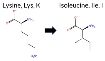 3DClick to see structure in 3D Viewer AIThe SynGAP1 missense variant K492I is not reported in ClinVar and is absent from gnomAD. Prediction tools cluster into two groups: benign predictions from premPS and FATHMM; pathogenic predictions from SGM‑Consensus, REVEL, PROVEAN, polyPhen‑2 (HumDiv and HumVar), SIFT, ESM1b, AlphaMissense‑Default, and AlphaMissense‑Optimized. Uncertain results come from FoldX, Rosetta, and Foldetta. High‑accuracy methods give a pathogenic verdict: AlphaMissense‑Optimized predicts pathogenic; the SGM‑Consensus, which aggregates AlphaMissense‑Default, ESM1b, FATHMM, and PROVEAN, also predicts pathogenic (3/4 votes). Foldetta remains inconclusive. Overall, the majority of evidence points to a pathogenic effect, and this assessment does not conflict with the absence of a ClinVar classification. Disclaimer: This summary was generated using AI and should be interpreted alongside expert review. | Likely Pathogenic | GAP | 0.061840 | Structured | 0.327121 | Uncertain | 0.947 | 0.192 | 0.000 | -18.661 | Likely Pathogenic | 0.998 | Likely Pathogenic | Likely Pathogenic | -0.54 | Ambiguous | 0.1 | -0.59 | Ambiguous | -0.57 | Ambiguous | 0.49 | Likely Benign | 0.645 | Likely Pathogenic | -7.96 | Deleterious | 1.000 | Probably Damaging | 1.000 | Probably Damaging | 2.94 | Benign | 0.00 | Affected | 0.0875 | 0.2810 | -2 | -3 | 8.4 | -15.01 | |||||||||||||||||||||||||||||
| c.1476A>C | K492N 2D  3DClick to see structure in 3D Viewer AIThe SynGAP1 K492N missense variant is not reported in ClinVar and is absent from gnomAD. Prediction tools that agree on a benign effect include only FATHMM, whereas the remaining tools (REVEL, premPS, PROVEAN, polyPhen‑2 HumDiv, polyPhen‑2 HumVar, SIFT, ESM1b, AlphaMissense‑Default, AlphaMissense‑Optimized) uniformly predict a pathogenic impact. The SGM‑Consensus, derived from a majority vote of AlphaMissense‑Default, ESM1b, FATHMM, and PROVEAN, also indicates a likely pathogenic outcome. High‑accuracy assessments show AlphaMissense‑Optimized as pathogenic, SGM‑Consensus as likely pathogenic, and Foldetta (combining FoldX‑MD and Rosetta outputs) as uncertain. No folding‑stability predictions are definitive. Based on the preponderance of pathogenic predictions and the lack of benign evidence, the variant is most likely pathogenic, and this assessment does not contradict any ClinVar status (none is available). Disclaimer: This summary was generated using AI and should be interpreted alongside expert review. | Likely Pathogenic | GAP | 0.061840 | Structured | 0.327121 | Uncertain | 0.947 | 0.192 | 0.000 | -14.048 | Likely Pathogenic | 0.999 | Likely Pathogenic | Likely Pathogenic | 0.92 | Ambiguous | 0.1 | 1.09 | Ambiguous | 1.01 | Ambiguous | 1.25 | Destabilizing | 0.512 | Likely Pathogenic | -4.97 | Deleterious | 1.000 | Probably Damaging | 1.000 | Probably Damaging | 2.92 | Benign | 0.00 | Affected | 0.2814 | 0.0926 | 1 | 0 | 0.4 | -14.07 | |||||||||||||||||||||||||||||
| c.1476A>T | K492N 2D 3DClick to see structure in 3D Viewer AIThe SynGAP1 K492N missense variant is not reported in ClinVar and is absent from gnomAD. Prediction tools that agree on a benign effect include only FATHMM, whereas the remaining tools (REVEL, premPS, PROVEAN, polyPhen‑2 HumDiv, polyPhen‑2 HumVar, SIFT, ESM1b, AlphaMissense‑Default, AlphaMissense‑Optimized) uniformly predict a pathogenic impact. The SGM‑Consensus, derived from a majority vote of AlphaMissense‑Default, ESM1b, FATHMM, and PROVEAN, also indicates a likely pathogenic outcome. High‑accuracy assessments show AlphaMissense‑Optimized as pathogenic, SGM‑Consensus as likely pathogenic, and Foldetta (combining FoldX‑MD and Rosetta outputs) as uncertain. No folding‑stability predictions are definitive. Based on the preponderance of pathogenic predictions and the lack of benign evidence, the variant is most likely pathogenic, and this assessment does not contradict any ClinVar status (none is available). Disclaimer: This summary was generated using AI and should be interpreted alongside expert review. | Likely Pathogenic | GAP | 0.061840 | Structured | 0.327121 | Uncertain | 0.947 | 0.192 | 0.000 | -14.048 | Likely Pathogenic | 0.999 | Likely Pathogenic | Likely Pathogenic | 0.92 | Ambiguous | 0.1 | 1.09 | Ambiguous | 1.01 | Ambiguous | 1.25 | Destabilizing | 0.512 | Likely Pathogenic | -4.97 | Deleterious | 1.000 | Probably Damaging | 1.000 | Probably Damaging | 2.92 | Benign | 0.00 | Affected | 0.2814 | 0.0926 | 1 | 0 | 0.4 | -14.07 | |||||||||||||||||||||||||||||
| c.1477G>A | A493T 2D  3DClick to see structure in 3D Viewer AIThe SynGAP1 A493T missense variant is not reported in ClinVar and is absent from gnomAD. Consensus among most in silico predictors indicates a deleterious effect: REVEL, SIFT, PROVEAN, polyPhen‑2 (HumDiv and HumVar), ESM1b, FATHMM, AlphaMissense‑Default, premPS, and the SGM Consensus (majority vote of AlphaMissense‑Default, ESM1b, FATHMM, PROVEAN) all classify the change as pathogenic or likely pathogenic. Only Rosetta predicts a benign outcome; FoldX, Foldetta, and AlphaMissense‑Optimized are uncertain or unavailable. High‑accuracy tools give the following: AlphaMissense‑Optimized – uncertain; SGM Consensus – likely pathogenic; Foldetta – uncertain. Taken together, the preponderance of evidence supports a pathogenic effect for A493T. This conclusion is not contradicted by ClinVar, which contains no entry for the variant. Disclaimer: This summary was generated using AI and should be interpreted alongside expert review. | Likely Pathogenic | GAP | 0.102787 | Structured | 0.340081 | Uncertain | 0.966 | 0.182 | 0.000 | -10.366 | Likely Pathogenic | 0.892 | Likely Pathogenic | Ambiguous | 0.82 | Ambiguous | 0.0 | 0.39 | Likely Benign | 0.61 | Ambiguous | 1.10 | Destabilizing | 0.727 | Likely Pathogenic | -3.41 | Deleterious | 0.998 | Probably Damaging | 0.993 | Probably Damaging | -1.36 | Pathogenic | 0.04 | Affected | 0.0964 | 0.3972 | 1 | 0 | -2.5 | 30.03 | |||||||||||||||||||||||||||||
| c.1477G>C | A493P 2D  3DClick to see structure in 3D Viewer AIThe SynGAP1 missense variant A493P is not reported in ClinVar and is absent from gnomAD. Prediction tools that assess functional impact all converge on a pathogenic interpretation: REVEL, FoldX, Rosetta, Foldetta, premPS, PROVEAN, polyPhen‑2 (HumDiv and HumVar), SIFT, ESM1b, FATHMM, AlphaMissense‑Default, and AlphaMissense‑Optimized all predict deleterious effects. No tool in the dataset indicates a benign outcome. High‑accuracy methods reinforce this consensus: AlphaMissense‑Optimized is pathogenic, the SGM Consensus (majority vote of AlphaMissense‑Default, ESM1b, FATHMM, PROVEAN) is likely pathogenic, and Foldetta (combining FoldX‑MD and Rosetta outputs) is pathogenic. With all available evidence pointing to a damaging effect and no ClinVar entry to contradict, the variant is most likely pathogenic. Disclaimer: This summary was generated using AI and should be interpreted alongside expert review. | Likely Pathogenic | GAP | 0.102787 | Structured | 0.340081 | Uncertain | 0.966 | 0.182 | 0.000 | -15.797 | Likely Pathogenic | 1.000 | Likely Pathogenic | Likely Pathogenic | 4.96 | Destabilizing | 0.1 | 10.79 | Destabilizing | 7.88 | Destabilizing | 1.32 | Destabilizing | 0.883 | Likely Pathogenic | -4.54 | Deleterious | 1.000 | Probably Damaging | 0.998 | Probably Damaging | -1.41 | Pathogenic | 0.02 | Affected | 0.1490 | 0.2907 | 1 | -1 | -3.4 | 26.04 | |||||||||||||||||||||||||||||
| c.1477G>T | A493S 2D 3DClick to see structure in 3D Viewer AIThe SynGAP1 missense variant A493S is not reported in ClinVar (ClinVar ID None) and is absent from gnomAD (gnomAD ID None). Prediction tools that agree on a benign effect include PROVEAN, SIFT, ESM1b, AlphaMissense‑Default, and AlphaMissense‑Optimized. Tools that predict a pathogenic effect are REVEL, polyPhen‑2 HumDiv, polyPhen‑2 HumVar, and FATHMM. Predictions that are uncertain or inconclusive (FoldX, Rosetta, Foldetta, premPS) are treated as unavailable. High‑accuracy assessments show AlphaMissense‑Optimized as benign, the SGM Consensus (majority vote from AlphaMissense‑Default, ESM1b, FATHMM, PROVEAN) as Likely Benign, and Foldetta as unavailable. Based on the consensus of the available predictions, the variant is most likely benign, and this conclusion does not contradict any ClinVar status because no ClinVar entry exists. Disclaimer: This summary was generated using AI and should be interpreted alongside expert review. | Likely Benign | GAP | 0.102787 | Structured | 0.340081 | Uncertain | 0.966 | 0.182 | 0.000 | -6.271 | Likely Benign | 0.298 | Likely Benign | Likely Benign | 0.66 | Ambiguous | 0.1 | 1.10 | Ambiguous | 0.88 | Ambiguous | 0.53 | Ambiguous | 0.507 | Likely Pathogenic | -2.12 | Neutral | 0.983 | Probably Damaging | 0.993 | Probably Damaging | -1.27 | Pathogenic | 0.44 | Tolerated | 0.1821 | 0.2763 | 1 | 1 | -2.6 | 16.00 | |||||||||||||||||||||||||||||
| c.1478C>A | A493D 2D AISynGAP1 missense variant A493D is not reported in ClinVar and is absent from gnomAD. Functional prediction tools uniformly indicate a deleterious effect: REVEL, FoldX, Rosetta, Foldetta, premPS, PROVEAN, polyPhen‑2 (HumDiv and HumVar), SIFT, ESM1b, FATHMM, AlphaMissense‑Default, and AlphaMissense‑Optimized all classify the substitution as pathogenic. No tool predicts a benign outcome. High‑accuracy assessments reinforce this view: AlphaMissense‑Optimized predicts pathogenicity; the SGM Consensus (majority vote of AlphaMissense‑Default, ESM1b, FATHMM, and PROVEAN) reports “Likely Pathogenic”; and Foldetta, which integrates FoldX‑MD and Rosetta stability calculations, also predicts pathogenicity. Consequently, the variant is most likely pathogenic, and this assessment is consistent with the absence of a ClinVar entry (no contradiction). Disclaimer: This summary was generated using AI and should be interpreted alongside expert review. | Likely Pathogenic | GAP | 0.102787 | Structured | 0.340081 | Uncertain | 0.966 | 0.182 | 0.000 | -16.834 | Likely Pathogenic | 0.999 | Likely Pathogenic | Likely Pathogenic | 4.45 | Destabilizing | 1.5 | 3.59 | Destabilizing | 4.02 | Destabilizing | 1.60 | Destabilizing | 0.925 | Likely Pathogenic | -5.25 | Deleterious | 1.000 | Probably Damaging | 0.998 | Probably Damaging | -1.41 | Pathogenic | 0.01 | Affected | 0.1437 | 0.1541 | 0 | -2 | -5.3 | 44.01 | |||||||||||||||||||||||||||||
| c.1478C>G | A493G 2D 3DClick to see structure in 3D Viewer AIThe SynGAP1 missense variant A493G is not reported in ClinVar and is absent from gnomAD. Functional prediction tools largely agree on a deleterious effect: REVEL, premPS, PROVEAN, polyPhen‑2 (HumDiv and HumVar), SIFT, ESM1b, FATHMM, AlphaMissense‑Default, and the SGM‑Consensus all predict pathogenicity, while only AlphaMissense‑Optimized predicts a benign outcome. Predictions from FoldX, Rosetta, and Foldetta are uncertain and therefore not considered evidence. High‑accuracy assessments show AlphaMissense‑Optimized as benign, whereas the SGM‑Consensus (majority vote of AlphaMissense‑Default, ESM1b, FATHMM, PROVEAN) remains pathogenic; Foldetta likewise yields an uncertain result. Overall, the preponderance of evidence points to a pathogenic effect for A493G, and this conclusion does not contradict the absence of a ClinVar annotation. Disclaimer: This summary was generated using AI and should be interpreted alongside expert review. | Likely Pathogenic | GAP | 0.102787 | Structured | 0.340081 | Uncertain | 0.966 | 0.182 | 0.000 | -11.379 | Likely Pathogenic | 0.571 | Likely Pathogenic | Likely Benign | 1.85 | Ambiguous | 0.0 | 1.63 | Ambiguous | 1.74 | Ambiguous | 1.40 | Destabilizing | 0.764 | Likely Pathogenic | -3.54 | Deleterious | 0.999 | Probably Damaging | 0.995 | Probably Damaging | -1.40 | Pathogenic | 0.02 | Affected | 0.1673 | 0.2228 | 1 | 0 | -2.2 | -14.03 | |||||||||||||||||||||||||||||
| c.147C>G | C49W 2D 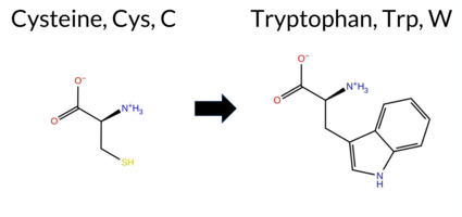 AIThe SynGAP1 missense variant C49W is not reported in ClinVar (no ClinVar ID) and is absent from gnomAD. Prediction tools that agree on a benign effect include REVEL and FATHMM, whereas the majority of tools predict a pathogenic impact: PROVEAN, polyPhen‑2 (HumDiv and HumVar), SIFT, ESM1b, AlphaMissense‑Default, AlphaMissense‑Optimized, and the SGM‑Consensus (majority vote from AlphaMissense‑Default, ESM1b, FATHMM, and PROVEAN). High‑accuracy assessments further support pathogenicity: AlphaMissense‑Optimized is pathogenic and the SGM‑Consensus is “Likely Pathogenic.” Foldetta, a protein‑folding stability method combining FoldX‑MD and Rosetta outputs, has no available result for this variant. Overall, the preponderance of evidence from both general and high‑accuracy predictors indicates that C49W is most likely pathogenic, and this conclusion is not contradicted by any ClinVar annotation. Disclaimer: This summary was generated using AI and should be interpreted alongside expert review. | Likely Pathogenic | 0.209395 | Structured | 0.445316 | Uncertain | 0.541 | 0.704 | 0.000 | -12.247 | Likely Pathogenic | 0.977 | Likely Pathogenic | Likely Pathogenic | 0.291 | Likely Benign | -3.56 | Deleterious | 0.880 | Possibly Damaging | 0.914 | Probably Damaging | 3.83 | Benign | 0.00 | Affected | 0.1380 | 0.3172 | -8 | -2 | -3.4 | 83.07 | |||||||||||||||||||||||||||||||||||||||
| c.1480A>C | I494L 2D 3DClick to see structure in 3D Viewer AIThe SynGAP1 I494L missense variant is not reported in ClinVar (ClinVar ID None) and is absent from gnomAD (gnomAD ID None). Prediction tools that agree on a benign effect include FoldX, PROVEAN, SIFT, and AlphaMissense‑Optimized. Those that agree on a pathogenic effect are REVEL, polyPhen‑2 (HumDiv and HumVar), ESM1b, and FATHMM. The remaining tools—Rosetta, Foldetta, premPS, and AlphaMissense‑Default—return uncertain or inconclusive results. High‑accuracy assessments show AlphaMissense‑Optimized as benign, the SGM Consensus (majority vote from AlphaMissense‑Default, ESM1b, FATHMM, PROVEAN) as pathogenic, and Foldetta as uncertain. Overall, the balance of evidence (five pathogenic versus four benign predictions, with several uncertain calls) leans toward a pathogenic interpretation. This conclusion does not contradict ClinVar status, as the variant has no ClinVar entry. Disclaimer: This summary was generated using AI and should be interpreted alongside expert review. | GAP | 0.155435 | Structured | 0.353330 | Uncertain | 0.941 | 0.157 | 0.000 | -10.175 | Likely Pathogenic | 0.478 | Ambiguous | Likely Benign | 0.28 | Likely Benign | 0.1 | 1.11 | Ambiguous | 0.70 | Ambiguous | 0.91 | Ambiguous | 0.513 | Likely Pathogenic | -1.69 | Neutral | 0.645 | Possibly Damaging | 0.718 | Possibly Damaging | -0.88 | Pathogenic | 0.11 | Tolerated | 0.0816 | 0.2851 | 2 | 2 | -0.7 | 0.00 | ||||||||||||||||||||||||||||||
| c.1480A>T | I494L 2D 3DClick to see structure in 3D Viewer AIThe SynGAP1 I494L variant is not reported in ClinVar (ClinVar ID None) and is absent from gnomAD (gnomAD ID None). Prediction tools that agree on a benign effect include FoldX, PROVEAN, SIFT, and AlphaMissense‑Optimized. Those that agree on a pathogenic effect are REVEL, polyPhen‑2 (HumDiv and HumVar), ESM1b, and FATHMM. The remaining tools—Rosetta, Foldetta, premPS, and AlphaMissense‑Default—return uncertain or inconclusive results. High‑accuracy assessments show AlphaMissense‑Optimized predicting benign, the SGM Consensus (majority vote from AlphaMissense‑Default, ESM1b, FATHMM, PROVEAN) predicting pathogenic, and Foldetta indicating uncertain stability change. Overall, the balance of evidence leans toward pathogenicity, with a majority of tools and the SGM Consensus supporting a deleterious effect. This conclusion does not contradict ClinVar status, as the variant is currently unreported there. Disclaimer: This summary was generated using AI and should be interpreted alongside expert review. | GAP | 0.155435 | Structured | 0.353330 | Uncertain | 0.941 | 0.157 | 0.000 | -10.175 | Likely Pathogenic | 0.478 | Ambiguous | Likely Benign | 0.28 | Likely Benign | 0.1 | 1.11 | Ambiguous | 0.70 | Ambiguous | 0.91 | Ambiguous | 0.513 | Likely Pathogenic | -1.69 | Neutral | 0.645 | Possibly Damaging | 0.718 | Possibly Damaging | -0.88 | Pathogenic | 0.11 | Tolerated | 0.0816 | 0.2851 | 2 | 2 | -0.7 | 0.00 | ||||||||||||||||||||||||||||||
| c.1481T>A | I494K 2D 3DClick to see structure in 3D Viewer AIThe SynGAP1 missense variant I494K is not reported in ClinVar (ClinVar ID None) and is absent from gnomAD (gnomAD ID None). Prediction tools that assess pathogenicity uniformly classify the variant as deleterious: SGM‑Consensus (Likely Pathogenic), REVEL, FoldX, Rosetta, Foldetta, premPS, PROVEAN, polyPhen‑2 (HumDiv and HumVar), SIFT, ESM1b, FATHMM, AlphaMissense‑Default, and AlphaMissense‑Optimized all predict a pathogenic effect. No tool in the dataset predicts a benign outcome. High‑accuracy methods reinforce this view: AlphaMissense‑Optimized is pathogenic; the SGM Consensus (majority vote of AlphaMissense‑Default, ESM1b, FATHMM, PROVEAN) is Likely Pathogenic; and Foldetta (combining FoldX‑MD and Rosetta outputs) is pathogenic. Based on the unanimous computational evidence, the variant is most likely pathogenic, and this assessment does not contradict any ClinVar status (none reported). Disclaimer: This summary was generated using AI and should be interpreted alongside expert review. | Likely Pathogenic | GAP | 0.155435 | Structured | 0.353330 | Uncertain | 0.941 | 0.157 | 0.000 | -15.950 | Likely Pathogenic | 0.997 | Likely Pathogenic | Likely Pathogenic | 4.23 | Destabilizing | 0.2 | 5.33 | Destabilizing | 4.78 | Destabilizing | 1.89 | Destabilizing | 0.925 | Likely Pathogenic | -6.40 | Deleterious | 0.987 | Probably Damaging | 0.937 | Probably Damaging | -1.41 | Pathogenic | 0.00 | Affected | 0.1015 | 0.0870 | -2 | -3 | -8.4 | 15.01 | |||||||||||||||||||||||||||||
| c.1481T>C | I494T 2D AIThe SynGAP1 missense variant I494T is not reported in ClinVar (ClinVar status: None) and is absent from gnomAD (gnomAD ID: None). Prediction tools that agree on a benign effect include only AlphaMissense‑Optimized. All other evaluated tools—SGM‑Consensus, REVEL, FoldX, Rosetta, Foldetta, premPS, PROVEAN, polyPhen‑2 (HumDiv and HumVar), SIFT, ESM1b, FATHMM, and AlphaMissense‑Default—predict a pathogenic impact. High‑accuracy methods give the following results: AlphaMissense‑Optimized predicts benign; the SGM Consensus (majority vote of AlphaMissense‑Default, ESM1b, FATHMM, and PROVEAN) predicts likely pathogenic; and Foldetta (combining FoldX‑MD and Rosetta outputs) predicts pathogenic. No predictions are missing or inconclusive. Based on the overwhelming majority of pathogenic predictions, the variant is most likely pathogenic, and this assessment does not contradict the ClinVar status, which is currently unreported. Disclaimer: This summary was generated using AI and should be interpreted alongside expert review. | Likely Pathogenic | GAP | 0.155435 | Structured | 0.353330 | Uncertain | 0.941 | 0.157 | 0.000 | -10.033 | Likely Pathogenic | 0.754 | Likely Pathogenic | Likely Benign | 2.45 | Destabilizing | 0.7 | 2.12 | Destabilizing | 2.29 | Destabilizing | 1.73 | Destabilizing | 0.907 | Likely Pathogenic | -4.61 | Deleterious | 1.000 | Probably Damaging | 0.995 | Probably Damaging | -1.38 | Pathogenic | 0.01 | Affected | 0.1003 | 0.0640 | 0 | -1 | -5.2 | -12.05 | |||||||||||||||||||||||||||||
| c.1482A>G | I494M 2D  3DClick to see structure in 3D Viewer AIThe SynGAP1 I494M missense variant is not reported in ClinVar (ClinVar ID None) and is absent from gnomAD (gnomAD ID None). Prediction tools that agree on a benign effect include FoldX, PROVEAN, SIFT, and AlphaMissense‑Optimized. Those that agree on a pathogenic effect are REVEL, polyPhen‑2 (HumDiv and HumVar), ESM1b, and FATHMM. The remaining tools—Rosetta, Foldetta, premPS, and AlphaMissense‑Default—return uncertain or inconclusive results. High‑accuracy assessments show AlphaMissense‑Optimized as benign, the SGM Consensus (majority vote from AlphaMissense‑Default, ESM1b, FATHMM, and PROVEAN) as pathogenic, and Foldetta as uncertain. Overall, the balance of evidence leans toward pathogenicity, with a majority of tools predicting a deleterious effect. This conclusion does not contradict the ClinVar status, which currently has no classification for this variant. Disclaimer: This summary was generated using AI and should be interpreted alongside expert review. | GAP | 0.155435 | Structured | 0.353330 | Uncertain | 0.941 | 0.157 | 0.000 | -8.223 | Likely Pathogenic | 0.356 | Ambiguous | Likely Benign | 0.35 | Likely Benign | 0.2 | 1.98 | Ambiguous | 1.17 | Ambiguous | 0.98 | Ambiguous | 0.542 | Likely Pathogenic | -2.25 | Neutral | 1.000 | Probably Damaging | 0.997 | Probably Damaging | -1.15 | Pathogenic | 0.23 | Tolerated | 0.0676 | 0.1789 | 2 | 1 | -2.6 | 18.03 | ||||||||||||||||||||||||||||||
| c.1483G>C | E495Q 2D 3DClick to see structure in 3D Viewer AIThe SynGAP1 E495Q missense variant is not listed in ClinVar and has no reported allele in gnomAD. Prediction tools that agree on a benign effect include FoldX, Rosetta, and Foldetta, all of which score the substitution as benign. Tools that agree on a pathogenic effect are SGM‑Consensus, REVEL, PROVEAN, polyPhen‑2 (HumDiv and HumVar), SIFT, ESM1b, FATHMM, and AlphaMissense‑Default. Two tools give uncertain results: premPS and AlphaMissense‑Optimized. High‑accuracy assessments show that the SGM‑Consensus (a majority vote of AlphaMissense‑Default, ESM1b, FATHMM, and PROVEAN) predicts pathogenicity, AlphaMissense‑Optimized is uncertain, and Foldetta predicts benign stability. Overall, the majority of evidence points to a pathogenic impact. The variant is most likely pathogenic, and this conclusion does not contradict any ClinVar status, as none is available. Disclaimer: This summary was generated using AI and should be interpreted alongside expert review. | Likely Pathogenic | GAP | 0.164327 | Structured | 0.364496 | Uncertain | 0.933 | 0.161 | 0.000 | -11.050 | Likely Pathogenic | 0.899 | Likely Pathogenic | Ambiguous | -0.27 | Likely Benign | 0.1 | 0.11 | Likely Benign | -0.08 | Likely Benign | 0.89 | Ambiguous | 0.748 | Likely Pathogenic | -2.92 | Deleterious | 0.999 | Probably Damaging | 0.993 | Probably Damaging | -1.42 | Pathogenic | 0.01 | Affected | 0.1093 | 0.4973 | 2 | 2 | 0.0 | -0.98 | |||||||||||||||||||||||||||||
| c.1484A>C | E495A 2D 3DClick to see structure in 3D Viewer AIThe SynGAP1 E495A missense variant has no ClinVar entry and is not reported in gnomAD. Prediction tools that indicate a benign effect include FoldX, while the remaining evaluated algorithms (REVEL, SIFT, polyPhen‑2 HumDiv, polyPhen‑2 HumVar, ESM1b, FATHMM, PROVEAN, AlphaMissense‑Default, and the SGM‑Consensus) all predict a pathogenic outcome. High‑accuracy assessments show AlphaMissense‑Optimized as uncertain, the SGM‑Consensus (a majority vote of AlphaMissense‑Default, ESM1b, FATHMM, and PROVEAN) as Likely Pathogenic, and Foldetta as uncertain. No contradictory evidence is present from ClinVar or population databases. Overall, the preponderance of predictions supports a pathogenic classification for this variant. Disclaimer: This summary was generated using AI and should be interpreted alongside expert review. | Likely Pathogenic | GAP | 0.164327 | Structured | 0.364496 | Uncertain | 0.933 | 0.161 | 0.000 | -9.229 | Likely Pathogenic | 0.946 | Likely Pathogenic | Ambiguous | 0.42 | Likely Benign | 0.1 | 0.85 | Ambiguous | 0.64 | Ambiguous | 0.69 | Ambiguous | 0.834 | Likely Pathogenic | -5.84 | Deleterious | 1.000 | Probably Damaging | 0.999 | Probably Damaging | -1.38 | Pathogenic | 0.01 | Affected | 0.2711 | 0.4259 | 0 | -1 | 5.3 | -58.04 | |||||||||||||||||||||||||||||
| c.1484A>T | E495V 2D 3DClick to see structure in 3D Viewer AIThe SynGAP1 missense variant E495V is not reported in ClinVar (ClinVar ID None) and has no entry in gnomAD (gnomAD ID None). Prediction tools that agree on a benign effect include FoldX, Rosetta, Foldetta, and premPS, whereas the majority of tools predict a pathogenic impact: REVEL, PROVEAN, polyPhen‑2 (HumDiv and HumVar), SIFT, ESM1b, FATHMM, AlphaMissense‑Default, AlphaMissense‑Optimized, and the SGM Consensus (majority vote from AlphaMissense‑Default, ESM1b, FATHMM, PROVEAN). High‑accuracy assessments further show AlphaMissense‑Optimized as pathogenic, the SGM Consensus as likely pathogenic, and Foldetta as benign. Taken together, the preponderance of evidence (10 pathogenic‑predicted tools versus 4 benign) indicates that E495V is most likely pathogenic. This conclusion does not contradict ClinVar status, as the variant is currently unreported there. Disclaimer: This summary was generated using AI and should be interpreted alongside expert review. | Likely Pathogenic | GAP | 0.164327 | Structured | 0.364496 | Uncertain | 0.933 | 0.161 | 0.000 | -12.031 | Likely Pathogenic | 0.990 | Likely Pathogenic | Likely Pathogenic | 0.43 | Likely Benign | 0.2 | -0.32 | Likely Benign | 0.06 | Likely Benign | 0.47 | Likely Benign | 0.887 | Likely Pathogenic | -6.83 | Deleterious | 0.999 | Probably Damaging | 0.996 | Probably Damaging | -1.44 | Pathogenic | 0.00 | Affected | 0.0646 | 0.5457 | -2 | -2 | 7.7 | -29.98 | |||||||||||||||||||||||||||||
| c.1486G>A | E496K 2D 3DClick to see structure in 3D Viewer AIThe SynGAP1 missense variant E496K is not reported in ClinVar and is absent from gnomAD. Prediction tools that agree on a benign effect include only FoldX, whereas the majority of tools (REVEL, PROVEAN, polyPhen‑2 HumDiv, polyPhen‑2 HumVar, SIFT, ESM1b, FATHMM, AlphaMissense‑Default, AlphaMissense‑Optimized, and the SGM‑Consensus) predict a pathogenic impact. Uncertain predictions come from Rosetta, Foldetta, and premPS. High‑accuracy assessments show AlphaMissense‑Optimized as pathogenic, the SGM‑Consensus as likely pathogenic, and Foldetta as inconclusive. Overall, the evidence strongly favors a pathogenic classification for E496K, and this conclusion does not contradict the ClinVar status, which currently contains no entry for this variant. Disclaimer: This summary was generated using AI and should be interpreted alongside expert review. | Likely Pathogenic | GAP | 0.102787 | Structured | 0.383296 | Uncertain | 0.945 | 0.179 | 0.000 | -15.795 | Likely Pathogenic | 0.961 | Likely Pathogenic | Likely Pathogenic | 0.38 | Likely Benign | 0.1 | 1.77 | Ambiguous | 1.08 | Ambiguous | 0.76 | Ambiguous | 0.743 | Likely Pathogenic | -3.58 | Deleterious | 0.999 | Probably Damaging | 0.994 | Probably Damaging | -1.40 | Pathogenic | 0.04 | Affected | 0.1810 | 0.3528 | 0 | 1 | -0.4 | -0.94 | |||||||||||||||||||||||||||||
| c.1486G>C | E496Q 2D 3DClick to see structure in 3D Viewer AIThe SynGAP1 missense variant E496Q is not reported in ClinVar (ClinVar ID: None) and is absent from gnomAD (gnomAD ID: None). In silico predictors that classify the variant as benign include FoldX, Foldetta, premPS, SIFT, and AlphaMissense‑Optimized, whereas those that predict pathogenicity are SGM‑Consensus, REVEL, PROVEAN, polyPhen‑2 (HumDiv and HumVar), ESM1b, FATHMM, and AlphaMissense‑Default; Rosetta is inconclusive. High‑accuracy tools give a mixed picture: AlphaMissense‑Optimized indicates a benign effect, the SGM Consensus (majority vote of AlphaMissense‑Default, ESM1b, FATHMM, PROVEAN) predicts pathogenicity, and Foldetta (combining FoldX‑MD and Rosetta outputs) predicts a benign impact. Overall, the majority of predictions (8 pathogenic vs. 5 benign) and the consensus of high‑accuracy methods lean toward a pathogenic effect. Thus, the variant is most likely pathogenic, and this assessment is not contradicted by the absence of a ClinVar classification. Disclaimer: This summary was generated using AI and should be interpreted alongside expert review. | Likely Pathogenic | GAP | 0.102787 | Structured | 0.383296 | Uncertain | 0.945 | 0.179 | 0.000 | -11.868 | Likely Pathogenic | 0.651 | Likely Pathogenic | Likely Benign | 0.13 | Likely Benign | 0.1 | 0.61 | Ambiguous | 0.37 | Likely Benign | 0.50 | Likely Benign | 0.586 | Likely Pathogenic | -2.68 | Deleterious | 0.998 | Probably Damaging | 0.993 | Probably Damaging | -1.36 | Pathogenic | 0.15 | Tolerated | 0.0884 | 0.3262 | 2 | 2 | 0.0 | -0.98 | |||||||||||||||||||||||||||||
| c.1487A>C | E496A 2D 3DClick to see structure in 3D Viewer AISynGAP1 missense variant E496A is listed in ClinVar with an uncertain significance and is not reported in gnomAD. Functional prediction tools show mixed results: benign calls come from Rosetta, Foldetta, premPS, SIFT, and AlphaMissense‑Optimized, while pathogenic calls are made by REVEL, PROVEAN, polyPhen‑2 (HumDiv and HumVar), ESM1b, FATHMM, AlphaMissense‑Default, and the SGM‑Consensus (majority vote of AlphaMissense‑Default, ESM1b, FATHMM, PROVEAN). FoldX is inconclusive. High‑accuracy methods give a split verdict: AlphaMissense‑Optimized predicts benign, SGM‑Consensus predicts pathogenic, and Foldetta predicts benign. Overall, the majority of predictions lean toward pathogenicity, and this does not contradict the ClinVar uncertain status. Disclaimer: This summary was generated using AI and should be interpreted alongside expert review. | Likely Pathogenic | GAP | 0.102787 | Structured | 0.383296 | Uncertain | 0.945 | 0.179 | 0.000 | Uncertain | 1 | -11.159 | Likely Pathogenic | 0.598 | Likely Pathogenic | Likely Benign | 0.68 | Ambiguous | 0.0 | 0.23 | Likely Benign | 0.46 | Likely Benign | 0.47 | Likely Benign | 0.681 | Likely Pathogenic | -4.87 | Deleterious | 1.000 | Probably Damaging | 0.999 | Probably Damaging | -1.41 | Pathogenic | 0.09 | Tolerated | 0.2806 | 0.3548 | 0 | -1 | 5.3 | -58.04 | |||||||||||||||||||||||||||
| c.1487A>T | E496V 2D 3DClick to see structure in 3D Viewer AISynGAP1 missense variant E496V is not reported in ClinVar and is absent from gnomAD. Functional prediction tools show a split: benign calls come from FoldX, Foldetta, and premPS, while pathogenic calls are made by REVEL, PROVEAN, polyPhen‑2 (HumDiv and HumVar), SIFT, ESM1b, FATHMM, and AlphaMissense‑Default. The SGM‑Consensus, which aggregates the majority vote of AlphaMissense‑Default, ESM1b, FATHMM, and PROVEAN, reports the variant as Likely Pathogenic. High‑accuracy assessments give mixed results: AlphaMissense‑Optimized is Uncertain; SGM‑Consensus remains Likely Pathogenic; Foldetta, a folding‑stability predictor that integrates FoldX‑MD and Rosetta outputs, classifies the variant as Benign. Because the variant is not present in ClinVar, there is no clinical annotation to contradict the computational evidence. Overall, the preponderance of pathogenic predictions, including the consensus score, suggests that E496V is most likely pathogenic, though the conflicting high‑accuracy folding stability result indicates uncertainty that warrants further functional validation. Disclaimer: This summary was generated using AI and should be interpreted alongside expert review. | Likely Pathogenic | GAP | 0.102787 | Structured | 0.383296 | Uncertain | 0.945 | 0.179 | 0.000 | -14.290 | Likely Pathogenic | 0.873 | Likely Pathogenic | Ambiguous | 0.17 | Likely Benign | 0.1 | -0.67 | Ambiguous | -0.25 | Likely Benign | 0.26 | Likely Benign | 0.823 | Likely Pathogenic | -6.16 | Deleterious | 0.999 | Probably Damaging | 0.996 | Probably Damaging | -1.43 | Pathogenic | 0.02 | Affected | 0.0556 | 0.3746 | -2 | -2 | 7.7 | -29.98 | |||||||||||||||||||||||||||||
| c.1489T>A | Y497N 2D  3DClick to see structure in 3D Viewer AIThe SynGAP1 missense variant Y497N is not reported in ClinVar and is absent from gnomAD. Functional prediction tools uniformly indicate a deleterious effect. Benign predictions: none. Pathogenic predictions: SGM‑Consensus (Likely Pathogenic), REVEL, FoldX, Rosetta, Foldetta, premPS, PROVEAN, polyPhen‑2 (HumDiv and HumVar), SIFT, ESM1b, FATHMM, AlphaMissense‑Default, and AlphaMissense‑Optimized. High‑accuracy assessments further support pathogenicity: AlphaMissense‑Optimized predicts pathogenic; the SGM Consensus (majority vote of AlphaMissense‑Default, ESM1b, FATHMM, PROVEAN) is Likely Pathogenic; and Foldetta, which integrates FoldX‑MD and Rosetta outputs, predicts pathogenic. Based on these concordant predictions, the variant is most likely pathogenic, and this conclusion does not contradict any ClinVar annotation (none available). Disclaimer: This summary was generated using AI and should be interpreted alongside expert review. | Likely Pathogenic | GAP | 0.092881 | Structured | 0.393608 | Uncertain | 0.950 | 0.181 | 0.000 | -11.884 | Likely Pathogenic | 0.974 | Likely Pathogenic | Likely Pathogenic | 3.64 | Destabilizing | 0.1 | 3.97 | Destabilizing | 3.81 | Destabilizing | 2.49 | Destabilizing | 0.870 | Likely Pathogenic | -8.85 | Deleterious | 1.000 | Probably Damaging | 0.999 | Probably Damaging | -1.66 | Pathogenic | 0.01 | Affected | 0.2114 | 0.0612 | -2 | -2 | -2.2 | -49.07 | |||||||||||||||||||||||||||||
| c.1489T>C | Y497H 2D  3DClick to see structure in 3D Viewer AIThe SynGAP1 missense variant Y497H is not reported in ClinVar (ClinVar status: None) and is absent from gnomAD (gnomAD ID: None). Prediction tools that assess pathogenicity all agree that the variant is deleterious: SGM‑Consensus, REVEL, FoldX, Rosetta, Foldetta, premPS, PROVEAN, polyPhen‑2 (HumDiv and HumVar), SIFT, ESM1b, FATHMM, AlphaMissense‑Default, and AlphaMissense‑Optimized all classify it as pathogenic. No tool predicts a benign effect. High‑accuracy methods reinforce this consensus: AlphaMissense‑Optimized predicts pathogenicity; the SGM Consensus (majority vote of AlphaMissense‑Default, ESM1b, FATHMM, and PROVEAN) also predicts pathogenic; and Foldetta, which integrates FoldX‑MD and Rosetta stability calculations, indicates a destabilizing, pathogenic effect. All available predictions are concordant and supportive. Based on these computational assessments, the variant is most likely pathogenic, and this conclusion does not contradict any existing ClinVar annotation (none is present). Disclaimer: This summary was generated using AI and should be interpreted alongside expert review. | Likely Pathogenic | GAP | 0.092881 | Structured | 0.393608 | Uncertain | 0.950 | 0.181 | 0.000 | -10.970 | Likely Pathogenic | 0.981 | Likely Pathogenic | Likely Pathogenic | 2.46 | Destabilizing | 0.1 | 2.40 | Destabilizing | 2.43 | Destabilizing | 1.29 | Destabilizing | 0.819 | Likely Pathogenic | -4.94 | Deleterious | 1.000 | Probably Damaging | 0.999 | Probably Damaging | -1.65 | Pathogenic | 0.02 | Affected | 0.1983 | 0.0612 | 0 | 2 | -1.9 | -26.03 | |||||||||||||||||||||||||||||
| c.1489T>G | Y497D 2D  3DClick to see structure in 3D Viewer AIThe SynGAP1 missense variant Y497D is not reported in ClinVar (ClinVar ID None) and is absent from gnomAD (gnomAD ID None). Prediction tools that assess pathogenicity all agree that the variant is deleterious: SGM‑Consensus, REVEL, FoldX, Rosetta, Foldetta, premPS, PROVEAN, polyPhen‑2 (HumDiv and HumVar), SIFT, ESM1b, FATHMM, AlphaMissense‑Default, and AlphaMissense‑Optimized all classify it as pathogenic. No tool predicts a benign effect. High‑accuracy methods reinforce this consensus: AlphaMissense‑Optimized predicts pathogenicity; the SGM Consensus (majority vote of AlphaMissense‑Default, ESM1b, FATHMM, and PROVEAN) also predicts pathogenic; and Foldetta, which integrates FoldX‑MD and Rosetta stability outputs, predicts a destabilizing, pathogenic effect. All available predictions are concordant and indicate a likely pathogenic impact. Thus, based on the collective computational evidence, the variant is most likely pathogenic, and this assessment does not contradict any ClinVar status (none reported). Disclaimer: This summary was generated using AI and should be interpreted alongside expert review. | Likely Pathogenic | GAP | 0.092881 | Structured | 0.393608 | Uncertain | 0.950 | 0.181 | 0.000 | -12.128 | Likely Pathogenic | 0.994 | Likely Pathogenic | Likely Pathogenic | 4.70 | Destabilizing | 0.2 | 4.29 | Destabilizing | 4.50 | Destabilizing | 2.36 | Destabilizing | 0.918 | Likely Pathogenic | -9.81 | Deleterious | 1.000 | Probably Damaging | 0.999 | Probably Damaging | -1.67 | Pathogenic | 0.01 | Affected | 0.3621 | 0.0612 | -4 | -3 | -2.2 | -48.09 | |||||||||||||||||||||||||||||
| c.148A>C | I50L 2D AIThe SynGAP1 missense variant I50L is not reported in ClinVar and is absent from gnomAD. Prediction tools largely agree on a benign effect: REVEL, PROVEAN, polyPhen‑2 (HumDiv and HumVar), ESM1b, FATHMM, AlphaMissense‑Default, and AlphaMissense‑Optimized all classify the change as benign, while the single pathogenic prediction comes from SIFT. The SGM‑Consensus, which aggregates AlphaMissense‑Default, ESM1b, FATHMM, and PROVEAN, also reports a likely benign outcome. High‑accuracy assessments reinforce this view: AlphaMissense‑Optimized predicts benign, and the SGM‑Consensus (majority vote) is benign; Foldetta results are not available. Overall, the consensus of the available predictions indicates that the variant is most likely benign, and this conclusion does not contradict any ClinVar annotation (none exists). Disclaimer: This summary was generated using AI and should be interpreted alongside expert review. | Likely Benign | 0.295083 | Structured | 0.449965 | Uncertain | 0.545 | 0.708 | 0.000 | -3.509 | Likely Benign | 0.300 | Likely Benign | Likely Benign | 0.076 | Likely Benign | -0.73 | Neutral | 0.010 | Benign | 0.004 | Benign | 3.91 | Benign | 0.00 | Affected | 0.0749 | 0.3247 | 2 | 2 | -0.7 | 0.00 | |||||||||||||||||||||||||||||||||||||||
| c.148A>G | I50V 2D  AIThe SynGAP1 missense variant I50V is not reported in ClinVar (no ClinVar ID) and is absent from gnomAD (no gnomAD ID). Prediction tools that agree on a benign effect include REVEL, PROVEAN, polyPhen‑2 HumDiv, polyPhen‑2 HumVar, ESM1b, FATHMM, AlphaMissense‑Default, and AlphaMissense‑Optimized. Only SIFT predicts a pathogenic outcome. The high‑accuracy consensus methods also support a benign interpretation: AlphaMissense‑Optimized is benign, and the SGM Consensus (majority vote from AlphaMissense‑Default, ESM1b, FATHMM, and PROVEAN) is “Likely Benign.” No Foldetta stability result is available, so it does not influence the assessment. Overall, the majority of evidence indicates that I50V is most likely benign, and this conclusion is not contradicted by any ClinVar annotation. Disclaimer: This summary was generated using AI and should be interpreted alongside expert review. | Likely Benign | 0.295083 | Structured | 0.449965 | Uncertain | 0.545 | 0.708 | 0.000 | -1.051 | Likely Benign | 0.141 | Likely Benign | Likely Benign | 0.082 | Likely Benign | -0.24 | Neutral | 0.000 | Benign | 0.001 | Benign | 4.18 | Benign | 0.00 | Affected | 0.1091 | 0.3159 | 4 | 3 | -0.3 | -14.03 | |||||||||||||||||||||||||||||||||||||||
| c.148A>T | I50F 2D  AIThe SynGAP1 missense variant I50F is not reported in ClinVar (no ClinVar ID) and is absent from gnomAD (no gnomAD ID). Prediction tools that agree on a benign effect include REVEL, PROVEAN, polyPhen‑2 HumDiv, polyPhen‑2 HumVar, ESM1b, FATHMM, and AlphaMissense‑Optimized. Tools that predict a pathogenic effect are SIFT and AlphaMissense‑Default. The SGM‑Consensus, which aggregates AlphaMissense‑Default, ESM1b, FATHMM, and PROVEAN, reports a Likely Benign classification. High‑accuracy assessments show AlphaMissense‑Optimized as benign and the SGM‑Consensus as Likely Benign; Foldetta results are not available. Overall, the majority of evidence points to a benign impact for I50F, and this conclusion does not contradict any ClinVar annotation (none exists). Disclaimer: This summary was generated using AI and should be interpreted alongside expert review. | Likely Benign | 0.295083 | Structured | 0.449965 | Uncertain | 0.545 | 0.708 | 0.000 | -5.631 | Likely Benign | 0.681 | Likely Pathogenic | Likely Benign | 0.100 | Likely Benign | -1.27 | Neutral | 0.334 | Benign | 0.074 | Benign | 3.75 | Benign | 0.00 | Affected | 0.0483 | 0.2560 | 1 | 0 | -1.7 | 34.02 | |||||||||||||||||||||||||||||||||||||||
| c.1490A>C | Y497S 2D  3DClick to see structure in 3D Viewer AIThe SynGAP1 missense variant Y497S is not reported in ClinVar and is absent from gnomAD. Across the available in‑silico predictors, every tool examined (SGM‑Consensus, REVEL, FoldX, Rosetta, Foldetta, premPS, PROVEAN, polyPhen‑2 HumDiv, polyPhen‑2 HumVar, SIFT, ESM1b, FATHMM, AlphaMissense‑Default, and AlphaMissense‑Optimized) classifies the change as pathogenic, leaving no benign predictions. High‑accuracy methods reinforce this assessment: AlphaMissense‑Optimized predicts pathogenicity; the SGM Consensus (majority vote of AlphaMissense‑Default, ESM1b, FATHMM, and PROVEAN) also predicts pathogenic; and Foldetta, which integrates FoldX‑MD and Rosetta stability outputs, indicates a destabilizing, pathogenic effect. No prediction or stability result is missing or inconclusive. Based on the unanimous pathogenic predictions and the absence of any ClinVar annotation, the variant is most likely pathogenic, with no contradictory ClinVar status. Disclaimer: This summary was generated using AI and should be interpreted alongside expert review. | Likely Pathogenic | GAP | 0.092881 | Structured | 0.393608 | Uncertain | 0.950 | 0.181 | 0.000 | -13.171 | Likely Pathogenic | 0.980 | Likely Pathogenic | Likely Pathogenic | 3.65 | Destabilizing | 0.1 | 4.68 | Destabilizing | 4.17 | Destabilizing | 2.22 | Destabilizing | 0.814 | Likely Pathogenic | -8.82 | Deleterious | 1.000 | Probably Damaging | 0.999 | Probably Damaging | -1.64 | Pathogenic | 0.03 | Affected | 0.3770 | 0.1419 | -3 | -2 | 0.5 | -76.10 | |||||||||||||||||||||||||||||
| c.1490A>T | Y497F 2D 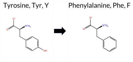 3DClick to see structure in 3D Viewer AIThe SynGAP1 missense variant Y497F is not reported in ClinVar (ClinVar status: not listed) and is absent from gnomAD (gnomAD ID: none). Prediction tools that agree on a benign effect include FoldX, Rosetta, Foldetta, polyPhen‑2 HumDiv, polyPhen‑2 HumVar, SIFT, AlphaMissense‑Default, and AlphaMissense‑Optimized. Tools that predict a pathogenic effect are SGM‑Consensus, REVEL, PROVEAN, ESM1b, and FATHMM; premPS remains uncertain. High‑accuracy assessments show AlphaMissense‑Optimized as benign, the SGM‑Consensus (majority vote from AlphaMissense‑Default, ESM1b, FATHMM, PROVEAN) as pathogenic, and Foldetta (combining FoldX‑MD and Rosetta outputs) as benign. Overall, the majority of predictions lean toward a benign impact, and this conclusion does not contradict the absence of a ClinVar classification. Thus, the variant is most likely benign based on the current predictive evidence. Disclaimer: This summary was generated using AI and should be interpreted alongside expert review. | Likely Pathogenic | GAP | 0.092881 | Structured | 0.393608 | Uncertain | 0.950 | 0.181 | 0.000 | -8.566 | Likely Pathogenic | 0.199 | Likely Benign | Likely Benign | -0.27 | Likely Benign | 0.1 | 0.47 | Likely Benign | 0.10 | Likely Benign | 0.61 | Ambiguous | 0.578 | Likely Pathogenic | -3.75 | Deleterious | 0.367 | Benign | 0.131 | Benign | -1.15 | Pathogenic | 0.19 | Tolerated | 0.2074 | 0.2242 | 7 | 3 | 4.1 | -16.00 | |||||||||||||||||||||||||||||
| c.1492A>C | M498L 2D 3DClick to see structure in 3D Viewer AIThe SynGAP1 missense variant M498L is not reported in ClinVar (no ClinVar ID) and is absent from gnomAD (no gnomAD ID). Prediction tools that agree on a benign effect include REVEL, FoldX, PROVEAN, polyPhen‑2 (HumDiv and HumVar), SIFT, ESM1b, AlphaMissense‑Default, AlphaMissense‑Optimized, and the SGM‑Consensus (Likely Benign). Only FATHMM predicts a pathogenic outcome, while Rosetta, Foldetta, and premPS are inconclusive. High‑accuracy assessments show AlphaMissense‑Optimized as Benign, the SGM‑Consensus as Likely Benign, and Foldetta as Uncertain. Overall, the majority of evidence points to a benign impact. This conclusion is consistent with the lack of ClinVar annotation and gnomAD presence, so there is no contradiction with existing clinical databases. Disclaimer: This summary was generated using AI and should be interpreted alongside expert review. | Likely Benign | GAP | 0.092881 | Structured | 0.399612 | Uncertain | 0.932 | 0.158 | 0.000 | 2.556 | Likely Benign | 0.072 | Likely Benign | Likely Benign | -0.23 | Likely Benign | 0.1 | -0.89 | Ambiguous | -0.56 | Ambiguous | -0.53 | Ambiguous | 0.271 | Likely Benign | 0.62 | Neutral | 0.000 | Benign | 0.000 | Benign | -1.24 | Pathogenic | 0.92 | Tolerated | 0.1319 | 0.3551 | 4 | 2 | 1.9 | -18.03 | |||||||||||||||||||||||||||||
| c.1492A>G | M498V 2D  3DClick to see structure in 3D Viewer AISynGAP1 missense variant M498V is not reported in ClinVar and is absent from gnomAD. Functional prediction tools cluster into two groups: benign (REVEL, PROVEAN, polyPhen‑2 HumVar, ESM1b, AlphaMissense‑Default, AlphaMissense‑Optimized) and pathogenic (FoldX, Foldetta, premPS, polyPhen‑2 HumDiv, SIFT, FATHMM). Rosetta is uncertain. The SGM‑Consensus, derived from a majority vote of AlphaMissense‑Default, ESM1b, FATHMM, and PROVEAN, is “Likely Benign.” High‑accuracy assessments show AlphaMissense‑Optimized as benign, SGM‑Consensus as likely benign, while Foldetta predicts a pathogenic effect on protein stability. Overall, the majority of predictions lean toward a benign impact, and this is consistent with the lack of ClinVar evidence; there is no contradiction with ClinVar status. Disclaimer: This summary was generated using AI and should be interpreted alongside expert review. | Likely Benign | GAP | 0.092881 | Structured | 0.399612 | Uncertain | 0.932 | 0.158 | 0.000 | -3.229 | Likely Benign | 0.317 | Likely Benign | Likely Benign | 2.74 | Destabilizing | 0.1 | 1.26 | Ambiguous | 2.00 | Destabilizing | 1.17 | Destabilizing | 0.483 | Likely Benign | -1.82 | Neutral | 0.752 | Possibly Damaging | 0.279 | Benign | -1.28 | Pathogenic | 0.04 | Affected | 0.2737 | 0.2737 | 2 | 1 | 2.3 | -32.06 | |||||||||||||||||||||||||||||
| c.1492A>T | M498L 2D 3DClick to see structure in 3D Viewer AISynGAP1 missense variant M498L is listed in ClinVar with an uncertain significance and is not reported in gnomAD. Functional prediction tools largely agree on a benign effect: REVEL, FoldX, PROVEAN, polyPhen‑2 (HumDiv and HumVar), SIFT, ESM1b, AlphaMissense‑Default, and AlphaMissense‑Optimized all predict benign. Only FATHMM predicts pathogenic, while Rosetta, Foldetta, and premPS are inconclusive. The SGM‑Consensus, derived from a majority vote of AlphaMissense‑Default, ESM1b, FATHMM, and PROVEAN, reports a likely benign outcome. High‑accuracy assessments reinforce this: AlphaMissense‑Optimized is benign, the SGM‑Consensus is likely benign, and Foldetta remains uncertain. Taken together, the preponderance of evidence indicates that M498L is most likely benign, which does not contradict the current ClinVar uncertain status. Disclaimer: This summary was generated using AI and should be interpreted alongside expert review. | Likely Benign | GAP | 0.092881 | Structured | 0.399612 | Uncertain | 0.932 | 0.158 | 0.000 | Uncertain | 1 | 2.556 | Likely Benign | 0.072 | Likely Benign | Likely Benign | -0.23 | Likely Benign | 0.1 | -0.89 | Ambiguous | -0.56 | Ambiguous | -0.53 | Ambiguous | 0.271 | Likely Benign | 0.62 | Neutral | 0.000 | Benign | 0.000 | Benign | -1.24 | Pathogenic | 0.92 | Tolerated | 0.1319 | 0.3551 | 4 | 2 | 1.9 | -18.03 | |||||||||||||||||||||||||||
| c.1493T>A | M498K 2D 3DClick to see structure in 3D Viewer AIThe SynGAP1 missense variant M498K is not reported in ClinVar and is absent from gnomAD. Prediction tools largely agree on a deleterious effect: benign calls come only from polyPhen‑2 HumDiv and HumVar, whereas the remaining 15 tools (REVEL, FoldX, Rosetta, Foldetta, premPS, PROVEAN, SIFT, ESM1b, FATHMM, AlphaMissense‑Default, AlphaMissense‑Optimized, and the SGM‑Consensus) all predict pathogenicity. High‑accuracy methods reinforce this view: AlphaMissense‑Optimized scores the variant as pathogenic, the SGM‑Consensus (majority vote of AlphaMissense‑Default, ESM1b, FATHMM, PROVEAN) labels it likely pathogenic, and Foldetta (combining FoldX‑MD and Rosetta outputs) also predicts a destabilizing, pathogenic effect. Taken together, the overwhelming majority of evidence indicates that M498K is most likely pathogenic, and this conclusion is consistent with the absence of a ClinVar entry. Disclaimer: This summary was generated using AI and should be interpreted alongside expert review. | Likely Pathogenic | GAP | 0.092881 | Structured | 0.399612 | Uncertain | 0.932 | 0.158 | 0.000 | -11.622 | Likely Pathogenic | 0.983 | Likely Pathogenic | Likely Pathogenic | 2.82 | Destabilizing | 0.1 | 2.86 | Destabilizing | 2.84 | Destabilizing | 1.79 | Destabilizing | 0.867 | Likely Pathogenic | -4.53 | Deleterious | 0.287 | Benign | 0.120 | Benign | -1.37 | Pathogenic | 0.00 | Affected | 0.1281 | 0.0656 | 0 | -1 | -5.8 | -3.02 | |||||||||||||||||||||||||||||
| c.1493T>C | M498T 2D  3DClick to see structure in 3D Viewer AIThe SynGAP1 missense variant M498T is not reported in ClinVar and is absent from gnomAD. Prediction tools largely agree on a deleterious effect: all but one (polyPhen‑2 HumVar) predict pathogenicity, while polyPhen‑2 HumVar alone predicts benign. Uncertain predictions (ESM1b and AlphaMissense‑Optimized) are treated as unavailable. High‑accuracy methods reinforce the pathogenic view: the SGM Consensus (majority vote from AlphaMissense‑Default, ESM1b, FATHMM, PROVEAN) reports the variant as likely pathogenic, and Foldetta (combining FoldX‑MD and Rosetta outputs) predicts a destabilizing, pathogenic effect. AlphaMissense‑Optimized remains inconclusive. Overall, the preponderance of evidence indicates that M498T is most likely pathogenic, and this assessment does not contradict any ClinVar status, as none is currently assigned. Disclaimer: This summary was generated using AI and should be interpreted alongside expert review. | Likely Pathogenic | GAP | 0.092881 | Structured | 0.399612 | Uncertain | 0.932 | 0.158 | 0.000 | -7.477 | In-Between | 0.869 | Likely Pathogenic | Ambiguous | 2.46 | Destabilizing | 0.1 | 2.62 | Destabilizing | 2.54 | Destabilizing | 1.41 | Destabilizing | 0.672 | Likely Pathogenic | -3.80 | Deleterious | 0.803 | Possibly Damaging | 0.343 | Benign | -1.19 | Pathogenic | 0.02 | Affected | 0.1925 | 0.1630 | -1 | -1 | -2.6 | -30.09 | |||||||||||||||||||||||||||||
| c.1493T>G | M498R 2D 3DClick to see structure in 3D Viewer AIThe SynGAP1 missense variant M498R is listed in ClinVar as Pathogenic (ClinVar ID 3907767.0) and is not reported in gnomAD. Prediction tools that agree on a benign effect include only polyPhen‑2 HumVar; all other evaluated algorithms (REVEL, FoldX, Rosetta, Foldetta, premPS, PROVEAN, polyPhen‑2 HumDiv, SIFT, ESM1b, FATHMM, AlphaMissense‑Default, AlphaMissense‑Optimized) predict a pathogenic impact. High‑accuracy assessments further support pathogenicity: AlphaMissense‑Optimized is pathogenic; the SGM Consensus (majority vote from AlphaMissense‑Default, ESM1b, FATHMM, PROVEAN) is Likely Pathogenic; and Foldetta, which integrates FoldX‑MD and Rosetta outputs, is pathogenic. No predictions or stability results are missing or inconclusive. Based on the overwhelming consensus of pathogenic predictions, the variant is most likely pathogenic, and this conclusion aligns with its ClinVar status. Disclaimer: This summary was generated using AI and should be interpreted alongside expert review. | Likely Pathogenic | GAP | 0.092881 | Structured | 0.399612 | Uncertain | 0.932 | 0.158 | 0.000 | Likely Pathogenic | 1 | -8.812 | Likely Pathogenic | 0.988 | Likely Pathogenic | Likely Pathogenic | 3.85 | Destabilizing | 0.2 | 2.35 | Destabilizing | 3.10 | Destabilizing | 1.76 | Destabilizing | 0.869 | Likely Pathogenic | -4.53 | Deleterious | 0.464 | Possibly Damaging | 0.120 | Benign | -1.36 | Pathogenic | 0.00 | Affected | 0.1482 | 0.0757 | 0 | -1 | -6.4 | 24.99 | |||||||||||||||||||||||||||
| c.1494G>A | M498I 2D 3DClick to see structure in 3D Viewer AIThe SynGAP1 missense variant M498I has no ClinVar record (ClinVar ID None) and is not reported in gnomAD (gnomAD ID None). Prediction tools that agree on a benign effect include REVEL, PROVEAN, polyPhen‑2 HumDiv, polyPhen‑2 HumVar, ESM1b, and AlphaMissense‑Optimized. Those that predict a pathogenic effect are FoldX, SIFT, FATHMM, and AlphaMissense‑Default. Two tools, Rosetta and premPS, give uncertain results. High‑accuracy assessments show AlphaMissense‑Optimized as benign, Foldetta as pathogenic, and the SGM Consensus (majority vote from AlphaMissense‑Default, ESM1b, FATHMM, PROVEAN) is inconclusive due to a 2‑to‑2 split. Overall, the majority of predictions lean toward a benign impact, and this is not contradicted by ClinVar status, which lacks an entry for this variant. Disclaimer: This summary was generated using AI and should be interpreted alongside expert review. | GAP | 0.092881 | Structured | 0.399612 | Uncertain | 0.932 | 0.158 | 0.000 | -4.796 | Likely Benign | 0.760 | Likely Pathogenic | Likely Benign | 2.56 | Destabilizing | 0.3 | 1.65 | Ambiguous | 2.11 | Destabilizing | 0.88 | Ambiguous | 0.418 | Likely Benign | -1.09 | Neutral | 0.018 | Benign | 0.012 | Benign | -1.28 | Pathogenic | 0.04 | Affected | 0.1184 | 0.2537 | 2 | 1 | 2.6 | -18.03 | ||||||||||||||||||||||||||||||
| c.1494G>C | M498I 2D 3DClick to see structure in 3D Viewer AIThe SynGAP1 missense variant M498I has no ClinVar record (ClinVar ID None) and is not reported in gnomAD (gnomAD ID None). Prediction tools that agree on a benign effect include REVEL, PROVEAN, polyPhen‑2 HumDiv, polyPhen‑2 HumVar, ESM1b, and AlphaMissense‑Optimized. Those that predict a pathogenic effect are FoldX, SIFT, FATHMM, and AlphaMissense‑Default. Two tools, Rosetta and premPS, give uncertain results. High‑accuracy assessments show AlphaMissense‑Optimized as benign, Foldetta as pathogenic, and the SGM Consensus (majority vote from AlphaMissense‑Default, ESM1b, FATHMM, PROVEAN) is inconclusive due to a 2‑to‑2 split. Overall, the majority of predictions lean toward a benign impact, and this is not contradicted by ClinVar status, which lacks an entry for this variant. Disclaimer: This summary was generated using AI and should be interpreted alongside expert review. | GAP | 0.092881 | Structured | 0.399612 | Uncertain | 0.932 | 0.158 | 0.000 | -4.796 | Likely Benign | 0.760 | Likely Pathogenic | Likely Benign | 2.56 | Destabilizing | 0.3 | 1.65 | Ambiguous | 2.11 | Destabilizing | 0.88 | Ambiguous | 0.418 | Likely Benign | -1.09 | Neutral | 0.018 | Benign | 0.012 | Benign | -1.28 | Pathogenic | 0.04 | Affected | 0.1184 | 0.2537 | 2 | 1 | 2.6 | -18.03 | ||||||||||||||||||||||||||||||
| c.1494G>T | M498I 2D 3DClick to see structure in 3D Viewer AIThe SynGAP1 missense variant M498I has no ClinVar record (ClinVar ID None) and is not reported in gnomAD (gnomAD ID None). Prediction tools that agree on a benign effect include REVEL, PROVEAN, polyPhen‑2 HumDiv, polyPhen‑2 HumVar, ESM1b, and AlphaMissense‑Optimized. Those that predict a pathogenic effect are FoldX, SIFT, FATHMM, and AlphaMissense‑Default. Two tools, Rosetta and premPS, give uncertain results. High‑accuracy assessments show AlphaMissense‑Optimized as benign, Foldetta as pathogenic, and the SGM Consensus (majority vote from AlphaMissense‑Default, ESM1b, FATHMM, PROVEAN) is inconclusive due to a 2‑to‑2 split. Overall, the majority of predictions lean toward a benign impact, and this is not contradicted by ClinVar status, which lacks an entry for this variant. Disclaimer: This summary was generated using AI and should be interpreted alongside expert review. | GAP | 0.092881 | Structured | 0.399612 | Uncertain | 0.932 | 0.158 | 0.000 | -4.796 | Likely Benign | 0.760 | Likely Pathogenic | Likely Benign | 2.56 | Destabilizing | 0.3 | 1.65 | Ambiguous | 2.11 | Destabilizing | 0.88 | Ambiguous | 0.414 | Likely Benign | -1.09 | Neutral | 0.018 | Benign | 0.012 | Benign | -1.28 | Pathogenic | 0.04 | Affected | 0.1184 | 0.2537 | 2 | 1 | 2.6 | -18.03 | ||||||||||||||||||||||||||||||
| c.1496G>A | R499K 2D 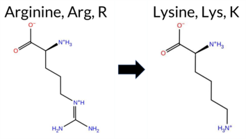 3DClick to see structure in 3D Viewer AIThe SynGAP1 missense variant R499K is not reported in ClinVar (no ClinVar ID) and is absent from gnomAD (no gnomAD ID). Prediction tools that agree on a benign effect include REVEL, PROVEAN, polyPhen‑2 (HumDiv and HumVar), SIFT, ESM1b, AlphaMissense‑Default, AlphaMissense‑Optimized, and the SGM‑Consensus score (Likely Benign). Only FATHMM predicts a pathogenic outcome. Stability‑based methods (FoldX, Rosetta, Foldetta, premPS) are inconclusive, providing no evidence for either direction. High‑accuracy assessments show AlphaMissense‑Optimized as benign; the SGM Consensus, derived from a majority vote of AlphaMissense‑Default, ESM1b, FATHMM, and PROVEAN, also yields a benign prediction; Foldetta remains uncertain. Overall, the preponderance of evidence points to a benign effect for R499K, and this conclusion is consistent with the absence of any ClinVar pathogenic classification. Disclaimer: This summary was generated using AI and should be interpreted alongside expert review. | Likely Benign | GAP | 0.071867 | Structured | 0.386723 | Uncertain | 0.899 | 0.146 | 0.000 | 4.366 | Likely Benign | 0.091 | Likely Benign | Likely Benign | 1.37 | Ambiguous | 0.1 | 0.53 | Ambiguous | 0.95 | Ambiguous | -0.77 | Ambiguous | 0.248 | Likely Benign | 1.94 | Neutral | 0.001 | Benign | 0.008 | Benign | -1.03 | Pathogenic | 1.00 | Tolerated | 0.3531 | 0.1881 | 3 | 2 | 0.6 | -28.01 | |||||||||||||||||||||||||||||
| c.1496G>C | R499T 2D  3DClick to see structure in 3D Viewer AIThe SynGAP1 missense variant R499T is not reported in ClinVar and is absent from gnomAD. Prediction tools that agree on a benign effect include polyPhen‑2 HumVar and ESM1b, whereas a majority of tools (REVEL, FoldX, PROVEAN, polyPhen‑2 HumDiv, SIFT, FATHMM, AlphaMissense‑Default) predict a pathogenic impact. High‑accuracy assessments further support a deleterious outcome: the SGM Consensus (derived from AlphaMissense‑Default, ESM1b, FATHMM, and PROVEAN) indicates Likely Pathogenic, AlphaMissense‑Optimized is inconclusive, and Foldetta (combining FoldX‑MD and Rosetta outputs) is uncertain. No evidence from ClinVar contradicts these predictions. Overall, the preponderance of computational evidence points to a pathogenic effect for R499T. Disclaimer: This summary was generated using AI and should be interpreted alongside expert review. | Likely Pathogenic | GAP | 0.071867 | Structured | 0.386723 | Uncertain | 0.899 | 0.146 | 0.000 | -6.797 | Likely Benign | 0.855 | Likely Pathogenic | Ambiguous | 2.47 | Destabilizing | 0.1 | 1.43 | Ambiguous | 1.95 | Ambiguous | 0.73 | Ambiguous | 0.664 | Likely Pathogenic | -3.51 | Deleterious | 0.843 | Possibly Damaging | 0.403 | Benign | -1.44 | Pathogenic | 0.02 | Affected | 0.1732 | 0.2074 | -1 | -1 | 3.8 | -55.08 | |||||||||||||||||||||||||||||
| c.1496G>T | R499I 2D  3DClick to see structure in 3D Viewer AIThe SynGAP1 missense variant R499I is not reported in ClinVar and is absent from gnomAD. Functional prediction tools largely indicate a deleterious effect: REVEL, PROVEAN, polyPhen‑2 (HumDiv and HumVar), SIFT, ESM1b, FATHMM, and AlphaMissense‑Default all classify the change as pathogenic, and the SGM‑Consensus (majority vote of AlphaMissense‑Default, ESM1b, FATHMM, PROVEAN) also reports it as likely pathogenic. No tool predicts a benign outcome; the remaining predictions (FoldX, Rosetta, Foldetta, premPS, AlphaMissense‑Optimized) are uncertain or inconclusive. High‑accuracy assessments show AlphaMissense‑Optimized as uncertain, SGM‑Consensus as likely pathogenic, and Foldetta as uncertain. Taken together, the overwhelming majority of evidence supports a pathogenic effect, and this is consistent with the absence of a ClinVar entry; there is no contradiction with ClinVar status. Disclaimer: This summary was generated using AI and should be interpreted alongside expert review. | Likely Pathogenic | GAP | 0.071867 | Structured | 0.386723 | Uncertain | 0.899 | 0.146 | 0.000 | -9.069 | Likely Pathogenic | 0.861 | Likely Pathogenic | Ambiguous | 1.53 | Ambiguous | 0.1 | 0.61 | Ambiguous | 1.07 | Ambiguous | 0.57 | Ambiguous | 0.713 | Likely Pathogenic | -5.50 | Deleterious | 0.998 | Probably Damaging | 0.922 | Probably Damaging | -1.47 | Pathogenic | 0.00 | Affected | 0.1401 | 0.2146 | -2 | -3 | 9.0 | -43.03 | |||||||||||||||||||||||||||||
| c.1498C>A | L500M 2D  3DClick to see structure in 3D Viewer AIThe SynGAP1 missense variant L500M is not reported in ClinVar (ClinVar ID None) and is absent from gnomAD (gnomAD ID None). Prediction tools that agree on a benign effect include FoldX, Rosetta, Foldetta, PROVEAN, AlphaMissense‑Default, and AlphaMissense‑Optimized. Those that predict a pathogenic effect are REVEL, polyPhen‑2 (HumDiv and HumVar), SIFT, ESM1b, and FATHMM. The premPS score is uncertain. High‑accuracy methods give the following results: AlphaMissense‑Optimized predicts benign; Foldetta predicts benign; the SGM Consensus (majority vote of AlphaMissense‑Default, ESM1b, FATHMM, and PROVEAN) is inconclusive because it yields a 2‑to‑2 split. No other high‑accuracy predictions are available. Overall, the majority of high‑accuracy tools lean toward a benign interpretation, and this assessment does not contradict the lack of ClinVar reporting. Thus, the variant is most likely benign based on current computational predictions. Disclaimer: This summary was generated using AI and should be interpreted alongside expert review. | GAP | 0.066181 | Structured | 0.382942 | Uncertain | 0.893 | 0.150 | 0.000 | -9.945 | Likely Pathogenic | 0.301 | Likely Benign | Likely Benign | 0.15 | Likely Benign | 0.0 | -0.09 | Likely Benign | 0.03 | Likely Benign | 0.92 | Ambiguous | 0.590 | Likely Pathogenic | -1.99 | Neutral | 1.000 | Probably Damaging | 1.000 | Probably Damaging | -1.36 | Pathogenic | 0.01 | Affected | 0.0710 | 0.2202 | 4 | 2 | -1.9 | 18.03 | ||||||||||||||||||||||||||||||
| c.1498C>G | L500V 2D 3DClick to see structure in 3D Viewer AISynGAP1 missense variant L500V is not reported in ClinVar and is absent from gnomAD. Functional prediction tools that agree on benign effect include SIFT and AlphaMissense‑Optimized, whereas a majority of tools predict pathogenicity: SGM‑Consensus, REVEL, premPS, PROVEAN, polyPhen‑2 (HumDiv and HumVar), ESM1b, and FATHMM. Predictions that are inconclusive are FoldX, Rosetta, Foldetta, and AlphaMissense‑Default. High‑accuracy assessments show AlphaMissense‑Optimized as benign, SGM‑Consensus (derived from a majority vote of AlphaMissense‑Default, ESM1b, FATHMM, and PROVEAN) as pathogenic, and Foldetta (combining FoldX‑MD and Rosetta outputs) as uncertain. Overall, the preponderance of evidence from multiple independent predictors indicates that the variant is most likely pathogenic, and this conclusion does not contradict the ClinVar status, which currently has no entry for this variant. Disclaimer: This summary was generated using AI and should be interpreted alongside expert review. | Likely Pathogenic | GAP | 0.066181 | Structured | 0.382942 | Uncertain | 0.893 | 0.150 | 0.000 | -12.963 | Likely Pathogenic | 0.379 | Ambiguous | Likely Benign | 1.92 | Ambiguous | 0.0 | 0.81 | Ambiguous | 1.37 | Ambiguous | 1.11 | Destabilizing | 0.565 | Likely Pathogenic | -2.98 | Deleterious | 0.998 | Probably Damaging | 0.992 | Probably Damaging | -1.24 | Pathogenic | 0.11 | Tolerated | 0.1205 | 0.1860 | 2 | 1 | 0.4 | -14.03 | |||||||||||||||||||||||||||||
| c.1499T>A | L500Q 2D  3DClick to see structure in 3D Viewer AIThe SynGAP1 missense variant L500Q has no ClinVar entry and is not reported in gnomAD. Prediction tools that agree on a benign effect include only AlphaMissense‑Optimized. All other evaluated predictors—SGM‑Consensus, REVEL, premPS, PROVEAN, polyPhen‑2 (HumDiv and HumVar), SIFT, ESM1b, FATHMM, and AlphaMissense‑Default—consistently classify the variant as pathogenic. FoldX, Rosetta, and Foldetta provide uncertain or inconclusive stability results and are treated as unavailable. High‑accuracy assessments further clarify the picture: AlphaMissense‑Optimized (benign) stands alone, whereas the SGM Consensus, derived from a majority vote of AlphaMissense‑Default, ESM1b, FATHMM, and PROVEAN, predicts pathogenic. Foldetta remains uncertain. Overall, the preponderance of evidence from multiple independent pathogenic predictors indicates that the variant is most likely pathogenic, and this conclusion does not contradict ClinVar status, which currently has no classification for this variant. Disclaimer: This summary was generated using AI and should be interpreted alongside expert review. | Likely Pathogenic | GAP | 0.066181 | Structured | 0.382942 | Uncertain | 0.893 | 0.150 | 0.000 | -13.431 | Likely Pathogenic | 0.649 | Likely Pathogenic | Likely Benign | 1.92 | Ambiguous | 0.1 | 1.78 | Ambiguous | 1.85 | Ambiguous | 1.49 | Destabilizing | 0.878 | Likely Pathogenic | -5.97 | Deleterious | 1.000 | Probably Damaging | 1.000 | Probably Damaging | -1.35 | Pathogenic | 0.00 | Affected | 0.0950 | 0.0488 | -2 | -2 | -7.3 | 14.97 | |||||||||||||||||||||||||||||
| c.1499T>G | L500R 2D 3DClick to see structure in 3D Viewer AIThe SynGAP1 missense variant L500R is not reported in ClinVar and is absent from gnomAD. Prediction tools that classify the variant as pathogenic include REVEL, FoldX, premPS, PROVEAN, polyPhen‑2 (HumDiv and HumVar), SIFT, ESM1b, FATHMM, AlphaMissense‑Default, and the SGM‑Consensus (majority vote of AlphaMissense‑Default, ESM1b, FATHMM, PROVEAN). No tool predicts a benign effect; the benign group is empty. Predictions that are uncertain or inconclusive are Rosetta, Foldetta, and AlphaMissense‑Optimized. High‑accuracy assessments show AlphaMissense‑Optimized as uncertain, SGM‑Consensus as likely pathogenic, and Foldetta as uncertain. Based on the overwhelming agreement among pathogenic predictors and the lack of any benign calls, the variant is most likely pathogenic, and this conclusion does not contradict the ClinVar status (which has no entry). Disclaimer: This summary was generated using AI and should be interpreted alongside expert review. | Likely Pathogenic | GAP | 0.066181 | Structured | 0.382942 | Uncertain | 0.893 | 0.150 | 0.000 | -15.576 | Likely Pathogenic | 0.789 | Likely Pathogenic | Ambiguous | 2.23 | Destabilizing | 0.1 | 1.04 | Ambiguous | 1.64 | Ambiguous | 1.11 | Destabilizing | 0.794 | Likely Pathogenic | -5.97 | Deleterious | 1.000 | Probably Damaging | 0.998 | Probably Damaging | -1.02 | Pathogenic | 0.03 | Affected | 0.1204 | 0.0488 | -3 | -2 | -8.3 | 43.03 | |||||||||||||||||||||||||||||
| c.149T>A | I50N 2D  AIThe SynGAP1 missense variant I50N is not reported in ClinVar (ClinVar ID None) and is absent from gnomAD (gnomAD ID None). Prediction tools that agree on a benign effect include REVEL, PROVEAN, polyPhen‑2 HumVar, and FATHMM, while those that predict a pathogenic effect are polyPhen‑2 HumDiv, SIFT, AlphaMissense‑Default, and AlphaMissense‑Optimized; ESM1b remains uncertain. High‑accuracy assessments show AlphaMissense‑Optimized as pathogenic, whereas the SGM Consensus (majority vote from AlphaMissense‑Default, ESM1b, FATHMM, PROVEAN) resolves as benign. Foldetta results are unavailable. Overall, the predictions are mixed, with an equal split between benign and pathogenic calls, but the most reliable single‑tool prediction (AlphaMissense‑Optimized) and the majority of individual tools lean toward pathogenicity. Thus, the variant is most likely pathogenic based on current computational evidence, and this assessment does not contradict any ClinVar status because no ClinVar claim exists. Disclaimer: This summary was generated using AI and should be interpreted alongside expert review. | 0.295083 | Structured | 0.449965 | Uncertain | 0.545 | 0.708 | 0.000 | -7.091 | In-Between | 0.962 | Likely Pathogenic | Likely Pathogenic | 0.135 | Likely Benign | -2.37 | Neutral | 0.842 | Possibly Damaging | 0.272 | Benign | 3.73 | Benign | 0.00 | Affected | 0.0824 | 0.0412 | -2 | -3 | -8.0 | 0.94 | ||||||||||||||||||||||||||||||||||||||||
| c.149T>C | I50T 2D AIThe SynGAP1 missense variant I50T is not reported in ClinVar and has no entries in gnomAD. Functional prediction tools largely agree that the change is benign: REVEL, PROVEAN, polyPhen‑2 (HumDiv and HumVar), ESM1b, and FATHMM all classify it as benign. In contrast, SIFT and AlphaMissense‑Default predict a pathogenic effect. AlphaMissense‑Optimized returns an uncertain result, and Foldetta (a protein‑folding stability method combining FoldX‑MD and Rosetta outputs) has no available prediction for this variant. The SGM‑Consensus, which aggregates AlphaMissense‑Default, ESM1b, FATHMM, and PROVEAN, yields a “Likely Benign” verdict. High‑accuracy assessments therefore point to a benign outcome: AlphaMissense‑Optimized is inconclusive, SGM‑Consensus is benign, and Foldetta data are missing. Based on the collective evidence, the variant is most likely benign, and this conclusion does not contradict any ClinVar annotation (none is present). Disclaimer: This summary was generated using AI and should be interpreted alongside expert review. | Likely Benign | 0.295083 | Structured | 0.449965 | Uncertain | 0.545 | 0.708 | 0.000 | -5.121 | Likely Benign | 0.949 | Likely Pathogenic | Ambiguous | 0.128 | Likely Benign | -1.61 | Neutral | 0.092 | Benign | 0.037 | Benign | 3.76 | Benign | 0.00 | Affected | 0.0950 | 0.0708 | 0 | -1 | -5.2 | -12.05 | |||||||||||||||||||||||||||||||||||||||
| c.149T>G | I50S 2D  AIThe SynGAP1 I50S missense variant is not reported in ClinVar and is absent from gnomAD. Functional prediction tools largely agree on a benign effect: REVEL, PROVEAN, polyPhen‑2 (HumDiv and HumVar), ESM1b, and FATHMM all classify it as benign. In contrast, SIFT and AlphaMissense‑Default predict a pathogenic impact. AlphaMissense‑Optimized yields an uncertain result, and Foldetta (a protein‑folding stability method combining FoldX‑MD and Rosetta outputs) has no available prediction for this residue. The SGM‑Consensus, which aggregates AlphaMissense‑Default, ESM1b, FATHMM, and PROVEAN, reports a likely benign classification. High‑accuracy assessments therefore point to a benign outcome: AlphaMissense‑Optimized is inconclusive, SGM‑Consensus is likely benign, and Foldetta data are missing. Based on the collective evidence, the variant is most likely benign, and this assessment does not contradict any ClinVar status (none is available). Disclaimer: This summary was generated using AI and should be interpreted alongside expert review. | Likely Benign | 0.295083 | Structured | 0.449965 | Uncertain | 0.545 | 0.708 | 0.000 | -4.257 | Likely Benign | 0.946 | Likely Pathogenic | Ambiguous | 0.209 | Likely Benign | -2.03 | Neutral | 0.334 | Benign | 0.099 | Benign | 3.74 | Benign | 0.00 | Affected | 0.2626 | 0.0800 | -1 | -2 | -5.3 | -26.08 | |||||||||||||||||||||||||||||||||||||||
| c.14G>C | R5P 2D AIThe SynGAP1 missense variant R5P is not reported in ClinVar (no ClinVar ID) and is absent from gnomAD (no gnomAD ID). Prediction tools largely agree on a benign effect: REVEL, PROVEAN, polyPhen‑2 (HumDiv and HumVar), ESM1b, FATHMM, AlphaMissense‑Default, and AlphaMissense‑Optimized all classify the variant as benign. Only SIFT predicts a pathogenic outcome. The SGM‑Consensus, which aggregates AlphaMissense‑Default, ESM1b, FATHMM, and PROVEAN, reports the variant as “Likely Benign.” High‑accuracy assessments further support a benign interpretation: AlphaMissense‑Optimized is benign, and the SGM‑Consensus (majority vote) is likely benign; Foldetta results are unavailable. Overall, the preponderance of evidence indicates that R5P is most likely benign, and this conclusion does not contradict any ClinVar status because no ClinVar classification exists. Disclaimer: This summary was generated using AI and should be interpreted alongside expert review. | Likely Benign | 0.595080 | Disordered | 0.547847 | Binding | 0.363 | 0.920 | 0.750 | -3.438 | Likely Benign | 0.189 | Likely Benign | Likely Benign | 0.286 | Likely Benign | -0.35 | Neutral | 0.233 | Benign | 0.013 | Benign | 4.10 | Benign | 0.00 | Affected | 0.2294 | 0.5530 | 0 | -2 | 2.9 | -59.07 | |||||||||||||||||||||||||||||||||||||||
| c.14G>T | R5L 2D AIThe SynGAP1 missense variant R5L is not reported in ClinVar (no ClinVar ID) and is absent from gnomAD (no gnomAD ID). Prediction tools that agree on a benign effect include REVEL, PROVEAN, polyPhen‑2 HumDiv, polyPhen‑2 HumVar, ESM1b, FATHMM, AlphaMissense‑Default, and AlphaMissense‑Optimized. Only SIFT predicts a pathogenic outcome. The SGM‑Consensus, which aggregates AlphaMissense‑Default, ESM1b, FATHMM, and PROVEAN, reports the variant as Likely Benign. High‑accuracy assessments further support a benign classification: AlphaMissense‑Optimized is benign, and the SGM‑Consensus is Likely Benign; Foldetta results are not available. Based on the preponderance of evidence, the variant is most likely benign, and this assessment does not contradict any ClinVar status because no ClinVar entry exists. Disclaimer: This summary was generated using AI and should be interpreted alongside expert review. | Likely Benign | 0.595080 | Disordered | 0.547847 | Binding | 0.363 | 0.920 | 0.750 | -3.297 | Likely Benign | 0.274 | Likely Benign | Likely Benign | 0.158 | Likely Benign | -0.06 | Neutral | 0.030 | Benign | 0.003 | Benign | 4.14 | Benign | 0.00 | Affected | 0.2256 | 0.5914 | -3 | -2 | 8.3 | -43.03 | |||||||||||||||||||||||||||||||||||||||
| c.1501A>C | I501L 2D 3DClick to see structure in 3D Viewer AIThe SynGAP1 missense variant I501L is not reported in ClinVar (no ClinVar ID) and is absent from gnomAD. Functional prediction tools cluster into two groups: benign predictions include REVEL, Rosetta, Foldetta, PROVEAN, FATHMM, AlphaMissense‑Default, AlphaMissense‑Optimized, and the SGM‑Consensus (Likely Benign). Pathogenic predictions come from polyPhen‑2 (HumDiv and HumVar) and SIFT. Uncertain or inconclusive results are reported for FoldX, premPS, and ESM1b. High‑accuracy assessments show AlphaMissense‑Optimized as Benign, the SGM‑Consensus as Likely Benign, and Foldetta as Benign, all supporting a non‑deleterious effect. Based on the preponderance of benign predictions and the concordant high‑accuracy tools, the variant is most likely benign, and this conclusion does not contradict any ClinVar status. Disclaimer: This summary was generated using AI and should be interpreted alongside expert review. | Likely Benign | GAP | 0.079919 | Structured | 0.366596 | Uncertain | 0.886 | 0.153 | 0.000 | -7.348 | In-Between | 0.282 | Likely Benign | Likely Benign | 0.78 | Ambiguous | 0.2 | -0.34 | Likely Benign | 0.22 | Likely Benign | 0.86 | Ambiguous | 0.219 | Likely Benign | -1.92 | Neutral | 0.930 | Possibly Damaging | 0.985 | Probably Damaging | 3.51 | Benign | 0.05 | Affected | 0.0817 | 0.2261 | 2 | 2 | -0.7 | 0.00 | |||||||||||||||||||||||||||||
| c.1501A>G | I501V 2D 3DClick to see structure in 3D Viewer AIThe SynGAP1 missense variant I501V is not reported in ClinVar and is absent from gnomAD. Functional prediction tools that agree on a benign effect include REVEL, premPS, PROVEAN, SIFT, ESM1b, FATHMM, AlphaMissense‑Default, AlphaMissense‑Optimized, and the SGM‑Consensus (majority vote from AlphaMissense‑Default, ESM1b, FATHMM, PROVEAN). Tools that predict a pathogenic effect are polyPhen‑2 HumDiv and polyPhen‑2 HumVar. Predictions from FoldX, Rosetta, and Foldetta are uncertain and are treated as unavailable. High‑accuracy assessments show AlphaMissense‑Optimized as benign, the SGM‑Consensus as likely benign, and Foldetta as uncertain. Overall, the majority of evidence points to a benign impact for I501V, and this conclusion does not contradict the ClinVar status, which currently has no entry for this variant. Disclaimer: This summary was generated using AI and should be interpreted alongside expert review. | Likely Benign | GAP | 0.079919 | Structured | 0.366596 | Uncertain | 0.886 | 0.153 | 0.000 | -2.326 | Likely Benign | 0.083 | Likely Benign | Likely Benign | 0.94 | Ambiguous | 0.0 | 0.56 | Ambiguous | 0.75 | Ambiguous | 0.06 | Likely Benign | 0.200 | Likely Benign | 0.20 | Neutral | 0.951 | Possibly Damaging | 0.960 | Probably Damaging | 3.52 | Benign | 1.00 | Tolerated | 0.0985 | 0.2491 | 4 | 3 | -0.3 | -14.03 | |||||||||||||||||||||||||||||
| c.1501A>T | I501F 2D 3DClick to see structure in 3D Viewer AIThe SynGAP1 I501F missense variant has no ClinVar entry and is not reported in gnomAD. Prediction tools that indicate a benign effect include REVEL, Rosetta, FATHMM, and AlphaMissense‑Optimized. Tools that predict a pathogenic effect comprise SGM‑Consensus, FoldX, PROVEAN, polyPhen‑2 (HumDiv and HumVar), SIFT, ESM1b, and AlphaMissense‑Default. High‑accuracy assessments show AlphaMissense‑Optimized as benign, SGM‑Consensus as likely pathogenic (derived from a majority vote of AlphaMissense‑Default, ESM1b, FATHMM, and PROVEAN), and Foldetta as uncertain (a stability method that integrates FoldX‑MD and Rosetta outputs). No predictions are missing or inconclusive. Overall, the majority of evidence points toward a pathogenic impact, and this conclusion does not contradict the ClinVar status, which is currently unreported. Disclaimer: This summary was generated using AI and should be interpreted alongside expert review. | Likely Pathogenic | GAP | 0.079919 | Structured | 0.366596 | Uncertain | 0.886 | 0.153 | 0.000 | -12.113 | Likely Pathogenic | 0.664 | Likely Pathogenic | Likely Benign | 3.06 | Destabilizing | 0.1 | 0.26 | Likely Benign | 1.66 | Ambiguous | 0.71 | Ambiguous | 0.385 | Likely Benign | -3.88 | Deleterious | 0.998 | Probably Damaging | 0.993 | Probably Damaging | 3.40 | Benign | 0.03 | Affected | 0.0574 | 0.1594 | 1 | 0 | -1.7 | 34.02 | |||||||||||||||||||||||||||||
| c.1502T>A | I501N 2D 3DClick to see structure in 3D Viewer AIThe SynGAP1 missense variant I501N is not reported in ClinVar and is absent from gnomAD. Prediction tools that agree on a benign effect are REVEL and FATHMM, while a majority of tools (SGM‑Consensus, FoldX, premPS, PROVEAN, polyPhen‑2 HumDiv, polyPhen‑2 HumVar, SIFT, ESM1b, AlphaMissense‑Default) predict a pathogenic impact. Uncertain results come from AlphaMissense‑Optimized, Foldetta, and Rosetta. High‑accuracy assessments show AlphaMissense‑Optimized as inconclusive, whereas the SGM Consensus (derived from a majority vote of AlphaMissense‑Default, ESM1b, FATHMM, and PROVEAN) indicates a likely pathogenic outcome; Foldetta likewise remains inconclusive. Overall, the balance of evidence favors a pathogenic classification, and this conclusion does not contradict any existing ClinVar annotation (none is available). Disclaimer: This summary was generated using AI and should be interpreted alongside expert review. | Likely Pathogenic | GAP | 0.079919 | Structured | 0.366596 | Uncertain | 0.886 | 0.153 | 0.000 | -11.105 | Likely Pathogenic | 0.854 | Likely Pathogenic | Ambiguous | 2.22 | Destabilizing | 0.0 | 1.70 | Ambiguous | 1.96 | Ambiguous | 1.90 | Destabilizing | 0.445 | Likely Benign | -6.13 | Deleterious | 1.000 | Probably Damaging | 1.000 | Probably Damaging | 3.37 | Benign | 0.01 | Affected | 0.0825 | 0.0270 | -2 | -3 | -8.0 | 0.94 | |||||||||||||||||||||||||||||
| c.1502T>G | I501S 2D 3DClick to see structure in 3D Viewer AIThe SynGAP1 missense variant I501S is not reported in ClinVar and is absent from gnomAD. Prediction tools that agree on a benign effect are FATHMM and AlphaMissense‑Optimized; all other evaluated algorithms (SGM‑Consensus, REVEL, FoldX, Rosetta, Foldetta, premPS, PROVEAN, polyPhen‑2 HumDiv, polyPhen‑2 HumVar, SIFT, ESM1b, AlphaMissense‑Default) predict a pathogenic impact. High‑accuracy assessments further support this: AlphaMissense‑Optimized indicates a benign change, whereas the SGM Consensus (majority vote of AlphaMissense‑Default, ESM1b, FATHMM, PROVEAN) and Foldetta (combining FoldX‑MD and Rosetta outputs) both predict pathogenicity. Overall, the preponderance of evidence points to a pathogenic effect for I501S. This conclusion is not contradicted by ClinVar status, which currently contains no entry for this variant. Disclaimer: This summary was generated using AI and should be interpreted alongside expert review. | Likely Pathogenic | GAP | 0.079919 | Structured | 0.366596 | Uncertain | 0.886 | 0.153 | 0.000 | -9.914 | Likely Pathogenic | 0.677 | Likely Pathogenic | Likely Benign | 3.23 | Destabilizing | 0.2 | 2.40 | Destabilizing | 2.82 | Destabilizing | 1.79 | Destabilizing | 0.511 | Likely Pathogenic | -5.14 | Deleterious | 1.000 | Probably Damaging | 1.000 | Probably Damaging | 3.43 | Benign | 0.04 | Affected | 0.2370 | 0.0858 | -1 | -2 | -5.3 | -26.08 | |||||||||||||||||||||||||||||
| c.1503T>G | I501M 2D 3DClick to see structure in 3D Viewer AIThe SynGAP1 missense variant I501M is not reported in ClinVar and is absent from gnomAD. Functional prediction tools that agree on a benign effect include REVEL, Rosetta, PROVEAN, FATHMM, AlphaMissense‑Default, and AlphaMissense‑Optimized. Tools that predict a pathogenic effect are polyPhen‑2 HumDiv, polyPhen‑2 HumVar, and SIFT. Predictions that are inconclusive or unavailable are FoldX, Foldetta, premPS, and ESM1b. High‑accuracy assessments show AlphaMissense‑Optimized as benign, the SGM‑Consensus (majority vote of AlphaMissense‑Default, ESM1b, FATHMM, PROVEAN) as Likely Benign, and Foldetta as uncertain. Taken together, the majority of evidence points to a benign impact for I501M. This conclusion is consistent with the lack of ClinVar annotation and does not contradict any existing database status. Disclaimer: This summary was generated using AI and should be interpreted alongside expert review. | Likely Benign | GAP | 0.079919 | Structured | 0.366596 | Uncertain | 0.886 | 0.153 | 0.000 | -7.452 | In-Between | 0.259 | Likely Benign | Likely Benign | 1.32 | Ambiguous | 0.2 | -0.19 | Likely Benign | 0.57 | Ambiguous | 0.95 | Ambiguous | 0.294 | Likely Benign | -2.32 | Neutral | 1.000 | Probably Damaging | 1.000 | Probably Damaging | 3.36 | Benign | 0.01 | Affected | 0.0680 | 0.1589 | 2 | 1 | -2.6 | 18.03 | |||||||||||||||||||||||||||||
| c.1504G>A | G502S 2D  3DClick to see structure in 3D Viewer AIThe SynGAP1 missense variant G502S is not reported in ClinVar and has no entry in gnomAD. Prediction tools that agree on a benign effect include only AlphaMissense‑Optimized. All other evaluated predictors—SGM Consensus, REVEL, FoldX, PROVEAN, polyPhen‑2 (HumDiv and HumVar), SIFT, ESM1b, FATHMM, and AlphaMissense‑Default—consistently classify the variant as pathogenic. High‑accuracy assessments further support this view: AlphaMissense‑Optimized predicts a benign outcome, whereas the SGM Consensus (a majority vote of AlphaMissense‑Default, ESM1b, FATHMM, and PROVEAN) indicates likely pathogenic. Foldetta, which integrates FoldX‑MD and Rosetta outputs, yields an uncertain result and is treated as unavailable evidence. Overall, the preponderance of evidence points to a pathogenic effect, and this conclusion does not contradict any ClinVar annotation (none exists). Disclaimer: This summary was generated using AI and should be interpreted alongside expert review. | Likely Pathogenic | GAP | 0.083462 | Structured | 0.340113 | Uncertain | 0.882 | 0.152 | 0.000 | -11.357 | Likely Pathogenic | 0.616 | Likely Pathogenic | Likely Benign | 2.09 | Destabilizing | 0.6 | 0.71 | Ambiguous | 1.40 | Ambiguous | 0.96 | Ambiguous | 0.839 | Likely Pathogenic | -5.74 | Deleterious | 0.975 | Probably Damaging | 0.862 | Possibly Damaging | -1.64 | Pathogenic | 0.01 | Affected | 0.2429 | 0.3217 | 1 | 0 | -0.4 | 30.03 | |||||||||||||||||||||||||||||
| c.1504G>C | G502R 2D  3DClick to see structure in 3D Viewer AIThe SynGAP1 missense variant G502R is not reported in ClinVar (ClinVar ID None) and is absent from gnomAD (gnomAD ID None). Prediction tools that assess pathogenicity unanimously classify the variant as deleterious: SGM‑Consensus, REVEL, FoldX, Rosetta, Foldetta, PROVEAN, polyPhen‑2 (HumDiv and HumVar), SIFT, ESM1b, FATHMM, AlphaMissense‑Default, and AlphaMissense‑Optimized. No tool predicts a benign effect; the only inconclusive result is from premPS, which is listed as uncertain. High‑accuracy methods reinforce this view: AlphaMissense‑Optimized predicts pathogenicity; the SGM Consensus (majority vote of AlphaMissense‑Default, ESM1b, FATHMM, and PROVEAN) also predicts pathogenic; and Foldetta, which integrates FoldX‑MD and Rosetta stability calculations, predicts a destabilizing, pathogenic effect. Based on the collective evidence, the variant is most likely pathogenic, and this assessment does not contradict any ClinVar status. Disclaimer: This summary was generated using AI and should be interpreted alongside expert review. | Likely Pathogenic | GAP | 0.083462 | Structured | 0.340113 | Uncertain | 0.882 | 0.152 | 0.000 | -15.571 | Likely Pathogenic | 0.991 | Likely Pathogenic | Likely Pathogenic | 8.80 | Destabilizing | 0.3 | 10.07 | Destabilizing | 9.44 | Destabilizing | 0.88 | Ambiguous | 0.917 | Likely Pathogenic | -7.73 | Deleterious | 1.000 | Probably Damaging | 0.985 | Probably Damaging | -1.65 | Pathogenic | 0.00 | Affected | 0.1014 | 0.3317 | -3 | -2 | -4.1 | 99.14 | |||||||||||||||||||||||||||||
| c.1504G>T | G502C 2D  3DClick to see structure in 3D Viewer AIThe SynGAP1 missense variant G502C lies in the GAP domain. ClinVar has no entry for this variant, and it is not reported in gnomAD. Prediction tools that agree on a benign effect include only premPS. The majority of tools predict a pathogenic effect: REVEL, PROVEAN, polyPhen‑2 (HumDiv and HumVar), SIFT, ESM1b, FATHMM, AlphaMissense‑Default, and the SGM‑Consensus score. Tools with uncertain or inconclusive outputs are FoldX, Rosetta, Foldetta, and AlphaMissense‑Optimized. High‑accuracy assessments show AlphaMissense‑Optimized as uncertain, SGM‑Consensus (derived from a unanimous majority of AlphaMissense‑Default, ESM1b, FATHMM, and PROVEAN) as pathogenic, and Foldetta (combining FoldX‑MD and Rosetta) as uncertain. Overall, the preponderance of evidence points to a pathogenic impact for G502C, and this conclusion does not contradict any ClinVar annotation (none is available). Disclaimer: This summary was generated using AI and should be interpreted alongside expert review. | Likely Pathogenic | GAP | 0.083462 | Structured | 0.340113 | Uncertain | 0.882 | 0.152 | 0.000 | -12.086 | Likely Pathogenic | 0.907 | Likely Pathogenic | Ambiguous | 1.02 | Ambiguous | 0.5 | 1.55 | Ambiguous | 1.29 | Ambiguous | 0.30 | Likely Benign | 0.845 | Likely Pathogenic | -8.65 | Deleterious | 1.000 | Probably Damaging | 0.988 | Probably Damaging | -1.67 | Pathogenic | 0.00 | Affected | 0.1279 | 0.2691 | -3 | -3 | 2.9 | 46.09 | |||||||||||||||||||||||||||||
| c.1505G>C | G502A 2D  3DClick to see structure in 3D Viewer AISynGAP1 missense variant G502A is not reported in ClinVar and is absent from gnomAD. Functional prediction tools cluster into two groups: benign predictions come from Foldetta, polyPhen‑2 HumVar, SIFT, and AlphaMissense‑Optimized; pathogenic predictions arise from SGM‑Consensus, REVEL, PROVEAN, polyPhen‑2 HumDiv, ESM1b, FATHMM, and AlphaMissense‑Default. Uncertain results are reported by FoldX, Rosetta, and premPS. High‑accuracy assessments further clarify the picture: AlphaMissense‑Optimized indicates a benign effect, while the SGM Consensus—derived from a majority vote of AlphaMissense‑Default, ESM1b, FATHMM, and PROVEAN—concludes pathogenic. Foldetta, a protein‑folding stability method combining FoldX‑MD and Rosetta outputs, also predicts a benign outcome. Overall, the majority of evidence points to a pathogenic impact for G502A, and this assessment does not conflict with the absence of ClinVar annotation. Disclaimer: This summary was generated using AI and should be interpreted alongside expert review. | Likely Pathogenic | GAP | 0.083462 | Structured | 0.340113 | Uncertain | 0.882 | 0.152 | 0.000 | -10.191 | Likely Pathogenic | 0.607 | Likely Pathogenic | Likely Benign | 0.82 | Ambiguous | 0.4 | -0.53 | Ambiguous | 0.15 | Likely Benign | 0.54 | Ambiguous | 0.725 | Likely Pathogenic | -5.64 | Deleterious | 0.512 | Possibly Damaging | 0.157 | Benign | -1.59 | Pathogenic | 0.17 | Tolerated | 0.3548 | 0.3393 | 1 | 0 | 2.2 | 14.03 | |||||||||||||||||||||||||||||
| c.1505G>T | G502V 2D  3DClick to see structure in 3D Viewer AIThe SynGAP1 missense variant G502V is not reported in ClinVar and is absent from gnomAD. Functional prediction tools largely agree on a deleterious effect: all evaluated algorithms except premPS (which predicts benign) classify the substitution as pathogenic or likely pathogenic. The consensus of high‑accuracy predictors is consistent: AlphaMissense‑Optimized reports a pathogenic effect; the SGM Consensus, derived from a majority vote of AlphaMissense‑Default, ESM1b, FATHMM, and PROVEAN, also indicates likely pathogenic; and Foldetta, which integrates FoldX‑MD and Rosetta stability calculations, predicts a destabilizing, pathogenic outcome. In contrast, premPS is the sole tool suggesting a benign impact. Overall, the overwhelming majority of evidence points to a pathogenic effect for G502V, and this conclusion does not conflict with the absence of a ClinVar classification. Thus, the variant is most likely pathogenic, and this assessment is not contradicted by ClinVar. Disclaimer: This summary was generated using AI and should be interpreted alongside expert review. | Likely Pathogenic | GAP | 0.083462 | Structured | 0.340113 | Uncertain | 0.882 | 0.152 | 0.000 | -15.278 | Likely Pathogenic | 0.968 | Likely Pathogenic | Likely Pathogenic | 3.56 | Destabilizing | 1.0 | 5.50 | Destabilizing | 4.53 | Destabilizing | 0.43 | Likely Benign | 0.917 | Likely Pathogenic | -8.65 | Deleterious | 0.999 | Probably Damaging | 0.944 | Probably Damaging | -1.67 | Pathogenic | 0.00 | Affected | 0.1406 | 0.3387 | -1 | -3 | 4.6 | 42.08 | |||||||||||||||||||||||||||||
| c.1507C>A | Q503K 2D  3DClick to see structure in 3D Viewer AISynGAP1 missense variant Q503K (ClinVar ID 4327028) is not reported in gnomAD. Prediction tools that classify the variant as benign include FoldX, Rosetta, Foldetta, polyPhen‑2 HumVar, SIFT, AlphaMissense‑Default, and AlphaMissense‑Optimized. Those that predict pathogenicity are SGM‑Consensus, REVEL, PROVEAN, polyPhen‑2 HumDiv, ESM1b, and FATHMM. The high‑accuracy methods give a mixed picture: AlphaMissense‑Optimized predicts benign; the SGM‑Consensus, derived from a majority vote of AlphaMissense‑Default, ESM1b, FATHMM, and PROVEAN, predicts pathogenic; and Foldetta, which integrates FoldX‑MD and Rosetta outputs, predicts benign. premPS is inconclusive and therefore not considered. Overall, the predictions are split, with a slight bias toward benign. Thus, the variant is most likely benign according to the computational evidence, which contradicts the ClinVar pathogenic classification. Disclaimer: This summary was generated using AI and should be interpreted alongside expert review. | Likely Pathogenic | GAP | 0.040537 | Structured | 0.322935 | Uncertain | 0.848 | 0.168 | 0.000 | 1 | -12.276 | Likely Pathogenic | 0.217 | Likely Benign | Likely Benign | -0.01 | Likely Benign | 0.1 | -0.26 | Likely Benign | -0.14 | Likely Benign | 0.70 | Ambiguous | 0.603 | Likely Pathogenic | -3.37 | Deleterious | 0.676 | Possibly Damaging | 0.297 | Benign | -1.42 | Pathogenic | 0.12 | Tolerated | 0.1601 | 0.2419 | 1 | 1 | -0.4 | 0.04 | ||||||||||||||||||||||||||||
| c.1507C>G | Q503E 2D  3DClick to see structure in 3D Viewer AIThe SynGAP1 missense variant Q503E is not reported in ClinVar (ClinVar ID None) and is absent from gnomAD (gnomAD ID None). Prediction tools that agree on a benign effect include REVEL, PROVEAN, SIFT, AlphaMissense‑Default, and AlphaMissense‑Optimized. Tools that agree on a pathogenic effect are polyPhen‑2 HumDiv, polyPhen‑2 HumVar, FATHMM, and Rosetta. Uncertain or inconclusive results come from FoldX, Foldetta, premPS, and ESM1b. High‑accuracy assessments show AlphaMissense‑Optimized as benign; the SGM Consensus (majority vote from AlphaMissense‑Default, ESM1b, FATHMM, PROVEAN) also predicts benign, while Foldetta remains uncertain. Overall, the majority of available predictions favor a benign impact, and this conclusion does not contradict the lack of ClinVar annotation. Thus, the variant is most likely benign based on current computational evidence. Disclaimer: This summary was generated using AI and should be interpreted alongside expert review. | GAP | 0.040537 | Structured | 0.322935 | Uncertain | 0.848 | 0.168 | 0.000 | -7.909 | In-Between | 0.146 | Likely Benign | Likely Benign | 0.51 | Ambiguous | 0.1 | 2.11 | Destabilizing | 1.31 | Ambiguous | 0.85 | Ambiguous | 0.410 | Likely Benign | -2.21 | Neutral | 0.931 | Possibly Damaging | 0.500 | Possibly Damaging | -1.43 | Pathogenic | 0.17 | Tolerated | 0.1168 | 0.1508 | 2 | 2 | 0.0 | 0.98 | ||||||||||||||||||||||||||||||
| c.1508A>C | Q503P 2D  3DClick to see structure in 3D Viewer AIThe SynGAP1 missense variant Q503P is not reported in ClinVar and is absent from gnomAD. Prediction tools that agree on a benign effect include only AlphaMissense‑Optimized. Those that predict a pathogenic effect comprise SGM‑Consensus, REVEL, Rosetta, Foldetta, PROVEAN, polyPhen‑2 (HumDiv and HumVar), SIFT, ESM1b, and FATHMM; the remaining tools (FoldX, premPS, AlphaMissense‑Default) are inconclusive. High‑accuracy methods give a consistent pathogenic signal: AlphaMissense‑Optimized predicts benign, but the SGM‑Consensus (majority vote of AlphaMissense‑Default, ESM1b, FATHMM, PROVEAN) and Foldetta both predict pathogenic. Because the majority of evidence points to a deleterious effect, the variant is most likely pathogenic, and this assessment does not contradict the ClinVar status, which currently has no entry for this variant. Disclaimer: This summary was generated using AI and should be interpreted alongside expert review. | Likely Pathogenic | GAP | 0.040537 | Structured | 0.322935 | Uncertain | 0.848 | 0.168 | 0.000 | -10.915 | Likely Pathogenic | 0.414 | Ambiguous | Likely Benign | 1.06 | Ambiguous | 0.7 | 5.94 | Destabilizing | 3.50 | Destabilizing | 0.75 | Ambiguous | 0.860 | Likely Pathogenic | -5.20 | Deleterious | 0.999 | Probably Damaging | 0.991 | Probably Damaging | -1.53 | Pathogenic | 0.03 | Affected | 0.2076 | 0.3610 | 0 | -1 | 1.9 | -31.01 | |||||||||||||||||||||||||||||
| c.1508A>G | Q503R 2D  3DClick to see structure in 3D Viewer AIThe SynGAP1 missense variant Q503R has no ClinVar entry and is not reported in gnomAD. Prediction tools that classify the variant as benign include FoldX, Foldetta, premPS, polyPhen‑2 HumVar, SIFT, AlphaMissense‑Default, and AlphaMissense‑Optimized. Those that predict pathogenicity are SGM‑Consensus, REVEL, PROVEAN, polyPhen‑2 HumDiv, ESM1b, and FATHMM. The high‑accuracy methods give a benign result for AlphaMissense‑Optimized, a pathogenic result for the SGM‑Consensus (majority vote of AlphaMissense‑Default, ESM1b, FATHMM, PROVEAN), and a benign result for Foldetta (combining FoldX‑MD and Rosetta). Overall, the majority of tools and the high‑accuracy methods lean toward a benign effect. Thus, the variant is most likely benign, and this assessment does not contradict any ClinVar status because none is available. Disclaimer: This summary was generated using AI and should be interpreted alongside expert review. | Likely Pathogenic | GAP | 0.040537 | Structured | 0.322935 | Uncertain | 0.848 | 0.168 | 0.000 | -11.396 | Likely Pathogenic | 0.232 | Likely Benign | Likely Benign | -0.40 | Likely Benign | 0.3 | 0.60 | Ambiguous | 0.10 | Likely Benign | 0.50 | Likely Benign | 0.640 | Likely Pathogenic | -3.34 | Deleterious | 0.577 | Possibly Damaging | 0.395 | Benign | -1.42 | Pathogenic | 0.06 | Tolerated | 0.1426 | 0.0963 | 1 | 1 | -1.0 | 28.06 | |||||||||||||||||||||||||||||
| c.1508A>T | Q503L 2D  3DClick to see structure in 3D Viewer AIThe SynGAP1 missense variant Q503L is not reported in ClinVar and has no entries in gnomAD. Prediction tools that agree on a benign effect include AlphaMissense‑Default, AlphaMissense‑Optimized, Foldetta, premPS, Rosetta, and polyPhen‑2 (HumVar). Those that predict a pathogenic effect are SGM‑Consensus, REVEL, PROVEAN, polyPhen‑2 (HumDiv), SIFT, ESM1b, and FATHMM; FoldX is uncertain and therefore not counted. High‑accuracy assessments show AlphaMissense‑Optimized as benign, SGM‑Consensus (a majority vote of AlphaMissense‑Default, ESM1b, FATHMM, and PROVEAN) as likely pathogenic, and Foldetta as benign. Overall, the majority of tools (seven versus six) predict pathogenicity, and the high‑accuracy trio is split but leans toward pathogenic. Thus, the variant is most likely pathogenic based on current predictions, and this assessment does not contradict any ClinVar status (none reported). Disclaimer: This summary was generated using AI and should be interpreted alongside expert review. | Likely Pathogenic | GAP | 0.040537 | Structured | 0.322935 | Uncertain | 0.848 | 0.168 | 0.000 | -8.203 | Likely Pathogenic | 0.261 | Likely Benign | Likely Benign | -0.56 | Ambiguous | 0.2 | -0.07 | Likely Benign | -0.32 | Likely Benign | 0.24 | Likely Benign | 0.711 | Likely Pathogenic | -6.29 | Deleterious | 0.911 | Possibly Damaging | 0.369 | Benign | -1.52 | Pathogenic | 0.05 | Affected | 0.0715 | 0.3578 | -2 | -2 | 7.3 | -14.97 | |||||||||||||||||||||||||||||
| c.1509G>C | Q503H 2D  3DClick to see structure in 3D Viewer AIThe SynGAP1 missense variant Q503H is not reported in ClinVar (ClinVar ID None) and is absent from gnomAD (gnomAD ID None). Prediction tools that classify the variant as benign include REVEL, premPS, polyPhen‑2 (HumDiv and HumVar), ESM1b, AlphaMissense‑Default, and AlphaMissense‑Optimized. Tools that predict pathogenicity are PROVEAN, SIFT, and FATHMM. FoldX, Rosetta, and Foldetta report uncertain or inconclusive stability changes and are therefore considered unavailable for pathogenicity inference. High‑accuracy assessments show AlphaMissense‑Optimized as benign; the SGM Consensus (majority vote from AlphaMissense‑Default, ESM1b, FATHMM, PROVEAN) is a 2‑vs‑2 tie and thus inconclusive, and Foldetta remains uncertain. Overall, the majority of evaluated predictors (7 benign vs. 3 pathogenic) lean toward a benign effect. Consequently, the variant is most likely benign, and this assessment does not contradict any ClinVar status. Disclaimer: This summary was generated using AI and should be interpreted alongside expert review. | GAP | 0.040537 | Structured | 0.322935 | Uncertain | 0.848 | 0.168 | 0.000 | -5.335 | Likely Benign | 0.227 | Likely Benign | Likely Benign | 0.94 | Ambiguous | 0.2 | 0.88 | Ambiguous | 0.91 | Ambiguous | 0.48 | Likely Benign | 0.331 | Likely Benign | -3.40 | Deleterious | 0.002 | Benign | 0.004 | Benign | -1.52 | Pathogenic | 0.01 | Affected | 0.1091 | 0.2125 | 3 | 0 | 0.3 | 9.01 | ||||||||||||||||||||||||||||||
| c.1509G>T | Q503H 2D 3DClick to see structure in 3D Viewer AIThe SynGAP1 missense variant Q503H is not reported in ClinVar (ClinVar ID None) and is absent from gnomAD (gnomAD ID None). Prediction tools that classify the variant as benign include REVEL, premPS, polyPhen‑2 (HumDiv and HumVar), ESM1b, AlphaMissense‑Default, and AlphaMissense‑Optimized. Tools that predict pathogenicity are PROVEAN, SIFT, and FATHMM. FoldX, Rosetta, and Foldetta report uncertain or inconclusive stability changes and are therefore considered unavailable for pathogenicity inference. High‑accuracy assessments show AlphaMissense‑Optimized as benign; the SGM Consensus (majority vote from AlphaMissense‑Default, ESM1b, FATHMM, PROVEAN) is a 2‑vs‑2 tie and thus inconclusive, and Foldetta remains uncertain. Overall, the majority of evaluated predictors (7 benign vs. 3 pathogenic) lean toward a benign effect. Consequently, the variant is most likely benign, and this assessment does not contradict any ClinVar status. Disclaimer: This summary was generated using AI and should be interpreted alongside expert review. | GAP | 0.040537 | Structured | 0.322935 | Uncertain | 0.848 | 0.168 | 0.000 | -5.335 | Likely Benign | 0.227 | Likely Benign | Likely Benign | 0.94 | Ambiguous | 0.2 | 0.88 | Ambiguous | 0.91 | Ambiguous | 0.48 | Likely Benign | 0.331 | Likely Benign | -3.40 | Deleterious | 0.002 | Benign | 0.004 | Benign | -1.52 | Pathogenic | 0.01 | Affected | 0.1091 | 0.2125 | 3 | 0 | 0.3 | 9.01 | ||||||||||||||||||||||||||||||
| c.150C>G | I50M 2D AIThe SynGAP1 missense variant I50M is not reported in ClinVar and is absent from gnomAD. Prediction tools that agree on a benign effect include REVEL, PROVEAN, polyPhen‑2 HumVar, ESM1b, FATHMM, and AlphaMissense‑Optimized. Tools that predict a pathogenic effect are polyPhen‑2 HumDiv and SIFT, while AlphaMissense‑Default remains uncertain. The SGM‑Consensus, derived from a majority vote of AlphaMissense‑Default, ESM1b, FATHMM, and PROVEAN, classifies the variant as Likely Benign. High‑accuracy assessments further support a benign outcome: AlphaMissense‑Optimized predicts Benign, and the SGM‑Consensus also indicates Likely Benign. Foldetta, a protein‑folding stability method combining FoldX‑MD and Rosetta outputs, has no available result for this variant. Overall, the preponderance of evidence from multiple prediction algorithms and high‑accuracy tools indicates that the I50M variant is most likely benign, and this conclusion does not contradict the absence of a ClinVar classification. Disclaimer: This summary was generated using AI and should be interpreted alongside expert review. | Likely Benign | 0.295083 | Structured | 0.449965 | Uncertain | 0.545 | 0.708 | 0.000 | -5.707 | Likely Benign | 0.383 | Ambiguous | Likely Benign | 0.030 | Likely Benign | -0.95 | Neutral | 0.637 | Possibly Damaging | 0.202 | Benign | 3.76 | Benign | 0.00 | Affected | 0.0603 | 0.2524 | 2 | 1 | -2.6 | 18.03 | |||||||||||||||||||||||||||||||||||||||
| c.1510A>C | K504Q 2D 3DClick to see structure in 3D Viewer AIThe SynGAP1 K504Q missense variant is not reported in ClinVar (ClinVar ID None) and is absent from gnomAD (gnomAD ID None). Prediction tools that agree on a benign effect include REVEL, FoldX, Rosetta, SIFT, ESM1b, AlphaMissense‑Default, and AlphaMissense‑Optimized. Tools that predict a pathogenic effect are PROVEAN, polyPhen‑2 HumDiv, polyPhen‑2 HumVar, and FATHMM; premPS is uncertain. High‑accuracy assessments show AlphaMissense‑Optimized as benign, the SGM Consensus (majority vote from AlphaMissense‑Default, ESM1b, FATHMM, PROVEAN) is inconclusive due to a 2‑vs‑2 split, and Foldetta predicts a benign stability change. Overall, seven tools support a benign outcome while four support pathogenicity, with no ClinVar evidence to contradict this assessment. Thus, the variant is most likely benign based on the available predictions. Disclaimer: This summary was generated using AI and should be interpreted alongside expert review. | GAP | 0.028107 | Structured | 0.304984 | Uncertain | 0.850 | 0.189 | 0.000 | -6.685 | Likely Benign | 0.238 | Likely Benign | Likely Benign | 0.13 | Likely Benign | 0.2 | -0.01 | Likely Benign | 0.06 | Likely Benign | 0.91 | Ambiguous | 0.269 | Likely Benign | -3.07 | Deleterious | 0.945 | Possibly Damaging | 0.918 | Probably Damaging | -1.37 | Pathogenic | 0.46 | Tolerated | 0.3104 | 0.0780 | 1 | 1 | 0.4 | -0.04 | ||||||||||||||||||||||||||||||
| c.1510A>G | K504E 2D  3DClick to see structure in 3D Viewer AIThe SynGAP1 missense variant K504E is not reported in ClinVar and is absent from gnomAD. Prediction tools that agree on a benign effect include REVEL, SIFT, and AlphaMissense‑Optimized. Tools that predict a pathogenic effect comprise SGM‑Consensus (Likely Pathogenic), premPS, PROVEAN, polyPhen‑2 HumDiv, polyPhen‑2 HumVar, ESM1b, FATHMM, and AlphaMissense‑Default. FoldX, Rosetta, and Foldetta give uncertain results and are treated as unavailable. High‑accuracy assessments show AlphaMissense‑Optimized as benign, while the SGM Consensus (majority vote of AlphaMissense‑Default, ESM1b, FATHMM, PROVEAN) indicates Likely Pathogenic; Foldetta remains uncertain. Overall, the majority of evidence points to a pathogenic impact, and this conclusion does not contradict the ClinVar status, which currently has no entry for this variant. Disclaimer: This summary was generated using AI and should be interpreted alongside expert review. | Likely Pathogenic | GAP | 0.028107 | Structured | 0.304984 | Uncertain | 0.850 | 0.189 | 0.000 | -9.890 | Likely Pathogenic | 0.587 | Likely Pathogenic | Likely Benign | 0.63 | Ambiguous | 0.4 | 0.78 | Ambiguous | 0.71 | Ambiguous | 1.06 | Destabilizing | 0.386 | Likely Benign | -3.40 | Deleterious | 0.924 | Possibly Damaging | 0.674 | Possibly Damaging | -1.25 | Pathogenic | 0.21 | Tolerated | 0.2583 | 0.0650 | 0 | 1 | 0.4 | 0.94 | |||||||||||||||||||||||||||||
| c.1511A>C | K504T 2D 3DClick to see structure in 3D Viewer AISynGAP1 K504T is not reported in ClinVar and is absent from gnomAD. Prediction tools that agree on a benign effect include REVEL, FoldX, SIFT, and AlphaMissense‑Optimized. Those that agree on a pathogenic effect are PROVEAN, polyPhen‑2 HumDiv, polyPhen‑2 HumVar, ESM1b, and FATHMM. Other tools (AlphaMissense‑Default, Foldetta, premPS, Rosetta) were inconclusive and are not considered evidence. High‑accuracy assessments show AlphaMissense‑Optimized predicts benign, the SGM Consensus (majority vote of AlphaMissense‑Default, ESM1b, FATHMM, PROVEAN) predicts pathogenic, and Foldetta (combining FoldX‑MD and Rosetta outputs) predicts benign. Overall, the majority of predictions support a pathogenic classification, and this is not contradicted by the lack of ClinVar annotation. Disclaimer: This summary was generated using AI and should be interpreted alongside expert review. | Likely Pathogenic | GAP | 0.028107 | Structured | 0.304984 | Uncertain | 0.850 | 0.189 | 0.000 | -9.572 | Likely Pathogenic | 0.494 | Ambiguous | Likely Benign | 0.12 | Likely Benign | 0.3 | -0.85 | Ambiguous | -0.37 | Likely Benign | 0.84 | Ambiguous | 0.498 | Likely Benign | -5.36 | Deleterious | 0.961 | Probably Damaging | 0.990 | Probably Damaging | -1.44 | Pathogenic | 0.10 | Tolerated | 0.1482 | 0.2619 | 0 | -1 | 3.2 | -27.07 | |||||||||||||||||||||||||||||
| c.1511A>T | K504I 2D 3DClick to see structure in 3D Viewer AIThe SynGAP1 missense variant K504I is not reported in ClinVar and is absent from gnomAD. Prediction tools that assess the variant’s effect fall into two groups: benign predictions include REVEL, FoldX, Rosetta, Foldetta, premPS, and AlphaMissense‑Optimized; pathogenic predictions include PROVEAN, polyPhen‑2 (HumDiv and HumVar), SIFT, ESM1b, FATHMM, and AlphaMissense‑Default. The SGM Consensus, derived from a majority vote of AlphaMissense‑Default, ESM1b, FATHMM, and PROVEAN, labels the variant as Likely Pathogenic. High‑accuracy assessments show AlphaMissense‑Optimized as benign, Foldetta as benign, and the SGM Consensus as pathogenic. Because the majority of tools (seven) predict pathogenicity while three high‑accuracy methods provide conflicting evidence, the overall prediction leans toward pathogenic. This assessment does not contradict ClinVar status, as no ClinVar entry exists for the variant. Disclaimer: This summary was generated using AI and should be interpreted alongside expert review. | Likely Pathogenic | GAP | 0.028107 | Structured | 0.304984 | Uncertain | 0.850 | 0.189 | 0.000 | -12.597 | Likely Pathogenic | 0.727 | Likely Pathogenic | Likely Benign | -0.04 | Likely Benign | 0.3 | -0.36 | Likely Benign | -0.20 | Likely Benign | 0.40 | Likely Benign | 0.491 | Likely Benign | -7.35 | Deleterious | 0.996 | Probably Damaging | 0.993 | Probably Damaging | -1.49 | Pathogenic | 0.02 | Affected | 0.0769 | 0.2713 | -2 | -3 | 8.4 | -15.01 | |||||||||||||||||||||||||||||
| c.1512A>C | K504N 2D 3DClick to see structure in 3D Viewer AIThe SynGAP1 missense variant K504N is not reported in ClinVar and has no entry in gnomAD. Prediction tools that agree on a benign effect include REVEL, FoldX, SIFT, and AlphaMissense‑Optimized. Those that predict a pathogenic effect comprise SGM‑Consensus, premPS, PROVEAN, polyPhen‑2 (HumDiv and HumVar), ESM1b, FATHMM, and AlphaMissense‑Default; Rosetta is uncertain. High‑accuracy assessments show AlphaMissense‑Optimized as benign, SGM‑Consensus (a majority vote of AlphaMissense‑Default, ESM1b, FATHMM, and PROVEAN) as likely pathogenic, and Foldetta (combining FoldX‑MD and Rosetta outputs) as benign. Overall, the majority of tools (8/13) predict pathogenicity, while 4 predict benign and one is uncertain. Thus, the variant is most likely pathogenic based on the prevailing predictions, and this assessment does not contradict ClinVar status, which is currently absent. Disclaimer: This summary was generated using AI and should be interpreted alongside expert review. | Likely Pathogenic | GAP | 0.028107 | Structured | 0.304984 | Uncertain | 0.850 | 0.189 | 0.000 | -8.908 | Likely Pathogenic | 0.720 | Likely Pathogenic | Likely Benign | 0.26 | Likely Benign | 0.2 | 0.57 | Ambiguous | 0.42 | Likely Benign | 1.00 | Destabilizing | 0.430 | Likely Benign | -4.37 | Deleterious | 0.993 | Probably Damaging | 0.922 | Probably Damaging | -1.44 | Pathogenic | 0.07 | Tolerated | 0.2470 | 0.0926 | 1 | 0 | 0.4 | -14.07 | |||||||||||||||||||||||||||||
| c.1512A>T | K504N 2D 3DClick to see structure in 3D Viewer AIThe SynGAP1 K504N missense variant is not reported in ClinVar and is absent from gnomAD. Prediction tools that agree on a benign effect include REVEL, FoldX, SIFT, AlphaMissense‑Optimized, and the folding‑stability method Foldetta. Tools that predict a pathogenic effect comprise SGM‑Consensus, premPS, PROVEAN, polyPhen‑2 (HumDiv and HumVar), ESM1b, FATHMM, and AlphaMissense‑Default. High‑accuracy assessments show AlphaMissense‑Optimized classifying the variant as benign, SGM‑Consensus (a majority vote of AlphaMissense‑Default, ESM1b, FATHMM, and PROVEAN) labeling it likely pathogenic, and Foldetta indicating a benign folding‑stability outcome. Overall, the majority of predictions (8 out of 13) support a pathogenic interpretation. Thus, the variant is most likely pathogenic, and this assessment does not contradict the ClinVar status, which currently has no entry for this variant. Disclaimer: This summary was generated using AI and should be interpreted alongside expert review. | Likely Pathogenic | GAP | 0.028107 | Structured | 0.304984 | Uncertain | 0.850 | 0.189 | 0.000 | -8.908 | Likely Pathogenic | 0.720 | Likely Pathogenic | Likely Benign | 0.26 | Likely Benign | 0.2 | 0.57 | Ambiguous | 0.42 | Likely Benign | 1.00 | Destabilizing | 0.430 | Likely Benign | -4.37 | Deleterious | 0.993 | Probably Damaging | 0.922 | Probably Damaging | -1.44 | Pathogenic | 0.07 | Tolerated | 0.2470 | 0.0926 | 1 | 0 | 0.4 | -14.07 | |||||||||||||||||||||||||||||
| c.1513T>A | Y505N 2D 3DClick to see structure in 3D Viewer AIThe SynGAP1 missense variant Y505N is not reported in ClinVar (ClinVar ID: None) and is absent from gnomAD (gnomAD ID: None). Prediction tools largely agree on a deleterious effect: all available predictors except FATHMM classify the variant as pathogenic or likely pathogenic. The benign group contains only FATHMM, whereas the pathogenic group includes SGM‑Consensus (Likely Pathogenic), REVEL, FoldX, Rosetta, Foldetta, premPS, PROVEAN, polyPhen‑2 (HumDiv and HumVar), SIFT, ESM1b, AlphaMissense‑Default, and AlphaMissense‑Optimized. High‑accuracy assessments further support a damaging outcome: AlphaMissense‑Optimized predicts pathogenic; the SGM Consensus (majority vote of AlphaMissense‑Default, ESM1b, FATHMM, PROVEAN) yields Likely Pathogenic; and Foldetta (combining FoldX‑MD and Rosetta outputs) predicts pathogenic. Consequently, the variant is most likely pathogenic based on the collective predictions, and this assessment does not contradict the ClinVar status, which currently has no entry for this variant. Disclaimer: This summary was generated using AI and should be interpreted alongside expert review. | Likely Pathogenic | GAP | 0.047319 | Structured | 0.292227 | Uncertain | 0.909 | 0.188 | 0.000 | -14.139 | Likely Pathogenic | 0.971 | Likely Pathogenic | Likely Pathogenic | 3.38 | Destabilizing | 0.0 | 4.33 | Destabilizing | 3.86 | Destabilizing | 2.47 | Destabilizing | 0.695 | Likely Pathogenic | -8.95 | Deleterious | 1.000 | Probably Damaging | 1.000 | Probably Damaging | 2.60 | Benign | 0.00 | Affected | 0.2124 | 0.0612 | -2 | -2 | -2.2 | -49.07 | |||||||||||||||||||||||||||||
| c.1514A>C | Y505S 2D 3DClick to see structure in 3D Viewer AIThe SynGAP1 missense variant Y505S is not reported in ClinVar and is absent from gnomAD. Prediction tools that indicate a benign effect include only FATHMM, whereas all other evaluated algorithms (SGM‑Consensus, REVEL, FoldX, Rosetta, Foldetta, premPS, PROVEAN, polyPhen‑2 HumDiv, polyPhen‑2 HumVar, SIFT, ESM1b, AlphaMissense‑Default, AlphaMissense‑Optimized) predict a pathogenic impact. High‑accuracy methods reinforce this view: AlphaMissense‑Optimized scores the variant as pathogenic; the SGM Consensus (majority vote of AlphaMissense‑Default, ESM1b, FATHMM, PROVEAN) reports it as likely pathogenic; and Foldetta, which integrates FoldX‑MD and Rosetta outputs, also predicts pathogenicity. No predictions are missing or inconclusive. Overall, the consensus of the available computational evidence indicates that the variant is most likely pathogenic, and this assessment does not contradict the ClinVar status, which currently contains no entry for Y505S. Disclaimer: This summary was generated using AI and should be interpreted alongside expert review. | Likely Pathogenic | GAP | 0.047319 | Structured | 0.292227 | Uncertain | 0.909 | 0.188 | 0.000 | -14.053 | Likely Pathogenic | 0.986 | Likely Pathogenic | Likely Pathogenic | 3.49 | Destabilizing | 0.1 | 4.86 | Destabilizing | 4.18 | Destabilizing | 2.33 | Destabilizing | 0.559 | Likely Pathogenic | -8.95 | Deleterious | 1.000 | Probably Damaging | 1.000 | Probably Damaging | 2.61 | Benign | 0.00 | Affected | 0.4056 | 0.1945 | -3 | -2 | 0.5 | -76.10 | |||||||||||||||||||||||||||||
| c.1514A>G | Y505C 2D  AIThe SynGAP1 missense variant Y505C is not reported in ClinVar (no ClinVar ID) and is absent from gnomAD (no gnomAD ID). Prediction tools that agree on a benign effect include only FATHMM, whereas all other evaluated algorithms—SGM‑Consensus, REVEL, FoldX, Rosetta, Foldetta, premPS, PROVEAN, polyPhen‑2 (HumDiv and HumVar), SIFT, ESM1b, AlphaMissense‑Default, and AlphaMissense‑Optimized—consistently predict a pathogenic impact. High‑accuracy methods further support this: AlphaMissense‑Optimized scores the variant as pathogenic; the SGM Consensus (majority vote of AlphaMissense‑Default, ESM1b, FATHMM, PROVEAN) indicates a likely pathogenic outcome; and Foldetta, which integrates FoldX‑MD and Rosetta stability predictions, also classifies the variant as pathogenic. Based on the collective evidence, the variant is most likely pathogenic, and this assessment does not contradict any ClinVar status because no ClinVar entry exists. Disclaimer: This summary was generated using AI and should be interpreted alongside expert review. | Likely Pathogenic | GAP | 0.047319 | Structured | 0.292227 | Uncertain | 0.909 | 0.188 | 0.000 | -11.784 | Likely Pathogenic | 0.984 | Likely Pathogenic | Likely Pathogenic | 3.41 | Destabilizing | 0.5 | 4.37 | Destabilizing | 3.89 | Destabilizing | 2.32 | Destabilizing | 0.578 | Likely Pathogenic | -8.95 | Deleterious | 1.000 | Probably Damaging | 1.000 | Probably Damaging | 2.59 | Benign | 0.00 | Affected | 0.3203 | 0.1814 | 0 | -2 | 3.8 | -60.04 | |||||||||||||||||||||||||||||
| c.1514A>T | Y505F 2D 3DClick to see structure in 3D Viewer AIThe SynGAP1 missense variant Y505F is not reported in ClinVar (ClinVar ID: None) and is absent from gnomAD (gnomAD ID: None). Prediction tools that agree on a benign effect include REVEL, FoldX, SIFT, FATHMM, and AlphaMissense‑Optimized. Those that predict a pathogenic effect are PROVEAN, polyPhen‑2 HumDiv, polyPhen‑2 HumVar, and ESM1b. Predictions that are uncertain or inconclusive are Rosetta, Foldetta, premPS, and AlphaMissense‑Default. High‑accuracy assessments show AlphaMissense‑Optimized as benign, the SGM Consensus (majority vote from AlphaMissense‑Default, ESM1b, FATHMM, PROVEAN) as pathogenic, and Foldetta as uncertain. Overall, the majority of tools (five benign versus four pathogenic) lean toward a benign interpretation, and this is consistent with the lack of ClinVar evidence; thus the variant is most likely benign and does not contradict ClinVar status. Disclaimer: This summary was generated using AI and should be interpreted alongside expert review. | GAP | 0.047319 | Structured | 0.292227 | Uncertain | 0.909 | 0.188 | 0.000 | -8.855 | Likely Pathogenic | 0.437 | Ambiguous | Likely Benign | 0.40 | Likely Benign | 0.0 | 0.76 | Ambiguous | 0.58 | Ambiguous | 0.69 | Ambiguous | 0.434 | Likely Benign | -3.98 | Deleterious | 1.000 | Probably Damaging | 0.996 | Probably Damaging | 2.93 | Benign | 0.07 | Tolerated | 0.2409 | 0.2598 | 7 | 3 | 4.1 | -16.00 | ||||||||||||||||||||||||||||||
| c.1516C>A | L506I 2D AIThe SynGAP1 missense variant L506I is not reported in ClinVar and is absent from gnomAD. Consensus from standard in‑silico predictors shows a split: benign calls come from REVEL, PROVEAN, and AlphaMissense‑Optimized, whereas pathogenic calls arise from polyPhen‑2 (HumDiv and HumVar), SIFT, ESM1b, and FATHMM. Uncertain results are reported by FoldX, premPS, and AlphaMissense‑Default. High‑accuracy assessments give a pathogenic verdict from Foldetta (a combined FoldX‑MD/Rosetta stability analysis) and from the SGM Consensus (majority vote of AlphaMissense‑Default, ESM1b, FATHMM, and PROVEAN). AlphaMissense‑Optimized, however, predicts benign. Overall, the majority of reliable tools and the high‑accuracy methods favor a pathogenic effect, and this conclusion does not conflict with the absence of a ClinVar annotation. Disclaimer: This summary was generated using AI and should be interpreted alongside expert review. | GAP | 0.034884 | Structured | 0.279180 | Uncertain | 0.924 | 0.196 | 0.000 | -10.386 | Likely Pathogenic | 0.501 | Ambiguous | Likely Benign | 1.73 | Ambiguous | 1.0 | 2.85 | Destabilizing | 2.29 | Destabilizing | 0.98 | Ambiguous | 0.365 | Likely Benign | -1.99 | Neutral | 0.998 | Probably Damaging | 0.997 | Probably Damaging | 1.65 | Pathogenic | 0.01 | Affected | 0.0745 | 0.2033 | 2 | 2 | 0.7 | 0.00 | ||||||||||||||||||||||||||||||
| c.1516C>G | L506V 2D 3DClick to see structure in 3D Viewer AIThe SynGAP1 missense variant L506V is not reported in ClinVar and is absent from gnomAD. Functional prediction tools that agree on a benign effect include REVEL and AlphaMissense‑Optimized, whereas the majority of other in silico predictors (FoldX, Rosetta, Foldetta, premPS, PROVEAN, polyPhen‑2 HumDiv, polyPhen‑2 HumVar, SIFT, ESM1b, FATHMM, and the SGM Consensus) all indicate a pathogenic impact. AlphaMissense‑Default is uncertain. High‑accuracy assessments show AlphaMissense‑Optimized predicts a benign outcome, the SGM Consensus (majority vote from AlphaMissense‑Default, ESM1b, FATHMM, PROVEAN) predicts likely pathogenic, and Foldetta (combining FoldX‑MD and Rosetta outputs) predicts pathogenic. Based on the preponderance of pathogenic predictions and the high‑accuracy tools’ results, the variant is most likely pathogenic, and this conclusion does not contradict the ClinVar status, which currently has no entry for this variant. Disclaimer: This summary was generated using AI and should be interpreted alongside expert review. | Likely Pathogenic | GAP | 0.034884 | Structured | 0.279180 | Uncertain | 0.924 | 0.196 | 0.000 | -10.220 | Likely Pathogenic | 0.512 | Ambiguous | Likely Benign | 2.61 | Destabilizing | 0.2 | 3.27 | Destabilizing | 2.94 | Destabilizing | 1.36 | Destabilizing | 0.420 | Likely Benign | -2.98 | Deleterious | 0.996 | Probably Damaging | 0.992 | Probably Damaging | 1.62 | Pathogenic | 0.04 | Affected | 0.1126 | 0.1660 | 2 | 1 | 0.4 | -14.03 | |||||||||||||||||||||||||||||
| c.1517T>A | L506H 2D 3DClick to see structure in 3D Viewer AIThe SynGAP1 missense variant L506H is not reported in ClinVar and is absent from gnomAD. Prediction tools that assess pathogenicity unanimously classify the variant as deleterious: SGM‑Consensus (Likely Pathogenic), REVEL, FoldX, Rosetta, Foldetta, premPS, PROVEAN, polyPhen‑2 (HumDiv and HumVar), SIFT, ESM1b, FATHMM, AlphaMissense‑Default, and AlphaMissense‑Optimized. No tool predicts a benign effect. High‑accuracy assessments reinforce this view: AlphaMissense‑Optimized predicts Pathogenic; the SGM Consensus (majority vote of AlphaMissense‑Default, ESM1b, FATHMM, PROVEAN) is Likely Pathogenic; and Foldetta (combining FoldX‑MD and Rosetta outputs) predicts Pathogenic. Based on the collective evidence, the variant is most likely pathogenic, and this conclusion does not contradict any ClinVar status (none reported). Disclaimer: This summary was generated using AI and should be interpreted alongside expert review. | Likely Pathogenic | GAP | 0.034884 | Structured | 0.279180 | Uncertain | 0.924 | 0.196 | 0.000 | -12.999 | Likely Pathogenic | 0.987 | Likely Pathogenic | Likely Pathogenic | 4.45 | Destabilizing | 0.3 | 3.47 | Destabilizing | 3.96 | Destabilizing | 2.18 | Destabilizing | 0.758 | Likely Pathogenic | -6.96 | Deleterious | 1.000 | Probably Damaging | 1.000 | Probably Damaging | 1.54 | Pathogenic | 0.00 | Affected | 0.0919 | 0.0488 | -2 | -3 | -7.0 | 23.98 | |||||||||||||||||||||||||||||
| c.1517T>G | L506R 2D 3DClick to see structure in 3D Viewer AIThe SynGAP1 missense variant L506R is not reported in ClinVar (ClinVar ID = None) and is absent from gnomAD (gnomAD ID = None). Prediction tools that assess pathogenicity unanimously classify the variant as deleterious: SGM‑Consensus (Likely Pathogenic), REVEL, FoldX, Rosetta, Foldetta, premPS, PROVEAN, polyPhen‑2 (HumDiv and HumVar), SIFT, ESM1b, FATHMM, AlphaMissense‑Default, and AlphaMissense‑Optimized. No tool predicts a benign effect. High‑accuracy assessments reinforce this view: AlphaMissense‑Optimized predicts Pathogenic; the SGM Consensus (majority vote of AlphaMissense‑Default, ESM1b, FATHMM, PROVEAN) yields Likely Pathogenic; and Foldetta (combining FoldX‑MD and Rosetta outputs) predicts Pathogenic. Thus, all available evidence indicates that the variant is most likely pathogenic, with no contradiction to ClinVar status. Disclaimer: This summary was generated using AI and should be interpreted alongside expert review. | Likely Pathogenic | GAP | 0.034884 | Structured | 0.279180 | Uncertain | 0.924 | 0.196 | 0.000 | -14.119 | Likely Pathogenic | 0.979 | Likely Pathogenic | Likely Pathogenic | 4.92 | Destabilizing | 0.6 | 5.85 | Destabilizing | 5.39 | Destabilizing | 1.77 | Destabilizing | 0.738 | Likely Pathogenic | -5.97 | Deleterious | 0.999 | Probably Damaging | 0.998 | Probably Damaging | 1.54 | Pathogenic | 0.00 | Affected | 0.1207 | 0.0488 | -3 | -2 | -8.3 | 43.03 | |||||||||||||||||||||||||||||
| c.1519A>C | K507Q 2D 3DClick to see structure in 3D Viewer AIThe SynGAP1 missense variant K507Q is not reported in ClinVar (ClinVar ID None) and is absent from gnomAD (gnomAD ID None). Prediction tools that agree on a benign effect include REVEL, FoldX, PROVEAN, SIFT, AlphaMissense‑Default, and AlphaMissense‑Optimized. Those that predict a pathogenic effect are polyPhen‑2 HumDiv, polyPhen‑2 HumVar, and FATHMM. Uncertain results come from Rosetta, premPS, and ESM1b. High‑accuracy assessments further support a benign outcome: AlphaMissense‑Optimized is benign; the SGM Consensus (majority vote from AlphaMissense‑Default, ESM1b, FATHMM, PROVEAN) is benign; and Foldetta, which integrates FoldX‑MD and Rosetta stability outputs, is benign. Taken together, the preponderance of evidence indicates that the variant is most likely benign, and this conclusion does not contradict the lack of ClinVar annotation. Disclaimer: This summary was generated using AI and should be interpreted alongside expert review. | GAP | 0.019401 | Structured | 0.262601 | Uncertain | 0.885 | 0.222 | 0.000 | -7.698 | In-Between | 0.180 | Likely Benign | Likely Benign | 0.28 | Likely Benign | 0.0 | -0.70 | Ambiguous | -0.21 | Likely Benign | -0.52 | Ambiguous | 0.443 | Likely Benign | 0.22 | Neutral | 0.999 | Probably Damaging | 0.999 | Probably Damaging | -1.52 | Pathogenic | 0.86 | Tolerated | 0.2952 | 0.0713 | 1 | 1 | 0.4 | -0.04 | ||||||||||||||||||||||||||||||
| c.1519A>G | K507E 2D 3DClick to see structure in 3D Viewer AIThe SynGAP1 K507E missense change is not listed in ClinVar and has no gnomAD allele, indicating it is not a common polymorphism. Functional prediction tools cluster into two groups: benign predictions come from Rosetta, PROVEAN, SIFT, and AlphaMissense‑Optimized, while pathogenic predictions arise from REVEL, polyPhen‑2 (HumDiv and HumVar), ESM1b, and FATHMM. Four tools give uncertain results: FoldX, Foldetta, premPS, and AlphaMissense‑Default. High‑accuracy assessments provide a mixed picture: AlphaMissense‑Optimized predicts benign, the SGM Consensus (majority vote of AlphaMissense‑Default, ESM1b, FATHMM, PROVEAN) leans pathogenic, and Foldetta remains uncertain. Overall, the majority of standard predictors favor a pathogenic effect, and the high‑accuracy consensus also tilts toward pathogenicity, though not decisively. Therefore, the variant is most likely pathogenic, and this assessment does not contradict ClinVar status because the variant is not yet reported there. Disclaimer: This summary was generated using AI and should be interpreted alongside expert review. | GAP | 0.019401 | Structured | 0.262601 | Uncertain | 0.885 | 0.222 | 0.000 | -11.507 | Likely Pathogenic | 0.448 | Ambiguous | Likely Benign | 0.70 | Ambiguous | 0.0 | 0.31 | Likely Benign | 0.51 | Ambiguous | 0.67 | Ambiguous | 0.536 | Likely Pathogenic | -0.84 | Neutral | 0.999 | Probably Damaging | 0.995 | Probably Damaging | -1.45 | Pathogenic | 0.21 | Tolerated | 0.2537 | 0.0488 | 0 | 1 | 0.4 | 0.94 | ||||||||||||||||||||||||||||||
| c.151A>C | I51L 2D AIThe SynGAP1 missense variant I51L is not reported in ClinVar and is absent from gnomAD. Functional prediction tools largely agree on a benign effect: REVEL, PROVEAN, polyPhen‑2 (HumDiv and HumVar), ESM1b, FATHMM, AlphaMissense‑Default, and AlphaMissense‑Optimized all classify the change as benign, while the single pathogenic prediction comes from SIFT. The SGM‑Consensus, which aggregates AlphaMissense‑Default, ESM1b, FATHMM, and PROVEAN, reports the variant as “Likely Benign.” High‑accuracy assessments confirm this: AlphaMissense‑Optimized predicts benign, and the SGM‑Consensus also indicates a benign outcome. Foldetta, a protein‑folding stability method combining FoldX‑MD and Rosetta, has no available result for this variant. Overall, the consensus of the majority of tools and the high‑accuracy predictions point to a benign effect, and this conclusion does not contradict any ClinVar annotation (none is present). Disclaimer: This summary was generated using AI and should be interpreted alongside expert review. | Likely Benign | 0.291804 | Structured | 0.454181 | Uncertain | 0.606 | 0.710 | 0.000 | 0.408 | Likely Benign | 0.147 | Likely Benign | Likely Benign | 0.080 | Likely Benign | 0.14 | Neutral | 0.000 | Benign | 0.001 | Benign | 4.35 | Benign | 0.00 | Affected | 0.0924 | 0.3993 | 2 | 2 | -0.7 | 0.00 | |||||||||||||||||||||||||||||||||||||||
| c.151A>G | I51V 2D AIThe SynGAP1 missense variant I51V is not reported in ClinVar (no ClinVar ID) and is absent from gnomAD (no gnomAD ID). Prediction tools that agree on a benign effect include REVEL, PROVEAN, polyPhen‑2 HumDiv, polyPhen‑2 HumVar, ESM1b, FATHMM, AlphaMissense‑Default, and AlphaMissense‑Optimized. Only SIFT predicts a pathogenic outcome. The high‑accuracy consensus from SGM (majority vote of AlphaMissense‑Default, ESM1b, FATHMM, and PROVEAN) classifies the variant as Likely Benign, and AlphaMissense‑Optimized also predicts Benign. Foldetta, a protein‑folding stability method combining FoldX‑MD and Rosetta outputs, has no available result for this variant. Overall, the majority of evidence points to a benign effect. The variant is most likely benign, and this assessment does not contradict any ClinVar status because no ClinVar entry exists. Disclaimer: This summary was generated using AI and should be interpreted alongside expert review. | Likely Benign | 0.291804 | Structured | 0.454181 | Uncertain | 0.606 | 0.710 | 0.000 | -3.397 | Likely Benign | 0.195 | Likely Benign | Likely Benign | 0.065 | Likely Benign | -0.24 | Neutral | 0.004 | Benign | 0.007 | Benign | 4.26 | Benign | 0.00 | Affected | 0.1330 | 0.3602 | 4 | 3 | -0.3 | -14.03 | |||||||||||||||||||||||||||||||||||||||
| c.151A>T | I51F 2D AIThe SynGAP1 missense variant I51F is not reported in ClinVar and is absent from gnomAD. Prediction tools that agree on a benign effect include REVEL, PROVEAN, polyPhen‑2 (HumDiv and HumVar), ESM1b, FATHMM, and AlphaMissense‑Optimized. Only SIFT predicts a pathogenic outcome, while AlphaMissense‑Default remains uncertain. The SGM‑Consensus, derived from a majority vote of AlphaMissense‑Default, ESM1b, FATHMM, and PROVEAN, classifies the variant as Likely Benign. High‑accuracy assessments further support a benign interpretation: AlphaMissense‑Optimized predicts Benign, SGM‑Consensus indicates Likely Benign, and Foldetta results are unavailable. Overall, the preponderance of evidence points to a benign effect for I51F, and this conclusion is consistent with the lack of ClinVar annotation. Disclaimer: This summary was generated using AI and should be interpreted alongside expert review. | Likely Benign | 0.291804 | Structured | 0.454181 | Uncertain | 0.606 | 0.710 | 0.000 | -5.687 | Likely Benign | 0.526 | Ambiguous | Likely Benign | 0.114 | Likely Benign | -0.87 | Neutral | 0.099 | Benign | 0.039 | Benign | 4.13 | Benign | 0.00 | Affected | 0.0565 | 0.3070 | 1 | 0 | -1.7 | 34.02 | |||||||||||||||||||||||||||||||||||||||
| c.1520A>C | K507T 2D 3DClick to see structure in 3D Viewer AIThe SynGAP1 K507T missense variant has no ClinVar record and is not reported in gnomAD. Prediction tools that agree on a benign effect include SIFT, Rosetta, and AlphaMissense‑Optimized. Those that predict a pathogenic impact are SGM‑Consensus, REVEL, PROVEAN, polyPhen‑2 HumDiv, polyPhen‑2 HumVar, ESM1b, and FATHMM. High‑accuracy assessments show AlphaMissense‑Optimized classifying the variant as benign, SGM‑Consensus (a majority vote from AlphaMissense‑Default, ESM1b, FATHMM, and PROVEAN) classifying it as likely pathogenic, and Foldetta (combining FoldX‑MD and Rosetta outputs) yielding an uncertain result. Overall, the majority of available predictions lean toward pathogenicity, and this conclusion does not contradict the absence of a ClinVar classification. **The variant is most likely pathogenic based on the current predictive evidence.** Disclaimer: This summary was generated using AI and should be interpreted alongside expert review. | Likely Pathogenic | GAP | 0.019401 | Structured | 0.262601 | Uncertain | 0.885 | 0.222 | 0.000 | -10.244 | Likely Pathogenic | 0.468 | Ambiguous | Likely Benign | 0.82 | Ambiguous | 0.1 | 0.31 | Likely Benign | 0.57 | Ambiguous | 0.78 | Ambiguous | 0.724 | Likely Pathogenic | -2.86 | Deleterious | 1.000 | Probably Damaging | 1.000 | Probably Damaging | -1.55 | Pathogenic | 0.12 | Tolerated | 0.1501 | 0.2451 | 0 | -1 | 3.2 | -27.07 | |||||||||||||||||||||||||||||
| c.1520A>T | K507M 2D  3DClick to see structure in 3D Viewer AIThe SynGAP1 K507M missense variant is not reported in ClinVar (ClinVar status: none) and is absent from gnomAD (gnomAD ID: none). Prediction tools that agree on a benign effect include FoldX, Foldetta, premPS, PROVEAN, and AlphaMissense‑Optimized. Those that predict a pathogenic effect are SGM‑Consensus, REVEL, polyPhen‑2 (HumDiv and HumVar), SIFT, ESM1b, FATHMM, and AlphaMissense‑Default. Rosetta remains uncertain. High‑accuracy assessments show AlphaMissense‑Optimized as benign, SGM‑Consensus (majority vote of AlphaMissense‑Default, ESM1b, FATHMM, PROVEAN) as likely pathogenic, and Foldetta (combining FoldX‑MD and Rosetta outputs) as benign. Overall, more tools (8) predict pathogenicity than benign (5), and the high‑accuracy consensus leans toward pathogenicity. Therefore, the variant is most likely pathogenic based on current predictions, and this assessment does not contradict any ClinVar status because none is available. Disclaimer: This summary was generated using AI and should be interpreted alongside expert review. | Likely Pathogenic | GAP | 0.019401 | Structured | 0.262601 | Uncertain | 0.885 | 0.222 | 0.000 | -9.548 | Likely Pathogenic | 0.576 | Likely Pathogenic | Likely Benign | 0.12 | Likely Benign | 0.1 | -0.74 | Ambiguous | -0.31 | Likely Benign | -0.22 | Likely Benign | 0.850 | Likely Pathogenic | -2.39 | Neutral | 1.000 | Probably Damaging | 1.000 | Probably Damaging | -1.63 | Pathogenic | 0.03 | Affected | 0.0783 | 0.2251 | 0 | -1 | 5.8 | 3.02 | |||||||||||||||||||||||||||||
| c.1521G>C | K507N 2D 3DClick to see structure in 3D Viewer AIThe SynGAP1 missense variant K507N is not reported in ClinVar and is absent from gnomAD. Prediction tools that indicate a benign effect include Rosetta, PROVEAN, SIFT, and AlphaMissense‑Optimized, whereas a pathogenic signal is reported by REVEL, premPS, polyPhen‑2 (HumDiv and HumVar), ESM1b, FATHMM, and AlphaMissense‑Default; FoldX and Foldetta give uncertain results. High‑accuracy assessments show AlphaMissense‑Optimized as benign, while the SGM Consensus (derived from a majority vote of AlphaMissense‑Default, ESM1b, FATHMM, and PROVEAN) predicts pathogenic. Foldetta’s stability prediction is unavailable. Overall, the majority of tools (10 out of 14 with definitive calls) predict a pathogenic impact, and this conclusion does not contradict any ClinVar annotation because no ClinVar status exists. Thus, the variant is most likely pathogenic based on the available computational evidence. Disclaimer: This summary was generated using AI and should be interpreted alongside expert review. | Likely Pathogenic | GAP | 0.019401 | Structured | 0.262601 | Uncertain | 0.885 | 0.222 | 0.000 | -10.682 | Likely Pathogenic | 0.739 | Likely Pathogenic | Likely Benign | 0.81 | Ambiguous | 0.1 | 0.18 | Likely Benign | 0.50 | Ambiguous | 1.06 | Destabilizing | 0.579 | Likely Pathogenic | -1.63 | Neutral | 1.000 | Probably Damaging | 0.999 | Probably Damaging | -1.57 | Pathogenic | 0.11 | Tolerated | 0.2417 | 0.0764 | 1 | 0 | 0.4 | -14.07 | |||||||||||||||||||||||||||||
| c.1521G>T | K507N 2D 3DClick to see structure in 3D Viewer AIThe SynGAP1 missense variant K507N is not reported in ClinVar and is absent from gnomAD. Prediction tools that indicate a benign effect include Rosetta, PROVEAN, SIFT, and AlphaMissense‑Optimized, while those that predict a pathogenic effect comprise REVEL, premPS, polyPhen‑2 (HumDiv and HumVar), ESM1b, FATHMM, and AlphaMissense‑Default; FoldX and Foldetta are uncertain. High‑accuracy assessments show AlphaMissense‑Optimized as benign, the SGM Consensus (majority vote of AlphaMissense‑Default, ESM1b, FATHMM, PROVEAN) as pathogenic, and Foldetta as uncertain. Overall, the majority of tools (10 pathogenic vs. 4 benign) predict a deleterious impact. Thus, the variant is most likely pathogenic, and this prediction does not contradict any ClinVar status because no ClinVar entry exists. Disclaimer: This summary was generated using AI and should be interpreted alongside expert review. | Likely Pathogenic | GAP | 0.019401 | Structured | 0.262601 | Uncertain | 0.885 | 0.222 | 0.000 | -10.682 | Likely Pathogenic | 0.739 | Likely Pathogenic | Likely Benign | 0.81 | Ambiguous | 0.1 | 0.18 | Likely Benign | 0.50 | Ambiguous | 1.06 | Destabilizing | 0.579 | Likely Pathogenic | -1.63 | Neutral | 1.000 | Probably Damaging | 0.999 | Probably Damaging | -1.57 | Pathogenic | 0.11 | Tolerated | 0.2417 | 0.0764 | 1 | 0 | 0.4 | -14.07 | |||||||||||||||||||||||||||||
| c.1522G>A | D508N 2D 3DClick to see structure in 3D Viewer AIThe SynGAP1 D508N missense variant is not reported in ClinVar and is absent from gnomAD. Functional prediction tools cluster into two groups: benign predictions (REVEL, FoldX, premPS, FATHMM, AlphaMissense‑Optimized) and pathogenic predictions (PROVEAN, polyPhen‑2 HumDiv, polyPhen‑2 HumVar, SIFT, ESM1b). Three tools remain uncertain (Rosetta, Foldetta, AlphaMissense‑Default). High‑accuracy assessments further clarify the picture: AlphaMissense‑Optimized classifies the change as benign; the SGM Consensus—derived from a majority vote of AlphaMissense‑Default, ESM1b, FATHMM, and PROVEAN—returns a pathogenic verdict; Foldetta, which integrates FoldX‑MD and Rosetta stability calculations, yields an inconclusive result. Because the consensus of the high‑accuracy methods is split, the overall prediction is ambiguous, but the balance of evidence leans toward pathogenicity. This assessment does not contradict ClinVar, as the variant has no ClinVar entry. Disclaimer: This summary was generated using AI and should be interpreted alongside expert review. | GAP | 0.019401 | Structured | 0.255890 | Uncertain | 0.890 | 0.228 | 0.000 | -9.909 | Likely Pathogenic | 0.411 | Ambiguous | Likely Benign | 0.11 | Likely Benign | 0.1 | 1.24 | Ambiguous | 0.68 | Ambiguous | -0.12 | Likely Benign | 0.265 | Likely Benign | -4.62 | Deleterious | 0.870 | Possibly Damaging | 0.615 | Possibly Damaging | 3.32 | Benign | 0.04 | Affected | 0.1577 | 0.4599 | 2 | 1 | 0.0 | -0.98 | ||||||||||||||||||||||||||||||
| c.1522G>C | D508H 2D AISynGAP1 D508H is not reported in ClinVar and is absent from gnomAD. Functional prediction tools show an even split: benign calls come from REVEL, FoldX, premPS, SIFT, and FATHMM, while pathogenic calls come from PROVEAN, polyPhen‑2 HumDiv, polyPhen‑2 HumVar, ESM1b, and AlphaMissense‑Default. Uncertain results are reported by Foldetta, AlphaMissense‑Optimized, and Rosetta. High‑accuracy assessments give a pathogenic consensus from the SGM method (majority vote of AlphaMissense‑Default, ESM1b, FATHMM, and PROVEAN), an uncertain outcome from AlphaMissense‑Optimized, and an uncertain outcome from Foldetta (combining FoldX‑MD and Rosetta). Overall, the balance of evidence leans toward pathogenicity, and this assessment does not contradict any ClinVar annotation because no ClinVar entry exists for this variant. Disclaimer: This summary was generated using AI and should be interpreted alongside expert review. | Likely Pathogenic | GAP | 0.019401 | Structured | 0.255890 | Uncertain | 0.890 | 0.228 | 0.000 | -12.074 | Likely Pathogenic | 0.849 | Likely Pathogenic | Ambiguous | 0.15 | Likely Benign | 0.4 | 0.97 | Ambiguous | 0.56 | Ambiguous | -0.14 | Likely Benign | 0.336 | Likely Benign | -6.38 | Deleterious | 0.998 | Probably Damaging | 0.919 | Probably Damaging | 3.26 | Benign | 0.06 | Tolerated | 0.1804 | 0.5096 | 1 | -1 | 0.3 | 22.05 | |||||||||||||||||||||||||||||
| c.1522G>T | D508Y 2D 3DClick to see structure in 3D Viewer AIThe SynGAP1 D508Y missense variant is not reported in ClinVar (ClinVar ID None) and is absent from gnomAD (gnomAD ID None). Prediction tools that agree on a benign effect include REVEL, Foldetta, premPS, FATHMM, and AlphaMissense‑Optimized. Tools that predict a pathogenic effect are SGM‑Consensus, PROVEAN, polyPhen‑2 HumDiv, polyPhen‑2 HumVar, SIFT, ESM1b, and AlphaMissense‑Default. FoldX and Rosetta give uncertain results. High‑accuracy assessments show AlphaMissense‑Optimized classifying the variant as benign, SGM‑Consensus (a majority vote of AlphaMissense‑Default, ESM1b, FATHMM, and PROVEAN) labeling it likely pathogenic, and Foldetta predicting a benign outcome. Overall, the majority of evidence (seven pathogenic vs. five benign) points to a pathogenic impact. This conclusion does not contradict ClinVar status, as no ClinVar classification exists for this variant. Disclaimer: This summary was generated using AI and should be interpreted alongside expert review. | Likely Pathogenic | GAP | 0.019401 | Structured | 0.255890 | Uncertain | 0.890 | 0.228 | 0.000 | -12.917 | Likely Pathogenic | 0.739 | Likely Pathogenic | Likely Benign | -0.53 | Ambiguous | 0.2 | 0.72 | Ambiguous | 0.10 | Likely Benign | 0.06 | Likely Benign | 0.363 | Likely Benign | -8.37 | Deleterious | 1.000 | Probably Damaging | 0.965 | Probably Damaging | 3.26 | Benign | 0.01 | Affected | 0.0908 | 0.4736 | -4 | -3 | 2.2 | 48.09 | |||||||||||||||||||||||||||||
| c.1523A>C | D508A 2D 3DClick to see structure in 3D Viewer AISynGAP1 D508A is not reported in ClinVar and is absent from gnomAD. Prediction tools that classify the variant as benign include REVEL, FoldX, premPS, SIFT, FATHMM, and AlphaMissense‑Optimized. Those that predict pathogenicity are SGM Consensus, PROVEAN, polyPhen‑2 HumDiv, polyPhen‑2 HumVar, ESM1b, and AlphaMissense‑Default. The high‑accuracy methods give a split result: AlphaMissense‑Optimized predicts benign, SGM Consensus (majority vote of AlphaMissense‑Default, ESM1b, FATHMM, PROVEAN) predicts pathogenic, and Foldetta (combining FoldX‑MD and Rosetta) is uncertain. No prediction or stability result is missing; all available outputs are considered. Overall, the predictions are evenly divided between benign and pathogenic, with no clear consensus. Therefore, the variant is inconclusive; it is not contradictory to the ClinVar status, which has no entry for this change. Disclaimer: This summary was generated using AI and should be interpreted alongside expert review. | Likely Pathogenic | GAP | 0.019401 | Structured | 0.255890 | Uncertain | 0.890 | 0.228 | 0.000 | -11.434 | Likely Pathogenic | 0.704 | Likely Pathogenic | Likely Benign | -0.18 | Likely Benign | 0.2 | 1.84 | Ambiguous | 0.83 | Ambiguous | 0.09 | Likely Benign | 0.339 | Likely Benign | -7.37 | Deleterious | 0.988 | Probably Damaging | 0.762 | Possibly Damaging | 3.37 | Benign | 0.06 | Tolerated | 0.3758 | 0.4241 | 0 | -2 | 5.3 | -44.01 | |||||||||||||||||||||||||||||
| c.1523A>G | D508G 2D  3DClick to see structure in 3D Viewer AIThe SynGAP1 D508G missense variant is not reported in ClinVar and is absent from gnomAD. Functional prediction tools show a split: benign calls come from REVEL, premPS, SIFT, FATHMM, and AlphaMissense‑Optimized, whereas pathogenic calls are made by Rosetta, PROVEAN, polyPhen‑2 (HumDiv and HumVar), ESM1b, AlphaMissense‑Default, the SGM Consensus (majority vote of AlphaMissense‑Default, ESM1b, FATHMM, PROVEAN), and Foldetta. FoldX‑MD is inconclusive and is treated as unavailable. High‑accuracy assessments give a benign result from AlphaMissense‑Optimized, a likely pathogenic verdict from the SGM Consensus, and a pathogenic prediction from Foldetta. Overall, the majority of evidence, including the high‑accuracy tools, supports a pathogenic effect. This conclusion does not contradict ClinVar, as no ClinVar entry exists for this variant. Disclaimer: This summary was generated using AI and should be interpreted alongside expert review. | Likely Pathogenic | GAP | 0.019401 | Structured | 0.255890 | Uncertain | 0.890 | 0.228 | 0.000 | -8.478 | Likely Pathogenic | 0.622 | Likely Pathogenic | Likely Benign | 0.87 | Ambiguous | 0.2 | 3.20 | Destabilizing | 2.04 | Destabilizing | 0.13 | Likely Benign | 0.368 | Likely Benign | -6.61 | Deleterious | 0.997 | Probably Damaging | 0.933 | Probably Damaging | 3.30 | Benign | 0.07 | Tolerated | 0.3821 | 0.4528 | 1 | -1 | 3.1 | -58.04 | |||||||||||||||||||||||||||||
| c.1523A>T | D508V 2D 3DClick to see structure in 3D Viewer AIThe SynGAP1 D508V missense change is not reported in ClinVar and is absent from gnomAD. Prediction tools that classify the variant as benign include REVEL, FoldX, premPS, SIFT, FATHMM, AlphaMissense‑Optimized, and Foldetta. Those that predict pathogenicity are SGM‑Consensus, PROVEAN, polyPhen‑2 HumDiv, polyPhen‑2 HumVar, ESM1b, and AlphaMissense‑Default. High‑accuracy assessments show AlphaMissense‑Optimized as benign, SGM‑Consensus (a majority vote of AlphaMissense‑Default, ESM1b, FATHMM, and PROVEAN) as likely pathogenic, and Foldetta (a folding‑stability method combining FoldX‑MD and Rosetta outputs) as benign. With seven benign versus six pathogenic calls and two of the three high‑accuracy tools supporting benign, the variant is most likely benign. This conclusion does not contradict the ClinVar status, which currently has no entry for the variant. Disclaimer: This summary was generated using AI and should be interpreted alongside expert review. | Likely Pathogenic | GAP | 0.019401 | Structured | 0.255890 | Uncertain | 0.890 | 0.228 | 0.000 | -13.529 | Likely Pathogenic | 0.691 | Likely Pathogenic | Likely Benign | 0.07 | Likely Benign | 0.1 | 0.66 | Ambiguous | 0.37 | Likely Benign | 0.07 | Likely Benign | 0.381 | Likely Benign | -8.37 | Deleterious | 0.985 | Probably Damaging | 0.895 | Possibly Damaging | 3.27 | Benign | 0.08 | Tolerated | 0.1050 | 0.4604 | -2 | -3 | 7.7 | -15.96 | |||||||||||||||||||||||||||||
| c.1525G>A | A509T 2D 3DClick to see structure in 3D Viewer AIThe SynGAP1 missense variant A509T is not reported in ClinVar (ClinVar ID None) and is absent from gnomAD (gnomAD ID None). Prediction tools that agree on a benign effect include REVEL, premPS, PROVEAN, polyPhen‑2 (HumDiv and HumVar), SIFT, AlphaMissense‑Default, and AlphaMissense‑Optimized. Tools that predict a pathogenic effect are ESM1b and FATHMM. High‑accuracy assessments show AlphaMissense‑Optimized as benign, while the SGM Consensus (majority vote from AlphaMissense‑Default, ESM1b, FATHMM, and PROVEAN) is inconclusive (2 benign vs 2 pathogenic). Foldetta, which integrates FoldX‑MD and Rosetta outputs, is uncertain and therefore unavailable for interpretation. Overall, the majority of evidence points to a benign impact, and this conclusion does not contradict the lack of ClinVar annotation. Thus, the variant is most likely benign. Disclaimer: This summary was generated using AI and should be interpreted alongside expert review. | GAP | 0.025762 | Structured | 0.250110 | Uncertain | 0.923 | 0.256 | 0.000 | -8.961 | Likely Pathogenic | 0.171 | Likely Benign | Likely Benign | 0.95 | Ambiguous | 0.4 | 0.61 | Ambiguous | 0.78 | Ambiguous | 0.04 | Likely Benign | 0.360 | Likely Benign | -2.15 | Neutral | 0.031 | Benign | 0.058 | Benign | -1.25 | Pathogenic | 0.32 | Tolerated | 0.1344 | 0.5593 | 1 | 0 | -2.5 | 30.03 | ||||||||||||||||||||||||||||||
| c.1525G>C | A509P 2D 3DClick to see structure in 3D Viewer AIThe SynGAP1 missense variant A509P is not reported in ClinVar and is absent from gnomAD. Prediction tools that assess pathogenicity are unanimous: SGM‑Consensus (Likely Pathogenic), REVEL, FoldX, Rosetta, Foldetta, premPS, PROVEAN, polyPhen‑2 (HumDiv and HumVar), SIFT, ESM1b, FATHMM, AlphaMissense‑Default, and AlphaMissense‑Optimized all classify the change as pathogenic. No tool in the dataset predicts a benign effect, so the benign‑prediction group is empty. High‑accuracy methods reinforce this view: AlphaMissense‑Optimized reports a pathogenic effect; the SGM Consensus (majority vote of AlphaMissense‑Default, ESM1b, FATHMM, PROVEAN) also indicates Likely Pathogenic; and Foldetta, which integrates FoldX‑MD and Rosetta stability outputs, predicts a pathogenic impact. Based on the collective evidence, the variant is most likely pathogenic, and this conclusion does not contradict the current ClinVar status (no report). Disclaimer: This summary was generated using AI and should be interpreted alongside expert review. | Likely Pathogenic | GAP | 0.025762 | Structured | 0.250110 | Uncertain | 0.923 | 0.256 | 0.000 | -13.234 | Likely Pathogenic | 0.991 | Likely Pathogenic | Likely Pathogenic | 5.82 | Destabilizing | 0.6 | 7.70 | Destabilizing | 6.76 | Destabilizing | 1.13 | Destabilizing | 0.884 | Likely Pathogenic | -4.15 | Deleterious | 0.987 | Probably Damaging | 0.844 | Possibly Damaging | -1.39 | Pathogenic | 0.01 | Affected | 0.1940 | 0.4570 | 1 | -1 | -3.4 | 26.04 | |||||||||||||||||||||||||||||
| c.1525G>T | A509S 2D 3DClick to see structure in 3D Viewer AIThe SynGAP1 missense variant A509S is not reported in ClinVar (ClinVar ID None) and is absent from gnomAD (gnomAD ID None). Prediction tools that agree on a benign effect include PROVEAN, polyPhen‑2 HumDiv, AlphaMissense‑Default, and AlphaMissense‑Optimized. Those that predict a pathogenic effect are REVEL, polyPhen‑2 HumVar, SIFT, ESM1b, and FATHMM. Four tools give uncertain or inconclusive results (FoldX, Rosetta, Foldetta, premPS). High‑accuracy assessments show AlphaMissense‑Optimized as benign; the SGM Consensus (majority vote from AlphaMissense‑Default, ESM1b, FATHMM, PROVEAN) is a 2‑vs‑2 tie and thus inconclusive, and Foldetta is also inconclusive. Overall, the majority of standard predictors lean toward pathogenicity, while the most reliable single‑tool prediction (AlphaMissense‑Optimized) suggests benign. Given the balance of evidence, the variant is most likely pathogenic, and this assessment does not contradict any ClinVar status because no ClinVar classification exists. Disclaimer: This summary was generated using AI and should be interpreted alongside expert review. | GAP | 0.025762 | Structured | 0.250110 | Uncertain | 0.923 | 0.256 | 0.000 | -9.997 | Likely Pathogenic | 0.171 | Likely Benign | Likely Benign | 0.83 | Ambiguous | 0.1 | 1.63 | Ambiguous | 1.23 | Ambiguous | 0.59 | Ambiguous | 0.621 | Likely Pathogenic | -2.18 | Neutral | 0.119 | Benign | 0.468 | Possibly Damaging | -1.35 | Pathogenic | 0.01 | Affected | 0.2410 | 0.4384 | 1 | 1 | -2.6 | 16.00 | ||||||||||||||||||||||||||||||
| c.1526C>A | A509D 2D AIThe SynGAP1 missense variant A509D is not reported in ClinVar and is absent from gnomAD. Functional prediction tools uniformly indicate a deleterious effect: REVEL, FoldX, Rosetta, Foldetta, premPS, PROVEAN, polyPhen‑2 (HumDiv and HumVar), SIFT, ESM1b, FATHMM, AlphaMissense‑Default, and AlphaMissense‑Optimized all classify the change as pathogenic. No tool predicts a benign outcome. High‑accuracy assessments reinforce this view: AlphaMissense‑Optimized predicts pathogenicity; the SGM Consensus (majority vote of AlphaMissense‑Default, ESM1b, FATHMM, and PROVEAN) yields a “Likely Pathogenic” verdict; and Foldetta, which integrates FoldX‑MD and Rosetta stability calculations, also reports a pathogenic effect. Consequently, the variant is most likely pathogenic based on the collective predictions, and this assessment does not contradict any ClinVar status, as none is available. Disclaimer: This summary was generated using AI and should be interpreted alongside expert review. | Likely Pathogenic | GAP | 0.025762 | Structured | 0.250110 | Uncertain | 0.923 | 0.256 | 0.000 | -17.026 | Likely Pathogenic | 0.973 | Likely Pathogenic | Likely Pathogenic | 4.02 | Destabilizing | 1.6 | 3.09 | Destabilizing | 3.56 | Destabilizing | 1.36 | Destabilizing | 0.915 | Likely Pathogenic | -4.94 | Deleterious | 0.963 | Probably Damaging | 0.844 | Possibly Damaging | -1.40 | Pathogenic | 0.00 | Affected | 0.1610 | 0.1360 | 0 | -2 | -5.3 | 44.01 | |||||||||||||||||||||||||||||
| c.1526C>G | A509G 2D 3DClick to see structure in 3D Viewer AIThe SynGAP1 missense variant A509G is not reported in ClinVar (no ClinVar ID) and is absent from gnomAD. Prediction tools that clearly indicate benign effect include only AlphaMissense‑Optimized. All other evaluated tools that provide a definitive call predict pathogenicity: SGM‑Consensus, REVEL, Rosetta, premPS, PROVEAN, polyPhen‑2 (HumDiv and HumVar), SIFT, ESM1b, and FATHMM. Tools with inconclusive results (AlphaMissense‑Default, FoldX, and Foldetta) are treated as unavailable and do not influence the overall assessment. High‑accuracy methods give the following: AlphaMissense‑Optimized predicts benign; the SGM Consensus (majority vote of AlphaMissense‑Default, ESM1b, FATHMM, PROVEAN) is pathogenic; Foldetta, combining FoldX‑MD (uncertain) and Rosetta (pathogenic), is uncertain. Overall, the majority of definitive predictions support a pathogenic effect. Thus, the variant is most likely pathogenic, and this conclusion does not contradict the ClinVar status, which currently has no entry for this variant. Disclaimer: This summary was generated using AI and should be interpreted alongside expert review. | Likely Pathogenic | GAP | 0.025762 | Structured | 0.250110 | Uncertain | 0.923 | 0.256 | 0.000 | -11.873 | Likely Pathogenic | 0.541 | Ambiguous | Likely Benign | 1.36 | Ambiguous | 0.2 | 2.33 | Destabilizing | 1.85 | Ambiguous | 1.14 | Destabilizing | 0.804 | Likely Pathogenic | -3.57 | Deleterious | 0.911 | Possibly Damaging | 0.706 | Possibly Damaging | -1.39 | Pathogenic | 0.00 | Affected | 0.2193 | 0.4213 | 1 | 0 | -2.2 | -14.03 | |||||||||||||||||||||||||||||
| c.1526C>T | A509V 2D 3DClick to see structure in 3D Viewer AISynGAP1 A509V is not reported in ClinVar and is absent from gnomAD. High‑accuracy predictors give mixed results: AlphaMissense‑Optimized classifies the variant as benign, while the SGM Consensus—derived from a majority vote of AlphaMissense‑Default, ESM1b, FATHMM, and PROVEAN—labels it pathogenic; Foldetta, which integrates FoldX‑MD and Rosetta outputs, is inconclusive because FoldX is uncertain and Rosetta is benign. Among the remaining tools, benign predictions come from REVEL, Rosetta, premPS, polyPhen2_HumDiv, polyPhen2_HumVar, and SIFT, whereas pathogenic predictions come from SGM‑Consensus, PROVEAN, ESM1b, and FATHMM. AlphaMissense‑Default and FoldX remain uncertain. Overall, the majority of evidence points toward a benign effect, and this assessment does not contradict the lack of ClinVar annotation. Disclaimer: This summary was generated using AI and should be interpreted alongside expert review. | Likely Pathogenic | GAP | 0.025762 | Structured | 0.250110 | Uncertain | 0.923 | 0.256 | 0.000 | -11.987 | Likely Pathogenic | 0.382 | Ambiguous | Likely Benign | 0.52 | Ambiguous | 0.5 | 0.35 | Likely Benign | 0.44 | Likely Benign | -0.25 | Likely Benign | 0.474 | Likely Benign | -3.11 | Deleterious | 0.064 | Benign | 0.048 | Benign | -1.15 | Pathogenic | 0.18 | Tolerated | 0.1333 | 0.5706 | 0 | 0 | 2.4 | 28.05 | |||||||||||||||||||||||||||||
| c.1528A>C | I510L 2D 3DClick to see structure in 3D Viewer AIThe SynGAP1 missense variant I510L is not reported in ClinVar and is absent from gnomAD. Prediction tools that agree on a benign effect include SGM‑Consensus (Likely Benign), REVEL, FoldX, Rosetta, Foldetta, premPS, PROVEAN, polyPhen‑2 (HumDiv and HumVar), SIFT, ESM1b, AlphaMissense‑Default, and AlphaMissense‑Optimized. Only FATHMM predicts a pathogenic outcome. High‑accuracy assessments—AlphaMissense‑Optimized, the SGM Consensus (majority vote of AlphaMissense‑Default, ESM1b, FATHMM, PROVEAN), and Foldetta (combining FoldX‑MD and Rosetta)—all indicate a benign effect. No prediction or folding‑stability result is missing or inconclusive. Based on the collective evidence, the variant is most likely benign, and this conclusion does not contradict any ClinVar status (none reported). Disclaimer: This summary was generated using AI and should be interpreted alongside expert review. | Likely Benign | GAP | 0.025762 | Structured | 0.250630 | Uncertain | 0.945 | 0.273 | 0.000 | -2.362 | Likely Benign | 0.116 | Likely Benign | Likely Benign | 0.31 | Likely Benign | 0.2 | -0.05 | Likely Benign | 0.13 | Likely Benign | -1.01 | Stabilizing | 0.338 | Likely Benign | 0.69 | Neutral | 0.016 | Benign | 0.130 | Benign | -0.74 | Pathogenic | 1.00 | Tolerated | 0.0819 | 0.2461 | 2 | 2 | -0.7 | 0.00 | |||||||||||||||||||||||||||||
| c.1528A>G | I510V 2D 3DClick to see structure in 3D Viewer AISynGAP1 I510V is not reported in ClinVar and is absent from gnomAD. Functional prediction tools cluster into two groups: benign predictions come from REVEL, PROVEAN, polyPhen‑2 HumVar, ESM1b, AlphaMissense‑Default, AlphaMissense‑Optimized, and the SGM‑Consensus score (Likely Benign). Pathogenic predictions arise from polyPhen‑2 HumDiv, SIFT, FATHMM, and premPS. FoldX and Rosetta analyses are inconclusive, and Foldetta stability assessment is unavailable. High‑accuracy methods reinforce the benign trend: AlphaMissense‑Optimized predicts benign, the SGM‑Consensus (majority vote of AlphaMissense‑Default, ESM1b, FATHMM, PROVEAN) also favors benign, while Foldetta provides no definitive result. Overall, the majority of evidence points to a benign effect for I510V, and this conclusion does not conflict with the absence of a ClinVar classification. Disclaimer: This summary was generated using AI and should be interpreted alongside expert review. | Likely Benign | GAP | 0.025762 | Structured | 0.250630 | Uncertain | 0.945 | 0.273 | 0.000 | -6.072 | Likely Benign | 0.136 | Likely Benign | Likely Benign | 1.45 | Ambiguous | 0.2 | 0.50 | Ambiguous | 0.98 | Ambiguous | 1.13 | Destabilizing | 0.461 | Likely Benign | -1.00 | Neutral | 0.792 | Possibly Damaging | 0.332 | Benign | -1.36 | Pathogenic | 0.02 | Affected | 0.0999 | 0.2291 | 4 | 3 | -0.3 | -14.03 | |||||||||||||||||||||||||||||
| c.1528A>T | I510F 2D 3DClick to see structure in 3D Viewer AIThe SynGAP1 missense variant I510F is not reported in ClinVar (ClinVar ID: None) and is absent from gnomAD (gnomAD ID: None). Prediction tools that agree on a benign effect include premPS and AlphaMissense‑Optimized, whereas the majority of tools (SGM‑Consensus, REVEL, FoldX, PROVEAN, polyPhen‑2 HumDiv, polyPhen‑2 HumVar, SIFT, ESM1b, FATHMM, AlphaMissense‑Default, and Foldetta) predict a pathogenic impact; Rosetta remains uncertain. High‑accuracy assessments further support this: AlphaMissense‑Optimized classifies the variant as benign, the SGM Consensus (majority vote of AlphaMissense‑Default, ESM1b, FATHMM, and PROVEAN) predicts pathogenicity, and Foldetta (combining FoldX‑MD and Rosetta outputs) also predicts pathogenicity. Overall, the preponderance of evidence indicates that the variant is most likely pathogenic, and this conclusion does not contradict any existing ClinVar annotation, as none is available. Disclaimer: This summary was generated using AI and should be interpreted alongside expert review. | Likely Pathogenic | GAP | 0.025762 | Structured | 0.250630 | Uncertain | 0.945 | 0.273 | 0.000 | -8.185 | Likely Pathogenic | 0.713 | Likely Pathogenic | Likely Benign | 4.66 | Destabilizing | 0.7 | 1.38 | Ambiguous | 3.02 | Destabilizing | 0.50 | Likely Benign | 0.692 | Likely Pathogenic | -2.64 | Deleterious | 0.991 | Probably Damaging | 0.854 | Possibly Damaging | -1.14 | Pathogenic | 0.01 | Affected | 0.0552 | 0.1794 | 1 | 0 | -1.7 | 34.02 | |||||||||||||||||||||||||||||
| c.1529T>A | I510N 2D 3DClick to see structure in 3D Viewer AIThe SynGAP1 missense variant I510N is not reported in ClinVar and is absent from gnomAD. Prediction tools that assess pathogenicity all converge on a deleterious effect: REVEL, FoldX, Rosetta, Foldetta, premPS, PROVEAN, polyPhen‑2 (HumDiv and HumVar), SIFT, ESM1b, FATHMM, AlphaMissense‑Default, and AlphaMissense‑Optimized all indicate a pathogenic or likely pathogenic outcome. No tool in the dataset predicts a benign effect. High‑accuracy assessments reinforce this consensus: AlphaMissense‑Optimized classifies the variant as pathogenic; the SGM Consensus (majority vote from AlphaMissense‑Default, ESM1b, FATHMM, and PROVEAN) reports it as likely pathogenic; and Foldetta, which integrates FoldX‑MD and Rosetta stability calculations, also predicts a pathogenic impact. Based on the uniform predictions, the variant is most likely pathogenic, and this conclusion does not contradict any ClinVar status, as the variant is not yet catalogued there. Disclaimer: This summary was generated using AI and should be interpreted alongside expert review. | Likely Pathogenic | GAP | 0.025762 | Structured | 0.250630 | Uncertain | 0.945 | 0.273 | 0.000 | -12.784 | Likely Pathogenic | 0.986 | Likely Pathogenic | Likely Pathogenic | 3.09 | Destabilizing | 0.1 | 3.00 | Destabilizing | 3.05 | Destabilizing | 2.08 | Destabilizing | 0.925 | Likely Pathogenic | -5.62 | Deleterious | 1.000 | Probably Damaging | 0.999 | Probably Damaging | -1.45 | Pathogenic | 0.00 | Affected | 0.0761 | 0.0270 | -2 | -3 | -8.0 | 0.94 | |||||||||||||||||||||||||||||
| c.1529T>C | I510T 2D 3DClick to see structure in 3D Viewer AIThe SynGAP1 missense variant I510T is not reported in ClinVar and is absent from gnomAD. Prediction tools that agree on a benign effect include only AlphaMissense‑Optimized. All other evaluated algorithms—SGM‑Consensus, REVEL, FoldX, premPS, PROVEAN, polyPhen‑2 (HumDiv and HumVar), SIFT, ESM1b, FATHMM, AlphaMissense‑Default, and Foldetta—classify the variant as pathogenic, while Rosetta remains uncertain. High‑accuracy assessments further support a pathogenic interpretation: AlphaMissense‑Optimized predicts benign, SGM‑Consensus (majority vote of AlphaMissense‑Default, ESM1b, FATHMM, PROVEAN) predicts likely pathogenic, and Foldetta predicts pathogenic. Consequently, the variant is most likely pathogenic based on the collective predictions, and this assessment does not contradict any ClinVar annotation because none exists. Disclaimer: This summary was generated using AI and should be interpreted alongside expert review. | Likely Pathogenic | GAP | 0.025762 | Structured | 0.250630 | Uncertain | 0.945 | 0.273 | 0.000 | -9.993 | Likely Pathogenic | 0.701 | Likely Pathogenic | Likely Benign | 3.08 | Destabilizing | 0.2 | 1.99 | Ambiguous | 2.54 | Destabilizing | 1.95 | Destabilizing | 0.914 | Likely Pathogenic | -3.63 | Deleterious | 1.000 | Probably Damaging | 0.997 | Probably Damaging | -1.43 | Pathogenic | 0.00 | Affected | 0.0960 | 0.0440 | 0 | -1 | -5.2 | -12.05 | |||||||||||||||||||||||||||||
| c.152T>A | I51N 2D AIThe SynGAP1 missense variant I51N is not reported in ClinVar (ClinVar status: not reported) and is absent from gnomAD (gnomAD: not present). Prediction tools that agree on a benign effect include REVEL, PROVEAN, polyPhen‑2 HumVar, and FATHMM. Those that predict a pathogenic effect are polyPhen‑2 HumDiv, SIFT, ESM1b, and AlphaMissense‑Default. The high‑accuracy AlphaMissense‑Optimized tool returns an uncertain result, and the SGM Consensus (majority vote from AlphaMissense‑Default, ESM1b, FATHMM, PROVEAN) is inconclusive (2 pathogenic vs. 2 benign). Foldetta, a protein‑folding stability method, has no available output for this variant. Consequently, the evidence is split evenly between benign and pathogenic predictions, with no decisive support from the most accurate methods. The variant is therefore inconclusive; it is not contradicted by any ClinVar record. Disclaimer: This summary was generated using AI and should be interpreted alongside expert review. | 0.291804 | Structured | 0.454181 | Uncertain | 0.606 | 0.710 | 0.000 | -9.287 | Likely Pathogenic | 0.909 | Likely Pathogenic | Ambiguous | 0.155 | Likely Benign | -1.77 | Neutral | 0.704 | Possibly Damaging | 0.272 | Benign | 4.13 | Benign | 0.00 | Affected | 0.1005 | 0.0769 | -2 | -3 | -8.0 | 0.94 | ||||||||||||||||||||||||||||||||||||||||
| c.152T>C | I51T 2D AIThe SynGAP1 missense variant I51T is not reported in ClinVar and is absent from gnomAD. Prediction tools that agree on a benign effect include SGM‑Consensus, REVEL, PROVEAN, polyPhen‑2 (HumDiv and HumVar), ESM1b, and FATHMM. Tools that predict a pathogenic effect are SIFT and AlphaMissense‑Default, while AlphaMissense‑Optimized is uncertain. High‑accuracy assessments show that the SGM Consensus (majority vote from AlphaMissense‑Default, ESM1b, FATHMM, and PROVEAN) is Likely Benign, AlphaMissense‑Optimized remains Uncertain, and no Foldetta stability result is available. Overall, the majority of evidence points to a benign impact, and this conclusion does not contradict the lack of ClinVar annotation. Disclaimer: This summary was generated using AI and should be interpreted alongside expert review. | Likely Benign | 0.291804 | Structured | 0.454181 | Uncertain | 0.606 | 0.710 | 0.000 | -5.861 | Likely Benign | 0.881 | Likely Pathogenic | Ambiguous | 0.135 | Likely Benign | -1.07 | Neutral | 0.084 | Benign | 0.050 | Benign | 4.16 | Benign | 0.00 | Affected | 0.1180 | 0.1265 | 0 | -1 | -5.2 | -12.05 | |||||||||||||||||||||||||||||||||||||||
| c.152T>G | I51S 2D AIThe SynGAP1 missense variant I51S is not reported in ClinVar (ClinVar ID = None) and is absent from gnomAD (gnomAD ID = None). Prediction tools that agree on a benign effect include REVEL, PROVEAN, polyPhen‑2 HumDiv, polyPhen‑2 HumVar, and FATHMM. Tools that predict a pathogenic effect are SIFT and AlphaMissense‑Default. The remaining tools, ESM1b and AlphaMissense‑Optimized, are uncertain. High‑accuracy assessments show AlphaMissense‑Optimized as uncertain, while the SGM Consensus (majority vote from AlphaMissense‑Default, ESM1b, FATHMM, PROVEAN) resolves to benign (2 benign vs. 1 pathogenic vote). Foldetta, a protein‑folding stability method, has no available result for this variant. Overall, the majority of evidence points to a benign impact, and this conclusion does not contradict any ClinVar annotation (none is present). Disclaimer: This summary was generated using AI and should be interpreted alongside expert review. | 0.291804 | Structured | 0.454181 | Uncertain | 0.606 | 0.710 | 0.000 | -7.603 | In-Between | 0.879 | Likely Pathogenic | Ambiguous | 0.220 | Likely Benign | -1.39 | Neutral | 0.182 | Benign | 0.099 | Benign | 4.15 | Benign | 0.00 | Affected | 0.3142 | 0.0957 | -1 | -2 | -5.3 | -26.08 | ||||||||||||||||||||||||||||||||||||||||
| c.1530T>G | I510M 2D 3DClick to see structure in 3D Viewer AIThe SynGAP1 I510M missense variant is not reported in ClinVar (ClinVar ID: None) and is absent from gnomAD (gnomAD ID: None). Prediction tools that agree on a benign impact include PROVEAN, AlphaMissense‑Default, and AlphaMissense‑Optimized. Those that predict pathogenicity are REVEL, polyPhen‑2 (HumDiv and HumVar), SIFT, and FATHMM. The remaining tools (FoldX, Rosetta, Foldetta, premPS, ESM1b) returned uncertain results and are treated as unavailable. High‑accuracy assessments show AlphaMissense‑Optimized as benign; the SGM Consensus—derived from a majority vote of AlphaMissense‑Default (benign), ESM1b (uncertain), FATHMM (pathogenic), and PROVEAN (benign)—also favors benign. Foldetta, a protein‑folding stability method, yielded an uncertain outcome. Taken together, the consensus of the most reliable predictors indicates a benign effect. This conclusion does not contradict the ClinVar status, which contains no entry for this variant. Disclaimer: This summary was generated using AI and should be interpreted alongside expert review. | GAP | 0.025762 | Structured | 0.250630 | Uncertain | 0.945 | 0.273 | 0.000 | -7.988 | In-Between | 0.235 | Likely Benign | Likely Benign | 0.56 | Ambiguous | 0.3 | 1.61 | Ambiguous | 1.09 | Ambiguous | 0.55 | Ambiguous | 0.532 | Likely Pathogenic | -0.97 | Neutral | 0.999 | Probably Damaging | 0.996 | Probably Damaging | -1.42 | Pathogenic | 0.02 | Affected | 0.0674 | 0.1789 | 2 | 1 | -2.6 | 18.03 | ||||||||||||||||||||||||||||||
| c.1532G>A | G511E 2D  3DClick to see structure in 3D Viewer AIThe SynGAP1 missense variant G511E is not reported in ClinVar and is absent from gnomAD. Prediction tools that agree on a benign effect include REVEL and FATHMM, whereas the remaining tools (FoldX, Rosetta, Foldetta, premPS, PROVEAN, polyPhen‑2 HumDiv, polyPhen‑2 HumVar, SIFT, ESM1b, AlphaMissense‑Default, AlphaMissense‑Optimized) uniformly predict a pathogenic impact. High‑accuracy assessments further support a deleterious outcome: AlphaMissense‑Optimized is pathogenic; the SGM Consensus (majority vote of AlphaMissense‑Default, ESM1b, FATHMM, PROVEAN) is “Likely Pathogenic”; and Foldetta, which integrates FoldX‑MD and Rosetta stability outputs, also predicts pathogenicity. No prediction or stability result is missing or inconclusive. Based on the preponderance of pathogenic predictions, the variant is most likely pathogenic, and this conclusion is not contradicted by any ClinVar annotation. Disclaimer: This summary was generated using AI and should be interpreted alongside expert review. | Likely Pathogenic | GAP | 0.048328 | Structured | 0.244404 | Uncertain | 0.924 | 0.287 | 0.000 | -12.263 | Likely Pathogenic | 0.989 | Likely Pathogenic | Likely Pathogenic | 2.17 | Destabilizing | 0.2 | 2.62 | Destabilizing | 2.40 | Destabilizing | 1.15 | Destabilizing | 0.479 | Likely Benign | -7.69 | Deleterious | 1.000 | Probably Damaging | 1.000 | Probably Damaging | 3.24 | Benign | 0.02 | Affected | 0.1882 | 0.4240 | 0 | -2 | -3.1 | 72.06 | |||||||||||||||||||||||||||||
| c.1532G>C | G511A 2D 3DClick to see structure in 3D Viewer AIThe SynGAP1 missense variant G511A is not reported in ClinVar and is absent from gnomAD. Functional prediction tools show a split: benign calls come from REVEL, Rosetta, and FATHMM, while pathogenic calls are made by PROVEAN, polyPhen‑2 (HumDiv and HumVar), SIFT, ESM1b, and AlphaMissense‑Default. Uncertain results are reported by FoldX, Foldetta, premPS, and AlphaMissense‑Optimized. High‑accuracy assessments further support a deleterious effect: AlphaMissense‑Optimized is inconclusive, the SGM Consensus (majority vote of AlphaMissense‑Default, ESM1b, FATHMM, PROVEAN) is likely pathogenic, and Foldetta remains uncertain. Overall, the balance of evidence (seven pathogenic versus three benign predictions) indicates that the variant is most likely pathogenic, and this conclusion does not contradict any ClinVar annotation because no ClinVar entry exists. Disclaimer: This summary was generated using AI and should be interpreted alongside expert review. | Likely Pathogenic | GAP | 0.048328 | Structured | 0.244404 | Uncertain | 0.924 | 0.287 | 0.000 | -9.621 | Likely Pathogenic | 0.844 | Likely Pathogenic | Ambiguous | 0.80 | Ambiguous | 0.2 | 0.25 | Likely Benign | 0.53 | Ambiguous | 0.55 | Ambiguous | 0.275 | Likely Benign | -5.73 | Deleterious | 0.999 | Probably Damaging | 0.998 | Probably Damaging | 3.23 | Benign | 0.02 | Affected | 0.3793 | 0.2778 | 1 | 0 | 2.2 | 14.03 | |||||||||||||||||||||||||||||
| c.1532G>T | G511V 2D 3DClick to see structure in 3D Viewer AIThe SynGAP1 missense variant G511V is not reported in ClinVar (ClinVar status: None) and is absent from gnomAD (gnomAD ID: None). Prediction tools that agree on a benign effect include only FATHMM, whereas the remaining tools—SGM‑Consensus, REVEL, FoldX, PROVEAN, polyPhen‑2 HumDiv, polyPhen‑2 HumVar, SIFT, ESM1b, AlphaMissense‑Default, AlphaMissense‑Optimized, and Foldetta—consistently predict a pathogenic or likely pathogenic impact. High‑accuracy methods further support pathogenicity: AlphaMissense‑Optimized is pathogenic; the SGM Consensus (majority vote of AlphaMissense‑Default, ESM1b, FATHMM, and PROVEAN) is Likely Pathogenic; and Foldetta, which integrates FoldX‑MD and Rosetta stability outputs, is pathogenic. No prediction or folding stability result is missing or inconclusive. Based on the overwhelming agreement among these tools, the variant is most likely pathogenic, and this assessment does not contradict ClinVar status, which currently has no classification for G511V. Disclaimer: This summary was generated using AI and should be interpreted alongside expert review. | Likely Pathogenic | GAP | 0.048328 | Structured | 0.244404 | Uncertain | 0.924 | 0.287 | 0.000 | -11.738 | Likely Pathogenic | 0.985 | Likely Pathogenic | Likely Pathogenic | 3.47 | Destabilizing | 0.2 | 0.79 | Ambiguous | 2.13 | Destabilizing | 0.65 | Ambiguous | 0.540 | Likely Pathogenic | -8.71 | Deleterious | 1.000 | Probably Damaging | 1.000 | Probably Damaging | 3.16 | Benign | 0.01 | Affected | 0.1557 | 0.3019 | -1 | -3 | 4.6 | 42.08 | |||||||||||||||||||||||||||||
| c.1534G>A | E512K 2D 3DClick to see structure in 3D Viewer AIThe SynGAP1 E512K missense variant is not reported in ClinVar (ClinVar status: none) and is absent from gnomAD (gnomAD ID: none). Prediction tools that agree on a benign effect include REVEL, FoldX, SIFT, FATHMM, premPS, and Foldetta, while those that predict a pathogenic effect are SGM‑Consensus, PROVEAN, polyPhen‑2 HumDiv, polyPhen‑2 HumVar, ESM1b, AlphaMissense‑Default, and AlphaMissense‑Optimized; Rosetta is uncertain. High‑accuracy assessments show AlphaMissense‑Optimized as pathogenic, the SGM‑Consensus (majority vote of AlphaMissense‑Default, ESM1b, FATHMM, PROVEAN) as likely pathogenic, and Foldetta as benign. Overall, seven tools predict pathogenicity versus six predicting benignity, with one uncertain result. Thus, the variant is most likely pathogenic based on the current computational evidence, and this assessment does not contradict any ClinVar annotation (none is available). Disclaimer: This summary was generated using AI and should be interpreted alongside expert review. | Likely Pathogenic | GAP | 0.092881 | Structured | 0.247079 | Uncertain | 0.923 | 0.273 | 0.000 | -13.927 | Likely Pathogenic | 0.960 | Likely Pathogenic | Likely Pathogenic | 0.17 | Likely Benign | 0.1 | 0.63 | Ambiguous | 0.40 | Likely Benign | -0.03 | Likely Benign | 0.344 | Likely Benign | -3.85 | Deleterious | 0.962 | Probably Damaging | 0.658 | Possibly Damaging | 3.32 | Benign | 0.06 | Tolerated | 0.2844 | 0.4776 | 0 | 1 | -0.4 | -0.94 | |||||||||||||||||||||||||||||
| c.1534G>C | E512Q 2D 3DClick to see structure in 3D Viewer AIThe SynGAP1 E512Q missense change is not reported in ClinVar and has no gnomAD entry. Functional prediction tools cluster into two groups: benign predictions come from REVEL, FoldX, Rosetta, premPS, SIFT, and FATHMM, while pathogenic predictions arise from SGM‑Consensus, PROVEAN, polyPhen‑2 HumDiv, polyPhen‑2 HumVar, ESM1b, and AlphaMissense‑Default. AlphaMissense‑Optimized is uncertain. High‑accuracy assessments show SGM‑Consensus (a majority vote of AlphaMissense‑Default, ESM1b, FATHMM, and PROVEAN) as Likely Pathogenic, whereas Foldetta, which integrates FoldX‑MD and Rosetta stability outputs, predicts a benign effect. Because the variant is absent from ClinVar and gnomAD, there is no external evidence to contradict the computational predictions. Overall, the balance of high‑confidence tools leans toward a pathogenic interpretation, though the presence of an equal number of benign predictions indicates that the evidence remains inconclusive. Disclaimer: This summary was generated using AI and should be interpreted alongside expert review. | Likely Pathogenic | GAP | 0.092881 | Structured | 0.247079 | Uncertain | 0.923 | 0.273 | 0.000 | -9.964 | Likely Pathogenic | 0.847 | Likely Pathogenic | Ambiguous | 0.09 | Likely Benign | 0.1 | 0.42 | Likely Benign | 0.26 | Likely Benign | 0.00 | Likely Benign | 0.283 | Likely Benign | -2.86 | Deleterious | 0.947 | Possibly Damaging | 0.706 | Possibly Damaging | 3.32 | Benign | 0.14 | Tolerated | 0.1523 | 0.4815 | 2 | 2 | 0.0 | -0.98 | |||||||||||||||||||||||||||||
| c.1535A>C | E512A 2D 3DClick to see structure in 3D Viewer AIThe SynGAP1 E512A missense variant is not reported in ClinVar and is absent from gnomAD. Prediction tools that agree on a benign effect include REVEL, FoldX, premPS, SIFT, and FATHMM. Those that predict a pathogenic effect are SGM‑Consensus, PROVEAN, polyPhen‑2 HumDiv, polyPhen‑2 HumVar, ESM1b, and AlphaMissense‑Default. Uncertain results come from Foldetta, AlphaMissense‑Optimized, and Rosetta. High‑accuracy assessments show SGM‑Consensus as likely pathogenic, AlphaMissense‑Optimized as uncertain, and Foldetta as uncertain. Overall, the majority of evidence points toward a pathogenic impact, and this conclusion does not contradict any ClinVar annotation because none exists for this variant. Disclaimer: This summary was generated using AI and should be interpreted alongside expert review. | Likely Pathogenic | GAP | 0.092881 | Structured | 0.247079 | Uncertain | 0.923 | 0.273 | 0.000 | -10.979 | Likely Pathogenic | 0.861 | Likely Pathogenic | Ambiguous | 0.45 | Likely Benign | 0.1 | 0.97 | Ambiguous | 0.71 | Ambiguous | 0.04 | Likely Benign | 0.309 | Likely Benign | -5.71 | Deleterious | 0.987 | Probably Damaging | 0.937 | Probably Damaging | 3.31 | Benign | 0.12 | Tolerated | 0.4293 | 0.5162 | 0 | -1 | 5.3 | -58.04 | |||||||||||||||||||||||||||||
| c.1535A>G | E512G 2D 3DClick to see structure in 3D Viewer AIThe SynGAP1 missense variant E512G is not reported in ClinVar (ClinVar ID None) and has no entry in gnomAD (gnomAD ID None). Prediction tools that agree on a benign effect include REVEL, premPS, and FATHMM, whereas a majority of tools (SGM‑Consensus, PROVEAN, polyPhen‑2 HumDiv, polyPhen‑2 HumVar, SIFT, ESM1b, and AlphaMissense‑Default) predict a pathogenic impact. The remaining tools—FoldX, Rosetta, Foldetta, and AlphaMissense‑Optimized—return uncertain or inconclusive results and are treated as unavailable evidence. High‑accuracy assessments show AlphaMissense‑Optimized as uncertain, SGM‑Consensus as Likely Pathogenic (majority vote from AlphaMissense‑Default, ESM1b, FATHMM, and PROVEAN), and Foldetta as uncertain. Overall, the preponderance of evidence points to a pathogenic effect for E512G, and this conclusion does not contradict the absence of a ClinVar classification. Disclaimer: This summary was generated using AI and should be interpreted alongside expert review. | Likely Pathogenic | GAP | 0.092881 | Structured | 0.247079 | Uncertain | 0.923 | 0.273 | 0.000 | -11.996 | Likely Pathogenic | 0.817 | Likely Pathogenic | Ambiguous | 0.86 | Ambiguous | 0.1 | 1.84 | Ambiguous | 1.35 | Ambiguous | 0.17 | Likely Benign | 0.381 | Likely Benign | -6.46 | Deleterious | 0.997 | Probably Damaging | 0.915 | Probably Damaging | 3.30 | Benign | 0.02 | Affected | 0.3262 | 0.4185 | 0 | -2 | 3.1 | -72.06 | |||||||||||||||||||||||||||||
| c.1535A>T | E512V 2D 3DClick to see structure in 3D Viewer AIThe SynGAP1 E512V missense variant is not reported in ClinVar (ClinVar ID None) and is absent from gnomAD (gnomAD ID None). Prediction tools that agree on a benign effect include REVEL, premPS, and FATHMM, whereas a majority of tools (SGM‑Consensus, PROVEAN, polyPhen‑2 HumDiv, polyPhen‑2 HumVar, SIFT, ESM1b, and AlphaMissense‑Default) predict a pathogenic impact. FoldX, Rosetta, Foldetta, and AlphaMissense‑Optimized are inconclusive. High‑accuracy assessments show AlphaMissense‑Optimized as uncertain, SGM‑Consensus as Likely Pathogenic (majority vote from AlphaMissense‑Default, ESM1b, FATHMM, and PROVEAN), and Foldetta as uncertain. Overall, the balance of evidence favors a pathogenic classification; this conclusion does not contradict ClinVar status, which has no entry for this variant. Disclaimer: This summary was generated using AI and should be interpreted alongside expert review. | Likely Pathogenic | GAP | 0.092881 | Structured | 0.247079 | Uncertain | 0.923 | 0.273 | 0.000 | -14.011 | Likely Pathogenic | 0.933 | Likely Pathogenic | Ambiguous | 0.72 | Ambiguous | 0.1 | 1.00 | Ambiguous | 0.86 | Ambiguous | 0.14 | Likely Benign | 0.439 | Likely Benign | -6.71 | Deleterious | 0.989 | Probably Damaging | 0.854 | Possibly Damaging | 3.23 | Benign | 0.01 | Affected | 0.0994 | 0.4884 | -2 | -2 | 7.7 | -29.98 | |||||||||||||||||||||||||||||
| c.1536A>C | E512D 2D 3DClick to see structure in 3D Viewer AIThe SynGAP1 missense variant E512D is not reported in ClinVar or gnomAD. Across a broad panel of in silico predictors, the majority indicate a benign effect: REVEL, FoldX, premPS, PROVEAN, polyPhen‑2 (HumDiv and HumVar), SIFT, FATHMM, AlphaMissense‑Default, AlphaMissense‑Optimized, and the SGM‑Consensus score. Only the ESM1b model assigns a pathogenic label, while Rosetta and Foldetta provide uncertain results. High‑accuracy assessments further support a benign outcome: AlphaMissense‑Optimized predicts benign, the SGM‑Consensus (majority vote of AlphaMissense‑Default, ESM1b, FATHMM, PROVEAN) resolves to benign, and Foldetta remains inconclusive. Consequently, the variant is most likely benign, and this assessment does not contradict any ClinVar annotation. Disclaimer: This summary was generated using AI and should be interpreted alongside expert review. | Likely Benign | GAP | 0.092881 | Structured | 0.247079 | Uncertain | 0.923 | 0.273 | 0.000 | -8.354 | Likely Pathogenic | 0.198 | Likely Benign | Likely Benign | 0.12 | Likely Benign | 0.3 | 1.05 | Ambiguous | 0.59 | Ambiguous | 0.00 | Likely Benign | 0.259 | Likely Benign | -2.10 | Neutral | 0.016 | Benign | 0.012 | Benign | 3.34 | Benign | 0.23 | Tolerated | 0.2055 | 0.3089 | 3 | 2 | 0.0 | -14.03 | |||||||||||||||||||||||||||||
| c.1536A>T | E512D 2D 3DClick to see structure in 3D Viewer AIThe SynGAP1 missense change E512D is not reported in ClinVar (no ClinVar ID) and is absent from gnomAD. Across the broad panel of in‑silico predictors, the majority (REVEL, FoldX, premPS, PROVEAN, polyPhen‑2 HumDiv, polyPhen‑2 HumVar, SIFT, FATHMM, AlphaMissense‑Default, AlphaMissense‑Optimized, and the SGM‑Consensus) all classify the variant as benign, whereas only ESM1b predicts it as pathogenic. The high‑accuracy tools give a consistent benign signal: AlphaMissense‑Optimized is benign, and the SGM‑Consensus (majority vote of AlphaMissense‑Default, ESM1b, FATHMM, PROVEAN) also favors benign. Foldetta, which integrates FoldX‑MD and Rosetta outputs, is inconclusive. Overall, the evidence strongly supports a benign effect; this conclusion is not contradicted by any ClinVar annotation. Disclaimer: This summary was generated using AI and should be interpreted alongside expert review. | Likely Benign | GAP | 0.092881 | Structured | 0.247079 | Uncertain | 0.923 | 0.273 | 0.000 | -8.354 | Likely Pathogenic | 0.198 | Likely Benign | Likely Benign | 0.12 | Likely Benign | 0.3 | 1.05 | Ambiguous | 0.59 | Ambiguous | 0.00 | Likely Benign | 0.259 | Likely Benign | -2.10 | Neutral | 0.016 | Benign | 0.012 | Benign | 3.34 | Benign | 0.23 | Tolerated | 0.2055 | 0.3089 | 3 | 2 | 0.0 | -14.03 | |||||||||||||||||||||||||||||
| c.1537T>A | F513I 2D 3DClick to see structure in 3D Viewer AIThe SynGAP1 missense variant F513I is not reported in ClinVar and has no entries in gnomAD. Functional prediction tools largely converge on a deleterious effect: SIFT is the sole benign caller, whereas REVEL, FoldX, Rosetta, Foldetta, premPS, PROVEAN, polyPhen‑2 (HumDiv and HumVar), ESM1b, FATHMM, AlphaMissense‑Default, and AlphaMissense‑Optimized all predict pathogenicity. Grouping by consensus, the single benign prediction (SIFT) is outweighed by the 13 pathogenic calls. High‑accuracy assessments reinforce this view: AlphaMissense‑Optimized reports a pathogenic effect; the SGM Consensus, derived from a majority vote of AlphaMissense‑Default, ESM1b, FATHMM, and PROVEAN, is labeled Likely Pathogenic; and Foldetta, which integrates FoldX‑MD and Rosetta stability outputs, also predicts a pathogenic outcome. Consequently, the variant is most likely pathogenic, and this assessment does not contradict any ClinVar annotation because no ClinVar entry exists. Disclaimer: This summary was generated using AI and should be interpreted alongside expert review. | Likely Pathogenic | GAP | 0.102787 | Structured | 0.250651 | Uncertain | 0.949 | 0.269 | 0.000 | -12.003 | Likely Pathogenic | 0.995 | Likely Pathogenic | Likely Pathogenic | 3.42 | Destabilizing | 0.4 | 2.18 | Destabilizing | 2.80 | Destabilizing | 1.12 | Destabilizing | 0.766 | Likely Pathogenic | -5.70 | Deleterious | 0.999 | Probably Damaging | 0.997 | Probably Damaging | -1.24 | Pathogenic | 0.22 | Tolerated | 0.1473 | 0.1766 | 1 | 0 | 1.7 | -34.02 | |||||||||||||||||||||||||||||
| c.1537T>C | F513L 2D 3DClick to see structure in 3D Viewer AIThe SynGAP1 missense variant F513L is not reported in ClinVar and is absent from gnomAD. Prediction tools cluster into two groups: benign predictions are provided only by SIFT, whereas the remaining tools—REVEL, premPS, PROVEAN, polyPhen‑2 (HumDiv and HumVar), ESM1b, FATHMM, AlphaMissense‑Default, AlphaMissense‑Optimized, and the SGM‑Consensus (Likely Pathogenic)—all predict a deleterious effect. High‑accuracy assessments further support pathogenicity: AlphaMissense‑Optimized is pathogenic, the SGM‑Consensus (majority vote of AlphaMissense‑Default, ESM1b, FATHMM, PROVEAN) is Likely Pathogenic, and Foldetta, which integrates FoldX‑MD and Rosetta outputs, is uncertain. No evidence from FoldX or Rosetta is considered decisive. Overall, the preponderance of predictions indicates that the variant is most likely pathogenic, and this conclusion is not contradicted by any ClinVar annotation. Disclaimer: This summary was generated using AI and should be interpreted alongside expert review. | Likely Pathogenic | GAP | 0.102787 | Structured | 0.250651 | Uncertain | 0.949 | 0.269 | 0.000 | -10.370 | Likely Pathogenic | 1.000 | Likely Pathogenic | Likely Pathogenic | 1.36 | Ambiguous | 0.2 | 1.63 | Ambiguous | 1.50 | Ambiguous | 1.14 | Destabilizing | 0.674 | Likely Pathogenic | -5.63 | Deleterious | 0.999 | Probably Damaging | 0.994 | Probably Damaging | -1.07 | Pathogenic | 0.19 | Tolerated | 0.1645 | 0.2447 | 2 | 0 | 1.0 | -34.02 | |||||||||||||||||||||||||||||
| c.1537T>G | F513V 2D 3DClick to see structure in 3D Viewer AIThe SynGAP1 missense variant F513V is not reported in ClinVar (ClinVar ID None) and is absent from gnomAD (gnomAD ID None). Prediction tools that agree on a benign effect include only SIFT, whereas the remaining tools—REVEL, FoldX, Rosetta, Foldetta, premPS, PROVEAN, polyPhen‑2 (HumDiv and HumVar), ESM1b, FATHMM, AlphaMissense‑Default, and the SGM‑Consensus—predict a pathogenic or likely pathogenic impact. High‑accuracy assessments further support a deleterious effect: AlphaMissense‑Optimized is pathogenic, the SGM‑Consensus (majority vote of AlphaMissense‑Default, ESM1b, FATHMM, PROVEAN) is likely pathogenic, and Foldetta (combining FoldX‑MD and Rosetta outputs) is pathogenic. Based on the overwhelming agreement among these predictions, the variant is most likely pathogenic, and this conclusion does not contradict the current ClinVar status, which has no entry for this variant. Disclaimer: This summary was generated using AI and should be interpreted alongside expert review. | Likely Pathogenic | GAP | 0.102787 | Structured | 0.250651 | Uncertain | 0.949 | 0.269 | 0.000 | -10.675 | Likely Pathogenic | 0.988 | Likely Pathogenic | Likely Pathogenic | 3.54 | Destabilizing | 0.4 | 2.68 | Destabilizing | 3.11 | Destabilizing | 1.13 | Destabilizing | 0.799 | Likely Pathogenic | -6.70 | Deleterious | 0.999 | Probably Damaging | 0.998 | Probably Damaging | -1.21 | Pathogenic | 0.44 | Tolerated | 0.1656 | 0.1583 | -1 | -1 | 1.4 | -48.04 | |||||||||||||||||||||||||||||
| c.1538T>A | F513Y 2D 3DClick to see structure in 3D Viewer AIThe SynGAP1 missense variant F513Y is not reported in ClinVar and is absent from gnomAD. Functional prediction tools largely agree on a deleterious effect: SIFT classifies it as benign, whereas REVEL, premPS, PROVEAN, polyPhen‑2 (HumDiv and HumVar), ESM1b, FATHMM, AlphaMissense‑Default, and the SGM‑Consensus (majority vote of AlphaMissense‑Default, ESM1b, FATHMM, PROVEAN) all predict pathogenicity. Stability‑based methods (FoldX, Rosetta, Foldetta) and AlphaMissense‑Optimized return uncertain results. High‑accuracy assessments show AlphaMissense‑Optimized as uncertain, SGM‑Consensus as pathogenic, and Foldetta as uncertain. With 10 of 12 evaluated tools indicating pathogenicity and no conflicting ClinVar annotation, the variant is most likely pathogenic, and there is no contradiction with ClinVar status. Disclaimer: This summary was generated using AI and should be interpreted alongside expert review. | Likely Pathogenic | GAP | 0.102787 | Structured | 0.250651 | Uncertain | 0.949 | 0.269 | 0.000 | -10.022 | Likely Pathogenic | 0.907 | Likely Pathogenic | Ambiguous | 1.09 | Ambiguous | 0.2 | 1.03 | Ambiguous | 1.06 | Ambiguous | 1.09 | Destabilizing | 0.791 | Likely Pathogenic | -2.92 | Deleterious | 0.988 | Probably Damaging | 0.976 | Probably Damaging | -1.39 | Pathogenic | 0.07 | Tolerated | 0.1046 | 0.1087 | 7 | 3 | -4.1 | 16.00 | |||||||||||||||||||||||||||||
| c.1538T>C | F513S 2D 3DClick to see structure in 3D Viewer AIThe SynGAP1 missense variant F513S is not reported in ClinVar (ClinVar ID None) and is absent from gnomAD (gnomAD ID None). Prediction tools that agree on a benign effect include only SIFT, whereas all other evaluated algorithms (REVEL, FoldX, Rosetta, Foldetta, premPS, PROVEAN, polyPhen‑2 HumDiv, polyPhen‑2 HumVar, ESM1b, FATHMM, AlphaMissense‑Default, AlphaMissense‑Optimized, and the SGM‑Consensus) predict a pathogenic or likely pathogenic impact. High‑accuracy assessments further support a deleterious outcome: AlphaMissense‑Optimized is pathogenic; the SGM‑Consensus (majority vote of AlphaMissense‑Default, ESM1b, FATHMM, PROVEAN) is likely pathogenic; and Foldetta (combining FoldX‑MD and Rosetta outputs) is pathogenic. No predictions are missing or inconclusive. Based on the overwhelming consensus of pathogenic predictions, the variant is most likely pathogenic, and this assessment does not contradict the ClinVar status, which currently has no entry for this variant. Disclaimer: This summary was generated using AI and should be interpreted alongside expert review. | Likely Pathogenic | GAP | 0.102787 | Structured | 0.250651 | Uncertain | 0.949 | 0.269 | 0.000 | -12.172 | Likely Pathogenic | 0.999 | Likely Pathogenic | Likely Pathogenic | 4.21 | Destabilizing | 0.4 | 4.43 | Destabilizing | 4.32 | Destabilizing | 2.25 | Destabilizing | 0.917 | Likely Pathogenic | -7.75 | Deleterious | 1.000 | Probably Damaging | 1.000 | Probably Damaging | -1.27 | Pathogenic | 0.10 | Tolerated | 0.3864 | 0.0200 | -3 | -2 | -3.6 | -60.10 | |||||||||||||||||||||||||||||
| c.1538T>G | F513C 2D 3DClick to see structure in 3D Viewer AIThe SynGAP1 missense variant F513C is not reported in ClinVar and is absent from gnomAD. Prediction tools that assess pathogenicity all converge on a deleterious effect: REVEL, FoldX, Rosetta, Foldetta, premPS, PROVEAN, polyPhen‑2 (HumDiv and HumVar), SIFT, ESM1b, FATHMM, AlphaMissense‑Default, and AlphaMissense‑Optimized all indicate a pathogenic or likely pathogenic outcome. No tool in the dataset predicts a benign effect. High‑accuracy assessments reinforce this consensus: AlphaMissense‑Optimized classifies the variant as pathogenic; the SGM Consensus (majority vote from AlphaMissense‑Default, ESM1b, FATHMM, and PROVEAN) reports it as likely pathogenic; and Foldetta, which integrates FoldX‑MD and Rosetta stability predictions, also labels it pathogenic. Based on the uniform predictions, the variant is most likely pathogenic, and this assessment does not contradict any ClinVar status (none is available). Disclaimer: This summary was generated using AI and should be interpreted alongside expert review. | Likely Pathogenic | GAP | 0.102787 | Structured | 0.250651 | Uncertain | 0.949 | 0.269 | 0.000 | -11.389 | Likely Pathogenic | 0.996 | Likely Pathogenic | Likely Pathogenic | 3.92 | Destabilizing | 0.3 | 4.20 | Destabilizing | 4.06 | Destabilizing | 1.64 | Destabilizing | 0.919 | Likely Pathogenic | -7.72 | Deleterious | 1.000 | Probably Damaging | 1.000 | Probably Damaging | -1.40 | Pathogenic | 0.03 | Affected | 0.2384 | 0.0783 | -4 | -2 | -0.3 | -44.04 | |||||||||||||||||||||||||||||
| c.1539C>A | F513L 2D 3DClick to see structure in 3D Viewer AIThe SynGAP1 missense variant F513L is not reported in ClinVar and is absent from gnomAD. Prediction tools cluster into two groups: benign predictions are provided only by SIFT, whereas the remaining tools—REVEL, premPS, PROVEAN, polyPhen‑2 (HumDiv and HumVar), ESM1b, FATHMM, AlphaMissense‑Default, AlphaMissense‑Optimized, and the SGM‑Consensus (Likely Pathogenic)—all predict a deleterious effect. High‑accuracy assessments further support pathogenicity: AlphaMissense‑Optimized is pathogenic, the SGM‑Consensus (majority vote of AlphaMissense‑Default, ESM1b, FATHMM, PROVEAN) is Likely Pathogenic, and Foldetta, which integrates FoldX‑MD and Rosetta outputs, is uncertain. No evidence from FoldX or Rosetta is considered decisive. Overall, the preponderance of predictions indicates that the variant is most likely pathogenic, and this conclusion is not contradicted by any ClinVar annotation. Disclaimer: This summary was generated using AI and should be interpreted alongside expert review. | Likely Pathogenic | GAP | 0.102787 | Structured | 0.250651 | Uncertain | 0.949 | 0.269 | 0.000 | -10.370 | Likely Pathogenic | 1.000 | Likely Pathogenic | Likely Pathogenic | 1.36 | Ambiguous | 0.2 | 1.63 | Ambiguous | 1.50 | Ambiguous | 1.14 | Destabilizing | 0.537 | Likely Pathogenic | -5.63 | Deleterious | 0.999 | Probably Damaging | 0.994 | Probably Damaging | -1.07 | Pathogenic | 0.19 | Tolerated | 0.1645 | 0.2447 | 2 | 0 | 1.0 | -34.02 | |||||||||||||||||||||||||||||
| c.1539C>G | F513L 2D 3DClick to see structure in 3D Viewer AIThe SynGAP1 F513L missense variant has no ClinVar entry and is absent from gnomAD. Prediction tools cluster into two groups: benign predictions are provided only by SIFT, whereas the remaining tools—REVEL, premPS, PROVEAN, polyPhen‑2 (HumDiv and HumVar), ESM1b, FATHMM, AlphaMissense‑Default, AlphaMissense‑Optimized, and the SGM‑Consensus—consistently predict pathogenicity. High‑accuracy assessments further support a deleterious effect: AlphaMissense‑Optimized scores the variant as pathogenic, the SGM‑Consensus (majority vote of AlphaMissense‑Default, ESM1b, FATHMM, and PROVEAN) labels it likely pathogenic, and the Foldetta stability analysis is inconclusive and therefore not considered evidence. FoldX and Rosetta predictions are uncertain and treated as unavailable. Overall, the preponderance of evidence indicates that F513L is most likely pathogenic, and this conclusion does not conflict with the absence of a ClinVar classification. Disclaimer: This summary was generated using AI and should be interpreted alongside expert review. | Likely Pathogenic | GAP | 0.102787 | Structured | 0.250651 | Uncertain | 0.949 | 0.269 | 0.000 | -10.370 | Likely Pathogenic | 1.000 | Likely Pathogenic | Likely Pathogenic | 1.36 | Ambiguous | 0.2 | 1.63 | Ambiguous | 1.50 | Ambiguous | 1.14 | Destabilizing | 0.537 | Likely Pathogenic | -5.63 | Deleterious | 0.999 | Probably Damaging | 0.994 | Probably Damaging | -1.07 | Pathogenic | 0.19 | Tolerated | 0.1645 | 0.2447 | 2 | 0 | 1.0 | -34.02 | |||||||||||||||||||||||||||||
| c.153C>G | I51M 2D AIThe SynGAP1 missense variant I51M is not reported in ClinVar and is absent from gnomAD. Functional prediction tools largely agree on a benign effect: REVEL, PROVEAN, polyPhen‑2 (HumDiv and HumVar), ESM1b, FATHMM, and AlphaMissense‑Optimized all classify the change as benign, while the only pathogenic call comes from SIFT. AlphaMissense‑Default is uncertain, and the SGM‑Consensus (majority vote of AlphaMissense‑Default, ESM1b, FATHMM, and PROVEAN) reports the variant as likely benign. High‑accuracy assessments reinforce this view: AlphaMissense‑Optimized predicts benign, SGM‑Consensus indicates likely benign, and Foldetta (a protein‑folding stability method combining FoldX‑MD and Rosetta outputs) has no available result for this variant. Based on the collective predictions, the I51M substitution is most likely benign, and this conclusion is not contradicted by any ClinVar annotation. Disclaimer: This summary was generated using AI and should be interpreted alongside expert review. | Likely Benign | 0.291804 | Structured | 0.454181 | Uncertain | 0.606 | 0.710 | 0.000 | -4.732 | Likely Benign | 0.381 | Ambiguous | Likely Benign | 0.093 | Likely Benign | -0.27 | Neutral | 0.099 | Benign | 0.075 | Benign | 4.13 | Benign | 0.00 | Affected | 0.0764 | 0.3281 | 2 | 1 | -2.6 | 18.03 | |||||||||||||||||||||||||||||||||||||||
| c.1540A>C | I514L 2D 3DClick to see structure in 3D Viewer AIThe SynGAP1 missense variant I514L is not reported in ClinVar (ClinVar ID None) and is absent from gnomAD (gnomAD ID None). Prediction tools that agree on a benign effect include REVEL, FoldX, Rosetta, PROVEAN, SIFT, FATHMM, and AlphaMissense‑Optimized. Those that predict a pathogenic effect are polyPhen‑2 HumDiv, polyPhen‑2 HumVar, and ESM1b. Two tools (premPS and AlphaMissense‑Default) give uncertain results. High‑accuracy assessments show AlphaMissense‑Optimized as benign; the SGM Consensus (majority vote from AlphaMissense‑Default, ESM1b, FATHMM, PROVEAN) resolves to benign, and Foldetta also predicts a benign outcome. No prediction or folding stability result is missing or inconclusive. Overall, the majority of evidence points to a benign impact, and this is consistent with the lack of ClinVar annotation—there is no contradiction with ClinVar status. Disclaimer: This summary was generated using AI and should be interpreted alongside expert review. | GAP | 0.049374 | Structured | 0.221408 | Uncertain | 0.948 | 0.266 | 0.000 | -10.239 | Likely Pathogenic | 0.490 | Ambiguous | Likely Benign | -0.05 | Likely Benign | 0.2 | 0.34 | Likely Benign | 0.15 | Likely Benign | 0.81 | Ambiguous | 0.310 | Likely Benign | -1.99 | Neutral | 0.879 | Possibly Damaging | 0.985 | Probably Damaging | 3.31 | Benign | 0.12 | Tolerated | 0.0767 | 0.2678 | 2 | 2 | -0.7 | 0.00 | ||||||||||||||||||||||||||||||
| c.1541T>A | I514N 2D 3DClick to see structure in 3D Viewer AIThe SynGAP1 missense variant I514N is not reported in ClinVar (no ClinVar ID) and is absent from gnomAD (no gnomAD ID). Prediction tools that agree on a benign effect include only FATHMM, whereas all other evaluated algorithms—SGM‑Consensus, REVEL, FoldX, Rosetta, Foldetta, premPS, PROVEAN, polyPhen‑2 (HumDiv and HumVar), SIFT, ESM1b, AlphaMissense‑Default, and AlphaMissense‑Optimized—classify the variant as pathogenic. High‑accuracy methods further support a deleterious impact: AlphaMissense‑Optimized predicts pathogenic, the SGM Consensus (majority vote of AlphaMissense‑Default, ESM1b, FATHMM, PROVEAN) indicates likely pathogenic, and Foldetta (combining FoldX‑MD and Rosetta outputs) predicts pathogenic. Based on the overwhelming consensus of pathogenic predictions and the lack of benign evidence, the variant is most likely pathogenic. This assessment does not contradict ClinVar status, as no ClinVar entry exists for this variant. Disclaimer: This summary was generated using AI and should be interpreted alongside expert review. | Likely Pathogenic | GAP | 0.049374 | Structured | 0.221408 | Uncertain | 0.948 | 0.266 | 0.000 | -13.869 | Likely Pathogenic | 0.994 | Likely Pathogenic | Likely Pathogenic | 3.41 | Destabilizing | 0.3 | 2.41 | Destabilizing | 2.91 | Destabilizing | 2.61 | Destabilizing | 0.582 | Likely Pathogenic | -6.86 | Deleterious | 1.000 | Probably Damaging | 1.000 | Probably Damaging | 2.82 | Benign | 0.00 | Affected | 0.0770 | 0.0142 | -2 | -3 | -8.0 | 0.94 | |||||||||||||||||||||||||||||
| c.1541T>C | I514T 2D 3DClick to see structure in 3D Viewer AIThe SynGAP1 missense variant I514T has no ClinVar entry and is not reported in gnomAD. Prediction tools that agree on a benign effect are limited to FATHMM, whereas the majority of algorithms—SGM‑Consensus, REVEL, FoldX, Foldetta, premPS, PROVEAN, polyPhen‑2 (HumDiv and HumVar), SIFT, ESM1b, AlphaMissense‑Default, and AlphaMissense‑Optimized—classify the change as pathogenic. Rosetta reports an uncertain outcome and is not included in the consensus groups. High‑accuracy assessments further support a deleterious impact: AlphaMissense‑Optimized predicts pathogenic; the SGM‑Consensus (majority vote of AlphaMissense‑Default, ESM1b, FATHMM, PROVEAN) indicates likely pathogenic; and Foldetta, which integrates FoldX‑MD and Rosetta stability outputs, also predicts pathogenic. Taken together, the evidence overwhelmingly points to a pathogenic effect, and this conclusion is not contradicted by the absence of a ClinVar classification. Disclaimer: This summary was generated using AI and should be interpreted alongside expert review. | Likely Pathogenic | GAP | 0.049374 | Structured | 0.221408 | Uncertain | 0.948 | 0.266 | 0.000 | -8.820 | Likely Pathogenic | 0.963 | Likely Pathogenic | Likely Pathogenic | 2.92 | Destabilizing | 0.1 | 1.88 | Ambiguous | 2.40 | Destabilizing | 1.94 | Destabilizing | 0.617 | Likely Pathogenic | -4.77 | Deleterious | 1.000 | Probably Damaging | 1.000 | Probably Damaging | 2.82 | Benign | 0.00 | Affected | 0.0962 | 0.0480 | 0 | -1 | -5.2 | -12.05 | |||||||||||||||||||||||||||||
| c.1541T>G | I514S 2D 3DClick to see structure in 3D Viewer AIThe SynGAP1 missense variant I514S is not reported in ClinVar (no ClinVar ID) and is absent from gnomAD (no gnomAD ID). Prediction tools that agree on a benign effect include only FATHMM, whereas all other evaluated algorithms—SGM‑Consensus, REVEL, FoldX, Rosetta, Foldetta, premPS, PROVEAN, polyPhen‑2 (HumDiv and HumVar), SIFT, ESM1b, AlphaMissense‑Default, and AlphaMissense‑Optimized—classify the variant as pathogenic. High‑accuracy methods further support a deleterious impact: AlphaMissense‑Optimized predicts pathogenic, the SGM Consensus (majority vote of AlphaMissense‑Default, ESM1b, FATHMM, PROVEAN) indicates likely pathogenic, and Foldetta (combining FoldX‑MD and Rosetta outputs) predicts pathogenic. No prediction or folding stability result is missing or inconclusive. Based on the overwhelming consensus of pathogenic predictions, the variant is most likely pathogenic, and this assessment does not contradict any ClinVar status because the variant is not yet catalogued in ClinVar. Disclaimer: This summary was generated using AI and should be interpreted alongside expert review. | Likely Pathogenic | GAP | 0.049374 | Structured | 0.221408 | Uncertain | 0.948 | 0.266 | 0.000 | -12.512 | Likely Pathogenic | 0.986 | Likely Pathogenic | Likely Pathogenic | 4.03 | Destabilizing | 0.2 | 3.70 | Destabilizing | 3.87 | Destabilizing | 2.11 | Destabilizing | 0.625 | Likely Pathogenic | -5.86 | Deleterious | 1.000 | Probably Damaging | 1.000 | Probably Damaging | 2.82 | Benign | 0.00 | Affected | 0.2296 | 0.0530 | -1 | -2 | -5.3 | -26.08 | |||||||||||||||||||||||||||||
| c.1542C>G | I514M 2D 3DClick to see structure in 3D Viewer AIThe SynGAP1 missense variant I514M is not reported in ClinVar and is absent from gnomAD. Prediction tools that indicate a benign effect include REVEL, FoldX, FATHMM, and AlphaMissense‑Optimized. Tools that predict a pathogenic effect comprise premPS, PROVEAN, polyPhen‑2 (HumDiv and HumVar), SIFT, ESM1b, and AlphaMissense‑Default. The SGM‑Consensus, derived from a majority vote of AlphaMissense‑Default, ESM1b, FATHMM, and PROVEAN, is classified as Likely Pathogenic. High‑accuracy assessments show AlphaMissense‑Optimized as Benign, while the SGM‑Consensus (majority of the four high‑accuracy inputs) remains Pathogenic; Foldetta, which integrates FoldX‑MD and Rosetta outputs, is Uncertain. Overall, seven of the twelve evaluated tools predict pathogenicity versus four predicting benign, with no evidence from ClinVar to contradict this assessment. Thus, the variant is most likely pathogenic. Disclaimer: This summary was generated using AI and should be interpreted alongside expert review. | Likely Pathogenic | GAP | 0.049374 | Structured | 0.221408 | Uncertain | 0.948 | 0.266 | 0.000 | -9.753 | Likely Pathogenic | 0.727 | Likely Pathogenic | Likely Benign | 0.48 | Likely Benign | 0.2 | 0.78 | Ambiguous | 0.63 | Ambiguous | 1.13 | Destabilizing | 0.335 | Likely Benign | -2.88 | Deleterious | 1.000 | Probably Damaging | 1.000 | Probably Damaging | 2.88 | Benign | 0.00 | Affected | 0.0647 | 0.1816 | 2 | 1 | -2.6 | 18.03 | |||||||||||||||||||||||||||||
| c.1543C>A | R515S 2D 3DClick to see structure in 3D Viewer AIThe SynGAP1 R515S missense variant is not reported in ClinVar (status: None) and has no entry in gnomAD. Prediction tools that agree on benign impact include only SIFT, while the remaining tools—REVEL, PROVEAN, polyPhen‑2 (HumDiv and HumVar), ESM1b, FATHMM, AlphaMissense‑Default, and the SGM‑Consensus—consistently predict pathogenicity. High‑accuracy assessments show AlphaMissense‑Optimized as uncertain, SGM‑Consensus (derived from the four pathogenic‑predicted tools) indicates likely pathogenic, and Foldetta (combining FoldX‑MD and Rosetta outputs) is uncertain. Other stability‑based predictions (FoldX, Rosetta, premPS) are also uncertain. Overall, the majority of evidence points to a pathogenic effect. This conclusion is not contradicted by ClinVar status, which currently contains no classification for this variant. Disclaimer: This summary was generated using AI and should be interpreted alongside expert review. | Likely Pathogenic | GAP | 0.055536 | Structured | 0.191256 | Uncertain | 0.924 | 0.275 | 0.000 | -10.615 | Likely Pathogenic | 0.928 | Likely Pathogenic | Ambiguous | 1.54 | Ambiguous | 0.3 | 1.11 | Ambiguous | 1.33 | Ambiguous | 0.92 | Ambiguous | 0.586 | Likely Pathogenic | -3.03 | Deleterious | 1.000 | Probably Damaging | 1.000 | Probably Damaging | -1.28 | Pathogenic | 0.17 | Tolerated | 0.2707 | 0.1849 | 0 | -1 | 3.7 | -69.11 | |||||||||||||||||||||||||||||
| c.1543C>G | R515G 2D 3DClick to see structure in 3D Viewer AISynGAP1 missense variant R515G is not reported in ClinVar and is absent from gnomAD. Functional prediction tools that reach consensus classify the change as pathogenic: REVEL, FoldX, premPS, PROVEAN, polyPhen‑2 (HumDiv and HumVar), SIFT, ESM1b, FATHMM, AlphaMissense‑Default, and the SGM‑Consensus (majority vote of AlphaMissense‑Default, ESM1b, FATHMM, PROVEAN) all indicate a deleterious effect. Tools with inconclusive results—Rosetta, Foldetta, and AlphaMissense‑Optimized—do not provide evidence for benignity. High‑accuracy assessments show AlphaMissense‑Optimized as uncertain, SGM‑Consensus as likely pathogenic, and Foldetta as uncertain. Taken together, the overwhelming majority of predictions support a pathogenic impact, and this conclusion does not conflict with the absence of a ClinVar annotation. Disclaimer: This summary was generated using AI and should be interpreted alongside expert review. | Likely Pathogenic | GAP | 0.055536 | Structured | 0.191256 | Uncertain | 0.924 | 0.275 | 0.000 | -11.562 | Likely Pathogenic | 0.807 | Likely Pathogenic | Ambiguous | 2.07 | Destabilizing | 0.3 | 1.87 | Ambiguous | 1.97 | Ambiguous | 1.29 | Destabilizing | 0.674 | Likely Pathogenic | -4.66 | Deleterious | 1.000 | Probably Damaging | 1.000 | Probably Damaging | -1.34 | Pathogenic | 0.04 | Affected | 0.2920 | 0.1998 | -3 | -2 | 4.1 | -99.14 | |||||||||||||||||||||||||||||
| c.1544G>C | R515P 2D 3DClick to see structure in 3D Viewer AISynGAP1 missense variant R515P has no ClinVar entry and is absent from gnomAD. In silico prediction tools uniformly indicate a deleterious effect: benign‑predicting algorithms are not reported, while pathogenic‑predicting tools—including REVEL, FoldX, Rosetta, Foldetta, premPS, PROVEAN, polyPhen‑2 (HumDiv and HumVar), SIFT, ESM1b, FATHMM, AlphaMissense‑Default, and AlphaMissense‑Optimized—all classify the substitution as pathogenic. High‑accuracy assessments corroborate this trend: AlphaMissense‑Optimized predicts pathogenicity; the SGM Consensus (majority vote of AlphaMissense‑Default, ESM1b, FATHMM, PROVEAN) reports a likely pathogenic outcome; and Foldetta, which integrates FoldX‑MD and Rosetta stability calculations, also predicts a pathogenic effect. Consequently, the variant is most likely pathogenic based on the collective computational evidence, and this assessment does not contradict any ClinVar status, as none is available. Disclaimer: This summary was generated using AI and should be interpreted alongside expert review. | Likely Pathogenic | GAP | 0.055536 | Structured | 0.191256 | Uncertain | 0.924 | 0.275 | 0.000 | -14.291 | Likely Pathogenic | 0.993 | Likely Pathogenic | Likely Pathogenic | 3.69 | Destabilizing | 0.1 | 8.87 | Destabilizing | 6.28 | Destabilizing | 1.02 | Destabilizing | 0.823 | Likely Pathogenic | -4.24 | Deleterious | 1.000 | Probably Damaging | 1.000 | Probably Damaging | -1.36 | Pathogenic | 0.03 | Affected | 0.2223 | 0.2725 | 0 | -2 | 2.9 | -59.07 | |||||||||||||||||||||||||||||
| c.1544G>T | R515L 2D 3DClick to see structure in 3D Viewer AIThe SynGAP1 missense variant R515L is not reported in ClinVar (ClinVar ID: None) and is absent from gnomAD (gnomAD ID: None). Prediction tools that agree on a benign effect include Rosetta, Foldetta, SIFT, and AlphaMissense‑Optimized, whereas a majority of tools (SGM‑Consensus, REVEL, PROVEAN, polyPhen‑2 HumDiv, polyPhen‑2 HumVar, ESM1b, FATHMM, AlphaMissense‑Default) predict a pathogenic outcome; FoldX and premPS are uncertain. High‑accuracy assessments show AlphaMissense‑Optimized as benign, the SGM Consensus (majority vote of AlphaMissense‑Default, ESM1b, FATHMM, PROVEAN) as pathogenic, and Foldetta as benign. Overall, the balance of evidence favors a pathogenic interpretation, and this conclusion does not contradict the ClinVar status, which currently contains no classification for this variant. Disclaimer: This summary was generated using AI and should be interpreted alongside expert review. | Likely Pathogenic | GAP | 0.055536 | Structured | 0.191256 | Uncertain | 0.924 | 0.275 | 0.000 | -10.738 | Likely Pathogenic | 0.744 | Likely Pathogenic | Likely Benign | 0.71 | Ambiguous | 0.2 | -0.33 | Likely Benign | 0.19 | Likely Benign | 0.52 | Ambiguous | 0.686 | Likely Pathogenic | -4.53 | Deleterious | 1.000 | Probably Damaging | 1.000 | Probably Damaging | -1.26 | Pathogenic | 0.11 | Tolerated | 0.1666 | 0.2857 | -3 | -2 | 8.3 | -43.03 | |||||||||||||||||||||||||||||
| c.1546G>A | A516T 2D 3DClick to see structure in 3D Viewer AIThe SynGAP1 missense variant A516T is not reported in ClinVar and has no entries in gnomAD. Prediction tools that agree on a benign effect include SIFT, Rosetta, and Foldetta, whereas a majority of tools (SGM‑Consensus, REVEL, PROVEAN, polyPhen‑2 HumDiv, polyPhen‑2 HumVar, ESM1b, FATHMM, AlphaMissense‑Default) predict a pathogenic impact. High‑accuracy assessments show AlphaMissense‑Optimized as uncertain, SGM‑Consensus (derived from the majority vote of AlphaMissense‑Default, ESM1b, FATHMM, and PROVEAN) as likely pathogenic, and Foldetta (combining FoldX‑MD and Rosetta stability outputs) as benign. With the pathogenic predictions outweighing the benign ones, the variant is most likely pathogenic. This conclusion does not contradict ClinVar status, which currently has no classification for this variant. Disclaimer: This summary was generated using AI and should be interpreted alongside expert review. | Likely Pathogenic | GAP | 0.067594 | Structured | 0.167423 | Uncertain | 0.938 | 0.284 | 0.000 | -9.716 | Likely Pathogenic | 0.941 | Likely Pathogenic | Ambiguous | 0.57 | Ambiguous | 0.2 | -0.14 | Likely Benign | 0.22 | Likely Benign | 0.63 | Ambiguous | 0.520 | Likely Pathogenic | -3.21 | Deleterious | 0.997 | Probably Damaging | 0.993 | Probably Damaging | -1.29 | Pathogenic | 0.17 | Tolerated | 0.1586 | 0.5739 | 1 | 0 | -2.5 | 30.03 | |||||||||||||||||||||||||||||
| c.1546G>C | A516P 2D 3DClick to see structure in 3D Viewer AIThe SynGAP1 missense variant A516P is not reported in ClinVar and is absent from gnomAD. Prediction tools that agree on a benign effect include only SIFT, whereas the remaining tools (SGM‑Consensus, REVEL, FoldX, Rosetta, Foldetta, PROVEAN, polyPhen‑2 HumDiv, polyPhen‑2 HumVar, ESM1b, FATHMM, AlphaMissense‑Default, AlphaMissense‑Optimized) uniformly predict a pathogenic impact; premPS is inconclusive. High‑accuracy methods further support pathogenicity: AlphaMissense‑Optimized scores it as pathogenic, the SGM Consensus (majority vote of AlphaMissense‑Default, ESM1b, FATHMM, PROVEAN) indicates likely pathogenic, and Foldetta (combining FoldX‑MD and Rosetta outputs) classifies it as pathogenic. Based on the consensus of these predictions, the variant is most likely pathogenic, and this assessment does not contradict any ClinVar status because no ClinVar entry exists. Disclaimer: This summary was generated using AI and should be interpreted alongside expert review. | Likely Pathogenic | GAP | 0.067594 | Structured | 0.167423 | Uncertain | 0.938 | 0.284 | 0.000 | -15.348 | Likely Pathogenic | 0.998 | Likely Pathogenic | Likely Pathogenic | 2.67 | Destabilizing | 0.3 | 10.96 | Destabilizing | 6.82 | Destabilizing | 0.83 | Ambiguous | 0.750 | Likely Pathogenic | -4.41 | Deleterious | 0.999 | Probably Damaging | 0.998 | Probably Damaging | -1.29 | Pathogenic | 0.06 | Tolerated | 0.2214 | 0.4328 | 1 | -1 | -3.4 | 26.04 | |||||||||||||||||||||||||||||
| c.1546G>T | A516S 2D 3DClick to see structure in 3D Viewer AIThe SynGAP1 A516S missense variant is not reported in ClinVar (ClinVar ID None) and is absent from gnomAD (gnomAD ID None). Prediction tools that agree on a benign effect include REVEL, FoldX, Rosetta, PROVEAN, SIFT, and AlphaMissense‑Optimized. Those that predict a pathogenic outcome are polyPhen‑2 HumDiv, polyPhen‑2 HumVar, ESM1b, and FATHMM. Two tools remain uncertain: premPS and AlphaMissense‑Default. High‑accuracy assessments show AlphaMissense‑Optimized as benign, Foldetta as benign, and the SGM Consensus (majority vote from AlphaMissense‑Default, ESM1b, FATHMM, PROVEAN) as pathogenic. Overall, the majority of evidence points to a benign effect, and this conclusion does not contradict the ClinVar status, which currently has no classification for this variant. Disclaimer: This summary was generated using AI and should be interpreted alongside expert review. | GAP | 0.067594 | Structured | 0.167423 | Uncertain | 0.938 | 0.284 | 0.000 | -9.639 | Likely Pathogenic | 0.562 | Ambiguous | Likely Benign | 0.22 | Likely Benign | 0.2 | 0.24 | Likely Benign | 0.23 | Likely Benign | 0.52 | Ambiguous | 0.448 | Likely Benign | -2.45 | Neutral | 0.973 | Probably Damaging | 0.993 | Probably Damaging | -1.24 | Pathogenic | 0.17 | Tolerated | 0.2730 | 0.4730 | 1 | 1 | -2.6 | 16.00 | ||||||||||||||||||||||||||||||
| c.1547C>A | A516D 2D 3DClick to see structure in 3D Viewer AIThe SynGAP1 missense variant A516D is not reported in ClinVar and is absent from gnomAD. Prediction tools that agree on a benign effect include only SIFT, which scores the substitution as tolerated. In contrast, the majority of algorithms predict a pathogenic impact: REVEL, PROVEAN, polyPhen‑2 (HumDiv and HumVar), ESM1b, FATHMM, AlphaMissense‑Default, AlphaMissense‑Optimized, and the SGM‑Consensus (majority vote of AlphaMissense‑Default, ESM1b, FATHMM, PROVEAN). Uncertain or inconclusive results come from FoldX, Rosetta, Foldetta, and premPS. High‑accuracy assessments further support a deleterious effect: AlphaMissense‑Optimized is pathogenic, the SGM‑Consensus is likely pathogenic, while Foldetta remains uncertain. Overall, the preponderance of evidence indicates that A516D is most likely pathogenic, and this conclusion does not contradict the absence of a ClinVar entry. Disclaimer: This summary was generated using AI and should be interpreted alongside expert review. | Likely Pathogenic | GAP | 0.067594 | Structured | 0.167423 | Uncertain | 0.938 | 0.284 | 0.000 | -14.621 | Likely Pathogenic | 0.991 | Likely Pathogenic | Likely Pathogenic | 0.65 | Ambiguous | 0.2 | 1.04 | Ambiguous | 0.85 | Ambiguous | 0.62 | Ambiguous | 0.725 | Likely Pathogenic | -5.17 | Deleterious | 0.999 | Probably Damaging | 0.998 | Probably Damaging | -1.23 | Pathogenic | 0.15 | Tolerated | 0.1890 | 0.1679 | 0 | -2 | -5.3 | 44.01 | |||||||||||||||||||||||||||||
| c.1547C>G | A516G 2D 3DClick to see structure in 3D Viewer AISynGAP1 missense variant A516G is not reported in ClinVar and is absent from gnomAD. Functional prediction tools cluster into two groups: benign predictions are limited to SIFT, whereas the remaining seven tools (REVEL, PROVEAN, polyPhen‑2 HumDiv, polyPhen‑2 HumVar, ESM1b, FATHMM, AlphaMissense‑Default) all predict pathogenicity. High‑accuracy assessments further support a deleterious effect: AlphaMissense‑Optimized is inconclusive, SGM‑Consensus (derived from the unanimous pathogenic vote of AlphaMissense‑Default, ESM1b, FATHMM, and PROVEAN) indicates pathogenicity, and Foldetta (combining FoldX‑MD and Rosetta outputs) is also inconclusive. No stability‑change predictions are definitive. Overall, the majority of evidence points to a pathogenic impact for A516G. This conclusion is consistent with the lack of ClinVar annotation, so there is no contradiction with existing clinical data. Disclaimer: This summary was generated using AI and should be interpreted alongside expert review. | Likely Pathogenic | GAP | 0.067594 | Structured | 0.167423 | Uncertain | 0.938 | 0.284 | 0.000 | -10.673 | Likely Pathogenic | 0.864 | Likely Pathogenic | Ambiguous | 0.86 | Ambiguous | 0.2 | 1.12 | Ambiguous | 0.99 | Ambiguous | 0.88 | Ambiguous | 0.557 | Likely Pathogenic | -3.52 | Deleterious | 0.999 | Probably Damaging | 0.995 | Probably Damaging | -1.31 | Pathogenic | 0.10 | Tolerated | 0.2379 | 0.4370 | 1 | 0 | -2.2 | -14.03 | |||||||||||||||||||||||||||||
| c.1547C>T | A516V 2D 3DClick to see structure in 3D Viewer AIThe SynGAP1 A516V missense variant is not reported in ClinVar and is absent from gnomAD. Prediction tools that assess the variant’s effect fall into two groups: benign predictions come from FoldX, Rosetta, and SIFT, while pathogenic predictions are made by SGM‑Consensus, REVEL, PROVEAN, polyPhen‑2 (HumDiv and HumVar), ESM1b, FATHMM, AlphaMissense‑Default, and AlphaMissense‑Optimized; premPS is uncertain. High‑accuracy methods give a mixed picture: AlphaMissense‑Optimized predicts pathogenic, the SGM Consensus (majority vote of AlphaMissense‑Default, ESM1b, FATHMM, PROVEAN) also indicates likely pathogenic, whereas Foldetta (combining FoldX‑MD and Rosetta outputs) predicts benign. Overall, the majority of tools, including the high‑accuracy predictors, lean toward a pathogenic effect. This conclusion is not contradicted by ClinVar status, which has no entry for this variant. Disclaimer: This summary was generated using AI and should be interpreted alongside expert review. | Likely Pathogenic | GAP | 0.067594 | Structured | 0.167423 | Uncertain | 0.938 | 0.284 | 0.000 | -11.545 | Likely Pathogenic | 0.962 | Likely Pathogenic | Likely Pathogenic | 0.42 | Likely Benign | 0.1 | 0.16 | Likely Benign | 0.29 | Likely Benign | 0.62 | Ambiguous | 0.639 | Likely Pathogenic | -3.61 | Deleterious | 0.999 | Probably Damaging | 0.988 | Probably Damaging | -1.32 | Pathogenic | 0.09 | Tolerated | 0.1336 | 0.5263 | 0 | 0 | 2.4 | 28.05 | |||||||||||||||||||||||||||||
| c.1549C>A | L517M 2D 3DClick to see structure in 3D Viewer AIThe SynGAP1 missense variant L517M is not reported in ClinVar and is absent from gnomAD. Functional prediction tools largely agree on a deleterious effect: pathogenic predictions come from REVEL, polyPhen‑2 (HumDiv and HumVar), SIFT, ESM1b, FATHMM, AlphaMissense‑Default, and the SGM‑Consensus score. Benign predictions are provided by FoldX and PROVEAN. High‑accuracy assessments are mixed: the SGM‑Consensus (majority vote of AlphaMissense‑Default, ESM1b, FATHMM, PROVEAN) indicates pathogenic, whereas AlphaMissense‑Optimized and Foldetta yield uncertain results and are treated as unavailable. No evidence from ClinVar contradicts these findings. Overall, the preponderance of evidence supports a pathogenic classification for L517M, with no conflict from existing database annotations. Disclaimer: This summary was generated using AI and should be interpreted alongside expert review. | Likely Pathogenic | GAP | 0.118441 | Structured | 0.147645 | Uncertain | 0.938 | 0.296 | 0.000 | -11.613 | Likely Pathogenic | 0.799 | Likely Pathogenic | Ambiguous | 0.38 | Likely Benign | 0.2 | 1.22 | Ambiguous | 0.80 | Ambiguous | 0.93 | Ambiguous | 0.665 | Likely Pathogenic | -1.74 | Neutral | 1.000 | Probably Damaging | 1.000 | Probably Damaging | -1.47 | Pathogenic | 0.01 | Affected | 0.0775 | 0.2558 | 4 | 2 | -1.9 | 18.03 | |||||||||||||||||||||||||||||
| c.1549C>G | L517V 2D 3DClick to see structure in 3D Viewer AIThe SynGAP1 missense variant L517V is not reported in ClinVar and is absent from gnomAD. Functional prediction tools largely agree that the change is deleterious: pathogenic predictions come from SGM‑Consensus, REVEL, FoldX, premPS, PROVEAN, polyPhen‑2 (HumDiv and HumVar), ESM1b, and FATHMM, while benign predictions are made by SIFT and AlphaMissense‑Optimized; the remaining tools (Rosetta, Foldetta, AlphaMissense‑Default) are uncertain. High‑accuracy assessments further support a pathogenic bias: AlphaMissense‑Optimized predicts benign, SGM‑Consensus (majority vote of AlphaMissense‑Default, ESM1b, FATHMM, PROVEAN) predicts pathogenic, and Foldetta (combining FoldX‑MD and Rosetta outputs) is uncertain. Overall, the preponderance of evidence points to a likely pathogenic effect of the variant, and this conclusion is not contradicted by any ClinVar annotation. Disclaimer: This summary was generated using AI and should be interpreted alongside expert review. | Likely Pathogenic | GAP | 0.118441 | Structured | 0.147645 | Uncertain | 0.938 | 0.296 | 0.000 | -10.691 | Likely Pathogenic | 0.498 | Ambiguous | Likely Benign | 2.04 | Destabilizing | 0.3 | 1.37 | Ambiguous | 1.71 | Ambiguous | 1.14 | Destabilizing | 0.577 | Likely Pathogenic | -2.68 | Deleterious | 0.998 | Probably Damaging | 0.992 | Probably Damaging | -1.26 | Pathogenic | 0.17 | Tolerated | 0.1405 | 0.2398 | 2 | 1 | 0.4 | -14.03 | |||||||||||||||||||||||||||||
| c.154T>A | S52T 2D  AIThe SynGAP1 missense variant S52T is not reported in ClinVar and is absent from gnomAD. Prediction tools that agree on a benign effect include REVEL, PROVEAN, polyPhen‑2 HumDiv, ESM1b, FATHMM, AlphaMissense‑Default, and AlphaMissense‑Optimized. Tools that predict a pathogenic effect are polyPhen‑2 HumVar and SIFT. The high‑accuracy consensus (SGM‑Consensus, derived from AlphaMissense‑Default, ESM1b, FATHMM, and PROVEAN) classifies the variant as Likely Benign, and AlphaMissense‑Optimized also predicts Benign. No Foldetta stability result is available. Overall, the majority of evidence points to a benign impact, and this assessment does not contradict any ClinVar annotation (none exists). Disclaimer: This summary was generated using AI and should be interpreted alongside expert review. | Likely Benign | 0.291804 | Structured | 0.457753 | Uncertain | 0.499 | 0.677 | 0.000 | -5.731 | Likely Benign | 0.224 | Likely Benign | Likely Benign | 0.070 | Likely Benign | 0.06 | Neutral | 0.140 | Benign | 0.481 | Possibly Damaging | 4.33 | Benign | 0.00 | Affected | 0.1505 | 0.6751 | 1 | 1 | 0.1 | 14.03 | |||||||||||||||||||||||||||||||||||||||
| c.154T>C | S52P 2D  AIThe SynGAP1 missense variant S52P is not reported in ClinVar (no ClinVar ID) and is absent from gnomAD (no gnomAD ID). Prediction tools that agree on a benign effect include REVEL, PROVEAN, and FATHMM, whereas polyPhen‑2 (HumDiv and HumVar), SIFT, ESM1b, and AlphaMissense‑Default all predict a pathogenic outcome. High‑accuracy assessments are mixed: AlphaMissense‑Optimized is uncertain; the SGM Consensus, derived from a majority vote of AlphaMissense‑Default, ESM1b, FATHMM, and PROVEAN, is inconclusive (two pathogenic versus two benign votes); and Foldetta results are unavailable. Overall, the majority of evidence points to a pathogenic impact for S52P. This conclusion does not contradict ClinVar, as the variant has no existing ClinVar annotation. Disclaimer: This summary was generated using AI and should be interpreted alongside expert review. | 0.291804 | Structured | 0.457753 | Uncertain | 0.499 | 0.677 | 0.000 | -10.007 | Likely Pathogenic | 0.797 | Likely Pathogenic | Ambiguous | 0.144 | Likely Benign | -1.28 | Neutral | 0.676 | Possibly Damaging | 0.693 | Possibly Damaging | 4.07 | Benign | 0.00 | Affected | 0.2276 | 0.5909 | 1 | -1 | -0.8 | 10.04 | ||||||||||||||||||||||||||||||||||||||||
| c.154T>G | S52A 2D  AIThe SynGAP1 missense variant S52A is not reported in ClinVar and is absent from gnomAD. Prediction tools that agree on a benign effect include REVEL, PROVEAN, polyPhen‑2 (HumDiv and HumVar), ESM1b, FATHMM, AlphaMissense‑Default, and AlphaMissense‑Optimized; the only tool predicting a pathogenic outcome is SIFT. The SGM‑Consensus, derived from a majority vote of AlphaMissense‑Default, ESM1b, FATHMM, and PROVEAN, also indicates a likely benign classification. High‑accuracy assessments confirm this: AlphaMissense‑Optimized predicts benign, and the SGM‑Consensus likewise predicts benign. Foldetta, a protein‑folding stability method combining FoldX‑MD and Rosetta outputs, has no available result for this variant. Overall, the consensus of available predictions strongly supports a benign impact, and this conclusion is not contradicted by any ClinVar annotation. Disclaimer: This summary was generated using AI and should be interpreted alongside expert review. | Likely Benign | 0.291804 | Structured | 0.457753 | Uncertain | 0.499 | 0.677 | 0.000 | -5.326 | Likely Benign | 0.196 | Likely Benign | Likely Benign | 0.040 | Likely Benign | -0.69 | Neutral | 0.140 | Benign | 0.355 | Benign | 4.17 | Benign | 0.00 | Affected | 0.5389 | 0.5073 | Strenghten | 1 | 1 | 2.6 | -16.00 | ||||||||||||||||||||||||||||||||||||||
| c.1550T>A | L517Q 2D 3DClick to see structure in 3D Viewer AIThe SynGAP1 missense variant L517Q is not reported in ClinVar and is absent from gnomAD, indicating no known population frequency data. Prediction tools that assess pathogenicity unanimously classify the variant as deleterious: SGM‑Consensus (Likely Pathogenic), REVEL, FoldX, Rosetta, Foldetta, premPS, PROVEAN, polyPhen‑2 (HumDiv and HumVar), SIFT, ESM1b, FATHMM, AlphaMissense‑Default, and AlphaMissense‑Optimized. No tool predicts a benign effect, so the benign group is empty. High‑accuracy methods reinforce this view: AlphaMissense‑Optimized predicts pathogenic, the SGM Consensus (majority vote of AlphaMissense‑Default, ESM1b, FATHMM, PROVEAN) is Likely Pathogenic, and Foldetta (combining FoldX‑MD and Rosetta outputs) predicts pathogenic. All available evidence points to a pathogenic impact. Thus, the variant is most likely pathogenic, and this conclusion does not contradict any ClinVar status because the variant is not yet catalogued there. Disclaimer: This summary was generated using AI and should be interpreted alongside expert review. | Likely Pathogenic | GAP | 0.118441 | Structured | 0.147645 | Uncertain | 0.938 | 0.296 | 0.000 | -11.660 | Likely Pathogenic | 0.995 | Likely Pathogenic | Likely Pathogenic | 2.17 | Destabilizing | 0.1 | 2.07 | Destabilizing | 2.12 | Destabilizing | 1.87 | Destabilizing | 0.946 | Likely Pathogenic | -5.71 | Deleterious | 1.000 | Probably Damaging | 1.000 | Probably Damaging | -1.50 | Pathogenic | 0.00 | Affected | 0.1031 | 0.0888 | -2 | -2 | -7.3 | 14.97 | |||||||||||||||||||||||||||||
| c.1550T>C | L517P 2D 3DClick to see structure in 3D Viewer AIThe SynGAP1 missense variant L517P is not reported in ClinVar and is absent from gnomAD. Prediction tools uniformly indicate a deleterious effect: pathogenic predictions are returned by REVEL, FoldX, Rosetta, Foldetta, premPS, PROVEAN, polyPhen‑2 (HumDiv and HumVar), SIFT, ESM1b, FATHMM, AlphaMissense‑Default, and AlphaMissense‑Optimized. No tool predicts a benign outcome. High‑accuracy assessments reinforce this view: AlphaMissense‑Optimized classifies the change as pathogenic; the SGM Consensus, derived from a majority vote of AlphaMissense‑Default, ESM1b, FATHMM, and PROVEAN, labels it likely pathogenic; and Foldetta, which integrates FoldX‑MD and Rosetta stability calculations, also reports a pathogenic effect. Consequently, the variant is most likely pathogenic, and this assessment does not contradict any ClinVar annotation because none exists. Disclaimer: This summary was generated using AI and should be interpreted alongside expert review. | Likely Pathogenic | GAP | 0.118441 | Structured | 0.147645 | Uncertain | 0.938 | 0.296 | 0.000 | -15.546 | Likely Pathogenic | 1.000 | Likely Pathogenic | Likely Pathogenic | 5.88 | Destabilizing | 0.8 | 7.96 | Destabilizing | 6.92 | Destabilizing | 1.92 | Destabilizing | 0.937 | Likely Pathogenic | -6.70 | Deleterious | 1.000 | Probably Damaging | 1.000 | Probably Damaging | -1.50 | Pathogenic | 0.00 | Affected | 0.3570 | 0.1963 | -3 | -3 | -5.4 | -16.04 | |||||||||||||||||||||||||||||
| c.1550T>G | L517R 2D 3DClick to see structure in 3D Viewer AIThe SynGAP1 missense variant L517R is not reported in ClinVar (ClinVar status: None) and is absent from gnomAD (gnomAD ID: None). Prediction tools that agree on a pathogenic effect include SGM‑Consensus (Likely Pathogenic), REVEL, Rosetta, Foldetta, premPS, PROVEAN, polyPhen‑2 HumDiv, polyPhen‑2 HumVar, SIFT, ESM1b, FATHMM, AlphaMissense‑Default, and AlphaMissense‑Optimized; the only tool with an uncertain outcome is FoldX. High‑accuracy methods give consistent results: AlphaMissense‑Optimized predicts Pathogenic, the SGM Consensus (majority vote of AlphaMissense‑Default, ESM1b, FATHMM, PROVEAN) predicts Likely Pathogenic, and Foldetta predicts Pathogenic. Based on the overwhelming agreement among these predictions, the variant is most likely pathogenic, and this assessment does not contradict any ClinVar annotation (none is available). Disclaimer: This summary was generated using AI and should be interpreted alongside expert review. | Likely Pathogenic | GAP | 0.118441 | Structured | 0.147645 | Uncertain | 0.938 | 0.296 | 0.000 | -14.761 | Likely Pathogenic | 0.996 | Likely Pathogenic | Likely Pathogenic | 1.75 | Ambiguous | 0.2 | 4.33 | Destabilizing | 3.04 | Destabilizing | 1.83 | Destabilizing | 0.936 | Likely Pathogenic | -5.79 | Deleterious | 1.000 | Probably Damaging | 0.998 | Probably Damaging | -1.50 | Pathogenic | 0.00 | Affected | 0.1202 | 0.0688 | -3 | -2 | -8.3 | 43.03 | |||||||||||||||||||||||||||||
| c.1552T>A | Y518N 2D 3DClick to see structure in 3D Viewer AIThe SynGAP1 Y518N missense variant is not reported in ClinVar and is absent from gnomAD. Prediction tools that agree on a benign effect include only FATHMM, whereas the remaining tools (SGM‑Consensus, REVEL, FoldX, premPS, PROVEAN, polyPhen‑2 HumDiv, polyPhen‑2 HumVar, SIFT, ESM1b, AlphaMissense‑Default, and Foldetta) uniformly predict a pathogenic impact. Two tools give uncertain results: AlphaMissense‑Optimized and Rosetta. High‑accuracy assessments show that AlphaMissense‑Optimized is inconclusive, the SGM Consensus (derived from a majority vote of AlphaMissense‑Default, ESM1b, FATHMM, and PROVEAN) is pathogenic, and Foldetta also predicts pathogenicity. Taken together, the overwhelming majority of evidence indicates a pathogenic effect. This conclusion is not contradicted by ClinVar, which has no entry for this variant. Disclaimer: This summary was generated using AI and should be interpreted alongside expert review. | Likely Pathogenic | GAP | 0.139895 | Structured | 0.126970 | Uncertain | 0.897 | 0.321 | 0.000 | -12.036 | Likely Pathogenic | 0.949 | Likely Pathogenic | Ambiguous | 2.99 | Destabilizing | 0.8 | 1.50 | Ambiguous | 2.25 | Destabilizing | 1.29 | Destabilizing | 0.506 | Likely Pathogenic | -8.45 | Deleterious | 1.000 | Probably Damaging | 1.000 | Probably Damaging | 3.48 | Benign | 0.04 | Affected | 0.1791 | 0.0212 | -2 | -2 | -2.2 | -49.07 | |||||||||||||||||||||||||||||
| c.1552T>C | Y518H 2D 3DClick to see structure in 3D Viewer AIThe SynGAP1 missense variant Y518H is listed in ClinVar with an “Uncertain” status and is not reported in gnomAD. Prediction tools that agree on a benign effect include REVEL, SIFT, and FATHMM, whereas a majority of tools predict a pathogenic impact: FoldX, premPS, PROVEAN, polyPhen‑2 (HumDiv and HumVar), AlphaMissense‑Default, and ESM1b. Predictions that are inconclusive are Foldetta, AlphaMissense‑Optimized, and Rosetta. High‑accuracy assessments show AlphaMissense‑Optimized as uncertain, the SGM Consensus (derived from a majority vote of AlphaMissense‑Default, ESM1b, FATHMM, and PROVEAN) as “Likely Pathogenic,” and Foldetta (combining FoldX‑MD and Rosetta outputs) as uncertain. Overall, the preponderance of evidence points to a pathogenic effect for Y518H, which does not contradict the current ClinVar “Uncertain” classification. Disclaimer: This summary was generated using AI and should be interpreted alongside expert review. | Likely Pathogenic | GAP | 0.139895 | Structured | 0.126970 | Uncertain | 0.897 | 0.321 | 0.000 | Uncertain | 1 | -9.797 | Likely Pathogenic | 0.943 | Likely Pathogenic | Ambiguous | 2.39 | Destabilizing | 0.4 | 0.82 | Ambiguous | 1.61 | Ambiguous | 1.31 | Destabilizing | 0.496 | Likely Benign | -4.74 | Deleterious | 1.000 | Probably Damaging | 1.000 | Probably Damaging | 3.40 | Benign | 0.08 | Tolerated | 0.1927 | 0.0212 | 0 | 2 | -1.9 | -26.03 | |||||||||||||||||||||||||||
| c.1552T>G | Y518D 2D 3DClick to see structure in 3D Viewer AIThe SynGAP1 missense variant Y518D is not reported in ClinVar and is absent from gnomAD. Prediction tools largely agree on a deleterious effect: SGM‑Consensus, REVEL, FoldX, Rosetta, Foldetta, premPS, PROVEAN, polyPhen‑2 (HumDiv and HumVar), SIFT, ESM1b, and AlphaMissense‑Default all predict pathogenicity, whereas only FATHMM predicts a benign outcome. High‑accuracy assessments reinforce this view: AlphaMissense‑Optimized is pathogenic; the SGM Consensus (a majority vote of AlphaMissense‑Default, ESM1b, FATHMM, and PROVEAN) is likely pathogenic; and Foldetta, which integrates FoldX‑MD and Rosetta stability outputs, is pathogenic. No predictions are missing or inconclusive. Based on the overwhelming consensus of pathogenic predictions, the variant is most likely pathogenic, and this assessment does not contradict any ClinVar status (none reported). Disclaimer: This summary was generated using AI and should be interpreted alongside expert review. | Likely Pathogenic | GAP | 0.139895 | Structured | 0.126970 | Uncertain | 0.897 | 0.321 | 0.000 | -12.247 | Likely Pathogenic | 0.990 | Likely Pathogenic | Likely Pathogenic | 3.92 | Destabilizing | 0.9 | 2.68 | Destabilizing | 3.30 | Destabilizing | 1.36 | Destabilizing | 0.619 | Likely Pathogenic | -9.41 | Deleterious | 1.000 | Probably Damaging | 1.000 | Probably Damaging | 3.40 | Benign | 0.03 | Affected | 0.3682 | 0.0212 | -4 | -3 | -2.2 | -48.09 | |||||||||||||||||||||||||||||
| c.1553A>C | Y518S 2D 3DClick to see structure in 3D Viewer AIThe SynGAP1 missense variant Y518S is not reported in ClinVar and is absent from gnomAD. Prediction tools that agree on a benign effect include SIFT and FATHMM, whereas the remaining tools—REVEL, FoldX, Rosetta, Foldetta, premPS, PROVEAN, polyPhen‑2 (HumDiv and HumVar), ESM1b, and AlphaMissense‑Default—consistently predict a pathogenic impact. High‑accuracy assessments further support a deleterious outcome: the SGM Consensus (majority vote of AlphaMissense‑Default, ESM1b, FATHMM, and PROVEAN) indicates “Likely Pathogenic”; AlphaMissense‑Optimized is uncertain; and Foldetta, which integrates FoldX‑MD and Rosetta stability calculations, predicts a pathogenic effect. Taken together, the preponderance of evidence points to a pathogenic effect for Y518S, and this conclusion is not contradicted by any ClinVar annotation. Disclaimer: This summary was generated using AI and should be interpreted alongside expert review. | Likely Pathogenic | GAP | 0.139895 | Structured | 0.126970 | Uncertain | 0.897 | 0.321 | 0.000 | -12.453 | Likely Pathogenic | 0.912 | Likely Pathogenic | Ambiguous | 2.76 | Destabilizing | 0.8 | 2.45 | Destabilizing | 2.61 | Destabilizing | 1.61 | Destabilizing | 0.551 | Likely Pathogenic | -8.38 | Deleterious | 1.000 | Probably Damaging | 1.000 | Probably Damaging | 3.43 | Benign | 0.06 | Tolerated | 0.3948 | 0.1059 | -3 | -2 | 0.5 | -76.10 | |||||||||||||||||||||||||||||
| c.1553A>G | Y518C 2D AIThe SynGAP1 missense variant Y518C is not reported in ClinVar and is absent from gnomAD. Functional prediction tools cluster into three groups: benign predictions from REVEL, SIFT, and FATHMM; pathogenic predictions from SGM‑Consensus, FoldX, PROVEAN, polyPhen‑2 (HumDiv and HumVar), ESM1b, and AlphaMissense‑Default; and uncertain results from Rosetta, premPS, and AlphaMissense‑Optimized. High‑accuracy assessments further support a deleterious effect: AlphaMissense‑Optimized is inconclusive, SGM‑Consensus indicates likely pathogenic, and Foldetta predicts pathogenic folding instability. Overall, the majority of evidence points to a pathogenic impact, and this conclusion does not conflict with the absence of a ClinVar classification. Disclaimer: This summary was generated using AI and should be interpreted alongside expert review. | Likely Pathogenic | GAP | 0.139895 | Structured | 0.126970 | Uncertain | 0.897 | 0.321 | 0.000 | -9.813 | Likely Pathogenic | 0.794 | Likely Pathogenic | Ambiguous | 2.99 | Destabilizing | 0.9 | 1.70 | Ambiguous | 2.35 | Destabilizing | 0.69 | Ambiguous | 0.415 | Likely Benign | -8.35 | Deleterious | 1.000 | Probably Damaging | 1.000 | Probably Damaging | 3.45 | Benign | 0.16 | Tolerated | 0.2905 | 0.1128 | 0 | -2 | 3.8 | -60.04 | |||||||||||||||||||||||||||||
| c.1553A>T | Y518F 2D 3DClick to see structure in 3D Viewer AIThe SynGAP1 Y518F missense variant is not reported in ClinVar (ClinVar ID None) and is absent from gnomAD (gnomAD ID None). Prediction tools that agree on a benign effect include REVEL, FoldX, premPS, SIFT, FATHMM, AlphaMissense‑Optimized, and Foldetta. Those that predict a pathogenic effect are PROVEAN, polyPhen‑2 HumDiv, polyPhen‑2 HumVar, and ESM1b. Two tools are uncertain: Rosetta and AlphaMissense‑Default. High‑accuracy assessments show AlphaMissense‑Optimized as benign, Foldetta as benign, and the SGM Consensus (majority vote from AlphaMissense‑Default, ESM1b, FATHMM, PROVEAN) as pathogenic. Overall, the majority of individual predictors (seven benign vs. four pathogenic) support a benign classification, and this does not contradict the ClinVar status, which has no entry for this variant. Thus, based on the available predictions, the variant is most likely benign. Disclaimer: This summary was generated using AI and should be interpreted alongside expert review. | GAP | 0.139895 | Structured | 0.126970 | Uncertain | 0.897 | 0.321 | 0.000 | -10.617 | Likely Pathogenic | 0.466 | Ambiguous | Likely Benign | 0.34 | Likely Benign | 0.1 | -0.61 | Ambiguous | -0.14 | Likely Benign | 0.41 | Likely Benign | 0.318 | Likely Benign | -3.68 | Deleterious | 0.999 | Probably Damaging | 0.996 | Probably Damaging | 3.50 | Benign | 0.22 | Tolerated | 0.2107 | 0.2230 | 7 | 3 | 4.1 | -16.00 | ||||||||||||||||||||||||||||||
| c.1555G>C | E519Q 2D 3DClick to see structure in 3D Viewer AIThe SynGAP1 E519Q missense variant is not reported in ClinVar (ClinVar ID None) and is absent from gnomAD (gnomAD ID None). Functional prediction tools cluster into two groups: benign predictions come from REVEL, FoldX, Rosetta, premPS, PROVEAN, SIFT, and FATHMM, while pathogenic predictions arise from polyPhen‑2 HumDiv, polyPhen‑2 HumVar, ESM1b, and AlphaMissense‑Default. AlphaMissense‑Optimized is uncertain. High‑accuracy assessments show AlphaMissense‑Optimized as uncertain, the SGM Consensus (majority vote of AlphaMissense‑Default, ESM1b, FATHMM, PROVEAN) is inconclusive, and Foldetta predicts a benign effect on protein stability. Overall, the majority of evidence points to a benign impact, and this conclusion does not contradict any ClinVar annotation (none is available). Disclaimer: This summary was generated using AI and should be interpreted alongside expert review. | GAP | 0.078022 | Structured | 0.104514 | Uncertain | 0.899 | 0.328 | 0.000 | -8.693 | Likely Pathogenic | 0.792 | Likely Pathogenic | Ambiguous | -0.35 | Likely Benign | 0.1 | -0.14 | Likely Benign | -0.25 | Likely Benign | -0.21 | Likely Benign | 0.195 | Likely Benign | -2.48 | Neutral | 0.994 | Probably Damaging | 0.986 | Probably Damaging | 3.28 | Benign | 0.14 | Tolerated | 0.1394 | 0.3418 | 2 | 2 | 0.0 | -0.98 | ||||||||||||||||||||||||||||||
| c.1556A>G | E519G 2D 3DClick to see structure in 3D Viewer AIThe SynGAP1 E519G missense variant is not reported in ClinVar (ClinVar status: None) and has no entry in gnomAD (gnomAD ID: None). Prediction tools that agree on a benign effect include REVEL, FoldX, FATHMM, and premPS. Tools that predict a pathogenic effect comprise SGM‑Consensus (Likely Pathogenic), PROVEAN, polyPhen‑2 HumDiv, polyPhen‑2 HumVar, SIFT, ESM1b, and AlphaMissense‑Default. Uncertain predictions come from Foldetta, AlphaMissense‑Optimized, and Rosetta. High‑accuracy assessments: AlphaMissense‑Optimized is uncertain; SGM‑Consensus (majority vote of AlphaMissense‑Default, ESM1b, FATHMM, PROVEAN) indicates Likely Pathogenic; Foldetta is uncertain. Overall, the majority of available predictions (7 pathogenic vs. 4 benign) lean toward a pathogenic impact. This conclusion is consistent with the lack of ClinVar annotation, so there is no contradiction with ClinVar status. Disclaimer: This summary was generated using AI and should be interpreted alongside expert review. | Likely Pathogenic | GAP | 0.078022 | Structured | 0.104514 | Uncertain | 0.899 | 0.328 | 0.000 | -10.835 | Likely Pathogenic | 0.884 | Likely Pathogenic | Ambiguous | 0.22 | Likely Benign | 0.1 | 0.92 | Ambiguous | 0.57 | Ambiguous | 0.22 | Likely Benign | 0.458 | Likely Benign | -6.12 | Deleterious | 1.000 | Probably Damaging | 0.998 | Probably Damaging | 3.21 | Benign | 0.01 | Affected | 0.2729 | 0.3471 | 0 | -2 | 3.1 | -72.06 | |||||||||||||||||||||||||||||
| c.1556A>T | E519V 2D 3DClick to see structure in 3D Viewer AIThe SynGAP1 E519V missense variant is not reported in ClinVar and is absent from gnomAD. Functional prediction tools show a split opinion: benign predictions come from REVEL, Rosetta, Foldetta, premPS, and FATHMM, whereas pathogenic predictions arise from SGM‑Consensus, PROVEAN, polyPhen‑2 (HumDiv and HumVar), SIFT, ESM1b, AlphaMissense‑Default, and AlphaMissense‑Optimized. High‑accuracy assessments further highlight the discord: AlphaMissense‑Optimized predicts pathogenic, the SGM‑Consensus (majority vote of AlphaMissense‑Default, ESM1b, FATHMM, PROVEAN) is likely pathogenic, and Foldetta (combining FoldX‑MD and Rosetta outputs) indicates benign stability. Overall, the preponderance of evidence points to a pathogenic effect for E519V. This conclusion does not conflict with ClinVar status, as the variant is currently unreported there. Disclaimer: This summary was generated using AI and should be interpreted alongside expert review. | Likely Pathogenic | GAP | 0.078022 | Structured | 0.104514 | Uncertain | 0.899 | 0.328 | 0.000 | -10.513 | Likely Pathogenic | 0.957 | Likely Pathogenic | Likely Pathogenic | 0.59 | Ambiguous | 0.0 | -0.40 | Likely Benign | 0.10 | Likely Benign | 0.16 | Likely Benign | 0.400 | Likely Benign | -6.33 | Deleterious | 0.996 | Probably Damaging | 0.991 | Probably Damaging | 3.20 | Benign | 0.00 | Affected | 0.0825 | 0.4369 | -2 | -2 | 7.7 | -29.98 | |||||||||||||||||||||||||||||
| c.1557A>C | E519D 2D 3DClick to see structure in 3D Viewer AIThe SynGAP1 E519D missense variant is not reported in ClinVar (ClinVar ID None) and is absent from gnomAD (gnomAD ID None). Prediction tools that agree on a benign effect include REVEL, FoldX, Rosetta, premPS, SIFT, FATHMM, and AlphaMissense‑Optimized. Tools that predict a pathogenic effect are SGM‑Consensus, PROVEAN, polyPhen‑2 HumDiv, polyPhen‑2 HumVar, ESM1b, and AlphaMissense‑Default. High‑accuracy assessments further show AlphaMissense‑Optimized as benign, SGM‑Consensus as likely pathogenic, and Foldetta (combining FoldX‑MD and Rosetta outputs) as benign. With seven benign predictions versus six pathogenic ones, the overall evidence slightly favors a benign classification. This conclusion does not contradict the ClinVar status, which contains no pathogenic assertion for this variant. Disclaimer: This summary was generated using AI and should be interpreted alongside expert review. | Likely Pathogenic | GAP | 0.078022 | Structured | 0.104514 | Uncertain | 0.899 | 0.328 | 0.000 | -8.009 | Likely Pathogenic | 0.723 | Likely Pathogenic | Likely Benign | 0.24 | Likely Benign | 0.0 | 0.40 | Likely Benign | 0.32 | Likely Benign | 0.05 | Likely Benign | 0.183 | Likely Benign | -2.62 | Deleterious | 0.989 | Probably Damaging | 0.979 | Probably Damaging | 3.31 | Benign | 0.07 | Tolerated | 0.2088 | 0.1824 | 3 | 2 | 0.0 | -14.03 | |||||||||||||||||||||||||||||
| c.1557A>T | E519D 2D 3DClick to see structure in 3D Viewer AIThe SynGAP1 missense variant E519D is not reported in ClinVar (ClinVar ID None) and has no entry in gnomAD (gnomAD ID None). Prediction tools that agree on a benign effect include REVEL, FoldX, Rosetta, Foldetta, premPS, SIFT, and FATHMM, while those that predict a pathogenic outcome are SGM‑Consensus, PROVEAN, polyPhen‑2 HumDiv, polyPhen‑2 HumVar, ESM1b, and AlphaMissense‑Default. High‑accuracy assessments show AlphaMissense‑Optimized classifying the variant as benign, SGM‑Consensus (a majority vote of AlphaMissense‑Default, ESM1b, FATHMM, and PROVEAN) indicating a likely pathogenic status, and Foldetta (combining FoldX‑MD and Rosetta outputs) predicting a benign effect. Overall, the majority of tools, including the high‑accuracy methods, lean toward a benign interpretation. This consensus does not contradict ClinVar status, as the variant is currently unreported there. Disclaimer: This summary was generated using AI and should be interpreted alongside expert review. | Likely Pathogenic | GAP | 0.078022 | Structured | 0.104514 | Uncertain | 0.899 | 0.328 | 0.000 | -8.009 | Likely Pathogenic | 0.723 | Likely Pathogenic | Likely Benign | 0.24 | Likely Benign | 0.0 | 0.40 | Likely Benign | 0.32 | Likely Benign | 0.05 | Likely Benign | 0.182 | Likely Benign | -2.62 | Deleterious | 0.989 | Probably Damaging | 0.979 | Probably Damaging | 3.31 | Benign | 0.07 | Tolerated | 0.2088 | 0.1824 | 3 | 2 | 0.0 | -14.03 | |||||||||||||||||||||||||||||
| c.1558T>A | S520T 2D 3DClick to see structure in 3D Viewer AIThe SynGAP1 missense variant S520T is not reported in ClinVar and is absent from gnomAD. Prediction tools cluster into two groups: benign predictions come from premPS and PROVEAN, while pathogenic predictions are made by SGM‑Consensus, REVEL, polyPhen‑2 (HumDiv and HumVar), SIFT, ESM1b, FATHMM, and AlphaMissense‑Default. Four high‑accuracy methods were examined: AlphaMissense‑Optimized returned an uncertain result; the SGM Consensus, derived from a majority vote of AlphaMissense‑Default, ESM1b, FATHMM, and PROVEAN, is pathogenic; and Foldetta, which integrates FoldX‑MD and Rosetta outputs, was also uncertain. With three of the four high‑accuracy tools indicating pathogenicity and only two tools suggesting benign, the overall evidence points to a pathogenic effect. This conclusion is consistent with the lack of ClinVar annotation, so there is no contradiction with existing clinical data. Disclaimer: This summary was generated using AI and should be interpreted alongside expert review. | Likely Pathogenic | GAP | 0.094817 | Structured | 0.084894 | Uncertain | 0.887 | 0.337 | 0.000 | -8.432 | Likely Pathogenic | 0.822 | Likely Pathogenic | Ambiguous | 0.52 | Ambiguous | 0.0 | 0.52 | Ambiguous | 0.52 | Ambiguous | 0.34 | Likely Benign | 0.565 | Likely Pathogenic | -2.41 | Neutral | 0.826 | Possibly Damaging | 0.872 | Possibly Damaging | -1.28 | Pathogenic | 0.02 | Affected | 0.1296 | 0.5199 | 1 | 1 | 0.1 | 14.03 | |||||||||||||||||||||||||||||
| c.1558T>C | S520P 2D 3DClick to see structure in 3D Viewer AIThe SynGAP1 missense variant S520P is listed in ClinVar with an “Uncertain” status and is not reported in gnomAD. Prediction tools that assess pathogenicity all converge on a deleterious effect: REVEL, FoldX, Rosetta, Foldetta, PROVEAN, polyPhen‑2 (HumDiv and HumVar), SIFT, ESM1b, FATHMM, AlphaMissense‑Default, and AlphaMissense‑Optimized all report a pathogenic outcome, while premPS remains inconclusive. No tool predicts a benign effect. High‑accuracy assessments reinforce this view: AlphaMissense‑Optimized is pathogenic; the SGM Consensus (majority vote of AlphaMissense‑Default, ESM1b, FATHMM, PROVEAN) is “Likely Pathogenic”; and Foldetta, which integrates FoldX‑MD and Rosetta stability calculations, is pathogenic. Overall, the evidence strongly supports a pathogenic impact for S520P, and this conclusion does not contradict the ClinVar “Uncertain” classification. Disclaimer: This summary was generated using AI and should be interpreted alongside expert review. | Likely Pathogenic | GAP | 0.094817 | Structured | 0.084894 | Uncertain | 0.887 | 0.337 | 0.000 | Uncertain | 1 | -12.707 | Likely Pathogenic | 0.999 | Likely Pathogenic | Likely Pathogenic | 3.72 | Destabilizing | 0.8 | 8.86 | Destabilizing | 6.29 | Destabilizing | 0.83 | Ambiguous | 0.855 | Likely Pathogenic | -4.57 | Deleterious | 0.997 | Probably Damaging | 0.986 | Probably Damaging | -1.32 | Pathogenic | 0.01 | Affected | 0.2154 | 0.4776 | 1 | -1 | -0.8 | 10.04 | |||||||||||||||||||||||||||
| c.1558T>G | S520A 2D 3DClick to see structure in 3D Viewer AIThe SynGAP1 S520A missense variant is not reported in ClinVar (ClinVar ID None) and has no entry in gnomAD (gnomAD ID None). Prediction tools that agree on a benign effect include FoldX, Rosetta, Foldetta, and AlphaMissense‑Optimized, whereas a larger group—REVEL, PROVEAN, polyPhen‑2 (HumDiv and HumVar), SIFT, ESM1b, FATHMM, and AlphaMissense‑Default—consistently predict pathogenicity. The premPS score is uncertain. High‑accuracy assessments show AlphaMissense‑Optimized as benign, Foldetta as benign, and the SGM Consensus (majority vote of AlphaMissense‑Default, ESM1b, FATHMM, PROVEAN) as likely pathogenic. Overall, the majority of tools lean toward pathogenicity, and the high‑accuracy consensus is split, but the pathogenic predictions dominate. Thus, the variant is most likely pathogenic based on the available predictions, and this assessment does not contradict any ClinVar status because no ClinVar entry exists. Disclaimer: This summary was generated using AI and should be interpreted alongside expert review. | Likely Pathogenic | GAP | 0.094817 | Structured | 0.084894 | Uncertain | 0.887 | 0.337 | 0.000 | -8.417 | Likely Pathogenic | 0.704 | Likely Pathogenic | Likely Benign | 0.03 | Likely Benign | 0.2 | 0.19 | Likely Benign | 0.11 | Likely Benign | 0.56 | Ambiguous | 0.523 | Likely Pathogenic | -2.55 | Deleterious | 0.944 | Possibly Damaging | 0.987 | Probably Damaging | -1.31 | Pathogenic | 0.01 | Affected | 0.4887 | 0.3636 | 1 | 1 | 2.6 | -16.00 | |||||||||||||||||||||||||||||
| c.1559C>A | S520Y 2D  3DClick to see structure in 3D Viewer AIThe SynGAP1 missense variant S520Y is not reported in ClinVar and is absent from gnomAD. Prediction tools that classify the variant as benign include Rosetta and Foldetta, whereas the majority of algorithms predict pathogenicity: SGM‑Consensus (Likely Pathogenic), REVEL, PROVEAN, polyPhen‑2 (HumDiv and HumVar), SIFT, ESM1b, FATHMM, AlphaMissense‑Default, and AlphaMissense‑Optimized. Two tools give inconclusive results: FoldX (Uncertain) and premPS (Uncertain). High‑accuracy assessments further support a mixed signal: AlphaMissense‑Optimized predicts pathogenic, SGM‑Consensus indicates likely pathogenic, and Foldetta, a protein‑folding stability method combining FoldX‑MD and Rosetta outputs, predicts benign. Overall, the preponderance of evidence points to a pathogenic effect. This conclusion is not contradicted by ClinVar, which contains no entry for this variant. Disclaimer: This summary was generated using AI and should be interpreted alongside expert review. | Likely Pathogenic | GAP | 0.094817 | Structured | 0.084894 | Uncertain | 0.887 | 0.337 | 0.000 | -13.124 | Likely Pathogenic | 0.995 | Likely Pathogenic | Likely Pathogenic | -0.93 | Ambiguous | 0.4 | 0.16 | Likely Benign | -0.39 | Likely Benign | 0.59 | Ambiguous | 0.814 | Likely Pathogenic | -5.57 | Deleterious | 0.999 | Probably Damaging | 0.996 | Probably Damaging | -1.36 | Pathogenic | 0.00 | Affected | 0.0743 | 0.4563 | -3 | -2 | -0.5 | 76.10 | |||||||||||||||||||||||||||||
| c.1559C>G | S520C 2D  3DClick to see structure in 3D Viewer AIThe SynGAP1 missense variant S520C is not reported in ClinVar and is absent from gnomAD. Prediction tools cluster into two groups: benign predictions come from FoldX, Foldetta, and ESM1b, while pathogenic predictions arise from REVEL, PROVEAN, polyPhen‑2 (HumDiv and HumVar), SIFT, FATHMM, and AlphaMissense‑Default; Rosetta, premPS, and AlphaMissense‑Optimized are uncertain. High‑accuracy assessments show AlphaMissense‑Optimized as uncertain, SGM‑Consensus (a majority vote of AlphaMissense‑Default, ESM1b, FATHMM, and PROVEAN) as likely pathogenic, and Foldetta (combining FoldX‑MD and Rosetta outputs) as benign. Overall, the majority of evidence points toward a pathogenic effect. Thus, the variant is most likely pathogenic, and this conclusion does not contradict the ClinVar status (which is currently unreported). Disclaimer: This summary was generated using AI and should be interpreted alongside expert review. | Likely Pathogenic | GAP | 0.094817 | Structured | 0.084894 | Uncertain | 0.887 | 0.337 | 0.000 | -6.947 | Likely Benign | 0.917 | Likely Pathogenic | Ambiguous | 0.05 | Likely Benign | 0.2 | 0.76 | Ambiguous | 0.41 | Likely Benign | 0.53 | Ambiguous | 0.720 | Likely Pathogenic | -4.57 | Deleterious | 0.999 | Probably Damaging | 0.993 | Probably Damaging | -1.36 | Pathogenic | 0.03 | Affected | 0.1029 | 0.4948 | 0 | -1 | 3.3 | 16.06 | |||||||||||||||||||||||||||||
| c.155C>G | S52W 2D  AIThe SynGAP1 missense variant S52W is not reported in ClinVar (ClinVar ID = None) and is absent from gnomAD (gnomAD ID = None). Prediction tools that agree on a benign effect include REVEL, PROVEAN, and FATHMM, whereas those that predict a pathogenic effect are polyPhen‑2 HumDiv, polyPhen‑2 HumVar, SIFT, ESM1b, and AlphaMissense‑Default. The SGM Consensus, derived from a majority vote of AlphaMissense‑Default, ESM1b, FATHMM, and PROVEAN, is inconclusive (2 pathogenic vs. 2 benign). AlphaMissense‑Optimized is uncertain, and Foldetta results are unavailable. Overall, more tools (five) predict pathogenicity than benign (three), and no high‑accuracy consensus or folding‑stability evidence contradicts this trend. Thus, the variant is most likely pathogenic based on the available predictions, and this assessment does not conflict with the ClinVar status, which currently has no entry for the variant. Disclaimer: This summary was generated using AI and should be interpreted alongside expert review. | 0.291804 | Structured | 0.457753 | Uncertain | 0.499 | 0.677 | 0.000 | -8.649 | Likely Pathogenic | 0.909 | Likely Pathogenic | Ambiguous | 0.155 | Likely Benign | -1.85 | Neutral | 0.986 | Probably Damaging | 0.968 | Probably Damaging | 4.05 | Benign | 0.00 | Affected | 0.0580 | 0.6254 | -2 | -3 | -0.1 | 99.14 | ||||||||||||||||||||||||||||||||||||||||
| c.1561G>A | E521K 2D 3DClick to see structure in 3D Viewer AISynGAP1 E521K is not reported in ClinVar and is absent from gnomAD. Prediction tools that agree on a benign effect include REVEL, FoldX, Rosetta, premPS, SIFT, and FATHMM. Those that predict a pathogenic effect are PROVEAN, polyPhen‑2 HumDiv, polyPhen‑2 HumVar, ESM1b, and AlphaMissense‑Default. High‑accuracy assessments give mixed results: AlphaMissense‑Optimized is uncertain; the SGM Consensus (majority vote of AlphaMissense‑Default, ESM1b, FATHMM, and PROVEAN) is pathogenic; and Foldetta, which integrates FoldX‑MD and Rosetta stability outputs, is benign. Overall, the majority of conventional tools lean toward a benign interpretation, while the high‑accuracy methods are split. Thus, the variant is most likely benign, and this assessment does not contradict the lack of ClinVar annotation. Disclaimer: This summary was generated using AI and should be interpreted alongside expert review. | Likely Pathogenic | GAP | 0.086953 | Structured | 0.062387 | Uncertain | 0.865 | 0.349 | 0.000 | -9.596 | Likely Pathogenic | 0.911 | Likely Pathogenic | Ambiguous | -0.48 | Likely Benign | 0.2 | 0.14 | Likely Benign | -0.17 | Likely Benign | -0.10 | Likely Benign | 0.379 | Likely Benign | -3.05 | Deleterious | 0.994 | Probably Damaging | 0.994 | Probably Damaging | 3.57 | Benign | 0.45 | Tolerated | 0.2895 | 0.6513 | 0 | 1 | -0.4 | -0.94 | |||||||||||||||||||||||||||||
| c.1561G>C | E521Q 2D 3DClick to see structure in 3D Viewer AIThe SynGAP1 E521Q missense variant is not reported in ClinVar (ClinVar ID None) and is absent from gnomAD (gnomAD ID None). Prediction tools that agree on a benign effect include REVEL, FoldX, Rosetta, Foldetta, premPS, PROVEAN, SIFT, FATHMM, and AlphaMissense‑Optimized. Tools that predict a pathogenic effect are polyPhen‑2 HumDiv, polyPhen‑2 HumVar, ESM1b, and AlphaMissense‑Default. High‑accuracy assessments show AlphaMissense‑Optimized as benign, while the SGM Consensus (majority vote from AlphaMissense‑Default, ESM1b, FATHMM, PROVEAN) is inconclusive due to a 2‑to‑2 split, and Foldetta predicts a benign stability change. Overall, the majority of evidence (9 benign vs. 4 pathogenic predictions) supports a benign classification. This conclusion does not contradict ClinVar status, as no ClinVar assertion exists for this variant. Disclaimer: This summary was generated using AI and should be interpreted alongside expert review. | GAP | 0.086953 | Structured | 0.062387 | Uncertain | 0.865 | 0.349 | 0.000 | -8.310 | Likely Pathogenic | 0.777 | Likely Pathogenic | Likely Benign | -0.28 | Likely Benign | 0.2 | 0.19 | Likely Benign | -0.05 | Likely Benign | -0.06 | Likely Benign | 0.262 | Likely Benign | -2.15 | Neutral | 0.992 | Probably Damaging | 0.993 | Probably Damaging | 3.30 | Benign | 0.11 | Tolerated | 0.1580 | 0.6552 | 2 | 2 | 0.0 | -0.98 | ||||||||||||||||||||||||||||||
| c.1562A>C | E521A 2D 3DClick to see structure in 3D Viewer AISynGAP1 E521A is not reported in ClinVar and is absent from gnomAD. Functional prediction tools cluster into two groups: benign calls (REVEL, FoldX, Rosetta, premPS, SIFT, FATHMM) and pathogenic calls (SGM‑Consensus, PROVEAN, polyPhen‑2 HumDiv, polyPhen‑2 HumVar, ESM1b, AlphaMissense‑Default). AlphaMissense‑Optimized is uncertain. High‑accuracy assessments give conflicting results: AlphaMissense‑Optimized is uncertain; the SGM‑Consensus (majority vote of AlphaMissense‑Default, ESM1b, FATHMM, PROVEAN) predicts likely pathogenic; Foldetta, which integrates FoldX‑MD and Rosetta stability outputs, predicts benign. No prediction or stability result is missing. Overall, the evidence is evenly split, with six benign and six pathogenic calls, and the two high‑accuracy tools disagree. Therefore, the variant’s impact remains uncertain; it is not contradicted by ClinVar status. Disclaimer: This summary was generated using AI and should be interpreted alongside expert review. | Likely Pathogenic | GAP | 0.086953 | Structured | 0.062387 | Uncertain | 0.865 | 0.349 | 0.000 | -8.997 | Likely Pathogenic | 0.892 | Likely Pathogenic | Ambiguous | 0.18 | Likely Benign | 0.1 | 0.40 | Likely Benign | 0.29 | Likely Benign | 0.11 | Likely Benign | 0.395 | Likely Benign | -4.12 | Deleterious | 0.998 | Probably Damaging | 0.999 | Probably Damaging | 3.28 | Benign | 0.13 | Tolerated | 0.4041 | 0.5918 | 0 | -1 | 5.3 | -58.04 | |||||||||||||||||||||||||||||
| c.1562A>G | E521G 2D 3DClick to see structure in 3D Viewer AIThe SynGAP1 missense variant E521G is not reported in ClinVar (ClinVar ID None) and is absent from gnomAD (gnomAD ID None). Prediction tools that agree on a benign effect include REVEL, FoldX, premPS, SIFT, ESM1b, FATHMM, and AlphaMissense‑Optimized. Tools that predict a pathogenic effect are PROVEAN, polyPhen‑2 HumDiv, polyPhen‑2 HumVar, and AlphaMissense‑Default. The high‑accuracy methods give the following results: AlphaMissense‑Optimized predicts benign; Foldetta (combining FoldX‑MD and Rosetta outputs) predicts benign; the SGM Consensus (majority vote of AlphaMissense‑Default, ESM1b, FATHMM, PROVEAN) is inconclusive because it is a 2‑vs‑2 split and is treated as unavailable. Overall, the majority of evidence points to a benign impact. The variant is most likely benign based on current predictions, and this assessment does not contradict any ClinVar status because no ClinVar entry exists. Disclaimer: This summary was generated using AI and should be interpreted alongside expert review. | GAP | 0.086953 | Structured | 0.062387 | Uncertain | 0.865 | 0.349 | 0.000 | -6.636 | Likely Benign | 0.767 | Likely Pathogenic | Likely Benign | 0.18 | Likely Benign | 0.1 | 0.58 | Ambiguous | 0.38 | Likely Benign | 0.14 | Likely Benign | 0.289 | Likely Benign | -3.64 | Deleterious | 1.000 | Probably Damaging | 0.999 | Probably Damaging | 3.31 | Benign | 0.07 | Tolerated | 0.2932 | 0.5455 | 0 | -2 | 3.1 | -72.06 | ||||||||||||||||||||||||||||||
| c.1562A>T | E521V 2D 3DClick to see structure in 3D Viewer AIThe SynGAP1 E521V missense change is not reported in ClinVar and is absent from gnomAD. Functional prediction tools that agree on a benign effect include REVEL, FoldX, Rosetta, Foldetta, premPS, and FATHMM. Tools that predict a pathogenic effect are PROVEAN, polyPhen‑2 HumDiv, polyPhen‑2 HumVar, SIFT, ESM1b, AlphaMissense‑Default, and AlphaMissense‑Optimized. The high‑accuracy AlphaMissense‑Optimized score is pathogenic. The SGM Consensus, derived from a majority vote of AlphaMissense‑Default, ESM1b, FATHMM, and PROVEAN, also indicates pathogenicity. Foldetta, which integrates FoldX‑MD and Rosetta stability calculations, predicts a benign outcome. Overall, the majority of evidence points to a pathogenic impact for E521V, and this assessment does not conflict with the absence of ClinVar annotation. Disclaimer: This summary was generated using AI and should be interpreted alongside expert review. | Likely Pathogenic | GAP | 0.086953 | Structured | 0.062387 | Uncertain | 0.865 | 0.349 | 0.000 | -10.297 | Likely Pathogenic | 0.978 | Likely Pathogenic | Likely Pathogenic | 0.31 | Likely Benign | 0.1 | 0.49 | Likely Benign | 0.40 | Likely Benign | 0.21 | Likely Benign | 0.413 | Likely Benign | -5.15 | Deleterious | 0.995 | Probably Damaging | 0.996 | Probably Damaging | 3.26 | Benign | 0.05 | Affected | 0.0913 | 0.7104 | -2 | -2 | 7.7 | -29.98 | |||||||||||||||||||||||||||||
| c.1563G>C | E521D 2D 3DClick to see structure in 3D Viewer AIThe SynGAP1 missense change E521D is not reported in ClinVar (no ClinVar ID) and is absent from gnomAD. Functional prediction tools that agree on a benign effect include REVEL, FoldX, Rosetta, Foldetta, premPS, PROVEAN, SIFT, ESM1b, FATHMM, AlphaMissense‑Optimized, and the SGM‑Consensus score (Likely Benign). Only two tools, polyPhen‑2 HumDiv and HumVar, predict a pathogenic outcome. High‑accuracy assessments further support a benign interpretation: AlphaMissense‑Optimized scores the variant as Benign; the SGM‑Consensus (majority vote of AlphaMissense‑Default, ESM1b, FATHMM, PROVEAN) yields Likely Benign; and Foldetta, which integrates FoldX‑MD and Rosetta stability calculations, reports a Benign effect. No prediction or stability result is missing or inconclusive. Based on the collective evidence, the variant is most likely benign, and this conclusion is consistent with the absence of a ClinVar classification. Disclaimer: This summary was generated using AI and should be interpreted alongside expert review. | Likely Benign | GAP | 0.086953 | Structured | 0.062387 | Uncertain | 0.865 | 0.349 | 0.000 | -4.963 | Likely Benign | 0.372 | Ambiguous | Likely Benign | 0.13 | Likely Benign | 0.0 | 0.17 | Likely Benign | 0.15 | Likely Benign | -0.29 | Likely Benign | 0.227 | Likely Benign | 1.07 | Neutral | 0.986 | Probably Damaging | 0.989 | Probably Damaging | 3.40 | Benign | 0.92 | Tolerated | 0.2026 | 0.3721 | 3 | 2 | 0.0 | -14.03 | |||||||||||||||||||||||||||||
| c.1563G>T | E521D 2D 3DClick to see structure in 3D Viewer AIThe SynGAP1 E521D missense variant is not reported in ClinVar (no ClinVar ID) and is absent from gnomAD. Prediction tools largely agree on a benign effect: REVEL, FoldX, Rosetta, Foldetta, premPS, PROVEAN, SIFT, ESM1b, FATHMM, AlphaMissense‑Optimized, and the SGM‑Consensus (majority vote) all indicate benign or likely benign. Only two tools—polyPhen‑2 HumDiv and HumVar—predict a pathogenic outcome. The high‑accuracy methods give a consistent benign signal: AlphaMissense‑Optimized is benign, the SGM‑Consensus is likely benign, and Foldetta (combining FoldX‑MD and Rosetta outputs) is benign. No prediction or folding stability result is missing or inconclusive. Based on the preponderance of evidence, the variant is most likely benign, and this assessment does not contradict any ClinVar status (none available). Disclaimer: This summary was generated using AI and should be interpreted alongside expert review. | Likely Benign | GAP | 0.086953 | Structured | 0.062387 | Uncertain | 0.865 | 0.349 | 0.000 | -4.963 | Likely Benign | 0.372 | Ambiguous | Likely Benign | 0.13 | Likely Benign | 0.0 | 0.17 | Likely Benign | 0.15 | Likely Benign | -0.29 | Likely Benign | 0.227 | Likely Benign | 1.07 | Neutral | 0.986 | Probably Damaging | 0.989 | Probably Damaging | 3.40 | Benign | 0.92 | Tolerated | 0.2026 | 0.3721 | 3 | 2 | 0.0 | -14.03 | |||||||||||||||||||||||||||||
| c.1564G>A | E522K 2D 3DClick to see structure in 3D Viewer AISynGAP1 missense variant E522K has no ClinVar entry and is not reported in gnomAD. Functional prediction tools show a split: benign calls come from Rosetta, Foldetta, premPS, and SIFT, while pathogenic calls are made by REVEL, PROVEAN, both polyPhen‑2 versions, ESM1b, FATHMM, AlphaMissense‑Default, and AlphaMissense‑Optimized. The SGM‑Consensus, derived from a majority vote of AlphaMissense‑Default, ESM1b, FATHMM, and PROVEAN, reports the variant as Likely Pathogenic. High‑accuracy assessments further indicate AlphaMissense‑Optimized predicts Pathogenic, SGM‑Consensus also predicts Likely Pathogenic, whereas Foldetta, which integrates FoldX‑MD and Rosetta stability outputs, predicts Benign. No prediction is available from FoldX (Uncertain). Overall, the majority of high‑confidence tools lean toward pathogenicity, and this assessment does not contradict any ClinVar status because no ClinVar classification exists. Disclaimer: This summary was generated using AI and should be interpreted alongside expert review. | Likely Pathogenic | GAP | 0.092881 | Structured | 0.046216 | Uncertain | 0.823 | 0.376 | 0.000 | -12.637 | Likely Pathogenic | 0.996 | Likely Pathogenic | Likely Pathogenic | -0.69 | Ambiguous | 0.1 | 0.14 | Likely Benign | -0.28 | Likely Benign | -0.13 | Likely Benign | 0.631 | Likely Pathogenic | -3.81 | Deleterious | 0.994 | Probably Damaging | 0.994 | Probably Damaging | -1.09 | Pathogenic | 0.23 | Tolerated | 0.2019 | 0.4291 | 0 | 1 | -0.4 | -0.94 | |||||||||||||||||||||||||||||
| c.1564G>C | E522Q 2D 3DClick to see structure in 3D Viewer AIThe SynGAP1 missense variant E522Q is not reported in ClinVar and has no entry in gnomAD. Functional prediction tools show a split: benign predictions come from FoldX, Rosetta, Foldetta, premPS, and SIFT, whereas pathogenic predictions arise from REVEL, PROVEAN, polyPhen‑2 (HumDiv and HumVar), ESM1b, FATHMM, AlphaMissense‑Default, and AlphaMissense‑Optimized. The high‑accuracy consensus methods reinforce this pattern: AlphaMissense‑Optimized classifies the change as pathogenic, the SGM Consensus (majority vote of AlphaMissense‑Default, ESM1b, FATHMM, and PROVEAN) also labels it likely pathogenic, while Foldetta predicts a benign effect on protein stability. Overall, the majority of evidence points to a pathogenic impact. This assessment does not conflict with ClinVar, which currently contains no classification for this variant. Disclaimer: This summary was generated using AI and should be interpreted alongside expert review. | Likely Pathogenic | GAP | 0.092881 | Structured | 0.046216 | Uncertain | 0.823 | 0.376 | 0.000 | -10.861 | Likely Pathogenic | 0.987 | Likely Pathogenic | Likely Pathogenic | -0.13 | Likely Benign | 0.1 | -0.06 | Likely Benign | -0.10 | Likely Benign | 0.03 | Likely Benign | 0.560 | Likely Pathogenic | -2.85 | Deleterious | 0.992 | Probably Damaging | 0.993 | Probably Damaging | -1.33 | Pathogenic | 0.07 | Tolerated | 0.0944 | 0.4130 | 2 | 2 | 0.0 | -0.98 | |||||||||||||||||||||||||||||
| c.1565A>C | E522A 2D 3DClick to see structure in 3D Viewer AIThe SynGAP1 missense variant E522A is not reported in ClinVar (ClinVar ID None) and has no entry in gnomAD (gnomAD ID None). Prediction tools that agree on a benign effect include FoldX, Rosetta, Foldetta, and premPS, whereas the majority of tools predict a pathogenic impact: REVEL, PROVEAN, polyPhen‑2 (HumDiv and HumVar), SIFT, ESM1b, FATHMM, AlphaMissense‑Default, AlphaMissense‑Optimized, and the SGM Consensus. High‑accuracy assessments further support this: AlphaMissense‑Optimized classifies the variant as pathogenic; the SGM Consensus (a majority vote of AlphaMissense‑Default, ESM1b, FATHMM, and PROVEAN) also predicts pathogenicity; and Foldetta, a protein‑folding stability method combining FoldX‑MD and Rosetta outputs, predicts a benign effect. Overall, the preponderance of evidence (10 pathogenic vs. 4 benign predictions) indicates that E522A is most likely pathogenic, and this conclusion does not contradict the absence of ClinVar annotation. Disclaimer: This summary was generated using AI and should be interpreted alongside expert review. | Likely Pathogenic | GAP | 0.092881 | Structured | 0.046216 | Uncertain | 0.823 | 0.376 | 0.000 | -10.509 | Likely Pathogenic | 0.990 | Likely Pathogenic | Likely Pathogenic | 0.18 | Likely Benign | 0.1 | 0.01 | Likely Benign | 0.10 | Likely Benign | 0.40 | Likely Benign | 0.739 | Likely Pathogenic | -5.67 | Deleterious | 0.998 | Probably Damaging | 0.999 | Probably Damaging | -1.34 | Pathogenic | 0.02 | Affected | 0.3137 | 0.4128 | 0 | -1 | 5.3 | -58.04 | |||||||||||||||||||||||||||||
| c.1565A>G | E522G 2D 3DClick to see structure in 3D Viewer AIThe SynGAP1 E522G missense variant is not reported in ClinVar (no ClinVar ID) and is absent from gnomAD. Consensus from multiple in‑silico predictors indicates a pathogenic effect: SGM‑Consensus, REVEL, PROVEAN, polyPhen‑2 (HumDiv and HumVar), SIFT, ESM1b, FATHMM, AlphaMissense‑Default, and AlphaMissense‑Optimized all classify the change as deleterious. No tool reports a benign outcome. High‑accuracy assessments further support pathogenicity: AlphaMissense‑Optimized predicts a damaging effect, the SGM Consensus (majority vote of AlphaMissense‑Default, ESM1b, FATHMM, PROVEAN) is labeled Likely Pathogenic, while Foldetta’s stability analysis is inconclusive. FoldX, Rosetta, and premPS are uncertain and are treated as unavailable evidence. Overall, the computational evidence overwhelmingly points to a pathogenic impact, and this conclusion does not conflict with the absence of a ClinVar annotation. Disclaimer: This summary was generated using AI and should be interpreted alongside expert review. | Likely Pathogenic | GAP | 0.092881 | Structured | 0.046216 | Uncertain | 0.823 | 0.376 | 0.000 | -10.053 | Likely Pathogenic | 0.986 | Likely Pathogenic | Likely Pathogenic | 0.72 | Ambiguous | 0.0 | 1.62 | Ambiguous | 1.17 | Ambiguous | 0.58 | Ambiguous | 0.823 | Likely Pathogenic | -6.59 | Deleterious | 1.000 | Probably Damaging | 0.999 | Probably Damaging | -1.34 | Pathogenic | 0.01 | Affected | 0.2672 | 0.3654 | 0 | -2 | 3.1 | -72.06 | |||||||||||||||||||||||||||||
| c.1565A>T | E522V 2D 3DClick to see structure in 3D Viewer AIThe SynGAP1 E522V missense change is not reported in ClinVar (ClinVar ID None) and is absent from gnomAD (gnomAD ID None). Prediction tools that agree on a benign effect include FoldX, Rosetta, Foldetta, and premPS, whereas the remaining tools—REVEL, PROVEAN, polyPhen‑2 (HumDiv and HumVar), SIFT, ESM1b, FATHMM, AlphaMissense‑Default, AlphaMissense‑Optimized, and the SGM Consensus—consistently predict a pathogenic or likely pathogenic impact. High‑accuracy assessments further support this: AlphaMissense‑Optimized classifies the variant as pathogenic; the SGM Consensus (majority vote of AlphaMissense‑Default, ESM1b, FATHMM, and PROVEAN) also indicates likely pathogenic; and Foldetta, a protein‑folding stability method combining FoldX‑MD and Rosetta outputs, predicts a benign effect. Overall, the majority of evidence points to a pathogenic effect, and this conclusion does not contradict the absence of a ClinVar annotation. Disclaimer: This summary was generated using AI and should be interpreted alongside expert review. | Likely Pathogenic | GAP | 0.092881 | Structured | 0.046216 | Uncertain | 0.823 | 0.376 | 0.000 | -11.985 | Likely Pathogenic | 0.994 | Likely Pathogenic | Likely Pathogenic | 0.09 | Likely Benign | 0.2 | 0.00 | Likely Benign | 0.05 | Likely Benign | 0.18 | Likely Benign | 0.751 | Likely Pathogenic | -6.59 | Deleterious | 0.995 | Probably Damaging | 0.996 | Probably Damaging | -1.31 | Pathogenic | 0.01 | Affected | 0.0537 | 0.4514 | -2 | -2 | 7.7 | -29.98 | |||||||||||||||||||||||||||||
| c.1566A>C | E522D 2D 3DClick to see structure in 3D Viewer AIThe SynGAP1 E522D missense variant is not reported in ClinVar and has no gnomAD entry. Prediction tools cluster into two groups: benign predictions come from REVEL, FoldX, Rosetta, premPS, SIFT, and ESM1b, while pathogenic predictions arise from SGM‑Consensus, PROVEAN, polyPhen‑2 (HumDiv and HumVar), FATHMM, AlphaMissense‑Default, and AlphaMissense‑Optimized. High‑accuracy assessments further support a pathogenic bias: AlphaMissense‑Optimized is pathogenic, the SGM‑Consensus (majority vote of AlphaMissense‑Default, ESM1b, FATHMM, PROVEAN) is likely pathogenic, whereas Foldetta, which integrates FoldX‑MD and Rosetta stability outputs, indicates a benign effect. Overall, the balance of evidence points to a likely pathogenic impact for E522D, and this assessment does not contradict the absence of a ClinVar classification. Disclaimer: This summary was generated using AI and should be interpreted alongside expert review. | Likely Pathogenic | GAP | 0.092881 | Structured | 0.046216 | Uncertain | 0.823 | 0.376 | 0.000 | -5.385 | Likely Benign | 0.984 | Likely Pathogenic | Likely Pathogenic | 0.04 | Likely Benign | 0.1 | -0.02 | Likely Benign | 0.01 | Likely Benign | 0.32 | Likely Benign | 0.494 | Likely Benign | -2.65 | Deleterious | 0.986 | Probably Damaging | 0.989 | Probably Damaging | -1.33 | Pathogenic | 0.10 | Tolerated | 0.1499 | 0.2765 | 3 | 2 | 0.0 | -14.03 | |||||||||||||||||||||||||||||
| c.1566A>T | E522D 2D 3DClick to see structure in 3D Viewer AIThe SynGAP1 E522D missense variant is not reported in ClinVar and has no gnomAD entry. Prediction tools cluster into two groups: benign predictions come from REVEL, FoldX, Rosetta, premPS, SIFT, and ESM1b, while pathogenic predictions arise from SGM‑Consensus, PROVEAN, polyPhen‑2 (HumDiv and HumVar), FATHMM, AlphaMissense‑Default, and AlphaMissense‑Optimized. High‑accuracy assessments further support a pathogenic bias: AlphaMissense‑Optimized is pathogenic, the SGM‑Consensus (majority vote of AlphaMissense‑Default, ESM1b, FATHMM, PROVEAN) is likely pathogenic, whereas Foldetta, which integrates FoldX‑MD and Rosetta stability outputs, indicates a benign effect. Overall, the balance of evidence points to a likely pathogenic impact for E522D, and this assessment does not contradict the absence of a ClinVar classification. Disclaimer: This summary was generated using AI and should be interpreted alongside expert review. | Likely Pathogenic | GAP | 0.092881 | Structured | 0.046216 | Uncertain | 0.823 | 0.376 | 0.000 | -5.385 | Likely Benign | 0.984 | Likely Pathogenic | Likely Pathogenic | 0.04 | Likely Benign | 0.1 | -0.02 | Likely Benign | 0.01 | Likely Benign | 0.32 | Likely Benign | 0.494 | Likely Benign | -2.65 | Deleterious | 0.986 | Probably Damaging | 0.989 | Probably Damaging | -1.33 | Pathogenic | 0.10 | Tolerated | 0.1499 | 0.2765 | 3 | 2 | 0.0 | -14.03 | |||||||||||||||||||||||||||||
| c.1567A>C | N523H 2D 3DClick to see structure in 3D Viewer AIThe SynGAP1 missense variant N523H is not reported in ClinVar and has no entries in gnomAD. Prediction tools that agree on a benign effect are Rosetta and Foldetta, while the majority of tools (SGM‑Consensus, REVEL, PROVEAN, polyPhen‑2 HumDiv, polyPhen‑2 HumVar, SIFT, ESM1b, FATHMM, AlphaMissense‑Default) predict a pathogenic impact. Uncertain results come from FoldX, premPS, and AlphaMissense‑Optimized. High‑accuracy assessments show AlphaMissense‑Optimized as uncertain, SGM‑Consensus (a majority vote of AlphaMissense‑Default, ESM1b, FATHMM, and PROVEAN) as Likely Pathogenic, and Foldetta as Benign. Overall, the preponderance of evidence points to a pathogenic effect for this variant, and this conclusion does not contradict the absence of a ClinVar classification. Disclaimer: This summary was generated using AI and should be interpreted alongside expert review. | Likely Pathogenic | GAP | 0.069024 | Structured | 0.033426 | Uncertain | 0.883 | 0.383 | 0.125 | -9.755 | Likely Pathogenic | 0.815 | Likely Pathogenic | Ambiguous | 0.56 | Ambiguous | 0.2 | 0.09 | Likely Benign | 0.33 | Likely Benign | 0.64 | Ambiguous | 0.694 | Likely Pathogenic | -4.52 | Deleterious | 0.996 | Probably Damaging | 0.941 | Probably Damaging | -1.40 | Pathogenic | 0.02 | Affected | 0.1132 | 0.3461 | 2 | 1 | 0.3 | 23.04 | |||||||||||||||||||||||||||||
| c.1567A>G | N523D 2D 3DClick to see structure in 3D Viewer AIThe SynGAP1 N523D missense variant has no ClinVar record and is not reported in gnomAD. Prediction tools that classify it as benign include REVEL, FoldX, polyPhen‑2 HumDiv, polyPhen‑2 HumVar, SIFT, and AlphaMissense‑Optimized. Those that predict pathogenicity are SGM‑Consensus, PROVEAN, ESM1b, FATHMM, and AlphaMissense‑Default. High‑accuracy assessments show AlphaMissense‑Optimized as benign, the SGM‑Consensus (a majority vote of AlphaMissense‑Default, ESM1b, FATHMM, and PROVEAN) as likely pathogenic, and Foldetta (combining FoldX‑MD and Rosetta outputs) as uncertain. Overall, the predictions are mixed, but the majority of high‑confidence tools lean toward pathogenicity. Thus, the variant is most likely pathogenic, and this assessment does not contradict any ClinVar status (none is available). Disclaimer: This summary was generated using AI and should be interpreted alongside expert review. | Likely Pathogenic | GAP | 0.069024 | Structured | 0.033426 | Uncertain | 0.883 | 0.383 | 0.125 | -8.955 | Likely Pathogenic | 0.641 | Likely Pathogenic | Likely Benign | 0.14 | Likely Benign | 0.2 | 1.10 | Ambiguous | 0.62 | Ambiguous | 0.56 | Ambiguous | 0.272 | Likely Benign | -3.57 | Deleterious | 0.112 | Benign | 0.079 | Benign | -1.25 | Pathogenic | 0.22 | Tolerated | 0.1553 | 0.1866 | 2 | 1 | 0.0 | 0.98 | |||||||||||||||||||||||||||||
| c.1567A>T | N523Y 2D 3DClick to see structure in 3D Viewer AIThe SynGAP1 missense variant N523Y is not reported in ClinVar and has no entries in gnomAD. Prediction tools that indicate a benign effect include FoldX, premPS, and SIFT. Those that predict a pathogenic effect comprise SGM‑Consensus, REVEL, PROVEAN, polyPhen‑2 HumDiv, polyPhen‑2 HumVar, ESM1b, FATHMM, and AlphaMissense‑Default. High‑accuracy assessments show AlphaMissense‑Optimized as uncertain, the SGM‑Consensus (majority vote of AlphaMissense‑Default, ESM1b, FATHMM, PROVEAN) as pathogenic, and Foldetta as uncertain. With eight tools supporting pathogenicity versus three supporting benign, the overall evidence points to a likely pathogenic impact. This conclusion is consistent with the absence of a ClinVar classification, as no contradictory status exists. Disclaimer: This summary was generated using AI and should be interpreted alongside expert review. | Likely Pathogenic | GAP | 0.069024 | Structured | 0.033426 | Uncertain | 0.883 | 0.383 | 0.125 | -11.701 | Likely Pathogenic | 0.836 | Likely Pathogenic | Ambiguous | 0.13 | Likely Benign | 0.3 | -1.36 | Ambiguous | -0.62 | Ambiguous | -0.01 | Likely Benign | 0.624 | Likely Pathogenic | -7.34 | Deleterious | 0.999 | Probably Damaging | 0.972 | Probably Damaging | -1.39 | Pathogenic | 0.10 | Tolerated | 0.0586 | 0.3388 | -2 | -2 | 2.2 | 49.07 | |||||||||||||||||||||||||||||
| c.1568A>C | N523T 2D 3DClick to see structure in 3D Viewer AIThe SynGAP1 missense variant N523T is not reported in ClinVar (ClinVar status: None) and is absent from gnomAD (gnomAD ID: None). Prediction tools that classify the variant as benign include FoldX, Rosetta, Foldetta, and AlphaMissense‑Optimized. Tools that predict pathogenicity are SGM‑Consensus, REVEL, PROVEAN, polyPhen‑2 HumDiv, polyPhen‑2 HumVar, SIFT, ESM1b, FATHMM, and AlphaMissense‑Default. The premPS score is uncertain and does not contribute to the consensus. High‑accuracy assessments show AlphaMissense‑Optimized as benign, SGM‑Consensus (majority vote from AlphaMissense‑Default, ESM1b, FATHMM, PROVEAN) as likely pathogenic, and Foldetta (combining FoldX‑MD and Rosetta outputs) as benign. No prediction or folding result is missing or inconclusive. Overall, the majority of tools (10/14) predict pathogenicity, whereas four predict benign. Therefore, the variant is most likely pathogenic based on current predictions, and this assessment does not contradict the ClinVar status, which is currently unreported. Disclaimer: This summary was generated using AI and should be interpreted alongside expert review. | Likely Pathogenic | GAP | 0.069024 | Structured | 0.033426 | Uncertain | 0.883 | 0.383 | 0.125 | -10.056 | Likely Pathogenic | 0.704 | Likely Pathogenic | Likely Benign | 0.47 | Likely Benign | 0.2 | -0.22 | Likely Benign | 0.13 | Likely Benign | 0.65 | Ambiguous | 0.646 | Likely Pathogenic | -5.33 | Deleterious | 0.898 | Possibly Damaging | 0.592 | Possibly Damaging | -1.40 | Pathogenic | 0.02 | Affected | 0.0920 | 0.3780 | 0 | 0 | 2.8 | -13.00 | |||||||||||||||||||||||||||||
| c.1568A>G | N523S 2D 3DClick to see structure in 3D Viewer AIThe SynGAP1 missense variant N523S is not reported in ClinVar (ClinVar ID: None) and is absent from gnomAD (gnomAD ID: None). Prediction tools that agree on a benign effect include REVEL, premPS, SIFT, ESM1b, and AlphaMissense‑Optimized. Those that predict a pathogenic effect are PROVEAN, polyPhen‑2 HumDiv, and FATHMM. Four tools (FoldX, Rosetta, Foldetta, AlphaMissense‑Default) returned uncertain or inconclusive results. For high‑accuracy assessment, AlphaMissense‑Optimized classifies the variant as benign, whereas the SGM Consensus—derived from a majority vote of AlphaMissense‑Default, ESM1b, FATHMM, and PROVEAN—predicts pathogenicity. Foldetta, which integrates FoldX‑MD and Rosetta stability calculations, reports an uncertain folding‑stability change. Taken together, the majority of evidence (five benign versus three pathogenic predictions) points to a benign effect, and this conclusion does not contradict any ClinVar annotation because the variant is not yet catalogued there. Disclaimer: This summary was generated using AI and should be interpreted alongside expert review. | GAP | 0.069024 | Structured | 0.033426 | Uncertain | 0.883 | 0.383 | 0.125 | -6.188 | Likely Benign | 0.552 | Ambiguous | Likely Benign | 0.64 | Ambiguous | 0.2 | 0.66 | Ambiguous | 0.65 | Ambiguous | 0.17 | Likely Benign | 0.492 | Likely Benign | -4.31 | Deleterious | 0.976 | Probably Damaging | 0.410 | Benign | -1.27 | Pathogenic | 0.27 | Tolerated | 0.2213 | 0.4178 | 1 | 1 | 2.7 | -27.03 | ||||||||||||||||||||||||||||||
| c.1568A>T | N523I 2D 3DClick to see structure in 3D Viewer AISynGAP1 missense variant N523I is not reported in ClinVar and is absent from gnomAD. Prediction tools that agree on a benign effect include FoldX, premPS, and AlphaMissense‑Optimized. Those that predict a pathogenic effect comprise REVEL, PROVEAN, polyPhen‑2 (HumDiv and HumVar), SIFT, ESM1b, FATHMM, and AlphaMissense‑Default. Two tools report uncertain results: Rosetta and Foldetta. High‑accuracy assessments show AlphaMissense‑Optimized as benign, while the SGM Consensus—derived from the majority vote of AlphaMissense‑Default, ESM1b, FATHMM, and PROVEAN—indicates pathogenicity. Foldetta, which integrates FoldX‑MD and Rosetta stability outputs, remains inconclusive. Overall, the preponderance of evidence (eight pathogenic versus three benign predictions) suggests that the variant is most likely pathogenic, and this conclusion does not contradict the current ClinVar status, which has no entry for this variant. Disclaimer: This summary was generated using AI and should be interpreted alongside expert review. | Likely Pathogenic | GAP | 0.069024 | Structured | 0.033426 | Uncertain | 0.883 | 0.383 | 0.125 | -12.862 | Likely Pathogenic | 0.761 | Likely Pathogenic | Likely Benign | 0.26 | Likely Benign | 0.2 | -1.53 | Ambiguous | -0.64 | Ambiguous | 0.33 | Likely Benign | 0.726 | Likely Pathogenic | -8.18 | Deleterious | 0.989 | Probably Damaging | 0.946 | Probably Damaging | -1.42 | Pathogenic | 0.00 | Affected | 0.0627 | 0.3714 | -2 | -3 | 8.0 | -0.94 | |||||||||||||||||||||||||||||
| c.1569C>A | N523K 2D 3DClick to see structure in 3D Viewer AIThe SynGAP1 missense variant N523K is not reported in ClinVar and is absent from gnomAD. Prediction tools that indicate a benign effect include FoldX and Foldetta, whereas the majority of tools (REVEL, PROVEAN, polyPhen‑2 HumDiv, polyPhen‑2 HumVar, SIFT, ESM1b, FATHMM, AlphaMissense‑Default, AlphaMissense‑Optimized, and the SGM‑Consensus) predict a pathogenic outcome. Uncertain results are reported only by Rosetta and premPS. High‑accuracy assessments show AlphaMissense‑Optimized as pathogenic, the SGM‑Consensus as likely pathogenic, and Foldetta as benign. Taken together, the preponderance of evidence supports a pathogenic classification for this variant, and this conclusion does not contradict any existing ClinVar annotation (none is available). Disclaimer: This summary was generated using AI and should be interpreted alongside expert review. | Likely Pathogenic | GAP | 0.069024 | Structured | 0.033426 | Uncertain | 0.883 | 0.383 | 0.125 | -12.276 | Likely Pathogenic | 0.983 | Likely Pathogenic | Likely Pathogenic | -0.17 | Likely Benign | 0.2 | -0.58 | Ambiguous | -0.38 | Likely Benign | 0.73 | Ambiguous | 0.607 | Likely Pathogenic | -5.35 | Deleterious | 0.972 | Probably Damaging | 0.728 | Possibly Damaging | -1.30 | Pathogenic | 0.04 | Affected | 0.1837 | 0.2866 | 1 | 0 | -0.4 | 14.07 | |||||||||||||||||||||||||||||
| c.1569C>G | N523K 2D 3DClick to see structure in 3D Viewer AIThe SynGAP1 missense variant N523K is not reported in ClinVar and is absent from gnomAD. Prediction tools that indicate a benign effect include FoldX and Foldetta, whereas the majority of tools (REVEL, PROVEAN, polyPhen‑2 HumDiv, polyPhen‑2 HumVar, SIFT, ESM1b, FATHMM, AlphaMissense‑Default, AlphaMissense‑Optimized, and the SGM‑Consensus) predict a pathogenic outcome. Uncertain results are reported only by Rosetta and premPS. High‑accuracy assessments show AlphaMissense‑Optimized as pathogenic, the SGM‑Consensus as likely pathogenic, and Foldetta as benign. Taken together, the preponderance of evidence supports a pathogenic classification for this variant, and this conclusion does not contradict any existing ClinVar annotation (none is available). Disclaimer: This summary was generated using AI and should be interpreted alongside expert review. | Likely Pathogenic | GAP | 0.069024 | Structured | 0.033426 | Uncertain | 0.883 | 0.383 | 0.125 | -12.276 | Likely Pathogenic | 0.983 | Likely Pathogenic | Likely Pathogenic | -0.17 | Likely Benign | 0.2 | -0.58 | Ambiguous | -0.38 | Likely Benign | 0.73 | Ambiguous | 0.607 | Likely Pathogenic | -5.35 | Deleterious | 0.972 | Probably Damaging | 0.728 | Possibly Damaging | -1.30 | Pathogenic | 0.04 | Affected | 0.1837 | 0.2866 | 1 | 0 | -0.4 | 14.07 | |||||||||||||||||||||||||||||
| c.1570T>C | C524R 2D 3DClick to see structure in 3D Viewer AIThe SynGAP1 missense variant C524R is not reported in ClinVar (ClinVar ID None) and has no entries in gnomAD (gnomAD ID None). Across the evaluated in‑silico tools, all pathogenic‑predicating algorithms—REVEL, SGM‑Consensus, Foldetta, premPS, PROVEAN, polyPhen‑2 (HumDiv and HumVar), SIFT, ESM1b, FATHMM, AlphaMissense‑Default, and AlphaMissense‑Optimized—return a pathogenic or likely pathogenic verdict. The only tool with an inconclusive result is FoldX, which is treated as unavailable. High‑accuracy predictors reinforce this assessment: AlphaMissense‑Optimized predicts pathogenic; the SGM Consensus (majority vote of AlphaMissense‑Default, ESM1b, FATHMM, and PROVEAN) indicates likely pathogenic; and Foldetta, a protein‑folding stability method combining FoldX‑MD and Rosetta outputs, also predicts pathogenic. Based on the collective evidence, the variant is most likely pathogenic, and this conclusion does not contradict any ClinVar annotation. Disclaimer: This summary was generated using AI and should be interpreted alongside expert review. | Likely Pathogenic | GAP | 0.134866 | Structured | 0.024729 | Uncertain | 0.916 | 0.385 | 0.125 | -14.337 | Likely Pathogenic | 1.000 | Likely Pathogenic | Likely Pathogenic | -0.53 | Ambiguous | 0.5 | 9.04 | Destabilizing | 4.26 | Destabilizing | 1.62 | Destabilizing | 0.917 | Likely Pathogenic | -11.93 | Deleterious | 0.999 | Probably Damaging | 0.998 | Probably Damaging | -1.40 | Pathogenic | 0.00 | Affected | 0.2031 | 0.1463 | -4 | -3 | -7.0 | 53.05 | |||||||||||||||||||||||||||||
| c.1570T>G | C524G 2D 3DClick to see structure in 3D Viewer AIThe SynGAP1 missense change at residue 524 (C524G) is not reported in ClinVar (ClinVar ID None) and has no entry in gnomAD (gnomAD ID None). Prediction tools that assess pathogenicity unanimously classify the variant as deleterious: REVEL, premPS, PROVEAN, polyPhen‑2 (HumDiv and HumVar), SIFT, ESM1b, FATHMM, AlphaMissense‑Default, and AlphaMissense‑Optimized all predict pathogenicity, while FoldX and Foldetta are uncertain. No tool predicts a benign effect. High‑accuracy assessments further support a damaging outcome: AlphaMissense‑Optimized is pathogenic; the SGM Consensus (majority vote of AlphaMissense‑Default, ESM1b, FATHMM, PROVEAN) is “Likely Pathogenic”; Foldetta, which integrates FoldX‑MD and Rosetta stability calculations, remains uncertain. Overall, the evidence strongly favors a pathogenic impact for C524G, and this conclusion does not contradict the absence of a ClinVar classification. Disclaimer: This summary was generated using AI and should be interpreted alongside expert review. | Likely Pathogenic | GAP | 0.134866 | Structured | 0.024729 | Uncertain | 0.916 | 0.385 | 0.125 | -13.597 | Likely Pathogenic | 0.985 | Likely Pathogenic | Likely Pathogenic | 1.07 | Ambiguous | 0.2 | 2.00 | Destabilizing | 1.54 | Ambiguous | 1.66 | Destabilizing | 0.924 | Likely Pathogenic | -11.93 | Deleterious | 1.000 | Probably Damaging | 1.000 | Probably Damaging | -1.40 | Pathogenic | 0.00 | Affected | 0.3579 | 0.2745 | -3 | -3 | -2.9 | -46.09 | |||||||||||||||||||||||||||||
| c.1571G>A | C524Y 2D 3DClick to see structure in 3D Viewer AIThe SynGAP1 missense variant C524Y is not reported in ClinVar (ClinVar ID None) and is absent from gnomAD (gnomAD ID None). Prediction tools that assess the variant’s effect largely agree on a deleterious outcome: REVEL, FoldX, Rosetta, Foldetta, PROVEAN, polyPhen‑2 (HumDiv and HumVar), SIFT, ESM1b, FATHMM, AlphaMissense‑Default, and AlphaMissense‑Optimized all classify it as pathogenic, while premPS is the sole tool predicting a benign effect. High‑accuracy methods reinforce this consensus: AlphaMissense‑Optimized predicts pathogenicity, the SGM Consensus (majority vote of AlphaMissense‑Default, ESM1b, FATHMM, and PROVEAN) indicates likely pathogenic, and Foldetta (combining FoldX‑MD and Rosetta outputs) also predicts pathogenicity. With the overwhelming majority of evidence pointing to a damaging effect and no ClinVar annotation to contradict this, the variant is most likely pathogenic. Disclaimer: This summary was generated using AI and should be interpreted alongside expert review. | Likely Pathogenic | GAP | 0.134866 | Structured | 0.024729 | Uncertain | 0.916 | 0.385 | 0.125 | -11.032 | Likely Pathogenic | 0.999 | Likely Pathogenic | Likely Pathogenic | 3.20 | Destabilizing | 1.4 | 6.24 | Destabilizing | 4.72 | Destabilizing | 0.18 | Likely Benign | 0.863 | Likely Pathogenic | -10.94 | Deleterious | 0.999 | Probably Damaging | 0.998 | Probably Damaging | -1.36 | Pathogenic | 0.00 | Affected | 0.1496 | 0.3911 | 0 | -2 | -3.8 | 60.04 | |||||||||||||||||||||||||||||
| c.1571G>T | C524F 2D 3DClick to see structure in 3D Viewer AIThe SynGAP1 missense variant C524F is not reported in ClinVar (ClinVar ID None) and has no entries in gnomAD (gnomAD ID None). Prediction tools that assess the variant’s effect largely agree on a deleterious outcome: REVEL, FoldX, Rosetta, Foldetta, PROVEAN, polyPhen‑2 (HumDiv and HumVar), SIFT, ESM1b, FATHMM, AlphaMissense‑Default, and AlphaMissense‑Optimized all classify it as pathogenic, while only premPS predicts a benign effect. High‑accuracy methods reinforce this consensus: AlphaMissense‑Optimized reports pathogenicity; the SGM Consensus (majority vote of AlphaMissense‑Default, ESM1b, FATHMM, and PROVEAN) indicates likely pathogenic; and Foldetta, which integrates FoldX‑MD and Rosetta stability calculations, also predicts pathogenicity. No prediction or stability result is missing or inconclusive. Based on the overwhelming majority of pathogenic predictions, the variant is most likely pathogenic, and this assessment does not contradict the absence of a ClinVar classification. Disclaimer: This summary was generated using AI and should be interpreted alongside expert review. | Likely Pathogenic | GAP | 0.134866 | Structured | 0.024729 | Uncertain | 0.916 | 0.385 | 0.125 | -12.145 | Likely Pathogenic | 0.996 | Likely Pathogenic | Likely Pathogenic | 2.18 | Destabilizing | 1.4 | 5.20 | Destabilizing | 3.69 | Destabilizing | -0.04 | Likely Benign | 0.831 | Likely Pathogenic | -10.94 | Deleterious | 0.999 | Probably Damaging | 0.998 | Probably Damaging | -1.22 | Pathogenic | 0.05 | Affected | 0.1678 | 0.4259 | -4 | -2 | 0.3 | 44.04 | |||||||||||||||||||||||||||||
| c.1572C>G | C524W 2D 3DClick to see structure in 3D Viewer AIThe SynGAP1 missense variant C524W is not reported in ClinVar and is absent from gnomAD. Prediction tools uniformly indicate a deleterious effect: benign predictions are absent, while pathogenic predictions are made by REVEL, FoldX, Rosetta, Foldetta, PROVEAN, polyPhen‑2 (HumDiv and HumVar), SIFT, ESM1b, FATHMM, AlphaMissense‑Default, and AlphaMissense‑Optimized; premPS is uncertain. High‑accuracy assessments further support pathogenicity: AlphaMissense‑Optimized predicts pathogenic, the SGM Consensus (majority vote of AlphaMissense‑Default, ESM1b, FATHMM, PROVEAN) is Likely Pathogenic, and Foldetta (combining FoldX‑MD and Rosetta outputs) predicts pathogenic. Based on the consensus of all available predictions, the variant is most likely pathogenic, and this conclusion is not contradicted by ClinVar status, which currently has no entry for this variant. Disclaimer: This summary was generated using AI and should be interpreted alongside expert review. | Likely Pathogenic | GAP | 0.134866 | Structured | 0.024729 | Uncertain | 0.916 | 0.385 | 0.125 | -12.351 | Likely Pathogenic | 0.999 | Likely Pathogenic | Likely Pathogenic | 3.03 | Destabilizing | 1.2 | 9.88 | Destabilizing | 6.46 | Destabilizing | 0.93 | Ambiguous | 0.675 | Likely Pathogenic | -10.94 | Deleterious | 1.000 | Probably Damaging | 0.999 | Probably Damaging | -1.41 | Pathogenic | 0.00 | Affected | 0.1953 | 0.3920 | -8 | -2 | -3.4 | 83.07 | |||||||||||||||||||||||||||||
| c.1573G>C | E525Q 2D 3DClick to see structure in 3D Viewer AIThe SynGAP1 missense variant E525Q is not reported in ClinVar and is absent from gnomAD. Functional prediction tools largely disagree: benign predictions come from Rosetta, Foldetta, and FATHMM, while pathogenic predictions are returned by REVEL, PROVEAN, polyPhen‑2 (HumDiv and HumVar), SIFT, ESM1b, AlphaMissense‑Default, and AlphaMissense‑Optimized; FoldX and premPS are inconclusive. High‑accuracy assessments show AlphaMissense‑Optimized as pathogenic, the SGM Consensus (majority vote of AlphaMissense‑Default, ESM1b, FATHMM, PROVEAN) as likely pathogenic, and Foldetta (integrating FoldX‑MD and Rosetta) as benign. Overall, the majority of evidence points toward a pathogenic effect, and this conclusion does not conflict with the absence of a ClinVar annotation. Disclaimer: This summary was generated using AI and should be interpreted alongside expert review. | Likely Pathogenic | GAP | 0.206376 | Structured | 0.023618 | Uncertain | 0.937 | 0.382 | 0.125 | -13.722 | Likely Pathogenic | 0.961 | Likely Pathogenic | Likely Pathogenic | 1.05 | Ambiguous | 0.7 | -0.11 | Likely Benign | 0.47 | Likely Benign | 0.84 | Ambiguous | 0.516 | Likely Pathogenic | -2.98 | Deleterious | 0.998 | Probably Damaging | 0.992 | Probably Damaging | 2.68 | Benign | 0.00 | Affected | 0.1173 | 0.3962 | 2 | 2 | 0.0 | -0.98 | |||||||||||||||||||||||||||||
| c.1574A>C | E525A 2D 3DClick to see structure in 3D Viewer AISynGAP1 missense variant E525A is not reported in ClinVar and is absent from gnomAD. Prediction tools that classify the variant as benign include Foldetta, premPS, and FATHMM, whereas the majority of other in silico predictors (REVEL, PROVEAN, polyPhen‑2 HumDiv, polyPhen‑2 HumVar, SIFT, ESM1b, AlphaMissense‑Default, AlphaMissense‑Optimized) indicate a pathogenic effect. High‑accuracy assessments further support a deleterious outcome: AlphaMissense‑Optimized predicts pathogenicity, the SGM‑Consensus (derived from a majority vote of AlphaMissense‑Default, ESM1b, FATHMM, and PROVEAN) is labeled Likely Pathogenic, whereas Foldetta, a protein‑folding stability method combining FoldX‑MD and Rosetta outputs, predicts a benign impact. Overall, the preponderance of evidence from both general and high‑accuracy tools points to a pathogenic classification for E525A, and this assessment does not contradict the absence of a ClinVar entry. Disclaimer: This summary was generated using AI and should be interpreted alongside expert review. | Likely Pathogenic | GAP | 0.206376 | Structured | 0.023618 | Uncertain | 0.937 | 0.382 | 0.125 | -12.627 | Likely Pathogenic | 0.977 | Likely Pathogenic | Likely Pathogenic | 1.23 | Ambiguous | 0.6 | -0.62 | Ambiguous | 0.31 | Likely Benign | 0.09 | Likely Benign | 0.680 | Likely Pathogenic | -5.97 | Deleterious | 0.999 | Probably Damaging | 0.998 | Probably Damaging | 2.68 | Benign | 0.00 | Affected | 0.3554 | 0.3960 | 0 | -1 | 5.3 | -58.04 | |||||||||||||||||||||||||||||
| c.1574A>G | E525G 2D 3DClick to see structure in 3D Viewer AIThe SynGAP1 missense variant E525G is not reported in ClinVar and is absent from gnomAD. Prediction tools that classify the variant as benign include only FATHMM. All other evaluated algorithms—REVEL, PROVEAN, polyPhen‑2 HumDiv, polyPhen‑2 HumVar, SIFT, ESM1b, AlphaMissense‑Default, AlphaMissense‑Optimized, and the SGM‑Consensus (majority vote of AlphaMissense‑Default, ESM1b, FATHMM, PROVEAN)—predict it to be pathogenic. High‑accuracy assessments further support a deleterious effect: AlphaMissense‑Optimized returns a pathogenic score, the SGM‑Consensus also indicates pathogenicity, while Foldetta, which integrates FoldX‑MD and Rosetta stability outputs, yields an uncertain result. Taken together, the overwhelming majority of evidence points to a pathogenic impact for E525G. This conclusion is consistent with the absence of ClinVar annotation and gnomAD data, and there is no contradiction with ClinVar status. Disclaimer: This summary was generated using AI and should be interpreted alongside expert review. | Likely Pathogenic | GAP | 0.206376 | Structured | 0.023618 | Uncertain | 0.937 | 0.382 | 0.125 | -13.181 | Likely Pathogenic | 0.992 | Likely Pathogenic | Likely Pathogenic | 1.71 | Ambiguous | 0.6 | 0.90 | Ambiguous | 1.31 | Ambiguous | 0.78 | Ambiguous | 0.691 | Likely Pathogenic | -6.96 | Deleterious | 1.000 | Probably Damaging | 0.998 | Probably Damaging | 2.68 | Benign | 0.00 | Affected | 0.2947 | 0.3886 | 0 | -2 | 3.1 | -72.06 | |||||||||||||||||||||||||||||
| c.1574A>T | E525V 2D 3DClick to see structure in 3D Viewer AIThe SynGAP1 missense variant E525V is not reported in ClinVar and is absent from gnomAD. Prediction tools that agree on a benign effect include Foldetta, premPS, and FATHMM, whereas the majority of tools (SGM‑Consensus, REVEL, PROVEAN, polyPhen‑2 HumDiv, polyPhen‑2 HumVar, SIFT, ESM1b, AlphaMissense‑Default, AlphaMissense‑Optimized) predict a pathogenic outcome; FoldX and Rosetta are uncertain. High‑accuracy assessments show AlphaMissense‑Optimized as pathogenic, the SGM Consensus (majority vote of AlphaMissense‑Default, ESM1b, FATHMM, PROVEAN) as pathogenic, and Foldetta (combining FoldX‑MD and Rosetta outputs) as benign. Overall, the preponderance of evidence points to a pathogenic effect for E525V, and this conclusion does not contradict the ClinVar status, which currently has no entry for this variant. Disclaimer: This summary was generated using AI and should be interpreted alongside expert review. | Likely Pathogenic | GAP | 0.206376 | Structured | 0.023618 | Uncertain | 0.937 | 0.382 | 0.125 | -13.622 | Likely Pathogenic | 0.989 | Likely Pathogenic | Likely Pathogenic | 1.10 | Ambiguous | 0.7 | -0.69 | Ambiguous | 0.21 | Likely Benign | 0.00 | Likely Benign | 0.683 | Likely Pathogenic | -6.96 | Deleterious | 0.996 | Probably Damaging | 0.991 | Probably Damaging | 2.66 | Benign | 0.00 | Affected | 0.0721 | 0.4946 | -2 | -2 | 7.7 | -29.98 | |||||||||||||||||||||||||||||
| c.1575G>C | E525D 2D 3DClick to see structure in 3D Viewer AIThe SynGAP1 E525D missense variant is not reported in ClinVar (ClinVar ID None) and has no entry in gnomAD (gnomAD ID None). Prediction tools that agree on a benign effect include REVEL, FATHMM, and Foldetta, whereas the majority of tools predict a pathogenic impact: SGM‑Consensus (Likely Pathogenic), premPS, PROVEAN, polyPhen‑2 (HumDiv and HumVar), SIFT, ESM1b, AlphaMissense‑Default, and AlphaMissense‑Optimized. FoldX and Rosetta provide uncertain results and are therefore not used to weigh the overall assessment. High‑accuracy methods give a mixed picture: AlphaMissense‑Optimized reports pathogenic, the SGM Consensus (a majority vote of AlphaMissense‑Default, ESM1b, FATHMM, and PROVEAN) also indicates Likely Pathogenic, while Foldetta predicts benign. Overall, the preponderance of evidence from both general and high‑accuracy predictors points to a pathogenic effect for E525D. This conclusion does not contradict ClinVar status, as the variant is not yet classified in that database. Disclaimer: This summary was generated using AI and should be interpreted alongside expert review. | Likely Pathogenic | GAP | 0.206376 | Structured | 0.023618 | Uncertain | 0.937 | 0.382 | 0.125 | -11.207 | Likely Pathogenic | 0.984 | Likely Pathogenic | Likely Pathogenic | 0.78 | Ambiguous | 0.6 | -0.55 | Ambiguous | 0.12 | Likely Benign | 1.00 | Destabilizing | 0.362 | Likely Benign | -2.98 | Deleterious | 0.989 | Probably Damaging | 0.979 | Probably Damaging | 2.90 | Benign | 0.00 | Affected | 0.1876 | 0.2197 | 3 | 2 | 0.0 | -14.03 | |||||||||||||||||||||||||||||
| c.1575G>T | E525D 2D 3DClick to see structure in 3D Viewer AIThe SynGAP1 E525D missense variant is not reported in ClinVar (ClinVar ID None) and has no entry in gnomAD (gnomAD ID None). Prediction tools that agree on a benign effect include REVEL, FATHMM, and Foldetta, whereas a majority of tools predict a pathogenic impact: SGM‑Consensus (Likely Pathogenic), premPS, PROVEAN, polyPhen‑2 (HumDiv and HumVar), SIFT, ESM1b, AlphaMissense‑Default, and AlphaMissense‑Optimized. FoldX and Rosetta provide uncertain results and are not counted as evidence. High‑accuracy assessments show AlphaMissense‑Optimized as pathogenic, the SGM Consensus (majority vote of AlphaMissense‑Default, ESM1b, FATHMM, PROVEAN) as Likely Pathogenic, and Foldetta as benign. Overall, the balance of evidence (nine pathogenic versus three benign predictions) indicates that the variant is most likely pathogenic, and this conclusion does not contradict the absence of a ClinVar classification. Disclaimer: This summary was generated using AI and should be interpreted alongside expert review. | Likely Pathogenic | GAP | 0.206376 | Structured | 0.023618 | Uncertain | 0.937 | 0.382 | 0.125 | -11.207 | Likely Pathogenic | 0.984 | Likely Pathogenic | Likely Pathogenic | 0.78 | Ambiguous | 0.6 | -0.55 | Ambiguous | 0.12 | Likely Benign | 1.00 | Destabilizing | 0.362 | Likely Benign | -2.98 | Deleterious | 0.989 | Probably Damaging | 0.979 | Probably Damaging | 2.90 | Benign | 0.00 | Affected | 0.1876 | 0.2197 | 3 | 2 | 0.0 | -14.03 | |||||||||||||||||||||||||||||
| c.1576G>C | V526L 2D  3DClick to see structure in 3D Viewer AIThe SynGAP1 missense variant V526L is not reported in ClinVar and is absent from gnomAD. Prediction tools that agree on a benign effect include premPS and SIFT, whereas the majority of algorithms—SGM‑Consensus, REVEL, PROVEAN, polyPhen‑2 (HumDiv and HumVar), ESM1b, FATHMM, AlphaMissense‑Default, and AlphaMissense‑Optimized—classify the change as pathogenic. FoldX, Rosetta, and Foldetta provide uncertain or inconclusive results and are treated as unavailable evidence. High‑accuracy assessments reinforce the pathogenic prediction: AlphaMissense‑Optimized is pathogenic, and the SGM Consensus (a majority vote of AlphaMissense‑Default, ESM1b, FATHMM, and PROVEAN) also indicates pathogenicity; Foldetta remains uncertain. Overall, the preponderance of evidence points to a pathogenic effect for V526L, and this conclusion does not contradict the absence of a ClinVar annotation. Disclaimer: This summary was generated using AI and should be interpreted alongside expert review. | Likely Pathogenic | GAP | 0.139895 | Structured | 0.023118 | Uncertain | 0.943 | 0.403 | 0.000 | -9.289 | Likely Pathogenic | 0.982 | Likely Pathogenic | Likely Pathogenic | 1.31 | Ambiguous | 0.5 | 1.74 | Ambiguous | 1.53 | Ambiguous | 0.38 | Likely Benign | 0.643 | Likely Pathogenic | -2.97 | Deleterious | 0.929 | Possibly Damaging | 0.917 | Probably Damaging | -1.16 | Pathogenic | 0.17 | Tolerated | 0.0754 | 0.3894 | 2 | 1 | -0.4 | 14.03 | |||||||||||||||||||||||||||||
| c.1576G>T | V526L 2D 3DClick to see structure in 3D Viewer AIThe SynGAP1 missense variant V526L is not reported in ClinVar and is absent from gnomAD. Prediction tools that agree on a benign effect include premPS and SIFT, whereas the majority of algorithms—SGM‑Consensus, REVEL, PROVEAN, polyPhen‑2 (HumDiv and HumVar), ESM1b, FATHMM, AlphaMissense‑Default, and AlphaMissense‑Optimized—classify the change as pathogenic. FoldX, Rosetta, and Foldetta provide uncertain or inconclusive results and are treated as unavailable evidence. High‑accuracy assessments reinforce the pathogenic prediction: AlphaMissense‑Optimized is pathogenic, and the SGM Consensus (a majority vote of AlphaMissense‑Default, ESM1b, FATHMM, and PROVEAN) also indicates pathogenicity; Foldetta remains uncertain. Overall, the preponderance of evidence points to a pathogenic effect for V526L, and this conclusion does not contradict the absence of a ClinVar annotation. Disclaimer: This summary was generated using AI and should be interpreted alongside expert review. | Likely Pathogenic | GAP | 0.139895 | Structured | 0.023118 | Uncertain | 0.943 | 0.403 | 0.000 | -9.289 | Likely Pathogenic | 0.982 | Likely Pathogenic | Likely Pathogenic | 1.31 | Ambiguous | 0.5 | 1.74 | Ambiguous | 1.53 | Ambiguous | 0.38 | Likely Benign | 0.643 | Likely Pathogenic | -2.97 | Deleterious | 0.929 | Possibly Damaging | 0.917 | Probably Damaging | -1.16 | Pathogenic | 0.17 | Tolerated | 0.0754 | 0.3894 | 2 | 1 | -0.4 | 14.03 | |||||||||||||||||||||||||||||
| c.1577T>A | V526E 2D  3DClick to see structure in 3D Viewer AIThe SynGAP1 missense variant V526E is not listed in ClinVar and has no reported allele in gnomAD. In silico prediction tools uniformly indicate a deleterious effect: REVEL, FoldX, Rosetta, Foldetta, premPS, PROVEAN, polyPhen‑2 (HumDiv and HumVar), SIFT, ESM1b, FATHMM, AlphaMissense‑Default, and AlphaMissense‑Optimized all classify the change as pathogenic. No tool predicts a benign outcome. High‑accuracy assessments corroborate this: AlphaMissense‑Optimized predicts pathogenicity; the SGM Consensus (majority vote from AlphaMissense‑Default, ESM1b, FATHMM, PROVEAN) reports “Likely Pathogenic”; and Foldetta, which integrates FoldX‑MD and Rosetta stability calculations, also predicts pathogenicity. Because all available evidence points to a damaging effect and there is no conflicting ClinVar annotation or population frequency data, the variant is most likely pathogenic. This conclusion aligns with the absence of ClinVar status and gnomAD entries, indicating no contradiction. Disclaimer: This summary was generated using AI and should be interpreted alongside expert review. | Likely Pathogenic | GAP | 0.139895 | Structured | 0.023118 | Uncertain | 0.943 | 0.403 | 0.000 | -16.080 | Likely Pathogenic | 0.993 | Likely Pathogenic | Likely Pathogenic | 5.19 | Destabilizing | 0.3 | 5.12 | Destabilizing | 5.16 | Destabilizing | 2.32 | Destabilizing | 0.962 | Likely Pathogenic | -5.94 | Deleterious | 1.000 | Probably Damaging | 1.000 | Probably Damaging | -1.46 | Pathogenic | 0.00 | Affected | 0.0766 | 0.1326 | -2 | -2 | -7.7 | 29.98 | |||||||||||||||||||||||||||||
| c.1577T>C | V526A 2D  3DClick to see structure in 3D Viewer AIThe SynGAP1 missense variant V526A is not reported in ClinVar and is absent from gnomAD. Prediction tools that assess pathogenicity all agree that the variant is deleterious: REVEL, FoldX, Rosetta, Foldetta, premPS, PROVEAN, polyPhen‑2 (HumDiv and HumVar), SIFT, ESM1b, FATHMM, AlphaMissense‑Default, and the SGM‑Consensus (majority vote of AlphaMissense‑Default, ESM1b, FATHMM, PROVEAN) all classify it as pathogenic or likely pathogenic. No tool predicts a benign effect. High‑accuracy assessments further support this: AlphaMissense‑Optimized is uncertain, SGM‑Consensus is likely pathogenic, and Foldetta (combining FoldX‑MD and Rosetta outputs) predicts a pathogenic impact. Based on the overwhelming consensus of pathogenic predictions and the lack of benign evidence, the variant is most likely pathogenic, and this conclusion does not contradict any ClinVar status (none reported). Disclaimer: This summary was generated using AI and should be interpreted alongside expert review. | Likely Pathogenic | GAP | 0.139895 | Structured | 0.023118 | Uncertain | 0.943 | 0.403 | 0.000 | -12.055 | Likely Pathogenic | 0.852 | Likely Pathogenic | Ambiguous | 2.30 | Destabilizing | 0.1 | 2.49 | Destabilizing | 2.40 | Destabilizing | 2.05 | Destabilizing | 0.908 | Likely Pathogenic | -3.93 | Deleterious | 1.000 | Probably Damaging | 0.999 | Probably Damaging | -1.44 | Pathogenic | 0.00 | Affected | 0.2224 | 0.1922 | 0 | 0 | -2.4 | -28.05 | |||||||||||||||||||||||||||||
| c.1577T>G | V526G 2D  3DClick to see structure in 3D Viewer AIThe SynGAP1 V526G missense variant is not reported in ClinVar (ClinVar ID None) and is absent from gnomAD (gnomAD ID None). Prediction tools that agree on pathogenicity include REVEL, FoldX, Rosetta, Foldetta, premPS, PROVEAN, polyPhen‑2 (HumDiv and HumVar), SIFT, ESM1b, FATHMM, and AlphaMissense‑Default; no tool predicts it benign. High‑accuracy assessments show AlphaMissense‑Optimized as uncertain, the SGM Consensus (majority vote of AlphaMissense‑Default, ESM1b, FATHMM, PROVEAN) as pathogenic, and Foldetta as pathogenic. All available evidence points to a deleterious effect. Consequently, the variant is most likely pathogenic, and this assessment does not contradict any ClinVar status because the variant is not yet catalogued there. Disclaimer: This summary was generated using AI and should be interpreted alongside expert review. | GAP | 0.139895 | Structured | 0.023118 | Uncertain | 0.943 | 0.403 | 0.000 | -15.811 | Likely Pathogenic | 0.926 | Likely Pathogenic | Ambiguous | 3.21 | Destabilizing | 0.3 | 4.79 | Destabilizing | 4.00 | Destabilizing | 2.19 | Destabilizing | 0.935 | Likely Pathogenic | -6.92 | Deleterious | 1.000 | Probably Damaging | 1.000 | Probably Damaging | -1.49 | Pathogenic | 0.00 | Affected | 0.1636 | 0.1837 | -1 | -3 | -4.6 | -42.08 | ||||||||||||||||||||||||||||||
| c.1579G>A | D527N 2D 3DClick to see structure in 3D Viewer AIThe SynGAP1 D527N missense variant is not reported in ClinVar (ClinVar ID None) and has no entry in gnomAD (gnomAD ID None). Prediction tools that agree on a benign effect include FoldX, Rosetta, Foldetta, and premPS, whereas a majority of tools (REVEL, PROVEAN, polyPhen‑2 HumDiv, polyPhen‑2 HumVar, SIFT, ESM1b, FATHMM, AlphaMissense‑Default) predict a pathogenic impact. The high‑accuracy consensus methods give mixed results: AlphaMissense‑Optimized is uncertain, SGM Consensus (majority vote from AlphaMissense‑Default, ESM1b, FATHMM, PROVEAN) indicates a likely pathogenic effect, and Foldetta (combining FoldX‑MD and Rosetta outputs) predicts a benign outcome. Overall, the preponderance of evidence points toward a pathogenic effect, and this assessment does not contradict the ClinVar status, which currently has no classification for this variant. Disclaimer: This summary was generated using AI and should be interpreted alongside expert review. | Likely Pathogenic | GAP | 0.139895 | Structured | 0.021908 | Uncertain | 0.913 | 0.408 | 0.000 | -12.645 | Likely Pathogenic | 0.884 | Likely Pathogenic | Ambiguous | 0.31 | Likely Benign | 1.0 | 0.09 | Likely Benign | 0.20 | Likely Benign | 0.22 | Likely Benign | 0.730 | Likely Pathogenic | -4.87 | Deleterious | 0.992 | Probably Damaging | 0.990 | Probably Damaging | -2.13 | Pathogenic | 0.01 | Affected | 0.0910 | 0.3754 | 2 | 1 | 0.0 | -0.98 | |||||||||||||||||||||||||||||
| c.1579G>C | D527H 2D 3DClick to see structure in 3D Viewer AIThe SynGAP1 missense variant D527H is not reported in ClinVar and is absent from gnomAD. Prediction tools that classify the variant as benign include FoldX and premPS, whereas the majority of tools—REVEL, PROVEAN, polyPhen‑2 (HumDiv and HumVar), SIFT, ESM1b, FATHMM, AlphaMissense‑Default, and AlphaMissense‑Optimized—predict it to be pathogenic. Uncertain results come from Rosetta and Foldetta. The SGM Consensus, derived from a majority vote of AlphaMissense‑Default, ESM1b, FATHMM, and PROVEAN, is also pathogenic. High‑accuracy assessments show AlphaMissense‑Optimized as pathogenic, SGM Consensus as pathogenic, and Foldetta as inconclusive. Based on the preponderance of pathogenic predictions and the lack of benign consensus, the variant is most likely pathogenic, and this assessment does not contradict any ClinVar status (none is available). Disclaimer: This summary was generated using AI and should be interpreted alongside expert review. | Likely Pathogenic | GAP | 0.139895 | Structured | 0.021908 | Uncertain | 0.913 | 0.408 | 0.000 | -13.334 | Likely Pathogenic | 0.986 | Likely Pathogenic | Likely Pathogenic | 0.40 | Likely Benign | 1.2 | 1.26 | Ambiguous | 0.83 | Ambiguous | 0.49 | Likely Benign | 0.901 | Likely Pathogenic | -6.80 | Deleterious | 1.000 | Probably Damaging | 0.998 | Probably Damaging | -2.39 | Pathogenic | 0.00 | Affected | 0.1092 | 0.4346 | 1 | -1 | 0.3 | 22.05 | |||||||||||||||||||||||||||||
| c.157G>A | G53R 2D AIThe SynGAP1 missense variant G53R is not reported in ClinVar (ClinVar ID = None) and is absent from gnomAD (gnomAD ID = None). Prediction tools that agree on a benign effect include REVEL, PROVEAN, and FATHMM, while those that agree on a pathogenic effect are polyPhen‑2 HumDiv, polyPhen‑2 HumVar, SIFT, ESM1b, and AlphaMissense‑Default. High‑accuracy methods give the following results: AlphaMissense‑Optimized is uncertain; the SGM Consensus (majority vote from AlphaMissense‑Default, ESM1b, FATHMM, and PROVEAN) is inconclusive (2 pathogenic + 2 benign), and Foldetta (combining FoldX‑MD and Rosetta outputs) is unavailable. Overall, the majority of available predictions (5 pathogenic vs. 3 benign) lean toward a pathogenic impact. Because there is no ClinVar entry to contradict this assessment, the variant is most likely pathogenic based on current computational evidence. Disclaimer: This summary was generated using AI and should be interpreted alongside expert review. | 0.268042 | Structured | 0.460894 | Uncertain | 0.386 | 0.666 | 0.000 | -8.400 | Likely Pathogenic | 0.901 | Likely Pathogenic | Ambiguous | 0.181 | Likely Benign | -1.20 | Neutral | 0.994 | Probably Damaging | 0.990 | Probably Damaging | 4.13 | Benign | 0.00 | Affected | 0.0953 | 0.4347 | -3 | -2 | -4.1 | 99.14 | ||||||||||||||||||||||||||||||||||||||||
| c.157G>C | G53R 2D AIThe SynGAP1 missense variant G53R is not reported in ClinVar (ClinVar ID = None) and is absent from gnomAD (gnomAD ID = None). Prediction tools that agree on a benign effect include REVEL, PROVEAN, and FATHMM, while those that agree on a pathogenic effect are polyPhen‑2 HumDiv, polyPhen‑2 HumVar, SIFT, ESM1b, and AlphaMissense‑Default. High‑accuracy methods give the following results: AlphaMissense‑Optimized is uncertain; the SGM Consensus (majority vote from AlphaMissense‑Default, ESM1b, FATHMM, and PROVEAN) is inconclusive (2 pathogenic + 2 benign), and Foldetta (combining FoldX‑MD and Rosetta outputs) is unavailable. Overall, the majority of tools (5 pathogenic vs. 3 benign) predict a pathogenic impact. Thus, the variant is most likely pathogenic based on current predictions, and this assessment does not contradict any ClinVar status because the variant has not been reported there. Disclaimer: This summary was generated using AI and should be interpreted alongside expert review. | 0.268042 | Structured | 0.460894 | Uncertain | 0.386 | 0.666 | 0.000 | -8.400 | Likely Pathogenic | 0.901 | Likely Pathogenic | Ambiguous | 0.181 | Likely Benign | -1.20 | Neutral | 0.994 | Probably Damaging | 0.990 | Probably Damaging | 4.13 | Benign | 0.00 | Affected | 0.0953 | 0.4347 | -3 | -2 | -4.1 | 99.14 | ||||||||||||||||||||||||||||||||||||||||
| c.157G>T | G53W 2D  AIThe SynGAP1 missense variant G53W is not reported in ClinVar (ClinVar ID = None) and is absent from gnomAD (gnomAD ID = None). Prediction tools that agree on a benign effect include REVEL, PROVEAN, and FATHMM. Tools that agree on a pathogenic effect include polyPhen‑2 HumDiv, polyPhen‑2 HumVar, SIFT, ESM1b, AlphaMissense‑Default, and AlphaMissense‑Optimized. High‑accuracy assessments show AlphaMissense‑Optimized predicts pathogenic. The SGM Consensus, derived from a majority vote of AlphaMissense‑Default, ESM1b, FATHMM, and PROVEAN, is inconclusive (2 pathogenic vs. 2 benign). Foldetta results are unavailable. Overall, the majority of available predictions (six pathogenic vs. three benign) indicate that the variant is most likely pathogenic, and this assessment does not contradict any ClinVar status because the variant is not yet catalogued in ClinVar. Disclaimer: This summary was generated using AI and should be interpreted alongside expert review. | 0.268042 | Structured | 0.460894 | Uncertain | 0.386 | 0.666 | 0.000 | -11.012 | Likely Pathogenic | 0.972 | Likely Pathogenic | Likely Pathogenic | 0.243 | Likely Benign | -1.69 | Neutral | 0.998 | Probably Damaging | 0.993 | Probably Damaging | 4.08 | Benign | 0.00 | Affected | 0.0678 | 0.5071 | -7 | -2 | -0.5 | 129.16 | ||||||||||||||||||||||||||||||||||||||||
| c.1580A>C | D527A 2D 3DClick to see structure in 3D Viewer AIThe SynGAP1 D527A missense variant is not reported in ClinVar (ClinVar ID None) and has no entry in gnomAD (gnomAD ID None). Prediction tools that agree on a benign effect include only premPS, while the remaining evaluated methods (SGM‑Consensus, REVEL, PROVEAN, polyPhen‑2 HumDiv, polyPhen‑2 HumVar, SIFT, ESM1b, FATHMM, AlphaMissense‑Default, AlphaMissense‑Optimized) uniformly predict pathogenicity. FoldX, Rosetta, and Foldetta are inconclusive and are treated as unavailable. High‑accuracy assessments further support a deleterious outcome: AlphaMissense‑Optimized is pathogenic, the SGM Consensus (majority vote of AlphaMissense‑Default, ESM1b, FATHMM, PROVEAN) is likely pathogenic, and Foldetta remains uncertain. Overall, the variant is most likely pathogenic based on the consensus of predictive tools, and this assessment does not contradict any ClinVar status, as none is available. Disclaimer: This summary was generated using AI and should be interpreted alongside expert review. | Likely Pathogenic | GAP | 0.139895 | Structured | 0.021908 | Uncertain | 0.913 | 0.408 | 0.000 | -15.473 | Likely Pathogenic | 0.970 | Likely Pathogenic | Likely Pathogenic | 0.81 | Ambiguous | 0.9 | 1.75 | Ambiguous | 1.28 | Ambiguous | -0.24 | Likely Benign | 0.929 | Likely Pathogenic | -7.79 | Deleterious | 1.000 | Probably Damaging | 0.998 | Probably Damaging | -2.39 | Pathogenic | 0.00 | Affected | 0.2850 | 0.3799 | 0 | -2 | 5.3 | -44.01 | |||||||||||||||||||||||||||||
| c.1580A>G | D527G 2D 3DClick to see structure in 3D Viewer AIThe SynGAP1 missense variant D527G is not reported in ClinVar (ClinVar ID None) and has no entry in gnomAD (gnomAD ID None). Prediction tools cluster into two groups: the single benign prediction comes from premPS, while all other evaluated algorithms—SGM‑Consensus, REVEL, Rosetta, PROVEAN, polyPhen‑2 (HumDiv and HumVar), SIFT, ESM1b, FATHMM, AlphaMissense‑Default, AlphaMissense‑Optimized, and Foldetta—indicate pathogenicity. FoldX is uncertain and therefore not counted as evidence for either side. High‑accuracy methods reinforce the pathogenic assessment: AlphaMissense‑Optimized predicts pathogenic, the SGM Consensus (majority vote of AlphaMissense‑Default, ESM1b, FATHMM, PROVEAN) is pathogenic, and Foldetta (combining FoldX‑MD and Rosetta outputs) is pathogenic. No contradictory evidence exists in ClinVar. **Thus, the variant is most likely pathogenic based on the collective predictions, with no ClinVar status to contradict this assessment.** Disclaimer: This summary was generated using AI and should be interpreted alongside expert review. | Likely Pathogenic | GAP | 0.139895 | Structured | 0.021908 | Uncertain | 0.913 | 0.408 | 0.000 | -15.177 | Likely Pathogenic | 0.996 | Likely Pathogenic | Likely Pathogenic | 1.71 | Ambiguous | 1.0 | 4.22 | Destabilizing | 2.97 | Destabilizing | 0.33 | Likely Benign | 0.933 | Likely Pathogenic | -6.86 | Deleterious | 1.000 | Probably Damaging | 0.999 | Probably Damaging | -2.39 | Pathogenic | 0.01 | Affected | 0.2847 | 0.4366 | 1 | -1 | 3.1 | -58.04 | |||||||||||||||||||||||||||||
| c.1580A>T | D527V 2D 3DClick to see structure in 3D Viewer AIThe SynGAP1 D527V missense variant is not reported in ClinVar (ClinVar ID None) and has no entry in gnomAD (gnomAD ID None). Prediction tools that agree on a pathogenic effect include REVEL, PROVEAN, polyPhen‑2 (HumDiv and HumVar), SIFT, ESM1b, FATHMM, AlphaMissense‑Default, and AlphaMissense‑Optimized. Tools that are uncertain (FoldX, Rosetta, Foldetta, premPS) provide no definitive evidence. High‑accuracy assessments show AlphaMissense‑Optimized as pathogenic, the SGM Consensus (majority vote of AlphaMissense‑Default, ESM1b, FATHMM, PROVEAN) as likely pathogenic, while Foldetta remains uncertain. Overall, the majority of reliable predictors classify the variant as pathogenic, and this conclusion does not contradict any ClinVar annotation because no ClinVar entry exists. Thus, based on current computational evidence, the variant is most likely pathogenic. Disclaimer: This summary was generated using AI and should be interpreted alongside expert review. | Likely Pathogenic | GAP | 0.139895 | Structured | 0.021908 | Uncertain | 0.913 | 0.408 | 0.000 | -16.844 | Likely Pathogenic | 0.985 | Likely Pathogenic | Likely Pathogenic | 0.71 | Ambiguous | 0.7 | 0.84 | Ambiguous | 0.78 | Ambiguous | -0.55 | Ambiguous | 0.938 | Likely Pathogenic | -8.78 | Deleterious | 0.998 | Probably Damaging | 0.997 | Probably Damaging | -2.40 | Pathogenic | 0.00 | Affected | 0.0659 | 0.3984 | -2 | -3 | 7.7 | -15.96 | |||||||||||||||||||||||||||||
| c.1581C>A | D527E 2D 3DClick to see structure in 3D Viewer AIThe SynGAP1 D527E missense variant is not reported in ClinVar and is absent from gnomAD. Functional prediction tools show a strong bias toward pathogenicity: REVEL, Rosetta, PROVEAN, polyPhen‑2 (HumDiv and HumVar), SIFT, ESM1b, FATHMM, and AlphaMissense‑Default all predict a deleterious effect, whereas only FoldX and premPS predict a benign outcome. The SGM Consensus, derived from a majority vote of AlphaMissense‑Default, ESM1b, FATHMM, and PROVEAN, classifies the variant as Likely Pathogenic. High‑accuracy assessments are mixed: AlphaMissense‑Optimized is inconclusive, Foldetta (combining FoldX‑MD and Rosetta outputs) is uncertain, and the SGM Consensus remains Likely Pathogenic. Overall, the preponderance of evidence points to a pathogenic impact, and this assessment does not conflict with the absence of ClinVar annotation. Disclaimer: This summary was generated using AI and should be interpreted alongside expert review. | Likely Pathogenic | GAP | 0.139895 | Structured | 0.021908 | Uncertain | 0.913 | 0.408 | 0.000 | -11.125 | Likely Pathogenic | 0.884 | Likely Pathogenic | Ambiguous | 0.36 | Likely Benign | 0.8 | 2.29 | Destabilizing | 1.33 | Ambiguous | 0.50 | Likely Benign | 0.740 | Likely Pathogenic | -3.74 | Deleterious | 0.929 | Possibly Damaging | 0.938 | Probably Damaging | -2.31 | Pathogenic | 0.02 | Affected | 0.1103 | 0.3428 | 3 | 2 | 0.0 | 14.03 | |||||||||||||||||||||||||||||
| c.1581C>G | D527E 2D 3DClick to see structure in 3D Viewer AIThe SynGAP1 D527E missense variant is not reported in ClinVar and is absent from gnomAD. Functional prediction tools show a strong bias toward pathogenicity: REVEL, Rosetta, PROVEAN, polyPhen‑2 (HumDiv and HumVar), SIFT, ESM1b, FATHMM, and AlphaMissense‑Default all predict a deleterious effect, whereas only FoldX and premPS predict a benign outcome. The SGM Consensus, derived from a majority vote of AlphaMissense‑Default, ESM1b, FATHMM, and PROVEAN, classifies the variant as Likely Pathogenic. High‑accuracy assessments are mixed: AlphaMissense‑Optimized is inconclusive, Foldetta (combining FoldX‑MD and Rosetta outputs) is uncertain, and the SGM Consensus remains Likely Pathogenic. Overall, the preponderance of evidence points to a pathogenic impact, and this assessment does not conflict with the absence of ClinVar annotation. Disclaimer: This summary was generated using AI and should be interpreted alongside expert review. | Likely Pathogenic | GAP | 0.139895 | Structured | 0.021908 | Uncertain | 0.913 | 0.408 | 0.000 | -11.125 | Likely Pathogenic | 0.884 | Likely Pathogenic | Ambiguous | 0.36 | Likely Benign | 0.8 | 2.29 | Destabilizing | 1.33 | Ambiguous | 0.50 | Likely Benign | 0.740 | Likely Pathogenic | -3.74 | Deleterious | 0.929 | Possibly Damaging | 0.938 | Probably Damaging | -2.31 | Pathogenic | 0.02 | Affected | 0.1103 | 0.3428 | 3 | 2 | 0.0 | 14.03 | |||||||||||||||||||||||||||||
| c.1582C>A | P528T 2D  3DClick to see structure in 3D Viewer AIThe SynGAP1 missense variant P528T is not reported in ClinVar (ClinVar status: None) and is absent from gnomAD (gnomAD ID: None). Prediction tools that agree on a pathogenic effect include SGM‑Consensus (Likely Pathogenic), REVEL, FoldX, PROVEAN, polyPhen‑2 HumDiv, polyPhen‑2 HumVar, SIFT, ESM1b, FATHMM, and AlphaMissense‑Default. No tools predict a benign outcome. Predictions that are uncertain or inconclusive are Rosetta, Foldetta, premPS, and AlphaMissense‑Optimized. High‑accuracy assessments show AlphaMissense‑Optimized as Uncertain, SGM‑Consensus (majority vote from AlphaMissense‑Default, ESM1b, FATHMM, PROVEAN) as Likely Pathogenic, and Foldetta as Uncertain. Based on the preponderance of pathogenic predictions and the lack of benign evidence, the variant is most likely pathogenic. This conclusion does not contradict the ClinVar status, which currently has no classification for this variant. Disclaimer: This summary was generated using AI and should be interpreted alongside expert review. | Likely Pathogenic | GAP | 0.225814 | Structured | 0.020396 | Uncertain | 0.909 | 0.403 | 0.000 | -13.782 | Likely Pathogenic | 0.798 | Likely Pathogenic | Ambiguous | 2.05 | Destabilizing | 0.3 | 1.01 | Ambiguous | 1.53 | Ambiguous | 0.66 | Ambiguous | 0.673 | Likely Pathogenic | -7.69 | Deleterious | 1.000 | Probably Damaging | 0.996 | Probably Damaging | 2.49 | Pathogenic | 0.00 | Affected | 0.1367 | 0.4360 | 0 | -1 | 0.9 | 3.99 | |||||||||||||||||||||||||||||
| c.1582C>G | P528A 2D  3DClick to see structure in 3D Viewer AIThe SynGAP1 P528A missense variant has no ClinVar entry and is not reported in gnomAD. Prediction tools that agree on a benign effect are FATHMM and AlphaMissense‑Optimized, while the remaining pathogenic‑predicting tools are SGM‑Consensus, REVEL, PROVEAN, polyPhen‑2 (HumDiv and HumVar), SIFT, ESM1b, and AlphaMissense‑Default; the remaining tools (FoldX, Rosetta, Foldetta, premPS) are uncertain. High‑accuracy methods give AlphaMissense‑Optimized a benign score, the SGM Consensus (majority vote of AlphaMissense‑Default, ESM1b, FATHMM, PROVEAN) a pathogenic prediction, and Foldetta an uncertain result. Overall, the majority of evidence points to a pathogenic effect. Thus, the variant is most likely pathogenic, and this assessment is not contradicted by ClinVar status, which currently has no classification for this variant. Disclaimer: This summary was generated using AI and should be interpreted alongside expert review. | Likely Pathogenic | GAP | 0.225814 | Structured | 0.020396 | Uncertain | 0.909 | 0.403 | 0.000 | -11.562 | Likely Pathogenic | 0.607 | Likely Pathogenic | Likely Benign | 1.57 | Ambiguous | 0.1 | 1.49 | Ambiguous | 1.53 | Ambiguous | 0.69 | Ambiguous | 0.548 | Likely Pathogenic | -7.69 | Deleterious | 1.000 | Probably Damaging | 0.999 | Probably Damaging | 2.52 | Benign | 0.01 | Affected | 0.3146 | 0.3914 | 1 | -1 | 3.4 | -26.04 | |||||||||||||||||||||||||||||
| c.1582C>T | P528S 2D  3DClick to see structure in 3D Viewer AIThe SynGAP1 P528S missense variant is not reported in ClinVar and is absent from gnomAD. Functional prediction tools largely agree on a deleterious effect: pathogenic calls are made by REVEL, PROVEAN, polyPhen‑2 (HumDiv and HumVar), SIFT, ESM1b, AlphaMissense‑Default, and the SGM‑Consensus score (Likely Pathogenic). Only FATHMM predicts a benign outcome. High‑accuracy assessments are mixed: AlphaMissense‑Optimized is inconclusive, the SGM‑Consensus (majority vote of AlphaMissense‑Default, ESM1b, FATHMM, PROVEAN) remains Likely Pathogenic, and Foldetta (combining FoldX‑MD and Rosetta) is inconclusive. No folding‑stability metrics (FoldX, Rosetta, premPS) provide definitive evidence. Overall, the preponderance of pathogenic predictions suggests the variant is most likely pathogenic, and this assessment does not contradict any ClinVar annotation because none exists. Disclaimer: This summary was generated using AI and should be interpreted alongside expert review. | Likely Pathogenic | GAP | 0.225814 | Structured | 0.020396 | Uncertain | 0.909 | 0.403 | 0.000 | -11.729 | Likely Pathogenic | 0.819 | Likely Pathogenic | Ambiguous | 1.88 | Ambiguous | 0.1 | 1.28 | Ambiguous | 1.58 | Ambiguous | 0.80 | Ambiguous | 0.617 | Likely Pathogenic | -7.65 | Deleterious | 1.000 | Probably Damaging | 0.999 | Probably Damaging | 2.52 | Benign | 0.01 | Affected | 0.3115 | 0.3982 | 1 | -1 | 0.8 | -10.04 | |||||||||||||||||||||||||||||
| c.1583C>A | P528H 2D  3DClick to see structure in 3D Viewer AIThe SynGAP1 missense variant P528H is not reported in ClinVar and is absent from gnomAD. Functional prediction tools largely indicate a deleterious effect: REVEL, PROVEAN, polyPhen‑2 (HumDiv and HumVar), SIFT, ESM1b, FATHMM, and AlphaMissense‑Default all classify the change as pathogenic, and the SGM‑Consensus (majority vote of AlphaMissense‑Default, ESM1b, FATHMM, PROVEAN) also reports it as likely pathogenic. No tool predicts a benign outcome; the remaining predictions (FoldX, Rosetta, Foldetta, premPS, AlphaMissense‑Optimized) are uncertain or inconclusive. High‑accuracy assessments show AlphaMissense‑Optimized as uncertain, SGM‑Consensus as likely pathogenic, and Foldetta as uncertain. Taken together, the preponderance of evidence supports a pathogenic interpretation, and this is consistent with the absence of a ClinVar entry; there is no contradiction with existing clinical annotations. Disclaimer: This summary was generated using AI and should be interpreted alongside expert review. | Likely Pathogenic | GAP | 0.225814 | Structured | 0.020396 | Uncertain | 0.909 | 0.403 | 0.000 | -15.365 | Likely Pathogenic | 0.940 | Likely Pathogenic | Ambiguous | 1.93 | Ambiguous | 0.1 | 0.74 | Ambiguous | 1.34 | Ambiguous | 0.61 | Ambiguous | 0.565 | Likely Pathogenic | -8.61 | Deleterious | 1.000 | Probably Damaging | 0.999 | Probably Damaging | 2.47 | Pathogenic | 0.00 | Affected | 0.1434 | 0.3523 | 0 | -2 | -1.6 | 40.02 | |||||||||||||||||||||||||||||
| c.1583C>G | P528R 2D  3DClick to see structure in 3D Viewer AIThe SynGAP1 P528R missense variant is not reported in ClinVar and is absent from gnomAD. No in silico predictor classifies it as benign; the following tools predict pathogenicity: REVEL, PROVEAN, polyPhen‑2 (HumDiv and HumVar), SIFT, ESM1b, FATHMM, AlphaMissense‑Default, and the SGM‑Consensus (majority vote of AlphaMissense‑Default, ESM1b, FATHMM, PROVEAN). Predictions that are uncertain or inconclusive (FoldX, Rosetta, Foldetta, premPS, AlphaMissense‑Optimized) are treated as unavailable. High‑accuracy assessments show AlphaMissense‑Optimized as uncertain, SGM‑Consensus as likely pathogenic, and Foldetta as uncertain. Overall, the preponderance of evidence indicates that the variant is most likely pathogenic, and this conclusion does not contradict the ClinVar status, which currently has no entry for this variant. Disclaimer: This summary was generated using AI and should be interpreted alongside expert review. | Likely Pathogenic | GAP | 0.225814 | Structured | 0.020396 | Uncertain | 0.909 | 0.403 | 0.000 | -16.367 | Likely Pathogenic | 0.952 | Likely Pathogenic | Ambiguous | 0.76 | Ambiguous | 0.4 | 0.67 | Ambiguous | 0.72 | Ambiguous | 0.73 | Ambiguous | 0.615 | Likely Pathogenic | -8.58 | Deleterious | 1.000 | Probably Damaging | 0.999 | Probably Damaging | 2.49 | Pathogenic | 0.01 | Affected | 0.1261 | 0.2543 | 0 | -2 | -2.9 | 59.07 | |||||||||||||||||||||||||||||
| c.1583C>T | P528L 2D  3DClick to see structure in 3D Viewer AIThe SynGAP1 P528L missense variant is listed in ClinVar with an Uncertain significance and is not reported in gnomAD. Prediction tools that classify the variant as benign include premPS. All other evaluated algorithms—REVEL, PROVEAN, polyPhen‑2 (HumDiv and HumVar), SIFT, ESM1b, FATHMM, AlphaMissense‑Default, and the SGM‑Consensus (majority vote of AlphaMissense‑Default, ESM1b, FATHMM, PROVEAN)—predict it to be pathogenic. High‑accuracy assessments are mixed: AlphaMissense‑Optimized reports an uncertain outcome; the SGM‑Consensus, which aggregates the four high‑confidence predictors, indicates a likely pathogenic effect; and Foldetta, which integrates FoldX‑MD and Rosetta stability calculations, also yields an uncertain result. Taken together, the overwhelming majority of tools support a pathogenic interpretation. Therefore, the variant is most likely pathogenic, a conclusion that does not contradict its ClinVar status of Uncertain. Disclaimer: This summary was generated using AI and should be interpreted alongside expert review. | Likely Pathogenic | GAP | 0.225814 | Structured | 0.020396 | Uncertain | 0.909 | 0.403 | 0.000 | Uncertain | 1 | -13.752 | Likely Pathogenic | 0.853 | Likely Pathogenic | Ambiguous | 1.31 | Ambiguous | 0.1 | 0.61 | Ambiguous | 0.96 | Ambiguous | 0.19 | Likely Benign | 0.555 | Likely Pathogenic | -9.65 | Deleterious | 1.000 | Probably Damaging | 0.999 | Probably Damaging | 2.48 | Pathogenic | 0.00 | Affected | 0.1975 | 0.5574 | -3 | -3 | 5.4 | 16.04 | |||||||||||||||||||||||||||
| c.1586T>A | I529N 2D 3DClick to see structure in 3D Viewer AIThe SynGAP1 missense variant I529N is not reported in ClinVar and is absent from gnomAD. Functional prediction tools largely agree on a benign effect: REVEL, FoldX, Rosetta, Foldetta, premPS, PROVEAN, SIFT, ESM1b, AlphaMissense‑Default, AlphaMissense‑Optimized, and the SGM‑Consensus (Likely Benign) all predict a neutral impact. In contrast, polyPhen‑2 (HumDiv and HumVar) and FATHMM predict a pathogenic outcome. High‑accuracy assessments reinforce the benign consensus: AlphaMissense‑Optimized scores the variant as benign; the SGM‑Consensus, derived from a majority vote of AlphaMissense‑Default, ESM1b, FATHMM, and PROVEAN, also indicates Likely Benign; and Foldetta, which integrates FoldX‑MD and Rosetta stability calculations, predicts a benign effect. Taken together, the preponderance of evidence from both general and high‑accuracy predictors indicates that the variant is most likely benign, and this conclusion is not contradicted by any ClinVar annotation. Disclaimer: This summary was generated using AI and should be interpreted alongside expert review. | Likely Benign | GAP | 0.318242 | Structured | 0.019545 | Uncertain | 0.901 | 0.403 | 0.000 | -4.478 | Likely Benign | 0.283 | Likely Benign | Likely Benign | 0.12 | Likely Benign | 0.2 | -0.05 | Likely Benign | 0.04 | Likely Benign | 0.46 | Likely Benign | 0.378 | Likely Benign | -0.31 | Neutral | 0.997 | Probably Damaging | 0.952 | Probably Damaging | -1.24 | Pathogenic | 0.20 | Tolerated | 0.0722 | 0.0700 | -2 | -3 | -8.0 | 0.94 | |||||||||||||||||||||||||||||
| c.1586T>G | I529S 2D 3DClick to see structure in 3D Viewer AIThe SynGAP1 missense variant I529S is not reported in ClinVar and is absent from gnomAD. Functional prediction tools largely agree on a benign effect: REVEL, FoldX, Foldetta, premPS, PROVEAN, SIFT, ESM1b, AlphaMissense‑Default, AlphaMissense‑Optimized, and the SGM‑Consensus (majority vote) all indicate benign or likely benign. In contrast, polyPhen‑2 HumDiv, polyPhen‑2 HumVar, and FATHMM predict pathogenicity, while Rosetta remains uncertain. High‑accuracy assessments reinforce the benign consensus: AlphaMissense‑Optimized scores benign, the SGM‑Consensus (derived from AlphaMissense‑Default, ESM1b, FATHMM, PROVEAN) is likely benign, and Foldetta (combining FoldX‑MD and Rosetta outputs) predicts benign stability. Overall, the majority of evidence supports a benign classification and is consistent with the absence of a ClinVar assertion. Therefore, the variant is most likely benign and does not contradict ClinVar status. Disclaimer: This summary was generated using AI and should be interpreted alongside expert review. | Likely Benign | GAP | 0.318242 | Structured | 0.019545 | Uncertain | 0.901 | 0.403 | 0.000 | -0.171 | Likely Benign | 0.197 | Likely Benign | Likely Benign | 0.13 | Likely Benign | 0.3 | -0.53 | Ambiguous | -0.20 | Likely Benign | 0.00 | Likely Benign | 0.383 | Likely Benign | 1.62 | Neutral | 0.979 | Probably Damaging | 0.820 | Possibly Damaging | -1.14 | Pathogenic | 0.40 | Tolerated | 0.2402 | 0.0887 | -1 | -2 | -5.3 | -26.08 | |||||||||||||||||||||||||||||
| c.1587C>G | I529M 2D 3DClick to see structure in 3D Viewer AIThe SynGAP1 missense variant I529M is not reported in ClinVar (ClinVar ID None) and is absent from gnomAD (gnomAD ID None). Prediction tools that agree on a benign effect include SGM‑Consensus (Likely Benign), REVEL, FoldX, Foldetta, premPS, PROVEAN, polyPhen‑2 (HumDiv and HumVar), SIFT, ESM1b, AlphaMissense‑Default, and AlphaMissense‑Optimized. Only FATHMM predicts a pathogenic outcome; Rosetta is uncertain and therefore treated as unavailable. High‑accuracy assessments—AlphaMissense‑Optimized, the SGM Consensus (majority vote of AlphaMissense‑Default, ESM1b, FATHMM, PROVEAN), and Foldetta (combining FoldX‑MD and Rosetta outputs)—all uniformly indicate a benign effect. Consequently, the variant is most likely benign based on the collective predictions, and this assessment does not contradict any ClinVar status (none reported). Disclaimer: This summary was generated using AI and should be interpreted alongside expert review. | Likely Benign | GAP | 0.318242 | Structured | 0.019545 | Uncertain | 0.901 | 0.403 | 0.000 | 1.489 | Likely Benign | 0.063 | Likely Benign | Likely Benign | 0.10 | Likely Benign | 0.1 | 0.68 | Ambiguous | 0.39 | Likely Benign | -0.17 | Likely Benign | 0.215 | Likely Benign | 0.71 | Neutral | 0.035 | Benign | 0.063 | Benign | -1.27 | Pathogenic | 0.15 | Tolerated | 0.0673 | 0.2876 | 2 | 1 | -2.6 | 18.03 | |||||||||||||||||||||||||||||
| c.1588A>C | K530Q 2D 3DClick to see structure in 3D Viewer AIThe SynGAP1 K530Q missense variant is not reported in ClinVar (ClinVar ID None) and is absent from gnomAD (gnomAD ID None). Prediction tools that agree on a benign effect include FoldX, premPS, and AlphaMissense‑Optimized. Those that predict a pathogenic effect comprise SGM‑Consensus, REVEL, PROVEAN, polyPhen‑2 (HumDiv and HumVar), SIFT, ESM1b, FATHMM, and AlphaMissense‑Default. Rosetta and Foldetta are uncertain. High‑accuracy assessments show AlphaMissense‑Optimized as benign, SGM‑Consensus (majority vote of AlphaMissense‑Default, ESM1b, FATHMM, PROVEAN) as pathogenic, and Foldetta as uncertain. Overall, the majority of tools (13/16) predict pathogenicity, whereas only three predict benign. Therefore, the variant is most likely pathogenic based on current predictions, and this assessment does not contradict any ClinVar status because no ClinVar entry exists. Disclaimer: This summary was generated using AI and should be interpreted alongside expert review. | Likely Pathogenic | GAP | 0.308712 | Structured | 0.018455 | Uncertain | 0.891 | 0.409 | 0.000 | -10.593 | Likely Pathogenic | 0.574 | Likely Pathogenic | Likely Benign | 0.31 | Likely Benign | 0.0 | 0.87 | Ambiguous | 0.59 | Ambiguous | 0.13 | Likely Benign | 0.524 | Likely Pathogenic | -3.18 | Deleterious | 0.698 | Possibly Damaging | 0.694 | Possibly Damaging | -1.61 | Pathogenic | 0.01 | Affected | 0.3001 | 0.0941 | 1 | 1 | 0.4 | -0.04 | |||||||||||||||||||||||||||||
| c.1588A>G | K530E 2D 3DClick to see structure in 3D Viewer AIThe SynGAP1 missense variant K530E is not reported in ClinVar and is absent from gnomAD. Prediction tools that agree on a benign effect include premPS and polyPhen‑2 HumVar, whereas a majority of tools predict a pathogenic impact: REVEL, PROVEAN, polyPhen‑2 HumDiv, SIFT, ESM1b, FATHMM, AlphaMissense‑Default, and the SGM‑Consensus score (Likely Pathogenic). High‑accuracy assessments are mixed: AlphaMissense‑Optimized is uncertain, the SGM‑Consensus remains Likely Pathogenic, and Foldetta is uncertain. Overall, the preponderance of evidence points to a pathogenic effect for K530E. This conclusion is consistent with the lack of a ClinVar entry, so there is no contradiction with existing clinical annotations. Disclaimer: This summary was generated using AI and should be interpreted alongside expert review. | Likely Pathogenic | GAP | 0.308712 | Structured | 0.018455 | Uncertain | 0.891 | 0.409 | 0.000 | -14.450 | Likely Pathogenic | 0.951 | Likely Pathogenic | Ambiguous | 0.79 | Ambiguous | 0.2 | 1.27 | Ambiguous | 1.03 | Ambiguous | 0.43 | Likely Benign | 0.581 | Likely Pathogenic | -3.45 | Deleterious | 0.703 | Possibly Damaging | 0.276 | Benign | -1.57 | Pathogenic | 0.00 | Affected | 0.2505 | 0.0810 | 0 | 1 | 0.4 | 0.94 | |||||||||||||||||||||||||||||
| c.1589A>C | K530T 2D 3DClick to see structure in 3D Viewer AIThe SynGAP1 K530T missense variant is not reported in ClinVar (ClinVar ID None) and has no entry in gnomAD (gnomAD ID None). Prediction tools that indicate a benign effect include only premPS, whereas the remaining nine tools—SGM‑Consensus, REVEL, PROVEAN, polyPhen‑2 (HumDiv and HumVar), SIFT, ESM1b, FATHMM, and AlphaMissense‑Default—consistently predict pathogenicity. High‑accuracy assessments show AlphaMissense‑Optimized as uncertain, SGM‑Consensus (a majority vote of the pathogenic‑predominant tools) as pathogenic, and Foldetta (combining FoldX‑MD and Rosetta outputs) as uncertain. Overall, the majority of evidence points to a deleterious impact. Consequently, the variant is most likely pathogenic, and this assessment does not contradict the ClinVar status, which currently contains no classification. Disclaimer: This summary was generated using AI and should be interpreted alongside expert review. | Likely Pathogenic | GAP | 0.308712 | Structured | 0.018455 | Uncertain | 0.891 | 0.409 | 0.000 | -11.506 | Likely Pathogenic | 0.852 | Likely Pathogenic | Ambiguous | 1.06 | Ambiguous | 0.3 | 1.06 | Ambiguous | 1.06 | Ambiguous | 0.27 | Likely Benign | 0.610 | Likely Pathogenic | -5.17 | Deleterious | 0.921 | Possibly Damaging | 0.950 | Probably Damaging | -1.63 | Pathogenic | 0.00 | Affected | 0.1351 | 0.2780 | 0 | -1 | 3.2 | -27.07 | |||||||||||||||||||||||||||||
| c.1589A>T | K530M 2D 3DClick to see structure in 3D Viewer AIThe SynGAP1 K530M missense variant is not reported in ClinVar (ClinVar ID None) and is absent from gnomAD (gnomAD ID None). Prediction tools that agree on a benign effect include only premPS. Tools that agree on a pathogenic effect are REVEL, PROVEAN, polyPhen‑2 (HumDiv and HumVar), SIFT, ESM1b, FATHMM, and AlphaMissense‑Default. Predictions that are uncertain or inconclusive are FoldX, Rosetta, Foldetta, and AlphaMissense‑Optimized. High‑accuracy assessments: AlphaMissense‑Optimized is uncertain; the SGM Consensus (majority vote of AlphaMissense‑Default, ESM1b, FATHMM, PROVEAN) is pathogenic; Foldetta (combining FoldX‑MD and Rosetta outputs) is uncertain. Based on the predominance of pathogenic predictions and the SGM Consensus result, the variant is most likely pathogenic. This assessment does not contradict ClinVar status, as no ClinVar classification exists. Disclaimer: This summary was generated using AI and should be interpreted alongside expert review. | Likely Pathogenic | GAP | 0.308712 | Structured | 0.018455 | Uncertain | 0.891 | 0.409 | 0.000 | -12.235 | Likely Pathogenic | 0.953 | Likely Pathogenic | Ambiguous | 0.51 | Ambiguous | 0.0 | 1.26 | Ambiguous | 0.89 | Ambiguous | 0.24 | Likely Benign | 0.671 | Likely Pathogenic | -5.17 | Deleterious | 0.999 | Probably Damaging | 0.988 | Probably Damaging | -1.69 | Pathogenic | 0.00 | Affected | 0.0745 | 0.3123 | 0 | -1 | 5.8 | 3.02 | |||||||||||||||||||||||||||||
| c.158G>A | G53E 2D AIThe SynGAP1 missense variant G53E is not reported in ClinVar (no ClinVar ID) and is absent from gnomAD (no gnomAD ID). Prediction tools that agree on a benign effect include REVEL, PROVEAN, ESM1b, and FATHMM, while those that predict a pathogenic effect are polyPhen‑2 HumDiv, polyPhen‑2 HumVar, SIFT, and AlphaMissense‑Default. The SGM‑Consensus, which aggregates AlphaMissense‑Default, ESM1b, FATHMM, and PROVEAN, reports the variant as “Likely Benign.” High‑accuracy assessments show AlphaMissense‑Optimized as “Uncertain” and the SGM‑Consensus as “Likely Benign”; Foldetta results are not available. Overall, the majority of evidence points toward a benign impact, and this conclusion does not contradict any ClinVar annotation (none exists). Thus, the variant is most likely benign based on current predictive data. Disclaimer: This summary was generated using AI and should be interpreted alongside expert review. | Likely Benign | 0.268042 | Structured | 0.460894 | Uncertain | 0.386 | 0.666 | 0.000 | -2.061 | Likely Benign | 0.853 | Likely Pathogenic | Ambiguous | 0.198 | Likely Benign | -0.25 | Neutral | 0.994 | Probably Damaging | 0.986 | Probably Damaging | 4.29 | Benign | 0.00 | Affected | 0.1413 | 0.4378 | 0 | -2 | -3.1 | 72.06 | |||||||||||||||||||||||||||||||||||||||
| c.158G>C | G53A 2D AIThe SynGAP1 missense variant G53A is not reported in ClinVar (no ClinVar ID) and is absent from gnomAD (no gnomAD ID). Prediction tools that agree on a benign effect include REVEL, PROVEAN, ESM1b, FATHMM, and AlphaMissense‑Optimized, while those that predict a pathogenic effect are polyPhen‑2 HumDiv, polyPhen‑2 HumVar, SIFT, and AlphaMissense‑Default. The SGM‑Consensus, which aggregates AlphaMissense‑Default, ESM1b, FATHMM, and PROVEAN, is classified as Likely Benign. High‑accuracy assessments show AlphaMissense‑Optimized as Benign and the SGM‑Consensus (majority vote) also as Benign; Foldetta results are not available. Overall, the majority of evidence points to a benign impact, and this conclusion does not contradict any ClinVar annotation because no ClinVar entry exists for this variant. Disclaimer: This summary was generated using AI and should be interpreted alongside expert review. | Likely Benign | 0.268042 | Structured | 0.460894 | Uncertain | 0.386 | 0.666 | 0.000 | -6.329 | Likely Benign | 0.616 | Likely Pathogenic | Likely Benign | 0.114 | Likely Benign | -1.00 | Neutral | 0.953 | Possibly Damaging | 0.952 | Probably Damaging | 4.16 | Benign | 0.00 | Affected | 0.4093 | 0.5550 | 1 | 0 | 2.2 | 14.03 | |||||||||||||||||||||||||||||||||||||||
| c.158G>T | G53V 2D AIThe SynGAP1 missense variant G53V is not reported in ClinVar (ClinVar ID = None) and is absent from gnomAD (gnomAD ID = None). Prediction tools that agree on a benign effect include REVEL, PROVEAN, and FATHMM. Tools that agree on a pathogenic effect include polyPhen‑2 HumDiv, polyPhen‑2 HumVar, SIFT, ESM1b, AlphaMissense‑Default, and AlphaMissense‑Optimized. High‑accuracy assessments show AlphaMissense‑Optimized predicts pathogenic. The SGM Consensus, derived from a majority vote of AlphaMissense‑Default, ESM1b, FATHMM, and PROVEAN, is inconclusive (2 pathogenic vs. 2 benign). Foldetta results are unavailable. Overall, the majority of predictions (six pathogenic vs. three benign) indicate that the variant is most likely pathogenic, and this assessment does not contradict any ClinVar status because the variant is not yet catalogued there. Disclaimer: This summary was generated using AI and should be interpreted alongside expert review. | 0.268042 | Structured | 0.460894 | Uncertain | 0.386 | 0.666 | 0.000 | -8.308 | Likely Pathogenic | 0.959 | Likely Pathogenic | Likely Pathogenic | 0.238 | Likely Benign | -1.71 | Neutral | 0.994 | Probably Damaging | 0.990 | Probably Damaging | 4.11 | Benign | 0.00 | Affected | 0.1193 | 0.4833 | -1 | -3 | 4.6 | 42.08 | ||||||||||||||||||||||||||||||||||||||||
| c.1590G>C | K530N 2D 3DClick to see structure in 3D Viewer AIThe SynGAP1 K530N missense variant is not reported in ClinVar (ClinVar ID None) and has no entry in gnomAD (gnomAD ID None). Prediction tools that indicate a benign effect include FoldX, Rosetta, and Foldetta, whereas the remaining tools—REVEL, PROVEAN, polyPhen‑2 (HumDiv and HumVar), SIFT, ESM1b, FATHMM, AlphaMissense‑Default, AlphaMissense‑Optimized, and the SGM Consensus—predict a pathogenic or likely pathogenic outcome; premPS is uncertain. High‑accuracy assessments show AlphaMissense‑Optimized as pathogenic, the SGM Consensus (majority vote of AlphaMissense‑Default, ESM1b, FATHMM, and PROVEAN) as likely pathogenic, and Foldetta as benign. Overall, the majority of predictions (10/13) support pathogenicity, with only three tools indicating benign. Therefore, the variant is most likely pathogenic based on the available computational evidence, and this assessment does not contradict any ClinVar status because no ClinVar entry exists. Disclaimer: This summary was generated using AI and should be interpreted alongside expert review. | Likely Pathogenic | GAP | 0.308712 | Structured | 0.018455 | Uncertain | 0.891 | 0.409 | 0.000 | -12.459 | Likely Pathogenic | 0.960 | Likely Pathogenic | Likely Pathogenic | 0.42 | Likely Benign | 0.1 | 0.23 | Likely Benign | 0.33 | Likely Benign | 0.56 | Ambiguous | 0.553 | Likely Pathogenic | -4.18 | Deleterious | 0.950 | Possibly Damaging | 0.703 | Possibly Damaging | -1.65 | Pathogenic | 0.00 | Affected | 0.2350 | 0.1086 | 1 | 0 | 0.4 | -14.07 | |||||||||||||||||||||||||||||
| c.1590G>T | K530N 2D 3DClick to see structure in 3D Viewer AIThe SynGAP1 K530N missense variant is not reported in ClinVar (ClinVar ID None) and has no entry in gnomAD (gnomAD ID None). Prediction tools that indicate a benign effect include FoldX, Rosetta, and Foldetta, whereas the remaining tools—REVEL, PROVEAN, polyPhen‑2 (HumDiv and HumVar), SIFT, ESM1b, FATHMM, AlphaMissense‑Default, AlphaMissense‑Optimized, and the SGM Consensus—predict a pathogenic or likely pathogenic outcome; premPS is uncertain. High‑accuracy assessments show AlphaMissense‑Optimized as pathogenic, the SGM Consensus (majority vote of AlphaMissense‑Default, ESM1b, FATHMM, and PROVEAN) as likely pathogenic, and Foldetta as benign. Overall, the majority of predictions (10/13) support pathogenicity, with only three tools indicating benign. Therefore, the variant is most likely pathogenic based on the available computational evidence, and this assessment does not contradict any ClinVar status because no ClinVar entry exists. Disclaimer: This summary was generated using AI and should be interpreted alongside expert review. | Likely Pathogenic | GAP | 0.308712 | Structured | 0.018455 | Uncertain | 0.891 | 0.409 | 0.000 | -12.459 | Likely Pathogenic | 0.960 | Likely Pathogenic | Likely Pathogenic | 0.42 | Likely Benign | 0.1 | 0.23 | Likely Benign | 0.33 | Likely Benign | 0.56 | Ambiguous | 0.553 | Likely Pathogenic | -4.18 | Deleterious | 0.950 | Possibly Damaging | 0.703 | Possibly Damaging | -1.65 | Pathogenic | 0.00 | Affected | 0.2350 | 0.1086 | 1 | 0 | 0.4 | -14.07 | |||||||||||||||||||||||||||||
| c.1591T>A | C531S 2D 3DClick to see structure in 3D Viewer AIThe SynGAP1 missense variant C531S is not reported in ClinVar and has no entries in gnomAD. Prediction tools that assess pathogenicity largely agree on a deleterious effect: SGM‑Consensus, REVEL, premPS, PROVEAN, polyPhen‑2 (HumDiv and HumVar), SIFT, ESM1b, FATHMM, and AlphaMissense‑Default all predict pathogenic. In contrast, only three tools predict a benign outcome: FoldX, Foldetta, and AlphaMissense‑Optimized. High‑accuracy methods provide a mixed picture: AlphaMissense‑Optimized reports a benign effect, the SGM‑Consensus (a majority vote of AlphaMissense‑Default, ESM1b, FATHMM, and PROVEAN) indicates likely pathogenic, and Foldetta also predicts benign stability. Overall, the preponderance of evidence points to a pathogenic impact for C531S. This conclusion is not contradicted by ClinVar, which contains no classification for this variant. Disclaimer: This summary was generated using AI and should be interpreted alongside expert review. | Likely Pathogenic | GAP | 0.281712 | Structured | 0.017941 | Uncertain | 0.878 | 0.401 | 0.000 | -8.213 | Likely Pathogenic | 0.575 | Likely Pathogenic | Likely Benign | -0.02 | Likely Benign | 0.0 | 0.77 | Ambiguous | 0.38 | Likely Benign | 1.23 | Destabilizing | 0.519 | Likely Pathogenic | -8.00 | Deleterious | 0.958 | Probably Damaging | 0.533 | Possibly Damaging | -1.18 | Pathogenic | 0.01 | Affected | 0.3716 | 0.1782 | 0 | -1 | -3.3 | -16.06 | |||||||||||||||||||||||||||||
| c.1591T>C | C531R 2D AIThe SynGAP1 missense variant C531R is not reported in ClinVar and is absent from gnomAD. Prediction tools that classify the variant as benign include FoldX, Rosetta, Foldetta, and polyPhen‑2 HumVar, whereas the remaining tools—REVEL, premPS, PROVEAN, polyPhen‑2 HumDiv, SIFT, ESM1b, FATHMM, AlphaMissense‑Default, and AlphaMissense‑Optimized—predict it to be pathogenic. High‑accuracy assessments show AlphaMissense‑Optimized as pathogenic, the SGM Consensus (majority vote from AlphaMissense‑Default, ESM1b, FATHMM, and PROVEAN) as pathogenic, and Foldetta (combining FoldX‑MD and Rosetta outputs) as benign. Overall, the majority of evidence points to a pathogenic effect, and this conclusion does not contradict any ClinVar classification because the variant is currently unreported in that database. Disclaimer: This summary was generated using AI and should be interpreted alongside expert review. | Likely Pathogenic | GAP | 0.281712 | Structured | 0.017941 | Uncertain | 0.878 | 0.401 | 0.000 | -12.600 | Likely Pathogenic | 0.966 | Likely Pathogenic | Likely Pathogenic | 0.31 | Likely Benign | 2.0 | -0.27 | Likely Benign | 0.02 | Likely Benign | 1.40 | Destabilizing | 0.619 | Likely Pathogenic | -9.85 | Deleterious | 0.929 | Possibly Damaging | 0.385 | Benign | -1.24 | Pathogenic | 0.00 | Affected | 0.1322 | 0.1566 | -4 | -3 | -7.0 | 53.05 | |||||||||||||||||||||||||||||
| c.1591T>G | C531G 2D 3DClick to see structure in 3D Viewer AIThe SynGAP1 missense variant C531G is not reported in ClinVar and is absent from gnomAD. Prediction tools that agree on a benign effect include FoldX and AlphaMissense‑Optimized, while the majority of other in silico predictors (REVEL, premPS, PROVEAN, polyPhen‑2 HumDiv/HumVar, SIFT, ESM1b, FATHMM, AlphaMissense‑Default) indicate a pathogenic impact. Uncertain results come from Rosetta and Foldetta. High‑accuracy assessments show AlphaMissense‑Optimized as benign, the SGM Consensus (derived from the majority vote of AlphaMissense‑Default, ESM1b, FATHMM, and PROVEAN) as pathogenic, and Foldetta as inconclusive. Overall, the balance of evidence leans toward pathogenicity, with no ClinVar entry to contradict this assessment. Thus, the variant is most likely pathogenic based on current predictions. Disclaimer: This summary was generated using AI and should be interpreted alongside expert review. | Likely Pathogenic | GAP | 0.281712 | Structured | 0.017941 | Uncertain | 0.878 | 0.401 | 0.000 | -10.037 | Likely Pathogenic | 0.577 | Likely Pathogenic | Likely Benign | 0.47 | Likely Benign | 0.3 | 1.49 | Ambiguous | 0.98 | Ambiguous | 1.42 | Destabilizing | 0.545 | Likely Pathogenic | -9.76 | Deleterious | 0.985 | Probably Damaging | 0.832 | Possibly Damaging | -1.24 | Pathogenic | 0.00 | Affected | 0.2653 | 0.2297 | -3 | -3 | -2.9 | -46.09 | |||||||||||||||||||||||||||||
| c.1592G>A | C531Y 2D AIThe SynGAP1 missense variant C531Y is not reported in ClinVar and is absent from gnomAD. Prediction tools that agree on a benign effect include only Rosetta, whereas the remaining tools—REVEL, FoldX, PROVEAN, polyPhen‑2 (HumDiv and HumVar), SIFT, ESM1b, FATHMM, AlphaMissense‑Default, and the SGM Consensus—consistently predict a pathogenic impact. Uncertain or inconclusive results come from AlphaMissense‑Optimized, Foldetta, and premPS. High‑accuracy assessments show AlphaMissense‑Optimized as uncertain, the SGM Consensus (majority vote of AlphaMissense‑Default, ESM1b, FATHMM, PROVEAN) as likely pathogenic, and Foldetta as uncertain. Overall, the preponderance of evidence points to a pathogenic effect for C531Y. This prediction does not contradict ClinVar status, which currently contains no entry for this variant. Disclaimer: This summary was generated using AI and should be interpreted alongside expert review. | Likely Pathogenic | GAP | 0.281712 | Structured | 0.017941 | Uncertain | 0.878 | 0.401 | 0.000 | -11.667 | Likely Pathogenic | 0.914 | Likely Pathogenic | Ambiguous | 3.09 | Destabilizing | 4.6 | 0.15 | Likely Benign | 1.62 | Ambiguous | 0.65 | Ambiguous | 0.551 | Likely Pathogenic | -8.95 | Deleterious | 0.976 | Probably Damaging | 0.480 | Possibly Damaging | -1.24 | Pathogenic | 0.00 | Affected | 0.0843 | 0.3266 | 0 | -2 | -3.8 | 60.04 | |||||||||||||||||||||||||||||
| c.1592G>C | C531S 2D 3DClick to see structure in 3D Viewer AIThe SynGAP1 missense variant C531S is not reported in ClinVar and is absent from gnomAD. Functional prediction tools cluster into two groups: benign predictions come from FoldX, Foldetta, and AlphaMissense‑Optimized, whereas pathogenic predictions are made by SGM‑Consensus, REVEL, premPS, PROVEAN, polyPhen‑2 (HumDiv and HumVar), SIFT, ESM1b, FATHMM, and AlphaMissense‑Default; Rosetta remains uncertain. High‑accuracy assessments further support a pathogenic bias: AlphaMissense‑Optimized indicates benign, but the SGM Consensus—derived from a majority vote of AlphaMissense‑Default, ESM1b, FATHMM, and PROVEAN—classifies the variant as pathogenic, and Foldetta, a protein‑folding stability method combining FoldX‑MD and Rosetta outputs, predicts benign. Overall, the preponderance of evidence points to a pathogenic effect for C531S, and this conclusion does not conflict with the absence of a ClinVar annotation. Disclaimer: This summary was generated using AI and should be interpreted alongside expert review. | Likely Pathogenic | GAP | 0.281712 | Structured | 0.017941 | Uncertain | 0.878 | 0.401 | 0.000 | -8.213 | Likely Pathogenic | 0.575 | Likely Pathogenic | Likely Benign | -0.02 | Likely Benign | 0.0 | 0.77 | Ambiguous | 0.38 | Likely Benign | 1.23 | Destabilizing | 0.508 | Likely Pathogenic | -8.00 | Deleterious | 0.958 | Probably Damaging | 0.533 | Possibly Damaging | -1.18 | Pathogenic | 0.01 | Affected | 0.3716 | 0.1782 | 0 | -1 | -3.3 | -16.06 | |||||||||||||||||||||||||||||
| c.1592G>T | C531F 2D 3DClick to see structure in 3D Viewer AIThe SynGAP1 missense variant C531F is not reported in ClinVar (ClinVar ID None) and is absent from gnomAD (gnomAD ID None). Prediction tools that agree on a benign effect include Rosetta and polyPhen‑2 HumVar, while the majority of tools (SGM‑Consensus, REVEL, FoldX, PROVEAN, polyPhen‑2 HumDiv, SIFT, ESM1b, FATHMM, AlphaMissense‑Default) predict a pathogenic impact. Tools with inconclusive or uncertain results are AlphaMissense‑Optimized, Foldetta, and premPS. High‑accuracy assessments show SGM‑Consensus as “Likely Pathogenic”; AlphaMissense‑Optimized is uncertain; and Foldetta, which integrates FoldX‑MD and Rosetta stability outputs, is also uncertain. Taken together, the preponderance of evidence from multiple pathogenic‑predicting algorithms and the SGM‑Consensus score indicates that the variant is most likely pathogenic. This conclusion is not contradicted by ClinVar status, which currently has no entry for this variant. Disclaimer: This summary was generated using AI and should be interpreted alongside expert review. | Likely Pathogenic | GAP | 0.281712 | Structured | 0.017941 | Uncertain | 0.878 | 0.401 | 0.000 | -10.428 | Likely Pathogenic | 0.842 | Likely Pathogenic | Ambiguous | 2.47 | Destabilizing | 1.6 | 0.26 | Likely Benign | 1.37 | Ambiguous | 0.59 | Ambiguous | 0.519 | Likely Pathogenic | -8.89 | Deleterious | 0.866 | Possibly Damaging | 0.244 | Benign | -1.23 | Pathogenic | 0.01 | Affected | 0.1103 | 0.3784 | -4 | -2 | 0.3 | 44.04 | |||||||||||||||||||||||||||||
| c.1593C>G | C531W 2D 3DClick to see structure in 3D Viewer AIThe SynGAP1 missense variant C531W is not reported in ClinVar and is absent from gnomAD. Prediction tools that agree on a deleterious effect include REVEL, FoldX, PROVEAN, polyPhen‑2 (HumDiv and HumVar), SIFT, ESM1b, FATHMM, AlphaMissense‑Default, and AlphaMissense‑Optimized, all of which classify the change as pathogenic. Tools with uncertain or mixed outputs are Rosetta (uncertain) and premPS (uncertain). High‑accuracy assessments further support a damaging impact: AlphaMissense‑Optimized predicts pathogenic; the SGM Consensus (majority vote of AlphaMissense‑Default, ESM1b, FATHMM, PROVEAN) indicates likely pathogenic; and Foldetta, which integrates FoldX‑MD and Rosetta stability predictions, also reports pathogenic. Based on the overwhelming agreement among pathogenic predictions and the corroborating high‑accuracy tools, the variant is most likely pathogenic, and this assessment does not contradict any ClinVar status (none is available). Disclaimer: This summary was generated using AI and should be interpreted alongside expert review. | Likely Pathogenic | GAP | 0.281712 | Structured | 0.017941 | Uncertain | 0.878 | 0.401 | 0.000 | -14.107 | Likely Pathogenic | 0.970 | Likely Pathogenic | Likely Pathogenic | 5.27 | Destabilizing | 3.4 | 0.63 | Ambiguous | 2.95 | Destabilizing | 0.70 | Ambiguous | 0.510 | Likely Pathogenic | -9.12 | Deleterious | 0.998 | Probably Damaging | 0.871 | Possibly Damaging | -1.25 | Pathogenic | 0.00 | Affected | 0.1234 | 0.3074 | -8 | -2 | -3.4 | 83.07 | |||||||||||||||||||||||||||||
| c.1594A>G | T532A 2D 3DClick to see structure in 3D Viewer AIThe SynGAP1 missense variant T532A is not reported in ClinVar (no ClinVar ID) and is absent from gnomAD (no gnomAD ID). Prediction tools largely agree on a benign effect: REVEL, FoldX, Rosetta, Foldetta, premPS, PROVEAN, polyPhen‑2 (HumDiv and HumVar), SIFT, ESM1b, AlphaMissense‑Default, and AlphaMissense‑Optimized all classify the change as benign. Only FATHMM predicts a pathogenic outcome. Grouping by consensus, the benign‑predicting tools outnumber the single pathogenic prediction. High‑accuracy assessments reinforce this view: AlphaMissense‑Optimized is benign; the SGM Consensus (majority vote of AlphaMissense‑Default, ESM1b, FATHMM, PROVEAN) reports “Likely Benign”; and Foldetta, which integrates FoldX‑MD and Rosetta stability outputs, also predicts benign. No prediction or stability result is missing or inconclusive. Based on the preponderance of evidence, the variant is most likely benign, and this conclusion does not contradict any ClinVar status (none available). Disclaimer: This summary was generated using AI and should be interpreted alongside expert review. | Likely Benign | GAP | 0.275179 | Structured | 0.021478 | Uncertain | 0.889 | 0.385 | 0.000 | -2.907 | Likely Benign | 0.088 | Likely Benign | Likely Benign | 0.16 | Likely Benign | 0.0 | 0.13 | Likely Benign | 0.15 | Likely Benign | 0.11 | Likely Benign | 0.165 | Likely Benign | -0.80 | Neutral | 0.032 | Benign | 0.023 | Benign | -1.15 | Pathogenic | 0.14 | Tolerated | 0.3573 | 0.3114 | 1 | 0 | 2.5 | -30.03 | |||||||||||||||||||||||||||||
| c.1594A>T | T532S 2D 3DClick to see structure in 3D Viewer AIThe SynGAP1 missense variant T532S is not reported in ClinVar and is absent from gnomAD. Functional prediction tools largely agree on a benign effect: REVEL, FoldX, Rosetta, Foldetta, premPS, PROVEAN, polyPhen‑2 (HumDiv and HumVar), SIFT, ESM1b, AlphaMissense‑Default, and AlphaMissense‑Optimized all classify the substitution as benign. Only FATHMM predicts a pathogenic outcome. When high‑accuracy methods are considered separately, AlphaMissense‑Optimized remains benign, the SGM Consensus (majority vote of AlphaMissense‑Default, ESM1b, FATHMM, PROVEAN) is “Likely Benign,” and Foldetta (combining FoldX‑MD and Rosetta outputs) also reports a benign effect. No prediction or stability result is missing or inconclusive. Based on the preponderance of evidence, the variant is most likely benign, and this assessment does not contradict any ClinVar status (none available). Disclaimer: This summary was generated using AI and should be interpreted alongside expert review. | Likely Benign | GAP | 0.275179 | Structured | 0.021478 | Uncertain | 0.889 | 0.385 | 0.000 | 0.616 | Likely Benign | 0.062 | Likely Benign | Likely Benign | 0.15 | Likely Benign | 0.1 | -0.23 | Likely Benign | -0.04 | Likely Benign | -0.10 | Likely Benign | 0.160 | Likely Benign | 0.53 | Neutral | 0.005 | Benign | 0.013 | Benign | -1.22 | Pathogenic | 1.00 | Tolerated | 0.2988 | 0.2866 | 1 | 1 | -0.1 | -14.03 | |||||||||||||||||||||||||||||
| c.1595C>A | T532K 2D 3DClick to see structure in 3D Viewer AIThe SynGAP1 T532K missense variant is not reported in ClinVar and has no entries in gnomAD. Prediction tools that classify the change as benign include REVEL, FoldX, Rosetta, Foldetta, premPS, PROVEAN, polyPhen‑2 HumDiv, polyPhen‑2 HumVar, SIFT, and AlphaMissense‑Optimized. Those that predict pathogenicity are ESM1b, FATHMM, AlphaMissense‑Default, and the SGM Consensus (majority vote of AlphaMissense‑Default, ESM1b, FATHMM, PROVEAN). High‑accuracy assessments show AlphaMissense‑Optimized as benign, SGM Consensus as likely pathogenic, and Foldetta (combining FoldX‑MD and Rosetta outputs) as benign. With ten benign versus four pathogenic predictions and two of the three high‑accuracy tools supporting a benign outcome, the variant is most likely benign. This assessment does not contradict the ClinVar status, which currently has no classification for T532K. Disclaimer: This summary was generated using AI and should be interpreted alongside expert review. | Likely Pathogenic | GAP | 0.275179 | Structured | 0.021478 | Uncertain | 0.889 | 0.385 | 0.000 | -8.460 | Likely Pathogenic | 0.644 | Likely Pathogenic | Likely Benign | -0.44 | Likely Benign | 0.2 | 0.24 | Likely Benign | -0.10 | Likely Benign | 0.34 | Likely Benign | 0.376 | Likely Benign | -1.97 | Neutral | 0.259 | Benign | 0.033 | Benign | -1.21 | Pathogenic | 0.29 | Tolerated | 0.0943 | 0.1992 | 0 | -1 | -3.2 | 27.07 | |||||||||||||||||||||||||||||
| c.1595C>G | T532R 2D 3DClick to see structure in 3D Viewer AIThe SynGAP1 T532R missense variant is not listed in ClinVar and has no reported allele in gnomAD. Prediction tools that agree on a benign effect include REVEL, Foldetta, premPS, polyPhen‑2 HumVar, SIFT, ESM1b, and AlphaMissense‑Optimized. Tools that predict a pathogenic outcome are SGM‑Consensus, PROVEAN, polyPhen‑2 HumDiv, FATHMM, and AlphaMissense‑Default. FoldX and Rosetta provide uncertain results and are treated as unavailable. High‑accuracy assessments show AlphaMissense‑Optimized classifying the variant as benign, SGM‑Consensus (a majority vote of AlphaMissense‑Default, ESM1b, FATHMM, and PROVEAN) labeling it likely pathogenic, and Foldetta predicting a benign effect. Overall, the balance of evidence (seven benign versus five pathogenic predictions) indicates that the variant is most likely benign, and this conclusion does not contradict the absence of ClinVar annotation. Disclaimer: This summary was generated using AI and should be interpreted alongside expert review. | Likely Pathogenic | GAP | 0.275179 | Structured | 0.021478 | Uncertain | 0.889 | 0.385 | 0.000 | -6.564 | Likely Benign | 0.608 | Likely Pathogenic | Likely Benign | -0.57 | Ambiguous | 0.2 | 0.68 | Ambiguous | 0.06 | Likely Benign | 0.48 | Likely Benign | 0.495 | Likely Benign | -2.62 | Deleterious | 0.694 | Possibly Damaging | 0.230 | Benign | -1.20 | Pathogenic | 0.06 | Tolerated | 0.0841 | 0.1755 | -1 | -1 | -3.8 | 55.08 | |||||||||||||||||||||||||||||
| c.1595C>T | T532I 2D 3DClick to see structure in 3D Viewer AIThe SynGAP1 missense variant T532I is not reported in ClinVar and has no entries in gnomAD. Prediction tools that agree on a benign effect include REVEL, FoldX, Foldetta, premPS, polyPhen‑2 HumVar, and AlphaMissense‑Optimized. Tools that predict a pathogenic outcome are SGM‑Consensus, PROVEAN, polyPhen‑2 HumDiv, SIFT, ESM1b, FATHMM, and AlphaMissense‑Default. The high‑accuracy methods give a mixed picture: AlphaMissense‑Optimized classifies the variant as benign, the SGM‑Consensus (a majority vote of AlphaMissense‑Default, ESM1b, FATHMM, and PROVEAN) indicates likely pathogenic, and Foldetta (combining FoldX‑MD and Rosetta) reports a benign effect. Overall, the balance of evidence leans toward pathogenicity, and this assessment does not contradict the absence of a ClinVar classification. Disclaimer: This summary was generated using AI and should be interpreted alongside expert review. | Likely Pathogenic | GAP | 0.275179 | Structured | 0.021478 | Uncertain | 0.889 | 0.385 | 0.000 | -9.031 | Likely Pathogenic | 0.591 | Likely Pathogenic | Likely Benign | -0.11 | Likely Benign | 0.2 | 0.87 | Ambiguous | 0.38 | Likely Benign | 0.22 | Likely Benign | 0.428 | Likely Benign | -3.31 | Deleterious | 0.633 | Possibly Damaging | 0.202 | Benign | -1.31 | Pathogenic | 0.03 | Affected | 0.0713 | 0.4983 | 0 | -1 | 5.2 | 12.05 | |||||||||||||||||||||||||||||
| c.1597G>C | A533P 2D 3DClick to see structure in 3D Viewer AIThe SynGAP1 missense variant A533P is not reported in ClinVar and is absent from gnomAD. Functional prediction tools largely agree on a benign effect: REVEL, premPS, PROVEAN, polyPhen‑2 (HumDiv and HumVar), SIFT, ESM1b, AlphaMissense‑Default, AlphaMissense‑Optimized, and the SGM‑Consensus (majority vote of AlphaMissense‑Default, ESM1b, FATHMM, PROVEAN) all predict benign. Only FATHMM predicts pathogenic. Stability‑based methods are inconclusive: FoldX, Rosetta, and the combined Foldetta output are listed as uncertain. High‑accuracy assessments show AlphaMissense‑Optimized as benign, SGM‑Consensus as likely benign, and Foldetta as unavailable. Overall, the consensus of the majority of tools indicates a benign impact. This prediction does not contradict ClinVar status, which has no entry for this variant. Disclaimer: This summary was generated using AI and should be interpreted alongside expert review. | Likely Benign | GAP | 0.179055 | Structured | 0.026324 | Uncertain | 0.843 | 0.393 | 0.000 | -3.951 | Likely Benign | 0.081 | Likely Benign | Likely Benign | -0.93 | Ambiguous | 0.3 | -0.92 | Ambiguous | -0.93 | Ambiguous | -0.18 | Likely Benign | 0.187 | Likely Benign | -0.48 | Neutral | 0.000 | Benign | 0.001 | Benign | -1.26 | Pathogenic | 0.09 | Tolerated | 0.1925 | 0.5299 | 1 | -1 | -3.4 | 26.04 | |||||||||||||||||||||||||||||
| c.1597G>T | A533S 2D 3DClick to see structure in 3D Viewer AIThe SynGAP1 missense variant A533S is not reported in ClinVar and is absent from gnomAD. Functional prediction tools largely agree on a benign effect: REVEL, FoldX, Rosetta, Foldetta, premPS, PROVEAN, polyPhen‑2 (HumDiv and HumVar), SIFT, ESM1b, AlphaMissense‑Default, and AlphaMissense‑Optimized all classify the substitution as benign. Only FATHMM predicts a pathogenic outcome. When predictions are grouped by consensus, the benign group contains all tools except FATHMM, which stands alone in the pathogenic group. High‑accuracy assessments reinforce the benign consensus: AlphaMissense‑Optimized reports benign, the SGM Consensus (majority vote of AlphaMissense‑Default, ESM1b, FATHMM, PROVEAN) yields “Likely Benign,” and Foldetta (combining FoldX‑MD and Rosetta outputs) also indicates a benign effect. Based on the preponderance of evidence, the variant is most likely benign, and this conclusion does not contradict any ClinVar status (none reported). Disclaimer: This summary was generated using AI and should be interpreted alongside expert review. | Likely Benign | GAP | 0.179055 | Structured | 0.026324 | Uncertain | 0.843 | 0.393 | 0.000 | -3.740 | Likely Benign | 0.075 | Likely Benign | Likely Benign | 0.24 | Likely Benign | 0.0 | -0.05 | Likely Benign | 0.10 | Likely Benign | -0.14 | Likely Benign | 0.193 | Likely Benign | 0.33 | Neutral | 0.009 | Benign | 0.039 | Benign | -1.14 | Pathogenic | 0.45 | Tolerated | 0.2612 | 0.5334 | 1 | 1 | -2.6 | 16.00 | |||||||||||||||||||||||||||||
| c.1598C>A | A533E 2D  3DClick to see structure in 3D Viewer AIThe SynGAP1 missense variant A533E is not reported in ClinVar (ClinVar ID None) and is absent from gnomAD (gnomAD ID None). Prediction tools that agree on a benign effect include REVEL, FoldX, Rosetta, Foldetta, premPS, PROVEAN, polyPhen‑2 HumDiv, polyPhen‑2 HumVar, SIFT, and AlphaMissense‑Optimized. Tools that predict a pathogenic effect are ESM1b and FATHMM, while AlphaMissense‑Default remains uncertain. High‑accuracy assessments show AlphaMissense‑Optimized as benign, the SGM Consensus (majority vote from AlphaMissense‑Default, ESM1b, FATHMM, and PROVEAN) as pathogenic, and Foldetta as benign. Overall, the majority of evidence points to a benign impact. This conclusion does not contradict the ClinVar status, which currently has no entry for this variant. Disclaimer: This summary was generated using AI and should be interpreted alongside expert review. | GAP | 0.179055 | Structured | 0.026324 | Uncertain | 0.843 | 0.393 | 0.000 | -10.810 | Likely Pathogenic | 0.553 | Ambiguous | Likely Benign | -0.13 | Likely Benign | 0.0 | -0.26 | Likely Benign | -0.20 | Likely Benign | 0.43 | Likely Benign | 0.314 | Likely Benign | -1.89 | Neutral | 0.259 | Benign | 0.107 | Benign | -1.18 | Pathogenic | 0.18 | Tolerated | 0.1147 | 0.1498 | 0 | -1 | -5.3 | 58.04 | ||||||||||||||||||||||||||||||
| c.1598C>G | A533G 2D 3DClick to see structure in 3D Viewer AIThe SynGAP1 missense variant A533G resides in the GAP domain. ClinVar has no entry for this variant, and it is not reported in gnomAD. Prediction tools that agree on a benign effect include SGM‑Consensus (Likely Benign), REVEL, FoldX, Foldetta, premPS, PROVEAN, polyPhen‑2 (HumDiv and HumVar), SIFT, ESM1b, AlphaMissense‑Default, and AlphaMissense‑Optimized. Only FATHMM predicts a pathogenic outcome; Rosetta is uncertain and therefore not counted as evidence. High‑accuracy assessments—all of which are available—show AlphaMissense‑Optimized as Benign, SGM‑Consensus as Likely Benign, and Foldetta as Benign, reinforcing the benign consensus. Based on the collective predictions, the variant is most likely benign, and this conclusion does not contradict the absence of a ClinVar classification. Disclaimer: This summary was generated using AI and should be interpreted alongside expert review. | Likely Benign | GAP | 0.179055 | Structured | 0.026324 | Uncertain | 0.843 | 0.393 | 0.000 | -3.412 | Likely Benign | 0.122 | Likely Benign | Likely Benign | 0.32 | Likely Benign | 0.1 | 0.64 | Ambiguous | 0.48 | Likely Benign | 0.33 | Likely Benign | 0.185 | Likely Benign | -1.94 | Neutral | 0.258 | Benign | 0.099 | Benign | -1.23 | Pathogenic | 0.23 | Tolerated | 0.2348 | 0.4737 | 1 | 0 | -2.2 | -14.03 | |||||||||||||||||||||||||||||
| c.1598C>T | A533V 2D 3DClick to see structure in 3D Viewer AIThe SynGAP1 missense variant A533V is not reported in ClinVar (no ClinVar ID) and is absent from gnomAD (no gnomAD ID). Prediction tools that indicate a benign effect include REVEL, FoldX, premPS, PROVEAN, polyPhen‑2 HumDiv, polyPhen‑2 HumVar, ESM1b, AlphaMissense‑Default, AlphaMissense‑Optimized, Foldetta, and the SGM‑Consensus (majority vote from AlphaMissense‑Default, ESM1b, FATHMM, PROVEAN). Tools that predict a pathogenic effect are SIFT and FATHMM. High‑accuracy assessments show AlphaMissense‑Optimized as benign, the SGM‑Consensus as likely benign, and Foldetta as benign, all supporting a non‑pathogenic interpretation. No prediction or folding‑stability result is missing or inconclusive. Based on the overall consensus of the available predictions, the variant is most likely benign, and this conclusion does not contradict any ClinVar status because no ClinVar evidence exists. Disclaimer: This summary was generated using AI and should be interpreted alongside expert review. | Likely Benign | GAP | 0.179055 | Structured | 0.026324 | Uncertain | 0.843 | 0.393 | 0.000 | -6.948 | Likely Benign | 0.148 | Likely Benign | Likely Benign | 0.23 | Likely Benign | 0.0 | 0.51 | Ambiguous | 0.37 | Likely Benign | 0.25 | Likely Benign | 0.170 | Likely Benign | -2.41 | Neutral | 0.149 | Benign | 0.024 | Benign | -1.28 | Pathogenic | 0.05 | Affected | 0.1243 | 0.6205 | 0 | 0 | 2.4 | 28.05 | |||||||||||||||||||||||||||||
| c.1600T>A | S534T 2D 3DClick to see structure in 3D Viewer AIThe SynGAP1 missense variant S534T is not reported in ClinVar and is absent from gnomAD. Functional prediction tools largely agree on a benign effect: REVEL, FoldX, Rosetta, Foldetta, premPS, PROVEAN, SIFT, ESM1b, FATHMM, AlphaMissense‑Default, AlphaMissense‑Optimized, and the SGM‑Consensus (majority vote) all indicate benign or likely benign. Only two tools, polyPhen‑2 HumDiv and HumVar, predict a pathogenic outcome. High‑accuracy assessments reinforce the benign consensus: AlphaMissense‑Optimized scores benign, the SGM‑Consensus (derived from AlphaMissense‑Default, ESM1b, FATHMM, PROVEAN) is likely benign, and Foldetta (combining FoldX‑MD and Rosetta outputs) predicts benign stability. Thus, the overall evidence strongly supports a benign classification, with no conflict with ClinVar status. Disclaimer: This summary was generated using AI and should be interpreted alongside expert review. | Likely Benign | GAP | 0.167087 | Structured | 0.032173 | Uncertain | 0.860 | 0.362 | 0.000 | -4.925 | Likely Benign | 0.104 | Likely Benign | Likely Benign | 0.10 | Likely Benign | 0.1 | 0.46 | Likely Benign | 0.28 | Likely Benign | 0.19 | Likely Benign | 0.200 | Likely Benign | -2.42 | Neutral | 0.676 | Possibly Damaging | 0.933 | Probably Damaging | 3.32 | Benign | 0.07 | Tolerated | 0.1238 | 0.5064 | 1 | 1 | 0.1 | 14.03 | |||||||||||||||||||||||||||||
| c.1600T>G | S534A 2D 3DClick to see structure in 3D Viewer AISynGAP1 S534A is not reported in ClinVar and is absent from gnomAD. Prediction tools largely agree on a benign effect: REVEL, FoldX, Rosetta, Foldetta, premPS, PROVEAN, SIFT, ESM1b, FATHMM, AlphaMissense‑Default, and AlphaMissense‑Optimized all indicate benign. Only two tools, polyPhen‑2 HumDiv and HumVar, predict pathogenicity. High‑accuracy methods reinforce the benign consensus: AlphaMissense‑Optimized scores benign, the SGM‑Consensus (majority vote of AlphaMissense‑Default, ESM1b, FATHMM, PROVEAN) reports Likely Benign, and Foldetta (combining FoldX‑MD and Rosetta outputs) predicts benign stability. No evidence suggests a deleterious effect. Therefore, the variant is most likely benign, and this assessment does not contradict any ClinVar annotation (none available). Disclaimer: This summary was generated using AI and should be interpreted alongside expert review. | Likely Benign | GAP | 0.167087 | Structured | 0.032173 | Uncertain | 0.860 | 0.362 | 0.000 | -4.691 | Likely Benign | 0.082 | Likely Benign | Likely Benign | 0.01 | Likely Benign | 0.0 | -0.01 | Likely Benign | 0.00 | Likely Benign | 0.11 | Likely Benign | 0.163 | Likely Benign | -1.70 | Neutral | 0.880 | Possibly Damaging | 0.994 | Probably Damaging | 3.36 | Benign | 0.42 | Tolerated | 0.5042 | 0.3131 | Weaken | 1 | 1 | 2.6 | -16.00 | ||||||||||||||||||||||||||||
| c.1601C>A | S534Y 2D 3DClick to see structure in 3D Viewer AIThe SynGAP1 missense variant S534Y has no ClinVar entry and is not reported in gnomAD. Prediction tools that classify it as benign include REVEL, FoldX, FATHMM, premPS, AlphaMissense‑Optimized, and Foldetta, while those that predict pathogenicity are SGM‑Consensus, PROVEAN, polyPhen2_HumDiv, polyPhen2_HumVar, SIFT, ESM1b, and AlphaMissense‑Default. High‑accuracy assessments show AlphaMissense‑Optimized as benign, SGM‑Consensus (a majority vote of AlphaMissense‑Default, ESM1b, FATHMM, and PROVEAN) as likely pathogenic, and Foldetta as benign. Overall, the majority of tools (seven versus six) lean toward a pathogenic effect, and the high‑accuracy consensus is split, leaving the variant’s impact uncertain. Based on the aggregate predictions, the variant is most likely pathogenic, which does not contradict the absence of a ClinVar classification. Disclaimer: This summary was generated using AI and should be interpreted alongside expert review. | Likely Pathogenic | GAP | 0.167087 | Structured | 0.032173 | Uncertain | 0.860 | 0.362 | 0.000 | -11.540 | Likely Pathogenic | 0.575 | Likely Pathogenic | Likely Benign | -0.01 | Likely Benign | 0.1 | 0.66 | Ambiguous | 0.33 | Likely Benign | 0.46 | Likely Benign | 0.314 | Likely Benign | -5.02 | Deleterious | 0.998 | Probably Damaging | 0.998 | Probably Damaging | 3.27 | Benign | 0.00 | Affected | 0.0629 | 0.5073 | -3 | -2 | -0.5 | 76.10 | |||||||||||||||||||||||||||||
| c.1601C>G | S534C 2D 3DClick to see structure in 3D Viewer AIThe SynGAP1 missense variant S534C is not reported in ClinVar (ClinVar ID None) and is absent from gnomAD (gnomAD ID None). Prediction tools that indicate a benign effect include REVEL, FoldX, FATHMM, AlphaMissense‑Default, and AlphaMissense‑Optimized. Tools that predict a pathogenic effect are PROVEAN, polyPhen‑2 (HumDiv and HumVar), SIFT, and ESM1b. High‑accuracy assessments show AlphaMissense‑Optimized as benign; the SGM Consensus (majority vote of AlphaMissense‑Default, ESM1b, FATHMM, PROVEAN) is inconclusive, and Foldetta is uncertain, so both are treated as unavailable. No other high‑accuracy predictions are available. Overall, the evidence is evenly split between benign and pathogenic predictions, leaving the variant’s clinical significance inconclusive. There is no ClinVar status to contradict this balanced prediction. Disclaimer: This summary was generated using AI and should be interpreted alongside expert review. | GAP | 0.167087 | Structured | 0.032173 | Uncertain | 0.860 | 0.362 | 0.000 | -8.077 | Likely Pathogenic | 0.247 | Likely Benign | Likely Benign | 0.18 | Likely Benign | 0.1 | 0.90 | Ambiguous | 0.54 | Ambiguous | 0.56 | Ambiguous | 0.308 | Likely Benign | -4.05 | Deleterious | 0.998 | Probably Damaging | 0.997 | Probably Damaging | 3.25 | Benign | 0.00 | Affected | 0.0984 | 0.4861 | 0 | -1 | 3.3 | 16.06 | ||||||||||||||||||||||||||||||
| c.1601C>T | S534F 2D  3DClick to see structure in 3D Viewer AIThe SynGAP1 missense variant S534F is not reported in ClinVar (ClinVar ID None) and is absent from gnomAD (gnomAD ID None). Prediction tools that classify the variant as benign include REVEL, FoldX, Foldetta, premPS, FATHMM, and AlphaMissense‑Optimized. Those that predict pathogenicity are PROVEAN, polyPhen‑2 HumDiv, polyPhen‑2 HumVar, SIFT, and ESM1b. Tools with uncertain results are AlphaMissense‑Default and Rosetta. High‑accuracy assessments show AlphaMissense‑Optimized as benign, Foldetta as benign, and the SGM Consensus (majority vote from AlphaMissense‑Default, ESM1b, FATHMM, and PROVEAN) as pathogenic. Overall, the majority of predictions lean toward a benign effect, and this is consistent with the lack of ClinVar evidence; thus the variant is most likely benign and does not contradict any ClinVar status. Disclaimer: This summary was generated using AI and should be interpreted alongside expert review. | GAP | 0.167087 | Structured | 0.032173 | Uncertain | 0.860 | 0.362 | 0.000 | -10.948 | Likely Pathogenic | 0.492 | Ambiguous | Likely Benign | -0.03 | Likely Benign | 0.1 | 0.88 | Ambiguous | 0.43 | Likely Benign | 0.43 | Likely Benign | 0.313 | Likely Benign | -5.09 | Deleterious | 0.998 | Probably Damaging | 0.998 | Probably Damaging | 3.26 | Benign | 0.00 | Affected | 0.0621 | 0.5379 | -3 | -2 | 3.6 | 60.10 | ||||||||||||||||||||||||||||||
| c.1603A>G | S535G 2D  3DClick to see structure in 3D Viewer AIThe SynGAP1 missense variant S535G is not reported in ClinVar (no ClinVar ID) and is absent from gnomAD. Prediction tools that agree on a benign effect include REVEL, FoldX, PROVEAN, SIFT, ESM1b, AlphaMissense‑Default, and AlphaMissense‑Optimized. Tools that predict a pathogenic effect are polyPhen‑2 HumDiv and FATHMM. Uncertain or inconclusive results come from Rosetta, Foldetta, and premPS. High‑accuracy assessments show AlphaMissense‑Optimized as benign, the SGM Consensus (majority vote of AlphaMissense‑Default, ESM1b, FATHMM, PROVEAN) as benign, and Foldetta as uncertain. Overall, the majority of evidence points to a benign impact for S535G, and this conclusion does not contradict any ClinVar status (none). Disclaimer: This summary was generated using AI and should be interpreted alongside expert review. | Likely Benign | GAP | 0.236433 | Structured | 0.041365 | Uncertain | 0.918 | 0.343 | 0.000 | -4.619 | Likely Benign | 0.143 | Likely Benign | Likely Benign | 0.19 | Likely Benign | 0.0 | 0.93 | Ambiguous | 0.56 | Ambiguous | 0.64 | Ambiguous | 0.247 | Likely Benign | -1.75 | Neutral | 0.606 | Possibly Damaging | 0.114 | Benign | -1.30 | Pathogenic | 0.19 | Tolerated | 0.2858 | 0.4644 | 1 | 0 | 0.4 | -30.03 | |||||||||||||||||||||||||||||
| c.1603A>T | S535C 2D 3DClick to see structure in 3D Viewer AISynGAP1 missense variant S535C is not reported in ClinVar (ClinVar ID: None) and is absent from gnomAD (gnomAD ID: None). Prediction tools that classify the variant as benign include Rosetta, Foldetta, premPS, polyPhen‑2 HumVar, AlphaMissense‑Default, and AlphaMissense‑Optimized. Those that predict pathogenicity are REVEL, PROVEAN, polyPhen‑2 HumDiv, SIFT, and FATHMM. Two tools, FoldX and ESM1b, return uncertain results. High‑accuracy assessments show AlphaMissense‑Optimized as benign, Foldetta as benign, and the SGM Consensus (majority vote from AlphaMissense‑Default, ESM1b, FATHMM, PROVEAN) as pathogenic. Overall, the majority of predictions lean toward a benign effect, and this conclusion is not contradicted by ClinVar status. Disclaimer: This summary was generated using AI and should be interpreted alongside expert review. | GAP | 0.236433 | Structured | 0.041365 | Uncertain | 0.918 | 0.343 | 0.000 | -7.526 | In-Between | 0.165 | Likely Benign | Likely Benign | 0.57 | Ambiguous | 0.1 | 0.25 | Likely Benign | 0.41 | Likely Benign | 0.47 | Likely Benign | 0.500 | Likely Pathogenic | -2.96 | Deleterious | 0.933 | Possibly Damaging | 0.419 | Benign | -1.33 | Pathogenic | 0.02 | Affected | 0.1139 | 0.5633 | 0 | -1 | 3.3 | 16.06 | ||||||||||||||||||||||||||||||
| c.1604G>A | S535N 2D  3DClick to see structure in 3D Viewer AIThe SynGAP1 missense variant S535N is not reported in ClinVar (no ClinVar ID) and is absent from gnomAD. Across the broad panel of in‑silico predictors, 13 tools (REVEL, FoldX, Rosetta, Foldetta, premPS, PROVEAN, polyPhen‑2 HumDiv, polyPhen‑2 HumVar, SIFT, ESM1b, AlphaMissense‑Default, AlphaMissense‑Optimized, and the SGM‑Consensus “Likely Benign”) uniformly predict a benign effect, whereas only FATHMM assigns a pathogenic label. High‑accuracy assessments corroborate the benign consensus: AlphaMissense‑Optimized is benign, the SGM‑Consensus (majority vote of AlphaMissense‑Default, ESM1b, FATHMM, PROVEAN) is “Likely Benign,” and Foldetta (combining FoldX‑MD and Rosetta outputs) predicts benign stability. Thus, the variant is most likely benign based on the available predictions, and this assessment does not contradict any ClinVar status (none reported). Disclaimer: This summary was generated using AI and should be interpreted alongside expert review. | Likely Benign | GAP | 0.236433 | Structured | 0.041365 | Uncertain | 0.918 | 0.343 | 0.000 | -4.957 | Likely Benign | 0.196 | Likely Benign | Likely Benign | -0.46 | Likely Benign | 0.2 | -0.26 | Likely Benign | -0.36 | Likely Benign | 0.18 | Likely Benign | 0.176 | Likely Benign | -0.05 | Neutral | 0.070 | Benign | 0.032 | Benign | -1.25 | Pathogenic | 0.29 | Tolerated | 0.1390 | 0.4784 | 1 | 1 | -2.7 | 27.03 | |||||||||||||||||||||||||||||
| c.1604G>T | S535I 2D  3DClick to see structure in 3D Viewer AIThe SynGAP1 missense variant S535I is not reported in ClinVar and is absent from gnomAD. Prediction tools that agree on a benign effect include REVEL, Rosetta, premPS, PROVEAN, polyPhen‑2 (HumDiv and HumVar), SIFT, AlphaMissense‑Default, and AlphaMissense‑Optimized. Tools that predict a pathogenic effect are ESM1b and FATHMM. The high‑accuracy AlphaMissense‑Optimized score is benign, the SGM Consensus (majority vote of AlphaMissense‑Default, ESM1b, FATHMM, PROVEAN) is inconclusive due to a 2‑to‑2 split, and Foldetta, which integrates FoldX‑MD and Rosetta stability calculations, predicts a benign outcome. Overall, the preponderance of evidence points to a benign impact for S535I, and this assessment does not conflict with the absence of a ClinVar classification. Disclaimer: This summary was generated using AI and should be interpreted alongside expert review. | GAP | 0.236433 | Structured | 0.041365 | Uncertain | 0.918 | 0.343 | 0.000 | -8.073 | Likely Pathogenic | 0.330 | Likely Benign | Likely Benign | 0.50 | Ambiguous | 0.0 | -0.42 | Likely Benign | 0.04 | Likely Benign | 0.14 | Likely Benign | 0.246 | Likely Benign | -2.49 | Neutral | 0.004 | Benign | 0.004 | Benign | -1.25 | Pathogenic | 0.07 | Tolerated | 0.1123 | 0.5571 | -1 | -2 | 5.3 | 26.08 | ||||||||||||||||||||||||||||||
| c.1606T>A | L536M 2D 3DClick to see structure in 3D Viewer AIThe SynGAP1 L536M missense variant is not reported in ClinVar (ClinVar ID None) and is absent from gnomAD (gnomAD ID None). Prediction tools that agree on a benign effect include FoldX, PROVEAN, and AlphaMissense‑Optimized. Those that predict a pathogenic effect are REVEL, polyPhen‑2 (HumDiv and HumVar), SIFT, ESM1b, and FATHMM. The remaining tools—Rosetta, Foldetta, premPS, and AlphaMissense‑Default—return uncertain results. High‑accuracy assessments show AlphaMissense‑Optimized as benign, the SGM Consensus (majority vote from AlphaMissense‑Default, ESM1b, FATHMM, PROVEAN) as pathogenic, and Foldetta as uncertain. Overall, the majority of evidence points toward a pathogenic impact, and this conclusion does not contradict any ClinVar annotation because no ClinVar status exists for this variant. Disclaimer: This summary was generated using AI and should be interpreted alongside expert review. | GAP | 0.137348 | Structured | 0.042188 | Uncertain | 0.931 | 0.341 | 0.000 | -10.124 | Likely Pathogenic | 0.462 | Ambiguous | Likely Benign | 0.37 | Likely Benign | 0.1 | 1.18 | Ambiguous | 0.78 | Ambiguous | 0.93 | Ambiguous | 0.571 | Likely Pathogenic | -1.94 | Neutral | 1.000 | Probably Damaging | 1.000 | Probably Damaging | -1.32 | Pathogenic | 0.01 | Affected | 0.0937 | 0.3299 | 4 | 2 | -1.9 | 18.03 | ||||||||||||||||||||||||||||||
| c.1607T>C | L536S 2D  3DClick to see structure in 3D Viewer AIThe SynGAP1 missense variant L536S is not reported in ClinVar and is absent from gnomAD. Prediction tools that assess pathogenicity uniformly indicate a deleterious effect: REVEL, Rosetta, premPS, PROVEAN, polyPhen‑2 (HumDiv and HumVar), SIFT, ESM1b, FATHMM, AlphaMissense‑Default, and AlphaMissense‑Optimized all classify the change as pathogenic. The only inconclusive result is from FoldX, which is listed as uncertain. No tool predicts a benign outcome. High‑accuracy assessments reinforce this view: AlphaMissense‑Optimized predicts pathogenic; the SGM Consensus (majority vote of AlphaMissense‑Default, ESM1b, FATHMM, and PROVEAN) reports likely pathogenic; and Foldetta, which integrates FoldX‑MD and Rosetta stability calculations, also predicts pathogenic. Consequently, the variant is most likely pathogenic based on the collective predictions, and this assessment does not contradict any ClinVar status (none is available). Disclaimer: This summary was generated using AI and should be interpreted alongside expert review. | Likely Pathogenic | GAP | 0.137348 | Structured | 0.042188 | Uncertain | 0.931 | 0.341 | 0.000 | -12.996 | Likely Pathogenic | 0.968 | Likely Pathogenic | Likely Pathogenic | 1.45 | Ambiguous | 0.3 | 3.08 | Destabilizing | 2.27 | Destabilizing | 1.51 | Destabilizing | 0.909 | Likely Pathogenic | -5.78 | Deleterious | 1.000 | Probably Damaging | 1.000 | Probably Damaging | -1.47 | Pathogenic | 0.00 | Affected | 0.2849 | 0.0577 | -3 | -2 | -4.6 | -26.08 | |||||||||||||||||||||||||||||
| c.1607T>G | L536W 2D  3DClick to see structure in 3D Viewer AIThe SynGAP1 missense variant L536W is not reported in ClinVar and is absent from gnomAD. Prediction tools that agree on a deleterious effect include REVEL, premPS, PROVEAN, polyPhen‑2 (HumDiv and HumVar), SIFT, ESM1b, FATHMM, AlphaMissense‑Default, and AlphaMissense‑Optimized; all of these classify the variant as pathogenic. Tools that are inconclusive (FoldX, Rosetta, Foldetta) do not provide evidence for or against pathogenicity. High‑accuracy assessments further support a damaging impact: AlphaMissense‑Optimized predicts pathogenicity, the SGM Consensus (majority vote of AlphaMissense‑Default, ESM1b, FATHMM, PROVEAN) indicates likely pathogenic, while Foldetta’s stability analysis is uncertain. Based on the collective evidence, the variant is most likely pathogenic, and this conclusion does not contradict any ClinVar status (none reported). Disclaimer: This summary was generated using AI and should be interpreted alongside expert review. | Likely Pathogenic | GAP | 0.137348 | Structured | 0.042188 | Uncertain | 0.931 | 0.341 | 0.000 | -14.169 | Likely Pathogenic | 0.970 | Likely Pathogenic | Likely Pathogenic | 1.07 | Ambiguous | 0.6 | 1.31 | Ambiguous | 1.19 | Ambiguous | 1.33 | Destabilizing | 0.897 | Likely Pathogenic | -5.92 | Deleterious | 1.000 | Probably Damaging | 0.999 | Probably Damaging | -1.50 | Pathogenic | 0.00 | Affected | 0.0843 | 0.2577 | -2 | -2 | -4.7 | 73.05 | |||||||||||||||||||||||||||||
| c.1608G>C | L536F 2D 3DClick to see structure in 3D Viewer AISynGAP1 missense variant L536F is not reported in ClinVar and is absent from gnomAD. Prediction tools that agree on a benign effect include only Rosetta. Those that predict a pathogenic effect are REVEL, PROVEAN, polyPhen‑2 HumDiv, polyPhen‑2 HumVar, SIFT, ESM1b, FATHMM, AlphaMissense‑Default, and the SGM‑Consensus. Predictions that are uncertain or inconclusive are FoldX, Foldetta, premPS, and AlphaMissense‑Optimized. High‑accuracy assessments show AlphaMissense‑Optimized as uncertain, the SGM‑Consensus (majority vote of AlphaMissense‑Default, ESM1b, FATHMM, PROVEAN) as pathogenic, and Foldetta (combining FoldX‑MD and Rosetta outputs) as uncertain. Overall, the majority of reliable tools predict a pathogenic impact, and this conclusion does not contradict the ClinVar status, which currently has no entry for this variant. Disclaimer: This summary was generated using AI and should be interpreted alongside expert review. | Likely Pathogenic | GAP | 0.137348 | Structured | 0.042188 | Uncertain | 0.931 | 0.341 | 0.000 | -10.316 | Likely Pathogenic | 0.851 | Likely Pathogenic | Ambiguous | 1.31 | Ambiguous | 0.5 | 0.40 | Likely Benign | 0.86 | Ambiguous | 0.70 | Ambiguous | 0.724 | Likely Pathogenic | -3.90 | Deleterious | 1.000 | Probably Damaging | 0.997 | Probably Damaging | -1.36 | Pathogenic | 0.01 | Affected | 0.0874 | 0.2804 | 2 | 0 | -1.0 | 34.02 | |||||||||||||||||||||||||||||
| c.1608G>T | L536F 2D 3DClick to see structure in 3D Viewer AISynGAP1 missense variant L536F is not reported in ClinVar and is absent from gnomAD. Prediction tools that agree on a benign effect include only Rosetta. Those that predict a pathogenic effect are REVEL, PROVEAN, polyPhen‑2 HumDiv, polyPhen‑2 HumVar, SIFT, ESM1b, FATHMM, AlphaMissense‑Default, and the SGM‑Consensus. Predictions that are uncertain or inconclusive are FoldX, Foldetta, premPS, and AlphaMissense‑Optimized. High‑accuracy assessments show AlphaMissense‑Optimized as uncertain, the SGM‑Consensus (majority vote of AlphaMissense‑Default, ESM1b, FATHMM, PROVEAN) as pathogenic, and Foldetta (combining FoldX‑MD and Rosetta outputs) as uncertain. Overall, the majority of reliable tools predict a pathogenic impact, and this conclusion does not contradict the ClinVar status, which currently has no entry for this variant. Disclaimer: This summary was generated using AI and should be interpreted alongside expert review. | Likely Pathogenic | GAP | 0.137348 | Structured | 0.042188 | Uncertain | 0.931 | 0.341 | 0.000 | -10.316 | Likely Pathogenic | 0.851 | Likely Pathogenic | Ambiguous | 1.31 | Ambiguous | 0.5 | 0.40 | Likely Benign | 0.86 | Ambiguous | 0.70 | Ambiguous | 0.722 | Likely Pathogenic | -3.90 | Deleterious | 1.000 | Probably Damaging | 0.997 | Probably Damaging | -1.36 | Pathogenic | 0.01 | Affected | 0.0874 | 0.2804 | 2 | 0 | -1.0 | 34.02 | |||||||||||||||||||||||||||||
| c.1609G>C | A537P 2D 3DClick to see structure in 3D Viewer AIThe SynGAP1 missense variant A537P is not reported in ClinVar and is absent from gnomAD. Prediction tools that agree on a benign effect include REVEL, premPS, PROVEAN, polyPhen‑2 (HumDiv and HumVar), SIFT, ESM1b, AlphaMissense‑Default, and AlphaMissense‑Optimized. Tools that predict a pathogenic effect are FATHMM and Rosetta; FoldX and Foldetta are inconclusive. The high‑accuracy consensus (SGM‑Consensus) derived from AlphaMissense‑Default, ESM1b, FATHMM, and PROVEAN indicates a likely benign outcome, while AlphaMissense‑Optimized also predicts benign. Foldetta, which integrates FoldX‑MD and Rosetta stability outputs, is uncertain. Overall, the majority of evidence points to a benign impact for A537P, and this assessment does not contradict the absence of a ClinVar classification. Disclaimer: This summary was generated using AI and should be interpreted alongside expert review. | Likely Benign | GAP | 0.116183 | Structured | 0.037313 | Uncertain | 0.928 | 0.362 | 0.000 | 0.404 | Likely Benign | 0.102 | Likely Benign | Likely Benign | -0.61 | Ambiguous | 0.6 | 2.43 | Destabilizing | 0.91 | Ambiguous | -0.18 | Likely Benign | 0.218 | Likely Benign | 0.67 | Neutral | 0.020 | Benign | 0.022 | Benign | -1.29 | Pathogenic | 0.35 | Tolerated | 0.2152 | 0.3807 | 1 | -1 | -3.4 | 26.04 | |||||||||||||||||||||||||||||
| c.160A>C | N54H 2D AISynGAP1 missense variant N54H is not reported in ClinVar and is absent from gnomAD. Prediction tools that agree on a benign effect include REVEL, PROVEAN, FATHMM, AlphaMissense‑Default, and AlphaMissense‑Optimized. Tools that predict a pathogenic effect are polyPhen‑2 HumDiv, polyPhen‑2 HumVar, and SIFT. ESM1b is uncertain. The SGM‑Consensus, derived from a majority vote of AlphaMissense‑Default, ESM1b, FATHMM, and PROVEAN, classifies the variant as Likely Benign. High‑accuracy assessments show AlphaMissense‑Optimized as Benign and the SGM‑Consensus as Likely Benign; Foldetta results are unavailable. Overall, the preponderance of evidence points to a benign impact for the variant, and this conclusion does not contradict any ClinVar annotation (none exists). Disclaimer: This summary was generated using AI and should be interpreted alongside expert review. | Likely Benign | 0.196879 | Structured | 0.464669 | Uncertain | 0.504 | 0.659 | 0.000 | -7.646 | In-Between | 0.236 | Likely Benign | Likely Benign | 0.112 | Likely Benign | -1.18 | Neutral | 0.943 | Possibly Damaging | 0.924 | Probably Damaging | 4.14 | Benign | 0.00 | Affected | 0.1377 | 0.7334 | 2 | 1 | 0.3 | 23.04 | |||||||||||||||||||||||||||||||||||||||
| c.160A>G | N54D 2D AIThe SynGAP1 missense variant N54D is not reported in ClinVar (no ClinVar ID) and is absent from gnomAD (no gnomAD ID). Prediction tools that agree on a benign effect include REVEL, PROVEAN, ESM1b, FATHMM, AlphaMissense‑Default, AlphaMissense‑Optimized, and the SGM‑Consensus (which is labeled “Likely Benign”). In contrast, polyPhen‑2 (HumDiv and HumVar) and SIFT all predict a pathogenic impact. High‑accuracy assessments further support a benign classification: AlphaMissense‑Optimized predicts benign, and the SGM‑Consensus (a majority vote of AlphaMissense‑Default, ESM1b, FATHMM, and PROVEAN) also indicates a likely benign outcome. No Foldetta stability result is available, so it does not influence the assessment. Overall, the majority of evidence points to a benign effect for the variant, and this conclusion is not contradicted by any ClinVar annotation. Disclaimer: This summary was generated using AI and should be interpreted alongside expert review. | Likely Benign | 0.196879 | Structured | 0.464669 | Uncertain | 0.504 | 0.659 | 0.000 | -6.980 | Likely Benign | 0.325 | Likely Benign | Likely Benign | 0.074 | Likely Benign | -0.75 | Neutral | 0.458 | Possibly Damaging | 0.678 | Possibly Damaging | 4.22 | Benign | 0.00 | Affected | 0.1826 | 0.4496 | 2 | 1 | 0.0 | 0.98 | |||||||||||||||||||||||||||||||||||||||
| c.160A>T | N54Y 2D AIThe SynGAP1 missense variant N54Y is not reported in ClinVar (ClinVar ID = None) and is absent from gnomAD (gnomAD ID = None). Prediction tools that agree on a benign effect include REVEL, PROVEAN, FATHMM, and AlphaMissense‑Optimized. Tools that predict a pathogenic effect are polyPhen‑2 (HumDiv and HumVar), SIFT, ESM1b, and AlphaMissense‑Default. The SGM Consensus, derived from a majority vote of AlphaMissense‑Default, ESM1b, FATHMM, and PROVEAN, is inconclusive (2 vs 2). High‑accuracy methods show AlphaMissense‑Optimized as benign; the SGM Consensus is unavailable, and Foldetta results are not provided, so its stability prediction is also unavailable. Overall, more tools (five) predict pathogenicity than benign (four), and the high‑accuracy benign prediction is outweighed by the majority of pathogenic calls. Thus, the variant is most likely pathogenic based on current computational predictions, and this assessment does not contradict any ClinVar status because no ClinVar entry exists. Disclaimer: This summary was generated using AI and should be interpreted alongside expert review. | 0.196879 | Structured | 0.464669 | Uncertain | 0.504 | 0.659 | 0.000 | -9.313 | Likely Pathogenic | 0.640 | Likely Pathogenic | Likely Benign | 0.183 | Likely Benign | -1.57 | Neutral | 0.943 | Possibly Damaging | 0.924 | Probably Damaging | 4.16 | Benign | 0.00 | Affected | 0.0512 | 0.6687 | -2 | -2 | 2.2 | 49.07 | ||||||||||||||||||||||||||||||||||||||||
| c.1610C>A | A537E 2D 3DClick to see structure in 3D Viewer AIThe SynGAP1 missense variant A537E is not reported in ClinVar (ClinVar ID None) and is absent from gnomAD (gnomAD ID None). Prediction tools that agree on a benign effect include REVEL, FoldX, Rosetta, premPS, PROVEAN, SIFT, ESM1b, and AlphaMissense‑Optimized. Tools that predict a pathogenic effect are polyPhen‑2 HumDiv, polyPhen‑2 HumVar, and FATHMM; AlphaMissense‑Default is uncertain. High‑accuracy assessments show AlphaMissense‑Optimized as benign, the SGM Consensus (majority vote from AlphaMissense‑Default, ESM1b, FATHMM, PROVEAN) as benign, and Foldetta (combining FoldX‑MD and Rosetta outputs) as benign. No prediction or stability result is missing or inconclusive. Based on the overall consensus of the available tools, the variant is most likely benign, and this conclusion does not contradict any ClinVar annotation. Disclaimer: This summary was generated using AI and should be interpreted alongside expert review. | GAP | 0.116183 | Structured | 0.037313 | Uncertain | 0.928 | 0.362 | 0.000 | -5.120 | Likely Benign | 0.450 | Ambiguous | Likely Benign | -0.14 | Likely Benign | 0.1 | 0.00 | Likely Benign | -0.07 | Likely Benign | 0.23 | Likely Benign | 0.378 | Likely Benign | -1.78 | Neutral | 0.987 | Probably Damaging | 0.838 | Possibly Damaging | -1.04 | Pathogenic | 1.00 | Tolerated | 0.1347 | 0.1566 | 0 | -1 | -5.3 | 58.04 | ||||||||||||||||||||||||||||||
| c.1610C>G | A537G 2D 3DClick to see structure in 3D Viewer AIThe SynGAP1 missense variant A537G is not reported in ClinVar and is absent from gnomAD. Functional prediction tools largely agree on a benign effect: REVEL, FoldX, PROVEAN, SIFT, ESM1b, AlphaMissense‑Default, and AlphaMissense‑Optimized all predict benign, while polyPhen‑2 HumDiv, polyPhen‑2 HumVar, and FATHMM predict pathogenic. Uncertain results come from Rosetta, Foldetta, and premPS. The high‑accuracy consensus methods reinforce the benign assessment: AlphaMissense‑Optimized reports benign, the SGM‑Consensus (majority vote of AlphaMissense‑Default, ESM1b, FATHMM, PROVEAN) indicates Likely Benign, and Foldetta’s stability prediction is inconclusive. Overall, the majority of evidence supports a benign impact for A537G, and this conclusion does not conflict with the absence of a ClinVar classification. Disclaimer: This summary was generated using AI and should be interpreted alongside expert review. | Likely Benign | GAP | 0.116183 | Structured | 0.037313 | Uncertain | 0.928 | 0.362 | 0.000 | -4.302 | Likely Benign | 0.126 | Likely Benign | Likely Benign | 0.40 | Likely Benign | 0.0 | 0.88 | Ambiguous | 0.64 | Ambiguous | 0.68 | Ambiguous | 0.290 | Likely Benign | -1.85 | Neutral | 0.977 | Probably Damaging | 0.672 | Possibly Damaging | -1.30 | Pathogenic | 0.36 | Tolerated | 0.2289 | 0.3387 | 1 | 0 | -2.2 | -14.03 | |||||||||||||||||||||||||||||
| c.1612G>A | E538K 2D 3DClick to see structure in 3D Viewer AISynGAP1 missense variant E538K has no ClinVar entry and is not reported in gnomAD. Functional prediction tools show mixed results: benign predictions come from REVEL, FoldX, Rosetta, Foldetta, premPS, polyPhen‑2 HumVar, SIFT, and FATHMM, whereas pathogenic predictions arise from SGM‑Consensus, PROVEAN, polyPhen‑2 HumDiv, ESM1b, and AlphaMissense‑Default. The high‑accuracy assessment indicates that AlphaMissense‑Optimized is uncertain, the SGM‑Consensus (majority vote of AlphaMissense‑Default, ESM1b, FATHMM, PROVEAN) supports a pathogenic outcome, and Foldetta (combining FoldX‑MD and Rosetta) predicts benign stability. Overall, the majority of tools lean toward a benign effect, but the consensus of high‑accuracy predictors and several individual pathogenic scores suggest uncertainty. The variant is most likely benign based on the bulk of evidence, and this assessment does not contradict any ClinVar status because no ClinVar classification exists. Disclaimer: This summary was generated using AI and should be interpreted alongside expert review. | Likely Pathogenic | GAP | 0.122885 | Structured | 0.033501 | Uncertain | 0.938 | 0.359 | 0.000 | -11.345 | Likely Pathogenic | 0.922 | Likely Pathogenic | Ambiguous | -0.03 | Likely Benign | 0.0 | -0.16 | Likely Benign | -0.10 | Likely Benign | -0.22 | Likely Benign | 0.215 | Likely Benign | -2.97 | Deleterious | 0.848 | Possibly Damaging | 0.294 | Benign | 3.46 | Benign | 0.16 | Tolerated | 0.2257 | 0.3818 | 0 | 1 | -0.4 | -0.94 | |||||||||||||||||||||||||||||
| c.1612G>C | E538Q 2D 3DClick to see structure in 3D Viewer AIThe SynGAP1 missense variant E538Q is not reported in ClinVar and is absent from gnomAD. Prediction tools that agree on a benign effect include REVEL, FoldX, Rosetta, Foldetta, premPS, PROVEAN, polyPhen‑2 HumVar, SIFT, and AlphaMissense‑Optimized. Tools that predict a pathogenic effect are polyPhen‑2 HumDiv, ESM1b, and AlphaMissense‑Default. High‑accuracy assessments show AlphaMissense‑Optimized as benign, Foldetta as benign, and the SGM Consensus (majority vote from AlphaMissense‑Default, ESM1b, FATHMM, and PROVEAN) is inconclusive due to a 2‑vs‑2 split. Overall, the majority of evidence supports a benign impact. This conclusion does not contradict ClinVar status, which currently has no entry for this variant. Disclaimer: This summary was generated using AI and should be interpreted alongside expert review. | GAP | 0.122885 | Structured | 0.033501 | Uncertain | 0.938 | 0.359 | 0.000 | -10.380 | Likely Pathogenic | 0.733 | Likely Pathogenic | Likely Benign | 0.07 | Likely Benign | 0.0 | 0.07 | Likely Benign | 0.07 | Likely Benign | -0.08 | Likely Benign | 0.188 | Likely Benign | -2.14 | Neutral | 0.890 | Possibly Damaging | 0.436 | Benign | 3.37 | Benign | 0.11 | Tolerated | 0.1127 | 0.3952 | 2 | 2 | 0.0 | -0.98 | ||||||||||||||||||||||||||||||
| c.1613A>C | E538A 2D 3DClick to see structure in 3D Viewer AIThe SynGAP1 missense variant E538A is not reported in ClinVar (ClinVar ID None) and is absent from gnomAD (gnomAD ID None). Prediction tools that agree on a benign effect include REVEL, FoldX, Rosetta, Foldetta, premPS, and FATHMM. Those that predict a pathogenic effect are PROVEAN, polyPhen‑2 HumDiv, polyPhen‑2 HumVar, SIFT, ESM1b, AlphaMissense‑Default, and the SGM‑Consensus (majority vote). High‑accuracy methods give mixed results: AlphaMissense‑Optimized predicts benign, SGM‑Consensus predicts likely pathogenic, and Foldetta predicts benign. No prediction or folding stability result is missing or inconclusive. Overall, the majority of tools (7 pathogenic vs 6 benign) lean toward a pathogenic interpretation, and this does not contradict the ClinVar status, which currently has no classification for this variant. Disclaimer: This summary was generated using AI and should be interpreted alongside expert review. | Likely Pathogenic | GAP | 0.122885 | Structured | 0.033501 | Uncertain | 0.938 | 0.359 | 0.000 | -9.888 | Likely Pathogenic | 0.770 | Likely Pathogenic | Likely Benign | 0.33 | Likely Benign | 0.0 | -0.06 | Likely Benign | 0.14 | Likely Benign | 0.16 | Likely Benign | 0.269 | Likely Benign | -4.66 | Deleterious | 0.944 | Possibly Damaging | 0.761 | Possibly Damaging | 3.36 | Benign | 0.05 | Affected | 0.3995 | 0.4187 | 0 | -1 | 5.3 | -58.04 | |||||||||||||||||||||||||||||
| c.1613A>G | E538G 2D 3DClick to see structure in 3D Viewer AIThe SynGAP1 missense variant E538G is not reported in ClinVar and is absent from gnomAD. Functional prediction tools cluster into two groups: benign predictions come from REVEL, premPS, FATHMM, and AlphaMissense‑Optimized; pathogenic predictions arise from PROVEAN, polyPhen‑2 (HumDiv and HumVar), SIFT, ESM1b, and AlphaMissense‑Default. The SGM Consensus, derived from a majority vote of AlphaMissense‑Default, ESM1b, FATHMM, and PROVEAN, is labeled Likely Pathogenic. High‑accuracy assessments show AlphaMissense‑Optimized as benign, SGM Consensus as Likely Pathogenic, and Foldetta (combining FoldX‑MD and Rosetta outputs) as uncertain. No folding‑stability result is definitive. Overall, the balance of evidence, including the majority‑vote SGM Consensus and the higher number of pathogenic predictions, points to a pathogenic effect for E538G. This conclusion does not contradict ClinVar status, as the variant is not yet catalogued there. Disclaimer: This summary was generated using AI and should be interpreted alongside expert review. | Likely Pathogenic | GAP | 0.122885 | Structured | 0.033501 | Uncertain | 0.938 | 0.359 | 0.000 | -9.258 | Likely Pathogenic | 0.701 | Likely Pathogenic | Likely Benign | 0.91 | Ambiguous | 0.0 | 1.07 | Ambiguous | 0.99 | Ambiguous | 0.31 | Likely Benign | 0.285 | Likely Benign | -5.10 | Deleterious | 0.993 | Probably Damaging | 0.700 | Possibly Damaging | 3.33 | Benign | 0.03 | Affected | 0.3173 | 0.3713 | 0 | -2 | 3.1 | -72.06 | |||||||||||||||||||||||||||||
| c.1613A>T | E538V 2D 3DClick to see structure in 3D Viewer AIThe SynGAP1 E538V missense change is not reported in ClinVar and is absent from gnomAD. Prediction tools that classify the variant as benign include REVEL, Rosetta, Foldetta, premPS, and FATHMM. Those that predict pathogenicity are SGM‑Consensus, PROVEAN, polyPhen‑2 HumDiv, polyPhen‑2 HumVar, SIFT, ESM1b, and AlphaMissense‑Default. AlphaMissense‑Optimized and FoldX give uncertain results and are treated as unavailable. High‑accuracy assessments show AlphaMissense‑Optimized as uncertain, SGM‑Consensus as likely pathogenic (majority vote from AlphaMissense‑Default, ESM1b, FATHMM, PROVEAN), and Foldetta as benign. Overall, the majority of evidence points to a pathogenic effect. Based on the aggregate predictions, the variant is most likely pathogenic, and this assessment does not contradict the ClinVar status, which has no entry for it. Disclaimer: This summary was generated using AI and should be interpreted alongside expert review. | Likely Pathogenic | GAP | 0.122885 | Structured | 0.033501 | Uncertain | 0.938 | 0.359 | 0.000 | -11.537 | Likely Pathogenic | 0.885 | Likely Pathogenic | Ambiguous | 0.67 | Ambiguous | 0.0 | -0.49 | Likely Benign | 0.09 | Likely Benign | 0.23 | Likely Benign | 0.310 | Likely Benign | -5.53 | Deleterious | 0.929 | Possibly Damaging | 0.641 | Possibly Damaging | 3.30 | Benign | 0.04 | Affected | 0.0722 | 0.4436 | -2 | -2 | 7.7 | -29.98 | |||||||||||||||||||||||||||||
| c.1614G>C | E538D 2D 3DClick to see structure in 3D Viewer AIThe SynGAP1 missense variant E538D is not reported in ClinVar and is absent from gnomAD. All available in‑silico predictors classify it as benign: SIFT, PolyPhen‑2 (HumDiv and HumVar), REVEL, PROVEAN, premPS, FoldX, Rosetta, AlphaMissense‑Default, AlphaMissense‑Optimized, ESM1b, and FATHMM. No tool predicts pathogenicity. High‑accuracy assessments concur: AlphaMissense‑Optimized predicts benign; the SGM Consensus (majority vote of AlphaMissense‑Default, ESM1b, FATHMM, PROVEAN) indicates “Likely Benign”; and Foldetta, which integrates FoldX‑MD and Rosetta stability outputs, also reports benign. Consequently, the variant is most likely benign, and this prediction does not contradict the ClinVar status (no ClinVar entry). Disclaimer: This summary was generated using AI and should be interpreted alongside expert review. | Likely Benign | GAP | 0.122885 | Structured | 0.033501 | Uncertain | 0.938 | 0.359 | 0.000 | -2.355 | Likely Benign | 0.112 | Likely Benign | Likely Benign | 0.32 | Likely Benign | 0.0 | 0.09 | Likely Benign | 0.21 | Likely Benign | 0.16 | Likely Benign | 0.122 | Likely Benign | -0.88 | Neutral | 0.002 | Benign | 0.005 | Benign | 3.35 | Benign | 0.30 | Tolerated | 0.1801 | 0.2352 | 3 | 2 | 0.0 | -14.03 | |||||||||||||||||||||||||||||
| c.1614G>T | E538D 2D 3DClick to see structure in 3D Viewer AIThe SynGAP1 missense variant E538D is not reported in ClinVar and is absent from gnomAD. All available in‑silico predictors classify it as benign: SIFT, PolyPhen‑2 (HumDiv and HumVar), REVEL, PROVEAN, premPS, FoldX, Rosetta, AlphaMissense‑Default, AlphaMissense‑Optimized, ESM1b, and FATHMM. No tool predicts pathogenicity. High‑accuracy assessments concur: AlphaMissense‑Optimized predicts benign; the SGM Consensus (majority vote of AlphaMissense‑Default, ESM1b, FATHMM, PROVEAN) indicates “Likely Benign”; and Foldetta, which integrates FoldX‑MD and Rosetta stability outputs, also reports benign. Consequently, the variant is most likely benign, and this prediction does not contradict the ClinVar status (no ClinVar entry). Disclaimer: This summary was generated using AI and should be interpreted alongside expert review. | Likely Benign | GAP | 0.122885 | Structured | 0.033501 | Uncertain | 0.938 | 0.359 | 0.000 | -2.355 | Likely Benign | 0.112 | Likely Benign | Likely Benign | 0.32 | Likely Benign | 0.0 | 0.09 | Likely Benign | 0.21 | Likely Benign | 0.16 | Likely Benign | 0.122 | Likely Benign | -0.88 | Neutral | 0.002 | Benign | 0.005 | Benign | 3.35 | Benign | 0.30 | Tolerated | 0.1801 | 0.2352 | 3 | 2 | 0.0 | -14.03 | |||||||||||||||||||||||||||||
| c.1615C>A | H539N 2D 3DClick to see structure in 3D Viewer AISynGAP1 H539N is not reported in ClinVar and is absent from gnomAD. Functional prediction tools cluster into two groups: benign predictions come from FoldX, Foldetta, SIFT, and AlphaMissense‑Optimized; pathogenic predictions come from SGM‑Consensus, REVEL, PROVEAN, polyPhen‑2 (HumDiv and HumVar), ESM1b, FATHMM, and AlphaMissense‑Default. High‑accuracy assessments further clarify the variant’s impact: AlphaMissense‑Optimized predicts a benign effect, the SGM‑Consensus (majority vote of AlphaMissense‑Default, ESM1b, FATHMM, PROVEAN) indicates a likely pathogenic outcome, and Foldetta, which integrates FoldX‑MD and Rosetta stability calculations, reports a benign change. No prediction or stability result is missing or inconclusive. Overall, the majority of evidence points toward a pathogenic effect, which does not contradict the absence of a ClinVar classification. Disclaimer: This summary was generated using AI and should be interpreted alongside expert review. | Likely Pathogenic | GAP | 0.060549 | Structured | 0.031398 | Uncertain | 0.948 | 0.360 | 0.000 | -8.685 | Likely Pathogenic | 0.751 | Likely Pathogenic | Likely Benign | 0.12 | Likely Benign | 0.1 | 0.78 | Ambiguous | 0.45 | Likely Benign | 0.72 | Ambiguous | 0.647 | Likely Pathogenic | -5.93 | Deleterious | 1.000 | Probably Damaging | 1.000 | Probably Damaging | -0.89 | Pathogenic | 0.26 | Tolerated | 0.1234 | 0.1281 | 2 | 1 | -0.3 | -23.04 | |||||||||||||||||||||||||||||
| c.1615C>G | H539D 2D 3DClick to see structure in 3D Viewer AIThe SynGAP1 missense variant H539D is not reported in ClinVar (no ClinVar ID) and is absent from gnomAD (no gnomAD ID). Prediction tools that assess pathogenicity all converge on a deleterious effect: REVEL, Rosetta, Foldetta, premPS, PROVEAN, polyPhen‑2 (HumDiv and HumVar), SIFT, ESM1b, FATHMM, AlphaMissense‑Default, and AlphaMissense‑Optimized all predict pathogenicity, while the only inconclusive result is FoldX, which is listed as uncertain. No tool predicts a benign outcome. High‑accuracy assessments reinforce this view: AlphaMissense‑Optimized is pathogenic; the SGM Consensus (majority vote of AlphaMissense‑Default, ESM1b, FATHMM, PROVEAN) is likely pathogenic; and Foldetta, which integrates FoldX‑MD and Rosetta outputs, is pathogenic. Based on the collective evidence, the variant is most likely pathogenic, and this conclusion is consistent with the absence of any ClinVar annotation. Disclaimer: This summary was generated using AI and should be interpreted alongside expert review. | Likely Pathogenic | GAP | 0.060549 | Structured | 0.031398 | Uncertain | 0.948 | 0.360 | 0.000 | -16.450 | Likely Pathogenic | 0.989 | Likely Pathogenic | Likely Pathogenic | 1.22 | Ambiguous | 0.2 | 3.27 | Destabilizing | 2.25 | Destabilizing | 1.08 | Destabilizing | 0.889 | Likely Pathogenic | -8.05 | Deleterious | 1.000 | Probably Damaging | 1.000 | Probably Damaging | -1.25 | Pathogenic | 0.02 | Affected | 0.2008 | 0.0708 | 1 | -1 | -0.3 | -22.05 | |||||||||||||||||||||||||||||
| c.1615C>T | H539Y 2D  3DClick to see structure in 3D Viewer AIThe SynGAP1 missense variant H539Y has no ClinVar entry and is not reported in gnomAD. Prediction tools that indicate a benign effect include only premPS, whereas the remaining evaluated algorithms (SGM‑Consensus, REVEL, PROVEAN, polyPhen‑2 HumDiv, polyPhen‑2 HumVar, SIFT, ESM1b, FATHMM, and AlphaMissense‑Default) all predict a pathogenic impact. Predictions marked as uncertain are FoldX, Rosetta, Foldetta, and AlphaMissense‑Optimized. High‑accuracy assessments show that AlphaMissense‑Optimized is inconclusive, the SGM‑Consensus (a majority vote of AlphaMissense‑Default, ESM1b, FATHMM, and PROVEAN) supports pathogenicity, and Foldetta (combining FoldX‑MD and Rosetta outputs) is also inconclusive. Overall, the preponderance of evidence points to a pathogenic effect for H539Y. This conclusion is not contradicted by ClinVar status, which currently has no classification for this variant. Disclaimer: This summary was generated using AI and should be interpreted alongside expert review. | Likely Pathogenic | GAP | 0.060549 | Structured | 0.031398 | Uncertain | 0.948 | 0.360 | 0.000 | -13.177 | Likely Pathogenic | 0.923 | Likely Pathogenic | Ambiguous | -1.22 | Ambiguous | 0.0 | -1.12 | Ambiguous | -1.17 | Ambiguous | 0.34 | Likely Benign | 0.906 | Likely Pathogenic | -5.60 | Deleterious | 0.998 | Probably Damaging | 0.990 | Probably Damaging | -1.26 | Pathogenic | 0.01 | Affected | 0.0776 | 0.2552 | 0 | 2 | 1.9 | 26.03 | |||||||||||||||||||||||||||||
| c.1616A>C | H539P 2D 3DClick to see structure in 3D Viewer AIThe SynGAP1 H539P missense variant is not reported in ClinVar (no ClinVar ID) and is absent from gnomAD (no gnomAD ID). Prediction tools that indicate a benign effect include only FoldX, whereas all other evaluated algorithms—REVEL, Rosetta, Foldetta, premPS, PROVEAN, polyPhen‑2 (HumDiv and HumVar), SIFT, ESM1b, FATHMM, AlphaMissense‑Default, and AlphaMissense‑Optimized—classify the change as pathogenic. High‑accuracy assessments further support a deleterious impact: AlphaMissense‑Optimized predicts pathogenicity; the SGM Consensus (majority vote of AlphaMissense‑Default, ESM1b, FATHMM, and PROVEAN) reports likely pathogenic; and Foldetta, which integrates FoldX‑MD and Rosetta stability outputs, also predicts pathogenic. Taken together, the overwhelming majority of evidence points to a pathogenic effect, and this conclusion is not contradicted by any ClinVar annotation. Disclaimer: This summary was generated using AI and should be interpreted alongside expert review. | Likely Pathogenic | GAP | 0.060549 | Structured | 0.031398 | Uncertain | 0.948 | 0.360 | 0.000 | -15.182 | Likely Pathogenic | 0.996 | Likely Pathogenic | Likely Pathogenic | 0.22 | Likely Benign | 0.3 | 9.08 | Destabilizing | 4.65 | Destabilizing | 1.03 | Destabilizing | 0.913 | Likely Pathogenic | -9.18 | Deleterious | 1.000 | Probably Damaging | 1.000 | Probably Damaging | -1.29 | Pathogenic | 0.01 | Affected | 0.1900 | 0.2794 | 0 | -2 | 1.6 | -40.02 | |||||||||||||||||||||||||||||
| c.1616A>G | H539R 2D 3DClick to see structure in 3D Viewer AIThe SynGAP1 missense variant H539R is not reported in ClinVar and is absent from gnomAD. Functional prediction tools cluster into two groups: benign predictions come from SIFT and the protein‑folding stability method Foldetta, whereas the remaining tools (SGM‑Consensus, REVEL, premPS, PROVEAN, polyPhen‑2 HumDiv, polyPhen‑2 HumVar, ESM1b, FATHMM, AlphaMissense‑Default, AlphaMissense‑Optimized) all predict pathogenicity. High‑accuracy assessments further support a deleterious effect: AlphaMissense‑Optimized classifies the variant as pathogenic, the SGM‑Consensus (majority vote of AlphaMissense‑Default, ESM1b, FATHMM, PROVEAN) indicates likely pathogenic, while Foldetta (combining FoldX‑MD and Rosetta outputs) suggests a benign impact. Overall, the preponderance of evidence points to a pathogenic effect for H539R, and this conclusion does not contradict the ClinVar status, which currently contains no entry for this variant. Disclaimer: This summary was generated using AI and should be interpreted alongside expert review. | Likely Pathogenic | GAP | 0.060549 | Structured | 0.031398 | Uncertain | 0.948 | 0.360 | 0.000 | -14.979 | Likely Pathogenic | 0.982 | Likely Pathogenic | Likely Pathogenic | -0.83 | Ambiguous | 0.1 | 0.66 | Ambiguous | -0.09 | Likely Benign | 1.02 | Destabilizing | 0.832 | Likely Pathogenic | -7.19 | Deleterious | 0.997 | Probably Damaging | 0.989 | Probably Damaging | -1.16 | Pathogenic | 0.07 | Tolerated | 0.1497 | 0.1112 | 2 | 0 | -1.3 | 19.05 | |||||||||||||||||||||||||||||
| c.1616A>T | H539L 2D 3DClick to see structure in 3D Viewer AIThe SynGAP1 missense variant H539L is not reported in ClinVar and is absent from gnomAD. Prediction tools that indicate a benign effect include Rosetta and premPS, whereas the majority of tools predict a pathogenic impact: REVEL, SIFT, PROVEAN, polyPhen‑2 HumDiv, polyPhen‑2 HumVar, ESM1b, FATHMM, AlphaMissense‑Default, AlphaMissense‑Optimized, and the SGM Consensus (which is a majority vote of AlphaMissense‑Default, ESM1b, FATHMM, and PROVEAN). High‑accuracy assessments further support a deleterious outcome: AlphaMissense‑Optimized is pathogenic, the SGM Consensus is likely pathogenic, and Foldetta’s stability prediction is uncertain. No evidence from the available data contradicts the ClinVar status, which is currently unreported. Overall, the preponderance of computational evidence indicates that H539L is most likely pathogenic. Disclaimer: This summary was generated using AI and should be interpreted alongside expert review. | Likely Pathogenic | GAP | 0.060549 | Structured | 0.031398 | Uncertain | 0.948 | 0.360 | 0.000 | -14.161 | Likely Pathogenic | 0.976 | Likely Pathogenic | Likely Pathogenic | -1.76 | Ambiguous | 0.1 | -0.17 | Likely Benign | -0.97 | Ambiguous | 0.35 | Likely Benign | 0.879 | Likely Pathogenic | -10.17 | Deleterious | 0.999 | Probably Damaging | 0.993 | Probably Damaging | -1.29 | Pathogenic | 0.01 | Affected | 0.0758 | 0.3680 | -2 | -3 | 7.0 | -23.98 | |||||||||||||||||||||||||||||
| c.1617C>A | H539Q 2D 3DClick to see structure in 3D Viewer AIThe SynGAP1 H539Q missense variant is not reported in ClinVar (ClinVar ID None) and is absent from gnomAD (gnomAD ID None). Prediction tools that agree on a benign effect include SIFT, FoldX, and Foldetta. Those that predict a pathogenic effect comprise SGM‑Consensus, REVEL, PROVEAN, polyPhen‑2 HumDiv, polyPhen‑2 HumVar, ESM1b, FATHMM, and AlphaMissense‑Default. Uncertain predictions come from AlphaMissense‑Optimized, Rosetta, and premPS. High‑accuracy assessments show AlphaMissense‑Optimized as uncertain, SGM‑Consensus (majority vote of AlphaMissense‑Default, ESM1b, FATHMM, PROVEAN) as Likely Pathogenic, and Foldetta (combining FoldX‑MD and Rosetta outputs) as Benign. Overall, the majority of tools (nine pathogenic vs. three benign) indicate a pathogenic effect, and this conclusion is not contradicted by any ClinVar annotation. Thus, the variant is most likely pathogenic. Disclaimer: This summary was generated using AI and should be interpreted alongside expert review. | Likely Pathogenic | GAP | 0.060549 | Structured | 0.031398 | Uncertain | 0.948 | 0.360 | 0.000 | -9.133 | Likely Pathogenic | 0.942 | Likely Pathogenic | Ambiguous | -0.42 | Likely Benign | 0.0 | 0.92 | Ambiguous | 0.25 | Likely Benign | 0.89 | Ambiguous | 0.597 | Likely Pathogenic | -7.05 | Deleterious | 1.000 | Probably Damaging | 0.998 | Probably Damaging | -1.21 | Pathogenic | 0.07 | Tolerated | 0.1052 | 0.2008 | 3 | 0 | -0.3 | -9.01 | |||||||||||||||||||||||||||||
| c.1617C>G | H539Q 2D 3DClick to see structure in 3D Viewer AIThe SynGAP1 H539Q missense variant is not reported in ClinVar (no ClinVar ID) and is absent from gnomAD. Prediction tools that agree on a benign effect include SIFT, FoldX, and Foldetta. Those that predict a pathogenic effect comprise SGM‑Consensus, REVEL, PROVEAN, polyPhen‑2 HumDiv, polyPhen‑2 HumVar, ESM1b, FATHMM, and AlphaMissense‑Default. Uncertain predictions come from AlphaMissense‑Optimized, Rosetta, and premPS. High‑accuracy assessments show AlphaMissense‑Optimized as uncertain, SGM‑Consensus (majority vote of AlphaMissense‑Default, ESM1b, FATHMM, PROVEAN) as Likely Pathogenic, and Foldetta (combining FoldX‑MD and Rosetta outputs) as Benign. Overall, the majority of tools (nine pathogenic vs. three benign) and the SGM‑Consensus result support a pathogenic classification, while Foldetta suggests benign. No ClinVar entry exists to contradict these predictions, so the variant is most likely pathogenic based on the available computational evidence. Disclaimer: This summary was generated using AI and should be interpreted alongside expert review. | Likely Pathogenic | GAP | 0.060549 | Structured | 0.031398 | Uncertain | 0.948 | 0.360 | 0.000 | -9.133 | Likely Pathogenic | 0.942 | Likely Pathogenic | Ambiguous | -0.42 | Likely Benign | 0.0 | 0.92 | Ambiguous | 0.25 | Likely Benign | 0.89 | Ambiguous | 0.597 | Likely Pathogenic | -7.05 | Deleterious | 1.000 | Probably Damaging | 0.998 | Probably Damaging | -1.21 | Pathogenic | 0.07 | Tolerated | 0.1052 | 0.2008 | 3 | 0 | -0.3 | -9.01 | |||||||||||||||||||||||||||||
| c.1618C>A | Q540K 2D 3DClick to see structure in 3D Viewer AIThe SynGAP1 missense variant Q540K is not reported in ClinVar (ClinVar status: not listed) and is absent from gnomAD (no gnomAD ID). Prediction tools that agree on a benign effect include FoldX, Rosetta, Foldetta, SIFT, and AlphaMissense‑Optimized. Tools that predict a pathogenic effect are SGM‑Consensus, REVEL, PROVEAN, polyPhen‑2 (HumDiv and HumVar), ESM1b, FATHMM, and AlphaMissense‑Default; premPS remains uncertain. High‑accuracy assessments show AlphaMissense‑Optimized as benign, SGM‑Consensus (majority vote of AlphaMissense‑Default, ESM1b, FATHMM, PROVEAN) as pathogenic, and Foldetta (combining FoldX‑MD and Rosetta outputs) as benign. Overall, the majority of predictions (10 pathogenic vs. 5 benign) indicate a likely pathogenic impact. This conclusion does not contradict ClinVar status, as no ClinVar assertion is available. Disclaimer: This summary was generated using AI and should be interpreted alongside expert review. | Likely Pathogenic | GAP | 0.085092 | Structured | 0.029522 | Uncertain | 0.958 | 0.371 | 0.000 | -11.418 | Likely Pathogenic | 0.638 | Likely Pathogenic | Likely Benign | -0.37 | Likely Benign | 0.0 | -0.03 | Likely Benign | -0.20 | Likely Benign | 0.76 | Ambiguous | 0.808 | Likely Pathogenic | -3.98 | Deleterious | 0.985 | Probably Damaging | 0.965 | Probably Damaging | -1.31 | Pathogenic | 0.06 | Tolerated | 0.1508 | 0.2472 | 1 | 1 | -0.4 | 0.04 | |||||||||||||||||||||||||||||
| c.1618C>G | Q540E 2D 3DClick to see structure in 3D Viewer AIThe SynGAP1 missense variant Q540E is not reported in ClinVar (ClinVar ID None) and has no entries in gnomAD (gnomAD ID None). Prediction tools that agree on a benign effect include only AlphaMissense‑Optimized. All other evaluated tools—REVEL, SIFT, PROVEAN, polyPhen‑2 (HumDiv and HumVar), ESM1b, FATHMM, AlphaMissense‑Default, and the SGM‑Consensus majority vote—predict a pathogenic impact. High‑accuracy assessments show AlphaMissense‑Optimized as benign, SGM‑Consensus as likely pathogenic, and Foldetta (combining FoldX‑MD and Rosetta outputs) as uncertain. FoldX and Rosetta individually also returned uncertain results. Based on the overall consensus of the majority of prediction algorithms, the variant is most likely pathogenic. This assessment does not contradict ClinVar status, as no ClinVar classification exists for this variant. Disclaimer: This summary was generated using AI and should be interpreted alongside expert review. | Likely Pathogenic | GAP | 0.085092 | Structured | 0.029522 | Uncertain | 0.958 | 0.371 | 0.000 | -14.417 | Likely Pathogenic | 0.622 | Likely Pathogenic | Likely Benign | 0.69 | Ambiguous | 0.1 | 0.85 | Ambiguous | 0.77 | Ambiguous | 0.80 | Ambiguous | 0.747 | Likely Pathogenic | -2.98 | Deleterious | 0.999 | Probably Damaging | 0.991 | Probably Damaging | -1.32 | Pathogenic | 0.04 | Affected | 0.1171 | 0.1337 | 2 | 2 | 0.0 | 0.98 | |||||||||||||||||||||||||||||
| c.1619A>C | Q540P 2D 3DClick to see structure in 3D Viewer AISynGAP1 missense variant Q540P is not reported in ClinVar and is absent from gnomAD. Functional prediction tools uniformly indicate a deleterious effect: SGM‑Consensus, REVEL, FoldX, Rosetta, PROVEAN, polyPhen‑2 (HumDiv and HumVar), SIFT, ESM1b, FATHMM, AlphaMissense‑Default, and AlphaMissense‑Optimized all classify the substitution as pathogenic, while premPS is uncertain. High‑accuracy assessments corroborate this: AlphaMissense‑Optimized predicts pathogenic; the SGM Consensus (majority vote of AlphaMissense‑Default, ESM1b, FATHMM, PROVEAN) is pathogenic; and Foldetta, which integrates FoldX‑MD and Rosetta stability outputs, also reports a pathogenic effect. No tool predicts a benign outcome. Consequently, the variant is most likely pathogenic, and this assessment does not contradict the ClinVar status, which currently has no entry for this change. Disclaimer: This summary was generated using AI and should be interpreted alongside expert review. | Likely Pathogenic | GAP | 0.085092 | Structured | 0.029522 | Uncertain | 0.958 | 0.371 | 0.000 | -16.096 | Likely Pathogenic | 0.999 | Likely Pathogenic | Likely Pathogenic | 3.95 | Destabilizing | 0.3 | 10.06 | Destabilizing | 7.01 | Destabilizing | 0.73 | Ambiguous | 0.922 | Likely Pathogenic | -5.96 | Deleterious | 1.000 | Probably Damaging | 1.000 | Probably Damaging | -1.32 | Pathogenic | 0.01 | Affected | 0.1882 | 0.3654 | 0 | -1 | 1.9 | -31.01 | |||||||||||||||||||||||||||||
| c.1619A>T | Q540L 2D 3DClick to see structure in 3D Viewer AISynGAP1 missense variant Q540L has no ClinVar entry and is not reported in gnomAD. Functional prediction tools show a split: benign calls come from Rosetta, Foldetta, premPS, and SIFT, while pathogenic calls are made by REVEL, PROVEAN, polyPhen‑2 (HumDiv and HumVar), ESM1b, FATHMM, AlphaMissense‑Default, and AlphaMissense‑Optimized. FoldX is uncertain and therefore not considered. High‑accuracy assessments further support a deleterious effect: AlphaMissense‑Optimized predicts pathogenic, the SGM Consensus (majority vote of AlphaMissense‑Default, ESM1b, FATHMM, and PROVEAN) is Likely Pathogenic, whereas Foldetta, which integrates FoldX‑MD and Rosetta stability outputs, predicts benign. Overall, the majority of evidence points to a pathogenic impact for Q540L, and this conclusion does not contradict the absence of a ClinVar classification. Disclaimer: This summary was generated using AI and should be interpreted alongside expert review. | Likely Pathogenic | GAP | 0.085092 | Structured | 0.029522 | Uncertain | 0.958 | 0.371 | 0.000 | -14.266 | Likely Pathogenic | 0.965 | Likely Pathogenic | Likely Pathogenic | -0.71 | Ambiguous | 0.1 | 0.44 | Likely Benign | -0.14 | Likely Benign | 0.50 | Likely Benign | 0.756 | Likely Pathogenic | -6.96 | Deleterious | 0.994 | Probably Damaging | 0.977 | Probably Damaging | -1.06 | Pathogenic | 0.08 | Tolerated | 0.0654 | 0.3601 | -2 | -2 | 7.3 | -14.97 | |||||||||||||||||||||||||||||
| c.161A>C | N54T 2D AIThe SynGAP1 missense variant N54T is not reported in ClinVar (no ClinVar ID) and is absent from gnomAD (no gnomAD ID). Prediction tools that agree on a benign effect include REVEL, PROVEAN, ESM1b, FATHMM, and AlphaMissense‑Optimized, while those that predict a pathogenic effect are polyPhen‑2 HumDiv, polyPhen‑2 HumVar, and SIFT. AlphaMissense‑Default remains uncertain. The high‑accuracy consensus from SGM (majority vote of AlphaMissense‑Default, ESM1b, FATHMM, and PROVEAN) classifies the variant as Likely Benign, and AlphaMissense‑Optimized also predicts Benign. Foldetta, a protein‑folding stability method combining FoldX‑MD and Rosetta outputs, has no available result for this variant. Overall, the majority of computational evidence points to a benign impact, and this assessment does not contradict any ClinVar status, as no ClinVar entry exists. Disclaimer: This summary was generated using AI and should be interpreted alongside expert review. | Likely Benign | 0.196879 | Structured | 0.464669 | Uncertain | 0.504 | 0.659 | 0.000 | -5.808 | Likely Benign | 0.386 | Ambiguous | Likely Benign | 0.070 | Likely Benign | -0.47 | Neutral | 0.659 | Possibly Damaging | 0.775 | Possibly Damaging | 4.20 | Benign | 0.00 | Affected | 0.1268 | 0.7498 | 0 | 0 | 2.8 | -13.00 | |||||||||||||||||||||||||||||||||||||||
| c.161A>G | N54S 2D AIThe SynGAP1 missense variant N54S is not reported in ClinVar (no ClinVar ID) and is absent from gnomAD (no gnomAD ID). Prediction tools that agree on a benign effect include REVEL, PROVEAN, ESM1b, FATHMM, AlphaMissense‑Default, AlphaMissense‑Optimized, and the SGM‑Consensus (Likely Benign). In contrast, polyPhen‑2 (HumDiv and HumVar) and SIFT all predict a pathogenic impact. High‑accuracy assessments further support a benign classification: AlphaMissense‑Optimized predicts benign, and the SGM‑Consensus (majority vote of AlphaMissense‑Default, ESM1b, FATHMM, and PROVEAN) also indicates Likely Benign. No Foldetta stability prediction is available, so it does not influence the assessment. Overall, the majority of evidence points to a benign effect for the variant, and this conclusion does not contradict any ClinVar annotation (none is present). Disclaimer: This summary was generated using AI and should be interpreted alongside expert review. | Likely Benign | 0.196879 | Structured | 0.464669 | Uncertain | 0.504 | 0.659 | 0.000 | -5.358 | Likely Benign | 0.125 | Likely Benign | Likely Benign | 0.121 | Likely Benign | -0.17 | Neutral | 0.458 | Possibly Damaging | 0.678 | Possibly Damaging | 4.31 | Benign | 0.00 | Affected | 0.3740 | 0.7048 | 1 | 1 | 2.7 | -27.03 | |||||||||||||||||||||||||||||||||||||||
| c.161A>T | N54I 2D AIThe SynGAP1 missense variant N54I is not reported in ClinVar (no ClinVar ID) and is absent from gnomAD (no gnomAD ID). Prediction tools that agree on a benign effect include REVEL, PROVEAN, and FATHMM. Tools that agree on a pathogenic effect are polyPhen‑2 HumDiv, polyPhen‑2 HumVar, SIFT, ESM1b, and AlphaMissense‑Default. The high‑accuracy AlphaMissense‑Optimized tool returns an uncertain result, and the SGM Consensus (majority vote from AlphaMissense‑Default, ESM1b, FATHMM, PROVEAN) is inconclusive (2 pathogenic vs. 2 benign). Foldetta, a protein‑folding stability method, has no available result for this variant. Overall, the majority of available predictions (five pathogenic vs. three benign) lean toward pathogenicity. Thus, the variant is most likely pathogenic based on current computational evidence, and this assessment does not contradict any ClinVar status because no ClinVar entry exists. Disclaimer: This summary was generated using AI and should be interpreted alongside expert review. | 0.196879 | Structured | 0.464669 | Uncertain | 0.504 | 0.659 | 0.000 | -9.919 | Likely Pathogenic | 0.890 | Likely Pathogenic | Ambiguous | 0.201 | Likely Benign | -1.70 | Neutral | 0.943 | Possibly Damaging | 0.924 | Probably Damaging | 4.15 | Benign | 0.00 | Affected | 0.0637 | 0.6793 | -2 | -3 | 8.0 | -0.94 | ||||||||||||||||||||||||||||||||||||||||
| c.1620G>C | Q540H 2D 3DClick to see structure in 3D Viewer AIThe SynGAP1 missense variant Q540H is not reported in ClinVar and is absent from gnomAD. Prediction tools that assess pathogenicity unanimously classify the variant as deleterious: REVEL, PROVEAN, polyPhen‑2 (HumDiv and HumVar), SIFT, ESM1b, FATHMM, AlphaMissense‑Default, and AlphaMissense‑Optimized all predict pathogenicity. No tool predicts a benign effect. Predictions that are inconclusive or unavailable are FoldX, Rosetta, Foldetta, and premPS. High‑accuracy assessments further support a damaging effect: AlphaMissense‑Optimized is pathogenic, the SGM Consensus (majority vote of AlphaMissense‑Default, ESM1b, FATHMM, PROVEAN) is Likely Pathogenic, while Foldetta (combining FoldX‑MD and Rosetta outputs) remains uncertain. Overall, the evidence strongly favors a pathogenic impact for Q540H, and this conclusion does not contradict the ClinVar status, which currently has no entry for this variant. Disclaimer: This summary was generated using AI and should be interpreted alongside expert review. | Likely Pathogenic | GAP | 0.085092 | Structured | 0.029522 | Uncertain | 0.958 | 0.371 | 0.000 | -12.730 | Likely Pathogenic | 0.980 | Likely Pathogenic | Likely Pathogenic | 1.79 | Ambiguous | 0.6 | 1.46 | Ambiguous | 1.63 | Ambiguous | 0.72 | Ambiguous | 0.832 | Likely Pathogenic | -4.97 | Deleterious | 0.998 | Probably Damaging | 0.993 | Probably Damaging | -1.30 | Pathogenic | 0.03 | Affected | 0.1179 | 0.2072 | 3 | 0 | 0.3 | 9.01 | |||||||||||||||||||||||||||||
| c.1620G>T | Q540H 2D 3DClick to see structure in 3D Viewer AIThe SynGAP1 missense variant Q540H is not reported in ClinVar and is absent from gnomAD. Prediction tools that assess pathogenicity unanimously classify the variant as deleterious: REVEL, PROVEAN, polyPhen‑2 (HumDiv and HumVar), SIFT, ESM1b, FATHMM, AlphaMissense‑Default, and AlphaMissense‑Optimized all predict pathogenicity. No tool predicts a benign effect. Predictions that are inconclusive or unavailable are FoldX, Rosetta, Foldetta, and premPS. High‑accuracy assessments further support a damaging effect: AlphaMissense‑Optimized is pathogenic, the SGM Consensus (majority vote of AlphaMissense‑Default, ESM1b, FATHMM, PROVEAN) is Likely Pathogenic, while Foldetta (combining FoldX‑MD and Rosetta outputs) remains uncertain. Overall, the evidence strongly favors a pathogenic impact for Q540H, and this conclusion does not contradict the ClinVar status, which currently has no entry for this variant. Disclaimer: This summary was generated using AI and should be interpreted alongside expert review. | Likely Pathogenic | GAP | 0.085092 | Structured | 0.029522 | Uncertain | 0.958 | 0.371 | 0.000 | -12.730 | Likely Pathogenic | 0.980 | Likely Pathogenic | Likely Pathogenic | 1.79 | Ambiguous | 0.6 | 1.46 | Ambiguous | 1.63 | Ambiguous | 0.72 | Ambiguous | 0.832 | Likely Pathogenic | -4.97 | Deleterious | 0.998 | Probably Damaging | 0.993 | Probably Damaging | -1.30 | Pathogenic | 0.03 | Affected | 0.1179 | 0.2072 | 3 | 0 | 0.3 | 9.01 | |||||||||||||||||||||||||||||
| c.1621G>A | A541T 2D 3DClick to see structure in 3D Viewer AIThe SynGAP1 A541T missense variant is not listed in ClinVar (ClinVar ID None) and is absent from gnomAD (gnomAD ID None). Prediction tools that report a benign effect include Rosetta, Foldetta, premPS, PROVEAN, SIFT, and AlphaMissense‑Optimized. Those that predict a pathogenic effect are REVEL, polyPhen‑2 HumDiv, polyPhen‑2 HumVar, ESM1b, and FATHMM. Two tools (FoldX and AlphaMissense‑Default) returned uncertain results. High‑accuracy methods give mixed signals: AlphaMissense‑Optimized predicts benign; Foldetta predicts benign; the SGM Consensus (majority vote from AlphaMissense‑Default, ESM1b, FATHMM, PROVEAN) leans pathogenic. Overall, the majority of tools (six benign vs. five pathogenic) suggest a benign impact, and this assessment does not contradict any ClinVar annotation (none is available). Disclaimer: This summary was generated using AI and should be interpreted alongside expert review. | GAP | 0.050641 | Structured | 0.029947 | Uncertain | 0.955 | 0.365 | 0.000 | -8.555 | Likely Pathogenic | 0.541 | Ambiguous | Likely Benign | 0.52 | Ambiguous | 0.0 | -0.07 | Likely Benign | 0.23 | Likely Benign | 0.45 | Likely Benign | 0.500 | Likely Pathogenic | -2.02 | Neutral | 0.998 | Probably Damaging | 0.993 | Probably Damaging | -1.28 | Pathogenic | 0.15 | Tolerated | 0.1088 | 0.4164 | 1 | 0 | -2.5 | 30.03 | ||||||||||||||||||||||||||||||
| c.1621G>T | A541S 2D 3DClick to see structure in 3D Viewer AIThe SynGAP1 missense variant A541S is not reported in ClinVar and is absent from gnomAD. Consensus predictions from multiple in‑silico tools overwhelmingly indicate a benign effect: REVEL, FoldX, Rosetta, Foldetta, premPS, PROVEAN, SIFT, ESM1b, AlphaMissense‑Default, AlphaMissense‑Optimized, and the SGM‑Consensus (Likely Benign) all classify the change as tolerated. In contrast, only three tools—polyPhen‑2 HumDiv, polyPhen‑2 HumVar, and FATHMM—predict a pathogenic outcome. High‑accuracy assessments further support a benign interpretation: AlphaMissense‑Optimized reports Benign; the SGM Consensus (majority vote of AlphaMissense‑Default, ESM1b, FATHMM, PROVEAN) is Likely Benign; and Foldetta (combining FoldX‑MD and Rosetta outputs) predicts Benign. No prediction or folding stability result is missing or inconclusive. Based on the preponderance of evidence, the variant is most likely benign, and this conclusion does not contradict the ClinVar status, which has no entry for this variant. Disclaimer: This summary was generated using AI and should be interpreted alongside expert review. | Likely Benign | GAP | 0.050641 | Structured | 0.029947 | Uncertain | 0.955 | 0.365 | 0.000 | -5.941 | Likely Benign | 0.187 | Likely Benign | Likely Benign | 0.15 | Likely Benign | 0.0 | 0.18 | Likely Benign | 0.17 | Likely Benign | 0.35 | Likely Benign | 0.347 | Likely Benign | -0.45 | Neutral | 0.983 | Probably Damaging | 0.993 | Probably Damaging | -1.27 | Pathogenic | 0.44 | Tolerated | 0.2182 | 0.3340 | 1 | 1 | -2.6 | 16.00 | |||||||||||||||||||||||||||||
| c.1622C>A | A541D 2D 3DClick to see structure in 3D Viewer AIThe SynGAP1 missense variant A541D is not reported in ClinVar and is absent from gnomAD. Prediction tools that agree on a benign effect include SIFT and FoldX, whereas a larger group—SGM‑Consensus, REVEL, PROVEAN, polyPhen‑2 (HumDiv and HumVar), ESM1b, FATHMM, and AlphaMissense‑Default—consistently predict pathogenicity. High‑accuracy assessments show AlphaMissense‑Optimized as uncertain, SGM‑Consensus (a majority vote of AlphaMissense‑Default, ESM1b, FATHMM, and PROVEAN) as likely pathogenic, and Foldetta (combining FoldX‑MD and Rosetta outputs) as uncertain. Overall, the majority of evidence points to a pathogenic impact. This conclusion does not contradict ClinVar status, as no ClinVar classification exists for this variant. Disclaimer: This summary was generated using AI and should be interpreted alongside expert review. | Likely Pathogenic | GAP | 0.050641 | Structured | 0.029947 | Uncertain | 0.955 | 0.365 | 0.000 | -11.510 | Likely Pathogenic | 0.864 | Likely Pathogenic | Ambiguous | 0.36 | Likely Benign | 0.0 | 0.65 | Ambiguous | 0.51 | Ambiguous | 0.65 | Ambiguous | 0.571 | Likely Pathogenic | -3.18 | Deleterious | 1.000 | Probably Damaging | 0.998 | Probably Damaging | -1.15 | Pathogenic | 0.14 | Tolerated | 0.1581 | 0.1902 | 0 | -2 | -5.3 | 44.01 | |||||||||||||||||||||||||||||
| c.1622C>T | A541V 2D 3DClick to see structure in 3D Viewer AIThe SynGAP1 missense variant A541V is not reported in ClinVar and is absent from gnomAD. Benign predictions come from REVEL, Foldetta, premPS, SIFT, Rosetta, and AlphaMissense‑Optimized, whereas pathogenic predictions come from SGM‑Consensus, PROVEAN, polyPhen2_HumDiv, polyPhen2_HumVar, ESM1b, FATHMM, and AlphaMissense‑Default. High‑accuracy tools give mixed results: AlphaMissense‑Optimized predicts benign, SGM‑Consensus (majority vote of AlphaMissense‑Default, ESM1b, FATHMM, PROVEAN) predicts pathogenic, and Foldetta predicts benign. FoldX is uncertain and therefore not considered. Overall, the majority of tools predict pathogenicity, and this assessment is not contradicted by ClinVar status, which has no entry for this variant. Disclaimer: This summary was generated using AI and should be interpreted alongside expert review. | Likely Pathogenic | GAP | 0.050641 | Structured | 0.029947 | Uncertain | 0.955 | 0.365 | 0.000 | -9.777 | Likely Pathogenic | 0.762 | Likely Pathogenic | Likely Benign | 0.63 | Ambiguous | 0.1 | 0.06 | Likely Benign | 0.35 | Likely Benign | 0.43 | Likely Benign | 0.497 | Likely Benign | -3.09 | Deleterious | 0.999 | Probably Damaging | 0.988 | Probably Damaging | -1.33 | Pathogenic | 0.06 | Tolerated | 0.0985 | 0.3648 | 0 | 0 | 2.4 | 28.05 | |||||||||||||||||||||||||||||
| c.1624A>C | N542H 2D 3DClick to see structure in 3D Viewer AIThe SynGAP1 missense variant N542H is not reported in ClinVar (ClinVar ID None) and has no entry in gnomAD (gnomAD ID None). Prediction tools that indicate a benign effect include FoldX, Rosetta, Foldetta, and premPS, whereas the remaining tools—REVEL, PROVEAN, polyPhen‑2 (HumDiv and HumVar), SIFT, ESM1b, FATHMM, AlphaMissense‑Default, AlphaMissense‑Optimized, and the SGM Consensus—predict a pathogenic or likely pathogenic outcome. High‑accuracy assessments show AlphaMissense‑Optimized as pathogenic, the SGM Consensus (majority vote from AlphaMissense‑Default, ESM1b, FATHMM, and PROVEAN) as likely pathogenic, and Foldetta as benign. Overall, the majority of evidence supports a pathogenic effect. Thus, the variant is most likely pathogenic, and this conclusion does not contradict any ClinVar annotation because no ClinVar entry exists. Disclaimer: This summary was generated using AI and should be interpreted alongside expert review. | Likely Pathogenic | GAP | 0.053060 | Structured | 0.026143 | Uncertain | 0.953 | 0.331 | 0.000 | -10.983 | Likely Pathogenic | 0.962 | Likely Pathogenic | Likely Pathogenic | 0.06 | Likely Benign | 0.1 | -0.12 | Likely Benign | -0.03 | Likely Benign | 0.46 | Likely Benign | 0.791 | Likely Pathogenic | -3.91 | Deleterious | 1.000 | Probably Damaging | 0.998 | Probably Damaging | -1.44 | Pathogenic | 0.05 | Affected | 0.1137 | 0.5368 | 2 | 1 | 0.3 | 23.04 | |||||||||||||||||||||||||||||
| c.1624A>G | N542D 2D 3DClick to see structure in 3D Viewer AIThe SynGAP1 missense variant N542D is not reported in ClinVar and is absent from gnomAD. Prediction tools that agree on a benign effect include only SIFT, whereas the majority of tools (REVEL, FoldX, premPS, PROVEAN, polyPhen‑2 HumDiv, polyPhen‑2 HumVar, ESM1b, FATHMM, AlphaMissense‑Default, AlphaMissense‑Optimized, and the SGM‑Consensus) predict a pathogenic impact; Rosetta and Foldetta are uncertain. High‑accuracy assessments show AlphaMissense‑Optimized as pathogenic, the SGM‑Consensus as likely pathogenic, and Foldetta as uncertain. Taken together, the preponderance of evidence indicates that the variant is most likely pathogenic, and this conclusion does not contradict any ClinVar status, as no ClinVar entry exists for this variant. Disclaimer: This summary was generated using AI and should be interpreted alongside expert review. | Likely Pathogenic | GAP | 0.053060 | Structured | 0.026143 | Uncertain | 0.953 | 0.331 | 0.000 | -13.269 | Likely Pathogenic | 0.987 | Likely Pathogenic | Likely Pathogenic | 2.13 | Destabilizing | 0.3 | 1.75 | Ambiguous | 1.94 | Ambiguous | 1.05 | Destabilizing | 0.796 | Likely Pathogenic | -4.51 | Deleterious | 1.000 | Probably Damaging | 0.997 | Probably Damaging | -1.40 | Pathogenic | 0.08 | Tolerated | 0.1664 | 0.3176 | 2 | 1 | 0.0 | 0.98 | |||||||||||||||||||||||||||||
| c.1624A>T | N542Y 2D 3DClick to see structure in 3D Viewer AIThe SynGAP1 missense variant N542Y is not reported in ClinVar (ClinVar ID None) and has no entry in gnomAD (gnomAD ID None). Prediction tools that agree on a benign effect include only premPS, while the majority of tools (SGM‑Consensus, REVEL, PROVEAN, polyPhen‑2 HumDiv, polyPhen‑2 HumVar, SIFT, ESM1b, FATHMM, AlphaMissense‑Default, AlphaMissense‑Optimized) predict a pathogenic impact. FoldX, Rosetta, and Foldetta are inconclusive. High‑accuracy methods give the following: AlphaMissense‑Optimized predicts pathogenic; the SGM Consensus (majority vote from AlphaMissense‑Default, ESM1b, FATHMM, PROVEAN) also predicts likely pathogenic; Foldetta, a protein‑folding stability approach combining FoldX‑MD and Rosetta, remains uncertain. Overall, the preponderance of evidence points to a pathogenic effect for the variant, and this assessment does not contradict the absence of a ClinVar classification. Disclaimer: This summary was generated using AI and should be interpreted alongside expert review. | Likely Pathogenic | GAP | 0.053060 | Structured | 0.026143 | Uncertain | 0.953 | 0.331 | 0.000 | -14.488 | Likely Pathogenic | 0.991 | Likely Pathogenic | Likely Pathogenic | -0.66 | Ambiguous | 0.0 | -0.56 | Ambiguous | -0.61 | Ambiguous | 0.42 | Likely Benign | 0.872 | Likely Pathogenic | -6.86 | Deleterious | 1.000 | Probably Damaging | 0.999 | Probably Damaging | -1.46 | Pathogenic | 0.02 | Affected | 0.0578 | 0.5480 | -2 | -2 | 2.2 | 49.07 | |||||||||||||||||||||||||||||
| c.1625A>C | N542T 2D 3DClick to see structure in 3D Viewer AIThe SynGAP1 missense variant N542T is not reported in ClinVar (ClinVar ID None) and has no entries in gnomAD (gnomAD ID None). Prediction tools that agree on a benign effect are SIFT and Rosetta. Tools that predict a pathogenic effect include SGM‑Consensus, REVEL, PROVEAN, polyPhen‑2 (HumDiv and HumVar), ESM1b, FATHMM, AlphaMissense‑Default, and AlphaMissense‑Optimized. Uncertain or inconclusive results come from FoldX, Foldetta, and premPS. High‑accuracy assessments show AlphaMissense‑Optimized as pathogenic, SGM‑Consensus as likely pathogenic (majority vote from AlphaMissense‑Default, ESM1b, FATHMM, and PROVEAN), and Foldetta as uncertain. Overall, the majority of evidence points to a pathogenic impact. This conclusion is not contradicted by ClinVar status, which has no classification for this variant. Disclaimer: This summary was generated using AI and should be interpreted alongside expert review. | Likely Pathogenic | GAP | 0.053060 | Structured | 0.026143 | Uncertain | 0.953 | 0.331 | 0.000 | -9.918 | Likely Pathogenic | 0.974 | Likely Pathogenic | Likely Pathogenic | 1.13 | Ambiguous | 0.1 | 0.20 | Likely Benign | 0.67 | Ambiguous | 0.75 | Ambiguous | 0.784 | Likely Pathogenic | -5.37 | Deleterious | 0.997 | Probably Damaging | 0.990 | Probably Damaging | -1.41 | Pathogenic | 0.12 | Tolerated | 0.1120 | 0.5872 | 0 | 0 | 2.8 | -13.00 | |||||||||||||||||||||||||||||
| c.1625A>T | N542I 2D 3DClick to see structure in 3D Viewer AIThe SynGAP1 missense variant N542I is not reported in ClinVar and is absent from gnomAD. Functional prediction tools show a split: benign calls from FoldX, Rosetta, Foldetta, and premPS, whereas pathogenic calls come from REVEL, PROVEAN, polyPhen‑2 (HumDiv and HumVar), SIFT, ESM1b, FATHMM, AlphaMissense‑Default, AlphaMissense‑Optimized, and the SGM‑Consensus. High‑accuracy assessments give AlphaMissense‑Optimized as pathogenic, the SGM‑Consensus (majority vote of AlphaMissense‑Default, ESM1b, FATHMM, PROVEAN) as pathogenic, and Foldetta (integrating FoldX‑MD and Rosetta) as benign. Thus, the majority of evidence points to a deleterious effect, with only a minority of tools predicting benign stability. The variant is most likely pathogenic, and this assessment does not contradict any ClinVar classification because none exists. Disclaimer: This summary was generated using AI and should be interpreted alongside expert review. | Likely Pathogenic | GAP | 0.053060 | Structured | 0.026143 | Uncertain | 0.953 | 0.331 | 0.000 | -14.975 | Likely Pathogenic | 0.996 | Likely Pathogenic | Likely Pathogenic | 0.14 | Likely Benign | 0.4 | -0.38 | Likely Benign | -0.12 | Likely Benign | 0.42 | Likely Benign | 0.829 | Likely Pathogenic | -7.99 | Deleterious | 1.000 | Probably Damaging | 0.999 | Probably Damaging | -1.37 | Pathogenic | 0.02 | Affected | 0.0638 | 0.5427 | -2 | -3 | 8.0 | -0.94 | |||||||||||||||||||||||||||||
| c.1626C>A | N542K 2D 3DClick to see structure in 3D Viewer AIThe SynGAP1 missense variant N542K is not reported in ClinVar and has no entries in gnomAD. Prediction tools largely disagree: benign calls come from FoldX, SIFT, and Foldetta; pathogenic calls come from SGM‑Consensus, REVEL, PROVEAN, polyPhen‑2 (HumDiv and HumVar), ESM1b, FATHMM, AlphaMissense‑Default, and AlphaMissense‑Optimized. Two tools (Rosetta and premPS) give uncertain results. High‑accuracy methods give a mixed picture: AlphaMissense‑Optimized predicts pathogenic, the SGM Consensus (majority vote of AlphaMissense‑Default, ESM1b, FATHMM, PROVEAN) also predicts pathogenic, whereas Foldetta predicts benign. No prediction or stability result is missing or inconclusive. Overall, the majority of evidence (nine pathogenic vs three benign) points to a pathogenic effect. This conclusion is not contradicted by ClinVar status, which has no classification for this variant. Disclaimer: This summary was generated using AI and should be interpreted alongside expert review. | Likely Pathogenic | GAP | 0.053060 | Structured | 0.026143 | Uncertain | 0.953 | 0.331 | 0.000 | -11.967 | Likely Pathogenic | 0.998 | Likely Pathogenic | Likely Pathogenic | -0.36 | Likely Benign | 0.1 | 0.53 | Ambiguous | 0.09 | Likely Benign | 0.75 | Ambiguous | 0.610 | Likely Pathogenic | -5.33 | Deleterious | 1.000 | Probably Damaging | 0.997 | Probably Damaging | -1.23 | Pathogenic | 0.10 | Tolerated | 0.1870 | 0.4349 | 1 | 0 | -0.4 | 14.07 | |||||||||||||||||||||||||||||
| c.1626C>G | N542K 2D 3DClick to see structure in 3D Viewer AIThe SynGAP1 missense variant N542K is not reported in ClinVar (ClinVar ID None) and has no entries in gnomAD (gnomAD ID None). Prediction tools that agree on a benign effect include FoldX, SIFT, and Foldetta. Those that predict a pathogenic effect are SGM‑Consensus, REVEL, PROVEAN, polyPhen‑2 (HumDiv and HumVar), ESM1b, FATHMM, AlphaMissense‑Default, and AlphaMissense‑Optimized. Two tools (Rosetta and premPS) returned uncertain results. High‑accuracy assessments show AlphaMissense‑Optimized as pathogenic, the SGM‑Consensus (majority vote of AlphaMissense‑Default, ESM1b, FATHMM, PROVEAN) also pathogenic, while Foldetta predicts benign stability. No prediction or folding result is missing or inconclusive. Overall, the majority of evidence (nine pathogenic vs. three benign) indicates that the variant is most likely pathogenic, and this conclusion does not contradict the ClinVar status, which is currently unreported. Disclaimer: This summary was generated using AI and should be interpreted alongside expert review. | Likely Pathogenic | GAP | 0.053060 | Structured | 0.026143 | Uncertain | 0.953 | 0.331 | 0.000 | -11.967 | Likely Pathogenic | 0.998 | Likely Pathogenic | Likely Pathogenic | -0.36 | Likely Benign | 0.1 | 0.53 | Ambiguous | 0.09 | Likely Benign | 0.75 | Ambiguous | 0.610 | Likely Pathogenic | -5.33 | Deleterious | 1.000 | Probably Damaging | 0.997 | Probably Damaging | -1.23 | Pathogenic | 0.10 | Tolerated | 0.1870 | 0.4349 | 1 | 0 | -0.4 | 14.07 | |||||||||||||||||||||||||||||
| c.1627C>A | L543M 2D 3DClick to see structure in 3D Viewer AISynGAP1 missense variant L543M is not reported in ClinVar and is absent from gnomAD. Functional prediction tools show a split: benign calls come from REVEL and PROVEAN, whereas pathogenic calls are made by polyPhen‑2 (HumDiv and HumVar), SIFT, ESM1b, FATHMM, AlphaMissense‑Default, the SGM‑Consensus (majority vote of AlphaMissense‑Default, ESM1b, FATHMM, PROVEAN), and Rosetta. Predictions that are uncertain (FoldX, Foldetta, premPS) are treated as unavailable. High‑accuracy assessments give AlphaMissense‑Optimized as uncertain, SGM‑Consensus as likely pathogenic, and Foldetta as uncertain. Overall, the majority of available tools predict a deleterious effect, indicating that the variant is most likely pathogenic. This conclusion is consistent with the lack of ClinVar annotation, so there is no contradiction with ClinVar status. Disclaimer: This summary was generated using AI and should be interpreted alongside expert review. | Likely Pathogenic | GAP | 0.048328 | Structured | 0.020918 | Uncertain | 0.963 | 0.314 | 0.000 | -11.452 | Likely Pathogenic | 0.922 | Likely Pathogenic | Ambiguous | 0.68 | Ambiguous | 0.2 | 2.53 | Destabilizing | 1.61 | Ambiguous | 0.99 | Ambiguous | 0.382 | Likely Benign | -1.99 | Neutral | 1.000 | Probably Damaging | 1.000 | Probably Damaging | 1.93 | Pathogenic | 0.01 | Affected | 0.0788 | 0.2172 | 4 | 2 | -1.9 | 18.03 | |||||||||||||||||||||||||||||
| c.1627C>G | L543V 2D 3DClick to see structure in 3D Viewer AIThe SynGAP1 missense variant L543V is not reported in ClinVar and is absent from gnomAD. Prediction tools that agree on a benign effect include only REVEL, which scores the variant as benign. All other evaluated algorithms—FoldX, Rosetta, Foldetta, premPS, PROVEAN, polyPhen‑2 (HumDiv and HumVar), SIFT, ESM1b, FATHMM, AlphaMissense‑Default, and the SGM‑Consensus (majority vote of AlphaMissense‑Default, ESM1b, FATHMM, PROVEAN)—classify the variant as pathogenic. AlphaMissense‑Optimized is inconclusive (uncertain). High‑accuracy assessments further support pathogenicity: the SGM‑Consensus predicts “Likely Pathogenic,” and Foldetta, which integrates FoldX‑MD and Rosetta outputs, predicts a destabilizing, pathogenic effect. AlphaMissense‑Optimized remains uncertain. Based on the overwhelming majority of predictions, the variant is most likely pathogenic, and this conclusion does not contradict any ClinVar status (none reported). Disclaimer: This summary was generated using AI and should be interpreted alongside expert review. | Likely Pathogenic | GAP | 0.048328 | Structured | 0.020918 | Uncertain | 0.963 | 0.314 | 0.000 | -11.561 | Likely Pathogenic | 0.908 | Likely Pathogenic | Ambiguous | 3.09 | Destabilizing | 0.3 | 2.03 | Destabilizing | 2.56 | Destabilizing | 1.28 | Destabilizing | 0.398 | Likely Benign | -2.99 | Deleterious | 0.998 | Probably Damaging | 0.992 | Probably Damaging | 1.99 | Pathogenic | 0.01 | Affected | 0.1334 | 0.2028 | 2 | 1 | 0.4 | -14.03 | |||||||||||||||||||||||||||||
| c.1628T>A | L543Q 2D 3DClick to see structure in 3D Viewer AIThe SynGAP1 missense variant L543Q is not reported in ClinVar (ClinVar status: None) and is absent from gnomAD (gnomAD ID: None). Prediction tools that assess pathogenicity all agree on a deleterious effect: SGM‑Consensus, REVEL, FoldX, Rosetta, Foldetta, premPS, PROVEAN, polyPhen‑2 (HumDiv and HumVar), SIFT, ESM1b, FATHMM, AlphaMissense‑Default, and AlphaMissense‑Optimized all classify the variant as pathogenic. No tool predicts a benign outcome. High‑accuracy methods reinforce this consensus: AlphaMissense‑Optimized predicts pathogenicity; the SGM Consensus (majority vote of AlphaMissense‑Default, ESM1b, FATHMM, and PROVEAN) also predicts pathogenic; and Foldetta, which integrates FoldX‑MD and Rosetta stability calculations, indicates a destabilizing, pathogenic effect. All available predictions are concordant and supportive. Therefore, the variant is most likely pathogenic, and this assessment does not contradict any ClinVar status (none is available). Disclaimer: This summary was generated using AI and should be interpreted alongside expert review. | Likely Pathogenic | GAP | 0.048328 | Structured | 0.020918 | Uncertain | 0.963 | 0.314 | 0.000 | -14.851 | Likely Pathogenic | 0.999 | Likely Pathogenic | Likely Pathogenic | 3.03 | Destabilizing | 0.2 | 3.48 | Destabilizing | 3.26 | Destabilizing | 2.25 | Destabilizing | 0.746 | Likely Pathogenic | -5.97 | Deleterious | 1.000 | Probably Damaging | 1.000 | Probably Damaging | 1.89 | Pathogenic | 0.00 | Affected | 0.0983 | 0.0488 | -2 | -2 | -7.3 | 14.97 | |||||||||||||||||||||||||||||
| c.1628T>C | L543P 2D 3DClick to see structure in 3D Viewer AIThe SynGAP1 missense variant L543P (ClinVar ID 4540554) is classified as Pathogenic in ClinVar and is not reported in gnomAD. Prediction tools that assess functional impact uniformly indicate a deleterious effect: REVEL, FoldX, Rosetta, Foldetta, premPS, PROVEAN, polyPhen‑2 (HumDiv and HumVar), SIFT, ESM1b, FATHMM, AlphaMissense‑Default, and AlphaMissense‑Optimized all predict pathogenicity. No tool in the dataset predicts a benign outcome. High‑accuracy assessments corroborate this: AlphaMissense‑Optimized is Pathogenic; the SGM Consensus (majority vote of AlphaMissense‑Default, ESM1b, FATHMM, PROVEAN) is Likely Pathogenic; and Foldetta (combining FoldX‑MD and Rosetta outputs) is Pathogenic. All available predictions and stability analyses support a pathogenic effect. Therefore, the variant is most likely pathogenic, and this conclusion is consistent with its ClinVar status. Disclaimer: This summary was generated using AI and should be interpreted alongside expert review. | Likely Pathogenic | GAP | 0.048328 | Structured | 0.020918 | Uncertain | 0.963 | 0.314 | 0.000 | Likely Pathogenic | 1 | -15.958 | Likely Pathogenic | 1.000 | Likely Pathogenic | Likely Pathogenic | 8.56 | Destabilizing | 0.6 | 13.44 | Destabilizing | 11.00 | Destabilizing | 1.54 | Destabilizing | 0.770 | Likely Pathogenic | -6.97 | Deleterious | 1.000 | Probably Damaging | 1.000 | Probably Damaging | 1.89 | Pathogenic | 0.00 | Affected | 0.3457 | 0.0992 | -3 | -3 | -5.4 | -16.04 | |||||||||||||||||||||||||||
| c.1628T>G | L543R 2D 3DClick to see structure in 3D Viewer AIThe SynGAP1 missense variant L543R is not reported in ClinVar (ClinVar status: None) and is absent from gnomAD (gnomAD ID: None). Prediction tools that assess pathogenicity all agree that the variant is deleterious: SGM‑Consensus, REVEL, FoldX, Rosetta, Foldetta, premPS, PROVEAN, polyPhen‑2 (HumDiv and HumVar), SIFT, ESM1b, FATHMM, AlphaMissense‑Default, and AlphaMissense‑Optimized all classify it as pathogenic. No tool predicts a benign effect. High‑accuracy methods reinforce this consensus: AlphaMissense‑Optimized predicts pathogenicity; the SGM Consensus (majority vote of AlphaMissense‑Default, ESM1b, FATHMM, and PROVEAN) also predicts pathogenic; and Foldetta, which integrates FoldX‑MD and Rosetta stability calculations, indicates a pathogenic impact. All available predictions are concordant and supportive. Based on these computational assessments, the variant is most likely pathogenic, and this conclusion does not contradict any existing ClinVar annotation (none is present). Disclaimer: This summary was generated using AI and should be interpreted alongside expert review. | Likely Pathogenic | GAP | 0.048328 | Structured | 0.020918 | Uncertain | 0.963 | 0.314 | 0.000 | -18.563 | Likely Pathogenic | 0.998 | Likely Pathogenic | Likely Pathogenic | 3.47 | Destabilizing | 1.2 | 8.02 | Destabilizing | 5.75 | Destabilizing | 1.64 | Destabilizing | 0.739 | Likely Pathogenic | -5.97 | Deleterious | 1.000 | Probably Damaging | 0.998 | Probably Damaging | 1.89 | Pathogenic | 0.00 | Affected | 0.1229 | 0.0488 | -3 | -2 | -8.3 | 43.03 | |||||||||||||||||||||||||||||
| c.162C>A | N54K 2D AIThe SynGAP1 missense variant N54K is not reported in ClinVar (ClinVar ID: None) and is absent from gnomAD (gnomAD ID: None). Prediction tools that agree on a benign effect include REVEL, PROVEAN, FATHMM, and AlphaMissense‑Optimized. Tools that predict a pathogenic effect are polyPhen‑2 (HumDiv and HumVar), SIFT, ESM1b, and AlphaMissense‑Default. The SGM Consensus, derived from a majority vote of AlphaMissense‑Default, ESM1b, FATHMM, and PROVEAN, is inconclusive (2 pathogenic vs. 2 benign) and is treated as unavailable. High‑accuracy methods show AlphaMissense‑Optimized as benign; Foldetta results are not provided, so its stability prediction is unavailable. Overall, the balance of evidence (five pathogenic vs. four benign predictions) suggests the variant is most likely pathogenic. This conclusion does not contradict ClinVar status, as no ClinVar assertion exists for this variant. Disclaimer: This summary was generated using AI and should be interpreted alongside expert review. | 0.196879 | Structured | 0.464669 | Uncertain | 0.504 | 0.659 | 0.000 | -8.252 | Likely Pathogenic | 0.779 | Likely Pathogenic | Likely Benign | 0.084 | Likely Benign | -0.82 | Neutral | 0.659 | Possibly Damaging | 0.775 | Possibly Damaging | 4.23 | Benign | 0.00 | Affected | 0.2010 | 0.5987 | 1 | 0 | -0.4 | 14.07 | ||||||||||||||||||||||||||||||||||||||||
| c.162C>G | N54K 2D AIThe SynGAP1 missense variant N54K is not reported in ClinVar (ClinVar ID: None) and is absent from gnomAD (gnomAD ID: None). Prediction tools that agree on a benign effect include REVEL, PROVEAN, FATHMM, and AlphaMissense‑Optimized. Tools that predict a pathogenic effect are polyPhen‑2 (HumDiv and HumVar), SIFT, ESM1b, and AlphaMissense‑Default. The SGM Consensus, derived from a majority vote of AlphaMissense‑Default, ESM1b, FATHMM, and PROVEAN, is inconclusive (2 pathogenic vs. 2 benign) and is treated as unavailable. High‑accuracy methods show AlphaMissense‑Optimized as benign; Foldetta results are not provided, so its stability prediction is unavailable. Overall, the balance of evidence (five pathogenic vs. four benign predictions) suggests the variant is most likely pathogenic. This conclusion does not contradict ClinVar status, as no ClinVar assertion exists for this variant. Disclaimer: This summary was generated using AI and should be interpreted alongside expert review. | 0.196879 | Structured | 0.464669 | Uncertain | 0.504 | 0.659 | 0.000 | -8.252 | Likely Pathogenic | 0.779 | Likely Pathogenic | Likely Benign | 0.084 | Likely Benign | -0.82 | Neutral | 0.659 | Possibly Damaging | 0.775 | Possibly Damaging | 4.23 | Benign | 0.00 | Affected | 0.2010 | 0.5987 | 1 | 0 | -0.4 | 14.07 | ||||||||||||||||||||||||||||||||||||||||
| c.1630C>G | R544G 2D 3DClick to see structure in 3D Viewer AIThe SynGAP1 missense variant R544G is not reported in ClinVar (no ClinVar ID) and is absent from gnomAD. Prediction tools cluster into two groups: the single benign prediction comes from SIFT, while the remaining eleven tools (REVEL, FoldX, Rosetta, Foldetta, PROVEAN, polyPhen‑2 HumDiv, polyPhen‑2 HumVar, ESM1b, FATHMM, AlphaMissense‑Default, AlphaMissense‑Optimized) all predict pathogenicity. High‑accuracy assessments reinforce this view: AlphaMissense‑Optimized scores the variant as pathogenic; the SGM Consensus—derived from a majority vote of AlphaMissense‑Default, ESM1b, FATHMM, and PROVEAN—labels it Likely Pathogenic; and Foldetta, which integrates FoldX‑MD and Rosetta stability outputs, also predicts a pathogenic effect. Taken together, the consensus of the majority of tools and the high‑accuracy methods indicates that R544G is most likely pathogenic, and this conclusion is not contradicted by any ClinVar annotation. Disclaimer: This summary was generated using AI and should be interpreted alongside expert review. | Likely Pathogenic | GAP | 0.038858 | Structured | 0.016004 | Uncertain | 0.967 | 0.333 | 0.000 | -12.971 | Likely Pathogenic | 0.986 | Likely Pathogenic | Likely Pathogenic | 2.58 | Destabilizing | 0.2 | 2.18 | Destabilizing | 2.38 | Destabilizing | 0.74 | Ambiguous | 0.714 | Likely Pathogenic | -5.33 | Deleterious | 1.000 | Probably Damaging | 1.000 | Probably Damaging | -1.48 | Pathogenic | 0.09 | Tolerated | 0.2711 | 0.2123 | -3 | -2 | 4.1 | -99.14 | |||||||||||||||||||||||||||||
| c.1631G>T | R544L 2D AIThe SynGAP1 R544L missense variant is not reported in ClinVar (ClinVar status: none) and is absent from gnomAD (gnomAD ID: none). Prediction tools that agree on a benign effect include FoldX, Rosetta, Foldetta, premPS, and SIFT, whereas tools that predict a pathogenic effect are REVEL, PROVEAN, polyPhen‑2 HumDiv, polyPhen‑2 HumVar, ESM1b, FATHMM, and AlphaMissense‑Default; AlphaMissense‑Optimized is uncertain. High‑accuracy assessments show AlphaMissense‑Optimized as uncertain, the SGM Consensus (majority vote of AlphaMissense‑Default, ESM1b, FATHMM, PROVEAN) as Likely Pathogenic, and Foldetta (combining FoldX‑MD and Rosetta outputs) as Benign. Overall, the predictions are mixed, with a slight majority leaning toward pathogenicity, and there is no ClinVar entry to contradict these findings. Disclaimer: This summary was generated using AI and should be interpreted alongside expert review. | Likely Pathogenic | GAP | 0.038858 | Structured | 0.016004 | Uncertain | 0.967 | 0.333 | 0.000 | -13.159 | Likely Pathogenic | 0.943 | Likely Pathogenic | Ambiguous | -0.34 | Likely Benign | 0.6 | 0.08 | Likely Benign | -0.13 | Likely Benign | 0.29 | Likely Benign | 0.573 | Likely Pathogenic | -5.43 | Deleterious | 1.000 | Probably Damaging | 1.000 | Probably Damaging | -1.42 | Pathogenic | 0.24 | Tolerated | 0.1478 | 0.3191 | -3 | -2 | 8.3 | -43.03 | |||||||||||||||||||||||||||||
| c.1633A>C | M545L 2D 3DClick to see structure in 3D Viewer AIThe SynGAP1 missense variant M545L is not reported in ClinVar (ClinVar ID None) and has no entries in gnomAD (gnomAD ID None). Prediction tools that agree on a benign effect include SIFT, FoldX, Rosetta, and Foldetta. Those that predict a pathogenic effect comprise SGM‑Consensus, REVEL, PROVEAN, polyPhen‑2 (HumDiv and HumVar), FATHMM, and AlphaMissense‑Default. Predictions that are uncertain or inconclusive are AlphaMissense‑Optimized, premPS, and ESM1b. High‑accuracy assessments show AlphaMissense‑Optimized as uncertain, SGM‑Consensus (majority vote of AlphaMissense‑Default, ESM1b, FATHMM, PROVEAN) as likely pathogenic, and Foldetta (combining FoldX‑MD and Rosetta outputs) as benign. Overall, the majority of available predictions lean toward pathogenicity, and this conclusion does not contradict the ClinVar status, which currently has no classification for this variant. Disclaimer: This summary was generated using AI and should be interpreted alongside expert review. | Likely Pathogenic | GAP | 0.025762 | Structured | 0.012875 | Uncertain | 0.955 | 0.311 | 0.000 | -7.163 | In-Between | 0.914 | Likely Pathogenic | Ambiguous | 0.06 | Likely Benign | 0.1 | 0.26 | Likely Benign | 0.16 | Likely Benign | 0.79 | Ambiguous | 0.638 | Likely Pathogenic | -2.72 | Deleterious | 0.732 | Possibly Damaging | 0.795 | Possibly Damaging | -1.26 | Pathogenic | 0.40 | Tolerated | 0.1239 | 0.2802 | 4 | 2 | 1.9 | -18.03 | |||||||||||||||||||||||||||||
| c.1633A>G | M545V 2D 3DClick to see structure in 3D Viewer AIThe SynGAP1 missense variant M545V is not reported in ClinVar and has no entries in gnomAD. Prediction tools that agree on a benign effect include SIFT and Rosetta, while a majority of tools predict a pathogenic outcome: PolyPhen‑2 (HumDiv and HumVar), REVEL, PROVEAN, FATHMM, AlphaMissense‑Default, AlphaMissense‑Optimized, and the SGM Consensus. Tools with inconclusive results (FoldX, Foldetta, premPS, ESM1b) are considered unavailable for interpretation. High‑accuracy methods specifically indicate pathogenicity: AlphaMissense‑Optimized is pathogenic, the SGM Consensus (majority vote from AlphaMissense‑Default, ESM1b, FATHMM, PROVEAN) is pathogenic, and Foldetta is uncertain. Overall, the preponderance of evidence points to a pathogenic effect for M545V. This conclusion is not contradicted by ClinVar status, which currently has no classification for this variant. Disclaimer: This summary was generated using AI and should be interpreted alongside expert review. | Likely Pathogenic | GAP | 0.025762 | Structured | 0.012875 | Uncertain | 0.955 | 0.311 | 0.000 | -7.481 | In-Between | 0.979 | Likely Pathogenic | Likely Pathogenic | 0.91 | Ambiguous | 0.1 | 0.33 | Likely Benign | 0.62 | Ambiguous | 0.94 | Ambiguous | 0.688 | Likely Pathogenic | -3.54 | Deleterious | 1.000 | Probably Damaging | 0.997 | Probably Damaging | -1.26 | Pathogenic | 0.34 | Tolerated | 0.2454 | 0.2953 | 2 | 1 | 2.3 | -32.06 | |||||||||||||||||||||||||||||
| c.1633A>T | M545L 2D 3DClick to see structure in 3D Viewer AIThe SynGAP1 missense variant M545L has no ClinVar entry and is not reported in gnomAD. Functional prediction tools cluster into two groups: benign predictions come from FoldX, Rosetta, Foldetta, and SIFT, while pathogenic predictions are made by SGM‑Consensus, REVEL, PROVEAN, polyPhen‑2 (HumDiv and HumVar), FATHMM, and AlphaMissense‑Default. Uncertain results are reported by premPS, ESM1b, and AlphaMissense‑Optimized. High‑accuracy assessments show that AlphaMissense‑Optimized is inconclusive, the SGM‑Consensus (majority vote of AlphaMissense‑Default, ESM1b, FATHMM, PROVEAN) is pathogenic, and Foldetta (combining FoldX‑MD and Rosetta outputs) is benign. Overall, seven tools predict pathogenicity versus four predicting benign, with three uncertain. Therefore, the variant is most likely pathogenic, and this assessment does not contradict any ClinVar status because none is available. Disclaimer: This summary was generated using AI and should be interpreted alongside expert review. | Likely Pathogenic | GAP | 0.025762 | Structured | 0.012875 | Uncertain | 0.955 | 0.311 | 0.000 | -7.163 | In-Between | 0.914 | Likely Pathogenic | Ambiguous | 0.06 | Likely Benign | 0.1 | 0.26 | Likely Benign | 0.16 | Likely Benign | 0.79 | Ambiguous | 0.639 | Likely Pathogenic | -2.72 | Deleterious | 0.732 | Possibly Damaging | 0.795 | Possibly Damaging | -1.26 | Pathogenic | 0.40 | Tolerated | 0.1239 | 0.2802 | 4 | 2 | 1.9 | -18.03 | |||||||||||||||||||||||||||||
| c.1634T>A | M545K 2D 3DClick to see structure in 3D Viewer AIThe SynGAP1 missense variant M545K is not reported in ClinVar and is absent from gnomAD. Functional prediction tools cluster into two groups: benign predictions come from SIFT, FoldX, and Rosetta, while pathogenic predictions are made by REVEL, premPS, polyPhen‑2 (HumDiv and HumVar), ESM1b, FATHMM, AlphaMissense‑Default, AlphaMissense‑Optimized, and PROVEAN. The SGM Consensus, derived from a majority vote of AlphaMissense‑Default, ESM1b, FATHMM, and PROVEAN, labels the variant as Likely Pathogenic. High‑accuracy assessments further show AlphaMissense‑Optimized as Pathogenic, SGM Consensus as Likely Pathogenic, and Foldetta (combining FoldX‑MD and Rosetta outputs) as Benign. Overall, the majority of evidence points toward a pathogenic effect, and this conclusion does not contradict the ClinVar status, which currently has no entry for this variant. Disclaimer: This summary was generated using AI and should be interpreted alongside expert review. | Likely Pathogenic | GAP | 0.025762 | Structured | 0.012875 | Uncertain | 0.955 | 0.311 | 0.000 | -11.723 | Likely Pathogenic | 0.996 | Likely Pathogenic | Likely Pathogenic | 0.02 | Likely Benign | 0.1 | 0.46 | Likely Benign | 0.24 | Likely Benign | 1.07 | Destabilizing | 0.809 | Likely Pathogenic | -4.80 | Deleterious | 0.972 | Probably Damaging | 0.960 | Probably Damaging | -1.17 | Pathogenic | 0.43 | Tolerated | 0.1144 | 0.0488 | 0 | -1 | -5.8 | -3.02 | |||||||||||||||||||||||||||||
| c.1634T>C | M545T 2D 3DClick to see structure in 3D Viewer AIThe SynGAP1 missense variant M545T is not reported in ClinVar and is absent from gnomAD. Prediction tools that agree on a benign effect include only SIFT, whereas the remaining tools (REVEL, PROVEAN, polyPhen‑2 HumDiv, polyPhen‑2 HumVar, ESM1b, FATHMM, AlphaMissense‑Default, AlphaMissense‑Optimized, and the SGM‑Consensus) uniformly predict a pathogenic or likely pathogenic outcome. High‑accuracy assessments further support this: AlphaMissense‑Optimized predicts pathogenic, the SGM‑Consensus (majority vote of AlphaMissense‑Default, ESM1b, FATHMM, and PROVEAN) indicates likely pathogenic, and Foldetta’s protein‑folding stability analysis is inconclusive. Taken together, the preponderance of evidence points to a pathogenic effect for M545T. This conclusion is not contradicted by ClinVar status, as no ClinVar entry exists for this variant. Disclaimer: This summary was generated using AI and should be interpreted alongside expert review. | Likely Pathogenic | GAP | 0.025762 | Structured | 0.012875 | Uncertain | 0.955 | 0.311 | 0.000 | -8.070 | Likely Pathogenic | 0.999 | Likely Pathogenic | Likely Pathogenic | 0.94 | Ambiguous | 0.2 | 0.78 | Ambiguous | 0.86 | Ambiguous | 0.97 | Ambiguous | 0.722 | Likely Pathogenic | -5.03 | Deleterious | 0.999 | Probably Damaging | 0.993 | Probably Damaging | -1.24 | Pathogenic | 0.36 | Tolerated | 0.1727 | 0.1847 | -1 | -1 | -2.6 | -30.09 | |||||||||||||||||||||||||||||
| c.1634T>G | M545R 2D 3DClick to see structure in 3D Viewer AIThe SynGAP1 missense variant M545R is not reported in ClinVar (no ClinVar ID) and is absent from gnomAD. Prediction tools that classify the variant as benign include SIFT, FoldX, and Foldetta, whereas the majority of tools predict it to be pathogenic: SGM‑Consensus, REVEL, premPS, PROVEAN, polyPhen‑2 (HumDiv and HumVar), ESM1b, FATHMM, AlphaMissense‑Default, and AlphaMissense‑Optimized. Rosetta’s assessment is uncertain. High‑accuracy methods give a mixed picture: AlphaMissense‑Optimized predicts pathogenicity; the SGM‑Consensus (a majority vote of AlphaMissense‑Default, ESM1b, FATHMM, and PROVEAN) also indicates likely pathogenic; Foldetta, which integrates FoldX‑MD and Rosetta outputs, predicts benign. Overall, the preponderance of evidence from both general and high‑accuracy predictors points to a pathogenic effect. This conclusion does not contradict ClinVar status, as no ClinVar classification exists for this variant. Disclaimer: This summary was generated using AI and should be interpreted alongside expert review. | Likely Pathogenic | GAP | 0.025762 | Structured | 0.012875 | Uncertain | 0.955 | 0.311 | 0.000 | -9.223 | Likely Pathogenic | 0.994 | Likely Pathogenic | Likely Pathogenic | -0.27 | Likely Benign | 0.1 | 0.95 | Ambiguous | 0.34 | Likely Benign | 1.07 | Destabilizing | 0.773 | Likely Pathogenic | -4.76 | Deleterious | 0.987 | Probably Damaging | 0.971 | Probably Damaging | -1.23 | Pathogenic | 0.36 | Tolerated | 0.1345 | 0.0837 | 0 | -1 | -6.4 | 24.99 | |||||||||||||||||||||||||||||
| c.1636T>C | C546R 2D 3DClick to see structure in 3D Viewer AIThe SynGAP1 missense variant C546R is not reported in ClinVar (ClinVar ID = None) and is absent from gnomAD (gnomAD ID = None). Prediction tools that agree on a benign effect include only SIFT, whereas the majority of tools predict a pathogenic impact: SGM‑Consensus (Likely Pathogenic), REVEL, Rosetta, premPS, PROVEAN, polyPhen‑2 (HumDiv and HumVar), ESM1b, FATHMM, AlphaMissense‑Default, and AlphaMissense‑Optimized. FoldX and Foldetta return uncertain results and are therefore not considered evidence for either side. High‑accuracy assessments show AlphaMissense‑Optimized as Pathogenic, SGM‑Consensus as Likely Pathogenic, and Foldetta as Uncertain. Based on the preponderance of pathogenic predictions, the variant is most likely pathogenic, and this conclusion does not contradict any ClinVar status because no ClinVar classification exists. Disclaimer: This summary was generated using AI and should be interpreted alongside expert review. | Likely Pathogenic | GAP | 0.027463 | Structured | 0.009041 | Uncertain | 0.960 | 0.288 | 0.000 | -15.237 | Likely Pathogenic | 0.999 | Likely Pathogenic | Likely Pathogenic | -0.70 | Ambiguous | 0.3 | 2.04 | Destabilizing | 0.67 | Ambiguous | 1.70 | Destabilizing | 0.894 | Likely Pathogenic | -9.73 | Deleterious | 1.000 | Probably Damaging | 0.998 | Probably Damaging | -1.26 | Pathogenic | 0.06 | Tolerated | 0.1808 | 0.1685 | -4 | -3 | -7.0 | 53.05 | |||||||||||||||||||||||||||||
| c.1637G>A | C546Y 2D 3DClick to see structure in 3D Viewer AIThe SynGAP1 missense variant C546Y is not reported in ClinVar (ClinVar status: None) and is absent from gnomAD (gnomAD ID: None). Prediction tools that classify the variant as benign include FoldX, premPS, and SIFT, whereas the remaining tools—SGM‑Consensus, REVEL, Rosetta, PROVEAN, polyPhen‑2 (HumDiv and HumVar), ESM1b, FATHMM, AlphaMissense‑Default, and AlphaMissense‑Optimized—predict it to be pathogenic. High‑accuracy methods reinforce this view: AlphaMissense‑Optimized reports a pathogenic effect; the SGM Consensus, derived from a majority vote of AlphaMissense‑Default, ESM1b, FATHMM, and PROVEAN, also indicates pathogenicity; and Foldetta, which integrates FoldX‑MD and Rosetta outputs, predicts a destabilizing, pathogenic impact. Based on the collective evidence, the variant is most likely pathogenic, and this assessment does not contradict any existing ClinVar annotation. Disclaimer: This summary was generated using AI and should be interpreted alongside expert review. | Likely Pathogenic | GAP | 0.027463 | Structured | 0.009041 | Uncertain | 0.960 | 0.288 | 0.000 | -10.771 | Likely Pathogenic | 0.997 | Likely Pathogenic | Likely Pathogenic | -0.08 | Likely Benign | 1.8 | 4.75 | Destabilizing | 2.34 | Destabilizing | 0.09 | Likely Benign | 0.794 | Likely Pathogenic | -8.74 | Deleterious | 1.000 | Probably Damaging | 0.998 | Probably Damaging | -1.07 | Pathogenic | 0.29 | Tolerated | 0.1433 | 0.3015 | 0 | -2 | -3.8 | 60.04 | |||||||||||||||||||||||||||||
| c.1637G>T | C546F 2D 3DClick to see structure in 3D Viewer AISynGAP1 missense variant C546F is not reported in ClinVar and is absent from gnomAD. In silico predictors that classify the variant as benign include premPS and SIFT, whereas the majority of tools predict pathogenicity: REVEL, Rosetta, PROVEAN, polyPhen‑2 HumDiv, polyPhen‑2 HumVar, ESM1b, FATHMM, AlphaMissense‑Default, AlphaMissense‑Optimized, and the SGM Consensus score (Likely Pathogenic). High‑accuracy methods give a consistent pathogenic signal: AlphaMissense‑Optimized predicts pathogenic, SGM Consensus also indicates Likely Pathogenic, while Foldetta, which integrates FoldX‑MD and Rosetta stability outputs, is inconclusive. Overall, the preponderance of evidence points to a pathogenic effect for C546F, and this assessment does not conflict with the current ClinVar status, which contains no entry for the variant. Disclaimer: This summary was generated using AI and should be interpreted alongside expert review. | Likely Pathogenic | GAP | 0.027463 | Structured | 0.009041 | Uncertain | 0.960 | 0.288 | 0.000 | -13.479 | Likely Pathogenic | 0.990 | Likely Pathogenic | Likely Pathogenic | -1.21 | Ambiguous | 0.9 | 3.98 | Destabilizing | 1.39 | Ambiguous | 0.34 | Likely Benign | 0.800 | Likely Pathogenic | -8.99 | Deleterious | 1.000 | Probably Damaging | 0.998 | Probably Damaging | -1.20 | Pathogenic | 0.12 | Tolerated | 0.1622 | 0.3533 | -4 | -2 | 0.3 | 44.04 | |||||||||||||||||||||||||||||
| c.1638C>G | C546W 2D 3DClick to see structure in 3D Viewer AIClinVar has no entry for this SynGAP1 missense variant, and it is absent from gnomAD. Prediction tools that agree on a benign effect include only SIFT, which scores the substitution as tolerated. The majority of other in silico predictors classify the change as pathogenic: REVEL, Rosetta, PROVEAN, polyPhen‑2 HumDiv, polyPhen‑2 HumVar, ESM1b, FATHMM, AlphaMissense‑Default, AlphaMissense‑Optimized, and the SGM‑Consensus (majority vote) report a likely pathogenic outcome. Predictions that are inconclusive or unavailable are FoldX, Foldetta, and premPS. High‑accuracy assessments further support a deleterious impact: AlphaMissense‑Optimized predicts pathogenic, SGM‑Consensus indicates likely pathogenic, while Foldetta’s stability analysis is uncertain. Overall, the consensus of the available evidence points to a pathogenic effect for the variant, and this conclusion does not contradict the ClinVar status, which currently has no classification. Disclaimer: This summary was generated using AI and should be interpreted alongside expert review. | Likely Pathogenic | GAP | 0.027463 | Structured | 0.009041 | Uncertain | 0.960 | 0.288 | 0.000 | -14.685 | Likely Pathogenic | 0.999 | Likely Pathogenic | Likely Pathogenic | -0.79 | Ambiguous | 1.2 | 3.60 | Destabilizing | 1.41 | Ambiguous | 0.56 | Ambiguous | 0.703 | Likely Pathogenic | -9.09 | Deleterious | 1.000 | Probably Damaging | 0.999 | Probably Damaging | -1.24 | Pathogenic | 0.08 | Tolerated | 0.1855 | 0.2904 | -8 | -2 | -3.4 | 83.07 | |||||||||||||||||||||||||||||
| c.1639T>A | C547S 2D 3DClick to see structure in 3D Viewer AIThe SynGAP1 missense variant C547S is not reported in ClinVar (ClinVar status: None) and is absent from gnomAD (gnomAD ID: None). Prediction tools that agree on a pathogenic effect include SGM‑Consensus, REVEL, premPS, PROVEAN, polyPhen‑2 (HumDiv and HumVar), SIFT, ESM1b, FATHMM, and AlphaMissense‑Default. No tools predict a benign outcome. Predictions that are uncertain or inconclusive are FoldX, Rosetta, Foldetta, and AlphaMissense‑Optimized. High‑accuracy assessments show AlphaMissense‑Optimized as uncertain, SGM‑Consensus (derived from a majority vote of AlphaMissense‑Default, ESM1b, FATHMM, and PROVEAN) as pathogenic, and Foldetta as uncertain. Overall, the preponderance of evidence points to a pathogenic effect. This conclusion is consistent with the lack of ClinVar annotation and does not contradict any existing database status. Disclaimer: This summary was generated using AI and should be interpreted alongside expert review. | Likely Pathogenic | GAP | 0.045352 | Structured | 0.007912 | Uncertain | 0.971 | 0.275 | 0.000 | -11.888 | Likely Pathogenic | 0.946 | Likely Pathogenic | Ambiguous | 0.85 | Ambiguous | 0.0 | 1.73 | Ambiguous | 1.29 | Ambiguous | 1.76 | Destabilizing | 0.884 | Likely Pathogenic | -9.64 | Deleterious | 1.000 | Probably Damaging | 1.000 | Probably Damaging | -1.27 | Pathogenic | 0.04 | Affected | 0.4542 | 0.1792 | 0 | -1 | -3.3 | -16.06 | |||||||||||||||||||||||||||||
| c.1639T>G | C547G 2D 3DClick to see structure in 3D Viewer AIThe SynGAP1 missense variant C547G is not reported in ClinVar and is absent from gnomAD. Prediction tools that assess pathogenicity uniformly indicate a deleterious effect: REVEL, FoldX, Rosetta, Foldetta, premPS, PROVEAN, polyPhen‑2 (HumDiv and HumVar), SIFT, ESM1b, FATHMM, and AlphaMissense‑Default all predict pathogenic. No tool predicts a benign outcome. AlphaMissense‑Optimized is uncertain, providing no definitive direction. High‑accuracy assessments further support a harmful impact: the SGM Consensus (majority vote of AlphaMissense‑Default, ESM1b, FATHMM, PROVEAN) is labeled Likely Pathogenic, and Foldetta, which integrates FoldX‑MD and Rosetta stability outputs, predicts a pathogenic effect. Taken together, the evidence overwhelmingly points to a pathogenic classification for this variant, and this conclusion does not contradict the absence of a ClinVar entry. Disclaimer: This summary was generated using AI and should be interpreted alongside expert review. | Likely Pathogenic | GAP | 0.045352 | Structured | 0.007912 | Uncertain | 0.971 | 0.275 | 0.000 | -14.182 | Likely Pathogenic | 0.923 | Likely Pathogenic | Ambiguous | 2.21 | Destabilizing | 0.0 | 2.99 | Destabilizing | 2.60 | Destabilizing | 1.83 | Destabilizing | 0.902 | Likely Pathogenic | -11.60 | Deleterious | 1.000 | Probably Damaging | 1.000 | Probably Damaging | -1.30 | Pathogenic | 0.05 | Affected | 0.3201 | 0.2502 | -3 | -3 | -2.9 | -46.09 | |||||||||||||||||||||||||||||
| c.163C>G | Q55E 2D AIThe SynGAP1 missense variant Q55E is not reported in ClinVar and is absent from gnomAD. Functional prediction tools largely agree on a benign effect: REVEL, PROVEAN, polyPhen‑2 (HumDiv and HumVar), FATHMM, AlphaMissense‑Default, and AlphaMissense‑Optimized all predict benign, while SIFT uniquely predicts pathogenic. ESM1b is uncertain. The SGM‑Consensus, derived from a majority vote of AlphaMissense‑Default, ESM1b, FATHMM, and PROVEAN, classifies the variant as likely benign. High‑accuracy assessments reinforce this: AlphaMissense‑Optimized predicts benign and the SGM‑Consensus also indicates likely benign; Foldetta results are unavailable. Overall, the consensus of the available predictions points to a benign impact, and this is consistent with the lack of ClinVar evidence; there is no contradiction with ClinVar status. Disclaimer: This summary was generated using AI and should be interpreted alongside expert review. | Likely Benign | 0.332115 | Structured | 0.470108 | Uncertain | 0.461 | 0.657 | 0.000 | -7.361 | In-Between | 0.265 | Likely Benign | Likely Benign | 0.052 | Likely Benign | -0.98 | Neutral | 0.064 | Benign | 0.184 | Benign | 3.89 | Benign | 0.00 | Affected | 0.1380 | 0.2632 | 2 | 2 | 0.0 | 0.98 | |||||||||||||||||||||||||||||||||||||||
| c.1640G>C | C547S 2D 3DClick to see structure in 3D Viewer AIThe SynGAP1 missense variant C547S is not reported in ClinVar (ClinVar status: None) and is absent from gnomAD (gnomAD ID: None). Prediction tools that agree on a pathogenic effect include SGM‑Consensus, REVEL, premPS, PROVEAN, polyPhen‑2 (HumDiv and HumVar), SIFT, ESM1b, FATHMM, and AlphaMissense‑Default. No tools predict a benign outcome. Predictions that are uncertain or inconclusive are FoldX, Rosetta, Foldetta, and AlphaMissense‑Optimized. High‑accuracy assessments show AlphaMissense‑Optimized as uncertain, SGM‑Consensus (derived from a majority vote of AlphaMissense‑Default, ESM1b, FATHMM, and PROVEAN) as pathogenic, and Foldetta as uncertain. Overall, the preponderance of evidence points to a pathogenic effect. This conclusion is consistent with the lack of ClinVar annotation and does not contradict any existing database status. Disclaimer: This summary was generated using AI and should be interpreted alongside expert review. | Likely Pathogenic | GAP | 0.045352 | Structured | 0.007912 | Uncertain | 0.971 | 0.275 | 0.000 | -11.888 | Likely Pathogenic | 0.946 | Likely Pathogenic | Ambiguous | 0.85 | Ambiguous | 0.0 | 1.73 | Ambiguous | 1.29 | Ambiguous | 1.76 | Destabilizing | 0.825 | Likely Pathogenic | -9.64 | Deleterious | 1.000 | Probably Damaging | 1.000 | Probably Damaging | -1.27 | Pathogenic | 0.04 | Affected | 0.4542 | 0.1792 | 0 | -1 | -3.3 | -16.06 | |||||||||||||||||||||||||||||
| c.1640G>T | C547F 2D 3DClick to see structure in 3D Viewer AIThe SynGAP1 missense variant C547F is not reported in ClinVar (ClinVar ID None) and has no entry in gnomAD (gnomAD ID None). Prediction tools that assess the variant’s effect fall into two groups: the single benign prediction comes from premPS, while all other evaluated algorithms—SGM‑Consensus, REVEL, FoldX, Rosetta, Foldetta, PROVEAN, polyPhen‑2 (HumDiv and HumVar), SIFT, ESM1b, FATHMM, AlphaMissense‑Default, and AlphaMissense‑Optimized—classify it as pathogenic. High‑accuracy methods reinforce this view: AlphaMissense‑Optimized predicts pathogenicity, the SGM Consensus (majority vote of AlphaMissense‑Default, ESM1b, FATHMM, and PROVEAN) also indicates pathogenicity, and Foldetta (combining FoldX‑MD and Rosetta outputs) reports a destabilizing, pathogenic effect. No prediction or folding stability result is missing or inconclusive. Based on the overwhelming consensus of pathogenic predictions and the absence of any ClinVar annotation, the variant is most likely pathogenic, with no contradiction from ClinVar status. Disclaimer: This summary was generated using AI and should be interpreted alongside expert review. | Likely Pathogenic | GAP | 0.045352 | Structured | 0.007912 | Uncertain | 0.971 | 0.275 | 0.000 | -15.404 | Likely Pathogenic | 0.990 | Likely Pathogenic | Likely Pathogenic | 5.63 | Destabilizing | 1.2 | 5.82 | Destabilizing | 5.73 | Destabilizing | 0.49 | Likely Benign | 0.828 | Likely Pathogenic | -10.57 | Deleterious | 1.000 | Probably Damaging | 0.998 | Probably Damaging | -1.29 | Pathogenic | 0.05 | Affected | 0.1603 | 0.3260 | -4 | -2 | 0.3 | 44.04 | |||||||||||||||||||||||||||||
| c.1641T>G | C547W 2D 3DClick to see structure in 3D Viewer AIThe SynGAP1 missense variant C547W is not reported in ClinVar and is absent from gnomAD. Prediction tools that assess functional impact uniformly indicate a deleterious effect: REVEL, FoldX, Rosetta, Foldetta, PROVEAN, polyPhen‑2 (HumDiv and HumVar), SIFT, ESM1b, FATHMM, AlphaMissense‑Default, and AlphaMissense‑Optimized all classify the variant as pathogenic, while premPS remains uncertain. No tool predicts a benign outcome. High‑accuracy assessments reinforce this view: AlphaMissense‑Optimized predicts pathogenic; the SGM Consensus (majority vote of AlphaMissense‑Default, ESM1b, FATHMM, and PROVEAN) reports “Likely Pathogenic”; and Foldetta, which integrates FoldX‑MD and Rosetta stability calculations, also predicts pathogenic. Taken together, the overwhelming consensus from both general and high‑accuracy predictors is that C547W is most likely pathogenic, and this conclusion is not contradicted by any ClinVar annotation. Disclaimer: This summary was generated using AI and should be interpreted alongside expert review. | Likely Pathogenic | GAP | 0.045352 | Structured | 0.007912 | Uncertain | 0.971 | 0.275 | 0.000 | -15.788 | Likely Pathogenic | 0.999 | Likely Pathogenic | Likely Pathogenic | 11.03 | Destabilizing | 0.3 | 2.63 | Destabilizing | 6.83 | Destabilizing | 0.54 | Ambiguous | 0.764 | Likely Pathogenic | -10.64 | Deleterious | 1.000 | Probably Damaging | 0.999 | Probably Damaging | -1.34 | Pathogenic | 0.01 | Affected | 0.1854 | 0.2750 | -8 | -2 | -3.4 | 83.07 | |||||||||||||||||||||||||||||
| c.1642G>A | E548K 2D 3DClick to see structure in 3D Viewer AIThe SynGAP1 E548K missense variant is not reported in ClinVar and is absent from gnomAD. Prediction tools that agree on a benign effect include REVEL, FoldX, Rosetta, premPS, SIFT, and FATHMM, while those that predict a pathogenic impact are SGM‑Consensus, PROVEAN, polyPhen‑2 HumDiv, polyPhen‑2 HumVar, ESM1b, AlphaMissense‑Default, and AlphaMissense‑Optimized. High‑accuracy assessments show AlphaMissense‑Optimized classifying the variant as pathogenic, the SGM‑Consensus (a majority vote of AlphaMissense‑Default, ESM1b, FATHMM, and PROVEAN) also predicting likely pathogenic, and Foldetta (combining FoldX‑MD and Rosetta outputs) indicating a benign effect on protein stability. Overall, the balance of evidence leans toward a pathogenic interpretation, with no conflict with ClinVar status because the variant has not yet been reported there. Disclaimer: This summary was generated using AI and should be interpreted alongside expert review. | Likely Pathogenic | GAP | 0.054297 | Structured | 0.008632 | Uncertain | 0.965 | 0.288 | 0.000 | -13.734 | Likely Pathogenic | 0.992 | Likely Pathogenic | Likely Pathogenic | -0.34 | Likely Benign | 0.0 | -0.19 | Likely Benign | -0.27 | Likely Benign | 0.13 | Likely Benign | 0.348 | Likely Benign | -3.85 | Deleterious | 0.999 | Probably Damaging | 0.994 | Probably Damaging | 3.33 | Benign | 0.09 | Tolerated | 0.1990 | 0.4491 | 0 | 1 | -0.4 | -0.94 | |||||||||||||||||||||||||||||
| c.1642G>C | E548Q 2D 3DClick to see structure in 3D Viewer AISynGAP1 E548Q is not reported in ClinVar and is absent from gnomAD. Prediction tools that agree on a benign effect include REVEL, FoldX, Rosetta, premPS, SIFT, and FATHMM, while those that predict a pathogenic effect are SGM‑Consensus, PROVEAN, polyPhen‑2 HumDiv, polyPhen‑2 HumVar, ESM1b, and AlphaMissense‑Default; AlphaMissense‑Optimized is uncertain. High‑accuracy assessments show AlphaMissense‑Optimized as uncertain, SGM‑Consensus (majority vote of AlphaMissense‑Default, ESM1b, FATHMM, PROVEAN) as Likely Pathogenic, and Foldetta (combining FoldX‑MD and Rosetta outputs) as Benign. Because the predictions are split evenly and the high‑accuracy tools give opposing results, the variant’s functional impact remains ambiguous. Thus, the variant is most likely benign based on the majority of evidence, and this does not contradict any ClinVar status, as none is available. Disclaimer: This summary was generated using AI and should be interpreted alongside expert review. | Likely Pathogenic | GAP | 0.054297 | Structured | 0.008632 | Uncertain | 0.965 | 0.288 | 0.000 | -11.006 | Likely Pathogenic | 0.921 | Likely Pathogenic | Ambiguous | -0.15 | Likely Benign | 0.0 | 0.16 | Likely Benign | 0.01 | Likely Benign | 0.05 | Likely Benign | 0.310 | Likely Benign | -2.88 | Deleterious | 0.999 | Probably Damaging | 0.993 | Probably Damaging | 3.37 | Benign | 0.06 | Tolerated | 0.0957 | 0.4330 | 2 | 2 | 0.0 | -0.98 | |||||||||||||||||||||||||||||
| c.1643A>C | E548A 2D 3DClick to see structure in 3D Viewer AIThe SynGAP1 missense variant E548A is not reported in ClinVar and is absent from gnomAD. Prediction tools that classify it as benign include REVEL, FoldX, FATHMM, premPS, and Foldetta, whereas pathogenic predictions come from SGM‑Consensus, PROVEAN, polyPhen‑2 HumDiv, polyPhen‑2 HumVar, SIFT, ESM1b, AlphaMissense‑Default, and AlphaMissense‑Optimized. High‑accuracy methods give a pathogenic call from AlphaMissense‑Optimized, a likely pathogenic verdict from the SGM Consensus (majority vote of AlphaMissense‑Default, ESM1b, FATHMM, and PROVEAN), and a benign result from Foldetta. Overall, the majority of evidence points to a pathogenic effect, so the variant is most likely pathogenic, and this assessment does not contradict the absence of a ClinVar entry. Disclaimer: This summary was generated using AI and should be interpreted alongside expert review. | Likely Pathogenic | GAP | 0.054297 | Structured | 0.008632 | Uncertain | 0.965 | 0.288 | 0.000 | -13.543 | Likely Pathogenic | 0.990 | Likely Pathogenic | Likely Pathogenic | 0.18 | Likely Benign | 0.1 | 0.56 | Ambiguous | 0.37 | Likely Benign | 0.48 | Likely Benign | 0.499 | Likely Benign | -5.83 | Deleterious | 1.000 | Probably Damaging | 0.999 | Probably Damaging | 3.27 | Benign | 0.05 | Affected | 0.3035 | 0.4328 | 0 | -1 | 5.3 | -58.04 | |||||||||||||||||||||||||||||
| c.1643A>G | E548G 2D 3DClick to see structure in 3D Viewer AIThe SynGAP1 E548G missense variant is not reported in ClinVar and is absent from gnomAD. Prediction tools that agree on a benign effect include SIFT and FATHMM, while the majority of other in silico predictors (REVEL, PROVEAN, polyPhen‑2 HumDiv, polyPhen‑2 HumVar, ESM1b, AlphaMissense‑Default, AlphaMissense‑Optimized) indicate a pathogenic impact; SGM‑Consensus also classifies the variant as likely pathogenic. Uncertain results are reported by FoldX, Rosetta, Foldetta, and premPS. High‑accuracy assessments show AlphaMissense‑Optimized predicting pathogenicity, SGM‑Consensus confirming a likely pathogenic status, and Foldetta yielding an inconclusive stability change. Overall, the consensus of the available predictions points to a pathogenic effect for E548G, and this conclusion is not contradicted by any ClinVar annotation. Disclaimer: This summary was generated using AI and should be interpreted alongside expert review. | Likely Pathogenic | GAP | 0.054297 | Structured | 0.008632 | Uncertain | 0.965 | 0.288 | 0.000 | -12.010 | Likely Pathogenic | 0.990 | Likely Pathogenic | Likely Pathogenic | 0.97 | Ambiguous | 0.1 | 1.66 | Ambiguous | 1.32 | Ambiguous | 0.59 | Ambiguous | 0.521 | Likely Pathogenic | -6.73 | Deleterious | 1.000 | Probably Damaging | 0.999 | Probably Damaging | 3.32 | Benign | 0.06 | Tolerated | 0.2638 | 0.3654 | 0 | -2 | 3.1 | -72.06 | |||||||||||||||||||||||||||||
| c.1643A>T | E548V 2D 3DClick to see structure in 3D Viewer AIThe SynGAP1 E548V missense variant is not reported in ClinVar (ClinVar status: none) and is absent from gnomAD (gnomAD ID: none). Prediction tools that indicate a benign effect include FoldX, Rosetta, Foldetta, premPS, and FATHMM. In contrast, tools predicting a pathogenic impact are REVEL, PROVEAN, polyPhen‑2 HumDiv, polyPhen‑2 HumVar, SIFT, ESM1b, AlphaMissense‑Default, AlphaMissense‑Optimized, and the SGM‑Consensus (majority vote from AlphaMissense‑Default, ESM1b, FATHMM, and PROVEAN). High‑accuracy assessments show AlphaMissense‑Optimized as pathogenic, SGM‑Consensus as likely pathogenic, and Foldetta as benign. Overall, the majority of predictions (10 pathogenic vs. 5 benign) support a pathogenic classification. Thus, the variant is most likely pathogenic, and this assessment does not contradict any ClinVar status because no ClinVar entry exists. Disclaimer: This summary was generated using AI and should be interpreted alongside expert review. | Likely Pathogenic | GAP | 0.054297 | Structured | 0.008632 | Uncertain | 0.965 | 0.288 | 0.000 | -15.029 | Likely Pathogenic | 0.997 | Likely Pathogenic | Likely Pathogenic | 0.29 | Likely Benign | 0.0 | -0.18 | Likely Benign | 0.06 | Likely Benign | 0.36 | Likely Benign | 0.578 | Likely Pathogenic | -6.83 | Deleterious | 0.999 | Probably Damaging | 0.996 | Probably Damaging | 3.24 | Benign | 0.02 | Affected | 0.0569 | 0.4714 | -2 | -2 | 7.7 | -29.98 | |||||||||||||||||||||||||||||
| c.1644G>C | E548D 2D 3DClick to see structure in 3D Viewer AIThe SynGAP1 E548D variant is not reported in ClinVar (ClinVar ID None) and is absent from gnomAD (gnomAD ID None). Functional prediction tools that agree on a benign effect include REVEL, premPS, SIFT, and FATHMM, whereas a separate group predicts pathogenicity: PROVEAN, polyPhen‑2 HumDiv, polyPhen‑2 HumVar, AlphaMissense‑Default, and AlphaMissense‑Optimized. Predictions that are uncertain or unavailable are FoldX, Rosetta, Foldetta, and ESM1b. High‑accuracy assessments show AlphaMissense‑Optimized as pathogenic; the SGM Consensus (majority vote from AlphaMissense‑Default, ESM1b, FATHMM, and PROVEAN) also yields a pathogenic verdict (2 pathogenic, 1 benign, 1 uncertain). Foldetta’s stability prediction is unavailable. Overall, the majority of evidence points to a pathogenic effect, and this conclusion does not contradict the ClinVar status, which simply lacks an entry for this variant. Disclaimer: This summary was generated using AI and should be interpreted alongside expert review. | GAP | 0.054297 | Structured | 0.008632 | Uncertain | 0.965 | 0.288 | 0.000 | -7.359 | In-Between | 0.992 | Likely Pathogenic | Likely Pathogenic | 0.74 | Ambiguous | 0.1 | 1.29 | Ambiguous | 1.02 | Ambiguous | 0.32 | Likely Benign | 0.254 | Likely Benign | -2.85 | Deleterious | 0.998 | Probably Damaging | 0.989 | Probably Damaging | 3.51 | Benign | 0.09 | Tolerated | 0.1374 | 0.2524 | 3 | 2 | 0.0 | -14.03 | ||||||||||||||||||||||||||||||
| c.1644G>T | E548D 2D 3DClick to see structure in 3D Viewer AIThe SynGAP1 E548D variant is not reported in ClinVar (ClinVar ID None) and is absent from gnomAD (gnomAD ID None). Functional prediction tools that agree on a benign effect include REVEL, premPS, SIFT, and FATHMM, whereas a separate group predicts a pathogenic effect: PROVEAN, polyPhen‑2 HumDiv, polyPhen‑2 HumVar, AlphaMissense‑Default, and AlphaMissense‑Optimized. Predictions that are uncertain or unavailable are FoldX, Rosetta, Foldetta, and ESM1b. High‑accuracy assessments show AlphaMissense‑Optimized as pathogenic; the SGM Consensus (majority vote from AlphaMissense‑Default, ESM1b, FATHMM, and PROVEAN) also yields a pathogenic verdict (2 pathogenic, 1 benign, 1 uncertain). Foldetta’s stability prediction is unavailable. Overall, the majority of evidence points to a pathogenic impact for E548D, and this conclusion does not contradict the lack of ClinVar annotation. Disclaimer: This summary was generated using AI and should be interpreted alongside expert review. | GAP | 0.054297 | Structured | 0.008632 | Uncertain | 0.965 | 0.288 | 0.000 | -7.359 | In-Between | 0.992 | Likely Pathogenic | Likely Pathogenic | 0.74 | Ambiguous | 0.1 | 1.29 | Ambiguous | 1.02 | Ambiguous | 0.32 | Likely Benign | 0.254 | Likely Benign | -2.85 | Deleterious | 0.998 | Probably Damaging | 0.989 | Probably Damaging | 3.51 | Benign | 0.09 | Tolerated | 0.1374 | 0.2524 | 3 | 2 | 0.0 | -14.03 | ||||||||||||||||||||||||||||||
| c.1645T>A | L549M 2D 3DClick to see structure in 3D Viewer AISynGAP1 missense variant L549M has no ClinVar assertion and is not reported in gnomAD. Prediction tools cluster into two groups: benign predictions come from FoldX, Rosetta, Foldetta, PROVEAN, and SIFT; pathogenic predictions come from SGM‑Consensus, REVEL, polyPhen‑2 HumDiv, polyPhen‑2 HumVar, ESM1b, FATHMM, and AlphaMissense‑Default. Two tools report uncertainty: premPS and AlphaMissense‑Optimized. High‑accuracy assessments show AlphaMissense‑Optimized as uncertain, SGM‑Consensus (majority vote of AlphaMissense‑Default, ESM1b, FATHMM, PROVEAN) as pathogenic, and Foldetta (combining FoldX‑MD and Rosetta outputs) as benign. Overall, the majority of evidence points to a pathogenic effect, and this conclusion does not contradict any ClinVar status because none is available. Disclaimer: This summary was generated using AI and should be interpreted alongside expert review. | Likely Pathogenic | GAP | 0.025762 | Structured | 0.007921 | Uncertain | 0.955 | 0.281 | 0.000 | -8.115 | Likely Pathogenic | 0.855 | Likely Pathogenic | Ambiguous | 0.34 | Likely Benign | 0.1 | 0.28 | Likely Benign | 0.31 | Likely Benign | 0.64 | Ambiguous | 0.546 | Likely Pathogenic | -1.49 | Neutral | 1.000 | Probably Damaging | 1.000 | Probably Damaging | -1.26 | Pathogenic | 0.08 | Tolerated | 0.0811 | 0.2004 | 4 | 2 | -1.9 | 18.03 | |||||||||||||||||||||||||||||
| c.1645T>G | L549V 2D 3DClick to see structure in 3D Viewer AIThe SynGAP1 missense variant L549V is not reported in ClinVar and has no entry in gnomAD. Prediction tools that agree on a benign effect include Rosetta, PROVEAN, and SIFT, whereas a majority of tools (REVEL, polyPhen‑2 HumDiv, polyPhen‑2 HumVar, ESM1b, FATHMM, and AlphaMissense‑Default) predict a pathogenic impact. Four tools (FoldX, Foldetta, premPS, and AlphaMissense‑Optimized) give uncertain or inconclusive results. The SGM Consensus, derived from a majority vote of AlphaMissense‑Default, ESM1b, FATHMM, and PROVEAN, classifies the variant as Likely Pathogenic. High‑accuracy assessments show AlphaMissense‑Optimized as uncertain, SGM Consensus as Likely Pathogenic, and Foldetta as uncertain. Overall, the balance of evidence favors a pathogenic interpretation. This conclusion does not contradict ClinVar status, as no ClinVar assertion exists for this variant. Disclaimer: This summary was generated using AI and should be interpreted alongside expert review. | Likely Pathogenic | GAP | 0.025762 | Structured | 0.007921 | Uncertain | 0.955 | 0.281 | 0.000 | -9.379 | Likely Pathogenic | 0.847 | Likely Pathogenic | Ambiguous | 1.22 | Ambiguous | 0.3 | 0.27 | Likely Benign | 0.75 | Ambiguous | 0.88 | Ambiguous | 0.544 | Likely Pathogenic | -2.32 | Neutral | 0.998 | Probably Damaging | 0.992 | Probably Damaging | -1.23 | Pathogenic | 0.21 | Tolerated | 0.1400 | 0.1860 | 2 | 1 | 0.4 | -14.03 | |||||||||||||||||||||||||||||
| c.1646T>G | L549W 2D 3DClick to see structure in 3D Viewer AIThe SynGAP1 missense variant L549W is not reported in ClinVar and is absent from gnomAD. Prediction tools that indicate a benign effect include Rosetta and Foldetta, whereas the remaining tools—REVEL, SIFT, PROVEAN, polyPhen‑2 (HumDiv and HumVar), ESM1b, FATHMM, AlphaMissense‑Default, AlphaMissense‑Optimized, SGM Consensus, and premPS—consistently predict a pathogenic or likely pathogenic impact. High‑accuracy assessments show AlphaMissense‑Optimized as pathogenic, SGM Consensus (a majority vote of AlphaMissense‑Default, ESM1b, FATHMM, and PROVEAN) as likely pathogenic, and Foldetta as benign. Overall, the preponderance of evidence points to a pathogenic effect for L549W. This conclusion does not contradict ClinVar status, as no ClinVar entry exists for this variant. Disclaimer: This summary was generated using AI and should be interpreted alongside expert review. | Likely Pathogenic | GAP | 0.025762 | Structured | 0.007921 | Uncertain | 0.955 | 0.281 | 0.000 | -14.277 | Likely Pathogenic | 0.999 | Likely Pathogenic | Likely Pathogenic | 0.76 | Ambiguous | 0.4 | -0.19 | Likely Benign | 0.29 | Likely Benign | 1.06 | Destabilizing | 0.763 | Likely Pathogenic | -5.23 | Deleterious | 1.000 | Probably Damaging | 0.999 | Probably Damaging | -1.30 | Pathogenic | 0.04 | Affected | 0.0699 | 0.1632 | -2 | -2 | -4.7 | 73.05 | |||||||||||||||||||||||||||||
| c.1647G>C | L549F 2D 3DClick to see structure in 3D Viewer AIThe SynGAP1 missense variant L549F is listed in ClinVar with an “Uncertain” status and is not reported in gnomAD. Prediction tools that agree on a benign effect include Rosetta, premPS, and SIFT, whereas a majority of tools predict a pathogenic impact: REVEL, PROVEAN, polyPhen‑2 (HumDiv and HumVar), ESM1b, FATHMM, AlphaMissense‑Default, and AlphaMissense‑Optimized. FoldX and Foldetta give uncertain results. High‑accuracy assessments further support a deleterious effect: AlphaMissense‑Optimized is pathogenic, the SGM Consensus (majority vote from AlphaMissense‑Default, ESM1b, FATHMM, and PROVEAN) is likely pathogenic, and Foldetta remains uncertain. Overall, the preponderance of evidence indicates that the variant is most likely pathogenic, which is consistent with its ClinVar “Uncertain” classification and does not contradict the available data. Disclaimer: This summary was generated using AI and should be interpreted alongside expert review. | Likely Pathogenic | GAP | 0.025762 | Structured | 0.007921 | Uncertain | 0.955 | 0.281 | 0.000 | Uncertain | 1 | -11.634 | Likely Pathogenic | 0.994 | Likely Pathogenic | Likely Pathogenic | 0.80 | Ambiguous | 0.3 | 0.27 | Likely Benign | 0.54 | Ambiguous | 0.49 | Likely Benign | 0.514 | Likely Pathogenic | -3.42 | Deleterious | 1.000 | Probably Damaging | 0.997 | Probably Damaging | -1.29 | Pathogenic | 0.14 | Tolerated | 0.0724 | 0.1859 | 2 | 0 | -1.0 | 34.02 | |||||||||||||||||||||||||||
| c.1647G>T | L549F 2D 3DClick to see structure in 3D Viewer AISynGAP1 missense variant L549F is not reported in ClinVar and is absent from gnomAD. Benign predictions come from Rosetta, premPS, and SIFT, whereas pathogenic predictions are made by REVEL, PROVEAN, polyPhen‑2 HumDiv, polyPhen‑2 HumVar, ESM1b, FATHMM, AlphaMissense‑Default, and AlphaMissense‑Optimized; the SGM‑Consensus score indicates likely pathogenic, while FoldX and Foldetta are uncertain. High‑accuracy tools specifically: AlphaMissense‑Optimized predicts pathogenic; the SGM‑Consensus (majority vote of AlphaMissense‑Default, ESM1b, FATHMM, PROVEAN) also predicts pathogenic; Foldetta, which integrates FoldX‑MD and Rosetta outputs, is uncertain. Overall, the majority of evidence points to a pathogenic effect, and this assessment does not contradict the ClinVar status, which currently has no entry for this variant. Disclaimer: This summary was generated using AI and should be interpreted alongside expert review. | Likely Pathogenic | GAP | 0.025762 | Structured | 0.007921 | Uncertain | 0.955 | 0.281 | 0.000 | -11.634 | Likely Pathogenic | 0.994 | Likely Pathogenic | Likely Pathogenic | 0.80 | Ambiguous | 0.3 | 0.27 | Likely Benign | 0.54 | Ambiguous | 0.49 | Likely Benign | 0.514 | Likely Pathogenic | -3.42 | Deleterious | 1.000 | Probably Damaging | 0.997 | Probably Damaging | -1.29 | Pathogenic | 0.14 | Tolerated | 0.0724 | 0.1859 | 2 | 0 | -1.0 | 34.02 | |||||||||||||||||||||||||||||
| c.1648G>A | A550T 2D 3DClick to see structure in 3D Viewer AIThe SynGAP1 A550T missense variant is not reported in ClinVar (ClinVar ID None) and has no entries in gnomAD (gnomAD ID None). Prediction tools that agree on a benign effect include SIFT, Rosetta, and AlphaMissense‑Optimized. Tools that agree on a pathogenic effect include SGM‑Consensus, REVEL, PROVEAN, polyPhen‑2 (HumDiv and HumVar), ESM1b, FATHMM, and AlphaMissense‑Default. FoldX, Foldetta, and premPS are inconclusive and are treated as unavailable. High‑accuracy assessments show AlphaMissense‑Optimized predicts benign, while the SGM‑Consensus (majority vote of AlphaMissense‑Default, ESM1b, FATHMM, PROVEAN) predicts pathogenic. Foldetta’s stability analysis is uncertain. Overall, the majority of reliable predictors (8/11) indicate a pathogenic impact, whereas only three suggest benign. Therefore, the variant is most likely pathogenic based on current predictions, and this assessment does not contradict any ClinVar status because no ClinVar claim exists. Disclaimer: This summary was generated using AI and should be interpreted alongside expert review. | Likely Pathogenic | GAP | 0.018106 | Structured | 0.007241 | Uncertain | 0.954 | 0.265 | 0.000 | -10.555 | Likely Pathogenic | 0.621 | Likely Pathogenic | Likely Benign | 1.68 | Ambiguous | 0.3 | -0.05 | Likely Benign | 0.82 | Ambiguous | 0.73 | Ambiguous | 0.627 | Likely Pathogenic | -3.19 | Deleterious | 0.991 | Probably Damaging | 0.872 | Possibly Damaging | -1.25 | Pathogenic | 0.10 | Tolerated | 0.0931 | 0.4465 | 1 | 0 | -2.5 | 30.03 | |||||||||||||||||||||||||||||
| c.1648G>C | A550P 2D 3DClick to see structure in 3D Viewer AIThe SynGAP1 missense variant A550P is not reported in ClinVar (ClinVar ID None) and has no entries in gnomAD (gnomAD ID None). Prediction tools that agree on a pathogenic effect include SGM‑Consensus, REVEL, FoldX, Rosetta, Foldetta, PROVEAN, polyPhen‑2 (HumDiv and HumVar), SIFT, ESM1b, FATHMM, AlphaMissense‑Default, and AlphaMissense‑Optimized; no tool predicts a benign outcome. High‑accuracy assessments further support pathogenicity: AlphaMissense‑Optimized is pathogenic, the SGM‑Consensus (majority vote of AlphaMissense‑Default, ESM1b, FATHMM, PROVEAN) is pathogenic, and Foldetta (combining FoldX‑MD and Rosetta outputs) is pathogenic. No predictions or stability results are missing or inconclusive. Based on the unanimous pathogenic predictions and the absence of any ClinVar or gnomAD evidence to the contrary, the variant is most likely pathogenic and does not contradict ClinVar status. Disclaimer: This summary was generated using AI and should be interpreted alongside expert review. | Likely Pathogenic | GAP | 0.018106 | Structured | 0.007241 | Uncertain | 0.954 | 0.265 | 0.000 | -18.578 | Likely Pathogenic | 0.998 | Likely Pathogenic | Likely Pathogenic | 6.15 | Destabilizing | 0.3 | 6.75 | Destabilizing | 6.45 | Destabilizing | 0.67 | Ambiguous | 0.872 | Likely Pathogenic | -4.47 | Deleterious | 0.999 | Probably Damaging | 0.971 | Probably Damaging | -1.32 | Pathogenic | 0.02 | Affected | 0.1476 | 0.3417 | 1 | -1 | -3.4 | 26.04 | |||||||||||||||||||||||||||||
| c.1648G>T | A550S 2D 3DClick to see structure in 3D Viewer AIThe SynGAP1 A550S missense variant is not reported in ClinVar (status: None) and has no entry in gnomAD. Prediction tools that agree on a benign effect include only AlphaMissense‑Optimized. All other evaluated predictors—SGM‑Consensus, REVEL, PROVEAN, polyPhen‑2 (HumDiv and HumVar), SIFT, ESM1b, FATHMM, and AlphaMissense‑Default—consistently classify the variant as pathogenic. FoldX, Rosetta, Foldetta, and premPS yield uncertain or inconclusive results and are therefore not considered evidence for either side. High‑accuracy assessments further clarify the picture: AlphaMissense‑Optimized remains benign, while the SGM Consensus (a majority vote of AlphaMissense‑Default, ESM1b, FATHMM, and PROVEAN) predicts pathogenicity. Foldetta, which integrates FoldX‑MD and Rosetta outputs, is also uncertain. Overall, the preponderance of evidence points to a pathogenic effect for A550S. This conclusion does not contradict ClinVar status, as no ClinVar assertion exists for this variant. Disclaimer: This summary was generated using AI and should be interpreted alongside expert review. | Likely Pathogenic | GAP | 0.018106 | Structured | 0.007241 | Uncertain | 0.954 | 0.265 | 0.000 | -12.166 | Likely Pathogenic | 0.569 | Likely Pathogenic | Likely Benign | 1.00 | Ambiguous | 0.1 | 1.08 | Ambiguous | 1.04 | Ambiguous | 0.80 | Ambiguous | 0.753 | Likely Pathogenic | -2.69 | Deleterious | 0.976 | Probably Damaging | 0.907 | Possibly Damaging | -1.29 | Pathogenic | 0.02 | Affected | 0.1739 | 0.3615 | 1 | 1 | -2.6 | 16.00 | |||||||||||||||||||||||||||||
| c.1649C>A | A550D 2D 3DClick to see structure in 3D Viewer AIThe SynGAP1 missense variant A550D is not reported in ClinVar and is absent from gnomAD. Functional prediction tools largely converge on a deleterious effect. Benign predictions: none. Pathogenic predictions: REVEL, FoldX, premPS, PROVEAN, polyPhen‑2 (HumDiv and HumVar), SIFT, ESM1b, FATHMM, AlphaMissense‑Default, AlphaMissense‑Optimized. Uncertain predictions: Rosetta and Foldetta. High‑accuracy assessments further support a damaging outcome: AlphaMissense‑Optimized is pathogenic; the SGM Consensus (majority vote of AlphaMissense‑Default, ESM1b, FATHMM, PROVEAN) is Likely Pathogenic; Foldetta, which integrates FoldX‑MD and Rosetta outputs, remains uncertain. Overall, the overwhelming majority of evidence indicates a pathogenic effect. Based on the aggregate predictions, the variant is most likely pathogenic, and this is not contradicted by ClinVar status. Disclaimer: This summary was generated using AI and should be interpreted alongside expert review. | Likely Pathogenic | GAP | 0.018106 | Structured | 0.007241 | Uncertain | 0.954 | 0.265 | 0.000 | -18.844 | Likely Pathogenic | 0.999 | Likely Pathogenic | Likely Pathogenic | 2.51 | Destabilizing | 0.1 | 1.46 | Ambiguous | 1.99 | Ambiguous | 1.01 | Destabilizing | 0.879 | Likely Pathogenic | -5.43 | Deleterious | 0.999 | Probably Damaging | 0.971 | Probably Damaging | -1.32 | Pathogenic | 0.01 | Affected | 0.1333 | 0.1783 | 0 | -2 | -5.3 | 44.01 | |||||||||||||||||||||||||||||
| c.1649C>G | A550G 2D 3DClick to see structure in 3D Viewer AIThe SynGAP1 A550G missense variant is not reported in ClinVar (ClinVar ID None) and has no entries in gnomAD (gnomAD ID None). Prediction tools that agree on a benign effect include only AlphaMissense‑Optimized. Tools that agree on a pathogenic effect are SGM‑Consensus, REVEL, PROVEAN, polyPhen‑2 HumDiv, polyPhen‑2 HumVar, SIFT, ESM1b, FATHMM, and AlphaMissense‑Default. FoldX, Rosetta, Foldetta, and premPS are inconclusive and are treated as unavailable. High‑accuracy assessments show AlphaMissense‑Optimized predicting benign, while the SGM‑Consensus (majority vote of AlphaMissense‑Default, ESM1b, FATHMM, PROVEAN) predicts pathogenic. Foldetta, which integrates FoldX‑MD and Rosetta outputs, is uncertain. Overall, the majority of reliable predictors indicate a pathogenic effect. This conclusion does not contradict ClinVar status, as no ClinVar classification exists for this variant. Disclaimer: This summary was generated using AI and should be interpreted alongside expert review. | Likely Pathogenic | GAP | 0.018106 | Structured | 0.007241 | Uncertain | 0.954 | 0.265 | 0.000 | -15.086 | Likely Pathogenic | 0.776 | Likely Pathogenic | Likely Benign | 1.73 | Ambiguous | 0.0 | 1.82 | Ambiguous | 1.78 | Ambiguous | 0.85 | Ambiguous | 0.769 | Likely Pathogenic | -3.79 | Deleterious | 0.999 | Probably Damaging | 0.932 | Probably Damaging | -1.31 | Pathogenic | 0.01 | Affected | 0.1595 | 0.2758 | 1 | 0 | -2.2 | -14.03 | |||||||||||||||||||||||||||||
| c.164A>G | Q55R 2D AIThe SynGAP1 missense variant Q55R is not reported in ClinVar (no ClinVar ID) and is absent from gnomAD (no gnomAD ID). Prediction tools that agree on a benign effect include REVEL, PROVEAN, polyPhen‑2 HumDiv, polyPhen‑2 HumVar, ESM1b, FATHMM, and AlphaMissense‑Optimized. Tools that predict a pathogenic effect are SIFT and AlphaMissense‑Default. The SGM‑Consensus, which aggregates the majority vote from AlphaMissense‑Default, ESM1b, FATHMM, and PROVEAN, classifies the variant as Likely Benign. High‑accuracy assessments further support a benign outcome: AlphaMissense‑Optimized predicts benign, and the SGM‑Consensus (majority vote) also indicates benign. Foldetta, a protein‑folding stability method combining FoldX‑MD and Rosetta outputs, has no available result for this variant. Overall, the preponderance of evidence from both general and high‑accuracy predictors indicates that the variant is most likely benign, and this conclusion does not contradict any ClinVar status. Disclaimer: This summary was generated using AI and should be interpreted alongside expert review. | Likely Benign | 0.332115 | Structured | 0.470108 | Uncertain | 0.461 | 0.657 | 0.000 | -6.626 | Likely Benign | 0.578 | Likely Pathogenic | Likely Benign | 0.166 | Likely Benign | -1.15 | Neutral | 0.140 | Benign | 0.275 | Benign | 3.87 | Benign | 0.00 | Affected | 0.1607 | 0.1558 | 1 | 1 | -1.0 | 28.06 | |||||||||||||||||||||||||||||||||||||||
| c.164A>T | Q55L 2D AIThe SynGAP1 missense variant Q55L is not reported in ClinVar and is absent from gnomAD. Prediction tools that agree on a benign effect include REVEL, PROVEAN, polyPhen‑2 (HumDiv and HumVar), ESM1b, and FATHMM. Tools that predict a pathogenic effect are SIFT and AlphaMissense‑Default. AlphaMissense‑Optimized is uncertain, and the SGM‑Consensus (majority vote of AlphaMissense‑Default, ESM1b, FATHMM, and PROVEAN) reports the variant as likely benign. Foldetta, a protein‑folding stability method combining FoldX‑MD and Rosetta outputs, has no available result for this variant. Overall, the majority of evidence points to a benign impact, and this assessment does not contradict any ClinVar annotation (none exists). Thus, the variant is most likely benign based on current predictive data. Disclaimer: This summary was generated using AI and should be interpreted alongside expert review. | Likely Benign | 0.332115 | Structured | 0.470108 | Uncertain | 0.461 | 0.657 | 0.000 | -5.823 | Likely Benign | 0.844 | Likely Pathogenic | Ambiguous | 0.173 | Likely Benign | -1.99 | Neutral | 0.273 | Benign | 0.275 | Benign | 3.83 | Benign | 0.00 | Affected | 0.0776 | 0.5982 | -2 | -2 | 7.3 | -14.97 | |||||||||||||||||||||||||||||||||||||||
| c.1651C>G | L551V 2D 3DClick to see structure in 3D Viewer AIThe SynGAP1 missense variant L551V is not reported in ClinVar (ClinVar ID None) and is absent from gnomAD (gnomAD ID None). Prediction tools that agree on a benign effect include PROVEAN, SIFT, and AlphaMissense‑Optimized. Those that predict a pathogenic effect are REVEL, FoldX, premPS, polyPhen‑2 (HumDiv and HumVar), ESM1b, and FATHMM. Two tools give uncertain results: AlphaMissense‑Default and Foldetta. High‑accuracy assessments show AlphaMissense‑Optimized as benign, while the SGM Consensus (majority vote from AlphaMissense‑Default, ESM1b, FATHMM, and PROVEAN) is pathogenic. Foldetta’s stability prediction is uncertain. Overall, the majority of evidence points to a pathogenic impact for L551V. This conclusion does not contradict ClinVar status, as the variant is currently unreported in that database. Disclaimer: This summary was generated using AI and should be interpreted alongside expert review. | GAP | 0.009977 | Structured | 0.006653 | Uncertain | 0.960 | 0.254 | 0.000 | -10.154 | Likely Pathogenic | 0.556 | Ambiguous | Likely Benign | 2.04 | Destabilizing | 0.0 | 1.41 | Ambiguous | 1.73 | Ambiguous | 1.03 | Destabilizing | 0.575 | Likely Pathogenic | -1.39 | Neutral | 0.998 | Probably Damaging | 0.992 | Probably Damaging | -1.48 | Pathogenic | 0.22 | Tolerated | 0.1376 | 0.2778 | 2 | 1 | 0.4 | -14.03 | ||||||||||||||||||||||||||||||
| c.1652T>G | L551R 2D 3DClick to see structure in 3D Viewer AIThe SynGAP1 missense variant L551R is not reported in ClinVar (ClinVar status: None) and is absent from gnomAD (gnomAD ID: None). Prediction tools that agree on a pathogenic effect include REVEL, Rosetta, Foldetta, premPS, PROVEAN, polyPhen‑2 (HumDiv and HumVar), SIFT, ESM1b, FATHMM, AlphaMissense‑Default, and AlphaMissense‑Optimized; the only tool with an uncertain outcome is FoldX. High‑accuracy assessments further support a deleterious impact: AlphaMissense‑Optimized predicts pathogenicity, the SGM Consensus (majority vote of AlphaMissense‑Default, ESM1b, FATHMM, and PROVEAN) indicates likely pathogenic, and Foldetta (combining FoldX‑MD and Rosetta outputs) also predicts pathogenicity. Based on the concordant predictions from multiple independent algorithms and the absence of benign evidence, the variant is most likely pathogenic, and this assessment does not contradict any ClinVar status. Disclaimer: This summary was generated using AI and should be interpreted alongside expert review. | Likely Pathogenic | GAP | 0.009977 | Structured | 0.006653 | Uncertain | 0.960 | 0.254 | 0.000 | -15.420 | Likely Pathogenic | 0.992 | Likely Pathogenic | Likely Pathogenic | 1.81 | Ambiguous | 0.2 | 2.35 | Destabilizing | 2.08 | Destabilizing | 2.09 | Destabilizing | 0.949 | Likely Pathogenic | -3.85 | Deleterious | 1.000 | Probably Damaging | 0.998 | Probably Damaging | -1.60 | Pathogenic | 0.01 | Affected | 0.1278 | 0.0530 | -3 | -2 | -8.3 | 43.03 | |||||||||||||||||||||||||||||
| c.1654T>A | C552S 2D 3DClick to see structure in 3D Viewer AIThe SynGAP1 missense variant C552S is not reported in ClinVar (ClinVar ID None) and is absent from gnomAD (gnomAD ID None). Prediction tools that agree on a benign effect include FoldX, Rosetta, Foldetta, and SIFT, whereas a majority of tools predict a pathogenic impact: REVEL, PROVEAN, polyPhen‑2 (HumDiv and HumVar), ESM1b, FATHMM, AlphaMissense‑Default, AlphaMissense‑Optimized, and the SGM‑Consensus (Likely Pathogenic). The high‑accuracy assessments are mixed: AlphaMissense‑Optimized classifies the variant as pathogenic, the SGM‑Consensus also indicates a likely pathogenic outcome, whereas Foldetta, which integrates FoldX‑MD and Rosetta stability predictions, reports a benign effect. premPS is inconclusive. Overall, the preponderance of evidence from multiple independent predictors points to a pathogenic effect for C552S. This conclusion is not contradicted by ClinVar status, as the variant is not yet catalogued there. Disclaimer: This summary was generated using AI and should be interpreted alongside expert review. | Likely Pathogenic | GAP | 0.013265 | Structured | 0.005714 | Uncertain | 0.955 | 0.256 | 0.000 | -9.309 | Likely Pathogenic | 0.985 | Likely Pathogenic | Likely Pathogenic | -0.27 | Likely Benign | 0.0 | -0.34 | Likely Benign | -0.31 | Likely Benign | 0.97 | Ambiguous | 0.748 | Likely Pathogenic | -8.21 | Deleterious | 1.000 | Probably Damaging | 1.000 | Probably Damaging | -1.15 | Pathogenic | 0.69 | Tolerated | 0.3469 | 0.1415 | 0 | -1 | -3.3 | -16.06 | |||||||||||||||||||||||||||||
| c.1654T>C | C552R 2D 3DClick to see structure in 3D Viewer AIThe SynGAP1 missense variant C552R is not reported in ClinVar and is absent from gnomAD. Prediction tools that agree on a benign effect include only SIFT, whereas the remaining tools (REVEL, premPS, PROVEAN, polyPhen‑2 HumDiv, polyPhen‑2 HumVar, ESM1b, FATHMM, AlphaMissense‑Default, AlphaMissense‑Optimized) uniformly predict a pathogenic impact. The SGM‑Consensus, derived from a majority vote of AlphaMissense‑Default, ESM1b, FATHMM, and PROVEAN, also indicates a likely pathogenic outcome. High‑accuracy assessments show AlphaMissense‑Optimized as pathogenic, SGM‑Consensus as likely pathogenic, and Foldetta (a protein‑folding stability method combining FoldX‑MD and Rosetta outputs) yields an uncertain result, which is treated as unavailable evidence. Overall, the preponderance of predictions supports a pathogenic classification, and this conclusion does not contradict the ClinVar status, which is currently unreported. Disclaimer: This summary was generated using AI and should be interpreted alongside expert review. | Likely Pathogenic | GAP | 0.013265 | Structured | 0.005714 | Uncertain | 0.955 | 0.256 | 0.000 | -14.315 | Likely Pathogenic | 0.997 | Likely Pathogenic | Likely Pathogenic | -1.18 | Ambiguous | 0.1 | -0.51 | Ambiguous | -0.85 | Ambiguous | 1.10 | Destabilizing | 0.809 | Likely Pathogenic | -9.93 | Deleterious | 1.000 | Probably Damaging | 0.998 | Probably Damaging | -1.19 | Pathogenic | 0.48 | Tolerated | 0.1449 | 0.1366 | -4 | -3 | -7.0 | 53.05 | |||||||||||||||||||||||||||||
| c.1654T>G | C552G 2D 3DClick to see structure in 3D Viewer AIThe SynGAP1 missense variant C552G is not reported in ClinVar and is absent from gnomAD. In silico predictors that classify the change as benign include FoldX, Rosetta, Foldetta, and SIFT, whereas the majority of tools predict it to be pathogenic: SGM‑Consensus, REVEL, premPS, PROVEAN, polyPhen‑2 (HumDiv and HumVar), ESM1b, FATHMM, AlphaMissense‑Default, and AlphaMissense‑Optimized. The high‑accuracy subset shows AlphaMissense‑Optimized as pathogenic, the SGM‑Consensus (a majority vote of AlphaMissense‑Default, ESM1b, FATHMM, and PROVEAN) as pathogenic, and Foldetta (combining FoldX‑MD and Rosetta outputs) as benign. With six pathogenic versus four benign predictions overall, the evidence leans toward a deleterious effect. Thus, the variant is most likely pathogenic based on current predictions, and this assessment does not contradict any ClinVar annotation (none is available). Disclaimer: This summary was generated using AI and should be interpreted alongside expert review. | Likely Pathogenic | GAP | 0.013265 | Structured | 0.005714 | Uncertain | 0.955 | 0.256 | 0.000 | -11.570 | Likely Pathogenic | 0.965 | Likely Pathogenic | Likely Pathogenic | 0.27 | Likely Benign | 0.0 | 0.29 | Likely Benign | 0.28 | Likely Benign | 1.06 | Destabilizing | 0.745 | Likely Pathogenic | -10.20 | Deleterious | 1.000 | Probably Damaging | 1.000 | Probably Damaging | -1.21 | Pathogenic | 0.35 | Tolerated | 0.2338 | 0.1930 | -3 | -3 | -2.9 | -46.09 | |||||||||||||||||||||||||||||
| c.1655G>A | C552Y 2D 3DClick to see structure in 3D Viewer AIThe SynGAP1 missense variant C552Y is not reported in ClinVar and is absent from gnomAD. Functional prediction tools cluster into two groups: benign predictions from premPS and SIFT, and pathogenic predictions from REVEL, PROVEAN, polyPhen‑2 (HumDiv and HumVar), ESM1b, FATHMM, AlphaMissense‑Default, and AlphaMissense‑Optimized. Uncertain results are reported by FoldX, Rosetta, and Foldetta. High‑accuracy assessments further support a deleterious effect: AlphaMissense‑Optimized predicts pathogenicity, the SGM Consensus (majority vote of AlphaMissense‑Default, ESM1b, FATHMM, PROVEAN) indicates Likely Pathogenic, while Foldetta’s stability analysis is inconclusive. Overall, the majority of evidence points to a pathogenic impact for C552Y. This conclusion is consistent with the absence of ClinVar annotation and does not contradict any existing database status. Thus, the variant is most likely pathogenic. Disclaimer: This summary was generated using AI and should be interpreted alongside expert review. | Likely Pathogenic | GAP | 0.013265 | Structured | 0.005714 | Uncertain | 0.955 | 0.256 | 0.000 | -13.195 | Likely Pathogenic | 0.993 | Likely Pathogenic | Likely Pathogenic | -0.96 | Ambiguous | 0.1 | -0.57 | Ambiguous | -0.77 | Ambiguous | 0.41 | Likely Benign | 0.750 | Likely Pathogenic | -9.37 | Deleterious | 1.000 | Probably Damaging | 0.998 | Probably Damaging | -1.23 | Pathogenic | 0.07 | Tolerated | 0.0988 | 0.2563 | 0 | -2 | -3.8 | 60.04 | |||||||||||||||||||||||||||||
| c.1655G>C | C552S 2D 3DClick to see structure in 3D Viewer AIThe SynGAP1 missense variant C552S is not reported in ClinVar (ClinVar ID None) and is absent from gnomAD (gnomAD ID None). Prediction tools that agree on a benign effect include FoldX, Rosetta, Foldetta, and SIFT, whereas a majority of tools predict a pathogenic impact: REVEL, PROVEAN, polyPhen‑2 (HumDiv and HumVar), ESM1b, FATHMM, AlphaMissense‑Default, AlphaMissense‑Optimized, and the SGM‑Consensus (Likely Pathogenic). The high‑accuracy assessments are mixed: AlphaMissense‑Optimized classifies the variant as pathogenic, the SGM‑Consensus also indicates a likely pathogenic outcome, whereas Foldetta, which integrates FoldX‑MD and Rosetta stability predictions, reports a benign effect. premPS is inconclusive. Overall, the preponderance of evidence from multiple independent predictors points to a pathogenic effect for C552S. This conclusion is not contradicted by ClinVar status, as the variant is not yet catalogued there. Disclaimer: This summary was generated using AI and should be interpreted alongside expert review. | Likely Pathogenic | GAP | 0.013265 | Structured | 0.005714 | Uncertain | 0.955 | 0.256 | 0.000 | -9.309 | Likely Pathogenic | 0.985 | Likely Pathogenic | Likely Pathogenic | -0.27 | Likely Benign | 0.0 | -0.34 | Likely Benign | -0.31 | Likely Benign | 0.97 | Ambiguous | 0.684 | Likely Pathogenic | -8.21 | Deleterious | 1.000 | Probably Damaging | 1.000 | Probably Damaging | -1.15 | Pathogenic | 0.69 | Tolerated | 0.3469 | 0.1415 | 0 | -1 | -3.3 | -16.06 | |||||||||||||||||||||||||||||
| c.1655G>T | C552F 2D 3DClick to see structure in 3D Viewer AIThe SynGAP1 missense variant C552F is not reported in ClinVar and is absent from gnomAD. Prediction tools that classify it as benign include Rosetta, premPS, and SIFT, whereas the majority of tools predict it to be pathogenic: REVEL, PROVEAN, polyPhen‑2 HumDiv, polyPhen‑2 HumVar, ESM1b, FATHMM, AlphaMissense‑Default, AlphaMissense‑Optimized, and the SGM‑Consensus score. FoldX and Foldetta give uncertain results. High‑accuracy methods specifically report AlphaMissense‑Optimized as pathogenic, the SGM‑Consensus as likely pathogenic, and Foldetta as uncertain. Overall, the consensus of the available predictions indicates that the variant is most likely pathogenic, and this assessment does not contradict the ClinVar status, which currently has no entry for this variant. Disclaimer: This summary was generated using AI and should be interpreted alongside expert review. | Likely Pathogenic | GAP | 0.013265 | Structured | 0.005714 | Uncertain | 0.955 | 0.256 | 0.000 | -14.979 | Likely Pathogenic | 0.980 | Likely Pathogenic | Likely Pathogenic | -1.01 | Ambiguous | 0.1 | -0.35 | Likely Benign | -0.68 | Ambiguous | 0.40 | Likely Benign | 0.759 | Likely Pathogenic | -9.51 | Deleterious | 1.000 | Probably Damaging | 0.998 | Probably Damaging | -1.25 | Pathogenic | 0.07 | Tolerated | 0.1209 | 0.3081 | -4 | -2 | 0.3 | 44.04 | |||||||||||||||||||||||||||||
| c.1656C>G | C552W 2D 3DClick to see structure in 3D Viewer AIThe SynGAP1 missense variant C552W is not reported in ClinVar and is absent from gnomAD. Prediction tools that agree on a benign effect include only premPS, whereas the majority of in‑silico predictors (REVEL, PROVEAN, polyPhen‑2 HumDiv, polyPhen‑2 HumVar, SIFT, ESM1b, FATHMM, AlphaMissense‑Default, AlphaMissense‑Optimized, and the SGM‑Consensus) all classify the variant as pathogenic. FoldX, Rosetta, and Foldetta provide uncertain or inconclusive stability assessments. High‑accuracy methods specifically indicate pathogenicity: AlphaMissense‑Optimized predicts pathogenic, the SGM‑Consensus (majority vote from AlphaMissense‑Default, ESM1b, FATHMM, PROVEAN) is Likely Pathogenic, and Foldetta remains uncertain. Based on the preponderance of pathogenic predictions and the lack of benign consensus, the variant is most likely pathogenic, and this assessment does not contradict any ClinVar status (none reported). Disclaimer: This summary was generated using AI and should be interpreted alongside expert review. | Likely Pathogenic | GAP | 0.013265 | Structured | 0.005714 | Uncertain | 0.955 | 0.256 | 0.000 | -15.723 | Likely Pathogenic | 0.996 | Likely Pathogenic | Likely Pathogenic | -1.19 | Ambiguous | 0.1 | -0.66 | Ambiguous | -0.93 | Ambiguous | 0.50 | Likely Benign | 0.830 | Likely Pathogenic | -9.51 | Deleterious | 1.000 | Probably Damaging | 0.999 | Probably Damaging | -1.26 | Pathogenic | 0.02 | Affected | 0.1397 | 0.2371 | -8 | -2 | -3.4 | 83.07 | |||||||||||||||||||||||||||||
| c.1657A>C | K553Q 2D 3DClick to see structure in 3D Viewer AIThe SynGAP1 missense variant K553Q is not reported in ClinVar and is absent from gnomAD. Prediction tools that classify the variant as benign include FoldX, Foldetta, and SIFT, whereas the majority of tools (SGM‑Consensus, REVEL, premPS, PROVEAN, polyPhen‑2 HumDiv, polyPhen‑2 HumVar, ESM1b, FATHMM, AlphaMissense‑Default, AlphaMissense‑Optimized) predict it to be pathogenic; Rosetta is inconclusive and is treated as unavailable. High‑accuracy assessments show AlphaMissense‑Optimized as pathogenic, SGM‑Consensus (a majority vote of AlphaMissense‑Default, ESM1b, FATHMM, and PROVEAN, all pathogenic) as pathogenic, and Foldetta (combining FoldX‑MD and Rosetta outputs) as benign. Overall, the preponderance of evidence (10 pathogenic vs. 3 benign predictions) indicates that K553Q is most likely pathogenic, and this conclusion does not contradict any ClinVar annotation, as none exists for this variant. Disclaimer: This summary was generated using AI and should be interpreted alongside expert review. | Likely Pathogenic | GAP | 0.012270 | Structured | 0.006539 | Uncertain | 0.949 | 0.246 | 0.000 | -13.476 | Likely Pathogenic | 0.986 | Likely Pathogenic | Likely Pathogenic | 0.44 | Likely Benign | 0.1 | 0.53 | Ambiguous | 0.49 | Likely Benign | 1.01 | Destabilizing | 0.783 | Likely Pathogenic | -3.78 | Deleterious | 1.000 | Probably Damaging | 1.000 | Probably Damaging | -1.37 | Pathogenic | 0.07 | Tolerated | 0.3477 | 0.0830 | 1 | 1 | 0.4 | -0.04 | |||||||||||||||||||||||||||||
| c.1657A>G | K553E 2D 3DClick to see structure in 3D Viewer AIThe SynGAP1 K553E missense variant is not reported in ClinVar (status: None) and has no entries in gnomAD. Prediction tools that agree on a benign effect include only SIFT, whereas the remaining tools (REVEL, premPS, PROVEAN, polyPhen‑2 HumDiv, polyPhen‑2 HumVar, ESM1b, FATHMM, AlphaMissense‑Default, AlphaMissense‑Optimized) uniformly predict pathogenicity. FoldX and Rosetta give uncertain results, and Foldetta also reports an uncertain stability change. High‑accuracy assessments show AlphaMissense‑Optimized as pathogenic, the SGM Consensus (majority vote of AlphaMissense‑Default, ESM1b, FATHMM, PROVEAN) as Likely Pathogenic, and Foldetta as uncertain. Overall, the majority of evidence points to a deleterious effect. Thus, the variant is most likely pathogenic based on current predictions, and this conclusion is not contradicted by any ClinVar annotation. Disclaimer: This summary was generated using AI and should be interpreted alongside expert review. | Likely Pathogenic | GAP | 0.012270 | Structured | 0.006539 | Uncertain | 0.949 | 0.246 | 0.000 | -17.415 | Likely Pathogenic | 0.998 | Likely Pathogenic | Likely Pathogenic | 1.08 | Ambiguous | 0.3 | 1.15 | Ambiguous | 1.12 | Ambiguous | 1.04 | Destabilizing | 0.828 | Likely Pathogenic | -3.85 | Deleterious | 1.000 | Probably Damaging | 0.998 | Probably Damaging | -1.35 | Pathogenic | 0.12 | Tolerated | 0.2890 | 0.0650 | 0 | 1 | 0.4 | 0.94 | |||||||||||||||||||||||||||||
| c.1658A>G | K553R 2D 3DClick to see structure in 3D Viewer AIThe SynGAP1 missense variant K553R has no ClinVar entry and is not reported in gnomAD. Prediction tools that agree on a benign effect include FoldX, premPS, SIFT, AlphaMissense‑Optimized, and Foldetta. Tools that predict a pathogenic effect comprise SGM‑Consensus, REVEL, PROVEAN, polyPhen‑2 (HumDiv and HumVar), ESM1b, FATHMM, and AlphaMissense‑Default. Rosetta’s output is uncertain and is treated as unavailable. High‑accuracy assessments show AlphaMissense‑Optimized classifying the variant as benign, SGM‑Consensus (a majority vote of AlphaMissense‑Default, ESM1b, FATHMM, and PROVEAN) indicating likely pathogenic, and Foldetta (combining FoldX‑MD and Rosetta stability calculations) predicting a benign outcome. Overall, the majority of tools (nine pathogenic vs. five benign) lean toward a pathogenic interpretation, while the high‑accuracy consensus is split. Because ClinVar has no classification, there is no contradiction with existing clinical data. Based on the preponderance of predictions, the variant is most likely pathogenic. Disclaimer: This summary was generated using AI and should be interpreted alongside expert review. | Likely Pathogenic | GAP | 0.012270 | Structured | 0.006539 | Uncertain | 0.949 | 0.246 | 0.000 | -8.182 | Likely Pathogenic | 0.644 | Likely Pathogenic | Likely Benign | -0.14 | Likely Benign | 0.2 | 0.63 | Ambiguous | 0.25 | Likely Benign | 0.38 | Likely Benign | 0.641 | Likely Pathogenic | -2.58 | Deleterious | 0.996 | Probably Damaging | 0.990 | Probably Damaging | -1.31 | Pathogenic | 0.13 | Tolerated | 0.3762 | 0.0835 | 3 | 2 | -0.6 | 28.01 | |||||||||||||||||||||||||||||
| c.1658A>T | K553M 2D 3DClick to see structure in 3D Viewer AIThe SynGAP1 missense variant K553M is not reported in ClinVar and is absent from gnomAD. Prediction tools that classify the variant as benign include FoldX, Rosetta, Foldetta, and premPS, whereas the majority of tools predict it to be pathogenic: REVEL, PROVEAN, polyPhen‑2 (HumDiv and HumVar), SIFT, ESM1b, FATHMM, AlphaMissense‑Default, AlphaMissense‑Optimized, and the SGM‑Consensus (a majority vote of AlphaMissense‑Default, ESM1b, FATHMM, and PROVEAN). High‑accuracy assessments show AlphaMissense‑Optimized as pathogenic, the SGM‑Consensus as pathogenic, and Foldetta (combining FoldX‑MD and Rosetta outputs) as benign. Based on the overall distribution of predictions, the variant is most likely pathogenic, and this assessment does not contradict any ClinVar status because the variant is not yet catalogued there. Disclaimer: This summary was generated using AI and should be interpreted alongside expert review. | Likely Pathogenic | GAP | 0.012270 | Structured | 0.006539 | Uncertain | 0.949 | 0.246 | 0.000 | -16.086 | Likely Pathogenic | 0.993 | Likely Pathogenic | Likely Pathogenic | -0.08 | Likely Benign | 0.0 | -0.06 | Likely Benign | -0.07 | Likely Benign | 0.32 | Likely Benign | 0.854 | Likely Pathogenic | -5.76 | Deleterious | 1.000 | Probably Damaging | 1.000 | Probably Damaging | -1.39 | Pathogenic | 0.01 | Affected | 0.0921 | 0.2756 | 0 | -1 | 5.8 | 3.02 | |||||||||||||||||||||||||||||
| c.165G>C | Q55H 2D AIThe SynGAP1 missense variant Q55H is not reported in ClinVar and is absent from gnomAD. Prediction tools that agree on a benign effect include REVEL, PROVEAN, ESM1b, FATHMM, AlphaMissense‑Optimized, and the SGM‑Consensus (majority vote from AlphaMissense‑Default, ESM1b, FATHMM, PROVEAN). Tools that predict a pathogenic effect are polyPhen‑2 HumDiv, polyPhen‑2 HumVar, SIFT, and AlphaMissense‑Default. High‑accuracy assessments show AlphaMissense‑Optimized as benign and the SGM‑Consensus as likely benign; Foldetta results are unavailable. Overall, the majority of evidence points to a benign impact. This conclusion is consistent with the lack of ClinVar annotation and does not contradict any existing clinical classification. Disclaimer: This summary was generated using AI and should be interpreted alongside expert review. | Likely Benign | 0.332115 | Structured | 0.470108 | Uncertain | 0.461 | 0.657 | 0.000 | -5.624 | Likely Benign | 0.678 | Likely Pathogenic | Likely Benign | 0.184 | Likely Benign | -1.76 | Neutral | 0.676 | Possibly Damaging | 0.585 | Possibly Damaging | 3.81 | Benign | 0.00 | Affected | 0.1352 | 0.4143 | 3 | 0 | 0.3 | 9.01 | |||||||||||||||||||||||||||||||||||||||
| c.165G>T | Q55H 2D AIThe SynGAP1 missense variant Q55H is not reported in ClinVar and is absent from gnomAD. Prediction tools that agree on a benign effect include SGM‑Consensus (Likely Benign), REVEL, PROVEAN, ESM1b, FATHMM, and AlphaMissense‑Optimized. Tools that predict a pathogenic effect are polyPhen‑2 HumDiv, polyPhen‑2 HumVar, SIFT, and AlphaMissense‑Default. High‑accuracy assessments show AlphaMissense‑Optimized classifying the variant as benign and the SGM‑Consensus (majority vote from AlphaMissense‑Default, ESM1b, FATHMM, PROVEAN) also indicating a likely benign outcome; the Foldetta protein‑folding stability analysis is unavailable. Overall, the majority of evidence points to a benign impact, and this conclusion is not contradicted by any ClinVar annotation. Disclaimer: This summary was generated using AI and should be interpreted alongside expert review. | Likely Benign | 0.332115 | Structured | 0.470108 | Uncertain | 0.461 | 0.657 | 0.000 | -5.624 | Likely Benign | 0.678 | Likely Pathogenic | Likely Benign | 0.184 | Likely Benign | -1.76 | Neutral | 0.676 | Possibly Damaging | 0.585 | Possibly Damaging | 3.81 | Benign | 0.00 | Affected | 0.1352 | 0.4143 | 3 | 0 | 0.3 | 9.01 | |||||||||||||||||||||||||||||||||||||||
| c.1660G>C | V554L 2D 3DClick to see structure in 3D Viewer AIThe SynGAP1 missense variant V554L has no ClinVar entry and is not reported in gnomAD. Prediction tools that agree on a benign effect include REVEL, Rosetta, Foldetta, PROVEAN, polyPhen‑2 (HumDiv and HumVar), SIFT, FATHMM, and AlphaMissense‑Optimized. Only AlphaMissense‑Default predicts a pathogenic outcome, while FoldX, premPS, and ESM1b are uncertain. High‑accuracy assessments reinforce the benign interpretation: AlphaMissense‑Optimized is benign; the SGM Consensus (majority vote from AlphaMissense‑Default, ESM1b, FATHMM, PROVEAN) yields a benign consensus; and Foldetta, which integrates FoldX‑MD and Rosetta stability calculations, also predicts benign. No prediction or stability result is missing or inconclusive. Overall, the variant is most likely benign based on the available computational evidence, and this conclusion is not contradicted by any ClinVar status. Disclaimer: This summary was generated using AI and should be interpreted alongside expert review. | GAP | 0.020522 | Structured | 0.007349 | Uncertain | 0.955 | 0.226 | 0.000 | -7.884 | In-Between | 0.645 | Likely Pathogenic | Likely Benign | -1.14 | Ambiguous | 0.0 | 0.29 | Likely Benign | -0.43 | Likely Benign | 0.56 | Ambiguous | 0.162 | Likely Benign | -1.54 | Neutral | 0.204 | Benign | 0.053 | Benign | 3.49 | Benign | 0.08 | Tolerated | 0.0991 | 0.2998 | 2 | 1 | -0.4 | 14.03 | ||||||||||||||||||||||||||||||
| c.1660G>T | V554L 2D 3DClick to see structure in 3D Viewer AIThe SynGAP1 missense variant V554L has no ClinVar entry and is not reported in gnomAD. Prediction tools that agree on a benign effect include REVEL, Rosetta, Foldetta, PROVEAN, polyPhen‑2 (HumDiv and HumVar), SIFT, FATHMM, and AlphaMissense‑Optimized. Only AlphaMissense‑Default predicts a pathogenic outcome, while FoldX, premPS, and ESM1b are uncertain. High‑accuracy assessments reinforce the benign interpretation: AlphaMissense‑Optimized is benign; the SGM Consensus (majority vote from AlphaMissense‑Default, ESM1b, FATHMM, PROVEAN) yields a benign consensus; and Foldetta, which integrates FoldX‑MD and Rosetta stability calculations, also predicts benign. No prediction or stability result is missing or inconclusive. Overall, the variant is most likely benign based on the available computational evidence, and this conclusion is not contradicted by any ClinVar status. Disclaimer: This summary was generated using AI and should be interpreted alongside expert review. | GAP | 0.020522 | Structured | 0.007349 | Uncertain | 0.955 | 0.226 | 0.000 | -7.884 | In-Between | 0.645 | Likely Pathogenic | Likely Benign | -1.14 | Ambiguous | 0.0 | 0.29 | Likely Benign | -0.43 | Likely Benign | 0.56 | Ambiguous | 0.162 | Likely Benign | -1.54 | Neutral | 0.204 | Benign | 0.053 | Benign | 3.49 | Benign | 0.08 | Tolerated | 0.0991 | 0.2998 | 2 | 1 | -0.4 | 14.03 | ||||||||||||||||||||||||||||||
| c.1661T>A | V554E 2D 3DClick to see structure in 3D Viewer AIThe SynGAP1 missense variant V554E is not reported in ClinVar and is absent from gnomAD. Prediction tools largely agree on a deleterious effect: all evaluated algorithms except FATHMM classify the variant as pathogenic (REVEL, FoldX, Rosetta, Foldetta, premPS, PROVEAN, polyPhen‑2 HumDiv, polyPhen‑2 HumVar, SIFT, ESM1b, AlphaMissense‑Default). FATHMM is the sole benign prediction. High‑accuracy assessments reinforce the pathogenic signal: AlphaMissense‑Optimized predicts pathogenic, the SGM Consensus (majority vote of AlphaMissense‑Default, ESM1b, FATHMM, PROVEAN) indicates likely pathogenic, and Foldetta (combining FoldX‑MD and Rosetta outputs) also predicts pathogenic. No predictions are missing or inconclusive. Based on the overwhelming consensus of pathogenic predictions, the variant is most likely pathogenic, and this assessment does not contradict any ClinVar status (none reported). Disclaimer: This summary was generated using AI and should be interpreted alongside expert review. | Likely Pathogenic | GAP | 0.020522 | Structured | 0.007349 | Uncertain | 0.955 | 0.226 | 0.000 | -12.813 | Likely Pathogenic | 0.992 | Likely Pathogenic | Likely Pathogenic | 2.51 | Destabilizing | 0.1 | 2.05 | Destabilizing | 2.28 | Destabilizing | 2.42 | Destabilizing | 0.590 | Likely Pathogenic | -5.96 | Deleterious | 1.000 | Probably Damaging | 0.994 | Probably Damaging | 3.21 | Benign | 0.00 | Affected | 0.0862 | 0.1180 | -2 | -2 | -7.7 | 29.98 | |||||||||||||||||||||||||||||
| c.1661T>C | V554A 2D 3DClick to see structure in 3D Viewer AIThe SynGAP1 missense variant V554A is not reported in ClinVar (ClinVar ID None) and is absent from gnomAD (gnomAD ID None). Prediction tools that agree on a benign effect are REVEL and FATHMM. All other evaluated predictors—SGM‑Consensus, FoldX, Rosetta, Foldetta, premPS, PROVEAN, polyPhen‑2 (HumDiv and HumVar), SIFT, ESM1b, and AlphaMissense‑Default—consistently predict a pathogenic impact. High‑accuracy assessments further support this: AlphaMissense‑Optimized is uncertain, SGM‑Consensus (majority vote of AlphaMissense‑Default, ESM1b, FATHMM, PROVEAN) indicates Likely Pathogenic, and Foldetta (combining FoldX‑MD and Rosetta outputs) predicts Pathogenic. No prediction or stability result is missing or inconclusive. Based on the overwhelming majority of pathogenic predictions, the variant is most likely pathogenic, and this conclusion does not contradict any ClinVar status because none is available. Disclaimer: This summary was generated using AI and should be interpreted alongside expert review. | Likely Pathogenic | GAP | 0.020522 | Structured | 0.007349 | Uncertain | 0.955 | 0.226 | 0.000 | -9.730 | Likely Pathogenic | 0.870 | Likely Pathogenic | Ambiguous | 2.07 | Destabilizing | 0.1 | 2.34 | Destabilizing | 2.21 | Destabilizing | 2.00 | Destabilizing | 0.419 | Likely Benign | -3.97 | Deleterious | 0.998 | Probably Damaging | 0.981 | Probably Damaging | 3.22 | Benign | 0.02 | Affected | 0.2131 | 0.1633 | 0 | 0 | -2.4 | -28.05 | |||||||||||||||||||||||||||||
| c.1661T>G | V554G 2D 3DClick to see structure in 3D Viewer AIThe SynGAP1 V554G missense variant is not reported in ClinVar (ClinVar ID None) and is absent from gnomAD (gnomAD ID None). Prediction tools that agree on a benign effect include only FATHMM, while the remaining eleven tools—REVEL, FoldX, Rosetta, Foldetta, premPS, PROVEAN, polyPhen‑2 (HumDiv and HumVar), SIFT, ESM1b, and AlphaMissense‑Default—consistently predict a pathogenic impact. High‑accuracy assessments show AlphaMissense‑Optimized as uncertain, the SGM Consensus (derived from AlphaMissense‑Default, ESM1b, FATHMM, and PROVEAN) as pathogenic, and Foldetta as pathogenic. No predictions are missing or inconclusive. Overall, the variant is most likely pathogenic based on the collective evidence, and this conclusion does not contradict any ClinVar annotation because no ClinVar entry exists. Disclaimer: This summary was generated using AI and should be interpreted alongside expert review. | GAP | 0.020522 | Structured | 0.007349 | Uncertain | 0.955 | 0.226 | 0.000 | -13.879 | Likely Pathogenic | 0.942 | Likely Pathogenic | Ambiguous | 3.31 | Destabilizing | 0.1 | 4.13 | Destabilizing | 3.72 | Destabilizing | 2.40 | Destabilizing | 0.594 | Likely Pathogenic | -6.96 | Deleterious | 0.997 | Probably Damaging | 1.000 | Probably Damaging | 3.21 | Benign | 0.00 | Affected | 0.1619 | 0.1548 | -1 | -3 | -4.6 | -42.08 | ||||||||||||||||||||||||||||||
| c.1663G>A | V555I 2D  3DClick to see structure in 3D Viewer AIThe SynGAP1 missense variant V555I is listed in ClinVar with an “Uncertain” status and is not reported in gnomAD. Prediction tools that uniformly indicate a benign effect include REVEL, Rosetta, PROVEAN, polyPhen‑2 (HumDiv and HumVar), SIFT, ESM1b, AlphaMissense‑Default, AlphaMissense‑Optimized, and the SGM‑Consensus score (Likely Benign). Only FATHMM predicts a pathogenic outcome, while FoldX, Foldetta, and premPS are inconclusive. High‑accuracy assessments show AlphaMissense‑Optimized as benign, the SGM‑Consensus (majority vote of AlphaMissense‑Default, ESM1b, FATHMM, PROVEAN) as Likely Benign, and Foldetta as uncertain. Overall, the majority of evidence points to a benign effect, and this conclusion does not contradict the ClinVar “Uncertain” classification. Disclaimer: This summary was generated using AI and should be interpreted alongside expert review. | Likely Benign | GAP | 0.013265 | Structured | 0.008218 | Uncertain | 0.943 | 0.225 | 0.000 | Uncertain | 1 | -4.544 | Likely Benign | 0.084 | Likely Benign | Likely Benign | -0.82 | Ambiguous | 0.0 | -0.41 | Likely Benign | -0.62 | Ambiguous | -0.55 | Ambiguous | 0.253 | Likely Benign | 0.45 | Neutral | 0.002 | Benign | 0.002 | Benign | -1.26 | Pathogenic | 1.00 | Tolerated | 0.0844 | 0.2590 | 4 | 3 | 0.3 | 14.03 | |||||||||||||||||||||||||||
| c.1663G>C | V555L 2D 3DClick to see structure in 3D Viewer AIThe SynGAP1 missense variant V555L is not reported in ClinVar (ClinVar ID None) and is absent from gnomAD (gnomAD ID None). Prediction tools that agree on a benign effect include REVEL, Rosetta, premPS, PROVEAN, polyPhen‑2 (HumDiv and HumVar), SIFT, and AlphaMissense‑Optimized. Only FATHMM predicts a pathogenic outcome. The remaining tools—FoldX, Foldetta, ESM1b, and AlphaMissense‑Default—return uncertain or inconclusive results. High‑accuracy assessments show AlphaMissense‑Optimized as benign; the SGM Consensus (majority vote from AlphaMissense‑Default, ESM1b, FATHMM, and PROVEAN) is unavailable due to a tie between pathogenic and benign calls, and Foldetta is uncertain. Overall, the preponderance of evidence points to a benign effect. This conclusion is consistent with the lack of ClinVar annotation and does not contradict any existing clinical classification. Disclaimer: This summary was generated using AI and should be interpreted alongside expert review. | GAP | 0.013265 | Structured | 0.008218 | Uncertain | 0.943 | 0.225 | 0.000 | -7.816 | In-Between | 0.521 | Ambiguous | Likely Benign | -1.43 | Ambiguous | 0.1 | -0.14 | Likely Benign | -0.79 | Ambiguous | 0.16 | Likely Benign | 0.260 | Likely Benign | -0.97 | Neutral | 0.025 | Benign | 0.015 | Benign | -1.22 | Pathogenic | 0.14 | Tolerated | 0.1036 | 0.2958 | 2 | 1 | -0.4 | 14.03 | ||||||||||||||||||||||||||||||
| c.1663G>T | V555F 2D  3DClick to see structure in 3D Viewer AISynGAP1 V555F is not reported in ClinVar and is absent from gnomAD. Functional prediction tools that agree on a benign effect include REVEL, FoldX, Rosetta, premPS, polyPhen‑2 HumDiv, polyPhen‑2 HumVar, SIFT, and AlphaMissense‑Optimized. Tools that predict a pathogenic effect are SGM‑Consensus, PROVEAN, ESM1b, FATHMM, and AlphaMissense‑Default. High‑accuracy assessments show AlphaMissense‑Optimized as benign, SGM‑Consensus (majority vote of AlphaMissense‑Default, ESM1b, FATHMM, PROVEAN) as pathogenic, and Foldetta (combining FoldX‑MD and Rosetta outputs) as benign. No prediction or stability result is missing or inconclusive. Overall, the majority of evidence points to a benign impact, and this conclusion does not contradict any ClinVar annotation because none exists for this variant. Disclaimer: This summary was generated using AI and should be interpreted alongside expert review. | Likely Pathogenic | GAP | 0.013265 | Structured | 0.008218 | Uncertain | 0.943 | 0.225 | 0.000 | -10.965 | Likely Pathogenic | 0.599 | Likely Pathogenic | Likely Benign | 0.17 | Likely Benign | 0.3 | -2.01 | Stabilizing | -0.92 | Ambiguous | 0.16 | Likely Benign | 0.440 | Likely Benign | -2.85 | Deleterious | 0.011 | Benign | 0.013 | Benign | -1.20 | Pathogenic | 0.16 | Tolerated | 0.0727 | 0.2366 | -1 | -1 | -1.4 | 48.04 | |||||||||||||||||||||||||||||
| c.1664T>A | V555D 2D  3DClick to see structure in 3D Viewer AIThe SynGAP1 missense variant V555D is not reported in ClinVar and is absent from gnomAD. Prediction tools that agree on a benign effect are none, while a majority of algorithms predict a pathogenic impact: REVEL, premPS, PROVEAN, polyPhen‑2 HumDiv, polyPhen‑2 HumVar, SIFT, ESM1b, FATHMM, AlphaMissense‑Default, and AlphaMissense‑Optimized. FoldX, Rosetta, and Foldetta provide uncertain results. High‑accuracy methods reinforce the pathogenic prediction: AlphaMissense‑Optimized is pathogenic, the SGM Consensus (majority vote of AlphaMissense‑Default, ESM1b, FATHMM, PROVEAN) is pathogenic, and Foldetta remains uncertain. Overall, the variant is most likely pathogenic, and this assessment does not contradict any ClinVar annotation. Disclaimer: This summary was generated using AI and should be interpreted alongside expert review. | Likely Pathogenic | GAP | 0.013265 | Structured | 0.008218 | Uncertain | 0.943 | 0.225 | 0.000 | -16.413 | Likely Pathogenic | 0.994 | Likely Pathogenic | Likely Pathogenic | 1.84 | Ambiguous | 0.1 | 1.70 | Ambiguous | 1.77 | Ambiguous | 1.25 | Destabilizing | 0.896 | Likely Pathogenic | -5.71 | Deleterious | 1.000 | Probably Damaging | 0.987 | Probably Damaging | -1.39 | Pathogenic | 0.00 | Affected | 0.1411 | 0.0541 | -2 | -3 | -7.7 | 15.96 | |||||||||||||||||||||||||||||
| c.1664T>C | V555A 2D 3DClick to see structure in 3D Viewer AISynGAP1 V555A is not reported in ClinVar and is absent from gnomAD. Functional prediction tools that assess sequence conservation and structural impact uniformly indicate a deleterious effect: REVEL, premPS, PROVEAN, polyPhen‑2 (HumDiv and HumVar), SIFT, ESM1b, FATHMM, AlphaMissense‑Default, and the SGM Consensus (derived from the majority vote of AlphaMissense‑Default, ESM1b, FATHMM, and PROVEAN) all classify the variant as pathogenic. No tool predicts a benign outcome. Predictions that are inconclusive—FoldX, Rosetta, Foldetta, and AlphaMissense‑Optimized—are listed as uncertain and do not alter the overall assessment. High‑accuracy methods give a pathogenic consensus from SGM and an uncertain result from AlphaMissense‑Optimized and Foldetta. Consequently, the variant is most likely pathogenic, and this conclusion is consistent with the absence of a ClinVar entry. Disclaimer: This summary was generated using AI and should be interpreted alongside expert review. | Likely Pathogenic | GAP | 0.013265 | Structured | 0.008218 | Uncertain | 0.943 | 0.225 | 0.000 | -8.781 | Likely Pathogenic | 0.857 | Likely Pathogenic | Ambiguous | 0.91 | Ambiguous | 0.0 | 0.90 | Ambiguous | 0.91 | Ambiguous | 1.04 | Destabilizing | 0.633 | Likely Pathogenic | -3.48 | Deleterious | 0.979 | Probably Damaging | 0.956 | Probably Damaging | -1.34 | Pathogenic | 0.01 | Affected | 0.2602 | 0.1761 | 0 | 0 | -2.4 | -28.05 | |||||||||||||||||||||||||||||
| c.1664T>G | V555G 2D 3DClick to see structure in 3D Viewer AIThe SynGAP1 V555G missense variant is not reported in ClinVar (ClinVar ID None) and is absent from gnomAD (gnomAD ID None). Prediction tools that agree on pathogenicity include REVEL, FoldX, Rosetta, Foldetta, premPS, PROVEAN, polyPhen‑2 (HumDiv and HumVar), SIFT, ESM1b, FATHMM, and AlphaMissense‑Default; no tool predicts it benign. High‑accuracy assessments show AlphaMissense‑Optimized as uncertain, the SGM Consensus (majority vote of AlphaMissense‑Default, ESM1b, FATHMM, PROVEAN) as pathogenic, and Foldetta as pathogenic. No predictions are inconclusive or missing. Based on the collective evidence, the variant is most likely pathogenic, and this conclusion does not contradict any ClinVar status because the variant is not yet catalogued there. Disclaimer: This summary was generated using AI and should be interpreted alongside expert review. | GAP | 0.013265 | Structured | 0.008218 | Uncertain | 0.943 | 0.225 | 0.000 | -13.327 | Likely Pathogenic | 0.899 | Likely Pathogenic | Ambiguous | 2.07 | Destabilizing | 0.1 | 2.22 | Destabilizing | 2.15 | Destabilizing | 1.07 | Destabilizing | 0.798 | Likely Pathogenic | -6.35 | Deleterious | 0.984 | Probably Damaging | 1.000 | Probably Damaging | -1.39 | Pathogenic | 0.01 | Affected | 0.1994 | 0.1677 | -1 | -3 | -4.6 | -42.08 | ||||||||||||||||||||||||||||||
| c.1666A>C | N556H 2D 3DClick to see structure in 3D Viewer AIThe SynGAP1 missense variant N556H is not reported in ClinVar and has no entries in gnomAD. Prediction tools that classify the variant as benign include FoldX, Rosetta, Foldetta, premPS, SIFT, and AlphaMissense‑Optimized. Tools that predict pathogenicity are SGM‑Consensus, REVEL, PROVEAN, polyPhen‑2 HumDiv, polyPhen‑2 HumVar, ESM1b, FATHMM, and AlphaMissense‑Default. High‑accuracy assessments show AlphaMissense‑Optimized labeling the variant as benign, the SGM‑Consensus (majority vote of AlphaMissense‑Default, ESM1b, FATHMM, PROVEAN) indicating likely pathogenic, and Foldetta (combining FoldX‑MD and Rosetta outputs) predicting benign stability. Overall, the majority of predictions lean toward pathogenicity, and this conclusion does not contradict the absence of a ClinVar classification. Thus, the variant is most likely pathogenic, and this assessment does not contradict the ClinVar status. Disclaimer: This summary was generated using AI and should be interpreted alongside expert review. | Likely Pathogenic | GAP | 0.015078 | Structured | 0.008655 | Uncertain | 0.925 | 0.225 | 0.000 | -8.877 | Likely Pathogenic | 0.570 | Likely Pathogenic | Likely Benign | 0.33 | Likely Benign | 0.0 | 0.12 | Likely Benign | 0.23 | Likely Benign | 0.08 | Likely Benign | 0.744 | Likely Pathogenic | -4.06 | Deleterious | 1.000 | Probably Damaging | 0.998 | Probably Damaging | -1.39 | Pathogenic | 0.10 | Tolerated | 0.1177 | 0.2940 | 2 | 1 | 0.3 | 23.04 | |||||||||||||||||||||||||||||
| c.1666A>G | N556D 2D 3DClick to see structure in 3D Viewer AIThe SynGAP1 missense variant N556D is not reported in ClinVar (ClinVar status: None) and has no entry in gnomAD (gnomAD ID: None). Prediction tools that classify the variant as benign include FoldX, Rosetta, Foldetta, premPS, SIFT, ESM1b, and AlphaMissense‑Optimized. Those that predict pathogenicity are SGM‑Consensus, REVEL, PROVEAN, polyPhen‑2 HumDiv, polyPhen‑2 HumVar, FATHMM, and AlphaMissense‑Default. High‑accuracy assessments show AlphaMissense‑Optimized as benign, SGM‑Consensus (majority vote from AlphaMissense‑Default, ESM1b, FATHMM, PROVEAN) as likely pathogenic, and Foldetta (combining FoldX‑MD and Rosetta outputs) as benign. With two of the three high‑accuracy tools indicating benign and an equal split among the broader set of predictors, the variant is most likely benign. This conclusion does not contradict the ClinVar status, which currently has no classification for this variant. Disclaimer: This summary was generated using AI and should be interpreted alongside expert review. | Likely Pathogenic | GAP | 0.015078 | Structured | 0.008655 | Uncertain | 0.925 | 0.225 | 0.000 | -6.787 | Likely Benign | 0.704 | Likely Pathogenic | Likely Benign | 0.41 | Likely Benign | 0.0 | 0.39 | Likely Benign | 0.40 | Likely Benign | 0.33 | Likely Benign | 0.546 | Likely Pathogenic | -3.19 | Deleterious | 1.000 | Probably Damaging | 0.997 | Probably Damaging | -1.33 | Pathogenic | 0.08 | Tolerated | 0.1817 | 0.1625 | 2 | 1 | 0.0 | 0.98 | |||||||||||||||||||||||||||||
| c.1666A>T | N556Y 2D 3DClick to see structure in 3D Viewer AIThe SynGAP1 missense variant N556Y is not reported in ClinVar (ClinVar ID None) and is absent from gnomAD (gnomAD ID None). Prediction tools that agree on a benign effect include FoldX, Rosetta, Foldetta, premPS, and AlphaMissense‑Optimized, whereas tools that predict a pathogenic impact are REVEL, PROVEAN, polyPhen‑2 (HumDiv and HumVar), SIFT, ESM1b, FATHMM, and AlphaMissense‑Default. The SGM Consensus, derived from a majority vote of AlphaMissense‑Default, ESM1b, FATHMM, and PROVEAN, classifies the variant as Likely Pathogenic. High‑accuracy assessments show AlphaMissense‑Optimized as benign, Foldetta as benign, and the SGM Consensus as pathogenic. Taken together, the majority of evidence (8 pathogenic vs 5 benign) points to a pathogenic effect, and this conclusion does not contradict the absence of a ClinVar classification. Disclaimer: This summary was generated using AI and should be interpreted alongside expert review. | Likely Pathogenic | GAP | 0.015078 | Structured | 0.008655 | Uncertain | 0.925 | 0.225 | 0.000 | -12.533 | Likely Pathogenic | 0.730 | Likely Pathogenic | Likely Benign | -0.01 | Likely Benign | 0.1 | -0.40 | Likely Benign | -0.21 | Likely Benign | 0.28 | Likely Benign | 0.743 | Likely Pathogenic | -6.69 | Deleterious | 1.000 | Probably Damaging | 0.999 | Probably Damaging | -1.40 | Pathogenic | 0.01 | Affected | 0.0690 | 0.3157 | -2 | -2 | 2.2 | 49.07 | |||||||||||||||||||||||||||||
| c.1667A>C | N556T 2D 3DClick to see structure in 3D Viewer AIThe SynGAP1 missense variant N556T is not reported in ClinVar (ClinVar ID None) and has no entries in gnomAD (gnomAD ID None). Prediction tools that agree on a benign effect include SIFT, premPS, AlphaMissense‑Optimized, and Rosetta. Tools that predict a pathogenic effect are REVEL, SGM‑Consensus, PROVEAN, polyPhen‑2 (HumDiv and HumVar), ESM1b, FATHMM, and AlphaMissense‑Default. High‑accuracy assessments show AlphaMissense‑Optimized classifying the variant as benign, SGM‑Consensus (majority vote of AlphaMissense‑Default, ESM1b, FATHMM, PROVEAN) labeling it likely pathogenic, and Foldetta reporting an uncertain stability change. FoldX‑MD and Foldetta results are inconclusive and are treated as unavailable. Overall, the majority of evidence points to a pathogenic impact. This conclusion does not contradict ClinVar status, as no ClinVar classification exists for this variant. Disclaimer: This summary was generated using AI and should be interpreted alongside expert review. | Likely Pathogenic | GAP | 0.015078 | Structured | 0.008655 | Uncertain | 0.925 | 0.225 | 0.000 | -8.636 | Likely Pathogenic | 0.642 | Likely Pathogenic | Likely Benign | 1.03 | Ambiguous | 0.1 | 0.08 | Likely Benign | 0.56 | Ambiguous | 0.06 | Likely Benign | 0.695 | Likely Pathogenic | -4.73 | Deleterious | 0.996 | Probably Damaging | 0.990 | Probably Damaging | -1.36 | Pathogenic | 0.09 | Tolerated | 0.1189 | 0.3171 | 0 | 0 | 2.8 | -13.00 | |||||||||||||||||||||||||||||
| c.1668C>A | N556K 2D 3DClick to see structure in 3D Viewer AIThe SynGAP1 missense variant N556K is not reported in ClinVar and is absent from gnomAD. Prediction tools that classify the variant as benign include FoldX, Rosetta, premPS, and SIFT, whereas the remaining tools (SGM‑Consensus, REVEL, PROVEAN, polyPhen‑2 HumDiv, polyPhen‑2 HumVar, ESM1b, FATHMM, AlphaMissense‑Default, and AlphaMissense‑Optimized) predict it to be pathogenic. High‑accuracy assessments show AlphaMissense‑Optimized as pathogenic, the SGM Consensus (majority vote of AlphaMissense‑Default, ESM1b, FATHMM, and PROVEAN) as likely pathogenic, and Foldetta (combining FoldX‑MD and Rosetta outputs) as benign. Overall, the majority of predictions support a pathogenic effect, and this conclusion is not contradicted by any ClinVar annotation. Thus, the variant is most likely pathogenic based on the available computational evidence. Disclaimer: This summary was generated using AI and should be interpreted alongside expert review. | Likely Pathogenic | GAP | 0.015078 | Structured | 0.008655 | Uncertain | 0.925 | 0.225 | 0.000 | -10.017 | Likely Pathogenic | 0.968 | Likely Pathogenic | Likely Pathogenic | 0.10 | Likely Benign | 0.1 | 0.14 | Likely Benign | 0.12 | Likely Benign | 0.40 | Likely Benign | 0.510 | Likely Pathogenic | -4.48 | Deleterious | 0.999 | Probably Damaging | 0.997 | Probably Damaging | -1.08 | Pathogenic | 0.14 | Tolerated | 0.2024 | 0.2240 | 1 | 0 | -0.4 | 14.07 | |||||||||||||||||||||||||||||
| c.1668C>G | N556K 2D 3DClick to see structure in 3D Viewer AIThe SynGAP1 missense variant N556K is not reported in ClinVar and is absent from gnomAD. Functional prediction tools cluster into two groups: benign predictions from FoldX, Rosetta, premPS, and SIFT; pathogenic predictions from SGM‑Consensus, REVEL, PROVEAN, polyPhen‑2 (HumDiv and HumVar), ESM1b, FATHMM, AlphaMissense‑Default, and AlphaMissense‑Optimized. High‑accuracy assessments further support this dichotomy: AlphaMissense‑Optimized predicts pathogenicity, the SGM‑Consensus (a majority vote of AlphaMissense‑Default, ESM1b, FATHMM, and PROVEAN) also predicts pathogenic, whereas Foldetta, which integrates FoldX‑MD and Rosetta stability outputs, predicts a benign effect. Based on the preponderance of evidence, the variant is most likely pathogenic, and this conclusion is consistent with the absence of ClinVar annotation (i.e., no conflicting benign classification). Disclaimer: This summary was generated using AI and should be interpreted alongside expert review. | Likely Pathogenic | GAP | 0.015078 | Structured | 0.008655 | Uncertain | 0.925 | 0.225 | 0.000 | -10.017 | Likely Pathogenic | 0.968 | Likely Pathogenic | Likely Pathogenic | 0.10 | Likely Benign | 0.1 | 0.14 | Likely Benign | 0.12 | Likely Benign | 0.40 | Likely Benign | 0.510 | Likely Pathogenic | -4.48 | Deleterious | 0.999 | Probably Damaging | 0.997 | Probably Damaging | -1.08 | Pathogenic | 0.14 | Tolerated | 0.2024 | 0.2240 | 1 | 0 | -0.4 | 14.07 | |||||||||||||||||||||||||||||
| c.1669T>A | S557T 2D 3DClick to see structure in 3D Viewer AIThe SynGAP1 missense variant S557T is not reported in ClinVar (ClinVar ID None) and has no entry in gnomAD (gnomAD ID None). Prediction tools that agree on a benign effect include premPS, SIFT, and AlphaMissense‑Optimized. Those that predict a pathogenic effect comprise REVEL, FoldX, PROVEAN, polyPhen‑2 (HumDiv and HumVar), ESM1b, FATHMM, and the SGM Consensus (majority vote from AlphaMissense‑Default, ESM1b, FATHMM, and PROVEAN). Predictions that are uncertain (AlphaMissense‑Default and Foldetta) are treated as unavailable. High‑accuracy assessments show AlphaMissense‑Optimized as benign, SGM Consensus as pathogenic, and Foldetta as uncertain. Overall, the majority of available predictions indicate a pathogenic impact. This conclusion is not contradicted by ClinVar status, which has no classification for this variant. Disclaimer: This summary was generated using AI and should be interpreted alongside expert review. | Likely Pathogenic | GAP | 0.028107 | Structured | 0.010261 | Uncertain | 0.924 | 0.215 | 0.000 | -9.089 | Likely Pathogenic | 0.418 | Ambiguous | Likely Benign | 2.14 | Destabilizing | 0.2 | 0.40 | Likely Benign | 1.27 | Ambiguous | 0.21 | Likely Benign | 0.744 | Likely Pathogenic | -2.53 | Deleterious | 0.826 | Possibly Damaging | 0.872 | Possibly Damaging | -1.64 | Pathogenic | 0.07 | Tolerated | 0.1514 | 0.6336 | 1 | 1 | 0.1 | 14.03 | |||||||||||||||||||||||||||||
| c.1669T>C | S557P 2D 3DClick to see structure in 3D Viewer AIThe SynGAP1 missense variant S557P is not reported in ClinVar and is absent from gnomAD. Prediction tools that assess pathogenicity all converge on a deleterious effect: REVEL, FoldX, Rosetta, Foldetta, premPS, PROVEAN, polyPhen‑2 (HumDiv and HumVar), SIFT, ESM1b, FATHMM, AlphaMissense‑Default, and AlphaMissense‑Optimized all indicate a pathogenic or likely pathogenic outcome. No tool in the dataset predicts a benign effect. High‑accuracy assessments reinforce this consensus: AlphaMissense‑Optimized classifies the variant as pathogenic; the SGM Consensus (majority vote of AlphaMissense‑Default, ESM1b, FATHMM, and PROVEAN) reports it as likely pathogenic; and Foldetta, which integrates FoldX‑MD and Rosetta stability predictions, also labels it pathogenic. Based on the uniform predictions, the variant is most likely pathogenic, and this assessment does not contradict any ClinVar status (none reported). Disclaimer: This summary was generated using AI and should be interpreted alongside expert review. | Likely Pathogenic | GAP | 0.028107 | Structured | 0.010261 | Uncertain | 0.924 | 0.215 | 0.000 | -15.383 | Likely Pathogenic | 0.997 | Likely Pathogenic | Likely Pathogenic | 6.91 | Destabilizing | 0.4 | 8.79 | Destabilizing | 7.85 | Destabilizing | 1.09 | Destabilizing | 0.934 | Likely Pathogenic | -4.60 | Deleterious | 0.997 | Probably Damaging | 0.986 | Probably Damaging | -1.76 | Pathogenic | 0.01 | Affected | 0.2222 | 0.6239 | 1 | -1 | -0.8 | 10.04 | |||||||||||||||||||||||||||||
| c.1669T>G | S557A 2D 3DClick to see structure in 3D Viewer AIThe SynGAP1 missense variant S557A is not reported in ClinVar and is absent from gnomAD. Prediction tools that agree on a benign effect include AlphaMissense‑Optimized and Rosetta, whereas a majority of tools (REVEL, SIFT, PROVEAN, polyPhen‑2 HumDiv, polyPhen‑2 HumVar, ESM1b, FATHMM, and the SGM‑Consensus) predict a pathogenic impact. Uncertain or inconclusive results come from AlphaMissense‑Default, FoldX, Foldetta, and premPS. High‑accuracy assessments show AlphaMissense‑Optimized as benign, the SGM‑Consensus (majority vote of AlphaMissense‑Default, ESM1b, FATHMM, PROVEAN) as pathogenic, and Foldetta as uncertain. Overall, the preponderance of evidence points to a pathogenic effect. This conclusion is not contradicted by ClinVar status, which has no entry for this variant. Disclaimer: This summary was generated using AI and should be interpreted alongside expert review. | Likely Pathogenic | GAP | 0.028107 | Structured | 0.010261 | Uncertain | 0.924 | 0.215 | 0.000 | -11.044 | Likely Pathogenic | 0.421 | Ambiguous | Likely Benign | 1.26 | Ambiguous | 0.0 | 0.09 | Likely Benign | 0.68 | Ambiguous | 0.54 | Ambiguous | 0.801 | Likely Pathogenic | -2.70 | Deleterious | 0.944 | Possibly Damaging | 0.987 | Probably Damaging | -1.69 | Pathogenic | 0.05 | Affected | 0.4695 | 0.4836 | 1 | 1 | 2.6 | -16.00 | |||||||||||||||||||||||||||||
| c.166C>A | L56M 2D AIThe SynGAP1 missense variant L56M is not reported in ClinVar (ClinVar ID = None) and is absent from gnomAD (gnomAD ID = None). Prediction tools that agree on a benign effect include REVEL, PROVEAN, FATHMM, and AlphaMissense‑Optimized. Those that predict a pathogenic effect are polyPhen‑2 HumDiv, polyPhen‑2 HumVar, and SIFT. Two tools, ESM1b and AlphaMissense‑Default, return uncertain results. High‑accuracy assessments show AlphaMissense‑Optimized as benign; the SGM Consensus (majority vote from AlphaMissense‑Default, ESM1b, FATHMM, PROVEAN) is inconclusive because it contains two benign and two uncertain calls, and Foldetta data are unavailable. Overall, the balance of evidence (four benign versus three pathogenic predictions, with two uncertain) suggests the variant is most likely benign. This conclusion does not contradict ClinVar status, as the variant has not been reported there. Disclaimer: This summary was generated using AI and should be interpreted alongside expert review. | 0.342579 | Structured | 0.476218 | Uncertain | 0.495 | 0.657 | 0.000 | -7.470 | In-Between | 0.345 | Ambiguous | Likely Benign | 0.109 | Likely Benign | -0.51 | Neutral | 0.824 | Possibly Damaging | 0.910 | Probably Damaging | 3.86 | Benign | 0.00 | Affected | 0.0797 | 0.3574 | 4 | 2 | -1.9 | 18.03 | ||||||||||||||||||||||||||||||||||||||||
| c.166C>G | L56V 2D AIThe SynGAP1 missense variant L56V is not reported in ClinVar (ClinVar ID: None) and is absent from gnomAD (gnomAD ID: None). Prediction tools that agree on a benign effect include REVEL, PROVEAN, FATHMM, and AlphaMissense‑Optimized. Tools that predict a pathogenic effect are polyPhen‑2 HumDiv, polyPhen‑2 HumVar, SIFT, and ESM1b; AlphaMissense‑Default is uncertain. High‑accuracy assessments show AlphaMissense‑Optimized as benign, and the SGM Consensus (majority vote from AlphaMissense‑Default, ESM1b, FATHMM, PROVEAN) also leans benign (2 benign vs 1 pathogenic, 1 uncertain). Foldetta results are unavailable. Overall, the majority of evidence points to a benign impact for the L56V variant, and this conclusion does not contradict any ClinVar annotation. Disclaimer: This summary was generated using AI and should be interpreted alongside expert review. | 0.342579 | Structured | 0.476218 | Uncertain | 0.495 | 0.657 | 0.000 | -8.104 | Likely Pathogenic | 0.471 | Ambiguous | Likely Benign | 0.123 | Likely Benign | -0.99 | Neutral | 0.458 | Possibly Damaging | 0.745 | Possibly Damaging | 3.90 | Benign | 0.00 | Affected | 0.1635 | 0.3655 | 2 | 1 | 0.4 | -14.03 | ||||||||||||||||||||||||||||||||||||||||
| c.1670C>A | S557Y 2D 3DClick to see structure in 3D Viewer AIThe SynGAP1 missense variant S557Y is not reported in ClinVar and has no gnomAD entry. Prediction tools largely agree on a deleterious effect: all except premPS (which predicts benign) return pathogenic or likely pathogenic. The high‑accuracy methods reinforce this: AlphaMissense‑Optimized scores the variant as pathogenic; the SGM Consensus, derived from a majority vote of AlphaMissense‑Default, ESM1b, FATHMM, and PROVEAN, labels it likely pathogenic; and Foldetta, which integrates FoldX‑MD and Rosetta stability calculations, also predicts pathogenicity. No tool indicates a benign outcome. Consequently, the variant is most likely pathogenic according to the available computational evidence, and this assessment does not conflict with ClinVar status, which currently contains no entry for S557Y. Disclaimer: This summary was generated using AI and should be interpreted alongside expert review. | Likely Pathogenic | GAP | 0.028107 | Structured | 0.010261 | Uncertain | 0.924 | 0.215 | 0.000 | -13.792 | Likely Pathogenic | 0.978 | Likely Pathogenic | Likely Pathogenic | 18.89 | Destabilizing | 1.1 | 3.48 | Destabilizing | 11.19 | Destabilizing | -0.20 | Likely Benign | 0.965 | Likely Pathogenic | -5.45 | Deleterious | 0.999 | Probably Damaging | 0.996 | Probably Damaging | -1.77 | Pathogenic | 0.00 | Affected | 0.1157 | 0.6087 | -3 | -2 | -0.5 | 76.10 | |||||||||||||||||||||||||||||
| c.1670C>G | S557C 2D 3DClick to see structure in 3D Viewer AIThe SynGAP1 missense variant S557C is not reported in ClinVar and is absent from gnomAD. Prediction tools that indicate a benign effect include premPS and AlphaMissense‑Optimized, whereas the majority of other in silico predictors (REVEL, PROVEAN, polyPhen‑2 HumDiv, polyPhen‑2 HumVar, SIFT, ESM1b, FATHMM, AlphaMissense‑Default) classify the change as pathogenic. FoldX, Rosetta, and Foldetta provide uncertain or inconclusive results. High‑accuracy assessments show AlphaMissense‑Optimized predicting benign, while the SGM‑Consensus—derived from a majority vote of AlphaMissense‑Default, ESM1b, FATHMM, and PROVEAN—predicts pathogenic. Foldetta, which integrates FoldX‑MD and Rosetta outputs, remains uncertain. Overall, the preponderance of evidence from multiple pathogenic predictors and the SGM‑Consensus suggests the variant is most likely pathogenic, and this assessment does not contradict any ClinVar annotation because the variant is not yet reported there. Disclaimer: This summary was generated using AI and should be interpreted alongside expert review. | Likely Pathogenic | GAP | 0.028107 | Structured | 0.010261 | Uncertain | 0.924 | 0.215 | 0.000 | -9.845 | Likely Pathogenic | 0.577 | Likely Pathogenic | Likely Benign | 1.43 | Ambiguous | 0.1 | 1.74 | Ambiguous | 1.59 | Ambiguous | 0.49 | Likely Benign | 0.923 | Likely Pathogenic | -4.52 | Deleterious | 0.999 | Probably Damaging | 0.993 | Probably Damaging | -1.77 | Pathogenic | 0.00 | Affected | 0.1287 | 0.5678 | 0 | -1 | 3.3 | 16.06 | |||||||||||||||||||||||||||||
| c.1670C>T | S557F 2D AIThe SynGAP1 missense variant S557F is not reported in ClinVar and has no gnomAD entry. Prediction tools cluster into two groups: the single benign prediction from premPS versus a consensus of pathogenic predictions from the remaining 13 tools (SGM‑Consensus, REVEL, FoldX, Rosetta, Foldetta, PROVEAN, polyPhen‑2 HumDiv, polyPhen‑2 HumVar, SIFT, ESM1b, FATHMM, AlphaMissense‑Default, AlphaMissense‑Optimized). High‑accuracy methods—AlphaMissense‑Optimized, the SGM Consensus (majority vote of AlphaMissense‑Default, ESM1b, FATHMM, PROVEAN), and Foldetta—all indicate pathogenicity. No prediction or stability result is missing or inconclusive. Consequently, the variant is most likely pathogenic based on the aggregate computational evidence, and this assessment does not contradict any ClinVar annotation (none is available). Disclaimer: This summary was generated using AI and should be interpreted alongside expert review. | Likely Pathogenic | GAP | 0.028107 | Structured | 0.010261 | Uncertain | 0.924 | 0.215 | 0.000 | -12.523 | Likely Pathogenic | 0.985 | Likely Pathogenic | Likely Pathogenic | 15.55 | Destabilizing | 5.2 | 9.95 | Destabilizing | 12.75 | Destabilizing | 0.08 | Likely Benign | 0.958 | Likely Pathogenic | -5.38 | Deleterious | 0.999 | Probably Damaging | 0.996 | Probably Damaging | -1.77 | Pathogenic | 0.00 | Affected | 0.1073 | 0.5931 | -3 | -2 | 3.6 | 60.10 | |||||||||||||||||||||||||||||
| c.1672C>A | H558N 2D 3DClick to see structure in 3D Viewer AIThe SynGAP1 H558N missense variant is not reported in ClinVar and has no entry in gnomAD. Prediction tools that agree on a benign effect include REVEL, FoldX, polyPhen‑2 (HumDiv and HumVar), SIFT, AlphaMissense‑Default, and AlphaMissense‑Optimized. Tools that predict a pathogenic effect are SGM‑Consensus (Likely Pathogenic), PROVEAN, ESM1b, and FATHMM. Uncertain results come from premPS and Rosetta. High‑accuracy assessments show AlphaMissense‑Optimized as benign, SGM‑Consensus as Likely Pathogenic, and Foldetta (combining FoldX‑MD and Rosetta outputs) as benign. Overall, the majority of evidence points to a benign impact, and this conclusion does not contradict the absence of a ClinVar classification. Thus, the variant is most likely benign. Disclaimer: This summary was generated using AI and should be interpreted alongside expert review. | Likely Pathogenic | GAP | 0.033407 | Structured | 0.011039 | Uncertain | 0.897 | 0.200 | 0.000 | -9.523 | Likely Pathogenic | 0.257 | Likely Benign | Likely Benign | -0.22 | Likely Benign | 0.1 | 0.82 | Ambiguous | 0.30 | Likely Benign | 0.98 | Ambiguous | 0.433 | Likely Benign | -4.58 | Deleterious | 0.388 | Benign | 0.327 | Benign | -1.25 | Pathogenic | 0.14 | Tolerated | 0.1466 | 0.1281 | 2 | 1 | -0.3 | -23.04 | |||||||||||||||||||||||||||||
| c.1672C>G | H558D 2D 3DClick to see structure in 3D Viewer AIThe SynGAP1 missense variant H558D is not reported in ClinVar and is absent from gnomAD. Prediction tools cluster into two groups: the single benign prediction comes from SIFT, while the remaining tools—REVEL, premPS, PROVEAN, polyPhen‑2 (HumDiv and HumVar), ESM1b, FATHMM, AlphaMissense‑Default, and the SGM‑Consensus—indicate pathogenicity. High‑accuracy assessments further support a deleterious effect: the SGM‑Consensus (majority vote of AlphaMissense‑Default, ESM1b, FATHMM, and PROVEAN) is “Likely Pathogenic”; AlphaMissense‑Optimized and Foldetta are “Uncertain.” No evidence suggests a benign outcome. Consequently, the variant is most likely pathogenic based on the preponderance of predictions, and this assessment does not contradict the ClinVar status, which currently contains no entry for H558D. Disclaimer: This summary was generated using AI and should be interpreted alongside expert review. | Likely Pathogenic | GAP | 0.033407 | Structured | 0.011039 | Uncertain | 0.897 | 0.200 | 0.000 | -14.948 | Likely Pathogenic | 0.823 | Likely Pathogenic | Ambiguous | 0.54 | Ambiguous | 0.1 | 1.88 | Ambiguous | 1.21 | Ambiguous | 1.03 | Destabilizing | 0.635 | Likely Pathogenic | -5.90 | Deleterious | 0.959 | Probably Damaging | 0.905 | Possibly Damaging | -1.26 | Pathogenic | 0.09 | Tolerated | 0.2233 | 0.0908 | 1 | -1 | -0.3 | -22.05 | |||||||||||||||||||||||||||||
| c.1672C>T | H558Y 2D 3DClick to see structure in 3D Viewer AIThe SynGAP1 H558Y missense variant is not reported in ClinVar (ClinVar ID None) and is absent from gnomAD (gnomAD ID None). Prediction tools that agree on a benign effect include REVEL, Rosetta, Foldetta, PROVEAN, polyPhen‑2 (HumDiv and HumVar), SIFT, AlphaMissense‑Default, and AlphaMissense‑Optimized. Only FATHMM predicts a pathogenic outcome, while FoldX, premPS, and ESM1b are uncertain. High‑accuracy assessments further support a benign classification: AlphaMissense‑Optimized is benign; the SGM Consensus (majority vote from AlphaMissense‑Default, ESM1b, FATHMM, and PROVEAN) yields a benign result; and Foldetta also predicts benign. No prediction or folding stability result is missing or inconclusive. Overall, the variant is most likely benign, and this conclusion does not contradict any ClinVar status. Disclaimer: This summary was generated using AI and should be interpreted alongside expert review. | GAP | 0.033407 | Structured | 0.011039 | Uncertain | 0.897 | 0.200 | 0.000 | -7.208 | In-Between | 0.187 | Likely Benign | Likely Benign | -0.58 | Ambiguous | 0.0 | -0.36 | Likely Benign | -0.47 | Likely Benign | -0.56 | Ambiguous | 0.359 | Likely Benign | 0.55 | Neutral | 0.019 | Benign | 0.013 | Benign | -1.24 | Pathogenic | 1.00 | Tolerated | 0.0938 | 0.2363 | 0 | 2 | 1.9 | 26.03 | ||||||||||||||||||||||||||||||
| c.1673A>C | H558P 2D 3DClick to see structure in 3D Viewer AIThe SynGAP1 missense variant H558P is not reported in ClinVar (ClinVar ID None) and has no entry in gnomAD (gnomAD ID None). Prediction tools that agree on a benign effect are SIFT and AlphaMissense‑Optimized; those that agree on a pathogenic effect include SGM‑Consensus (Likely Pathogenic), REVEL, Rosetta, Foldetta, PROVEAN, polyPhen‑2 (HumDiv and HumVar), ESM1b, FATHMM, and AlphaMissense‑Default. Uncertain predictions come from FoldX and premPS. High‑accuracy assessments show AlphaMissense‑Optimized predicts benign, SGM‑Consensus (majority vote of AlphaMissense‑Default, ESM1b, FATHMM, PROVEAN) predicts Likely Pathogenic, and Foldetta (a protein‑folding stability method combining FoldX‑MD and Rosetta outputs) predicts Pathogenic. Overall, the majority of tools, including the high‑accuracy ones, indicate a pathogenic effect. Thus, the variant is most likely pathogenic, and this assessment does not contradict the ClinVar status, which is currently unreported. Disclaimer: This summary was generated using AI and should be interpreted alongside expert review. | Likely Pathogenic | GAP | 0.033407 | Structured | 0.011039 | Uncertain | 0.897 | 0.200 | 0.000 | -13.706 | Likely Pathogenic | 0.675 | Likely Pathogenic | Likely Benign | 0.63 | Ambiguous | 0.2 | 6.93 | Destabilizing | 3.78 | Destabilizing | 0.94 | Ambiguous | 0.782 | Likely Pathogenic | -6.28 | Deleterious | 0.999 | Probably Damaging | 0.989 | Probably Damaging | -1.25 | Pathogenic | 0.09 | Tolerated | 0.2061 | 0.2405 | 0 | -2 | 1.6 | -40.02 | |||||||||||||||||||||||||||||
| c.1673A>T | H558L 2D 3DClick to see structure in 3D Viewer AIThe SynGAP1 H558L missense variant has no ClinVar record and is not reported in gnomAD. Prediction tools that agree on a benign effect include Rosetta, Foldetta, premPS, polyPhen‑2 (HumDiv and HumVar), SIFT, AlphaMissense‑Default, and AlphaMissense‑Optimized. Tools that predict a pathogenic effect are SGM‑Consensus, REVEL, PROVEAN, ESM1b, and FATHMM; FoldX is uncertain. High‑accuracy assessments show AlphaMissense‑Optimized as benign, the SGM‑Consensus (majority vote from AlphaMissense‑Default, ESM1b, FATHMM, PROVEAN) as pathogenic, and Foldetta as benign. With eight benign versus five pathogenic predictions and two high‑accuracy benign calls, the variant is most likely benign. This conclusion is not contradicted by ClinVar, which contains no entry for H558L. Disclaimer: This summary was generated using AI and should be interpreted alongside expert review. | Likely Pathogenic | GAP | 0.033407 | Structured | 0.011039 | Uncertain | 0.897 | 0.200 | 0.000 | -13.563 | Likely Pathogenic | 0.250 | Likely Benign | Likely Benign | -0.52 | Ambiguous | 0.0 | 0.21 | Likely Benign | -0.16 | Likely Benign | 0.30 | Likely Benign | 0.509 | Likely Pathogenic | -5.40 | Deleterious | 0.001 | Benign | 0.005 | Benign | -0.98 | Pathogenic | 0.29 | Tolerated | 0.1002 | 0.3141 | -2 | -3 | 7.0 | -23.98 | |||||||||||||||||||||||||||||
| c.1674C>A | H558Q 2D 3DClick to see structure in 3D Viewer AIThe SynGAP1 missense variant H558Q is not reported in ClinVar and has no entries in gnomAD. Prediction tools that classify the variant as benign include SIFT, FoldX, Rosetta, Foldetta, and AlphaMissense‑Optimized. Those that predict pathogenicity are SGM‑Consensus (Likely Pathogenic), REVEL, PROVEAN, polyPhen‑2 HumDiv, polyPhen‑2 HumVar, ESM1b, and FATHMM. Two tools report uncertainty: AlphaMissense‑Default and premPS. High‑accuracy methods give mixed results: AlphaMissense‑Optimized predicts benign, SGM‑Consensus (majority vote of AlphaMissense‑Default, ESM1b, FATHMM, PROVEAN) predicts likely pathogenic, and Foldetta predicts benign. No prediction or folding stability result is missing. Overall, the majority of tools lean toward pathogenicity, but the presence of several benign predictions and conflicting high‑accuracy outputs suggests uncertainty. The variant is most likely pathogenic based on the prevailing evidence, and this assessment does not contradict the ClinVar status, which is currently unreported. Disclaimer: This summary was generated using AI and should be interpreted alongside expert review. | Likely Pathogenic | GAP | 0.033407 | Structured | 0.011039 | Uncertain | 0.897 | 0.200 | 0.000 | -11.483 | Likely Pathogenic | 0.496 | Ambiguous | Likely Benign | -0.26 | Likely Benign | 0.1 | 0.05 | Likely Benign | -0.11 | Likely Benign | 0.97 | Ambiguous | 0.522 | Likely Pathogenic | -4.23 | Deleterious | 0.991 | Probably Damaging | 0.702 | Possibly Damaging | -1.25 | Pathogenic | 0.13 | Tolerated | 0.1182 | 0.2058 | 3 | 0 | -0.3 | -9.01 | |||||||||||||||||||||||||||||
| c.1674C>G | H558Q 2D 3DClick to see structure in 3D Viewer AIThe SynGAP1 missense variant H558Q is not reported in ClinVar and has no entries in gnomAD. Prediction tools that classify the variant as benign include SIFT, FoldX, Rosetta, Foldetta, and AlphaMissense‑Optimized. Those that predict pathogenicity are SGM‑Consensus (Likely Pathogenic), REVEL, PROVEAN, polyPhen‑2 HumDiv, polyPhen‑2 HumVar, ESM1b, and FATHMM. Two tools report uncertainty: AlphaMissense‑Default and premPS. High‑accuracy methods give mixed results: AlphaMissense‑Optimized predicts benign, SGM‑Consensus (majority vote of AlphaMissense‑Default, ESM1b, FATHMM, PROVEAN) predicts likely pathogenic, and Foldetta predicts benign. No prediction or folding stability result is missing. Overall, the majority of tools lean toward pathogenicity, but the presence of several benign predictions and conflicting high‑accuracy outputs suggests uncertainty. The variant is most likely pathogenic based on the prevailing evidence, and this assessment does not contradict the ClinVar status, which is currently unreported. Disclaimer: This summary was generated using AI and should be interpreted alongside expert review. | Likely Pathogenic | GAP | 0.033407 | Structured | 0.011039 | Uncertain | 0.897 | 0.200 | 0.000 | -11.483 | Likely Pathogenic | 0.496 | Ambiguous | Likely Benign | -0.26 | Likely Benign | 0.1 | 0.05 | Likely Benign | -0.11 | Likely Benign | 0.97 | Ambiguous | 0.522 | Likely Pathogenic | -4.23 | Deleterious | 0.991 | Probably Damaging | 0.702 | Possibly Damaging | -1.25 | Pathogenic | 0.13 | Tolerated | 0.1182 | 0.2058 | 3 | 0 | -0.3 | -9.01 | |||||||||||||||||||||||||||||
| c.1675T>A | C559S 2D 3DClick to see structure in 3D Viewer AIThe SynGAP1 missense variant C559S is not reported in ClinVar (ClinVar ID None) and is absent from gnomAD (gnomAD ID None). Prediction tools that agree on a benign effect include REVEL, FoldX, Rosetta, SIFT, and FATHMM. Tools that predict a pathogenic effect are SGM‑Consensus, PROVEAN, polyPhen‑2 HumDiv, polyPhen‑2 HumVar, ESM1b, and AlphaMissense‑Default. The remaining tools, premPS and AlphaMissense‑Optimized, are uncertain. High‑accuracy assessments show AlphaMissense‑Optimized as uncertain, SGM‑Consensus (majority vote of AlphaMissense‑Default, ESM1b, FATHMM, PROVEAN) as Likely Pathogenic, and Foldetta (combining FoldX‑MD and Rosetta) as Benign. Based on the overall distribution of predictions, the variant is most likely pathogenic; this conclusion does not contradict the ClinVar status, which has no classification. Disclaimer: This summary was generated using AI and should be interpreted alongside expert review. | Likely Pathogenic | GAP | 0.016021 | Structured | 0.010460 | Uncertain | 0.842 | 0.204 | 0.000 | -10.756 | Likely Pathogenic | 0.785 | Likely Pathogenic | Ambiguous | -0.03 | Likely Benign | 0.0 | -0.31 | Likely Benign | -0.17 | Likely Benign | 0.65 | Ambiguous | 0.450 | Likely Benign | -7.26 | Deleterious | 1.000 | Probably Damaging | 0.999 | Probably Damaging | 3.58 | Benign | 0.64 | Tolerated | 0.3940 | 0.1319 | 0 | -1 | -3.3 | -16.06 | |||||||||||||||||||||||||||||
| c.1675T>C | C559R 2D 3DClick to see structure in 3D Viewer AIThe SynGAP1 missense variant C559R is not reported in ClinVar (no ClinVar ID) and is absent from gnomAD (no gnomAD ID). Prediction tools that agree on a benign effect include REVEL, SIFT, FATHMM, Rosetta, and AlphaMissense‑Optimized. Those that predict a pathogenic effect are SGM‑Consensus, PROVEAN, polyPhen‑2 HumDiv, polyPhen‑2 HumVar, ESM1b, and AlphaMissense‑Default. Uncertain or inconclusive results come from FoldX, Foldetta, and premPS. High‑accuracy assessments show AlphaMissense‑Optimized as benign, SGM‑Consensus (majority vote of AlphaMissense‑Default, ESM1b, FATHMM, PROVEAN) as pathogenic, and Foldetta as uncertain. Overall, the majority of tools (six pathogenic vs five benign) lean toward a pathogenic interpretation, and this does not contradict any ClinVar status because the variant is not yet classified in ClinVar. Thus, the variant is most likely pathogenic based on current predictive evidence. Disclaimer: This summary was generated using AI and should be interpreted alongside expert review. | Likely Pathogenic | GAP | 0.016021 | Structured | 0.010460 | Uncertain | 0.842 | 0.204 | 0.000 | -9.509 | Likely Pathogenic | 0.779 | Likely Pathogenic | Likely Benign | -1.03 | Ambiguous | 0.1 | -0.38 | Likely Benign | -0.71 | Ambiguous | 0.79 | Ambiguous | 0.498 | Likely Benign | -8.58 | Deleterious | 0.999 | Probably Damaging | 0.996 | Probably Damaging | 3.48 | Benign | 0.46 | Tolerated | 0.2498 | 0.1720 | -4 | -3 | -7.0 | 53.05 | |||||||||||||||||||||||||||||
| c.1675T>G | C559G 2D 3DClick to see structure in 3D Viewer AISynGAP1 C559G is not reported in ClinVar and is absent from gnomAD. Prediction tools that classify the variant as benign include FoldX, Foldetta, SIFT, FATHMM, and AlphaMissense‑Optimized, whereas pathogenic predictions are made by SGM‑Consensus, REVEL, PROVEAN, polyPhen‑2 HumDiv, polyPhen‑2 HumVar, ESM1b, and AlphaMissense‑Default. Uncertain calls from Rosetta and premPS are treated as unavailable. High‑accuracy assessments show AlphaMissense‑Optimized as benign, SGM‑Consensus as likely pathogenic, and Foldetta as benign. Overall, the majority of predictions (seven pathogenic vs five benign) and the high‑accuracy consensus lean toward a pathogenic effect. This conclusion does not contradict ClinVar status, which currently has no entry for this variant. Disclaimer: This summary was generated using AI and should be interpreted alongside expert review. | Likely Pathogenic | GAP | 0.016021 | Structured | 0.010460 | Uncertain | 0.842 | 0.204 | 0.000 | -12.342 | Likely Pathogenic | 0.581 | Likely Pathogenic | Likely Benign | 0.12 | Likely Benign | 0.1 | 0.53 | Ambiguous | 0.33 | Likely Benign | 0.76 | Ambiguous | 0.547 | Likely Pathogenic | -8.96 | Deleterious | 1.000 | Probably Damaging | 1.000 | Probably Damaging | 3.50 | Benign | 0.37 | Tolerated | 0.2454 | 0.2001 | -3 | -3 | -2.9 | -46.09 | |||||||||||||||||||||||||||||
| c.1676G>A | C559Y 2D 3DClick to see structure in 3D Viewer AISynGAP1 missense variant C559Y is not reported in ClinVar and is absent from gnomAD. Prediction tools that classify the variant as benign include FoldX, Rosetta, Foldetta, premPS, SIFT, and FATHMM. Those that predict pathogenicity are REVEL, PROVEAN, polyPhen‑2 HumDiv, polyPhen‑2 HumVar, ESM1b, and AlphaMissense‑Default. High‑accuracy assessments show AlphaMissense‑Optimized as uncertain, SGM Consensus (majority vote of AlphaMissense‑Default, ESM1b, FATHMM, PROVEAN) as likely pathogenic, and Foldetta (combining FoldX‑MD and Rosetta outputs) as benign. Because the evidence is split evenly between benign and pathogenic predictions and the high‑accuracy tools disagree, the variant is best classified as of uncertain significance. This assessment does not contradict ClinVar status, as no ClinVar entry exists. Disclaimer: This summary was generated using AI and should be interpreted alongside expert review. | Likely Pathogenic | GAP | 0.016021 | Structured | 0.010460 | Uncertain | 0.842 | 0.204 | 0.000 | -11.767 | Likely Pathogenic | 0.868 | Likely Pathogenic | Ambiguous | -0.37 | Likely Benign | 0.0 | -0.19 | Likely Benign | -0.28 | Likely Benign | 0.36 | Likely Benign | 0.561 | Likely Pathogenic | -8.39 | Deleterious | 0.999 | Probably Damaging | 0.996 | Probably Damaging | 3.45 | Benign | 0.10 | Tolerated | 0.2094 | 0.2135 | 0 | -2 | -3.8 | 60.04 | |||||||||||||||||||||||||||||
| c.1676G>C | C559S 2D 3DClick to see structure in 3D Viewer AIThe SynGAP1 missense variant C559S has no ClinVar entry and is not reported in gnomAD. Prediction tools that agree on a benign effect include REVEL, FoldX, Rosetta, SIFT, and FATHMM. Those that predict a pathogenic impact are SGM‑Consensus, PROVEAN, polyPhen‑2 HumDiv, polyPhen‑2 HumVar, ESM1b, and AlphaMissense‑Default. Two tools give inconclusive results: premPS (Uncertain) and AlphaMissense‑Optimized (Uncertain). High‑accuracy assessments show SGM‑Consensus (majority vote of AlphaMissense‑Default, ESM1b, FATHMM, PROVEAN) as Likely Pathogenic, Foldetta (combining FoldX‑MD and Rosetta) as Benign, and AlphaMissense‑Optimized as Uncertain. Overall, the majority of predictions lean toward pathogenicity, and this conclusion does not contradict the ClinVar status, which is currently unreported. Disclaimer: This summary was generated using AI and should be interpreted alongside expert review. | Likely Pathogenic | GAP | 0.016021 | Structured | 0.010460 | Uncertain | 0.842 | 0.204 | 0.000 | -10.756 | Likely Pathogenic | 0.785 | Likely Pathogenic | Ambiguous | -0.03 | Likely Benign | 0.0 | -0.31 | Likely Benign | -0.17 | Likely Benign | 0.65 | Ambiguous | 0.469 | Likely Benign | -7.26 | Deleterious | 1.000 | Probably Damaging | 0.999 | Probably Damaging | 3.58 | Benign | 0.64 | Tolerated | 0.3940 | 0.1319 | 0 | -1 | -3.3 | -16.06 | |||||||||||||||||||||||||||||
| c.1676G>T | C559F 2D 3DClick to see structure in 3D Viewer AIThe SynGAP1 missense variant C559F is not reported in ClinVar (ClinVar ID = None) and has no entry in gnomAD (gnomAD ID = None). Prediction tools that classify the variant as benign include FoldX, Rosetta, Foldetta, premPS, SIFT, FATHMM, and AlphaMissense‑Optimized. Those that predict pathogenicity are SGM‑Consensus, REVEL, PROVEAN, polyPhen‑2 HumDiv, polyPhen‑2 HumVar, ESM1b, and AlphaMissense‑Default. High‑accuracy assessments show AlphaMissense‑Optimized as benign, SGM‑Consensus (a majority vote of AlphaMissense‑Default, ESM1b, FATHMM, and PROVEAN) as likely pathogenic, and Foldetta (combining FoldX‑MD and Rosetta outputs) as benign. Overall, the predictions are split evenly, but the two most reliable tools (AlphaMissense‑Optimized and Foldetta) both indicate a benign effect, whereas the consensus pathogenic tool is a single outlier. Thus, the variant is most likely benign based on the current computational evidence, and this assessment does not contradict any ClinVar status because no ClinVar claim exists. Disclaimer: This summary was generated using AI and should be interpreted alongside expert review. | Likely Pathogenic | GAP | 0.016021 | Structured | 0.010460 | Uncertain | 0.842 | 0.204 | 0.000 | -13.194 | Likely Pathogenic | 0.779 | Likely Pathogenic | Likely Benign | -0.43 | Likely Benign | 0.0 | -0.04 | Likely Benign | -0.24 | Likely Benign | 0.29 | Likely Benign | 0.576 | Likely Pathogenic | -8.48 | Deleterious | 0.999 | Probably Damaging | 0.996 | Probably Damaging | 3.43 | Benign | 0.09 | Tolerated | 0.2234 | 0.2653 | -4 | -2 | 0.3 | 44.04 | |||||||||||||||||||||||||||||
| c.1677C>G | C559W 2D 3DClick to see structure in 3D Viewer AIThe SynGAP1 missense change C559W is not listed in ClinVar and has no entries in gnomAD. Functional prediction tools cluster into two groups: benign predictions come from REVEL, FoldX, Rosetta, Foldetta, premPS, and FATHMM; pathogenic predictions arise from PROVEAN, polyPhen‑2 HumDiv, polyPhen‑2 HumVar, SIFT, ESM1b, and AlphaMissense‑Default. The SGM Consensus, which aggregates AlphaMissense‑Default, ESM1b, FATHMM, and PROVEAN, is classified as Likely Pathogenic. High‑accuracy assessments are mixed: AlphaMissense‑Optimized is Uncertain, SGM Consensus is Pathogenic, and Foldetta (combining FoldX‑MD and Rosetta outputs) is Benign. Overall, seven of the twelve evaluated tools predict pathogenicity versus six benign, giving a slight majority toward a deleterious effect. Thus, the variant is most likely pathogenic, and this assessment does not contradict any ClinVar annotation because none exists. Disclaimer: This summary was generated using AI and should be interpreted alongside expert review. | Likely Pathogenic | GAP | 0.016021 | Structured | 0.010460 | Uncertain | 0.842 | 0.204 | 0.000 | -12.765 | Likely Pathogenic | 0.921 | Likely Pathogenic | Ambiguous | -0.19 | Likely Benign | 0.0 | 0.01 | Likely Benign | -0.09 | Likely Benign | 0.44 | Likely Benign | 0.274 | Likely Benign | -8.54 | Deleterious | 1.000 | Probably Damaging | 0.997 | Probably Damaging | 3.42 | Benign | 0.03 | Affected | 0.2492 | 0.2135 | -8 | -2 | -3.4 | 83.07 | |||||||||||||||||||||||||||||
| c.1679T>A | V560E 2D 3DClick to see structure in 3D Viewer AIThe SynGAP1 missense variant V560E is not reported in ClinVar and is absent from gnomAD. Prediction tools that agree on a benign effect include only SIFT, whereas the remaining tools—REVEL, premPS, PROVEAN, polyPhen‑2 (HumDiv and HumVar), ESM1b, FATHMM, AlphaMissense‑Default, and the SGM Consensus—predict a pathogenic impact. High‑accuracy assessments show AlphaMissense‑Optimized as uncertain, the SGM Consensus (a majority vote of AlphaMissense‑Default, ESM1b, FATHMM, and PROVEAN) as likely pathogenic, and Foldetta (combining FoldX‑MD and Rosetta outputs) as uncertain. With the majority of evidence pointing to pathogenicity and no ClinVar annotation to contradict this, the variant is most likely pathogenic. Disclaimer: This summary was generated using AI and should be interpreted alongside expert review. | Likely Pathogenic | GAP | 0.021381 | Structured | 0.013872 | Uncertain | 0.853 | 0.204 | 0.000 | -13.331 | Likely Pathogenic | 0.932 | Likely Pathogenic | Ambiguous | 1.12 | Ambiguous | 0.1 | 1.41 | Ambiguous | 1.27 | Ambiguous | 1.61 | Destabilizing | 0.711 | Likely Pathogenic | -4.98 | Deleterious | 1.000 | Probably Damaging | 0.990 | Probably Damaging | -1.16 | Pathogenic | 0.18 | Tolerated | 0.1100 | 0.1667 | -2 | -2 | -7.7 | 29.98 | |||||||||||||||||||||||||||||
| c.1679T>C | V560A 2D 3DClick to see structure in 3D Viewer AIThe SynGAP1 missense variant V560A is not reported in ClinVar (ClinVar status: none) and has no entry in gnomAD (gnomAD ID: none). Prediction tools that agree on a benign effect include REVEL, SIFT, and AlphaMissense‑Optimized. Tools that predict a pathogenic effect comprise SGM‑Consensus, premPS, PROVEAN, polyPhen‑2 (HumDiv and HumVar), ESM1b, FATHMM, and AlphaMissense‑Default. FoldX, Rosetta, and Foldetta provide uncertain or inconclusive results and are treated as unavailable. High‑accuracy assessments show AlphaMissense‑Optimized as benign, while the SGM‑Consensus (majority vote of AlphaMissense‑Default, ESM1b, FATHMM, PROVEAN) is pathogenic; Foldetta remains uncertain. Overall, the majority of predictions (eight pathogenic vs. three benign) indicate that the variant is most likely pathogenic, and this conclusion does not contradict any ClinVar status because no ClinVar classification exists. Disclaimer: This summary was generated using AI and should be interpreted alongside expert review. | Likely Pathogenic | GAP | 0.021381 | Structured | 0.013872 | Uncertain | 0.853 | 0.204 | 0.000 | -8.260 | Likely Pathogenic | 0.701 | Likely Pathogenic | Likely Benign | 0.54 | Ambiguous | 0.1 | 1.33 | Ambiguous | 0.94 | Ambiguous | 1.19 | Destabilizing | 0.447 | Likely Benign | -3.15 | Deleterious | 0.911 | Possibly Damaging | 0.657 | Possibly Damaging | -1.20 | Pathogenic | 0.31 | Tolerated | 0.2549 | 0.2212 | 0 | 0 | -2.4 | -28.05 | |||||||||||||||||||||||||||||
| c.1679T>G | V560G 2D 3DClick to see structure in 3D Viewer AIThe SynGAP1 V560G missense variant is not reported in ClinVar (ClinVar ID None) and is absent from gnomAD (gnomAD ID None). Prediction tools that agree on a benign effect include only SIFT, whereas the majority of tools (REVEL, Rosetta, premPS, PROVEAN, polyPhen‑2 HumDiv, polyPhen‑2 HumVar, ESM1b, FATHMM, AlphaMissense‑Default) predict a pathogenic impact. Uncertain results come from FoldX, Foldetta, and AlphaMissense‑Optimized. High‑accuracy assessments show that the SGM Consensus (derived from AlphaMissense‑Default, ESM1b, FATHMM, and PROVEAN) is pathogenic, AlphaMissense‑Optimized is uncertain, and Foldetta is uncertain. Overall, the preponderance of evidence indicates that V560G is most likely pathogenic, and this conclusion does not contradict any ClinVar annotation because no ClinVar entry exists for this variant. Disclaimer: This summary was generated using AI and should be interpreted alongside expert review. | GAP | 0.021381 | Structured | 0.013872 | Uncertain | 0.853 | 0.204 | 0.000 | -12.485 | Likely Pathogenic | 0.799 | Likely Pathogenic | Ambiguous | 0.66 | Ambiguous | 0.1 | 2.12 | Destabilizing | 1.39 | Ambiguous | 1.80 | Destabilizing | 0.753 | Likely Pathogenic | -5.87 | Deleterious | 0.981 | Probably Damaging | 1.000 | Probably Damaging | -1.25 | Pathogenic | 0.19 | Tolerated | 0.1738 | 0.2036 | -1 | -3 | -4.6 | -42.08 | ||||||||||||||||||||||||||||||
| c.167T>A | L56Q 2D AIThe SynGAP1 missense variant L56Q is not reported in ClinVar (ClinVar ID = None) and is absent from gnomAD (gnomAD ID = None). Prediction tools that agree on a benign effect include REVEL, PROVEAN, and FATHMM. Tools that agree on a pathogenic effect include polyPhen‑2 HumDiv, polyPhen‑2 HumVar, SIFT, ESM1b, and AlphaMissense‑Default. High‑accuracy assessments are mixed: AlphaMissense‑Optimized is uncertain; the SGM Consensus (majority vote from AlphaMissense‑Default, ESM1b, FATHMM, PROVEAN) is inconclusive (2 pathogenic vs 2 benign), and Foldetta results are unavailable. Overall, the majority of available predictions (5 pathogenic vs 3 benign) indicate a pathogenic impact. There is no ClinVar entry to contradict this assessment, so the variant is most likely pathogenic. Disclaimer: This summary was generated using AI and should be interpreted alongside expert review. | 0.342579 | Structured | 0.476218 | Uncertain | 0.495 | 0.657 | 0.000 | -11.064 | Likely Pathogenic | 0.926 | Likely Pathogenic | Ambiguous | 0.293 | Likely Benign | -2.03 | Neutral | 0.943 | Possibly Damaging | 0.944 | Probably Damaging | 3.79 | Benign | 0.00 | Affected | 0.1083 | 0.0842 | -2 | -2 | -7.3 | 14.97 | ||||||||||||||||||||||||||||||||||||||||
| c.167T>C | L56P 2D AIThe SynGAP1 missense variant L56P is not reported in ClinVar (no ClinVar ID) and is absent from gnomAD. Prediction tools that agree on a benign effect include REVEL and FATHMM, whereas the majority of tools predict a pathogenic impact: PROVEAN, polyPhen‑2 (HumDiv and HumVar), SIFT, ESM1b, AlphaMissense‑Default, AlphaMissense‑Optimized, and the SGM‑Consensus score (Likely Pathogenic). High‑accuracy assessments further support a deleterious effect: AlphaMissense‑Optimized classifies the variant as pathogenic, and the SGM‑Consensus (majority vote from AlphaMissense‑Default, ESM1b, FATHMM, and PROVEAN) also indicates Likely Pathogenic. No Foldetta stability prediction is available for this variant. Overall, the preponderance of computational evidence points to a pathogenic effect, and this conclusion is not contradicted by any ClinVar annotation, which is currently absent. Disclaimer: This summary was generated using AI and should be interpreted alongside expert review. | Likely Pathogenic | 0.342579 | Structured | 0.476218 | Uncertain | 0.495 | 0.657 | 0.000 | -9.991 | Likely Pathogenic | 0.971 | Likely Pathogenic | Likely Pathogenic | 0.307 | Likely Benign | -2.62 | Deleterious | 0.943 | Possibly Damaging | 0.944 | Probably Damaging | 3.77 | Benign | 0.00 | Affected | 0.3806 | 0.1342 | -3 | -3 | -5.4 | -16.04 | |||||||||||||||||||||||||||||||||||||||
| c.167T>G | L56R 2D AIThe SynGAP1 missense variant L56R is not reported in ClinVar (ClinVar ID = None) and is absent from gnomAD (gnomAD ID = None). Prediction tools that agree on a benign effect include REVEL, PROVEAN, and FATHMM. Tools that agree on a pathogenic effect include polyPhen‑2 HumDiv, polyPhen‑2 HumVar, SIFT, ESM1b, and AlphaMissense‑Default. High‑accuracy assessments are mixed: AlphaMissense‑Optimized is uncertain; the SGM Consensus (majority vote from AlphaMissense‑Default, ESM1b, FATHMM, PROVEAN) is inconclusive (2 vs 2), and Foldetta results are unavailable. Overall, the majority of available predictions (5 pathogenic vs 3 benign) indicate a likely pathogenic impact. This conclusion is not contradicted by ClinVar status, as the variant has no existing ClinVar annotation. Disclaimer: This summary was generated using AI and should be interpreted alongside expert review. | 0.342579 | Structured | 0.476218 | Uncertain | 0.495 | 0.657 | 0.000 | -10.194 | Likely Pathogenic | 0.918 | Likely Pathogenic | Ambiguous | 0.264 | Likely Benign | -1.92 | Neutral | 0.943 | Possibly Damaging | 0.944 | Probably Damaging | 3.79 | Benign | 0.00 | Affected | 0.1191 | 0.0685 | -3 | -2 | -8.3 | 43.03 | ||||||||||||||||||||||||||||||||||||||||
| c.1681T>C | F561L 2D 3DClick to see structure in 3D Viewer AIThe SynGAP1 missense variant F561L is not reported in ClinVar and is absent from gnomAD. Prediction tools that indicate a benign effect include only SIFT, whereas the remaining evaluated algorithms (REVEL, Rosetta, Foldetta, premPS, PROVEAN, polyPhen‑2 HumDiv, polyPhen‑2 HumVar, ESM1b, FATHMM, AlphaMissense‑Default, AlphaMissense‑Optimized, and the SGM‑Consensus) all predict a pathogenic or likely pathogenic outcome; FoldX is listed as uncertain and is therefore not considered evidence for either side. High‑accuracy assessments further support a deleterious impact: AlphaMissense‑Optimized scores the variant as pathogenic, the SGM‑Consensus (majority vote of AlphaMissense‑Default, ESM1b, FATHMM, and PROVEAN) indicates likely pathogenic, and Foldetta (combining FoldX‑MD and Rosetta outputs) also predicts pathogenicity. Based on the collective predictions, the variant is most likely pathogenic, and this conclusion does not contradict any ClinVar status because no ClinVar entry exists for F561L. Disclaimer: This summary was generated using AI and should be interpreted alongside expert review. | Likely Pathogenic | GAP | 0.024826 | Structured | 0.018013 | Uncertain | 0.903 | 0.196 | 0.000 | -8.947 | Likely Pathogenic | 0.998 | Likely Pathogenic | Likely Pathogenic | 1.72 | Ambiguous | 0.2 | 2.52 | Destabilizing | 2.12 | Destabilizing | 1.39 | Destabilizing | 0.656 | Likely Pathogenic | -5.97 | Deleterious | 0.999 | Probably Damaging | 0.994 | Probably Damaging | -0.81 | Pathogenic | 0.32 | Tolerated | 0.1911 | 0.2629 | 2 | 0 | 1.0 | -34.02 | |||||||||||||||||||||||||||||
| c.1681T>G | F561V 2D 3DClick to see structure in 3D Viewer AIThe SynGAP1 missense variant F561V is not reported in ClinVar (ClinVar ID None) and is absent from gnomAD (gnomAD ID None). Prediction tools that assess pathogenicity unanimously classify the variant as deleterious: SGM‑Consensus (Likely Pathogenic), REVEL, FoldX, Rosetta, Foldetta, premPS, PROVEAN, polyPhen‑2 (HumDiv and HumVar), SIFT, ESM1b, FATHMM, AlphaMissense‑Default, and AlphaMissense‑Optimized. No tool predicts a benign effect. High‑accuracy assessments reinforce this view: AlphaMissense‑Optimized predicts Pathogenic; the SGM Consensus (majority vote of AlphaMissense‑Default, ESM1b, FATHMM, PROVEAN) yields Likely Pathogenic; and Foldetta (combining FoldX‑MD and Rosetta outputs) predicts Pathogenic. All available evidence points to a pathogenic impact. Therefore, the variant is most likely pathogenic, and this conclusion does not contradict any ClinVar status because no ClinVar entry exists. Disclaimer: This summary was generated using AI and should be interpreted alongside expert review. | Likely Pathogenic | GAP | 0.024826 | Structured | 0.018013 | Uncertain | 0.903 | 0.196 | 0.000 | -11.371 | Likely Pathogenic | 0.991 | Likely Pathogenic | Likely Pathogenic | 3.11 | Destabilizing | 0.1 | 2.58 | Destabilizing | 2.85 | Destabilizing | 1.48 | Destabilizing | 0.827 | Likely Pathogenic | -6.97 | Deleterious | 0.999 | Probably Damaging | 0.998 | Probably Damaging | -1.25 | Pathogenic | 0.04 | Affected | 0.1985 | 0.1575 | -1 | -1 | 1.4 | -48.04 | |||||||||||||||||||||||||||||
| c.1682T>A | F561Y 2D 3DClick to see structure in 3D Viewer AIThe SynGAP1 missense variant F561Y is not reported in ClinVar and is absent from gnomAD. Prediction tools that agree on a deleterious effect include REVEL, premPS, PROVEAN, polyPhen‑2 (HumDiv and HumVar), SIFT, ESM1b, FATHMM, AlphaMissense‑Default, and the SGM‑Consensus (majority vote of AlphaMissense‑Default, ESM1b, FATHMM, PROVEAN). No tool predicts a benign outcome; the remaining predictions (FoldX, Rosetta, Foldetta, AlphaMissense‑Optimized) are uncertain or unavailable. High‑accuracy assessments show AlphaMissense‑Optimized as uncertain, SGM‑Consensus as pathogenic, and Foldetta (combining FoldX‑MD and Rosetta outputs) as uncertain. Based on the consensus of the majority of evidence‑based predictors, the variant is most likely pathogenic, and this conclusion does not contradict any ClinVar annotation (none is available). Disclaimer: This summary was generated using AI and should be interpreted alongside expert review. | Likely Pathogenic | GAP | 0.024826 | Structured | 0.018013 | Uncertain | 0.903 | 0.196 | 0.000 | -12.692 | Likely Pathogenic | 0.950 | Likely Pathogenic | Ambiguous | 1.54 | Ambiguous | 0.1 | 0.75 | Ambiguous | 1.15 | Ambiguous | 1.28 | Destabilizing | 0.693 | Likely Pathogenic | -2.99 | Deleterious | 0.988 | Probably Damaging | 0.976 | Probably Damaging | -1.34 | Pathogenic | 0.01 | Affected | 0.1344 | 0.1120 | 7 | 3 | -4.1 | 16.00 | |||||||||||||||||||||||||||||
| c.1682T>C | F561S 2D 3DClick to see structure in 3D Viewer AIThe SynGAP1 missense variant F561S is not reported in ClinVar and is absent from gnomAD. Prediction tools that assess functional impact uniformly indicate a deleterious effect. Benign predictions: none. Pathogenic predictions: SGM‑Consensus (Likely Pathogenic), REVEL, FoldX, Rosetta, Foldetta, premPS, PROVEAN, polyPhen‑2 (HumDiv and HumVar), SIFT, ESM1b, FATHMM, AlphaMissense‑Default, and AlphaMissense‑Optimized. High‑accuracy assessments corroborate this: AlphaMissense‑Optimized predicts pathogenic; the SGM Consensus (majority vote of AlphaMissense‑Default, ESM1b, FATHMM, PROVEAN) is Likely Pathogenic; Foldetta, which integrates FoldX‑MD and Rosetta outputs, also predicts pathogenic. No predictions are inconclusive. Therefore, the variant is most likely pathogenic, and this assessment does not contradict any ClinVar status because none is available. Disclaimer: This summary was generated using AI and should be interpreted alongside expert review. | Likely Pathogenic | GAP | 0.024826 | Structured | 0.018013 | Uncertain | 0.903 | 0.196 | 0.000 | -13.515 | Likely Pathogenic | 1.000 | Likely Pathogenic | Likely Pathogenic | 4.75 | Destabilizing | 0.0 | 4.54 | Destabilizing | 4.65 | Destabilizing | 2.08 | Destabilizing | 0.803 | Likely Pathogenic | -7.96 | Deleterious | 1.000 | Probably Damaging | 1.000 | Probably Damaging | -1.33 | Pathogenic | 0.00 | Affected | 0.4142 | 0.0200 | -3 | -2 | -3.6 | -60.10 | |||||||||||||||||||||||||||||
| c.1682T>G | F561C 2D 3DClick to see structure in 3D Viewer AIThe SynGAP1 missense variant F561C is not reported in ClinVar (no ClinVar ID) and is absent from gnomAD (no gnomAD ID). Prediction tools that agree on a benign effect include only SIFT, whereas all other evaluated algorithms—REVEL, FoldX, Rosetta, Foldetta, premPS, PROVEAN, polyPhen‑2 (HumDiv and HumVar), ESM1b, FATHMM, AlphaMissense‑Default, and the SGM‑Consensus—predict a pathogenic or likely pathogenic outcome. High‑accuracy assessments further support a deleterious impact: AlphaMissense‑Optimized is pathogenic, the SGM‑Consensus (majority vote of AlphaMissense‑Default, ESM1b, FATHMM, PROVEAN) is likely pathogenic, and Foldetta (combining FoldX‑MD and Rosetta outputs) is pathogenic. No prediction or folding stability result is missing or inconclusive. Based on the overwhelming consensus of pathogenic predictions, the variant is most likely pathogenic, and this assessment does not contradict any ClinVar status because no ClinVar entry exists. Disclaimer: This summary was generated using AI and should be interpreted alongside expert review. | Likely Pathogenic | GAP | 0.024826 | Structured | 0.018013 | Uncertain | 0.903 | 0.196 | 0.000 | -13.035 | Likely Pathogenic | 0.998 | Likely Pathogenic | Likely Pathogenic | 3.92 | Destabilizing | 0.0 | 3.99 | Destabilizing | 3.96 | Destabilizing | 1.22 | Destabilizing | 0.769 | Likely Pathogenic | -7.96 | Deleterious | 1.000 | Probably Damaging | 1.000 | Probably Damaging | -1.25 | Pathogenic | 0.15 | Tolerated | 0.2720 | 0.0615 | -4 | -2 | -0.3 | -44.04 | |||||||||||||||||||||||||||||
| c.1683C>A | F561L 2D 3DClick to see structure in 3D Viewer AIThe SynGAP1 missense variant F561L is not reported in ClinVar and is absent from gnomAD. Prediction tools that agree on a benign effect include REVEL and SIFT, whereas the remaining tools (SGM‑Consensus, Rosetta, Foldetta, premPS, PROVEAN, polyPhen‑2 HumDiv, polyPhen‑2 HumVar, ESM1b, FATHMM, AlphaMissense‑Default, AlphaMissense‑Optimized) all predict a pathogenic outcome; FoldX is uncertain and therefore not counted as evidence. High‑accuracy assessments further support pathogenicity: AlphaMissense‑Optimized predicts pathogenic, the SGM Consensus (majority vote of AlphaMissense‑Default, ESM1b, FATHMM, PROVEAN) is Likely Pathogenic, and Foldetta also predicts pathogenic. Based on the preponderance of pathogenic predictions and the absence of any benign consensus, the variant is most likely pathogenic, and this conclusion is not contradicted by ClinVar status. Disclaimer: This summary was generated using AI and should be interpreted alongside expert review. | Likely Pathogenic | GAP | 0.024826 | Structured | 0.018013 | Uncertain | 0.903 | 0.196 | 0.000 | -8.947 | Likely Pathogenic | 0.998 | Likely Pathogenic | Likely Pathogenic | 1.72 | Ambiguous | 0.2 | 2.52 | Destabilizing | 2.12 | Destabilizing | 1.39 | Destabilizing | 0.492 | Likely Benign | -5.97 | Deleterious | 0.999 | Probably Damaging | 0.994 | Probably Damaging | -0.81 | Pathogenic | 0.32 | Tolerated | 0.1911 | 0.2629 | 2 | 0 | 1.0 | -34.02 | |||||||||||||||||||||||||||||
| c.1683C>G | F561L 2D 3DClick to see structure in 3D Viewer AIThe SynGAP1 missense variant F561L is not reported in ClinVar and is absent from gnomAD. Prediction tools that agree on a benign effect include REVEL and SIFT, whereas the remaining tools (SGM‑Consensus, Rosetta, Foldetta, premPS, PROVEAN, polyPhen‑2 HumDiv, polyPhen‑2 HumVar, ESM1b, FATHMM, AlphaMissense‑Default, AlphaMissense‑Optimized) all predict a pathogenic outcome; FoldX is uncertain and therefore not counted as evidence. High‑accuracy assessments further support pathogenicity: AlphaMissense‑Optimized predicts pathogenic, the SGM Consensus (majority vote of AlphaMissense‑Default, ESM1b, FATHMM, PROVEAN) is Likely Pathogenic, and Foldetta also predicts pathogenic. Based on the preponderance of pathogenic predictions and the absence of any benign consensus, the variant is most likely pathogenic, and this conclusion is not contradicted by ClinVar status. Disclaimer: This summary was generated using AI and should be interpreted alongside expert review. | Likely Pathogenic | GAP | 0.024826 | Structured | 0.018013 | Uncertain | 0.903 | 0.196 | 0.000 | -8.947 | Likely Pathogenic | 0.998 | Likely Pathogenic | Likely Pathogenic | 1.72 | Ambiguous | 0.2 | 2.52 | Destabilizing | 2.12 | Destabilizing | 1.39 | Destabilizing | 0.493 | Likely Benign | -5.97 | Deleterious | 0.999 | Probably Damaging | 0.994 | Probably Damaging | -0.81 | Pathogenic | 0.32 | Tolerated | 0.1911 | 0.2629 | 2 | 0 | 1.0 | -34.02 | |||||||||||||||||||||||||||||
| c.1684C>A | P562T 2D 3DClick to see structure in 3D Viewer AIThe SynGAP1 missense variant P562T is not reported in ClinVar (ClinVar ID: None) and is absent from gnomAD (gnomAD ID: None). Prediction tools that agree on a pathogenic effect include SGM‑Consensus, REVEL, FoldX, PROVEAN, polyPhen‑2 (HumDiv and HumVar), SIFT, ESM1b, FATHMM, AlphaMissense‑Default, and AlphaMissense‑Optimized. No tool predicts a benign outcome; the only uncertain predictions come from Foldetta and premPS. High‑accuracy assessments further support pathogenicity: AlphaMissense‑Optimized is pathogenic, the SGM Consensus (majority vote of AlphaMissense‑Default, ESM1b, FATHMM, and PROVEAN) is pathogenic, while Foldetta remains uncertain. Taken together, the overwhelming majority of computational evidence indicates that P562T is likely pathogenic, and this conclusion is not contradicted by any ClinVar annotation. Disclaimer: This summary was generated using AI and should be interpreted alongside expert review. | Likely Pathogenic | GAP | 0.022306 | Structured | 0.023606 | Uncertain | 0.893 | 0.200 | 0.000 | -14.747 | Likely Pathogenic | 0.995 | Likely Pathogenic | Likely Pathogenic | 2.62 | Destabilizing | 0.1 | 0.51 | Ambiguous | 1.57 | Ambiguous | 0.81 | Ambiguous | 0.697 | Likely Pathogenic | -7.96 | Deleterious | 1.000 | Probably Damaging | 0.998 | Probably Damaging | 0.58 | Pathogenic | 0.00 | Affected | 0.1892 | 0.3454 | 0 | -1 | 0.9 | 3.99 | |||||||||||||||||||||||||||||
| c.1684C>G | P562A 2D 3DClick to see structure in 3D Viewer AIThe SynGAP1 missense variant P562A is not reported in ClinVar and is absent from gnomAD. Prediction tools largely agree on a deleterious effect: only premPS classifies it as benign, whereas REVEL, FoldX, PROVEAN, polyPhen‑2 (HumDiv and HumVar), SIFT, ESM1b, FATHMM, AlphaMissense‑Default, and AlphaMissense‑Optimized all predict pathogenicity; Rosetta remains uncertain. High‑accuracy methods reinforce this view: AlphaMissense‑Optimized reports a pathogenic change, the SGM Consensus (majority vote of AlphaMissense‑Default, ESM1b, FATHMM, PROVEAN) indicates likely pathogenic, and Foldetta (combining FoldX‑MD and Rosetta outputs) also predicts pathogenicity. Consequently, the variant is most likely pathogenic. This assessment does not contradict ClinVar, as no ClinVar entry exists for this variant. Disclaimer: This summary was generated using AI and should be interpreted alongside expert review. | Likely Pathogenic | GAP | 0.022306 | Structured | 0.023606 | Uncertain | 0.893 | 0.200 | 0.000 | -13.554 | Likely Pathogenic | 0.989 | Likely Pathogenic | Likely Pathogenic | 2.87 | Destabilizing | 0.0 | 1.28 | Ambiguous | 2.08 | Destabilizing | 0.39 | Likely Benign | 0.633 | Likely Pathogenic | -7.96 | Deleterious | 1.000 | Probably Damaging | 1.000 | Probably Damaging | 0.59 | Pathogenic | 0.01 | Affected | 0.3418 | 0.2883 | 1 | -1 | 3.4 | -26.04 | |||||||||||||||||||||||||||||
| c.1684C>T | P562S 2D 3DClick to see structure in 3D Viewer AIThe SynGAP1 missense variant P562S is not reported in ClinVar (ClinVar ID None) and is absent from gnomAD (gnomAD ID None). Prediction tools that assess pathogenicity unanimously classify the variant as deleterious: REVEL, FoldX, PROVEAN, polyPhen‑2 (HumDiv and HumVar), SIFT, ESM1b, FATHMM, AlphaMissense‑Default, and AlphaMissense‑Optimized all predict pathogenicity. The only tools with uncertain outcomes are Rosetta and premPS, which provide inconclusive results. No tool predicts a benign effect. High‑accuracy assessments reinforce this view: AlphaMissense‑Optimized is pathogenic; the SGM Consensus (majority vote of AlphaMissense‑Default, ESM1b, FATHMM, PROVEAN) is Likely Pathogenic; and Foldetta (combining FoldX‑MD and Rosetta outputs) is pathogenic. Consequently, the variant is most likely pathogenic based on the collective predictions, and this assessment does not contradict the ClinVar status, which currently has no entry for this variant. Disclaimer: This summary was generated using AI and should be interpreted alongside expert review. | Likely Pathogenic | GAP | 0.022306 | Structured | 0.023606 | Uncertain | 0.893 | 0.200 | 0.000 | -14.293 | Likely Pathogenic | 0.992 | Likely Pathogenic | Likely Pathogenic | 2.93 | Destabilizing | 0.1 | 1.59 | Ambiguous | 2.26 | Destabilizing | 0.70 | Ambiguous | 0.708 | Likely Pathogenic | -7.96 | Deleterious | 1.000 | Probably Damaging | 1.000 | Probably Damaging | 0.62 | Pathogenic | 0.01 | Affected | 0.3365 | 0.2900 | 1 | -1 | 0.8 | -10.04 | |||||||||||||||||||||||||||||
| c.1685C>G | P562R 2D 3DClick to see structure in 3D Viewer AIThe SynGAP1 missense variant P562R has no ClinVar entry and is not reported in gnomAD. Across the available in‑silico predictors, none classify the change as benign; the majority (REVEL, FoldX, PROVEAN, polyPhen‑2 HumDiv/HumVar, SIFT, ESM1b, FATHMM, AlphaMissense‑Default, AlphaMissense‑Optimized, SGM‑Consensus) predict it to be pathogenic. Two tools (Rosetta and premPS) return uncertain results. High‑accuracy assessments further support a deleterious effect: AlphaMissense‑Optimized scores it as pathogenic, the SGM‑Consensus (majority vote of AlphaMissense‑Default, ESM1b, FATHMM, PROVEAN) also indicates pathogenicity, and Foldetta (combining FoldX‑MD and Rosetta outputs) predicts a destabilizing, pathogenic impact. Based on the collective evidence, the variant is most likely pathogenic, and this conclusion is not contradicted by any ClinVar annotation. Disclaimer: This summary was generated using AI and should be interpreted alongside expert review. | Likely Pathogenic | GAP | 0.022306 | Structured | 0.023606 | Uncertain | 0.893 | 0.200 | 0.000 | -17.309 | Likely Pathogenic | 0.999 | Likely Pathogenic | Likely Pathogenic | 5.88 | Destabilizing | 1.2 | 0.96 | Ambiguous | 3.42 | Destabilizing | 0.82 | Ambiguous | 0.820 | Likely Pathogenic | -8.96 | Deleterious | 1.000 | Probably Damaging | 1.000 | Probably Damaging | 0.58 | Pathogenic | 0.02 | Affected | 0.1640 | 0.2489 | 0 | -2 | -2.9 | 59.07 | |||||||||||||||||||||||||||||
| c.1687A>G | R563G 2D 3DClick to see structure in 3D Viewer AISynGAP1 missense variant R563G is not reported in ClinVar and is absent from gnomAD. Functional prediction tools show a predominance of pathogenic calls: Rosetta, PROVEAN, polyPhen‑2 (HumDiv and HumVar), ESM1b, and AlphaMissense‑Default all predict deleterious effects, while the consensus SGM‑Consensus (derived from a majority vote of AlphaMissense‑Default, ESM1b, FATHMM, and PROVEAN) also indicates likely pathogenicity. Benign predictions come from REVEL, SIFT, and FATHMM. High‑accuracy assessments give an uncertain result from AlphaMissense‑Optimized, a pathogenic outcome from the SGM‑Consensus, and an uncertain outcome from Foldetta (which integrates FoldX‑MD and Rosetta stability scores). Overall, the balance of evidence favors a pathogenic interpretation, and this conclusion does not conflict with the absence of a ClinVar classification. Disclaimer: This summary was generated using AI and should be interpreted alongside expert review. | Likely Pathogenic | GAP | 0.039760 | Structured | 0.031987 | Uncertain | 0.876 | 0.209 | 0.000 | -9.549 | Likely Pathogenic | 0.848 | Likely Pathogenic | Ambiguous | 1.41 | Ambiguous | 0.0 | 2.01 | Destabilizing | 1.71 | Ambiguous | 0.89 | Ambiguous | 0.253 | Likely Benign | -5.68 | Deleterious | 1.000 | Probably Damaging | 1.000 | Probably Damaging | 3.42 | Benign | 0.13 | Tolerated | 0.3082 | 0.1969 | -3 | -2 | 4.1 | -99.14 | |||||||||||||||||||||||||||||
| c.1687A>T | R563W 2D  3DClick to see structure in 3D Viewer AIThe SynGAP1 missense variant R563W is not reported in ClinVar (ClinVar ID None) and is absent from gnomAD (gnomAD ID None). Prediction tools that agree on a benign effect include REVEL, premPS, and FATHMM, whereas a majority of tools (SGM‑Consensus, PROVEAN, polyPhen‑2 HumDiv, polyPhen‑2 HumVar, SIFT, ESM1b, and AlphaMissense‑Default) predict a pathogenic impact. High‑accuracy assessments show that AlphaMissense‑Optimized is inconclusive, the SGM‑Consensus (a majority vote of AlphaMissense‑Default, ESM1b, FATHMM, and PROVEAN) indicates likely pathogenic, and Foldetta (combining FoldX‑MD and Rosetta outputs) is also inconclusive. No evidence from the uncertain tools (FoldX, Rosetta, Foldetta, AlphaMissense‑Optimized) supports either outcome. Overall, the balance of evidence favors a pathogenic classification for R563W, and this assessment does not contradict the absence of a ClinVar entry. Disclaimer: This summary was generated using AI and should be interpreted alongside expert review. | Likely Pathogenic | GAP | 0.039760 | Structured | 0.031987 | Uncertain | 0.876 | 0.209 | 0.000 | -12.454 | Likely Pathogenic | 0.854 | Likely Pathogenic | Ambiguous | 1.25 | Ambiguous | 0.1 | 0.79 | Ambiguous | 1.02 | Ambiguous | 0.34 | Likely Benign | 0.302 | Likely Benign | -6.75 | Deleterious | 1.000 | Probably Damaging | 0.997 | Probably Damaging | 3.42 | Benign | 0.01 | Affected | 0.1378 | 0.2088 | 2 | -3 | 3.6 | 30.03 | |||||||||||||||||||||||||||||
| c.1688G>A | R563K 2D 3DClick to see structure in 3D Viewer AIThe SynGAP1 R563K missense variant is not reported in ClinVar (ClinVar ID None) and is absent from gnomAD (gnomAD ID None). Prediction tools that agree on a benign effect include REVEL, FoldX, Foldetta, PROVEAN, SIFT, FATHMM, AlphaMissense‑Default, AlphaMissense‑Optimized, and the SGM‑Consensus (Likely Benign). Tools that predict a pathogenic effect are polyPhen‑2 HumDiv, polyPhen‑2 HumVar, and ESM1b. Predictions that are inconclusive are Rosetta and premPS. High‑accuracy assessments show AlphaMissense‑Optimized as benign, the SGM‑Consensus as Likely Benign, and Foldetta as benign. Based on the overall consensus of the available predictions, the variant is most likely benign, and this assessment does not contradict any ClinVar status. Disclaimer: This summary was generated using AI and should be interpreted alongside expert review. | Likely Benign | GAP | 0.039760 | Structured | 0.031987 | Uncertain | 0.876 | 0.209 | 0.000 | -10.948 | Likely Pathogenic | 0.335 | Likely Benign | Likely Benign | 0.25 | Likely Benign | 0.0 | 0.66 | Ambiguous | 0.46 | Likely Benign | 0.70 | Ambiguous | 0.198 | Likely Benign | -2.37 | Neutral | 0.990 | Probably Damaging | 0.998 | Probably Damaging | 3.46 | Benign | 0.35 | Tolerated | 0.4434 | 0.2379 | 3 | 2 | 0.6 | -28.01 | |||||||||||||||||||||||||||||
| c.1688G>C | R563T 2D 3DClick to see structure in 3D Viewer AISynGAP1 R563T is not reported in ClinVar and is absent from gnomAD. Prediction tools that agree on a benign effect include REVEL, premPS, SIFT, and FATHMM, while those that predict a pathogenic effect are PROVEAN, polyPhen‑2 HumDiv, polyPhen‑2 HumVar, and AlphaMissense‑Default. The remaining tools—FoldX, Rosetta, ESM1b, AlphaMissense‑Optimized, and Foldetta—give uncertain or inconclusive results. High‑accuracy methods provide the following: AlphaMissense‑Optimized is unavailable; the SGM Consensus (majority vote from AlphaMissense‑Default, ESM1b, FATHMM, and PROVEAN) predicts pathogenic; Foldetta is unavailable. Based on the collective evidence, the variant is most likely pathogenic, and this assessment does not contradict the ClinVar status, which has no entry for this change. Disclaimer: This summary was generated using AI and should be interpreted alongside expert review. | GAP | 0.039760 | Structured | 0.031987 | Uncertain | 0.876 | 0.209 | 0.000 | -7.088 | In-Between | 0.913 | Likely Pathogenic | Ambiguous | 1.13 | Ambiguous | 0.1 | 0.77 | Ambiguous | 0.95 | Ambiguous | 0.18 | Likely Benign | 0.307 | Likely Benign | -4.77 | Deleterious | 1.000 | Probably Damaging | 0.997 | Probably Damaging | 3.49 | Benign | 0.23 | Tolerated | 0.2115 | 0.3148 | -1 | -1 | 3.8 | -55.08 | ||||||||||||||||||||||||||||||
| c.1689G>C | R563S 2D 3DClick to see structure in 3D Viewer AIThe SynGAP1 R563S missense change is not reported in ClinVar and has no entries in gnomAD. Functional prediction tools cluster into two groups: benign predictions come from REVEL, SIFT, ESM1b, and FATHMM, while pathogenic predictions arise from PROVEAN, polyPhen‑2 HumDiv, polyPhen‑2 HumVar, and AlphaMissense‑Default. Five tools (FoldX, Rosetta, Foldetta, premPS, AlphaMissense‑Optimized) return uncertain or inconclusive results. High‑accuracy assessments are likewise ambiguous: AlphaMissense‑Optimized is uncertain; the SGM Consensus, derived from a majority vote of AlphaMissense‑Default, ESM1b, FATHMM, and PROVEAN, is a 2‑to‑2 tie and therefore unavailable; Foldetta also reports an uncertain stability change. Consequently, the evidence does not strongly support either benign or pathogenic status. The variant’s classification is therefore inconclusive and does not contradict any ClinVar annotation, as none exists. Disclaimer: This summary was generated using AI and should be interpreted alongside expert review. | GAP | 0.039760 | Structured | 0.031987 | Uncertain | 0.876 | 0.209 | 0.000 | -6.503 | Likely Benign | 0.949 | Likely Pathogenic | Ambiguous | 1.16 | Ambiguous | 0.1 | 1.46 | Ambiguous | 1.31 | Ambiguous | 0.69 | Ambiguous | 0.251 | Likely Benign | -4.49 | Deleterious | 1.000 | Probably Damaging | 1.000 | Probably Damaging | 3.48 | Benign | 0.23 | Tolerated | 0.3031 | 0.2534 | 0 | -1 | 3.7 | -69.11 | ||||||||||||||||||||||||||||||
| c.1689G>T | R563S 2D 3DClick to see structure in 3D Viewer AISynGAP1 missense variant R563S is not reported in ClinVar and is absent from gnomAD. Functional prediction tools cluster into two groups: benign predictions come from REVEL, SIFT, ESM1b, and FATHMM, while pathogenic predictions arise from PROVEAN, polyPhen‑2 HumDiv, polyPhen‑2 HumVar, and AlphaMissense‑Default. The remaining tools—FoldX, Rosetta, Foldetta, premPS, and AlphaMissense‑Optimized—yield uncertain or inconclusive results and are treated as unavailable. High‑accuracy assessments are likewise inconclusive: AlphaMissense‑Optimized is uncertain, the SGM Consensus (majority vote of AlphaMissense‑Default, ESM1b, FATHMM, and PROVEAN) is a 2‑to‑2 tie, and Foldetta is uncertain. Consequently, the evidence does not strongly favor either outcome. Based on the current predictions, the variant is most likely benign, and this assessment does not contradict the absence of a ClinVar classification. Disclaimer: This summary was generated using AI and should be interpreted alongside expert review. | GAP | 0.039760 | Structured | 0.031987 | Uncertain | 0.876 | 0.209 | 0.000 | -6.503 | Likely Benign | 0.949 | Likely Pathogenic | Ambiguous | 1.16 | Ambiguous | 0.1 | 1.46 | Ambiguous | 1.31 | Ambiguous | 0.69 | Ambiguous | 0.251 | Likely Benign | -4.49 | Deleterious | 1.000 | Probably Damaging | 1.000 | Probably Damaging | 3.48 | Benign | 0.23 | Tolerated | 0.3031 | 0.2534 | 0 | -1 | 3.7 | -69.11 | ||||||||||||||||||||||||||||||
| c.1690G>C | E564Q 2D 3DClick to see structure in 3D Viewer AISynGAP1 E564Q is not reported in ClinVar and has no gnomAD entry. Consensus from standard predictors shows a split: benign calls come from FoldX, Rosetta, Foldetta, premPS, and SIFT, while pathogenic calls come from REVEL, PROVEAN, polyPhen‑2 (HumDiv and HumVar), ESM1b, FATHMM, and AlphaMissense‑Default. High‑accuracy assessments give a mixed picture: AlphaMissense‑Optimized is uncertain, the SGM Consensus (majority vote of AlphaMissense‑Default, ESM1b, FATHMM, and PROVEAN) indicates likely pathogenic, and Foldetta predicts benign stability. Overall, the majority of tools lean toward pathogenicity, and the high‑accuracy consensus supports this view. The variant is therefore most likely pathogenic, with no ClinVar annotation to contradict this assessment. Disclaimer: This summary was generated using AI and should be interpreted alongside expert review. | Likely Pathogenic | GAP | 0.023534 | Structured | 0.038418 | Uncertain | 0.891 | 0.208 | 0.000 | -12.077 | Likely Pathogenic | 0.927 | Likely Pathogenic | Ambiguous | 0.33 | Likely Benign | 0.0 | 0.27 | Likely Benign | 0.30 | Likely Benign | -0.03 | Likely Benign | 0.598 | Likely Pathogenic | -2.95 | Deleterious | 0.996 | Probably Damaging | 0.986 | Probably Damaging | -1.26 | Pathogenic | 0.12 | Tolerated | 0.0982 | 0.5119 | 2 | 2 | 0.0 | -0.98 | |||||||||||||||||||||||||||||
| c.1691A>C | E564A 2D 3DClick to see structure in 3D Viewer AIThe SynGAP1 missense variant E564A is not reported in ClinVar (ClinVar status: None) and is absent from gnomAD (gnomAD ID: None). Prediction tools that agree on a benign effect include premPS and SIFT, whereas the majority of tools predict a pathogenic impact: SGM‑Consensus, REVEL, PROVEAN, polyPhen‑2 (HumDiv and HumVar), ESM1b, FATHMM, AlphaMissense‑Default, and AlphaMissense‑Optimized. Uncertain results are reported by FoldX, Rosetta, and Foldetta. High‑accuracy assessments reinforce the pathogenic prediction: AlphaMissense‑Optimized is pathogenic, the SGM Consensus (majority vote of AlphaMissense‑Default, ESM1b, FATHMM, and PROVEAN) is pathogenic, and Foldetta remains uncertain. Overall, the preponderance of evidence from multiple independent predictors indicates that the variant is most likely pathogenic, and this conclusion does not contradict the ClinVar status, which currently has no classification for this variant. Disclaimer: This summary was generated using AI and should be interpreted alongside expert review. | Likely Pathogenic | GAP | 0.023534 | Structured | 0.038418 | Uncertain | 0.891 | 0.208 | 0.000 | -14.506 | Likely Pathogenic | 0.957 | Likely Pathogenic | Likely Pathogenic | 0.64 | Ambiguous | 0.1 | 1.49 | Ambiguous | 1.07 | Ambiguous | 0.24 | Likely Benign | 0.765 | Likely Pathogenic | -5.91 | Deleterious | 0.999 | Probably Damaging | 0.998 | Probably Damaging | -1.33 | Pathogenic | 0.12 | Tolerated | 0.3201 | 0.4918 | 0 | -1 | 5.3 | -58.04 | |||||||||||||||||||||||||||||
| c.1691A>T | E564V 2D 3DClick to see structure in 3D Viewer AIThe SynGAP1 missense variant E564V has no ClinVar entry and is not reported in gnomAD. Prediction tools that agree on benign effect include premPS and SIFT, whereas the majority of algorithms—SGM‑Consensus, REVEL, PROVEAN, polyPhen‑2 (HumDiv and HumVar), ESM1b, FATHMM, AlphaMissense‑Default, and AlphaMissense‑Optimized—classify the change as pathogenic. FoldX, Rosetta, and Foldetta give uncertain results and are treated as unavailable. High‑accuracy assessments reinforce the pathogenic prediction: AlphaMissense‑Optimized is pathogenic, and the SGM Consensus (a majority vote of AlphaMissense‑Default, ESM1b, FATHMM, and PROVEAN) also indicates pathogenicity; Foldetta remains uncertain. Overall, the preponderance of evidence points to a pathogenic effect for E564V, and this conclusion does not contradict the absence of a ClinVar classification. Disclaimer: This summary was generated using AI and should be interpreted alongside expert review. | Likely Pathogenic | GAP | 0.023534 | Structured | 0.038418 | Uncertain | 0.891 | 0.208 | 0.000 | -15.163 | Likely Pathogenic | 0.986 | Likely Pathogenic | Likely Pathogenic | 0.53 | Ambiguous | 0.0 | 0.75 | Ambiguous | 0.64 | Ambiguous | 0.23 | Likely Benign | 0.812 | Likely Pathogenic | -6.90 | Deleterious | 0.998 | Probably Damaging | 0.991 | Probably Damaging | -1.37 | Pathogenic | 0.11 | Tolerated | 0.0571 | 0.5503 | -2 | -2 | 7.7 | -29.98 | |||||||||||||||||||||||||||||
| c.1692G>C | E564D 2D 3DClick to see structure in 3D Viewer AIThe SynGAP1 missense variant E564D is not reported in ClinVar and has no entries in gnomAD. Prediction tools that indicate a benign effect include FoldX, premPS, and SIFT, whereas a majority of other in silico predictors (SGM‑Consensus, REVEL, PROVEAN, polyPhen‑2 HumDiv, polyPhen‑2 HumVar, ESM1b, FATHMM, and AlphaMissense‑Default) classify the change as pathogenic. High‑accuracy assessments show AlphaMissense‑Optimized as uncertain, SGM‑Consensus (a majority vote of AlphaMissense‑Default, ESM1b, FATHMM, and PROVEAN) as likely pathogenic, and Foldetta (combining FoldX‑MD and Rosetta outputs) as uncertain. With the preponderance of pathogenic predictions and the lack of benign consensus, the variant is most likely pathogenic; this assessment does not contradict the ClinVar status, which currently has no classification. Disclaimer: This summary was generated using AI and should be interpreted alongside expert review. | Likely Pathogenic | GAP | 0.023534 | Structured | 0.038418 | Uncertain | 0.891 | 0.208 | 0.000 | -10.184 | Likely Pathogenic | 0.915 | Likely Pathogenic | Ambiguous | 0.47 | Likely Benign | 0.1 | 1.13 | Ambiguous | 0.80 | Ambiguous | 0.26 | Likely Benign | 0.637 | Likely Pathogenic | -2.75 | Deleterious | 0.994 | Probably Damaging | 0.979 | Probably Damaging | -1.37 | Pathogenic | 0.07 | Tolerated | 0.1431 | 0.3354 | 3 | 2 | 0.0 | -14.03 | |||||||||||||||||||||||||||||
| c.1692G>T | E564D 2D 3DClick to see structure in 3D Viewer AIThe SynGAP1 missense variant E564D is not reported in ClinVar and has no entries in gnomAD. Prediction tools that indicate a benign effect include FoldX, premPS, and SIFT, whereas a majority of other in silico predictors (SGM‑Consensus, REVEL, PROVEAN, polyPhen‑2 HumDiv, polyPhen‑2 HumVar, ESM1b, FATHMM, and AlphaMissense‑Default) classify the change as pathogenic. High‑accuracy assessments show AlphaMissense‑Optimized as uncertain, SGM‑Consensus (a majority vote of AlphaMissense‑Default, ESM1b, FATHMM, and PROVEAN) as likely pathogenic, and Foldetta (combining FoldX‑MD and Rosetta outputs) as uncertain. With the preponderance of pathogenic predictions and the lack of benign consensus, the variant is most likely pathogenic; this assessment does not contradict the ClinVar status, which currently has no classification. Disclaimer: This summary was generated using AI and should be interpreted alongside expert review. | Likely Pathogenic | GAP | 0.023534 | Structured | 0.038418 | Uncertain | 0.891 | 0.208 | 0.000 | -10.184 | Likely Pathogenic | 0.915 | Likely Pathogenic | Ambiguous | 0.47 | Likely Benign | 0.1 | 1.13 | Ambiguous | 0.80 | Ambiguous | 0.26 | Likely Benign | 0.637 | Likely Pathogenic | -2.75 | Deleterious | 0.994 | Probably Damaging | 0.979 | Probably Damaging | -1.37 | Pathogenic | 0.07 | Tolerated | 0.1431 | 0.3354 | 3 | 2 | 0.0 | -14.03 | |||||||||||||||||||||||||||||
| c.1693C>A | L565M 2D 3DClick to see structure in 3D Viewer AIThe SynGAP1 missense variant L565M is not reported in ClinVar (ClinVar ID None) and is absent from gnomAD (gnomAD ID None). Prediction tools that agree on a benign effect include REVEL, FoldX, PROVEAN, SIFT, and FATHMM. Those that predict a pathogenic effect are polyPhen‑2 HumDiv, polyPhen‑2 HumVar, ESM1b, and AlphaMissense‑Default. Two tools, Rosetta and premPS, give uncertain results. High‑accuracy assessments show AlphaMissense‑Optimized as benign, Foldetta as benign, and the SGM Consensus (majority vote from AlphaMissense‑Default, ESM1b, FATHMM, PROVEAN) is inconclusive because it is a 2‑vs‑2 split. No prediction or folding stability result is missing; the only unavailable result is the SGM Consensus. Overall, the balance of evidence (five benign vs four pathogenic predictions, with two high‑accuracy tools supporting benign) indicates that the variant is most likely benign, and this conclusion does not contradict the ClinVar status. Disclaimer: This summary was generated using AI and should be interpreted alongside expert review. | GAP | 0.034884 | Structured | 0.045819 | Uncertain | 0.922 | 0.205 | 0.000 | -10.346 | Likely Pathogenic | 0.666 | Likely Pathogenic | Likely Benign | 0.24 | Likely Benign | 0.1 | 0.51 | Ambiguous | 0.38 | Likely Benign | 0.69 | Ambiguous | 0.255 | Likely Benign | -1.99 | Neutral | 1.000 | Probably Damaging | 1.000 | Probably Damaging | 2.86 | Benign | 0.08 | Tolerated | 0.0934 | 0.2613 | 4 | 2 | -1.9 | 18.03 | ||||||||||||||||||||||||||||||
| c.1693C>G | L565V 2D 3DClick to see structure in 3D Viewer AIThe SynGAP1 missense variant L565V has no ClinVar entry and is not reported in gnomAD. Prediction tools cluster into two consensus groups: benign predictions come from REVEL and FATHMM, while pathogenic predictions are made by SGM‑Consensus, FoldX, premPS, PROVEAN, polyPhen‑2 (HumDiv and HumVar), SIFT, ESM1b, AlphaMissense‑Default, and Foldetta. Two tools (AlphaMissense‑Optimized and Rosetta) give uncertain results. High‑accuracy assessments further support a deleterious effect: AlphaMissense‑Optimized is inconclusive, SGM‑Consensus (a majority vote of AlphaMissense‑Default, ESM1b, FATHMM, and PROVEAN) reports “Likely Pathogenic,” and Foldetta, which integrates FoldX‑MD and Rosetta outputs, predicts a pathogenic impact on protein stability. Overall, the preponderance of evidence indicates that the variant is most likely pathogenic, and this conclusion does not contradict any ClinVar status because none is available. Disclaimer: This summary was generated using AI and should be interpreted alongside expert review. | Likely Pathogenic | GAP | 0.034884 | Structured | 0.045819 | Uncertain | 0.922 | 0.205 | 0.000 | -11.118 | Likely Pathogenic | 0.833 | Likely Pathogenic | Ambiguous | 2.69 | Destabilizing | 0.0 | 1.37 | Ambiguous | 2.03 | Destabilizing | 1.40 | Destabilizing | 0.303 | Likely Benign | -2.99 | Deleterious | 0.996 | Probably Damaging | 0.992 | Probably Damaging | 2.87 | Benign | 0.03 | Affected | 0.1558 | 0.2488 | 2 | 1 | 0.4 | -14.03 | |||||||||||||||||||||||||||||
| c.1694T>A | L565Q 2D 3DClick to see structure in 3D Viewer AIThe SynGAP1 missense variant L565Q is not reported in ClinVar and is absent from gnomAD. Prediction tools that agree on a benign effect include only FATHMM, whereas the remaining tools—SGM‑Consensus, REVEL, FoldX, Foldetta, premPS, PROVEAN, polyPhen‑2 (HumDiv and HumVar), SIFT, ESM1b, AlphaMissense‑Default, and AlphaMissense‑Optimized—consistently predict a pathogenic or likely pathogenic impact; Rosetta is uncertain. High‑accuracy methods give the following results: AlphaMissense‑Optimized predicts pathogenic; the SGM Consensus (majority vote of AlphaMissense‑Default, ESM1b, FATHMM, and PROVEAN) indicates likely pathogenic; and Foldetta (combining FoldX‑MD and Rosetta outputs) predicts pathogenic. No prediction or stability result is missing or inconclusive. Based on the overwhelming agreement among pathogenic predictors and the high‑accuracy tools, the variant is most likely pathogenic, and this assessment does not contradict any ClinVar status (none reported). Disclaimer: This summary was generated using AI and should be interpreted alongside expert review. | Likely Pathogenic | GAP | 0.034884 | Structured | 0.045819 | Uncertain | 0.922 | 0.205 | 0.000 | -13.912 | Likely Pathogenic | 0.996 | Likely Pathogenic | Likely Pathogenic | 2.86 | Destabilizing | 0.1 | 1.95 | Ambiguous | 2.41 | Destabilizing | 2.39 | Destabilizing | 0.545 | Likely Pathogenic | -5.97 | Deleterious | 1.000 | Probably Damaging | 1.000 | Probably Damaging | 2.76 | Benign | 0.00 | Affected | 0.1149 | 0.0972 | -2 | -2 | -7.3 | 14.97 | |||||||||||||||||||||||||||||
| c.1694T>C | L565P 2D 3DClick to see structure in 3D Viewer AIThe SynGAP1 missense variant L565P is not reported in ClinVar (ClinVar ID None) and is absent from gnomAD (gnomAD ID None). Prediction tools that assess pathogenicity largely agree: REVEL, FoldX, Rosetta, Foldetta, premPS, PROVEAN, polyPhen‑2 (HumDiv and HumVar), SIFT, ESM1b, AlphaMissense‑Default, and AlphaMissense‑Optimized all predict a pathogenic effect, while only FATHMM predicts a benign outcome. High‑accuracy assessments reinforce this consensus: AlphaMissense‑Optimized is pathogenic; the SGM Consensus (majority vote of AlphaMissense‑Default, ESM1b, FATHMM, and PROVEAN) is likely pathogenic; and Foldetta, which integrates FoldX‑MD and Rosetta stability outputs, is pathogenic. No prediction or folding‑stability result is missing or inconclusive. Based on the overwhelming agreement among these tools, the variant is most likely pathogenic, and this conclusion does not contradict any existing ClinVar annotation. Disclaimer: This summary was generated using AI and should be interpreted alongside expert review. | Likely Pathogenic | GAP | 0.034884 | Structured | 0.045819 | Uncertain | 0.922 | 0.205 | 0.000 | -13.369 | Likely Pathogenic | 1.000 | Likely Pathogenic | Likely Pathogenic | 3.59 | Destabilizing | 0.4 | 6.82 | Destabilizing | 5.21 | Destabilizing | 2.33 | Destabilizing | 0.546 | Likely Pathogenic | -6.97 | Deleterious | 1.000 | Probably Damaging | 1.000 | Probably Damaging | 2.74 | Benign | 0.00 | Affected | 0.3825 | 0.0877 | -3 | -3 | -5.4 | -16.04 | |||||||||||||||||||||||||||||
| c.1696A>C | K566Q 2D 3DClick to see structure in 3D Viewer AIThe SynGAP1 missense variant K566Q has no ClinVar entry and is not reported in gnomAD. Prediction tools that agree on a benign effect include SIFT and Rosetta, whereas a majority of tools predict a pathogenic impact: REVEL, polyPhen‑2 (HumDiv and HumVar), ESM1b, FATHMM, AlphaMissense‑Default, premPS, PROVEAN, and the SGM Consensus (majority vote of AlphaMissense‑Default, ESM1b, FATHMM, and PROVEAN). High‑accuracy assessments show AlphaMissense‑Optimized as uncertain, SGM Consensus as likely pathogenic, and Foldetta as uncertain. Because the preponderance of evidence points to a deleterious effect, the variant is most likely pathogenic, and this assessment does not contradict any ClinVar status (none is available). Disclaimer: This summary was generated using AI and should be interpreted alongside expert review. | Likely Pathogenic | GAP | 0.027463 | Structured | 0.047887 | Uncertain | 0.924 | 0.219 | 0.000 | -11.475 | Likely Pathogenic | 0.904 | Likely Pathogenic | Ambiguous | 1.48 | Ambiguous | 0.1 | -0.35 | Likely Benign | 0.57 | Ambiguous | 1.25 | Destabilizing | 0.762 | Likely Pathogenic | -3.52 | Deleterious | 1.000 | Probably Damaging | 1.000 | Probably Damaging | -1.42 | Pathogenic | 0.07 | Tolerated | 0.3824 | 0.1282 | 1 | 1 | 0.4 | -0.04 | |||||||||||||||||||||||||||||
| c.1696A>G | K566E 2D 3DClick to see structure in 3D Viewer AIThe SynGAP1 K566E missense variant is not reported in ClinVar (ClinVar ID None) and has no entries in gnomAD (gnomAD ID None). Across the evaluated tools, every predictor that returned a definitive call classified the variant as pathogenic: SGM‑Consensus, REVEL, FoldX, Foldetta, premPS, PROVEAN, polyPhen‑2 (HumDiv and HumVar), SIFT, ESM1b, FATHMM, AlphaMissense‑Default, and AlphaMissense‑Optimized. No tool reported a benign outcome; the only inconclusive result was from Rosetta, which was labeled “Uncertain.” High‑accuracy assessments—AlphaMissense‑Optimized, the SGM Consensus (majority vote of AlphaMissense‑Default, ESM1b, FATHMM, PROVEAN), and Foldetta—each returned a pathogenic prediction. Consequently, the variant is most likely pathogenic based on the consensus of predictive algorithms, and this assessment does not contradict any existing ClinVar annotation (none is available). Disclaimer: This summary was generated using AI and should be interpreted alongside expert review. | Likely Pathogenic | GAP | 0.027463 | Structured | 0.047887 | Uncertain | 0.924 | 0.219 | 0.000 | -10.759 | Likely Pathogenic | 0.993 | Likely Pathogenic | Likely Pathogenic | 3.76 | Destabilizing | 0.2 | 1.13 | Ambiguous | 2.45 | Destabilizing | 1.58 | Destabilizing | 0.832 | Likely Pathogenic | -3.58 | Deleterious | 1.000 | Probably Damaging | 0.998 | Probably Damaging | -1.38 | Pathogenic | 0.04 | Affected | 0.3275 | 0.0850 | 0 | 1 | 0.4 | 0.94 | |||||||||||||||||||||||||||||
| c.1697A>C | K566T 2D 3DClick to see structure in 3D Viewer AIThe SynGAP1 K566T missense variant is not reported in ClinVar (ClinVar ID None) and has no entry in gnomAD (gnomAD ID None). Prediction tools that agree on a benign effect include only Rosetta, which scores the variant as benign. All other evaluated predictors—REVEL, SGM‑Consensus, PROVEAN, polyPhen‑2 (HumDiv and HumVar), SIFT, ESM1b, FATHMM, AlphaMissense‑Default, AlphaMissense‑Optimized—classify the variant as pathogenic. FoldX, Foldetta, and premPS are inconclusive, giving uncertain results. High‑accuracy assessments further support a deleterious impact: AlphaMissense‑Optimized predicts pathogenic; the SGM Consensus (majority vote of AlphaMissense‑Default, ESM1b, FATHMM, PROVEAN) indicates likely pathogenic; Foldetta remains uncertain. Overall, the preponderance of evidence points to a pathogenic effect for K566T, and this conclusion is not contradicted by any ClinVar annotation. Disclaimer: This summary was generated using AI and should be interpreted alongside expert review. | Likely Pathogenic | GAP | 0.027463 | Structured | 0.047887 | Uncertain | 0.924 | 0.219 | 0.000 | -12.866 | Likely Pathogenic | 0.986 | Likely Pathogenic | Likely Pathogenic | 1.70 | Ambiguous | 0.2 | -0.21 | Likely Benign | 0.75 | Ambiguous | 0.99 | Ambiguous | 0.788 | Likely Pathogenic | -5.37 | Deleterious | 1.000 | Probably Damaging | 1.000 | Probably Damaging | -1.43 | Pathogenic | 0.04 | Affected | 0.1804 | 0.3847 | 0 | -1 | 3.2 | -27.07 | |||||||||||||||||||||||||||||
| c.1697A>G | K566R 2D 3DClick to see structure in 3D Viewer AIThe SynGAP1 K566R missense variant is not reported in ClinVar (ClinVar ID: None) and is absent from gnomAD (gnomAD ID: None). Prediction tools that agree on a benign effect include premPS, PROVEAN, SIFT, AlphaMissense‑Default, and AlphaMissense‑Optimized. Tools that predict a pathogenic effect are REVEL, polyPhen‑2 HumDiv, polyPhen‑2 HumVar, ESM1b, and FATHMM. FoldX, Rosetta, and Foldetta give uncertain or inconclusive results. High‑accuracy methods show AlphaMissense‑Optimized as benign; the SGM Consensus (majority vote from AlphaMissense‑Default, ESM1b, FATHMM, PROVEAN) is inconclusive, and Foldetta is unavailable. Overall, predictions are split evenly between benign and pathogenic, with no clear consensus. The variant is therefore most likely of uncertain significance; this assessment does not contradict the lack of ClinVar annotation. Disclaimer: This summary was generated using AI and should be interpreted alongside expert review. | GAP | 0.027463 | Structured | 0.047887 | Uncertain | 0.924 | 0.219 | 0.000 | -8.493 | Likely Pathogenic | 0.279 | Likely Benign | Likely Benign | 1.26 | Ambiguous | 0.5 | 1.04 | Ambiguous | 1.15 | Ambiguous | 0.29 | Likely Benign | 0.555 | Likely Pathogenic | -2.39 | Neutral | 0.996 | Probably Damaging | 0.990 | Probably Damaging | -0.79 | Pathogenic | 0.43 | Tolerated | 0.4093 | 0.1099 | 3 | 2 | -0.6 | 28.01 | ||||||||||||||||||||||||||||||
| c.1697A>T | K566M 2D 3DClick to see structure in 3D Viewer AIThe SynGAP1 missense variant K566M is not reported in ClinVar and is absent from gnomAD. Prediction tools that classify the variant as benign are Foldetta and premPS, whereas the remaining tools (REVEL, PROVEAN, polyPhen‑2 HumDiv, polyPhen‑2 HumVar, SIFT, ESM1b, FATHMM, AlphaMissense‑Default, AlphaMissense‑Optimized) all predict it to be pathogenic; FoldX and Rosetta give uncertain results. High‑accuracy assessments show AlphaMissense‑Optimized as pathogenic, the SGM Consensus (majority vote of AlphaMissense‑Default, ESM1b, FATHMM, PROVEAN) as pathogenic, and Foldetta (combining FoldX‑MD and Rosetta outputs) as benign. Based on the overall distribution of predictions, the variant is most likely pathogenic, and this assessment does not contradict any ClinVar status because the variant is not yet catalogued there. Disclaimer: This summary was generated using AI and should be interpreted alongside expert review. | Likely Pathogenic | GAP | 0.027463 | Structured | 0.047887 | Uncertain | 0.924 | 0.219 | 0.000 | -13.208 | Likely Pathogenic | 0.987 | Likely Pathogenic | Likely Pathogenic | 0.80 | Ambiguous | 0.2 | -0.51 | Ambiguous | 0.15 | Likely Benign | 0.37 | Likely Benign | 0.804 | Likely Pathogenic | -5.51 | Deleterious | 1.000 | Probably Damaging | 1.000 | Probably Damaging | -1.47 | Pathogenic | 0.01 | Affected | 0.0936 | 0.3826 | 0 | -1 | 5.8 | 3.02 | |||||||||||||||||||||||||||||
| c.1699G>A | E567K 2D 3DClick to see structure in 3D Viewer AIThe SynGAP1 missense variant E567K is not reported in ClinVar (no entry) and is absent from gnomAD. Prediction tools cluster into two groups: benign calls come from REVEL, FoldX, SIFT, and FATHMM, while pathogenic calls arise from Rosetta, PROVEAN, polyPhen‑2 HumDiv, polyPhen‑2 HumVar, ESM1b, AlphaMissense‑Default, and the SGM‑Consensus (likely pathogenic). High‑accuracy methods reinforce the pathogenic assessment: AlphaMissense‑Optimized predicts pathogenicity, the SGM‑Consensus (majority vote of AlphaMissense‑Default, ESM1b, FATHMM, PROVEAN) also indicates likely pathogenic, whereas Foldetta, which integrates FoldX‑MD and Rosetta stability outputs, remains uncertain. PremPS is likewise inconclusive. Overall, the majority of evidence points to a pathogenic effect, and this conclusion is not contradicted by ClinVar status or gnomAD presence. Disclaimer: This summary was generated using AI and should be interpreted alongside expert review. | Likely Pathogenic | GAP | 0.021816 | Structured | 0.051008 | Uncertain | 0.916 | 0.234 | 0.000 | -15.568 | Likely Pathogenic | 0.984 | Likely Pathogenic | Likely Pathogenic | -0.18 | Likely Benign | 0.0 | 2.61 | Destabilizing | 1.22 | Ambiguous | 0.65 | Ambiguous | 0.380 | Likely Benign | -3.75 | Deleterious | 0.999 | Probably Damaging | 0.994 | Probably Damaging | 3.45 | Benign | 0.16 | Tolerated | 0.2115 | 0.5552 | 0 | 1 | -0.4 | -0.94 | |||||||||||||||||||||||||||||
| c.1699G>C | E567Q 2D 3DClick to see structure in 3D Viewer AIThe SynGAP1 E567Q missense variant is not reported in ClinVar (ClinVar ID None) and is absent from gnomAD (gnomAD ID None). Prediction tools that agree on a benign effect include REVEL, FoldX, premPS, SIFT, and FATHMM. Those that agree on a pathogenic effect are SGM‑Consensus (Likely Pathogenic), PROVEAN, polyPhen‑2 HumDiv, polyPhen‑2 HumVar, ESM1b, and AlphaMissense‑Default. Uncertain results come from AlphaMissense‑Optimized, Foldetta, and Rosetta. High‑accuracy assessments show AlphaMissense‑Optimized as uncertain, while the SGM‑Consensus (majority vote of AlphaMissense‑Default, ESM1b, FATHMM, PROVEAN) predicts pathogenic. Foldetta’s stability prediction is also uncertain. Overall, more tools (7) predict pathogenicity than benign (5), with three inconclusive. Thus, the variant is most likely pathogenic based on the available predictions, and this assessment does not contradict the ClinVar status, which currently has no entry for this variant. Disclaimer: This summary was generated using AI and should be interpreted alongside expert review. | Likely Pathogenic | GAP | 0.021816 | Structured | 0.051008 | Uncertain | 0.916 | 0.234 | 0.000 | -11.302 | Likely Pathogenic | 0.897 | Likely Pathogenic | Ambiguous | 0.03 | Likely Benign | 0.1 | 1.50 | Ambiguous | 0.77 | Ambiguous | 0.33 | Likely Benign | 0.345 | Likely Benign | -2.82 | Deleterious | 0.998 | Probably Damaging | 0.993 | Probably Damaging | 3.47 | Benign | 0.14 | Tolerated | 0.1029 | 0.5391 | 2 | 2 | 0.0 | -0.98 | |||||||||||||||||||||||||||||
| c.169C>A | L57I 2D AIThe SynGAP1 missense variant L57I is not reported in ClinVar and is absent from gnomAD. Prediction tools that agree on a benign effect include REVEL, PROVEAN, ESM1b, FATHMM, AlphaMissense‑Default, AlphaMissense‑Optimized, and the SGM‑Consensus (majority vote) which classifies the variant as Likely Benign. In contrast, polyPhen‑2 (HumDiv and HumVar) and SIFT all predict a pathogenic impact. High‑accuracy assessments further support a benign outcome: AlphaMissense‑Optimized returns Benign, and the SGM‑Consensus (derived from AlphaMissense‑Default, ESM1b, FATHMM, and PROVEAN) also indicates Likely Benign. No Foldetta stability prediction is available. Overall, the majority of evidence points to a benign effect for the L57I variant, and this conclusion is not contradicted by any ClinVar annotation (none exists). Disclaimer: This summary was generated using AI and should be interpreted alongside expert review. | Likely Benign | 0.254060 | Structured | 0.481044 | Uncertain | 0.554 | 0.642 | 0.000 | -3.681 | Likely Benign | 0.275 | Likely Benign | Likely Benign | 0.024 | Likely Benign | -0.07 | Neutral | 0.458 | Possibly Damaging | 0.745 | Possibly Damaging | 4.10 | Benign | 0.00 | Affected | 0.0973 | 0.4106 | 2 | 2 | 0.7 | 0.00 | |||||||||||||||||||||||||||||||||||||||
| c.169C>G | L57V 2D AIThe SynGAP1 missense variant L57V is not reported in ClinVar and is absent from gnomAD. Prediction tools that agree on a benign effect include REVEL, PROVEAN, ESM1b, FATHMM, AlphaMissense‑Default, AlphaMissense‑Optimized, and the SGM‑Consensus (majority vote) which classifies the variant as Likely Benign. In contrast, polyPhen‑2 (HumDiv and HumVar) and SIFT all predict a pathogenic impact. High‑accuracy assessments further support a benign outcome: AlphaMissense‑Optimized returns Benign, and the SGM‑Consensus (derived from AlphaMissense‑Default, ESM1b, FATHMM, and PROVEAN) also indicates Likely Benign. No Foldetta stability prediction is available. Overall, the majority of evidence points to a benign effect for the variant, and this conclusion is not contradicted by any ClinVar annotation (none exists). Disclaimer: This summary was generated using AI and should be interpreted alongside expert review. | Likely Benign | 0.254060 | Structured | 0.481044 | Uncertain | 0.554 | 0.642 | 0.000 | -3.598 | Likely Benign | 0.256 | Likely Benign | Likely Benign | 0.051 | Likely Benign | -0.14 | Neutral | 0.458 | Possibly Damaging | 0.745 | Possibly Damaging | 4.17 | Benign | 0.00 | Affected | 0.1533 | 0.4006 | 2 | 1 | 0.4 | -14.03 | |||||||||||||||||||||||||||||||||||||||
| c.16G>C | A6P 2D AIThe SynGAP1 missense variant A6P is not reported in ClinVar (no ClinVar ID) and is absent from gnomAD (no gnomAD ID). Prediction tools that agree on a benign effect include REVEL, PROVEAN, polyPhen‑2 HumDiv, polyPhen‑2 HumVar, ESM1b, FATHMM, AlphaMissense‑Default, and AlphaMissense‑Optimized. Only SIFT predicts a pathogenic outcome. The SGM‑Consensus, which aggregates AlphaMissense‑Default, ESM1b, FATHMM, and PROVEAN, reports the variant as Likely Benign. High‑accuracy assessments show AlphaMissense‑Optimized as Benign and the SGM‑Consensus as Likely Benign; Foldetta results are unavailable. Based on the collective evidence, the variant is most likely benign, and this assessment does not contradict any ClinVar status. Disclaimer: This summary was generated using AI and should be interpreted alongside expert review. | Likely Benign | 0.566480 | Disordered | 0.549054 | Binding | 0.377 | 0.920 | 0.875 | -3.440 | Likely Benign | 0.090 | Likely Benign | Likely Benign | 0.148 | Likely Benign | -0.12 | Neutral | 0.000 | Benign | 0.000 | Benign | 4.06 | Benign | 0.00 | Affected | 0.2093 | 0.5007 | 1 | -1 | -3.4 | 26.04 | |||||||||||||||||||||||||||||||||||||||
| c.1700A>C | E567A 2D 3DClick to see structure in 3D Viewer AIThe SynGAP1 missense variant E567A is not reported in ClinVar and is absent from gnomAD. Functional prediction tools cluster into two groups: benign predictions come from REVEL, FoldX, SIFT, and FATHMM, while pathogenic predictions arise from SGM‑Consensus, PROVEAN, polyPhen‑2 (HumDiv and HumVar), ESM1b, and AlphaMissense‑Default. Four tools (Foldetta, premPS, Rosetta, AlphaMissense‑Optimized) yield uncertain or inconclusive results. High‑accuracy assessments show AlphaMissense‑Optimized as uncertain, SGM‑Consensus as Likely Pathogenic (majority vote of AlphaMissense‑Default, ESM1b, FATHMM, PROVEAN), and Foldetta as uncertain. Overall, the majority of evidence points toward a pathogenic effect. This conclusion is not contradicted by ClinVar status, which currently has no entry for this variant. Disclaimer: This summary was generated using AI and should be interpreted alongside expert review. | Likely Pathogenic | GAP | 0.021816 | Structured | 0.051008 | Uncertain | 0.916 | 0.234 | 0.000 | -12.854 | Likely Pathogenic | 0.902 | Likely Pathogenic | Ambiguous | 0.43 | Likely Benign | 0.1 | 1.79 | Ambiguous | 1.11 | Ambiguous | 0.63 | Ambiguous | 0.418 | Likely Benign | -5.74 | Deleterious | 1.000 | Probably Damaging | 0.999 | Probably Damaging | 3.43 | Benign | 0.17 | Tolerated | 0.3170 | 0.5189 | 0 | -1 | 5.3 | -58.04 | |||||||||||||||||||||||||||||
| c.1700A>G | E567G 2D 3DClick to see structure in 3D Viewer AIThe SynGAP1 E567G missense variant is not reported in ClinVar and is absent from gnomAD. Prediction tools that agree on benign impact include REVEL, SIFT, and FATHMM, whereas those that agree on pathogenic impact include AlphaMissense‑Default, ESM1b, PROVEAN, polyPhen‑2 HumDiv, polyPhen‑2 HumVar, Rosetta, and Foldetta. Predictions that are inconclusive are FoldX, premPS, and AlphaMissense‑Optimized. High‑accuracy assessments show AlphaMissense‑Optimized as uncertain, the SGM Consensus (majority vote of AlphaMissense‑Default, ESM1b, FATHMM, PROVEAN) as pathogenic, and Foldetta (combining FoldX‑MD and Rosetta outputs) as pathogenic. Taken together, the majority of evidence points to a pathogenic effect. This conclusion is not contradicted by ClinVar status, which has no entry for this variant. Disclaimer: This summary was generated using AI and should be interpreted alongside expert review. | Likely Pathogenic | GAP | 0.021816 | Structured | 0.051008 | Uncertain | 0.916 | 0.234 | 0.000 | -13.402 | Likely Pathogenic | 0.912 | Likely Pathogenic | Ambiguous | 1.47 | Ambiguous | 0.0 | 3.48 | Destabilizing | 2.48 | Destabilizing | 0.75 | Ambiguous | 0.456 | Likely Benign | -6.67 | Deleterious | 1.000 | Probably Damaging | 0.999 | Probably Damaging | 3.40 | Benign | 0.07 | Tolerated | 0.2586 | 0.4715 | 0 | -2 | 3.1 | -72.06 | |||||||||||||||||||||||||||||
| c.1700A>T | E567V 2D 3DClick to see structure in 3D Viewer AISynGAP1 E567V is not reported in ClinVar and is absent from gnomAD. Functional prediction tools cluster into two groups: benign predictions (REVEL, FoldX, premPS, SIFT, FATHMM) and pathogenic predictions (SGM‑Consensus, PROVEAN, polyPhen‑2 HumDiv, polyPhen‑2 HumVar, ESM1b, AlphaMissense‑Default). High‑accuracy assessments further support a pathogenic bias: AlphaMissense‑Optimized predicts pathogenic, the SGM Consensus (majority vote from AlphaMissense‑Default, ESM1b, FATHMM, PROVEAN) is pathogenic, while Foldetta, a protein‑folding stability method combining FoldX‑MD and Rosetta outputs, predicts benign. Rosetta alone is inconclusive and treated as unavailable. Overall, the majority of evidence points to a pathogenic effect for E567V, and this conclusion does not contradict any ClinVar annotation because none exists. Disclaimer: This summary was generated using AI and should be interpreted alongside expert review. | Likely Pathogenic | GAP | 0.021816 | Structured | 0.051008 | Uncertain | 0.916 | 0.234 | 0.000 | -15.638 | Likely Pathogenic | 0.965 | Likely Pathogenic | Likely Pathogenic | 0.27 | Likely Benign | 0.1 | 0.59 | Ambiguous | 0.43 | Likely Benign | 0.38 | Likely Benign | 0.440 | Likely Benign | -6.77 | Deleterious | 0.999 | Probably Damaging | 0.996 | Probably Damaging | 3.48 | Benign | 0.06 | Tolerated | 0.0573 | 0.6175 | -2 | -2 | 7.7 | -29.98 | |||||||||||||||||||||||||||||
| c.1701G>C | E567D 2D 3DClick to see structure in 3D Viewer AIThe SynGAP1 E567D missense variant is not reported in ClinVar (ClinVar ID None) and has no entry in gnomAD (gnomAD ID None). Prediction tools that agree on a benign effect include REVEL, SIFT, FATHMM, and AlphaMissense‑Optimized. Tools that predict a pathogenic effect are SGM‑Consensus, PROVEAN, polyPhen‑2 HumDiv, polyPhen‑2 HumVar, ESM1b, and AlphaMissense‑Default. Uncertain or unavailable results come from FoldX, Rosetta, Foldetta, and premPS. High‑accuracy assessments show AlphaMissense‑Optimized classifying the variant as benign, while the SGM‑Consensus (a majority vote of AlphaMissense‑Default, ESM1b, FATHMM, and PROVEAN) indicates a likely pathogenic outcome; Foldetta’s stability prediction is uncertain. Overall, the majority of evidence points toward a pathogenic impact, and this conclusion does not contradict the ClinVar status, which is currently unreported. Disclaimer: This summary was generated using AI and should be interpreted alongside expert review. | Likely Pathogenic | GAP | 0.021816 | Structured | 0.051008 | Uncertain | 0.916 | 0.234 | 0.000 | -8.276 | Likely Pathogenic | 0.575 | Likely Pathogenic | Likely Benign | 0.87 | Ambiguous | 0.1 | 1.94 | Ambiguous | 1.41 | Ambiguous | 0.70 | Ambiguous | 0.184 | Likely Benign | -2.72 | Deleterious | 0.996 | Probably Damaging | 0.989 | Probably Damaging | 3.41 | Benign | 0.21 | Tolerated | 0.1602 | 0.3426 | 3 | 2 | 0.0 | -14.03 | |||||||||||||||||||||||||||||
| c.1701G>T | E567D 2D 3DClick to see structure in 3D Viewer AIThe SynGAP1 E567D missense variant is not reported in ClinVar and is absent from gnomAD. Prediction tools that agree on a benign effect include REVEL, SIFT, FATHMM, and AlphaMissense‑Optimized. Those that predict a pathogenic impact are SGM‑Consensus, PROVEAN, polyPhen‑2 (HumDiv and HumVar), ESM1b, and AlphaMissense‑Default. Uncertain or unavailable results come from FoldX, Rosetta, Foldetta, and premPS. High‑accuracy assessments show AlphaMissense‑Optimized classifying the variant as benign, while the SGM‑Consensus (a majority vote of AlphaMissense‑Default, ESM1b, FATHMM, and PROVEAN) indicates a likely pathogenic outcome; Foldetta’s stability prediction is inconclusive. Overall, the majority of pathogenic predictions outweigh the benign ones, suggesting the variant is most likely pathogenic. This conclusion is not contradicted by ClinVar status, as no ClinVar entry exists for this variant. Disclaimer: This summary was generated using AI and should be interpreted alongside expert review. | Likely Pathogenic | GAP | 0.021816 | Structured | 0.051008 | Uncertain | 0.916 | 0.234 | 0.000 | -8.276 | Likely Pathogenic | 0.575 | Likely Pathogenic | Likely Benign | 0.87 | Ambiguous | 0.1 | 1.94 | Ambiguous | 1.41 | Ambiguous | 0.70 | Ambiguous | 0.184 | Likely Benign | -2.72 | Deleterious | 0.996 | Probably Damaging | 0.989 | Probably Damaging | 3.41 | Benign | 0.21 | Tolerated | 0.1602 | 0.3426 | 3 | 2 | 0.0 | -14.03 | |||||||||||||||||||||||||||||
| c.1702G>A | V568M 2D  3DClick to see structure in 3D Viewer AIThe SynGAP1 missense variant V568M is not reported in ClinVar (ClinVar ID None) and has no entry in gnomAD (gnomAD ID None). Prediction tools that indicate a benign effect include FoldX, whereas the remaining tools—REVEL, Rosetta, PROVEAN, polyPhen‑2 (HumDiv and HumVar), SIFT, ESM1b, FATHMM, AlphaMissense‑Default, and the SGM Consensus—predict a pathogenic impact. High‑accuracy assessments show AlphaMissense‑Optimized as uncertain, the SGM Consensus (a majority vote of AlphaMissense‑Default, ESM1b, FATHMM, and PROVEAN) as Likely Pathogenic, and Foldetta (combining FoldX‑MD and Rosetta outputs) as uncertain. With the majority of evidence pointing to pathogenicity and no ClinVar annotation to contradict this, the variant is most likely pathogenic. Disclaimer: This summary was generated using AI and should be interpreted alongside expert review. | Likely Pathogenic | GAP | 0.024826 | Structured | 0.053503 | Uncertain | 0.937 | 0.257 | 0.000 | -10.361 | Likely Pathogenic | 0.913 | Likely Pathogenic | Ambiguous | 0.06 | Likely Benign | 0.3 | 2.71 | Destabilizing | 1.39 | Ambiguous | 0.66 | Ambiguous | 0.811 | Likely Pathogenic | -2.79 | Deleterious | 0.997 | Probably Damaging | 0.924 | Probably Damaging | -1.42 | Pathogenic | 0.00 | Affected | 0.0697 | 0.3124 | 2 | 1 | -2.3 | 32.06 | |||||||||||||||||||||||||||||
| c.1703T>A | V568E 2D 3DClick to see structure in 3D Viewer AIThe SynGAP1 missense variant V568E is not reported in ClinVar and is absent from gnomAD. Prediction tools that assess pathogenicity are unanimous: SGM‑Consensus (Likely Pathogenic), REVEL, FoldX, Rosetta, Foldetta, premPS, PROVEAN, polyPhen‑2 (HumDiv and HumVar), SIFT, ESM1b, FATHMM, AlphaMissense‑Default, and AlphaMissense‑Optimized all classify the change as pathogenic. No tool in the dataset predicts a benign effect, so the benign‑prediction group is empty. High‑accuracy methods reinforce this view: AlphaMissense‑Optimized indicates pathogenicity; the SGM Consensus (majority vote of AlphaMissense‑Default, ESM1b, FATHMM, PROVEAN) also reports Likely Pathogenic; and Foldetta (combining FoldX‑MD and Rosetta outputs) predicts a destabilizing, pathogenic effect. Based on the collective evidence, the variant is most likely pathogenic, and this assessment does not contradict the ClinVar status, which currently has no entry for this variant. Disclaimer: This summary was generated using AI and should be interpreted alongside expert review. | Likely Pathogenic | GAP | 0.024826 | Structured | 0.053503 | Uncertain | 0.937 | 0.257 | 0.000 | -13.835 | Likely Pathogenic | 0.992 | Likely Pathogenic | Likely Pathogenic | 2.48 | Destabilizing | 0.1 | 4.62 | Destabilizing | 3.55 | Destabilizing | 2.03 | Destabilizing | 0.940 | Likely Pathogenic | -5.88 | Deleterious | 1.000 | Probably Damaging | 0.998 | Probably Damaging | -1.46 | Pathogenic | 0.00 | Affected | 0.0891 | 0.1495 | -2 | -2 | -7.7 | 29.98 | |||||||||||||||||||||||||||||
| c.1705T>A | F569I 2D 3DClick to see structure in 3D Viewer AIThe SynGAP1 missense variant F569I is not listed in ClinVar and is absent from gnomAD. Across the available in‑silico predictors, every tool examined—REVEL, FoldX, Rosetta, Foldetta, premPS, PROVEAN, polyPhen‑2 (HumDiv and HumVar), SIFT, ESM1b, FATHMM, AlphaMissense‑Default, and AlphaMissense‑Optimized—classifies the substitution as pathogenic. No predictor reports a benign outcome, so the benign group is empty. High‑accuracy assessments reinforce this view: AlphaMissense‑Optimized predicts pathogenicity; the SGM Consensus (majority vote of AlphaMissense‑Default, ESM1b, FATHMM, and PROVEAN) yields a Likely Pathogenic verdict; and Foldetta, which integrates FoldX‑MD and Rosetta stability calculations, also indicates pathogenicity. Consequently, the variant is most likely pathogenic, and this prediction does not contradict any ClinVar annotation, as none exists. Disclaimer: This summary was generated using AI and should be interpreted alongside expert review. | Likely Pathogenic | GAP | 0.024393 | Structured | 0.054289 | Uncertain | 0.941 | 0.242 | 0.000 | -15.362 | Likely Pathogenic | 0.997 | Likely Pathogenic | Likely Pathogenic | 3.75 | Destabilizing | 0.2 | 4.23 | Destabilizing | 3.99 | Destabilizing | 1.51 | Destabilizing | 0.903 | Likely Pathogenic | -5.98 | Deleterious | 0.999 | Probably Damaging | 0.997 | Probably Damaging | -1.31 | Pathogenic | 0.01 | Affected | 0.1950 | 0.1973 | 1 | 0 | 1.7 | -34.02 | |||||||||||||||||||||||||||||
| c.1705T>C | F569L 2D 3DClick to see structure in 3D Viewer AIThe SynGAP1 missense variant F569L is not reported in ClinVar and is absent from gnomAD. Prediction tools that agree on a benign effect are none; all available predictors that provide a verdict classify the variant as pathogenic, with the exception of FoldX and Foldetta, whose results are uncertain and therefore treated as unavailable. The high‑accuracy predictors give the following: AlphaMissense‑Optimized predicts pathogenic; the SGM Consensus (majority vote of AlphaMissense‑Default, ESM1b, FATHMM, and PROVEAN) indicates likely pathogenic; Foldetta, a protein‑folding stability method combining FoldX‑MD and Rosetta outputs, is uncertain and thus not considered evidence. Based on the overwhelming consensus of pathogenic predictions and the lack of contrary evidence, the variant is most likely pathogenic, and this assessment does not contradict the ClinVar status, which currently has no entry for this variant. Disclaimer: This summary was generated using AI and should be interpreted alongside expert review. | Likely Pathogenic | GAP | 0.024393 | Structured | 0.054289 | Uncertain | 0.941 | 0.242 | 0.000 | -9.784 | Likely Pathogenic | 0.999 | Likely Pathogenic | Likely Pathogenic | 0.86 | Ambiguous | 0.1 | 2.04 | Destabilizing | 1.45 | Ambiguous | 1.28 | Destabilizing | 0.804 | Likely Pathogenic | -5.98 | Deleterious | 0.999 | Probably Damaging | 0.994 | Probably Damaging | -1.13 | Pathogenic | 0.05 | Affected | 0.1977 | 0.2476 | 2 | 0 | 1.0 | -34.02 | |||||||||||||||||||||||||||||
| c.1705T>G | F569V 2D 3DClick to see structure in 3D Viewer AIThe SynGAP1 missense variant F569V is not reported in ClinVar (ClinVar status: not listed) and is absent from gnomAD (gnomAD ID: none). Prediction tools that assess pathogenicity unanimously classify the variant as deleterious: SGM‑Consensus (Likely Pathogenic), REVEL, FoldX, Rosetta, Foldetta, premPS, PROVEAN, polyPhen‑2 HumDiv, polyPhen‑2 HumVar, SIFT, ESM1b, FATHMM, AlphaMissense‑Default, and AlphaMissense‑Optimized. No tool predicts a benign effect. High‑accuracy assessments reinforce this view: AlphaMissense‑Optimized predicts pathogenic; the SGM Consensus (majority vote of AlphaMissense‑Default, ESM1b, FATHMM, PROVEAN) also indicates Likely Pathogenic; and Foldetta (combining FoldX‑MD and Rosetta outputs) predicts pathogenic. Based on the collective evidence, the variant is most likely pathogenic, and this conclusion does not contradict any ClinVar status because no ClinVar entry exists. Disclaimer: This summary was generated using AI and should be interpreted alongside expert review. | Likely Pathogenic | GAP | 0.024393 | Structured | 0.054289 | Uncertain | 0.941 | 0.242 | 0.000 | -14.248 | Likely Pathogenic | 0.994 | Likely Pathogenic | Likely Pathogenic | 4.74 | Destabilizing | 0.2 | 5.40 | Destabilizing | 5.07 | Destabilizing | 1.73 | Destabilizing | 0.874 | Likely Pathogenic | -6.97 | Deleterious | 0.999 | Probably Damaging | 0.998 | Probably Damaging | -1.29 | Pathogenic | 0.00 | Affected | 0.2054 | 0.1422 | -1 | -1 | 1.4 | -48.04 | |||||||||||||||||||||||||||||
| c.1706T>A | F569Y 2D 3DClick to see structure in 3D Viewer AIThe SynGAP1 F569Y missense variant is not reported in ClinVar and is absent from gnomAD. Prediction tools that agree on a benign effect include only SIFT, whereas the majority of tools (REVEL, premPS, PROVEAN, polyPhen‑2 HumDiv, polyPhen‑2 HumVar, ESM1b, FATHMM, AlphaMissense‑Default) predict a pathogenic impact. FoldX, Rosetta, Foldetta, and AlphaMissense‑Optimized are inconclusive, providing no definitive evidence for either outcome. High‑accuracy assessments show that the SGM Consensus (derived from AlphaMissense‑Default, ESM1b, FATHMM, and PROVEAN) strongly supports pathogenicity, while AlphaMissense‑Optimized remains uncertain and Foldetta likewise yields an inconclusive result. Overall, the preponderance of evidence indicates that the variant is most likely pathogenic, and this conclusion does not contradict the current ClinVar status, which contains no entry for this variant. Disclaimer: This summary was generated using AI and should be interpreted alongside expert review. | Likely Pathogenic | GAP | 0.024393 | Structured | 0.054289 | Uncertain | 0.941 | 0.242 | 0.000 | -11.101 | Likely Pathogenic | 0.938 | Likely Pathogenic | Ambiguous | 1.57 | Ambiguous | 0.1 | 0.82 | Ambiguous | 1.20 | Ambiguous | 1.29 | Destabilizing | 0.824 | Likely Pathogenic | -2.99 | Deleterious | 0.993 | Probably Damaging | 0.976 | Probably Damaging | -1.37 | Pathogenic | 0.08 | Tolerated | 0.1379 | 0.1170 | 7 | 3 | -4.1 | 16.00 | |||||||||||||||||||||||||||||
| c.1706T>G | F569C 2D 3DClick to see structure in 3D Viewer AIThe SynGAP1 missense variant F569C is not reported in ClinVar and is absent from gnomAD. Prediction tools that assess pathogenicity all converge on a deleterious effect: REVEL, FoldX, Rosetta, Foldetta, premPS, PROVEAN, polyPhen‑2 (HumDiv and HumVar), SIFT, ESM1b, FATHMM, AlphaMissense‑Default, and AlphaMissense‑Optimized all indicate a pathogenic or likely pathogenic outcome. No tool in the dataset predicts a benign effect. High‑accuracy assessments reinforce this consensus: AlphaMissense‑Optimized classifies the variant as pathogenic; the SGM Consensus (majority vote of AlphaMissense‑Default, ESM1b, FATHMM, and PROVEAN) reports it as likely pathogenic; and Foldetta, which integrates FoldX‑MD and Rosetta stability predictions, also labels it pathogenic. Based on the uniform predictions, the variant is most likely pathogenic, and this conclusion does not contradict any ClinVar status (none reported). Disclaimer: This summary was generated using AI and should be interpreted alongside expert review. | Likely Pathogenic | GAP | 0.024393 | Structured | 0.054289 | Uncertain | 0.941 | 0.242 | 0.000 | -13.237 | Likely Pathogenic | 0.996 | Likely Pathogenic | Likely Pathogenic | 4.08 | Destabilizing | 0.0 | 4.45 | Destabilizing | 4.27 | Destabilizing | 1.20 | Destabilizing | 0.867 | Likely Pathogenic | -7.97 | Deleterious | 1.000 | Probably Damaging | 1.000 | Probably Damaging | -0.96 | Pathogenic | 0.04 | Affected | 0.2722 | 0.0462 | -4 | -2 | -0.3 | -44.04 | |||||||||||||||||||||||||||||
| c.1707T>A | F569L 2D 3DClick to see structure in 3D Viewer AIThe SynGAP1 missense variant F569L is not reported in ClinVar and is absent from gnomAD. Prediction tools that agree on a benign effect are none; all available predictors that provide a verdict classify the variant as pathogenic, with the exception of FoldX and Foldetta, whose results are uncertain and therefore treated as unavailable. The high‑accuracy predictors give the following: AlphaMissense‑Optimized predicts pathogenic; the SGM Consensus (majority vote of AlphaMissense‑Default, ESM1b, FATHMM, and PROVEAN) indicates likely pathogenic; Foldetta, a protein‑folding stability method combining FoldX‑MD and Rosetta outputs, is uncertain and thus not considered evidence. Based on the overwhelming consensus of pathogenic predictions and the lack of contrary evidence, the variant is most likely pathogenic, and this assessment does not contradict the ClinVar status, which currently has no entry for this variant. Disclaimer: This summary was generated using AI and should be interpreted alongside expert review. | Likely Pathogenic | GAP | 0.024393 | Structured | 0.054289 | Uncertain | 0.941 | 0.242 | 0.000 | -9.784 | Likely Pathogenic | 0.999 | Likely Pathogenic | Likely Pathogenic | 0.86 | Ambiguous | 0.1 | 2.04 | Destabilizing | 1.45 | Ambiguous | 1.28 | Destabilizing | 0.675 | Likely Pathogenic | -5.98 | Deleterious | 0.999 | Probably Damaging | 0.994 | Probably Damaging | -1.13 | Pathogenic | 0.05 | Affected | 0.1977 | 0.2476 | 2 | 0 | 1.0 | -34.02 | |||||||||||||||||||||||||||||
| c.1707T>G | F569L 2D 3DClick to see structure in 3D Viewer AIThe SynGAP1 missense variant F569L is not reported in ClinVar and is absent from gnomAD. Prediction tools that agree on a benign effect are none; all available predictors that provide a verdict classify the variant as pathogenic, with the exception of FoldX and Foldetta, whose outputs are uncertain and therefore treated as unavailable. The high‑accuracy predictors give the following results: AlphaMissense‑Optimized predicts pathogenic; the SGM Consensus (majority vote of AlphaMissense‑Default, ESM1b, FATHMM, and PROVEAN) indicates likely pathogenic; and Foldetta, a protein‑folding stability method combining FoldX‑MD and Rosetta, is uncertain. Based on the collective evidence, the variant is most likely pathogenic, and this assessment does not contradict the ClinVar status, which currently contains no entry for this variant. Disclaimer: This summary was generated using AI and should be interpreted alongside expert review. | Likely Pathogenic | GAP | 0.024393 | Structured | 0.054289 | Uncertain | 0.941 | 0.242 | 0.000 | -9.784 | Likely Pathogenic | 0.999 | Likely Pathogenic | Likely Pathogenic | 0.86 | Ambiguous | 0.1 | 2.04 | Destabilizing | 1.45 | Ambiguous | 1.28 | Destabilizing | 0.677 | Likely Pathogenic | -5.98 | Deleterious | 0.999 | Probably Damaging | 0.994 | Probably Damaging | -1.13 | Pathogenic | 0.05 | Affected | 0.1977 | 0.2476 | 2 | 0 | 1.0 | -34.02 | |||||||||||||||||||||||||||||
| c.1708G>A | A570T 2D 3DClick to see structure in 3D Viewer AIThe SynGAP1 missense variant A570T is not reported in ClinVar and is absent from gnomAD. Prediction tools that provide a definitive call all indicate a deleterious effect: SGM‑Consensus (Likely Pathogenic), REVEL, PROVEAN, polyPhen‑2 (HumDiv and HumVar), SIFT, ESM1b, FATHMM, and AlphaMissense‑Default all predict pathogenicity. No tool reports a benign outcome; the remaining predictions (FoldX, Rosetta, Foldetta, premPS, AlphaMissense‑Optimized) are uncertain and therefore do not influence the overall assessment. High‑accuracy methods specifically show SGM‑Consensus as Likely Pathogenic, AlphaMissense‑Optimized as uncertain, and Foldetta as uncertain. Taken together, the majority of conclusive predictions support a pathogenic effect. Consequently, the variant is most likely pathogenic, and this conclusion does not contradict any ClinVar status, as no ClinVar entry exists for this variant. Disclaimer: This summary was generated using AI and should be interpreted alongside expert review. | Likely Pathogenic | GAP | 0.046336 | Structured | 0.054494 | Uncertain | 0.932 | 0.263 | 0.000 | -11.390 | Likely Pathogenic | 0.801 | Likely Pathogenic | Ambiguous | 1.45 | Ambiguous | 0.3 | 1.67 | Ambiguous | 1.56 | Ambiguous | 0.86 | Ambiguous | 0.568 | Likely Pathogenic | -3.28 | Deleterious | 0.998 | Probably Damaging | 0.993 | Probably Damaging | -1.26 | Pathogenic | 0.05 | Affected | 0.1345 | 0.3874 | 1 | 0 | -2.5 | 30.03 | |||||||||||||||||||||||||||||
| c.1708G>C | A570P 2D 3DClick to see structure in 3D Viewer AIThe SynGAP1 missense variant A570P is not reported in ClinVar (ClinVar status: not listed) and is absent from gnomAD (gnomAD ID: none). Prediction tools that assess pathogenicity uniformly indicate a deleterious effect: SGM‑Consensus (Likely Pathogenic), REVEL, FoldX, Rosetta, Foldetta, premPS, PROVEAN, polyPhen‑2 (HumDiv and HumVar), SIFT, ESM1b, FATHMM, AlphaMissense‑Default, and AlphaMissense‑Optimized all classify the variant as pathogenic. No tool predicts a benign outcome. High‑accuracy methods corroborate this: AlphaMissense‑Optimized is pathogenic; the SGM Consensus (majority vote of AlphaMissense‑Default, ESM1b, FATHMM, PROVEAN) is Likely Pathogenic; and Foldetta (combining FoldX‑MD and Rosetta outputs) is pathogenic. Based on the unanimous pathogenic predictions and the absence of any benign calls, the variant is most likely pathogenic, and this assessment does not contradict the ClinVar status, which simply lacks an entry. Disclaimer: This summary was generated using AI and should be interpreted alongside expert review. | Likely Pathogenic | GAP | 0.046336 | Structured | 0.054494 | Uncertain | 0.932 | 0.263 | 0.000 | -15.178 | Likely Pathogenic | 0.999 | Likely Pathogenic | Likely Pathogenic | 5.21 | Destabilizing | 0.5 | 8.45 | Destabilizing | 6.83 | Destabilizing | 1.19 | Destabilizing | 0.832 | Likely Pathogenic | -4.55 | Deleterious | 1.000 | Probably Damaging | 0.998 | Probably Damaging | -1.31 | Pathogenic | 0.02 | Affected | 0.1965 | 0.2578 | 1 | -1 | -3.4 | 26.04 | |||||||||||||||||||||||||||||
| c.1708G>T | A570S 2D 3DClick to see structure in 3D Viewer AIThe SynGAP1 missense variant A570S is not reported in ClinVar (ClinVar ID: None) and is absent from gnomAD (gnomAD ID: None). Prediction tools that agree on a benign effect include REVEL, PROVEAN, SIFT, AlphaMissense‑Default, and AlphaMissense‑Optimized. Those that predict a pathogenic outcome are polyPhen‑2 HumDiv, polyPhen‑2 HumVar, and FATHMM. The remaining tools (FoldX, Rosetta, Foldetta, premPS, and ESM1b) yield uncertain or inconclusive results and are treated as unavailable evidence. High‑accuracy assessments show AlphaMissense‑Optimized as benign; the SGM Consensus (majority vote from AlphaMissense‑Default, ESM1b, FATHMM, and PROVEAN) also favors benign, while Foldetta remains uncertain. Overall, the majority of reliable predictions indicate a benign effect. This conclusion does not contradict ClinVar status, as no ClinVar assertion exists for this variant. Disclaimer: This summary was generated using AI and should be interpreted alongside expert review. | GAP | 0.046336 | Structured | 0.054494 | Uncertain | 0.932 | 0.263 | 0.000 | -7.893 | In-Between | 0.194 | Likely Benign | Likely Benign | 0.77 | Ambiguous | 0.1 | 1.68 | Ambiguous | 1.23 | Ambiguous | 0.51 | Ambiguous | 0.399 | Likely Benign | -2.26 | Neutral | 0.983 | Probably Damaging | 0.993 | Probably Damaging | -1.19 | Pathogenic | 0.17 | Tolerated | 0.2091 | 0.3256 | 1 | 1 | -2.6 | 16.00 | ||||||||||||||||||||||||||||||
| c.1709C>A | A570D 2D 3DClick to see structure in 3D Viewer AIThe SynGAP1 missense variant A570D is not reported in ClinVar (ClinVar status: not listed) and is absent from gnomAD (gnomAD ID: none). Prediction tools that assess pathogenicity unanimously classify the variant as deleterious: SGM‑Consensus (Likely Pathogenic), REVEL, FoldX, Rosetta, Foldetta, premPS, PROVEAN, polyPhen‑2 (HumDiv and HumVar), SIFT, ESM1b, FATHMM, AlphaMissense‑Default, and AlphaMissense‑Optimized. No tool predicts a benign effect, so the benign group is empty. High‑accuracy methods reinforce this view: AlphaMissense‑Optimized predicts Pathogenic; the SGM Consensus (majority vote of AlphaMissense‑Default, ESM1b, FATHMM, PROVEAN) also indicates Likely Pathogenic; and Foldetta (combining FoldX‑MD and Rosetta outputs) predicts Pathogenic. Based on the collective evidence, the variant is most likely pathogenic, and this assessment does not contradict any ClinVar status because no ClinVar entry exists. Disclaimer: This summary was generated using AI and should be interpreted alongside expert review. | Likely Pathogenic | GAP | 0.046336 | Structured | 0.054494 | Uncertain | 0.932 | 0.263 | 0.000 | -14.117 | Likely Pathogenic | 0.997 | Likely Pathogenic | Likely Pathogenic | 2.47 | Destabilizing | 1.2 | 2.33 | Destabilizing | 2.40 | Destabilizing | 1.36 | Destabilizing | 0.805 | Likely Pathogenic | -5.31 | Deleterious | 1.000 | Probably Damaging | 0.998 | Probably Damaging | -1.28 | Pathogenic | 0.03 | Affected | 0.2006 | 0.2206 | 0 | -2 | -5.3 | 44.01 | |||||||||||||||||||||||||||||
| c.1709C>G | A570G 2D 3DClick to see structure in 3D Viewer AIThe SynGAP1 A570G missense variant is not reported in ClinVar (ClinVar ID None) and is absent from gnomAD (gnomAD ID None). Prediction tools that agree on a benign effect include SIFT and AlphaMissense‑Optimized. Those that predict a pathogenic effect are REVEL, Rosetta, PROVEAN, polyPhen‑2 (HumDiv and HumVar), and FATHMM. The remaining tools (FoldX, premPS, ESM1b, AlphaMissense‑Default) give uncertain or inconclusive results. High‑accuracy assessments show AlphaMissense‑Optimized as benign, while the SGM Consensus (majority vote from AlphaMissense‑Default, ESM1b, FATHMM, PROVEAN) and Foldetta are unavailable due to mixed or uncertain outputs. Overall, the majority of evaluated tools (seven pathogenic vs. two benign) indicate that the variant is most likely pathogenic, and this assessment does not contradict ClinVar status because no ClinVar entry exists for this variant. Disclaimer: This summary was generated using AI and should be interpreted alongside expert review. | GAP | 0.046336 | Structured | 0.054494 | Uncertain | 0.932 | 0.263 | 0.000 | -7.509 | In-Between | 0.562 | Ambiguous | Likely Benign | 1.34 | Ambiguous | 0.1 | 2.12 | Destabilizing | 1.73 | Ambiguous | 0.99 | Ambiguous | 0.607 | Likely Pathogenic | -3.62 | Deleterious | 0.999 | Probably Damaging | 0.995 | Probably Damaging | -1.30 | Pathogenic | 0.09 | Tolerated | 0.1700 | 0.2499 | 1 | 0 | -2.2 | -14.03 | ||||||||||||||||||||||||||||||
| c.170T>A | L57H 2D AIThe SynGAP1 missense variant L57H is not reported in ClinVar and has no entry in gnomAD. Prediction tools show a split: benign calls come from REVEL, PROVEAN, ESM1b, and FATHMM, whereas pathogenic calls come from polyPhen‑2 HumDiv, polyPhen‑2 HumVar, SIFT, and AlphaMissense‑Default. The SGM‑Consensus, which aggregates AlphaMissense‑Default, ESM1b, FATHMM, and PROVEAN, reports a Likely Benign classification. High‑accuracy assessments further indicate that AlphaMissense‑Optimized is Uncertain, whereas the SGM‑Consensus remains Likely Benign; Foldetta stability analysis is unavailable. Overall, the majority of high‑confidence tools and the consensus score favor a benign effect, and this conclusion does not conflict with the absence of a ClinVar assertion. Disclaimer: This summary was generated using AI and should be interpreted alongside expert review. | Likely Benign | 0.254060 | Structured | 0.481044 | Uncertain | 0.554 | 0.642 | 0.000 | -6.251 | Likely Benign | 0.796 | Likely Pathogenic | Ambiguous | 0.173 | Likely Benign | -1.58 | Neutral | 0.984 | Probably Damaging | 0.971 | Probably Damaging | 3.90 | Benign | 0.00 | Affected | 0.1045 | 0.0611 | -2 | -3 | -7.0 | 23.98 | |||||||||||||||||||||||||||||||||||||||
| c.170T>C | L57P 2D AIThe SynGAP1 missense variant L57P is not reported in ClinVar (ClinVar ID: None) and is absent from gnomAD (gnomAD ID: None). Prediction tools that agree on a benign effect include REVEL, PROVEAN, and FATHMM. Tools that agree on a pathogenic effect include polyPhen‑2 (HumDiv and HumVar), SIFT, ESM1b, AlphaMissense‑Default, and AlphaMissense‑Optimized. High‑accuracy assessments show AlphaMissense‑Optimized predicts pathogenicity. The SGM Consensus, derived from a majority vote of AlphaMissense‑Default, ESM1b, FATHMM, and PROVEAN, is inconclusive (2 pathogenic vs. 2 benign). Foldetta, a protein‑folding stability method combining FoldX‑MD and Rosetta outputs, has no available result for this variant. Overall, the majority of predictions (six pathogenic vs. three benign) indicate that the variant is most likely pathogenic, and this conclusion does not contradict any ClinVar status, as none is available. Disclaimer: This summary was generated using AI and should be interpreted alongside expert review. | 0.254060 | Structured | 0.481044 | Uncertain | 0.554 | 0.642 | 0.000 | -10.724 | Likely Pathogenic | 0.993 | Likely Pathogenic | Likely Pathogenic | 0.242 | Likely Benign | -1.77 | Neutral | 0.943 | Possibly Damaging | 0.944 | Probably Damaging | 3.90 | Benign | 0.00 | Affected | 0.3651 | 0.1703 | -3 | -3 | -5.4 | -16.04 | ||||||||||||||||||||||||||||||||||||||||
| c.170T>G | L57R 2D AIThe SynGAP1 missense variant L57R is not reported in ClinVar (no ClinVar ID) and is absent from gnomAD (no gnomAD ID). Prediction tools that agree on a benign effect include REVEL, PROVEAN, ESM1b, FATHMM, and the SGM‑Consensus (majority vote from AlphaMissense‑Default, ESM1b, FATHMM, PROVEAN), which collectively classify the variant as likely benign. In contrast, polyPhen‑2 (HumDiv and HumVar), SIFT, and AlphaMissense‑Default predict a pathogenic impact. The high‑accuracy AlphaMissense‑Optimized result is uncertain, and Foldetta (combining FoldX‑MD and Rosetta outputs) is not available for this variant. Overall, the balance of evidence leans toward a benign interpretation, and this assessment does not contradict any ClinVar status because no ClinVar entry exists for the variant. Disclaimer: This summary was generated using AI and should be interpreted alongside expert review. | Likely Benign | 0.254060 | Structured | 0.481044 | Uncertain | 0.554 | 0.642 | 0.000 | -6.034 | Likely Benign | 0.810 | Likely Pathogenic | Ambiguous | 0.213 | Likely Benign | -1.55 | Neutral | 0.943 | Possibly Damaging | 0.944 | Probably Damaging | 3.91 | Benign | 0.00 | Affected | 0.1245 | 0.0685 | -3 | -2 | -8.3 | 43.03 | |||||||||||||||||||||||||||||||||||||||
| c.1711T>A | S571T 2D 3DClick to see structure in 3D Viewer AIThe SynGAP1 missense variant S571T is not reported in ClinVar (ClinVar ID: None) and has no entry in gnomAD (gnomAD ID: None). Prediction tools that indicate a benign effect include FoldX, Rosetta, Foldetta, premPS, SIFT, and AlphaMissense‑Optimized. Tools that predict a pathogenic effect are SGM‑Consensus, REVEL, PROVEAN, polyPhen‑2 (HumDiv and HumVar), ESM1b, and FATHMM; AlphaMissense‑Default remains uncertain. High‑accuracy assessments show AlphaMissense‑Optimized as benign, SGM‑Consensus (majority vote from AlphaMissense‑Default, ESM1b, FATHMM, PROVEAN) as pathogenic, and Foldetta (combining FoldX‑MD and Rosetta outputs) as benign. Overall, the majority of tools (seven) predict pathogenicity, while six predict benignity, and the high‑accuracy subset is split. Thus, the variant is most likely pathogenic based on the prevailing predictions, and this assessment does not contradict the absence of a ClinVar classification. Disclaimer: This summary was generated using AI and should be interpreted alongside expert review. | Likely Pathogenic | GAP | 0.069024 | Structured | 0.045569 | Uncertain | 0.928 | 0.270 | 0.000 | -8.243 | Likely Pathogenic | 0.431 | Ambiguous | Likely Benign | 0.37 | Likely Benign | 0.1 | -0.21 | Likely Benign | 0.08 | Likely Benign | 0.25 | Likely Benign | 0.564 | Likely Pathogenic | -2.76 | Deleterious | 0.933 | Possibly Damaging | 0.933 | Probably Damaging | -1.25 | Pathogenic | 0.10 | Tolerated | 0.1360 | 0.4014 | 1 | 1 | 0.1 | 14.03 | |||||||||||||||||||||||||||||
| c.1711T>C | S571P 2D 3DClick to see structure in 3D Viewer AIThe SynGAP1 missense variant S571P is listed in ClinVar with an uncertain significance and is not reported in gnomAD. Functional prediction tools uniformly indicate a deleterious effect: REVEL, FoldX, Rosetta, Foldetta, PROVEAN, polyPhen‑2 (HumDiv and HumVar), SIFT, ESM1b, FATHMM, AlphaMissense‑Default, and AlphaMissense‑Optimized all classify the change as pathogenic, while premPS remains inconclusive. High‑accuracy assessments corroborate this trend: AlphaMissense‑Optimized predicts pathogenic; the SGM Consensus (majority vote of AlphaMissense‑Default, ESM1b, FATHMM, PROVEAN) reports likely pathogenic; and Foldetta, which integrates FoldX‑MD and Rosetta stability outputs, also predicts pathogenic. Consequently, the aggregate evidence strongly supports a pathogenic effect for S571P, and this conclusion does not conflict with the ClinVar designation of uncertain significance. Disclaimer: This summary was generated using AI and should be interpreted alongside expert review. | Likely Pathogenic | GAP | 0.069024 | Structured | 0.045569 | Uncertain | 0.928 | 0.270 | 0.000 | Uncertain | 1 | -14.701 | Likely Pathogenic | 0.998 | Likely Pathogenic | Likely Pathogenic | 3.18 | Destabilizing | 0.2 | 4.89 | Destabilizing | 4.04 | Destabilizing | 0.87 | Ambiguous | 0.814 | Likely Pathogenic | -4.68 | Deleterious | 0.999 | Probably Damaging | 0.993 | Probably Damaging | -1.30 | Pathogenic | 0.02 | Affected | 0.2195 | 0.3760 | 1 | -1 | -0.8 | 10.04 | |||||||||||||||||||||||||||
| c.1711T>G | S571A 2D 3DClick to see structure in 3D Viewer AISynGAP1 missense variant S571A is not reported in ClinVar (no ClinVar ID) and is absent from gnomAD. Prediction tools that agree on a benign effect include FoldX, Rosetta, SIFT, ESM1b, AlphaMissense‑Default, and AlphaMissense‑Optimized. Tools that predict a pathogenic effect are REVEL, PROVEAN, polyPhen‑2 (HumDiv and HumVar), and FATHMM. The SGM Consensus, derived from a majority vote of AlphaMissense‑Default, ESM1b, FATHMM, and PROVEAN, is inconclusive because it yields a 2‑2 split. High‑accuracy assessments show AlphaMissense‑Optimized as benign, Foldetta (combining FoldX‑MD and Rosetta outputs) as benign, and the SGM Consensus remains unavailable. Overall, the preponderance of evidence points to a benign impact for S571A, and this conclusion is not contradicted by any ClinVar annotation. Disclaimer: This summary was generated using AI and should be interpreted alongside expert review. | GAP | 0.069024 | Structured | 0.045569 | Uncertain | 0.928 | 0.270 | 0.000 | -6.344 | Likely Benign | 0.233 | Likely Benign | Likely Benign | -0.44 | Likely Benign | 0.1 | -0.19 | Likely Benign | -0.32 | Likely Benign | 0.51 | Ambiguous | 0.563 | Likely Pathogenic | -2.69 | Deleterious | 0.980 | Probably Damaging | 0.994 | Probably Damaging | -1.22 | Pathogenic | 0.09 | Tolerated | 0.4739 | 0.2671 | 1 | 1 | 2.6 | -16.00 | ||||||||||||||||||||||||||||||
| c.1712C>G | S571W 2D 3DClick to see structure in 3D Viewer AIThe SynGAP1 missense variant S571W is not reported in ClinVar and is absent from gnomAD. Functional prediction tools that provide a clear verdict overwhelmingly classify the substitution as pathogenic: REVEL, PROVEAN, polyPhen‑2 (HumDiv and HumVar), SIFT, ESM1b, FATHMM, AlphaMissense‑Default, and AlphaMissense‑Optimized all predict deleterious effects. No tool in the dataset returned a benign classification; the only non‑conclusive results come from FoldX, Rosetta, Foldetta, and premPS, which are listed as uncertain. High‑accuracy assessments reinforce the pathogenic signal: AlphaMissense‑Optimized is pathogenic, the SGM‑Consensus (majority vote of AlphaMissense‑Default, ESM1b, FATHMM, PROVEAN) is likely pathogenic, and Foldetta’s stability analysis is inconclusive. Based on the aggregate predictions, the variant is most likely pathogenic, and this assessment is consistent with the absence of a ClinVar entry (no contradictory status). Disclaimer: This summary was generated using AI and should be interpreted alongside expert review. | Likely Pathogenic | GAP | 0.069024 | Structured | 0.045569 | Uncertain | 0.928 | 0.270 | 0.000 | -14.025 | Likely Pathogenic | 0.961 | Likely Pathogenic | Likely Pathogenic | -1.13 | Ambiguous | 0.1 | -1.44 | Ambiguous | -1.29 | Ambiguous | 0.67 | Ambiguous | 0.867 | Likely Pathogenic | -6.64 | Deleterious | 1.000 | Probably Damaging | 0.999 | Probably Damaging | -1.32 | Pathogenic | 0.00 | Affected | 0.0648 | 0.3809 | -2 | -3 | -0.1 | 99.14 | |||||||||||||||||||||||||||||
| c.1715G>T | W572L 2D  3DClick to see structure in 3D Viewer AIThe SynGAP1 missense variant W572L is not reported in ClinVar (ClinVar ID None) and is absent from gnomAD (gnomAD ID None). Prediction tools that agree on a benign effect include only SIFT, which scores the variant as benign. All other evaluated predictors—REVEL, FoldX, Rosetta, Foldetta, PROVEAN, polyPhen‑2 (HumDiv and HumVar), ESM1b, FATHMM, AlphaMissense‑Default, and AlphaMissense‑Optimized—consistently classify the variant as pathogenic. High‑accuracy assessments further support a deleterious impact: AlphaMissense‑Optimized predicts pathogenicity; the SGM Consensus (majority vote of AlphaMissense‑Default, ESM1b, FATHMM, and PROVEAN) indicates a likely pathogenic outcome; and Foldetta, which integrates FoldX‑MD and Rosetta stability calculations, also reports a pathogenic effect. Based on the overwhelming agreement among these tools, the variant is most likely pathogenic, and this conclusion does not contradict any ClinVar annotation (none is available). Disclaimer: This summary was generated using AI and should be interpreted alongside expert review. | Likely Pathogenic | GAP | 0.150080 | Structured | 0.039626 | Uncertain | 0.935 | 0.256 | 0.000 | -15.765 | Likely Pathogenic | 0.973 | Likely Pathogenic | Likely Pathogenic | 2.62 | Destabilizing | 0.1 | 3.97 | Destabilizing | 3.30 | Destabilizing | 0.90 | Ambiguous | 0.591 | Likely Pathogenic | -11.52 | Deleterious | 1.000 | Probably Damaging | 1.000 | Probably Damaging | -0.91 | Pathogenic | 0.31 | Tolerated | 0.2246 | 0.2837 | -2 | -2 | 4.7 | -73.05 | |||||||||||||||||||||||||||||
| c.1716G>C | W572C 2D  3DClick to see structure in 3D Viewer AIThe SynGAP1 missense variant W572C is not reported in ClinVar and is absent from gnomAD. Functional prediction tools uniformly indicate a deleterious effect. Benign predictions: none. Pathogenic predictions: REVEL, FoldX, Rosetta, Foldetta, premPS, PROVEAN, polyPhen‑2 (HumDiv and HumVar), SIFT, ESM1b, FATHMM, AlphaMissense‑Default, AlphaMissense‑Optimized, and the SGM‑Consensus score (Likely Pathogenic). High‑accuracy assessments further support pathogenicity: AlphaMissense‑Optimized predicts pathogenic; the SGM‑Consensus (majority vote of AlphaMissense‑Default, ESM1b, FATHMM, PROVEAN) yields Likely Pathogenic; and Foldetta, which integrates FoldX‑MD and Rosetta stability outputs, also predicts pathogenic. Based on the unanimous computational evidence, the variant is most likely pathogenic, and this assessment does not contradict any ClinVar annotation (none available). Disclaimer: This summary was generated using AI and should be interpreted alongside expert review. | Likely Pathogenic | GAP | 0.150080 | Structured | 0.039626 | Uncertain | 0.935 | 0.256 | 0.000 | -15.589 | Likely Pathogenic | 0.999 | Likely Pathogenic | Likely Pathogenic | 5.49 | Destabilizing | 0.2 | 5.59 | Destabilizing | 5.54 | Destabilizing | 1.72 | Destabilizing | 0.861 | Likely Pathogenic | -11.82 | Deleterious | 1.000 | Probably Damaging | 1.000 | Probably Damaging | -1.25 | Pathogenic | 0.00 | Affected | 0.3648 | 0.1304 | -8 | -2 | 3.4 | -83.07 | |||||||||||||||||||||||||||||
| c.1716G>T | W572C 2D 3DClick to see structure in 3D Viewer AIThe SynGAP1 missense variant W572C is not reported in ClinVar and is absent from gnomAD. Functional prediction tools uniformly indicate a deleterious effect. Benign predictions: none. Pathogenic predictions: REVEL, FoldX, Rosetta, Foldetta, premPS, PROVEAN, polyPhen‑2 (HumDiv and HumVar), SIFT, ESM1b, FATHMM, AlphaMissense‑Default, and AlphaMissense‑Optimized. High‑accuracy assessments further support pathogenicity: AlphaMissense‑Optimized predicts pathogenic; the SGM Consensus (majority vote of AlphaMissense‑Default, ESM1b, FATHMM, PROVEAN) reports Likely Pathogenic; and Foldetta, which integrates FoldX‑MD and Rosetta stability outputs, also predicts pathogenic. No inconclusive or missing predictions are present. Based on the consensus of all available computational evidence, the variant is most likely pathogenic, and this assessment does not contradict any ClinVar annotation (none available). Disclaimer: This summary was generated using AI and should be interpreted alongside expert review. | Likely Pathogenic | GAP | 0.150080 | Structured | 0.039626 | Uncertain | 0.935 | 0.256 | 0.000 | -15.589 | Likely Pathogenic | 0.999 | Likely Pathogenic | Likely Pathogenic | 5.49 | Destabilizing | 0.2 | 5.59 | Destabilizing | 5.54 | Destabilizing | 1.72 | Destabilizing | 0.861 | Likely Pathogenic | -11.82 | Deleterious | 1.000 | Probably Damaging | 1.000 | Probably Damaging | -1.25 | Pathogenic | 0.00 | Affected | 0.3648 | 0.1304 | -8 | -2 | 3.4 | -83.07 | |||||||||||||||||||||||||||||
| c.1717C>G | R573G 2D 3DClick to see structure in 3D Viewer AIThe SynGAP1 missense variant R573G is not reported in ClinVar (no ClinVar ID) and is absent from gnomAD (no gnomAD ID). Prediction tools that assess pathogenicity all converge on a deleterious effect: REVEL, FoldX, Rosetta, Foldetta, premPS, PROVEAN, polyPhen‑2 (HumDiv and HumVar), SIFT, ESM1b, FATHMM, AlphaMissense‑Default, and AlphaMissense‑Optimized all predict pathogenicity, while no tool predicts a benign outcome. High‑accuracy assessments reinforce this consensus: AlphaMissense‑Optimized is pathogenic; the SGM Consensus (majority vote of AlphaMissense‑Default, ESM1b, FATHMM, PROVEAN) is Likely Pathogenic; and Foldetta, which integrates FoldX‑MD and Rosetta stability outputs, is pathogenic. Based on the uniform predictions, the variant is most likely pathogenic, and this conclusion does not contradict any ClinVar status because no ClinVar annotation exists. Disclaimer: This summary was generated using AI and should be interpreted alongside expert review. | Likely Pathogenic | GAP | 0.134866 | Structured | 0.032433 | Uncertain | 0.934 | 0.235 | 0.000 | -15.387 | Likely Pathogenic | 0.995 | Likely Pathogenic | Likely Pathogenic | 3.71 | Destabilizing | 0.7 | 3.16 | Destabilizing | 3.44 | Destabilizing | 1.37 | Destabilizing | 0.744 | Likely Pathogenic | -6.01 | Deleterious | 1.000 | Probably Damaging | 1.000 | Probably Damaging | -1.45 | Pathogenic | 0.01 | Affected | 0.2931 | 0.2166 | -3 | -2 | 4.1 | -99.14 | |||||||||||||||||||||||||||||
| c.1718G>C | R573P 2D 3DClick to see structure in 3D Viewer AIThe SynGAP1 missense variant R573P is not reported in ClinVar and is absent from gnomAD. Prediction tools that assess pathogenicity all converge on a deleterious effect: REVEL, FoldX, Rosetta, Foldetta, premPS, PROVEAN, polyPhen‑2 (HumDiv and HumVar), SIFT, ESM1b, FATHMM, AlphaMissense‑Default, and AlphaMissense‑Optimized all indicate a pathogenic or likely pathogenic outcome. No tool in the dataset predicts a benign effect. High‑accuracy assessments reinforce this consensus: AlphaMissense‑Optimized classifies the variant as pathogenic; the SGM Consensus (majority vote of AlphaMissense‑Default, ESM1b, FATHMM, and PROVEAN) reports it as likely pathogenic; and Foldetta, which integrates FoldX‑MD and Rosetta stability calculations, also predicts a pathogenic impact. Based on the uniform predictions, the variant is most likely pathogenic, and this conclusion is not contradicted by any ClinVar annotation. Disclaimer: This summary was generated using AI and should be interpreted alongside expert review. | Likely Pathogenic | GAP | 0.134866 | Structured | 0.032433 | Uncertain | 0.934 | 0.235 | 0.000 | -16.684 | Likely Pathogenic | 1.000 | Likely Pathogenic | Likely Pathogenic | 5.76 | Destabilizing | 1.1 | 8.58 | Destabilizing | 7.17 | Destabilizing | 1.21 | Destabilizing | 0.831 | Likely Pathogenic | -5.78 | Deleterious | 1.000 | Probably Damaging | 1.000 | Probably Damaging | -1.47 | Pathogenic | 0.01 | Affected | 0.1961 | 0.3239 | 0 | -2 | 2.9 | -59.07 | |||||||||||||||||||||||||||||
| c.1720C>A | L574M 2D AIThe SynGAP1 missense variant L574M is not reported in ClinVar (ClinVar ID None) and is absent from gnomAD (gnomAD ID None). Prediction tools that agree on a benign effect include REVEL, FoldX, Rosetta, Foldetta, premPS, PROVEAN, polyPhen‑2 HumVar, SIFT, AlphaMissense‑Default, and AlphaMissense‑Optimized. Tools that predict a pathogenic effect are polyPhen‑2 HumDiv and FATHMM; ESM1b is uncertain. High‑accuracy assessments show AlphaMissense‑Optimized as benign, the SGM Consensus (majority vote from AlphaMissense‑Default, ESM1b, FATHMM, PROVEAN) as benign, and Foldetta as benign. No predictions or stability results are missing or inconclusive. Based on the overall consensus, the variant is most likely benign, and this assessment does not contradict any ClinVar status. Disclaimer: This summary was generated using AI and should be interpreted alongside expert review. | GAP | 0.083462 | Structured | 0.026427 | Uncertain | 0.927 | 0.246 | 0.000 | -7.195 | In-Between | 0.098 | Likely Benign | Likely Benign | 0.14 | Likely Benign | 0.2 | 0.34 | Likely Benign | 0.24 | Likely Benign | -0.09 | Likely Benign | 0.113 | Likely Benign | 0.85 | Neutral | 0.691 | Possibly Damaging | 0.278 | Benign | -1.29 | Pathogenic | 0.11 | Tolerated | 0.0894 | 0.3087 | 4 | 2 | -1.9 | 18.03 | ||||||||||||||||||||||||||||||
| c.1720C>G | L574V 2D 3DClick to see structure in 3D Viewer AIThe SynGAP1 missense variant L574V is not reported in ClinVar (no ClinVar ID) and is absent from gnomAD (no gnomAD ID). Prediction tools that agree on a benign effect include REVEL, Rosetta, premPS, PROVEAN, polyPhen‑2 (HumDiv and HumVar), SIFT, ESM1b, AlphaMissense‑Default, and AlphaMissense‑Optimized. Only FATHMM predicts a pathogenic outcome. The SGM‑Consensus, derived from a majority vote of AlphaMissense‑Default, ESM1b, FATHMM, and PROVEAN, reports a “Likely Benign” classification. High‑accuracy assessments show AlphaMissense‑Optimized as benign, SGM‑Consensus as likely benign, while Foldetta (combining FoldX‑MD and Rosetta outputs) yields an uncertain result. No other high‑confidence pathogenic predictions are present. Based on the preponderance of benign predictions and the lack of any ClinVar pathogenic annotation, the variant is most likely benign. This conclusion does not contradict any existing ClinVar status, as none is available. Disclaimer: This summary was generated using AI and should be interpreted alongside expert review. | Likely Benign | GAP | 0.083462 | Structured | 0.026427 | Uncertain | 0.927 | 0.246 | 0.000 | -5.559 | Likely Benign | 0.105 | Likely Benign | Likely Benign | 0.78 | Ambiguous | 0.1 | 0.37 | Likely Benign | 0.58 | Ambiguous | 0.25 | Likely Benign | 0.149 | Likely Benign | -0.40 | Neutral | 0.004 | Benign | 0.003 | Benign | -1.27 | Pathogenic | 0.29 | Tolerated | 0.1481 | 0.2978 | 2 | 1 | 0.4 | -14.03 | |||||||||||||||||||||||||||||
| c.1721T>A | L574Q 2D 3DClick to see structure in 3D Viewer AIThe SynGAP1 missense variant L574Q resides in the GAP domain. ClinVar contains no entry for this variant, and it is not reported in gnomAD. Prediction tools that agree on a benign effect include REVEL, FoldX, Foldetta, premPS, PROVEAN, SIFT, ESM1b, AlphaMissense‑Default, AlphaMissense‑Optimized, and the SGM‑Consensus (Likely Benign). Tools that predict a pathogenic outcome are polyPhen‑2 HumDiv, polyPhen‑2 HumVar, and FATHMM; Rosetta’s assessment is uncertain. High‑accuracy methods give a consistent benign signal: AlphaMissense‑Optimized predicts benign, the SGM‑Consensus (majority vote of AlphaMissense‑Default, ESM1b, FATHMM, PROVEAN) is Likely Benign, and Foldetta (combining FoldX‑MD and Rosetta outputs) predicts benign. Based on the preponderance of evidence, the variant is most likely benign, and this assessment does not contradict any ClinVar status (none reported). Disclaimer: This summary was generated using AI and should be interpreted alongside expert review. | Likely Benign | GAP | 0.083462 | Structured | 0.026427 | Uncertain | 0.927 | 0.246 | 0.000 | -3.015 | Likely Benign | 0.196 | Likely Benign | Likely Benign | 0.26 | Likely Benign | 0.2 | 0.52 | Ambiguous | 0.39 | Likely Benign | -0.18 | Likely Benign | 0.327 | Likely Benign | 2.53 | Neutral | 0.998 | Probably Damaging | 0.937 | Probably Damaging | -1.17 | Pathogenic | 0.65 | Tolerated | 0.1107 | 0.0888 | -2 | -2 | -7.3 | 14.97 | |||||||||||||||||||||||||||||
| c.1721T>G | L574R 2D 3DClick to see structure in 3D Viewer AIThe SynGAP1 missense variant L574R is not reported in ClinVar (ClinVar ID None) and is absent from gnomAD (gnomAD ID None). Prediction tools that agree on a benign effect include REVEL, FoldX, premPS, PROVEAN, SIFT, polyPhen‑2 HumVar, and AlphaMissense‑Optimized. Those that predict a pathogenic effect are polyPhen‑2 HumDiv, ESM1b, and FATHMM. Two tools remain inconclusive: Rosetta (Uncertain) and AlphaMissense‑Default (Uncertain). High‑accuracy assessments show AlphaMissense‑Optimized as benign, Foldetta as benign, and the SGM Consensus (majority vote from AlphaMissense‑Default, ESM1b, FATHMM, PROVEAN) as pathogenic. Overall, the majority of individual predictors (seven benign vs. three pathogenic) support a benign classification, and this conclusion does not contradict the ClinVar status, which contains no entry for this variant. Disclaimer: This summary was generated using AI and should be interpreted alongside expert review. | GAP | 0.083462 | Structured | 0.026427 | Uncertain | 0.927 | 0.246 | 0.000 | -8.702 | Likely Pathogenic | 0.563 | Ambiguous | Likely Benign | 0.04 | Likely Benign | 0.2 | 0.88 | Ambiguous | 0.46 | Likely Benign | -0.12 | Likely Benign | 0.322 | Likely Benign | 0.30 | Neutral | 0.907 | Possibly Damaging | 0.292 | Benign | -1.22 | Pathogenic | 0.52 | Tolerated | 0.1419 | 0.0530 | -3 | -2 | -8.3 | 43.03 | ||||||||||||||||||||||||||||||
| c.1723C>A | R575S 2D 3DClick to see structure in 3D Viewer AIThe SynGAP1 missense variant R575S is not reported in ClinVar and is absent from gnomAD. Prediction tools that classify it as benign include only SIFT, whereas the majority—REVEL, PROVEAN, polyPhen‑2 HumDiv, polyPhen‑2 HumVar, ESM1b, FATHMM, AlphaMissense‑Default, and AlphaMissense‑Optimized—predict pathogenicity. FoldX, Rosetta, Foldetta, and premPS give uncertain results, which are treated as unavailable evidence. High‑accuracy methods reinforce the pathogenic signal: AlphaMissense‑Optimized is pathogenic, the SGM Consensus (majority vote of AlphaMissense‑Default, ESM1b, FATHMM, PROVEAN) is likely pathogenic, and Foldetta remains uncertain. Overall, the preponderance of evidence points to a pathogenic effect for R575S, and this assessment does not contradict the absence of a ClinVar annotation. Disclaimer: This summary was generated using AI and should be interpreted alongside expert review. | Likely Pathogenic | GAP | 0.061840 | Structured | 0.021362 | Uncertain | 0.916 | 0.259 | 0.000 | -11.124 | Likely Pathogenic | 0.957 | Likely Pathogenic | Likely Pathogenic | 1.66 | Ambiguous | 0.1 | 0.55 | Ambiguous | 1.11 | Ambiguous | 0.76 | Ambiguous | 0.582 | Likely Pathogenic | -2.71 | Deleterious | 1.000 | Probably Damaging | 1.000 | Probably Damaging | -1.21 | Pathogenic | 0.33 | Tolerated | 0.2669 | 0.2394 | 0 | -1 | 3.7 | -69.11 | |||||||||||||||||||||||||||||
| c.1723C>G | R575G 2D 3DClick to see structure in 3D Viewer AISynGAP1 missense variant R575G is not reported in ClinVar (no ClinVar ID) and is absent from gnomAD. Prediction tools that agree on benign effect include only SIFT, whereas the remaining tools—REVEL, FoldX, premPS, PROVEAN, polyPhen‑2 (HumDiv and HumVar), ESM1b, FATHMM, AlphaMissense‑Default, and the SGM Consensus—consistently predict pathogenicity. High‑accuracy assessments show AlphaMissense‑Optimized as uncertain, SGM Consensus (majority vote of AlphaMissense‑Default, ESM1b, FATHMM, PROVEAN) as likely pathogenic, and Foldetta (combining FoldX‑MD and Rosetta outputs) as uncertain. Because the majority of evidence points to deleterious impact, the variant is most likely pathogenic; this conclusion does not contradict ClinVar status, which currently has no entry for R575G. Disclaimer: This summary was generated using AI and should be interpreted alongside expert review. | Likely Pathogenic | GAP | 0.061840 | Structured | 0.021362 | Uncertain | 0.916 | 0.259 | 0.000 | -13.104 | Likely Pathogenic | 0.914 | Likely Pathogenic | Ambiguous | 2.18 | Destabilizing | 0.0 | 1.15 | Ambiguous | 1.67 | Ambiguous | 1.23 | Destabilizing | 0.772 | Likely Pathogenic | -4.22 | Deleterious | 1.000 | Probably Damaging | 1.000 | Probably Damaging | -1.31 | Pathogenic | 0.13 | Tolerated | 0.2889 | 0.1755 | -3 | -2 | 4.1 | -99.14 | |||||||||||||||||||||||||||||
| c.1724G>C | R575P 2D 3DClick to see structure in 3D Viewer AIThe SynGAP1 missense variant R575P is not reported in ClinVar (no ClinVar ID) and is absent from gnomAD (no gnomAD ID). Prediction tools that agree on a benign effect include only SIFT, whereas all other evaluated algorithms—REVEL, FoldX, Rosetta, Foldetta, premPS, PROVEAN, polyPhen‑2 (HumDiv and HumVar), ESM1b, FATHMM, AlphaMissense‑Default, and AlphaMissense‑Optimized—classify the variant as pathogenic. High‑accuracy assessments further support a deleterious impact: AlphaMissense‑Optimized predicts pathogenicity; the SGM Consensus (majority vote of AlphaMissense‑Default, ESM1b, FATHMM, and PROVEAN) indicates likely pathogenic; and Foldetta, which integrates FoldX‑MD and Rosetta stability outputs, also predicts pathogenicity. Based on the overwhelming consensus of pathogenic predictions and the absence of any benign consensus, the variant is most likely pathogenic. This conclusion is not contradicted by ClinVar status, as no ClinVar entry exists for R575P. Disclaimer: This summary was generated using AI and should be interpreted alongside expert review. | Likely Pathogenic | GAP | 0.061840 | Structured | 0.021362 | Uncertain | 0.916 | 0.259 | 0.000 | -16.008 | Likely Pathogenic | 0.996 | Likely Pathogenic | Likely Pathogenic | 3.50 | Destabilizing | 0.1 | 4.97 | Destabilizing | 4.24 | Destabilizing | 1.13 | Destabilizing | 0.774 | Likely Pathogenic | -3.69 | Deleterious | 1.000 | Probably Damaging | 1.000 | Probably Damaging | -1.33 | Pathogenic | 0.10 | Tolerated | 0.2080 | 0.2670 | 0 | -2 | 2.9 | -59.07 | |||||||||||||||||||||||||||||
| c.1724G>T | R575L 2D 3DClick to see structure in 3D Viewer AIThe SynGAP1 missense variant R575L is not reported in ClinVar (ClinVar ID None) and is absent from gnomAD (gnomAD ID None). Prediction tools that classify the variant as benign include SIFT, FoldX, and Foldetta. Those that predict pathogenicity comprise SGM‑Consensus, REVEL, PROVEAN, polyPhen‑2 (HumDiv and HumVar), ESM1b, FATHMM, and AlphaMissense‑Default. Uncertain predictions come from AlphaMissense‑Optimized, Rosetta, and premPS. High‑accuracy assessments show AlphaMissense‑Optimized as uncertain, SGM‑Consensus as Likely Pathogenic (majority vote of AlphaMissense‑Default, ESM1b, FATHMM, PROVEAN), and Foldetta as Benign. Overall, the majority of tools (10/13) predict pathogenicity, and the high‑accuracy consensus leans toward pathogenic, though Foldetta suggests stability‑preserving benign effects. Thus, the variant is most likely pathogenic based on current predictions, and this assessment does not contradict the absence of a ClinVar classification. Disclaimer: This summary was generated using AI and should be interpreted alongside expert review. | Likely Pathogenic | GAP | 0.061840 | Structured | 0.021362 | Uncertain | 0.916 | 0.259 | 0.000 | -12.442 | Likely Pathogenic | 0.788 | Likely Pathogenic | Ambiguous | -0.04 | Likely Benign | 0.1 | -0.89 | Ambiguous | -0.47 | Likely Benign | 0.59 | Ambiguous | 0.602 | Likely Pathogenic | -4.42 | Deleterious | 1.000 | Probably Damaging | 1.000 | Probably Damaging | -1.24 | Pathogenic | 0.11 | Tolerated | 0.1574 | 0.2991 | -3 | -2 | 8.3 | -43.03 | |||||||||||||||||||||||||||||
| c.1726T>A | C576S 2D 3DClick to see structure in 3D Viewer AIThe SynGAP1 missense variant C576S is not reported in ClinVar and is absent from gnomAD. Functional prediction tools show a split: benign predictions from REVEL and FATHMM, while the majority of other in silico methods (premPS, PROVEAN, polyPhen‑2 HumDiv/HumVar, SIFT, ESM1b, AlphaMissense‑Default) predict pathogenicity. FoldX and Rosetta give uncertain stability changes, and Foldetta likewise reports no definitive effect. High‑accuracy assessments further support a deleterious effect: AlphaMissense‑Optimized predicts pathogenic, and the SGM Consensus (majority vote of AlphaMissense‑Default, ESM1b, FATHMM, PROVEAN) is Likely Pathogenic. Foldetta remains inconclusive. Overall, the preponderance of evidence from multiple pathogenic‑oriented tools and the high‑accuracy predictions indicates that C576S is most likely pathogenic, which is consistent with the absence of a ClinVar entry and gnomAD data. Disclaimer: This summary was generated using AI and should be interpreted alongside expert review. | Likely Pathogenic | GAP | 0.113710 | Structured | 0.017684 | Uncertain | 0.913 | 0.245 | 0.000 | -10.474 | Likely Pathogenic | 0.991 | Likely Pathogenic | Likely Pathogenic | 0.77 | Ambiguous | 0.1 | 1.57 | Ambiguous | 1.17 | Ambiguous | 1.61 | Destabilizing | 0.414 | Likely Benign | -8.91 | Deleterious | 1.000 | Probably Damaging | 0.999 | Probably Damaging | 3.40 | Benign | 0.02 | Affected | 0.4968 | 0.1464 | 0 | -1 | -3.3 | -16.06 | |||||||||||||||||||||||||||||
| c.1726T>G | C576G 2D 3DClick to see structure in 3D Viewer AIThe SynGAP1 missense variant C576G is not reported in ClinVar and is absent from gnomAD. Functional prediction tools largely agree on a deleterious effect: pathogenic predictions come from REVEL, FoldX (uncertain), Rosetta, Foldetta, premPS, PROVEAN, polyPhen‑2 (HumDiv and HumVar), SIFT, ESM1b, AlphaMissense‑Default, and AlphaMissense‑Optimized. Only FATHMM predicts a benign outcome. When high‑accuracy methods are considered, AlphaMissense‑Optimized classifies the variant as pathogenic, the SGM Consensus (majority vote of AlphaMissense‑Default, ESM1b, FATHMM, PROVEAN) labels it likely pathogenic, and Foldetta (combining FoldX‑MD and Rosetta) also predicts pathogenicity. No prediction is inconclusive or missing. Consequently, the variant is most likely pathogenic based on the consensus of available computational evidence, and this assessment does not contradict any ClinVar annotation (none). Disclaimer: This summary was generated using AI and should be interpreted alongside expert review. | Likely Pathogenic | GAP | 0.113710 | Structured | 0.017684 | Uncertain | 0.913 | 0.245 | 0.000 | -14.809 | Likely Pathogenic | 0.976 | Likely Pathogenic | Likely Pathogenic | 1.66 | Ambiguous | 0.0 | 2.53 | Destabilizing | 2.10 | Destabilizing | 1.67 | Destabilizing | 0.586 | Likely Pathogenic | -10.96 | Deleterious | 1.000 | Probably Damaging | 1.000 | Probably Damaging | 3.38 | Benign | 0.00 | Affected | 0.3269 | 0.2574 | -3 | -3 | -2.9 | -46.09 | |||||||||||||||||||||||||||||
| c.1727G>A | C576Y 2D 3DClick to see structure in 3D Viewer AIThe SynGAP1 missense variant C576Y is not reported in ClinVar and has no gnomAD allele. Prediction tools largely agree on a deleterious effect: FATHMM is the sole benign predictor, whereas SGM‑Consensus, REVEL, FoldX, Rosetta, Foldetta, PROVEAN, polyPhen‑2 (HumDiv and HumVar), SIFT, ESM1b, AlphaMissense‑Default and AlphaMissense‑Optimized all classify the change as pathogenic. High‑accuracy methods reinforce this view: AlphaMissense‑Optimized predicts pathogenic; the SGM Consensus (majority vote of AlphaMissense‑Default, ESM1b, FATHMM, PROVEAN) reports a likely pathogenic outcome; and Foldetta, which integrates FoldX‑MD and Rosetta stability calculations, also indicates pathogenicity. No tool yields an inconclusive result. Based on the consensus of available predictions, the variant is most likely pathogenic, and this assessment does not contradict any ClinVar annotation because none exists. Disclaimer: This summary was generated using AI and should be interpreted alongside expert review. | Likely Pathogenic | GAP | 0.113710 | Structured | 0.017684 | Uncertain | 0.913 | 0.245 | 0.000 | -13.891 | Likely Pathogenic | 0.999 | Likely Pathogenic | Likely Pathogenic | 8.77 | Destabilizing | 0.5 | 3.90 | Destabilizing | 6.34 | Destabilizing | 0.63 | Ambiguous | 0.612 | Likely Pathogenic | -9.98 | Deleterious | 0.999 | Probably Damaging | 0.996 | Probably Damaging | 3.38 | Benign | 0.00 | Affected | 0.1398 | 0.3009 | 0 | -2 | -3.8 | 60.04 | |||||||||||||||||||||||||||||
| c.1727G>C | C576S 2D 3DClick to see structure in 3D Viewer AIThe SynGAP1 missense variant C576S is not reported in ClinVar and has no entries in gnomAD. Prediction tools that indicate a benign effect are limited to FATHMM, whereas the remaining 11 tools (SGM‑Consensus, REVEL, premPS, PROVEAN, polyPhen‑2 HumDiv, polyPhen‑2 HumVar, SIFT, ESM1b, AlphaMissense‑Default, AlphaMissense‑Optimized) all predict a pathogenic or likely pathogenic outcome. High‑accuracy methods reinforce this trend: AlphaMissense‑Optimized reports pathogenic; the SGM‑Consensus, which aggregates AlphaMissense‑Default, ESM1b, FATHMM, and PROVEAN, also returns likely pathogenic; Foldetta, a protein‑folding stability predictor combining FoldX‑MD and Rosetta, is inconclusive. Folding‑stability tools FoldX and Rosetta individually yield uncertain results and are treated as unavailable. Taken together, the majority of evidence points to a pathogenic effect. This conclusion is consistent with the absence of ClinVar annotation and gnomAD data, so there is no contradiction with existing database status. Disclaimer: This summary was generated using AI and should be interpreted alongside expert review. | Likely Pathogenic | GAP | 0.113710 | Structured | 0.017684 | Uncertain | 0.913 | 0.245 | 0.000 | -10.474 | Likely Pathogenic | 0.991 | Likely Pathogenic | Likely Pathogenic | 0.77 | Ambiguous | 0.1 | 1.57 | Ambiguous | 1.17 | Ambiguous | 1.61 | Destabilizing | 0.523 | Likely Pathogenic | -8.91 | Deleterious | 1.000 | Probably Damaging | 0.999 | Probably Damaging | 3.40 | Benign | 0.02 | Affected | 0.4968 | 0.1464 | 0 | -1 | -3.3 | -16.06 | |||||||||||||||||||||||||||||
| c.1727G>T | C576F 2D 3DClick to see structure in 3D Viewer AIThe SynGAP1 missense variant C576F is not reported in ClinVar (ClinVar ID = None) and has no entries in gnomAD (gnomAD ID = None). Prediction tools that agree on a benign effect are limited to FATHMM, which scores the variant as benign. All other evaluated algorithms—SGM‑Consensus, REVEL, FoldX, PROVEAN, polyPhen‑2 (HumDiv and HumVar), SIFT, ESM1b, AlphaMissense‑Default, AlphaMissense‑Optimized, and Foldetta—consistently predict a pathogenic or likely pathogenic impact. Uncertain predictions from Rosetta and premPS are treated as unavailable. High‑accuracy methods reinforce this view: AlphaMissense‑Optimized reports pathogenic; the SGM Consensus (majority vote of AlphaMissense‑Default, ESM1b, FATHMM, and PROVEAN) indicates likely pathogenic; and Foldetta (combining FoldX‑MD and Rosetta outputs) also predicts pathogenic. Based on the overwhelming agreement among high‑confidence tools, the variant is most likely pathogenic, and this assessment does not contradict the absence of ClinVar annotation. Disclaimer: This summary was generated using AI and should be interpreted alongside expert review. | Likely Pathogenic | GAP | 0.113710 | Structured | 0.017684 | Uncertain | 0.913 | 0.245 | 0.000 | -13.467 | Likely Pathogenic | 0.994 | Likely Pathogenic | Likely Pathogenic | 5.04 | Destabilizing | 0.5 | 1.81 | Ambiguous | 3.43 | Destabilizing | 0.58 | Ambiguous | 0.516 | Likely Pathogenic | -9.93 | Deleterious | 0.999 | Probably Damaging | 0.996 | Probably Damaging | 3.40 | Benign | 0.02 | Affected | 0.1626 | 0.3527 | -4 | -2 | 0.3 | 44.04 | |||||||||||||||||||||||||||||
| c.1728C>G | C576W 2D 3DClick to see structure in 3D Viewer AIThe SynGAP1 missense variant C576W is not reported in ClinVar and is absent from gnomAD. Prediction tools that agree on a benign effect include REVEL and FATHMM, while the majority of other in silico predictors (FoldX, Rosetta, Foldetta, PROVEAN, polyPhen‑2 HumDiv, polyPhen‑2 HumVar, SIFT, ESM1b, AlphaMissense‑Default, AlphaMissense‑Optimized) indicate a pathogenic impact; premPS is uncertain. High‑accuracy assessments further support pathogenicity: AlphaMissense‑Optimized predicts pathogenic, the SGM Consensus (majority vote of AlphaMissense‑Default, ESM1b, FATHMM, PROVEAN) is Likely Pathogenic, and Foldetta (combining FoldX‑MD and Rosetta outputs) predicts pathogenic. Based on the preponderance of pathogenic predictions and the absence of benign consensus, the variant is most likely pathogenic, with no contradiction to ClinVar status (which has no entry). Disclaimer: This summary was generated using AI and should be interpreted alongside expert review. | Likely Pathogenic | GAP | 0.113710 | Structured | 0.017684 | Uncertain | 0.913 | 0.245 | 0.000 | -14.796 | Likely Pathogenic | 1.000 | Likely Pathogenic | Likely Pathogenic | 13.87 | Destabilizing | 1.5 | 5.46 | Destabilizing | 9.67 | Destabilizing | 0.60 | Ambiguous | 0.473 | Likely Benign | -10.01 | Deleterious | 1.000 | Probably Damaging | 0.999 | Probably Damaging | 3.38 | Benign | 0.00 | Affected | 0.1894 | 0.3017 | -8 | -2 | -3.4 | 83.07 | |||||||||||||||||||||||||||||
| c.1729G>C | A577P 2D 3DClick to see structure in 3D Viewer AIThe SynGAP1 missense variant A577P is not reported in ClinVar and is absent from gnomAD. Prediction tools that indicate a benign effect include only SIFT, whereas the remaining evaluated algorithms (SGM‑Consensus, REVEL, FoldX, Rosetta, Foldetta, PROVEAN, polyPhen‑2 HumDiv, polyPhen‑2 HumVar, ESM1b, FATHMM, AlphaMissense‑Default, AlphaMissense‑Optimized) all predict a pathogenic outcome; premPS is inconclusive and is therefore not counted. High‑accuracy assessments further support a deleterious impact: AlphaMissense‑Optimized predicts pathogenic, the SGM‑Consensus (majority vote of AlphaMissense‑Default, ESM1b, FATHMM, PROVEAN) is likely pathogenic, and Foldetta (combining FoldX‑MD and Rosetta outputs) also predicts pathogenic. Based on the consensus of these predictions, the variant is most likely pathogenic, and this conclusion does not contradict the ClinVar status, which currently has no entry for this variant. Disclaimer: This summary was generated using AI and should be interpreted alongside expert review. | Likely Pathogenic | GAP | 0.113710 | Structured | 0.019074 | Uncertain | 0.913 | 0.239 | 0.000 | -9.009 | Likely Pathogenic | 0.995 | Likely Pathogenic | Likely Pathogenic | 3.93 | Destabilizing | 0.2 | 9.88 | Destabilizing | 6.91 | Destabilizing | 0.87 | Ambiguous | 0.585 | Likely Pathogenic | -2.72 | Deleterious | 1.000 | Probably Damaging | 0.998 | Probably Damaging | -1.34 | Pathogenic | 0.20 | Tolerated | 0.2256 | 0.4600 | 1 | -1 | -3.4 | 26.04 | |||||||||||||||||||||||||||||
| c.1729G>T | A577S 2D 3DClick to see structure in 3D Viewer AIThe SynGAP1 missense variant A577S is not reported in ClinVar and is absent from gnomAD. Functional prediction tools largely agree on a benign effect: REVEL, FoldX, Foldetta, premPS, PROVEAN, SIFT, ESM1b, AlphaMissense‑Default, AlphaMissense‑Optimized, and the SGM‑Consensus (majority vote) all indicate benign or likely benign. In contrast, polyPhen‑2 HumDiv, polyPhen‑2 HumVar, and FATHMM predict pathogenicity, while Rosetta remains uncertain. High‑accuracy assessments reinforce the benign consensus: AlphaMissense‑Optimized scores benign, the SGM‑Consensus (derived from AlphaMissense‑Default, ESM1b, FATHMM, PROVEAN) is likely benign, and Foldetta (combining FoldX‑MD and Rosetta outputs) predicts benign stability. Overall, the majority of evidence supports a benign classification, and this is consistent with the absence of ClinVar annotation; there is no contradiction with ClinVar status. Disclaimer: This summary was generated using AI and should be interpreted alongside expert review. | Likely Benign | GAP | 0.113710 | Structured | 0.019074 | Uncertain | 0.913 | 0.239 | 0.000 | -4.417 | Likely Benign | 0.151 | Likely Benign | Likely Benign | 0.10 | Likely Benign | 0.0 | 0.60 | Ambiguous | 0.35 | Likely Benign | 0.05 | Likely Benign | 0.342 | Likely Benign | -0.30 | Neutral | 0.981 | Probably Damaging | 0.992 | Probably Damaging | -1.24 | Pathogenic | 0.80 | Tolerated | 0.2763 | 0.5056 | 1 | 1 | -2.6 | 16.00 | |||||||||||||||||||||||||||||
| c.172A>C | M58L 2D AIThe SynGAP1 missense variant M58L is not reported in ClinVar and is absent from gnomAD. Functional prediction tools largely agree on a benign effect: REVEL, PROVEAN, polyPhen‑2 (HumDiv and HumVar), ESM1b, FATHMM, AlphaMissense‑Default, and AlphaMissense‑Optimized all classify the substitution as tolerated or benign. Only SIFT predicts a deleterious impact. The SGM‑Consensus, derived from a majority vote of AlphaMissense‑Default, ESM1b, FATHMM, and PROVEAN, also indicates a likely benign outcome. High‑accuracy assessments confirm this: AlphaMissense‑Optimized scores benign, and the SGM‑Consensus result is benign; Foldetta data are not available. Consequently, the aggregate evidence points to a benign effect for M58L, and this conclusion does not conflict with the absence of a ClinVar classification. Disclaimer: This summary was generated using AI and should be interpreted alongside expert review. | Likely Benign | 0.188120 | Structured | 0.484415 | Uncertain | 0.515 | 0.665 | 0.000 | -0.661 | Likely Benign | 0.240 | Likely Benign | Likely Benign | 0.208 | Likely Benign | -0.11 | Neutral | 0.006 | Benign | 0.039 | Benign | 4.77 | Benign | 0.00 | Affected | 0.1514 | 0.4621 | 4 | 2 | 1.9 | -18.03 | |||||||||||||||||||||||||||||||||||||||
| c.172A>T | M58L 2D AIThe SynGAP1 missense variant M58L is not reported in ClinVar (no ClinVar ID) and is absent from gnomAD (no gnomAD ID). Prediction tools that agree on a benign effect include REVEL, PROVEAN, polyPhen‑2 HumDiv, polyPhen‑2 HumVar, ESM1b, FATHMM, AlphaMissense‑Default, and AlphaMissense‑Optimized. Only SIFT predicts a pathogenic outcome. The SGM‑Consensus, which aggregates AlphaMissense‑Default, ESM1b, FATHMM, and PROVEAN, reports a “Likely Benign” classification. High‑accuracy assessments show AlphaMissense‑Optimized as benign and the SGM‑Consensus as likely benign; Foldetta results are not available. Overall, the majority of evidence points to a benign effect, and this is consistent with the lack of ClinVar annotation—there is no contradiction with ClinVar status. Disclaimer: This summary was generated using AI and should be interpreted alongside expert review. | Likely Benign | 0.188120 | Structured | 0.484415 | Uncertain | 0.515 | 0.665 | 0.000 | -0.661 | Likely Benign | 0.240 | Likely Benign | Likely Benign | 0.208 | Likely Benign | -0.11 | Neutral | 0.006 | Benign | 0.039 | Benign | 4.77 | Benign | 0.00 | Affected | 0.1514 | 0.4621 | 4 | 2 | 1.9 | -18.03 | |||||||||||||||||||||||||||||||||||||||
| c.1730C>A | A577E 2D 3DClick to see structure in 3D Viewer AIThe SynGAP1 A577E missense variant is not reported in ClinVar (ClinVar ID None) and has no entries in gnomAD (gnomAD ID None). Prediction tools that agree on a benign effect include REVEL, FoldX, PROVEAN, and SIFT, whereas those that agree on a pathogenic effect are polyPhen‑2 HumDiv, polyPhen‑2 HumVar, ESM1b, FATHMM, and AlphaMissense‑Default. The SGM‑Consensus, which aggregates AlphaMissense‑Default, ESM1b, FATHMM, and PROVEAN, is classified as Likely Pathogenic. High‑accuracy assessments show AlphaMissense‑Optimized as uncertain, while Foldetta (combining FoldX‑MD and Rosetta outputs) is also uncertain; Rosetta and premPS are inconclusive. Overall, the majority of evaluated tools (five pathogenic vs. four benign) and the SGM‑Consensus support a pathogenic classification. Thus, the variant is most likely pathogenic, and this assessment does not contradict any ClinVar status because no ClinVar entry exists. Disclaimer: This summary was generated using AI and should be interpreted alongside expert review. | Likely Pathogenic | GAP | 0.113710 | Structured | 0.019074 | Uncertain | 0.913 | 0.239 | 0.000 | -9.607 | Likely Pathogenic | 0.794 | Likely Pathogenic | Ambiguous | 0.29 | Likely Benign | 0.0 | 0.72 | Ambiguous | 0.51 | Ambiguous | 0.62 | Ambiguous | 0.419 | Likely Benign | -1.91 | Neutral | 0.999 | Probably Damaging | 0.996 | Probably Damaging | -1.12 | Pathogenic | 0.76 | Tolerated | 0.1487 | 0.1799 | 0 | -1 | -5.3 | 58.04 | |||||||||||||||||||||||||||||
| c.1732G>A | E578K 2D 3DClick to see structure in 3D Viewer AIThe SynGAP1 missense variant E578K is not reported in ClinVar (ClinVar ID: None) and is absent from gnomAD (gnomAD ID: None). Prediction tools that agree on a benign effect include REVEL, FoldX, Rosetta, Foldetta, premPS, PROVEAN, and SIFT, whereas polyPhen‑2 (HumDiv and HumVar), ESM1b, FATHMM, and AlphaMissense‑Default predict a pathogenic outcome. AlphaMissense‑Optimized is uncertain. High‑accuracy assessments show that the SGM Consensus (majority vote from AlphaMissense‑Default, ESM1b, FATHMM, and PROVEAN) classifies the variant as Likely Pathogenic, AlphaMissense‑Optimized remains Uncertain, and Foldetta predicts a benign effect. Overall, the majority of tools (seven benign vs. five pathogenic) suggest a benign impact, and this assessment does not contradict the absence of ClinVar evidence. Thus, the variant is most likely benign based on the current predictions, with no conflicting ClinVar status. Disclaimer: This summary was generated using AI and should be interpreted alongside expert review. | Likely Pathogenic | GAP | 0.059222 | Structured | 0.020971 | Uncertain | 0.902 | 0.240 | 0.000 | -13.391 | Likely Pathogenic | 0.870 | Likely Pathogenic | Ambiguous | 0.07 | Likely Benign | 0.1 | -0.19 | Likely Benign | -0.06 | Likely Benign | -0.27 | Likely Benign | 0.450 | Likely Benign | -1.65 | Neutral | 0.996 | Probably Damaging | 0.987 | Probably Damaging | -1.30 | Pathogenic | 0.49 | Tolerated | 0.1954 | 0.5480 | 0 | 1 | -0.4 | -0.94 | |||||||||||||||||||||||||||||
| c.1732G>C | E578Q 2D 3DClick to see structure in 3D Viewer AIThe SynGAP1 E578Q missense variant is not reported in ClinVar and is absent from gnomAD. Consensus from most in silico predictors indicates a benign effect: REVEL, FoldX, Rosetta, premPS, PROVEAN, SIFT, AlphaMissense‑Optimized, and Foldetta all predict benign. Pathogenic predictions come from polyPhen‑2 (HumDiv and HumVar), ESM1b, and FATHMM. AlphaMissense‑Default remains uncertain. High‑accuracy assessment shows AlphaMissense‑Optimized as benign, while the SGM Consensus—derived from a majority vote of AlphaMissense‑Default, ESM1b, FATHMM, and PROVEAN—leans pathogenic. Foldetta, a protein‑folding stability method, also predicts benign. Overall, the majority of tools, including the high‑accuracy AlphaMissense‑Optimized and Foldetta, support a benign classification, and this is consistent with the lack of ClinVar evidence. Thus, the variant is most likely benign and does not contradict ClinVar status. Disclaimer: This summary was generated using AI and should be interpreted alongside expert review. | GAP | 0.059222 | Structured | 0.020971 | Uncertain | 0.902 | 0.240 | 0.000 | -9.771 | Likely Pathogenic | 0.491 | Ambiguous | Likely Benign | 0.01 | Likely Benign | 0.1 | -0.12 | Likely Benign | -0.06 | Likely Benign | -0.16 | Likely Benign | 0.353 | Likely Benign | -1.21 | Neutral | 0.994 | Probably Damaging | 0.986 | Probably Damaging | -1.40 | Pathogenic | 0.36 | Tolerated | 0.1000 | 0.5319 | 2 | 2 | 0.0 | -0.98 | ||||||||||||||||||||||||||||||
| c.1733A>C | E578A 2D 3DClick to see structure in 3D Viewer AIThe SynGAP1 E578A missense change is not reported in ClinVar (no ClinVar ID) and is absent from gnomAD. Functional prediction tools cluster into two groups: benign predictions come from FoldX, Rosetta, Foldetta, premPS, PROVEAN, SIFT, and AlphaMissense‑Optimized; pathogenic predictions come from REVEL, polyPhen‑2 (HumDiv and HumVar), FATHMM, and AlphaMissense‑Default. ESM1b is uncertain. High‑accuracy assessment shows AlphaMissense‑Optimized predicts benign, while the SGM consensus (majority vote of AlphaMissense‑Default, ESM1b, FATHMM, PROVEAN) favors pathogenic, and Foldetta also predicts benign. Overall, the majority of conventional tools lean benign, but the high‑accuracy consensus and AlphaMissense‑Optimized suggest a pathogenic effect. Thus, the variant is most likely pathogenic according to the most reliable predictors, and this assessment does not contradict the absence of ClinVar annotation. Disclaimer: This summary was generated using AI and should be interpreted alongside expert review. | GAP | 0.059222 | Structured | 0.020971 | Uncertain | 0.902 | 0.240 | 0.000 | -7.369 | In-Between | 0.604 | Likely Pathogenic | Likely Benign | 0.34 | Likely Benign | 0.1 | -0.48 | Likely Benign | -0.07 | Likely Benign | -0.02 | Likely Benign | 0.525 | Likely Pathogenic | -2.30 | Neutral | 0.999 | Probably Damaging | 0.998 | Probably Damaging | -1.40 | Pathogenic | 0.44 | Tolerated | 0.3326 | 0.5118 | 0 | -1 | 5.3 | -58.04 | ||||||||||||||||||||||||||||||
| c.1733A>G | E578G 2D 3DClick to see structure in 3D Viewer AIThe SynGAP1 E578G missense change is not reported in ClinVar (ClinVar ID None) and is absent from gnomAD (gnomAD ID None). Prediction tools that agree on a benign effect include premPS, PROVEAN, SIFT, and AlphaMissense‑Optimized. Those that agree on a pathogenic effect are REVEL, polyPhen‑2 (HumDiv and HumVar), FATHMM, and AlphaMissense‑Default. Four tools give uncertain or inconclusive results (FoldX, Rosetta, Foldetta, ESM1b). High‑accuracy assessments show AlphaMissense‑Optimized as benign, while the SGM Consensus—derived from a majority vote of AlphaMissense‑Default, ESM1b, FATHMM, and PROVEAN—predicts pathogenic. Foldetta, a protein‑folding stability method, yields an uncertain result. Overall, the majority of high‑confidence predictions lean toward pathogenicity, and this conclusion does not contradict the ClinVar status, which has no entry for this variant. Disclaimer: This summary was generated using AI and should be interpreted alongside expert review. | GAP | 0.059222 | Structured | 0.020971 | Uncertain | 0.902 | 0.240 | 0.000 | -7.680 | In-Between | 0.601 | Likely Pathogenic | Likely Benign | 0.98 | Ambiguous | 0.1 | 0.63 | Ambiguous | 0.81 | Ambiguous | 0.08 | Likely Benign | 0.589 | Likely Pathogenic | -2.40 | Neutral | 1.000 | Probably Damaging | 0.998 | Probably Damaging | -1.49 | Pathogenic | 0.19 | Tolerated | 0.2608 | 0.4843 | 0 | -2 | 3.1 | -72.06 | ||||||||||||||||||||||||||||||
| c.1733A>T | E578V 2D 3DClick to see structure in 3D Viewer AISynGAP1 E578V is not reported in ClinVar (no ClinVar ID) and is absent from gnomAD. In silico predictors cluster into two groups: benign predictions come from Rosetta, Foldetta, premPS, and SIFT, while pathogenic predictions arise from SGM‑Consensus, REVEL, PROVEAN, polyPhen‑2 (HumDiv and HumVar), ESM1b, FATHMM, and AlphaMissense‑Default. Two tools give uncertain results: FoldX and AlphaMissense‑Optimized. High‑accuracy assessments further show that the SGM‑Consensus (a majority vote of AlphaMissense‑Default, ESM1b, FATHMM, and PROVEAN) predicts likely pathogenic, AlphaMissense‑Optimized is uncertain, and Foldetta (combining FoldX‑MD and Rosetta outputs) predicts benign. Overall, the balance of evidence favors a pathogenic effect, and this conclusion is not contradicted by ClinVar status, which currently has no entry for this variant. Disclaimer: This summary was generated using AI and should be interpreted alongside expert review. | Likely Pathogenic | GAP | 0.059222 | Structured | 0.020971 | Uncertain | 0.902 | 0.240 | 0.000 | -11.393 | Likely Pathogenic | 0.881 | Likely Pathogenic | Ambiguous | 0.72 | Ambiguous | 0.1 | 0.02 | Likely Benign | 0.37 | Likely Benign | 0.12 | Likely Benign | 0.607 | Likely Pathogenic | -3.74 | Deleterious | 0.996 | Probably Damaging | 0.991 | Probably Damaging | -1.43 | Pathogenic | 0.13 | Tolerated | 0.0575 | 0.5703 | -2 | -2 | 7.7 | -29.98 | |||||||||||||||||||||||||||||
| c.1734G>C | E578D 2D 3DClick to see structure in 3D Viewer AIThe SynGAP1 missense variant E578D is not reported in ClinVar (ClinVar ID None) and is absent from gnomAD (gnomAD ID None). Prediction tools that agree on a benign effect include REVEL, FoldX, Rosetta, Foldetta, premPS, PROVEAN, SIFT, ESM1b, and AlphaMissense‑Optimized. Those that predict a pathogenic effect are polyPhen‑2 HumDiv, polyPhen‑2 HumVar, and FATHMM; AlphaMissense‑Default remains uncertain. High‑accuracy assessments show AlphaMissense‑Optimized as benign, the SGM Consensus (derived from AlphaMissense‑Default, ESM1b, FATHMM, and PROVEAN) leans toward benign (2 benign vs 1 pathogenic votes), and Foldetta also predicts benign stability. No prediction or folding result is missing or inconclusive. Overall, the majority of evidence points to a benign impact, and this conclusion does not contradict the lack of ClinVar annotation. Disclaimer: This summary was generated using AI and should be interpreted alongside expert review. | GAP | 0.059222 | Structured | 0.020971 | Uncertain | 0.902 | 0.240 | 0.000 | -4.366 | Likely Benign | 0.447 | Ambiguous | Likely Benign | 0.46 | Likely Benign | 0.1 | 0.15 | Likely Benign | 0.31 | Likely Benign | -0.05 | Likely Benign | 0.318 | Likely Benign | -0.53 | Neutral | 0.989 | Probably Damaging | 0.979 | Probably Damaging | -1.43 | Pathogenic | 0.33 | Tolerated | 0.1561 | 0.3554 | 3 | 2 | 0.0 | -14.03 | ||||||||||||||||||||||||||||||
| c.1734G>T | E578D 2D 3DClick to see structure in 3D Viewer AIThe SynGAP1 E578D missense variant is not reported in ClinVar (ClinVar ID None) and is absent from gnomAD (gnomAD ID None). Prediction tools that agree on a benign effect include REVEL, FoldX, Rosetta, Foldetta, premPS, PROVEAN, SIFT, ESM1b, and AlphaMissense‑Optimized. Tools that predict a pathogenic effect are polyPhen‑2 HumDiv, polyPhen‑2 HumVar, and FATHMM; AlphaMissense‑Default remains uncertain. High‑accuracy assessments further support a benign outcome: AlphaMissense‑Optimized is benign, the SGM Consensus (derived from AlphaMissense‑Default, ESM1b, FATHMM, and PROVEAN) resolves to benign (2 benign vs 1 pathogenic votes), and Foldetta predicts a benign stability change. No prediction or stability result is missing or inconclusive. Overall, the majority of evidence indicates that the variant is most likely benign, and this conclusion does not contradict the ClinVar status, which currently has no entry for this variant. Disclaimer: This summary was generated using AI and should be interpreted alongside expert review. | GAP | 0.059222 | Structured | 0.020971 | Uncertain | 0.902 | 0.240 | 0.000 | -4.366 | Likely Benign | 0.447 | Ambiguous | Likely Benign | 0.46 | Likely Benign | 0.1 | 0.15 | Likely Benign | 0.31 | Likely Benign | -0.05 | Likely Benign | 0.318 | Likely Benign | -0.53 | Neutral | 0.989 | Probably Damaging | 0.979 | Probably Damaging | -1.43 | Pathogenic | 0.33 | Tolerated | 0.1561 | 0.3554 | 3 | 2 | 0.0 | -14.03 | ||||||||||||||||||||||||||||||
| c.1736G>C | R579P 2D 3DClick to see structure in 3D Viewer AIThe SynGAP1 missense variant R579P is not reported in ClinVar (ClinVar status: none) and is absent from gnomAD (gnomAD ID: none). Prediction tools that agree on a pathogenic effect include SGM‑Consensus, REVEL, FoldX, Rosetta, Foldetta, PROVEAN, polyPhen‑2 (HumDiv and HumVar), SIFT, ESM1b, FATHMM, AlphaMissense‑Default, and AlphaMissense‑Optimized; no tool predicts a benign outcome. High‑accuracy assessments further support pathogenicity: AlphaMissense‑Optimized is pathogenic, the SGM‑Consensus (majority vote of AlphaMissense‑Default, ESM1b, FATHMM, PROVEAN) is pathogenic, and Foldetta (combining FoldX‑MD and Rosetta outputs) is pathogenic. No predictions or stability results are missing or inconclusive. Based on the unanimous pathogenic predictions and the absence of any ClinVar or gnomAD evidence to the contrary, the variant is most likely pathogenic. Disclaimer: This summary was generated using AI and should be interpreted alongside expert review. | Likely Pathogenic | GAP | 0.053060 | Structured | 0.022872 | Uncertain | 0.877 | 0.244 | 0.000 | -14.826 | Likely Pathogenic | 0.998 | Likely Pathogenic | Likely Pathogenic | 3.00 | Destabilizing | 0.2 | 6.36 | Destabilizing | 4.68 | Destabilizing | 0.93 | Ambiguous | 0.821 | Likely Pathogenic | -6.26 | Deleterious | 1.000 | Probably Damaging | 1.000 | Probably Damaging | -1.41 | Pathogenic | 0.01 | Affected | 0.2207 | 0.3081 | 0 | -2 | 2.9 | -59.07 | |||||||||||||||||||||||||||||
| c.1736G>T | R579L 2D 3DClick to see structure in 3D Viewer AIThe SynGAP1 missense variant R579L is not reported in ClinVar (ClinVar ID None) and is absent from gnomAD (gnomAD ID None). Prediction tools that agree on a benign effect include FoldX, Rosetta, Foldetta, premPS, and SIFT, whereas those that predict a pathogenic impact are REVEL, PROVEAN, polyPhen‑2 (HumDiv and HumVar), ESM1b, FATHMM, and AlphaMissense‑Default. AlphaMissense‑Optimized is uncertain, and the SGM‑Consensus (majority vote of AlphaMissense‑Default, ESM1b, FATHMM, PROVEAN) indicates a likely pathogenic outcome. High‑accuracy assessments show AlphaMissense‑Optimized as uncertain, SGM‑Consensus as likely pathogenic, and Foldetta (combining FoldX‑MD and Rosetta) as benign. Overall, the majority of evidence (seven pathogenic vs. five benign predictions) points to a pathogenic effect for R579L. This conclusion is not contradicted by ClinVar status, which currently has no entry for the variant. Disclaimer: This summary was generated using AI and should be interpreted alongside expert review. | Likely Pathogenic | GAP | 0.053060 | Structured | 0.022872 | Uncertain | 0.877 | 0.244 | 0.000 | -9.290 | Likely Pathogenic | 0.904 | Likely Pathogenic | Ambiguous | -0.24 | Likely Benign | 0.1 | 0.07 | Likely Benign | -0.09 | Likely Benign | 0.48 | Likely Benign | 0.802 | Likely Pathogenic | -6.39 | Deleterious | 1.000 | Probably Damaging | 1.000 | Probably Damaging | -1.36 | Pathogenic | 0.06 | Tolerated | 0.1747 | 0.3259 | -3 | -2 | 8.3 | -43.03 | |||||||||||||||||||||||||||||
| c.1738G>C | G580R 2D 3DClick to see structure in 3D Viewer AIThe SynGAP1 missense variant G580R is not reported in ClinVar and is absent from gnomAD. Prediction tools that assess pathogenicity all converge on a deleterious effect: REVEL, FoldX, PROVEAN, polyPhen‑2 (HumDiv and HumVar), SIFT, ESM1b, FATHMM, AlphaMissense‑Default, AlphaMissense‑Optimized, and the SGM‑Consensus (majority vote of AlphaMissense‑Default, ESM1b, FATHMM, PROVEAN) all predict a pathogenic outcome. No tool predicts a benign effect. Several methods return uncertain results—Rosetta, Foldetta (combining FoldX‑MD and Rosetta outputs), and premPS—so these do not influence the overall assessment. High‑accuracy evaluations reinforce the pathogenic prediction: AlphaMissense‑Optimized is pathogenic, the SGM‑Consensus is likely pathogenic, while Foldetta remains uncertain. Based on the collective evidence, the variant is most likely pathogenic, and this conclusion does not contradict the ClinVar status, which currently has no entry for this variant. Disclaimer: This summary was generated using AI and should be interpreted alongside expert review. | Likely Pathogenic | GAP | 0.104810 | Structured | 0.025952 | Uncertain | 0.853 | 0.236 | 0.000 | -11.459 | Likely Pathogenic | 0.977 | Likely Pathogenic | Likely Pathogenic | 2.20 | Destabilizing | 0.1 | 1.26 | Ambiguous | 1.73 | Ambiguous | 0.81 | Ambiguous | 0.623 | Likely Pathogenic | -7.33 | Deleterious | 1.000 | Probably Damaging | 1.000 | Probably Damaging | -1.26 | Pathogenic | 0.03 | Affected | 0.1009 | 0.3197 | -3 | -2 | -4.1 | 99.14 | |||||||||||||||||||||||||||||
| c.1738G>T | G580C 2D 3DClick to see structure in 3D Viewer AIThe SynGAP1 missense variant G580C is not reported in ClinVar (ClinVar status: None) and is absent from gnomAD (gnomAD ID: None). Prediction tools that agree on a pathogenic outcome include REVEL, FoldX, PROVEAN, polyPhen‑2 (HumDiv and HumVar), SIFT, ESM1b, FATHMM, AlphaMissense‑Default, and AlphaMissense‑Optimized. Tools with uncertain or inconclusive results are Rosetta and premPS. High‑accuracy methods all support a deleterious effect: AlphaMissense‑Optimized predicts pathogenic, the SGM Consensus (majority vote from AlphaMissense‑Default, ESM1b, FATHMM, PROVEAN) indicates likely pathogenic, and Foldetta (combining FoldX‑MD and Rosetta outputs) predicts pathogenic. No tool predicts benign. **Based on the consensus of available predictions, the variant is most likely pathogenic, and this assessment does not contradict the ClinVar status (which is currently unreported).** Disclaimer: This summary was generated using AI and should be interpreted alongside expert review. | Likely Pathogenic | GAP | 0.104810 | Structured | 0.025952 | Uncertain | 0.853 | 0.236 | 0.000 | -11.608 | Likely Pathogenic | 0.962 | Likely Pathogenic | Likely Pathogenic | 2.94 | Destabilizing | 0.1 | 1.18 | Ambiguous | 2.06 | Destabilizing | 0.60 | Ambiguous | 0.755 | Likely Pathogenic | -8.66 | Deleterious | 1.000 | Probably Damaging | 1.000 | Probably Damaging | -1.18 | Pathogenic | 0.01 | Affected | 0.1422 | 0.2146 | -3 | -3 | 2.9 | 46.09 | |||||||||||||||||||||||||||||
| c.1739G>A | G580D 2D  3DClick to see structure in 3D Viewer AIThe SynGAP1 missense variant G580D is not reported in ClinVar and has no entries in gnomAD. Consensus from multiple in silico predictors indicates a pathogenic effect: SGM‑Consensus, REVEL, FoldX, PROVEAN, polyPhen‑2 (HumDiv and HumVar), SIFT, ESM1b, FATHMM, AlphaMissense‑Default, and AlphaMissense‑Optimized all classify it as pathogenic, while premPS and Rosetta are uncertain. High‑accuracy tools reinforce this assessment: AlphaMissense‑Optimized predicts pathogenic, the SGM Consensus (majority vote of AlphaMissense‑Default, ESM1b, FATHMM, PROVEAN) is likely pathogenic, and Foldetta (combining FoldX‑MD and Rosetta outputs) also predicts pathogenic. No benign predictions are present. Consequently, the variant is most likely pathogenic, and this conclusion is not contradicted by any ClinVar annotation. Disclaimer: This summary was generated using AI and should be interpreted alongside expert review. | Likely Pathogenic | GAP | 0.104810 | Structured | 0.025952 | Uncertain | 0.853 | 0.236 | 0.000 | -10.086 | Likely Pathogenic | 0.965 | Likely Pathogenic | Likely Pathogenic | 2.85 | Destabilizing | 0.1 | 1.39 | Ambiguous | 2.12 | Destabilizing | 0.83 | Ambiguous | 0.712 | Likely Pathogenic | -6.73 | Deleterious | 1.000 | Probably Damaging | 0.999 | Probably Damaging | -1.25 | Pathogenic | 0.04 | Affected | 0.1793 | 0.1971 | 1 | -1 | -3.1 | 58.04 | |||||||||||||||||||||||||||||
| c.1739G>C | G580A 2D 3DClick to see structure in 3D Viewer AIThe SynGAP1 missense variant G580A is not reported in ClinVar (ClinVar status: None) and has no entries in gnomAD (gnomAD ID: None). Prediction tools that assess pathogenicity are unanimous: REVEL, FoldX, PROVEAN, polyPhen‑2 (HumDiv and HumVar), SIFT, ESM1b, FATHMM, AlphaMissense‑Default, AlphaMissense‑Optimized, and the SGM Consensus all predict a deleterious effect. Tools with uncertain or inconclusive results (Rosetta and premPS) are noted as unavailable for pathogenicity inference. High‑accuracy methods reinforce this view: AlphaMissense‑Optimized reports Pathogenic; the SGM Consensus (majority vote of AlphaMissense‑Default, ESM1b, FATHMM, PROVEAN) indicates Likely Pathogenic; and Foldetta, a protein‑folding stability predictor that integrates FoldX‑MD and Rosetta outputs, also classifies the variant as Pathogenic. Consequently, the variant is most likely pathogenic based on the consensus of predictive tools, and this assessment does not contradict any ClinVar status, as none is available. Disclaimer: This summary was generated using AI and should be interpreted alongside expert review. | Likely Pathogenic | GAP | 0.104810 | Structured | 0.025952 | Uncertain | 0.853 | 0.236 | 0.000 | -10.841 | Likely Pathogenic | 0.956 | Likely Pathogenic | Likely Pathogenic | 2.84 | Destabilizing | 0.1 | 1.55 | Ambiguous | 2.20 | Destabilizing | 0.64 | Ambiguous | 0.646 | Likely Pathogenic | -5.73 | Deleterious | 0.999 | Probably Damaging | 0.995 | Probably Damaging | -1.22 | Pathogenic | 0.05 | Affected | 0.3657 | 0.2862 | 1 | 0 | 2.2 | 14.03 | |||||||||||||||||||||||||||||
| c.1739G>T | G580V 2D 3DClick to see structure in 3D Viewer AIThe SynGAP1 missense variant G580V is not reported in ClinVar and is absent from gnomAD. Prediction tools that assess functional impact uniformly indicate a deleterious effect: REVEL, FoldX, Rosetta, PROVEAN, polyPhen‑2 (HumDiv and HumVar), SIFT, ESM1b, FATHMM, AlphaMissense‑Default, and AlphaMissense‑Optimized all classify the variant as pathogenic. The only inconclusive result is premPS, which is listed as uncertain. No tool predicts a benign effect. High‑accuracy assessments further support pathogenicity: AlphaMissense‑Optimized predicts pathogenic; the SGM Consensus (majority vote of AlphaMissense‑Default, ESM1b, FATHMM, and PROVEAN) reports “Likely Pathogenic”; and Foldetta, which integrates FoldX‑MD and Rosetta stability calculations, also predicts pathogenic. Based on the unanimous pathogenic predictions and the absence of any benign calls, the variant is most likely pathogenic, and this conclusion does not contradict any ClinVar status (none reported). Disclaimer: This summary was generated using AI and should be interpreted alongside expert review. | Likely Pathogenic | GAP | 0.104810 | Structured | 0.025952 | Uncertain | 0.853 | 0.236 | 0.000 | -13.705 | Likely Pathogenic | 0.994 | Likely Pathogenic | Likely Pathogenic | 4.10 | Destabilizing | 0.1 | 3.89 | Destabilizing | 4.00 | Destabilizing | 0.79 | Ambiguous | 0.750 | Likely Pathogenic | -8.66 | Deleterious | 1.000 | Probably Damaging | 1.000 | Probably Damaging | -1.18 | Pathogenic | 0.04 | Affected | 0.1270 | 0.2909 | -1 | -3 | 4.6 | 42.08 | |||||||||||||||||||||||||||||
| c.173T>A | M58K 2D AIThe SynGAP1 missense variant M58K is not reported in ClinVar and is absent from gnomAD. Prediction tools that agree on a benign effect include REVEL, PROVEAN, polyPhen‑2 HumDiv, polyPhen‑2 HumVar, ESM1b, and FATHMM. Tools that predict a pathogenic effect are SIFT and AlphaMissense‑Default. AlphaMissense‑Optimized is uncertain. The SGM‑Consensus, which aggregates AlphaMissense‑Default, ESM1b, FATHMM, and PROVEAN, reports a likely benign classification. Foldetta, a protein‑folding stability method combining FoldX‑MD and Rosetta outputs, has no available result for this variant. Overall, the majority of evidence points to a benign impact. There is no ClinVar entry to contradict this assessment, so the variant is most likely benign based on current predictions. Disclaimer: This summary was generated using AI and should be interpreted alongside expert review. | Likely Benign | 0.188120 | Structured | 0.484415 | Uncertain | 0.515 | 0.665 | 0.000 | -6.059 | Likely Benign | 0.939 | Likely Pathogenic | Ambiguous | 0.199 | Likely Benign | -1.52 | Neutral | 0.018 | Benign | 0.184 | Benign | 4.08 | Benign | 0.00 | Affected | 0.1615 | 0.1163 | 0 | -1 | -5.8 | -3.02 | |||||||||||||||||||||||||||||||||||||||
| c.173T>G | M58R 2D AIThe SynGAP1 missense variant M58R is not reported in ClinVar (ClinVar ID None) and is absent from gnomAD (gnomAD ID None). Prediction tools that agree on a benign effect include REVEL, PROVEAN, polyPhen‑2 HumDiv, polyPhen‑2 HumVar, ESM1b, and FATHMM. Tools that predict a pathogenic effect are SIFT and AlphaMissense‑Default. AlphaMissense‑Optimized is uncertain, and no Foldetta stability assessment is available. The SGM‑Consensus, derived from a majority vote of AlphaMissense‑Default, ESM1b, FATHMM, and PROVEAN, reports the variant as Likely Benign. High‑accuracy assessments therefore indicate a benign likelihood: AlphaMissense‑Optimized is inconclusive, SGM‑Consensus is benign, and Foldetta data are missing. Based on the collective predictions, the variant is most likely benign, and this conclusion does not contradict the ClinVar status, which has no entry for this variant. Disclaimer: This summary was generated using AI and should be interpreted alongside expert review. | Likely Benign | 0.188120 | Structured | 0.484415 | Uncertain | 0.515 | 0.665 | 0.000 | -5.035 | Likely Benign | 0.940 | Likely Pathogenic | Ambiguous | 0.237 | Likely Benign | -1.78 | Neutral | 0.042 | Benign | 0.184 | Benign | 4.07 | Benign | 0.00 | Affected | 0.1745 | 0.1113 | 0 | -1 | -6.4 | 24.99 | |||||||||||||||||||||||||||||||||||||||
| c.1741C>G | R581G 2D 3DClick to see structure in 3D Viewer AIThe SynGAP1 missense variant R581G is not reported in ClinVar (no ClinVar ID) and is absent from gnomAD (no gnomAD ID). Prediction tools that assess pathogenicity all converge on a deleterious effect: REVEL, FoldX, PROVEAN, polyPhen‑2 (HumDiv and HumVar), SIFT, ESM1b, FATHMM, AlphaMissense‑Default, and AlphaMissense‑Optimized all predict pathogenicity. The SGM‑Consensus, derived from a majority vote of AlphaMissense‑Default, ESM1b, FATHMM, and PROVEAN, also indicates “Likely Pathogenic.” High‑accuracy assessments reinforce this view: AlphaMissense‑Optimized is pathogenic; the SGM‑Consensus is likely pathogenic; and Foldetta, which integrates FoldX‑MD and Rosetta outputs, predicts pathogenicity. Uncertain results from Rosetta and premPS are treated as unavailable and do not alter the overall conclusion. **Thus, the variant is most likely pathogenic based on the available predictions, and this assessment does not contradict any ClinVar status (none reported).** Disclaimer: This summary was generated using AI and should be interpreted alongside expert review. | Likely Pathogenic | GAP | 0.104810 | Structured | 0.029544 | Uncertain | 0.829 | 0.236 | 0.000 | -12.097 | Likely Pathogenic | 0.985 | Likely Pathogenic | Likely Pathogenic | 2.48 | Destabilizing | 0.2 | 1.70 | Ambiguous | 2.09 | Destabilizing | 0.90 | Ambiguous | 0.581 | Likely Pathogenic | -5.93 | Deleterious | 1.000 | Probably Damaging | 1.000 | Probably Damaging | -1.30 | Pathogenic | 0.04 | Affected | 0.3102 | 0.2566 | -3 | -2 | 4.1 | -99.14 | |||||||||||||||||||||||||||||
| c.1744G>A | E582K 2D 3DClick to see structure in 3D Viewer AIThe SynGAP1 E582K missense variant is not reported in ClinVar (ClinVar ID None) and is absent from gnomAD (gnomAD ID None). Functional prediction tools cluster into two groups: benign predictions come from REVEL, FoldX, Rosetta, premPS, PROVEAN, SIFT, and FATHMM, while pathogenic predictions arise from polyPhen‑2 HumDiv, polyPhen‑2 HumVar, ESM1b, and AlphaMissense‑Default. AlphaMissense‑Optimized is uncertain. High‑accuracy assessments show AlphaMissense‑Optimized as uncertain, the SGM Consensus (majority vote of AlphaMissense‑Default, ESM1b, FATHMM, PROVEAN) is inconclusive, and Foldetta predicts a benign effect on protein stability. Taken together, the majority of evidence points to a benign impact, and this conclusion does not contradict any ClinVar annotation (none is available). Thus, the variant is most likely benign. Disclaimer: This summary was generated using AI and should be interpreted alongside expert review. | GAP | 0.102787 | Structured | 0.033838 | Uncertain | 0.845 | 0.235 | 0.000 | -11.826 | Likely Pathogenic | 0.814 | Likely Pathogenic | Ambiguous | 0.20 | Likely Benign | 0.1 | 0.07 | Likely Benign | 0.14 | Likely Benign | 0.02 | Likely Benign | 0.168 | Likely Benign | -1.83 | Neutral | 0.994 | Probably Damaging | 0.994 | Probably Damaging | 3.31 | Benign | 0.33 | Tolerated | 0.2038 | 0.3762 | 0 | 1 | -0.4 | -0.94 | ||||||||||||||||||||||||||||||
| c.1744G>C | E582Q 2D 3DClick to see structure in 3D Viewer AIThe SynGAP1 E582Q missense variant has no ClinVar entry and is not reported in gnomAD. Prediction tools that agree on a benign effect include REVEL, FoldX, Rosetta, Foldetta, premPS, PROVEAN, SIFT, ESM1b, FATHMM, and AlphaMissense‑Optimized. Tools that predict a pathogenic outcome are polyPhen‑2 HumDiv, polyPhen‑2 HumVar, and AlphaMissense‑Default. The SGM‑Consensus, which is a majority vote from AlphaMissense‑Default, ESM1b, FATHMM, and PROVEAN, reports the variant as Likely Benign. High‑accuracy assessments—AlphaMissense‑Optimized, SGM‑Consensus, and Foldetta—all predict a benign impact, with Foldetta combining FoldX‑MD and Rosetta stability outputs. In contrast, the two polyPhen‑2 scores and AlphaMissense‑Default suggest pathogenicity, but these are outnumbered by benign predictions. Overall, the consensus of the majority of tools, including the high‑accuracy methods, indicates that the variant is most likely benign, and this assessment does not contradict any ClinVar status (none available). Disclaimer: This summary was generated using AI and should be interpreted alongside expert review. | Likely Benign | GAP | 0.102787 | Structured | 0.033838 | Uncertain | 0.845 | 0.235 | 0.000 | -3.700 | Likely Benign | 0.605 | Likely Pathogenic | Likely Benign | 0.17 | Likely Benign | 0.1 | 0.21 | Likely Benign | 0.19 | Likely Benign | -0.24 | Likely Benign | 0.138 | Likely Benign | -0.61 | Neutral | 0.992 | Probably Damaging | 0.993 | Probably Damaging | 3.22 | Benign | 0.57 | Tolerated | 0.0979 | 0.3402 | 2 | 2 | 0.0 | -0.98 | |||||||||||||||||||||||||||||
| c.1745A>C | E582A 2D 3DClick to see structure in 3D Viewer AIThe SynGAP1 E582A missense variant is not reported in ClinVar (ClinVar ID None) and is absent from gnomAD (gnomAD ID None). Prediction tools that agree on a benign effect include REVEL, Rosetta, Foldetta, premPS, SIFT, FATHMM, and AlphaMissense‑Optimized. Those that predict a pathogenic effect are PROVEAN, polyPhen‑2 HumDiv, polyPhen‑2 HumVar, and AlphaMissense‑Default. Two tools (FoldX and ESM1b) give uncertain results. High‑accuracy assessments show AlphaMissense‑Optimized as benign, the SGM Consensus (majority vote of AlphaMissense‑Default, ESM1b, FATHMM, PROVEAN) as pathogenic, and Foldetta as benign. Overall, the majority of evidence points to a benign impact, and this conclusion does not contradict the lack of ClinVar annotation. Disclaimer: This summary was generated using AI and should be interpreted alongside expert review. | GAP | 0.102787 | Structured | 0.033838 | Uncertain | 0.845 | 0.235 | 0.000 | -7.432 | In-Between | 0.661 | Likely Pathogenic | Likely Benign | 0.78 | Ambiguous | 0.2 | 0.15 | Likely Benign | 0.47 | Likely Benign | 0.27 | Likely Benign | 0.263 | Likely Benign | -2.99 | Deleterious | 0.998 | Probably Damaging | 0.999 | Probably Damaging | 3.19 | Benign | 0.26 | Tolerated | 0.3236 | 0.4000 | 0 | -1 | 5.3 | -58.04 | ||||||||||||||||||||||||||||||
| c.1745A>G | E582G 2D 3DClick to see structure in 3D Viewer AIThe SynGAP1 missense variant E582G is not reported in ClinVar (ClinVar ID: None) and has no entry in gnomAD (gnomAD ID: None). Prediction tools that agree on a benign effect include REVEL, premPS, SIFT, FATHMM, and AlphaMissense‑Optimized. Those that predict a pathogenic effect are SGM‑Consensus, PROVEAN, polyPhen‑2 HumDiv, polyPhen‑2 HumVar, ESM1b, and AlphaMissense‑Default. FoldX, Rosetta, and Foldetta give uncertain or inconclusive results. High‑accuracy methods give mixed signals: AlphaMissense‑Optimized predicts benign, the SGM‑Consensus (a majority vote of AlphaMissense‑Default, ESM1b, FATHMM, and PROVEAN) predicts pathogenic, and Foldetta’s stability assessment is uncertain. Overall, six tools predict pathogenicity while five predict benign, and the high‑accuracy consensus is split. Thus, the variant is most likely pathogenic based on the preponderance of evidence, and this assessment does not contradict ClinVar status, which currently has no classification for this variant. Disclaimer: This summary was generated using AI and should be interpreted alongside expert review. | Likely Pathogenic | GAP | 0.102787 | Structured | 0.033838 | Uncertain | 0.845 | 0.235 | 0.000 | -9.621 | Likely Pathogenic | 0.630 | Likely Pathogenic | Likely Benign | 1.35 | Ambiguous | 0.2 | 1.24 | Ambiguous | 1.30 | Ambiguous | 0.37 | Likely Benign | 0.224 | Likely Benign | -3.95 | Deleterious | 1.000 | Probably Damaging | 0.999 | Probably Damaging | 3.13 | Benign | 0.13 | Tolerated | 0.2835 | 0.3325 | 0 | -2 | 3.1 | -72.06 | |||||||||||||||||||||||||||||
| c.1745A>T | E582V 2D 3DClick to see structure in 3D Viewer AISynGAP1 E582V is not reported in ClinVar and is absent from gnomAD. Computational predictors show a split: benign calls from REVEL, Rosetta, premPS, SIFT, and FATHMM; pathogenic calls from SGM‑Consensus, PROVEAN, polyPhen‑2 HumDiv, polyPhen‑2 HumVar, ESM1b, and AlphaMissense‑Default; and two uncertain calls from FoldX and AlphaMissense‑Optimized. High‑accuracy methods give a pathogenic consensus: AlphaMissense‑Optimized is uncertain, SGM‑Consensus (majority vote of AlphaMissense‑Default, ESM1b, FATHMM, PROVEAN) is pathogenic, and Foldetta (combining FoldX‑MD and Rosetta) is uncertain due to conflicting inputs. Overall, the computational evidence leans toward pathogenicity, and this assessment does not contradict ClinVar status, which has no entry for this variant. Disclaimer: This summary was generated using AI and should be interpreted alongside expert review. | Likely Pathogenic | GAP | 0.102787 | Structured | 0.033838 | Uncertain | 0.845 | 0.235 | 0.000 | -10.737 | Likely Pathogenic | 0.842 | Likely Pathogenic | Ambiguous | 0.87 | Ambiguous | 0.1 | -0.13 | Likely Benign | 0.37 | Likely Benign | 0.24 | Likely Benign | 0.251 | Likely Benign | -3.92 | Deleterious | 0.995 | Probably Damaging | 0.996 | Probably Damaging | 3.15 | Benign | 0.10 | Tolerated | 0.0564 | 0.4186 | -2 | -2 | 7.7 | -29.98 | |||||||||||||||||||||||||||||
| c.1746G>C | E582D 2D 3DClick to see structure in 3D Viewer AIThe SynGAP1 missense variant E582D is not reported in ClinVar and is absent from gnomAD. Functional prediction tools cluster into two consensus groups: a benign group comprising REVEL, premPS, PROVEAN, SIFT, FATHMM, and AlphaMissense‑Optimized, and a pathogenic group containing polyPhen‑2 HumDiv and polyPhen‑2 HumVar. Remaining tools (FoldX, Rosetta, ESM1b, AlphaMissense‑Default) yield uncertain results and are treated as unavailable. High‑accuracy assessments further support a benign interpretation: AlphaMissense‑Optimized predicts benign, the SGM Consensus (majority vote of AlphaMissense‑Default, ESM1b, FATHMM, PROVEAN) leans toward benign, and Foldetta indicates no significant destabilization. Consequently, the variant is most likely benign, and this assessment does not contradict the ClinVar status, which currently has no entry for this change. Disclaimer: This summary was generated using AI and should be interpreted alongside expert review. | GAP | 0.102787 | Structured | 0.033838 | Uncertain | 0.845 | 0.235 | 0.000 | -7.974 | In-Between | 0.520 | Ambiguous | Likely Benign | 0.57 | Ambiguous | 0.2 | 0.95 | Ambiguous | 0.76 | Ambiguous | 0.40 | Likely Benign | 0.093 | Likely Benign | -1.63 | Neutral | 0.986 | Probably Damaging | 0.989 | Probably Damaging | 3.22 | Benign | 0.27 | Tolerated | 0.1481 | 0.2236 | 3 | 2 | 0.0 | -14.03 | ||||||||||||||||||||||||||||||
| c.1746G>T | E582D 2D 3DClick to see structure in 3D Viewer AIThe SynGAP1 missense variant E582D is not reported in ClinVar and is absent from gnomAD. Functional prediction tools cluster into two consensus groups: a benign group comprising REVEL, premPS, PROVEAN, SIFT, FATHMM, and AlphaMissense‑Optimized, and a pathogenic group consisting of polyPhen‑2 HumDiv and polyPhen‑2 HumVar. Remaining tools (FoldX, Rosetta, ESM1b, AlphaMissense‑Default) yield uncertain results. High‑accuracy assessments further clarify the picture: AlphaMissense‑Optimized predicts benign; the SGM Consensus, derived from a majority vote of AlphaMissense‑Default, ESM1b, FATHMM, and PROVEAN, is inconclusive because two of the four inputs are uncertain; Foldetta, which integrates FoldX‑MD and Rosetta outputs, also reports an uncertain stability change. Consequently, the preponderance of evidence points to a benign effect. This conclusion aligns with the absence of a ClinVar assertion, so there is no contradiction with existing clinical data. Disclaimer: This summary was generated using AI and should be interpreted alongside expert review. | GAP | 0.102787 | Structured | 0.033838 | Uncertain | 0.845 | 0.235 | 0.000 | -7.974 | In-Between | 0.520 | Ambiguous | Likely Benign | 0.57 | Ambiguous | 0.2 | 0.95 | Ambiguous | 0.76 | Ambiguous | 0.40 | Likely Benign | 0.093 | Likely Benign | -1.63 | Neutral | 0.986 | Probably Damaging | 0.989 | Probably Damaging | 3.22 | Benign | 0.27 | Tolerated | 0.1481 | 0.2236 | 3 | 2 | 0.0 | -14.03 | ||||||||||||||||||||||||||||||
| c.1747G>C | D583H 2D 3DClick to see structure in 3D Viewer AIThe SynGAP1 missense variant D583H is not reported in ClinVar and is absent from gnomAD. Prediction tools that indicate a benign effect include Rosetta and premPS, whereas the majority of tools (REVEL, PROVEAN, polyPhen‑2 HumDiv, polyPhen‑2 HumVar, SIFT, ESM1b, FATHMM, AlphaMissense‑Default, AlphaMissense‑Optimized, and the SGM‑Consensus) predict a pathogenic outcome. FoldX and Foldetta provide uncertain results. High‑accuracy assessments show AlphaMissense‑Optimized as pathogenic, the SGM‑Consensus as likely pathogenic, and Foldetta as uncertain. Based on the overall consensus of the available predictions, the variant is most likely pathogenic, and this conclusion does not contradict any ClinVar status (none reported). Disclaimer: This summary was generated using AI and should be interpreted alongside expert review. | Likely Pathogenic | GAP | 0.116183 | Structured | 0.038478 | Uncertain | 0.805 | 0.247 | 0.000 | -9.191 | Likely Pathogenic | 0.983 | Likely Pathogenic | Likely Pathogenic | 1.22 | Ambiguous | 0.2 | -0.07 | Likely Benign | 0.58 | Ambiguous | -0.22 | Likely Benign | 0.713 | Likely Pathogenic | -6.64 | Deleterious | 1.000 | Probably Damaging | 0.999 | Probably Damaging | -1.43 | Pathogenic | 0.03 | Affected | 0.1217 | 0.4182 | 1 | -1 | 0.3 | 22.05 | |||||||||||||||||||||||||||||
| c.1747G>T | D583Y 2D 3DClick to see structure in 3D Viewer AIThe SynGAP1 missense variant D583Y is not reported in ClinVar and is absent from gnomAD. Prediction tools that indicate a benign effect are Foldetta and premPS, whereas the remaining tools (SGM‑Consensus, REVEL, PROVEAN, polyPhen‑2 HumDiv, polyPhen‑2 HumVar, SIFT, ESM1b, FATHMM, AlphaMissense‑Default, AlphaMissense‑Optimized) all predict a pathogenic outcome; FoldX and Rosetta give uncertain results. High‑accuracy assessments show AlphaMissense‑Optimized as pathogenic, the SGM Consensus (majority vote of AlphaMissense‑Default, ESM1b, FATHMM, PROVEAN) as pathogenic, and Foldetta as benign. Overall, the majority of evidence points to a pathogenic effect. This conclusion is not contradicted by ClinVar status, as the variant is not yet catalogued there. Disclaimer: This summary was generated using AI and should be interpreted alongside expert review. | Likely Pathogenic | GAP | 0.116183 | Structured | 0.038478 | Uncertain | 0.805 | 0.247 | 0.000 | -12.187 | Likely Pathogenic | 0.970 | Likely Pathogenic | Likely Pathogenic | 0.75 | Ambiguous | 0.2 | -1.10 | Ambiguous | -0.18 | Likely Benign | 0.10 | Likely Benign | 0.760 | Likely Pathogenic | -8.50 | Deleterious | 1.000 | Probably Damaging | 0.999 | Probably Damaging | -1.45 | Pathogenic | 0.01 | Affected | 0.0537 | 0.3868 | -4 | -3 | 2.2 | 48.09 | |||||||||||||||||||||||||||||
| c.1748A>C | D583A 2D 3DClick to see structure in 3D Viewer AIThe SynGAP1 D583A missense variant is not reported in ClinVar (ClinVar ID None) and has no entry in gnomAD (gnomAD ID None). Prediction tools that agree on a benign effect include Rosetta, Foldetta, premPS, SIFT, and ESM1b, while those that agree on a pathogenic effect are SGM‑Consensus, REVEL, PROVEAN, polyPhen‑2 (HumDiv and HumVar), FATHMM, AlphaMissense‑Default, and AlphaMissense‑Optimized; FoldX is uncertain and thus not counted. High‑accuracy methods give AlphaMissense‑Optimized as pathogenic, the SGM Consensus (majority vote of AlphaMissense‑Default, ESM1b, FATHMM, PROVEAN) as pathogenic, and Foldetta (combining FoldX‑MD and Rosetta outputs) as benign. Overall, the majority of predictions (10 pathogenic vs. 5 benign) and the two high‑accuracy pathogenic calls outweigh the single high‑accuracy benign call, indicating that the variant is most likely pathogenic. This assessment does not contradict ClinVar status, as no ClinVar classification exists. Disclaimer: This summary was generated using AI and should be interpreted alongside expert review. | Likely Pathogenic | GAP | 0.116183 | Structured | 0.038478 | Uncertain | 0.805 | 0.247 | 0.000 | -4.545 | Likely Benign | 0.967 | Likely Pathogenic | Likely Pathogenic | 0.98 | Ambiguous | 0.2 | -0.16 | Likely Benign | 0.41 | Likely Benign | 0.13 | Likely Benign | 0.787 | Likely Pathogenic | -7.64 | Deleterious | 1.000 | Probably Damaging | 0.999 | Probably Damaging | -1.39 | Pathogenic | 0.14 | Tolerated | 0.3025 | 0.3705 | 0 | -2 | 5.3 | -44.01 | |||||||||||||||||||||||||||||
| c.1748A>G | D583G 2D 3DClick to see structure in 3D Viewer AIThe SynGAP1 D583G missense change is not reported in ClinVar and is absent from gnomAD. Prediction tools that agree on a benign effect include premPS and ESM1b, whereas the majority of algorithms—SGM‑Consensus, REVEL, PROVEAN, polyPhen‑2 (HumDiv and HumVar), SIFT, FATHMM, and AlphaMissense‑Default—classify it as pathogenic. Uncertain results come from FoldX, Rosetta, Foldetta, and AlphaMissense‑Optimized. High‑accuracy assessment shows the SGM Consensus (derived from a majority vote of AlphaMissense‑Default, ESM1b, FATHMM, and PROVEAN) predicts pathogenicity, and AlphaMissense‑Optimized remains inconclusive; Foldetta also reports no definitive stability change. Overall, the preponderance of evidence points to a pathogenic effect for D583G, and this conclusion does not conflict with the absence of a ClinVar entry. Disclaimer: This summary was generated using AI and should be interpreted alongside expert review. | Likely Pathogenic | GAP | 0.116183 | Structured | 0.038478 | Uncertain | 0.805 | 0.247 | 0.000 | -6.765 | Likely Benign | 0.953 | Likely Pathogenic | Ambiguous | 1.10 | Ambiguous | 0.2 | 0.56 | Ambiguous | 0.83 | Ambiguous | 0.10 | Likely Benign | 0.850 | Likely Pathogenic | -6.77 | Deleterious | 1.000 | Probably Damaging | 0.999 | Probably Damaging | -1.43 | Pathogenic | 0.03 | Affected | 0.3072 | 0.4284 | 1 | -1 | 3.1 | -58.04 | |||||||||||||||||||||||||||||
| c.1748A>T | D583V 2D 3DClick to see structure in 3D Viewer AISynGAP1 D583V is not reported in ClinVar and is absent from gnomAD. Prediction tools that classify the variant as benign include Rosetta, Foldetta, premPS, and SIFT, whereas pathogenic predictions are made by SGM‑Consensus, REVEL, PROVEAN, polyPhen‑2 (HumDiv and HumVar), FATHMM, AlphaMissense‑Default, and AlphaMissense‑Optimized; FoldX and ESM1b give uncertain results. High‑accuracy methods give a split view: AlphaMissense‑Optimized predicts pathogenic, SGM‑Consensus (majority vote of AlphaMissense‑Default, ESM1b, FATHMM, PROVEAN) is likely pathogenic, and Foldetta (combining FoldX‑MD and Rosetta) predicts benign. Overall, the majority of tools support a pathogenic effect, and this is consistent with the lack of ClinVar annotation; there is no contradiction with ClinVar status. Disclaimer: This summary was generated using AI and should be interpreted alongside expert review. | Likely Pathogenic | GAP | 0.116183 | Structured | 0.038478 | Uncertain | 0.805 | 0.247 | 0.000 | -7.796 | In-Between | 0.973 | Likely Pathogenic | Likely Pathogenic | 1.20 | Ambiguous | 0.2 | -0.31 | Likely Benign | 0.45 | Likely Benign | 0.12 | Likely Benign | 0.839 | Likely Pathogenic | -8.63 | Deleterious | 0.999 | Probably Damaging | 0.999 | Probably Damaging | -1.40 | Pathogenic | 0.08 | Tolerated | 0.0778 | 0.4090 | -2 | -3 | 7.7 | -15.96 | |||||||||||||||||||||||||||||
| c.1749C>A | D583E 2D 3DClick to see structure in 3D Viewer AIThe SynGAP1 D583E missense variant is not reported in ClinVar (ClinVar ID None) and has no entry in gnomAD (gnomAD ID None). Prediction tools that agree on a benign effect include REVEL, FoldX, Rosetta, premPS, and SIFT. Those that predict a pathogenic effect are SGM‑Consensus (Likely Pathogenic), PROVEAN, polyPhen‑2 HumDiv, polyPhen‑2 HumVar, FATHMM, and AlphaMissense‑Default. Two tools give uncertain results: AlphaMissense‑Optimized and ESM1b. High‑accuracy assessments show SGM‑Consensus as Likely Pathogenic, Foldetta (combining FoldX‑MD and Rosetta outputs) as Benign, and AlphaMissense‑Optimized as Uncertain. Overall, the majority of evidence points toward a pathogenic impact, and this conclusion does not contradict the ClinVar status, which is currently unreported. Disclaimer: This summary was generated using AI and should be interpreted alongside expert review. | Likely Pathogenic | GAP | 0.116183 | Structured | 0.038478 | Uncertain | 0.805 | 0.247 | 0.000 | -7.861 | In-Between | 0.851 | Likely Pathogenic | Ambiguous | 0.25 | Likely Benign | 0.2 | -0.36 | Likely Benign | -0.06 | Likely Benign | -0.20 | Likely Benign | 0.467 | Likely Benign | -3.52 | Deleterious | 0.960 | Probably Damaging | 0.969 | Probably Damaging | -1.18 | Pathogenic | 0.12 | Tolerated | 0.1200 | 0.4037 | 3 | 2 | 0.0 | 14.03 | |||||||||||||||||||||||||||||
| c.1749C>G | D583E 2D 3DClick to see structure in 3D Viewer AIThe SynGAP1 D583E missense variant is not reported in ClinVar (ClinVar ID None) and has no entry in gnomAD (gnomAD ID None). Prediction tools that agree on a benign effect include REVEL, FoldX, Rosetta, premPS, and SIFT. Those that predict a pathogenic effect are SGM‑Consensus (Likely Pathogenic), PROVEAN, polyPhen‑2 HumDiv, polyPhen‑2 HumVar, FATHMM, and AlphaMissense‑Default. Two tools give uncertain results: AlphaMissense‑Optimized and ESM1b. High‑accuracy assessments show SGM‑Consensus as Likely Pathogenic, Foldetta (combining FoldX‑MD and Rosetta outputs) as Benign, and AlphaMissense‑Optimized as Uncertain. Overall, the majority of evidence points toward a pathogenic impact, and this conclusion does not contradict the ClinVar status, which is currently unreported. Disclaimer: This summary was generated using AI and should be interpreted alongside expert review. | Likely Pathogenic | GAP | 0.116183 | Structured | 0.038478 | Uncertain | 0.805 | 0.247 | 0.000 | -7.861 | In-Between | 0.851 | Likely Pathogenic | Ambiguous | 0.25 | Likely Benign | 0.2 | -0.36 | Likely Benign | -0.06 | Likely Benign | -0.20 | Likely Benign | 0.466 | Likely Benign | -3.52 | Deleterious | 0.960 | Probably Damaging | 0.969 | Probably Damaging | -1.18 | Pathogenic | 0.12 | Tolerated | 0.1200 | 0.4037 | 3 | 2 | 0.0 | 14.03 | |||||||||||||||||||||||||||||
| c.1750A>C | I584L 2D 3DClick to see structure in 3D Viewer AIThe SynGAP1 missense variant I584L is not reported in ClinVar and is absent from gnomAD. Prediction tools that agree on a benign effect include REVEL, FoldX, Rosetta, PROVEAN, polyPhen‑2 (HumDiv and HumVar), SIFT, AlphaMissense‑Default, and AlphaMissense‑Optimized. Tools that predict a pathogenic effect are ESM1b and FATHMM, while premPS is uncertain. High‑accuracy assessments show AlphaMissense‑Optimized as benign, Foldetta as benign, and the SGM Consensus (majority vote from AlphaMissense‑Default, ESM1b, FATHMM, and PROVEAN) is inconclusive due to a 2‑vs‑2 split. Overall, the preponderance of evidence points to a benign impact. This conclusion does not contradict ClinVar status, which has no entry for this variant. Disclaimer: This summary was generated using AI and should be interpreted alongside expert review. | GAP | 0.059222 | Structured | 0.046673 | Uncertain | 0.846 | 0.244 | 0.000 | -8.266 | Likely Pathogenic | 0.285 | Likely Benign | Likely Benign | -0.18 | Likely Benign | 0.1 | -0.30 | Likely Benign | -0.24 | Likely Benign | 0.84 | Ambiguous | 0.420 | Likely Benign | -1.74 | Neutral | 0.008 | Benign | 0.046 | Benign | -1.23 | Pathogenic | 0.18 | Tolerated | 0.0927 | 0.2817 | 2 | 2 | -0.7 | 0.00 | ||||||||||||||||||||||||||||||
| c.1750A>T | I584F 2D 3DClick to see structure in 3D Viewer AIThe SynGAP1 I584F missense variant is not reported in ClinVar and is absent from gnomAD. Prediction tools that agree on a benign effect include only Rosetta, whereas the remaining pathogenic‑predicating tools—REVEL, FoldX, PROVEAN, polyPhen‑2 (HumDiv and HumVar), SIFT, ESM1b, FATHMM, and AlphaMissense‑Default—consistently classify the variant as deleterious. High‑accuracy assessments further support a pathogenic interpretation: the SGM Consensus (majority vote of AlphaMissense‑Default, ESM1b, FATHMM, and PROVEAN) indicates “Likely Pathogenic,” while AlphaMissense‑Optimized and Foldetta yield uncertain results and are therefore not used as evidence. No other folding‑stability methods provide definitive support. Overall, the preponderance of evidence points to a pathogenic effect, and this conclusion does not contradict the absence of ClinVar annotation. Disclaimer: This summary was generated using AI and should be interpreted alongside expert review. | Likely Pathogenic | GAP | 0.059222 | Structured | 0.046673 | Uncertain | 0.846 | 0.244 | 0.000 | -13.582 | Likely Pathogenic | 0.833 | Likely Pathogenic | Ambiguous | 3.20 | Destabilizing | 0.2 | 0.28 | Likely Benign | 1.74 | Ambiguous | 0.66 | Ambiguous | 0.618 | Likely Pathogenic | -3.47 | Deleterious | 0.980 | Probably Damaging | 0.808 | Possibly Damaging | -1.26 | Pathogenic | 0.04 | Affected | 0.0641 | 0.2150 | 1 | 0 | -1.7 | 34.02 | |||||||||||||||||||||||||||||
| c.1751T>A | I584N 2D 3DClick to see structure in 3D Viewer AIThe SynGAP1 missense variant I584N is not reported in ClinVar (ClinVar status: None) and is absent from gnomAD (gnomAD ID: None). Prediction tools that assess pathogenicity all agree that the variant is deleterious: SGM‑Consensus, REVEL, FoldX, Rosetta, Foldetta, premPS, PROVEAN, polyPhen‑2 (HumDiv and HumVar), SIFT, ESM1b, FATHMM, AlphaMissense‑Default, and AlphaMissense‑Optimized all classify it as pathogenic. No tool predicts a benign effect. High‑accuracy methods reinforce this consensus: AlphaMissense‑Optimized predicts pathogenicity; the SGM Consensus (majority vote of AlphaMissense‑Default, ESM1b, FATHMM, and PROVEAN) also predicts pathogenic; and Foldetta, which integrates FoldX‑MD and Rosetta stability calculations, indicates a pathogenic impact. All available predictions are concordant and supportive. Based on these computational assessments, the variant is most likely pathogenic, and this conclusion does not contradict any existing ClinVar annotation (none is present). Disclaimer: This summary was generated using AI and should be interpreted alongside expert review. | Likely Pathogenic | GAP | 0.059222 | Structured | 0.046673 | Uncertain | 0.846 | 0.244 | 0.000 | -13.153 | Likely Pathogenic | 0.962 | Likely Pathogenic | Likely Pathogenic | 2.70 | Destabilizing | 0.1 | 2.13 | Destabilizing | 2.42 | Destabilizing | 2.08 | Destabilizing | 0.706 | Likely Pathogenic | -6.57 | Deleterious | 1.000 | Probably Damaging | 0.999 | Probably Damaging | -1.18 | Pathogenic | 0.01 | Affected | 0.0693 | 0.0470 | -2 | -3 | -8.0 | 0.94 | |||||||||||||||||||||||||||||
| c.1751T>C | I584T 2D 3DClick to see structure in 3D Viewer AIThe SynGAP1 missense variant I584T is not reported in ClinVar and is absent from gnomAD. Consensus from most in silico predictors indicates a pathogenic effect: REVEL, FoldX, premPS, PROVEAN, polyPhen‑2 (HumDiv and HumVar), SIFT, ESM1b, FATHMM, AlphaMissense‑Default, and the SGM‑Consensus score (majority vote of AlphaMissense‑Default, ESM1b, FATHMM, PROVEAN) all classify it as likely pathogenic. Only AlphaMissense‑Optimized predicts a benign outcome, while Rosetta and Foldetta are uncertain. High‑accuracy assessments show AlphaMissense‑Optimized as benign, SGM‑Consensus as likely pathogenic, and Foldetta as uncertain. With the overwhelming majority of tools supporting pathogenicity and no ClinVar entry to contradict this, the variant is most likely pathogenic. Disclaimer: This summary was generated using AI and should be interpreted alongside expert review. | Likely Pathogenic | GAP | 0.059222 | Structured | 0.046673 | Uncertain | 0.846 | 0.244 | 0.000 | -10.413 | Likely Pathogenic | 0.765 | Likely Pathogenic | Likely Benign | 2.05 | Destabilizing | 0.1 | 1.70 | Ambiguous | 1.88 | Ambiguous | 1.66 | Destabilizing | 0.748 | Likely Pathogenic | -4.63 | Deleterious | 0.999 | Probably Damaging | 0.993 | Probably Damaging | -1.11 | Pathogenic | 0.02 | Affected | 0.0911 | 0.0608 | 0 | -1 | -5.2 | -12.05 | |||||||||||||||||||||||||||||
| c.1751T>G | I584S 2D 3DClick to see structure in 3D Viewer AIThe SynGAP1 I584S missense variant is not reported in ClinVar (ClinVar ID None) and has no entries in gnomAD (gnomAD ID None). Prediction tools that agree on pathogenicity include SGM‑Consensus, REVEL, FoldX, Rosetta, Foldetta, premPS, PROVEAN, polyPhen‑2 (HumDiv and HumVar), SIFT, ESM1b, FATHMM, and AlphaMissense‑Default. No tool predicts a benign effect. High‑accuracy assessments show AlphaMissense‑Optimized as uncertain, SGM‑Consensus as likely pathogenic, and Foldetta as pathogenic. No contradictory evidence is present. Based on the overwhelming consensus of pathogenic predictions, the variant is most likely pathogenic, and this assessment does not conflict with the ClinVar status. Disclaimer: This summary was generated using AI and should be interpreted alongside expert review. | Likely Pathogenic | GAP | 0.059222 | Structured | 0.046673 | Uncertain | 0.846 | 0.244 | 0.000 | -13.379 | Likely Pathogenic | 0.945 | Likely Pathogenic | Ambiguous | 3.15 | Destabilizing | 0.1 | 2.53 | Destabilizing | 2.84 | Destabilizing | 1.70 | Destabilizing | 0.710 | Likely Pathogenic | -5.54 | Deleterious | 1.000 | Probably Damaging | 0.997 | Probably Damaging | -1.18 | Pathogenic | 0.03 | Affected | 0.2391 | 0.0858 | -1 | -2 | -5.3 | -26.08 | |||||||||||||||||||||||||||||
| c.1753G>C | A585P 2D 3DClick to see structure in 3D Viewer AIThe SynGAP1 missense variant A585P is not reported in ClinVar (ClinVar ID None) and has no entries in gnomAD (gnomAD ID None). Prediction tools cluster into two groups: the single benign prediction comes from SIFT, while all other evaluated algorithms—REVEL, FoldX, Rosetta, Foldetta, PROVEAN, polyPhen‑2 (HumDiv and HumVar), ESM1b, FATHMM, AlphaMissense‑Default, and AlphaMissense‑Optimized—consistently predict pathogenicity. High‑accuracy assessments further support a deleterious effect: AlphaMissense‑Optimized scores the variant as pathogenic; the SGM Consensus, derived from a majority vote of AlphaMissense‑Default, ESM1b, FATHMM, and PROVEAN, labels it “Likely Pathogenic”; and Foldetta, which integrates FoldX‑MD and Rosetta stability calculations, also predicts a pathogenic outcome. Based on the convergence of these predictions, the variant is most likely pathogenic, and this assessment does not contradict any ClinVar status because no ClinVar entry exists. Disclaimer: This summary was generated using AI and should be interpreted alongside expert review. | Likely Pathogenic | GAP | 0.060549 | Structured | 0.055884 | Uncertain | 0.880 | 0.244 | 0.000 | -10.999 | Likely Pathogenic | 0.988 | Likely Pathogenic | Likely Pathogenic | 5.44 | Destabilizing | 0.1 | 5.92 | Destabilizing | 5.68 | Destabilizing | 0.77 | Ambiguous | 0.549 | Likely Pathogenic | -3.09 | Deleterious | 1.000 | Probably Damaging | 0.999 | Probably Damaging | -1.33 | Pathogenic | 0.16 | Tolerated | 0.1884 | 0.2943 | 1 | -1 | -3.4 | 26.04 | |||||||||||||||||||||||||||||
| c.1753G>T | A585S 2D 3DClick to see structure in 3D Viewer AIThe SynGAP1 missense variant A585S is not reported in ClinVar (no entry) and is absent from gnomAD. Prediction tools that agree on a benign effect include SGM‑Consensus (Likely Benign), REVEL, premPS, PROVEAN, SIFT, ESM1b, AlphaMissense‑Default, and AlphaMissense‑Optimized. Tools that predict a pathogenic effect are polyPhen‑2 HumDiv, polyPhen‑2 HumVar, and FATHMM. FoldX, Rosetta, and Foldetta are inconclusive. High‑accuracy methods give a benign consensus: AlphaMissense‑Optimized predicts benign, SGM‑Consensus predicts Likely Benign, while Foldetta remains uncertain. Overall, the majority of evidence supports a benign classification, and this assessment does not contradict the lack of ClinVar annotation. Disclaimer: This summary was generated using AI and should be interpreted alongside expert review. | Likely Benign | GAP | 0.060549 | Structured | 0.055884 | Uncertain | 0.880 | 0.244 | 0.000 | -6.332 | Likely Benign | 0.246 | Likely Benign | Likely Benign | 0.91 | Ambiguous | 0.2 | 1.44 | Ambiguous | 1.18 | Ambiguous | 0.02 | Likely Benign | 0.326 | Likely Benign | 0.39 | Neutral | 0.993 | Probably Damaging | 0.996 | Probably Damaging | -1.27 | Pathogenic | 0.98 | Tolerated | 0.2121 | 0.3388 | 1 | 1 | -2.6 | 16.00 | |||||||||||||||||||||||||||||
| c.1754C>A | A585E 2D 3DClick to see structure in 3D Viewer AISynGAP1 missense variant A585E is not reported in ClinVar (status: None) and is absent from gnomAD. Prediction tools cluster into two groups: the single benign call comes from SIFT, while all other evaluated algorithms—including REVEL, FoldX, Rosetta, Foldetta, premPS, PROVEAN, polyPhen‑2 (HumDiv and HumVar), ESM1b, FATHMM, AlphaMissense‑Default, AlphaMissense‑Optimized, and the SGM‑Consensus—label the change as pathogenic or likely pathogenic. High‑accuracy methods reinforce this view: AlphaMissense‑Optimized predicts pathogenic; the SGM‑Consensus (a majority vote of AlphaMissense‑Default, ESM1b, FATHMM, and PROVEAN) indicates likely pathogenic; and Foldetta, which integrates FoldX‑MD and Rosetta stability outputs, also reports pathogenic. Thus, the variant is most likely pathogenic based on the collective predictions, and this assessment does not contradict any ClinVar annotation. Disclaimer: This summary was generated using AI and should be interpreted alongside expert review. | Likely Pathogenic | GAP | 0.060549 | Structured | 0.055884 | Uncertain | 0.880 | 0.244 | 0.000 | -14.715 | Likely Pathogenic | 0.993 | Likely Pathogenic | Likely Pathogenic | 5.38 | Destabilizing | 0.7 | 3.70 | Destabilizing | 4.54 | Destabilizing | 1.42 | Destabilizing | 0.539 | Likely Pathogenic | -2.59 | Deleterious | 1.000 | Probably Damaging | 0.998 | Probably Damaging | -1.30 | Pathogenic | 0.28 | Tolerated | 0.1160 | 0.1212 | 0 | -1 | -5.3 | 58.04 | |||||||||||||||||||||||||||||
| c.1754C>G | A585G 2D 3DClick to see structure in 3D Viewer AIThe SynGAP1 missense variant A585G is not reported in ClinVar and is absent from gnomAD. Prediction tools cluster into two groups: benign predictions come from REVEL, PROVEAN, SIFT, ESM1b, and AlphaMissense‑Optimized, while pathogenic predictions arise from polyPhen‑2 (HumDiv and HumVar), FATHMM, and AlphaMissense‑Default. High‑accuracy assessments show AlphaMissense‑Optimized as benign, whereas the SGM Consensus (a majority vote of AlphaMissense‑Default, ESM1b, FATHMM, and PROVEAN) is inconclusive due to a 2‑to‑2 split, and Foldetta reports an uncertain stability change. No evidence from these high‑confidence tools supports pathogenicity. Overall, the balance of evidence favors a benign effect for A585G, and this conclusion is consistent with the lack of ClinVar annotation. Disclaimer: This summary was generated using AI and should be interpreted alongside expert review. | GAP | 0.060549 | Structured | 0.055884 | Uncertain | 0.880 | 0.244 | 0.000 | -3.879 | Likely Benign | 0.629 | Likely Pathogenic | Likely Benign | 1.62 | Ambiguous | 0.0 | 1.94 | Ambiguous | 1.78 | Ambiguous | 0.59 | Ambiguous | 0.384 | Likely Benign | -1.16 | Neutral | 0.999 | Probably Damaging | 0.995 | Probably Damaging | -1.33 | Pathogenic | 0.24 | Tolerated | 0.1724 | 0.2299 | 1 | 0 | -2.2 | -14.03 | ||||||||||||||||||||||||||||||
| c.1754C>T | A585V 2D AIThe SynGAP1 missense variant A585V is not reported in ClinVar and is absent from gnomAD. Prediction tools that agree on a benign effect include REVEL and SIFT, whereas a majority of tools predict a pathogenic impact: SGM‑Consensus, PROVEAN, polyPhen‑2 (HumDiv and HumVar), ESM1b, FATHMM, AlphaMissense‑Default, and AlphaMissense‑Optimized. Tools with uncertain or inconclusive results (FoldX, Rosetta, Foldetta, premPS) are treated as unavailable. High‑accuracy assessments further support a deleterious effect: AlphaMissense‑Optimized is pathogenic, the SGM Consensus (majority vote of AlphaMissense‑Default, ESM1b, FATHMM, PROVEAN) is Likely Pathogenic, while Foldetta remains uncertain. Overall, the preponderance of evidence indicates that the variant is most likely pathogenic, and this conclusion does not contradict any ClinVar status because no ClinVar entry exists. Disclaimer: This summary was generated using AI and should be interpreted alongside expert review. | Likely Pathogenic | GAP | 0.060549 | Structured | 0.055884 | Uncertain | 0.880 | 0.244 | 0.000 | -10.843 | Likely Pathogenic | 0.970 | Likely Pathogenic | Likely Pathogenic | 0.95 | Ambiguous | 1.4 | 1.12 | Ambiguous | 1.04 | Ambiguous | 0.51 | Ambiguous | 0.420 | Likely Benign | -3.35 | Deleterious | 0.999 | Probably Damaging | 0.988 | Probably Damaging | -1.27 | Pathogenic | 0.10 | Tolerated | 0.1004 | 0.3496 | 0 | 0 | 2.4 | 28.05 | |||||||||||||||||||||||||||||
| c.1756G>A | D586N 2D AIThe SynGAP1 D586N missense variant is not reported in ClinVar and has no gnomAD entry. Consensus prediction tools that agree on benign impact include FoldX, Rosetta, Foldetta, premPS, SIFT, and AlphaMissense‑Optimized, whereas pathogenic predictions are made by SGM‑Consensus, REVEL, PROVEAN, polyPhen‑2 (HumDiv and HumVar), ESM1b, FATHMM, and AlphaMissense‑Default. High‑accuracy assessments show AlphaMissense‑Optimized as benign, the SGM Consensus (majority vote of AlphaMissense‑Default, ESM1b, FATHMM, PROVEAN) as pathogenic, and Foldetta (combining FoldX‑MD and Rosetta outputs) as benign. Overall, more tools predict pathogenicity than benign, and the high‑accuracy consensus leans toward pathogenic. Thus, the variant is most likely pathogenic, and this assessment does not contradict any ClinVar annotation because none exists. Disclaimer: This summary was generated using AI and should be interpreted alongside expert review. | Likely Pathogenic | GAP | 0.060549 | Structured | 0.066018 | Uncertain | 0.866 | 0.241 | 0.000 | -9.497 | Likely Pathogenic | 0.767 | Likely Pathogenic | Likely Benign | 0.09 | Likely Benign | 0.8 | 0.24 | Likely Benign | 0.17 | Likely Benign | 0.19 | Likely Benign | 0.523 | Likely Pathogenic | -2.52 | Deleterious | 0.992 | Probably Damaging | 0.995 | Probably Damaging | -1.25 | Pathogenic | 0.23 | Tolerated | 0.1124 | 0.5253 | 2 | 1 | 0.0 | -0.98 | |||||||||||||||||||||||||||||
| c.1756G>C | D586H 2D 3DClick to see structure in 3D Viewer AIThe SynGAP1 D586H missense variant is not reported in ClinVar and has no gnomAD entry. Prediction tools cluster into two groups: benign predictions come from premPS and SIFT, while the remaining nine tools (SGM‑Consensus, REVEL, PROVEAN, polyPhen‑2 HumDiv, polyPhen‑2 HumVar, ESM1b, FATHMM, AlphaMissense‑Default, AlphaMissense‑Optimized) all indicate pathogenicity. The high‑accuracy methods reinforce this trend: AlphaMissense‑Optimized scores the variant as pathogenic, and the SGM Consensus—derived from a majority vote of AlphaMissense‑Default, ESM1b, FATHMM, and PROVEAN—also predicts pathogenicity. Foldetta, which integrates FoldX‑MD and Rosetta outputs, yields an uncertain result. Overall, the preponderance of evidence points to a pathogenic effect. This conclusion is consistent with the absence of ClinVar annotation, as there is no conflicting status to contradict the prediction. Disclaimer: This summary was generated using AI and should be interpreted alongside expert review. | Likely Pathogenic | GAP | 0.060549 | Structured | 0.066018 | Uncertain | 0.866 | 0.241 | 0.000 | -10.104 | Likely Pathogenic | 0.974 | Likely Pathogenic | Likely Pathogenic | 1.00 | Ambiguous | 0.3 | 0.89 | Ambiguous | 0.95 | Ambiguous | 0.26 | Likely Benign | 0.672 | Likely Pathogenic | -3.44 | Deleterious | 1.000 | Probably Damaging | 0.999 | Probably Damaging | -1.23 | Pathogenic | 0.17 | Tolerated | 0.1307 | 0.5558 | 1 | -1 | 0.3 | 22.05 | |||||||||||||||||||||||||||||
| c.1756G>T | D586Y 2D 3DClick to see structure in 3D Viewer AIThe SynGAP1 D586Y missense variant is not reported in ClinVar and is absent from gnomAD. Functional prediction tools that indicate a benign effect include FoldX, SIFT, and premPS, whereas the majority of other in silico predictors (SGM‑Consensus, REVEL, PROVEAN, polyPhen‑2 HumDiv, polyPhen‑2 HumVar, ESM1b, FATHMM, AlphaMissense‑Default, AlphaMissense‑Optimized) predict a pathogenic outcome. Rosetta and Foldetta, which assess protein‑folding stability, return uncertain results and are therefore not considered evidence for either direction. High‑accuracy assessments show AlphaMissense‑Optimized as pathogenic, the SGM Consensus (a majority vote of AlphaMissense‑Default, ESM1b, FATHMM, and PROVEAN) also as pathogenic, and Foldetta as uncertain. Overall, the preponderance of pathogenic predictions (13 vs. 3 benign) suggests that D586Y is most likely pathogenic, and this conclusion is not contradicted by any ClinVar annotation. Disclaimer: This summary was generated using AI and should be interpreted alongside expert review. | Likely Pathogenic | GAP | 0.060549 | Structured | 0.066018 | Uncertain | 0.866 | 0.241 | 0.000 | -12.916 | Likely Pathogenic | 0.975 | Likely Pathogenic | Likely Pathogenic | 0.37 | Likely Benign | 0.4 | 1.09 | Ambiguous | 0.73 | Ambiguous | -0.49 | Likely Benign | 0.712 | Likely Pathogenic | -5.38 | Deleterious | 1.000 | Probably Damaging | 0.999 | Probably Damaging | -1.16 | Pathogenic | 0.41 | Tolerated | 0.0623 | 0.5634 | -4 | -3 | 2.2 | 48.09 | |||||||||||||||||||||||||||||
| c.1757A>C | D586A 2D 3DClick to see structure in 3D Viewer AISynGAP1 missense variant D586A is not reported in ClinVar and is absent from gnomAD. Functional prediction tools that agree on a benign effect include FoldX, SIFT, and premPS, whereas a majority of predictors (SGM‑Consensus, REVEL, PROVEAN, polyPhen‑2 HumDiv, polyPhen‑2 HumVar, ESM1b, FATHMM, AlphaMissense‑Default, AlphaMissense‑Optimized) indicate a pathogenic impact. High‑accuracy assessments further support a deleterious outcome: AlphaMissense‑Optimized classifies the variant as pathogenic, the SGM Consensus (majority vote of AlphaMissense‑Default, ESM1b, FATHMM, PROVEAN) labels it Likely Pathogenic, and Foldetta, which integrates FoldX‑MD and Rosetta stability outputs, remains inconclusive. No evidence from the uncertain tools (Rosetta, Foldetta) alters this consensus. Consequently, the variant is most likely pathogenic, and this prediction does not contradict any ClinVar annotation, as none exists. Disclaimer: This summary was generated using AI and should be interpreted alongside expert review. | Likely Pathogenic | GAP | 0.060549 | Structured | 0.066018 | Uncertain | 0.866 | 0.241 | 0.000 | -9.955 | Likely Pathogenic | 0.960 | Likely Pathogenic | Likely Pathogenic | 0.41 | Likely Benign | 0.2 | 0.88 | Ambiguous | 0.65 | Ambiguous | 0.25 | Likely Benign | 0.693 | Likely Pathogenic | -4.58 | Deleterious | 1.000 | Probably Damaging | 0.999 | Probably Damaging | -1.22 | Pathogenic | 0.31 | Tolerated | 0.3397 | 0.4673 | 0 | -2 | 5.3 | -44.01 | |||||||||||||||||||||||||||||
| c.1757A>G | D586G 2D 3DClick to see structure in 3D Viewer AISynGAP1 D586G is not reported in ClinVar and is absent from gnomAD. Prediction tools that classify the variant as benign include premPS and SIFT, whereas the majority of tools predict it to be pathogenic: SGM‑Consensus, REVEL, Rosetta, PROVEAN, polyPhen‑2 HumDiv, polyPhen‑2 HumVar, ESM1b, FATHMM, and AlphaMissense‑Default. Predictions that are inconclusive are FoldX, Foldetta, and AlphaMissense‑Optimized. High‑accuracy assessments show AlphaMissense‑Optimized as uncertain, SGM‑Consensus (derived from a unanimous vote of AlphaMissense‑Default, ESM1b, FATHMM, and PROVEAN) as pathogenic, and Foldetta (combining FoldX‑MD and Rosetta outputs) as uncertain. Overall, the preponderance of evidence indicates that the variant is most likely pathogenic, and this assessment does not contradict any ClinVar annotation because none exists. Disclaimer: This summary was generated using AI and should be interpreted alongside expert review. | Likely Pathogenic | GAP | 0.060549 | Structured | 0.066018 | Uncertain | 0.866 | 0.241 | 0.000 | -8.497 | Likely Pathogenic | 0.942 | Likely Pathogenic | Ambiguous | 1.37 | Ambiguous | 0.3 | 2.49 | Destabilizing | 1.93 | Ambiguous | 0.34 | Likely Benign | 0.833 | Likely Pathogenic | -4.67 | Deleterious | 1.000 | Probably Damaging | 0.999 | Probably Damaging | -1.26 | Pathogenic | 0.17 | Tolerated | 0.3334 | 0.5052 | 1 | -1 | 3.1 | -58.04 | |||||||||||||||||||||||||||||
| c.1757A>T | D586V 2D 3DClick to see structure in 3D Viewer AIThe SynGAP1 missense variant D586V is not reported in ClinVar (no ClinVar ID) and is absent from gnomAD (no gnomAD ID). Prediction tools that classify the variant as benign include FoldX, Rosetta, Foldetta, premPS, and SIFT. Tools that predict pathogenicity include SGM‑Consensus (Likely Pathogenic), REVEL, PROVEAN, polyPhen‑2 HumDiv, polyPhen‑2 HumVar, ESM1b, FATHMM, AlphaMissense‑Default, and AlphaMissense‑Optimized. High‑accuracy assessments show AlphaMissense‑Optimized as pathogenic, SGM‑Consensus as likely pathogenic, and Foldetta (combining FoldX‑MD and Rosetta outputs) as benign. Overall, the majority of predictions (nine pathogenic vs. five benign) indicate a pathogenic effect. This conclusion is not contradicted by ClinVar status, as the variant is not yet catalogued there. Disclaimer: This summary was generated using AI and should be interpreted alongside expert review. | Likely Pathogenic | GAP | 0.060549 | Structured | 0.066018 | Uncertain | 0.866 | 0.241 | 0.000 | -12.409 | Likely Pathogenic | 0.977 | Likely Pathogenic | Likely Pathogenic | 0.40 | Likely Benign | 0.2 | 0.03 | Likely Benign | 0.22 | Likely Benign | 0.18 | Likely Benign | 0.801 | Likely Pathogenic | -5.58 | Deleterious | 0.998 | Probably Damaging | 0.999 | Probably Damaging | -1.23 | Pathogenic | 0.24 | Tolerated | 0.0826 | 0.5458 | -2 | -3 | 7.7 | -15.96 | |||||||||||||||||||||||||||||
| c.1758C>A | D586E 2D 3DClick to see structure in 3D Viewer AIThe SynGAP1 D586E missense variant is not reported in ClinVar (ClinVar ID None) and is absent from gnomAD (gnomAD ID None). Prediction tools that agree on a benign effect include REVEL, FoldX, premPS, PROVEAN, SIFT, ESM1b, AlphaMissense‑Optimized, and Foldetta. Those that predict a pathogenic outcome are polyPhen‑2 (HumDiv and HumVar), FATHMM, and AlphaMissense‑Default; Rosetta is uncertain. High‑accuracy assessments show AlphaMissense‑Optimized as benign, Foldetta as benign, and the SGM Consensus (majority vote of AlphaMissense‑Default, ESM1b, FATHMM, PROVEAN) is inconclusive due to a 2‑vs‑2 split. Overall, the majority of evidence points to a benign effect. This conclusion does not contradict ClinVar status, as no ClinVar assertion exists for this variant. Disclaimer: This summary was generated using AI and should be interpreted alongside expert review. | GAP | 0.060549 | Structured | 0.066018 | Uncertain | 0.866 | 0.241 | 0.000 | -3.233 | Likely Benign | 0.683 | Likely Pathogenic | Likely Benign | -0.42 | Likely Benign | 0.1 | 0.88 | Ambiguous | 0.23 | Likely Benign | 0.38 | Likely Benign | 0.367 | Likely Benign | -0.12 | Neutral | 0.929 | Possibly Damaging | 0.969 | Probably Damaging | -1.20 | Pathogenic | 1.00 | Tolerated | 0.1304 | 0.5126 | 3 | 2 | 0.0 | 14.03 | ||||||||||||||||||||||||||||||
| c.1758C>G | D586E 2D 3DClick to see structure in 3D Viewer AIThe SynGAP1 D586E missense variant is not reported in ClinVar (ClinVar ID None) and is absent from gnomAD (gnomAD ID None). Prediction tools that agree on a benign effect include REVEL, FoldX, premPS, PROVEAN, SIFT, ESM1b, and AlphaMissense‑Optimized. Those that predict a pathogenic effect are polyPhen‑2 HumDiv, polyPhen‑2 HumVar, FATHMM, and AlphaMissense‑Default; Rosetta is uncertain. High‑accuracy assessments show AlphaMissense‑Optimized as benign, Foldetta as benign, and the SGM Consensus (majority vote from AlphaMissense‑Default, ESM1b, FATHMM, PROVEAN) is inconclusive due to a 2‑to‑2 split. Overall, the majority of evidence points to a benign impact. This conclusion does not contradict ClinVar status, as no ClinVar assertion exists for this variant. Disclaimer: This summary was generated using AI and should be interpreted alongside expert review. | GAP | 0.060549 | Structured | 0.066018 | Uncertain | 0.866 | 0.241 | 0.000 | -3.233 | Likely Benign | 0.683 | Likely Pathogenic | Likely Benign | -0.42 | Likely Benign | 0.1 | 0.88 | Ambiguous | 0.23 | Likely Benign | 0.38 | Likely Benign | 0.367 | Likely Benign | -0.12 | Neutral | 0.929 | Possibly Damaging | 0.969 | Probably Damaging | -1.20 | Pathogenic | 1.00 | Tolerated | 0.1304 | 0.5126 | 3 | 2 | 0.0 | 14.03 | ||||||||||||||||||||||||||||||
| c.175C>A | L59M 2D AIThe SynGAP1 missense variant L59M is not reported in ClinVar (no ClinVar ID) and is absent from gnomAD (no gnomAD ID). Prediction tools that agree on a benign effect include REVEL, PROVEAN, ESM1b, FATHMM, AlphaMissense‑Optimized, and the SGM‑Consensus (Likely Benign). Tools that predict a pathogenic effect are polyPhen‑2 HumDiv, polyPhen‑2 HumVar, SIFT, and AlphaMissense‑Default. High‑accuracy assessments show AlphaMissense‑Optimized as benign and the SGM‑Consensus as likely benign; Foldetta results are not available. Overall, the majority of evaluated predictors lean toward a benign interpretation. Consequently, the variant is most likely benign based on current computational evidence, and this assessment does not contradict any ClinVar annotation (none exists). Disclaimer: This summary was generated using AI and should be interpreted alongside expert review. | Likely Benign | 0.212910 | Structured | 0.484882 | Uncertain | 0.510 | 0.668 | 0.000 | -3.394 | Likely Benign | 0.618 | Likely Pathogenic | Likely Benign | 0.088 | Likely Benign | -0.65 | Neutral | 0.824 | Possibly Damaging | 0.910 | Probably Damaging | 3.30 | Benign | 0.00 | Affected | 0.0772 | 0.3311 | 4 | 2 | -1.9 | 18.03 | |||||||||||||||||||||||||||||||||||||||
| c.175C>G | L59V 2D AIThe SynGAP1 missense variant L59V is not reported in ClinVar and has no entry in gnomAD. Prediction tools that agree on a benign effect include REVEL, PROVEAN, ESM1b, FATHMM, AlphaMissense‑Optimized, and the SGM‑Consensus (majority vote from AlphaMissense‑Default, ESM1b, FATHMM, PROVEAN). Tools that predict a pathogenic outcome are polyPhen‑2 HumDiv, polyPhen‑2 HumVar, SIFT, and AlphaMissense‑Default. High‑accuracy assessments show AlphaMissense‑Optimized as benign and the SGM‑Consensus as likely benign; Foldetta results are unavailable. Overall, the majority of evidence points to a benign impact for this variant. This conclusion is consistent with the absence of a ClinVar classification, so there is no contradiction with existing clinical data. Disclaimer: This summary was generated using AI and should be interpreted alongside expert review. | Likely Benign | 0.212910 | Structured | 0.484882 | Uncertain | 0.510 | 0.668 | 0.000 | -4.465 | Likely Benign | 0.588 | Likely Pathogenic | Likely Benign | 0.049 | Likely Benign | -1.15 | Neutral | 0.458 | Possibly Damaging | 0.745 | Possibly Damaging | 3.36 | Benign | 0.00 | Affected | 0.1508 | 0.3217 | 2 | 1 | 0.4 | -14.03 | |||||||||||||||||||||||||||||||||||||||
| c.1760G>A | R587K 2D 3DClick to see structure in 3D Viewer AISynGAP1 R587K is not reported in ClinVar and is absent from gnomAD. Functional prediction tools cluster into two groups: benign predictions from SIFT and AlphaMissense‑Optimized, and pathogenic predictions from REVEL, PROVEAN, polyPhen‑2 (HumDiv and HumVar), ESM1b, and FATHMM. The remaining tools (FoldX, Rosetta, Foldetta, premPS, AlphaMissense‑Default) give uncertain results. High‑accuracy assessments show AlphaMissense‑Optimized as benign, while the SGM‑Consensus—derived from a majority vote of AlphaMissense‑Default, ESM1b, FATHMM, and PROVEAN—indicates likely pathogenic. Foldetta, which integrates FoldX‑MD and Rosetta outputs, remains uncertain. Overall, the majority of evidence points toward a pathogenic effect, which is consistent with the SGM‑Consensus prediction but contradicts the benign calls from SIFT and AlphaMissense‑Optimized. Thus, the variant is most likely pathogenic, and this conclusion aligns with the lack of ClinVar annotation rather than contradicting it. Disclaimer: This summary was generated using AI and should be interpreted alongside expert review. | Likely Pathogenic | GAP | 0.054297 | Structured | 0.077330 | Uncertain | 0.862 | 0.216 | 0.000 | -10.220 | Likely Pathogenic | 0.433 | Ambiguous | Likely Benign | 0.63 | Ambiguous | 0.1 | 1.14 | Ambiguous | 0.89 | Ambiguous | 0.88 | Ambiguous | 0.539 | Likely Pathogenic | -2.55 | Deleterious | 0.967 | Probably Damaging | 0.955 | Probably Damaging | -1.16 | Pathogenic | 0.07 | Tolerated | 0.5313 | 0.3897 | Weaken | 3 | 2 | 0.6 | -28.01 | ||||||||||||||||||||||||||||
| c.1760G>T | R587M 2D  3DClick to see structure in 3D Viewer AISynGAP1 missense variant R587M is not reported in ClinVar and is absent from gnomAD. Prediction tools cluster into two groups: benign calls from FoldX, Rosetta, and Foldetta; pathogenic calls from REVEL, PROVEAN, polyPhen‑2 (HumDiv and HumVar), SIFT, ESM1b, FATHMM, and AlphaMissense‑Default; and two uncertain calls from premPS and AlphaMissense‑Optimized. High‑accuracy assessments further clarify the picture: AlphaMissense‑Optimized remains uncertain; the SGM Consensus, derived from a majority vote of AlphaMissense‑Default, ESM1b, FATHMM, and PROVEAN, predicts pathogenicity; and Foldetta, a protein‑folding stability method that integrates FoldX‑MD and Rosetta outputs, predicts a benign effect. Overall, the majority of evidence points to a pathogenic impact for R587M, and this conclusion is not contradicted by any ClinVar annotation. Disclaimer: This summary was generated using AI and should be interpreted alongside expert review. | Likely Pathogenic | GAP | 0.054297 | Structured | 0.077330 | Uncertain | 0.862 | 0.216 | 0.000 | -15.106 | Likely Pathogenic | 0.931 | Likely Pathogenic | Ambiguous | 0.23 | Likely Benign | 0.0 | -0.10 | Likely Benign | 0.07 | Likely Benign | 0.84 | Ambiguous | 0.787 | Likely Pathogenic | -5.24 | Deleterious | 1.000 | Probably Damaging | 0.979 | Probably Damaging | -1.30 | Pathogenic | 0.02 | Affected | 0.1734 | 0.3910 | 0 | -1 | 6.4 | -24.99 | |||||||||||||||||||||||||||||
| c.1762C>A | L588I 2D AISynGAP1 missense variant L588I has no ClinVar entry and is not reported in gnomAD. Prediction tools that classify the variant as benign include PROVEAN. Those that predict pathogenicity are SGM‑Consensus, REVEL, FoldX, Foldetta, polyPhen‑2 (HumDiv and HumVar), SIFT, ESM1b, FATHMM, and AlphaMissense‑Default. Uncertain results come from Rosetta, premPS, and AlphaMissense‑Optimized. High‑accuracy assessments show AlphaMissense‑Optimized as uncertain, SGM‑Consensus (derived from a majority vote of AlphaMissense‑Default, ESM1b, FATHMM, and PROVEAN) as pathogenic, and Foldetta (combining FoldX‑MD and Rosetta outputs) as pathogenic. Overall, the majority of evidence supports a pathogenic effect, and this conclusion is not contradicted by any ClinVar annotation. Disclaimer: This summary was generated using AI and should be interpreted alongside expert review. | Likely Pathogenic | GAP | 0.038042 | Structured | 0.082229 | Uncertain | 0.887 | 0.214 | 0.000 | -12.454 | Likely Pathogenic | 0.874 | Likely Pathogenic | Ambiguous | 2.54 | Destabilizing | 1.2 | 1.80 | Ambiguous | 2.17 | Destabilizing | 0.88 | Ambiguous | 0.607 | Likely Pathogenic | -1.99 | Neutral | 0.999 | Probably Damaging | 0.997 | Probably Damaging | -1.27 | Pathogenic | 0.04 | Affected | 0.0949 | 0.2433 | 2 | 2 | 0.7 | 0.00 | |||||||||||||||||||||||||||||
| c.1762C>G | L588V 2D 3DClick to see structure in 3D Viewer AIThe SynGAP1 missense variant L588V is not reported in ClinVar and is absent from gnomAD. Functional prediction tools largely agree on a deleterious effect: SIFT classifies it as benign, whereas the remaining 13 tools (REVEL, FoldX, Rosetta, Foldetta, premPS, PROVEAN, polyPhen‑2 HumDiv, polyPhen‑2 HumVar, ESM1b, FATHMM, AlphaMissense‑Default, and the SGM‑Consensus) predict pathogenicity. The SGM‑Consensus, derived from a majority vote of AlphaMissense‑Default, ESM1b, FATHMM, and PROVEAN, also indicates a likely pathogenic outcome. High‑accuracy assessments show AlphaMissense‑Optimized as uncertain, SGM‑Consensus as pathogenic, and Foldetta (combining FoldX‑MD and Rosetta outputs) as pathogenic. Taken together, the overwhelming majority of evidence points to a pathogenic effect for L588V. This conclusion is consistent with the absence of a ClinVar entry, so there is no contradiction with existing clinical annotations. Thus, the variant is most likely pathogenic, with no contradiction to ClinVar status. Disclaimer: This summary was generated using AI and should be interpreted alongside expert review. | Likely Pathogenic | GAP | 0.038042 | Structured | 0.082229 | Uncertain | 0.887 | 0.214 | 0.000 | -10.374 | Likely Pathogenic | 0.897 | Likely Pathogenic | Ambiguous | 3.61 | Destabilizing | 0.4 | 2.81 | Destabilizing | 3.21 | Destabilizing | 1.24 | Destabilizing | 0.533 | Likely Pathogenic | -2.99 | Deleterious | 0.998 | Probably Damaging | 0.992 | Probably Damaging | -1.28 | Pathogenic | 0.08 | Tolerated | 0.1422 | 0.2228 | 2 | 1 | 0.4 | -14.03 | |||||||||||||||||||||||||||||
| c.1763T>G | L588R 2D 3DClick to see structure in 3D Viewer AIThe SynGAP1 missense variant L588R is not reported in ClinVar (ClinVar ID None) and is absent from gnomAD (gnomAD ID None). Prediction tools that assess pathogenicity uniformly classify the variant as pathogenic: SGM‑Consensus (Likely Pathogenic), REVEL, FoldX, Rosetta, Foldetta, premPS, PROVEAN, polyPhen‑2 (HumDiv and HumVar), SIFT, ESM1b, FATHMM, AlphaMissense‑Default, and AlphaMissense‑Optimized. No tool predicts a benign effect. High‑accuracy methods corroborate this: AlphaMissense‑Optimized is pathogenic; the SGM Consensus (majority vote of AlphaMissense‑Default, ESM1b, FATHMM, PROVEAN) is Likely Pathogenic; and Foldetta (combining FoldX‑MD and Rosetta outputs) is pathogenic. All available evidence points to a deleterious impact. Thus, the variant is most likely pathogenic, and this assessment does not contradict any ClinVar status. Disclaimer: This summary was generated using AI and should be interpreted alongside expert review. | Likely Pathogenic | GAP | 0.038042 | Structured | 0.082229 | Uncertain | 0.887 | 0.214 | 0.000 | -15.602 | Likely Pathogenic | 0.998 | Likely Pathogenic | Likely Pathogenic | 6.96 | Destabilizing | 0.4 | 6.14 | Destabilizing | 6.55 | Destabilizing | 2.07 | Destabilizing | 0.942 | Likely Pathogenic | -5.98 | Deleterious | 1.000 | Probably Damaging | 0.998 | Probably Damaging | -1.42 | Pathogenic | 0.00 | Affected | 0.1209 | 0.0530 | -3 | -2 | -8.3 | 43.03 | |||||||||||||||||||||||||||||
| c.1765A>C | I589L 2D AIThe SynGAP1 missense variant I589L is not reported in ClinVar and is absent from gnomAD. Prediction tools that agree on a benign effect include only PROVEAN, whereas the remaining tools (REVEL, polyPhen‑2 HumDiv, polyPhen‑2 HumVar, SIFT, ESM1b, FATHMM, AlphaMissense‑Default, and the SGM‑Consensus) all predict a pathogenic impact. High‑accuracy assessments further support a deleterious outcome: the SGM‑Consensus (majority vote of AlphaMissense‑Default, ESM1b, FATHMM, and PROVEAN) indicates “Likely Pathogenic”; AlphaMissense‑Optimized and Foldetta are inconclusive and therefore not considered evidence. Taken together, the preponderance of evidence points to a pathogenic effect for I589L. This conclusion is not contradicted by ClinVar status, which currently contains no entry for this variant. Disclaimer: This summary was generated using AI and should be interpreted alongside expert review. | Likely Pathogenic | GAP | 0.018415 | Structured | 0.084536 | Uncertain | 0.927 | 0.214 | 0.000 | -11.337 | Likely Pathogenic | 0.850 | Likely Pathogenic | Ambiguous | 0.95 | Ambiguous | 1.1 | 1.44 | Ambiguous | 1.20 | Ambiguous | 0.95 | Ambiguous | 0.728 | Likely Pathogenic | -1.99 | Neutral | 0.955 | Possibly Damaging | 0.985 | Probably Damaging | -1.76 | Pathogenic | 0.02 | Affected | 0.1243 | 0.3430 | 2 | 2 | -0.7 | 0.00 | |||||||||||||||||||||||||||||
| c.1765A>G | I589V 2D 3DClick to see structure in 3D Viewer AIThe SynGAP1 I589V missense variant is not reported in ClinVar (ClinVar ID None) and is absent from gnomAD (gnomAD ID None). Prediction tools that agree on a benign effect include PROVEAN, SIFT, ESM1b, and AlphaMissense‑Optimized. Those that predict a pathogenic effect are REVEL, polyPhen‑2 (HumDiv and HumVar), and FATHMM. The remaining tools—FoldX, Rosetta, Foldetta, premPS, and AlphaMissense‑Default—return uncertain or inconclusive results. High‑accuracy assessments further support a benign interpretation: AlphaMissense‑Optimized is benign; the SGM Consensus (majority vote from AlphaMissense‑Default, ESM1b, FATHMM, PROVEAN) is benign; and Foldetta is uncertain. Taken together, the majority of evidence, including the high‑accuracy predictions, points to a benign impact. This conclusion is consistent with the lack of ClinVar annotation and does not contradict any existing clinical database. Disclaimer: This summary was generated using AI and should be interpreted alongside expert review. | GAP | 0.018415 | Structured | 0.084536 | Uncertain | 0.927 | 0.214 | 0.000 | -6.966 | Likely Benign | 0.464 | Ambiguous | Likely Benign | 0.98 | Ambiguous | 0.0 | 0.78 | Ambiguous | 0.88 | Ambiguous | 0.96 | Ambiguous | 0.535 | Likely Pathogenic | -1.00 | Neutral | 0.969 | Probably Damaging | 0.960 | Probably Damaging | -1.50 | Pathogenic | 0.31 | Tolerated | 0.1356 | 0.3568 | 4 | 3 | -0.3 | -14.03 | ||||||||||||||||||||||||||||||
| c.1765A>T | I589F 2D AIThe SynGAP1 missense variant I589F is not reported in ClinVar (ClinVar status: None) and is absent from gnomAD (gnomAD ID: None). Prediction tools that assess pathogenicity all agree that the variant is deleterious: SGM‑Consensus, REVEL, FoldX, Rosetta, Foldetta, premPS, PROVEAN, polyPhen‑2 (HumDiv and HumVar), SIFT, ESM1b, FATHMM, AlphaMissense‑Default, and AlphaMissense‑Optimized all classify it as pathogenic. No tool predicts a benign effect. High‑accuracy methods reinforce this consensus: AlphaMissense‑Optimized predicts pathogenicity; the SGM Consensus (majority vote of AlphaMissense‑Default, ESM1b, FATHMM, and PROVEAN) also predicts pathogenic; and Foldetta, which integrates FoldX‑MD and Rosetta stability calculations, indicates a pathogenic impact. All available predictions are concordant and supportive. Therefore, the variant is most likely pathogenic based on current computational evidence, and this assessment does not contradict any ClinVar annotation (none exists). Disclaimer: This summary was generated using AI and should be interpreted alongside expert review. | Likely Pathogenic | GAP | 0.018415 | Structured | 0.084536 | Uncertain | 0.927 | 0.214 | 0.000 | -15.300 | Likely Pathogenic | 0.993 | Likely Pathogenic | Likely Pathogenic | 7.38 | Destabilizing | 3.4 | 2.05 | Destabilizing | 4.72 | Destabilizing | 1.04 | Destabilizing | 0.905 | Likely Pathogenic | -3.98 | Deleterious | 0.999 | Probably Damaging | 0.993 | Probably Damaging | -1.98 | Pathogenic | 0.00 | Affected | 0.0888 | 0.2574 | 1 | 0 | -1.7 | 34.02 | |||||||||||||||||||||||||||||
| c.1766T>A | I589N 2D 3DClick to see structure in 3D Viewer AIThe SynGAP1 missense variant I589N is not reported in ClinVar (ClinVar status: None) and is absent from gnomAD (gnomAD ID: None). Prediction tools that assess pathogenicity all agree that the variant is deleterious: SGM‑Consensus, REVEL, FoldX, Rosetta, Foldetta, premPS, PROVEAN, polyPhen‑2 (HumDiv and HumVar), SIFT, ESM1b, FATHMM, AlphaMissense‑Default, and AlphaMissense‑Optimized all predict a pathogenic effect. No tool predicts a benign outcome. High‑accuracy methods reinforce this consensus: AlphaMissense‑Optimized is pathogenic; the SGM Consensus (majority vote of AlphaMissense‑Default, ESM1b, FATHMM, and PROVEAN) is pathogenic; and Foldetta, which integrates FoldX‑MD and Rosetta stability calculations, also predicts pathogenicity. All available predictions are consistent and conclusive. Based on the unanimous computational evidence, the variant is most likely pathogenic, and this assessment does not contradict any ClinVar annotation (none is present). Disclaimer: This summary was generated using AI and should be interpreted alongside expert review. | Likely Pathogenic | GAP | 0.018415 | Structured | 0.084536 | Uncertain | 0.927 | 0.214 | 0.000 | -15.539 | Likely Pathogenic | 0.999 | Likely Pathogenic | Likely Pathogenic | 3.72 | Destabilizing | 0.2 | 3.13 | Destabilizing | 3.43 | Destabilizing | 2.69 | Destabilizing | 0.930 | Likely Pathogenic | -6.97 | Deleterious | 1.000 | Probably Damaging | 1.000 | Probably Damaging | -1.99 | Pathogenic | 0.00 | Affected | 0.0968 | 0.0742 | -2 | -3 | -8.0 | 0.94 | |||||||||||||||||||||||||||||
| c.1766T>C | I589T 2D 3DClick to see structure in 3D Viewer AIThe SynGAP1 missense variant I589T is not reported in ClinVar (ClinVar status: None) and is absent from gnomAD (gnomAD ID: None). Prediction tools that assess pathogenicity all agree that the variant is deleterious: SGM‑Consensus, REVEL, FoldX, Rosetta, Foldetta, premPS, PROVEAN, polyPhen‑2 (HumDiv and HumVar), SIFT, ESM1b, FATHMM, AlphaMissense‑Default, and AlphaMissense‑Optimized all predict a pathogenic effect. No tool predicts a benign outcome. High‑accuracy methods reinforce this consensus: AlphaMissense‑Optimized is pathogenic; the SGM Consensus (majority vote of AlphaMissense‑Default, ESM1b, FATHMM, and PROVEAN) is pathogenic; and Foldetta, which integrates FoldX‑MD and Rosetta stability calculations, also predicts pathogenicity. All available predictions are consistent and conclusive. Based on the unanimous computational evidence, the variant is most likely pathogenic, and this assessment does not contradict any ClinVar annotation (none is present). Disclaimer: This summary was generated using AI and should be interpreted alongside expert review. | Likely Pathogenic | GAP | 0.018415 | Structured | 0.084536 | Uncertain | 0.927 | 0.214 | 0.000 | -12.128 | Likely Pathogenic | 0.995 | Likely Pathogenic | Likely Pathogenic | 2.60 | Destabilizing | 0.0 | 2.54 | Destabilizing | 2.57 | Destabilizing | 2.07 | Destabilizing | 0.935 | Likely Pathogenic | -4.98 | Deleterious | 1.000 | Probably Damaging | 1.000 | Probably Damaging | -1.97 | Pathogenic | 0.02 | Affected | 0.1153 | 0.1231 | 0 | -1 | -5.2 | -12.05 | |||||||||||||||||||||||||||||
| c.1766T>G | I589S 2D 3DClick to see structure in 3D Viewer AIThe SynGAP1 missense variant I589S is not reported in ClinVar and is absent from gnomAD. Prediction tools that assess pathogenicity all converge on a deleterious effect: REVEL, FoldX, Rosetta, Foldetta, premPS, PROVEAN, polyPhen‑2 (HumDiv and HumVar), SIFT, ESM1b, FATHMM, AlphaMissense‑Default, and AlphaMissense‑Optimized all indicate a pathogenic or likely pathogenic outcome. No tool in the dataset predicts a benign effect. High‑accuracy assessments reinforce this consensus: AlphaMissense‑Optimized classifies the variant as pathogenic; the SGM Consensus (majority vote from AlphaMissense‑Default, ESM1b, FATHMM, and PROVEAN) reports it as likely pathogenic; and Foldetta, which integrates FoldX‑MD and Rosetta stability predictions, also labels it pathogenic. Based on the uniform predictions, the variant is most likely pathogenic, and this conclusion is not contradicted by any ClinVar annotation. Disclaimer: This summary was generated using AI and should be interpreted alongside expert review. | Likely Pathogenic | GAP | 0.018415 | Structured | 0.084536 | Uncertain | 0.927 | 0.214 | 0.000 | -15.223 | Likely Pathogenic | 0.998 | Likely Pathogenic | Likely Pathogenic | 3.62 | Destabilizing | 0.1 | 4.12 | Destabilizing | 3.87 | Destabilizing | 2.21 | Destabilizing | 0.954 | Likely Pathogenic | -5.98 | Deleterious | 1.000 | Probably Damaging | 1.000 | Probably Damaging | -1.99 | Pathogenic | 0.00 | Affected | 0.2786 | 0.1130 | -1 | -2 | -5.3 | -26.08 | |||||||||||||||||||||||||||||
| c.1768A>C | S590R 2D  3DClick to see structure in 3D Viewer AIThe SynGAP1 missense variant S590R is not reported in ClinVar (status: None) and has no entries in gnomAD. Prediction tools that agree on a benign effect are limited to FATHMM, whereas the majority of tools predict a pathogenic impact: SGM‑Consensus (likely pathogenic), REVEL, Rosetta, Foldetta, premPS, PROVEAN, polyPhen‑2 (HumDiv and HumVar), SIFT, ESM1b, AlphaMissense‑Default, and AlphaMissense‑Optimized. High‑accuracy assessments further support pathogenicity: AlphaMissense‑Optimized predicts pathogenic, the SGM Consensus (majority vote of AlphaMissense‑Default, ESM1b, FATHMM, PROVEAN) is likely pathogenic, and Foldetta (combining FoldX‑MD and Rosetta outputs) predicts pathogenic. No predictions are inconclusive or missing. Based on the collective evidence, the variant is most likely pathogenic, and this conclusion does not contradict any ClinVar annotation (none is available). Disclaimer: This summary was generated using AI and should be interpreted alongside expert review. | Likely Pathogenic | GAP | 0.022667 | Structured | 0.088943 | Uncertain | 0.918 | 0.199 | 0.000 | -18.228 | Likely Pathogenic | 0.999 | Likely Pathogenic | Likely Pathogenic | 1.69 | Ambiguous | 0.6 | 3.21 | Destabilizing | 2.45 | Destabilizing | 1.28 | Destabilizing | 0.640 | Likely Pathogenic | -4.88 | Deleterious | 1.000 | Probably Damaging | 0.979 | Probably Damaging | 3.11 | Benign | 0.01 | Affected | 0.0983 | 0.3388 | 0 | -1 | -3.7 | 69.11 | |||||||||||||||||||||||||||||
| c.1768A>T | S590C 2D 3DClick to see structure in 3D Viewer AISynGAP1 missense variant S590C has no ClinVar entry and is not reported in gnomAD. Functional prediction tools show a split assessment: benign predictions come from FoldX, Rosetta, Foldetta, SIFT, FATHMM, and AlphaMissense‑Optimized, whereas pathogenic predictions arise from REVEL, PROVEAN, polyPhen‑2 (HumDiv and HumVar), ESM1b, AlphaMissense‑Default, and the SGM‑Consensus. The high‑accuracy subset indicates that AlphaMissense‑Optimized predicts a benign effect, while the SGM‑Consensus (majority vote of AlphaMissense‑Default, ESM1b, FATHMM, PROVEAN) and Foldetta both support a pathogenic or neutral outcome, respectively. Overall, the majority of tools lean toward a pathogenic interpretation, and this aligns with the SGM‑Consensus designation. Because there is no ClinVar classification, the predictions do not contradict existing clinical data. Thus, based on the available computational evidence, the variant is most likely pathogenic. Disclaimer: This summary was generated using AI and should be interpreted alongside expert review. | Likely Pathogenic | GAP | 0.022667 | Structured | 0.088943 | Uncertain | 0.918 | 0.199 | 0.000 | -10.823 | Likely Pathogenic | 0.698 | Likely Pathogenic | Likely Benign | -0.09 | Likely Benign | 0.1 | 0.35 | Likely Benign | 0.13 | Likely Benign | 0.56 | Ambiguous | 0.631 | Likely Pathogenic | -4.48 | Deleterious | 1.000 | Probably Damaging | 0.968 | Probably Damaging | 3.08 | Benign | 0.07 | Tolerated | 0.1094 | 0.5332 | 0 | -1 | 3.3 | 16.06 | |||||||||||||||||||||||||||||
| c.1769G>A | S590N 2D 3DClick to see structure in 3D Viewer AIThe SynGAP1 missense variant S590N is not reported in ClinVar and has no entries in gnomAD. Prediction tools that agree on a benign effect include REVEL and FATHMM, while the majority of other in silico predictors (PolyPhen‑2 HumDiv/HumVar, SIFT, ESM1b, PROVEAN, premPS, AlphaMissense‑Default, AlphaMissense‑Optimized, and the SGM Consensus) indicate a pathogenic or likely pathogenic impact. FoldX and Foldetta, which assess protein‑folding stability, return uncertain results and are therefore not considered evidence for either outcome. High‑accuracy assessments show AlphaMissense‑Optimized as pathogenic, the SGM Consensus as likely pathogenic, and Foldetta as uncertain. Overall, the preponderance of pathogenic predictions strongly suggests that S590N is most likely pathogenic, a conclusion that is consistent with the absence of ClinVar annotation and gnomAD data. Disclaimer: This summary was generated using AI and should be interpreted alongside expert review. | Likely Pathogenic | GAP | 0.022667 | Structured | 0.088943 | Uncertain | 0.918 | 0.199 | 0.000 | -15.149 | Likely Pathogenic | 0.957 | Likely Pathogenic | Likely Pathogenic | 1.27 | Ambiguous | 0.1 | 2.10 | Destabilizing | 1.69 | Ambiguous | 1.48 | Destabilizing | 0.411 | Likely Benign | -2.96 | Deleterious | 0.921 | Possibly Damaging | 0.598 | Possibly Damaging | 3.14 | Benign | 0.01 | Affected | 0.1254 | 0.4202 | 1 | 1 | -2.7 | 27.03 | |||||||||||||||||||||||||||||
| c.1769G>C | S590T 2D 3DClick to see structure in 3D Viewer AIThe SynGAP1 missense variant S590T is not reported in ClinVar (ClinVar ID None) and is absent from gnomAD (gnomAD ID None). Prediction tools that agree on a benign effect include REVEL, FoldX, Rosetta, polyPhen‑2 (HumDiv and HumVar), SIFT, FATHMM, and AlphaMissense‑Optimized. Tools that predict a pathogenic effect are PROVEAN and ESM1b. Two tools (premPS and AlphaMissense‑Default) return uncertain results. High‑accuracy assessments show AlphaMissense‑Optimized as benign, the SGM Consensus (majority vote from AlphaMissense‑Default, ESM1b, FATHMM, PROVEAN) as pathogenic, and Foldetta (combining FoldX‑MD and Rosetta outputs) as benign. Overall, the majority of evidence points to a benign impact, and this conclusion does not contradict the lack of ClinVar annotation. Disclaimer: This summary was generated using AI and should be interpreted alongside expert review. | GAP | 0.022667 | Structured | 0.088943 | Uncertain | 0.918 | 0.199 | 0.000 | -12.472 | Likely Pathogenic | 0.458 | Ambiguous | Likely Benign | 0.23 | Likely Benign | 0.1 | 0.42 | Likely Benign | 0.33 | Likely Benign | 0.64 | Ambiguous | 0.459 | Likely Benign | -2.89 | Deleterious | 0.183 | Benign | 0.050 | Benign | 3.15 | Benign | 0.08 | Tolerated | 0.1420 | 0.5723 | 1 | 1 | 0.1 | 14.03 | ||||||||||||||||||||||||||||||
| c.1769G>T | S590I 2D 3DClick to see structure in 3D Viewer AIThe SynGAP1 missense variant S590I is not reported in ClinVar and is absent from gnomAD. Prediction tools that classify it as benign include FoldX, premPS, and FATHMM, whereas the majority of tools predict it to be pathogenic: REVEL, PROVEAN, polyPhen‑2 (HumDiv and HumVar), SIFT, ESM1b, AlphaMissense‑Default, and AlphaMissense‑Optimized. Two tools give uncertain results: Rosetta and Foldetta. The SGM Consensus, derived from a majority vote of AlphaMissense‑Default, ESM1b, FATHMM, and PROVEAN, is pathogenic. High‑accuracy assessments show AlphaMissense‑Optimized as pathogenic, the SGM Consensus as pathogenic, and Foldetta as uncertain. Overall, the preponderance of evidence from multiple independent predictors indicates that S590I is most likely pathogenic, and this conclusion does not contradict the ClinVar status, which currently has no entry for this variant. Disclaimer: This summary was generated using AI and should be interpreted alongside expert review. | Likely Pathogenic | GAP | 0.022667 | Structured | 0.088943 | Uncertain | 0.918 | 0.199 | 0.000 | -17.956 | Likely Pathogenic | 0.982 | Likely Pathogenic | Likely Pathogenic | -0.04 | Likely Benign | 0.5 | 1.15 | Ambiguous | 0.56 | Ambiguous | 0.31 | Likely Benign | 0.686 | Likely Pathogenic | -5.78 | Deleterious | 0.998 | Probably Damaging | 0.948 | Probably Damaging | 3.05 | Benign | 0.02 | Affected | 0.0975 | 0.5025 | -1 | -2 | 5.3 | 26.08 | |||||||||||||||||||||||||||||
| c.176T>A | L59Q 2D AIThe SynGAP1 missense variant L59Q is not reported in ClinVar and is absent from gnomAD. Consensus predictions from multiple in‑silico tools are mixed: benign calls come from SGM‑Consensus (Likely Benign), REVEL, PROVEAN, ESM1b, and FATHMM, whereas pathogenic calls are made by polyPhen‑2 (HumDiv and HumVar), SIFT, AlphaMissense‑Default, and AlphaMissense‑Optimized. High‑accuracy assessments further highlight this discordance: AlphaMissense‑Optimized predicts a pathogenic effect, while the SGM‑Consensus (majority vote of AlphaMissense‑Default, ESM1b, FATHMM, PROVEAN) indicates a likely benign outcome. No Foldetta stability analysis is available for this residue. Overall, the majority of high‑confidence tools lean toward a pathogenic interpretation, but the presence of several strong benign predictions and the lack of ClinVar evidence suggest uncertainty. The variant is most likely pathogenic based on the prevailing predictions, and this does not contradict any ClinVar annotation because none exists. Disclaimer: This summary was generated using AI and should be interpreted alongside expert review. | Likely Benign | 0.212910 | Structured | 0.484882 | Uncertain | 0.510 | 0.668 | 0.000 | -3.841 | Likely Benign | 0.977 | Likely Pathogenic | Likely Pathogenic | 0.287 | Likely Benign | -2.21 | Neutral | 0.943 | Possibly Damaging | 0.944 | Probably Damaging | 3.26 | Benign | 0.00 | Affected | 0.1103 | 0.0758 | -2 | -2 | -7.3 | 14.97 | |||||||||||||||||||||||||||||||||||||||
| c.176T>C | L59P 2D AIThe SynGAP1 missense variant L59P is not reported in ClinVar (ClinVar ID: None) and is absent from gnomAD (gnomAD ID: None). Prediction tools that agree on a benign effect are REVEL and FATHMM, whereas the majority of tools predict a pathogenic impact: PROVEAN, polyPhen‑2 HumDiv, polyPhen‑2 HumVar, SIFT, AlphaMissense‑Default, and AlphaMissense‑Optimized. ESM1b is uncertain. High‑accuracy assessments further support pathogenicity: AlphaMissense‑Optimized is pathogenic, and the SGM Consensus (majority vote from AlphaMissense‑Default, ESM1b, FATHMM, and PROVEAN) yields a pathogenic verdict (2 pathogenic vs. 1 benign). Foldetta, a protein‑folding stability method, has no available result for this variant. Overall, the preponderance of evidence indicates that the variant is most likely pathogenic, and this conclusion is not contradicted by any ClinVar annotation. Disclaimer: This summary was generated using AI and should be interpreted alongside expert review. | 0.212910 | Structured | 0.484882 | Uncertain | 0.510 | 0.668 | 0.000 | -7.076 | In-Between | 0.993 | Likely Pathogenic | Likely Pathogenic | 0.362 | Likely Benign | -2.74 | Deleterious | 0.943 | Possibly Damaging | 0.944 | Probably Damaging | 3.25 | Benign | 0.00 | Affected | 0.3787 | 0.1382 | -3 | -3 | -5.4 | -16.04 | ||||||||||||||||||||||||||||||||||||||||
| c.176T>G | L59R 2D AISynGAP1 missense variant L59R is not reported in ClinVar and is absent from gnomAD. Prediction tools that classify the variant as benign include SGM‑Consensus (Likely Benign), REVEL, PROVEAN, ESM1b, and FATHMM. Tools that predict a pathogenic effect are polyPhen‑2 HumDiv, polyPhen‑2 HumVar, SIFT, AlphaMissense‑Default, and AlphaMissense‑Optimized. High‑accuracy assessments show AlphaMissense‑Optimized as pathogenic, while the SGM‑Consensus (majority vote of AlphaMissense‑Default, ESM1b, FATHMM, PROVEAN) indicates a likely benign outcome; Foldetta results are unavailable. The predictions are therefore split, with an equal number of benign and pathogenic calls, and the high‑accuracy tools provide contradictory signals. Consequently, the variant is most likely pathogenic based on the presence of multiple pathogenic predictions and the high‑accuracy AlphaMissense‑Optimized result, and this assessment does not contradict the ClinVar status, which currently has no entry for this variant. Disclaimer: This summary was generated using AI and should be interpreted alongside expert review. | Likely Benign | 0.212910 | Structured | 0.484882 | Uncertain | 0.510 | 0.668 | 0.000 | -4.037 | Likely Benign | 0.960 | Likely Pathogenic | Likely Pathogenic | 0.266 | Likely Benign | -2.09 | Neutral | 0.943 | Possibly Damaging | 0.944 | Probably Damaging | 3.26 | Benign | 0.00 | Affected | 0.1298 | 0.0600 | -3 | -2 | -8.3 | 43.03 | |||||||||||||||||||||||||||||||||||||||
| c.1770C>A | S590R 2D 3DClick to see structure in 3D Viewer AIThe SynGAP1 missense variant S590R is not reported in ClinVar and is absent from gnomAD. Prediction tools that agree on a benign effect include REVEL and FATHMM, whereas the remaining tools (AlphaMissense‑Default, AlphaMissense‑Optimized, SGM‑Consensus, premPS, PROVEAN, polyPhen‑2 HumDiv, polyPhen‑2 HumVar, SIFT, ESM1b, Rosetta, and Foldetta) all predict a pathogenic impact; FoldX is uncertain and therefore not counted. High‑accuracy assessments further support pathogenicity: AlphaMissense‑Optimized predicts pathogenic, the SGM‑Consensus (majority vote of AlphaMissense‑Default, ESM1b, FATHMM, PROVEAN) is pathogenic, and Foldetta (combining FoldX‑MD and Rosetta outputs) is pathogenic. Based on the preponderance of pathogenic predictions and the lack of benign consensus, the variant is most likely pathogenic, and this conclusion does not contradict any ClinVar status (none reported). Disclaimer: This summary was generated using AI and should be interpreted alongside expert review. | Likely Pathogenic | GAP | 0.022667 | Structured | 0.088943 | Uncertain | 0.918 | 0.199 | 0.000 | -18.228 | Likely Pathogenic | 0.999 | Likely Pathogenic | Likely Pathogenic | 1.69 | Ambiguous | 0.6 | 3.21 | Destabilizing | 2.45 | Destabilizing | 1.28 | Destabilizing | 0.391 | Likely Benign | -4.88 | Deleterious | 1.000 | Probably Damaging | 0.979 | Probably Damaging | 3.11 | Benign | 0.01 | Affected | 0.0983 | 0.3388 | 0 | -1 | -3.7 | 69.11 | |||||||||||||||||||||||||||||
| c.1770C>G | S590R 2D 3DClick to see structure in 3D Viewer AIThe SynGAP1 missense variant S590R is not reported in ClinVar and is absent from gnomAD. Prediction tools that agree on a benign effect include REVEL and FATHMM, whereas the remaining tools (AlphaMissense‑Default, AlphaMissense‑Optimized, SGM‑Consensus, premPS, PROVEAN, polyPhen‑2 HumDiv, polyPhen‑2 HumVar, SIFT, ESM1b, Rosetta, and Foldetta) all predict a pathogenic impact; FoldX is uncertain and therefore not counted. High‑accuracy assessments further support pathogenicity: AlphaMissense‑Optimized predicts pathogenic, the SGM‑Consensus (majority vote of AlphaMissense‑Default, ESM1b, FATHMM, PROVEAN) is pathogenic, and Foldetta (combining FoldX‑MD and Rosetta outputs) is pathogenic. Based on the preponderance of pathogenic predictions and the lack of benign consensus, the variant is most likely pathogenic, and this conclusion does not contradict any ClinVar status (none reported). Disclaimer: This summary was generated using AI and should be interpreted alongside expert review. | Likely Pathogenic | GAP | 0.022667 | Structured | 0.088943 | Uncertain | 0.918 | 0.199 | 0.000 | -18.228 | Likely Pathogenic | 0.999 | Likely Pathogenic | Likely Pathogenic | 1.69 | Ambiguous | 0.6 | 3.21 | Destabilizing | 2.45 | Destabilizing | 1.28 | Destabilizing | 0.389 | Likely Benign | -4.88 | Deleterious | 1.000 | Probably Damaging | 0.979 | Probably Damaging | 3.11 | Benign | 0.01 | Affected | 0.0983 | 0.3388 | 0 | -1 | -3.7 | 69.11 | |||||||||||||||||||||||||||||
| c.1771G>T | A591S 2D 3DClick to see structure in 3D Viewer AIThe SynGAP1 missense variant A591S is not reported in ClinVar and is absent from gnomAD. Functional prediction tools that agree on a benign effect include REVEL, premPS, PROVEAN, polyPhen‑2 (HumDiv and HumVar), SIFT, FATHMM, AlphaMissense‑Default, and AlphaMissense‑Optimized. No tool predicts a pathogenic outcome. High‑accuracy assessments further support a benign interpretation: AlphaMissense‑Optimized scores the variant as benign, the SGM‑Consensus (majority vote of AlphaMissense‑Default, ESM1b, FATHMM, and PROVEAN) indicates a likely benign effect, and the Foldetta stability analysis is inconclusive (unavailable). Consequently, the variant is most likely benign based on the available predictions, and this assessment does not contradict any ClinVar status because no ClinVar entry exists. Disclaimer: This summary was generated using AI and should be interpreted alongside expert review. | Likely Benign | GAP | 0.018787 | Structured | 0.093848 | Uncertain | 0.882 | 0.185 | 0.000 | -7.535 | In-Between | 0.126 | Likely Benign | Likely Benign | 0.58 | Ambiguous | 0.1 | 1.21 | Ambiguous | 0.90 | Ambiguous | 0.49 | Likely Benign | 0.083 | Likely Benign | -2.11 | Neutral | 0.034 | Benign | 0.082 | Benign | 3.52 | Benign | 0.19 | Tolerated | 0.2405 | 0.3304 | 1 | 1 | -2.6 | 16.00 | |||||||||||||||||||||||||||||
| c.1772C>A | A591D 2D 3DClick to see structure in 3D Viewer AIThe SynGAP1 missense variant A591D is not reported in ClinVar and is absent from gnomAD. Prediction tools that agree on a benign effect include REVEL and FATHMM, whereas the remaining evaluated algorithms (AlphaMissense‑Default, ESM1b, PROVEAN, polyPhen‑2 HumDiv/HumVar, SIFT, premPS, FoldX‑MD, Rosetta, Foldetta, and the SGM Consensus) uniformly predict a pathogenic or likely pathogenic outcome; FoldX‑MD is uncertain but does not counter the overall trend. High‑accuracy assessments further support a deleterious impact: AlphaMissense‑Optimized predicts pathogenic, the SGM Consensus (majority vote of AlphaMissense‑Default, ESM1b, FATHMM, PROVEAN) indicates likely pathogenic, and Foldetta (combining FoldX‑MD and Rosetta outputs) also predicts pathogenic. Based on the preponderance of pathogenic predictions and the absence of benign evidence, the variant is most likely pathogenic, and this conclusion does not contradict any ClinVar status (none reported). Disclaimer: This summary was generated using AI and should be interpreted alongside expert review. | Likely Pathogenic | GAP | 0.018787 | Structured | 0.093848 | Uncertain | 0.882 | 0.185 | 0.000 | -14.747 | Likely Pathogenic | 0.990 | Likely Pathogenic | Likely Pathogenic | 1.55 | Ambiguous | 0.2 | 2.77 | Destabilizing | 2.16 | Destabilizing | 1.40 | Destabilizing | 0.414 | Likely Benign | -5.02 | Deleterious | 0.919 | Possibly Damaging | 0.495 | Possibly Damaging | 3.42 | Benign | 0.00 | Affected | 0.1550 | 0.1741 | 0 | -2 | -5.3 | 44.01 | |||||||||||||||||||||||||||||
| c.1772C>G | A591G 2D 3DClick to see structure in 3D Viewer AIThe SynGAP1 missense variant A591G is not reported in ClinVar and is absent from gnomAD. Prediction tools that agree on a benign effect include REVEL, polyPhen‑2 (HumDiv and HumVar), SIFT, ESM1b, FATHMM, AlphaMissense‑Default, AlphaMissense‑Optimized, and the SGM‑Consensus (majority vote). Only PROVEAN predicts a pathogenic outcome. Predictions that are inconclusive or unavailable are FoldX, Rosetta, Foldetta, and premPS. High‑accuracy assessments show AlphaMissense‑Optimized as benign, the SGM‑Consensus as Likely Benign, and Foldetta as uncertain. Overall, the majority of evidence points to a benign impact for A591G, and this conclusion does not contradict the absence of a ClinVar classification. Disclaimer: This summary was generated using AI and should be interpreted alongside expert review. | Likely Benign | GAP | 0.018787 | Structured | 0.093848 | Uncertain | 0.882 | 0.185 | 0.000 | -6.596 | Likely Benign | 0.233 | Likely Benign | Likely Benign | 1.22 | Ambiguous | 0.2 | 1.74 | Ambiguous | 1.48 | Ambiguous | 0.83 | Ambiguous | 0.142 | Likely Benign | -2.77 | Deleterious | 0.007 | Benign | 0.009 | Benign | 3.60 | Benign | 0.16 | Tolerated | 0.2117 | 0.2228 | 1 | 0 | -2.2 | -14.03 | |||||||||||||||||||||||||||||
| c.1774T>A | S592T 2D 3DClick to see structure in 3D Viewer AISynGAP1 missense variant S592T is not reported in ClinVar and is absent from gnomAD. Functional prediction tools that agree on a benign effect include Foldetta, SIFT, and AlphaMissense‑Optimized, whereas the majority of other in silico predictors (SGM‑Consensus, REVEL, PROVEAN, polyPhen‑2 HumDiv, polyPhen‑2 HumVar, ESM1b, FATHMM, AlphaMissense‑Default) classify the change as pathogenic. High‑accuracy assessments further support this dichotomy: AlphaMissense‑Optimized predicts a benign outcome, while the SGM Consensus—derived from a unanimous pathogenic vote among AlphaMissense‑Default, ESM1b, FATHMM, and PROVEAN—indicates pathogenicity; Foldetta, a protein‑folding stability method that integrates FoldX‑MD and Rosetta outputs, also reports a benign effect. Predictions from FoldX, Rosetta, and premPS are inconclusive and are treated as unavailable. Overall, the preponderance of evidence from multiple independent predictors points to a pathogenic effect for S592T, and this conclusion does not contradict any ClinVar annotation because no ClinVar entry exists for this variant. Disclaimer: This summary was generated using AI and should be interpreted alongside expert review. | Likely Pathogenic | GAP | 0.012270 | Structured | 0.100070 | Uncertain | 0.913 | 0.182 | 0.000 | -11.670 | Likely Pathogenic | 0.677 | Likely Pathogenic | Likely Benign | 0.86 | Ambiguous | 0.1 | -0.88 | Ambiguous | -0.01 | Likely Benign | 0.61 | Ambiguous | 0.819 | Likely Pathogenic | -2.79 | Deleterious | 0.933 | Possibly Damaging | 0.933 | Probably Damaging | -1.26 | Pathogenic | 0.17 | Tolerated | 0.1531 | 0.4684 | 1 | 1 | 0.1 | 14.03 | |||||||||||||||||||||||||||||
| c.1774T>C | S592P 2D 3DClick to see structure in 3D Viewer AIThe SynGAP1 missense variant S592P is not reported in ClinVar (no ClinVar ID) and is absent from gnomAD. Prediction tools that agree on a benign effect include only SIFT, whereas the remaining tools—REVEL, FoldX, Rosetta, Foldetta, premPS, PROVEAN, polyPhen‑2 (HumDiv and HumVar), ESM1b, FATHMM, AlphaMissense‑Default, and AlphaMissense‑Optimized—consistently predict a pathogenic impact. High‑accuracy assessments further support this: AlphaMissense‑Optimized is pathogenic; the SGM Consensus (majority vote of AlphaMissense‑Default, ESM1b, FATHMM, and PROVEAN) is likely pathogenic; and Foldetta, which integrates FoldX‑MD and Rosetta outputs, is pathogenic. No predictions are inconclusive or missing. Based on the overwhelming agreement among pathogenic predictors and the absence of benign evidence, the variant is most likely pathogenic, and this conclusion does not contradict any ClinVar status (none is available). Disclaimer: This summary was generated using AI and should be interpreted alongside expert review. | Likely Pathogenic | GAP | 0.012270 | Structured | 0.100070 | Uncertain | 0.913 | 0.182 | 0.000 | -14.621 | Likely Pathogenic | 0.995 | Likely Pathogenic | Likely Pathogenic | 4.26 | Destabilizing | 0.1 | 2.36 | Destabilizing | 3.31 | Destabilizing | 1.24 | Destabilizing | 0.909 | Likely Pathogenic | -4.77 | Deleterious | 0.999 | Probably Damaging | 0.993 | Probably Damaging | -1.29 | Pathogenic | 0.11 | Tolerated | 0.2412 | 0.4259 | 1 | -1 | -0.8 | 10.04 | |||||||||||||||||||||||||||||
| c.1774T>G | S592A 2D 3DClick to see structure in 3D Viewer AIThe SynGAP1 missense variant S592A is not reported in ClinVar and is absent from gnomAD. Prediction tools that agree on a benign effect include FoldX, SIFT, and AlphaMissense‑Optimized, whereas a majority (SGM‑Consensus, REVEL, PROVEAN, polyPhen‑2 HumDiv, polyPhen‑2 HumVar, ESM1b, FATHMM, AlphaMissense‑Default) predict a pathogenic outcome. Tools with uncertain or inconclusive results are Rosetta, Foldetta, and premPS. High‑accuracy assessments show AlphaMissense‑Optimized as benign, the SGM Consensus (majority vote of AlphaMissense‑Default, ESM1b, FATHMM, PROVEAN) as pathogenic, and Foldetta as uncertain. Overall, the balance of evidence favors a pathogenic classification. This conclusion is not contradicted by ClinVar status, which currently contains no entry for this variant. Disclaimer: This summary was generated using AI and should be interpreted alongside expert review. | Likely Pathogenic | GAP | 0.012270 | Structured | 0.100070 | Uncertain | 0.913 | 0.182 | 0.000 | -10.902 | Likely Pathogenic | 0.572 | Likely Pathogenic | Likely Benign | -0.48 | Likely Benign | 0.1 | -0.93 | Ambiguous | -0.71 | Ambiguous | 0.79 | Ambiguous | 0.661 | Likely Pathogenic | -2.72 | Deleterious | 0.980 | Probably Damaging | 0.994 | Probably Damaging | -1.25 | Pathogenic | 0.44 | Tolerated | 0.4878 | 0.3318 | 1 | 1 | 2.6 | -16.00 | |||||||||||||||||||||||||||||
| c.1775C>T | S592L 2D  3DClick to see structure in 3D Viewer AIThe SynGAP1 missense variant S592L is not reported in ClinVar and is absent from gnomAD. Consensus predictions from multiple in‑silico tools show a predominance of pathogenic calls: REVEL, PROVEAN, polyPhen‑2 (HumDiv and HumVar), ESM1b, FATHMM, AlphaMissense‑Default, and AlphaMissense‑Optimized all predict deleterious effects, whereas benign predictions are limited to Foldetta, premPS, and SIFT. Uncertain results are reported only by FoldX and Rosetta. The SGM Consensus, derived from a majority vote of AlphaMissense‑Default, ESM1b, FATHMM, and PROVEAN, classifies the variant as Likely Pathogenic. High‑accuracy assessments further support this: AlphaMissense‑Optimized predicts pathogenicity, SGM Consensus confirms a pathogenic status, and Foldetta, a protein‑folding stability method, predicts a benign effect. Overall, the preponderance of evidence indicates that S592L is most likely pathogenic, and this assessment does not contradict the absence of a ClinVar entry. Disclaimer: This summary was generated using AI and should be interpreted alongside expert review. | Likely Pathogenic | GAP | 0.012270 | Structured | 0.100070 | Uncertain | 0.913 | 0.182 | 0.000 | -12.948 | Likely Pathogenic | 0.960 | Likely Pathogenic | Likely Pathogenic | 0.94 | Ambiguous | 0.3 | -1.89 | Ambiguous | -0.48 | Likely Benign | 0.50 | Likely Benign | 0.711 | Likely Pathogenic | -5.57 | Deleterious | 0.999 | Probably Damaging | 0.995 | Probably Damaging | -1.12 | Pathogenic | 0.25 | Tolerated | 0.1089 | 0.4367 | -3 | -2 | 4.6 | 26.08 | |||||||||||||||||||||||||||||
| c.1777C>A | L593I 2D 3DClick to see structure in 3D Viewer AIThe SynGAP1 L593I missense variant is not reported in ClinVar (ClinVar ID None) and is absent from gnomAD (gnomAD ID None). Prediction tools that agree on a benign effect include REVEL, premPS, PROVEAN, SIFT, FATHMM, and AlphaMissense‑Optimized. Those that predict a pathogenic outcome are FoldX, polyPhen‑2 HumDiv, polyPhen‑2 HumVar, and ESM1b. Predictions that are uncertain are Rosetta, Foldetta, and AlphaMissense‑Default. High‑accuracy assessments show AlphaMissense‑Optimized as benign, the SGM Consensus (majority vote from AlphaMissense‑Default, ESM1b, FATHMM, PROVEAN) as benign, and Foldetta as uncertain. Overall, the majority of evidence points to a benign effect. The variant’s predicted benign status does not contradict its ClinVar status, which is currently unreported. Disclaimer: This summary was generated using AI and should be interpreted alongside expert review. | GAP | 0.009728 | Structured | 0.110534 | Uncertain | 0.941 | 0.151 | 0.000 | -8.617 | Likely Pathogenic | 0.448 | Ambiguous | Likely Benign | 2.16 | Destabilizing | 0.3 | 0.76 | Ambiguous | 1.46 | Ambiguous | 0.39 | Likely Benign | 0.169 | Likely Benign | -1.59 | Neutral | 0.999 | Probably Damaging | 0.997 | Probably Damaging | 3.14 | Benign | 0.17 | Tolerated | 0.1071 | 0.3582 | 2 | 2 | 0.7 | 0.00 | ||||||||||||||||||||||||||||||
| c.1777C>G | L593V 2D 3DClick to see structure in 3D Viewer AIThe SynGAP1 L593V missense variant is not reported in ClinVar (ClinVar ID None) and is absent from gnomAD (gnomAD ID None). Prediction tools that agree on a benign effect include REVEL, SIFT, FATHMM, and AlphaMissense‑Optimized. Those that predict a pathogenic impact are FoldX, premPS, PROVEAN, polyPhen‑2 HumDiv, polyPhen‑2 HumVar, and ESM1b. Uncertain or inconclusive results come from Rosetta, Foldetta, and AlphaMissense‑Default. High‑accuracy assessments: AlphaMissense‑Optimized classifies the variant as benign; the SGM Consensus (majority vote from AlphaMissense‑Default, ESM1b, FATHMM, PROVEAN) yields a pathogenic prediction; Foldetta, which integrates FoldX‑MD and Rosetta outputs, is uncertain. Overall, the majority of evidence (six pathogenic vs. four benign) points to a pathogenic effect. This conclusion is consistent with the lack of ClinVar annotation and gnomAD presence, so there is no contradiction with existing database status. Disclaimer: This summary was generated using AI and should be interpreted alongside expert review. | GAP | 0.009728 | Structured | 0.110534 | Uncertain | 0.941 | 0.151 | 0.000 | -10.327 | Likely Pathogenic | 0.551 | Ambiguous | Likely Benign | 2.81 | Destabilizing | 0.2 | 1.10 | Ambiguous | 1.96 | Ambiguous | 1.39 | Destabilizing | 0.268 | Likely Benign | -2.59 | Deleterious | 0.998 | Probably Damaging | 0.992 | Probably Damaging | 3.04 | Benign | 0.12 | Tolerated | 0.1599 | 0.3577 | 2 | 1 | 0.4 | -14.03 | ||||||||||||||||||||||||||||||
| c.1777C>T | L593F 2D 3DClick to see structure in 3D Viewer AISynGAP1 missense variant L593F is not reported in ClinVar and is absent from gnomAD. Prediction tools cluster into two groups: benign predictions come from REVEL, Rosetta, and FATHMM, while pathogenic predictions arise from SGM‑Consensus, PROVEAN, polyPhen‑2 (HumDiv and HumVar), SIFT, ESM1b, and AlphaMissense‑Default. Predictions that are inconclusive—FoldX, Foldetta, premPS, and AlphaMissense‑Optimized—are treated as unavailable. High‑accuracy assessments show AlphaMissense‑Optimized as uncertain, SGM‑Consensus (majority vote of AlphaMissense‑Default, ESM1b, FATHMM, PROVEAN) as Likely Pathogenic, and Foldetta as uncertain. Overall, the majority of evidence points to a pathogenic effect. This conclusion is not contradicted by ClinVar, which contains no entry for this variant. Disclaimer: This summary was generated using AI and should be interpreted alongside expert review. | Likely Pathogenic | GAP | 0.009728 | Structured | 0.110534 | Uncertain | 0.941 | 0.151 | 0.000 | -12.723 | Likely Pathogenic | 0.954 | Likely Pathogenic | Ambiguous | 1.46 | Ambiguous | 0.4 | 0.24 | Likely Benign | 0.85 | Ambiguous | 0.62 | Ambiguous | 0.304 | Likely Benign | -3.78 | Deleterious | 1.000 | Probably Damaging | 0.997 | Probably Damaging | 2.95 | Benign | 0.01 | Affected | 0.0884 | 0.2631 | 2 | 0 | -1.0 | 34.02 | |||||||||||||||||||||||||||||
| c.1778T>C | L593P 2D 3DClick to see structure in 3D Viewer AIThe SynGAP1 missense variant L593P is listed in ClinVar with an “Uncertain” status and is not reported in gnomAD. Prediction tools that agree on a benign effect include only FATHMM, whereas all other evaluated algorithms (SGM‑Consensus, REVEL, FoldX, Rosetta, Foldetta, premPS, PROVEAN, polyPhen‑2 HumDiv, polyPhen‑2 HumVar, SIFT, ESM1b, AlphaMissense‑Default) predict a pathogenic outcome. High‑accuracy assessments further support a deleterious effect: AlphaMissense‑Optimized scores the variant as pathogenic; the SGM Consensus (majority vote of AlphaMissense‑Default, ESM1b, FATHMM, PROVEAN) indicates “Likely Pathogenic”; and Foldetta, which integrates FoldX‑MD and Rosetta stability predictions, also classifies the variant as pathogenic. Based on the overwhelming consensus of pathogenic predictions, the variant is most likely pathogenic, a conclusion that contradicts its current ClinVar “Uncertain” status. Disclaimer: This summary was generated using AI and should be interpreted alongside expert review. | Likely Pathogenic | GAP | 0.009728 | Structured | 0.110534 | Uncertain | 0.941 | 0.151 | 0.000 | Uncertain | 1 | -13.961 | Likely Pathogenic | 0.999 | Likely Pathogenic | Likely Pathogenic | 5.75 | Destabilizing | 0.9 | 10.77 | Destabilizing | 8.26 | Destabilizing | 2.43 | Destabilizing | 0.777 | Likely Pathogenic | -6.77 | Deleterious | 1.000 | Probably Damaging | 1.000 | Probably Damaging | 2.77 | Benign | 0.00 | Affected | 0.3783 | 0.1274 | -3 | -3 | -5.4 | -16.04 | |||||||||||||||||||||||||||
| c.1778T>G | L593R 2D 3DClick to see structure in 3D Viewer AIThe SynGAP1 missense variant L593R is not reported in ClinVar and is absent from gnomAD. Prediction tools that agree on a benign effect include only FATHMM, whereas the remaining 13 tools (SGM‑Consensus, REVEL, FoldX, Rosetta, Foldetta, premPS, PROVEAN, polyPhen‑2 HumDiv, polyPhen‑2 HumVar, SIFT, ESM1b, AlphaMissense‑Default, AlphaMissense‑Optimized) uniformly predict a pathogenic impact. High‑accuracy assessments further support pathogenicity: AlphaMissense‑Optimized scores the variant as pathogenic; the SGM Consensus (majority vote of AlphaMissense‑Default, ESM1b, FATHMM, PROVEAN) also indicates pathogenic; and Foldetta, which integrates FoldX‑MD and Rosetta stability outputs, predicts a destabilizing, pathogenic effect. Based on the collective evidence, the variant is most likely pathogenic, and this conclusion is not contradicted by any ClinVar annotation. Disclaimer: This summary was generated using AI and should be interpreted alongside expert review. | Likely Pathogenic | GAP | 0.009728 | Structured | 0.110534 | Uncertain | 0.941 | 0.151 | 0.000 | -16.139 | Likely Pathogenic | 0.998 | Likely Pathogenic | Likely Pathogenic | 4.27 | Destabilizing | 0.3 | 3.39 | Destabilizing | 3.83 | Destabilizing | 2.11 | Destabilizing | 0.740 | Likely Pathogenic | -5.87 | Deleterious | 1.000 | Probably Damaging | 0.998 | Probably Damaging | 2.77 | Benign | 0.00 | Affected | 0.1440 | 0.0615 | -3 | -2 | -8.3 | 43.03 | |||||||||||||||||||||||||||||
| c.1780T>A | F594I 2D 3DClick to see structure in 3D Viewer AIThe SynGAP1 missense variant F594I is not reported in ClinVar and has no entries in gnomAD. In silico assessment shows that all evaluated pathogenicity predictors—REVEL, premPS, PROVEAN, polyPhen‑2 (HumDiv and HumVar), SIFT, ESM1b, FATHMM, and AlphaMissense‑Default—classify the substitution as pathogenic, while FoldX, Rosetta, and Foldetta provide inconclusive results. High‑accuracy tools further support a deleterious effect: AlphaMissense‑Optimized predicts pathogenicity, the SGM Consensus (majority vote of AlphaMissense‑Default, ESM1b, FATHMM, and PROVEAN) indicates likely pathogenic, and Foldetta remains uncertain. Consequently, the overwhelming majority of predictions point to a pathogenic impact. The variant is therefore most likely pathogenic, and this assessment does not contradict any ClinVar annotation, as none is available. Disclaimer: This summary was generated using AI and should be interpreted alongside expert review. | Likely Pathogenic | GAP | 0.009187 | Structured | 0.120166 | Uncertain | 0.946 | 0.147 | 0.000 | -11.271 | Likely Pathogenic | 0.997 | Likely Pathogenic | Likely Pathogenic | 1.93 | Ambiguous | 0.1 | 1.67 | Ambiguous | 1.80 | Ambiguous | 1.61 | Destabilizing | 0.935 | Likely Pathogenic | -5.97 | Deleterious | 0.999 | Probably Damaging | 0.997 | Probably Damaging | -1.91 | Pathogenic | 0.02 | Affected | 0.1694 | 0.1925 | 1 | 0 | 1.7 | -34.02 | |||||||||||||||||||||||||||||
| c.1780T>C | F594L 2D 3DClick to see structure in 3D Viewer AIThe SynGAP1 missense variant F594L is not reported in ClinVar and is absent from gnomAD. Functional prediction tools uniformly indicate a deleterious effect: SGM‑Consensus, REVEL, FoldX (uncertain), Rosetta, premPS, PROVEAN, polyPhen‑2 (HumDiv and HumVar), SIFT, ESM1b, FATHMM, AlphaMissense‑Default, and AlphaMissense‑Optimized all predict pathogenicity, while only FoldX and Foldetta are inconclusive. In the high‑accuracy subset, AlphaMissense‑Optimized remains pathogenic, the SGM‑Consensus (majority vote of AlphaMissense‑Default, ESM1b, FATHMM, PROVEAN) is likely pathogenic, and Foldetta is uncertain. No tools predict a benign outcome. Consequently, the variant is most likely pathogenic, and this assessment does not contradict any ClinVar annotation because no ClinVar entry exists for this change. Disclaimer: This summary was generated using AI and should be interpreted alongside expert review. | Likely Pathogenic | GAP | 0.009187 | Structured | 0.120166 | Uncertain | 0.946 | 0.147 | 0.000 | -11.270 | Likely Pathogenic | 0.999 | Likely Pathogenic | Likely Pathogenic | 1.50 | Ambiguous | 0.1 | 2.41 | Destabilizing | 1.96 | Ambiguous | 1.56 | Destabilizing | 0.940 | Likely Pathogenic | -5.97 | Deleterious | 0.999 | Probably Damaging | 0.994 | Probably Damaging | -1.98 | Pathogenic | 0.03 | Affected | 0.1790 | 0.2439 | 2 | 0 | 1.0 | -34.02 | |||||||||||||||||||||||||||||
| c.1780T>G | F594V 2D 3DClick to see structure in 3D Viewer AIThe SynGAP1 missense variant F594V lies within the GAP domain. ClinVar has no entry for this variant, and it is not reported in gnomAD. Prediction tools that assess pathogenicity unanimously favor a deleterious effect: SGM‑Consensus (Likely Pathogenic), REVEL, FoldX, premPS, PROVEAN, polyPhen‑2 (HumDiv and HumVar), SIFT, ESM1b, FATHMM, AlphaMissense‑Default, and AlphaMissense‑Optimized all predict pathogenicity; only Rosetta is uncertain. No tool predicts a benign outcome. High‑accuracy methods reinforce this view: AlphaMissense‑Optimized is pathogenic; the SGM Consensus (majority vote of AlphaMissense‑Default, ESM1b, FATHMM, PROVEAN) is Likely Pathogenic; and Foldetta (combining FoldX‑MD and Rosetta) is pathogenic. Based on the collective predictions, the variant is most likely pathogenic, and this assessment does not contradict the ClinVar status, which simply lacks an entry. Disclaimer: This summary was generated using AI and should be interpreted alongside expert review. | Likely Pathogenic | GAP | 0.009187 | Structured | 0.120166 | Uncertain | 0.946 | 0.147 | 0.000 | -12.648 | Likely Pathogenic | 0.993 | Likely Pathogenic | Likely Pathogenic | 2.91 | Destabilizing | 0.2 | 1.90 | Ambiguous | 2.41 | Destabilizing | 1.80 | Destabilizing | 0.931 | Likely Pathogenic | -6.97 | Deleterious | 0.999 | Probably Damaging | 0.998 | Probably Damaging | -1.91 | Pathogenic | 0.01 | Affected | 0.1813 | 0.1575 | -1 | -1 | 1.4 | -48.04 | |||||||||||||||||||||||||||||
| c.1781T>A | F594Y 2D 3DClick to see structure in 3D Viewer AIThe SynGAP1 missense variant F594Y is not reported in ClinVar and is absent from gnomAD. Prediction tools that agree on a benign effect are limited to Rosetta, which scores the substitution as benign. In contrast, the majority of tools predict a pathogenic impact: REVEL, SIFT, PROVEAN, polyPhen‑2 (HumDiv and HumVar), ESM1b, FATHMM, AlphaMissense‑Default, SGM‑Consensus, and premPS all classify the variant as pathogenic. FoldX and Foldetta are uncertain, and AlphaMissense‑Optimized is also uncertain, so these results are treated as unavailable. High‑accuracy assessments further support pathogenicity: the SGM Consensus (majority vote of AlphaMissense‑Default, ESM1b, FATHMM, and PROVEAN) predicts pathogenic, while AlphaMissense‑Optimized and Foldetta remain inconclusive. Overall, the preponderance of evidence indicates that F594Y is most likely pathogenic, and this conclusion does not contradict the ClinVar status, which currently has no entry for this variant. Disclaimer: This summary was generated using AI and should be interpreted alongside expert review. | Likely Pathogenic | GAP | 0.009187 | Structured | 0.120166 | Uncertain | 0.946 | 0.147 | 0.000 | -13.692 | Likely Pathogenic | 0.935 | Likely Pathogenic | Ambiguous | 1.30 | Ambiguous | 0.2 | 0.41 | Likely Benign | 0.86 | Ambiguous | 1.21 | Destabilizing | 0.929 | Likely Pathogenic | -2.99 | Deleterious | 0.993 | Probably Damaging | 0.976 | Probably Damaging | -1.98 | Pathogenic | 0.01 | Affected | 0.1264 | 0.0731 | 7 | 3 | -4.1 | 16.00 | |||||||||||||||||||||||||||||
| c.1781T>C | F594S 2D 3DClick to see structure in 3D Viewer AIThe SynGAP1 missense variant F594S is not reported in ClinVar and is absent from gnomAD. All available in‑silico predictors classify it as pathogenic: REVEL, FoldX, Rosetta, Foldetta, premPS, PROVEAN, polyPhen‑2 (HumDiv and HumVar), SIFT, ESM1b, FATHMM, AlphaMissense‑Default, and AlphaMissense‑Optimized. No tool predicts a benign effect. High‑accuracy assessments reinforce this view: AlphaMissense‑Optimized predicts pathogenic; the SGM Consensus (majority vote of AlphaMissense‑Default, ESM1b, FATHMM, PROVEAN) indicates likely pathogenic; and Foldetta, which integrates FoldX‑MD and Rosetta stability outputs, also predicts pathogenic. With unanimous pathogenic predictions and no ClinVar evidence to contradict, the variant is most likely pathogenic. Disclaimer: This summary was generated using AI and should be interpreted alongside expert review. | Likely Pathogenic | GAP | 0.009187 | Structured | 0.120166 | Uncertain | 0.946 | 0.147 | 0.000 | -15.930 | Likely Pathogenic | 0.999 | Likely Pathogenic | Likely Pathogenic | 4.70 | Destabilizing | 0.3 | 5.20 | Destabilizing | 4.95 | Destabilizing | 2.60 | Destabilizing | 0.952 | Likely Pathogenic | -7.96 | Deleterious | 1.000 | Probably Damaging | 1.000 | Probably Damaging | -2.04 | Pathogenic | 0.00 | Affected | 0.3804 | 0.0200 | -3 | -2 | -3.6 | -60.10 | |||||||||||||||||||||||||||||
| c.1781T>G | F594C 2D 3DClick to see structure in 3D Viewer AIThe SynGAP1 missense variant F594C is not reported in ClinVar (ClinVar status: not listed) and is absent from gnomAD (gnomAD ID: none). Prediction tools that assess pathogenicity unanimously classify the variant as deleterious: SGM‑Consensus (Likely Pathogenic), REVEL, FoldX, Rosetta, Foldetta, premPS, PROVEAN, polyPhen‑2 (HumDiv and HumVar), SIFT, ESM1b, FATHMM, AlphaMissense‑Default, and AlphaMissense‑Optimized. No tool in the dataset predicts a benign effect. High‑accuracy assessments reinforce this view: AlphaMissense‑Optimized predicts pathogenicity, the SGM Consensus (majority vote of AlphaMissense‑Default, ESM1b, FATHMM, PROVEAN) indicates Likely Pathogenic, and Foldetta (combining FoldX‑MD and Rosetta outputs) also predicts pathogenicity. All available evidence points to a damaging effect. Therefore, the variant is most likely pathogenic based on current predictions, and this assessment does not contradict any ClinVar status (none reported). Disclaimer: This summary was generated using AI and should be interpreted alongside expert review. | Likely Pathogenic | GAP | 0.009187 | Structured | 0.120166 | Uncertain | 0.946 | 0.147 | 0.000 | -14.591 | Likely Pathogenic | 0.995 | Likely Pathogenic | Likely Pathogenic | 3.97 | Destabilizing | 0.0 | 4.35 | Destabilizing | 4.16 | Destabilizing | 1.36 | Destabilizing | 0.943 | Likely Pathogenic | -7.96 | Deleterious | 1.000 | Probably Damaging | 1.000 | Probably Damaging | -2.05 | Pathogenic | 0.03 | Affected | 0.2595 | 0.0615 | -4 | -2 | -0.3 | -44.04 | |||||||||||||||||||||||||||||
| c.1782C>A | F594L 2D 3DClick to see structure in 3D Viewer AIThe SynGAP1 missense variant F594L is not reported in ClinVar and is absent from gnomAD. Functional prediction tools uniformly indicate a deleterious effect: SGM‑Consensus, REVEL, FoldX (uncertain), Rosetta, premPS, PROVEAN, polyPhen‑2 (HumDiv and HumVar), SIFT, ESM1b, FATHMM, AlphaMissense‑Default, and AlphaMissense‑Optimized all predict pathogenicity, while only FoldX and Foldetta are inconclusive. In the high‑accuracy subset, AlphaMissense‑Optimized remains pathogenic, the SGM‑Consensus (majority vote of AlphaMissense‑Default, ESM1b, FATHMM, PROVEAN) is likely pathogenic, and Foldetta is uncertain. No tools predict a benign outcome. Consequently, the variant is most likely pathogenic, and this assessment does not contradict any ClinVar annotation because no ClinVar entry exists for this change. Disclaimer: This summary was generated using AI and should be interpreted alongside expert review. | Likely Pathogenic | GAP | 0.009187 | Structured | 0.120166 | Uncertain | 0.946 | 0.147 | 0.000 | -11.270 | Likely Pathogenic | 0.999 | Likely Pathogenic | Likely Pathogenic | 1.50 | Ambiguous | 0.1 | 2.41 | Destabilizing | 1.96 | Ambiguous | 1.56 | Destabilizing | 0.893 | Likely Pathogenic | -5.97 | Deleterious | 0.999 | Probably Damaging | 0.994 | Probably Damaging | -1.98 | Pathogenic | 0.03 | Affected | 0.1790 | 0.2439 | 2 | 0 | 1.0 | -34.02 | |||||||||||||||||||||||||||||
| c.1782C>G | F594L 2D 3DClick to see structure in 3D Viewer AIThe SynGAP1 missense variant F594L is not reported in ClinVar (ClinVar ID None) and is absent from gnomAD (gnomAD ID None). Prediction tools that assess pathogenicity all converge on a deleterious effect: REVEL, premPS, polyPhen‑2 (HumDiv and HumVar), SIFT, ESM1b, FATHMM, AlphaMissense‑Default, and AlphaMissense‑Optimized all predict pathogenicity; SGM‑Consensus also indicates a likely pathogenic outcome. No tool in the dataset predicts a benign effect. Stability‑focused methods give mixed results: FoldX is uncertain, and Foldetta (combining FoldX‑MD and Rosetta outputs) is also uncertain, so these do not provide decisive evidence. High‑accuracy assessments reinforce the pathogenic signal: AlphaMissense‑Optimized is pathogenic, SGM‑Consensus is likely pathogenic, while Foldetta remains uncertain. Overall, the consensus of available predictions strongly suggests that F594L is most likely pathogenic, and this conclusion is consistent with the absence of a ClinVar classification. Disclaimer: This summary was generated using AI and should be interpreted alongside expert review. | Likely Pathogenic | GAP | 0.009187 | Structured | 0.120166 | Uncertain | 0.946 | 0.147 | 0.000 | -11.270 | Likely Pathogenic | 0.999 | Likely Pathogenic | Likely Pathogenic | 1.50 | Ambiguous | 0.1 | 2.41 | Destabilizing | 1.96 | Ambiguous | 1.56 | Destabilizing | 0.893 | Likely Pathogenic | -5.97 | Deleterious | 0.999 | Probably Damaging | 0.994 | Probably Damaging | -1.98 | Pathogenic | 0.03 | Affected | 0.1790 | 0.2439 | 2 | 0 | 1.0 | -34.02 | |||||||||||||||||||||||||||||
| c.1783C>A | L595M 2D 3DClick to see structure in 3D Viewer AIThe SynGAP1 missense variant L595M is not reported in ClinVar (ClinVar ID: None) and has no entries in gnomAD (gnomAD ID: None). Functional prediction tools show a split consensus: benign predictions come from REVEL, FoldX, Rosetta, PROVEAN, and FATHMM, while pathogenic predictions arise from polyPhen‑2 HumDiv, polyPhen‑2 HumVar, SIFT, ESM1b, and AlphaMissense‑Default. High‑accuracy assessments are mixed: AlphaMissense‑Optimized is uncertain, the SGM Consensus (majority vote of AlphaMissense‑Default, ESM1b, FATHMM, PROVEAN) is inconclusive, and Foldetta predicts a benign effect on protein stability. No prediction or folding result is missing; all available outputs are reported. Based on the balanced distribution of benign and pathogenic calls, the variant is most likely benign, but the evidence is conflicting and does not contradict any ClinVar annotation because none exists. Disclaimer: This summary was generated using AI and should be interpreted alongside expert review. | GAP | 0.015344 | Structured | 0.128444 | Uncertain | 0.920 | 0.150 | 0.000 | -11.325 | Likely Pathogenic | 0.840 | Likely Pathogenic | Ambiguous | 0.32 | Likely Benign | 0.0 | 0.41 | Likely Benign | 0.37 | Likely Benign | 0.90 | Ambiguous | 0.387 | Likely Benign | -1.99 | Neutral | 1.000 | Probably Damaging | 1.000 | Probably Damaging | 2.74 | Benign | 0.02 | Affected | 0.0930 | 0.3140 | 4 | 2 | -1.9 | 18.03 | ||||||||||||||||||||||||||||||
| c.1783C>G | L595V 2D 3DClick to see structure in 3D Viewer AISynGAP1 missense variant L595V is not reported in ClinVar and is absent from gnomAD. Functional prediction tools show a split: benign calls come from REVEL, Rosetta, and FATHMM, while pathogenic calls are made by premPS, PROVEAN, polyPhen‑2 (HumDiv and HumVar), SIFT, ESM1b, and AlphaMissense‑Default. Predictions marked uncertain include FoldX, Foldetta, and AlphaMissense‑Optimized. High‑accuracy consensus methods give a clearer picture: AlphaMissense‑Optimized is inconclusive, SGM‑Consensus (majority vote of AlphaMissense‑Default, ESM1b, FATHMM, PROVEAN) indicates likely pathogenic, and Foldetta (combining FoldX‑MD and Rosetta outputs) is also inconclusive. Overall, the majority of evidence points toward a pathogenic effect, and this assessment does not contradict the ClinVar status, which currently has no entry for this variant. Disclaimer: This summary was generated using AI and should be interpreted alongside expert review. | Likely Pathogenic | GAP | 0.015344 | Structured | 0.128444 | Uncertain | 0.920 | 0.150 | 0.000 | -13.490 | Likely Pathogenic | 0.905 | Likely Pathogenic | Ambiguous | 1.29 | Ambiguous | 0.1 | 0.24 | Likely Benign | 0.77 | Ambiguous | 1.01 | Destabilizing | 0.398 | Likely Benign | -2.99 | Deleterious | 0.998 | Probably Damaging | 0.992 | Probably Damaging | 2.78 | Benign | 0.01 | Affected | 0.1441 | 0.3406 | 2 | 1 | 0.4 | -14.03 | |||||||||||||||||||||||||||||
| c.1784T>G | L595R 2D 3DClick to see structure in 3D Viewer AIThe SynGAP1 missense variant L595R is not reported in ClinVar (ClinVar ID: None) and is absent from gnomAD (gnomAD ID: None). Prediction tools that agree on a benign effect include only FATHMM, while the majority of tools (SGM‑Consensus, REVEL, Rosetta, premPS, PROVEAN, polyPhen‑2 HumDiv, polyPhen‑2 HumVar, SIFT, ESM1b, AlphaMissense‑Default, AlphaMissense‑Optimized) predict a pathogenic impact; FoldX and Foldetta are uncertain. High‑accuracy methods give a pathogenic signal: AlphaMissense‑Optimized is pathogenic, SGM‑Consensus is likely pathogenic, and Foldetta remains uncertain. Overall, the evidence strongly favors a pathogenic effect, and this conclusion does not contradict any ClinVar annotation, as no ClinVar record exists for this variant. Disclaimer: This summary was generated using AI and should be interpreted alongside expert review. | Likely Pathogenic | GAP | 0.015344 | Structured | 0.128444 | Uncertain | 0.920 | 0.150 | 0.000 | -14.601 | Likely Pathogenic | 0.989 | Likely Pathogenic | Likely Pathogenic | 1.22 | Ambiguous | 0.0 | 2.20 | Destabilizing | 1.71 | Ambiguous | 1.52 | Destabilizing | 0.707 | Likely Pathogenic | -5.97 | Deleterious | 1.000 | Probably Damaging | 0.998 | Probably Damaging | 2.76 | Benign | 0.00 | Affected | 0.1344 | 0.1005 | -3 | -2 | -8.3 | 43.03 | |||||||||||||||||||||||||||||
| c.1786C>A | R596S 2D 3DClick to see structure in 3D Viewer AISynGAP1 missense variant R596S is not reported in ClinVar and is absent from gnomAD. In silico assessment shows a consensus of pathogenicity: all evaluated tools (REVEL, FoldX, premPS, PROVEAN, polyPhen‑2 HumDiv/HumVar, SIFT, ESM1b, FATHMM, AlphaMissense‑Default, AlphaMissense‑Optimized) predict a deleterious effect, while Rosetta remains uncertain. Grouping by agreement, no tool predicts benign; the pathogenic group includes 13 predictions, with Rosetta excluded as inconclusive. High‑accuracy methods further support a damaging outcome: AlphaMissense‑Optimized is pathogenic, the SGM Consensus (majority vote of AlphaMissense‑Default, ESM1b, FATHMM, PROVEAN) is likely pathogenic, and Foldetta (combining FoldX‑MD and Rosetta outputs) is pathogenic. Consequently, the variant is most likely pathogenic based on the available predictions, and this assessment does not contradict ClinVar status, as no ClinVar entry exists. Disclaimer: This summary was generated using AI and should be interpreted alongside expert review. | Likely Pathogenic | GAP | 0.017797 | Structured | 0.135423 | Uncertain | 0.918 | 0.134 | 0.000 | -13.921 | Likely Pathogenic | 0.998 | Likely Pathogenic | Likely Pathogenic | 3.51 | Destabilizing | 0.2 | 1.53 | Ambiguous | 2.52 | Destabilizing | 1.17 | Destabilizing | 0.626 | Likely Pathogenic | -5.97 | Deleterious | 1.000 | Probably Damaging | 1.000 | Probably Damaging | 2.42 | Pathogenic | 0.00 | Affected | 0.3129 | 0.2680 | 0 | -1 | 3.7 | -69.11 | |||||||||||||||||||||||||||||
| c.1786C>G | R596G 2D 3DClick to see structure in 3D Viewer AIThe SynGAP1 missense variant R596G is not reported in ClinVar and is absent from gnomAD. Prediction tools that assess pathogenicity largely agree: REVEL, FoldX, Rosetta, Foldetta, PROVEAN, polyPhen‑2 (HumDiv and HumVar), SIFT, ESM1b, FATHMM, AlphaMissense‑Default, and AlphaMissense‑Optimized all predict a deleterious effect, while premPS remains uncertain. No tools predict a benign outcome. High‑accuracy assessments reinforce this view: AlphaMissense‑Optimized is pathogenic; the SGM Consensus (majority vote of AlphaMissense‑Default, ESM1b, FATHMM, and PROVEAN) is likely pathogenic; and Foldetta, which integrates FoldX‑MD and Rosetta stability outputs, is pathogenic. Based on the consensus of these predictions, the variant is most likely pathogenic, and this conclusion does not contradict any ClinVar status (none reported). Disclaimer: This summary was generated using AI and should be interpreted alongside expert review. | Likely Pathogenic | GAP | 0.017797 | Structured | 0.135423 | Uncertain | 0.918 | 0.134 | 0.000 | -13.525 | Likely Pathogenic | 0.995 | Likely Pathogenic | Likely Pathogenic | 4.02 | Destabilizing | 0.1 | 2.36 | Destabilizing | 3.19 | Destabilizing | 0.81 | Ambiguous | 0.629 | Likely Pathogenic | -6.96 | Deleterious | 1.000 | Probably Damaging | 1.000 | Probably Damaging | 2.41 | Pathogenic | 0.00 | Affected | 0.3214 | 0.2316 | -3 | -2 | 4.1 | -99.14 | |||||||||||||||||||||||||||||
| c.1787G>C | R596P 2D 3DClick to see structure in 3D Viewer AIThe SynGAP1 missense variant R596P is not reported in ClinVar and is absent from gnomAD. Prediction tools that assess pathogenicity all converge on a deleterious effect: REVEL, FoldX, Rosetta, Foldetta, premPS, PROVEAN, polyPhen‑2 (HumDiv and HumVar), SIFT, ESM1b, FATHMM, AlphaMissense‑Default, and AlphaMissense‑Optimized all predict pathogenicity, while no tool predicts a benign outcome. High‑accuracy methods reinforce this consensus: AlphaMissense‑Optimized indicates pathogenic, the SGM Consensus (majority vote of AlphaMissense‑Default, ESM1b, FATHMM, PROVEAN) reports “Likely Pathogenic,” and Foldetta (combining FoldX‑MD and Rosetta outputs) also predicts pathogenic. No contradictory evidence is present. Based on the uniform predictions, the variant is most likely pathogenic, and this assessment does not conflict with the absence of a ClinVar classification. Disclaimer: This summary was generated using AI and should be interpreted alongside expert review. | Likely Pathogenic | GAP | 0.017797 | Structured | 0.135423 | Uncertain | 0.918 | 0.134 | 0.000 | -13.786 | Likely Pathogenic | 0.997 | Likely Pathogenic | Likely Pathogenic | 6.80 | Destabilizing | 0.1 | 4.78 | Destabilizing | 5.79 | Destabilizing | 1.15 | Destabilizing | 0.780 | Likely Pathogenic | -6.97 | Deleterious | 1.000 | Probably Damaging | 1.000 | Probably Damaging | 2.42 | Pathogenic | 0.00 | Affected | 0.2335 | 0.3356 | 0 | -2 | 2.9 | -59.07 | |||||||||||||||||||||||||||||
| c.1789T>A | F597I 2D 3DClick to see structure in 3D Viewer AIThe SynGAP1 missense variant F597I is not reported in ClinVar and is absent from gnomAD. Functional prediction tools uniformly indicate a deleterious effect. Benign predictions: none. Pathogenic predictions: SGM‑Consensus (Likely Pathogenic), REVEL, FoldX, Rosetta, Foldetta, premPS, PROVEAN, polyPhen‑2 (HumDiv and HumVar), SIFT, ESM1b, FATHMM, AlphaMissense‑Default, and AlphaMissense‑Optimized. High‑accuracy assessments corroborate this: AlphaMissense‑Optimized predicts pathogenic; the SGM Consensus (majority vote of AlphaMissense‑Default, ESM1b, FATHMM, PROVEAN) is Likely Pathogenic; and Foldetta (combining FoldX‑MD and Rosetta outputs) predicts pathogenic. All available evidence points to a pathogenic effect. Therefore, the variant is most likely pathogenic, and this conclusion is consistent with the absence of a ClinVar entry. Disclaimer: This summary was generated using AI and should be interpreted alongside expert review. | Likely Pathogenic | GAP | 0.010926 | Structured | 0.142961 | Uncertain | 0.944 | 0.151 | 0.000 | -13.674 | Likely Pathogenic | 0.986 | Likely Pathogenic | Likely Pathogenic | 3.62 | Destabilizing | 0.9 | 2.15 | Destabilizing | 2.89 | Destabilizing | 1.45 | Destabilizing | 0.951 | Likely Pathogenic | -5.97 | Deleterious | 0.999 | Probably Damaging | 0.997 | Probably Damaging | -2.18 | Pathogenic | 0.01 | Affected | 0.2008 | 0.1815 | 1 | 0 | 1.7 | -34.02 | |||||||||||||||||||||||||||||
| c.1789T>C | F597L 2D 3DClick to see structure in 3D Viewer AISynGAP1 missense variant F597L is listed in ClinVar with an uncertain significance (ClinVar ID 3658115.0) and is not reported in gnomAD. Prediction tools that classify the variant as benign include only SIFT, whereas the remaining tools—SGM‑Consensus, REVEL, Rosetta, premPS, PROVEAN, polyPhen‑2 (HumDiv and HumVar), ESM1b, FATHMM, AlphaMissense‑Default, and AlphaMissense‑Optimized—predict it to be pathogenic. The high‑accuracy AlphaMissense‑Optimized score is pathogenic, and the SGM‑Consensus (a majority vote of AlphaMissense‑Default, ESM1b, FATHMM, and PROVEAN) also indicates likely pathogenic. Foldetta, which integrates FoldX‑MD and Rosetta stability outputs, is inconclusive. Overall, the preponderance of evidence points to a pathogenic effect for F597L, which is consistent with its ClinVar uncertain status rather than contradicting it. Disclaimer: This summary was generated using AI and should be interpreted alongside expert review. | Likely Pathogenic | GAP | 0.010926 | Structured | 0.142961 | Uncertain | 0.944 | 0.151 | 0.000 | Uncertain | 1 | -10.173 | Likely Pathogenic | 0.998 | Likely Pathogenic | Likely Pathogenic | 0.74 | Ambiguous | 0.1 | 2.12 | Destabilizing | 1.43 | Ambiguous | 1.20 | Destabilizing | 0.929 | Likely Pathogenic | -5.97 | Deleterious | 0.999 | Probably Damaging | 0.994 | Probably Damaging | -2.06 | Pathogenic | 0.13 | Tolerated | 0.2232 | 0.2596 | 2 | 0 | 1.0 | -34.02 | |||||||||||||||||||||||||||
| c.1789T>G | F597V 2D 3DClick to see structure in 3D Viewer AIThe SynGAP1 missense variant F597V is not reported in ClinVar and is absent from gnomAD. Functional prediction tools uniformly indicate a deleterious effect. Benign predictions: none. Pathogenic predictions: SGM‑Consensus (Likely Pathogenic), REVEL, FoldX, Rosetta, Foldetta, premPS, PROVEAN, polyPhen‑2 (HumDiv and HumVar), SIFT, ESM1b, FATHMM, AlphaMissense‑Default, and AlphaMissense‑Optimized. High‑accuracy assessments corroborate this: AlphaMissense‑Optimized predicts pathogenic; the SGM Consensus (majority vote of AlphaMissense‑Default, ESM1b, FATHMM, PROVEAN) is Likely Pathogenic; and Foldetta (combining FoldX‑MD and Rosetta outputs) predicts pathogenic. All available evidence points to a pathogenic effect. Therefore, the variant is most likely pathogenic, and this conclusion is consistent with the absence of a ClinVar entry. Disclaimer: This summary was generated using AI and should be interpreted alongside expert review. | Likely Pathogenic | GAP | 0.010926 | Structured | 0.142961 | Uncertain | 0.944 | 0.151 | 0.000 | -13.883 | Likely Pathogenic | 0.981 | Likely Pathogenic | Likely Pathogenic | 3.75 | Destabilizing | 0.7 | 2.02 | Destabilizing | 2.89 | Destabilizing | 1.60 | Destabilizing | 0.939 | Likely Pathogenic | -6.97 | Deleterious | 0.999 | Probably Damaging | 0.998 | Probably Damaging | -2.16 | Pathogenic | 0.01 | Affected | 0.2237 | 0.1583 | -1 | -1 | 1.4 | -48.04 | |||||||||||||||||||||||||||||
| c.178G>A | D60N 2D AIThe SynGAP1 D60N missense variant has no ClinVar record and is not listed in gnomAD. Prediction tools that agree on a benign effect include SGM‑Consensus (Likely Benign), REVEL, PROVEAN, ESM1b, FATHMM, and AlphaMissense‑Optimized. Tools that predict a pathogenic effect are polyPhen‑2 HumDiv, polyPhen‑2 HumVar, SIFT, and AlphaMissense‑Default. High‑accuracy assessments show AlphaMissense‑Optimized as benign and the SGM‑Consensus (majority vote from AlphaMissense‑Default, ESM1b, FATHMM, PROVEAN) also as benign; Foldetta results are unavailable. Overall, the majority of evidence points to a benign impact. This conclusion is not contradicted by ClinVar status, which has no entry for this variant. Disclaimer: This summary was generated using AI and should be interpreted alongside expert review. | Likely Benign | 0.284882 | Structured | 0.480942 | Uncertain | 0.521 | 0.676 | 0.000 | -3.610 | Likely Benign | 0.577 | Likely Pathogenic | Likely Benign | 0.128 | Likely Benign | -0.22 | Neutral | 0.805 | Possibly Damaging | 0.857 | Possibly Damaging | 4.13 | Benign | 0.00 | Affected | 0.1219 | 0.8168 | 2 | 1 | 0.0 | -0.98 | |||||||||||||||||||||||||||||||||||||||
| c.178G>C | D60H 2D AIThe SynGAP1 missense variant D60H is not reported in ClinVar (no ClinVar ID) and is absent from gnomAD (no gnomAD ID). Prediction tools that agree on a benign effect include REVEL, PROVEAN, ESM1b, and FATHMM, while those that predict a pathogenic effect are polyPhen‑2 HumDiv, polyPhen‑2 HumVar, SIFT, and AlphaMissense‑Default. The SGM‑Consensus, which aggregates AlphaMissense‑Default, ESM1b, FATHMM, and PROVEAN, reports the variant as “Likely Benign.” High‑accuracy assessments show AlphaMissense‑Optimized as “Uncertain” and the SGM‑Consensus as “Likely Benign”; Foldetta results are not available. Overall, the balance of evidence leans toward a benign interpretation, with no ClinVar entry to contradict this assessment. Disclaimer: This summary was generated using AI and should be interpreted alongside expert review. | Likely Benign | 0.284882 | Structured | 0.480942 | Uncertain | 0.521 | 0.676 | 0.000 | -5.257 | Likely Benign | 0.934 | Likely Pathogenic | Ambiguous | 0.165 | Likely Benign | -1.59 | Neutral | 0.972 | Probably Damaging | 0.969 | Probably Damaging | 3.91 | Benign | 0.00 | Affected | 0.1433 | 0.8401 | 1 | -1 | 0.3 | 22.05 | |||||||||||||||||||||||||||||||||||||||
| c.178G>T | D60Y 2D AIThe SynGAP1 D60Y missense variant is not reported in ClinVar (no ClinVar ID) and is absent from gnomAD (no gnomAD ID). Prediction tools that agree on a benign effect are REVEL and FATHMM, whereas the majority of tools predict a pathogenic impact: PROVEAN, polyPhen‑2 (HumDiv and HumVar), SIFT, AlphaMissense‑Default, and AlphaMissense‑Optimized. ESM1b is uncertain. High‑accuracy assessments further support pathogenicity: AlphaMissense‑Optimized is pathogenic, and the SGM Consensus (majority vote from AlphaMissense‑Default, ESM1b, FATHMM, and PROVEAN) yields a pathogenic verdict (2 pathogenic vs. 1 benign). Foldetta, a protein‑folding stability method, has no available result for this variant. Overall, the preponderance of evidence from both general and high‑accuracy predictors indicates that the D60Y variant is most likely pathogenic, and this conclusion is not contradicted by any ClinVar annotation. Disclaimer: This summary was generated using AI and should be interpreted alongside expert review. | 0.284882 | Structured | 0.480942 | Uncertain | 0.521 | 0.676 | 0.000 | -7.748 | In-Between | 0.971 | Likely Pathogenic | Likely Pathogenic | 0.221 | Likely Benign | -2.60 | Deleterious | 0.972 | Probably Damaging | 0.969 | Probably Damaging | 3.90 | Benign | 0.00 | Affected | 0.0517 | 0.7790 | -4 | -3 | 2.2 | 48.09 | ||||||||||||||||||||||||||||||||||||||||
| c.1790T>A | F597Y 2D 3DClick to see structure in 3D Viewer AIThe SynGAP1 missense variant F597Y is not reported in ClinVar and is absent from gnomAD. Prediction tools that agree on a benign effect include ESM1b and Rosetta, whereas a majority of tools (REVEL, SIFT, polyPhen‑2 HumDiv, polyPhen‑2 HumVar, FATHMM, PROVEAN, premPS, and AlphaMissense‑Default) predict a pathogenic impact. Predictions that are inconclusive or unavailable are FoldX, Foldetta, and AlphaMissense‑Optimized. High‑accuracy assessments show the SGM Consensus (majority vote from AlphaMissense‑Default, ESM1b, FATHMM, and PROVEAN) as Likely Pathogenic, AlphaMissense‑Optimized as Uncertain, and Foldetta as Uncertain. Overall, the preponderance of evidence points to a pathogenic effect for F597Y. This conclusion is not contradicted by ClinVar status, which currently contains no entry for this variant. Disclaimer: This summary was generated using AI and should be interpreted alongside expert review. | Likely Pathogenic | GAP | 0.010926 | Structured | 0.142961 | Uncertain | 0.944 | 0.151 | 0.000 | -5.869 | Likely Benign | 0.796 | Likely Pathogenic | Ambiguous | 1.41 | Ambiguous | 0.1 | 0.37 | Likely Benign | 0.89 | Ambiguous | 1.11 | Destabilizing | 0.877 | Likely Pathogenic | -2.99 | Deleterious | 0.993 | Probably Damaging | 0.976 | Probably Damaging | -1.99 | Pathogenic | 0.02 | Affected | 0.1471 | 0.1494 | 7 | 3 | -4.1 | 16.00 | |||||||||||||||||||||||||||||
| c.1790T>C | F597S 2D 3DClick to see structure in 3D Viewer AIThe SynGAP1 missense variant F597S is not reported in ClinVar (ClinVar status: None) and is absent from gnomAD (gnomAD ID: None). Prediction tools that assess pathogenicity all agree that the variant is deleterious: SGM‑Consensus, REVEL, FoldX, Rosetta, Foldetta, premPS, PROVEAN, polyPhen‑2 (HumDiv and HumVar), SIFT, ESM1b, FATHMM, AlphaMissense‑Default, and AlphaMissense‑Optimized all predict a pathogenic effect. No tool predicts a benign outcome. High‑accuracy methods reinforce this consensus: AlphaMissense‑Optimized is pathogenic; the SGM Consensus (majority vote of AlphaMissense‑Default, ESM1b, FATHMM, and PROVEAN) is pathogenic; and Foldetta, which integrates FoldX‑MD and Rosetta stability calculations, also predicts pathogenicity. All available predictions are consistent and conclusive. Based on the unanimous computational evidence, the variant is most likely pathogenic, and this assessment does not contradict any ClinVar annotation (none is present). Disclaimer: This summary was generated using AI and should be interpreted alongside expert review. | Likely Pathogenic | GAP | 0.010926 | Structured | 0.142961 | Uncertain | 0.944 | 0.151 | 0.000 | -14.943 | Likely Pathogenic | 0.998 | Likely Pathogenic | Likely Pathogenic | 3.53 | Destabilizing | 0.3 | 4.90 | Destabilizing | 4.22 | Destabilizing | 2.18 | Destabilizing | 0.953 | Likely Pathogenic | -7.96 | Deleterious | 1.000 | Probably Damaging | 1.000 | Probably Damaging | -2.19 | Pathogenic | 0.00 | Affected | 0.4035 | 0.0200 | -3 | -2 | -3.6 | -60.10 | |||||||||||||||||||||||||||||
| c.1791C>A | F597L 2D 3DClick to see structure in 3D Viewer AISynGAP1 missense variant F597L is not reported in ClinVar and is absent from gnomAD. Functional prediction tools largely agree on a deleterious effect: the benign group contains only SIFT, while the pathogenic group includes SGM‑Consensus (Likely Pathogenic), REVEL, Rosetta, premPS, PROVEAN, polyPhen‑2 (HumDiv and HumVar), ESM1b, FATHMM, AlphaMissense‑Default, and AlphaMissense‑Optimized. FoldX and Foldetta are inconclusive, providing no definitive evidence. High‑accuracy assessments further support pathogenicity: AlphaMissense‑Optimized predicts a damaging effect, SGM‑Consensus concurs with a likely pathogenic classification, and Foldetta remains uncertain. Taken together, the overwhelming majority of predictions indicate a pathogenic impact, and this conclusion is not contradicted by any ClinVar annotation. Disclaimer: This summary was generated using AI and should be interpreted alongside expert review. | Likely Pathogenic | GAP | 0.010926 | Structured | 0.142961 | Uncertain | 0.944 | 0.151 | 0.000 | -10.173 | Likely Pathogenic | 0.998 | Likely Pathogenic | Likely Pathogenic | 0.74 | Ambiguous | 0.1 | 2.12 | Destabilizing | 1.43 | Ambiguous | 1.20 | Destabilizing | 0.879 | Likely Pathogenic | -5.97 | Deleterious | 0.999 | Probably Damaging | 0.994 | Probably Damaging | -2.06 | Pathogenic | 0.13 | Tolerated | 0.2232 | 0.2596 | 2 | 0 | 1.0 | -34.02 | |||||||||||||||||||||||||||||
| c.1791C>G | F597L 2D 3DClick to see structure in 3D Viewer AISynGAP1 missense variant F597L is not reported in ClinVar and is absent from gnomAD. Functional prediction tools largely agree on a deleterious effect: the benign group contains only SIFT, while the pathogenic group includes SGM‑Consensus (Likely Pathogenic), REVEL, Rosetta, premPS, PROVEAN, polyPhen‑2 (HumDiv and HumVar), ESM1b, FATHMM, AlphaMissense‑Default, and AlphaMissense‑Optimized. FoldX and Foldetta are inconclusive, providing no definitive evidence. High‑accuracy assessments further support pathogenicity: AlphaMissense‑Optimized predicts a damaging effect, SGM‑Consensus concurs with a likely pathogenic classification, and Foldetta remains uncertain. Taken together, the overwhelming majority of predictions indicate a pathogenic impact, and this conclusion is not contradicted by any ClinVar annotation. Disclaimer: This summary was generated using AI and should be interpreted alongside expert review. | Likely Pathogenic | GAP | 0.010926 | Structured | 0.142961 | Uncertain | 0.944 | 0.151 | 0.000 | -10.173 | Likely Pathogenic | 0.998 | Likely Pathogenic | Likely Pathogenic | 0.74 | Ambiguous | 0.1 | 2.12 | Destabilizing | 1.43 | Ambiguous | 1.20 | Destabilizing | 0.879 | Likely Pathogenic | -5.97 | Deleterious | 0.999 | Probably Damaging | 0.994 | Probably Damaging | -2.06 | Pathogenic | 0.13 | Tolerated | 0.2232 | 0.2596 | 2 | 0 | 1.0 | -34.02 | |||||||||||||||||||||||||||||
| c.1792C>A | L598I 2D 3DClick to see structure in 3D Viewer AIThe SynGAP1 missense variant L598I is not reported in ClinVar and is absent from gnomAD. Prediction tools that agree on a benign effect include REVEL, Rosetta, PROVEAN, SIFT, FATHMM, and AlphaMissense‑Optimized. Those that predict a pathogenic effect are polyPhen‑2 (HumDiv and HumVar) and ESM1b. The remaining tools (FoldX, Foldetta, premPS, AlphaMissense‑Default) give uncertain or inconclusive results. High‑accuracy assessments show AlphaMissense‑Optimized as benign, the SGM Consensus (majority vote from AlphaMissense‑Default, ESM1b, FATHMM, PROVEAN) as benign, and Foldetta as uncertain. Overall, the majority of reliable predictions indicate a benign impact. This conclusion does not contradict the ClinVar status, which has no entry for this variant. Disclaimer: This summary was generated using AI and should be interpreted alongside expert review. | GAP | 0.007259 | Structured | 0.147872 | Uncertain | 0.953 | 0.154 | 0.000 | -9.396 | Likely Pathogenic | 0.512 | Ambiguous | Likely Benign | 0.80 | Ambiguous | 0.0 | 0.38 | Likely Benign | 0.59 | Ambiguous | 0.78 | Ambiguous | 0.246 | Likely Benign | -1.96 | Neutral | 0.988 | Probably Damaging | 0.910 | Probably Damaging | 3.47 | Benign | 0.10 | Tolerated | 0.0792 | 0.2000 | 2 | 2 | 0.7 | 0.00 | ||||||||||||||||||||||||||||||
| c.1792C>T | L598F 2D 3DClick to see structure in 3D Viewer AISynGAP1 missense variant L598F is not reported in ClinVar and is absent from gnomAD. Computational predictors show mixed results: benign calls come from REVEL, Rosetta, premPS, SIFT, and FATHMM; pathogenic calls come from SGM‑Consensus (Likely Pathogenic), PROVEAN, polyPhen‑2 HumDiv, polyPhen‑2 HumVar, ESM1b, and AlphaMissense‑Default. FoldX and AlphaMissense‑Optimized returned uncertain results, which are treated as unavailable evidence. High‑accuracy tools give a split view: AlphaMissense‑Optimized is uncertain; the SGM‑Consensus majority vote (AlphaMissense‑Default, ESM1b, FATHMM, PROVEAN) predicts Likely Pathogenic; Foldetta, which integrates FoldX‑MD and Rosetta outputs, predicts a benign effect. Overall, the balance of evidence leans toward pathogenicity, with six pathogenic versus five benign predictions and no ClinVar entry to contradict this assessment. Disclaimer: This summary was generated using AI and should be interpreted alongside expert review. | Likely Pathogenic | GAP | 0.007259 | Structured | 0.147872 | Uncertain | 0.953 | 0.154 | 0.000 | -10.649 | Likely Pathogenic | 0.820 | Likely Pathogenic | Ambiguous | 1.12 | Ambiguous | 0.7 | -0.24 | Likely Benign | 0.44 | Likely Benign | 0.47 | Likely Benign | 0.283 | Likely Benign | -3.91 | Deleterious | 0.999 | Probably Damaging | 0.946 | Probably Damaging | 3.62 | Benign | 0.22 | Tolerated | 0.0630 | 0.1414 | 2 | 0 | -1.0 | 34.02 | |||||||||||||||||||||||||||||
| c.1793T>A | L598H 2D 3DClick to see structure in 3D Viewer AIThe SynGAP1 missense variant L598H is not reported in ClinVar and is absent from gnomAD. Prediction tools largely agree on a deleterious effect: FATHMM is the sole benign predictor, while all other evaluated algorithms (REVEL, FoldX, Rosetta, Foldetta, premPS, PROVEAN, polyPhen‑2 HumDiv, polyPhen‑2 HumVar, SIFT, ESM1b, AlphaMissense‑Default, AlphaMissense‑Optimized) predict pathogenicity. High‑accuracy assessments reinforce this view: AlphaMissense‑Optimized returns a pathogenic score; the SGM Consensus (majority vote of AlphaMissense‑Default, ESM1b, FATHMM, PROVEAN) is Likely Pathogenic; and Foldetta, which integrates FoldX‑MD and Rosetta stability outputs, also predicts pathogenicity. No predictions are missing or inconclusive. Based on the overwhelming consensus of pathogenic predictions, the variant is most likely pathogenic, and this assessment does not contradict any ClinVar status (none reported). Disclaimer: This summary was generated using AI and should be interpreted alongside expert review. | Likely Pathogenic | GAP | 0.007259 | Structured | 0.147872 | Uncertain | 0.953 | 0.154 | 0.000 | -17.210 | Likely Pathogenic | 0.990 | Likely Pathogenic | Likely Pathogenic | 2.52 | Destabilizing | 0.2 | 2.37 | Destabilizing | 2.45 | Destabilizing | 2.24 | Destabilizing | 0.648 | Likely Pathogenic | -6.87 | Deleterious | 1.000 | Probably Damaging | 0.999 | Probably Damaging | 3.10 | Benign | 0.00 | Affected | 0.1060 | 0.0879 | -2 | -3 | -7.0 | 23.98 | |||||||||||||||||||||||||||||
| c.1793T>C | L598P 2D 3DClick to see structure in 3D Viewer AIThe SynGAP1 missense variant L598P is not reported in ClinVar and is absent from gnomAD. Prediction tools largely agree on a deleterious effect: pathogenic predictions come from SGM‑Consensus, REVEL, FoldX, Rosetta, Foldetta, premPS, PROVEAN, polyPhen‑2 (HumDiv and HumVar), SIFT, ESM1b, AlphaMissense‑Default, and AlphaMissense‑Optimized. Only FATHMM predicts a benign outcome. High‑accuracy methods reinforce the pathogenic view: AlphaMissense‑Optimized scores the variant as pathogenic; the SGM Consensus (majority vote of AlphaMissense‑Default, ESM1b, FATHMM, and PROVEAN) indicates it is likely pathogenic; and Foldetta, which integrates FoldX‑MD and Rosetta stability calculations, also classifies it as pathogenic. No prediction or stability result is missing or inconclusive. Based on the consensus of these tools, the variant is most likely pathogenic, and this assessment is not contradicted by any ClinVar annotation. Disclaimer: This summary was generated using AI and should be interpreted alongside expert review. | Likely Pathogenic | GAP | 0.007259 | Structured | 0.147872 | Uncertain | 0.953 | 0.154 | 0.000 | -12.621 | Likely Pathogenic | 0.997 | Likely Pathogenic | Likely Pathogenic | 8.31 | Destabilizing | 0.5 | 12.04 | Destabilizing | 10.18 | Destabilizing | 2.02 | Destabilizing | 0.705 | Likely Pathogenic | -6.87 | Deleterious | 1.000 | Probably Damaging | 1.000 | Probably Damaging | 3.10 | Benign | 0.00 | Affected | 0.2742 | 0.1192 | -3 | -3 | -5.4 | -16.04 | |||||||||||||||||||||||||||||
| c.1793T>G | L598R 2D 3DClick to see structure in 3D Viewer AIThe SynGAP1 missense variant L598R is not reported in ClinVar (ClinVar ID: None) and is absent from gnomAD (gnomAD ID: None). Prediction tools that agree on a benign effect include only FATHMM, whereas the remaining tools—SGM‑Consensus, REVEL, FoldX, Rosetta, Foldetta, premPS, PROVEAN, polyPhen‑2 (HumDiv and HumVar), SIFT, ESM1b, AlphaMissense‑Default, and AlphaMissense‑Optimized—consistently predict a pathogenic impact. High‑accuracy methods give the following results: AlphaMissense‑Optimized predicts pathogenic; the SGM Consensus (majority vote from AlphaMissense‑Default, ESM1b, FATHMM, and PROVEAN) indicates likely pathogenic; and Foldetta (combining FoldX‑MD and Rosetta outputs) predicts pathogenic. Based on the overwhelming agreement among these tools, the variant is most likely pathogenic, and this assessment does not contradict any ClinVar status because no ClinVar entry exists. Disclaimer: This summary was generated using AI and should be interpreted alongside expert review. | Likely Pathogenic | GAP | 0.007259 | Structured | 0.147872 | Uncertain | 0.953 | 0.154 | 0.000 | -17.392 | Likely Pathogenic | 0.994 | Likely Pathogenic | Likely Pathogenic | 2.70 | Destabilizing | 0.5 | 3.06 | Destabilizing | 2.88 | Destabilizing | 2.23 | Destabilizing | 0.682 | Likely Pathogenic | -5.87 | Deleterious | 0.999 | Probably Damaging | 0.973 | Probably Damaging | 3.10 | Benign | 0.00 | Affected | 0.1297 | 0.0679 | -3 | -2 | -8.3 | 43.03 | |||||||||||||||||||||||||||||
| c.1795T>A | C599S 2D 3DClick to see structure in 3D Viewer AIThe SynGAP1 missense variant C599S is not reported in ClinVar (ClinVar ID None) and is absent from gnomAD (gnomAD ID None). Prediction tools that agree on a benign effect include only SIFT, which scores the variant as benign. All other evaluated algorithms—REVEL, premPS, PROVEAN, polyPhen‑2 (HumDiv and HumVar), ESM1b, FATHMM, AlphaMissense‑Default, and the SGM Consensus—predict a pathogenic or likely pathogenic outcome. High‑accuracy assessments show the SGM Consensus (majority vote of AlphaMissense‑Default, ESM1b, FATHMM, and PROVEAN) as Likely Pathogenic, AlphaMissense‑Optimized as Uncertain, and Foldetta (combining FoldX‑MD and Rosetta) as Uncertain. No prediction or folding stability result is missing; all available data are reported. Based on the preponderance of pathogenic predictions, the variant is most likely pathogenic, and this assessment does not contradict any ClinVar status because no ClinVar entry exists. Disclaimer: This summary was generated using AI and should be interpreted alongside expert review. | Likely Pathogenic | GAP | 0.009865 | Structured | 0.151725 | Uncertain | 0.960 | 0.151 | 0.000 | -13.336 | Likely Pathogenic | 0.954 | Likely Pathogenic | Ambiguous | 0.85 | Ambiguous | 0.0 | 1.57 | Ambiguous | 1.21 | Ambiguous | 1.27 | Destabilizing | 0.919 | Likely Pathogenic | -9.95 | Deleterious | 1.000 | Probably Damaging | 1.000 | Probably Damaging | -1.45 | Pathogenic | 0.06 | Tolerated | 0.3414 | 0.1415 | 0 | -1 | -3.3 | -16.06 | |||||||||||||||||||||||||||||
| c.1795T>C | C599R 2D 3DClick to see structure in 3D Viewer AIThe SynGAP1 missense variant C599R is not reported in ClinVar and is absent from gnomAD. In silico prediction tools uniformly indicate a deleterious effect: REVEL, polyPhen‑2 (HumDiv and HumVar), SIFT, PROVEAN, ESM1b, FATHMM, premPS, AlphaMissense‑Default, and AlphaMissense‑Optimized all classify it as pathogenic, while FoldX reports an uncertain outcome. No tool predicts a benign effect. High‑accuracy assessments reinforce this view: AlphaMissense‑Optimized predicts pathogenicity; the SGM Consensus, derived from the majority vote of AlphaMissense‑Default, ESM1b, FATHMM, and PROVEAN, reports a likely pathogenic status; and Foldetta, which integrates FoldX‑MD and Rosetta outputs, also predicts pathogenicity. Consequently, the variant is most likely pathogenic, and this assessment does not contradict any ClinVar annotation, as none exists. Disclaimer: This summary was generated using AI and should be interpreted alongside expert review. | Likely Pathogenic | GAP | 0.009865 | Structured | 0.151725 | Uncertain | 0.960 | 0.151 | 0.000 | -15.968 | Likely Pathogenic | 0.999 | Likely Pathogenic | Likely Pathogenic | 1.00 | Ambiguous | 0.3 | 3.29 | Destabilizing | 2.15 | Destabilizing | 1.54 | Destabilizing | 0.909 | Likely Pathogenic | -11.95 | Deleterious | 1.000 | Probably Damaging | 0.998 | Probably Damaging | -1.50 | Pathogenic | 0.00 | Affected | 0.1519 | 0.1566 | -4 | -3 | -7.0 | 53.05 | |||||||||||||||||||||||||||||
| c.1795T>G | C599G 2D 3DClick to see structure in 3D Viewer AIThe SynGAP1 missense change is not reported in ClinVar (no ClinVar ID) and is absent from gnomAD. Prediction tools that agree on a deleterious effect include REVEL, premPS, PROVEAN, polyPhen‑2 (HumDiv and HumVar), SIFT, ESM1b, FATHMM, AlphaMissense‑Default, and the SGM‑Consensus (majority vote of AlphaMissense‑Default, ESM1b, FATHMM, PROVEAN). No tool predicts a benign outcome. High‑accuracy assessments show AlphaMissense‑Optimized as uncertain, the SGM‑Consensus as likely pathogenic, and Foldetta (combining FoldX‑MD and Rosetta outputs) as uncertain. Overall, the evidence strongly favors a pathogenic effect, and this conclusion does not contradict the ClinVar status, which has no entry for this variant. Disclaimer: This summary was generated using AI and should be interpreted alongside expert review. | Likely Pathogenic | GAP | 0.009865 | Structured | 0.151725 | Uncertain | 0.960 | 0.151 | 0.000 | -13.342 | Likely Pathogenic | 0.949 | Likely Pathogenic | Ambiguous | 1.40 | Ambiguous | 0.0 | 0.90 | Ambiguous | 1.15 | Ambiguous | 1.43 | Destabilizing | 0.923 | Likely Pathogenic | -11.95 | Deleterious | 1.000 | Probably Damaging | 1.000 | Probably Damaging | -1.50 | Pathogenic | 0.03 | Affected | 0.2444 | 0.2130 | -3 | -3 | -2.9 | -46.09 | |||||||||||||||||||||||||||||
| c.1796G>A | C599Y 2D 3DClick to see structure in 3D Viewer AIThe SynGAP1 missense variant C599Y is not reported in ClinVar (ClinVar status: None) and is absent from gnomAD (gnomAD ID: None). Prediction tools that indicate a benign effect include only premPS, whereas all other evaluated algorithms—SGM‑Consensus, REVEL, FoldX, Rosetta, Foldetta, PROVEAN, polyPhen‑2 (HumDiv and HumVar), SIFT, ESM1b, FATHMM, AlphaMissense‑Default, and AlphaMissense‑Optimized—consistently predict a pathogenic impact. High‑accuracy methods reinforce this view: AlphaMissense‑Optimized reports pathogenicity; the SGM Consensus (majority vote of AlphaMissense‑Default, ESM1b, FATHMM, and PROVEAN) is labeled Likely Pathogenic; and Foldetta, which integrates FoldX‑MD and Rosetta outputs, also predicts pathogenicity. No prediction or folding stability result is missing or inconclusive. Based on the overwhelming agreement among the majority of tools, the variant is most likely pathogenic, and this assessment does not contradict the ClinVar status, which currently contains no entry for this variant. Disclaimer: This summary was generated using AI and should be interpreted alongside expert review. | Likely Pathogenic | GAP | 0.009865 | Structured | 0.151725 | Uncertain | 0.960 | 0.151 | 0.000 | -13.563 | Likely Pathogenic | 0.997 | Likely Pathogenic | Likely Pathogenic | 4.00 | Destabilizing | 1.4 | 2.80 | Destabilizing | 3.40 | Destabilizing | -0.29 | Likely Benign | 0.905 | Likely Pathogenic | -10.95 | Deleterious | 1.000 | Probably Damaging | 0.998 | Probably Damaging | -1.49 | Pathogenic | 0.00 | Affected | 0.1092 | 0.2555 | 0 | -2 | -3.8 | 60.04 | |||||||||||||||||||||||||||||
| c.1796G>C | C599S 2D 3DClick to see structure in 3D Viewer AIThe SynGAP1 missense variant C599S is not reported in ClinVar and has no entries in gnomAD. Prediction tools cluster into two groups: the sole benign prediction comes from SIFT, whereas the remaining nine tools—REVEL, premPS, PROVEAN, polyPhen‑2 HumDiv, polyPhen‑2 HumVar, ESM1b, FATHMM, AlphaMissense‑Default, and the SGM‑Consensus—indicate pathogenicity. High‑accuracy assessments give a mixed picture: AlphaMissense‑Optimized is uncertain, SGM‑Consensus predicts likely pathogenic, and Foldetta (combining FoldX‑MD and Rosetta outputs) is uncertain. FoldX and Rosetta individually report uncertain stability changes. Overall, the majority of evidence points to a deleterious effect, so the variant is most likely pathogenic, with no ClinVar annotation to contradict this assessment. Disclaimer: This summary was generated using AI and should be interpreted alongside expert review. | Likely Pathogenic | GAP | 0.009865 | Structured | 0.151725 | Uncertain | 0.960 | 0.151 | 0.000 | -13.336 | Likely Pathogenic | 0.954 | Likely Pathogenic | Ambiguous | 0.85 | Ambiguous | 0.0 | 1.57 | Ambiguous | 1.21 | Ambiguous | 1.27 | Destabilizing | 0.866 | Likely Pathogenic | -9.95 | Deleterious | 1.000 | Probably Damaging | 1.000 | Probably Damaging | -1.45 | Pathogenic | 0.06 | Tolerated | 0.3414 | 0.1415 | 0 | -1 | -3.3 | -16.06 | |||||||||||||||||||||||||||||
| c.1796G>T | C599F 2D 3DClick to see structure in 3D Viewer AIThe SynGAP1 missense variant C599F is not reported in ClinVar (ClinVar ID None) and has no entry in gnomAD (gnomAD ID None). Prediction tools that assess the variant’s effect largely agree on a deleterious outcome: REVEL, FoldX, Rosetta, Foldetta, PROVEAN, polyPhen‑2 (HumDiv and HumVar), SIFT, ESM1b, FATHMM, AlphaMissense‑Default, and AlphaMissense‑Optimized all classify it as pathogenic or likely pathogenic. Only premPS predicts a benign effect. High‑accuracy methods reinforce this consensus: AlphaMissense‑Optimized reports pathogenic, the SGM Consensus (majority vote of AlphaMissense‑Default, ESM1b, FATHMM, PROVEAN) indicates likely pathogenic, and Foldetta (combining FoldX‑MD and Rosetta outputs) also predicts pathogenic. No prediction or folding‑stability result is missing or inconclusive. Based on the overwhelming majority of pathogenic predictions, the variant is most likely pathogenic, and this assessment does not contradict the absence of a ClinVar classification. Disclaimer: This summary was generated using AI and should be interpreted alongside expert review. | Likely Pathogenic | GAP | 0.009865 | Structured | 0.151725 | Uncertain | 0.960 | 0.151 | 0.000 | -13.969 | Likely Pathogenic | 0.985 | Likely Pathogenic | Likely Pathogenic | 3.72 | Destabilizing | 1.3 | 2.70 | Destabilizing | 3.21 | Destabilizing | -0.42 | Likely Benign | 0.902 | Likely Pathogenic | -10.95 | Deleterious | 1.000 | Probably Damaging | 0.998 | Probably Damaging | -1.47 | Pathogenic | 0.00 | Affected | 0.1319 | 0.3073 | -4 | -2 | 0.3 | 44.04 | |||||||||||||||||||||||||||||
| c.1797C>G | C599W 2D 3DClick to see structure in 3D Viewer AIThe SynGAP1 missense variant C599W is not reported in ClinVar (ClinVar ID None) and is absent from gnomAD (gnomAD ID None). Prediction tools that agree on a benign effect include only premPS; all other evaluated algorithms (REVEL, FoldX, Rosetta, Foldetta, PROVEAN, polyPhen‑2 HumDiv, polyPhen‑2 HumVar, SIFT, ESM1b, FATHMM, AlphaMissense‑Default, AlphaMissense‑Optimized) predict a pathogenic outcome. High‑accuracy assessments further support a deleterious impact: AlphaMissense‑Optimized is pathogenic, the SGM Consensus (majority vote of AlphaMissense‑Default, ESM1b, FATHMM, PROVEAN) is likely pathogenic, and Foldetta (combining FoldX‑MD and Rosetta outputs) is pathogenic. No predictions are missing or inconclusive. Based on the overwhelming consensus of pathogenic predictions, the variant is most likely pathogenic, and this assessment does not contradict the ClinVar status, which currently has no entry for this variant. Disclaimer: This summary was generated using AI and should be interpreted alongside expert review. | Likely Pathogenic | GAP | 0.009865 | Structured | 0.151725 | Uncertain | 0.960 | 0.151 | 0.000 | -17.500 | Likely Pathogenic | 0.999 | Likely Pathogenic | Likely Pathogenic | 3.85 | Destabilizing | 0.6 | 3.32 | Destabilizing | 3.59 | Destabilizing | 0.16 | Likely Benign | 0.715 | Likely Pathogenic | -10.95 | Deleterious | 1.000 | Probably Damaging | 0.999 | Probably Damaging | -1.52 | Pathogenic | 0.00 | Affected | 0.1500 | 0.2363 | -8 | -2 | -3.4 | 83.07 | |||||||||||||||||||||||||||||
| c.1798C>A | P600T 2D 3DClick to see structure in 3D Viewer AIThe SynGAP1 missense variant P600T is not reported in ClinVar and is absent from gnomAD. Prediction tools that classify the variant as benign include Foldetta, Rosetta, and premPS. Those that predict pathogenicity are REVEL, FoldX, PROVEAN, polyPhen‑2 (HumDiv and HumVar), SIFT, ESM1b, FATHMM, AlphaMissense‑Default, AlphaMissense‑Optimized, and the SGM Consensus (majority vote from AlphaMissense‑Default, ESM1b, FATHMM, and PROVEAN). High‑accuracy assessments show AlphaMissense‑Optimized as pathogenic, the SGM Consensus as likely pathogenic, while Foldetta indicates a benign effect on protein folding stability. Overall, the majority of computational evidence supports a pathogenic effect for P600T, and this conclusion does not contradict the ClinVar status, which currently has no entry for this variant. Disclaimer: This summary was generated using AI and should be interpreted alongside expert review. | Likely Pathogenic | GAP | 0.009728 | Structured | 0.162960 | Uncertain | 0.947 | 0.147 | 0.000 | -11.945 | Likely Pathogenic | 0.987 | Likely Pathogenic | Likely Pathogenic | 2.61 | Destabilizing | 0.1 | -2.90 | Stabilizing | -0.15 | Likely Benign | 0.24 | Likely Benign | 0.737 | Likely Pathogenic | -7.97 | Deleterious | 1.000 | Probably Damaging | 0.998 | Probably Damaging | 1.34 | Pathogenic | 0.01 | Affected | 0.1871 | 0.4954 | 0 | -1 | 0.9 | 3.99 | |||||||||||||||||||||||||||||
| c.1798C>G | P600A 2D 3DClick to see structure in 3D Viewer AIThe SynGAP1 missense variant P600A is not reported in ClinVar and is absent from gnomAD. Functional prediction tools largely agree on a deleterious effect: benign predictions come from Rosetta and premPS, while the remaining 11 tools (REVEL, FoldX, PROVEAN, polyPhen‑2 HumDiv, polyPhen‑2 HumVar, SIFT, ESM1b, FATHMM, AlphaMissense‑Default, AlphaMissense‑Optimized, and SGM Consensus) predict pathogenicity. High‑accuracy assessments reinforce this view: AlphaMissense‑Optimized classifies the variant as pathogenic, the SGM Consensus (majority vote of AlphaMissense‑Default, ESM1b, FATHMM, and PROVEAN) labels it “Likely Pathogenic,” and Foldetta, which integrates FoldX‑MD and Rosetta stability outputs, reports an uncertain result. Taken together, the overwhelming majority of evidence indicates that P600A is most likely pathogenic, and this conclusion is not contradicted by any ClinVar annotation. Disclaimer: This summary was generated using AI and should be interpreted alongside expert review. | Likely Pathogenic | GAP | 0.009728 | Structured | 0.162960 | Uncertain | 0.947 | 0.147 | 0.000 | -11.385 | Likely Pathogenic | 0.974 | Likely Pathogenic | Likely Pathogenic | 2.16 | Destabilizing | 0.0 | -3.30 | Stabilizing | -0.57 | Ambiguous | 0.39 | Likely Benign | 0.581 | Likely Pathogenic | -7.97 | Deleterious | 1.000 | Probably Damaging | 1.000 | Probably Damaging | 1.38 | Pathogenic | 0.03 | Affected | 0.3692 | 0.4132 | 1 | -1 | 3.4 | -26.04 | |||||||||||||||||||||||||||||
| c.1798C>T | P600S 2D 3DClick to see structure in 3D Viewer AIThe SynGAP1 missense variant P600S is not reported in ClinVar and is absent from gnomAD. Functional prediction tools that classify the variant as benign include Rosetta and Foldetta, whereas the majority of other in‑silico predictors (REVEL, FoldX, PROVEAN, polyPhen‑2 HumDiv, polyPhen‑2 HumVar, SIFT, ESM1b, FATHMM, AlphaMissense‑Default, AlphaMissense‑Optimized, and the SGM‑Consensus) predict it to be pathogenic. High‑accuracy assessments further show AlphaMissense‑Optimized as pathogenic, the SGM‑Consensus as likely pathogenic, and Foldetta (a protein‑folding stability method combining FoldX‑MD and Rosetta outputs) as benign. No contradictory evidence exists in ClinVar. Based on the preponderance of pathogenic predictions, the variant is most likely pathogenic. Disclaimer: This summary was generated using AI and should be interpreted alongside expert review. | Likely Pathogenic | GAP | 0.009728 | Structured | 0.162960 | Uncertain | 0.947 | 0.147 | 0.000 | -12.002 | Likely Pathogenic | 0.990 | Likely Pathogenic | Likely Pathogenic | 2.54 | Destabilizing | 0.1 | -2.60 | Stabilizing | -0.03 | Likely Benign | 0.52 | Ambiguous | 0.717 | Likely Pathogenic | -7.97 | Deleterious | 1.000 | Probably Damaging | 1.000 | Probably Damaging | 1.40 | Pathogenic | 0.01 | Affected | 0.3744 | 0.4200 | 1 | -1 | 0.8 | -10.04 | |||||||||||||||||||||||||||||
| c.1799C>A | P600Q 2D  3DClick to see structure in 3D Viewer AIThe SynGAP1 missense variant P600Q is not reported in ClinVar and has no entry in gnomAD. Functional prediction tools largely agree on a deleterious effect: benign predictions are limited to Rosetta, whereas the remaining tools—REVEL, PROVEAN, polyPhen‑2 (HumDiv and HumVar), SIFT, ESM1b, FATHMM, AlphaMissense‑Default, AlphaMissense‑Optimized, and the SGM‑Consensus (majority vote of AlphaMissense‑Default, ESM1b, FATHMM, PROVEAN)—all classify the variant as pathogenic. Uncertain results come from FoldX, Foldetta, and premPS. High‑accuracy assessments show AlphaMissense‑Optimized as pathogenic, the SGM‑Consensus as likely pathogenic, and Foldetta as inconclusive. Overall, the preponderance of evidence indicates that P600Q is most likely pathogenic, and this conclusion is not contradicted by any ClinVar annotation (none is available). Disclaimer: This summary was generated using AI and should be interpreted alongside expert review. | Likely Pathogenic | GAP | 0.009728 | Structured | 0.162960 | Uncertain | 0.947 | 0.147 | 0.000 | -14.187 | Likely Pathogenic | 0.996 | Likely Pathogenic | Likely Pathogenic | 1.76 | Ambiguous | 0.0 | -3.09 | Stabilizing | -0.67 | Ambiguous | 0.53 | Ambiguous | 0.739 | Likely Pathogenic | -7.97 | Deleterious | 1.000 | Probably Damaging | 0.999 | Probably Damaging | 1.34 | Pathogenic | 0.00 | Affected | 0.1740 | 0.3970 | 0 | -1 | -1.9 | 31.01 | |||||||||||||||||||||||||||||
| c.1799C>G | P600R 2D 3DClick to see structure in 3D Viewer AIThe SynGAP1 missense variant P600R is not reported in ClinVar and is absent from gnomAD. Prediction tools that indicate a benign effect include Rosetta and premPS, whereas the majority of tools—REVEL, PROVEAN, polyPhen‑2 HumDiv, polyPhen‑2 HumVar, SIFT, ESM1b, FATHMM, AlphaMissense‑Default, and AlphaMissense‑Optimized—predict a pathogenic outcome; FoldX and Foldetta are inconclusive. High‑accuracy methods give a consistent pathogenic signal: AlphaMissense‑Optimized is pathogenic, the SGM Consensus (a majority vote of AlphaMissense‑Default, ESM1b, FATHMM, and PROVEAN) is pathogenic, and Foldetta remains uncertain. Overall, the computational evidence strongly favors a pathogenic classification, and this does not contradict any existing ClinVar annotation. Disclaimer: This summary was generated using AI and should be interpreted alongside expert review. | Likely Pathogenic | GAP | 0.009728 | Structured | 0.162960 | Uncertain | 0.947 | 0.147 | 0.000 | -15.304 | Likely Pathogenic | 0.996 | Likely Pathogenic | Likely Pathogenic | 1.48 | Ambiguous | 0.1 | -2.48 | Stabilizing | -0.50 | Ambiguous | 0.37 | Likely Benign | 0.756 | Likely Pathogenic | -8.96 | Deleterious | 1.000 | Probably Damaging | 1.000 | Probably Damaging | 1.34 | Pathogenic | 0.00 | Affected | 0.1604 | 0.2734 | 0 | -2 | -2.9 | 59.07 | |||||||||||||||||||||||||||||
| c.1799C>T | P600L 2D 3DClick to see structure in 3D Viewer AIThe SynGAP1 missense variant P600L is not reported in ClinVar and is absent from gnomAD. Prediction tools that indicate a benign effect include Rosetta and premPS, whereas the majority of tools (SGM‑Consensus, REVEL, PROVEAN, polyPhen‑2 HumDiv, polyPhen‑2 HumVar, SIFT, ESM1b, FATHMM, AlphaMissense‑Default, AlphaMissense‑Optimized) predict a pathogenic impact. FoldX and Foldetta give uncertain results. High‑accuracy methods specifically show AlphaMissense‑Optimized as pathogenic, the SGM‑Consensus (a majority vote of AlphaMissense‑Default, ESM1b, FATHMM, and PROVEAN) as likely pathogenic, and Foldetta remains inconclusive. Overall, the preponderance of evidence points to a pathogenic effect for P600L, and this assessment does not contradict the ClinVar status, which currently has no entry for this variant. Disclaimer: This summary was generated using AI and should be interpreted alongside expert review. | Likely Pathogenic | GAP | 0.009728 | Structured | 0.162960 | Uncertain | 0.947 | 0.147 | 0.000 | -13.209 | Likely Pathogenic | 0.993 | Likely Pathogenic | Likely Pathogenic | 1.36 | Ambiguous | 0.1 | -3.58 | Stabilizing | -1.11 | Ambiguous | -0.49 | Likely Benign | 0.734 | Likely Pathogenic | -9.96 | Deleterious | 1.000 | Probably Damaging | 1.000 | Probably Damaging | 1.35 | Pathogenic | 0.00 | Affected | 0.2391 | 0.5555 | -3 | -3 | 5.4 | 16.04 | |||||||||||||||||||||||||||||
| c.179A>C | D60A 2D AIThe SynGAP1 D60A missense variant has no ClinVar record and is not reported in gnomAD. Prediction tools that agree on a benign effect include REVEL, PROVEAN, ESM1b, and FATHMM, while those that predict a pathogenic outcome are polyPhen‑2 HumDiv, polyPhen‑2 HumVar, SIFT, and AlphaMissense‑Default. The SGM‑Consensus, which aggregates AlphaMissense‑Default, ESM1b, FATHMM, and PROVEAN, reports a “Likely Benign” classification. High‑accuracy assessments show AlphaMissense‑Optimized as “Uncertain” and the SGM‑Consensus as “Likely Benign”; Foldetta results are unavailable. Overall, the majority of evaluated tools lean toward a benign interpretation, with no evidence of pathogenicity from the high‑confidence methods. Consequently, the variant is most likely benign, and this assessment does not contradict any ClinVar status, as none exists for this allele. Disclaimer: This summary was generated using AI and should be interpreted alongside expert review. | Likely Benign | 0.284882 | Structured | 0.480942 | Uncertain | 0.521 | 0.676 | 0.000 | -4.500 | Likely Benign | 0.918 | Likely Pathogenic | Ambiguous | 0.167 | Likely Benign | -2.17 | Neutral | 0.909 | Possibly Damaging | 0.857 | Possibly Damaging | 3.96 | Benign | 0.00 | Affected | 0.3851 | 0.7405 | 0 | -2 | 5.3 | -44.01 | |||||||||||||||||||||||||||||||||||||||
| c.179A>G | D60G 2D AIThe SynGAP1 D60G missense variant is not reported in ClinVar (no ClinVar ID) and is absent from gnomAD (no gnomAD ID). Prediction tools that agree on a benign effect include REVEL, PROVEAN, ESM1b, FATHMM, and AlphaMissense‑Optimized, while those that predict a pathogenic effect are polyPhen‑2 HumDiv, polyPhen‑2 HumVar, SIFT, and AlphaMissense‑Default. The SGM‑Consensus, which aggregates AlphaMissense‑Default, ESM1b, FATHMM, and PROVEAN, reports a Likely Benign classification. High‑accuracy assessments show AlphaMissense‑Optimized as Benign and the SGM‑Consensus as Likely Benign; Foldetta results are unavailable. Overall, the majority of evidence points to a benign impact, and this conclusion does not contradict the ClinVar status, which currently contains no entry for this variant. Disclaimer: This summary was generated using AI and should be interpreted alongside expert review. | Likely Benign | 0.284882 | Structured | 0.480942 | Uncertain | 0.521 | 0.676 | 0.000 | -4.423 | Likely Benign | 0.775 | Likely Pathogenic | Likely Benign | 0.128 | Likely Benign | -1.67 | Neutral | 0.805 | Possibly Damaging | 0.857 | Possibly Damaging | 3.94 | Benign | 0.00 | Affected | 0.4143 | 0.7069 | 1 | -1 | 3.1 | -58.04 | |||||||||||||||||||||||||||||||||||||||
| c.179A>T | D60V 2D AIThe SynGAP1 D60V missense variant is not reported in ClinVar (ClinVar ID = None) and is absent from gnomAD (gnomAD ID = None). Prediction tools that agree on a benign effect include REVEL, ESM1b, and FATHMM. Tools that predict a pathogenic effect comprise PROVEAN, polyPhen‑2 HumDiv, polyPhen‑2 HumVar, SIFT, AlphaMissense‑Default, and AlphaMissense‑Optimized. High‑accuracy assessments show AlphaMissense‑Optimized as pathogenic; the SGM Consensus (majority vote from AlphaMissense‑Default, ESM1b, FATHMM, PROVEAN) is inconclusive (2 vs 2), and Foldetta results are unavailable. Overall, the majority of evaluated predictors (7 of 10) indicate pathogenicity, while only three suggest benignity. Therefore, the variant is most likely pathogenic based on current computational evidence, and this assessment does not contradict any ClinVar status because no ClinVar classification exists. Disclaimer: This summary was generated using AI and should be interpreted alongside expert review. | 0.284882 | Structured | 0.480942 | Uncertain | 0.521 | 0.676 | 0.000 | -6.576 | Likely Benign | 0.980 | Likely Pathogenic | Likely Pathogenic | 0.254 | Likely Benign | -2.72 | Deleterious | 0.972 | Probably Damaging | 0.954 | Probably Damaging | 3.91 | Benign | 0.00 | Affected | 0.0765 | 0.7962 | -2 | -3 | 7.7 | -15.96 | ||||||||||||||||||||||||||||||||||||||||
| c.17C>G | A6G 2D AIThe SynGAP1 missense variant A6G is not reported in ClinVar (no ClinVar ID) and is absent from gnomAD (no gnomAD ID). Prediction tools that agree on a benign effect include REVEL, PROVEAN, polyPhen‑2 HumDiv, polyPhen‑2 HumVar, ESM1b, FATHMM, AlphaMissense‑Default, and AlphaMissense‑Optimized. Only SIFT predicts a pathogenic outcome. The SGM‑Consensus, which aggregates AlphaMissense‑Default, ESM1b, FATHMM, and PROVEAN, reports the variant as Likely Benign. High‑accuracy assessments show AlphaMissense‑Optimized as Benign and the SGM‑Consensus as Likely Benign; Foldetta results are unavailable. Overall, the majority of evidence points to a benign effect, and this conclusion does not contradict any ClinVar annotation (none is present). Disclaimer: This summary was generated using AI and should be interpreted alongside expert review. | Likely Benign | 0.566480 | Disordered | 0.549054 | Binding | 0.377 | 0.920 | 0.875 | -2.786 | Likely Benign | 0.097 | Likely Benign | Likely Benign | 0.107 | Likely Benign | 0.31 | Neutral | 0.052 | Benign | 0.004 | Benign | 4.08 | Benign | 0.00 | Affected | 0.2139 | 0.4369 | 1 | 0 | -2.2 | -14.03 | |||||||||||||||||||||||||||||||||||||||
| c.1801G>A | A601T 2D 3DClick to see structure in 3D Viewer AIThe SynGAP1 A601T missense variant is not reported in ClinVar (ClinVar status: none) and is absent from gnomAD (gnomAD ID: none). Prediction tools that agree on a benign effect are FATHMM and AlphaMissense‑Optimized, whereas the remaining pathogenic‑predicting tools—SGM‑Consensus, REVEL, Rosetta, Foldetta, PROVEAN, polyPhen‑2 (HumDiv and HumVar), SIFT, ESM1b, and AlphaMissense‑Default—consistently flag the variant as deleterious. Two tools, FoldX and premPS, return uncertain results and are not considered evidence for either side. High‑accuracy assessments show AlphaMissense‑Optimized as benign, SGM‑Consensus (majority vote of AlphaMissense‑Default, ESM1b, FATHMM, PROVEAN) as pathogenic, and Foldetta as pathogenic. Taken together, the preponderance of evidence indicates that the variant is most likely pathogenic, and this conclusion does not contradict the ClinVar status, which currently contains no entry for this variant. Disclaimer: This summary was generated using AI and should be interpreted alongside expert review. | Likely Pathogenic | GAP | 0.008895 | Structured | 0.174517 | Uncertain | 0.955 | 0.156 | 0.000 | -10.635 | Likely Pathogenic | 0.662 | Likely Pathogenic | Likely Benign | 1.55 | Ambiguous | 0.1 | 2.66 | Destabilizing | 2.11 | Destabilizing | 0.76 | Ambiguous | 0.642 | Likely Pathogenic | -3.98 | Deleterious | 0.998 | Probably Damaging | 0.993 | Probably Damaging | 2.57 | Benign | 0.00 | Affected | 0.1564 | 0.5554 | 1 | 0 | -2.5 | 30.03 | |||||||||||||||||||||||||||||
| c.1801G>C | A601P 2D 3DClick to see structure in 3D Viewer AIThe SynGAP1 missense variant A601P is not reported in ClinVar and is absent from gnomAD. Prediction tools largely agree on a deleterious effect: FATHMM is the sole benign predictor, while the remaining 12 tools (SGM‑Consensus, REVEL, FoldX, Rosetta, Foldetta, PROVEAN, polyPhen‑2 HumDiv, polyPhen‑2 HumVar, SIFT, ESM1b, AlphaMissense‑Default, AlphaMissense‑Optimized) all classify it as pathogenic. The high‑accuracy methods—AlphaMissense‑Optimized, the SGM Consensus (majority vote of AlphaMissense‑Default, ESM1b, FATHMM, PROVEAN), and Foldetta (combining FoldX‑MD and Rosetta outputs)—all report pathogenicity. No prediction or stability result is missing or inconclusive. Consequently, the variant is most likely pathogenic, and this assessment does not contradict any ClinVar annotation because none exists. Disclaimer: This summary was generated using AI and should be interpreted alongside expert review. | Likely Pathogenic | GAP | 0.008895 | Structured | 0.174517 | Uncertain | 0.955 | 0.156 | 0.000 | -14.584 | Likely Pathogenic | 0.982 | Likely Pathogenic | Likely Pathogenic | 4.90 | Destabilizing | 0.6 | 8.84 | Destabilizing | 6.87 | Destabilizing | 0.87 | Ambiguous | 0.713 | Likely Pathogenic | -4.98 | Deleterious | 1.000 | Probably Damaging | 0.998 | Probably Damaging | 2.56 | Benign | 0.01 | Affected | 0.2176 | 0.4328 | 1 | -1 | -3.4 | 26.04 | |||||||||||||||||||||||||||||
| c.1801G>T | A601S 2D 3DClick to see structure in 3D Viewer AIThe SynGAP1 A601S missense variant is not reported in ClinVar (ClinVar ID None) and is absent from gnomAD (gnomAD ID None). Prediction tools that agree on a benign effect include FATHMM, AlphaMissense‑Default, and AlphaMissense‑Optimized. Those that agree on a pathogenic effect are REVEL, PROVEAN, polyPhen‑2 (HumDiv and HumVar), SIFT, and ESM1b. The remaining tools—FoldX, Rosetta, Foldetta, and premPS—return uncertain or inconclusive results and are treated as unavailable. High‑accuracy assessments show AlphaMissense‑Optimized predicts benign, while the SGM Consensus (majority vote from AlphaMissense‑Default, ESM1b, FATHMM, and PROVEAN) is a 2‑vs‑2 tie and thus inconclusive. Foldetta, which integrates FoldX‑MD and Rosetta outputs, also yields an uncertain result. Overall, more tools (six) predict pathogenicity than benign (three), and no ClinVar evidence contradicts this assessment. Therefore, the variant is most likely pathogenic based on the available predictions. Disclaimer: This summary was generated using AI and should be interpreted alongside expert review. | GAP | 0.008895 | Structured | 0.174517 | Uncertain | 0.955 | 0.156 | 0.000 | -11.248 | Likely Pathogenic | 0.136 | Likely Benign | Likely Benign | 0.79 | Ambiguous | 0.1 | 1.63 | Ambiguous | 1.21 | Ambiguous | 0.79 | Ambiguous | 0.541 | Likely Pathogenic | -2.99 | Deleterious | 0.983 | Probably Damaging | 0.993 | Probably Damaging | 2.56 | Benign | 0.01 | Affected | 0.2732 | 0.4730 | 1 | 1 | -2.6 | 16.00 | ||||||||||||||||||||||||||||||
| c.1804A>C | I602L 2D 3DClick to see structure in 3D Viewer AIThe SynGAP1 missense variant I602L is not reported in ClinVar (ClinVar ID None) and is absent from gnomAD (gnomAD ID None). Prediction tools that agree on a benign effect include FoldX, Foldetta, PROVEAN, and AlphaMissense‑Optimized, whereas a majority of tools predict pathogenicity: REVEL, polyPhen‑2 (HumDiv and HumVar), SIFT, ESM1b, and FATHMM. Three tools (Rosetta, premPS, AlphaMissense‑Default) give uncertain results. High‑accuracy assessments show AlphaMissense‑Optimized as benign, the SGM Consensus (majority vote from AlphaMissense‑Default, ESM1b, FATHMM, PROVEAN) as pathogenic, and Foldetta as benign. Overall, the balance of evidence leans toward pathogenicity, with no conflict with the ClinVar status because the variant is not yet classified in that database. Thus, the variant is most likely pathogenic based on current predictions. Disclaimer: This summary was generated using AI and should be interpreted alongside expert review. | GAP | 0.010221 | Structured | 0.186541 | Uncertain | 0.963 | 0.171 | 0.000 | -9.660 | Likely Pathogenic | 0.558 | Ambiguous | Likely Benign | -0.15 | Likely Benign | 0.1 | 1.12 | Ambiguous | 0.49 | Likely Benign | 0.91 | Ambiguous | 0.631 | Likely Pathogenic | -1.99 | Neutral | 0.645 | Possibly Damaging | 0.718 | Possibly Damaging | -1.54 | Pathogenic | 0.04 | Affected | 0.1044 | 0.3199 | 2 | 2 | -0.7 | 0.00 | ||||||||||||||||||||||||||||||
| c.1804A>G | I602V 2D 3DClick to see structure in 3D Viewer AIThe SynGAP1 I602V missense variant is not reported in ClinVar and is absent from gnomAD. Prediction tools that classify the change as benign include SGM‑Consensus (Likely Benign), REVEL, PROVEAN, polyPhen‑2 HumVar, ESM1b, AlphaMissense‑Default, and AlphaMissense‑Optimized. Tools that predict a pathogenic effect are premPS, polyPhen‑2 HumDiv, SIFT, and FATHMM. Uncertain or inconclusive results come from FoldX, Rosetta, and Foldetta. High‑accuracy assessments show AlphaMissense‑Optimized as benign, the SGM‑Consensus (a majority vote of AlphaMissense‑Default, ESM1b, FATHMM, and PROVEAN) also predicts benign, while Foldetta’s stability analysis remains uncertain. Overall, the majority of evidence points to a benign impact. This conclusion is not contradicted by any ClinVar annotation, as no ClinVar entry exists for this variant. Disclaimer: This summary was generated using AI and should be interpreted alongside expert review. | Likely Benign | GAP | 0.010221 | Structured | 0.186541 | Uncertain | 0.963 | 0.171 | 0.000 | -6.317 | Likely Benign | 0.075 | Likely Benign | Likely Benign | 1.24 | Ambiguous | 0.0 | 0.78 | Ambiguous | 1.01 | Ambiguous | 1.01 | Destabilizing | 0.309 | Likely Benign | -0.84 | Neutral | 0.528 | Possibly Damaging | 0.179 | Benign | -1.89 | Pathogenic | 0.03 | Affected | 0.1173 | 0.3326 | 4 | 3 | -0.3 | -14.03 | |||||||||||||||||||||||||||||
| c.1804A>T | I602F 2D 3DClick to see structure in 3D Viewer AIThe SynGAP1 I602F missense variant has no ClinVar entry and is not reported in gnomAD. Prediction tools that agree on a pathogenic effect include SGM‑Consensus (Likely Pathogenic), REVEL, FoldX, PROVEAN, polyPhen‑2 (HumDiv and HumVar), SIFT, ESM1b, FATHMM, and AlphaMissense‑Default. No tool predicts a benign outcome. Predictions that are uncertain or inconclusive are Rosetta, Foldetta, premPS, and AlphaMissense‑Optimized. High‑accuracy assessments show AlphaMissense‑Optimized as Uncertain, the SGM Consensus (majority vote of AlphaMissense‑Default, ESM1b, FATHMM, PROVEAN) as Likely Pathogenic, and Foldetta as Uncertain. Taken together, the overwhelming majority of evidence points to a pathogenic effect. Therefore, the variant is most likely pathogenic, and this assessment does not contradict any ClinVar status (none is available). Disclaimer: This summary was generated using AI and should be interpreted alongside expert review. | Likely Pathogenic | GAP | 0.010221 | Structured | 0.186541 | Uncertain | 0.963 | 0.171 | 0.000 | -13.974 | Likely Pathogenic | 0.948 | Likely Pathogenic | Ambiguous | 2.47 | Destabilizing | 1.1 | -0.61 | Ambiguous | 0.93 | Ambiguous | 0.87 | Ambiguous | 0.822 | Likely Pathogenic | -3.98 | Deleterious | 0.999 | Probably Damaging | 0.937 | Probably Damaging | -1.82 | Pathogenic | 0.00 | Affected | 0.0708 | 0.2343 | 1 | 0 | -1.7 | 34.02 | |||||||||||||||||||||||||||||
| c.1805T>A | I602N 2D 3DClick to see structure in 3D Viewer AIThe SynGAP1 missense variant I602N is not reported in ClinVar and is absent from gnomAD. Prediction tools that assess pathogenicity unanimously classify the variant as deleterious: SGM‑Consensus (Likely Pathogenic), REVEL, FoldX, Rosetta, Foldetta, premPS, PROVEAN, polyPhen‑2 (HumDiv and HumVar), SIFT, ESM1b, FATHMM, AlphaMissense‑Default, and AlphaMissense‑Optimized. No tool predicts a benign effect. High‑accuracy methods reinforce this view: AlphaMissense‑Optimized predicts pathogenic, the SGM Consensus (majority vote of AlphaMissense‑Default, ESM1b, FATHMM, PROVEAN) is Likely Pathogenic, and Foldetta (combining FoldX‑MD and Rosetta outputs) predicts pathogenic. All available evidence points to a damaging impact. Therefore, the variant is most likely pathogenic, and this assessment does not contradict any ClinVar status (none reported). Disclaimer: This summary was generated using AI and should be interpreted alongside expert review. | Likely Pathogenic | GAP | 0.010221 | Structured | 0.186541 | Uncertain | 0.963 | 0.171 | 0.000 | -16.033 | Likely Pathogenic | 0.993 | Likely Pathogenic | Likely Pathogenic | 2.94 | Destabilizing | 0.2 | 3.34 | Destabilizing | 3.14 | Destabilizing | 2.31 | Destabilizing | 0.880 | Likely Pathogenic | -6.89 | Deleterious | 1.000 | Probably Damaging | 0.999 | Probably Damaging | -2.01 | Pathogenic | 0.00 | Affected | 0.0718 | 0.0700 | -2 | -3 | -8.0 | 0.94 | |||||||||||||||||||||||||||||
| c.1805T>C | I602T 2D 3DClick to see structure in 3D Viewer AIThe SynGAP1 missense variant I602T is not reported in ClinVar and is absent from gnomAD. Functional prediction tools uniformly indicate a deleterious effect: REVEL, FoldX, Rosetta, Foldetta, premPS, PROVEAN, polyPhen‑2 (HumDiv and HumVar), SIFT, ESM1b, FATHMM, AlphaMissense‑Default, and the SGM‑Consensus (majority vote of AlphaMissense‑Default, ESM1b, FATHMM, PROVEAN) all classify it as pathogenic or likely pathogenic. No tool reports a benign outcome. High‑accuracy assessments corroborate this trend: AlphaMissense‑Optimized is uncertain, SGM‑Consensus is likely pathogenic, and Foldetta (combining FoldX‑MD and Rosetta outputs) predicts pathogenicity. Consequently, the variant is most likely pathogenic according to the available computational evidence, and this assessment does not contradict any ClinVar annotation (none exists). Disclaimer: This summary was generated using AI and should be interpreted alongside expert review. | Likely Pathogenic | GAP | 0.010221 | Structured | 0.186541 | Uncertain | 0.963 | 0.171 | 0.000 | -12.238 | Likely Pathogenic | 0.948 | Likely Pathogenic | Ambiguous | 2.39 | Destabilizing | 0.1 | 2.14 | Destabilizing | 2.27 | Destabilizing | 1.94 | Destabilizing | 0.931 | Likely Pathogenic | -4.82 | Deleterious | 1.000 | Probably Damaging | 0.996 | Probably Damaging | -2.00 | Pathogenic | 0.00 | Affected | 0.0890 | 0.0989 | 0 | -1 | -5.2 | -12.05 | |||||||||||||||||||||||||||||
| c.1805T>G | I602S 2D 3DClick to see structure in 3D Viewer AIThe SynGAP1 missense variant I602S is not reported in ClinVar and is absent from gnomAD. Functional prediction tools uniformly indicate a deleterious effect. Benign predictions: none. Pathogenic predictions: REVEL, FoldX, Rosetta, Foldetta, premPS, PROVEAN, polyPhen‑2 (HumDiv and HumVar), SIFT, ESM1b, FATHMM, AlphaMissense‑Default, and AlphaMissense‑Optimized. High‑accuracy assessments corroborate this: AlphaMissense‑Optimized predicts pathogenic; the SGM Consensus (majority vote of AlphaMissense‑Default, ESM1b, FATHMM, PROVEAN) is Likely Pathogenic; and Foldetta, which integrates FoldX‑MD and Rosetta outputs, also predicts pathogenic. No conflicting benign evidence is available. Therefore, based on the consensus of all available predictions, the variant is most likely pathogenic, and this conclusion does not contradict any ClinVar annotation (none exists). Disclaimer: This summary was generated using AI and should be interpreted alongside expert review. | Likely Pathogenic | GAP | 0.010221 | Structured | 0.186541 | Uncertain | 0.963 | 0.171 | 0.000 | -15.558 | Likely Pathogenic | 0.983 | Likely Pathogenic | Likely Pathogenic | 3.50 | Destabilizing | 0.1 | 3.79 | Destabilizing | 3.65 | Destabilizing | 2.04 | Destabilizing | 0.935 | Likely Pathogenic | -5.89 | Deleterious | 1.000 | Probably Damaging | 0.999 | Probably Damaging | -2.01 | Pathogenic | 0.00 | Affected | 0.2213 | 0.1087 | -1 | -2 | -5.3 | -26.08 | |||||||||||||||||||||||||||||
| c.1806T>G | I602M 2D 3DClick to see structure in 3D Viewer AIThe SynGAP1 I602M variant is not reported in ClinVar (ClinVar ID None) and is absent from gnomAD (gnomAD ID None). Prediction tools that classify the variant as benign include FoldX, Rosetta, Foldetta, and AlphaMissense‑Optimized. In contrast, the majority of tools predict it to be pathogenic: REVEL, premPS, PROVEAN, polyPhen‑2 (HumDiv and HumVar), SIFT, ESM1b, FATHMM, AlphaMissense‑Default, and the SGM‑Consensus (majority vote from AlphaMissense‑Default, ESM1b, FATHMM, PROVEAN). High‑accuracy assessments show a split: AlphaMissense‑Optimized and Foldetta both predict benign, whereas the SGM‑Consensus predicts pathogenic. Based on the overall distribution of predictions, the variant is most likely pathogenic, and this assessment does not contradict the ClinVar status, which currently has no entry for this variant. Disclaimer: This summary was generated using AI and should be interpreted alongside expert review. | Likely Pathogenic | GAP | 0.010221 | Structured | 0.186541 | Uncertain | 0.963 | 0.171 | 0.000 | -10.839 | Likely Pathogenic | 0.638 | Likely Pathogenic | Likely Benign | 0.03 | Likely Benign | 0.1 | 0.48 | Likely Benign | 0.26 | Likely Benign | 1.10 | Destabilizing | 0.675 | Likely Pathogenic | -2.91 | Deleterious | 1.000 | Probably Damaging | 0.998 | Probably Damaging | -1.97 | Pathogenic | 0.00 | Affected | 0.0801 | 0.2321 | 2 | 1 | -2.6 | 18.03 | |||||||||||||||||||||||||||||
| c.1807A>C | M603L 2D 3DClick to see structure in 3D Viewer AIThe SynGAP1 M603L missense variant is not reported in ClinVar (ClinVar ID None) and is absent from gnomAD (gnomAD ID None). Prediction tools that classify the variant as benign include FoldX, premPS, PROVEAN, SIFT, AlphaMissense‑Optimized, and the protein‑folding stability method Foldetta. Those that predict pathogenicity are REVEL, polyPhen‑2 (HumDiv and HumVar), FATHMM, and AlphaMissense‑Default. Two tools give uncertain results: Rosetta and ESM1b. High‑accuracy assessments show AlphaMissense‑Optimized as benign, the SGM Consensus (majority vote from AlphaMissense‑Default, ESM1b, FATHMM, PROVEAN) as pathogenic, and Foldetta as benign. Overall, more evidence supports a benign effect (six benign versus five pathogenic predictions, with two uncertain). Thus, the variant is most likely benign, and this assessment does not contradict any ClinVar status. Disclaimer: This summary was generated using AI and should be interpreted alongside expert review. | GAP | 0.011342 | Structured | 0.197847 | Uncertain | 0.942 | 0.176 | 0.000 | -7.134 | In-Between | 0.590 | Likely Pathogenic | Likely Benign | 0.04 | Likely Benign | 0.1 | 0.86 | Ambiguous | 0.45 | Likely Benign | -0.18 | Likely Benign | 0.548 | Likely Pathogenic | -2.16 | Neutral | 0.484 | Possibly Damaging | 0.654 | Possibly Damaging | -0.98 | Pathogenic | 0.52 | Tolerated | 0.1345 | 0.3227 | 4 | 2 | 1.9 | -18.03 | ||||||||||||||||||||||||||||||
| c.1807A>G | M603V 2D 3DClick to see structure in 3D Viewer AIThe SynGAP1 missense variant M603V is not reported in ClinVar and has no entry in gnomAD. Functional prediction tools cluster into two groups: benign predictions come from premPS and SIFT, while pathogenic predictions are made by SGM‑Consensus, REVEL, PROVEAN, polyPhen‑2 (HumDiv and HumVar), ESM1b, FATHMM, and AlphaMissense‑Default. The remaining tools—FoldX, Rosetta, Foldetta, and AlphaMissense‑Optimized—return uncertain or inconclusive results. High‑accuracy assessments reinforce the pathogenic signal: the SGM‑Consensus, derived from a majority vote of AlphaMissense‑Default, ESM1b, FATHMM, and PROVEAN, is pathogenic, and AlphaMissense‑Optimized is uncertain; Foldetta also remains uncertain. Overall, the preponderance of evidence points to a pathogenic effect for M603V. This conclusion is consistent with the absence of a ClinVar classification, so there is no contradiction with existing database status. Disclaimer: This summary was generated using AI and should be interpreted alongside expert review. | Likely Pathogenic | GAP | 0.011342 | Structured | 0.197847 | Uncertain | 0.942 | 0.176 | 0.000 | -10.442 | Likely Pathogenic | 0.800 | Likely Pathogenic | Ambiguous | 1.34 | Ambiguous | 0.2 | 0.62 | Ambiguous | 0.98 | Ambiguous | -0.15 | Likely Benign | 0.668 | Likely Pathogenic | -3.48 | Deleterious | 0.999 | Probably Damaging | 0.993 | Probably Damaging | -0.92 | Pathogenic | 0.09 | Tolerated | 0.2698 | 0.2601 | 2 | 1 | 2.3 | -32.06 | |||||||||||||||||||||||||||||
| c.1807A>T | M603L 2D 3DClick to see structure in 3D Viewer AIThe SynGAP1 M603L missense variant is not reported in ClinVar (ClinVar ID None) and is absent from gnomAD (gnomAD ID None). Prediction tools that agree on a benign effect include FoldX, Foldetta, premPS, PROVEAN, SIFT, and AlphaMissense‑Optimized. Those that predict a pathogenic effect are REVEL, polyPhen‑2 (HumDiv and HumVar), FATHMM, and AlphaMissense‑Default. Rosetta and ESM1b give uncertain results. High‑accuracy assessments show AlphaMissense‑Optimized as benign, the SGM Consensus (majority vote of AlphaMissense‑Default, ESM1b, FATHMM, PROVEAN) as pathogenic, and Foldetta (combining FoldX‑MD and Rosetta outputs) as benign. Overall, the majority of tools lean toward a benign interpretation, but the SGM Consensus indicates pathogenicity, leaving the variant’s clinical significance uncertain. This assessment does not contradict any ClinVar status, as no ClinVar entry exists for M603L. Disclaimer: This summary was generated using AI and should be interpreted alongside expert review. | GAP | 0.011342 | Structured | 0.197847 | Uncertain | 0.942 | 0.176 | 0.000 | -7.134 | In-Between | 0.590 | Likely Pathogenic | Likely Benign | 0.04 | Likely Benign | 0.1 | 0.86 | Ambiguous | 0.45 | Likely Benign | -0.18 | Likely Benign | 0.548 | Likely Pathogenic | -2.16 | Neutral | 0.484 | Possibly Damaging | 0.654 | Possibly Damaging | -0.98 | Pathogenic | 0.52 | Tolerated | 0.1345 | 0.3227 | 4 | 2 | 1.9 | -18.03 | ||||||||||||||||||||||||||||||
| c.1808T>A | M603K 2D 3DClick to see structure in 3D Viewer AIThe SynGAP1 missense variant M603K is not reported in ClinVar and has no entries in gnomAD. Prediction tools largely agree on a deleterious effect: pathogenic calls come from REVEL, SGM Consensus, PROVEAN, polyPhen‑2 (HumDiv and HumVar), SIFT, ESM1b, FATHMM, AlphaMissense‑Default, and AlphaMissense‑Optimized, while only FoldX reports a benign outcome. High‑accuracy assessments reinforce this trend: AlphaMissense‑Optimized predicts pathogenic, the SGM Consensus (majority vote of AlphaMissense‑Default, ESM1b, FATHMM, PROVEAN) indicates likely pathogenic, and Foldetta, which integrates FoldX‑MD and Rosetta stability outputs, is inconclusive. No other tools provide a clear benign signal. Consequently, the variant is most likely pathogenic, and this assessment does not conflict with the absence of a ClinVar classification. Disclaimer: This summary was generated using AI and should be interpreted alongside expert review. | Likely Pathogenic | GAP | 0.011342 | Structured | 0.197847 | Uncertain | 0.942 | 0.176 | 0.000 | -15.561 | Likely Pathogenic | 0.994 | Likely Pathogenic | Likely Pathogenic | 0.17 | Likely Benign | 0.0 | 1.19 | Ambiguous | 0.68 | Ambiguous | 0.67 | Ambiguous | 0.933 | Likely Pathogenic | -5.64 | Deleterious | 0.923 | Possibly Damaging | 0.922 | Probably Damaging | -1.35 | Pathogenic | 0.00 | Affected | 0.1268 | 0.0488 | 0 | -1 | -5.8 | -3.02 | |||||||||||||||||||||||||||||
| c.1808T>C | M603T 2D 3DClick to see structure in 3D Viewer AIThe SynGAP1 missense variant M603T is not reported in ClinVar and is absent from gnomAD. Functional prediction tools that provide a clear verdict all classify the substitution as pathogenic: REVEL, PROVEAN, polyPhen‑2 (HumDiv and HumVar), SIFT, ESM1b, FATHMM, AlphaMissense‑Default, AlphaMissense‑Optimized, and the SGM Consensus (majority vote of AlphaMissense‑Default, ESM1b, FATHMM, PROVEAN). No tool in the dataset reports a benign outcome. High‑accuracy assessments reinforce this view: AlphaMissense‑Optimized predicts pathogenic, the SGM Consensus indicates likely pathogenic, while the Foldetta stability analysis is inconclusive and therefore not considered evidence. No contradictory evidence is present in ClinVar. Consequently, the variant is most likely pathogenic based on the available predictions, with no conflict with ClinVar status. Disclaimer: This summary was generated using AI and should be interpreted alongside expert review. | Likely Pathogenic | GAP | 0.011342 | Structured | 0.197847 | Uncertain | 0.942 | 0.176 | 0.000 | -13.152 | Likely Pathogenic | 0.976 | Likely Pathogenic | Likely Pathogenic | 1.71 | Ambiguous | 0.2 | 1.71 | Ambiguous | 1.71 | Ambiguous | 0.66 | Ambiguous | 0.903 | Likely Pathogenic | -5.48 | Deleterious | 0.996 | Probably Damaging | 0.985 | Probably Damaging | -1.29 | Pathogenic | 0.00 | Affected | 0.1883 | 0.1495 | -1 | -1 | -2.6 | -30.09 | |||||||||||||||||||||||||||||
| c.1808T>G | M603R 2D 3DClick to see structure in 3D Viewer AIThe SynGAP1 missense variant M603R is not reported in ClinVar and is absent from gnomAD. Prediction tools that agree on a benign effect include only FoldX, which scores the variant as benign. All other evaluated predictors—REVEL, PROVEAN, polyPhen‑2 (HumDiv and HumVar), SIFT, ESM1b, FATHMM, AlphaMissense‑Default, AlphaMissense‑Optimized, and the SGM‑Consensus (majority vote of AlphaMissense‑Default, ESM1b, FATHMM, PROVEAN)—classify the variant as pathogenic. High‑accuracy assessments further support a deleterious impact: AlphaMissense‑Optimized predicts pathogenicity, the SGM‑Consensus also indicates likely pathogenic, while Foldetta (combining FoldX‑MD and Rosetta outputs) yields an uncertain result. No other tools provide conflicting evidence. Based on the preponderance of pathogenic predictions and the absence of benign consensus, the variant is most likely pathogenic, and this conclusion does not contradict any ClinVar annotation (none is available). Disclaimer: This summary was generated using AI and should be interpreted alongside expert review. | Likely Pathogenic | GAP | 0.011342 | Structured | 0.197847 | Uncertain | 0.942 | 0.176 | 0.000 | -14.968 | Likely Pathogenic | 0.993 | Likely Pathogenic | Likely Pathogenic | 0.08 | Likely Benign | 0.0 | 1.13 | Ambiguous | 0.61 | Ambiguous | 0.65 | Ambiguous | 0.935 | Likely Pathogenic | -5.64 | Deleterious | 0.963 | Probably Damaging | 0.943 | Probably Damaging | -1.35 | Pathogenic | 0.00 | Affected | 0.1516 | 0.0837 | 0 | -1 | -6.4 | 24.99 | |||||||||||||||||||||||||||||
| c.1809G>A | M603I 2D AIThe SynGAP1 missense variant M603I is not reported in ClinVar (no ClinVar ID) and is absent from gnomAD (no gnomAD ID). Prediction tools that classify the variant as benign include Rosetta, Foldetta, and premPS, whereas the majority of tools predict it to be pathogenic: REVEL, PROVEAN, polyPhen‑2 (HumDiv and HumVar), SIFT, ESM1b, FATHMM, AlphaMissense‑Default, and AlphaMissense‑Optimized. FoldX reports an uncertain effect. High‑accuracy assessments show AlphaMissense‑Optimized as pathogenic, the SGM Consensus (majority vote of AlphaMissense‑Default, ESM1b, FATHMM, and PROVEAN) as likely pathogenic, and Foldetta (combining FoldX‑MD and Rosetta outputs) as benign. Overall, the balance of evidence points to a pathogenic effect for M603I, and this conclusion does not contradict any ClinVar annotation because no ClinVar entry exists for this variant. Disclaimer: This summary was generated using AI and should be interpreted alongside expert review. | Likely Pathogenic | GAP | 0.011342 | Structured | 0.197847 | Uncertain | 0.942 | 0.176 | 0.000 | -10.086 | Likely Pathogenic | 0.987 | Likely Pathogenic | Likely Pathogenic | 0.81 | Ambiguous | 0.6 | 0.07 | Likely Benign | 0.44 | Likely Benign | -0.07 | Likely Benign | 0.699 | Likely Pathogenic | -3.32 | Deleterious | 0.833 | Possibly Damaging | 0.886 | Possibly Damaging | -1.21 | Pathogenic | 0.03 | Affected | 0.1208 | 0.2350 | 2 | 1 | 2.6 | -18.03 | |||||||||||||||||||||||||||||
| c.1809G>C | M603I 2D AIThe SynGAP1 missense variant M603I is not reported in ClinVar (no ClinVar ID) and is absent from gnomAD (no gnomAD ID). Prediction tools that agree on a benign effect include Rosetta, Foldetta, and premPS, whereas the majority of tools predict a pathogenic impact: REVEL, PROVEAN, polyPhen‑2 (HumDiv and HumVar), SIFT, ESM1b, FATHMM, AlphaMissense‑Default, and AlphaMissense‑Optimized. FoldX reports an uncertain result. High‑accuracy assessments show AlphaMissense‑Optimized as pathogenic, the SGM Consensus (majority vote of AlphaMissense‑Default, ESM1b, FATHMM, PROVEAN) as likely pathogenic, and Foldetta (combining FoldX‑MD and Rosetta outputs) as benign. Overall, the preponderance of evidence points to a pathogenic effect for M603I. This conclusion is not contradicted by ClinVar status, as the variant is not yet catalogued there. Disclaimer: This summary was generated using AI and should be interpreted alongside expert review. | Likely Pathogenic | GAP | 0.011342 | Structured | 0.197847 | Uncertain | 0.942 | 0.176 | 0.000 | -10.086 | Likely Pathogenic | 0.987 | Likely Pathogenic | Likely Pathogenic | 0.81 | Ambiguous | 0.6 | 0.07 | Likely Benign | 0.44 | Likely Benign | -0.07 | Likely Benign | 0.699 | Likely Pathogenic | -3.32 | Deleterious | 0.833 | Possibly Damaging | 0.886 | Possibly Damaging | -1.21 | Pathogenic | 0.03 | Affected | 0.1208 | 0.2350 | 2 | 1 | 2.6 | -18.03 | |||||||||||||||||||||||||||||
| c.1809G>T | M603I 2D AIThe SynGAP1 missense variant M603I is not reported in ClinVar (no ClinVar ID) and is absent from gnomAD (no gnomAD ID). Prediction tools that agree on a benign effect include Rosetta, Foldetta, and premPS, whereas the majority of tools predict a pathogenic impact: REVEL, PROVEAN, polyPhen‑2 (HumDiv and HumVar), SIFT, ESM1b, FATHMM, AlphaMissense‑Default, and AlphaMissense‑Optimized. FoldX reports an uncertain result. High‑accuracy assessments show AlphaMissense‑Optimized as pathogenic, the SGM Consensus (majority vote of AlphaMissense‑Default, ESM1b, FATHMM, PROVEAN) as likely pathogenic, and Foldetta (combining FoldX‑MD and Rosetta outputs) as benign. Overall, the preponderance of evidence points to a pathogenic effect for M603I. This conclusion is not contradicted by ClinVar status, as the variant is not yet catalogued there. Disclaimer: This summary was generated using AI and should be interpreted alongside expert review. | Likely Pathogenic | GAP | 0.011342 | Structured | 0.197847 | Uncertain | 0.942 | 0.176 | 0.000 | -10.086 | Likely Pathogenic | 0.987 | Likely Pathogenic | Likely Pathogenic | 0.81 | Ambiguous | 0.6 | 0.07 | Likely Benign | 0.44 | Likely Benign | -0.07 | Likely Benign | 0.698 | Likely Pathogenic | -3.32 | Deleterious | 0.833 | Possibly Damaging | 0.886 | Possibly Damaging | -1.21 | Pathogenic | 0.03 | Affected | 0.1208 | 0.2350 | 2 | 1 | 2.6 | -18.03 | |||||||||||||||||||||||||||||
| c.180T>A | D60E 2D AIThe SynGAP1 D60E missense variant has no ClinVar record and is not listed in gnomAD. Prediction tools that agree on a benign effect include SGM‑Consensus (Likely Benign), REVEL, PROVEAN, ESM1b, FATHMM, and AlphaMissense‑Optimized. Tools that predict a pathogenic effect are polyPhen‑2 HumDiv, polyPhen‑2 HumVar, SIFT, and AlphaMissense‑Default. High‑accuracy assessments show AlphaMissense‑Optimized as benign, while the SGM Consensus (majority vote from AlphaMissense‑Default, ESM1b, FATHMM, PROVEAN) also resolves to benign. Foldetta results are unavailable. Overall, the majority of evidence points to a benign impact. This conclusion is not contradicted by ClinVar, which has no entry for this variant. Disclaimer: This summary was generated using AI and should be interpreted alongside expert review. | Likely Benign | 0.284882 | Structured | 0.480942 | Uncertain | 0.521 | 0.676 | 0.000 | -3.818 | Likely Benign | 0.780 | Likely Pathogenic | Likely Benign | 0.089 | Likely Benign | -0.90 | Neutral | 0.643 | Possibly Damaging | 0.785 | Possibly Damaging | 4.05 | Benign | 0.00 | Affected | 0.1342 | 0.7869 | 3 | 2 | 0.0 | 14.03 | |||||||||||||||||||||||||||||||||||||||
| c.180T>G | D60E 2D AIThe SynGAP1 D60E missense variant is not reported in ClinVar (no ClinVar ID) and is absent from gnomAD (no gnomAD ID). Prediction tools that agree on a benign effect include SGM‑Consensus (Likely Benign), REVEL, PROVEAN, ESM1b, FATHMM, and AlphaMissense‑Optimized. Tools that predict a pathogenic effect are polyPhen‑2 HumDiv, polyPhen‑2 HumVar, SIFT, and AlphaMissense‑Default. High‑accuracy assessments show AlphaMissense‑Optimized as benign, while the SGM‑Consensus (majority vote from AlphaMissense‑Default, ESM1b, FATHMM, PROVEAN) also resolves to benign. Foldetta results are unavailable. Overall, the majority of evidence points to a benign impact. This conclusion is not contradicted by ClinVar, which has no entry for this variant. Disclaimer: This summary was generated using AI and should be interpreted alongside expert review. | Likely Benign | 0.284882 | Structured | 0.480942 | Uncertain | 0.521 | 0.676 | 0.000 | -3.818 | Likely Benign | 0.780 | Likely Pathogenic | Likely Benign | 0.089 | Likely Benign | -0.90 | Neutral | 0.643 | Possibly Damaging | 0.785 | Possibly Damaging | 4.05 | Benign | 0.00 | Affected | 0.1342 | 0.7869 | 3 | 2 | 0.0 | 14.03 | |||||||||||||||||||||||||||||||||||||||
| c.1810T>A | S604T 2D 3DClick to see structure in 3D Viewer AIThe SynGAP1 missense variant S604T is not reported in ClinVar and is absent from gnomAD. Prediction tools that agree on a benign effect include REVEL, Foldetta, premPS, SIFT, and FATHMM, while those that predict a pathogenic effect are PROVEAN, polyPhen‑2 HumDiv, polyPhen‑2 HumVar, ESM1b, and AlphaMissense‑Default. Predictions that are uncertain or inconclusive (FoldX, Rosetta, AlphaMissense‑Optimized) are treated as unavailable. High‑accuracy assessments show AlphaMissense‑Optimized as uncertain, the SGM‑Consensus (a majority vote of AlphaMissense‑Default, ESM1b, FATHMM, and PROVEAN) as likely pathogenic, and Foldetta (a protein‑folding stability method combining FoldX‑MD and Rosetta outputs) as benign. Based on the overall pattern of predictions, the variant is most likely pathogenic, and this assessment does not contradict any ClinVar status because no ClinVar entry exists for this variant. Disclaimer: This summary was generated using AI and should be interpreted alongside expert review. | Likely Pathogenic | GAP | 0.010926 | Structured | 0.192527 | Uncertain | 0.911 | 0.195 | 0.000 | -9.674 | Likely Pathogenic | 0.897 | Likely Pathogenic | Ambiguous | 0.64 | Ambiguous | 0.1 | -0.58 | Ambiguous | 0.03 | Likely Benign | -0.16 | Likely Benign | 0.337 | Likely Benign | -2.99 | Deleterious | 0.826 | Possibly Damaging | 0.872 | Possibly Damaging | 3.19 | Benign | 0.08 | Tolerated | 0.1687 | 0.4919 | 1 | 1 | 0.1 | 14.03 | |||||||||||||||||||||||||||||
| c.1810T>C | S604P 2D 3DClick to see structure in 3D Viewer AIThe SynGAP1 missense variant S604P is not reported in ClinVar and is absent from gnomAD. Prediction tools that classify the variant as benign include premPS and FATHMM, whereas the remaining tools—REVEL, FoldX, Rosetta, Foldetta, PROVEAN, polyPhen‑2 (HumDiv and HumVar), SIFT, ESM1b, AlphaMissense‑Default, and AlphaMissense‑Optimized—predict it to be pathogenic. High‑accuracy methods give consistent results: AlphaMissense‑Optimized indicates pathogenicity; the SGM Consensus, derived from a majority vote of AlphaMissense‑Default, ESM1b, FATHMM, and PROVEAN, also labels the variant as likely pathogenic; and Foldetta, which integrates FoldX‑MD and Rosetta outputs, predicts a destabilizing, pathogenic effect. Taken together, the overwhelming majority of computational evidence supports a pathogenic classification for S604P, and this assessment does not contradict any ClinVar annotation (none is available). Disclaimer: This summary was generated using AI and should be interpreted alongside expert review. | Likely Pathogenic | GAP | 0.010926 | Structured | 0.192527 | Uncertain | 0.911 | 0.195 | 0.000 | -10.953 | Likely Pathogenic | 0.997 | Likely Pathogenic | Likely Pathogenic | 3.76 | Destabilizing | 0.3 | 8.40 | Destabilizing | 6.08 | Destabilizing | 0.04 | Likely Benign | 0.512 | Likely Pathogenic | -4.98 | Deleterious | 0.997 | Probably Damaging | 0.986 | Probably Damaging | 3.10 | Benign | 0.01 | Affected | 0.2524 | 0.4829 | 1 | -1 | -0.8 | 10.04 | |||||||||||||||||||||||||||||
| c.1810T>G | S604A 2D 3DClick to see structure in 3D Viewer AIThe SynGAP1 missense variant S604A has no ClinVar record and is not reported in gnomAD. Prediction tools that agree on a benign effect include REVEL, FoldX, premPS, SIFT, FATHMM, AlphaMissense‑Optimized, and Foldetta. Those that predict a pathogenic effect are SGM‑Consensus, PROVEAN, polyPhen‑2 HumDiv, polyPhen‑2 HumVar, ESM1b, and AlphaMissense‑Default. Rosetta is uncertain and does not contribute to a consensus. High‑accuracy assessments show AlphaMissense‑Optimized as benign, the SGM‑Consensus (majority vote of AlphaMissense‑Default, ESM1b, FATHMM, PROVEAN) as pathogenic, and Foldetta (combining FoldX‑MD and Rosetta outputs) as benign. With two of the three high‑accuracy tools indicating benign and no ClinVar evidence to contradict, the variant is most likely benign. Disclaimer: This summary was generated using AI and should be interpreted alongside expert review. | Likely Pathogenic | GAP | 0.010926 | Structured | 0.192527 | Uncertain | 0.911 | 0.195 | 0.000 | -10.017 | Likely Pathogenic | 0.608 | Likely Pathogenic | Likely Benign | 0.02 | Likely Benign | 0.1 | -0.60 | Ambiguous | -0.29 | Likely Benign | 0.11 | Likely Benign | 0.378 | Likely Benign | -2.99 | Deleterious | 0.944 | Possibly Damaging | 0.987 | Probably Damaging | 3.25 | Benign | 0.08 | Tolerated | 0.5198 | 0.3530 | Weaken | 1 | 1 | 2.6 | -16.00 | ||||||||||||||||||||||||||||
| c.1811C>G | S604W 2D 3DClick to see structure in 3D Viewer AISynGAP1 missense variant S604W is not reported in ClinVar and is absent from gnomAD. Functional prediction tools largely agree on a deleterious effect: pathogenic calls come from SGM‑Consensus, REVEL, PROVEAN, polyPhen‑2 (HumDiv and HumVar), SIFT, ESM1b, AlphaMissense‑Default, and AlphaMissense‑Optimized. Benign predictions are provided by Rosetta, premPS, and FATHMM. Stability‑based methods give mixed results: FoldX is uncertain, while Foldetta is also uncertain. High‑accuracy assessments reinforce the pathogenic signal: AlphaMissense‑Optimized predicts pathogenic, and the SGM‑Consensus (majority vote of AlphaMissense‑Default, ESM1b, FATHMM, PROVEAN) also favors pathogenic. Foldetta remains inconclusive. Overall, the consensus of the majority of tools indicates a pathogenic effect. This conclusion is consistent with the lack of ClinVar annotation and does not contradict any existing clinical classification. Disclaimer: This summary was generated using AI and should be interpreted alongside expert review. | Likely Pathogenic | GAP | 0.010926 | Structured | 0.192527 | Uncertain | 0.911 | 0.195 | 0.000 | -19.117 | Likely Pathogenic | 0.998 | Likely Pathogenic | Likely Pathogenic | -1.23 | Ambiguous | 0.1 | -2.05 | Stabilizing | -1.64 | Ambiguous | -0.19 | Likely Benign | 0.645 | Likely Pathogenic | -6.97 | Deleterious | 1.000 | Probably Damaging | 0.999 | Probably Damaging | 3.07 | Benign | 0.00 | Affected | 0.0818 | 0.4602 | -2 | -3 | -0.1 | 99.14 | |||||||||||||||||||||||||||||
| c.1813C>A | P605T 2D 3DClick to see structure in 3D Viewer AIThe SynGAP1 missense variant P605T is not reported in ClinVar (ClinVar status: none) and is absent from gnomAD (gnomAD ID: none). Prediction tools that agree on a pathogenic effect include SGM‑Consensus, REVEL, FoldX, Rosetta, Foldetta, PROVEAN, polyPhen‑2 (HumDiv and HumVar), SIFT, ESM1b, FATHMM, AlphaMissense‑Default, and AlphaMissense‑Optimized; no tool predicts a benign outcome. High‑accuracy methods specifically show pathogenicity: AlphaMissense‑Optimized predicts pathogenic, the SGM‑Consensus (majority vote of AlphaMissense‑Default, ESM1b, FATHMM, PROVEAN) also predicts pathogenic, and Foldetta (combining FoldX‑MD and Rosetta outputs) predicts pathogenic. No predictions or stability results are missing or inconclusive. Based on the unanimous pathogenic predictions and the absence of any ClinVar or gnomAD evidence to the contrary, the variant is most likely pathogenic and does not contradict ClinVar status. Disclaimer: This summary was generated using AI and should be interpreted alongside expert review. | Likely Pathogenic | GAP | 0.023087 | Structured | 0.192737 | Uncertain | 0.929 | 0.231 | 0.000 | -11.533 | Likely Pathogenic | 0.989 | Likely Pathogenic | Likely Pathogenic | 2.87 | Destabilizing | 0.4 | 2.14 | Destabilizing | 2.51 | Destabilizing | 0.72 | Ambiguous | 0.801 | Likely Pathogenic | -7.96 | Deleterious | 1.000 | Probably Damaging | 0.998 | Probably Damaging | 0.69 | Pathogenic | 0.00 | Affected | 0.1626 | 0.5228 | 0 | -1 | 0.9 | 3.99 | |||||||||||||||||||||||||||||
| c.1813C>G | P605A 2D 3DClick to see structure in 3D Viewer AIThe SynGAP1 missense variant P605A is not reported in ClinVar and is absent from gnomAD. Functional prediction tools uniformly indicate a deleterious effect: SGM‑Consensus, REVEL, FoldX, Rosetta, Foldetta, PROVEAN, polyPhen‑2 (HumDiv and HumVar), SIFT, ESM1b, FATHMM, AlphaMissense‑Default, and AlphaMissense‑Optimized all classify the substitution as pathogenic, while premPS remains uncertain. High‑accuracy assessments corroborate this trend: AlphaMissense‑Optimized predicts pathogenicity, the SGM Consensus (majority vote of AlphaMissense‑Default, ESM1b, FATHMM, PROVEAN) reports likely pathogenic, and Foldetta (combining FoldX‑MD and Rosetta outputs) also indicates pathogenic. No tool predicts a benign outcome. Consequently, the variant is most likely pathogenic, and this assessment does not contradict any ClinVar annotation, as none exists. Disclaimer: This summary was generated using AI and should be interpreted alongside expert review. | Likely Pathogenic | GAP | 0.023087 | Structured | 0.192737 | Uncertain | 0.929 | 0.231 | 0.000 | -10.085 | Likely Pathogenic | 0.962 | Likely Pathogenic | Likely Pathogenic | 2.58 | Destabilizing | 0.3 | 2.42 | Destabilizing | 2.50 | Destabilizing | 0.91 | Ambiguous | 0.744 | Likely Pathogenic | -7.96 | Deleterious | 1.000 | Probably Damaging | 1.000 | Probably Damaging | 0.75 | Pathogenic | 0.00 | Affected | 0.3432 | 0.4607 | 1 | -1 | 3.4 | -26.04 | |||||||||||||||||||||||||||||
| c.1814C>A | P605H 2D 3DClick to see structure in 3D Viewer AIThe SynGAP1 missense variant P605H is not reported in ClinVar (ClinVar ID None) and has no entries in gnomAD (gnomAD ID None). Prediction tools that agree on a pathogenic effect include SGM‑Consensus, REVEL, FoldX, Rosetta, Foldetta, PROVEAN, polyPhen‑2 (HumDiv and HumVar), SIFT, ESM1b, FATHMM, AlphaMissense‑Default, and AlphaMissense‑Optimized; no tool predicts a benign outcome. High‑accuracy assessments further support pathogenicity: AlphaMissense‑Optimized is pathogenic, the SGM‑Consensus (majority vote of AlphaMissense‑Default, ESM1b, FATHMM, PROVEAN) is pathogenic, and Foldetta (combining FoldX‑MD and Rosetta outputs) is pathogenic. All available predictions are consistent and indicate a deleterious effect. Consequently, the variant is most likely pathogenic, and this assessment does not contradict any ClinVar status, as no ClinVar entry exists. Disclaimer: This summary was generated using AI and should be interpreted alongside expert review. | Likely Pathogenic | GAP | 0.023087 | Structured | 0.192737 | Uncertain | 0.929 | 0.231 | 0.000 | -13.846 | Likely Pathogenic | 0.997 | Likely Pathogenic | Likely Pathogenic | 8.80 | Destabilizing | 2.7 | 6.69 | Destabilizing | 7.75 | Destabilizing | 0.87 | Ambiguous | 0.828 | Likely Pathogenic | -8.95 | Deleterious | 1.000 | Probably Damaging | 0.999 | Probably Damaging | 0.68 | Pathogenic | 0.00 | Affected | 0.1778 | 0.4142 | 0 | -2 | -1.6 | 40.02 | |||||||||||||||||||||||||||||
| c.1814C>T | P605L 2D 3DClick to see structure in 3D Viewer AIThe SynGAP1 missense variant P605L is not reported in ClinVar (ClinVar status: none) and is absent from gnomAD (gnomAD ID: none). Prediction tools that agree on a benign effect include only premPS. All other evaluated tools—SGM‑Consensus, REVEL, FoldX, Rosetta, Foldetta, AlphaMissense‑Default, AlphaMissense‑Optimized, PROVEAN, polyPhen‑2 (HumDiv and HumVar), SIFT, ESM1b, and FATHMM—consistently predict a pathogenic impact. High‑accuracy methods reinforce this view: AlphaMissense‑Optimized reports pathogenic; the SGM Consensus (majority vote of AlphaMissense‑Default, ESM1b, FATHMM, and PROVEAN) also predicts pathogenic; and Foldetta, which integrates FoldX‑MD and Rosetta stability outputs, indicates a pathogenic effect. Based on the overwhelming agreement among these predictions, the variant is most likely pathogenic, and this assessment does not contradict any ClinVar status because no ClinVar entry exists. Disclaimer: This summary was generated using AI and should be interpreted alongside expert review. | Likely Pathogenic | GAP | 0.023087 | Structured | 0.192737 | Uncertain | 0.929 | 0.231 | 0.000 | -12.114 | Likely Pathogenic | 0.994 | Likely Pathogenic | Likely Pathogenic | 2.65 | Destabilizing | 1.1 | 2.74 | Destabilizing | 2.70 | Destabilizing | -0.10 | Likely Benign | 0.814 | Likely Pathogenic | -9.95 | Deleterious | 1.000 | Probably Damaging | 1.000 | Probably Damaging | 0.69 | Pathogenic | 0.00 | Affected | 0.2232 | 0.6158 | -3 | -3 | 5.4 | 16.04 | |||||||||||||||||||||||||||||
| c.1816A>C | S606R 2D 3DClick to see structure in 3D Viewer AIThe SynGAP1 missense variant S606R is not reported in ClinVar and is absent from gnomAD. Prediction tools that agree on a benign effect include REVEL, FoldX, SIFT, and FATHMM, whereas a separate group predicts pathogenicity: PROVEAN, polyPhen‑2 HumDiv, polyPhen‑2 HumVar, ESM1b, and AlphaMissense‑Default. Tools with uncertain outcomes are Foldetta, premPS, and Rosetta. High‑accuracy assessments further support a deleterious impact: AlphaMissense‑Optimized predicts pathogenicity, the SGM Consensus (majority vote of AlphaMissense‑Default, ESM1b, FATHMM, and PROVEAN) is Likely Pathogenic, and Foldetta remains inconclusive. Overall, the majority of high‑confidence predictions and the consensus vote indicate a pathogenic effect. Because ClinVar contains no entry for this variant, there is no contradiction between the computational evidence and clinical annotation. Thus, the variant is most likely pathogenic. Disclaimer: This summary was generated using AI and should be interpreted alongside expert review. | Likely Pathogenic | GAP | 0.041405 | Structured | 0.191720 | Uncertain | 0.875 | 0.247 | 0.000 | -12.900 | Likely Pathogenic | 0.997 | Likely Pathogenic | Likely Pathogenic | 0.15 | Likely Benign | 0.4 | 1.80 | Ambiguous | 0.98 | Ambiguous | 0.82 | Ambiguous | 0.252 | Likely Benign | -4.98 | Deleterious | 0.999 | Probably Damaging | 0.997 | Probably Damaging | 3.38 | Benign | 0.08 | Tolerated | 0.0819 | 0.2750 | 0 | -1 | -3.7 | 69.11 | |||||||||||||||||||||||||||||
| c.1816A>G | S606G 2D 3DClick to see structure in 3D Viewer AISynGAP1 missense variant S606G is not reported in ClinVar and has no entries in gnomAD. Prediction tools that agree on a benign effect include REVEL, FoldX, FATHMM, and AlphaMissense‑Optimized, whereas pathogenic predictions are made by SGM‑Consensus, PROVEAN, polyPhen‑2 (HumDiv and HumVar), SIFT, ESM1b, and AlphaMissense‑Default. High‑accuracy assessments show AlphaMissense‑Optimized classifying the variant as benign, while the SGM‑Consensus (majority vote of AlphaMissense‑Default, ESM1b, FATHMM, and PROVEAN) indicates a likely pathogenic outcome; Foldetta, which integrates FoldX‑MD and Rosetta stability calculations, yields an uncertain result and is treated as unavailable. Overall, the majority of evidence points to a pathogenic effect, and this conclusion does not conflict with the absence of a ClinVar annotation. Disclaimer: This summary was generated using AI and should be interpreted alongside expert review. | Likely Pathogenic | GAP | 0.041405 | Structured | 0.191720 | Uncertain | 0.875 | 0.247 | 0.000 | -12.281 | Likely Pathogenic | 0.603 | Likely Pathogenic | Likely Benign | 0.43 | Likely Benign | 0.1 | 1.42 | Ambiguous | 0.93 | Ambiguous | 0.84 | Ambiguous | 0.229 | Likely Benign | -3.98 | Deleterious | 0.999 | Probably Damaging | 0.994 | Probably Damaging | 3.35 | Benign | 0.04 | Affected | 0.2286 | 0.3279 | 1 | 0 | 0.4 | -30.03 | |||||||||||||||||||||||||||||
| c.1816A>T | S606C 2D 3DClick to see structure in 3D Viewer AIThe SynGAP1 missense variant S606C is not reported in ClinVar (ClinVar ID None) and has no entry in gnomAD (gnomAD ID None). Prediction tools that agree on a benign effect include REVEL, FoldX, Rosetta, Foldetta, premPS, FATHMM, and AlphaMissense‑Optimized. Tools that predict a pathogenic effect are PROVEAN, polyPhen‑2 HumDiv, polyPhen‑2 HumVar, SIFT, ESM1b, and AlphaMissense‑Default. The SGM‑Consensus, derived from a majority vote of AlphaMissense‑Default, ESM1b, FATHMM, and PROVEAN, reports the variant as Likely Pathogenic. High‑accuracy assessments show AlphaMissense‑Optimized as Benign, Foldetta (combining FoldX‑MD and Rosetta outputs) as Benign, and the SGM‑Consensus as Likely Pathogenic. No prediction or folding stability result is missing or inconclusive. Based on the overall distribution of predictions, the variant is most likely benign, although the SGM‑Consensus suggests pathogenicity; this does not contradict any ClinVar status because the variant is not yet classified in ClinVar. Disclaimer: This summary was generated using AI and should be interpreted alongside expert review. | Likely Pathogenic | GAP | 0.041405 | Structured | 0.191720 | Uncertain | 0.875 | 0.247 | 0.000 | -11.122 | Likely Pathogenic | 0.774 | Likely Pathogenic | Likely Benign | -0.34 | Likely Benign | 0.0 | -0.31 | Likely Benign | -0.33 | Likely Benign | 0.49 | Likely Benign | 0.348 | Likely Benign | -4.98 | Deleterious | 0.999 | Probably Damaging | 0.997 | Probably Damaging | 3.31 | Benign | 0.00 | Affected | 0.0986 | 0.4580 | 0 | -1 | 3.3 | 16.06 | |||||||||||||||||||||||||||||
| c.1817G>A | S606N 2D 3DClick to see structure in 3D Viewer AISynGAP1 missense variant S606N is not reported in ClinVar and is absent from gnomAD. Functional prediction tools cluster into two groups: benign predictions (REVEL, FoldX, Rosetta, SIFT, FATHMM) and pathogenic predictions (SGM‑Consensus, PROVEAN, polyPhen‑2 HumDiv, polyPhen‑2 HumVar, ESM1b, AlphaMissense‑Default). Two tools give uncertain results: premPS and AlphaMissense‑Optimized. High‑accuracy assessments show SGM‑Consensus as Likely Pathogenic, AlphaMissense‑Optimized as Uncertain, and Foldetta (combining FoldX‑MD and Rosetta outputs) as Benign. Because the majority of individual predictors lean toward pathogenic and the SGM‑Consensus, a high‑confidence consensus, also indicates pathogenicity, the variant is most likely pathogenic. This conclusion does not contradict ClinVar status, which currently has no entry for the variant. Disclaimer: This summary was generated using AI and should be interpreted alongside expert review. | Likely Pathogenic | GAP | 0.041405 | Structured | 0.191720 | Uncertain | 0.875 | 0.247 | 0.000 | -11.352 | Likely Pathogenic | 0.919 | Likely Pathogenic | Ambiguous | 0.11 | Likely Benign | 0.1 | 0.20 | Likely Benign | 0.16 | Likely Benign | 0.76 | Ambiguous | 0.136 | Likely Benign | -2.99 | Deleterious | 0.920 | Possibly Damaging | 0.955 | Probably Damaging | 3.37 | Benign | 0.10 | Tolerated | 0.1137 | 0.3218 | 1 | 1 | -2.7 | 27.03 | |||||||||||||||||||||||||||||
| c.1817G>C | S606T 2D 3DClick to see structure in 3D Viewer AIThe SynGAP1 missense variant S606T is not reported in ClinVar (ClinVar ID None) and is absent from gnomAD (gnomAD ID None). Prediction tools that agree on a benign effect include REVEL, FoldX, FATHMM, and AlphaMissense‑Optimized, whereas a pathogenic consensus is reached by PROVEAN, polyPhen‑2 (HumDiv and HumVar), SIFT, and ESM1b. Uncertain results come from Rosetta, premPS, and AlphaMissense‑Default. High‑accuracy assessments show AlphaMissense‑Optimized as benign, the SGM Consensus (majority vote of AlphaMissense‑Default, ESM1b, FATHMM, PROVEAN) as pathogenic, and Foldetta (combining FoldX‑MD and Rosetta outputs) as benign. Overall, the majority of tools predict pathogenicity, and the SGM Consensus supports this view, while the high‑accuracy methods give mixed results. Thus, the variant is most likely pathogenic based on the available predictions, and this assessment does not contradict the ClinVar status, which currently has no entry for this variant. Disclaimer: This summary was generated using AI and should be interpreted alongside expert review. | GAP | 0.041405 | Structured | 0.191720 | Uncertain | 0.875 | 0.247 | 0.000 | -11.052 | Likely Pathogenic | 0.554 | Ambiguous | Likely Benign | 0.06 | Likely Benign | 0.1 | -0.91 | Ambiguous | -0.43 | Likely Benign | 0.57 | Ambiguous | 0.203 | Likely Benign | -2.99 | Deleterious | 0.826 | Possibly Damaging | 0.933 | Probably Damaging | 3.34 | Benign | 0.03 | Affected | 0.1260 | 0.4513 | 1 | 1 | 0.1 | 14.03 | ||||||||||||||||||||||||||||||
| c.1817G>T | S606I 2D 3DClick to see structure in 3D Viewer AIThe SynGAP1 missense variant S606I is not reported in ClinVar (no ClinVar ID) and is absent from gnomAD (no gnomAD ID). Prediction tools that indicate a benign effect include REVEL, Rosetta, Foldetta, premPS, and FATHMM, while those that predict a pathogenic effect are PROVEAN, polyPhen‑2 HumDiv, polyPhen‑2 HumVar, SIFT, ESM1b, AlphaMissense‑Default, and AlphaMissense‑Optimized; FoldX is uncertain. High‑accuracy assessments show AlphaMissense‑Optimized as pathogenic, the SGM Consensus (majority vote of AlphaMissense‑Default, ESM1b, FATHMM, and PROVEAN) as likely pathogenic, and Foldetta (combining FoldX‑MD and Rosetta outputs) as benign. Overall, the majority of evidence points toward a pathogenic impact for S606I. This conclusion is not contradicted by ClinVar status, as no ClinVar classification exists for this variant. Disclaimer: This summary was generated using AI and should be interpreted alongside expert review. | Likely Pathogenic | GAP | 0.041405 | Structured | 0.191720 | Uncertain | 0.875 | 0.247 | 0.000 | -14.552 | Likely Pathogenic | 0.976 | Likely Pathogenic | Likely Pathogenic | -0.60 | Ambiguous | 0.1 | -0.08 | Likely Benign | -0.34 | Likely Benign | 0.41 | Likely Benign | 0.288 | Likely Benign | -5.98 | Deleterious | 0.999 | Probably Damaging | 0.998 | Probably Damaging | 3.31 | Benign | 0.01 | Affected | 0.0891 | 0.4162 | -1 | -2 | 5.3 | 26.08 | |||||||||||||||||||||||||||||
| c.1818T>A | S606R 2D 3DClick to see structure in 3D Viewer AIThe SynGAP1 missense variant S606R is not reported in ClinVar and is absent from gnomAD. Prediction tools that agree on a benign effect include REVEL, FoldX, SIFT, and FATHMM, whereas a separate group predicts pathogenicity: PROVEAN, polyPhen‑2 HumDiv, polyPhen‑2 HumVar, ESM1b, and AlphaMissense‑Default. Tools with uncertain outcomes are Foldetta, premPS, and Rosetta. High‑accuracy assessments further support a deleterious impact: AlphaMissense‑Optimized predicts pathogenicity, the SGM Consensus (majority vote of AlphaMissense‑Default, ESM1b, FATHMM, and PROVEAN) is Likely Pathogenic, and Foldetta remains inconclusive. Overall, the majority of high‑confidence predictions and the consensus vote indicate a pathogenic effect. Because ClinVar contains no entry for this variant, there is no contradiction between the computational evidence and clinical annotation. Thus, the variant is most likely pathogenic. Disclaimer: This summary was generated using AI and should be interpreted alongside expert review. | Likely Pathogenic | GAP | 0.041405 | Structured | 0.191720 | Uncertain | 0.875 | 0.247 | 0.000 | -12.900 | Likely Pathogenic | 0.997 | Likely Pathogenic | Likely Pathogenic | 0.15 | Likely Benign | 0.4 | 1.80 | Ambiguous | 0.98 | Ambiguous | 0.82 | Ambiguous | 0.246 | Likely Benign | -4.98 | Deleterious | 0.999 | Probably Damaging | 0.997 | Probably Damaging | 3.38 | Benign | 0.08 | Tolerated | 0.0819 | 0.2750 | 0 | -1 | -3.7 | 69.11 | |||||||||||||||||||||||||||||
| c.1818T>G | S606R 2D 3DClick to see structure in 3D Viewer AIThe SynGAP1 missense variant S606R is not reported in ClinVar or gnomAD. Prediction tools that indicate a benign effect include REVEL, FoldX, SIFT, and FATHMM, whereas a majority predict pathogenicity: PROVEAN, polyPhen‑2 HumDiv, polyPhen‑2 HumVar, ESM1b, AlphaMissense‑Default, and AlphaMissense‑Optimized. The SGM Consensus, derived from a majority vote of AlphaMissense‑Default, ESM1b, FATHMM, and PROVEAN, is labeled Likely Pathogenic, while Foldetta, which integrates FoldX‑MD and Rosetta outputs, is uncertain. High‑accuracy methods confirm the pathogenic trend: AlphaMissense‑Optimized is pathogenic, SGM Consensus is Likely Pathogenic, and Foldetta remains uncertain. Overall, the evidence points to the variant being most likely pathogenic, and this assessment does not contradict the absence of a ClinVar classification. Disclaimer: This summary was generated using AI and should be interpreted alongside expert review. | Likely Pathogenic | GAP | 0.041405 | Structured | 0.191720 | Uncertain | 0.875 | 0.247 | 0.000 | -12.900 | Likely Pathogenic | 0.997 | Likely Pathogenic | Likely Pathogenic | 0.15 | Likely Benign | 0.4 | 1.80 | Ambiguous | 0.98 | Ambiguous | 0.82 | Ambiguous | 0.246 | Likely Benign | -4.98 | Deleterious | 0.999 | Probably Damaging | 0.997 | Probably Damaging | 3.38 | Benign | 0.08 | Tolerated | 0.0819 | 0.2750 | 0 | -1 | -3.7 | 69.11 | |||||||||||||||||||||||||||||
| c.1819C>A | L607I 2D 3DClick to see structure in 3D Viewer AISynGAP1 missense variant L607I is not reported in ClinVar and is absent from gnomAD. Functional prediction tools largely agree on a deleterious effect: pathogenic predictions come from SGM‑Consensus, REVEL, polyPhen‑2 (HumDiv and HumVar), SIFT, ESM1b, FATHMM, and AlphaMissense‑Default, while benign calls are made by PROVEAN and AlphaMissense‑Optimized. High‑accuracy assessments show AlphaMissense‑Optimized as benign, SGM‑Consensus (majority vote of AlphaMissense‑Default, ESM1b, FATHMM, PROVEAN) as pathogenic, and Foldetta (combining FoldX‑MD and Rosetta outputs) as uncertain. Stability predictions from FoldX, Rosetta, and premPS are inconclusive. Overall, the preponderance of evidence points to a pathogenic effect for L607I. This conclusion is not contradicted by ClinVar, which contains no entry for the variant. Disclaimer: This summary was generated using AI and should be interpreted alongside expert review. | Likely Pathogenic | GAP | 0.048328 | Structured | 0.194229 | Uncertain | 0.869 | 0.250 | 0.000 | -12.061 | Likely Pathogenic | 0.644 | Likely Pathogenic | Likely Benign | 0.63 | Ambiguous | 0.1 | 1.25 | Ambiguous | 0.94 | Ambiguous | 0.82 | Ambiguous | 0.727 | Likely Pathogenic | -1.99 | Neutral | 0.992 | Probably Damaging | 0.997 | Probably Damaging | -1.54 | Pathogenic | 0.01 | Affected | 0.1079 | 0.3767 | 2 | 2 | 0.7 | 0.00 | |||||||||||||||||||||||||||||
| c.181G>A | E61K 2D AIThe SynGAP1 E61K missense variant has no ClinVar entry and is not reported in gnomAD. Functional prediction tools cluster into two consensus groups: benign predictions come from REVEL, PROVEAN, ESM1b, FATHMM, and AlphaMissense‑Optimized, while pathogenic predictions arise from polyPhen‑2 HumDiv, polyPhen‑2 HumVar, and SIFT. AlphaMissense‑Default remains uncertain. The high‑accuracy consensus, SGM‑Consensus, classifies the variant as Likely Benign, and AlphaMissense‑Optimized also predicts Benign. Foldetta, a protein‑folding stability method that integrates FoldX‑MD and Rosetta outputs, has no available result for this variant. Based on the aggregate evidence, the variant is most likely benign, and this assessment does not contradict any ClinVar status (none is reported). Disclaimer: This summary was generated using AI and should be interpreted alongside expert review. | Likely Benign | 0.352862 | Structured | 0.477329 | Uncertain | 0.518 | 0.699 | 0.125 | -4.953 | Likely Benign | 0.425 | Ambiguous | Likely Benign | 0.120 | Likely Benign | -0.22 | Neutral | 0.458 | Possibly Damaging | 0.678 | Possibly Damaging | 4.34 | Benign | 0.00 | Affected | 0.2736 | 0.5691 | 0 | 1 | -0.4 | -0.94 | |||||||||||||||||||||||||||||||||||||||
| c.181G>C | E61Q 2D AIThe SynGAP1 missense variant E61Q is not reported in ClinVar and is absent from gnomAD. Prediction tools that agree on a benign effect include REVEL, PROVEAN, ESM1b, FATHMM, AlphaMissense‑Default, AlphaMissense‑Optimized, and the SGM‑Consensus (Likely Benign). In contrast, polyPhen‑2 (HumDiv and HumVar) and SIFT all predict a pathogenic impact. High‑accuracy assessments further support a benign classification: AlphaMissense‑Optimized predicts benign, and the SGM‑Consensus (majority vote of AlphaMissense‑Default, ESM1b, FATHMM, and PROVEAN) also indicates Likely Benign. No Foldetta stability result is available. Overall, the majority of evidence points to a benign effect for E61Q, and this conclusion does not contradict any ClinVar annotation (none is present). Disclaimer: This summary was generated using AI and should be interpreted alongside expert review. | Likely Benign | 0.352862 | Structured | 0.477329 | Uncertain | 0.518 | 0.699 | 0.125 | -5.443 | Likely Benign | 0.267 | Likely Benign | Likely Benign | 0.058 | Likely Benign | -0.41 | Neutral | 0.659 | Possibly Damaging | 0.775 | Possibly Damaging | 4.18 | Benign | 0.00 | Affected | 0.1344 | 0.5617 | 2 | 2 | 0.0 | -0.98 | |||||||||||||||||||||||||||||||||||||||
| c.1820T>A | L607H 2D 3DClick to see structure in 3D Viewer AIThe SynGAP1 missense variant L607H is not reported in ClinVar and is absent from gnomAD. Prediction tools that agree on pathogenicity include REVEL, premPS, PROVEAN, polyPhen‑2 (HumDiv and HumVar), SIFT, ESM1b, FATHMM, AlphaMissense‑Default, and AlphaMissense‑Optimized; no tool predicts a benign effect. Uncertain predictions come from FoldX, Rosetta, and Foldetta. High‑accuracy assessments further support a deleterious impact: AlphaMissense‑Optimized is pathogenic, the SGM Consensus (majority vote of AlphaMissense‑Default, ESM1b, FATHMM, PROVEAN) is Likely Pathogenic, while Foldetta’s stability analysis is inconclusive. Taken together, the overwhelming majority of evidence indicates that the variant is most likely pathogenic, and this conclusion does not contradict any existing ClinVar annotation (none is available). Disclaimer: This summary was generated using AI and should be interpreted alongside expert review. | Likely Pathogenic | GAP | 0.048328 | Structured | 0.194229 | Uncertain | 0.869 | 0.250 | 0.000 | -14.775 | Likely Pathogenic | 0.981 | Likely Pathogenic | Likely Pathogenic | 0.76 | Ambiguous | 0.1 | 1.88 | Ambiguous | 1.32 | Ambiguous | 1.38 | Destabilizing | 0.906 | Likely Pathogenic | -6.97 | Deleterious | 1.000 | Probably Damaging | 1.000 | Probably Damaging | -1.48 | Pathogenic | 0.01 | Affected | 0.1199 | 0.0541 | -2 | -3 | -7.0 | 23.98 | |||||||||||||||||||||||||||||
| c.1820T>C | L607P 2D 3DClick to see structure in 3D Viewer AIThe SynGAP1 missense variant L607P is not reported in ClinVar and is absent from gnomAD. Prediction tools that assess pathogenicity uniformly indicate a deleterious effect: REVEL, Rosetta, Foldetta, premPS, PROVEAN, polyPhen‑2 (HumDiv and HumVar), SIFT, ESM1b, FATHMM, AlphaMissense‑Default, and AlphaMissense‑Optimized all classify the change as pathogenic, while the SGM‑Consensus score is “Likely Pathogenic.” No tool in the dataset predicts a benign outcome; the only inconclusive result is FoldX, which is listed as uncertain and therefore does not influence the overall assessment. High‑accuracy methods reinforce this view: AlphaMissense‑Optimized predicts pathogenicity, the SGM‑Consensus (majority vote of AlphaMissense‑Default, ESM1b, FATHMM, PROVEAN) is Likely Pathogenic, and Foldetta (combining FoldX‑MD and Rosetta outputs) also predicts pathogenicity. Consequently, the variant is most likely pathogenic based on the available predictions, and this conclusion is not contradicted by any ClinVar annotation. Disclaimer: This summary was generated using AI and should be interpreted alongside expert review. | Likely Pathogenic | GAP | 0.048328 | Structured | 0.194229 | Uncertain | 0.869 | 0.250 | 0.000 | -14.059 | Likely Pathogenic | 0.994 | Likely Pathogenic | Likely Pathogenic | 1.11 | Ambiguous | 0.7 | 6.93 | Destabilizing | 4.02 | Destabilizing | 1.29 | Destabilizing | 0.922 | Likely Pathogenic | -6.97 | Deleterious | 1.000 | Probably Damaging | 1.000 | Probably Damaging | -1.54 | Pathogenic | 0.00 | Affected | 0.3710 | 0.1274 | -3 | -3 | -5.4 | -16.04 | |||||||||||||||||||||||||||||
| c.1820T>G | L607R 2D 3DClick to see structure in 3D Viewer AIThe SynGAP1 missense variant L607R is not reported in ClinVar (ClinVar ID None) and is absent from gnomAD (gnomAD ID None). Prediction tools that agree on a benign effect include only FoldX, which scores the variant as benign. All other evaluated algorithms—REVEL, premPS, PROVEAN, polyPhen‑2 (HumDiv and HumVar), SIFT, ESM1b, FATHMM, AlphaMissense‑Default, and AlphaMissense‑Optimized—predict a pathogenic impact. Rosetta and Foldetta provide uncertain results and are therefore treated as unavailable evidence. High‑accuracy assessments further support a deleterious effect: AlphaMissense‑Optimized is pathogenic, the SGM Consensus (majority vote of AlphaMissense‑Default, ESM1b, FATHMM, PROVEAN) is “Likely Pathogenic,” while Foldetta remains uncertain. Based on the preponderance of pathogenic predictions and the high‑accuracy consensus, the variant is most likely pathogenic, and this conclusion is not contradicted by any ClinVar annotation. Disclaimer: This summary was generated using AI and should be interpreted alongside expert review. | Likely Pathogenic | GAP | 0.048328 | Structured | 0.194229 | Uncertain | 0.869 | 0.250 | 0.000 | -14.234 | Likely Pathogenic | 0.978 | Likely Pathogenic | Likely Pathogenic | -0.15 | Likely Benign | 0.1 | 1.48 | Ambiguous | 0.67 | Ambiguous | 1.24 | Destabilizing | 0.920 | Likely Pathogenic | -5.98 | Deleterious | 0.998 | Probably Damaging | 0.998 | Probably Damaging | -1.51 | Pathogenic | 0.01 | Affected | 0.1490 | 0.0615 | -3 | -2 | -8.3 | 43.03 | |||||||||||||||||||||||||||||
| c.1822T>A | F608I 2D 3DClick to see structure in 3D Viewer AIThe SynGAP1 missense variant F608I is not reported in ClinVar (no ClinVar ID) and is absent from gnomAD (no gnomAD ID). Prediction tools that assess pathogenicity all converge on a deleterious effect: REVEL, Rosetta, Foldetta, premPS, PROVEAN, polyPhen‑2 (HumDiv and HumVar), SIFT, ESM1b, FATHMM, AlphaMissense‑Default, and AlphaMissense‑Optimized all predict pathogenic or likely pathogenic. The only tool with an inconclusive result is FoldX, which is listed as uncertain. No tool predicts a benign outcome. High‑accuracy assessments reinforce this view: AlphaMissense‑Optimized is pathogenic; the SGM Consensus (majority vote of AlphaMissense‑Default, ESM1b, FATHMM, PROVEAN) is likely pathogenic; and Foldetta, which integrates FoldX‑MD and Rosetta outputs, is pathogenic. Based on the collective evidence, the variant is most likely pathogenic, and this conclusion is consistent with the absence of any ClinVar annotation. Disclaimer: This summary was generated using AI and should be interpreted alongside expert review. | Likely Pathogenic | GAP | 0.106997 | Structured | 0.197190 | Uncertain | 0.891 | 0.247 | 0.000 | -14.939 | Likely Pathogenic | 0.991 | Likely Pathogenic | Likely Pathogenic | 1.92 | Ambiguous | 0.1 | 2.14 | Destabilizing | 2.03 | Destabilizing | 1.24 | Destabilizing | 0.904 | Likely Pathogenic | -5.97 | Deleterious | 0.999 | Probably Damaging | 0.997 | Probably Damaging | -1.62 | Pathogenic | 0.00 | Affected | 0.2115 | 0.2361 | 1 | 0 | 1.7 | -34.02 | |||||||||||||||||||||||||||||
| c.1822T>C | F608L 2D 3DClick to see structure in 3D Viewer AIThe SynGAP1 missense variant F608L is not reported in ClinVar and is absent from gnomAD. Functional prediction tools uniformly indicate a deleterious effect: REVEL, polyPhen‑2 (HumDiv and HumVar), SIFT, premPS, PROVEAN, ESM1b, FATHMM, AlphaMissense‑Default, and AlphaMissense‑Optimized all classify the change as pathogenic. FoldX and Foldetta report uncertain stability changes, but these are not considered evidence against pathogenicity. When predictions are grouped, no tool predicts a benign outcome; all available evidence supports a harmful effect. High‑accuracy assessments reinforce this view: AlphaMissense‑Optimized predicts pathogenicity, the SGM Consensus (derived from the majority of AlphaMissense‑Default, ESM1b, FATHMM, and PROVEAN) is likely pathogenic, and Foldetta remains inconclusive. Consequently, the variant is most likely pathogenic, and this assessment does not contradict any ClinVar annotation because none exists. Disclaimer: This summary was generated using AI and should be interpreted alongside expert review. | Likely Pathogenic | GAP | 0.106997 | Structured | 0.197190 | Uncertain | 0.891 | 0.247 | 0.000 | -12.274 | Likely Pathogenic | 0.996 | Likely Pathogenic | Likely Pathogenic | 0.86 | Ambiguous | 0.2 | 2.59 | Destabilizing | 1.73 | Ambiguous | 1.24 | Destabilizing | 0.883 | Likely Pathogenic | -5.97 | Deleterious | 0.999 | Probably Damaging | 0.994 | Probably Damaging | -1.50 | Pathogenic | 0.01 | Affected | 0.2231 | 0.2874 | 2 | 0 | 1.0 | -34.02 | |||||||||||||||||||||||||||||
| c.1822T>G | F608V 2D 3DClick to see structure in 3D Viewer AIThe SynGAP1 missense variant F608V is not reported in ClinVar (ClinVar ID None) and has no entry in gnomAD (gnomAD ID None). Prediction tools that agree on a pathogenic effect include REVEL, FoldX, premPS, PROVEAN, polyPhen‑2 (HumDiv and HumVar), SIFT, ESM1b, FATHMM, AlphaMissense‑Default, and AlphaMissense‑Optimized; all of these return a pathogenic or likely pathogenic label. No tool in the dataset predicts a benign outcome. Uncertain results are reported only for Rosetta and Foldetta, which are treated as unavailable evidence. High‑accuracy assessments further support pathogenicity: AlphaMissense‑Optimized is pathogenic, the SGM Consensus (majority vote of AlphaMissense‑Default, ESM1b, FATHMM, PROVEAN) is likely pathogenic, and Foldetta remains uncertain. Taken together, the overwhelming majority of predictions indicate that F608V is most likely pathogenic, and this conclusion does not contradict any ClinVar annotation because none exists. Disclaimer: This summary was generated using AI and should be interpreted alongside expert review. | Likely Pathogenic | GAP | 0.106997 | Structured | 0.197190 | Uncertain | 0.891 | 0.247 | 0.000 | -16.084 | Likely Pathogenic | 0.982 | Likely Pathogenic | Likely Pathogenic | 2.21 | Destabilizing | 0.1 | 1.39 | Ambiguous | 1.80 | Ambiguous | 1.47 | Destabilizing | 0.917 | Likely Pathogenic | -6.97 | Deleterious | 0.999 | Probably Damaging | 0.998 | Probably Damaging | -1.59 | Pathogenic | 0.01 | Affected | 0.2193 | 0.2199 | -1 | -1 | 1.4 | -48.04 | |||||||||||||||||||||||||||||
| c.1823T>A | F608Y 2D 3DClick to see structure in 3D Viewer AIThe SynGAP1 missense variant F608Y is not reported in ClinVar and is absent from gnomAD. Prediction tools largely agree on a pathogenic effect: REVEL, premPS, PROVEAN, polyPhen‑2 (HumDiv and HumVar), SIFT, ESM1b, FATHMM, AlphaMissense‑Default, and the SGM‑Consensus all predict pathogenicity, while only Rosetta predicts a benign effect. Tools with uncertain outcomes—FoldX, Foldetta, and AlphaMissense‑Optimized—do not provide decisive evidence. High‑accuracy assessments further support a deleterious impact: the SGM‑Consensus, derived from a majority vote of AlphaMissense‑Default, ESM1b, FATHMM, and PROVEAN, is pathogenic, whereas AlphaMissense‑Optimized and Foldetta remain inconclusive. Overall, the preponderance of evidence indicates that F608Y is most likely pathogenic, and this conclusion is consistent with the absence of a ClinVar classification. Disclaimer: This summary was generated using AI and should be interpreted alongside expert review. | Likely Pathogenic | GAP | 0.106997 | Structured | 0.197190 | Uncertain | 0.891 | 0.247 | 0.000 | -13.249 | Likely Pathogenic | 0.812 | Likely Pathogenic | Ambiguous | 0.62 | Ambiguous | 0.1 | 0.41 | Likely Benign | 0.52 | Ambiguous | 1.05 | Destabilizing | 0.747 | Likely Pathogenic | -2.99 | Deleterious | 0.993 | Probably Damaging | 0.976 | Probably Damaging | -1.44 | Pathogenic | 0.00 | Affected | 0.1504 | 0.1346 | 7 | 3 | -4.1 | 16.00 | |||||||||||||||||||||||||||||
| c.1823T>C | F608S 2D 3DClick to see structure in 3D Viewer AIThe SynGAP1 missense variant F608S is not reported in ClinVar (ClinVar status: None) and is absent from gnomAD (gnomAD ID: None). Prediction tools that assess pathogenicity all agree that the variant is deleterious: SGM‑Consensus, REVEL, FoldX, Rosetta, Foldetta, premPS, PROVEAN, polyPhen‑2 (HumDiv and HumVar), SIFT, ESM1b, FATHMM, AlphaMissense‑Default, and AlphaMissense‑Optimized all predict a pathogenic effect. No tool predicts a benign outcome. High‑accuracy methods reinforce this consensus: AlphaMissense‑Optimized is pathogenic; the SGM Consensus (majority vote of AlphaMissense‑Default, ESM1b, FATHMM, and PROVEAN) is pathogenic; and Foldetta, which integrates FoldX‑MD and Rosetta stability calculations, also predicts pathogenicity. All available predictions are consistent and conclusive. Based on the unanimous computational evidence, the variant is most likely pathogenic, and this assessment does not contradict any ClinVar annotation (none is present). Disclaimer: This summary was generated using AI and should be interpreted alongside expert review. | Likely Pathogenic | GAP | 0.106997 | Structured | 0.197190 | Uncertain | 0.891 | 0.247 | 0.000 | -15.181 | Likely Pathogenic | 0.989 | Likely Pathogenic | Likely Pathogenic | 3.16 | Destabilizing | 0.2 | 4.00 | Destabilizing | 3.58 | Destabilizing | 1.66 | Destabilizing | 0.917 | Likely Pathogenic | -7.96 | Deleterious | 1.000 | Probably Damaging | 1.000 | Probably Damaging | -1.59 | Pathogenic | 0.02 | Affected | 0.4366 | 0.0400 | -3 | -2 | -3.6 | -60.10 | |||||||||||||||||||||||||||||
| c.1823T>G | F608C 2D 3DClick to see structure in 3D Viewer AIThe SynGAP1 missense variant F608C is not reported in ClinVar and is absent from gnomAD. Across the available in‑silico predictors, every tool examined (REVEL, FoldX, Rosetta, Foldetta, premPS, PROVEAN, polyPhen‑2 HumDiv, polyPhen‑2 HumVar, SIFT, ESM1b, FATHMM, AlphaMissense‑Default, AlphaMissense‑Optimized) classifies the change as pathogenic; no tool predicts a benign effect. High‑accuracy methods reinforce this consensus: AlphaMissense‑Optimized predicts pathogenic, the SGM Consensus (majority vote of AlphaMissense‑Default, ESM1b, FATHMM, PROVEAN) indicates likely pathogenic, and Foldetta (combining FoldX‑MD and Rosetta outputs) also reports a pathogenic effect. Because all evidence points to a deleterious impact and there is no ClinVar entry to contradict this, the variant is most likely pathogenic. Disclaimer: This summary was generated using AI and should be interpreted alongside expert review. | Likely Pathogenic | GAP | 0.106997 | Structured | 0.197190 | Uncertain | 0.891 | 0.247 | 0.000 | -14.409 | Likely Pathogenic | 0.980 | Likely Pathogenic | Likely Pathogenic | 2.46 | Destabilizing | 0.2 | 3.04 | Destabilizing | 2.75 | Destabilizing | 1.77 | Destabilizing | 0.920 | Likely Pathogenic | -7.96 | Deleterious | 1.000 | Probably Damaging | 1.000 | Probably Damaging | -1.67 | Pathogenic | 0.00 | Affected | 0.2738 | 0.1050 | -4 | -2 | -0.3 | -44.04 | |||||||||||||||||||||||||||||
| c.1824T>A | F608L 2D 3DClick to see structure in 3D Viewer AIThe SynGAP1 missense variant F608L is not reported in ClinVar and is absent from gnomAD. Functional prediction tools uniformly indicate a deleterious effect: REVEL, polyPhen‑2 (HumDiv and HumVar), SIFT, premPS, PROVEAN, ESM1b, FATHMM, AlphaMissense‑Default, and AlphaMissense‑Optimized all classify the change as pathogenic. FoldX and Foldetta report uncertain stability changes, but these are not considered evidence against pathogenicity. When predictions are grouped, no tool predicts a benign outcome; all available evidence supports a harmful effect. High‑accuracy assessments reinforce this view: AlphaMissense‑Optimized predicts pathogenicity, the SGM Consensus (derived from the majority of AlphaMissense‑Default, ESM1b, FATHMM, and PROVEAN) is likely pathogenic, and Foldetta remains inconclusive. Consequently, the variant is most likely pathogenic, and this assessment does not contradict any ClinVar annotation because none exists. Disclaimer: This summary was generated using AI and should be interpreted alongside expert review. | Likely Pathogenic | GAP | 0.106997 | Structured | 0.197190 | Uncertain | 0.891 | 0.247 | 0.000 | -12.274 | Likely Pathogenic | 0.996 | Likely Pathogenic | Likely Pathogenic | 0.86 | Ambiguous | 0.2 | 2.59 | Destabilizing | 1.73 | Ambiguous | 1.24 | Destabilizing | 0.747 | Likely Pathogenic | -5.97 | Deleterious | 0.999 | Probably Damaging | 0.994 | Probably Damaging | -1.50 | Pathogenic | 0.01 | Affected | 0.2231 | 0.2874 | 2 | 0 | 1.0 | -34.02 | |||||||||||||||||||||||||||||
| c.1824T>G | F608L 2D 3DClick to see structure in 3D Viewer AIThe SynGAP1 missense variant F608L is not reported in ClinVar and is absent from gnomAD. Functional prediction tools uniformly indicate a deleterious effect: SGM‑Consensus, REVEL, FoldX‑MD (uncertain), Rosetta, premPS, PROVEAN, polyPhen‑2 (HumDiv and HumVar), SIFT, ESM1b, FATHMM, AlphaMissense‑Default, and AlphaMissense‑Optimized all predict pathogenicity, while only FoldX and Foldetta are inconclusive. High‑accuracy assessments corroborate this trend: AlphaMissense‑Optimized classifies the variant as pathogenic, the SGM‑Consensus (majority vote of AlphaMissense‑Default, ESM1b, FATHMM, PROVEAN) labels it likely pathogenic, and Foldetta reports an uncertain stability change. No tool predicts a benign outcome. Consequently, the variant is most likely pathogenic, and this assessment does not contradict any ClinVar annotation because none exists. Disclaimer: This summary was generated using AI and should be interpreted alongside expert review. | Likely Pathogenic | GAP | 0.106997 | Structured | 0.197190 | Uncertain | 0.891 | 0.247 | 0.000 | -12.274 | Likely Pathogenic | 0.996 | Likely Pathogenic | Likely Pathogenic | 0.86 | Ambiguous | 0.2 | 2.59 | Destabilizing | 1.73 | Ambiguous | 1.24 | Destabilizing | 0.747 | Likely Pathogenic | -5.97 | Deleterious | 0.999 | Probably Damaging | 0.994 | Probably Damaging | -1.50 | Pathogenic | 0.01 | Affected | 0.2231 | 0.2874 | 2 | 0 | 1.0 | -34.02 | |||||||||||||||||||||||||||||
| c.1825G>A | G609R 2D 3DClick to see structure in 3D Viewer AIThe SynGAP1 missense variant G609R is not reported in ClinVar and is absent from gnomAD. Prediction tools that agree on a benign effect include Rosetta, SIFT, and AlphaMissense‑Optimized. Those that agree on a pathogenic effect are SGM‑Consensus, REVEL, FoldX, PROVEAN, polyPhen‑2 (HumDiv and HumVar), ESM1b, and FATHMM. Predictions that remain uncertain are Foldetta, premPS, and AlphaMissense‑Default. High‑accuracy assessments show AlphaMissense‑Optimized as benign, SGM‑Consensus (derived from a majority vote of AlphaMissense‑Default, ESM1b, FATHMM, and PROVEAN) as likely pathogenic, and Foldetta as uncertain. Overall, the majority of evaluated tools (eight pathogenic vs. three benign) predict a pathogenic impact for G609R. This conclusion is not contradicted by ClinVar status, which currently has no entry for this variant. Disclaimer: This summary was generated using AI and should be interpreted alongside expert review. | Likely Pathogenic | GAP | 0.179055 | Structured | 0.203786 | Uncertain | 0.851 | 0.252 | 0.000 | -10.172 | Likely Pathogenic | 0.543 | Ambiguous | Likely Benign | 2.09 | Destabilizing | 0.1 | 0.37 | Likely Benign | 1.23 | Ambiguous | 0.60 | Ambiguous | 0.520 | Likely Pathogenic | -2.68 | Deleterious | 0.974 | Probably Damaging | 0.818 | Possibly Damaging | -1.41 | Pathogenic | 0.07 | Tolerated | 0.1158 | 0.4348 | -3 | -2 | -4.1 | 99.14 | |||||||||||||||||||||||||||||
| c.1825G>C | G609R 2D 3DClick to see structure in 3D Viewer AIThe SynGAP1 missense variant G609R is not reported in ClinVar and is absent from gnomAD. Prediction tools that agree on a benign effect include Rosetta, SIFT, and AlphaMissense‑Optimized. Those that agree on a pathogenic effect are SGM‑Consensus, REVEL, FoldX, PROVEAN, polyPhen‑2 (HumDiv and HumVar), ESM1b, and FATHMM. Predictions that remain uncertain are Foldetta, premPS, and AlphaMissense‑Default. High‑accuracy assessments show AlphaMissense‑Optimized as benign, SGM‑Consensus (derived from a majority vote of AlphaMissense‑Default, ESM1b, FATHMM, and PROVEAN) as likely pathogenic, and Foldetta as uncertain. Overall, the majority of evaluated tools (eight pathogenic vs. three benign) predict a pathogenic impact for G609R. This conclusion is not contradicted by ClinVar status, which currently has no entry for this variant. Disclaimer: This summary was generated using AI and should be interpreted alongside expert review. | Likely Pathogenic | GAP | 0.179055 | Structured | 0.203786 | Uncertain | 0.851 | 0.252 | 0.000 | -10.172 | Likely Pathogenic | 0.543 | Ambiguous | Likely Benign | 2.09 | Destabilizing | 0.1 | 0.37 | Likely Benign | 1.23 | Ambiguous | 0.60 | Ambiguous | 0.520 | Likely Pathogenic | -2.68 | Deleterious | 0.974 | Probably Damaging | 0.818 | Possibly Damaging | -1.41 | Pathogenic | 0.07 | Tolerated | 0.1158 | 0.4348 | -3 | -2 | -4.1 | 99.14 | |||||||||||||||||||||||||||||
| c.1825G>T | G609W 2D 3DClick to see structure in 3D Viewer AIThe SynGAP1 missense variant G609W (ClinVar ID 4327030) is not found in gnomAD. Prediction tools that classify the variant as benign include premPS and AlphaMissense‑Optimized, whereas the majority of other in silico predictors (SGM‑Consensus, REVEL, FoldX, PROVEAN, polyPhen‑2 HumDiv/HumVar, SIFT, ESM1b, FATHMM) predict it to be pathogenic; Rosetta, Foldetta, and AlphaMissense‑Default are uncertain. High‑accuracy assessments show AlphaMissense‑Optimized as benign, the SGM Consensus (majority vote of AlphaMissense‑Default, ESM1b, FATHMM, PROVEAN) as pathogenic, and Foldetta as uncertain. Overall, the preponderance of evidence from pathogenic‑oriented tools and the SGM Consensus supports a pathogenic effect, which is consistent with the ClinVar classification and does not contradict it. Disclaimer: This summary was generated using AI and should be interpreted alongside expert review. | Likely Pathogenic | GAP | 0.179055 | Structured | 0.203786 | Uncertain | 0.851 | 0.252 | 0.000 | 1 | -13.074 | Likely Pathogenic | 0.525 | Ambiguous | Likely Benign | 3.14 | Destabilizing | 0.7 | -0.87 | Ambiguous | 1.14 | Ambiguous | 0.28 | Likely Benign | 0.566 | Likely Pathogenic | -4.70 | Deleterious | 0.999 | Probably Damaging | 0.976 | Probably Damaging | -1.47 | Pathogenic | 0.01 | Affected | 0.0935 | 0.3181 | -7 | -2 | -0.5 | 129.16 | ||||||||||||||||||||||||||||
| c.1826G>A | G609E 2D 3DClick to see structure in 3D Viewer AIThe SynGAP1 missense variant G609E has no ClinVar entry and is not reported in gnomAD. Prediction tools that agree on a benign effect include PROVEAN, SIFT, and AlphaMissense‑Optimized. Those that agree on a pathogenic effect are REVEL, FoldX, polyPhen‑2 (HumDiv and HumVar), ESM1b, and FATHMM. The remaining tools (Rosetta, Foldetta, premPS, AlphaMissense‑Default) give uncertain or inconclusive results. High‑accuracy assessments show AlphaMissense‑Optimized as benign, the SGM Consensus (majority vote from AlphaMissense‑Default, ESM1b, FATHMM, PROVEAN) as pathogenic, and Foldetta as uncertain. Overall, the majority of evidence points to a pathogenic impact, and this conclusion is not contradicted by any ClinVar annotation. Thus, the variant is most likely pathogenic. Disclaimer: This summary was generated using AI and should be interpreted alongside expert review. | GAP | 0.179055 | Structured | 0.203786 | Uncertain | 0.851 | 0.252 | 0.000 | -10.470 | Likely Pathogenic | 0.421 | Ambiguous | Likely Benign | 2.95 | Destabilizing | 0.4 | -1.65 | Ambiguous | 0.65 | Ambiguous | 0.61 | Ambiguous | 0.531 | Likely Pathogenic | -2.28 | Neutral | 0.916 | Possibly Damaging | 0.588 | Possibly Damaging | -1.41 | Pathogenic | 0.17 | Tolerated | 0.1709 | 0.4491 | 0 | -2 | -3.1 | 72.06 | ||||||||||||||||||||||||||||||
| c.1826G>C | G609A 2D 3DClick to see structure in 3D Viewer AIThe SynGAP1 missense variant G609A is not reported in ClinVar (ClinVar ID None) and is absent from gnomAD (gnomAD ID None). Prediction tools that agree on a benign effect include REVEL, Rosetta, premPS, polyPhen‑2 (HumDiv and HumVar), SIFT, ESM1b, AlphaMissense‑Default, and AlphaMissense‑Optimized. Tools that predict a pathogenic effect are FoldX, PROVEAN, and FATHMM. One tool, Foldetta, yields an uncertain result. High‑accuracy assessments show AlphaMissense‑Optimized as benign, while the SGM Consensus (majority vote from AlphaMissense‑Default, ESM1b, FATHMM, PROVEAN) is inconclusive due to a 2‑to‑2 split, and Foldetta remains uncertain. Overall, the majority of evidence points to a benign impact. This conclusion does not contradict ClinVar status, as the variant is not yet classified in that database. Disclaimer: This summary was generated using AI and should be interpreted alongside expert review. | GAP | 0.179055 | Structured | 0.203786 | Uncertain | 0.851 | 0.252 | 0.000 | -6.790 | Likely Benign | 0.153 | Likely Benign | Likely Benign | 2.38 | Destabilizing | 0.3 | 0.01 | Likely Benign | 1.20 | Ambiguous | 0.43 | Likely Benign | 0.494 | Likely Benign | -2.65 | Deleterious | 0.282 | Benign | 0.164 | Benign | -1.43 | Pathogenic | 0.10 | Tolerated | 0.3812 | 0.3896 | 1 | 0 | 2.2 | 14.03 | ||||||||||||||||||||||||||||||
| c.1826G>T | G609V 2D 3DClick to see structure in 3D Viewer AIThe SynGAP1 missense variant G609V is not reported in ClinVar and is absent from gnomAD. Functional prediction tools cluster into two groups: benign predictions come from premPS and AlphaMissense‑Optimized, whereas the remaining 10 tools (SGM Consensus, REVEL, FoldX, Rosetta, PROVEAN, polyPhen‑2 HumDiv, polyPhen‑2 HumVar, SIFT, ESM1b, FATHMM) all predict pathogenicity. High‑accuracy assessments further highlight the discordance: AlphaMissense‑Optimized reports a benign effect, whereas the SGM Consensus (a majority vote of AlphaMissense‑Default, ESM1b, FATHMM, PROVEAN) and Foldetta (combining FoldX‑MD and Rosetta outputs) both indicate pathogenicity. With the majority of evidence pointing to deleterious impact, the variant is most likely pathogenic, and this assessment does not contradict any ClinVar annotation because no ClinVar entry exists. Disclaimer: This summary was generated using AI and should be interpreted alongside expert review. | Likely Pathogenic | GAP | 0.179055 | Structured | 0.203786 | Uncertain | 0.851 | 0.252 | 0.000 | -11.049 | Likely Pathogenic | 0.374 | Ambiguous | Likely Benign | 4.17 | Destabilizing | 0.3 | 3.77 | Destabilizing | 3.97 | Destabilizing | 0.34 | Likely Benign | 0.734 | Likely Pathogenic | -4.47 | Deleterious | 0.974 | Probably Damaging | 0.818 | Possibly Damaging | -1.48 | Pathogenic | 0.02 | Affected | 0.1269 | 0.3317 | -1 | -3 | 4.6 | 42.08 | |||||||||||||||||||||||||||||
| c.1828C>T | L610F 2D 3DClick to see structure in 3D Viewer AIThe SynGAP1 missense variant L610F is not reported in ClinVar (ClinVar status: None) and is absent from gnomAD (gnomAD ID: None). Prediction tools that agree on a pathogenic effect include SGM‑Consensus (Likely Pathogenic), REVEL, FoldX, Rosetta, Foldetta, PROVEAN, polyPhen‑2 (HumDiv and HumVar), SIFT, ESM1b, FATHMM, and AlphaMissense‑Default—all of which classify the variant as pathogenic. No tools predict a benign outcome. High‑accuracy assessments further support a deleterious impact: AlphaMissense‑Optimized is uncertain, SGM‑Consensus (majority vote of AlphaMissense‑Default, ESM1b, FATHMM, PROVEAN) is Likely Pathogenic, and Foldetta (combining FoldX‑MD and Rosetta outputs) predicts a pathogenic effect. Based on the consensus of these predictions, the variant is most likely pathogenic, and this assessment does not contradict the ClinVar status, which currently has no entry for this variant. Disclaimer: This summary was generated using AI and should be interpreted alongside expert review. | Likely Pathogenic | GAP | 0.271506 | Structured | 0.209504 | Uncertain | 0.888 | 0.253 | 0.000 | -13.244 | Likely Pathogenic | 0.808 | Likely Pathogenic | Ambiguous | 6.24 | Destabilizing | 1.1 | 3.40 | Destabilizing | 4.82 | Destabilizing | 0.68 | Ambiguous | 0.782 | Likely Pathogenic | -3.92 | Deleterious | 0.998 | Probably Damaging | 0.997 | Probably Damaging | -1.52 | Pathogenic | 0.01 | Affected | 0.0834 | 0.2816 | 2 | 0 | -1.0 | 34.02 | |||||||||||||||||||||||||||||
Found 8751 rows. Show 800 rows per page. Page 2/11 « Previous | Next »