
Table of SynGAP1 Isoform α2 (UniProt Q96PV0-1) Missense Variants.
| c.dna | Variant | SGM Consensus | Domain and Structure information: based on WT protein | Annotated databases | Deep learning-based pathogenicity predictions | Folding stability-based pathogenicity predictions | Sequence/structure-based pathogenicity predictions | Phase Separation | Evolutionary/physical properties | Molecular Dynamics-based analysis | DOI | ||||||||||||||||||||||||||||||||||||||||||||||||||||||||||||
|---|---|---|---|---|---|---|---|---|---|---|---|---|---|---|---|---|---|---|---|---|---|---|---|---|---|---|---|---|---|---|---|---|---|---|---|---|---|---|---|---|---|---|---|---|---|---|---|---|---|---|---|---|---|---|---|---|---|---|---|---|---|---|---|---|---|---|---|---|---|---|---|
| Domain | IUPred2 | ANCHOR2 | AlphaFold | MobiDB | PhosphoSitePlus | ClinVar | gnomAD | ESM1b | AlphaMissense | FoldX | Rosetta | Foldetta | PremPS | REVEL | PROVEAN | PolyPhen-2 HumDiv | PolyPhen-2 HumVar | FATHMM | SIFT | PSMutPred | PAM | Physical | SASA | Normalized B-factor backbone | Normalized B-factor sidechain | SynGAP Structural Annotation | |||||||||||||||||||||||||||||||||||||||||||||
| Score | Prediction | Score | Prediction | pLDDT | disorder | disorder | LTP | HTP | KL | PTM | Clinical Status | Review | Subm. | ID | Allele count | Allele freq. | LLR score | Prediction | Pathogenicity | Class | Optimized | Average ΔΔG | Prediction | StdDev | ΔΔG | Prediction | ΔΔG | Prediction | ΔΔG | Prediction | Score | Prediction | Score | Prediction | pph2_prob | Prediction | pph2_prob | Prediction | Nervous System Score | Prediction | Prediction | Status | Conservation | Sequences | IP RF | SP RF | Prediction | PAM250 | PAM120 | Hydropathy Δ | MW Δ | Average | Δ | Δ | StdDev | Δ | StdDev | Secondary | Tertiary bonds | Inside out | GAP-Ras interface | At membrane | No effect | MD Alert | Verdict | Description | |||||
| c.1086G>C | W362C 2D  3DClick to see structure in 3D Viewer AIThe SynGAP1 missense variant W362C is not reported in ClinVar and is absent from gnomAD. All available in‑silico predictors classify it as pathogenic: SGM‑Consensus (Likely Pathogenic), REVEL, FoldX, Rosetta, Foldetta, premPS, PROVEAN, polyPhen‑2 (HumDiv and HumVar), SIFT, ESM1b, FATHMM, AlphaMissense‑Default, and AlphaMissense‑Optimized. No tool predicts a benign effect, so the benign group is empty. High‑accuracy methods reinforce this assessment: AlphaMissense‑Optimized predicts pathogenic; the SGM Consensus (majority vote of AlphaMissense‑Default, ESM1b, FATHMM, PROVEAN) also indicates Likely Pathogenic; and Foldetta (combining FoldX‑MD and Rosetta outputs) predicts pathogenic. Consequently, the variant is most likely pathogenic based on the consensus of all predictions, and this conclusion does not contradict the ClinVar status, which simply lacks an entry for this variant. Disclaimer: This summary was generated using AI and should be interpreted alongside expert review. | Likely Pathogenic | C2 | 0.328603 | Structured | 0.430310 | Uncertain | 0.957 | 0.552 | 0.125 | -11.200 | Likely Pathogenic | 0.998 | Likely Pathogenic | Likely Pathogenic | 3.68 | Destabilizing | 0.1 | 4.06 | Destabilizing | 3.87 | Destabilizing | 1.00 | Destabilizing | 0.618 | Likely Pathogenic | -11.95 | Deleterious | 1.000 | Probably Damaging | 0.996 | Probably Damaging | 1.24 | Pathogenic | 0.00 | Affected | 0.4158 | 0.1622 | -8 | -2 | 3.4 | -83.07 | |||||||||||||||||||||||||||||
| c.1086G>T | W362C 2D 3DClick to see structure in 3D Viewer AIThe SynGAP1 missense variant W362C is not reported in ClinVar (ClinVar status: not listed) and is absent from gnomAD (gnomAD ID: none). Prediction tools that assess pathogenicity uniformly classify the variant as deleterious: SGM‑Consensus (Likely Pathogenic), REVEL, FoldX, Rosetta, Foldetta, premPS, PROVEAN, polyPhen‑2 (HumDiv and HumVar), SIFT, ESM1b, FATHMM, AlphaMissense‑Default, and AlphaMissense‑Optimized all predict a pathogenic effect. No tool predicts a benign outcome. High‑accuracy assessments reinforce this view: AlphaMissense‑Optimized indicates pathogenicity; the SGM Consensus (majority vote of AlphaMissense‑Default, ESM1b, FATHMM, PROVEAN) also reports Likely Pathogenic; and Foldetta (combining FoldX‑MD and Rosetta outputs) predicts a pathogenic impact on protein stability. Based on the unanimous pathogenic predictions and the absence of any benign calls, the variant is most likely pathogenic, and this conclusion does not contradict the ClinVar status, which simply lacks an entry. Disclaimer: This summary was generated using AI and should be interpreted alongside expert review. | Likely Pathogenic | C2 | 0.328603 | Structured | 0.430310 | Uncertain | 0.957 | 0.552 | 0.125 | -11.200 | Likely Pathogenic | 0.998 | Likely Pathogenic | Likely Pathogenic | 3.68 | Destabilizing | 0.1 | 4.06 | Destabilizing | 3.87 | Destabilizing | 1.00 | Destabilizing | 0.618 | Likely Pathogenic | -11.95 | Deleterious | 1.000 | Probably Damaging | 0.996 | Probably Damaging | 1.24 | Pathogenic | 0.00 | Affected | 0.4158 | 0.1622 | -8 | -2 | 3.4 | -83.07 | |||||||||||||||||||||||||||||
| c.1191C>G | C397W 2D  3DClick to see structure in 3D Viewer AIThe SynGAP1 missense variant C397W is not reported in ClinVar and is absent from gnomAD. Functional prediction tools show a split opinion: benign calls come from premPS, PROVEAN, ESM1b, FATHMM, AlphaMissense‑Optimized, and the SGM‑Consensus, whereas pathogenic calls are made by REVEL, FoldX, Rosetta, Foldetta, polyPhen‑2 (HumDiv and HumVar), SIFT, and AlphaMissense‑Default. High‑accuracy assessments give a mixed picture: AlphaMissense‑Optimized predicts benign, the SGM‑Consensus (majority vote of AlphaMissense‑Default, ESM1b, FATHMM, PROVEAN) also predicts benign, but Foldetta, which integrates FoldX‑MD and Rosetta stability calculations, predicts pathogenic. Overall, the majority of tools lean toward a benign effect, and this is consistent with the lack of ClinVar evidence. Therefore, the variant is most likely benign, and there is no contradiction with ClinVar status. Disclaimer: This summary was generated using AI and should be interpreted alongside expert review. | Likely Benign | C2 | 0.429200 | Structured | 0.395774 | Uncertain | 0.778 | 0.551 | 0.250 | -6.857 | Likely Benign | 0.614 | Likely Pathogenic | Likely Benign | 2.40 | Destabilizing | 2.3 | 4.46 | Destabilizing | 3.43 | Destabilizing | 0.19 | Likely Benign | 0.545 | Likely Pathogenic | -1.87 | Neutral | 0.987 | Probably Damaging | 0.814 | Possibly Damaging | 4.64 | Benign | 0.02 | Affected | 0.1637 | 0.5092 | -8 | -2 | -3.4 | 83.07 | |||||||||||||||||||||||||||||
| c.1296T>G | C432W 2D 3DClick to see structure in 3D Viewer AIThe SynGAP1 missense variant C432W is not reported in ClinVar (no ClinVar ID) and is absent from gnomAD (no gnomAD ID). Prediction tools that agree on a benign effect include REVEL and FATHMM, whereas the majority of tools predict a pathogenic impact: PROVEAN, polyPhen‑2 (HumDiv and HumVar), SIFT, ESM1b, AlphaMissense‑Default, AlphaMissense‑Optimized, and the SGM‑Consensus (majority vote from AlphaMissense‑Default, ESM1b, FATHMM, PROVEAN). High‑accuracy assessments reinforce this: AlphaMissense‑Optimized classifies the variant as pathogenic, the SGM‑Consensus indicates a likely pathogenic status, and Foldetta (combining FoldX‑MD and Rosetta outputs) yields an uncertain result, providing no counter‑evidence. Overall, the preponderance of evidence from both general and high‑accuracy predictors points to a pathogenic effect. This conclusion is consistent with the absence of ClinVar annotation, so there is no contradiction with existing clinical data. Disclaimer: This summary was generated using AI and should be interpreted alongside expert review. | Likely Pathogenic | GAP | 0.111485 | Structured | 0.362533 | Uncertain | 0.960 | 0.285 | 0.000 | -13.936 | Likely Pathogenic | 0.995 | Likely Pathogenic | Likely Pathogenic | 1.17 | Ambiguous | 0.4 | 0.72 | Ambiguous | 0.95 | Ambiguous | 0.58 | Ambiguous | 0.433 | Likely Benign | -10.50 | Deleterious | 1.000 | Probably Damaging | 0.995 | Probably Damaging | 3.43 | Benign | 0.00 | Affected | 0.1217 | 0.3246 | -8 | -2 | -3.4 | 83.07 | |||||||||||||||||||||||||||||
| c.132G>C | W44C 2D AIThe SynGAP1 missense variant W44C is not reported in ClinVar (ClinVar ID = None) and is absent from gnomAD (gnomAD ID = None). Prediction tools that agree on a benign effect include REVEL, ESM1b, and FATHMM, whereas those that predict a pathogenic effect are PROVEAN, polyPhen‑2 (HumDiv and HumVar), SIFT, AlphaMissense‑Default, and AlphaMissense‑Optimized. High‑accuracy assessments show AlphaMissense‑Optimized as pathogenic; the SGM Consensus (majority vote from AlphaMissense‑Default, ESM1b, FATHMM, and PROVEAN) is inconclusive (2 vs 2), and Foldetta results are unavailable. Overall, the majority of evaluated tools (seven pathogenic vs. three benign) indicate a pathogenic impact. Thus, the variant is most likely pathogenic, and this prediction does not contradict any ClinVar status because the variant has not yet been classified in ClinVar. Disclaimer: This summary was generated using AI and should be interpreted alongside expert review. | 0.301917 | Structured | 0.431379 | Uncertain | 0.377 | 0.748 | 0.375 | -6.216 | Likely Benign | 0.975 | Likely Pathogenic | Likely Pathogenic | 0.321 | Likely Benign | -4.70 | Deleterious | 0.943 | Possibly Damaging | 0.941 | Probably Damaging | 3.14 | Benign | 0.00 | Affected | 0.3542 | 0.2370 | -8 | -2 | 3.4 | -83.07 | ||||||||||||||||||||||||||||||||||||||||
| c.132G>T | W44C 2D AIThe SynGAP1 missense variant W44C is not reported in ClinVar (ClinVar ID = None) and is absent from gnomAD (gnomAD ID = None). Prediction tools that agree on a benign effect include REVEL, ESM1b, and FATHMM. Tools that agree on a pathogenic effect are PROVEAN, polyPhen‑2 HumDiv, polyPhen‑2 HumVar, SIFT, AlphaMissense‑Default, and AlphaMissense‑Optimized. High‑accuracy assessments show AlphaMissense‑Optimized predicts pathogenic. The SGM Consensus, derived from a majority vote of AlphaMissense‑Default, ESM1b, FATHMM, and PROVEAN, is inconclusive (two pathogenic vs. two benign). Foldetta, a protein‑folding stability method combining FoldX‑MD and Rosetta outputs, has no available result for this variant. Overall, the majority of evaluated tools (seven pathogenic vs. three benign) indicate a pathogenic effect. This prediction is consistent with the lack of ClinVar annotation; there is no contradiction with ClinVar status. Disclaimer: This summary was generated using AI and should be interpreted alongside expert review. | 0.301917 | Structured | 0.431379 | Uncertain | 0.377 | 0.748 | 0.375 | -6.216 | Likely Benign | 0.975 | Likely Pathogenic | Likely Pathogenic | 0.321 | Likely Benign | -4.70 | Deleterious | 0.943 | Possibly Damaging | 0.941 | Probably Damaging | 3.14 | Benign | 0.00 | Affected | 0.3542 | 0.2370 | -8 | -2 | 3.4 | -83.07 | ||||||||||||||||||||||||||||||||||||||||
| c.147C>G | C49W 2D AIThe SynGAP1 missense variant C49W is not reported in ClinVar (no ClinVar ID) and is absent from gnomAD. Prediction tools that agree on a benign effect include REVEL and FATHMM, whereas the majority of tools predict a pathogenic impact: PROVEAN, polyPhen‑2 (HumDiv and HumVar), SIFT, ESM1b, AlphaMissense‑Default, AlphaMissense‑Optimized, and the SGM‑Consensus (majority vote from AlphaMissense‑Default, ESM1b, FATHMM, and PROVEAN). High‑accuracy assessments further support pathogenicity: AlphaMissense‑Optimized is pathogenic and the SGM‑Consensus is “Likely Pathogenic.” Foldetta, a protein‑folding stability method combining FoldX‑MD and Rosetta outputs, has no available result for this variant. Overall, the preponderance of evidence from both general and high‑accuracy predictors indicates that C49W is most likely pathogenic, and this conclusion is not contradicted by any ClinVar annotation. Disclaimer: This summary was generated using AI and should be interpreted alongside expert review. | Likely Pathogenic | 0.209395 | Structured | 0.445316 | Uncertain | 0.541 | 0.704 | 0.000 | -12.247 | Likely Pathogenic | 0.977 | Likely Pathogenic | Likely Pathogenic | 0.291 | Likely Benign | -3.56 | Deleterious | 0.880 | Possibly Damaging | 0.914 | Probably Damaging | 3.83 | Benign | 0.00 | Affected | 0.1380 | 0.3172 | -8 | -2 | -3.4 | 83.07 | |||||||||||||||||||||||||||||||||||||||
| c.1572C>G | C524W 2D 3DClick to see structure in 3D Viewer AIThe SynGAP1 missense variant C524W is not reported in ClinVar and is absent from gnomAD. Prediction tools uniformly indicate a deleterious effect: benign predictions are absent, while pathogenic predictions are made by REVEL, FoldX, Rosetta, Foldetta, PROVEAN, polyPhen‑2 (HumDiv and HumVar), SIFT, ESM1b, FATHMM, AlphaMissense‑Default, and AlphaMissense‑Optimized; premPS is uncertain. High‑accuracy assessments further support pathogenicity: AlphaMissense‑Optimized predicts pathogenic, the SGM Consensus (majority vote of AlphaMissense‑Default, ESM1b, FATHMM, PROVEAN) is Likely Pathogenic, and Foldetta (combining FoldX‑MD and Rosetta outputs) predicts pathogenic. Based on the consensus of all available predictions, the variant is most likely pathogenic, and this conclusion is not contradicted by ClinVar status, which currently has no entry for this variant. Disclaimer: This summary was generated using AI and should be interpreted alongside expert review. | Likely Pathogenic | GAP | 0.134866 | Structured | 0.024729 | Uncertain | 0.916 | 0.385 | 0.125 | -12.351 | Likely Pathogenic | 0.999 | Likely Pathogenic | Likely Pathogenic | 3.03 | Destabilizing | 1.2 | 9.88 | Destabilizing | 6.46 | Destabilizing | 0.93 | Ambiguous | 0.675 | Likely Pathogenic | -10.94 | Deleterious | 1.000 | Probably Damaging | 0.999 | Probably Damaging | -1.41 | Pathogenic | 0.00 | Affected | 0.1953 | 0.3920 | -8 | -2 | -3.4 | 83.07 | |||||||||||||||||||||||||||||
| c.1593C>G | C531W 2D 3DClick to see structure in 3D Viewer AIThe SynGAP1 missense variant C531W is not reported in ClinVar and is absent from gnomAD. Prediction tools that agree on a deleterious effect include REVEL, FoldX, PROVEAN, polyPhen‑2 (HumDiv and HumVar), SIFT, ESM1b, FATHMM, AlphaMissense‑Default, and AlphaMissense‑Optimized, all of which classify the change as pathogenic. Tools with uncertain or mixed outputs are Rosetta (uncertain) and premPS (uncertain). High‑accuracy assessments further support a damaging impact: AlphaMissense‑Optimized predicts pathogenic; the SGM Consensus (majority vote of AlphaMissense‑Default, ESM1b, FATHMM, PROVEAN) indicates likely pathogenic; and Foldetta, which integrates FoldX‑MD and Rosetta stability predictions, also reports pathogenic. Based on the overwhelming agreement among pathogenic predictions and the corroborating high‑accuracy tools, the variant is most likely pathogenic, and this assessment does not contradict any ClinVar status (none is available). Disclaimer: This summary was generated using AI and should be interpreted alongside expert review. | Likely Pathogenic | GAP | 0.281712 | Structured | 0.017941 | Uncertain | 0.878 | 0.401 | 0.000 | -14.107 | Likely Pathogenic | 0.970 | Likely Pathogenic | Likely Pathogenic | 5.27 | Destabilizing | 3.4 | 0.63 | Ambiguous | 2.95 | Destabilizing | 0.70 | Ambiguous | 0.510 | Likely Pathogenic | -9.12 | Deleterious | 0.998 | Probably Damaging | 0.871 | Possibly Damaging | -1.25 | Pathogenic | 0.00 | Affected | 0.1234 | 0.3074 | -8 | -2 | -3.4 | 83.07 | |||||||||||||||||||||||||||||
| c.1638C>G | C546W 2D 3DClick to see structure in 3D Viewer AIClinVar has no entry for this SynGAP1 missense variant, and it is absent from gnomAD. Prediction tools that agree on a benign effect include only SIFT, which scores the substitution as tolerated. The majority of other in silico predictors classify the change as pathogenic: REVEL, Rosetta, PROVEAN, polyPhen‑2 HumDiv, polyPhen‑2 HumVar, ESM1b, FATHMM, AlphaMissense‑Default, AlphaMissense‑Optimized, and the SGM‑Consensus (majority vote) report a likely pathogenic outcome. Predictions that are inconclusive or unavailable are FoldX, Foldetta, and premPS. High‑accuracy assessments further support a deleterious impact: AlphaMissense‑Optimized predicts pathogenic, SGM‑Consensus indicates likely pathogenic, while Foldetta’s stability analysis is uncertain. Overall, the consensus of the available evidence points to a pathogenic effect for the variant, and this conclusion does not contradict the ClinVar status, which currently has no classification. Disclaimer: This summary was generated using AI and should be interpreted alongside expert review. | Likely Pathogenic | GAP | 0.027463 | Structured | 0.009041 | Uncertain | 0.960 | 0.288 | 0.000 | -14.685 | Likely Pathogenic | 0.999 | Likely Pathogenic | Likely Pathogenic | -0.79 | Ambiguous | 1.2 | 3.60 | Destabilizing | 1.41 | Ambiguous | 0.56 | Ambiguous | 0.703 | Likely Pathogenic | -9.09 | Deleterious | 1.000 | Probably Damaging | 0.999 | Probably Damaging | -1.24 | Pathogenic | 0.08 | Tolerated | 0.1855 | 0.2904 | -8 | -2 | -3.4 | 83.07 | |||||||||||||||||||||||||||||
| c.1641T>G | C547W 2D 3DClick to see structure in 3D Viewer AIThe SynGAP1 missense variant C547W is not reported in ClinVar and is absent from gnomAD. Prediction tools that assess functional impact uniformly indicate a deleterious effect: REVEL, FoldX, Rosetta, Foldetta, PROVEAN, polyPhen‑2 (HumDiv and HumVar), SIFT, ESM1b, FATHMM, AlphaMissense‑Default, and AlphaMissense‑Optimized all classify the variant as pathogenic, while premPS remains uncertain. No tool predicts a benign outcome. High‑accuracy assessments reinforce this view: AlphaMissense‑Optimized predicts pathogenic; the SGM Consensus (majority vote of AlphaMissense‑Default, ESM1b, FATHMM, and PROVEAN) reports “Likely Pathogenic”; and Foldetta, which integrates FoldX‑MD and Rosetta stability calculations, also predicts pathogenic. Taken together, the overwhelming consensus from both general and high‑accuracy predictors is that C547W is most likely pathogenic, and this conclusion is not contradicted by any ClinVar annotation. Disclaimer: This summary was generated using AI and should be interpreted alongside expert review. | Likely Pathogenic | GAP | 0.045352 | Structured | 0.007912 | Uncertain | 0.971 | 0.275 | 0.000 | -15.788 | Likely Pathogenic | 0.999 | Likely Pathogenic | Likely Pathogenic | 11.03 | Destabilizing | 0.3 | 2.63 | Destabilizing | 6.83 | Destabilizing | 0.54 | Ambiguous | 0.764 | Likely Pathogenic | -10.64 | Deleterious | 1.000 | Probably Damaging | 0.999 | Probably Damaging | -1.34 | Pathogenic | 0.01 | Affected | 0.1854 | 0.2750 | -8 | -2 | -3.4 | 83.07 | |||||||||||||||||||||||||||||
| c.1656C>G | C552W 2D 3DClick to see structure in 3D Viewer AIThe SynGAP1 missense variant C552W is not reported in ClinVar and is absent from gnomAD. Prediction tools that agree on a benign effect include only premPS, whereas the majority of in‑silico predictors (REVEL, PROVEAN, polyPhen‑2 HumDiv, polyPhen‑2 HumVar, SIFT, ESM1b, FATHMM, AlphaMissense‑Default, AlphaMissense‑Optimized, and the SGM‑Consensus) all classify the variant as pathogenic. FoldX, Rosetta, and Foldetta provide uncertain or inconclusive stability assessments. High‑accuracy methods specifically indicate pathogenicity: AlphaMissense‑Optimized predicts pathogenic, the SGM‑Consensus (majority vote from AlphaMissense‑Default, ESM1b, FATHMM, PROVEAN) is Likely Pathogenic, and Foldetta remains uncertain. Based on the preponderance of pathogenic predictions and the lack of benign consensus, the variant is most likely pathogenic, and this assessment does not contradict any ClinVar status (none reported). Disclaimer: This summary was generated using AI and should be interpreted alongside expert review. | Likely Pathogenic | GAP | 0.013265 | Structured | 0.005714 | Uncertain | 0.955 | 0.256 | 0.000 | -15.723 | Likely Pathogenic | 0.996 | Likely Pathogenic | Likely Pathogenic | -1.19 | Ambiguous | 0.1 | -0.66 | Ambiguous | -0.93 | Ambiguous | 0.50 | Likely Benign | 0.830 | Likely Pathogenic | -9.51 | Deleterious | 1.000 | Probably Damaging | 0.999 | Probably Damaging | -1.26 | Pathogenic | 0.02 | Affected | 0.1397 | 0.2371 | -8 | -2 | -3.4 | 83.07 | |||||||||||||||||||||||||||||
| c.1677C>G | C559W 2D 3DClick to see structure in 3D Viewer AIThe SynGAP1 missense change C559W is not listed in ClinVar and has no entries in gnomAD. Functional prediction tools cluster into two groups: benign predictions come from REVEL, FoldX, Rosetta, Foldetta, premPS, and FATHMM; pathogenic predictions arise from PROVEAN, polyPhen‑2 HumDiv, polyPhen‑2 HumVar, SIFT, ESM1b, and AlphaMissense‑Default. The SGM Consensus, which aggregates AlphaMissense‑Default, ESM1b, FATHMM, and PROVEAN, is classified as Likely Pathogenic. High‑accuracy assessments are mixed: AlphaMissense‑Optimized is Uncertain, SGM Consensus is Pathogenic, and Foldetta (combining FoldX‑MD and Rosetta outputs) is Benign. Overall, seven of the twelve evaluated tools predict pathogenicity versus six benign, giving a slight majority toward a deleterious effect. Thus, the variant is most likely pathogenic, and this assessment does not contradict any ClinVar annotation because none exists. Disclaimer: This summary was generated using AI and should be interpreted alongside expert review. | Likely Pathogenic | GAP | 0.016021 | Structured | 0.010460 | Uncertain | 0.842 | 0.204 | 0.000 | -12.765 | Likely Pathogenic | 0.921 | Likely Pathogenic | Ambiguous | -0.19 | Likely Benign | 0.0 | 0.01 | Likely Benign | -0.09 | Likely Benign | 0.44 | Likely Benign | 0.274 | Likely Benign | -8.54 | Deleterious | 1.000 | Probably Damaging | 0.997 | Probably Damaging | 3.42 | Benign | 0.03 | Affected | 0.2492 | 0.2135 | -8 | -2 | -3.4 | 83.07 | |||||||||||||||||||||||||||||
| c.1716G>C | W572C 2D 3DClick to see structure in 3D Viewer AIThe SynGAP1 missense variant W572C is not reported in ClinVar and is absent from gnomAD. Functional prediction tools uniformly indicate a deleterious effect. Benign predictions: none. Pathogenic predictions: REVEL, FoldX, Rosetta, Foldetta, premPS, PROVEAN, polyPhen‑2 (HumDiv and HumVar), SIFT, ESM1b, FATHMM, AlphaMissense‑Default, AlphaMissense‑Optimized, and the SGM‑Consensus score (Likely Pathogenic). High‑accuracy assessments further support pathogenicity: AlphaMissense‑Optimized predicts pathogenic; the SGM‑Consensus (majority vote of AlphaMissense‑Default, ESM1b, FATHMM, PROVEAN) yields Likely Pathogenic; and Foldetta, which integrates FoldX‑MD and Rosetta stability outputs, also predicts pathogenic. Based on the unanimous computational evidence, the variant is most likely pathogenic, and this assessment does not contradict any ClinVar annotation (none available). Disclaimer: This summary was generated using AI and should be interpreted alongside expert review. | Likely Pathogenic | GAP | 0.150080 | Structured | 0.039626 | Uncertain | 0.935 | 0.256 | 0.000 | -15.589 | Likely Pathogenic | 0.999 | Likely Pathogenic | Likely Pathogenic | 5.49 | Destabilizing | 0.2 | 5.59 | Destabilizing | 5.54 | Destabilizing | 1.72 | Destabilizing | 0.861 | Likely Pathogenic | -11.82 | Deleterious | 1.000 | Probably Damaging | 1.000 | Probably Damaging | -1.25 | Pathogenic | 0.00 | Affected | 0.3648 | 0.1304 | -8 | -2 | 3.4 | -83.07 | |||||||||||||||||||||||||||||
| c.1716G>T | W572C 2D 3DClick to see structure in 3D Viewer AIThe SynGAP1 missense variant W572C is not reported in ClinVar and is absent from gnomAD. Functional prediction tools uniformly indicate a deleterious effect. Benign predictions: none. Pathogenic predictions: REVEL, FoldX, Rosetta, Foldetta, premPS, PROVEAN, polyPhen‑2 (HumDiv and HumVar), SIFT, ESM1b, FATHMM, AlphaMissense‑Default, and AlphaMissense‑Optimized. High‑accuracy assessments further support pathogenicity: AlphaMissense‑Optimized predicts pathogenic; the SGM Consensus (majority vote of AlphaMissense‑Default, ESM1b, FATHMM, PROVEAN) reports Likely Pathogenic; and Foldetta, which integrates FoldX‑MD and Rosetta stability outputs, also predicts pathogenic. No inconclusive or missing predictions are present. Based on the consensus of all available computational evidence, the variant is most likely pathogenic, and this assessment does not contradict any ClinVar annotation (none available). Disclaimer: This summary was generated using AI and should be interpreted alongside expert review. | Likely Pathogenic | GAP | 0.150080 | Structured | 0.039626 | Uncertain | 0.935 | 0.256 | 0.000 | -15.589 | Likely Pathogenic | 0.999 | Likely Pathogenic | Likely Pathogenic | 5.49 | Destabilizing | 0.2 | 5.59 | Destabilizing | 5.54 | Destabilizing | 1.72 | Destabilizing | 0.861 | Likely Pathogenic | -11.82 | Deleterious | 1.000 | Probably Damaging | 1.000 | Probably Damaging | -1.25 | Pathogenic | 0.00 | Affected | 0.3648 | 0.1304 | -8 | -2 | 3.4 | -83.07 | |||||||||||||||||||||||||||||
| c.1728C>G | C576W 2D 3DClick to see structure in 3D Viewer AIThe SynGAP1 missense variant C576W is not reported in ClinVar and is absent from gnomAD. Prediction tools that agree on a benign effect include REVEL and FATHMM, while the majority of other in silico predictors (FoldX, Rosetta, Foldetta, PROVEAN, polyPhen‑2 HumDiv, polyPhen‑2 HumVar, SIFT, ESM1b, AlphaMissense‑Default, AlphaMissense‑Optimized) indicate a pathogenic impact; premPS is uncertain. High‑accuracy assessments further support pathogenicity: AlphaMissense‑Optimized predicts pathogenic, the SGM Consensus (majority vote of AlphaMissense‑Default, ESM1b, FATHMM, PROVEAN) is Likely Pathogenic, and Foldetta (combining FoldX‑MD and Rosetta outputs) predicts pathogenic. Based on the preponderance of pathogenic predictions and the absence of benign consensus, the variant is most likely pathogenic, with no contradiction to ClinVar status (which has no entry). Disclaimer: This summary was generated using AI and should be interpreted alongside expert review. | Likely Pathogenic | GAP | 0.113710 | Structured | 0.017684 | Uncertain | 0.913 | 0.245 | 0.000 | -14.796 | Likely Pathogenic | 1.000 | Likely Pathogenic | Likely Pathogenic | 13.87 | Destabilizing | 1.5 | 5.46 | Destabilizing | 9.67 | Destabilizing | 0.60 | Ambiguous | 0.473 | Likely Benign | -10.01 | Deleterious | 1.000 | Probably Damaging | 0.999 | Probably Damaging | 3.38 | Benign | 0.00 | Affected | 0.1894 | 0.3017 | -8 | -2 | -3.4 | 83.07 | |||||||||||||||||||||||||||||
| c.1797C>G | C599W 2D 3DClick to see structure in 3D Viewer AIThe SynGAP1 missense variant C599W is not reported in ClinVar (ClinVar ID None) and is absent from gnomAD (gnomAD ID None). Prediction tools that agree on a benign effect include only premPS; all other evaluated algorithms (REVEL, FoldX, Rosetta, Foldetta, PROVEAN, polyPhen‑2 HumDiv, polyPhen‑2 HumVar, SIFT, ESM1b, FATHMM, AlphaMissense‑Default, AlphaMissense‑Optimized) predict a pathogenic outcome. High‑accuracy assessments further support a deleterious impact: AlphaMissense‑Optimized is pathogenic, the SGM Consensus (majority vote of AlphaMissense‑Default, ESM1b, FATHMM, PROVEAN) is likely pathogenic, and Foldetta (combining FoldX‑MD and Rosetta outputs) is pathogenic. No predictions are missing or inconclusive. Based on the overwhelming consensus of pathogenic predictions, the variant is most likely pathogenic, and this assessment does not contradict the ClinVar status, which currently has no entry for this variant. Disclaimer: This summary was generated using AI and should be interpreted alongside expert review. | Likely Pathogenic | GAP | 0.009865 | Structured | 0.151725 | Uncertain | 0.960 | 0.151 | 0.000 | -17.500 | Likely Pathogenic | 0.999 | Likely Pathogenic | Likely Pathogenic | 3.85 | Destabilizing | 0.6 | 3.32 | Destabilizing | 3.59 | Destabilizing | 0.16 | Likely Benign | 0.715 | Likely Pathogenic | -10.95 | Deleterious | 1.000 | Probably Damaging | 0.999 | Probably Damaging | -1.52 | Pathogenic | 0.00 | Affected | 0.1500 | 0.2363 | -8 | -2 | -3.4 | 83.07 | |||||||||||||||||||||||||||||
| c.1971G>C | W657C 2D AISynGAP1 missense variant W657C is listed in ClinVar with an uncertain significance and is not reported in gnomAD. Functional prediction tools that classify the variant as benign include REVEL and FATHMM. Those that predict a deleterious effect are FoldX, premPS, PROVEAN, polyPhen‑2 (HumDiv and HumVar), SIFT, ESM1b, AlphaMissense‑Default, and AlphaMissense‑Optimized; Rosetta reports an uncertain outcome. High‑accuracy assessments further support a damaging interpretation: AlphaMissense‑Optimized is pathogenic, the SGM Consensus (majority vote of AlphaMissense‑Default, ESM1b, FATHMM, PROVEAN) is likely pathogenic, and Foldetta (combining FoldX‑MD and Rosetta outputs) is pathogenic. Overall, the preponderance of evidence indicates that W657C is most likely pathogenic, which does not contradict the current ClinVar status of uncertain significance. Disclaimer: This summary was generated using AI and should be interpreted alongside expert review. | Likely Pathogenic | GAP | 0.030611 | Structured | 0.208729 | Uncertain | 0.941 | 0.245 | 0.000 | Uncertain | 1 | -12.035 | Likely Pathogenic | 0.997 | Likely Pathogenic | Likely Pathogenic | 2.74 | Destabilizing | 0.3 | 1.69 | Ambiguous | 2.22 | Destabilizing | 1.30 | Destabilizing | 0.463 | Likely Benign | -11.06 | Deleterious | 1.000 | Probably Damaging | 0.982 | Probably Damaging | 3.43 | Benign | 0.03 | Affected | 0.3834 | 0.0766 | -8 | -2 | 3.4 | -83.07 | |||||||||||||||||||||||||||
| c.1971G>T | W657C 2D AIThe SynGAP1 W657C missense variant is not reported in ClinVar (ClinVar ID None) and is absent from gnomAD (gnomAD ID None). Prediction tools that agree on a benign effect are REVEL and FATHMM, whereas the majority of tools (SGM‑Consensus, FoldX, premPS, PROVEAN, polyPhen‑2 HumDiv, polyPhen‑2 HumVar, SIFT, ESM1b, AlphaMissense‑Default, AlphaMissense‑Optimized) predict a pathogenic impact; Foldetta also supports pathogenicity, while Rosetta remains uncertain. High‑accuracy assessments further reinforce a deleterious outcome: AlphaMissense‑Optimized classifies the variant as pathogenic; the SGM Consensus (majority vote of AlphaMissense‑Default, ESM1b, FATHMM, PROVEAN) indicates likely pathogenic; and Foldetta, a protein‑folding stability method combining FoldX‑MD and Rosetta outputs, predicts a pathogenic effect. Overall, the preponderance of evidence points to the variant being most likely pathogenic, and this conclusion is not contradicted by any ClinVar annotation. Disclaimer: This summary was generated using AI and should be interpreted alongside expert review. | Likely Pathogenic | GAP | 0.030611 | Structured | 0.208729 | Uncertain | 0.941 | 0.245 | 0.000 | -12.035 | Likely Pathogenic | 0.997 | Likely Pathogenic | Likely Pathogenic | 2.74 | Destabilizing | 0.3 | 1.69 | Ambiguous | 2.22 | Destabilizing | 1.30 | Destabilizing | 0.463 | Likely Benign | -11.06 | Deleterious | 1.000 | Probably Damaging | 0.982 | Probably Damaging | 3.43 | Benign | 0.03 | Affected | 0.3834 | 0.0766 | -8 | -2 | 3.4 | -83.07 | |||||||||||||||||||||||||||||
| c.2091G>C | W697C 2D 3DClick to see structure in 3D Viewer AISynGAP1 W697C is not reported in ClinVar and is absent from gnomAD. Functional prediction tools cluster into two groups: benign predictions come from REVEL, SIFT, ESM1b, and FATHMM, while pathogenic predictions arise from PROVEAN, polyPhen‑2 HumDiv, polyPhen‑2 HumVar, and AlphaMissense‑Default. The remaining tools—FoldX, Rosetta, Foldetta, premPS, and AlphaMissense‑Optimized—return uncertain or inconclusive results. High‑accuracy assessments are likewise ambiguous: AlphaMissense‑Optimized is uncertain; the SGM Consensus (majority vote of AlphaMissense‑Default, ESM1b, FATHMM, and PROVEAN) is a 2‑vs‑2 tie, and Foldetta is uncertain. Consequently, the evidence does not favor either outcome; the variant’s impact remains indeterminate. This lack of consensus does not contradict ClinVar, which has no entry for the variant. Disclaimer: This summary was generated using AI and should be interpreted alongside expert review. | GAP | 0.175930 | Structured | 0.400169 | Uncertain | 0.945 | 0.297 | 0.000 | -5.683 | Likely Benign | 0.811 | Likely Pathogenic | Ambiguous | 1.84 | Ambiguous | 0.1 | 1.01 | Ambiguous | 1.43 | Ambiguous | 0.98 | Ambiguous | 0.318 | Likely Benign | -8.65 | Deleterious | 1.000 | Probably Damaging | 0.999 | Probably Damaging | 3.48 | Benign | 0.10 | Tolerated | 0.3575 | 0.1015 | -8 | -2 | 3.4 | -83.07 | ||||||||||||||||||||||||||||||
| c.2091G>T | W697C 2D 3DClick to see structure in 3D Viewer AISynGAP1 W697C is not reported in ClinVar and is absent from gnomAD. Functional prediction tools cluster into two groups: benign predictions come from REVEL, SIFT, ESM1b, and FATHMM, while pathogenic predictions arise from PROVEAN, polyPhen‑2 HumDiv, polyPhen‑2 HumVar, and AlphaMissense‑Default. Five tools (FoldX, Rosetta, Foldetta, premPS, AlphaMissense‑Optimized) yield uncertain or missing results. High‑accuracy assessments are likewise inconclusive: AlphaMissense‑Optimized is uncertain; the SGM Consensus, derived from a majority vote of AlphaMissense‑Default, ESM1b, FATHMM, and PROVEAN, is a 2‑vs‑2 tie; and Foldetta is uncertain. Consequently, the evidence does not strongly favor either outcome. The variant is therefore best classified as of uncertain significance; this assessment does not contradict the lack of ClinVar annotation. Disclaimer: This summary was generated using AI and should be interpreted alongside expert review. | GAP | 0.175930 | Structured | 0.400169 | Uncertain | 0.945 | 0.297 | 0.000 | -5.683 | Likely Benign | 0.811 | Likely Pathogenic | Ambiguous | 1.84 | Ambiguous | 0.1 | 1.01 | Ambiguous | 1.43 | Ambiguous | 0.98 | Ambiguous | 0.318 | Likely Benign | -8.65 | Deleterious | 1.000 | Probably Damaging | 0.999 | Probably Damaging | 3.48 | Benign | 0.10 | Tolerated | 0.3575 | 0.1015 | -8 | -2 | 3.4 | -83.07 | ||||||||||||||||||||||||||||||
| c.2463C>G | C821W 2D AIThe SynGAP1 missense variant C821W is not reported in ClinVar (ClinVar ID: None) and is absent from gnomAD (gnomAD ID: None). Prediction tools that agree on a benign effect include REVEL, ESM1b, and FATHMM. Tools that agree on a pathogenic effect include PROVEAN, polyPhen‑2 HumDiv, polyPhen‑2 HumVar, SIFT, AlphaMissense‑Default, and AlphaMissense‑Optimized. The SGM Consensus, which aggregates AlphaMissense‑Default, ESM1b, FATHMM, and PROVEAN, is inconclusive (two pathogenic vs two benign votes). Foldetta, a protein‑folding stability method combining FoldX‑MD and Rosetta outputs, has no available result for this variant. Overall, the majority of evaluated predictors (six pathogenic vs three benign) indicate a pathogenic impact. This prediction is not contradicted by ClinVar status, as the variant is not yet catalogued there. Disclaimer: This summary was generated using AI and should be interpreted alongside expert review. | 0.745909 | Disordered | 0.672821 | Binding | 0.351 | 0.883 | 0.750 | -4.843 | Likely Benign | 0.993 | Likely Pathogenic | Likely Pathogenic | 0.201 | Likely Benign | -3.30 | Deleterious | 1.000 | Probably Damaging | 0.998 | Probably Damaging | 2.64 | Benign | 0.01 | Affected | 0.1742 | 0.3268 | -8 | -2 | -3.4 | 83.07 | ||||||||||||||||||||||||||||||||||||||||
| c.3471G>C | W1157C 2D AIThe SynGAP1 missense variant W1157C is not reported in ClinVar and is absent from gnomAD. Prediction tools that agree on a benign effect include REVEL and ESM1b, whereas the remaining tools—PROVEAN, polyPhen‑2 (HumDiv and HumVar), SIFT, FATHMM, AlphaMissense‑Default, and AlphaMissense‑Optimized—consistently predict a pathogenic impact. High‑accuracy assessments further support a deleterious outcome: AlphaMissense‑Optimized classifies the variant as pathogenic, and the SGM‑Consensus (majority vote from AlphaMissense‑Default, ESM1b, FATHMM, and PROVEAN) reports it as Likely Pathogenic. No Foldetta stability prediction is available for this variant. Overall, the majority of computational evidence indicates that W1157C is most likely pathogenic, and this conclusion does not contradict any ClinVar annotation, as none exists. Disclaimer: This summary was generated using AI and should be interpreted alongside expert review. | Likely Pathogenic | 0.694846 | Disordered | 0.877471 | Binding | 0.364 | 0.861 | 0.375 | -4.730 | Likely Benign | 1.000 | Likely Pathogenic | Likely Pathogenic | 0.478 | Likely Benign | -9.46 | Deleterious | 1.000 | Probably Damaging | 0.998 | Probably Damaging | 1.04 | Pathogenic | 0.00 | Affected | 0.3927 | 0.1406 | -8 | -2 | 3.4 | -83.07 | |||||||||||||||||||||||||||||||||||||||
| c.3471G>T | W1157C 2D AIThe SynGAP1 missense variant W1157C is not reported in ClinVar and is absent from gnomAD. Prediction tools that agree on a benign effect include REVEL and ESM1b, whereas the remaining tools—PROVEAN, polyPhen‑2 (HumDiv and HumVar), SIFT, FATHMM, AlphaMissense‑Default, and AlphaMissense‑Optimized—consistently predict a pathogenic impact. High‑accuracy assessments further support a deleterious outcome: AlphaMissense‑Optimized classifies the variant as pathogenic, and the SGM‑Consensus (majority vote from AlphaMissense‑Default, ESM1b, FATHMM, and PROVEAN) reports it as Likely Pathogenic. No Foldetta stability prediction is available for this variant. Overall, the majority of computational evidence indicates that W1157C is most likely pathogenic, and this conclusion does not contradict any ClinVar annotation, as none exists. Disclaimer: This summary was generated using AI and should be interpreted alongside expert review. | Likely Pathogenic | 0.694846 | Disordered | 0.877471 | Binding | 0.364 | 0.861 | 0.375 | -4.730 | Likely Benign | 1.000 | Likely Pathogenic | Likely Pathogenic | 0.478 | Likely Benign | -9.46 | Deleterious | 1.000 | Probably Damaging | 0.998 | Probably Damaging | 1.04 | Pathogenic | 0.00 | Affected | 0.3927 | 0.1406 | -8 | -2 | 3.4 | -83.07 | |||||||||||||||||||||||||||||||||||||||
| c.3945G>C | W1315C 2D AIThe SynGAP1 missense variant W1315C is not reported in ClinVar and is absent from gnomAD. Prediction tools that agree on a benign effect include REVEL, PROVEAN, ESM1b, FATHMM, and AlphaMissense‑Optimized, while those that predict a pathogenic outcome are polyPhen‑2 (HumDiv and HumVar) and SIFT, together with AlphaMissense‑Default. The SGM‑Consensus, derived from a majority vote of AlphaMissense‑Default, ESM1b, FATHMM, and PROVEAN, classifies the variant as Likely Benign. High‑accuracy assessments further support a benign interpretation: AlphaMissense‑Optimized predicts benign, and the SGM‑Consensus also indicates Likely Benign. No Foldetta stability analysis is available for this variant. Overall, the majority of computational evidence points to a benign effect, and this conclusion does not contradict any ClinVar status, as the variant is currently unreported in that database. Disclaimer: This summary was generated using AI and should be interpreted alongside expert review. | Likely Benign | 0.885302 | Disordered | 0.973142 | Binding | 0.403 | 0.889 | 0.750 | -2.355 | Likely Benign | 0.671 | Likely Pathogenic | Likely Benign | 0.082 | Likely Benign | -1.54 | Neutral | 0.872 | Possibly Damaging | 0.468 | Possibly Damaging | 4.21 | Benign | 0.02 | Affected | 0.3427 | 0.1929 | -8 | -2 | 3.4 | -83.07 | |||||||||||||||||||||||||||||||||||||||
| c.3945G>T | W1315C 2D AIThe SynGAP1 missense variant W1315C is not reported in ClinVar and is absent from gnomAD. Prediction tools that agree on a benign effect include REVEL, PROVEAN, ESM1b, FATHMM, AlphaMissense‑Optimized, and the SGM‑Consensus (majority vote from AlphaMissense‑Default, ESM1b, FATHMM, PROVEAN). Tools that predict a pathogenic outcome are polyPhen‑2 HumDiv, polyPhen‑2 HumVar, SIFT, and AlphaMissense‑Default. High‑accuracy assessments show AlphaMissense‑Optimized as benign and the SGM‑Consensus as likely benign; the Foldetta protein‑folding stability analysis is unavailable. Overall, the majority of evidence points to a benign impact for W1315C, and this conclusion does not contradict any ClinVar annotation, as none exists for this variant. Disclaimer: This summary was generated using AI and should be interpreted alongside expert review. | Likely Benign | 0.885302 | Disordered | 0.973142 | Binding | 0.403 | 0.889 | 0.750 | -2.355 | Likely Benign | 0.671 | Likely Pathogenic | Likely Benign | 0.082 | Likely Benign | -1.54 | Neutral | 0.872 | Possibly Damaging | 0.468 | Possibly Damaging | 4.21 | Benign | 0.02 | Affected | 0.3427 | 0.1929 | -8 | -2 | 3.4 | -83.07 | |||||||||||||||||||||||||||||||||||||||
| c.657T>G | C219W 2D 3DClick to see structure in 3D Viewer AIThe SynGAP1 missense variant C219W is not reported in ClinVar (ClinVar ID: None) and is absent from gnomAD (gnomAD ID: None). Prediction tools that agree on a benign effect include only FATHMM, whereas the majority of tools (SGM‑Consensus, REVEL, FoldX, Foldetta, PROVEAN, polyPhen‑2 HumDiv, polyPhen‑2 HumVar, SIFT, ESM1b, AlphaMissense‑Default, AlphaMissense‑Optimized) predict a pathogenic impact; Rosetta and premPS are inconclusive. High‑accuracy methods give consistent pathogenic results: AlphaMissense‑Optimized is pathogenic, the SGM Consensus (majority vote of AlphaMissense‑Default, ESM1b, FATHMM, PROVEAN) is Likely Pathogenic, and Foldetta (combining FoldX‑MD and Rosetta outputs) is pathogenic. Taken together, the overwhelming evidence from both general and high‑accuracy predictors indicates that the variant is most likely pathogenic, and this assessment does not contradict any existing ClinVar annotation. Disclaimer: This summary was generated using AI and should be interpreted alongside expert review. | Likely Pathogenic | PH | 0.254060 | Structured | 0.426845 | Uncertain | 0.903 | 0.279 | 0.000 | -16.565 | Likely Pathogenic | 1.000 | Likely Pathogenic | Likely Pathogenic | 10.10 | Destabilizing | 3.6 | 1.20 | Ambiguous | 5.65 | Destabilizing | 0.52 | Ambiguous | 0.706 | Likely Pathogenic | -9.23 | Deleterious | 0.997 | Probably Damaging | 0.808 | Possibly Damaging | 5.77 | Benign | 0.00 | Affected | 0.1131 | 0.3285 | -8 | -2 | -3.4 | 83.07 | |||||||||||||||||||||||||||||
| c.690C>G | C230W 2D 3DClick to see structure in 3D Viewer AIThe SynGAP1 missense variant C230W is not reported in ClinVar and is absent from gnomAD. Functional prediction tools cluster into two groups: benign predictions are made by FATHMM and FoldX, whereas the majority of other in silico predictors—REVEL, PROVEAN, polyPhen‑2 (HumDiv and HumVar), SIFT, ESM1b, AlphaMissense‑Default, and AlphaMissense‑Optimized—classify the change as pathogenic. The SGM‑Consensus, derived from a majority vote of AlphaMissense‑Default, ESM1b, FATHMM, and PROVEAN, also indicates a likely pathogenic effect. High‑accuracy assessments further support this view: AlphaMissense‑Optimized predicts pathogenicity, SGM‑Consensus confirms a likely pathogenic outcome, and Foldetta, which integrates FoldX‑MD and Rosetta stability calculations, yields an uncertain result. Overall, the preponderance of evidence points to a pathogenic impact for C230W, and this conclusion is not contradicted by any ClinVar annotation, as none exists. Disclaimer: This summary was generated using AI and should be interpreted alongside expert review. | Likely Pathogenic | PH | 0.268042 | Structured | 0.308076 | Uncertain | 0.870 | 0.308 | 0.000 | -11.022 | Likely Pathogenic | 0.998 | Likely Pathogenic | Likely Pathogenic | -0.21 | Likely Benign | 0.0 | -1.87 | Ambiguous | -1.04 | Ambiguous | 0.74 | Ambiguous | 0.739 | Likely Pathogenic | -8.79 | Deleterious | 0.983 | Probably Damaging | 0.841 | Possibly Damaging | 5.84 | Benign | 0.01 | Affected | 0.1589 | 0.3663 | -8 | -2 | -3.4 | 83.07 | |||||||||||||||||||||||||||||
| c.699T>G | C233W 2D 3DClick to see structure in 3D Viewer AIThe SynGAP1 missense variant C233W is not reported in ClinVar and is absent from gnomAD. Prediction tools largely agree on a deleterious effect: pathogenic calls come from SGM‑Consensus (Likely Pathogenic), REVEL, FoldX, Foldetta, PROVEAN, polyPhen‑2 (HumDiv and HumVar), SIFT, ESM1b, AlphaMissense‑Default, and AlphaMissense‑Optimized. Only FATHMM predicts a benign outcome, while Rosetta and premPS are uncertain. High‑accuracy assessments reinforce the pathogenic prediction: AlphaMissense‑Optimized is pathogenic; the SGM Consensus, derived from a majority vote of AlphaMissense‑Default, ESM1b, FATHMM, and PROVEAN, is Likely Pathogenic; and Foldetta, which integrates FoldX‑MD and Rosetta stability outputs, is pathogenic. Taken together, the overwhelming majority of evidence indicates that C233W is most likely pathogenic, and this conclusion is not contradicted by ClinVar status, which currently has no entry for this variant. Disclaimer: This summary was generated using AI and should be interpreted alongside expert review. | Likely Pathogenic | PH | 0.239899 | Structured | 0.306787 | Uncertain | 0.868 | 0.322 | 0.000 | -17.899 | Likely Pathogenic | 1.000 | Likely Pathogenic | Likely Pathogenic | 14.72 | Destabilizing | 6.6 | 1.07 | Ambiguous | 7.90 | Destabilizing | 0.54 | Ambiguous | 0.772 | Likely Pathogenic | -9.79 | Deleterious | 0.983 | Probably Damaging | 0.715 | Possibly Damaging | 5.71 | Benign | 0.00 | Affected | 0.1727 | 0.5292 | -8 | -2 | -3.4 | 83.07 | |||||||||||||||||||||||||||||
| c.726G>C | W242C 2D 3DClick to see structure in 3D Viewer AIThe SynGAP1 W242C missense variant is not reported in ClinVar (no ClinVar ID) and is absent from gnomAD. Prediction tools that agree on a pathogenic effect include REVEL, PROVEAN, polyPhen‑2 (HumDiv and HumVar), SIFT, ESM1b, FATHMM, AlphaMissense‑Default, and AlphaMissense‑Optimized. Tools that are uncertain or inconclusive are FoldX, Rosetta, Foldetta, and premPS, and no tools predict a benign outcome. High‑accuracy assessments show AlphaMissense‑Optimized as pathogenic, the SGM Consensus (majority vote of AlphaMissense‑Default, ESM1b, FATHMM, PROVEAN) as Likely Pathogenic, and Foldetta as uncertain. Based on the collective predictions, the variant is most likely pathogenic. This assessment does not contradict ClinVar status, as ClinVar has not yet classified the variant. Disclaimer: This summary was generated using AI and should be interpreted alongside expert review. | Likely Pathogenic | PH | 0.328603 | Structured | 0.352582 | Uncertain | 0.847 | 0.341 | 0.000 | -13.117 | Likely Pathogenic | 1.000 | Likely Pathogenic | Likely Pathogenic | 1.77 | Ambiguous | 0.4 | 1.62 | Ambiguous | 1.70 | Ambiguous | 0.63 | Ambiguous | 0.889 | Likely Pathogenic | -11.80 | Deleterious | 0.999 | Probably Damaging | 0.887 | Possibly Damaging | 1.52 | Pathogenic | 0.00 | Affected | 0.3626 | 0.1617 | -8 | -2 | 3.4 | -83.07 | |||||||||||||||||||||||||||||
| c.726G>T | W242C 2D 3DClick to see structure in 3D Viewer AIThe SynGAP1 missense variant W242C is not reported in ClinVar (no ClinVar ID) and is absent from gnomAD. Prediction tools that agree on a pathogenic effect include REVEL, PROVEAN, polyPhen‑2 (HumDiv and HumVar), SIFT, ESM1b, FATHMM, AlphaMissense‑Default, and AlphaMissense‑Optimized. Tools that are uncertain or inconclusive are FoldX, Rosetta, Foldetta, and premPS, and no tools predict a benign outcome. High‑accuracy assessments show AlphaMissense‑Optimized as pathogenic, the SGM Consensus (majority vote of AlphaMissense‑Default, ESM1b, FATHMM, PROVEAN) as Likely Pathogenic, and Foldetta as uncertain. Based on the collective predictions, the variant is most likely pathogenic. This assessment does not contradict ClinVar status, as ClinVar has not yet classified the variant. Disclaimer: This summary was generated using AI and should be interpreted alongside expert review. | Likely Pathogenic | PH | 0.328603 | Structured | 0.352582 | Uncertain | 0.847 | 0.341 | 0.000 | -13.117 | Likely Pathogenic | 1.000 | Likely Pathogenic | Likely Pathogenic | 1.77 | Ambiguous | 0.4 | 1.62 | Ambiguous | 1.70 | Ambiguous | 0.63 | Ambiguous | 0.889 | Likely Pathogenic | -11.80 | Deleterious | 0.999 | Probably Damaging | 0.887 | Possibly Damaging | 1.52 | Pathogenic | 0.00 | Affected | 0.3626 | 0.1617 | -8 | -2 | 3.4 | -83.07 | |||||||||||||||||||||||||||||
| c.801G>C | W267C 2D 3DClick to see structure in 3D Viewer AIThe SynGAP1 missense variant W267C is not reported in ClinVar and is absent from gnomAD. All available in‑silico predictors classify it as pathogenic: SGM‑Consensus (Likely Pathogenic), REVEL, FoldX, Rosetta, Foldetta, premPS, PROVEAN, polyPhen‑2 (HumDiv and HumVar), SIFT, ESM1b, FATHMM, AlphaMissense‑Default, and AlphaMissense‑Optimized. No tool predicts a benign effect. High‑accuracy assessments reinforce this: AlphaMissense‑Optimized predicts pathogenic; the SGM Consensus (majority vote of AlphaMissense‑Default, ESM1b, FATHMM, PROVEAN) also indicates Likely Pathogenic; and Foldetta (combining FoldX‑MD and Rosetta outputs) predicts pathogenic. Thus, the variant is most likely pathogenic based on the consensus of all predictions, and this conclusion does not contradict the current ClinVar status, which has no entry for this variant. Disclaimer: This summary was generated using AI and should be interpreted alongside expert review. | Likely Pathogenic | C2 | 0.216401 | Structured | 0.298060 | Uncertain | 0.943 | 0.274 | 0.000 | -12.351 | Likely Pathogenic | 0.996 | Likely Pathogenic | Likely Pathogenic | 2.81 | Destabilizing | 0.2 | 2.20 | Destabilizing | 2.51 | Destabilizing | 1.00 | Destabilizing | 0.705 | Likely Pathogenic | -11.95 | Deleterious | 1.000 | Probably Damaging | 0.999 | Probably Damaging | 1.90 | Pathogenic | 0.01 | Affected | 0.3573 | 0.1700 | -8 | -2 | 3.4 | -83.07 | |||||||||||||||||||||||||||||
| c.801G>T | W267C 2D 3DClick to see structure in 3D Viewer AIThe SynGAP1 missense variant W267C is not reported in ClinVar and is absent from gnomAD. All available in‑silico predictors classify it as pathogenic: SGM‑Consensus (Likely Pathogenic), REVEL, FoldX, Rosetta, Foldetta, premPS, PROVEAN, polyPhen‑2 (HumDiv and HumVar), SIFT, ESM1b, FATHMM, AlphaMissense‑Default, and AlphaMissense‑Optimized. No tool predicts a benign effect. High‑accuracy assessments reinforce this: AlphaMissense‑Optimized predicts pathogenic; the SGM Consensus (majority vote of AlphaMissense‑Default, ESM1b, FATHMM, PROVEAN) also indicates Likely Pathogenic; and Foldetta (combining FoldX‑MD and Rosetta outputs) predicts pathogenic. Thus, the variant is most likely pathogenic based on the consensus of all predictions, and this conclusion does not contradict the current ClinVar status, which has no entry for this variant. Disclaimer: This summary was generated using AI and should be interpreted alongside expert review. | Likely Pathogenic | C2 | 0.216401 | Structured | 0.298060 | Uncertain | 0.943 | 0.274 | 0.000 | -12.351 | Likely Pathogenic | 0.996 | Likely Pathogenic | Likely Pathogenic | 2.81 | Destabilizing | 0.2 | 2.20 | Destabilizing | 2.51 | Destabilizing | 1.00 | Destabilizing | 0.705 | Likely Pathogenic | -11.95 | Deleterious | 1.000 | Probably Damaging | 0.999 | Probably Damaging | 1.90 | Pathogenic | 0.01 | Affected | 0.3573 | 0.1700 | -8 | -2 | 3.4 | -83.07 | |||||||||||||||||||||||||||||
| c.846T>G | C282W 2D 3DClick to see structure in 3D Viewer AIThe SynGAP1 missense change C282W is not reported in ClinVar (ClinVar ID None) and has no entry in gnomAD (gnomAD ID None). Prediction tools that agree on a benign effect are limited to REVEL, which scores the variant as benign. All other evaluated predictors—FoldX, Foldetta, SGM‑Consensus, PROVEAN, polyPhen‑2 (HumDiv and HumVar), SIFT, ESM1b, FATHMM, AlphaMissense‑Default, and AlphaMissense‑Optimized—classify the variant as pathogenic. Uncertain results come only from premPS and Rosetta, which are treated as unavailable. High‑accuracy assessments further support pathogenicity: AlphaMissense‑Optimized predicts pathogenic; the SGM‑Consensus (majority vote of AlphaMissense‑Default, ESM1b, FATHMM, PROVEAN) reports a likely pathogenic status; and Foldetta, a protein‑folding stability method combining FoldX‑MD and Rosetta outputs, also predicts pathogenic. **Thus, the variant is most likely pathogenic based on the collective predictions, and this conclusion is not contradicted by any ClinVar annotation.** Disclaimer: This summary was generated using AI and should be interpreted alongside expert review. | Likely Pathogenic | C2 | 0.098513 | Structured | 0.348535 | Uncertain | 0.942 | 0.250 | 0.000 | -15.392 | Likely Pathogenic | 1.000 | Likely Pathogenic | Likely Pathogenic | 10.69 | Destabilizing | 1.7 | -1.05 | Ambiguous | 4.82 | Destabilizing | 0.70 | Ambiguous | 0.418 | Likely Benign | -10.11 | Deleterious | 1.000 | Probably Damaging | 0.998 | Probably Damaging | 1.63 | Pathogenic | 0.03 | Affected | 0.1457 | 0.2978 | -8 | -2 | -3.4 | 83.07 | |||||||||||||||||||||||||||||
| c.855C>G | C285W 2D 3DClick to see structure in 3D Viewer AISynGAP1 missense variant C285W is not reported in ClinVar and is absent from gnomAD. Functional prediction tools cluster into two groups: benign predictions come from REVEL, SIFT, and Rosetta, whereas pathogenic predictions are made by SGM‑Consensus, FoldX, PROVEAN, polyPhen‑2 (HumDiv and HumVar), ESM1b, FATHMM, AlphaMissense‑Default, and AlphaMissense‑Optimized. The high‑accuracy subset confirms this trend: AlphaMissense‑Optimized predicts pathogenicity, the SGM‑Consensus (majority vote of AlphaMissense‑Default, ESM1b, FATHMM, PROVEAN) also indicates pathogenicity, and Foldetta remains uncertain. No prediction tool is missing or inconclusive enough to alter the overall assessment. Consequently, the variant is most likely pathogenic, and this conclusion is not contradicted by any ClinVar annotation. Disclaimer: This summary was generated using AI and should be interpreted alongside expert review. | Likely Pathogenic | C2 | 0.144935 | Structured | 0.375400 | Uncertain | 0.946 | 0.250 | 0.000 | -9.017 | Likely Pathogenic | 0.969 | Likely Pathogenic | Likely Pathogenic | 3.35 | Destabilizing | 1.1 | -2.28 | Stabilizing | 0.54 | Ambiguous | 0.61 | Ambiguous | 0.327 | Likely Benign | -8.97 | Deleterious | 1.000 | Probably Damaging | 0.998 | Probably Damaging | 1.78 | Pathogenic | 0.11 | Tolerated | 0.1934 | 0.3697 | -8 | -2 | -3.4 | 83.07 | |||||||||||||||||||||||||||||
| c.924G>C | W308C 2D 3DClick to see structure in 3D Viewer AIThe SynGAP1 missense variant W308C is listed in ClinVar as Pathogenic (ClinVar ID 981381.0) and is not reported in gnomAD. Prediction tools that assess functional impact uniformly indicate a deleterious effect: REVEL, FoldX, Rosetta, Foldetta, premPS, PROVEAN, polyPhen‑2 (HumDiv and HumVar), SIFT, ESM1b, FATHMM, AlphaMissense‑Default, and AlphaMissense‑Optimized all classify the variant as pathogenic. No tool predicts a benign outcome. High‑accuracy assessments corroborate this: AlphaMissense‑Optimized predicts pathogenic; the SGM Consensus (majority vote of AlphaMissense‑Default, ESM1b, FATHMM, PROVEAN) yields “Likely Pathogenic”; and Foldetta (combining FoldX‑MD and Rosetta outputs) also predicts pathogenic. Thus, the variant is most likely pathogenic, and this prediction aligns with its ClinVar status. Disclaimer: This summary was generated using AI and should be interpreted alongside expert review. | Likely Pathogenic | C2 | 0.328603 | Structured | 0.333942 | Uncertain | 0.907 | 0.314 | 0.125 | Pathogenic/Likely path. | 2 | -12.791 | Likely Pathogenic | 1.000 | Likely Pathogenic | Likely Pathogenic | 5.56 | Destabilizing | 0.3 | 4.38 | Destabilizing | 4.97 | Destabilizing | 1.26 | Destabilizing | 0.738 | Likely Pathogenic | -11.95 | Deleterious | 1.000 | Probably Damaging | 0.999 | Probably Damaging | 0.48 | Pathogenic | 0.00 | Affected | 3.38 | 19 | 0.4390 | 0.1621 | -8 | -2 | 3.4 | -83.07 | 230.8 | 60.5 | -0.3 | 0.1 | -0.4 | 0.4 | X | Potentially Pathogenic | The indole ring of Trp308, located in an anti-parallel β sheet strand (res. Thr305-Asn315), packs against multiple hydrophobic residues (e.g., Ile268, Val306, Cys282). The indole group of Trp308 also hydrogen bonds with the backbone atoms of the C2 domain residues forming the anti-parallel β sheet (e.g., Tyr280, Thr294). The introduced Cys308 is smaller than the tryptophan it replaced. The thiol group of the Cys308 side chain is well-suited for the inner hydrophobic part of the C2 domain. Although the negative effects are essentially missing from the simulations, the side chain size difference between the residues is likely to disrupt the hydrophobic packing during folding. At a minimum, the residue swap could affect the C2 domain stability and membrane association. | ||||||||||||||||
| c.924G>T | W308C 2D 3DClick to see structure in 3D Viewer AIThe SynGAP1 missense variant W308C is not reported in ClinVar and is absent from gnomAD. Prediction tools that assess functional impact uniformly classify the variant as pathogenic: SGM‑Consensus (Likely Pathogenic), REVEL, FoldX, Rosetta, Foldetta, premPS, PROVEAN, polyPhen‑2 (HumDiv and HumVar), SIFT, ESM1b, FATHMM, AlphaMissense‑Default, and AlphaMissense‑Optimized. No tool predicts a benign effect. High‑accuracy assessments reinforce this view: AlphaMissense‑Optimized predicts pathogenic, the SGM Consensus (majority vote of AlphaMissense‑Default, ESM1b, FATHMM, PROVEAN) is Likely Pathogenic, and Foldetta (combining FoldX‑MD and Rosetta outputs) predicts pathogenic. All available evidence points to a deleterious effect. Therefore, the variant is most likely pathogenic, and this conclusion does not contradict any ClinVar status (none reported). Disclaimer: This summary was generated using AI and should be interpreted alongside expert review. | Likely Pathogenic | C2 | 0.328603 | Structured | 0.333942 | Uncertain | 0.907 | 0.314 | 0.125 | -12.791 | Likely Pathogenic | 1.000 | Likely Pathogenic | Likely Pathogenic | 5.56 | Destabilizing | 0.3 | 4.38 | Destabilizing | 4.97 | Destabilizing | 1.26 | Destabilizing | 0.738 | Likely Pathogenic | -11.95 | Deleterious | 1.000 | Probably Damaging | 0.999 | Probably Damaging | 0.48 | Pathogenic | 0.00 | Affected | 3.38 | 19 | 0.4390 | 0.1621 | -8 | -2 | 3.4 | -83.07 | 230.8 | 60.5 | -0.3 | 0.1 | -0.4 | 0.4 | X | Potentially Pathogenic | The indole ring of Trp308, located in an anti-parallel β sheet strand (res. Thr305-Asn315), packs against multiple hydrophobic residues (e.g., Ile268, Val306, Cys282). The indole group of Trp308 also hydrogen bonds with the backbone atoms of the C2 domain residues forming the anti-parallel β sheet (e.g., Tyr280, Thr294). The introduced Cys308 is smaller than the tryptophan it replaced. The thiol group of the Cys308 side chain is well-suited for the inner hydrophobic part of the C2 domain. Although the negative effects are essentially missing from the simulations, the side chain size difference between the residues is likely to disrupt the hydrophobic packing during folding. At a minimum, the residue swap could affect the C2 domain stability and membrane association. | ||||||||||||||||||
| c.1063G>T | G355W 2D  3DClick to see structure in 3D Viewer AIThe SynGAP1 missense variant G355W is not reported in ClinVar and has no entries in gnomAD. Functional prediction tools cluster into two groups: benign predictions from REVEL and premPS, and pathogenic predictions from SIFT, PROVEAN, polyPhen‑2 (HumDiv and HumVar), ESM1b, FATHMM, and AlphaMissense‑Default. Uncertain results are reported by FoldX, Rosetta, Foldetta, and AlphaMissense‑Optimized. High‑accuracy assessments show AlphaMissense‑Optimized as inconclusive, SGM‑Consensus (derived from the unanimous pathogenic vote of AlphaMissense‑Default, ESM1b, FATHMM, and PROVEAN) as pathogenic, and Foldetta as inconclusive. Overall, the majority of evidence points to a pathogenic effect, and this conclusion does not conflict with ClinVar status, which currently has no classification for this variant. Disclaimer: This summary was generated using AI and should be interpreted alongside expert review. | Likely Pathogenic | C2 | 0.155435 | Structured | 0.388832 | Uncertain | 0.810 | 0.354 | 0.125 | -12.316 | Likely Pathogenic | 0.938 | Likely Pathogenic | Ambiguous | 1.16 | Ambiguous | 0.5 | 0.88 | Ambiguous | 1.02 | Ambiguous | 0.18 | Likely Benign | 0.455 | Likely Benign | -6.92 | Deleterious | 1.000 | Probably Damaging | 1.000 | Probably Damaging | 1.73 | Pathogenic | 0.00 | Affected | 0.0796 | 0.4360 | -7 | -2 | -0.5 | 129.16 | |||||||||||||||||||||||||||||
| c.1084T>G | W362G 2D  3DClick to see structure in 3D Viewer AIThe SynGAP1 missense variant W362G is not reported in ClinVar (no ClinVar ID) and is absent from gnomAD, so no population frequency data are available. Prediction tools that assess pathogenicity all converge on a deleterious effect: REVEL, FoldX, Rosetta, Foldetta, PROVEAN, polyPhen‑2 (HumDiv and HumVar), SIFT, ESM1b, FATHMM, AlphaMissense‑Default, and AlphaMissense‑Optimized all predict pathogenicity, while the single uncertain call (premPS) does not alter the overall consensus. No tool predicts a benign outcome. High‑accuracy assessments reinforce this view: AlphaMissense‑Optimized is pathogenic; the SGM Consensus (majority vote of AlphaMissense‑Default, ESM1b, FATHMM, PROVEAN) is Likely Pathogenic; and Foldetta, which integrates FoldX‑MD and Rosetta stability outputs, is pathogenic. Thus, based on the collective predictions, the variant is most likely pathogenic, and this conclusion is consistent with the absence of any ClinVar annotation. Disclaimer: This summary was generated using AI and should be interpreted alongside expert review. | Likely Pathogenic | C2 | 0.328603 | Structured | 0.430310 | Uncertain | 0.957 | 0.552 | 0.125 | -14.242 | Likely Pathogenic | 0.985 | Likely Pathogenic | Likely Pathogenic | 4.22 | Destabilizing | 0.1 | 5.28 | Destabilizing | 4.75 | Destabilizing | 0.95 | Ambiguous | 0.707 | Likely Pathogenic | -11.95 | Deleterious | 0.997 | Probably Damaging | 0.986 | Probably Damaging | 1.25 | Pathogenic | 0.00 | Affected | 0.4751 | 0.1281 | -7 | -2 | 0.5 | -129.16 | |||||||||||||||||||||||||||||
| c.1114G>T | G372W 2D 3DClick to see structure in 3D Viewer AIThe SynGAP1 missense variant G372W has no ClinVar entry and is not reported in gnomAD. Prediction tools that agree on a benign effect include premPS, PROVEAN, polyPhen‑2 HumVar, and AlphaMissense‑Optimized. Those that agree on a pathogenic effect are REVEL, FoldX, polyPhen‑2 HumDiv, SIFT, ESM1b, and FATHMM. The remaining tools (Rosetta, Foldetta, AlphaMissense‑Default) give uncertain results. High‑accuracy assessments show AlphaMissense‑Optimized as benign, the SGM Consensus (majority vote from AlphaMissense‑Default, ESM1b, FATHMM, PROVEAN) as pathogenic, and Foldetta as uncertain. Overall, the majority of evidence points to a pathogenic impact, and this conclusion is not contradicted by any ClinVar annotation. Disclaimer: This summary was generated using AI and should be interpreted alongside expert review. | C2 | 0.433034 | Structured | 0.430335 | Uncertain | 0.322 | 0.774 | 0.375 | -8.262 | Likely Pathogenic | 0.478 | Ambiguous | Likely Benign | 2.28 | Destabilizing | 0.5 | 1.14 | Ambiguous | 1.71 | Ambiguous | 0.21 | Likely Benign | 0.649 | Likely Pathogenic | -1.25 | Neutral | 0.657 | Possibly Damaging | 0.075 | Benign | -0.74 | Pathogenic | 0.00 | Affected | 0.1057 | 0.4368 | -7 | -2 | -0.5 | 129.16 | ||||||||||||||||||||||||||||||
| c.1156G>T | G386W 2D 3DClick to see structure in 3D Viewer AIThe SynGAP1 missense variant G386W is not reported in ClinVar (ClinVar ID None) and is absent from gnomAD (gnomAD ID None). Computational predictors that classify the change as benign include REVEL, premPS, PROVEAN, FATHMM, and AlphaMissense‑Optimized. Those that predict a pathogenic effect are FoldX, polyPhen‑2 (HumDiv and HumVar), SIFT, and ESM1b. High‑accuracy assessments give a benign verdict from AlphaMissense‑Optimized, a benign consensus from the SGM method (majority of the four contributing tools are benign), and a pathogenic result from Foldetta. Uncertain calls from AlphaMissense‑Default and Rosetta are treated as unavailable. Overall, the majority of evidence points to a benign impact, and this conclusion does not conflict with the lack of a ClinVar classification. Disclaimer: This summary was generated using AI and should be interpreted alongside expert review. | C2 | 0.733139 | Disordered | 0.424156 | Uncertain | 0.334 | 0.898 | 0.750 | -10.389 | Likely Pathogenic | 0.519 | Ambiguous | Likely Benign | 6.07 | Destabilizing | 5.5 | 1.28 | Ambiguous | 3.68 | Destabilizing | -0.23 | Likely Benign | 0.471 | Likely Benign | -0.85 | Neutral | 0.996 | Probably Damaging | 0.920 | Probably Damaging | 3.90 | Benign | 0.00 | Affected | 0.1058 | 0.4676 | -7 | -2 | -0.5 | 129.16 | ||||||||||||||||||||||||||||||
| c.1168G>T | G390W 2D 3DClick to see structure in 3D Viewer AIThe SynGAP1 missense variant G390W is not reported in ClinVar (ClinVar ID None) and is absent from gnomAD (gnomAD ID None). Prediction tools that agree on a benign effect include AlphaMissense‑Optimized, premPS, and PROVEAN, whereas the remaining tools—REVEL, FoldX, Rosetta, polyPhen‑2 (HumDiv and HumVar), SIFT, ESM1b, and FATHMM—consistently predict a pathogenic impact. High‑accuracy assessments further support pathogenicity: AlphaMissense‑Optimized predicts benign, but the SGM Consensus (majority vote from AlphaMissense‑Default, ESM1b, FATHMM, PROVEAN) yields a pathogenic verdict, and Foldetta also indicates pathogenic. With the majority of evidence pointing to a deleterious effect, the variant is most likely pathogenic, and this conclusion does not contradict the ClinVar status, which currently contains no entry for this variant. Disclaimer: This summary was generated using AI and should be interpreted alongside expert review. | C2 | 0.626927 | Disordered | 0.413274 | Uncertain | 0.304 | 0.763 | 0.875 | -9.405 | Likely Pathogenic | 0.487 | Ambiguous | Likely Benign | 3.69 | Destabilizing | 1.9 | 5.31 | Destabilizing | 4.50 | Destabilizing | 0.14 | Likely Benign | 0.639 | Likely Pathogenic | -1.11 | Neutral | 0.987 | Probably Damaging | 0.744 | Possibly Damaging | 1.31 | Pathogenic | 0.00 | Affected | 0.0917 | 0.4368 | -7 | -2 | -0.5 | 129.16 | ||||||||||||||||||||||||||||||
| c.130T>G | W44G 2D AIThe SynGAP1 missense variant W44G is not reported in ClinVar (ClinVar ID = None) and is absent from gnomAD (gnomAD ID = None). Prediction tools that agree on a benign effect include REVEL, ESM1b, and FATHMM. Tools that agree on a pathogenic effect are PROVEAN, polyPhen‑2 HumDiv, polyPhen‑2 HumVar, SIFT, and AlphaMissense‑Default. AlphaMissense‑Optimized is uncertain. High‑accuracy assessments are inconclusive: the SGM Consensus (majority vote from AlphaMissense‑Default, ESM1b, FATHMM, PROVEAN) is a 2‑vs‑2 tie, and Foldetta results are unavailable. Overall, more tools predict pathogenicity (5) than benign (3), and no ClinVar evidence contradicts this assessment. Thus, the variant is most likely pathogenic based on the available predictions. Disclaimer: This summary was generated using AI and should be interpreted alongside expert review. | 0.301917 | Structured | 0.431379 | Uncertain | 0.377 | 0.748 | 0.375 | -4.658 | Likely Benign | 0.850 | Likely Pathogenic | Ambiguous | 0.323 | Likely Benign | -4.80 | Deleterious | 0.659 | Possibly Damaging | 0.693 | Possibly Damaging | 3.16 | Benign | 0.00 | Affected | 0.4198 | 0.2164 | -7 | -2 | 0.5 | -129.16 | ||||||||||||||||||||||||||||||||||||||||
| c.157G>T | G53W 2D AIThe SynGAP1 missense variant G53W is not reported in ClinVar (ClinVar ID = None) and is absent from gnomAD (gnomAD ID = None). Prediction tools that agree on a benign effect include REVEL, PROVEAN, and FATHMM. Tools that agree on a pathogenic effect include polyPhen‑2 HumDiv, polyPhen‑2 HumVar, SIFT, ESM1b, AlphaMissense‑Default, and AlphaMissense‑Optimized. High‑accuracy assessments show AlphaMissense‑Optimized predicts pathogenic. The SGM Consensus, derived from a majority vote of AlphaMissense‑Default, ESM1b, FATHMM, and PROVEAN, is inconclusive (2 pathogenic vs. 2 benign). Foldetta results are unavailable. Overall, the majority of available predictions (six pathogenic vs. three benign) indicate that the variant is most likely pathogenic, and this assessment does not contradict any ClinVar status because the variant is not yet catalogued in ClinVar. Disclaimer: This summary was generated using AI and should be interpreted alongside expert review. | 0.268042 | Structured | 0.460894 | Uncertain | 0.386 | 0.666 | 0.000 | -11.012 | Likely Pathogenic | 0.972 | Likely Pathogenic | Likely Pathogenic | 0.243 | Likely Benign | -1.69 | Neutral | 0.998 | Probably Damaging | 0.993 | Probably Damaging | 4.08 | Benign | 0.00 | Affected | 0.0678 | 0.5071 | -7 | -2 | -0.5 | 129.16 | ||||||||||||||||||||||||||||||||||||||||
| c.1714T>G | W572G 2D 3DClick to see structure in 3D Viewer AIThe SynGAP1 missense variant W572G is listed in ClinVar with an “Uncertain” status and is not reported in gnomAD. Prediction tools that assess pathogenicity all converge on a deleterious effect: REVEL, FoldX, Rosetta, Foldetta, premPS, PROVEAN, polyPhen‑2 (HumDiv and HumVar), SIFT, ESM1b, FATHMM, AlphaMissense‑Default, and AlphaMissense‑Optimized all report a pathogenic outcome. No tool in the dataset predicts a benign effect. High‑accuracy assessments reinforce this consensus: AlphaMissense‑Optimized is pathogenic; the SGM Consensus (majority vote of AlphaMissense‑Default, ESM1b, FATHMM, PROVEAN) is “Likely Pathogenic”; and Foldetta, which integrates FoldX‑MD and Rosetta stability calculations, is pathogenic. Based on the uniform pathogenic predictions from both general and high‑accuracy tools, the variant is most likely pathogenic, a conclusion that contradicts its current ClinVar “Uncertain” classification. Disclaimer: This summary was generated using AI and should be interpreted alongside expert review. | Likely Pathogenic | GAP | 0.150080 | Structured | 0.039626 | Uncertain | 0.935 | 0.256 | 0.000 | Uncertain | 1 | -17.692 | Likely Pathogenic | 0.997 | Likely Pathogenic | Likely Pathogenic | 6.57 | Destabilizing | 0.2 | 7.57 | Destabilizing | 7.07 | Destabilizing | 1.83 | Destabilizing | 0.900 | Likely Pathogenic | -11.98 | Deleterious | 1.000 | Probably Damaging | 1.000 | Probably Damaging | -1.24 | Pathogenic | 0.00 | Affected | 3.37 | 35 | 0.4128 | 0.1285 | -7 | -2 | 0.5 | -129.16 | 195.2 | 127.9 | 0.0 | 0.0 | -1.0 | 0.0 | X | Potentially Pathogenic | The introduced residue Gly572, located in an α-helix (res. Arg563-Glu578), is considerably smaller than the tryptophan it replaced. The indole ring of the Trp572 side chain lies in a hydrophobic inter-helix space, where it makes extensive hydrophobic interactions with nearby residues such as Met470, Phe569, Leu588, and Ile589. In the variant simulations, all these favorable packing interactions are completely removed, as the introduced residue Gly572 essentially lacks a side chain altogether. Although not observed in the simulations, the residue swap could also weaken the integrity of the helix (res. Arg563-Glu578), as glycine is known as an “α-helix breaker.” Overall, the residue swap is highly likely to cause critical protein folding problems that are underestimated based on the effects seen in the variant simulations. | ||||||||||||||||
| c.1825G>T | G609W 2D 3DClick to see structure in 3D Viewer AIThe SynGAP1 missense variant G609W (ClinVar ID 4327030) is not found in gnomAD. Prediction tools that classify the variant as benign include premPS and AlphaMissense‑Optimized, whereas the majority of other in silico predictors (SGM‑Consensus, REVEL, FoldX, PROVEAN, polyPhen‑2 HumDiv/HumVar, SIFT, ESM1b, FATHMM) predict it to be pathogenic; Rosetta, Foldetta, and AlphaMissense‑Default are uncertain. High‑accuracy assessments show AlphaMissense‑Optimized as benign, the SGM Consensus (majority vote of AlphaMissense‑Default, ESM1b, FATHMM, PROVEAN) as pathogenic, and Foldetta as uncertain. Overall, the preponderance of evidence from pathogenic‑oriented tools and the SGM Consensus supports a pathogenic effect, which is consistent with the ClinVar classification and does not contradict it. Disclaimer: This summary was generated using AI and should be interpreted alongside expert review. | Likely Pathogenic | GAP | 0.179055 | Structured | 0.203786 | Uncertain | 0.851 | 0.252 | 0.000 | 1 | -13.074 | Likely Pathogenic | 0.525 | Ambiguous | Likely Benign | 3.14 | Destabilizing | 0.7 | -0.87 | Ambiguous | 1.14 | Ambiguous | 0.28 | Likely Benign | 0.566 | Likely Pathogenic | -4.70 | Deleterious | 0.999 | Probably Damaging | 0.976 | Probably Damaging | -1.47 | Pathogenic | 0.01 | Affected | 0.0935 | 0.3181 | -7 | -2 | -0.5 | 129.16 | ||||||||||||||||||||||||||||
| c.1969T>G | W657G 2D 3DClick to see structure in 3D Viewer AIThe SynGAP1 missense variant W657G is not reported in ClinVar and is absent from gnomAD. Functional prediction tools that agree on a benign effect include REVEL, SIFT, and FATHMM. In contrast, the majority of other in silico predictors (AlphaMissense‑Default, AlphaMissense‑Optimized, ESM1b, PROVEAN, polyPhen‑2 HumDiv, polyPhen‑2 HumVar, FoldX, Rosetta, premPS, Foldetta) all classify the variant as pathogenic. High‑accuracy assessments further support a deleterious impact: AlphaMissense‑Optimized predicts pathogenicity; the SGM Consensus (majority vote from AlphaMissense‑Default, ESM1b, FATHMM, PROVEAN) reports “Likely Pathogenic”; and Foldetta, which integrates FoldX‑MD and Rosetta stability outputs, also predicts pathogenicity. No predictions are missing or inconclusive. Based on the preponderance of evidence, the variant is most likely pathogenic, and this conclusion does not contradict any ClinVar status because the variant is not yet catalogued there. Disclaimer: This summary was generated using AI and should be interpreted alongside expert review. | Likely Pathogenic | GAP | 0.030611 | Structured | 0.208729 | Uncertain | 0.941 | 0.245 | 0.000 | -12.559 | Likely Pathogenic | 0.968 | Likely Pathogenic | Likely Pathogenic | 3.04 | Destabilizing | 0.2 | 2.80 | Destabilizing | 2.92 | Destabilizing | 1.28 | Destabilizing | 0.389 | Likely Benign | -11.16 | Deleterious | 0.999 | Probably Damaging | 0.941 | Probably Damaging | 3.46 | Benign | 0.15 | Tolerated | 0.4470 | 0.0885 | -7 | -2 | 0.5 | -129.16 | |||||||||||||||||||||||||||||
| c.2089T>G | W697G 2D 3DClick to see structure in 3D Viewer AIThe SynGAP1 W697G missense change is not reported in ClinVar (ClinVar ID None) and is absent from gnomAD (gnomAD ID None). Functional prediction tools that agree on a benign effect include REVEL, SIFT, FATHMM, and AlphaMissense‑Optimized. In contrast, a majority of tools predict a pathogenic outcome: FoldX, Rosetta, Foldetta, premPS, PROVEAN, polyPhen‑2 (HumDiv and HumVar). AlphaMissense‑Default and ESM1b are uncertain, and the SGM Consensus (majority vote from AlphaMissense‑Default, ESM1b, FATHMM, PROVEAN) is inconclusive because it receives one benign, one pathogenic, and two uncertain calls. High‑accuracy assessments show AlphaMissense‑Optimized as benign, Foldetta as pathogenic, and the SGM Consensus remains unavailable. Overall, the preponderance of evidence from multiple independent pathogenicity predictors and the protein‑stability assessment by Foldetta indicates that W697G is most likely pathogenic. This conclusion is not contradicted by ClinVar, as no ClinVar classification exists for this variant. Disclaimer: This summary was generated using AI and should be interpreted alongside expert review. | GAP | 0.175930 | Structured | 0.400169 | Uncertain | 0.945 | 0.297 | 0.000 | -7.429 | In-Between | 0.480 | Ambiguous | Likely Benign | 2.57 | Destabilizing | 0.1 | 2.38 | Destabilizing | 2.48 | Destabilizing | 1.21 | Destabilizing | 0.485 | Likely Benign | -8.93 | Deleterious | 1.000 | Probably Damaging | 0.987 | Probably Damaging | 3.44 | Benign | 0.06 | Tolerated | 0.3965 | 0.1482 | -7 | -2 | 0.5 | -129.16 | ||||||||||||||||||||||||||||||
| c.2248G>T | G750W 2D AIThe SynGAP1 missense variant G750W is not reported in ClinVar (no ClinVar ID) and is absent from gnomAD (no gnomAD ID). Prediction tools that agree on a benign effect include REVEL and AlphaMissense‑Optimized, while the majority of tools predict a pathogenic impact: PROVEAN, polyPhen‑2 (HumDiv and HumVar), SIFT, ESM1b, and FATHMM all classify the variant as damaging. AlphaMissense‑Default is uncertain, and the SGM‑Consensus (majority vote from AlphaMissense‑Default, ESM1b, FATHMM, and PROVEAN) reports the variant as Likely Pathogenic. High‑accuracy assessments show AlphaMissense‑Optimized as benign, SGM‑Consensus as pathogenic, and Foldetta (a protein‑folding stability method combining FoldX‑MD and Rosetta outputs) is unavailable for this variant. Overall, the preponderance of evidence from multiple in silico predictors indicates that G750W is most likely pathogenic, and this assessment does not contradict any ClinVar status because no ClinVar classification exists. Disclaimer: This summary was generated using AI and should be interpreted alongside expert review. | Likely Pathogenic | 0.618285 | Disordered | 0.646832 | Binding | 0.348 | 0.866 | 0.625 | -8.433 | Likely Pathogenic | 0.427 | Ambiguous | Likely Benign | 0.135 | Likely Benign | -3.30 | Deleterious | 1.000 | Probably Damaging | 0.994 | Probably Damaging | 2.41 | Pathogenic | 0.00 | Affected | 0.0738 | 0.4009 | -7 | -2 | -0.5 | 129.16 | |||||||||||||||||||||||||||||||||||||||
| c.2548G>T | G850W 2D AIThe SynGAP1 missense variant G850W is not reported in ClinVar (ClinVar ID = None) and is absent from gnomAD (gnomAD ID = None). Prediction tools that agree on a benign effect include REVEL, PROVEAN, FATHMM, and AlphaMissense‑Optimized. Those that predict a pathogenic effect are polyPhen‑2 HumDiv, polyPhen‑2 HumVar, and SIFT. Two tools—ESM1b and AlphaMissense‑Default—return uncertain results. High‑accuracy assessments show AlphaMissense‑Optimized as benign; the SGM Consensus (majority vote from AlphaMissense‑Default, ESM1b, FATHMM, PROVEAN) is inconclusive (two benign, two uncertain), and Foldetta data are unavailable. Overall, the balance of evidence (four benign vs. three pathogenic predictions, with a benign high‑accuracy result and no contradictory ClinVar annotation) indicates that the variant is most likely benign. Disclaimer: This summary was generated using AI and should be interpreted alongside expert review. | 0.648219 | Disordered | 0.540897 | Binding | 0.312 | 0.820 | 0.500 | -7.826 | In-Between | 0.351 | Ambiguous | Likely Benign | 0.170 | Likely Benign | -1.94 | Neutral | 0.996 | Probably Damaging | 0.933 | Probably Damaging | 4.16 | Benign | 0.00 | Affected | 0.0734 | 0.4002 | -7 | -2 | -0.5 | 129.16 | ||||||||||||||||||||||||||||||||||||||||
| c.2599G>T | G867W 2D AIThe SynGAP1 missense variant G867W is not reported in ClinVar (no ClinVar ID) and is absent from gnomAD (no gnomAD ID). Prediction tools that indicate a benign effect include REVEL, FATHMM, and AlphaMissense‑Optimized, whereas pathogenic predictions come from PROVEAN, polyPhen‑2 (HumDiv and HumVar), SIFT, ESM1b, and AlphaMissense‑Default. The SGM‑Consensus, derived from a majority vote of AlphaMissense‑Default, ESM1b, FATHMM, and PROVEAN, classifies the variant as Likely Pathogenic. High‑accuracy assessments show AlphaMissense‑Optimized as benign, while the SGM‑Consensus remains pathogenic; Foldetta, a protein‑folding stability method combining FoldX‑MD and Rosetta outputs, has no available result for this variant. Overall, the majority of evidence points toward a pathogenic effect, and this conclusion does not contradict any existing ClinVar annotation (none is present). Disclaimer: This summary was generated using AI and should be interpreted alongside expert review. | Likely Pathogenic | 0.517562 | Disordered | 0.657954 | Binding | 0.285 | 0.801 | 0.250 | -8.983 | Likely Pathogenic | 0.776 | Likely Pathogenic | Likely Benign | 0.163 | Likely Benign | -2.95 | Deleterious | 1.000 | Probably Damaging | 0.996 | Probably Damaging | 2.64 | Benign | 0.01 | Affected | 0.0726 | 0.4584 | -7 | -2 | -0.5 | 129.16 | |||||||||||||||||||||||||||||||||||||||
| c.2644G>T | G882W 2D AIThe SynGAP1 missense variant G882W has no ClinVar entry and is absent from gnomAD. Prediction tools cluster into two groups: benign predictions come from REVEL, PROVEAN, and AlphaMissense‑Optimized; pathogenic predictions come from polyPhen‑2 (HumDiv and HumVar), SIFT, ESM1b, FATHMM, and AlphaMissense‑Default. The SGM‑Consensus, which aggregates the majority vote of AlphaMissense‑Default, ESM1b, FATHMM, and PROVEAN, reports the variant as Likely Pathogenic. High‑accuracy assessments further show AlphaMissense‑Optimized as benign, while the SGM‑Consensus remains pathogenic; Foldetta stability analysis is unavailable. Overall, the preponderance of evidence points to a pathogenic effect, and this conclusion does not conflict with the ClinVar status, which currently contains no classification for this variant. Disclaimer: This summary was generated using AI and should be interpreted alongside expert review. | Likely Pathogenic | 0.690604 | Disordered | 0.632888 | Binding | 0.306 | 0.878 | 0.250 | -8.637 | Likely Pathogenic | 0.606 | Likely Pathogenic | Likely Benign | 0.166 | Likely Benign | -2.35 | Neutral | 0.983 | Probably Damaging | 0.813 | Possibly Damaging | 2.01 | Pathogenic | 0.00 | Affected | 0.0829 | 0.4616 | -7 | -2 | -0.5 | 129.16 | |||||||||||||||||||||||||||||||||||||||
| c.2680G>T | G894W 2D AIThe SynGAP1 missense variant G894W is not reported in ClinVar (ClinVar ID = None) and is absent from gnomAD (gnomAD ID = None). Prediction tools that agree on a benign effect include REVEL, ESM1b, and FATHMM, whereas those that predict a pathogenic effect are PROVEAN, polyPhen‑2 (HumDiv and HumVar), SIFT, and AlphaMissense‑Default. The high‑accuracy AlphaMissense‑Optimized tool returns an uncertain result, and the SGM Consensus (majority vote from AlphaMissense‑Default, ESM1b, FATHMM, and PROVEAN) is inconclusive (two pathogenic vs. two benign votes). Foldetta, which assesses protein‑folding stability, has no available output for this variant. Overall, the majority of evaluated predictors (five pathogenic vs. three benign) lean toward a pathogenic interpretation. Because there is no ClinVar annotation to contradict this assessment, the variant is most likely pathogenic based on current computational predictions. Disclaimer: This summary was generated using AI and should be interpreted alongside expert review. | 0.788093 | Disordered | 0.425700 | Uncertain | 0.310 | 0.925 | 0.750 | -6.927 | Likely Benign | 0.814 | Likely Pathogenic | Ambiguous | 0.177 | Likely Benign | -2.70 | Deleterious | 1.000 | Probably Damaging | 1.000 | Probably Damaging | 2.59 | Benign | 0.00 | Affected | 0.0698 | 0.4184 | -7 | -2 | -0.5 | 129.16 | ||||||||||||||||||||||||||||||||||||||||
| c.274G>T | G92W 2D AIThe SynGAP1 missense variant G92W is not reported in ClinVar (ClinVar ID = None) and is absent from gnomAD (gnomAD ID = None). Prediction tools that agree on a benign effect include REVEL, ESM1b, and FATHMM. Tools that agree on a pathogenic effect include PROVEAN, polyPhen‑2 HumDiv, polyPhen‑2 HumVar, SIFT, and AlphaMissense‑Default. High‑accuracy assessments are mixed: AlphaMissense‑Optimized is uncertain; the SGM Consensus (majority vote from AlphaMissense‑Default, ESM1b, FATHMM, and PROVEAN) is inconclusive (2 vs 2), and Foldetta results are unavailable. Overall, the majority of available predictions (five pathogenic vs. three benign) suggest a pathogenic impact. This conclusion is not contradicted by ClinVar status, which has no entry for this variant. Disclaimer: This summary was generated using AI and should be interpreted alongside expert review. | 0.575842 | Disordered | 0.537848 | Binding | 0.337 | 0.874 | 0.625 | -6.502 | Likely Benign | 0.789 | Likely Pathogenic | Ambiguous | 0.224 | Likely Benign | -2.61 | Deleterious | 1.000 | Probably Damaging | 0.979 | Probably Damaging | 3.95 | Benign | 0.00 | Affected | 0.0640 | 0.4546 | -7 | -2 | -0.5 | 129.16 | ||||||||||||||||||||||||||||||||||||||||
| c.2812G>T | G938W 2D AIThe SynGAP1 missense variant G938W is not reported in ClinVar (ClinVar ID: None) and is absent from gnomAD (gnomAD ID: None). Prediction tools that agree on a benign effect include REVEL, PROVEAN, FATHMM, and AlphaMissense‑Optimized. Tools that agree on a pathogenic effect are polyPhen‑2 HumDiv, polyPhen‑2 HumVar, SIFT, ESM1b, and AlphaMissense‑Default. High‑accuracy assessments show AlphaMissense‑Optimized predicts a benign outcome, while the SGM Consensus (majority vote from AlphaMissense‑Default, ESM1b, FATHMM, and PROVEAN) is inconclusive due to a 2‑vs‑2 split, and Foldetta results are unavailable. Overall, the majority of predictions (five pathogenic vs. four benign) indicate that the variant is most likely pathogenic, and this conclusion does not contradict any ClinVar status because no ClinVar annotation exists. Disclaimer: This summary was generated using AI and should be interpreted alongside expert review. | 0.905695 | Disordered | 0.949795 | Binding | 0.318 | 0.883 | 0.625 | -8.763 | Likely Pathogenic | 0.604 | Likely Pathogenic | Likely Benign | 0.194 | Likely Benign | -1.71 | Neutral | 1.000 | Probably Damaging | 0.999 | Probably Damaging | 2.70 | Benign | 0.04 | Affected | 0.0762 | 0.4008 | -7 | -2 | -0.5 | 129.16 | ||||||||||||||||||||||||||||||||||||||||
| c.2839G>T | G947W 2D AIThe SynGAP1 missense variant G947W has no ClinVar record and is not reported in gnomAD. Prediction tools that agree on a benign effect include REVEL, PROVEAN, FATHMM, AlphaMissense‑Default, and AlphaMissense‑Optimized. Tools that predict a pathogenic outcome are polyPhen‑2 HumDiv, polyPhen‑2 HumVar, SIFT, and ESM1b. The SGM‑Consensus, which aggregates AlphaMissense‑Default, ESM1b, FATHMM, and PROVEAN, reports the variant as Likely Benign. High‑accuracy assessments show AlphaMissense‑Optimized as Benign and the SGM‑Consensus as Likely Benign; Foldetta results are unavailable. Overall, the majority of evidence points to a benign effect, and this conclusion does not contradict any ClinVar annotation (none exists). Thus, the variant is most likely benign. Disclaimer: This summary was generated using AI and should be interpreted alongside expert review. | Likely Benign | 0.988695 | Disordered | 0.850554 | Binding | 0.358 | 0.919 | 0.750 | -10.819 | Likely Pathogenic | 0.285 | Likely Benign | Likely Benign | 0.349 | Likely Benign | -1.21 | Neutral | 0.983 | Probably Damaging | 0.868 | Possibly Damaging | 4.90 | Benign | 0.01 | Affected | 0.0914 | 0.4059 | -7 | -2 | -0.5 | 129.16 | |||||||||||||||||||||||||||||||||||||||
| c.2917G>T | G973W 2D AIThe SynGAP1 missense variant G973W is not reported in ClinVar and is absent from gnomAD. Prediction tools that agree on a benign effect include REVEL, PROVEAN, ESM1b, FATHMM, AlphaMissense‑Default, AlphaMissense‑Optimized, and the SGM‑Consensus (Likely Benign). In contrast, polyPhen‑2 (HumDiv and HumVar) and SIFT all predict a pathogenic impact. High‑accuracy assessments further support a benign classification: AlphaMissense‑Optimized reports Benign, and the SGM‑Consensus (majority vote of AlphaMissense‑Default, ESM1b, FATHMM, and PROVEAN) also indicates Likely Benign. No Foldetta stability result is available, so it does not influence the assessment. Overall, the majority of evidence points to a benign effect for G973W, and this conclusion is not contradicted by any ClinVar annotation (none exists). Disclaimer: This summary was generated using AI and should be interpreted alongside expert review. | Likely Benign | 0.887230 | Disordered | 0.959498 | Binding | 0.390 | 0.897 | 0.625 | -6.896 | Likely Benign | 0.329 | Likely Benign | Likely Benign | 0.105 | Likely Benign | -1.53 | Neutral | 0.983 | Probably Damaging | 0.813 | Possibly Damaging | 4.10 | Benign | 0.00 | Affected | 0.0846 | 0.4046 | -7 | -2 | -0.5 | 129.16 | |||||||||||||||||||||||||||||||||||||||
| c.2971G>T | G991W 2D AIThe SynGAP1 missense variant G991W is not reported in ClinVar and is absent from gnomAD. Prediction tools that agree on a benign effect include REVEL, PROVEAN, ESM1b, FATHMM, AlphaMissense‑Default, AlphaMissense‑Optimized, and the SGM‑Consensus (Likely Benign). In contrast, polyPhen‑2 (HumDiv and HumVar) and SIFT all predict a pathogenic impact. High‑accuracy assessments further support a benign classification: AlphaMissense‑Optimized predicts benign, and the SGM‑Consensus (majority vote of AlphaMissense‑Default, ESM1b, FATHMM, and PROVEAN) also indicates Likely Benign. No Foldetta stability result is available, so it does not influence the overall assessment. Based on the preponderance of evidence, the variant is most likely benign, and this conclusion does not contradict any ClinVar annotation (none exists). Disclaimer: This summary was generated using AI and should be interpreted alongside expert review. | Likely Benign | 0.745909 | Disordered | 0.911393 | Binding | 0.286 | 0.920 | 0.750 | -6.281 | Likely Benign | 0.336 | Likely Benign | Likely Benign | 0.124 | Likely Benign | -2.32 | Neutral | 0.997 | Probably Damaging | 0.975 | Probably Damaging | 4.07 | Benign | 0.00 | Affected | 0.0745 | 0.3654 | -7 | -2 | -0.5 | 129.16 | |||||||||||||||||||||||||||||||||||||||
| c.3142G>T | G1048W 2D AIThe SynGAP1 missense variant G1048W is not reported in ClinVar (no ClinVar ID) and is absent from gnomAD (no gnomAD ID). Prediction tools that agree on a benign effect include REVEL, PROVEAN, FATHMM, AlphaMissense‑Default, AlphaMissense‑Optimized, and the SGM‑Consensus (which is “Likely Benign”). Tools that predict a pathogenic effect are polyPhen‑2 HumDiv, polyPhen‑2 HumVar, SIFT, and ESM1b. High‑accuracy assessments show AlphaMissense‑Optimized as benign and the SGM‑Consensus as likely benign; Foldetta results are not available. Overall, the majority of evidence points to a benign impact, and this conclusion is not contradicted by any ClinVar annotation. Disclaimer: This summary was generated using AI and should be interpreted alongside expert review. | Likely Benign | 0.977651 | Disordered | 0.923876 | Binding | 0.346 | 0.916 | 0.750 | -8.803 | Likely Pathogenic | 0.340 | Likely Benign | Likely Benign | 0.498 | Likely Benign | -1.52 | Neutral | 0.996 | Probably Damaging | 0.961 | Probably Damaging | 2.54 | Benign | 0.02 | Affected | 0.0852 | 0.4046 | -7 | -2 | -0.5 | 129.16 | |||||||||||||||||||||||||||||||||||||||
| c.3154G>T | G1052W 2D AIThe SynGAP1 missense variant G1052W is not reported in ClinVar (no ClinVar ID) and is absent from gnomAD (no gnomAD ID). Prediction tools that agree on a benign effect include REVEL, PROVEAN, FATHMM, AlphaMissense‑Default, AlphaMissense‑Optimized, and the SGM‑Consensus (majority vote) which classifies the variant as Likely Benign. Tools that predict a pathogenic effect are polyPhen‑2 HumDiv, polyPhen‑2 HumVar, SIFT, and ESM1b. High‑accuracy assessments show AlphaMissense‑Optimized as Benign and the SGM‑Consensus as Likely Benign; Foldetta results are unavailable. Overall, the majority of evidence points to a benign impact, and this conclusion does not contradict the ClinVar status, which currently has no entry for this variant. Disclaimer: This summary was generated using AI and should be interpreted alongside expert review. | Likely Benign | 0.984420 | Disordered | 0.892068 | Binding | 0.367 | 0.938 | 0.875 | -11.322 | Likely Pathogenic | 0.309 | Likely Benign | Likely Benign | 0.457 | Likely Benign | -0.90 | Neutral | 0.997 | Probably Damaging | 0.946 | Probably Damaging | 3.90 | Benign | 0.02 | Affected | 0.0872 | 0.4046 | -7 | -2 | -0.5 | 129.16 | |||||||||||||||||||||||||||||||||||||||
| c.3163G>T | G1055W 2D AIThe SynGAP1 missense variant G1055W is not reported in ClinVar (no ClinVar ID) and is absent from gnomAD (no gnomAD ID). Prediction tools that agree on a benign effect include REVEL, PROVEAN, FATHMM, AlphaMissense‑Default, AlphaMissense‑Optimized, and the SGM‑Consensus (majority vote) which classifies the variant as Likely Benign. Tools that predict a pathogenic effect are polyPhen‑2 HumDiv, polyPhen‑2 HumVar, SIFT, and ESM1b. High‑accuracy assessments show AlphaMissense‑Optimized as Benign and the SGM‑Consensus as Likely Benign; Foldetta results are not available. Overall, the majority of evidence points to a benign impact. This conclusion is not contradicted by ClinVar status, as no ClinVar classification exists for this variant. Disclaimer: This summary was generated using AI and should be interpreted alongside expert review. | Likely Benign | 0.982235 | Disordered | 0.872113 | Binding | 0.379 | 0.935 | 0.875 | -11.436 | Likely Pathogenic | 0.317 | Likely Benign | Likely Benign | 0.367 | Likely Benign | -0.77 | Neutral | 0.997 | Probably Damaging | 0.946 | Probably Damaging | 3.28 | Benign | 0.01 | Affected | 0.0899 | 0.4246 | -7 | -2 | -0.5 | 129.16 | |||||||||||||||||||||||||||||||||||||||
| c.3175G>T | G1059W 2D AIThe SynGAP1 missense variant G1059W is not reported in ClinVar (no ClinVar ID) and is absent from gnomAD (no gnomAD ID). Prediction tools that agree on a benign effect include REVEL, PROVEAN, FATHMM, AlphaMissense‑Default, AlphaMissense‑Optimized, and the SGM‑Consensus (majority vote) which classifies the variant as Likely Benign. Tools that predict a pathogenic effect are polyPhen‑2 HumDiv, polyPhen‑2 HumVar, SIFT, and ESM1b. High‑accuracy assessments show AlphaMissense‑Optimized as Benign and the SGM‑Consensus as Likely Benign; Foldetta results are not available. Overall, the majority of evidence points to a benign impact. This conclusion is consistent with the lack of a ClinVar entry, so there is no contradiction with existing clinical annotations. Disclaimer: This summary was generated using AI and should be interpreted alongside expert review. | Likely Benign | 0.983019 | Disordered | 0.898939 | Binding | 0.399 | 0.926 | 0.875 | -11.549 | Likely Pathogenic | 0.312 | Likely Benign | Likely Benign | 0.446 | Likely Benign | -1.18 | Neutral | 0.983 | Probably Damaging | 0.813 | Possibly Damaging | 2.53 | Benign | 0.00 | Affected | 0.0925 | 0.4046 | -7 | -2 | -0.5 | 129.16 | |||||||||||||||||||||||||||||||||||||||
| c.3184G>T | G1062W 2D AIThe SynGAP1 missense variant G1062W is not reported in ClinVar and is absent from gnomAD. Functional prediction tools cluster into two groups: benign predictions include REVEL, PROVEAN, FATHMM, AlphaMissense‑Default, AlphaMissense‑Optimized, and the SGM‑Consensus (majority vote) which labels the variant as “Likely Benign.” Pathogenic predictions come from polyPhen‑2 (HumDiv and HumVar), SIFT, and ESM1b, all of which classify the change as damaging. High‑accuracy assessments further support a benign outcome: AlphaMissense‑Optimized predicts benign, and the SGM‑Consensus (derived from AlphaMissense‑Default, ESM1b, FATHMM, and PROVEAN) also indicates likely benign. Foldetta, a protein‑folding stability method combining FoldX‑MD and Rosetta outputs, has no available result for this variant. Overall, the majority of evidence points to a benign effect, and this conclusion does not contradict the lack of ClinVar annotation. Disclaimer: This summary was generated using AI and should be interpreted alongside expert review. | Likely Benign | 0.976962 | Disordered | 0.936972 | Binding | 0.368 | 0.917 | 0.875 | -9.667 | Likely Pathogenic | 0.315 | Likely Benign | Likely Benign | 0.401 | Likely Benign | -1.38 | Neutral | 0.993 | Probably Damaging | 0.890 | Possibly Damaging | 4.09 | Benign | 0.00 | Affected | 0.0908 | 0.4246 | -7 | -2 | -0.5 | 129.16 | |||||||||||||||||||||||||||||||||||||||
| c.3265G>T | G1089W 2D AIThe SynGAP1 missense variant G1089W is not reported in ClinVar and has no allele in gnomAD. Functional prediction tools show a split: benign calls come from REVEL and ESM1b, whereas pathogenic calls are made by PROVEAN, polyPhen‑2 (HumDiv and HumVar), SIFT, FATHMM, AlphaMissense‑Default, and the consensus SGM‑Consensus score (Likely Pathogenic). High‑accuracy assessments give an uncertain result from AlphaMissense‑Optimized, a Likely Pathogenic verdict from the SGM‑Consensus (majority vote of AlphaMissense‑Default, ESM1b, FATHMM, and PROVEAN), and no available Foldetta stability data. Overall, the majority of evidence points to a deleterious effect, so the variant is most likely pathogenic, and this assessment does not contradict any ClinVar annotation because none exists. Disclaimer: This summary was generated using AI and should be interpreted alongside expert review. | Likely Pathogenic | 0.891961 | Disordered | 0.976771 | Binding | 0.366 | 0.890 | 1.000 | -6.561 | Likely Benign | 0.863 | Likely Pathogenic | Ambiguous | 0.236 | Likely Benign | -3.45 | Deleterious | 1.000 | Probably Damaging | 0.988 | Probably Damaging | 2.37 | Pathogenic | 0.00 | Affected | 0.0840 | 0.4610 | -7 | -2 | -0.5 | 129.16 | |||||||||||||||||||||||||||||||||||||||
| c.334G>T | G112W 2D AIThe SynGAP1 missense variant G112W is not reported in ClinVar (ClinVar ID = None) and is absent from gnomAD (gnomAD ID = None). Prediction tools that agree on a benign effect include REVEL, ESM1b, FATHMM, and AlphaMissense‑Optimized. Tools that agree on a pathogenic effect are PROVEAN, polyPhen‑2 HumDiv, polyPhen‑2 HumVar, SIFT, and AlphaMissense‑Default. High‑accuracy assessments show AlphaMissense‑Optimized predicts a benign outcome, while the SGM Consensus (majority vote from AlphaMissense‑Default, ESM1b, FATHMM, and PROVEAN) is inconclusive (2 pathogenic vs. 2 benign votes). Foldetta, a protein‑folding stability method combining FoldX‑MD and Rosetta outputs, has no available result for this variant. Overall, the majority of predictions (five pathogenic vs. four benign) lean toward a pathogenic interpretation. This conclusion is not contradicted by ClinVar status, as the variant is not yet catalogued there. Disclaimer: This summary was generated using AI and should be interpreted alongside expert review. | 0.728858 | Disordered | 0.640153 | Binding | 0.332 | 0.867 | 0.750 | -6.382 | Likely Benign | 0.720 | Likely Pathogenic | Likely Benign | 0.190 | Likely Benign | -3.98 | Deleterious | 0.983 | Probably Damaging | 0.778 | Possibly Damaging | 3.90 | Benign | 0.00 | Affected | 0.0631 | 0.4212 | -7 | -2 | -0.5 | 129.16 | ||||||||||||||||||||||||||||||||||||||||
| c.3373G>T | G1125W 2D AIThe SynGAP1 missense variant G1125W is not reported in ClinVar (ClinVar ID = None) and is absent from gnomAD (gnomAD ID = None). Prediction tools that agree on a benign effect include REVEL, PROVEAN, FATHMM, and AlphaMissense‑Optimized. Tools that predict a pathogenic effect are polyPhen‑2 (HumDiv and HumVar), SIFT, and ESM1b; AlphaMissense‑Default remains uncertain. High‑accuracy assessments show AlphaMissense‑Optimized as benign, and the SGM Consensus (majority vote from AlphaMissense‑Default, ESM1b, FATHMM, PROVEAN) also favors a benign outcome. Foldetta, a protein‑folding stability method, has no available result for this variant. Overall, the balance of evidence, especially from the high‑accuracy tools, points to a benign classification. This conclusion is not contradicted by any ClinVar annotation, as none exists for this variant. Disclaimer: This summary was generated using AI and should be interpreted alongside expert review. | 0.834292 | Disordered | 0.835839 | Binding | 0.339 | 0.923 | 0.875 | -11.684 | Likely Pathogenic | 0.372 | Ambiguous | Likely Benign | 0.402 | Likely Benign | -1.10 | Neutral | 1.000 | Probably Damaging | 0.999 | Probably Damaging | 4.50 | Benign | 0.00 | Affected | 0.0924 | 0.4046 | -7 | -2 | -0.5 | 129.16 | ||||||||||||||||||||||||||||||||||||||||
| c.337G>T | G113W 2D AIThe SynGAP1 missense variant G113W is not reported in ClinVar (no ClinVar ID) and is absent from gnomAD (no gnomAD ID). Prediction tools that agree on a benign effect include REVEL, PROVEAN, ESM1b, FATHMM, AlphaMissense‑Optimized, and the SGM‑Consensus (Likely Benign). Tools that predict a pathogenic effect are polyPhen‑2 HumDiv, polyPhen‑2 HumVar, SIFT, and AlphaMissense‑Default. High‑accuracy assessments show AlphaMissense‑Optimized as benign and the SGM‑Consensus as likely benign; Foldetta results are unavailable. Overall, the majority of evidence (six benign versus four pathogenic predictions) supports a benign classification. This conclusion is consistent with the lack of ClinVar annotation, so there is no contradiction with existing clinical data. Disclaimer: This summary was generated using AI and should be interpreted alongside expert review. | Likely Benign | 0.784345 | Disordered | 0.639486 | Binding | 0.350 | 0.870 | 0.750 | -5.635 | Likely Benign | 0.620 | Likely Pathogenic | Likely Benign | 0.162 | Likely Benign | -2.44 | Neutral | 0.983 | Probably Damaging | 0.717 | Possibly Damaging | 4.10 | Benign | 0.01 | Affected | 0.0700 | 0.4462 | -7 | -2 | -0.5 | 129.16 | |||||||||||||||||||||||||||||||||||||||
| c.3382G>T | G1128W 2D AIThe SynGAP1 missense variant G1128W is not reported in ClinVar (ClinVar ID = None) and is absent from gnomAD (gnomAD ID = None). Prediction tools that agree on a benign effect include REVEL, PROVEAN, polyPhen‑2 (HumDiv and HumVar), FATHMM, and AlphaMissense‑Optimized. Only SIFT predicts a pathogenic outcome, while ESM1b and AlphaMissense‑Default are uncertain. High‑accuracy assessments show AlphaMissense‑Optimized as benign; the SGM Consensus, derived from a majority vote of AlphaMissense‑Default, ESM1b, FATHMM, and PROVEAN, yields an inconclusive result (two benign, two uncertain), and Foldetta data are unavailable. Overall, the balance of evidence favors a benign classification. This conclusion does not contradict ClinVar status, as the variant has no existing ClinVar annotation. Disclaimer: This summary was generated using AI and should be interpreted alongside expert review. | 0.896620 | Disordered | 0.865136 | Binding | 0.309 | 0.911 | 0.875 | -7.341 | In-Between | 0.402 | Ambiguous | Likely Benign | 0.464 | Likely Benign | -1.36 | Neutral | 0.011 | Benign | 0.008 | Benign | 4.36 | Benign | 0.02 | Affected | 0.0796 | 0.4540 | -7 | -2 | -0.5 | 129.16 | ||||||||||||||||||||||||||||||||||||||||
| c.3469T>G | W1157G 2D AIThe SynGAP1 missense variant W1157G is not reported in ClinVar and is absent from gnomAD. Functional prediction tools largely converge on a deleterious effect: REVEL, PROVEAN, polyPhen‑2 (HumDiv and HumVar), SIFT, FATHMM, AlphaMissense‑Default, and the SGM‑Consensus (majority vote of AlphaMissense‑Default, ESM1b, FATHMM, PROVEAN) all indicate pathogenicity. Only ESM1b predicts a benign outcome, representing the sole disagreement. High‑accuracy assessments further support a harmful impact: AlphaMissense‑Optimized classifies the variant as pathogenic, and the SGM‑Consensus labels it as Likely Pathogenic. No Foldetta stability prediction is available, so it does not influence the assessment. Overall, the consensus of the majority of tools and the high‑accuracy predictions point to a pathogenic effect, and this conclusion is consistent with the absence of ClinVar annotation, so there is no contradiction with ClinVar status. Thus, the variant is most likely pathogenic, and this assessment does not contradict the ClinVar status. Disclaimer: This summary was generated using AI and should be interpreted alongside expert review. | Likely Pathogenic | 0.694846 | Disordered | 0.877471 | Binding | 0.364 | 0.861 | 0.375 | -2.328 | Likely Benign | 0.999 | Likely Pathogenic | Likely Pathogenic | 0.558 | Likely Pathogenic | -9.50 | Deleterious | 0.997 | Probably Damaging | 0.995 | Probably Damaging | 1.06 | Pathogenic | 0.00 | Affected | 0.4631 | 0.2065 | -7 | -2 | 0.5 | -129.16 | |||||||||||||||||||||||||||||||||||||||
| c.3943T>G | W1315G 2D AIThe SynGAP1 missense variant W1315G is not reported in ClinVar and is absent from gnomAD, indicating no known population frequency data. Functional prediction tools uniformly classify the variant as benign: REVEL, PROVEAN, polyPhen‑2 (HumDiv and HumVar), SIFT, ESM1b, FATHMM, AlphaMissense‑Default, and AlphaMissense‑Optimized all predict a benign effect. No tool in the dataset indicates pathogenicity. High‑accuracy assessments corroborate this benign prediction: AlphaMissense‑Optimized reports benign, and the SGM Consensus (majority vote of AlphaMissense‑Default, ESM1b, FATHMM, and PROVEAN) yields a “Likely Benign” verdict. Foldetta, a protein‑folding stability method combining FoldX‑MD and Rosetta outputs, has no available result for this variant. Overall, the consensus of all available predictions strongly supports a benign classification, and this conclusion is consistent with the lack of ClinVar evidence. Disclaimer: This summary was generated using AI and should be interpreted alongside expert review. | Likely Benign | 0.885302 | Disordered | 0.973142 | Binding | 0.403 | 0.889 | 0.750 | 0.687 | Likely Benign | 0.193 | Likely Benign | Likely Benign | 0.113 | Likely Benign | -1.36 | Neutral | 0.208 | Benign | 0.077 | Benign | 4.21 | Benign | 0.12 | Tolerated | 0.4126 | 0.1745 | -7 | -2 | 0.5 | -129.16 | |||||||||||||||||||||||||||||||||||||||
| c.724T>G | W242G 2D 3DClick to see structure in 3D Viewer AIThe SynGAP1 missense variant W242G is not reported in ClinVar (no ClinVar ID) and is absent from gnomAD, so no population frequency data are available. Prediction tools that assess pathogenicity all converge on a deleterious effect: REVEL, FoldX, Rosetta, Foldetta, PROVEAN, polyPhen‑2 (HumDiv and HumVar), SIFT, ESM1b, FATHMM, AlphaMissense‑Default, and AlphaMissense‑Optimized all predict pathogenicity, while the single uncertain call from premPS is treated as unavailable. No tool predicts a benign outcome. High‑accuracy assessments reinforce this view: AlphaMissense‑Optimized is pathogenic; the SGM Consensus (majority vote of AlphaMissense‑Default, ESM1b, FATHMM, PROVEAN) is Likely Pathogenic; and Foldetta (combining FoldX‑MD and Rosetta outputs) is pathogenic. Based on the unanimous pathogenic predictions and the lack of benign evidence, the variant is most likely pathogenic, and this conclusion does not contradict any ClinVar status (none reported). Disclaimer: This summary was generated using AI and should be interpreted alongside expert review. | Likely Pathogenic | PH | 0.328603 | Structured | 0.352582 | Uncertain | 0.847 | 0.341 | 0.000 | -14.319 | Likely Pathogenic | 0.998 | Likely Pathogenic | Likely Pathogenic | 2.70 | Destabilizing | 0.3 | 2.48 | Destabilizing | 2.59 | Destabilizing | 0.83 | Ambiguous | 0.883 | Likely Pathogenic | -11.80 | Deleterious | 0.900 | Possibly Damaging | 0.452 | Possibly Damaging | 1.52 | Pathogenic | 0.00 | Affected | 0.4378 | 0.1851 | -7 | -2 | 0.5 | -129.16 | |||||||||||||||||||||||||||||
| c.799T>G | W267G 2D 3DClick to see structure in 3D Viewer AIThe SynGAP1 missense variant W267G is not reported in ClinVar and is absent from gnomAD. Functional prediction tools uniformly classify it as pathogenic: SGM‑Consensus (Likely Pathogenic), REVEL, FoldX, Rosetta, Foldetta, premPS, PROVEAN, polyPhen‑2 HumDiv, polyPhen‑2 HumVar, SIFT, ESM1b, FATHMM, AlphaMissense‑Default, and AlphaMissense‑Optimized. No tool predicts a benign effect. High‑accuracy methods corroborate this: AlphaMissense‑Optimized predicts pathogenic, SGM‑Consensus (majority vote from AlphaMissense‑Default, ESM1b, FATHMM, PROVEAN) is Likely Pathogenic, and Foldetta (combining FoldX‑MD and Rosetta outputs) predicts pathogenic. Thus, the variant is most likely pathogenic, with no contradiction to its ClinVar status. Disclaimer: This summary was generated using AI and should be interpreted alongside expert review. | Likely Pathogenic | C2 | 0.216401 | Structured | 0.298060 | Uncertain | 0.943 | 0.274 | 0.000 | -15.020 | Likely Pathogenic | 0.993 | Likely Pathogenic | Likely Pathogenic | 4.40 | Destabilizing | 0.2 | 3.99 | Destabilizing | 4.20 | Destabilizing | 1.20 | Destabilizing | 0.678 | Likely Pathogenic | -11.95 | Deleterious | 0.999 | Probably Damaging | 0.996 | Probably Damaging | 1.96 | Pathogenic | 0.03 | Affected | 0.4049 | 0.1481 | -7 | -2 | 0.5 | -129.16 | |||||||||||||||||||||||||||||
| c.907G>T | G303W 2D 3DClick to see structure in 3D Viewer AIClinVar reports no entry for this SynGAP1 G303W variant, and it is absent from gnomAD. Prediction tools that agree on a benign effect include REVEL, premPS, PROVEAN, FATHMM, and AlphaMissense‑Optimized. Those that predict a pathogenic outcome are polyPhen‑2 (HumDiv and HumVar), SIFT, ESM1b, and AlphaMissense‑Default. High‑accuracy assessments show AlphaMissense‑Optimized as benign, while the SGM Consensus (majority vote of AlphaMissense‑Default, ESM1b, FATHMM, PROVEAN) is inconclusive, and Foldetta’s stability prediction is also unavailable. Overall, the evidence is evenly split between benign and pathogenic predictions, providing no clear bias toward either outcome. This balanced prediction does not contradict ClinVar status, which currently has no classification for the variant. Disclaimer: This summary was generated using AI and should be interpreted alongside expert review. | C2 | 0.450668 | Structured | 0.271087 | Uncertain | 0.630 | 0.254 | 0.250 | -9.041 | Likely Pathogenic | 0.579 | Likely Pathogenic | Likely Benign | 1.69 | Ambiguous | 0.3 | 1.11 | Ambiguous | 1.40 | Ambiguous | 0.28 | Likely Benign | 0.157 | Likely Benign | -1.63 | Neutral | 0.983 | Probably Damaging | 0.813 | Possibly Damaging | 3.93 | Benign | 0.00 | Affected | 0.0663 | 0.4553 | -7 | -2 | -0.5 | 129.16 | ||||||||||||||||||||||||||||||
| c.922T>G | W308G 2D 3DClick to see structure in 3D Viewer AIThe SynGAP1 missense variant W308G is not reported in ClinVar (ClinVar status: not reported) and is absent from gnomAD (gnomAD ID: none). Prediction tools that assess pathogenicity all converge on a deleterious effect: SGM‑Consensus (Likely Pathogenic), REVEL, FoldX, Rosetta, Foldetta, premPS, PROVEAN, polyPhen‑2 (HumDiv and HumVar), SIFT, ESM1b, FATHMM, AlphaMissense‑Default, and AlphaMissense‑Optimized all predict a pathogenic outcome. No tool predicts a benign effect, so the benign group is empty. High‑accuracy methods reinforce this view: AlphaMissense‑Optimized is pathogenic; the SGM Consensus (majority vote from AlphaMissense‑Default, ESM1b, FATHMM, PROVEAN) is Likely Pathogenic; and Foldetta (combining FoldX‑MD and Rosetta outputs) is pathogenic. Based on the unanimous predictions, the variant is most likely pathogenic, and this assessment does not contradict the ClinVar status, which simply lacks an entry. Disclaimer: This summary was generated using AI and should be interpreted alongside expert review. | Likely Pathogenic | C2 | 0.328603 | Structured | 0.333942 | Uncertain | 0.907 | 0.314 | 0.125 | -16.271 | Likely Pathogenic | 0.997 | Likely Pathogenic | Likely Pathogenic | 6.88 | Destabilizing | 0.2 | 5.07 | Destabilizing | 5.98 | Destabilizing | 1.97 | Destabilizing | 0.860 | Likely Pathogenic | -11.95 | Deleterious | 0.999 | Probably Damaging | 0.996 | Probably Damaging | 0.48 | Pathogenic | 0.00 | Affected | 0.4884 | 0.1644 | -7 | -2 | 0.5 | -129.16 | |||||||||||||||||||||||||||||
| c.100T>G | Y34D 2D  AIThe SynGAP1 missense variant Y34D is not reported in ClinVar (no ClinVar ID) and is absent from gnomAD (no gnomAD ID). Prediction tools that agree on a benign effect include REVEL, PROVEAN, ESM1b, FATHMM, and AlphaMissense‑Optimized, while polyPhen‑2 (HumDiv and HumVar) and SIFT all predict a pathogenic impact. AlphaMissense‑Default remains uncertain. The high‑accuracy consensus from SGM (majority vote of AlphaMissense‑Default, ESM1b, FATHMM, and PROVEAN) classifies the variant as Likely Benign, and AlphaMissense‑Optimized independently predicts Benign. Foldetta, a protein‑folding stability method combining FoldX‑MD and Rosetta outputs, has no available result for this variant. Overall, the majority of evidence—including the high‑accuracy tools—points to a benign effect. This conclusion is consistent with the lack of ClinVar annotation; there is no contradictory ClinVar status. Disclaimer: This summary was generated using AI and should be interpreted alongside expert review. | Likely Benign | 0.529623 | Disordered | 0.435847 | Uncertain | 0.303 | 0.855 | 0.375 | -2.653 | Likely Benign | 0.357 | Ambiguous | Likely Benign | 0.199 | Likely Benign | -1.20 | Neutral | 0.824 | Possibly Damaging | 0.828 | Possibly Damaging | 4.15 | Benign | 0.00 | Affected | 0.4092 | 0.1322 | -4 | -3 | -2.2 | -48.09 | |||||||||||||||||||||||||||||||||||||||
| c.1012G>T | D338Y 2D  AIThe SynGAP1 D338Y missense variant is not reported in ClinVar and has no entries in gnomAD. Functional prediction tools largely agree on a deleterious effect: premPS is the sole predictor labeling it benign, whereas the remaining seven tools—SGM‑Consensus, REVEL, PROVEAN, polyPhen‑2 (HumDiv and HumVar), SIFT, ESM1b, and FATHMM—classify it as pathogenic. Predictions from FoldX, Rosetta, Foldetta, and AlphaMissense‑Optimized are inconclusive. High‑accuracy assessments further support a pathogenic interpretation: the SGM Consensus (derived from a unanimous majority of AlphaMissense‑Default, ESM1b, FATHMM, and PROVEAN) is pathogenic, AlphaMissense‑Optimized remains uncertain, and Foldetta is also uncertain. Overall, the preponderance of evidence indicates that D338Y is most likely pathogenic, and this conclusion does not conflict with the absence of a ClinVar classification. Disclaimer: This summary was generated using AI and should be interpreted alongside expert review. | Likely Pathogenic | C2 | 0.335645 | Structured | 0.363354 | Uncertain | 0.460 | 0.438 | 0.375 | -14.190 | Likely Pathogenic | 0.945 | Likely Pathogenic | Ambiguous | 1.10 | Ambiguous | 1.3 | 0.91 | Ambiguous | 1.01 | Ambiguous | 0.22 | Likely Benign | 0.552 | Likely Pathogenic | -6.49 | Deleterious | 0.989 | Probably Damaging | 0.832 | Possibly Damaging | 1.66 | Pathogenic | 0.00 | Affected | 0.0687 | 0.5609 | -4 | -3 | 2.2 | 48.09 | |||||||||||||||||||||||||||||
| c.1024T>G | Y342D 2D 3DClick to see structure in 3D Viewer AIThe SynGAP1 missense variant Y342D is not reported in ClinVar and is absent from gnomAD. Prediction tools that classify the variant as benign include premPS and SIFT, whereas the majority of other in silico predictors (SGM‑Consensus, REVEL, Rosetta, PROVEAN, polyPhen‑2 HumDiv, polyPhen‑2 HumVar, ESM1b, FATHMM, AlphaMissense‑Default, AlphaMissense‑Optimized) predict it to be pathogenic. FoldX reports an uncertain effect, while Foldetta, which integrates FoldX‑MD and Rosetta outputs, predicts a pathogenic impact. High‑accuracy assessments further support pathogenicity: AlphaMissense‑Optimized is pathogenic; SGM‑Consensus (a majority vote of AlphaMissense‑Default, ESM1b, FATHMM, and PROVEAN) is likely pathogenic; and Foldetta is pathogenic. Based on the preponderance of pathogenic predictions and the absence of benign evidence, the variant is most likely pathogenic, and this conclusion does not contradict any ClinVar status (none is available). Disclaimer: This summary was generated using AI and should be interpreted alongside expert review. | Likely Pathogenic | C2 | 0.366687 | Structured | 0.408200 | Uncertain | 0.866 | 0.487 | 0.250 | -10.940 | Likely Pathogenic | 0.985 | Likely Pathogenic | Likely Pathogenic | 1.60 | Ambiguous | 0.1 | 2.70 | Destabilizing | 2.15 | Destabilizing | 0.13 | Likely Benign | 0.668 | Likely Pathogenic | -7.19 | Deleterious | 1.000 | Probably Damaging | 0.999 | Probably Damaging | 1.71 | Pathogenic | 0.07 | Tolerated | 0.3925 | 0.0862 | -4 | -3 | -2.2 | -48.09 | |||||||||||||||||||||||||||||
| c.1066C>T | R356C 2D  3DClick to see structure in 3D Viewer AIThe SynGAP1 missense variant R356C is listed in ClinVar as Benign (ClinVar ID 469145.0) and is present in gnomAD (ID 6‑33437971‑C‑T). Functional prediction tools cluster into two groups: benign predictions from REVEL and AlphaMissense‑Optimized, and pathogenic predictions from PROVEAN, polyPhen‑2 (HumDiv and HumVar), SIFT, ESM1b, FATHMM, AlphaMissense‑Default, and the SGM‑Consensus score. Uncertain results are reported by FoldX, Rosetta, Foldetta, and premPS. High‑accuracy assessments show AlphaMissense‑Optimized as benign, the SGM‑Consensus (majority vote of AlphaMissense‑Default, ESM1b, FATHMM, PROVEAN) as pathogenic, and Foldetta as inconclusive. Overall, the majority of evidence points to a pathogenic effect, contradicting the ClinVar benign classification. Disclaimer: This summary was generated using AI and should be interpreted alongside expert review. | Likely Pathogenic | C2 | 0.219301 | Structured | 0.395028 | Uncertain | 0.802 | 0.373 | 0.250 | Likely Benign | 1 | 6-33437971-C-T | 5 | 3.10e-6 | -11.827 | Likely Pathogenic | 0.774 | Likely Pathogenic | Likely Benign | 0.76 | Ambiguous | 0.0 | 1.19 | Ambiguous | 0.98 | Ambiguous | 0.84 | Ambiguous | 0.312 | Likely Benign | -7.12 | Deleterious | 1.000 | Probably Damaging | 0.990 | Probably Damaging | 1.67 | Pathogenic | 0.00 | Affected | 3.39 | 22 | 0.3238 | 0.3618 | -4 | -3 | 7.0 | -53.05 | 212.3 | 91.0 | -0.1 | 0.3 | -0.3 | 0.1 | X | Potentially Pathogenic | Arg356 is located in a loop that includes a short helical section and connects two anti-parallel β sheet strands (res. Gly341-Pro349, res. Thr359-Pro364). In the WT simulations, the guanidinium group of Arg356 alternately forms salt bridges with the carboxylate groups of the GAP domain residues, Glu446 and Glu698. Arg356 also forms hydrogen bonds with the hydroxyl group of the GAP domain residue Thr691 and interacts with Met409 at the C2-GAP interface.In the variant simulations, the Cys356 mutation fails to maintain any of the Arg356 interactions and only occasionally forms weak hydrogen bonds with nearby C2 domain residues (e.g., Gln407). Although no negative structural effects are observed during the simulations, Arg356 is located at the C2 and GAP domain interface, making the residue swap potentially detrimental to the tertiary structure assembly. | |||||||||||||
| c.1073T>G | F358C 2D  3DClick to see structure in 3D Viewer AIThe SynGAP1 missense variant F358C is not reported in ClinVar and is absent from gnomAD. Consensus from standard in‑silico predictors shows a split: benign calls come from REVEL, SIFT, and FATHMM, whereas pathogenic calls arise from Rosetta, premPS, PROVEAN, polyPhen‑2 (HumDiv and HumVar) and AlphaMissense‑Default. High‑accuracy assessments are less definitive: AlphaMissense‑Optimized is inconclusive, Foldetta is inconclusive, and the SGM Consensus—derived from the majority vote of AlphaMissense‑Default, ESM1b, FATHMM, and PROVEAN—leans toward pathogenic. Because the majority of available predictions favor a damaging effect, the variant is most likely pathogenic, and this assessment does not contradict any ClinVar annotation (none exists). Disclaimer: This summary was generated using AI and should be interpreted alongside expert review. | C2 | 0.222385 | Structured | 0.407113 | Uncertain | 0.912 | 0.441 | 0.250 | -7.966 | In-Between | 0.927 | Likely Pathogenic | Ambiguous | 1.68 | Ambiguous | 0.1 | 2.19 | Destabilizing | 1.94 | Ambiguous | 1.18 | Destabilizing | 0.460 | Likely Benign | -6.36 | Deleterious | 0.999 | Probably Damaging | 0.993 | Probably Damaging | 4.02 | Benign | 0.06 | Tolerated | 0.2364 | 0.1800 | -4 | -2 | -0.3 | -44.04 | ||||||||||||||||||||||||||||||
| c.1087T>G | Y363D 2D 3DClick to see structure in 3D Viewer AIThe SynGAP1 missense variant Y363D is not reported in ClinVar (ClinVar ID None) and is absent from gnomAD (gnomAD ID None). Prediction tools that agree on a benign effect are none; all evaluated algorithms predict a deleterious impact. Pathogenic predictions come from SGM‑Consensus (Likely Pathogenic), REVEL, FoldX, Rosetta, Foldetta, premPS, PROVEAN, polyPhen‑2 (HumDiv and HumVar), SIFT, ESM1b, FATHMM, AlphaMissense‑Default, and AlphaMissense‑Optimized. High‑accuracy assessments further support pathogenicity: AlphaMissense‑Optimized is pathogenic, the SGM Consensus (majority vote of AlphaMissense‑Default, ESM1b, FATHMM, PROVEAN) is Likely Pathogenic, and Foldetta (combining FoldX‑MD and Rosetta outputs) is pathogenic. No predictions or stability results are missing or inconclusive. Based on the unanimous computational evidence, the variant is most likely pathogenic, and this conclusion does not contradict any ClinVar status because no ClinVar entry exists. Disclaimer: This summary was generated using AI and should be interpreted alongside expert review. | Likely Pathogenic | C2 | 0.321458 | Structured | 0.435392 | Uncertain | 0.954 | 0.586 | 0.125 | -13.840 | Likely Pathogenic | 0.997 | Likely Pathogenic | Likely Pathogenic | 2.86 | Destabilizing | 0.2 | 3.03 | Destabilizing | 2.95 | Destabilizing | 1.73 | Destabilizing | 0.635 | Likely Pathogenic | -8.94 | Deleterious | 0.999 | Probably Damaging | 0.994 | Probably Damaging | 1.54 | Pathogenic | 0.00 | Affected | 0.4720 | 0.1853 | -4 | -3 | -2.2 | -48.09 | |||||||||||||||||||||||||||||
| c.115T>G | Y39D 2D AIThe SynGAP1 missense variant Y39D is not reported in ClinVar and is absent from gnomAD. Prediction tools that agree on a benign effect include REVEL, PROVEAN, ESM1b, FATHMM, AlphaMissense‑Default, AlphaMissense‑Optimized, and the SGM‑Consensus (majority vote) which classifies the variant as Likely Benign. In contrast, polyPhen‑2 (HumDiv and HumVar) and SIFT all predict a pathogenic impact. High‑accuracy assessments further support a benign outcome: AlphaMissense‑Optimized returns Benign, and the SGM‑Consensus (derived from AlphaMissense‑Default, ESM1b, FATHMM, and PROVEAN) also indicates Likely Benign. No Foldetta stability prediction is available. Overall, the majority of evidence points to a benign effect for Y39D, and this conclusion is not contradicted by any ClinVar annotation (none exists). Disclaimer: This summary was generated using AI and should be interpreted alongside expert review. | Likely Benign | 0.425610 | Structured | 0.432876 | Uncertain | 0.343 | 0.787 | 0.375 | -2.338 | Likely Benign | 0.333 | Likely Benign | Likely Benign | 0.166 | Likely Benign | -1.86 | Neutral | 0.824 | Possibly Damaging | 0.828 | Possibly Damaging | 3.98 | Benign | 0.00 | Affected | 0.4099 | 0.0903 | -4 | -3 | -2.2 | -48.09 | |||||||||||||||||||||||||||||||||||||||
| c.1189T>C | C397R 2D 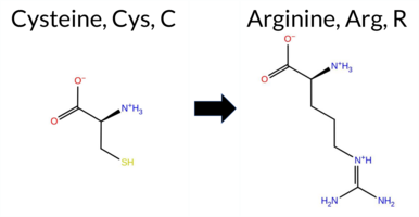 3DClick to see structure in 3D Viewer AIThe SynGAP1 missense variant C397R is not reported in ClinVar (ClinVar ID None) and is absent from gnomAD (gnomAD ID None). Prediction tools that agree on a benign effect include Rosetta, PROVEAN, SIFT, FATHMM, and AlphaMissense‑Optimized. Tools that predict a pathogenic effect are REVEL, polyPhen‑2 HumDiv, and AlphaMissense‑Default. The remaining tools (FoldX, premPS, ESM1b) are uncertain. High‑accuracy assessments show AlphaMissense‑Optimized as benign; the SGM Consensus (majority vote from AlphaMissense‑Default, ESM1b, FATHMM, PROVEAN) resolves to benign (2 benign vs. 1 pathogenic, 1 uncertain), and Foldetta predicts a benign impact on protein folding stability. Overall, the majority of evidence points to a benign effect. This conclusion is consistent with the lack of ClinVar annotation and does not contradict any existing clinical classification. Disclaimer: This summary was generated using AI and should be interpreted alongside expert review. | C2 | 0.429200 | Structured | 0.395774 | Uncertain | 0.778 | 0.551 | 0.250 | -7.094 | In-Between | 0.708 | Likely Pathogenic | Likely Benign | 0.55 | Ambiguous | 0.8 | 0.40 | Likely Benign | 0.48 | Likely Benign | 0.96 | Ambiguous | 0.514 | Likely Pathogenic | -1.46 | Neutral | 0.480 | Possibly Damaging | 0.226 | Benign | 4.65 | Benign | 0.11 | Tolerated | 0.1780 | 0.1853 | -4 | -3 | -7.0 | 53.05 | ||||||||||||||||||||||||||||||
| c.118G>T | D40Y 2D AIThe SynGAP1 D40Y missense variant is not reported in ClinVar (no ClinVar ID) and is absent from gnomAD (no gnomAD ID). Prediction tools that agree on a benign effect include REVEL, PROVEAN, polyPhen‑2 HumDiv, polyPhen‑2 HumVar, ESM1b, FATHMM, and AlphaMissense‑Optimized. Only SIFT predicts a pathogenic outcome, while AlphaMissense‑Default remains uncertain. The SGM‑Consensus, derived from a majority vote of AlphaMissense‑Default, ESM1b, FATHMM, and PROVEAN, classifies the variant as Likely Benign. High‑accuracy assessments further support a benign interpretation: AlphaMissense‑Optimized predicts Benign, SGM‑Consensus indicates Likely Benign, and Foldetta results are unavailable. Overall, the preponderance of evidence points to a benign effect for D40Y, and this conclusion does not contradict any existing ClinVar annotation (none is present). Disclaimer: This summary was generated using AI and should be interpreted alongside expert review. | Likely Benign | 0.384043 | Structured | 0.432002 | Uncertain | 0.319 | 0.769 | 0.375 | -4.313 | Likely Benign | 0.483 | Ambiguous | Likely Benign | 0.182 | Likely Benign | -1.72 | Neutral | 0.388 | Benign | 0.328 | Benign | 3.98 | Benign | 0.00 | Affected | 0.1173 | 0.7918 | -4 | -3 | 2.2 | 48.09 | |||||||||||||||||||||||||||||||||||||||
| c.1213C>T | R405C 2D 3DClick to see structure in 3D Viewer AIThe SynGAP1 R405C missense variant is listed in ClinVar with an “Uncertain” status and is present in gnomAD (variant ID 6‑33438118‑C‑T). Prediction tools that indicate a benign effect include REVEL, FATHMM, and AlphaMissense‑Optimized, whereas a majority of tools (premPS, PROVEAN, polyPhen‑2 HumDiv, polyPhen‑2 HumVar, SIFT, ESM1b, AlphaMissense‑Default) predict a pathogenic outcome. The SGM‑Consensus, derived from a majority vote of AlphaMissense‑Default, ESM1b, FATHMM, and PROVEAN, also classifies the variant as likely pathogenic. High‑accuracy assessments show AlphaMissense‑Optimized as benign, SGM‑Consensus as pathogenic, and Foldetta (combining FoldX‑MD and Rosetta outputs) remains uncertain. Overall, the balance of evidence points to a pathogenic effect, which does not contradict the ClinVar “Uncertain” classification. Disclaimer: This summary was generated using AI and should be interpreted alongside expert review. | Likely Pathogenic | C2 | 0.250310 | Structured | 0.404888 | Uncertain | 0.949 | 0.315 | 0.000 | Conflicting | 3 | 6-33438118-C-T | 6 | 3.72e-6 | -9.206 | Likely Pathogenic | 0.713 | Likely Pathogenic | Likely Benign | 0.72 | Ambiguous | 0.1 | 1.51 | Ambiguous | 1.12 | Ambiguous | 1.21 | Destabilizing | 0.427 | Likely Benign | -7.27 | Deleterious | 1.000 | Probably Damaging | 1.000 | Probably Damaging | 3.61 | Benign | 0.02 | Affected | 3.38 | 28 | 0.3227 | 0.3964 | -4 | -3 | 7.0 | -53.05 | 221.3 | 82.6 | -0.1 | 0.0 | -0.2 | 0.3 | X | X | Potentially Pathogenic | The guanidinium group of Arg405, located in an anti-parallel β sheet strand of the C2 domain (res. Ala399-Ile411), forms a salt bridge with the carboxylate group of the Glu446 side chain from an opposing α helix (res. Val441-Ser457) in the GAP domain. The positively charged Arg405 side chain also stacks with the aromatic ring of the Phe358 side chain from a loop preceding the β strand (res. Thr359-Thr366), which could assist in maintaining the anti-parallel strand arrangement.In the variant simulations, the thiol-containing side chain of Cys405 is neutral and smaller compared to the arginine side chain. The lack of Arg405-Phe358 stacking affects the loop structure, causing it to assume a β strand form—an effect that could be exacerbated during protein folding. Moreover, the inability of Cys405 to form a salt bridge with Glu446 could affect the tertiary structure assembly, although this is not apparent based on the variant simulations. | ||||||||||||
| c.1216T>G | Y406D 2D 3DClick to see structure in 3D Viewer AIThe SynGAP1 missense variant Y406D is not reported in ClinVar (ClinVar ID None) and is absent from gnomAD (gnomAD ID None). Prediction tools that agree on a benign effect include REVEL and FATHMM. In contrast, the majority of tools predict a pathogenic impact: FoldX, Rosetta, Foldetta, PROVEAN, polyPhen‑2 (HumDiv and HumVar), SIFT, ESM1b, AlphaMissense‑Default, and AlphaMissense‑Optimized. The SGM‑Consensus score is “Likely Pathogenic,” while premPS is uncertain. High‑accuracy assessments further support pathogenicity: AlphaMissense‑Optimized is pathogenic, the SGM‑Consensus (majority vote from AlphaMissense‑Default, ESM1b, FATHMM, PROVEAN) is likely pathogenic, and Foldetta (combining FoldX‑MD and Rosetta outputs) is pathogenic. Based on the preponderance of evidence, the variant is most likely pathogenic, and this assessment does not contradict the ClinVar status (which is currently unreported). Disclaimer: This summary was generated using AI and should be interpreted alongside expert review. | Likely Pathogenic | C2 | 0.179055 | Structured | 0.393707 | Uncertain | 0.946 | 0.291 | 0.000 | -14.832 | Likely Pathogenic | 0.998 | Likely Pathogenic | Likely Pathogenic | 3.82 | Destabilizing | 0.3 | 4.28 | Destabilizing | 4.05 | Destabilizing | 0.98 | Ambiguous | 0.347 | Likely Benign | -7.64 | Deleterious | 0.999 | Probably Damaging | 0.950 | Probably Damaging | 3.77 | Benign | 0.01 | Affected | 0.4439 | 0.0903 | -4 | -3 | -2.2 | -48.09 | |||||||||||||||||||||||||||||
| c.121C>T | R41C 2D AIThe SynGAP1 missense variant R41C is listed in ClinVar with an “Uncertain” status and is present in gnomAD (ID 6‑33423530‑C‑T). Prediction tools that agree on a benign effect include REVEL, PROVEAN, ESM1b, FATHMM, AlphaMissense‑Default, AlphaMissense‑Optimized, and the SGM‑Consensus (majority vote from AlphaMissense‑Default, ESM1b, FATHMM, and PROVEAN). Tools that predict a pathogenic effect are polyPhen‑2 (HumDiv and HumVar) and SIFT. High‑accuracy assessments show AlphaMissense‑Optimized as benign, the SGM‑Consensus as benign, and no result is available from Foldetta (protein‑folding stability). Taken together, the majority of evidence points to a benign impact for R41C, and this conclusion does not contradict the ClinVar “Uncertain” classification. Disclaimer: This summary was generated using AI and should be interpreted alongside expert review. | Likely Benign | 0.311707 | Structured | 0.431757 | Uncertain | 0.344 | 0.765 | 0.375 | Conflicting | 3 | 6-33423530-C-T | 7 | 4.34e-6 | -4.745 | Likely Benign | 0.207 | Likely Benign | Likely Benign | 0.093 | Likely Benign | -1.10 | Neutral | 0.976 | Probably Damaging | 0.919 | Probably Damaging | 4.13 | Benign | 0.00 | Affected | 4.32 | 1 | 0.3485 | 0.4520 | -4 | -3 | 7.0 | -53.05 | ||||||||||||||||||||||||||||||||
| c.1249T>G | Y417D 2D 3DClick to see structure in 3D Viewer AIThe SynGAP1 missense variant Y417D is not reported in ClinVar (ClinVar status: none) and is absent from gnomAD (gnomAD ID: none). Prediction tools that agree on a benign effect include only FATHMM, whereas all other evaluated algorithms—SGM‑Consensus, REVEL, FoldX, Rosetta, Foldetta, premPS, PROVEAN, polyPhen‑2 (HumDiv and HumVar), SIFT, ESM1b, AlphaMissense‑Default, and AlphaMissense‑Optimized—consistently predict a pathogenic impact. High‑accuracy assessments further support this: AlphaMissense‑Optimized predicts pathogenic; the SGM Consensus (majority vote of AlphaMissense‑Default, ESM1b, FATHMM, PROVEAN) indicates likely pathogenic; and Foldetta (combining FoldX‑MD and Rosetta outputs) also predicts pathogenic. Based on the collective evidence, the variant is most likely pathogenic, and this conclusion does not contradict any existing ClinVar annotation because no ClinVar entry exists for this variant. Disclaimer: This summary was generated using AI and should be interpreted alongside expert review. | Likely Pathogenic | GAP | 0.179055 | Structured | 0.346865 | Uncertain | 0.958 | 0.250 | 0.000 | -14.955 | Likely Pathogenic | 0.999 | Likely Pathogenic | Likely Pathogenic | 5.13 | Destabilizing | 0.1 | 5.37 | Destabilizing | 5.25 | Destabilizing | 2.43 | Destabilizing | 0.688 | Likely Pathogenic | -9.55 | Deleterious | 1.000 | Probably Damaging | 1.000 | Probably Damaging | 2.96 | Benign | 0.00 | Affected | 0.4388 | 0.0400 | -4 | -3 | -2.2 | -48.09 | |||||||||||||||||||||||||||||
| c.1259T>G | F420C 2D 3DClick to see structure in 3D Viewer AIThe SynGAP1 missense variant F420C is not reported in ClinVar (ClinVar status: not listed) and is absent from gnomAD (gnomAD ID: none). Prediction tools that assess pathogenicity largely agree: REVEL, FoldX, Rosetta, Foldetta, premPS, PROVEAN, polyPhen‑2 (HumDiv and HumVar), SIFT, ESM1b, AlphaMissense‑Default, and AlphaMissense‑Optimized all predict a pathogenic effect. Only FATHMM predicts a benign outcome. When grouped by consensus, the benign prediction is singular (FATHMM), whereas the pathogenic predictions comprise the remaining eleven tools. High‑accuracy methods reinforce this view: AlphaMissense‑Optimized is pathogenic; the SGM Consensus (majority vote from AlphaMissense‑Default, ESM1b, FATHMM, and PROVEAN) is likely pathogenic; and Foldetta, which integrates FoldX‑MD and Rosetta outputs, is pathogenic. No predictions are missing or inconclusive. Based on the overwhelming agreement among these tools, the variant is most likely pathogenic, and this assessment does not contradict any ClinVar status (none reported). Disclaimer: This summary was generated using AI and should be interpreted alongside expert review. | Likely Pathogenic | GAP | 0.104810 | Structured | 0.384475 | Uncertain | 0.974 | 0.255 | 0.000 | -11.931 | Likely Pathogenic | 0.999 | Likely Pathogenic | Likely Pathogenic | 4.31 | Destabilizing | 0.1 | 5.11 | Destabilizing | 4.71 | Destabilizing | 2.49 | Destabilizing | 0.500 | Likely Pathogenic | -7.40 | Deleterious | 0.998 | Probably Damaging | 0.869 | Possibly Damaging | 3.09 | Benign | 0.00 | Affected | 0.2458 | 0.0662 | -4 | -2 | -0.3 | -44.04 | |||||||||||||||||||||||||||||
| c.1267T>G | Y423D 2D 3DClick to see structure in 3D Viewer AIThe SynGAP1 missense variant Y423D is not reported in ClinVar and is absent from gnomAD. Prediction tools largely agree on a deleterious effect: benign predictions are limited to FATHMM, while the remaining tools—SGM‑Consensus, REVEL, FoldX, Rosetta, Foldetta, premPS, PROVEAN, polyPhen‑2 (HumDiv and HumVar), SIFT, ESM1b, AlphaMissense‑Default, and AlphaMissense‑Optimized—classify the variant as pathogenic. High‑accuracy assessments reinforce this view: AlphaMissense‑Optimized predicts pathogenic; the SGM Consensus (majority vote from AlphaMissense‑Default, ESM1b, FATHMM, and PROVEAN) indicates likely pathogenic; and Foldetta, which integrates FoldX‑MD and Rosetta stability outputs, also predicts pathogenic. Based on the preponderance of evidence, the variant is most likely pathogenic, and this conclusion is not contradicted by any ClinVar annotation. Disclaimer: This summary was generated using AI and should be interpreted alongside expert review. | Likely Pathogenic | GAP | 0.088832 | Structured | 0.421885 | Uncertain | 0.975 | 0.242 | 0.000 | -13.279 | Likely Pathogenic | 0.993 | Likely Pathogenic | Likely Pathogenic | 4.67 | Destabilizing | 0.1 | 4.79 | Destabilizing | 4.73 | Destabilizing | 1.78 | Destabilizing | 0.553 | Likely Pathogenic | -8.28 | Deleterious | 1.000 | Probably Damaging | 1.000 | Probably Damaging | 3.38 | Benign | 0.04 | Affected | 0.3262 | 0.0412 | -4 | -3 | -2.2 | -48.09 | |||||||||||||||||||||||||||||
| c.1282T>G | Y428D 2D 3DClick to see structure in 3D Viewer AISynGAP1 missense variant Y428D is not reported in ClinVar and is absent from gnomAD. Across a broad panel of in silico predictors, 15 tools (REVEL, FoldX, Rosetta, Foldetta, premPS, PROVEAN, polyPhen‑2 HumDiv, polyPhen‑2 HumVar, SIFT, ESM1b, AlphaMissense‑Default, AlphaMissense‑Optimized, and the SGM‑Consensus) uniformly predict a deleterious effect, whereas only FATHMM classifies it as benign. The SGM‑Consensus, derived from a majority vote of AlphaMissense‑Default, ESM1b, FATHMM, and PROVEAN, also indicates a likely pathogenic outcome. High‑accuracy assessments further support this: AlphaMissense‑Optimized predicts pathogenicity, the SGM‑Consensus (majority vote) is likely pathogenic, and Foldetta, which integrates FoldX‑MD and Rosetta stability calculations, reports a pathogenic effect. No prediction or stability result is missing or inconclusive. Consequently, the variant is most likely pathogenic, and this assessment does not contradict any ClinVar annotation because none exists. Disclaimer: This summary was generated using AI and should be interpreted alongside expert review. | Likely Pathogenic | GAP | 0.118441 | Structured | 0.389652 | Uncertain | 0.965 | 0.292 | 0.000 | -13.473 | Likely Pathogenic | 0.995 | Likely Pathogenic | Likely Pathogenic | 3.24 | Destabilizing | 0.0 | 4.19 | Destabilizing | 3.72 | Destabilizing | 1.64 | Destabilizing | 0.554 | Likely Pathogenic | -9.55 | Deleterious | 1.000 | Probably Damaging | 1.000 | Probably Damaging | 3.50 | Benign | 0.01 | Affected | 0.4458 | 0.1003 | -4 | -3 | -2.2 | -48.09 | |||||||||||||||||||||||||||||
| c.1294T>C | C432R 2D 3DClick to see structure in 3D Viewer AIClinVar has no entry for this SynGAP1 missense variant, and it is absent from gnomAD. Prediction tools that agree on a benign effect include only FATHMM. The majority of algorithms predict a pathogenic impact: REVEL, Rosetta, premPS, PROVEAN, polyPhen‑2 (HumDiv and HumVar), SIFT, ESM1b, AlphaMissense‑Default, AlphaMissense‑Optimized, and the SGM‑Consensus score (Likely Pathogenic). FoldX and Foldetta report uncertain results. High‑accuracy assessments further support a deleterious effect: AlphaMissense‑Optimized is pathogenic, the SGM‑Consensus is likely pathogenic, while Foldetta remains inconclusive. Overall, the consensus of the available predictions indicates that the variant is most likely pathogenic, and this assessment does not contradict the ClinVar status, which currently has no classification. Disclaimer: This summary was generated using AI and should be interpreted alongside expert review. | Likely Pathogenic | GAP | 0.111485 | Structured | 0.362533 | Uncertain | 0.960 | 0.285 | 0.000 | -13.858 | Likely Pathogenic | 0.996 | Likely Pathogenic | Likely Pathogenic | 1.03 | Ambiguous | 0.2 | 2.05 | Destabilizing | 1.54 | Ambiguous | 1.73 | Destabilizing | 0.690 | Likely Pathogenic | -11.46 | Deleterious | 0.999 | Probably Damaging | 0.993 | Probably Damaging | 3.45 | Benign | 0.01 | Affected | 0.1359 | 0.1766 | -4 | -3 | -7.0 | 53.05 | |||||||||||||||||||||||||||||
| c.1295G>T | C432F 2D  3DClick to see structure in 3D Viewer AIThe SynGAP1 missense variant C432F is not reported in ClinVar (ClinVar status: none) and is absent from gnomAD (gnomAD ID: none). Prediction tools that agree on a benign effect include REVEL, FoldX, Rosetta, and FATHMM, whereas a majority of tools (SGM‑Consensus, PROVEAN, polyPhen‑2 HumDiv, polyPhen‑2 HumVar, SIFT, ESM1b, and AlphaMissense‑Default) predict a pathogenic impact; AlphaMissense‑Optimized and premPS are uncertain. High‑accuracy assessments show AlphaMissense‑Optimized as uncertain, SGM‑Consensus (a majority vote of AlphaMissense‑Default, ESM1b, FATHMM, and PROVEAN) as likely pathogenic, and Foldetta (combining FoldX‑MD and Rosetta outputs) as benign. Overall, the balance of evidence favors a pathogenic classification, and this conclusion does not contradict the lack of ClinVar annotation. Disclaimer: This summary was generated using AI and should be interpreted alongside expert review. | Likely Pathogenic | GAP | 0.111485 | Structured | 0.362533 | Uncertain | 0.960 | 0.285 | 0.000 | -12.862 | Likely Pathogenic | 0.943 | Likely Pathogenic | Ambiguous | -0.44 | Likely Benign | 0.2 | -0.34 | Likely Benign | -0.39 | Likely Benign | 0.57 | Ambiguous | 0.434 | Likely Benign | -10.50 | Deleterious | 0.999 | Probably Damaging | 0.993 | Probably Damaging | 3.46 | Benign | 0.01 | Affected | 0.1090 | 0.3956 | -4 | -2 | 0.3 | 44.04 | |||||||||||||||||||||||||||||
| c.1387G>T | D463Y 2D 3DClick to see structure in 3D Viewer AIClinVar reports no entry for this SynGAP1 D463Y variant, and it is absent from gnomAD. Prediction tools that agree on a benign effect include REVEL, FoldX, premPS, SIFT, and FATHMM. Those that predict a pathogenic effect are PROVEAN, polyPhen‑2 HumDiv, polyPhen‑2 HumVar, ESM1b, and AlphaMissense‑Default. Uncertain results come from AlphaMissense‑Optimized and Rosetta. High‑accuracy assessments show AlphaMissense‑Optimized as uncertain, the SGM Consensus (majority vote of AlphaMissense‑Default, ESM1b, FATHMM, PROVEAN) as Likely Pathogenic, and Foldetta (combining FoldX‑MD and Rosetta outputs) as Benign. Overall, the majority of conventional tools lean toward pathogenicity, while the high‑accuracy Foldetta suggests benign. Thus, the variant is most likely pathogenic based on the prevailing predictions, and this assessment does not contradict any ClinVar status because none is available. Disclaimer: This summary was generated using AI and should be interpreted alongside expert review. | Likely Pathogenic | GAP | 0.260850 | Structured | 0.305622 | Uncertain | 0.940 | 0.176 | 0.000 | -14.387 | Likely Pathogenic | 0.904 | Likely Pathogenic | Ambiguous | -0.14 | Likely Benign | 0.1 | 0.77 | Ambiguous | 0.32 | Likely Benign | 0.07 | Likely Benign | 0.399 | Likely Benign | -7.95 | Deleterious | 0.998 | Probably Damaging | 0.904 | Possibly Damaging | 3.35 | Benign | 0.14 | Tolerated | 0.0540 | 0.6213 | -4 | -3 | 2.2 | 48.09 | |||||||||||||||||||||||||||||
| c.1391T>G | F464C 2D 3DClick to see structure in 3D Viewer AIThe SynGAP1 missense variant F464C is not reported in ClinVar (no ClinVar ID) and is absent from gnomAD (no gnomAD ID). Prediction tools that agree on a benign effect include only FATHMM, whereas all other evaluated algorithms—SGM‑Consensus, REVEL, FoldX, Rosetta, Foldetta, premPS, PROVEAN, polyPhen‑2 (HumDiv and HumVar), SIFT, ESM1b, AlphaMissense‑Default, and AlphaMissense‑Optimized—classify the variant as pathogenic. High‑accuracy assessments further support a deleterious impact: AlphaMissense‑Optimized predicts pathogenic, the SGM Consensus (majority vote of AlphaMissense‑Default, ESM1b, FATHMM, PROVEAN) indicates likely pathogenic, and Foldetta (combining FoldX‑MD and Rosetta outputs) predicts pathogenic. No prediction or folding stability result is missing or inconclusive. Based on the overwhelming consensus of pathogenic predictions, the variant is most likely pathogenic, and this assessment does not contradict any ClinVar status because no ClinVar classification exists. Disclaimer: This summary was generated using AI and should be interpreted alongside expert review. | Likely Pathogenic | GAP | 0.268042 | Structured | 0.313424 | Uncertain | 0.961 | 0.178 | 0.000 | -13.011 | Likely Pathogenic | 0.998 | Likely Pathogenic | Likely Pathogenic | 4.18 | Destabilizing | 0.0 | 4.31 | Destabilizing | 4.25 | Destabilizing | 2.20 | Destabilizing | 0.740 | Likely Pathogenic | -7.96 | Deleterious | 1.000 | Probably Damaging | 0.999 | Probably Damaging | 3.26 | Benign | 0.00 | Affected | 0.2298 | 0.1219 | -4 | -2 | -0.3 | -44.04 | |||||||||||||||||||||||||||||
| c.1399G>T | D467Y 2D 3DClick to see structure in 3D Viewer AIThe SynGAP1 missense variant D467Y is not reported in ClinVar and has no entries in gnomAD. Functional prediction tools show a split: benign calls come from FoldX, Foldetta, premPS, and SIFT, whereas pathogenic calls are made by SGM‑Consensus, REVEL, PROVEAN, polyPhen‑2 (HumDiv and HumVar), ESM1b, FATHMM, AlphaMissense‑Default, and AlphaMissense‑Optimized. The high‑accuracy methods give a clearer picture: AlphaMissense‑Optimized predicts pathogenic, the SGM‑Consensus (majority vote of AlphaMissense‑Default, ESM1b, FATHMM, PROVEAN) also indicates likely pathogenic, while Foldetta (combining FoldX‑MD and Rosetta outputs) predicts benign. No evidence from Rosetta is available due to its uncertain status. Overall, the majority of high‑confidence predictions lean toward pathogenicity, and this does not contradict any ClinVar annotation because none exists. Thus, the variant is most likely pathogenic. Disclaimer: This summary was generated using AI and should be interpreted alongside expert review. | Likely Pathogenic | GAP | 0.268042 | Structured | 0.329932 | Uncertain | 0.940 | 0.246 | 0.000 | -14.373 | Likely Pathogenic | 0.991 | Likely Pathogenic | Likely Pathogenic | 0.36 | Likely Benign | 0.1 | -0.60 | Ambiguous | -0.12 | Likely Benign | 0.07 | Likely Benign | 0.846 | Likely Pathogenic | -8.70 | Deleterious | 1.000 | Probably Damaging | 0.999 | Probably Damaging | -1.28 | Pathogenic | 0.07 | Tolerated | 0.0517 | 0.5521 | -4 | -3 | 2.2 | 48.09 | |||||||||||||||||||||||||||||
| c.1420G>T | D474Y 2D AIThe SynGAP1 missense variant D474Y is not reported in ClinVar and has no entry in gnomAD. Prediction tools that agree on a benign effect include FoldX, Foldetta, and premPS, whereas the majority of tools (REVEL, PROVEAN, polyPhen‑2 HumDiv, polyPhen‑2 HumVar, SIFT, ESM1b, FATHMM, AlphaMissense‑Default, AlphaMissense‑Optimized) predict a pathogenic impact; Rosetta is uncertain. High‑accuracy assessments show AlphaMissense‑Optimized as pathogenic, the SGM Consensus (majority vote from AlphaMissense‑Default, ESM1b, FATHMM, PROVEAN) as likely pathogenic, and Foldetta as benign. Overall, the preponderance of evidence points to a pathogenic effect for D474Y. This conclusion does not contradict ClinVar status, as the variant is currently unreported there. Disclaimer: This summary was generated using AI and should be interpreted alongside expert review. | Likely Pathogenic | GAP | 0.102787 | Structured | 0.373433 | Uncertain | 0.864 | 0.255 | 0.000 | -14.647 | Likely Pathogenic | 0.989 | Likely Pathogenic | Likely Pathogenic | 0.27 | Likely Benign | 0.5 | -0.74 | Ambiguous | -0.24 | Likely Benign | 0.20 | Likely Benign | 0.864 | Likely Pathogenic | -7.72 | Deleterious | 1.000 | Probably Damaging | 0.999 | Probably Damaging | -1.30 | Pathogenic | 0.01 | Affected | 0.0493 | 0.4237 | -4 | -3 | 2.2 | 48.09 | |||||||||||||||||||||||||||||
| c.143T>G | F48C 2D AIThe SynGAP1 missense variant F48C is not reported in ClinVar (ClinVar ID = None) and has no entries in gnomAD (gnomAD ID = None). Prediction tools that agree on a benign effect include REVEL, PROVEAN, polyPhen‑2 HumVar, ESM1b, FATHMM, and AlphaMissense‑Optimized. Tools that predict a pathogenic effect are polyPhen‑2 HumDiv, SIFT, and AlphaMissense‑Default. The SGM‑Consensus, which aggregates the majority vote of AlphaMissense‑Default, ESM1b, FATHMM, and PROVEAN, is classified as Likely Benign (3 benign vs. 1 pathogenic). High‑accuracy assessments show AlphaMissense‑Optimized as Benign and the SGM‑Consensus as Likely Benign; Foldetta results are unavailable. Overall, the majority of evidence points to a benign impact, and this conclusion does not contradict any ClinVar annotation (none is present). Disclaimer: This summary was generated using AI and should be interpreted alongside expert review. | Likely Benign | 0.298791 | Structured | 0.440452 | Uncertain | 0.558 | 0.707 | 0.125 | -5.950 | Likely Benign | 0.676 | Likely Pathogenic | Likely Benign | 0.153 | Likely Benign | -2.31 | Neutral | 0.953 | Possibly Damaging | 0.431 | Benign | 3.93 | Benign | 0.00 | Affected | 0.2657 | 0.1962 | -4 | -2 | -0.3 | -44.04 | |||||||||||||||||||||||||||||||||||||||
| c.1451T>G | F484C 2D 3DClick to see structure in 3D Viewer AIThe SynGAP1 missense variant F484C is not reported in ClinVar and is absent from gnomAD. Functional prediction tools largely agree on a deleterious effect: SGM‑Consensus, REVEL, FoldX, Rosetta, Foldetta, premPS, PROVEAN, polyPhen‑2 (HumDiv and HumVar), SIFT, ESM1b, and AlphaMissense‑Default all predict pathogenicity, whereas only FATHMM predicts a benign outcome. High‑accuracy assessments reinforce this view: AlphaMissense‑Optimized indicates pathogenicity; the SGM Consensus, derived from a majority vote of AlphaMissense‑Default, ESM1b, FATHMM, and PROVEAN, is Likely Pathogenic; and Foldetta, which integrates FoldX‑MD and Rosetta stability outputs, also predicts pathogenicity. No prediction or stability result is missing or inconclusive. Consequently, the variant is most likely pathogenic, and this assessment does not contradict any ClinVar annotation (none is available). Disclaimer: This summary was generated using AI and should be interpreted alongside expert review. | Likely Pathogenic | GAP | 0.182256 | Structured | 0.403079 | Uncertain | 0.798 | 0.245 | 0.125 | -14.988 | Likely Pathogenic | 0.998 | Likely Pathogenic | Likely Pathogenic | 4.01 | Destabilizing | 0.0 | 3.54 | Destabilizing | 3.78 | Destabilizing | 2.07 | Destabilizing | 0.675 | Likely Pathogenic | -7.73 | Deleterious | 1.000 | Probably Damaging | 0.969 | Probably Damaging | 2.66 | Benign | 0.00 | Affected | 0.2449 | 0.0902 | -4 | -2 | -0.3 | -44.04 | |||||||||||||||||||||||||||||
| c.1453C>T | R485C 2D 3DClick to see structure in 3D Viewer AIThe SynGAP1 missense variant R485C (gnomAD ID 6‑33438485‑C‑T) is listed in ClinVar with an uncertain significance. Functional prediction tools largely disagree: benign calls come from Rosetta and premPS, whereas pathogenic calls are made by REVEL, PROVEAN, polyPhen‑2 (HumDiv and HumVar), SIFT, ESM1b, FATHMM, AlphaMissense‑Default, AlphaMissense‑Optimized, and the SGM Consensus (which aggregates AlphaMissense‑Default, ESM1b, FATHMM, and PROVEAN). High‑accuracy assessments further support a deleterious effect: AlphaMissense‑Optimized predicts pathogenic, the SGM Consensus is labeled likely pathogenic, and Foldetta (combining FoldX‑MD and Rosetta outputs) remains uncertain. With the majority of evidence pointing to pathogenicity and no contradictory data from ClinVar, the variant is most likely pathogenic, although ClinVar has not yet reached a definitive classification. Disclaimer: This summary was generated using AI and should be interpreted alongside expert review. | Likely Pathogenic | GAP | 0.188120 | Structured | 0.377409 | Uncertain | 0.805 | 0.246 | 0.125 | Uncertain | 2 | 6-33438485-C-T | 9 | 5.58e-6 | -14.294 | Likely Pathogenic | 0.976 | Likely Pathogenic | Likely Pathogenic | 1.00 | Ambiguous | 0.1 | 0.26 | Likely Benign | 0.63 | Ambiguous | 0.44 | Likely Benign | 0.597 | Likely Pathogenic | -7.96 | Deleterious | 1.000 | Probably Damaging | 1.000 | Probably Damaging | 1.90 | Pathogenic | 0.00 | Affected | 3.37 | 35 | 0.3350 | 0.2762 | -4 | -3 | 7.0 | -53.05 | 225.5 | 99.6 | -0.1 | 0.0 | -0.3 | 0.2 | X | Uncertain | The guanidinium group of Arg485 is located in a short helical structure (res. Glu480-Leu482) within an α-α loop connecting the two α-helices (res. Ala461-Phe476 and Leu489-Glu519) at the GAP-Ras interface. The side chain of Arg485 acts as the “arginine finger” of SynGAP, playing a crucial role in Ras-GTPase activation. Consequently, the residue swap inhibits the conversion of GTP to GDP at the enzyme’s active site. Although no negative effects on the protein structure are observed during the simulations, no definite conclusions can be drawn due to the critical role of Arg485 in GTPase activation. | |||||||||||||
| c.145T>C | C49R 2D AIThe SynGAP1 missense variant C49R has no ClinVar entry and is not reported in gnomAD. Prediction tools that agree on a benign effect are REVEL and FATHMM, whereas the majority of tools (PROVEAN, polyPhen‑2 HumDiv, polyPhen‑2 HumVar, SIFT, ESM1b, AlphaMissense‑Default) predict a pathogenic impact. The SGM‑Consensus, which aggregates AlphaMissense‑Default, ESM1b, FATHMM, and PROVEAN, reports the variant as Likely Pathogenic. High‑accuracy assessments show AlphaMissense‑Optimized as Uncertain and no Foldetta (FoldX‑MD/Rosetta) result is available. Taken together, the preponderance of evidence points to a pathogenic effect, and this conclusion does not contradict ClinVar status, as no ClinVar classification exists for this variant. Disclaimer: This summary was generated using AI and should be interpreted alongside expert review. | Likely Pathogenic | 0.209395 | Structured | 0.445316 | Uncertain | 0.541 | 0.704 | 0.000 | -10.000 | Likely Pathogenic | 0.948 | Likely Pathogenic | Ambiguous | 0.320 | Likely Benign | -3.53 | Deleterious | 0.676 | Possibly Damaging | 0.761 | Possibly Damaging | 3.95 | Benign | 0.00 | Affected | 0.1605 | 0.2120 | -4 | -3 | -7.0 | 53.05 | |||||||||||||||||||||||||||||||||||||||
| c.146G>T | C49F 2D AIThe SynGAP1 missense variant C49F is not reported in ClinVar (ClinVar ID = None) and is absent from gnomAD (gnomAD ID = None). Prediction tools that agree on a benign effect are REVEL and FATHMM, while a majority of tools (PROVEAN, polyPhen‑2 HumDiv, polyPhen‑2 HumVar, SIFT, AlphaMissense‑Default) predict a pathogenic impact. The remaining tools, ESM1b and AlphaMissense‑Optimized, return uncertain results. High‑accuracy assessments further support a deleterious interpretation: AlphaMissense‑Optimized is uncertain, but the SGM Consensus—derived from a majority vote of AlphaMissense‑Default (pathogenic), ESM1b (uncertain), FATHMM (benign), and PROVEAN (pathogenic)—classifies the variant as pathogenic. Foldetta, a protein‑folding stability method, has no available result for this variant. Overall, the preponderance of evidence indicates that the variant is most likely pathogenic, and this conclusion does not contradict any ClinVar annotation, as none exists. Disclaimer: This summary was generated using AI and should be interpreted alongside expert review. | 0.209395 | Structured | 0.445316 | Uncertain | 0.541 | 0.704 | 0.000 | -7.194 | In-Between | 0.893 | Likely Pathogenic | Ambiguous | 0.301 | Likely Benign | -3.21 | Deleterious | 0.676 | Possibly Damaging | 0.695 | Possibly Damaging | 3.86 | Benign | 0.00 | Affected | 0.1199 | 0.3690 | -4 | -2 | 0.3 | 44.04 | ||||||||||||||||||||||||||||||||||||||||
| c.1489T>G | Y497D 2D 3DClick to see structure in 3D Viewer AIThe SynGAP1 missense variant Y497D is not reported in ClinVar (ClinVar ID None) and is absent from gnomAD (gnomAD ID None). Prediction tools that assess pathogenicity all agree that the variant is deleterious: SGM‑Consensus, REVEL, FoldX, Rosetta, Foldetta, premPS, PROVEAN, polyPhen‑2 (HumDiv and HumVar), SIFT, ESM1b, FATHMM, AlphaMissense‑Default, and AlphaMissense‑Optimized all classify it as pathogenic. No tool predicts a benign effect. High‑accuracy methods reinforce this consensus: AlphaMissense‑Optimized predicts pathogenicity; the SGM Consensus (majority vote of AlphaMissense‑Default, ESM1b, FATHMM, and PROVEAN) also predicts pathogenic; and Foldetta, which integrates FoldX‑MD and Rosetta stability outputs, predicts a destabilizing, pathogenic effect. All available predictions are concordant and indicate a likely pathogenic impact. Thus, based on the collective computational evidence, the variant is most likely pathogenic, and this assessment does not contradict any ClinVar status (none reported). Disclaimer: This summary was generated using AI and should be interpreted alongside expert review. | Likely Pathogenic | GAP | 0.092881 | Structured | 0.393608 | Uncertain | 0.950 | 0.181 | 0.000 | -12.128 | Likely Pathogenic | 0.994 | Likely Pathogenic | Likely Pathogenic | 4.70 | Destabilizing | 0.2 | 4.29 | Destabilizing | 4.50 | Destabilizing | 2.36 | Destabilizing | 0.918 | Likely Pathogenic | -9.81 | Deleterious | 1.000 | Probably Damaging | 0.999 | Probably Damaging | -1.67 | Pathogenic | 0.01 | Affected | 0.3621 | 0.0612 | -4 | -3 | -2.2 | -48.09 | |||||||||||||||||||||||||||||
| c.1522G>T | D508Y 2D 3DClick to see structure in 3D Viewer AIThe SynGAP1 D508Y missense variant is not reported in ClinVar (ClinVar ID None) and is absent from gnomAD (gnomAD ID None). Prediction tools that agree on a benign effect include REVEL, Foldetta, premPS, FATHMM, and AlphaMissense‑Optimized. Tools that predict a pathogenic effect are SGM‑Consensus, PROVEAN, polyPhen‑2 HumDiv, polyPhen‑2 HumVar, SIFT, ESM1b, and AlphaMissense‑Default. FoldX and Rosetta give uncertain results. High‑accuracy assessments show AlphaMissense‑Optimized classifying the variant as benign, SGM‑Consensus (a majority vote of AlphaMissense‑Default, ESM1b, FATHMM, and PROVEAN) labeling it likely pathogenic, and Foldetta predicting a benign outcome. Overall, the majority of evidence (seven pathogenic vs. five benign) points to a pathogenic impact. This conclusion does not contradict ClinVar status, as no ClinVar classification exists for this variant. Disclaimer: This summary was generated using AI and should be interpreted alongside expert review. | Likely Pathogenic | GAP | 0.019401 | Structured | 0.255890 | Uncertain | 0.890 | 0.228 | 0.000 | -12.917 | Likely Pathogenic | 0.739 | Likely Pathogenic | Likely Benign | -0.53 | Ambiguous | 0.2 | 0.72 | Ambiguous | 0.10 | Likely Benign | 0.06 | Likely Benign | 0.363 | Likely Benign | -8.37 | Deleterious | 1.000 | Probably Damaging | 0.965 | Probably Damaging | 3.26 | Benign | 0.01 | Affected | 0.0908 | 0.4736 | -4 | -3 | 2.2 | 48.09 | |||||||||||||||||||||||||||||
| c.1538T>G | F513C 2D 3DClick to see structure in 3D Viewer AIThe SynGAP1 missense variant F513C is not reported in ClinVar and is absent from gnomAD. Prediction tools that assess pathogenicity all converge on a deleterious effect: REVEL, FoldX, Rosetta, Foldetta, premPS, PROVEAN, polyPhen‑2 (HumDiv and HumVar), SIFT, ESM1b, FATHMM, AlphaMissense‑Default, and AlphaMissense‑Optimized all indicate a pathogenic or likely pathogenic outcome. No tool in the dataset predicts a benign effect. High‑accuracy assessments reinforce this consensus: AlphaMissense‑Optimized classifies the variant as pathogenic; the SGM Consensus (majority vote from AlphaMissense‑Default, ESM1b, FATHMM, and PROVEAN) reports it as likely pathogenic; and Foldetta, which integrates FoldX‑MD and Rosetta stability predictions, also labels it pathogenic. Based on the uniform predictions, the variant is most likely pathogenic, and this assessment does not contradict any ClinVar status (none is available). Disclaimer: This summary was generated using AI and should be interpreted alongside expert review. | Likely Pathogenic | GAP | 0.102787 | Structured | 0.250651 | Uncertain | 0.949 | 0.269 | 0.000 | -11.389 | Likely Pathogenic | 0.996 | Likely Pathogenic | Likely Pathogenic | 3.92 | Destabilizing | 0.3 | 4.20 | Destabilizing | 4.06 | Destabilizing | 1.64 | Destabilizing | 0.919 | Likely Pathogenic | -7.72 | Deleterious | 1.000 | Probably Damaging | 1.000 | Probably Damaging | -1.40 | Pathogenic | 0.03 | Affected | 0.2384 | 0.0783 | -4 | -2 | -0.3 | -44.04 | |||||||||||||||||||||||||||||
| c.1552T>G | Y518D 2D 3DClick to see structure in 3D Viewer AIThe SynGAP1 missense variant Y518D is not reported in ClinVar and is absent from gnomAD. Prediction tools largely agree on a deleterious effect: SGM‑Consensus, REVEL, FoldX, Rosetta, Foldetta, premPS, PROVEAN, polyPhen‑2 (HumDiv and HumVar), SIFT, ESM1b, and AlphaMissense‑Default all predict pathogenicity, whereas only FATHMM predicts a benign outcome. High‑accuracy assessments reinforce this view: AlphaMissense‑Optimized is pathogenic; the SGM Consensus (a majority vote of AlphaMissense‑Default, ESM1b, FATHMM, and PROVEAN) is likely pathogenic; and Foldetta, which integrates FoldX‑MD and Rosetta stability outputs, is pathogenic. No predictions are missing or inconclusive. Based on the overwhelming consensus of pathogenic predictions, the variant is most likely pathogenic, and this assessment does not contradict any ClinVar status (none reported). Disclaimer: This summary was generated using AI and should be interpreted alongside expert review. | Likely Pathogenic | GAP | 0.139895 | Structured | 0.126970 | Uncertain | 0.897 | 0.321 | 0.000 | -12.247 | Likely Pathogenic | 0.990 | Likely Pathogenic | Likely Pathogenic | 3.92 | Destabilizing | 0.9 | 2.68 | Destabilizing | 3.30 | Destabilizing | 1.36 | Destabilizing | 0.619 | Likely Pathogenic | -9.41 | Deleterious | 1.000 | Probably Damaging | 1.000 | Probably Damaging | 3.40 | Benign | 0.03 | Affected | 0.3682 | 0.0212 | -4 | -3 | -2.2 | -48.09 | |||||||||||||||||||||||||||||
| c.1570T>C | C524R 2D 3DClick to see structure in 3D Viewer AIThe SynGAP1 missense variant C524R is not reported in ClinVar (ClinVar ID None) and has no entries in gnomAD (gnomAD ID None). Across the evaluated in‑silico tools, all pathogenic‑predicating algorithms—REVEL, SGM‑Consensus, Foldetta, premPS, PROVEAN, polyPhen‑2 (HumDiv and HumVar), SIFT, ESM1b, FATHMM, AlphaMissense‑Default, and AlphaMissense‑Optimized—return a pathogenic or likely pathogenic verdict. The only tool with an inconclusive result is FoldX, which is treated as unavailable. High‑accuracy predictors reinforce this assessment: AlphaMissense‑Optimized predicts pathogenic; the SGM Consensus (majority vote of AlphaMissense‑Default, ESM1b, FATHMM, and PROVEAN) indicates likely pathogenic; and Foldetta, a protein‑folding stability method combining FoldX‑MD and Rosetta outputs, also predicts pathogenic. Based on the collective evidence, the variant is most likely pathogenic, and this conclusion does not contradict any ClinVar annotation. Disclaimer: This summary was generated using AI and should be interpreted alongside expert review. | Likely Pathogenic | GAP | 0.134866 | Structured | 0.024729 | Uncertain | 0.916 | 0.385 | 0.125 | -14.337 | Likely Pathogenic | 1.000 | Likely Pathogenic | Likely Pathogenic | -0.53 | Ambiguous | 0.5 | 9.04 | Destabilizing | 4.26 | Destabilizing | 1.62 | Destabilizing | 0.917 | Likely Pathogenic | -11.93 | Deleterious | 0.999 | Probably Damaging | 0.998 | Probably Damaging | -1.40 | Pathogenic | 0.00 | Affected | 0.2031 | 0.1463 | -4 | -3 | -7.0 | 53.05 | |||||||||||||||||||||||||||||
| c.1571G>T | C524F 2D 3DClick to see structure in 3D Viewer AIThe SynGAP1 missense variant C524F is not reported in ClinVar (ClinVar ID None) and has no entries in gnomAD (gnomAD ID None). Prediction tools that assess the variant’s effect largely agree on a deleterious outcome: REVEL, FoldX, Rosetta, Foldetta, PROVEAN, polyPhen‑2 (HumDiv and HumVar), SIFT, ESM1b, FATHMM, AlphaMissense‑Default, and AlphaMissense‑Optimized all classify it as pathogenic, while only premPS predicts a benign effect. High‑accuracy methods reinforce this consensus: AlphaMissense‑Optimized reports pathogenicity; the SGM Consensus (majority vote of AlphaMissense‑Default, ESM1b, FATHMM, and PROVEAN) indicates likely pathogenic; and Foldetta, which integrates FoldX‑MD and Rosetta stability calculations, also predicts pathogenicity. No prediction or stability result is missing or inconclusive. Based on the overwhelming majority of pathogenic predictions, the variant is most likely pathogenic, and this assessment does not contradict the absence of a ClinVar classification. Disclaimer: This summary was generated using AI and should be interpreted alongside expert review. | Likely Pathogenic | GAP | 0.134866 | Structured | 0.024729 | Uncertain | 0.916 | 0.385 | 0.125 | -12.145 | Likely Pathogenic | 0.996 | Likely Pathogenic | Likely Pathogenic | 2.18 | Destabilizing | 1.4 | 5.20 | Destabilizing | 3.69 | Destabilizing | -0.04 | Likely Benign | 0.831 | Likely Pathogenic | -10.94 | Deleterious | 0.999 | Probably Damaging | 0.998 | Probably Damaging | -1.22 | Pathogenic | 0.05 | Affected | 0.1678 | 0.4259 | -4 | -2 | 0.3 | 44.04 | |||||||||||||||||||||||||||||
| c.1579G>T | D527Y 2D 3DClick to see structure in 3D Viewer AISynGAP1 missense variant D527Y is listed in ClinVar with an uncertain significance (ClinVar ID 1698369.0) and is not reported in gnomAD. Functional prediction tools cluster into two groups: the single benign prediction from premPS versus a consensus of pathogenic predictions from the remaining 12 tools (REVEL, SGM‑Consensus, PROVEAN, polyPhen‑2 HumDiv, polyPhen‑2 HumVar, SIFT, ESM1b, FATHMM, AlphaMissense‑Default, AlphaMissense‑Optimized). High‑accuracy assessments further support a deleterious effect: AlphaMissense‑Optimized predicts pathogenic, the SGM Consensus (majority vote of AlphaMissense‑Default, ESM1b, FATHMM, PROVEAN) is likely pathogenic, and Foldetta (combining FoldX‑MD and Rosetta outputs) is inconclusive. Protein‑stability calculations from FoldX and Rosetta are also uncertain. Overall, the preponderance of evidence indicates that D527Y is most likely pathogenic, which does not contradict the current ClinVar uncertain status. Disclaimer: This summary was generated using AI and should be interpreted alongside expert review. | Likely Pathogenic | GAP | 0.139895 | Structured | 0.021908 | Uncertain | 0.913 | 0.408 | 0.000 | Uncertain | 1 | -15.386 | Likely Pathogenic | 0.978 | Likely Pathogenic | Likely Pathogenic | -0.77 | Ambiguous | 0.2 | 1.89 | Ambiguous | 0.56 | Ambiguous | -0.14 | Likely Benign | 0.905 | Likely Pathogenic | -8.79 | Deleterious | 1.000 | Probably Damaging | 0.999 | Probably Damaging | -2.41 | Pathogenic | 0.00 | Affected | 3.37 | 35 | 0.0554 | 0.4229 | -4 | -3 | 2.2 | 48.09 | 270.9 | -45.7 | 0.1 | 0.1 | -0.1 | 0.0 | X | Potentially Pathogenic | Asp527 is located on an α-α loop between the two α-helices (res. Gly502-Tyr518 and Ala533-Val560). In the WT simulations, the carboxylate group of the Asp527 side chain forms hydrogen bonds with the backbone atoms of loop residues (e.g., Ile529, Lys530) facing the membrane surface. In the variant simulations, Tyr527 is a bulkier residue that faces away from the loop and stacks with Phe646 in a nearby α-helix (res. Ser614-Ser668). Regardless, no negative structural effects are observed during the variant simulations. However, due to its location near the SynGAP-membrane interface, the effect of the residue swap cannot be fully addressed using the SynGAP solvent-only simulations. | ||||||||||||||||
| c.1591T>C | C531R 2D AIThe SynGAP1 missense variant C531R is not reported in ClinVar and is absent from gnomAD. Prediction tools that classify the variant as benign include FoldX, Rosetta, Foldetta, and polyPhen‑2 HumVar, whereas the remaining tools—REVEL, premPS, PROVEAN, polyPhen‑2 HumDiv, SIFT, ESM1b, FATHMM, AlphaMissense‑Default, and AlphaMissense‑Optimized—predict it to be pathogenic. High‑accuracy assessments show AlphaMissense‑Optimized as pathogenic, the SGM Consensus (majority vote from AlphaMissense‑Default, ESM1b, FATHMM, and PROVEAN) as pathogenic, and Foldetta (combining FoldX‑MD and Rosetta outputs) as benign. Overall, the majority of evidence points to a pathogenic effect, and this conclusion does not contradict any ClinVar classification because the variant is currently unreported in that database. Disclaimer: This summary was generated using AI and should be interpreted alongside expert review. | Likely Pathogenic | GAP | 0.281712 | Structured | 0.017941 | Uncertain | 0.878 | 0.401 | 0.000 | -12.600 | Likely Pathogenic | 0.966 | Likely Pathogenic | Likely Pathogenic | 0.31 | Likely Benign | 2.0 | -0.27 | Likely Benign | 0.02 | Likely Benign | 1.40 | Destabilizing | 0.619 | Likely Pathogenic | -9.85 | Deleterious | 0.929 | Possibly Damaging | 0.385 | Benign | -1.24 | Pathogenic | 0.00 | Affected | 0.1322 | 0.1566 | -4 | -3 | -7.0 | 53.05 | |||||||||||||||||||||||||||||
| c.1592G>T | C531F 2D 3DClick to see structure in 3D Viewer AIThe SynGAP1 missense variant C531F is not reported in ClinVar (ClinVar ID None) and is absent from gnomAD (gnomAD ID None). Prediction tools that agree on a benign effect include Rosetta and polyPhen‑2 HumVar, while the majority of tools (SGM‑Consensus, REVEL, FoldX, PROVEAN, polyPhen‑2 HumDiv, SIFT, ESM1b, FATHMM, AlphaMissense‑Default) predict a pathogenic impact. Tools with inconclusive or uncertain results are AlphaMissense‑Optimized, Foldetta, and premPS. High‑accuracy assessments show SGM‑Consensus as “Likely Pathogenic”; AlphaMissense‑Optimized is uncertain; and Foldetta, which integrates FoldX‑MD and Rosetta stability outputs, is also uncertain. Taken together, the preponderance of evidence from multiple pathogenic‑predicting algorithms and the SGM‑Consensus score indicates that the variant is most likely pathogenic. This conclusion is not contradicted by ClinVar status, which currently has no entry for this variant. Disclaimer: This summary was generated using AI and should be interpreted alongside expert review. | Likely Pathogenic | GAP | 0.281712 | Structured | 0.017941 | Uncertain | 0.878 | 0.401 | 0.000 | -10.428 | Likely Pathogenic | 0.842 | Likely Pathogenic | Ambiguous | 2.47 | Destabilizing | 1.6 | 0.26 | Likely Benign | 1.37 | Ambiguous | 0.59 | Ambiguous | 0.519 | Likely Pathogenic | -8.89 | Deleterious | 0.866 | Possibly Damaging | 0.244 | Benign | -1.23 | Pathogenic | 0.01 | Affected | 0.1103 | 0.3784 | -4 | -2 | 0.3 | 44.04 | |||||||||||||||||||||||||||||
| c.1636T>C | C546R 2D 3DClick to see structure in 3D Viewer AIThe SynGAP1 missense variant C546R is not reported in ClinVar (ClinVar ID = None) and is absent from gnomAD (gnomAD ID = None). Prediction tools that agree on a benign effect include only SIFT, whereas the majority of tools predict a pathogenic impact: SGM‑Consensus (Likely Pathogenic), REVEL, Rosetta, premPS, PROVEAN, polyPhen‑2 (HumDiv and HumVar), ESM1b, FATHMM, AlphaMissense‑Default, and AlphaMissense‑Optimized. FoldX and Foldetta return uncertain results and are therefore not considered evidence for either side. High‑accuracy assessments show AlphaMissense‑Optimized as Pathogenic, SGM‑Consensus as Likely Pathogenic, and Foldetta as Uncertain. Based on the preponderance of pathogenic predictions, the variant is most likely pathogenic, and this conclusion does not contradict any ClinVar status because no ClinVar classification exists. Disclaimer: This summary was generated using AI and should be interpreted alongside expert review. | Likely Pathogenic | GAP | 0.027463 | Structured | 0.009041 | Uncertain | 0.960 | 0.288 | 0.000 | -15.237 | Likely Pathogenic | 0.999 | Likely Pathogenic | Likely Pathogenic | -0.70 | Ambiguous | 0.3 | 2.04 | Destabilizing | 0.67 | Ambiguous | 1.70 | Destabilizing | 0.894 | Likely Pathogenic | -9.73 | Deleterious | 1.000 | Probably Damaging | 0.998 | Probably Damaging | -1.26 | Pathogenic | 0.06 | Tolerated | 0.1808 | 0.1685 | -4 | -3 | -7.0 | 53.05 | |||||||||||||||||||||||||||||
| c.1637G>T | C546F 2D 3DClick to see structure in 3D Viewer AISynGAP1 missense variant C546F is not reported in ClinVar and is absent from gnomAD. In silico predictors that classify the variant as benign include premPS and SIFT, whereas the majority of tools predict pathogenicity: REVEL, Rosetta, PROVEAN, polyPhen‑2 HumDiv, polyPhen‑2 HumVar, ESM1b, FATHMM, AlphaMissense‑Default, AlphaMissense‑Optimized, and the SGM Consensus score (Likely Pathogenic). High‑accuracy methods give a consistent pathogenic signal: AlphaMissense‑Optimized predicts pathogenic, SGM Consensus also indicates Likely Pathogenic, while Foldetta, which integrates FoldX‑MD and Rosetta stability outputs, is inconclusive. Overall, the preponderance of evidence points to a pathogenic effect for C546F, and this assessment does not conflict with the current ClinVar status, which contains no entry for the variant. Disclaimer: This summary was generated using AI and should be interpreted alongside expert review. | Likely Pathogenic | GAP | 0.027463 | Structured | 0.009041 | Uncertain | 0.960 | 0.288 | 0.000 | -13.479 | Likely Pathogenic | 0.990 | Likely Pathogenic | Likely Pathogenic | -1.21 | Ambiguous | 0.9 | 3.98 | Destabilizing | 1.39 | Ambiguous | 0.34 | Likely Benign | 0.800 | Likely Pathogenic | -8.99 | Deleterious | 1.000 | Probably Damaging | 0.998 | Probably Damaging | -1.20 | Pathogenic | 0.12 | Tolerated | 0.1622 | 0.3533 | -4 | -2 | 0.3 | 44.04 | |||||||||||||||||||||||||||||
| c.1639T>C | C547R 2D 3DClick to see structure in 3D Viewer AIThe SynGAP1 missense variant C547R is listed in ClinVar with an “Uncertain” status and is not reported in gnomAD. Prediction tools that assess pathogenicity all converge on a deleterious effect: REVEL, FoldX, Rosetta, Foldetta, premPS, PROVEAN, polyPhen‑2 (HumDiv and HumVar), SIFT, ESM1b, FATHMM, AlphaMissense‑Default, and AlphaMissense‑Optimized all predict pathogenicity, while no tool in the dataset predicts a benign outcome. High‑accuracy methods reinforce this view: AlphaMissense‑Optimized is pathogenic; the SGM Consensus (majority vote of AlphaMissense‑Default, ESM1b, FATHMM, PROVEAN) is “Likely Pathogenic”; and Foldetta, which integrates FoldX‑MD and Rosetta stability calculations, also predicts pathogenicity. Based on the unanimous computational evidence, the variant is most likely pathogenic, a conclusion that contradicts the current ClinVar “Uncertain” classification. Disclaimer: This summary was generated using AI and should be interpreted alongside expert review. | Likely Pathogenic | GAP | 0.045352 | Structured | 0.007912 | Uncertain | 0.971 | 0.275 | 0.000 | Uncertain | 1 | -16.967 | Likely Pathogenic | 0.999 | Likely Pathogenic | Likely Pathogenic | 7.76 | Destabilizing | 0.8 | 5.83 | Destabilizing | 6.80 | Destabilizing | 1.69 | Destabilizing | 0.900 | Likely Pathogenic | -11.60 | Deleterious | 1.000 | Probably Damaging | 0.998 | Probably Damaging | -1.33 | Pathogenic | 0.02 | Affected | 3.37 | 35 | 0.1802 | 0.1408 | -4 | -3 | -7.0 | 53.05 | 267.4 | -90.3 | 0.0 | 0.0 | -0.1 | 0.1 | X | X | X | X | Potentially Pathogenic | Cys547 is located in an α-helix (res. Ala533-Val560). The thiol side chain of Cys is situated in a hydrophobic inter-helix space, where it packs hydrophobically with other residues such as Ile626, Leu551, and Phe652. Additionally, the thiol side chain of Cys547 weakly hydrogen bonds with the carbonyl group of Leu543 in the same α-helix. In the variant simulations, the bulkier, positively charged guanidinium group of Arg547 must rotate out of the hydrophobic space. Consequently, it forms ionic interactions with the carboxylate groups of Glu548 in the same helix and Glu656 in the neighboring α-helix (res. Glu666-Asp644). This causes the two helices to slightly separate, significantly affecting the secondary structure integrity of the latter helix. These negative structural effects could be more pronounced during protein folding and are likely to be undermined in the MD simulations. | |||||||||||||
| c.1640G>T | C547F 2D 3DClick to see structure in 3D Viewer AIThe SynGAP1 missense variant C547F is not reported in ClinVar (ClinVar ID None) and has no entry in gnomAD (gnomAD ID None). Prediction tools that assess the variant’s effect fall into two groups: the single benign prediction comes from premPS, while all other evaluated algorithms—SGM‑Consensus, REVEL, FoldX, Rosetta, Foldetta, PROVEAN, polyPhen‑2 (HumDiv and HumVar), SIFT, ESM1b, FATHMM, AlphaMissense‑Default, and AlphaMissense‑Optimized—classify it as pathogenic. High‑accuracy methods reinforce this view: AlphaMissense‑Optimized predicts pathogenicity, the SGM Consensus (majority vote of AlphaMissense‑Default, ESM1b, FATHMM, and PROVEAN) also indicates pathogenicity, and Foldetta (combining FoldX‑MD and Rosetta outputs) reports a destabilizing, pathogenic effect. No prediction or folding stability result is missing or inconclusive. Based on the overwhelming consensus of pathogenic predictions and the absence of any ClinVar annotation, the variant is most likely pathogenic, with no contradiction from ClinVar status. Disclaimer: This summary was generated using AI and should be interpreted alongside expert review. | Likely Pathogenic | GAP | 0.045352 | Structured | 0.007912 | Uncertain | 0.971 | 0.275 | 0.000 | -15.404 | Likely Pathogenic | 0.990 | Likely Pathogenic | Likely Pathogenic | 5.63 | Destabilizing | 1.2 | 5.82 | Destabilizing | 5.73 | Destabilizing | 0.49 | Likely Benign | 0.828 | Likely Pathogenic | -10.57 | Deleterious | 1.000 | Probably Damaging | 0.998 | Probably Damaging | -1.29 | Pathogenic | 0.05 | Affected | 0.1603 | 0.3260 | -4 | -2 | 0.3 | 44.04 | |||||||||||||||||||||||||||||
| c.1654T>C | C552R 2D 3DClick to see structure in 3D Viewer AIThe SynGAP1 missense variant C552R is not reported in ClinVar and is absent from gnomAD. Prediction tools that agree on a benign effect include only SIFT, whereas the remaining tools (REVEL, premPS, PROVEAN, polyPhen‑2 HumDiv, polyPhen‑2 HumVar, ESM1b, FATHMM, AlphaMissense‑Default, AlphaMissense‑Optimized) uniformly predict a pathogenic impact. The SGM‑Consensus, derived from a majority vote of AlphaMissense‑Default, ESM1b, FATHMM, and PROVEAN, also indicates a likely pathogenic outcome. High‑accuracy assessments show AlphaMissense‑Optimized as pathogenic, SGM‑Consensus as likely pathogenic, and Foldetta (a protein‑folding stability method combining FoldX‑MD and Rosetta outputs) yields an uncertain result, which is treated as unavailable evidence. Overall, the preponderance of predictions supports a pathogenic classification, and this conclusion does not contradict the ClinVar status, which is currently unreported. Disclaimer: This summary was generated using AI and should be interpreted alongside expert review. | Likely Pathogenic | GAP | 0.013265 | Structured | 0.005714 | Uncertain | 0.955 | 0.256 | 0.000 | -14.315 | Likely Pathogenic | 0.997 | Likely Pathogenic | Likely Pathogenic | -1.18 | Ambiguous | 0.1 | -0.51 | Ambiguous | -0.85 | Ambiguous | 1.10 | Destabilizing | 0.809 | Likely Pathogenic | -9.93 | Deleterious | 1.000 | Probably Damaging | 0.998 | Probably Damaging | -1.19 | Pathogenic | 0.48 | Tolerated | 0.1449 | 0.1366 | -4 | -3 | -7.0 | 53.05 | |||||||||||||||||||||||||||||
| c.1655G>T | C552F 2D 3DClick to see structure in 3D Viewer AIThe SynGAP1 missense variant C552F is not reported in ClinVar and is absent from gnomAD. Prediction tools that classify it as benign include Rosetta, premPS, and SIFT, whereas the majority of tools predict it to be pathogenic: REVEL, PROVEAN, polyPhen‑2 HumDiv, polyPhen‑2 HumVar, ESM1b, FATHMM, AlphaMissense‑Default, AlphaMissense‑Optimized, and the SGM‑Consensus score. FoldX and Foldetta give uncertain results. High‑accuracy methods specifically report AlphaMissense‑Optimized as pathogenic, the SGM‑Consensus as likely pathogenic, and Foldetta as uncertain. Overall, the consensus of the available predictions indicates that the variant is most likely pathogenic, and this assessment does not contradict the ClinVar status, which currently has no entry for this variant. Disclaimer: This summary was generated using AI and should be interpreted alongside expert review. | Likely Pathogenic | GAP | 0.013265 | Structured | 0.005714 | Uncertain | 0.955 | 0.256 | 0.000 | -14.979 | Likely Pathogenic | 0.980 | Likely Pathogenic | Likely Pathogenic | -1.01 | Ambiguous | 0.1 | -0.35 | Likely Benign | -0.68 | Ambiguous | 0.40 | Likely Benign | 0.759 | Likely Pathogenic | -9.51 | Deleterious | 1.000 | Probably Damaging | 0.998 | Probably Damaging | -1.25 | Pathogenic | 0.07 | Tolerated | 0.1209 | 0.3081 | -4 | -2 | 0.3 | 44.04 | |||||||||||||||||||||||||||||
| c.1675T>C | C559R 2D 3DClick to see structure in 3D Viewer AIThe SynGAP1 missense variant C559R is not reported in ClinVar (no ClinVar ID) and is absent from gnomAD (no gnomAD ID). Prediction tools that agree on a benign effect include REVEL, SIFT, FATHMM, Rosetta, and AlphaMissense‑Optimized. Those that predict a pathogenic effect are SGM‑Consensus, PROVEAN, polyPhen‑2 HumDiv, polyPhen‑2 HumVar, ESM1b, and AlphaMissense‑Default. Uncertain or inconclusive results come from FoldX, Foldetta, and premPS. High‑accuracy assessments show AlphaMissense‑Optimized as benign, SGM‑Consensus (majority vote of AlphaMissense‑Default, ESM1b, FATHMM, PROVEAN) as pathogenic, and Foldetta as uncertain. Overall, the majority of tools (six pathogenic vs five benign) lean toward a pathogenic interpretation, and this does not contradict any ClinVar status because the variant is not yet classified in ClinVar. Thus, the variant is most likely pathogenic based on current predictive evidence. Disclaimer: This summary was generated using AI and should be interpreted alongside expert review. | Likely Pathogenic | GAP | 0.016021 | Structured | 0.010460 | Uncertain | 0.842 | 0.204 | 0.000 | -9.509 | Likely Pathogenic | 0.779 | Likely Pathogenic | Likely Benign | -1.03 | Ambiguous | 0.1 | -0.38 | Likely Benign | -0.71 | Ambiguous | 0.79 | Ambiguous | 0.498 | Likely Benign | -8.58 | Deleterious | 0.999 | Probably Damaging | 0.996 | Probably Damaging | 3.48 | Benign | 0.46 | Tolerated | 0.2498 | 0.1720 | -4 | -3 | -7.0 | 53.05 | |||||||||||||||||||||||||||||
| c.1676G>T | C559F 2D 3DClick to see structure in 3D Viewer AIThe SynGAP1 missense variant C559F is not reported in ClinVar (ClinVar ID = None) and has no entry in gnomAD (gnomAD ID = None). Prediction tools that classify the variant as benign include FoldX, Rosetta, Foldetta, premPS, SIFT, FATHMM, and AlphaMissense‑Optimized. Those that predict pathogenicity are SGM‑Consensus, REVEL, PROVEAN, polyPhen‑2 HumDiv, polyPhen‑2 HumVar, ESM1b, and AlphaMissense‑Default. High‑accuracy assessments show AlphaMissense‑Optimized as benign, SGM‑Consensus (a majority vote of AlphaMissense‑Default, ESM1b, FATHMM, and PROVEAN) as likely pathogenic, and Foldetta (combining FoldX‑MD and Rosetta outputs) as benign. Overall, the predictions are split evenly, but the two most reliable tools (AlphaMissense‑Optimized and Foldetta) both indicate a benign effect, whereas the consensus pathogenic tool is a single outlier. Thus, the variant is most likely benign based on the current computational evidence, and this assessment does not contradict any ClinVar status because no ClinVar claim exists. Disclaimer: This summary was generated using AI and should be interpreted alongside expert review. | Likely Pathogenic | GAP | 0.016021 | Structured | 0.010460 | Uncertain | 0.842 | 0.204 | 0.000 | -13.194 | Likely Pathogenic | 0.779 | Likely Pathogenic | Likely Benign | -0.43 | Likely Benign | 0.0 | -0.04 | Likely Benign | -0.24 | Likely Benign | 0.29 | Likely Benign | 0.576 | Likely Pathogenic | -8.48 | Deleterious | 0.999 | Probably Damaging | 0.996 | Probably Damaging | 3.43 | Benign | 0.09 | Tolerated | 0.2234 | 0.2653 | -4 | -2 | 0.3 | 44.04 | |||||||||||||||||||||||||||||
| c.1682T>G | F561C 2D 3DClick to see structure in 3D Viewer AIThe SynGAP1 missense variant F561C is not reported in ClinVar (no ClinVar ID) and is absent from gnomAD (no gnomAD ID). Prediction tools that agree on a benign effect include only SIFT, whereas all other evaluated algorithms—REVEL, FoldX, Rosetta, Foldetta, premPS, PROVEAN, polyPhen‑2 (HumDiv and HumVar), ESM1b, FATHMM, AlphaMissense‑Default, and the SGM‑Consensus—predict a pathogenic or likely pathogenic outcome. High‑accuracy assessments further support a deleterious impact: AlphaMissense‑Optimized is pathogenic, the SGM‑Consensus (majority vote of AlphaMissense‑Default, ESM1b, FATHMM, PROVEAN) is likely pathogenic, and Foldetta (combining FoldX‑MD and Rosetta outputs) is pathogenic. No prediction or folding stability result is missing or inconclusive. Based on the overwhelming consensus of pathogenic predictions, the variant is most likely pathogenic, and this assessment does not contradict any ClinVar status because no ClinVar entry exists. Disclaimer: This summary was generated using AI and should be interpreted alongside expert review. | Likely Pathogenic | GAP | 0.024826 | Structured | 0.018013 | Uncertain | 0.903 | 0.196 | 0.000 | -13.035 | Likely Pathogenic | 0.998 | Likely Pathogenic | Likely Pathogenic | 3.92 | Destabilizing | 0.0 | 3.99 | Destabilizing | 3.96 | Destabilizing | 1.22 | Destabilizing | 0.769 | Likely Pathogenic | -7.96 | Deleterious | 1.000 | Probably Damaging | 1.000 | Probably Damaging | -1.25 | Pathogenic | 0.15 | Tolerated | 0.2720 | 0.0615 | -4 | -2 | -0.3 | -44.04 | |||||||||||||||||||||||||||||
| c.1706T>G | F569C 2D 3DClick to see structure in 3D Viewer AIThe SynGAP1 missense variant F569C is not reported in ClinVar and is absent from gnomAD. Prediction tools that assess pathogenicity all converge on a deleterious effect: REVEL, FoldX, Rosetta, Foldetta, premPS, PROVEAN, polyPhen‑2 (HumDiv and HumVar), SIFT, ESM1b, FATHMM, AlphaMissense‑Default, and AlphaMissense‑Optimized all indicate a pathogenic or likely pathogenic outcome. No tool in the dataset predicts a benign effect. High‑accuracy assessments reinforce this consensus: AlphaMissense‑Optimized classifies the variant as pathogenic; the SGM Consensus (majority vote of AlphaMissense‑Default, ESM1b, FATHMM, and PROVEAN) reports it as likely pathogenic; and Foldetta, which integrates FoldX‑MD and Rosetta stability predictions, also labels it pathogenic. Based on the uniform predictions, the variant is most likely pathogenic, and this conclusion does not contradict any ClinVar status (none reported). Disclaimer: This summary was generated using AI and should be interpreted alongside expert review. | Likely Pathogenic | GAP | 0.024393 | Structured | 0.054289 | Uncertain | 0.941 | 0.242 | 0.000 | -13.237 | Likely Pathogenic | 0.996 | Likely Pathogenic | Likely Pathogenic | 4.08 | Destabilizing | 0.0 | 4.45 | Destabilizing | 4.27 | Destabilizing | 1.20 | Destabilizing | 0.867 | Likely Pathogenic | -7.97 | Deleterious | 1.000 | Probably Damaging | 1.000 | Probably Damaging | -0.96 | Pathogenic | 0.04 | Affected | 0.2722 | 0.0462 | -4 | -2 | -0.3 | -44.04 | |||||||||||||||||||||||||||||
| c.1723C>T | R575C 2D 3DClick to see structure in 3D Viewer AIThe SynGAP1 missense variant R575C is listed in ClinVar with an “Uncertain” status (ClinVar ID 537013.0) and is present in gnomAD (ID 6‑33440775‑C‑T). Prediction tools that indicate a benign effect include only AlphaMissense‑Optimized. All other evaluated algorithms predict a pathogenic impact: REVEL, PROVEAN, polyPhen‑2 (HumDiv and HumVar), SIFT, ESM1b, FATHMM, AlphaMissense‑Default, and the SGM‑Consensus (majority vote from AlphaMissense‑Default, ESM1b, FATHMM, PROVEAN). Four tools (FoldX, Rosetta, Foldetta, premPS) returned uncertain results and are treated as unavailable evidence. High‑accuracy assessments show AlphaMissense‑Optimized as benign, SGM‑Consensus as likely pathogenic, and Foldetta as uncertain. Overall, the majority of predictions support a pathogenic effect. Thus, the variant is most likely pathogenic, which is consistent with the ClinVar “Uncertain” classification rather than contradicting it. Disclaimer: This summary was generated using AI and should be interpreted alongside expert review. | Likely Pathogenic | GAP | 0.061840 | Structured | 0.021362 | Uncertain | 0.916 | 0.259 | 0.000 | Conflicting | 3 | 6-33440775-C-T | 23 | 1.43e-5 | -11.179 | Likely Pathogenic | 0.630 | Likely Pathogenic | Likely Benign | 1.39 | Ambiguous | 0.2 | 0.50 | Ambiguous | 0.95 | Ambiguous | 0.73 | Ambiguous | 0.715 | Likely Pathogenic | -5.43 | Deleterious | 1.000 | Probably Damaging | 1.000 | Probably Damaging | -1.30 | Pathogenic | 0.02 | Affected | 3.37 | 35 | 0.2969 | 0.1692 | -4 | -3 | 7.0 | -53.05 | 227.7 | 99.2 | 0.0 | 0.0 | 0.0 | 0.1 | X | Potentially Pathogenic | The guanidinium group of Arg575, located in an α-helix (res. Arg563-Glu578), forms salt bridges with the carboxylate groups of Asp463 and Asp467, and it also hydrogen bonds with the hydroxyl group of Ser466 on an opposing α-helix (res. Ala461-Phe476) in the WT simulations. In the variant simulations, the thiol group of the Cys575 side chain, which is neither positively charged nor particularly hydrophilic, packs against the hydrophobic Met470 on an opposing α-helix (res. Ala461-Arg475). Additionally, although the thiol group is not an effective hydrogen bonder, the Cys575 side chain rotates to hydrogen bond with the backbone carbonyl group of Ser571 in the same α-helix, which could theoretically lower the helix integrity. Overall, the residue swap has the potential to substantially affect the tertiary structure assembly during the protein folding process. | |||||||||||||
| c.1727G>T | C576F 2D 3DClick to see structure in 3D Viewer AIThe SynGAP1 missense variant C576F is not reported in ClinVar (ClinVar ID = None) and has no entries in gnomAD (gnomAD ID = None). Prediction tools that agree on a benign effect are limited to FATHMM, which scores the variant as benign. All other evaluated algorithms—SGM‑Consensus, REVEL, FoldX, PROVEAN, polyPhen‑2 (HumDiv and HumVar), SIFT, ESM1b, AlphaMissense‑Default, AlphaMissense‑Optimized, and Foldetta—consistently predict a pathogenic or likely pathogenic impact. Uncertain predictions from Rosetta and premPS are treated as unavailable. High‑accuracy methods reinforce this view: AlphaMissense‑Optimized reports pathogenic; the SGM Consensus (majority vote of AlphaMissense‑Default, ESM1b, FATHMM, and PROVEAN) indicates likely pathogenic; and Foldetta (combining FoldX‑MD and Rosetta outputs) also predicts pathogenic. Based on the overwhelming agreement among high‑confidence tools, the variant is most likely pathogenic, and this assessment does not contradict the absence of ClinVar annotation. Disclaimer: This summary was generated using AI and should be interpreted alongside expert review. | Likely Pathogenic | GAP | 0.113710 | Structured | 0.017684 | Uncertain | 0.913 | 0.245 | 0.000 | -13.467 | Likely Pathogenic | 0.994 | Likely Pathogenic | Likely Pathogenic | 5.04 | Destabilizing | 0.5 | 1.81 | Ambiguous | 3.43 | Destabilizing | 0.58 | Ambiguous | 0.516 | Likely Pathogenic | -9.93 | Deleterious | 0.999 | Probably Damaging | 0.996 | Probably Damaging | 3.40 | Benign | 0.02 | Affected | 0.1626 | 0.3527 | -4 | -2 | 0.3 | 44.04 | |||||||||||||||||||||||||||||
| c.1747G>T | D583Y 2D 3DClick to see structure in 3D Viewer AIThe SynGAP1 missense variant D583Y is not reported in ClinVar and is absent from gnomAD. Prediction tools that indicate a benign effect are Foldetta and premPS, whereas the remaining tools (SGM‑Consensus, REVEL, PROVEAN, polyPhen‑2 HumDiv, polyPhen‑2 HumVar, SIFT, ESM1b, FATHMM, AlphaMissense‑Default, AlphaMissense‑Optimized) all predict a pathogenic outcome; FoldX and Rosetta give uncertain results. High‑accuracy assessments show AlphaMissense‑Optimized as pathogenic, the SGM Consensus (majority vote of AlphaMissense‑Default, ESM1b, FATHMM, PROVEAN) as pathogenic, and Foldetta as benign. Overall, the majority of evidence points to a pathogenic effect. This conclusion is not contradicted by ClinVar status, as the variant is not yet catalogued there. Disclaimer: This summary was generated using AI and should be interpreted alongside expert review. | Likely Pathogenic | GAP | 0.116183 | Structured | 0.038478 | Uncertain | 0.805 | 0.247 | 0.000 | -12.187 | Likely Pathogenic | 0.970 | Likely Pathogenic | Likely Pathogenic | 0.75 | Ambiguous | 0.2 | -1.10 | Ambiguous | -0.18 | Likely Benign | 0.10 | Likely Benign | 0.760 | Likely Pathogenic | -8.50 | Deleterious | 1.000 | Probably Damaging | 0.999 | Probably Damaging | -1.45 | Pathogenic | 0.01 | Affected | 0.0537 | 0.3868 | -4 | -3 | 2.2 | 48.09 | |||||||||||||||||||||||||||||
| c.1756G>T | D586Y 2D 3DClick to see structure in 3D Viewer AIThe SynGAP1 D586Y missense variant is not reported in ClinVar and is absent from gnomAD. Functional prediction tools that indicate a benign effect include FoldX, SIFT, and premPS, whereas the majority of other in silico predictors (SGM‑Consensus, REVEL, PROVEAN, polyPhen‑2 HumDiv, polyPhen‑2 HumVar, ESM1b, FATHMM, AlphaMissense‑Default, AlphaMissense‑Optimized) predict a pathogenic outcome. Rosetta and Foldetta, which assess protein‑folding stability, return uncertain results and are therefore not considered evidence for either direction. High‑accuracy assessments show AlphaMissense‑Optimized as pathogenic, the SGM Consensus (a majority vote of AlphaMissense‑Default, ESM1b, FATHMM, and PROVEAN) also as pathogenic, and Foldetta as uncertain. Overall, the preponderance of pathogenic predictions (13 vs. 3 benign) suggests that D586Y is most likely pathogenic, and this conclusion is not contradicted by any ClinVar annotation. Disclaimer: This summary was generated using AI and should be interpreted alongside expert review. | Likely Pathogenic | GAP | 0.060549 | Structured | 0.066018 | Uncertain | 0.866 | 0.241 | 0.000 | -12.916 | Likely Pathogenic | 0.975 | Likely Pathogenic | Likely Pathogenic | 0.37 | Likely Benign | 0.4 | 1.09 | Ambiguous | 0.73 | Ambiguous | -0.49 | Likely Benign | 0.712 | Likely Pathogenic | -5.38 | Deleterious | 1.000 | Probably Damaging | 0.999 | Probably Damaging | -1.16 | Pathogenic | 0.41 | Tolerated | 0.0623 | 0.5634 | -4 | -3 | 2.2 | 48.09 | |||||||||||||||||||||||||||||
| c.1781T>G | F594C 2D 3DClick to see structure in 3D Viewer AIThe SynGAP1 missense variant F594C is not reported in ClinVar (ClinVar status: not listed) and is absent from gnomAD (gnomAD ID: none). Prediction tools that assess pathogenicity unanimously classify the variant as deleterious: SGM‑Consensus (Likely Pathogenic), REVEL, FoldX, Rosetta, Foldetta, premPS, PROVEAN, polyPhen‑2 (HumDiv and HumVar), SIFT, ESM1b, FATHMM, AlphaMissense‑Default, and AlphaMissense‑Optimized. No tool in the dataset predicts a benign effect. High‑accuracy assessments reinforce this view: AlphaMissense‑Optimized predicts pathogenicity, the SGM Consensus (majority vote of AlphaMissense‑Default, ESM1b, FATHMM, PROVEAN) indicates Likely Pathogenic, and Foldetta (combining FoldX‑MD and Rosetta outputs) also predicts pathogenicity. All available evidence points to a damaging effect. Therefore, the variant is most likely pathogenic based on current predictions, and this assessment does not contradict any ClinVar status (none reported). Disclaimer: This summary was generated using AI and should be interpreted alongside expert review. | Likely Pathogenic | GAP | 0.009187 | Structured | 0.120166 | Uncertain | 0.946 | 0.147 | 0.000 | -14.591 | Likely Pathogenic | 0.995 | Likely Pathogenic | Likely Pathogenic | 3.97 | Destabilizing | 0.0 | 4.35 | Destabilizing | 4.16 | Destabilizing | 1.36 | Destabilizing | 0.943 | Likely Pathogenic | -7.96 | Deleterious | 1.000 | Probably Damaging | 1.000 | Probably Damaging | -2.05 | Pathogenic | 0.03 | Affected | 0.2595 | 0.0615 | -4 | -2 | -0.3 | -44.04 | |||||||||||||||||||||||||||||
| c.1786C>T | R596C 2D 3DClick to see structure in 3D Viewer AIThe SynGAP1 missense variant R596C is listed in ClinVar with an “Uncertain” status and is present in gnomAD (ID 6‑33440838‑C‑T). Prediction tools that indicate a benign effect include only premPS. All other evaluated algorithms—REVEL, FoldX, Foldetta, PROVEAN, polyPhen‑2 (HumDiv and HumVar), SIFT, ESM1b, FATHMM, AlphaMissense‑Default, AlphaMissense‑Optimized, and the SGM‑Consensus—classify the variant as pathogenic or likely pathogenic, while Rosetta remains inconclusive. High‑accuracy assessments further support a deleterious impact: AlphaMissense‑Optimized predicts pathogenic; the SGM‑Consensus (majority vote from AlphaMissense‑Default, ESM1b, FATHMM, PROVEAN) indicates likely pathogenic; and Foldetta, which integrates FoldX‑MD and Rosetta outputs, also predicts pathogenic. **Thus, the variant is most likely pathogenic based on the collective predictions, which does not contradict the ClinVar uncertain status.** Disclaimer: This summary was generated using AI and should be interpreted alongside expert review. | Likely Pathogenic | GAP | 0.017797 | Structured | 0.135423 | Uncertain | 0.918 | 0.134 | 0.000 | Conflicting | 2 | 6-33440838-C-T | 6 | 3.72e-6 | -10.805 | Likely Pathogenic | 0.972 | Likely Pathogenic | Likely Pathogenic | 2.94 | Destabilizing | 0.0 | 1.49 | Ambiguous | 2.22 | Destabilizing | -0.03 | Likely Benign | 0.633 | Likely Pathogenic | -7.96 | Deleterious | 1.000 | Probably Damaging | 1.000 | Probably Damaging | 2.41 | Pathogenic | 0.00 | Affected | 3.37 | 35 | 0.3429 | 0.2211 | -4 | -3 | 7.0 | -53.05 | 230.7 | 97.9 | -0.1 | 0.0 | -0.3 | 0.4 | X | X | Potentially Pathogenic | The guanidinium group of Arg596, located in an α helix (res. Glu582-Met603), forms a salt bridge with the carboxylate group of Glu495 from another α helix (res. Leu489-Glu519). In the WT simulations, the side chain of Arg596 hydrogen bonds with the backbone carbonyl groups of Asn487, Glu486, Arg485, and Phe484. Additionally, Arg596 can hydrogen bond with the carboxamide group of the Asn487 side chain on an opposing loop that links two α helices (res. Ala461-Arg475, res. Leu489-Glu519).In the variant simulations, the thiol group of the Cys596 side chain is unable to form salt bridges or any of the hydrogen bonds that the Arg596 side chain can. Thus, the residue swap could affect the tertiary structure assembly more profoundly than observed in the simulations. Notably, Arg596 plays a key role in positioning the aforementioned loop, which is crucial for the placement of the “arginine finger” or the Arg485 side chain during RasGTPase activation. | ||||||||||||
| c.178G>T | D60Y 2D AIThe SynGAP1 D60Y missense variant is not reported in ClinVar (no ClinVar ID) and is absent from gnomAD (no gnomAD ID). Prediction tools that agree on a benign effect are REVEL and FATHMM, whereas the majority of tools predict a pathogenic impact: PROVEAN, polyPhen‑2 (HumDiv and HumVar), SIFT, AlphaMissense‑Default, and AlphaMissense‑Optimized. ESM1b is uncertain. High‑accuracy assessments further support pathogenicity: AlphaMissense‑Optimized is pathogenic, and the SGM Consensus (majority vote from AlphaMissense‑Default, ESM1b, FATHMM, and PROVEAN) yields a pathogenic verdict (2 pathogenic vs. 1 benign). Foldetta, a protein‑folding stability method, has no available result for this variant. Overall, the preponderance of evidence from both general and high‑accuracy predictors indicates that the D60Y variant is most likely pathogenic, and this conclusion is not contradicted by any ClinVar annotation. Disclaimer: This summary was generated using AI and should be interpreted alongside expert review. | 0.284882 | Structured | 0.480942 | Uncertain | 0.521 | 0.676 | 0.000 | -7.748 | In-Between | 0.971 | Likely Pathogenic | Likely Pathogenic | 0.221 | Likely Benign | -2.60 | Deleterious | 0.972 | Probably Damaging | 0.969 | Probably Damaging | 3.90 | Benign | 0.00 | Affected | 0.0517 | 0.7790 | -4 | -3 | 2.2 | 48.09 | ||||||||||||||||||||||||||||||||||||||||
| c.1795T>C | C599R 2D 3DClick to see structure in 3D Viewer AIThe SynGAP1 missense variant C599R is not reported in ClinVar and is absent from gnomAD. In silico prediction tools uniformly indicate a deleterious effect: REVEL, polyPhen‑2 (HumDiv and HumVar), SIFT, PROVEAN, ESM1b, FATHMM, premPS, AlphaMissense‑Default, and AlphaMissense‑Optimized all classify it as pathogenic, while FoldX reports an uncertain outcome. No tool predicts a benign effect. High‑accuracy assessments reinforce this view: AlphaMissense‑Optimized predicts pathogenicity; the SGM Consensus, derived from the majority vote of AlphaMissense‑Default, ESM1b, FATHMM, and PROVEAN, reports a likely pathogenic status; and Foldetta, which integrates FoldX‑MD and Rosetta outputs, also predicts pathogenicity. Consequently, the variant is most likely pathogenic, and this assessment does not contradict any ClinVar annotation, as none exists. Disclaimer: This summary was generated using AI and should be interpreted alongside expert review. | Likely Pathogenic | GAP | 0.009865 | Structured | 0.151725 | Uncertain | 0.960 | 0.151 | 0.000 | -15.968 | Likely Pathogenic | 0.999 | Likely Pathogenic | Likely Pathogenic | 1.00 | Ambiguous | 0.3 | 3.29 | Destabilizing | 2.15 | Destabilizing | 1.54 | Destabilizing | 0.909 | Likely Pathogenic | -11.95 | Deleterious | 1.000 | Probably Damaging | 0.998 | Probably Damaging | -1.50 | Pathogenic | 0.00 | Affected | 0.1519 | 0.1566 | -4 | -3 | -7.0 | 53.05 | |||||||||||||||||||||||||||||
| c.1796G>T | C599F 2D 3DClick to see structure in 3D Viewer AIThe SynGAP1 missense variant C599F is not reported in ClinVar (ClinVar ID None) and has no entry in gnomAD (gnomAD ID None). Prediction tools that assess the variant’s effect largely agree on a deleterious outcome: REVEL, FoldX, Rosetta, Foldetta, PROVEAN, polyPhen‑2 (HumDiv and HumVar), SIFT, ESM1b, FATHMM, AlphaMissense‑Default, and AlphaMissense‑Optimized all classify it as pathogenic or likely pathogenic. Only premPS predicts a benign effect. High‑accuracy methods reinforce this consensus: AlphaMissense‑Optimized reports pathogenic, the SGM Consensus (majority vote of AlphaMissense‑Default, ESM1b, FATHMM, PROVEAN) indicates likely pathogenic, and Foldetta (combining FoldX‑MD and Rosetta outputs) also predicts pathogenic. No prediction or folding‑stability result is missing or inconclusive. Based on the overwhelming majority of pathogenic predictions, the variant is most likely pathogenic, and this assessment does not contradict the absence of a ClinVar classification. Disclaimer: This summary was generated using AI and should be interpreted alongside expert review. | Likely Pathogenic | GAP | 0.009865 | Structured | 0.151725 | Uncertain | 0.960 | 0.151 | 0.000 | -13.969 | Likely Pathogenic | 0.985 | Likely Pathogenic | Likely Pathogenic | 3.72 | Destabilizing | 1.3 | 2.70 | Destabilizing | 3.21 | Destabilizing | -0.42 | Likely Benign | 0.902 | Likely Pathogenic | -10.95 | Deleterious | 1.000 | Probably Damaging | 0.998 | Probably Damaging | -1.47 | Pathogenic | 0.00 | Affected | 0.1319 | 0.3073 | -4 | -2 | 0.3 | 44.04 | |||||||||||||||||||||||||||||
| c.1823T>G | F608C 2D 3DClick to see structure in 3D Viewer AIThe SynGAP1 missense variant F608C is not reported in ClinVar and is absent from gnomAD. Across the available in‑silico predictors, every tool examined (REVEL, FoldX, Rosetta, Foldetta, premPS, PROVEAN, polyPhen‑2 HumDiv, polyPhen‑2 HumVar, SIFT, ESM1b, FATHMM, AlphaMissense‑Default, AlphaMissense‑Optimized) classifies the change as pathogenic; no tool predicts a benign effect. High‑accuracy methods reinforce this consensus: AlphaMissense‑Optimized predicts pathogenic, the SGM Consensus (majority vote of AlphaMissense‑Default, ESM1b, FATHMM, PROVEAN) indicates likely pathogenic, and Foldetta (combining FoldX‑MD and Rosetta outputs) also reports a pathogenic effect. Because all evidence points to a deleterious impact and there is no ClinVar entry to contradict this, the variant is most likely pathogenic. Disclaimer: This summary was generated using AI and should be interpreted alongside expert review. | Likely Pathogenic | GAP | 0.106997 | Structured | 0.197190 | Uncertain | 0.891 | 0.247 | 0.000 | -14.409 | Likely Pathogenic | 0.980 | Likely Pathogenic | Likely Pathogenic | 2.46 | Destabilizing | 0.2 | 3.04 | Destabilizing | 2.75 | Destabilizing | 1.77 | Destabilizing | 0.920 | Likely Pathogenic | -7.96 | Deleterious | 1.000 | Probably Damaging | 1.000 | Probably Damaging | -1.67 | Pathogenic | 0.00 | Affected | 0.2738 | 0.1050 | -4 | -2 | -0.3 | -44.04 | |||||||||||||||||||||||||||||
| c.1840T>G | Y614D 2D 3DClick to see structure in 3D Viewer AIThe SynGAP1 missense variant Y614D is not reported in ClinVar and is absent from gnomAD. Prediction tools largely agree on a deleterious effect: FATHMM predicts it to be benign, whereas the remaining 13 tools (SGM‑Consensus, REVEL, FoldX, Rosetta, Foldetta, premPS, PROVEAN, polyPhen‑2 HumDiv, polyPhen‑2 HumVar, SIFT, ESM1b, AlphaMissense‑Default, AlphaMissense‑Optimized) all classify it as pathogenic. High‑accuracy methods reinforce this view: AlphaMissense‑Optimized predicts pathogenicity, the SGM Consensus (majority vote of AlphaMissense‑Default, ESM1b, FATHMM, PROVEAN) indicates likely pathogenic, and Foldetta (combining FoldX‑MD and Rosetta outputs) also predicts pathogenic. No prediction or stability result is missing or inconclusive. Based on the collective evidence, the variant is most likely pathogenic, and this assessment does not contradict any ClinVar status, as none is available. Disclaimer: This summary was generated using AI and should be interpreted alongside expert review. | Likely Pathogenic | GAP | 0.314870 | Structured | 0.182134 | Uncertain | 0.852 | 0.254 | 0.000 | -15.073 | Likely Pathogenic | 0.995 | Likely Pathogenic | Likely Pathogenic | 2.14 | Destabilizing | 0.6 | 4.16 | Destabilizing | 3.15 | Destabilizing | 1.69 | Destabilizing | 0.597 | Likely Pathogenic | -9.83 | Deleterious | 1.000 | Probably Damaging | 1.000 | Probably Damaging | 3.40 | Benign | 0.02 | Affected | 0.4013 | 0.0573 | -4 | -3 | -2.2 | -48.09 | |||||||||||||||||||||||||||||
| c.1846G>T | D616Y 2D 3DClick to see structure in 3D Viewer AIThe SynGAP1 missense variant D616Y is not reported in ClinVar (ClinVar ID None) and is absent from gnomAD (gnomAD ID None). Prediction tools that indicate a benign effect include REVEL, premPS, and FATHMM, whereas a majority of tools predict a pathogenic outcome: SIFT, PROVEAN, polyPhen‑2 (HumDiv and HumVar), ESM1b, AlphaMissense‑Default, and AlphaMissense‑Optimized. The SGM Consensus, derived from a majority vote of AlphaMissense‑Default, ESM1b, FATHMM, and PROVEAN, also classifies the variant as likely pathogenic. High‑accuracy assessments show AlphaMissense‑Optimized as pathogenic, SGM Consensus as likely pathogenic, and Foldetta (combining FoldX‑MD and Rosetta outputs) as uncertain. No evidence from FoldX or Rosetta alone is conclusive. Overall, the balance of evidence favors a pathogenic interpretation, and this conclusion does not contradict any existing ClinVar annotation. Disclaimer: This summary was generated using AI and should be interpreted alongside expert review. | Likely Pathogenic | GAP | 0.129801 | Structured | 0.166689 | Uncertain | 0.867 | 0.252 | 0.000 | -12.638 | Likely Pathogenic | 0.957 | Likely Pathogenic | Likely Pathogenic | 1.70 | Ambiguous | 0.3 | 1.32 | Ambiguous | 1.51 | Ambiguous | 0.35 | Likely Benign | 0.374 | Likely Benign | -7.43 | Deleterious | 0.999 | Probably Damaging | 0.970 | Probably Damaging | 3.28 | Benign | 0.01 | Affected | 0.0465 | 0.4069 | -4 | -3 | 2.2 | 48.09 | |||||||||||||||||||||||||||||
| c.184G>T | D62Y 2D AIThe SynGAP1 missense variant D62Y is not reported in ClinVar (no ClinVar ID) and is absent from gnomAD. Prediction tools that agree on a benign effect include REVEL, PROVEAN, polyPhen‑2 HumDiv, polyPhen‑2 HumVar, ESM1b, FATHMM, and AlphaMissense‑Optimized. Tools that predict a pathogenic effect are SIFT and AlphaMissense‑Default. The SGM‑Consensus, which aggregates the majority vote from AlphaMissense‑Default, ESM1b, FATHMM, and PROVEAN, classifies the variant as Likely Benign. High‑accuracy assessments further support a benign outcome: AlphaMissense‑Optimized predicts benign, and the SGM‑Consensus also indicates Likely Benign. Foldetta, a protein‑folding stability method combining FoldX‑MD and Rosetta outputs, has no available result for this variant. Overall, the majority of computational evidence points to a benign impact, and this conclusion does not contradict any ClinVar status (none reported). Disclaimer: This summary was generated using AI and should be interpreted alongside expert review. | Likely Benign | 0.425610 | Structured | 0.476010 | Uncertain | 0.575 | 0.720 | 0.125 | -6.313 | Likely Benign | 0.569 | Likely Pathogenic | Likely Benign | 0.109 | Likely Benign | -2.17 | Neutral | 0.388 | Benign | 0.328 | Benign | 4.03 | Benign | 0.00 | Affected | 0.0657 | 0.5588 | -4 | -3 | 2.2 | 48.09 | |||||||||||||||||||||||||||||||||||||||
| c.1916T>G | F639C 2D 3DClick to see structure in 3D Viewer AIThe SynGAP1 missense variant F639C is not reported in ClinVar and is absent from gnomAD. Functional prediction tools largely converge on a deleterious effect: SGM‑Consensus, REVEL, FoldX, Rosetta, Foldetta, premPS, PROVEAN, polyPhen‑2 (both HumDiv and HumVar), SIFT, and ESM1b all indicate pathogenicity, while only FATHMM predicts a benign outcome. High‑accuracy assessments reinforce this view: AlphaMissense‑Optimized classifies the change as pathogenic; the SGM Consensus (majority vote of AlphaMissense‑Default, ESM1b, FATHMM, and PROVEAN) reports it as likely pathogenic; and Foldetta, which integrates FoldX‑MD and Rosetta stability calculations, also predicts a pathogenic effect. No predictions are missing or inconclusive. Consequently, the variant is most likely pathogenic, and this assessment does not contradict any ClinVar annotation (none is available). Disclaimer: This summary was generated using AI and should be interpreted alongside expert review. | Likely Pathogenic | GAP | 0.038042 | Structured | 0.117665 | Uncertain | 0.942 | 0.284 | 0.000 | -12.532 | Likely Pathogenic | 0.984 | Likely Pathogenic | Likely Pathogenic | 4.02 | Destabilizing | 0.2 | 2.80 | Destabilizing | 3.41 | Destabilizing | 1.38 | Destabilizing | 0.615 | Likely Pathogenic | -7.97 | Deleterious | 1.000 | Probably Damaging | 0.935 | Probably Damaging | 3.05 | Benign | 0.01 | Affected | 0.2451 | 0.0783 | -4 | -2 | -0.3 | -44.04 | |||||||||||||||||||||||||||||
| c.1930G>T | D644Y 2D 3DClick to see structure in 3D Viewer AISynGAP1 D644Y is not reported in ClinVar and is absent from gnomAD. Prediction tools that classify the variant as benign include REVEL, FoldX, premPS, polyPhen‑2 HumVar, FATHMM, and AlphaMissense‑Optimized. Those that predict pathogenicity are PROVEAN, polyPhen‑2 HumDiv, SIFT, ESM1b, and AlphaMissense‑Default; the SGM‑Consensus, which aggregates AlphaMissense‑Default, ESM1b, FATHMM, and PROVEAN, reports it as Likely Pathogenic. High‑accuracy assessments show AlphaMissense‑Optimized as Benign, SGM‑Consensus as Likely Pathogenic, and Foldetta (combining FoldX‑MD and Rosetta outputs) as Uncertain. The remaining tools, FoldX, REVEL, premPS, polyPhen‑2 HumVar, and FATHMM, support a benign effect, while PROVEAN, polyPhen‑2 HumDiv, SIFT, ESM1b, and AlphaMissense‑Default support a pathogenic effect. Overall, the majority of evidence leans toward pathogenicity, and this assessment does not contradict the ClinVar status, which currently has no entry for this variant. Disclaimer: This summary was generated using AI and should be interpreted alongside expert review. | Likely Pathogenic | GAP | 0.066181 | Structured | 0.248888 | Uncertain | 0.883 | 0.320 | 0.000 | -10.143 | Likely Pathogenic | 0.721 | Likely Pathogenic | Likely Benign | 0.03 | Likely Benign | 0.1 | -1.18 | Ambiguous | -0.58 | Ambiguous | -0.04 | Likely Benign | 0.318 | Likely Benign | -4.93 | Deleterious | 0.968 | Probably Damaging | 0.311 | Benign | 3.44 | Benign | 0.02 | Affected | 0.0679 | 0.6094 | -4 | -3 | 2.2 | 48.09 | |||||||||||||||||||||||||||||
| c.1934T>G | F645C 2D 3DClick to see structure in 3D Viewer AIThe SynGAP1 missense variant F645C is not reported in ClinVar and is absent from gnomAD. Prediction tools that agree on a benign effect include REVEL, polyPhen‑2 HumVar, and FATHMM, whereas the majority of tools predict a pathogenic outcome: FoldX, Rosetta, Foldetta, premPS, PROVEAN, polyPhen‑2 HumDiv, SIFT, ESM1b, and AlphaMissense‑Default. AlphaMissense‑Optimized is inconclusive. High‑accuracy assessments further support a deleterious impact: AlphaMissense‑Optimized remains uncertain; the SGM Consensus (majority vote of AlphaMissense‑Default, ESM1b, FATHMM, PROVEAN) is pathogenic; and Foldetta, which integrates FoldX‑MD and Rosetta outputs, is also pathogenic. Based on the preponderance of pathogenic predictions and the high‑accuracy consensus, the variant is most likely pathogenic, and this assessment does not contradict the ClinVar status, which currently has no entry for this variant. Disclaimer: This summary was generated using AI and should be interpreted alongside expert review. | Likely Pathogenic | GAP | 0.046336 | Structured | 0.276445 | Uncertain | 0.921 | 0.325 | 0.000 | -9.182 | Likely Pathogenic | 0.828 | Likely Pathogenic | Ambiguous | 2.25 | Destabilizing | 0.1 | 2.44 | Destabilizing | 2.35 | Destabilizing | 1.38 | Destabilizing | 0.286 | Likely Benign | -5.41 | Deleterious | 0.967 | Probably Damaging | 0.389 | Benign | 3.35 | Benign | 0.01 | Affected | 0.2421 | 0.1372 | -4 | -2 | -0.3 | -44.04 | |||||||||||||||||||||||||||||
| c.1943T>G | F648C 2D 3DClick to see structure in 3D Viewer AIThe SynGAP1 missense variant F648C is not reported in ClinVar (ClinVar ID None) and is absent from gnomAD (gnomAD ID None). Prediction tools largely agree on a deleterious effect: all evaluated algorithms except FATHMM predict pathogenicity, while FATHMM alone predicts benign. High‑accuracy assessments reinforce this view: AlphaMissense‑Optimized classifies the variant as pathogenic; the SGM Consensus (majority vote of AlphaMissense‑Default, ESM1b, FATHMM, PROVEAN) indicates a likely pathogenic outcome; and Foldetta, which integrates FoldX‑MD and Rosetta stability predictions, also reports a pathogenic effect. In summary, the overwhelming consensus from both general and high‑accuracy predictors is that F648C is pathogenic. This conclusion is consistent with the absence of any ClinVar annotation, so there is no contradiction with existing clinical data. Disclaimer: This summary was generated using AI and should be interpreted alongside expert review. | Likely Pathogenic | GAP | 0.051831 | Structured | 0.346782 | Uncertain | 0.943 | 0.339 | 0.000 | -10.547 | Likely Pathogenic | 0.997 | Likely Pathogenic | Likely Pathogenic | 3.28 | Destabilizing | 0.1 | 3.35 | Destabilizing | 3.32 | Destabilizing | 1.22 | Destabilizing | 0.621 | Likely Pathogenic | -7.98 | Deleterious | 1.000 | Probably Damaging | 0.998 | Probably Damaging | 3.40 | Benign | 0.03 | Affected | 0.2283 | 0.0783 | -4 | -2 | -0.3 | -44.04 | |||||||||||||||||||||||||||||
| c.1955T>G | F652C 2D 3DClick to see structure in 3D Viewer AIThe SynGAP1 missense variant F652C is not reported in ClinVar and is absent from gnomAD. Functional prediction tools largely converge on a deleterious effect: SGM‑Consensus, REVEL, FoldX, Rosetta, Foldetta, premPS, PROVEAN, polyPhen‑2 (HumDiv and HumVar), SIFT, ESM1b, AlphaMissense‑Default, and AlphaMissense‑Optimized all indicate pathogenicity, whereas only FATHMM predicts a benign outcome. High‑accuracy assessments reinforce this view: AlphaMissense‑Optimized classifies the change as pathogenic; the SGM‑Consensus (majority vote of AlphaMissense‑Default, ESM1b, FATHMM, PROVEAN) reports it as likely pathogenic; and Foldetta, which integrates FoldX‑MD and Rosetta stability calculations, also predicts a pathogenic effect. No prediction is inconclusive or missing. Consequently, the variant is most likely pathogenic, and this assessment does not contradict any ClinVar annotation (none is available). Disclaimer: This summary was generated using AI and should be interpreted alongside expert review. | Likely Pathogenic | GAP | 0.098513 | Structured | 0.356594 | Uncertain | 0.966 | 0.338 | 0.000 | -12.023 | Likely Pathogenic | 0.998 | Likely Pathogenic | Likely Pathogenic | 3.27 | Destabilizing | 0.1 | 4.35 | Destabilizing | 3.81 | Destabilizing | 2.54 | Destabilizing | 0.695 | Likely Pathogenic | -7.74 | Deleterious | 1.000 | Probably Damaging | 0.983 | Probably Damaging | 2.99 | Benign | 0.00 | Affected | 0.2293 | 0.0783 | -4 | -2 | -0.3 | -44.04 | |||||||||||||||||||||||||||||
| c.1988T>G | F663C 2D 3DClick to see structure in 3D Viewer AIThe SynGAP1 missense variant F663C is not reported in ClinVar (no ClinVar ID) and is absent from gnomAD (no gnomAD ID). Prediction tools that agree on a benign effect include only FATHMM, whereas all other evaluated algorithms—SGM‑Consensus, REVEL, FoldX, Rosetta, Foldetta, premPS, PROVEAN, polyPhen‑2 (HumDiv and HumVar), SIFT, ESM1b, AlphaMissense‑Default, and AlphaMissense‑Optimized—classify the variant as pathogenic. High‑accuracy assessments further support a deleterious impact: AlphaMissense‑Optimized predicts pathogenic, the SGM Consensus (majority vote of AlphaMissense‑Default, ESM1b, FATHMM, PROVEAN) indicates likely pathogenic, and Foldetta (combining FoldX‑MD and Rosetta outputs) predicts pathogenic. No prediction or folding stability result is missing or inconclusive. Based on the overwhelming consensus of pathogenic predictions, the variant is most likely pathogenic, and this conclusion does not contradict any ClinVar status because no ClinVar classification exists. Disclaimer: This summary was generated using AI and should be interpreted alongside expert review. | Likely Pathogenic | GAP | 0.056825 | Structured | 0.093963 | Uncertain | 0.944 | 0.355 | 0.000 | -13.544 | Likely Pathogenic | 0.997 | Likely Pathogenic | Likely Pathogenic | 3.83 | Destabilizing | 0.1 | 4.54 | Destabilizing | 4.19 | Destabilizing | 1.91 | Destabilizing | 0.772 | Likely Pathogenic | -7.98 | Deleterious | 1.000 | Probably Damaging | 0.998 | Probably Damaging | 2.96 | Benign | 0.00 | Affected | 0.2343 | 0.0783 | -4 | -2 | -0.3 | -44.04 | |||||||||||||||||||||||||||||
| c.1993T>G | Y665D 2D 3DClick to see structure in 3D Viewer AIThe SynGAP1 missense variant Y665D is not reported in ClinVar and is absent from gnomAD. Prediction tools cluster into two groups: benign predictions come from REVEL, premPS, SIFT, FATHMM, and AlphaMissense‑Optimized; pathogenic predictions come from SGM‑Consensus, PROVEAN, polyPhen‑2 (HumDiv and HumVar), ESM1b, and AlphaMissense‑Default. High‑accuracy assessments show AlphaMissense‑Optimized as benign, SGM‑Consensus as Likely Pathogenic (majority vote of AlphaMissense‑Default, ESM1b, FATHMM, and PROVEAN), and Foldetta as Uncertain. No evidence from these tools contradicts the ClinVar status, which is currently unreported. Overall, the balance of evidence—seven pathogenic versus four benign predictions, with a pathogenic consensus from SGM‑Consensus—suggests the variant is most likely pathogenic. Disclaimer: This summary was generated using AI and should be interpreted alongside expert review. | Likely Pathogenic | GAP | 0.098513 | Structured | 0.086641 | Uncertain | 0.922 | 0.361 | 0.000 | -10.101 | Likely Pathogenic | 0.737 | Likely Pathogenic | Likely Benign | 1.15 | Ambiguous | 0.2 | 1.30 | Ambiguous | 1.23 | Ambiguous | 0.15 | Likely Benign | 0.227 | Likely Benign | -3.67 | Deleterious | 1.000 | Probably Damaging | 0.996 | Probably Damaging | 3.75 | Benign | 1.00 | Tolerated | 0.3838 | 0.0573 | -4 | -3 | -2.2 | -48.09 | |||||||||||||||||||||||||||||
| c.2011G>T | D671Y 2D 3DClick to see structure in 3D Viewer AIThe SynGAP1 D671Y missense variant is not reported in ClinVar and is absent from gnomAD. Functional prediction tools show a split consensus: benign predictions come from REVEL, FoldX, Rosetta, Foldetta, premPS, and FATHMM, whereas pathogenic predictions arise from PROVEAN, polyPhen‑2 (HumDiv and HumVar), SIFT, ESM1b, AlphaMissense‑Default, AlphaMissense‑Optimized, and the SGM‑Consensus score (Likely Pathogenic). High‑accuracy methods give a mixed signal: AlphaMissense‑Optimized and the SGM‑Consensus both indicate pathogenicity, while Foldetta predicts a benign effect on protein stability. Because the majority of standard predictors lean toward pathogenicity and the two high‑confidence tools also favor a deleterious outcome, the variant is most likely pathogenic. This assessment does not contradict ClinVar, which currently has no entry for this variant. Disclaimer: This summary was generated using AI and should be interpreted alongside expert review. | Likely Pathogenic | GAP | 0.194234 | Structured | 0.096749 | Uncertain | 0.677 | 0.370 | 0.000 | -13.107 | Likely Pathogenic | 0.964 | Likely Pathogenic | Likely Pathogenic | 0.31 | Likely Benign | 0.1 | 0.05 | Likely Benign | 0.18 | Likely Benign | 0.11 | Likely Benign | 0.385 | Likely Benign | -6.05 | Deleterious | 1.000 | Probably Damaging | 0.961 | Probably Damaging | 3.29 | Benign | 0.00 | Affected | 0.0564 | 0.6376 | -4 | -3 | 2.2 | 48.09 | |||||||||||||||||||||||||||||
| c.2036T>G | F679C 2D 3DClick to see structure in 3D Viewer AIThe SynGAP1 missense variant F679C has no ClinVar entry and is not reported in gnomAD. Prediction tools largely agree on a deleterious effect: FATHMM is the sole benign predictor, whereas SGM‑Consensus, REVEL, Rosetta, premPS, PROVEAN, polyPhen‑2 (HumDiv and HumVar), SIFT, ESM1b, AlphaMissense‑Default, and AlphaMissense‑Optimized all classify the variant as pathogenic. FoldX and Foldetta are uncertain and are treated as unavailable evidence. High‑accuracy assessments reinforce this view: AlphaMissense‑Optimized predicts pathogenic, the SGM Consensus (majority vote of AlphaMissense‑Default, ESM1b, FATHMM, and PROVEAN) indicates likely pathogenic, and Foldetta remains inconclusive. Overall, the preponderance of evidence points to a pathogenic impact for F679C. This prediction is not contradicted by ClinVar status, which currently lacks any classification for this variant. Disclaimer: This summary was generated using AI and should be interpreted alongside expert review. | Likely Pathogenic | GAP | 0.222385 | Structured | 0.129316 | Uncertain | 0.700 | 0.320 | 0.000 | -10.269 | Likely Pathogenic | 0.958 | Likely Pathogenic | Likely Pathogenic | 1.65 | Ambiguous | 0.3 | 2.02 | Destabilizing | 1.84 | Ambiguous | 1.17 | Destabilizing | 0.532 | Likely Pathogenic | -7.86 | Deleterious | 0.999 | Probably Damaging | 0.993 | Probably Damaging | 3.40 | Benign | 0.00 | Affected | 0.2344 | 0.0949 | -4 | -2 | -0.3 | -44.04 | |||||||||||||||||||||||||||||
| c.2044T>G | Y682D 2D 3DClick to see structure in 3D Viewer AIThe SynGAP1 missense variant Y682D is not reported in ClinVar and is absent from gnomAD. Prediction tools largely agree on a deleterious effect: FATHMM is the sole benign predictor, whereas SGM‑Consensus, REVEL, FoldX, Rosetta, Foldetta, PROVEAN, polyPhen‑2 (HumDiv and HumVar), SIFT, ESM1b, AlphaMissense‑Default, and AlphaMissense‑Optimized all classify the change as pathogenic. The high‑accuracy methods reinforce this view: AlphaMissense‑Optimized predicts pathogenicity, the SGM Consensus (majority vote of AlphaMissense‑Default, ESM1b, FATHMM, and PROVEAN) indicates likely pathogenic, and Foldetta (combining FoldX‑MD and Rosetta outputs) also reports a pathogenic effect. With no ClinVar assertion to oppose these findings, the variant is most likely pathogenic. Disclaimer: This summary was generated using AI and should be interpreted alongside expert review. | Likely Pathogenic | GAP | 0.206376 | Structured | 0.141467 | Uncertain | 0.758 | 0.328 | 0.000 | -15.094 | Likely Pathogenic | 0.957 | Likely Pathogenic | Likely Pathogenic | 2.81 | Destabilizing | 0.4 | 3.06 | Destabilizing | 2.94 | Destabilizing | 0.96 | Ambiguous | 0.639 | Likely Pathogenic | -9.60 | Deleterious | 1.000 | Probably Damaging | 0.999 | Probably Damaging | 3.32 | Benign | 0.01 | Affected | 0.4191 | 0.0760 | -4 | -3 | -2.2 | -48.09 | |||||||||||||||||||||||||||||
| c.2050G>T | D684Y 2D 3DClick to see structure in 3D Viewer AIThe SynGAP1 missense variant D684Y is not reported in ClinVar (ClinVar ID: None) and is absent from gnomAD (gnomAD ID: None). Prediction tools that assess the variant’s effect largely agree on a deleterious outcome: SGM‑Consensus, REVEL, FoldX, Rosetta, Foldetta, PROVEAN, polyPhen‑2 (HumDiv and HumVar), SIFT, ESM1b, AlphaMissense‑Default, and AlphaMissense‑Optimized all predict pathogenicity. Only premPS and FATHMM predict a benign effect. High‑accuracy methods reinforce the pathogenic signal: AlphaMissense‑Optimized is pathogenic; the SGM Consensus (majority vote of AlphaMissense‑Default, ESM1b, FATHMM, PROVEAN) is Likely Pathogenic; and Foldetta (combining FoldX‑MD and Rosetta outputs) is pathogenic. No prediction or folding stability result is missing or inconclusive. Based on the overwhelming consensus of pathogenic predictions, the variant is most likely pathogenic, and this assessment does not contradict the absence of ClinVar reporting. Disclaimer: This summary was generated using AI and should be interpreted alongside expert review. | Likely Pathogenic | GAP | 0.254060 | Structured | 0.153798 | Uncertain | 0.870 | 0.282 | 0.000 | -15.224 | Likely Pathogenic | 0.994 | Likely Pathogenic | Likely Pathogenic | 3.65 | Destabilizing | 1.5 | 2.12 | Destabilizing | 2.89 | Destabilizing | -0.06 | Likely Benign | 0.600 | Likely Pathogenic | -8.98 | Deleterious | 1.000 | Probably Damaging | 0.963 | Probably Damaging | 3.44 | Benign | 0.00 | Affected | 0.0575 | 0.6564 | -4 | -3 | 2.2 | 48.09 | |||||||||||||||||||||||||||||
| c.211G>T | D71Y 2D AIThe SynGAP1 missense variant D71Y is not reported in ClinVar (no ClinVar ID) and is absent from gnomAD (no gnomAD ID). Prediction tools that agree on a benign effect include REVEL, PROVEAN, polyPhen‑2 HumVar, ESM1b, FATHMM, and AlphaMissense‑Optimized. Tools that predict a pathogenic effect are polyPhen‑2 HumDiv, SIFT, and AlphaMissense‑Default. The SGM‑Consensus, which aggregates AlphaMissense‑Default, ESM1b, FATHMM, and PROVEAN, reports a Likely Benign classification. High‑accuracy assessments show AlphaMissense‑Optimized as Benign and the SGM‑Consensus as Likely Benign; Foldetta results are not available. Overall, the majority of evidence points to a benign impact. Thus, the variant is most likely benign, and this conclusion does not contradict any ClinVar status (none reported). Disclaimer: This summary was generated using AI and should be interpreted alongside expert review. | Likely Benign | 0.575842 | Disordered | 0.456046 | Uncertain | 0.350 | 0.799 | 0.375 | -4.090 | Likely Benign | 0.740 | Likely Pathogenic | Likely Benign | 0.188 | Likely Benign | -2.49 | Neutral | 0.842 | Possibly Damaging | 0.189 | Benign | 4.00 | Benign | 0.00 | Affected | 0.0584 | 0.6086 | -4 | -3 | 2.2 | 48.09 | |||||||||||||||||||||||||||||||||||||||
| c.2158G>T | D720Y 2D 3DClick to see structure in 3D Viewer AIThe SynGAP1 missense variant D720Y is not reported in ClinVar and is absent from gnomAD. Functional prediction tools cluster into two groups: benign predictions come from REVEL and premPS, while pathogenic predictions are made by SIFT, PROVEAN, polyPhen‑2 (HumDiv and HumVar), ESM1b, FATHMM, and AlphaMissense‑Default. Predictions from FoldX, Rosetta, Foldetta, and AlphaMissense‑Optimized are inconclusive. The high‑accuracy consensus (SGM Consensus) classifies the variant as Likely Pathogenic, and Foldetta likewise yields an uncertain stability change. AlphaMissense‑Optimized remains inconclusive. Overall, the majority of evidence points to a pathogenic effect, and this assessment does not conflict with any ClinVar annotation because none exists. Disclaimer: This summary was generated using AI and should be interpreted alongside expert review. | Likely Pathogenic | GAP | 0.374039 | Structured | 0.450695 | Uncertain | 0.955 | 0.417 | 0.125 | -14.771 | Likely Pathogenic | 0.946 | Likely Pathogenic | Ambiguous | -0.74 | Ambiguous | 0.1 | -1.38 | Ambiguous | -1.06 | Ambiguous | 0.20 | Likely Benign | 0.499 | Likely Benign | -7.05 | Deleterious | 1.000 | Probably Damaging | 1.000 | Probably Damaging | 2.11 | Pathogenic | 0.00 | Affected | 0.0626 | 0.5973 | -4 | -3 | 2.2 | 48.09 | |||||||||||||||||||||||||||||
| c.2206C>T | R736C 2D AISynGAP1 missense variant R736C is listed in ClinVar with an uncertain significance and is present in gnomAD (ID 6‑33441671‑C‑T). Functional prediction tools cluster into two groups: benign predictions from REVEL, PROVEAN, AlphaMissense‑Default, and AlphaMissense‑Optimized; pathogenic predictions from polyPhen‑2 (HumDiv and HumVar), SIFT, and FATHMM, while ESM1b remains uncertain. High‑accuracy assessments reinforce the benign trend: AlphaMissense‑Optimized scores benign, and the SGM Consensus (majority vote of AlphaMissense‑Default, ESM1b, FATHMM, PROVEAN) also returns benign. Foldetta, a protein‑folding stability method combining FoldX‑MD and Rosetta outputs, has no available result for this variant. Overall, the majority of computational evidence indicates a benign effect, which does not conflict with the ClinVar uncertain designation. Disclaimer: This summary was generated using AI and should be interpreted alongside expert review. | 0.926919 | Disordered | 0.415259 | Uncertain | 0.305 | 0.771 | 0.875 | Conflicting | 3 | 6-33441671-C-T | 8 | 4.96e-6 | -7.113 | In-Between | 0.120 | Likely Benign | Likely Benign | 0.190 | Likely Benign | -2.06 | Neutral | 0.999 | Probably Damaging | 0.825 | Possibly Damaging | 2.48 | Pathogenic | 0.00 | Affected | 4.07 | 3 | 0.3740 | 0.1691 | -4 | -3 | 7.0 | -53.05 | |||||||||||||||||||||||||||||||||
| c.2272T>G | Y758D 2D AIThe SynGAP1 missense variant Y758D is not reported in ClinVar (no ClinVar ID) and is absent from gnomAD (no gnomAD ID). Prediction tools that agree on a benign effect include REVEL, PROVEAN, ESM1b, FATHMM, and AlphaMissense‑Optimized, while polyPhen‑2 (HumDiv and HumVar) and SIFT all predict a pathogenic impact. AlphaMissense‑Default remains uncertain. The high‑accuracy consensus from SGM (majority vote of AlphaMissense‑Default, ESM1b, FATHMM, and PROVEAN) classifies the variant as Likely Benign, and AlphaMissense‑Optimized independently predicts Benign. Foldetta, a protein‑folding stability method combining FoldX‑MD and Rosetta outputs, has no available result for this variant. Overall, the majority of evidence—including the high‑accuracy tools—points to a benign effect. This conclusion is consistent with the lack of ClinVar annotation, so there is no contradiction with existing clinical databases. Disclaimer: This summary was generated using AI and should be interpreted alongside expert review. | Likely Benign | 0.517562 | Disordered | 0.856063 | Binding | 0.289 | 0.871 | 0.375 | -3.688 | Likely Benign | 0.481 | Ambiguous | Likely Benign | 0.160 | Likely Benign | -1.89 | Neutral | 0.973 | Probably Damaging | 0.796 | Possibly Damaging | 2.71 | Benign | 0.02 | Affected | 0.4647 | 0.0331 | -4 | -3 | -2.2 | -48.09 | |||||||||||||||||||||||||||||||||||||||
| c.2284G>T | D762Y 2D AIThe SynGAP1 D762Y variant is not reported in ClinVar (no ClinVar ID) and is absent from gnomAD (no gnomAD ID). Prediction tools that agree on a benign effect include REVEL and ESM1b, whereas a larger group predicts a pathogenic impact: PROVEAN, polyPhen‑2 HumDiv, polyPhen‑2 HumVar, SIFT, FATHMM, and AlphaMissense‑Default. The high‑accuracy consensus methods give the following results: AlphaMissense‑Optimized is uncertain; the SGM‑Consensus (majority vote of AlphaMissense‑Default, ESM1b, FATHMM, and PROVEAN) indicates “Likely Pathogenic”; Foldetta (a protein‑folding stability approach combining FoldX‑MD and Rosetta) has no available output for this variant. Overall, the majority of evidence points to a pathogenic effect, and this assessment does not contradict any ClinVar annotation because none exists. Thus, the variant is most likely pathogenic. Disclaimer: This summary was generated using AI and should be interpreted alongside expert review. | Likely Pathogenic | 0.405110 | Structured | 0.910475 | Binding | 0.308 | 0.859 | 0.125 | -6.959 | Likely Benign | 0.905 | Likely Pathogenic | Ambiguous | 0.219 | Likely Benign | -3.24 | Deleterious | 1.000 | Probably Damaging | 0.989 | Probably Damaging | 2.07 | Pathogenic | 0.01 | Affected | 0.0700 | 0.7929 | -4 | -3 | 2.2 | 48.09 | |||||||||||||||||||||||||||||||||||||||
| c.2302G>T | D768Y 2D AIThe SynGAP1 missense variant D768Y is listed in ClinVar with status “Uncertain” (ClinVar ID 1061652.0) and is present in gnomAD (variant ID 6‑33442460‑G‑T). Prediction tools that agree on a benign effect include REVEL, SIFT, and FATHMM, whereas those that predict a pathogenic effect are PROVEAN, polyPhen‑2 HumDiv, polyPhen‑2 HumVar, ESM1b, and AlphaMissense‑Default. The SGM‑Consensus, derived from a majority vote of AlphaMissense‑Default, ESM1b, FATHMM, and PROVEAN, reports the variant as “Likely Pathogenic.” High‑accuracy assessments show AlphaMissense‑Optimized as “Uncertain,” SGM‑Consensus as “Likely Pathogenic,” and Foldetta (combining FoldX‑MD and Rosetta outputs) is unavailable for this variant. Overall, the majority of computational evidence points to a pathogenic impact, which does not contradict the ClinVar designation of uncertainty. Thus, based on current predictions, the variant is most likely pathogenic. Disclaimer: This summary was generated using AI and should be interpreted alongside expert review. | Likely Pathogenic | 0.332115 | Structured | 0.928237 | Binding | 0.314 | 0.877 | 0.250 | Uncertain | 1 | 6-33442460-G-T | -9.866 | Likely Pathogenic | 0.824 | Likely Pathogenic | Ambiguous | 0.234 | Likely Benign | -2.86 | Deleterious | 0.989 | Probably Damaging | 0.806 | Possibly Damaging | 4.01 | Benign | 0.07 | Tolerated | 3.64 | 6 | 0.0581 | 0.7525 | -4 | -3 | 2.2 | 48.09 | ||||||||||||||||||||||||||||||||||
| c.2315T>G | F772C 2D AIThe SynGAP1 missense variant F772C is not reported in ClinVar (no ClinVar ID) and is absent from gnomAD (no gnomAD ID). Prediction tools that agree on a benign effect include REVEL, PROVEAN, SIFT, ESM1b, FATHMM, AlphaMissense‑Default, and AlphaMissense‑Optimized. Tools that predict a pathogenic effect are polyPhen‑2 HumDiv and polyPhen‑2 HumVar. The SGM‑Consensus, which aggregates AlphaMissense‑Default, ESM1b, FATHMM, and PROVEAN, reports the variant as Likely Benign. High‑accuracy assessments show AlphaMissense‑Optimized as Benign and the SGM‑Consensus as Likely Benign; Foldetta results are unavailable. Overall, the majority of evidence points to a benign impact. The predictions do not contradict any ClinVar status, as no ClinVar claim exists for this variant. Disclaimer: This summary was generated using AI and should be interpreted alongside expert review. | Likely Benign | 0.450668 | Structured | 0.922662 | Binding | 0.329 | 0.884 | 0.250 | -4.498 | Likely Benign | 0.248 | Likely Benign | Likely Benign | 0.156 | Likely Benign | -1.46 | Neutral | 0.979 | Probably Damaging | 0.985 | Probably Damaging | 4.14 | Benign | 0.10 | Tolerated | 0.2450 | 0.1419 | -4 | -2 | -0.3 | -44.04 | |||||||||||||||||||||||||||||||||||||||
| c.232C>T | R78C 2D AIThe SynGAP1 missense variant R78C is not reported in ClinVar (no ClinVar ID) and is absent from gnomAD (no gnomAD ID). Prediction tools that agree on a benign effect include REVEL, PROVEAN, polyPhen‑2 HumVar, ESM1b, FATHMM, and AlphaMissense‑Optimized. Tools that predict a pathogenic effect are polyPhen‑2 HumDiv and SIFT. AlphaMissense‑Default is uncertain, and the SGM‑Consensus (majority vote from AlphaMissense‑Default, ESM1b, FATHMM, PROVEAN) indicates a likely benign outcome. High‑accuracy assessments show AlphaMissense‑Optimized as benign and the SGM‑Consensus as likely benign; Foldetta results are unavailable. Overall, the majority of evidence points to a benign impact for R78C, and this conclusion is not contradicted by any ClinVar annotation (none exists). Disclaimer: This summary was generated using AI and should be interpreted alongside expert review. | Likely Benign | 0.505461 | Disordered | 0.448183 | Uncertain | 0.304 | 0.866 | 0.500 | -6.079 | Likely Benign | 0.467 | Ambiguous | Likely Benign | 0.114 | Likely Benign | -2.13 | Neutral | 0.991 | Probably Damaging | 0.194 | Benign | 3.80 | Benign | 0.00 | Affected | 0.3255 | 0.2804 | -4 | -3 | 7.0 | -53.05 | |||||||||||||||||||||||||||||||||||||||
| c.2344G>T | D782Y 2D AIThe SynGAP1 missense variant D782Y is not reported in ClinVar and has no entries in gnomAD, indicating it is not catalogued in these databases. Functional prediction tools largely converge on a deleterious effect: pathogenic calls are made by PROVEAN, polyPhen‑2 (HumDiv and HumVar), SIFT, ESM1b, FATHMM, and AlphaMissense‑Default, while only REVEL predicts a benign outcome. High‑accuracy assessments reinforce this trend: AlphaMissense‑Optimized is uncertain, but the SGM‑Consensus (derived from the majority vote of AlphaMissense‑Default, ESM1b, FATHMM, and PROVEAN) is classified as Likely Pathogenic. Foldetta, a protein‑folding stability predictor that integrates FoldX‑MD and Rosetta outputs, has no available result for this variant. Overall, the preponderance of evidence points to a pathogenic effect, and this conclusion is not contradicted by any ClinVar annotation. Disclaimer: This summary was generated using AI and should be interpreted alongside expert review. | Likely Pathogenic | 0.604312 | Disordered | 0.768342 | Binding | 0.285 | 0.883 | 0.625 | -8.785 | Likely Pathogenic | 0.919 | Likely Pathogenic | Ambiguous | 0.382 | Likely Benign | -3.75 | Deleterious | 1.000 | Probably Damaging | 0.989 | Probably Damaging | 1.91 | Pathogenic | 0.00 | Affected | 0.0559 | 0.6202 | -4 | -3 | 2.2 | 48.09 | |||||||||||||||||||||||||||||||||||||||
| c.2353C>T | R785C 2D AIThe SynGAP1 R785C missense variant is listed in ClinVar with an “Uncertain” status and is present in gnomAD (variant ID 6‑33442905‑C‑T). Prediction tools that agree on a benign effect include REVEL, polyPhen‑2 HumDiv, polyPhen‑2 HumVar, ESM1b, and AlphaMissense‑Optimized. Those that predict a pathogenic effect are PROVEAN, SIFT, FATHMM, and AlphaMissense‑Default. High‑accuracy assessments show AlphaMissense‑Optimized as benign, while the SGM Consensus—derived from a majority vote of AlphaMissense‑Default, ESM1b, FATHMM, and PROVEAN—indicates a likely pathogenic outcome. Foldetta, a protein‑folding stability method combining FoldX‑MD and Rosetta outputs, has no available result for this variant. Overall, the majority of computational evidence points toward a pathogenic impact, which does not contradict the ClinVar “Uncertain” classification. Disclaimer: This summary was generated using AI and should be interpreted alongside expert review. | Likely Pathogenic | SH3-binding motif | 0.859585 | Disordered | 0.681730 | Binding | 0.325 | 0.896 | 0.625 | Uncertain | 1 | 6-33442905-C-T | 29 | 1.80e-5 | -5.887 | Likely Benign | 0.662 | Likely Pathogenic | Likely Benign | 0.126 | Likely Benign | -5.06 | Deleterious | 0.144 | Benign | 0.046 | Benign | 2.22 | Pathogenic | 0.00 | Affected | 3.64 | 6 | 0.3775 | 0.3530 | -4 | -3 | 7.0 | -53.05 | |||||||||||||||||||||||||||||||
| c.2410G>T | D804Y 2D AIThe SynGAP1 missense variant D804Y is not reported in ClinVar and is absent from gnomAD. Prediction tools that indicate a benign effect include REVEL and AlphaMissense‑Optimized, whereas the remaining tools (PROVEAN, polyPhen‑2 HumDiv, polyPhen‑2 HumVar, SIFT, ESM1b, FATHMM, AlphaMissense‑Default) all predict a pathogenic outcome. The SGM‑Consensus, which is a majority vote from AlphaMissense‑Default, ESM1b, FATHMM, and PROVEAN, therefore classifies the variant as Likely Pathogenic. High‑accuracy assessments show AlphaMissense‑Optimized as benign, while the SGM‑Consensus remains pathogenic; Foldetta results are unavailable. Overall, the majority of predictions support a pathogenic effect, and this conclusion does not contradict any ClinVar annotation (none exists). Thus, the variant is most likely pathogenic. Disclaimer: This summary was generated using AI and should be interpreted alongside expert review. | Likely Pathogenic | SH3-binding motif | 0.801317 | Disordered | 0.786762 | Binding | 0.294 | 0.900 | 0.625 | -8.356 | Likely Pathogenic | 0.782 | Likely Pathogenic | Likely Benign | 0.357 | Likely Benign | -5.16 | Deleterious | 0.999 | Probably Damaging | 0.983 | Probably Damaging | 1.18 | Pathogenic | 0.01 | Affected | 0.0886 | 0.6653 | -4 | -3 | 2.2 | 48.09 | ||||||||||||||||||||||||||||||||||||||
| c.2417T>G | F806C 2D AIThe SynGAP1 missense variant F806C is not reported in ClinVar (no ClinVar ID) and is absent from gnomAD (no gnomAD ID). Prediction tools that agree on a benign effect include only REVEL, which scores the variant as benign. All other evaluated tools—PROVEAN, polyPhen‑2 (HumDiv and HumVar), SIFT, ESM1b, FATHMM, and AlphaMissense‑Default—predict a pathogenic impact, and the SGM‑Consensus (a majority vote of AlphaMissense‑Default, ESM1b, FATHMM, and PROVEAN) also indicates a likely pathogenic outcome. High‑accuracy assessments show AlphaMissense‑Optimized as uncertain, while SGM‑Consensus remains likely pathogenic; Foldetta results are unavailable. Based on the predominance of pathogenic predictions and the SGM‑Consensus outcome, the variant is most likely pathogenic. This assessment does not contradict ClinVar status, as no ClinVar classification exists for this variant. Disclaimer: This summary was generated using AI and should be interpreted alongside expert review. | Likely Pathogenic | SH3-binding motif | 0.736850 | Disordered | 0.847454 | Binding | 0.276 | 0.904 | 0.500 | -8.565 | Likely Pathogenic | 0.809 | Likely Pathogenic | Ambiguous | 0.266 | Likely Benign | -4.46 | Deleterious | 1.000 | Probably Damaging | 0.998 | Probably Damaging | 2.11 | Pathogenic | 0.00 | Affected | 0.3071 | 0.1125 | -4 | -2 | -0.3 | -44.04 | ||||||||||||||||||||||||||||||||||||||
| c.2419T>G | Y807D 2D AIThe SynGAP1 missense variant Y807D is not reported in ClinVar and has no entry in gnomAD. Prediction tools that agree on a benign effect are limited to REVEL, while the majority of algorithms (PROVEAN, polyPhen‑2 HumDiv, polyPhen‑2 HumVar, SIFT, FATHMM, AlphaMissense‑Default) predict a pathogenic impact. Uncertain calls come from ESM1b and AlphaMissense‑Optimized. The SGM‑Consensus, derived from a majority vote of AlphaMissense‑Default, ESM1b, FATHMM, and PROVEAN, also indicates a likely pathogenic outcome. High‑accuracy assessments show AlphaMissense‑Optimized as uncertain, SGM‑Consensus as pathogenic, and Foldetta results are unavailable. Overall, the preponderance of evidence points to a pathogenic effect for Y807D, and this conclusion does not contradict the absence of a ClinVar classification. Disclaimer: This summary was generated using AI and should be interpreted alongside expert review. | Likely Pathogenic | SH3-binding motif | 0.699094 | Disordered | 0.853760 | Binding | 0.336 | 0.901 | 0.500 | -7.598 | In-Between | 0.862 | Likely Pathogenic | Ambiguous | 0.186 | Likely Benign | -4.40 | Deleterious | 0.966 | Probably Damaging | 0.773 | Possibly Damaging | 2.42 | Pathogenic | 0.00 | Affected | 0.3944 | 0.0704 | -4 | -3 | -2.2 | -48.09 | ||||||||||||||||||||||||||||||||||||||
| c.2443C>T | R815C 2D AIThe SynGAP1 missense variant R815C is listed in ClinVar (ID 660618.0) with an “Uncertain” status and is present in gnomAD (variant ID 6‑33442995‑C‑T). Prediction tools that agree on a benign effect include REVEL and FATHMM, whereas the majority of tools (PROVEAN, polyPhen‑2 HumDiv, polyPhen‑2 HumVar, SIFT, ESM1b, AlphaMissense‑Default) predict a pathogenic impact. The high‑accuracy AlphaMissense‑Optimized result is “Uncertain.” The SGM‑Consensus, derived from a majority vote of AlphaMissense‑Default, ESM1b, FATHMM, and PROVEAN, is “Likely Pathogenic.” Foldetta, a protein‑folding stability method combining FoldX‑MD and Rosetta outputs, has no available result for this variant. Overall, the preponderance of predictions indicates a pathogenic effect, which does not contradict the ClinVar “Uncertain” classification but suggests that the variant is more likely pathogenic rather than benign. Disclaimer: This summary was generated using AI and should be interpreted alongside expert review. | Likely Pathogenic | SH3-binding motif | 0.394753 | Structured | 0.780568 | Binding | 0.278 | 0.907 | 0.250 | Uncertain | 1 | 6-33442995-C-T | 5 | 3.10e-6 | -9.373 | Likely Pathogenic | 0.828 | Likely Pathogenic | Ambiguous | 0.174 | Likely Benign | -3.89 | Deleterious | 1.000 | Probably Damaging | 0.998 | Probably Damaging | 2.59 | Benign | 0.00 | Affected | 4.32 | 4 | 0.3389 | 0.3682 | -4 | -3 | 7.0 | -53.05 | |||||||||||||||||||||||||||||||
| c.2458T>G | Y820D 2D AIThe SynGAP1 missense variant Y820D is not reported in ClinVar (no ClinVar ID) and is absent from gnomAD (no gnomAD ID). Prediction tools that agree on a benign effect include REVEL, SIFT, and FATHMM. In contrast, a majority of tools predict a pathogenic impact: PROVEAN, polyPhen‑2 HumDiv, polyPhen‑2 HumVar, ESM1b, AlphaMissense‑Default, AlphaMissense‑Optimized, and the SGM‑Consensus (majority vote from AlphaMissense‑Default, ESM1b, FATHMM, and PROVEAN) all indicate likely pathogenicity. High‑accuracy assessments further support this: AlphaMissense‑Optimized classifies the variant as pathogenic, and the SGM‑Consensus also reports it as Likely Pathogenic. Foldetta, a protein‑folding stability method combining FoldX‑MD and Rosetta outputs, has no available result for this variant. Overall, the preponderance of evidence from multiple prediction algorithms and high‑accuracy tools suggests that Y820D is most likely pathogenic, with no ClinVar status to contradict this assessment. Disclaimer: This summary was generated using AI and should be interpreted alongside expert review. | Likely Pathogenic | 0.733139 | Disordered | 0.695550 | Binding | 0.293 | 0.883 | 0.625 | -10.497 | Likely Pathogenic | 0.963 | Likely Pathogenic | Likely Pathogenic | 0.169 | Likely Benign | -2.77 | Deleterious | 0.999 | Probably Damaging | 0.977 | Probably Damaging | 2.73 | Benign | 0.08 | Tolerated | 0.4113 | 0.0704 | -4 | -3 | -2.2 | -48.09 | |||||||||||||||||||||||||||||||||||||||
| c.2461T>C | C821R 2D AIThe SynGAP1 missense variant C821R is not reported in ClinVar (ClinVar ID = None) and is absent from gnomAD (gnomAD ID = None). Prediction tools that agree on a benign effect include REVEL, ESM1b, and FATHMM. Tools that agree on a pathogenic effect include PROVEAN, polyPhen‑2 (HumDiv and HumVar), SIFT, AlphaMissense‑Default, and AlphaMissense‑Optimized. The SGM Consensus, which aggregates AlphaMissense‑Default, ESM1b, FATHMM, and PROVEAN, is inconclusive (2 pathogenic vs. 2 benign votes). Foldetta, a protein‑folding stability method combining FoldX‑MD and Rosetta outputs, has no available result for this variant. Overall, the majority of high‑accuracy predictors (six out of nine) indicate a pathogenic impact, while three suggest benign. Thus, the variant is most likely pathogenic based on current computational evidence, and this assessment does not contradict any ClinVar status because the variant is not yet catalogued there. Disclaimer: This summary was generated using AI and should be interpreted alongside expert review. | 0.745909 | Disordered | 0.672821 | Binding | 0.351 | 0.883 | 0.750 | -3.997 | Likely Benign | 0.993 | Likely Pathogenic | Likely Pathogenic | 0.183 | Likely Benign | -2.93 | Deleterious | 0.999 | Probably Damaging | 0.998 | Probably Damaging | 2.69 | Benign | 0.03 | Affected | 0.1944 | 0.1746 | -4 | -3 | -7.0 | 53.05 | ||||||||||||||||||||||||||||||||||||||||
| c.2462G>T | C821F 2D AIThe SynGAP1 missense variant C821F is not reported in ClinVar (ClinVar ID = None) and is absent from gnomAD (gnomAD ID = None). Prediction tools that agree on a benign effect include REVEL, ESM1b, and FATHMM, whereas a majority of tools (PROVEAN, polyPhen‑2 HumDiv, polyPhen‑2 HumVar, SIFT, and AlphaMissense‑Default) predict a pathogenic impact; AlphaMissense‑Optimized is uncertain. High‑accuracy assessments are inconclusive: AlphaMissense‑Optimized is uncertain, the SGM Consensus (majority vote from AlphaMissense‑Default, ESM1b, FATHMM, and PROVEAN) is a 2‑vs‑2 tie, and Foldetta results are unavailable. Overall, the balance of evidence favors a pathogenic classification, and this assessment does not contradict any existing ClinVar status because the variant has not yet been reported there. Disclaimer: This summary was generated using AI and should be interpreted alongside expert review. | 0.745909 | Disordered | 0.672821 | Binding | 0.351 | 0.883 | 0.750 | 0.971 | Likely Benign | 0.924 | Likely Pathogenic | Ambiguous | 0.338 | Likely Benign | -3.33 | Deleterious | 0.999 | Probably Damaging | 0.998 | Probably Damaging | 2.66 | Benign | 0.01 | Affected | 0.1504 | 0.3505 | -4 | -2 | 0.3 | 44.04 | ||||||||||||||||||||||||||||||||||||||||
| c.2476G>T | D826Y 2D AIThe SynGAP1 missense variant D826Y is not reported in ClinVar and is absent from gnomAD. Functional prediction tools largely converge on a deleterious effect: REVEL classifies it as benign, whereas the remaining predictors—PROVEAN, polyPhen‑2 (HumDiv and HumVar), SIFT, ESM1b, FATHMM, AlphaMissense‑Default, and AlphaMissense‑Optimized—label it pathogenic. The consensus score from the SGM framework, which aggregates AlphaMissense‑Default, ESM1b, FATHMM, and PROVEAN, reports the variant as Likely Pathogenic. High‑accuracy assessments further support this view: AlphaMissense‑Optimized predicts pathogenicity, and the SGM Consensus also indicates Likely Pathogenic; Foldetta results are not available. Taken together, the overwhelming majority of evidence points to a pathogenic effect for D826Y. Thus, the variant is most likely pathogenic, and this assessment does not contradict the current ClinVar status. Disclaimer: This summary was generated using AI and should be interpreted alongside expert review. | Likely Pathogenic | 0.666105 | Disordered | 0.627309 | Binding | 0.327 | 0.886 | 0.625 | -8.029 | Likely Pathogenic | 0.988 | Likely Pathogenic | Likely Pathogenic | 0.336 | Likely Benign | -4.25 | Deleterious | 1.000 | Probably Damaging | 0.999 | Probably Damaging | 2.47 | Pathogenic | 0.00 | Affected | 0.0683 | 0.7410 | -4 | -3 | 2.2 | 48.09 | |||||||||||||||||||||||||||||||||||||||
| c.250C>T | R84C 2D AIThe SynGAP1 missense variant R84C is not reported in ClinVar (no ClinVar ID) and is absent from gnomAD (no gnomAD ID). Prediction tools that agree on a benign effect include REVEL and FATHMM, whereas the majority of tools predict a pathogenic impact: PROVEAN, polyPhen‑2 HumDiv, polyPhen‑2 HumVar, SIFT, ESM1b, AlphaMissense‑Default, AlphaMissense‑Optimized, and the SGM‑Consensus score (Likely Pathogenic). High‑accuracy assessments further support a deleterious effect: AlphaMissense‑Optimized classifies the variant as pathogenic, and the SGM‑Consensus (a majority vote from AlphaMissense‑Default, ESM1b, FATHMM, and PROVEAN) also indicates Likely Pathogenic. Foldetta, a protein‑folding stability method combining FoldX‑MD and Rosetta outputs, has no available result for this variant. Overall, the preponderance of evidence from both general and high‑accuracy predictors indicates that R84C is most likely pathogenic, and this assessment does not contradict any ClinVar status because no ClinVar classification exists. Disclaimer: This summary was generated using AI and should be interpreted alongside expert review. | Likely Pathogenic | 0.666105 | Disordered | 0.529205 | Binding | 0.298 | 0.888 | 0.500 | -9.044 | Likely Pathogenic | 0.984 | Likely Pathogenic | Likely Pathogenic | 0.243 | Likely Benign | -3.25 | Deleterious | 0.999 | Probably Damaging | 0.876 | Possibly Damaging | 3.66 | Benign | 0.00 | Affected | 0.3281 | 0.3657 | -4 | -3 | 7.0 | -53.05 | |||||||||||||||||||||||||||||||||||||||
| c.2533G>T | D845Y 2D AIThe SynGAP1 missense variant D845Y is not reported in ClinVar (no ClinVar ID) and is absent from gnomAD (no gnomAD ID). Prediction tools that indicate a benign effect include only REVEL, whereas the remaining tools—PROVEAN, polyPhen‑2 HumDiv, polyPhen‑2 HumVar, SIFT, ESM1b, FATHMM, AlphaMissense‑Default, AlphaMissense‑Optimized, and the SGM‑Consensus—consistently predict a pathogenic or likely pathogenic impact. High‑accuracy assessments further support a deleterious effect: AlphaMissense‑Optimized classifies the variant as pathogenic, and the SGM‑Consensus (majority vote from AlphaMissense‑Default, ESM1b, FATHMM, and PROVEAN) reports it as likely pathogenic. Foldetta results are not available for this variant. Overall, the preponderance of evidence from multiple in silico predictors indicates that D845Y is most likely pathogenic, and this conclusion does not contradict any ClinVar annotation (none is present). Disclaimer: This summary was generated using AI and should be interpreted alongside expert review. | Likely Pathogenic | 0.553315 | Disordered | 0.599971 | Binding | 0.297 | 0.827 | 0.500 | -9.917 | Likely Pathogenic | 0.984 | Likely Pathogenic | Likely Pathogenic | 0.384 | Likely Benign | -6.55 | Deleterious | 1.000 | Probably Damaging | 0.999 | Probably Damaging | 1.91 | Pathogenic | 0.00 | Affected | 0.0590 | 0.6699 | -4 | -3 | 2.2 | 48.09 | |||||||||||||||||||||||||||||||||||||||
| c.2545G>T | D849Y 2D AIThe SynGAP1 missense variant D849Y has no ClinVar entry and is not reported in gnomAD. Consensus from multiple in‑silico predictors shows a split: benign calls come from REVEL, PROVEAN, ESM1b, FATHMM, and AlphaMissense‑Optimized, while pathogenic calls arise from polyPhen‑2 HumDiv, polyPhen‑2 HumVar, and SIFT. AlphaMissense‑Default remains uncertain. The SGM‑Consensus, which aggregates AlphaMissense‑Default, ESM1b, FATHMM, and PROVEAN, reports a “Likely Benign” status. High‑accuracy tools further support a benign interpretation: AlphaMissense‑Optimized predicts benign, SGM‑Consensus is likely benign, and Foldetta (a protein‑folding stability method combining FoldX‑MD and Rosetta outputs) has no available result for this variant. Taken together, the majority of reliable predictors and consensus analyses indicate a benign effect. This conclusion is consistent with the absence of a ClinVar pathogenic classification, so there is no contradiction with existing clinical evidence. Disclaimer: This summary was generated using AI and should be interpreted alongside expert review. | Likely Benign | 0.712013 | Disordered | 0.554191 | Binding | 0.319 | 0.813 | 0.500 | -6.200 | Likely Benign | 0.383 | Ambiguous | Likely Benign | 0.150 | Likely Benign | -1.92 | Neutral | 0.971 | Probably Damaging | 0.773 | Possibly Damaging | 4.13 | Benign | 0.00 | Affected | 0.0767 | 0.7690 | -4 | -3 | 2.2 | 48.09 | |||||||||||||||||||||||||||||||||||||||
| c.2602G>T | D868Y 2D AIThe SynGAP1 missense variant D868Y is not reported in ClinVar (no ClinVar ID) and is absent from gnomAD (no gnomAD ID). Prediction tools that agree on a benign effect include REVEL and ESM1b, whereas a majority of tools predict a pathogenic impact: PROVEAN, polyPhen‑2 HumDiv, polyPhen‑2 HumVar, SIFT, FATHMM, and AlphaMissense‑Default all indicate deleteriousness. The SGM‑Consensus, derived from a majority vote of AlphaMissense‑Default, ESM1b, FATHMM, and PROVEAN, classifies the variant as Likely Pathogenic. High‑accuracy assessments show AlphaMissense‑Optimized as Uncertain, SGM‑Consensus as Likely Pathogenic, and Foldetta (combining FoldX‑MD and Rosetta outputs) is unavailable for this variant. Overall, the preponderance of evidence from multiple in silico predictors points to a pathogenic effect. This conclusion is not contradicted by ClinVar status, as no ClinVar classification exists for D868Y. Disclaimer: This summary was generated using AI and should be interpreted alongside expert review. | Likely Pathogenic | 0.525368 | Disordered | 0.676362 | Binding | 0.262 | 0.815 | 0.250 | -6.031 | Likely Benign | 0.815 | Likely Pathogenic | Ambiguous | 0.256 | Likely Benign | -2.90 | Deleterious | 1.000 | Probably Damaging | 0.983 | Probably Damaging | 2.47 | Pathogenic | 0.05 | Affected | 0.1115 | 0.6847 | -4 | -3 | 2.2 | 48.09 | |||||||||||||||||||||||||||||||||||||||
| c.2668C>T | R890C 2D AIThe SynGAP1 missense variant R890C is listed in ClinVar as benign and is present in gnomAD (6-33443220-C‑T). Functional prediction tools show mixed results: benign predictions come from REVEL, ESM1b, FATHMM, AlphaMissense‑Optimized, and the SGM Consensus (majority vote of AlphaMissense‑Default, ESM1b, FATHMM, PROVEAN). Pathogenic predictions are reported by PROVEAN, polyPhen‑2 HumDiv, polyPhen‑2 HumVar, and SIFT, while AlphaMissense‑Default remains uncertain. High‑accuracy assessments further support a benign outcome: AlphaMissense‑Optimized predicts benign, and the SGM Consensus also indicates benign; Foldetta, a protein‑folding stability method, has no available result for this variant. Overall, the balance of evidence leans toward a benign effect, which is consistent with the ClinVar classification and does not contradict the reported status. Disclaimer: This summary was generated using AI and should be interpreted alongside expert review. | 0.720929 | Disordered | 0.531156 | Binding | 0.284 | 0.928 | 0.625 | Benign | 1 | 6-33443220-C-T | 9 | 5.58e-6 | -5.786 | Likely Benign | 0.402 | Ambiguous | Likely Benign | 0.200 | Likely Benign | -3.38 | Deleterious | 1.000 | Probably Damaging | 0.971 | Probably Damaging | 3.94 | Benign | 0.04 | Affected | 4.32 | 4 | 0.3626 | 0.2206 | -4 | -3 | 7.0 | -53.05 | |||||||||||||||||||||||||||||||||
| c.2713C>T | R905C 2D AIThe SynGAP1 missense variant R905C (ClinVar ID 469152.0) is listed as Uncertain in ClinVar and is present in gnomAD (ID 6‑33443265‑C‑T). Prediction tools that agree on a benign effect include REVEL, ESM1b, FATHMM, and AlphaMissense‑Optimized. Those that predict a pathogenic effect are PROVEAN, polyPhen‑2 HumDiv, polyPhen‑2 HumVar, SIFT, and AlphaMissense‑Default. The SGM Consensus, derived from a majority vote of AlphaMissense‑Default, ESM1b, FATHMM, and PROVEAN, is inconclusive (2 benign vs. 2 pathogenic). Foldetta, a protein‑folding stability method combining FoldX‑MD and Rosetta outputs, has no available result for this variant. Overall, the majority of standard predictors indicate a pathogenic impact, whereas the high‑accuracy AlphaMissense‑Optimized tool suggests a benign effect. Consequently, the variant is most likely pathogenic based on the prevailing predictions, and this assessment does not contradict the ClinVar status of Uncertain. Disclaimer: This summary was generated using AI and should be interpreted alongside expert review. | 0.671169 | Disordered | 0.618085 | Binding | 0.291 | 0.920 | 0.250 | Conflicting | 2 | 6-33443265-C-T | 15 | 9.31e-6 | -5.578 | Likely Benign | 0.723 | Likely Pathogenic | Likely Benign | 0.194 | Likely Benign | -3.14 | Deleterious | 1.000 | Probably Damaging | 0.980 | Probably Damaging | 2.57 | Benign | 0.01 | Affected | 3.77 | 5 | 0.3231 | 0.3642 | -4 | -3 | 7.0 | -53.05 | |||||||||||||||||||||||||||||||||
| c.2740G>T | D914Y 2D AIThe SynGAP1 missense variant D914Y is not reported in ClinVar (ClinVar ID = None) and is absent from gnomAD (gnomAD ID = None). Prediction tools that agree on a benign effect include REVEL, ESM1b, FATHMM, and AlphaMissense‑Optimized. Those that predict a pathogenic effect are PROVEAN, polyPhen‑2 HumDiv, polyPhen‑2 HumVar, SIFT, and AlphaMissense‑Default. High‑accuracy assessments show AlphaMissense‑Optimized as benign; the SGM Consensus (majority vote from AlphaMissense‑Default, ESM1b, FATHMM, PROVEAN) is inconclusive due to a 2‑to‑2 split, and Foldetta results are unavailable. Overall, the majority of tools (five pathogenic vs. four benign) and the lack of a benign consensus from the high‑accuracy methods suggest that D914Y is most likely pathogenic. This prediction does not contradict ClinVar status, as the variant has not yet been classified in that database. Disclaimer: This summary was generated using AI and should be interpreted alongside expert review. | 0.699094 | Disordered | 0.785987 | Binding | 0.320 | 0.892 | 0.250 | -4.768 | Likely Benign | 0.740 | Likely Pathogenic | Likely Benign | 0.239 | Likely Benign | -2.65 | Deleterious | 1.000 | Probably Damaging | 0.993 | Probably Damaging | 2.62 | Benign | 0.01 | Affected | 0.0907 | 0.7583 | -4 | -3 | 2.2 | 48.09 | ||||||||||||||||||||||||||||||||||||||||
| c.2780T>G | F927C 2D AIThe SynGAP1 missense variant F927C is not reported in ClinVar (no ClinVar ID) and is absent from gnomAD (no gnomAD ID). Functional prediction tools uniformly indicate a deleterious effect: REVEL, PROVEAN, polyPhen‑2 (HumDiv and HumVar), SIFT, ESM1b, FATHMM, AlphaMissense‑Default, and AlphaMissense‑Optimized all classify the variant as pathogenic. No tool in the dataset predicts a benign outcome. High‑accuracy assessments corroborate this: AlphaMissense‑Optimized predicts pathogenic, and the SGM Consensus (majority vote of AlphaMissense‑Default, ESM1b, FATHMM, and PROVEAN) reports the variant as Likely Pathogenic. Foldetta results are not available. Based on the consensus of all available predictions, the variant is most likely pathogenic, and this conclusion is consistent with the absence of a ClinVar entry. Disclaimer: This summary was generated using AI and should be interpreted alongside expert review. | Likely Pathogenic | 0.529623 | Disordered | 0.985043 | Binding | 0.324 | 0.854 | 0.250 | -8.298 | Likely Pathogenic | 0.999 | Likely Pathogenic | Likely Pathogenic | 0.523 | Likely Pathogenic | -6.02 | Deleterious | 1.000 | Probably Damaging | 0.998 | Probably Damaging | 1.31 | Pathogenic | 0.00 | Affected | 0.2921 | 0.1291 | -4 | -2 | -0.3 | -44.04 | |||||||||||||||||||||||||||||||||||||||
| c.2795T>G | F932C 2D AIThe SynGAP1 missense variant F932C is not reported in ClinVar (no ClinVar ID) and is absent from gnomAD (no gnomAD ID). Prediction tools that indicate a benign effect include only REVEL, whereas the remaining tools—PROVEAN, polyPhen‑2 (HumDiv and HumVar), SIFT, ESM1b, FATHMM, AlphaMissense‑Default, and AlphaMissense‑Optimized—consistently predict a pathogenic impact. The SGM‑Consensus, derived from a majority vote of AlphaMissense‑Default, ESM1b, FATHMM, and PROVEAN, classifies the variant as Likely Pathogenic. High‑accuracy assessments further support pathogenicity: AlphaMissense‑Optimized predicts Pathogenic, and the SGM‑Consensus also indicates Likely Pathogenic. Foldetta results are unavailable. Overall, the preponderance of evidence from multiple in silico predictors and high‑accuracy tools indicates that the F932C variant is most likely pathogenic, and this conclusion does not contradict any existing ClinVar annotation (none is present). Disclaimer: This summary was generated using AI and should be interpreted alongside expert review. | Likely Pathogenic | 0.675549 | Disordered | 0.989197 | Binding | 0.293 | 0.858 | 0.500 | -8.601 | Likely Pathogenic | 0.982 | Likely Pathogenic | Likely Pathogenic | 0.309 | Likely Benign | -4.62 | Deleterious | 1.000 | Probably Damaging | 0.998 | Probably Damaging | 2.30 | Pathogenic | 0.00 | Affected | 0.2446 | 0.1506 | -4 | -2 | -0.3 | -44.04 | |||||||||||||||||||||||||||||||||||||||
| c.2809G>T | D937Y 2D AIThe SynGAP1 missense variant D937Y is not reported in ClinVar (ClinVar ID: None) and is absent from gnomAD (gnomAD ID: None). Prediction tools that agree on a benign effect include REVEL, ESM1b, FATHMM, and AlphaMissense‑Optimized. Tools that predict a pathogenic effect are PROVEAN, polyPhen‑2 HumDiv, polyPhen‑2 HumVar, SIFT, and AlphaMissense‑Default. High‑accuracy assessments show AlphaMissense‑Optimized as benign, while the SGM Consensus (majority vote from AlphaMissense‑Default, ESM1b, FATHMM, PROVEAN) is inconclusive due to a 2‑to‑2 split, and Foldetta results are unavailable. Overall, the majority of predictions (five pathogenic versus four benign) lean toward a pathogenic impact. Thus, the variant is most likely pathogenic based on current computational evidence, and this conclusion does not contradict any ClinVar annotation, as none exists. Disclaimer: This summary was generated using AI and should be interpreted alongside expert review. | 0.819762 | Disordered | 0.963385 | Binding | 0.348 | 0.883 | 0.625 | -4.720 | Likely Benign | 0.711 | Likely Pathogenic | Likely Benign | 0.137 | Likely Benign | -2.52 | Deleterious | 1.000 | Probably Damaging | 0.983 | Probably Damaging | 2.67 | Benign | 0.05 | Affected | 0.1305 | 0.7494 | -4 | -3 | 2.2 | 48.09 | ||||||||||||||||||||||||||||||||||||||||
| c.2920G>T | D974Y 2D AIThe SynGAP1 missense variant D974Y is not reported in ClinVar and has no entries in gnomAD. Prediction tools that agree on a benign effect include REVEL, PROVEAN, polyPhen‑2 HumVar, ESM1b, FATHMM, and AlphaMissense‑Optimized. Tools that predict a pathogenic effect are polyPhen‑2 HumDiv and SIFT, while AlphaMissense‑Default remains uncertain. The high‑accuracy consensus from SGM (majority vote of AlphaMissense‑Default, ESM1b, FATHMM, and PROVEAN) classifies the variant as Likely Benign, and AlphaMissense‑Optimized also predicts Benign. Foldetta, a protein‑folding stability method combining FoldX‑MD and Rosetta outputs, did not provide a result for this variant. Overall, the majority of computational evidence points to a benign impact, which is consistent with the absence of ClinVar pathogenic classification and gnomAD observations. Thus, the variant is most likely benign, and this assessment does not contradict any existing ClinVar status. Disclaimer: This summary was generated using AI and should be interpreted alongside expert review. | Likely Benign | 0.879233 | Disordered | 0.964377 | Binding | 0.389 | 0.897 | 0.625 | -4.290 | Likely Benign | 0.384 | Ambiguous | Likely Benign | 0.130 | Likely Benign | -1.85 | Neutral | 0.716 | Possibly Damaging | 0.284 | Benign | 4.13 | Benign | 0.01 | Affected | 0.1072 | 0.6627 | -4 | -3 | 2.2 | 48.09 | |||||||||||||||||||||||||||||||||||||||
| c.2927T>G | F976C 2D AIThe SynGAP1 missense variant F976C is not reported in ClinVar and is absent from gnomAD. In silico predictors cluster into two groups: benign predictions come from REVEL, PROVEAN, SIFT, ESM1b, FATHMM, and AlphaMissense‑Optimized, whereas pathogenic predictions arise from polyPhen‑2 HumDiv and polyPhen‑2 HumVar. The SGM‑Consensus, derived from a majority vote of AlphaMissense‑Default, ESM1b, FATHMM, and PROVEAN, classifies the variant as Likely Benign. High‑accuracy assessments further support a benign outcome: AlphaMissense‑Optimized predicts benign and the SGM‑Consensus likewise indicates Likely Benign; Foldetta results are unavailable. Overall, the preponderance of evidence points to a benign impact for F976C, and this conclusion is not in conflict with the absence of a ClinVar assertion. Disclaimer: This summary was generated using AI and should be interpreted alongside expert review. | Likely Benign | 0.876521 | Disordered | 0.975061 | Binding | 0.311 | 0.894 | 0.625 | -5.490 | Likely Benign | 0.490 | Ambiguous | Likely Benign | 0.103 | Likely Benign | -1.10 | Neutral | 0.977 | Probably Damaging | 0.840 | Possibly Damaging | 4.09 | Benign | 0.10 | Tolerated | 0.2961 | 0.2505 | -4 | -2 | -0.3 | -44.04 | |||||||||||||||||||||||||||||||||||||||
| c.2936T>G | F979C 2D AIThe SynGAP1 missense variant F979C is not reported in ClinVar and has no gnomAD entry. Consensus from high‑accuracy predictors is benign: AlphaMissense‑Optimized scores it benign, and the SGM‑Consensus (majority vote of AlphaMissense‑Default, ESM1b, FATHMM, PROVEAN) also indicates likely benign. Other tools that agree with benign include REVEL, PROVEAN, ESM1b, FATHMM, and AlphaMissense‑Optimized. Pathogenic predictions come from polyPhen‑2 (HumDiv and HumVar), SIFT, and AlphaMissense‑Default. Foldetta, a protein‑folding stability method combining FoldX‑MD and Rosetta, did not return a result for this variant, so its stability impact is unavailable. Overall, the majority of evidence points to a benign effect, and this is consistent with the absence of a ClinVar pathogenic classification. Disclaimer: This summary was generated using AI and should be interpreted alongside expert review. | Likely Benign | 0.816150 | Disordered | 0.977500 | Binding | 0.274 | 0.889 | 0.625 | -6.395 | Likely Benign | 0.589 | Likely Pathogenic | Likely Benign | 0.160 | Likely Benign | -0.94 | Neutral | 0.994 | Probably Damaging | 0.888 | Possibly Damaging | 4.15 | Benign | 0.00 | Affected | 0.2646 | 0.2179 | -4 | -2 | -0.3 | -44.04 | |||||||||||||||||||||||||||||||||||||||
| c.2944T>G | Y982D 2D AIThe SynGAP1 missense variant Y982D is not reported in ClinVar and is absent from gnomAD. Prediction tools that agree on a benign effect include REVEL, PROVEAN, ESM1b, FATHMM, and the SGM‑Consensus (majority vote from AlphaMissense‑Default, ESM1b, FATHMM, PROVEAN), which collectively suggest a likely benign outcome. Tools that predict a pathogenic effect are polyPhen‑2 (HumDiv and HumVar), SIFT, and AlphaMissense‑Default. The high‑accuracy AlphaMissense‑Optimized score is uncertain, and Foldetta (combining FoldX‑MD and Rosetta) has no available result for this variant. Overall, the balance of evidence leans toward a benign interpretation, and this assessment does not contradict any ClinVar annotation (none exists). Disclaimer: This summary was generated using AI and should be interpreted alongside expert review. | Likely Benign | 0.707965 | Disordered | 0.966717 | Binding | 0.272 | 0.895 | 0.625 | -5.021 | Likely Benign | 0.952 | Likely Pathogenic | Ambiguous | 0.194 | Likely Benign | -1.29 | Neutral | 0.990 | Probably Damaging | 0.856 | Possibly Damaging | 3.87 | Benign | 0.00 | Affected | 0.3959 | 0.0545 | -4 | -3 | -2.2 | -48.09 | |||||||||||||||||||||||||||||||||||||||
| c.2959G>T | D987Y 2D AIThe SynGAP1 missense variant D987Y is not reported in ClinVar and has no entries in gnomAD, indicating it is not catalogued in these databases. Functional prediction tools cluster into two groups: benign predictions come from REVEL and ESM1b, whereas pathogenic predictions are made by PROVEAN, polyPhen‑2 (HumDiv and HumVar), SIFT, FATHMM, and AlphaMissense‑Default. The high‑accuracy consensus, SGM‑Consensus, which aggregates AlphaMissense‑Default, ESM1b, FATHMM, and PROVEAN, resolves as likely pathogenic (3 pathogenic vs 1 benign). AlphaMissense‑Optimized returns an uncertain result, and Foldetta stability analysis is unavailable. Overall, the majority of evidence points to a pathogenic effect, and this assessment does not conflict with the absence of a ClinVar classification. Disclaimer: This summary was generated using AI and should be interpreted alongside expert review. | Likely Pathogenic | 0.823549 | Disordered | 0.919118 | Binding | 0.299 | 0.903 | 0.750 | -6.208 | Likely Benign | 0.831 | Likely Pathogenic | Ambiguous | 0.274 | Likely Benign | -4.41 | Deleterious | 0.999 | Probably Damaging | 0.966 | Probably Damaging | 2.33 | Pathogenic | 0.01 | Affected | 0.0632 | 0.6818 | -4 | -3 | 2.2 | 48.09 | |||||||||||||||||||||||||||||||||||||||
| c.298T>G | Y100D 2D AIThe SynGAP1 missense variant Y100D is not reported in ClinVar (no ClinVar ID) and is absent from gnomAD (no gnomAD ID). Prediction tools that agree on a benign effect include REVEL, PROVEAN, polyPhen‑2 HumDiv, polyPhen‑2 HumVar, ESM1b, FATHMM, AlphaMissense‑Default, and AlphaMissense‑Optimized. Only SIFT predicts a pathogenic outcome. The SGM‑Consensus, which aggregates AlphaMissense‑Default, ESM1b, FATHMM, and PROVEAN, reports the variant as Likely Benign. High‑accuracy assessments confirm this: AlphaMissense‑Optimized is benign, and the SGM‑Consensus is Likely Benign. Foldetta, a protein‑folding stability method combining FoldX‑MD and Rosetta outputs, has no available result for this variant. Overall, the majority of evidence points to a benign effect, and this conclusion does not contradict any ClinVar annotation (none is present). Disclaimer: This summary was generated using AI and should be interpreted alongside expert review. | Likely Benign | 0.699094 | Disordered | 0.675421 | Binding | 0.341 | 0.880 | 0.625 | -1.269 | Likely Benign | 0.215 | Likely Benign | Likely Benign | 0.233 | Likely Benign | -1.01 | Neutral | 0.003 | Benign | 0.003 | Benign | 4.22 | Benign | 0.00 | Affected | 0.4465 | 0.0373 | -4 | -3 | -2.2 | -48.09 | |||||||||||||||||||||||||||||||||||||||
| c.3022G>T | D1008Y 2D AIThe SynGAP1 missense variant D1008Y is not reported in ClinVar (ClinVar ID = None) and is absent from gnomAD (gnomAD ID = None). Prediction tools that agree on a benign effect include REVEL, ESM1b, and FATHMM, whereas a majority of tools (PROVEAN, polyPhen‑2 HumDiv, polyPhen‑2 HumVar, SIFT, and AlphaMissense‑Default) predict a pathogenic impact; AlphaMissense‑Optimized is uncertain. High‑accuracy assessments are inconclusive: AlphaMissense‑Optimized is uncertain, the SGM Consensus (majority vote from AlphaMissense‑Default, ESM1b, FATHMM, and PROVEAN) is a tie and thus unavailable, and Foldetta results are not provided. Overall, the balance of evidence favors a pathogenic classification, and this assessment does not contradict any ClinVar status because the variant is not yet catalogued there. Disclaimer: This summary was generated using AI and should be interpreted alongside expert review. | 0.694846 | Disordered | 0.919416 | Binding | 0.280 | 0.899 | 0.625 | -5.371 | Likely Benign | 0.928 | Likely Pathogenic | Ambiguous | 0.237 | Likely Benign | -3.71 | Deleterious | 1.000 | Probably Damaging | 0.999 | Probably Damaging | 2.62 | Benign | 0.00 | Affected | 0.1043 | 0.6293 | -4 | -3 | 2.2 | 48.09 | ||||||||||||||||||||||||||||||||||||||||
| c.3029T>G | F1010C 2D AIThe SynGAP1 missense variant F1010C is not reported in ClinVar (ClinVar ID: None) and is absent from gnomAD (gnomAD ID: None). Prediction tools that agree on a benign effect include REVEL, PROVEAN, ESM1b, and AlphaMissense‑Optimized. Those that predict a pathogenic effect are polyPhen‑2 (HumDiv and HumVar), SIFT, FATHMM, and AlphaMissense‑Default. High‑accuracy assessment shows AlphaMissense‑Optimized classifying the variant as benign, while the SGM Consensus—derived from a majority vote of AlphaMissense‑Default, ESM1b, FATHMM, and PROVEAN—remains inconclusive, and Foldetta results are unavailable. Overall, the balance of evidence leans toward a pathogenic interpretation, with no ClinVar entry to contradict this assessment. Disclaimer: This summary was generated using AI and should be interpreted alongside expert review. | 0.741537 | Disordered | 0.912572 | Binding | 0.286 | 0.881 | 0.625 | -4.442 | Likely Benign | 0.755 | Likely Pathogenic | Likely Benign | 0.153 | Likely Benign | -2.31 | Neutral | 1.000 | Probably Damaging | 0.961 | Probably Damaging | 2.48 | Pathogenic | 0.00 | Affected | 0.2646 | 0.1605 | -4 | -2 | -0.3 | -44.04 | ||||||||||||||||||||||||||||||||||||||||
| c.3046G>T | D1016Y 2D AIThe SynGAP1 missense variant D1016Y is not reported in ClinVar (no ClinVar ID) and is absent from gnomAD (no gnomAD ID). Prediction tools that agree on a benign effect include REVEL and ESM1b, whereas the majority of tools predict a pathogenic impact: PROVEAN, polyPhen‑2 (HumDiv and HumVar), SIFT, FATHMM, and AlphaMissense‑Default all indicate pathogenicity. The SGM‑Consensus, derived from a majority vote of AlphaMissense‑Default, ESM1b, FATHMM, and PROVEAN, also classifies the variant as Likely Pathogenic. AlphaMissense‑Optimized returns an uncertain result, and Foldetta (a protein‑folding stability method combining FoldX‑MD and Rosetta outputs) has no available prediction for this variant. High‑accuracy assessment therefore points to a Likely Pathogenic classification from SGM‑Consensus, with AlphaMissense‑Optimized inconclusive and Foldetta missing. Based on the preponderance of pathogenic predictions and the lack of contradictory evidence from ClinVar, the variant is most likely pathogenic. Disclaimer: This summary was generated using AI and should be interpreted alongside expert review. | Likely Pathogenic | 0.801317 | Disordered | 0.944705 | Binding | 0.323 | 0.811 | 0.625 | -4.432 | Likely Benign | 0.832 | Likely Pathogenic | Ambiguous | 0.350 | Likely Benign | -3.86 | Deleterious | 0.998 | Probably Damaging | 0.947 | Probably Damaging | 2.43 | Pathogenic | 0.00 | Affected | 0.1111 | 0.6531 | -4 | -3 | 2.2 | 48.09 | |||||||||||||||||||||||||||||||||||||||
| c.3050T>G | F1017C 2D AIThe SynGAP1 missense variant F1017C is not reported in ClinVar (no ClinVar ID) and is absent from gnomAD (no gnomAD ID). Prediction tools that agree on a benign effect include REVEL, ESM1b, and AlphaMissense‑Optimized. Tools that predict a pathogenic effect are PROVEAN, polyPhen‑2 HumDiv, polyPhen‑2 HumVar, SIFT, FATHMM, and AlphaMissense‑Default. The SGM‑Consensus, which aggregates the majority vote of AlphaMissense‑Default, ESM1b, FATHMM, and PROVEAN, classifies the variant as Likely Pathogenic. High‑accuracy assessments show AlphaMissense‑Optimized as benign, SGM‑Consensus as Likely Pathogenic, and the Foldetta stability analysis is unavailable. Overall, the majority of evidence points to a pathogenic impact. This conclusion is not contradicted by ClinVar status, as no ClinVar classification exists for this variant. Disclaimer: This summary was generated using AI and should be interpreted alongside expert review. | Likely Pathogenic | 0.889439 | Disordered | 0.954171 | Binding | 0.322 | 0.801 | 0.625 | -5.769 | Likely Benign | 0.706 | Likely Pathogenic | Likely Benign | 0.133 | Likely Benign | -3.71 | Deleterious | 0.999 | Probably Damaging | 0.944 | Probably Damaging | 2.42 | Pathogenic | 0.00 | Affected | 0.2488 | 0.1137 | -4 | -2 | -0.3 | -44.04 | |||||||||||||||||||||||||||||||||||||||
| c.3055C>T | R1019C 2D AIThe SynGAP1 missense variant R1019C is listed in ClinVar with an “Uncertain” status (ClinVar ID 1676922.0) and is present in gnomAD (ID 6‑33443607‑C‑T). Prediction tools that agree on a benign effect include REVEL and AlphaMissense‑Optimized, whereas a majority of tools (PROVEAN, polyPhen‑2 HumDiv, polyPhen‑2 HumVar, SIFT, FATHMM, AlphaMissense‑Default) predict a pathogenic impact; ESM1b remains uncertain. The SGM‑Consensus, derived from a majority vote of AlphaMissense‑Default, ESM1b, FATHMM, and PROVEAN, reports the variant as “Likely Pathogenic.” High‑accuracy assessments show AlphaMissense‑Optimized as benign, while the SGM‑Consensus (majority vote) remains pathogenic; Foldetta results are unavailable. Overall, the preponderance of evidence points to a pathogenic effect, which does not contradict the ClinVar “Uncertain” classification. Disclaimer: This summary was generated using AI and should be interpreted alongside expert review. | Likely Pathogenic | 0.856457 | Disordered | 0.966400 | Binding | 0.315 | 0.794 | 0.500 | Conflicting | 2 | 6-33443607-C-T | 10 | 6.19e-6 | -7.386 | In-Between | 0.646 | Likely Pathogenic | Likely Benign | 0.168 | Likely Benign | -4.00 | Deleterious | 0.999 | Probably Damaging | 0.880 | Possibly Damaging | 2.36 | Pathogenic | 0.00 | Affected | 3.77 | 5 | 0.3016 | 0.3664 | -4 | -3 | 7.0 | -53.05 | 10.1016/j.ajhg.2020.11.011 | |||||||||||||||||||||||||||||||
| c.3076G>T | D1026Y 2D AIThe SynGAP1 missense variant D1026Y is not reported in ClinVar (no ClinVar ID) and is absent from gnomAD (no gnomAD ID). Prediction tools that agree on a benign effect include REVEL and ESM1b, whereas the majority of tools predict a pathogenic impact: PROVEAN, polyPhen‑2 HumDiv, polyPhen‑2 HumVar, SIFT, FATHMM, and AlphaMissense‑Default all classify the variant as damaging. The SGM‑Consensus, which aggregates AlphaMissense‑Default, ESM1b, FATHMM, and PROVEAN, reports the variant as Likely Pathogenic. High‑accuracy assessments show AlphaMissense‑Optimized as Uncertain, SGM‑Consensus as Likely Pathogenic, and Foldetta results are unavailable. Based on the preponderance of pathogenic predictions and the SGM‑Consensus outcome, the variant is most likely pathogenic; this conclusion is not contradicted by any ClinVar status. Disclaimer: This summary was generated using AI and should be interpreted alongside expert review. | Likely Pathogenic | 0.894241 | Disordered | 0.993931 | Binding | 0.324 | 0.739 | 0.500 | -6.999 | Likely Benign | 0.892 | Likely Pathogenic | Ambiguous | 0.200 | Likely Benign | -3.08 | Deleterious | 0.938 | Possibly Damaging | 0.596 | Possibly Damaging | 2.47 | Pathogenic | 0.00 | Affected | 0.0676 | 0.4936 | -4 | -3 | 2.2 | 48.09 | |||||||||||||||||||||||||||||||||||||||
| c.3295T>G | Y1099D 2D AIThe SynGAP1 missense variant Y1099D is not reported in ClinVar and is absent from gnomAD. Consensus from multiple in‑silico predictors indicates a benign effect: REVEL, PROVEAN, polyPhen‑2 HumVar, SIFT, ESM1b, FATHMM, and AlphaMissense‑Optimized all score the change as tolerated or benign, while only polyPhen‑2 HumDiv flags it as pathogenic. The SGM‑Consensus, which aggregates AlphaMissense‑Default, ESM1b, FATHMM, and PROVEAN, classifies the variant as Likely Benign. High‑accuracy assessments further support a benign interpretation: AlphaMissense‑Optimized predicts benign, SGM‑Consensus is Likely Benign, and Foldetta results are unavailable. Taken together, the majority of evidence points to a benign impact. This conclusion is consistent with the absence of a ClinVar assertion, so there is no contradiction with existing clinical annotations. Disclaimer: This summary was generated using AI and should be interpreted alongside expert review. | Likely Benign | 0.885302 | Disordered | 0.974267 | Binding | 0.400 | 0.862 | 1.000 | -4.193 | Likely Benign | 0.429 | Ambiguous | Likely Benign | 0.130 | Likely Benign | -1.08 | Neutral | 0.818 | Possibly Damaging | 0.435 | Benign | 2.79 | Benign | 0.13 | Tolerated | 0.3819 | 0.0935 | -4 | -3 | -2.2 | -48.09 | |||||||||||||||||||||||||||||||||||||||
| c.346T>G | Y116D 2D AIThe SynGAP1 missense variant Y116D is not reported in ClinVar and is absent from gnomAD, indicating no known population frequency data. Functional prediction tools that agree on a benign effect include REVEL, PROVEAN, polyPhen‑2 (HumDiv and HumVar), SIFT, ESM1b, FATHMM, and AlphaMissense‑Optimized. No tool predicts a pathogenic outcome; AlphaMissense‑Default is uncertain. The SGM‑Consensus, derived from a majority vote of AlphaMissense‑Default, ESM1b, FATHMM, and PROVEAN, classifies the variant as Likely Benign. High‑accuracy assessments further support a benign interpretation: AlphaMissense‑Optimized predicts benign, and the SGM‑Consensus likewise indicates Likely Benign. Foldetta, a protein‑folding stability method combining FoldX‑MD and Rosetta outputs, has no available result for this variant. Overall, the consensus of available predictions strongly suggests that Y116D is most likely benign, and this conclusion does not contradict any ClinVar annotation (none is present). Disclaimer: This summary was generated using AI and should be interpreted alongside expert review. | Likely Benign | 0.661982 | Disordered | 0.670235 | Binding | 0.381 | 0.878 | 0.625 | 0.542 | Likely Benign | 0.524 | Ambiguous | Likely Benign | 0.302 | Likely Benign | -0.14 | Neutral | 0.000 | Benign | 0.001 | Benign | 4.24 | Benign | 0.83 | Tolerated | 0.4603 | 0.0545 | -4 | -3 | -2.2 | -48.09 | |||||||||||||||||||||||||||||||||||||||
| c.3499G>T | D1167Y 2D AIThe SynGAP1 missense variant D1167Y is not reported in ClinVar (ClinVar ID: None) and is absent from gnomAD (gnomAD ID: None). Prediction tools that agree on a benign effect include REVEL, PROVEAN, and ESM1b, while those that predict a pathogenic effect are polyPhen‑2 HumDiv, polyPhen‑2 HumVar, SIFT, FATHMM, AlphaMissense‑Default, and AlphaMissense‑Optimized. The SGM Consensus, which aggregates AlphaMissense‑Default, ESM1b, FATHMM, and PROVEAN, yields a 2‑to‑2 split and is therefore inconclusive. Foldetta, a protein‑folding stability method combining FoldX‑MD and Rosetta outputs, has no available result for this variant. Overall, the majority of high‑confidence predictors (six pathogenic vs. three benign) indicate that D1167Y is most likely pathogenic. This assessment does not contradict any ClinVar status, as no ClinVar entry exists for the variant. Disclaimer: This summary was generated using AI and should be interpreted alongside expert review. | 0.599170 | Disordered | 0.783999 | Binding | 0.336 | 0.798 | 0.500 | -4.050 | Likely Benign | 0.990 | Likely Pathogenic | Likely Pathogenic | 0.281 | Likely Benign | -2.46 | Neutral | 1.000 | Probably Damaging | 0.995 | Probably Damaging | 2.25 | Pathogenic | 0.00 | Affected | 0.0640 | 0.7307 | -4 | -3 | 2.2 | 48.09 | ||||||||||||||||||||||||||||||||||||||||
| c.3532T>G | Y1178D 2D AIThe SynGAP1 missense variant Y1178D is not reported in ClinVar (ClinVar ID None) and has no entry in gnomAD (gnomAD ID None). Prediction tools that agree on a benign effect include REVEL, PROVEAN, SIFT, ESM1b, and FATHMM, while those that predict a pathogenic outcome are polyPhen‑2 HumDiv, polyPhen‑2 HumVar, and AlphaMissense‑Default. The SGM‑Consensus, derived from a majority vote of AlphaMissense‑Default, ESM1b, FATHMM, and PROVEAN, reports the variant as Likely Benign. AlphaMissense‑Optimized yields an Uncertain result, and Foldetta (a protein‑folding stability method combining FoldX‑MD and Rosetta outputs) has no available prediction for this variant. Overall, the balance of evidence from high‑accuracy tools and consensus predictions leans toward a benign classification. This conclusion is not contradicted by ClinVar status, which currently contains no entry for the variant. Disclaimer: This summary was generated using AI and should be interpreted alongside expert review. | Likely Benign | Coiled-coil | 0.541878 | Disordered | 0.568433 | Binding | 0.554 | 0.688 | 0.250 | -1.250 | Likely Benign | 0.801 | Likely Pathogenic | Ambiguous | 0.434 | Likely Benign | -1.33 | Neutral | 0.995 | Probably Damaging | 0.846 | Possibly Damaging | 5.55 | Benign | 0.07 | Tolerated | 0.4562 | 0.0173 | -4 | -3 | -2.2 | -48.09 | ||||||||||||||||||||||||||||||||||||||
| c.3547T>G | Y1183D 2D AISynGAP1 missense variant Y1183D is not reported in ClinVar (no ClinVar ID) and is absent from gnomAD. Functional prediction tools show mixed results: benign calls come from REVEL, PROVEAN, SIFT, ESM1b, and FATHMM, while pathogenic calls come from polyPhen‑2 HumDiv, polyPhen‑2 HumVar, AlphaMissense‑Default, and AlphaMissense‑Optimized. The SGM‑Consensus, a majority‑vote model of AlphaMissense‑Default, ESM1b, FATHMM, and PROVEAN, predicts the variant as likely benign. High‑accuracy assessments further indicate AlphaMissense‑Optimized as pathogenic, whereas Foldetta (FoldX‑MD/Rosetta stability analysis) is not available for this residue. Overall, the balance of evidence leans toward a benign effect, and this assessment does not conflict with ClinVar, which has no entry for the variant. Disclaimer: This summary was generated using AI and should be interpreted alongside expert review. | Likely Benign | Coiled-coil | 0.566480 | Disordered | 0.527818 | Binding | 0.523 | 0.652 | 0.500 | -2.718 | Likely Benign | 0.972 | Likely Pathogenic | Likely Pathogenic | 0.097 | Likely Benign | -1.10 | Neutral | 0.986 | Probably Damaging | 0.787 | Possibly Damaging | 2.83 | Benign | 0.61 | Tolerated | 0.4602 | 0.0243 | -4 | -3 | -2.2 | -48.09 | ||||||||||||||||||||||||||||||||||||||
| c.3562G>T | D1188Y 2D AIThe SynGAP1 D1188Y missense variant is not reported in ClinVar (ClinVar ID None) and is absent from gnomAD (gnomAD ID None). Prediction tools that agree on a benign effect include REVEL, ESM1b, and FATHMM, whereas those that predict a pathogenic effect comprise PROVEAN, polyPhen‑2 HumDiv, polyPhen‑2 HumVar, SIFT, AlphaMissense‑Default, and AlphaMissense‑Optimized. High‑accuracy assessments show AlphaMissense‑Optimized as pathogenic; the SGM Consensus (majority vote from AlphaMissense‑Default, ESM1b, FATHMM, and PROVEAN) is inconclusive due to a 2‑to‑2 split, and Foldetta results are unavailable. Overall, the majority of evaluated tools (seven pathogenic versus three benign) indicate a pathogenic impact. This prediction aligns with the lack of ClinVar annotation, so there is no contradiction with existing clinical data. Disclaimer: This summary was generated using AI and should be interpreted alongside expert review. | Coiled-coil | 0.476583 | Structured | 0.484322 | Uncertain | 0.687 | 0.626 | 0.625 | -6.482 | Likely Benign | 0.990 | Likely Pathogenic | Likely Pathogenic | 0.490 | Likely Benign | -4.49 | Deleterious | 1.000 | Probably Damaging | 0.999 | Probably Damaging | 5.40 | Benign | 0.00 | Affected | 0.0450 | 0.4714 | -4 | -3 | 2.2 | 48.09 | |||||||||||||||||||||||||||||||||||||||
| c.3577G>T | D1193Y 2D AIThe SynGAP1 missense variant D1193Y is not reported in ClinVar and is absent from gnomAD. Prediction tools that agree on a benign effect include REVEL and FATHMM, whereas the majority of tools (PROVEAN, polyPhen‑2 HumDiv, polyPhen‑2 HumVar, SIFT, ESM1b, AlphaMissense‑Default) predict a pathogenic impact. The SGM‑Consensus, derived from a majority vote of AlphaMissense‑Default, ESM1b, FATHMM, and PROVEAN, classifies the variant as Likely Pathogenic. High‑accuracy assessments show AlphaMissense‑Optimized as Uncertain, SGM‑Consensus as Likely Pathogenic, and Foldetta results are unavailable. Based on the overall pattern of predictions, the variant is most likely pathogenic, and this conclusion is not contradicted by any ClinVar annotation. Disclaimer: This summary was generated using AI and should be interpreted alongside expert review. | Likely Pathogenic | Coiled-coil | 0.585406 | Disordered | 0.433390 | Uncertain | 0.807 | 0.600 | 0.375 | -8.233 | Likely Pathogenic | 0.883 | Likely Pathogenic | Ambiguous | 0.484 | Likely Benign | -2.94 | Deleterious | 0.992 | Probably Damaging | 0.947 | Probably Damaging | 5.50 | Benign | 0.00 | Affected | 0.0477 | 0.4111 | -4 | -3 | 2.2 | 48.09 | ||||||||||||||||||||||||||||||||||||||
| c.3592T>G | Y1198D 2D AIThe SynGAP1 missense variant Y1198D is not reported in ClinVar (ClinVar ID None) and is absent from gnomAD (gnomAD ID None). Prediction tools that agree on a benign effect include REVEL and SIFT, whereas the majority of tools predict a pathogenic impact: PROVEAN, polyPhen‑2 (HumDiv and HumVar), ESM1b, FATHMM, AlphaMissense‑Default, AlphaMissense‑Optimized, and the SGM‑Consensus (Likely Pathogenic). High‑accuracy assessments further support a deleterious outcome: AlphaMissense‑Optimized classifies the variant as pathogenic, and the SGM‑Consensus (derived from a majority vote of AlphaMissense‑Default, ESM1b, FATHMM, and PROVEAN) also indicates a likely pathogenic effect. Foldetta, a protein‑folding stability method combining FoldX‑MD and Rosetta outputs, has no available result for this variant. Overall, the preponderance of evidence from multiple prediction algorithms and high‑accuracy tools suggests that the variant is most likely pathogenic, and this conclusion does not contradict any ClinVar status because no ClinVar entry exists. Disclaimer: This summary was generated using AI and should be interpreted alongside expert review. | Likely Pathogenic | Coiled-coil | 0.626927 | Disordered | 0.439379 | Uncertain | 0.853 | 0.593 | 0.250 | -14.709 | Likely Pathogenic | 0.998 | Likely Pathogenic | Likely Pathogenic | 0.325 | Likely Benign | -6.61 | Deleterious | 1.000 | Probably Damaging | 0.999 | Probably Damaging | 1.44 | Pathogenic | 0.18 | Tolerated | 0.4310 | 0.0373 | -4 | -3 | -2.2 | -48.09 | ||||||||||||||||||||||||||||||||||||||
| c.3655T>G | Y1219D 2D AIThe SynGAP1 missense variant Y1219D is not reported in ClinVar and is absent from gnomAD. Prediction tools that indicate a benign effect include only REVEL, whereas the remaining tools—PROVEAN, polyPhen‑2 (HumDiv and HumVar), SIFT, ESM1b, FATHMM, AlphaMissense‑Default, AlphaMissense‑Optimized, and the SGM‑Consensus (majority vote of AlphaMissense‑Default, ESM1b, FATHMM, PROVEAN)—all predict a pathogenic or likely pathogenic outcome. High‑accuracy assessments further support a deleterious impact: AlphaMissense‑Optimized classifies the variant as pathogenic, and the SGM‑Consensus also reports it as likely pathogenic. Foldetta results are not available for this variant. Based on the preponderance of pathogenic predictions, Y1219D is most likely pathogenic, and this assessment does not contradict any ClinVar status, as no ClinVar entry exists. Disclaimer: This summary was generated using AI and should be interpreted alongside expert review. | Likely Pathogenic | Coiled-coil | 0.613573 | Disordered | 0.474748 | Uncertain | 0.855 | 0.557 | 0.375 | -15.860 | Likely Pathogenic | 0.997 | Likely Pathogenic | Likely Pathogenic | 0.443 | Likely Benign | -7.11 | Deleterious | 1.000 | Probably Damaging | 0.999 | Probably Damaging | 2.15 | Pathogenic | 0.00 | Affected | 0.4549 | 0.0573 | -4 | -3 | -2.2 | -48.09 | ||||||||||||||||||||||||||||||||||||||
| c.3709T>G | Y1237D 2D AIThe SynGAP1 missense variant Y1237D is not reported in ClinVar (ClinVar ID = None) and is absent from gnomAD (gnomAD ID = None). Functional prediction tools that agree on a benign effect include only REVEL, which scores the variant as benign. All other evaluated predictors—PROVEAN, polyPhen‑2 (HumDiv and HumVar), SIFT, ESM1b, FATHMM, AlphaMissense‑Default, and AlphaMissense‑Optimized—classify the change as pathogenic. The SGM‑Consensus, derived from a majority vote of AlphaMissense‑Default, ESM1b, FATHMM, and PROVEAN, also reports the variant as likely pathogenic. High‑accuracy assessments further support a deleterious impact: AlphaMissense‑Optimized predicts pathogenicity, and the SGM‑Consensus confirms a likely pathogenic status. Foldetta results are unavailable, so no additional folding‑stability evidence is provided. Overall, the preponderance of predictions indicates that the variant is most likely pathogenic, and this conclusion is consistent with the absence of any ClinVar annotation. Disclaimer: This summary was generated using AI and should be interpreted alongside expert review. | Likely Pathogenic | Coiled-coil | 0.653063 | Disordered | 0.563444 | Binding | 0.842 | 0.535 | 0.125 | -12.849 | Likely Pathogenic | 0.999 | Likely Pathogenic | Likely Pathogenic | 0.447 | Likely Benign | -8.04 | Deleterious | 1.000 | Probably Damaging | 0.999 | Probably Damaging | 1.44 | Pathogenic | 0.00 | Affected | 0.4353 | 0.0173 | -4 | -3 | -2.2 | -48.09 | ||||||||||||||||||||||||||||||||||||||
| c.3766G>T | D1256Y 2D AIThe SynGAP1 missense variant D1256Y is not reported in ClinVar and is absent from gnomAD, indicating no known population frequency data. Functional prediction tools uniformly classify the variant as pathogenic: REVEL, PROVEAN, polyPhen‑2 (HumDiv and HumVar), SIFT, ESM1b, FATHMM, AlphaMissense‑Default, and AlphaMissense‑Optimized all predict a deleterious effect. No tool in the dataset predicts a benign outcome. High‑accuracy assessments corroborate this: AlphaMissense‑Optimized is pathogenic, and the SGM Consensus (majority vote of AlphaMissense‑Default, ESM1b, FATHMM, and PROVEAN) is “Likely Pathogenic.” Foldetta, a protein‑folding stability method combining FoldX‑MD and Rosetta outputs, has no available result for this variant. Overall, the consensus of available predictions indicates that D1256Y is most likely pathogenic, and this conclusion is not contradicted by ClinVar status, which currently has no entry for the variant. Disclaimer: This summary was generated using AI and should be interpreted alongside expert review. | Likely Pathogenic | Coiled-coil | 0.549308 | Disordered | 0.445789 | Uncertain | 0.876 | 0.571 | 0.625 | -15.855 | Likely Pathogenic | 0.991 | Likely Pathogenic | Likely Pathogenic | 0.507 | Likely Pathogenic | -6.92 | Deleterious | 1.000 | Probably Damaging | 0.999 | Probably Damaging | 1.62 | Pathogenic | 0.00 | Affected | 0.0446 | 0.4275 | -4 | -3 | 2.2 | 48.09 | ||||||||||||||||||||||||||||||||||||||
| c.377T>G | F126C 2D AIThe SynGAP1 missense variant F126C is not reported in ClinVar (ClinVar ID = None) and is absent from gnomAD (gnomAD ID = None). Prediction tools that agree on a benign effect include REVEL, ESM1b, and FATHMM. Those that agree on a pathogenic effect are PROVEAN, polyPhen‑2 HumDiv, polyPhen‑2 HumVar, SIFT, and AlphaMissense‑Default. AlphaMissense‑Optimized is uncertain. High‑accuracy assessments are inconclusive: AlphaMissense‑Optimized is uncertain; the SGM Consensus (majority vote from AlphaMissense‑Default, ESM1b, FATHMM, PROVEAN) is a 2‑vs‑2 tie, and Foldetta results are unavailable. Overall, more tools predict pathogenicity (five) than benignity (three), and no ClinVar evidence contradicts this assessment. Thus, the variant is most likely pathogenic based on the available predictions. Disclaimer: This summary was generated using AI and should be interpreted alongside expert review. | 0.486429 | Structured | 0.712056 | Binding | 0.316 | 0.874 | 0.500 | -2.553 | Likely Benign | 0.824 | Likely Pathogenic | Ambiguous | 0.109 | Likely Benign | -3.19 | Deleterious | 0.952 | Possibly Damaging | 0.570 | Possibly Damaging | 3.88 | Benign | 0.00 | Affected | 0.2682 | 0.1270 | -4 | -2 | -0.3 | -44.04 | ||||||||||||||||||||||||||||||||||||||||
| c.3820C>T | R1274C 2D AIThe SynGAP1 missense variant R1274C is listed in ClinVar with an “Uncertain” significance and is present in gnomAD (ID 6‑33447868‑C‑T). Prediction tools that agree on benign impact include REVEL, ESM1b, and AlphaMissense‑Optimized, while those that predict pathogenicity are PROVEAN, polyPhen‑2 (HumDiv and HumVar), SIFT, FATHMM, and the SGM Consensus (majority vote from AlphaMissense‑Default, ESM1b, FATHMM, and PROVEAN). AlphaMissense‑Default remains uncertain, and Foldetta results are unavailable. High‑accuracy assessments show AlphaMissense‑Optimized as benign, the SGM Consensus as pathogenic, and no Foldetta data to weigh in. Overall, the majority of evaluated tools (seven pathogenic vs. three benign) suggest a pathogenic effect. This consensus does not contradict the ClinVar “Uncertain” status, which remains inconclusive. Disclaimer: This summary was generated using AI and should be interpreted alongside expert review. | 0.613573 | Disordered | 0.779985 | Binding | 0.746 | 0.688 | 0.625 | Uncertain | 1 | 6-33447868-C-T | -6.467 | Likely Benign | 0.439 | Ambiguous | Likely Benign | 0.170 | Likely Benign | -5.22 | Deleterious | 1.000 | Probably Damaging | 0.996 | Probably Damaging | 2.46 | Pathogenic | 0.00 | Affected | 3.77 | 5 | 0.3232 | 0.1517 | -4 | -3 | 7.0 | -53.05 | |||||||||||||||||||||||||||||||||||
| c.3877G>T | D1293Y 2D AIThe SynGAP1 missense variant D1293Y is not reported in ClinVar (ClinVar ID = None) and is absent from gnomAD (gnomAD ID = None). Prediction tools that agree on a benign effect include REVEL, ESM1b, and AlphaMissense‑Optimized. Tools that agree on a pathogenic effect are PROVEAN, polyPhen‑2 (HumDiv and HumVar), SIFT, and FATHMM; AlphaMissense‑Default is uncertain. High‑accuracy assessments show AlphaMissense‑Optimized predicting benign, while the SGM Consensus (majority vote from AlphaMissense‑Default, ESM1b, FATHMM, and PROVEAN) predicts pathogenic. Foldetta, a protein‑folding stability method combining FoldX‑MD and Rosetta outputs, has no available result for this variant. Overall, the majority of predictions (five pathogenic vs. three benign) and the SGM Consensus support a pathogenic classification, with no ClinVar entry to contradict this assessment. Thus, the variant is most likely pathogenic. Disclaimer: This summary was generated using AI and should be interpreted alongside expert review. | 0.779859 | Disordered | 0.892346 | Binding | 0.569 | 0.801 | 0.625 | -5.452 | Likely Benign | 0.392 | Ambiguous | Likely Benign | 0.310 | Likely Benign | -6.15 | Deleterious | 0.995 | Probably Damaging | 0.897 | Possibly Damaging | 2.15 | Pathogenic | 0.00 | Affected | 0.0627 | 0.3577 | -4 | -3 | 2.2 | 48.09 | ||||||||||||||||||||||||||||||||||||||||
| c.3922C>T | R1308C 2D AIThe SynGAP1 missense variant R1308C is listed in ClinVar with an “Uncertain” significance and is present in the gnomAD database (ID 6‑33451796‑C‑T). Prediction tools that agree on a benign effect include REVEL, ESM1b, and AlphaMissense‑Optimized. Those that predict a pathogenic effect are PROVEAN, polyPhen‑2 (HumDiv and HumVar), SIFT, and FATHMM; AlphaMissense‑Default remains uncertain. The high‑accuracy consensus (SGM Consensus) derived from AlphaMissense‑Default, ESM1b, FATHMM, and PROVEAN yields a pathogenic verdict. Foldetta, a protein‑folding stability method combining FoldX‑MD and Rosetta outputs, has no available result for this variant. Overall, the majority of evidence points to a pathogenic impact, and this assessment does not contradict the current ClinVar “Uncertain” status. Disclaimer: This summary was generated using AI and should be interpreted alongside expert review. | 0.741537 | Disordered | 0.930652 | Binding | 0.378 | 0.904 | 0.750 | Conflicting | 2 | 6-33451796-C-T | 4 | 2.48e-6 | -4.994 | Likely Benign | 0.421 | Ambiguous | Likely Benign | 0.352 | Likely Benign | -4.89 | Deleterious | 0.999 | Probably Damaging | 0.993 | Probably Damaging | 2.31 | Pathogenic | 0.00 | Affected | 3.77 | 5 | 0.3139 | 0.4274 | -4 | -3 | 7.0 | -53.05 | |||||||||||||||||||||||||||||||||
| c.395T>G | F132C 2D AIThe SynGAP1 missense variant F132C is not reported in ClinVar (no ClinVar ID) and is absent from gnomAD (no gnomAD ID). Prediction tools that agree on a benign effect are REVEL and FATHMM, whereas the majority of tools predict a pathogenic impact: PROVEAN, polyPhen‑2 HumDiv, polyPhen‑2 HumVar, SIFT, ESM1b, AlphaMissense‑Default, AlphaMissense‑Optimized, and the SGM‑Consensus (which is “Likely Pathogenic” based on a majority vote of AlphaMissense‑Default, ESM1b, FATHMM, and PROVEAN). High‑accuracy assessments reinforce this view: AlphaMissense‑Optimized predicts pathogenicity, and the SGM‑Consensus (derived from the four high‑accuracy predictors) also indicates likely pathogenic. Foldetta results are unavailable. Overall, the preponderance of evidence from both general and high‑accuracy predictors indicates that the variant is most likely pathogenic, and this conclusion is not contradicted by any ClinVar annotation. Disclaimer: This summary was generated using AI and should be interpreted alongside expert review. | Likely Pathogenic | 0.450668 | Structured | 0.727897 | Binding | 0.345 | 0.892 | 0.250 | -10.013 | Likely Pathogenic | 0.996 | Likely Pathogenic | Likely Pathogenic | 0.331 | Likely Benign | -4.55 | Deleterious | 0.938 | Possibly Damaging | 0.498 | Possibly Damaging | 3.29 | Benign | 0.00 | Affected | 0.2677 | 0.0925 | -4 | -2 | -0.3 | -44.04 | |||||||||||||||||||||||||||||||||||||||
| c.4010T>G | F1337C 2D AIThe SynGAP1 missense variant F1337C is not reported in ClinVar (ClinVar ID = None) and is absent from gnomAD (gnomAD ID = None). Prediction tools that agree on a benign effect include REVEL, ESM1b, and FATHMM. Tools that agree on a pathogenic effect include PROVEAN, polyPhen‑2 (HumDiv and HumVar), SIFT, AlphaMissense‑Default, and AlphaMissense‑Optimized. High‑accuracy assessments show AlphaMissense‑Optimized predicts pathogenicity. The SGM Consensus, derived from a majority vote of AlphaMissense‑Default, ESM1b, FATHMM, and PROVEAN, is inconclusive (2 vs 2). Foldetta, a protein‑folding stability method combining FoldX‑MD and Rosetta outputs, has no available result for this variant. Overall, the balance of evidence (six pathogenic vs. three benign predictions) indicates that the variant is most likely pathogenic, and this assessment does not contradict any ClinVar status because the variant is not yet catalogued there. Disclaimer: This summary was generated using AI and should be interpreted alongside expert review. | 0.823549 | Disordered | 0.979265 | Binding | 0.388 | 0.712 | 0.625 | -4.628 | Likely Benign | 0.998 | Likely Pathogenic | Likely Pathogenic | 0.357 | Likely Benign | -4.57 | Deleterious | 0.996 | Probably Damaging | 0.984 | Probably Damaging | 2.73 | Benign | 0.00 | Affected | 0.2619 | 0.1905 | -4 | -2 | -0.3 | -44.04 | ||||||||||||||||||||||||||||||||||||||||
| c.451G>T | D151Y 2D AIThe SynGAP1 D151Y variant is not reported in ClinVar (no ClinVar ID) and is absent from gnomAD (no gnomAD ID). Prediction tools that agree on a benign effect include REVEL and FATHMM, whereas the majority of tools predict a pathogenic impact: PROVEAN, polyPhen‑2 HumDiv, polyPhen‑2 HumVar, SIFT, ESM1b, AlphaMissense‑Default, AlphaMissense‑Optimized, and the SGM‑Consensus (majority vote). High‑accuracy assessments further support pathogenicity: AlphaMissense‑Optimized classifies the variant as pathogenic, and the SGM‑Consensus (derived from AlphaMissense‑Default, ESM1b, FATHMM, and PROVEAN) labels it as Likely Pathogenic. Foldetta, a protein‑folding stability method combining FoldX‑MD and Rosetta outputs, has no available result for this variant. Overall, the preponderance of evidence from both general and high‑accuracy predictors indicates that the D151Y variant is most likely pathogenic, and this conclusion is not contradicted by any ClinVar annotation. Disclaimer: This summary was generated using AI and should be interpreted alongside expert review. | Likely Pathogenic | 0.529623 | Disordered | 0.503277 | Binding | 0.342 | 0.841 | 0.625 | -13.174 | Likely Pathogenic | 0.990 | Likely Pathogenic | Likely Pathogenic | 0.340 | Likely Benign | -5.16 | Deleterious | 0.999 | Probably Damaging | 0.995 | Probably Damaging | 3.85 | Benign | 0.00 | Affected | 0.0567 | 0.7560 | -4 | -3 | 2.2 | 48.09 | |||||||||||||||||||||||||||||||||||||||
| c.467T>G | F156C 2D AIThe SynGAP1 missense variant F156C (ClinVar ID 3651386) is annotated as benign in ClinVar and is not reported in gnomAD. Functional prediction tools show a split opinion: benign calls come from REVEL and FATHMM, whereas pathogenic calls are made by PROVEAN, polyPhen‑2 (HumDiv and HumVar), SIFT, ESM1b, AlphaMissense‑Default, AlphaMissense‑Optimized, and the SGM‑Consensus (majority vote of AlphaMissense‑Default, ESM1b, FATHMM, PROVEAN). High‑accuracy assessments further support a deleterious effect: AlphaMissense‑Optimized predicts pathogenic, and the SGM‑Consensus is labeled likely pathogenic. No Foldetta stability analysis is available for this variant. Overall, the majority of computational evidence points toward a pathogenic effect, which contradicts the benign classification in ClinVar. Disclaimer: This summary was generated using AI and should be interpreted alongside expert review. | Likely Pathogenic | 0.476583 | Structured | 0.521964 | Binding | 0.284 | 0.785 | 0.500 | Likely Benign | 1 | -13.658 | Likely Pathogenic | 0.988 | Likely Pathogenic | Likely Pathogenic | 0.297 | Likely Benign | -3.54 | Deleterious | 0.999 | Probably Damaging | 0.990 | Probably Damaging | 3.92 | Benign | 0.00 | Affected | 0.3030 | 0.1291 | -4 | -2 | -0.3 | -44.04 | |||||||||||||||||||||||||||||||||||||
| c.484C>T | R162C 2D AIThe SynGAP1 missense variant R162C is listed in ClinVar as Pathogenic and is not reported in gnomAD. Prediction tools that agree on a benign effect include REVEL, PROVEAN, SIFT, and FATHMM, whereas tools that predict a pathogenic effect are polyPhen‑2 HumDiv, polyPhen‑2 HumVar, ESM1b, and AlphaMissense‑Default. The high‑accuracy AlphaMissense‑Optimized score is uncertain, the SGM Consensus (majority vote from AlphaMissense‑Default, ESM1b, FATHMM, and PROVEAN) is inconclusive, and Foldetta stability analysis is unavailable. Overall, the available predictions are split evenly between benign and pathogenic, with no single method providing decisive evidence. Thus, the variant’s pathogenicity remains uncertain based on computational predictions, which contradicts the ClinVar pathogenic classification. Disclaimer: This summary was generated using AI and should be interpreted alongside expert review. | 0.501700 | Disordered | 0.516348 | Binding | 0.315 | 0.692 | 0.250 | Pathogenic | 2 | -8.157 | Likely Pathogenic | 0.787 | Likely Pathogenic | Ambiguous | 0.150 | Likely Benign | -2.05 | Neutral | 0.988 | Probably Damaging | 0.513 | Possibly Damaging | 4.00 | Benign | 0.11 | Tolerated | 3.74 | 4 | 0.3364 | 0.4292 | -4 | -3 | 7.0 | -53.05 | ||||||||||||||||||||||||||||||||||||
| c.488T>G | F163C 2D AIThe SynGAP1 missense variant F163C is not reported in ClinVar (no ClinVar ID) and is absent from gnomAD (no gnomAD ID). Prediction tools that agree on a benign effect are REVEL and FATHMM. All other evaluated tools—PROVEAN, polyPhen‑2 (HumDiv and HumVar), SIFT, ESM1b, AlphaMissense‑Default, AlphaMissense‑Optimized, and the SGM‑Consensus (majority vote of AlphaMissense‑Default, ESM1b, FATHMM, PROVEAN)—predict a pathogenic or likely pathogenic impact. High‑accuracy assessments further support this: AlphaMissense‑Optimized classifies the variant as pathogenic, and the SGM‑Consensus indicates a likely pathogenic status. The Foldetta protein‑folding stability analysis is unavailable for this variant. Overall, the majority of predictions, including the high‑accuracy tools, point to a pathogenic effect, and this conclusion does not contradict any ClinVar annotation (none is present). Disclaimer: This summary was generated using AI and should be interpreted alongside expert review. | Likely Pathogenic | 0.433034 | Structured | 0.513928 | Binding | 0.325 | 0.686 | 0.375 | -12.221 | Likely Pathogenic | 0.970 | Likely Pathogenic | Likely Pathogenic | 0.282 | Likely Benign | -2.99 | Deleterious | 0.999 | Probably Damaging | 0.990 | Probably Damaging | 4.01 | Benign | 0.00 | Affected | 0.2860 | 0.1232 | -4 | -2 | -0.3 | -44.04 | |||||||||||||||||||||||||||||||||||||||
| c.499G>T | D167Y 2D AIThe SynGAP1 missense variant D167Y is not reported in ClinVar and is absent from gnomAD. Functional prediction tools show a split: benign calls come from REVEL and FATHMM, whereas the remaining eight predictors—PROVEAN, polyPhen‑2 (HumDiv and HumVar), SIFT, ESM1b, AlphaMissense‑Default, AlphaMissense‑Optimized—and the SGM‑Consensus (majority vote of AlphaMissense‑Default, ESM1b, FATHMM, PROVEAN) all indicate pathogenicity. High‑accuracy assessments further support a deleterious effect: AlphaMissense‑Optimized predicts pathogenic, and the SGM‑Consensus is labeled Likely Pathogenic. No Foldetta stability analysis is available for this residue. Overall, the majority of evidence points to a pathogenic impact for D167Y, and this conclusion is not contradicted by any ClinVar annotation, which is currently absent. Disclaimer: This summary was generated using AI and should be interpreted alongside expert review. | Likely Pathogenic | 0.429200 | Structured | 0.502306 | Binding | 0.377 | 0.667 | 0.375 | -14.228 | Likely Pathogenic | 0.976 | Likely Pathogenic | Likely Pathogenic | 0.414 | Likely Benign | -4.23 | Deleterious | 0.898 | Possibly Damaging | 0.557 | Possibly Damaging | 3.90 | Benign | 0.00 | Affected | 0.0493 | 0.6358 | -4 | -3 | 2.2 | 48.09 | |||||||||||||||||||||||||||||||||||||||
| c.505G>T | D169Y 2D AIThe SynGAP1 missense variant D169Y is not reported in ClinVar (no ClinVar ID) and is absent from gnomAD (no gnomAD ID). Prediction tools that agree on a benign effect include REVEL, polyPhen‑2 HumVar, and FATHMM, whereas those that predict a pathogenic effect are PROVEAN, polyPhen‑2 HumDiv, SIFT, ESM1b, and AlphaMissense‑Default. The high‑accuracy consensus from SGM (majority vote of AlphaMissense‑Default, ESM1b, FATHMM, and PROVEAN) classifies the variant as Likely Pathogenic. AlphaMissense‑Optimized returns an uncertain result, and no Foldetta (FoldX‑MD/Rosetta) stability data are available. Based on the overall pattern of predictions, the variant is most likely pathogenic, and this assessment does not contradict any ClinVar status because none exists. Disclaimer: This summary was generated using AI and should be interpreted alongside expert review. | Likely Pathogenic | 0.418646 | Structured | 0.497160 | Uncertain | 0.420 | 0.675 | 0.125 | -13.524 | Likely Pathogenic | 0.933 | Likely Pathogenic | Ambiguous | 0.282 | Likely Benign | -3.71 | Deleterious | 0.651 | Possibly Damaging | 0.347 | Benign | 4.01 | Benign | 0.00 | Affected | 0.0571 | 0.6896 | -4 | -3 | 2.2 | 48.09 | |||||||||||||||||||||||||||||||||||||||
| c.52T>G | Y18D 2D AIThe SynGAP1 missense variant Y18D is not reported in ClinVar (ClinVar ID = None) and is absent from gnomAD (gnomAD ID = None). Prediction tools that agree on a benign effect include REVEL, PROVEAN, ESM1b, FATHMM, AlphaMissense‑Optimized, and polyPhen‑2 HumVar, while those that predict a pathogenic effect are polyPhen‑2 HumDiv, SIFT, and AlphaMissense‑Default. The SGM‑Consensus, derived from a majority vote of AlphaMissense‑Default, ESM1b, FATHMM, and PROVEAN, classifies the variant as Likely Benign. High‑accuracy assessments further support a benign outcome: AlphaMissense‑Optimized predicts benign, and the SGM‑Consensus also indicates Likely Benign. No Foldetta stability prediction is available for this variant. Overall, the computational evidence overwhelmingly suggests that Y18D is most likely benign, and this conclusion is consistent with the absence of any ClinVar pathogenic annotation. Disclaimer: This summary was generated using AI and should be interpreted alongside expert review. | Likely Benign | 0.483068 | Structured | 0.446314 | Uncertain | 0.345 | 0.908 | 0.375 | -2.783 | Likely Benign | 0.580 | Likely Pathogenic | Likely Benign | 0.115 | Likely Benign | -0.44 | Neutral | 0.659 | Possibly Damaging | 0.072 | Benign | 4.06 | Benign | 0.00 | Affected | 0.4207 | 0.1253 | -4 | -3 | -2.2 | -48.09 | |||||||||||||||||||||||||||||||||||||||
| c.530T>G | F177C 2D AIThe SynGAP1 missense variant F177C is not reported in ClinVar (ClinVar ID: None) and is absent from gnomAD (gnomAD ID: None). Prediction tools that agree on a benign effect include REVEL, PROVEAN, and FATHMM. Tools that predict a pathogenic effect are polyPhen‑2 HumDiv, polyPhen‑2 HumVar, SIFT, ESM1b, AlphaMissense‑Default, and AlphaMissense‑Optimized. The SGM Consensus, which aggregates AlphaMissense‑Default, ESM1b, FATHMM, and PROVEAN, is inconclusive (two pathogenic vs two benign votes). Foldetta, a protein‑folding stability method combining FoldX‑MD and Rosetta outputs, has no available result for this variant. Overall, the majority of high‑confidence predictors (six pathogenic vs three benign) indicate a pathogenic impact. Thus, the variant is most likely pathogenic, and this assessment does not contradict any ClinVar status because no ClinVar entry exists. Disclaimer: This summary was generated using AI and should be interpreted alongside expert review. | 0.505461 | Disordered | 0.461817 | Uncertain | 0.357 | 0.598 | 0.500 | -11.487 | Likely Pathogenic | 0.996 | Likely Pathogenic | Likely Pathogenic | 0.241 | Likely Benign | -2.20 | Neutral | 0.983 | Probably Damaging | 0.635 | Possibly Damaging | 4.07 | Benign | 0.01 | Affected | 0.2797 | 0.2539 | -4 | -2 | -0.3 | -44.04 | ||||||||||||||||||||||||||||||||||||||||
| c.601G>T | D201Y 2D 3DClick to see structure in 3D Viewer AIThe SynGAP1 D201Y missense variant is not reported in ClinVar (status: None) and has no entry in gnomAD. Prediction tools that indicate a benign effect include REVEL, FoldX, premPS, and FATHMM. Those that predict a pathogenic effect comprise SGM Consensus, PROVEAN, polyPhen‑2 HumDiv, polyPhen‑2 HumVar, SIFT, ESM1b, AlphaMissense‑Default, and Rosetta. High‑accuracy methods give the following results: AlphaMissense‑Optimized is uncertain; SGM Consensus predicts likely pathogenic; Foldetta is uncertain. Because the majority of consensus and individual predictors (seven pathogenic vs four benign) lean toward a damaging outcome, the variant is most likely pathogenic. This assessment is not contradicted by ClinVar, which contains no classification for this variant. Disclaimer: This summary was generated using AI and should be interpreted alongside expert review. | Likely Pathogenic | PH | 0.366687 | Structured | 0.428570 | Uncertain | 0.698 | 0.447 | 0.125 | -10.916 | Likely Pathogenic | 0.892 | Likely Pathogenic | Ambiguous | 0.28 | Likely Benign | 0.2 | 2.39 | Destabilizing | 1.34 | Ambiguous | 0.31 | Likely Benign | 0.334 | Likely Benign | -4.93 | Deleterious | 1.000 | Probably Damaging | 0.960 | Probably Damaging | 4.02 | Benign | 0.02 | Affected | 0.0431 | 0.5371 | -4 | -3 | 2.2 | 48.09 | |||||||||||||||||||||||||||||
| c.607G>T | D203Y 2D 3DClick to see structure in 3D Viewer AIThe SynGAP1 D203Y missense variant is not reported in ClinVar and is absent from gnomAD. Prediction tools cluster into two groups: benign predictions come from REVEL, FoldX, Rosetta, Foldetta, premPS, FATHMM, and AlphaMissense‑Optimized; pathogenic predictions come from SGM‑Consensus, PROVEAN, polyPhen‑2 (HumDiv and HumVar), SIFT, ESM1b, and AlphaMissense‑Default. High‑accuracy assessments show AlphaMissense‑Optimized as benign, SGM‑Consensus (majority vote of AlphaMissense‑Default, ESM1b, FATHMM, PROVEAN) as likely pathogenic, and Foldetta (combining FoldX‑MD and Rosetta) as benign. No prediction or folding result is missing or inconclusive. Overall, the majority of individual tools lean toward benign, and two of the three high‑accuracy methods also predict benign, so the variant is most likely benign. This assessment does not contradict ClinVar status, as no ClinVar classification exists. Disclaimer: This summary was generated using AI and should be interpreted alongside expert review. | Likely Pathogenic | PH | 0.314870 | Structured | 0.427620 | Uncertain | 0.740 | 0.407 | 0.125 | -11.651 | Likely Pathogenic | 0.597 | Likely Pathogenic | Likely Benign | -0.36 | Likely Benign | 0.0 | 0.09 | Likely Benign | -0.14 | Likely Benign | 0.05 | Likely Benign | 0.260 | Likely Benign | -4.38 | Deleterious | 0.999 | Probably Damaging | 0.936 | Probably Damaging | 3.96 | Benign | 0.01 | Affected | 0.0382 | 0.4436 | -4 | -3 | 2.2 | 48.09 | |||||||||||||||||||||||||||||
| c.62T>G | F21C 2D AIThe SynGAP1 missense variant F21C is not reported in ClinVar (no ClinVar ID) and is absent from gnomAD (no gnomAD ID). Prediction tools that agree on a benign effect include REVEL, PROVEAN, ESM1b, FATHMM, AlphaMissense‑Optimized, and the SGM‑Consensus (Likely Benign). Tools that predict a pathogenic effect are polyPhen‑2 HumDiv, polyPhen‑2 HumVar, SIFT, and AlphaMissense‑Default. High‑accuracy assessments show AlphaMissense‑Optimized as benign, the SGM‑Consensus (majority vote of AlphaMissense‑Default, ESM1b, FATHMM, PROVEAN) as benign, and Foldetta results are unavailable. Overall, the majority of evidence points to a benign impact for F21C. This conclusion is consistent with the lack of ClinVar annotation, so there is no contradiction with ClinVar status. Disclaimer: This summary was generated using AI and should be interpreted alongside expert review. | Likely Benign | 0.534167 | Disordered | 0.443284 | Uncertain | 0.369 | 0.897 | 0.500 | -3.698 | Likely Benign | 0.686 | Likely Pathogenic | Likely Benign | 0.151 | Likely Benign | -0.31 | Neutral | 0.880 | Possibly Damaging | 0.759 | Possibly Damaging | 4.12 | Benign | 0.00 | Affected | 0.2873 | 0.2171 | -4 | -2 | -0.3 | -44.04 | |||||||||||||||||||||||||||||||||||||||
| c.655T>C | C219R 2D 3DClick to see structure in 3D Viewer AIThe SynGAP1 missense variant C219R is not reported in ClinVar (ClinVar ID None) and has no entries in gnomAD (gnomAD ID None). Prediction tools that agree on a benign effect include only FATHMM, while the remaining 13 tools—SGM‑Consensus, REVEL, FoldX, Rosetta, Foldetta, premPS, PROVEAN, polyPhen‑2 (HumDiv and HumVar), SIFT, ESM1b, AlphaMissense‑Default, and AlphaMissense‑Optimized—consistently predict a pathogenic or likely pathogenic impact. High‑accuracy methods reinforce this view: AlphaMissense‑Optimized reports Pathogenic; the SGM Consensus (majority vote of AlphaMissense‑Default, ESM1b, FATHMM, PROVEAN) indicates Likely Pathogenic; and Foldetta (combining FoldX‑MD and Rosetta outputs) also predicts Pathogenic. No prediction or folding stability result is missing or inconclusive. Based on the collective evidence, the variant is most likely pathogenic, and this assessment does not contradict any ClinVar status because no ClinVar classification exists. Disclaimer: This summary was generated using AI and should be interpreted alongside expert review. | Likely Pathogenic | PH | 0.254060 | Structured | 0.426845 | Uncertain | 0.903 | 0.279 | 0.000 | -15.727 | Likely Pathogenic | 1.000 | Likely Pathogenic | Likely Pathogenic | 3.00 | Destabilizing | 1.5 | 2.20 | Destabilizing | 2.60 | Destabilizing | 1.33 | Destabilizing | 0.904 | Likely Pathogenic | -9.90 | Deleterious | 0.969 | Probably Damaging | 0.680 | Possibly Damaging | 5.80 | Benign | 0.00 | Affected | 0.1381 | 0.1837 | -4 | -3 | -7.0 | 53.05 | |||||||||||||||||||||||||||||
| c.656G>T | C219F 2D 3DClick to see structure in 3D Viewer AIThe SynGAP1 missense variant C219F is not reported in ClinVar (ClinVar ID None) and has no entry in gnomAD (gnomAD ID None). Prediction tools that agree on a benign effect are premPS and FATHMM, while the remaining 13 tools—including SGM‑Consensus, REVEL, FoldX, Rosetta, Foldetta, PROVEAN, polyPhen‑2 (HumDiv and HumVar), SIFT, ESM1b, AlphaMissense‑Default, and AlphaMissense‑Optimized—consistently predict a pathogenic or likely pathogenic impact. High‑accuracy assessments reinforce this: AlphaMissense‑Optimized reports pathogenic; the SGM Consensus (majority vote of AlphaMissense‑Default, ESM1b, FATHMM, and PROVEAN) indicates Likely Pathogenic; and Foldetta, which integrates FoldX‑MD and Rosetta stability outputs, also predicts pathogenic. No predictions are missing or inconclusive. Based on the overwhelming agreement among the majority of tools, the variant is most likely pathogenic, and this conclusion is not contradicted by any ClinVar annotation. Disclaimer: This summary was generated using AI and should be interpreted alongside expert review. | Likely Pathogenic | PH | 0.254060 | Structured | 0.426845 | Uncertain | 0.903 | 0.279 | 0.000 | -14.322 | Likely Pathogenic | 0.999 | Likely Pathogenic | Likely Pathogenic | 7.31 | Destabilizing | 1.9 | 2.45 | Destabilizing | 4.88 | Destabilizing | 0.47 | Likely Benign | 0.814 | Likely Pathogenic | -9.18 | Deleterious | 0.940 | Possibly Damaging | 0.505 | Possibly Damaging | 5.82 | Benign | 0.00 | Affected | 0.0959 | 0.3804 | -4 | -2 | 0.3 | 44.04 | |||||||||||||||||||||||||||||
| c.659T>G | F220C 2D 3DClick to see structure in 3D Viewer AIThe SynGAP1 missense variant F220C is not reported in ClinVar and is absent from gnomAD. Prediction tools that classify the variant as benign include only FATHMM. All other evaluated algorithms—SGM‑Consensus, REVEL, FoldX, Rosetta, Foldetta, premPS, PROVEAN, polyPhen‑2 (HumDiv and HumVar), SIFT, ESM1b, AlphaMissense‑Default, and AlphaMissense‑Optimized—predict it to be pathogenic. High‑accuracy assessments further support a deleterious effect: AlphaMissense‑Optimized is pathogenic, the SGM Consensus (majority vote from AlphaMissense‑Default, ESM1b, FATHMM, PROVEAN) is likely pathogenic, and Foldetta (combining FoldX‑MD and Rosetta outputs) is pathogenic. Based on the overwhelming agreement among these predictions, the variant is most likely pathogenic, and this conclusion does not contradict any ClinVar status (none reported). Disclaimer: This summary was generated using AI and should be interpreted alongside expert review. | Likely Pathogenic | PH | 0.219301 | Structured | 0.429422 | Uncertain | 0.898 | 0.295 | 0.000 | -12.948 | Likely Pathogenic | 1.000 | Likely Pathogenic | Likely Pathogenic | 3.67 | Destabilizing | 0.0 | 5.03 | Destabilizing | 4.35 | Destabilizing | 2.22 | Destabilizing | 0.941 | Likely Pathogenic | -6.72 | Deleterious | 0.994 | Probably Damaging | 0.753 | Possibly Damaging | 4.03 | Benign | 0.00 | Affected | 0.2264 | 0.2012 | -4 | -2 | -0.3 | -44.04 | |||||||||||||||||||||||||||||
| c.688T>C | C230R 2D 3DClick to see structure in 3D Viewer AIThe SynGAP1 missense variant C230R is not reported in ClinVar and is absent from gnomAD. Functional prediction tools largely agree on a deleterious effect: benign predictions come from FoldX and FATHMM, while pathogenic predictions are made by REVEL, premPS, PROVEAN, polyPhen‑2 (HumDiv and HumVar), SIFT, ESM1b, AlphaMissense‑Default, and AlphaMissense‑Optimized. Uncertain results are reported by Rosetta and Foldetta. High‑accuracy assessments further support pathogenicity: AlphaMissense‑Optimized predicts a pathogenic effect; the SGM Consensus, derived from a majority vote of AlphaMissense‑Default, ESM1b, FATHMM, and PROVEAN, also indicates likely pathogenic; Foldetta remains inconclusive. Overall, the preponderance of evidence points to a pathogenic impact for C230R, and this conclusion is not contradicted by any ClinVar annotation. Disclaimer: This summary was generated using AI and should be interpreted alongside expert review. | Likely Pathogenic | PH | 0.268042 | Structured | 0.308076 | Uncertain | 0.870 | 0.308 | 0.000 | -12.478 | Likely Pathogenic | 1.000 | Likely Pathogenic | Likely Pathogenic | -0.48 | Likely Benign | 0.0 | -1.61 | Ambiguous | -1.05 | Ambiguous | 1.12 | Destabilizing | 0.905 | Likely Pathogenic | -9.88 | Deleterious | 0.838 | Possibly Damaging | 0.548 | Possibly Damaging | 5.91 | Benign | 0.01 | Affected | 0.1744 | 0.1637 | -4 | -3 | -7.0 | 53.05 | |||||||||||||||||||||||||||||
| c.689G>T | C230F 2D 3DClick to see structure in 3D Viewer AIThe SynGAP1 missense variant C230F is not reported in ClinVar (ClinVar ID None) and is absent from gnomAD (gnomAD ID None). Prediction tools that agree on a benign effect include FoldX, Rosetta, and FATHMM, whereas the majority of tools predict a pathogenic impact: REVEL, PROVEAN, polyPhen‑2 (HumDiv and HumVar), SIFT, ESM1b, AlphaMissense‑Default, and AlphaMissense‑Optimized. Foldetta and premPS give uncertain results. High‑accuracy assessments further support a deleterious effect: AlphaMissense‑Optimized predicts pathogenic; the SGM Consensus (majority vote of AlphaMissense‑Default, ESM1b, FATHMM, and PROVEAN) is “Likely Pathogenic”; Foldetta remains uncertain. Overall, the preponderance of evidence indicates that C230F is most likely pathogenic, and this conclusion does not contradict the ClinVar status, which is currently unreported. Disclaimer: This summary was generated using AI and should be interpreted alongside expert review. | Likely Pathogenic | PH | 0.268042 | Structured | 0.308076 | Uncertain | 0.870 | 0.308 | 0.000 | -11.134 | Likely Pathogenic | 0.988 | Likely Pathogenic | Likely Pathogenic | -0.09 | Likely Benign | 0.1 | -2.03 | Stabilizing | -1.06 | Ambiguous | 0.51 | Ambiguous | 0.840 | Likely Pathogenic | -8.73 | Deleterious | 0.940 | Possibly Damaging | 0.641 | Possibly Damaging | 5.96 | Benign | 0.03 | Affected | 0.1329 | 0.4173 | -4 | -2 | 0.3 | 44.04 | |||||||||||||||||||||||||||||
| c.692T>G | F231C 2D 3DClick to see structure in 3D Viewer AIThe SynGAP1 missense variant F231C is not reported in ClinVar and is absent from gnomAD. Prediction tools that assess pathogenicity largely agree: REVEL, FoldX, Rosetta, Foldetta, premPS, PROVEAN, polyPhen‑2 (HumDiv and HumVar), SIFT, ESM1b, AlphaMissense‑Default, and AlphaMissense‑Optimized all predict a deleterious effect, while only FATHMM predicts a benign outcome. High‑accuracy assessments reinforce the pathogenic signal: AlphaMissense‑Optimized is pathogenic, the SGM Consensus (majority vote of AlphaMissense‑Default, ESM1b, FATHMM, and PROVEAN) is likely pathogenic, and Foldetta (combining FoldX‑MD and Rosetta outputs) is pathogenic. No predictions are missing or inconclusive. Based on the overwhelming agreement among the majority of tools and the high‑accuracy methods, the variant is most likely pathogenic, and this conclusion does not contradict the ClinVar status, which currently has no entry for this variant. Disclaimer: This summary was generated using AI and should be interpreted alongside expert review. | Likely Pathogenic | PH | 0.366687 | Structured | 0.306467 | Uncertain | 0.895 | 0.300 | 0.000 | -13.315 | Likely Pathogenic | 1.000 | Likely Pathogenic | Likely Pathogenic | 2.26 | Destabilizing | 0.3 | 2.82 | Destabilizing | 2.54 | Destabilizing | 2.04 | Destabilizing | 0.937 | Likely Pathogenic | -6.89 | Deleterious | 0.992 | Probably Damaging | 0.707 | Possibly Damaging | 5.49 | Benign | 0.00 | Affected | 0.2527 | 0.1612 | -4 | -2 | -0.3 | -44.04 | |||||||||||||||||||||||||||||
| c.697T>C | C233R 2D 3DClick to see structure in 3D Viewer AIThe SynGAP1 missense variant C233R is not reported in ClinVar (no ClinVar ID) and is absent from gnomAD (no gnomAD ID). Prediction tools that agree on a benign effect include polyPhen‑2 HumDiv, polyPhen‑2 HumVar, and FATHMM. In contrast, the majority of tools predict a pathogenic impact: SGM‑Consensus (Likely Pathogenic), REVEL, FoldX, Rosetta, Foldetta, premPS, PROVEAN, SIFT, ESM1b, AlphaMissense‑Default, and AlphaMissense‑Optimized. High‑accuracy methods reinforce the pathogenic prediction: AlphaMissense‑Optimized is pathogenic, SGM‑Consensus is Likely Pathogenic, and Foldetta (combining FoldX‑MD and Rosetta outputs) is pathogenic. No predictions are missing or inconclusive. Based on the overall consensus of the majority of tools and the high‑accuracy methods, the variant is most likely pathogenic, which is consistent with the lack of ClinVar annotation and gnomAD presence. Disclaimer: This summary was generated using AI and should be interpreted alongside expert review. | Likely Pathogenic | PH | 0.239899 | Structured | 0.306787 | Uncertain | 0.868 | 0.322 | 0.000 | -16.789 | Likely Pathogenic | 1.000 | Likely Pathogenic | Likely Pathogenic | 3.79 | Destabilizing | 3.4 | 2.17 | Destabilizing | 2.98 | Destabilizing | 1.75 | Destabilizing | 0.830 | Likely Pathogenic | -10.68 | Deleterious | 0.002 | Benign | 0.002 | Benign | 5.71 | Benign | 0.01 | Affected | 0.1733 | 0.2212 | -4 | -3 | -7.0 | 53.05 | |||||||||||||||||||||||||||||
| c.698G>T | C233F 2D 3DClick to see structure in 3D Viewer AIThe SynGAP1 missense variant C233F is not reported in ClinVar (ClinVar ID None) and is absent from gnomAD (gnomAD ID None). Prediction tools that indicate a benign effect include Rosetta, premPS, and FATHMM, whereas the remaining tools—REVEL, FoldX, PROVEAN, polyPhen‑2 (HumDiv and HumVar), SIFT, ESM1b, AlphaMissense‑Default, AlphaMissense‑Optimized, SGM‑Consensus, and Foldetta—predict a pathogenic or likely pathogenic impact. High‑accuracy methods give consistent results: AlphaMissense‑Optimized predicts pathogenic; the SGM Consensus (majority vote of AlphaMissense‑Default, ESM1b, FATHMM, and PROVEAN) indicates likely pathogenic; and Foldetta (combining FoldX‑MD and Rosetta outputs) predicts pathogenic. No prediction or folding stability result is missing or inconclusive. Overall, the majority of evidence supports a pathogenic effect, and this conclusion does not contradict the ClinVar status, which simply lacks an entry for this variant. Disclaimer: This summary was generated using AI and should be interpreted alongside expert review. | Likely Pathogenic | PH | 0.239899 | Structured | 0.306787 | Uncertain | 0.868 | 0.322 | 0.000 | -14.564 | Likely Pathogenic | 0.998 | Likely Pathogenic | Likely Pathogenic | 9.36 | Destabilizing | 3.0 | -0.19 | Likely Benign | 4.59 | Destabilizing | 0.37 | Likely Benign | 0.901 | Likely Pathogenic | -9.79 | Deleterious | 0.940 | Possibly Damaging | 0.459 | Possibly Damaging | 5.73 | Benign | 0.00 | Affected | 0.1524 | 0.5231 | -4 | -2 | 0.3 | 44.04 | |||||||||||||||||||||||||||||
| c.718G>T | D240Y 2D 3DClick to see structure in 3D Viewer AIThe SynGAP1 D240Y missense variant has no ClinVar entry and is not reported in gnomAD. Functional prediction tools cluster into two groups: benign predictions come from FoldX, premPS, and FATHMM; pathogenic predictions come from REVEL, PROVEAN, polyPhen‑2 (HumDiv and HumVar), SIFT, ESM1b, AlphaMissense‑Default, and AlphaMissense‑Optimized. The SGM Consensus, derived from a majority vote of AlphaMissense‑Default, ESM1b, FATHMM, and PROVEAN, is also pathogenic. High‑accuracy assessments show AlphaMissense‑Optimized as pathogenic, while Foldetta, which integrates FoldX‑MD and Rosetta outputs, is inconclusive and therefore not considered evidence. Taken together, the overwhelming majority of reliable predictors indicate a pathogenic effect for D240Y. This conclusion is consistent with the lack of a ClinVar classification, so there is no contradiction with existing clinical data. Disclaimer: This summary was generated using AI and should be interpreted alongside expert review. | Likely Pathogenic | PH | 0.127496 | Structured | 0.343480 | Uncertain | 0.822 | 0.333 | 0.000 | -16.890 | Likely Pathogenic | 0.985 | Likely Pathogenic | Likely Pathogenic | 0.29 | Likely Benign | 0.3 | 1.07 | Ambiguous | 0.68 | Ambiguous | -0.01 | Likely Benign | 0.899 | Likely Pathogenic | -7.90 | Deleterious | 0.999 | Probably Damaging | 0.995 | Probably Damaging | 5.77 | Benign | 0.00 | Affected | 0.0439 | 0.4932 | -4 | -3 | 2.2 | 48.09 | |||||||||||||||||||||||||||||
| c.763G>T | D255Y 2D 3DClick to see structure in 3D Viewer AIThe SynGAP1 missense variant D255Y is not reported in ClinVar (no ClinVar ID) and is absent from gnomAD (no gnomAD ID). Prediction tools that agree on a benign effect include premPS and FATHMM, while the majority of tools predict a pathogenic impact: SGM‑Consensus (Likely Pathogenic), REVEL, PROVEAN, polyPhen‑2 (HumDiv and HumVar), SIFT, ESM1b, AlphaMissense‑Default, and AlphaMissense‑Optimized. Predictions from FoldX, Rosetta, and Foldetta are uncertain and therefore not used as evidence. High‑accuracy assessments show AlphaMissense‑Optimized as Pathogenic, SGM‑Consensus as Likely Pathogenic, and Foldetta as Uncertain. Based on the overall consensus of the available predictions, the variant is most likely pathogenic, and this conclusion does not contradict any ClinVar status because no ClinVar classification exists. Disclaimer: This summary was generated using AI and should be interpreted alongside expert review. | Likely Pathogenic | 0.501700 | Disordered | 0.219132 | Uncertain | 0.801 | 0.273 | 0.250 | -13.180 | Likely Pathogenic | 0.994 | Likely Pathogenic | Likely Pathogenic | 1.25 | Ambiguous | 0.2 | 0.96 | Ambiguous | 1.11 | Ambiguous | 0.13 | Likely Benign | 0.832 | Likely Pathogenic | -7.77 | Deleterious | 1.000 | Probably Damaging | 0.997 | Probably Damaging | 5.73 | Benign | 0.00 | Affected | 0.0464 | 0.5178 | -4 | -3 | 2.2 | 48.09 | ||||||||||||||||||||||||||||||
| c.781G>T | D261Y 2D 3DClick to see structure in 3D Viewer AIThe SynGAP1 D261Y missense variant is not reported in ClinVar (ClinVar ID None) and has no entries in gnomAD (gnomAD ID None). Prediction tools that agree on a benign effect are premPS and FATHMM, while the remaining tools—SGM‑Consensus, REVEL, FoldX, PROVEAN, polyPhen‑2 (HumDiv and HumVar), SIFT, ESM1b, and AlphaMissense‑Default—consistently predict a pathogenic impact. High‑accuracy methods give the following results: AlphaMissense‑Optimized is uncertain; the SGM Consensus (majority vote of AlphaMissense‑Default, ESM1b, FATHMM, and PROVEAN) indicates a likely pathogenic effect; and Foldetta (combining FoldX‑MD and Rosetta outputs) is uncertain. No prediction or folding stability result is missing or inconclusive beyond the stated uncertainties. Overall, the preponderance of evidence points to a pathogenic effect for D261Y, and this conclusion is not contradicted by any ClinVar annotation. Disclaimer: This summary was generated using AI and should be interpreted alongside expert review. | Likely Pathogenic | C2 | 0.284882 | Structured | 0.422514 | Uncertain | 0.883 | 0.264 | 0.125 | -14.961 | Likely Pathogenic | 0.955 | Likely Pathogenic | Ambiguous | 2.93 | Destabilizing | 2.1 | 0.81 | Ambiguous | 1.87 | Ambiguous | -0.12 | Likely Benign | 0.886 | Likely Pathogenic | -5.50 | Deleterious | 1.000 | Probably Damaging | 0.997 | Probably Damaging | 5.73 | Benign | 0.01 | Affected | 0.0474 | 0.5476 | -4 | -3 | 2.2 | 48.09 | |||||||||||||||||||||||||||||
| c.838T>G | Y280D 2D 3DClick to see structure in 3D Viewer AIThe SynGAP1 missense variant Y280D is not reported in ClinVar and is absent from gnomAD. Prediction tools that assess pathogenicity all converge on a deleterious effect: SGM‑Consensus (Likely Pathogenic), REVEL, FoldX, Rosetta, Foldetta, premPS, PROVEAN, polyPhen‑2 (HumDiv and HumVar), SIFT, ESM1b, FATHMM, AlphaMissense‑Default, and AlphaMissense‑Optimized all indicate a pathogenic outcome. No tool predicts a benign effect. High‑accuracy assessments reinforce this view: AlphaMissense‑Optimized is pathogenic; the SGM Consensus (majority vote of AlphaMissense‑Default, ESM1b, FATHMM, PROVEAN) is Likely Pathogenic; and Foldetta (combining FoldX‑MD and Rosetta outputs) is pathogenic. With all available evidence pointing to a damaging effect and no ClinVar entry to contradict, the variant is most likely pathogenic. Disclaimer: This summary was generated using AI and should be interpreted alongside expert review. | Likely Pathogenic | C2 | 0.161087 | Structured | 0.321243 | Uncertain | 0.917 | 0.248 | 0.125 | -16.885 | Likely Pathogenic | 0.999 | Likely Pathogenic | Likely Pathogenic | 5.91 | Destabilizing | 0.2 | 5.89 | Destabilizing | 5.90 | Destabilizing | 2.00 | Destabilizing | 0.580 | Likely Pathogenic | -9.19 | Deleterious | 1.000 | Probably Damaging | 0.999 | Probably Damaging | 1.94 | Pathogenic | 0.02 | Affected | 0.3593 | 0.0212 | -4 | -3 | -2.2 | -48.09 | |||||||||||||||||||||||||||||
| c.841T>G | Y281D 2D 3DClick to see structure in 3D Viewer AIThe SynGAP1 missense variant Y281D is not reported in ClinVar and is absent from gnomAD. Prediction tools largely agree on a deleterious effect: SIFT classifies it as benign, whereas the remaining 13 tools (SGM‑Consensus, REVEL, FoldX, Rosetta, Foldetta, premPS, PROVEAN, polyPhen‑2 HumDiv, polyPhen‑2 HumVar, ESM1b, FATHMM, AlphaMissense‑Default, AlphaMissense‑Optimized) predict it to be pathogenic. High‑accuracy methods reinforce this view: AlphaMissense‑Optimized predicts pathogenicity; the SGM‑Consensus, derived from a majority vote of AlphaMissense‑Default, ESM1b, FATHMM, and PROVEAN, also predicts pathogenic; and Foldetta, which integrates FoldX‑MD and Rosetta outputs, predicts pathogenic. No tool yields an inconclusive result. Based on the collective predictions, the variant is most likely pathogenic, and this assessment does not contradict any ClinVar status, as no ClinVar entry exists. Disclaimer: This summary was generated using AI and should be interpreted alongside expert review. | Likely Pathogenic | C2 | 0.098513 | Structured | 0.337647 | Uncertain | 0.927 | 0.254 | 0.000 | -15.506 | Likely Pathogenic | 0.990 | Likely Pathogenic | Likely Pathogenic | 4.27 | Destabilizing | 0.0 | 4.53 | Destabilizing | 4.40 | Destabilizing | 1.48 | Destabilizing | 0.813 | Likely Pathogenic | -6.73 | Deleterious | 1.000 | Probably Damaging | 0.999 | Probably Damaging | 0.98 | Pathogenic | 0.06 | Tolerated | 0.4173 | 0.1175 | -4 | -3 | -2.2 | -48.09 | |||||||||||||||||||||||||||||
| c.844T>C | C282R 2D 3DClick to see structure in 3D Viewer AIThe SynGAP1 missense variant C282R is listed in ClinVar as Pathogenic (ClinVar ID 635755.0) and is not reported in gnomAD. Prediction tools that agree on a benign effect are limited to REVEL, which scores the variant as benign. All other evaluated algorithms predict a pathogenic outcome: FoldX, Foldetta, premPS, PROVEAN, polyPhen‑2 (HumDiv and HumVar), SIFT, ESM1b, FATHMM, AlphaMissense‑Default, AlphaMissense‑Optimized, and the SGM‑Consensus (majority vote from AlphaMissense‑Default, ESM1b, FATHMM, PROVEAN). Rosetta’s output is uncertain and is therefore not counted as evidence. High‑accuracy assessments further support pathogenicity: AlphaMissense‑Optimized predicts pathogenic, the SGM‑Consensus indicates Likely Pathogenic, and Foldetta (combining FoldX‑MD and Rosetta) also predicts pathogenic. Based on the overwhelming agreement among these predictions, the variant is most likely pathogenic, which aligns with its ClinVar status. Disclaimer: This summary was generated using AI and should be interpreted alongside expert review. | Likely Pathogenic | C2 | 0.098513 | Structured | 0.348535 | Uncertain | 0.942 | 0.250 | 0.000 | Pathogenic | 2 | -16.378 | Likely Pathogenic | 0.999 | Likely Pathogenic | Likely Pathogenic | 3.13 | Destabilizing | 0.6 | 1.58 | Ambiguous | 2.36 | Destabilizing | 1.70 | Destabilizing | 0.466 | Likely Benign | -11.03 | Deleterious | 0.999 | Probably Damaging | 0.998 | Probably Damaging | 1.63 | Pathogenic | 0.00 | Affected | 3.39 | 18 | 0.1696 | 0.1557 | -4 | -3 | -7.0 | 53.05 | 297.4 | -98.2 | -0.1 | 0.1 | 0.5 | 0.0 | X | X | X | Potentially Pathogenic | The thiol-containing side chain of Cys282, located at the beginning of an anti-parallel β sheet strand (res. Arg279-Leu286), is packed against multiple hydrophobic residues (e.g., Ile268, Leu284, Trp308, Leu327). In the variant simulations, the bulky side chain of Arg282 with its positively charged guanidinium group is not suitable for this hydrophobic niche. Consequently, the hydrophobic residues must either make room to accommodate Arg282 or it must escape the hydrophobic C2 domain core.As a result, new hydrogen bonds are formed with the backbone carbonyl groups of the surrounding β sheet residues Ala399, Leu325, and His326, which decreases the unity of the secondary structure elements. Notably, it is likely that the residue swap causes major problems during the C2 domain folding that are not visible in the variant simulations. In fact, even increased lability in the C2 domain could adversely affect the establishment of a stable SynGAP-membrane association. | ||||||||||||||
| c.845G>T | C282F 2D 3DClick to see structure in 3D Viewer AIThe SynGAP1 missense variant C282F is not listed in ClinVar and is absent from gnomAD. Functional prediction tools cluster into two groups: benign predictions are provided by REVEL and premPS, whereas the remaining 13 tools (FoldX, Rosetta, Foldetta, PROVEAN, polyPhen‑2 HumDiv, polyPhen‑2 HumVar, SIFT, ESM1b, FATHMM, AlphaMissense‑Default, AlphaMissense‑Optimized, and the SGM‑Consensus) all indicate pathogenicity. High‑accuracy assessments further support a deleterious effect: AlphaMissense‑Optimized predicts pathogenic, the SGM‑Consensus (a majority vote of AlphaMissense‑Default, ESM1b, FATHMM, and PROVEAN) is labeled Likely Pathogenic, and Foldetta (combining FoldX‑MD and Rosetta outputs) also reports a pathogenic outcome. Taken together, the overwhelming majority of evidence points to a pathogenic impact for C282F. This conclusion is consistent with the absence of a ClinVar entry, which does not contradict the prediction. Thus, the variant is most likely pathogenic. Disclaimer: This summary was generated using AI and should be interpreted alongside expert review. | Likely Pathogenic | C2 | 0.098513 | Structured | 0.348535 | Uncertain | 0.942 | 0.250 | 0.000 | -13.288 | Likely Pathogenic | 0.983 | Likely Pathogenic | Likely Pathogenic | 6.22 | Destabilizing | 1.4 | 2.38 | Destabilizing | 4.30 | Destabilizing | 0.42 | Likely Benign | 0.430 | Likely Benign | -10.11 | Deleterious | 0.999 | Probably Damaging | 0.998 | Probably Damaging | 1.65 | Pathogenic | 0.00 | Affected | 0.1267 | 0.3607 | -4 | -2 | 0.3 | 44.04 | |||||||||||||||||||||||||||||
| c.853T>C | C285R 2D 3DClick to see structure in 3D Viewer AIThe SynGAP1 missense variant C285R is not reported in ClinVar (ClinVar ID None) and is absent from gnomAD (gnomAD ID None). Prediction tools that agree on a benign effect include only FoldX, which scores the variant as benign. All other evaluated predictors—REVEL, premPS, PROVEAN, polyPhen‑2 (HumDiv and HumVar), SIFT, ESM1b, FATHMM, AlphaMissense‑Default, and AlphaMissense‑Optimized—classify the variant as pathogenic. The SGM‑Consensus, derived from a majority vote of AlphaMissense‑Default, ESM1b, FATHMM, and PROVEAN, also indicates a likely pathogenic outcome. High‑accuracy assessments further support pathogenicity: AlphaMissense‑Optimized predicts pathogenic, SGM‑Consensus confirms a likely pathogenic status, while Foldetta (combining FoldX‑MD and Rosetta outputs) remains uncertain. Consequently, the preponderance of evidence points to a pathogenic effect for C285R, and this conclusion does not contradict any existing ClinVar annotation, as none is available. Disclaimer: This summary was generated using AI and should be interpreted alongside expert review. | Likely Pathogenic | C2 | 0.144935 | Structured | 0.375400 | Uncertain | 0.946 | 0.250 | 0.000 | -9.127 | Likely Pathogenic | 0.981 | Likely Pathogenic | Likely Pathogenic | -0.47 | Likely Benign | 0.2 | -0.63 | Ambiguous | -0.55 | Ambiguous | 1.53 | Destabilizing | 0.583 | Likely Pathogenic | -9.66 | Deleterious | 0.999 | Probably Damaging | 0.998 | Probably Damaging | 1.83 | Pathogenic | 0.04 | Affected | 0.1947 | 0.1453 | -4 | -3 | -7.0 | 53.05 | |||||||||||||||||||||||||||||
| c.854G>T | C285F 2D 3DClick to see structure in 3D Viewer AIThe SynGAP1 missense variant C285F is not reported in ClinVar and is absent from gnomAD. Prediction tools that agree on a benign effect include premPS, SIFT, and AlphaMissense‑Optimized, whereas a majority of tools (SGM‑Consensus, REVEL, FoldX, PROVEAN, polyPhen‑2 HumDiv, polyPhen‑2 HumVar, ESM1b, FATHMM, AlphaMissense‑Default) predict a pathogenic outcome; Foldetta is uncertain. High‑accuracy methods give a mixed picture: AlphaMissense‑Optimized predicts benign, the SGM Consensus (majority vote of AlphaMissense‑Default, ESM1b, FATHMM, PROVEAN) predicts pathogenic, and Foldetta remains uncertain. Overall, the preponderance of evidence (12 pathogenic vs. 3 benign predictions) indicates that the variant is most likely pathogenic, and this assessment does not contradict the ClinVar status, which currently has no entry for this variant. Disclaimer: This summary was generated using AI and should be interpreted alongside expert review. | Likely Pathogenic | C2 | 0.144935 | Structured | 0.375400 | Uncertain | 0.946 | 0.250 | 0.000 | -9.397 | Likely Pathogenic | 0.716 | Likely Pathogenic | Likely Benign | 3.27 | Destabilizing | 1.1 | -1.72 | Ambiguous | 0.78 | Ambiguous | 0.35 | Likely Benign | 0.500 | Likely Pathogenic | -8.84 | Deleterious | 0.999 | Probably Damaging | 0.998 | Probably Damaging | 1.81 | Pathogenic | 0.12 | Tolerated | 0.1626 | 0.4007 | -4 | -2 | 0.3 | 44.04 | |||||||||||||||||||||||||||||
| c.859G>T | D287Y 2D 3DClick to see structure in 3D Viewer AIThe SynGAP1 D287Y missense variant is reported in ClinVar as Pathogenic (ClinVar ID 2263930.0) and is not found in gnomAD. Functional prediction tools show a split: benign calls come from FoldX, Rosetta, Foldetta, and premPS, whereas pathogenic calls are made by REVEL, PROVEAN, polyPhen‑2 (HumDiv and HumVar), SIFT, ESM1b, FATHMM, AlphaMissense‑Default, AlphaMissense‑Optimized, and the SGM‑Consensus. High‑accuracy assessments give a pathogenic verdict from AlphaMissense‑Optimized, a Likely Pathogenic consensus from SGM, and a benign outcome from Foldetta. Overall, the majority of evidence points to a deleterious effect. Therefore, the variant is most likely pathogenic, in agreement with its ClinVar classification. Disclaimer: This summary was generated using AI and should be interpreted alongside expert review. | Likely Pathogenic | C2 | 0.102787 | Structured | 0.389029 | Uncertain | 0.912 | 0.268 | 0.000 | Likely Pathogenic | 1 | -12.877 | Likely Pathogenic | 0.999 | Likely Pathogenic | Likely Pathogenic | 0.21 | Likely Benign | 0.2 | 0.48 | Likely Benign | 0.35 | Likely Benign | 0.27 | Likely Benign | 0.663 | Likely Pathogenic | -8.27 | Deleterious | 1.000 | Probably Damaging | 0.999 | Probably Damaging | 1.51 | Pathogenic | 0.00 | Affected | 3.38 | 23 | 0.0597 | 0.7361 | -4 | -3 | 2.2 | 48.09 | 257.8 | -44.4 | -0.6 | 1.6 | 0.2 | 0.3 | X | X | Potentially Pathogenic | The carboxylate group of Asp287, located at the beginning of a β hairpin loop linking two anti-parallel β sheet strands (res. Arg279-Leu286, res. Met289-Pro298), maintains a salt bridge with the guanidinium group of Arg324 in the β sheet during the WT simulations. In the variant simulations, the phenol group of the Tyr287 side chain is unable to form a salt bridge with the guanidinium group of Arg324, which could weaken the tertiary structure assembly of the C2 domain. However, the phenol group of Tyr287 frequently stacks with the Arg324 guanidinium side chain, which could help maintain the tertiary structure, especially compared to the D287H variant. The destabilization of the C2 domain could adversely affect the stability of the SynGAP-membrane association. | |||||||||||||||
| c.862G>T | D288Y 2D 3DClick to see structure in 3D Viewer AIThe SynGAP1 D288Y missense variant is not reported in ClinVar and is absent from gnomAD. Functional prediction tools cluster into two groups: benign predictions from REVEL, FoldX, and premPS; pathogenic predictions from SIFT, PROVEAN, polyPhen‑2 (HumDiv and HumVar), ESM1b, FATHMM, AlphaMissense‑Default, and AlphaMissense‑Optimized. The SGM‑Consensus, derived from a majority vote of AlphaMissense‑Default, ESM1b, FATHMM, and PROVEAN, labels the variant as Likely Pathogenic. High‑accuracy assessments further support pathogenicity: AlphaMissense‑Optimized predicts pathogenic, SGM‑Consensus confirms Likely Pathogenic, while Foldetta, which integrates FoldX‑MD and Rosetta outputs, yields an uncertain result. No evidence from the available tools suggests a benign effect. Therefore, the variant is most likely pathogenic, and this assessment does not contradict the ClinVar status, which currently has no entry for this variant. Disclaimer: This summary was generated using AI and should be interpreted alongside expert review. | Likely Pathogenic | C2 | 0.125101 | Structured | 0.395525 | Uncertain | 0.873 | 0.261 | 0.000 | -15.269 | Likely Pathogenic | 0.979 | Likely Pathogenic | Likely Pathogenic | 0.11 | Likely Benign | 0.2 | 1.10 | Ambiguous | 0.61 | Ambiguous | 0.10 | Likely Benign | 0.472 | Likely Benign | -7.11 | Deleterious | 1.000 | Probably Damaging | 0.999 | Probably Damaging | 1.64 | Pathogenic | 0.00 | Affected | 0.0635 | 0.5009 | -4 | -3 | 2.2 | 48.09 | |||||||||||||||||||||||||||||
| c.871T>G | Y291D 2D 3DClick to see structure in 3D Viewer AIThe SynGAP1 missense variant Y291D is not reported in ClinVar and is absent from gnomAD. Prediction tools that assess pathogenicity all converge on a deleterious effect: SGM‑Consensus (Likely Pathogenic), REVEL, FoldX, Rosetta, Foldetta, premPS, PROVEAN, polyPhen‑2 (HumDiv and HumVar), SIFT, ESM1b, FATHMM, AlphaMissense‑Default, and AlphaMissense‑Optimized all indicate a pathogenic outcome. No tool predicts a benign effect. High‑accuracy assessments reinforce this view: AlphaMissense‑Optimized is pathogenic; the SGM Consensus (majority vote of AlphaMissense‑Default, ESM1b, FATHMM, PROVEAN) is Likely Pathogenic; and Foldetta (combining FoldX‑MD and Rosetta outputs) is pathogenic. With all available evidence pointing to a damaging effect and no ClinVar entry to contradict, the variant is most likely pathogenic. Disclaimer: This summary was generated using AI and should be interpreted alongside expert review. | Likely Pathogenic | C2 | 0.173081 | Structured | 0.383842 | Uncertain | 0.912 | 0.251 | 0.000 | -17.570 | Likely Pathogenic | 1.000 | Likely Pathogenic | Likely Pathogenic | 4.13 | Destabilizing | 0.6 | 3.86 | Destabilizing | 4.00 | Destabilizing | 1.91 | Destabilizing | 0.650 | Likely Pathogenic | -8.19 | Deleterious | 1.000 | Probably Damaging | 0.999 | Probably Damaging | 1.75 | Pathogenic | 0.01 | Affected | 0.4300 | 0.0331 | -4 | -3 | -2.2 | -48.09 | |||||||||||||||||||||||||||||
| c.877C>T | R293C 2D 3DClick to see structure in 3D Viewer AISynGAP1 missense variant R293C is listed in ClinVar with an uncertain significance (ClinVar ID 2500611.0) and is present in gnomAD (6‑33437782‑C‑T). Prediction tools that classify the variant as benign include premPS, whereas the remaining tools—REVEL, PROVEAN, polyPhen‑2 (HumDiv and HumVar), SIFT, ESM1b, FATHMM, AlphaMissense‑Default, and AlphaMissense‑Optimized—predict it to be pathogenic. High‑accuracy assessments further support a deleterious effect: AlphaMissense‑Optimized is pathogenic, and the SGM Consensus (a majority vote of AlphaMissense‑Default, ESM1b, FATHMM, and PROVEAN) is also pathogenic. Foldetta, which integrates FoldX‑MD and Rosetta outputs, yields an uncertain result. Consequently, the overwhelming majority of computational evidence indicates a pathogenic impact for R293C. This prediction aligns with the ClinVar designation of uncertain significance, not contradicting it. Disclaimer: This summary was generated using AI and should be interpreted alongside expert review. | Likely Pathogenic | C2 | 0.335645 | Structured | 0.338192 | Uncertain | 0.924 | 0.269 | 0.125 | Uncertain | 1 | 6-33437782-C-T | 3 | 1.86e-6 | -12.844 | Likely Pathogenic | 0.985 | Likely Pathogenic | Likely Pathogenic | 1.38 | Ambiguous | 0.1 | 0.62 | Ambiguous | 1.00 | Ambiguous | 0.02 | Likely Benign | 0.579 | Likely Pathogenic | -7.35 | Deleterious | 1.000 | Probably Damaging | 0.998 | Probably Damaging | 1.46 | Pathogenic | 0.00 | Affected | 3.38 | 23 | 0.3031 | 0.4363 | -4 | -3 | 7.0 | -53.05 | 226.0 | 96.5 | 0.0 | 0.0 | 0.1 | 0.1 | X | X | X | Potentially Pathogenic | The guanidinium group of the Arg293 side chain, located in an anti-parallel β sheet strand (res. Met289-Pro298), packs against the phenol ring of the Tyr281 side chain or forms a salt bridge with the carboxylate group of Glu283 on the outer side of the C2 domain. The positively charged guanidinium side chain of arginine is on the outside surface of the hydrophobic C2 domain, resulting in a twist in the β strand. Although this twist is maintained in the variant simulations, replacing the positively charged residue with a more hydrophobic one, such as cysteine, could remove the twist during protein folding.Because Arg293 is positioned at the C2 and PH domain interface, the residue swap could significantly impact the tertiary structure assembly. Notably, Arg293 is located at the SynGAP-Ras interface, and its role in complex formation cannot be fully understood through solvent-only simulations. | |||||||||||
| c.895C>T | R299C 2D 3DClick to see structure in 3D Viewer AISynGAP1 missense variant R299C is listed in ClinVar with an uncertain significance (ClinVar ID 1335623.0) and is present in gnomAD (ID 6‑33437800‑C‑T). Prediction tools that classify the variant as benign include REVEL, SIFT, ESM1b, and AlphaMissense‑Optimized. Those that predict pathogenicity are PROVEAN, polyPhen‑2 HumDiv, polyPhen‑2 HumVar, FATHMM, and AlphaMissense‑Default. The SGM‑Consensus, derived from a majority vote of AlphaMissense‑Default, ESM1b, FATHMM, and PROVEAN, reports the variant as likely pathogenic. High‑accuracy assessments show AlphaMissense‑Optimized as benign, SGM‑Consensus as pathogenic, and Foldetta (combining FoldX‑MD and Rosetta outputs) as uncertain. Other stability predictors (FoldX, Rosetta, premPS) are also uncertain. Overall, the balance of evidence favors a pathogenic interpretation, which does not contradict the ClinVar uncertain status but suggests a higher likelihood of disease relevance. Disclaimer: This summary was generated using AI and should be interpreted alongside expert review. | Likely Pathogenic | C2 | 0.321458 | Structured | 0.262979 | Uncertain | 0.819 | 0.295 | 0.500 | Conflicting | 2 | 6-33437800-C-T | 3 | 1.86e-6 | -6.326 | Likely Benign | 0.572 | Likely Pathogenic | Likely Benign | 1.85 | Ambiguous | 0.4 | 0.61 | Ambiguous | 1.23 | Ambiguous | 0.76 | Ambiguous | 0.344 | Likely Benign | -3.54 | Deleterious | 1.000 | Probably Damaging | 0.998 | Probably Damaging | 1.65 | Pathogenic | 0.06 | Tolerated | 3.39 | 19 | 0.3035 | 0.4564 | -4 | -3 | 7.0 | -53.05 | 210.7 | 91.3 | 0.1 | 0.0 | 0.0 | 0.2 | X | X | Potentially Pathogenic | The guanidinium group of Arg299, located in a β hairpin loop linking two anti-parallel β sheet strands (res. Met289-Pro298, res. Thr305-Asn315), forms hydrogen bonds that stabilize the tight turn. In the WT simulations, the Arg299 side chain hydrogen bonds with the loop backbone carbonyl groups (e.g., Ser302, Thr305, Leu274, Gly303), the hydroxyl group of Ser300, and even forms a salt bridge with the carboxylate group of Asp304.In the variant simulations, the thiol group of the Cys299 side chain is unable to form any of these well-coordinated or strong interactions, which could affect the initial formation of the secondary hairpin loop during folding. β hairpins are potential nucleation sites during the initial stages of protein folding, so even minor changes in them could be significant. Moreover, the positively charged Arg299 side chain faces the polar head group region of the inner leaflet membrane and could directly anchor the C2 domain to the membrane. In short, the residue swap could negatively affect both protein folding and the stability of the SynGAP-membrane association. | ||||||||||||
| c.910G>T | D304Y 2D 3DClick to see structure in 3D Viewer AIThe SynGAP1 D304Y missense variant is not reported in ClinVar and is absent from gnomAD. Functional prediction tools cluster into two groups: benign predictions come from REVEL, Rosetta, Foldetta, and premPS, whereas pathogenic predictions arise from PROVEAN, polyPhen‑2 (HumDiv and HumVar), SIFT, ESM1b, FATHMM, and AlphaMissense‑Default. The SGM‑Consensus, which aggregates AlphaMissense‑Default, ESM1b, FATHMM, and PROVEAN, is also labeled Likely Pathogenic. High‑accuracy assessments show AlphaMissense‑Optimized as Uncertain, SGM‑Consensus as Likely Pathogenic, and Foldetta (combining FoldX‑MD and Rosetta outputs) as Benign. With the majority of evidence pointing toward pathogenicity, the variant is most likely pathogenic; this assessment does not conflict with ClinVar, which contains no entry for D304Y. Disclaimer: This summary was generated using AI and should be interpreted alongside expert review. | Likely Pathogenic | C2 | 0.352862 | Structured | 0.285053 | Uncertain | 0.764 | 0.271 | 0.250 | -10.495 | Likely Pathogenic | 0.845 | Likely Pathogenic | Ambiguous | 0.60 | Ambiguous | 0.2 | 0.14 | Likely Benign | 0.37 | Likely Benign | 0.37 | Likely Benign | 0.468 | Likely Benign | -6.29 | Deleterious | 1.000 | Probably Damaging | 0.999 | Probably Damaging | 1.76 | Pathogenic | 0.00 | Affected | 0.0623 | 0.6566 | -4 | -3 | 2.2 | 48.09 | |||||||||||||||||||||||||||||
| c.920T>G | F307C 2D 3DClick to see structure in 3D Viewer AIThe SynGAP1 F307C missense variant is not reported in ClinVar (no ClinVar ID) and is absent from gnomAD (no gnomAD ID). In silico predictors overwhelmingly indicate a deleterious effect: all tools that provide a definitive call—REVEL, premPS, PROVEAN, polyPhen‑2 (HumDiv and HumVar), SIFT, ESM1b, FATHMM, AlphaMissense‑Default, and AlphaMissense‑Optimized—classify the variant as pathogenic. The only predictions that are inconclusive are FoldX, Rosetta, and Foldetta, which are treated as unavailable. High‑accuracy methods give the following results: AlphaMissense‑Optimized predicts pathogenicity; the SGM Consensus (majority vote of AlphaMissense‑Default, ESM1b, FATHMM, and PROVEAN) reports a likely pathogenic status; and Foldetta, which integrates FoldX‑MD and Rosetta outputs, remains uncertain. **Based on the consensus of the available predictions, the variant is most likely pathogenic, and this assessment is not contradicted by any ClinVar status.** Disclaimer: This summary was generated using AI and should be interpreted alongside expert review. | Likely Pathogenic | C2 | 0.298791 | Structured | 0.327302 | Uncertain | 0.900 | 0.315 | 0.125 | -11.484 | Likely Pathogenic | 0.995 | Likely Pathogenic | Likely Pathogenic | 1.36 | Ambiguous | 0.1 | 1.44 | Ambiguous | 1.40 | Ambiguous | 1.05 | Destabilizing | 0.754 | Likely Pathogenic | -7.35 | Deleterious | 1.000 | Probably Damaging | 0.998 | Probably Damaging | 1.92 | Pathogenic | 0.00 | Affected | 0.2729 | 0.1628 | -4 | -2 | -0.3 | -44.04 | |||||||||||||||||||||||||||||
| c.935T>G | F312C 2D 3DClick to see structure in 3D Viewer AIThe SynGAP1 missense variant F312C is not reported in ClinVar (ClinVar status: not listed) and is absent from gnomAD (gnomAD ID: none). Prediction tools that assess pathogenicity uniformly classify the variant as deleterious: SGM‑Consensus (Likely Pathogenic), REVEL, FoldX, Rosetta, Foldetta, premPS, PROVEAN, polyPhen‑2 (HumDiv and HumVar), SIFT, ESM1b, FATHMM, AlphaMissense‑Default, and AlphaMissense‑Optimized all predict a pathogenic effect. No tool predicts a benign outcome. High‑accuracy assessments reinforce this view: AlphaMissense‑Optimized indicates pathogenicity; the SGM Consensus (majority vote of AlphaMissense‑Default, ESM1b, FATHMM, PROVEAN) also reports Likely Pathogenic; and Foldetta, which integrates FoldX‑MD and Rosetta stability calculations, predicts a pathogenic impact. Based on the unanimous pathogenic predictions and the absence of benign calls, the variant is most likely pathogenic, and this conclusion does not contradict the ClinVar status, which simply lacks an entry. Disclaimer: This summary was generated using AI and should be interpreted alongside expert review. | Likely Pathogenic | C2 | 0.225814 | Structured | 0.361163 | Uncertain | 0.915 | 0.317 | 0.000 | -11.991 | Likely Pathogenic | 0.998 | Likely Pathogenic | Likely Pathogenic | 2.98 | Destabilizing | 0.3 | 4.97 | Destabilizing | 3.98 | Destabilizing | 1.46 | Destabilizing | 0.870 | Likely Pathogenic | -7.35 | Deleterious | 1.000 | Probably Damaging | 0.998 | Probably Damaging | 1.16 | Pathogenic | 0.00 | Affected | 0.2293 | 0.2010 | -4 | -2 | -0.3 | -44.04 | |||||||||||||||||||||||||||||
| c.941T>G | F314C 2D 3DClick to see structure in 3D Viewer AIThe SynGAP1 missense variant F314C is not reported in ClinVar (ClinVar ID None) and is absent from gnomAD (gnomAD ID None). Prediction tools that assess pathogenicity unanimously classify the variant as deleterious: SGM‑Consensus, REVEL, FoldX, Rosetta, Foldetta, premPS, PROVEAN, polyPhen‑2 (HumDiv and HumVar), SIFT, ESM1b, FATHMM, AlphaMissense‑Default, and AlphaMissense‑Optimized. No tool predicts a benign effect. High‑accuracy assessments reinforce this view: AlphaMissense‑Optimized predicts pathogenic; the SGM Consensus (majority vote of AlphaMissense‑Default, ESM1b, FATHMM, PROVEAN) indicates likely pathogenic; and Foldetta, which integrates FoldX‑MD and Rosetta stability outputs, also predicts pathogenic. All available evidence points to a damaging effect. Consequently, the variant is most likely pathogenic, and this conclusion does not contradict any ClinVar status because no ClinVar entry exists. Disclaimer: This summary was generated using AI and should be interpreted alongside expert review. | Likely Pathogenic | C2 | 0.173081 | Structured | 0.374049 | Uncertain | 0.900 | 0.271 | 0.125 | -12.458 | Likely Pathogenic | 0.996 | Likely Pathogenic | Likely Pathogenic | 3.37 | Destabilizing | 0.3 | 2.64 | Destabilizing | 3.01 | Destabilizing | 2.49 | Destabilizing | 0.674 | Likely Pathogenic | -6.89 | Deleterious | 1.000 | Probably Damaging | 0.998 | Probably Damaging | 1.13 | Pathogenic | 0.00 | Affected | 0.2401 | 0.1075 | -4 | -2 | -0.3 | -44.04 | |||||||||||||||||||||||||||||
| c.982T>G | Y328D 2D 3DClick to see structure in 3D Viewer AIThe SynGAP1 missense variant Y328D is not reported in ClinVar and is absent from gnomAD. Across the available in‑silico predictors, every tool examined (SGM‑Consensus, REVEL, FoldX, Rosetta, Foldetta, premPS, PROVEAN, polyPhen‑2 HumDiv, polyPhen‑2 HumVar, SIFT, ESM1b, FATHMM, AlphaMissense‑Default, AlphaMissense‑Optimized) classifies the substitution as pathogenic; no tool predicts a benign effect. High‑accuracy methods reinforce this view: AlphaMissense‑Optimized predicts pathogenic, the SGM Consensus (majority vote of AlphaMissense‑Default, ESM1b, FATHMM, PROVEAN) indicates likely pathogenic, and Foldetta (combining FoldX‑MD and Rosetta outputs) also predicts pathogenic. No prediction or stability result is missing or inconclusive. Based on the unanimous pathogenic predictions, the variant is most likely pathogenic, and this assessment is not contradicted by any ClinVar annotation. Disclaimer: This summary was generated using AI and should be interpreted alongside expert review. | Likely Pathogenic | C2 | 0.411940 | Structured | 0.392519 | Uncertain | 0.916 | 0.497 | 0.000 | -13.701 | Likely Pathogenic | 0.999 | Likely Pathogenic | Likely Pathogenic | 4.32 | Destabilizing | 0.2 | 4.97 | Destabilizing | 4.65 | Destabilizing | 1.87 | Destabilizing | 0.680 | Likely Pathogenic | -8.93 | Deleterious | 1.000 | Probably Damaging | 0.999 | Probably Damaging | 1.52 | Pathogenic | 0.01 | Affected | 0.4233 | 0.0703 | -4 | -3 | -2.2 | -48.09 | |||||||||||||||||||||||||||||
| c.988G>T | D330Y 2D 3DClick to see structure in 3D Viewer AIThe SynGAP1 missense variant D330Y is not reported in ClinVar (no ClinVar ID) and is absent from gnomAD (no gnomAD ID). Prediction tools that agree on a benign effect are REVEL and premPS, whereas the remaining tools—including SGM‑Consensus, Foldetta, PROVEAN, polyPhen‑2 (HumDiv and HumVar), SIFT, ESM1b, FATHMM, AlphaMissense‑Default, and AlphaMissense‑Optimized—consistently predict a pathogenic impact; FoldX is uncertain and therefore not counted. High‑accuracy methods all support pathogenicity: AlphaMissense‑Optimized is pathogenic, SGM‑Consensus is likely pathogenic, and Foldetta is pathogenic. Taken together, the overwhelming majority of evidence indicates that D330Y is most likely pathogenic, and this conclusion is not contradicted by any ClinVar annotation (none is available). Disclaimer: This summary was generated using AI and should be interpreted alongside expert review. | Likely Pathogenic | C2 | 0.380708 | Structured | 0.360008 | Uncertain | 0.805 | 0.488 | 0.250 | -14.200 | Likely Pathogenic | 0.956 | Likely Pathogenic | Likely Pathogenic | 1.98 | Ambiguous | 0.6 | 2.90 | Destabilizing | 2.44 | Destabilizing | 0.35 | Likely Benign | 0.472 | Likely Benign | -6.47 | Deleterious | 0.998 | Probably Damaging | 0.948 | Probably Damaging | 0.89 | Pathogenic | 0.00 | Affected | 0.0539 | 0.4689 | -4 | -3 | 2.2 | 48.09 | |||||||||||||||||||||||||||||
| c.994G>T | D332Y 2D 3DClick to see structure in 3D Viewer AIThe SynGAP1 missense variant D332Y is not reported in ClinVar and is absent from gnomAD. Functional prediction tools cluster into two groups: benign predictions come from REVEL, Rosetta, and premPS, while pathogenic predictions are made by PROVEAN, polyPhen‑2 (HumDiv and HumVar), SIFT, ESM1b, FATHMM, AlphaMissense‑Default, and AlphaMissense‑Optimized. The SGM‑Consensus, which aggregates AlphaMissense‑Default, ESM1b, FATHMM, and PROVEAN, labels the variant as Likely Pathogenic. High‑accuracy assessments further support a deleterious effect: AlphaMissense‑Optimized predicts pathogenicity, SGM‑Consensus confirms a likely pathogenic status, and Foldetta, which integrates FoldX‑MD and Rosetta stability outputs, yields an uncertain result. Overall, the majority of evidence points to a pathogenic impact for D332Y, and this conclusion does not contradict the absence of a ClinVar entry. Disclaimer: This summary was generated using AI and should be interpreted alongside expert review. | Likely Pathogenic | C2 | 0.339168 | Structured | 0.336528 | Uncertain | 0.537 | 0.445 | 0.375 | -14.210 | Likely Pathogenic | 0.966 | Likely Pathogenic | Likely Pathogenic | 1.15 | Ambiguous | 0.6 | -0.07 | Likely Benign | 0.54 | Ambiguous | 0.41 | Likely Benign | 0.485 | Likely Benign | -7.33 | Deleterious | 1.000 | Probably Damaging | 0.999 | Probably Damaging | 1.20 | Pathogenic | 0.00 | Affected | 0.0372 | 0.4018 | -4 | -3 | 2.2 | 48.09 | |||||||||||||||||||||||||||||
| c.1003C>G | R335G 2D  3DClick to see structure in 3D Viewer AIThe SynGAP1 missense variant R335G is not reported in ClinVar and is absent from gnomAD. Prediction tools that agree on a benign effect include REVEL, Rosetta, premPS, and SIFT, whereas those that agree on a pathogenic effect include SGM‑Consensus, PROVEAN, polyPhen‑2 HumDiv, polyPhen‑2 HumVar, ESM1b, FATHMM, and AlphaMissense‑Default. High‑accuracy assessments show AlphaMissense‑Optimized as uncertain, the SGM‑Consensus (derived from a majority vote of AlphaMissense‑Default, ESM1b, FATHMM, and PROVEAN) as pathogenic, and Foldetta (combining FoldX‑MD and Rosetta outputs) as uncertain. Overall, more tools predict pathogenicity than benignity, and the high‑accuracy consensus also leans pathogenic. Therefore, the variant is most likely pathogenic based on current predictions, and this assessment does not contradict any ClinVar status because no ClinVar entry exists. Disclaimer: This summary was generated using AI and should be interpreted alongside expert review. | Likely Pathogenic | C2 | 0.305330 | Structured | 0.331028 | Uncertain | 0.483 | 0.428 | 0.500 | -11.860 | Likely Pathogenic | 0.880 | Likely Pathogenic | Ambiguous | 1.01 | Ambiguous | 0.1 | 0.44 | Likely Benign | 0.73 | Ambiguous | 0.20 | Likely Benign | 0.194 | Likely Benign | -4.77 | Deleterious | 0.999 | Probably Damaging | 0.997 | Probably Damaging | 2.01 | Pathogenic | 0.10 | Tolerated | 0.3000 | 0.3554 | -3 | -2 | 4.1 | -99.14 | |||||||||||||||||||||||||||||
| c.1003C>T | R335C 2D 3DClick to see structure in 3D Viewer AISynGAP1 missense variant R335C is listed in ClinVar with an uncertain significance (ClinVar ID 2835865.0) and is present in gnomAD (ID 6‑33437908‑C‑T). Functional prediction tools cluster into two groups: benign predictions come from REVEL and premPS, whereas pathogenic predictions are made by PROVEAN, polyPhen‑2 HumDiv, polyPhen‑2 HumVar, SIFT, ESM1b, FATHMM, and AlphaMissense‑Default. Predictions that are inconclusive are AlphaMissense‑Optimized, FoldX, Rosetta, and Foldetta. High‑accuracy assessments show AlphaMissense‑Optimized as uncertain, the SGM Consensus (derived from the unanimous pathogenic vote of AlphaMissense‑Default, ESM1b, FATHMM, and PROVEAN) as pathogenic, and Foldetta as uncertain. Overall, the majority of evidence points to a pathogenic effect. This conclusion aligns with the ClinVar designation of uncertain significance, which does not contradict the prediction that the variant is most likely pathogenic. Disclaimer: This summary was generated using AI and should be interpreted alongside expert review. | Likely Pathogenic | C2 | 0.305330 | Structured | 0.331028 | Uncertain | 0.483 | 0.428 | 0.500 | Uncertain | 1 | 6-33437908-C-T | 1 | 6.20e-7 | -14.354 | Likely Pathogenic | 0.938 | Likely Pathogenic | Ambiguous | 0.53 | Ambiguous | 0.1 | 0.85 | Ambiguous | 0.69 | Ambiguous | 0.46 | Likely Benign | 0.277 | Likely Benign | -5.69 | Deleterious | 1.000 | Probably Damaging | 0.998 | Probably Damaging | 1.67 | Pathogenic | 0.01 | Affected | 3.38 | 22 | 0.2882 | 0.3290 | -3 | -4 | 7.0 | -53.05 | ||||||||||||||||||||||
| c.1004G>T | R335L 2D  3DClick to see structure in 3D Viewer AIThe SynGAP1 missense variant R335L is not listed in ClinVar (ClinVar ID None) and has no reported allele in gnomAD (gnomAD ID None). Prediction tools that agree on a benign effect include REVEL, Rosetta, Foldetta, and premPS. Tools that agree on a pathogenic effect include SGM‑Consensus (Likely Pathogenic), PROVEAN, polyPhen‑2 HumDiv, polyPhen‑2 HumVar, SIFT, ESM1b, FATHMM, and AlphaMissense‑Default. AlphaMissense‑Optimized and FoldX are uncertain and are treated as unavailable for pathogenicity inference. High‑accuracy assessments: AlphaMissense‑Optimized is uncertain; the SGM‑Consensus, derived from a majority vote of AlphaMissense‑Default, ESM1b, FATHMM, and PROVEAN, predicts pathogenic; Foldetta, which integrates FoldX‑MD and Rosetta outputs, predicts benign. Overall, the majority of predictions (8 pathogenic vs. 4 benign) indicate that the variant is most likely pathogenic. This conclusion does not contradict ClinVar status, as no ClinVar assertion is present. Disclaimer: This summary was generated using AI and should be interpreted alongside expert review. | Likely Pathogenic | C2 | 0.305330 | Structured | 0.331028 | Uncertain | 0.483 | 0.428 | 0.500 | -13.226 | Likely Pathogenic | 0.938 | Likely Pathogenic | Ambiguous | 0.51 | Ambiguous | 0.0 | -0.19 | Likely Benign | 0.16 | Likely Benign | 0.40 | Likely Benign | 0.196 | Likely Benign | -4.77 | Deleterious | 0.999 | Probably Damaging | 0.997 | Probably Damaging | 1.73 | Pathogenic | 0.04 | Affected | 0.1382 | 0.4753 | -3 | -2 | 8.3 | -43.03 | |||||||||||||||||||||||||||||
| c.101A>C | Y34S 2D  AIThe SynGAP1 missense variant Y34S is not reported in ClinVar and is absent from gnomAD. Prediction tools that agree on a benign effect include REVEL, PROVEAN, ESM1b, FATHMM, AlphaMissense‑Default, AlphaMissense‑Optimized, and the SGM‑Consensus (Likely Benign). In contrast, polyPhen‑2 (HumDiv and HumVar) and SIFT all predict a pathogenic impact. High‑accuracy assessments further support a benign classification: AlphaMissense‑Optimized predicts benign, and the SGM‑Consensus (majority vote of AlphaMissense‑Default, ESM1b, FATHMM, and PROVEAN) also indicates Likely Benign. No Foldetta stability result is available, so it does not influence the assessment. Overall, the majority of evidence points to a benign effect for Y34S, and this conclusion is not contradicted by any ClinVar annotation. Disclaimer: This summary was generated using AI and should be interpreted alongside expert review. | Likely Benign | 0.529623 | Disordered | 0.435847 | Uncertain | 0.303 | 0.855 | 0.375 | -0.994 | Likely Benign | 0.237 | Likely Benign | Likely Benign | 0.178 | Likely Benign | -0.60 | Neutral | 0.824 | Possibly Damaging | 0.775 | Possibly Damaging | 4.19 | Benign | 0.00 | Affected | 0.4760 | 0.2884 | -3 | -2 | 0.5 | -76.10 | |||||||||||||||||||||||||||||||||||||||
| c.1021G>C | G341R 2D  3DClick to see structure in 3D Viewer AIThe SynGAP1 missense variant G341R is not reported in ClinVar and is absent from gnomAD. Prediction tools that agree on a benign effect include REVEL, FoldX, premPS, PROVEAN, SIFT, polyPhen‑2 HumVar, and Foldetta. Those that predict a pathogenic effect are SGM‑Consensus (Likely Pathogenic), polyPhen‑2 HumDiv, ESM1b, FATHMM, and AlphaMissense‑Default. Two tools give uncertain results: AlphaMissense‑Optimized and Rosetta. High‑accuracy assessments show that the SGM‑Consensus (majority vote of AlphaMissense‑Default, ESM1b, FATHMM, PROVEAN) predicts a likely pathogenic outcome, while Foldetta predicts a benign effect; AlphaMissense‑Optimized remains inconclusive. Overall, the majority of predictions lean toward a benign impact, and this does not contradict the lack of ClinVar annotation. Thus, the variant is most likely benign, though the evidence is not definitive. Disclaimer: This summary was generated using AI and should be interpreted alongside expert review. | Likely Pathogenic | C2 | 0.356642 | Structured | 0.431164 | Uncertain | 0.745 | 0.479 | 0.250 | -8.263 | Likely Pathogenic | 0.945 | Likely Pathogenic | Ambiguous | 0.28 | Likely Benign | 0.2 | -1.25 | Ambiguous | -0.49 | Likely Benign | 0.18 | Likely Benign | 0.379 | Likely Benign | -1.03 | Neutral | 0.801 | Possibly Damaging | 0.417 | Benign | 0.34 | Pathogenic | 0.17 | Tolerated | 0.0838 | 0.3618 | -3 | -2 | -4.1 | 99.14 | |||||||||||||||||||||||||||||
| c.1021G>T | G341C 2D  AIThe SynGAP1 missense variant G341C has no ClinVar entry and is not reported in gnomAD. Prediction tools that agree on a benign effect include FoldX, Foldetta, premPS, PROVEAN, ESM1b, AlphaMissense‑Default, AlphaMissense‑Optimized, and the SGM‑Consensus (likely benign). Tools that predict a pathogenic effect are REVEL, polyPhen‑2 (HumDiv and HumVar), SIFT, and FATHMM; Rosetta remains uncertain. High‑accuracy assessments show AlphaMissense‑Optimized as benign, the SGM‑Consensus (majority vote from AlphaMissense‑Default, ESM1b, FATHMM, PROVEAN) as likely benign, and Foldetta (combining FoldX‑MD and Rosetta outputs) as benign. Based on the overall consensus of the available predictions, the variant is most likely benign, and this assessment does not contradict any ClinVar status (none is reported). Disclaimer: This summary was generated using AI and should be interpreted alongside expert review. | Likely Benign | C2 | 0.356642 | Structured | 0.431164 | Uncertain | 0.745 | 0.479 | 0.250 | -6.156 | Likely Benign | 0.190 | Likely Benign | Likely Benign | 0.42 | Likely Benign | 1.2 | -0.92 | Ambiguous | -0.25 | Likely Benign | 0.24 | Likely Benign | 0.501 | Likely Pathogenic | -2.40 | Neutral | 0.997 | Probably Damaging | 0.870 | Possibly Damaging | 0.34 | Pathogenic | 0.01 | Affected | 0.1285 | 0.3650 | -3 | -3 | 2.9 | 46.09 | |||||||||||||||||||||||||||||
| c.1025A>C | Y342S 2D 3DClick to see structure in 3D Viewer AISynGAP1 variant Y342S is listed in ClinVar with an uncertain significance and is not reported in gnomAD. Prediction tools that agree on a benign effect include only REVEL, whereas the majority of algorithms predict a pathogenic impact: FoldX, Rosetta, PROVEAN, polyPhen‑2 HumDiv, polyPhen‑2 HumVar, SIFT, FATHMM, AlphaMissense‑Default, the SGM‑Consensus (majority vote of AlphaMissense‑Default, ESM1b, FATHMM, PROVEAN), and the Foldetta stability assessment (combining FoldX‑MD and Rosetta). Uncertain results come from premPS, ESM1b, and AlphaMissense‑Optimized. High‑accuracy methods specifically give AlphaMissense‑Optimized as uncertain, SGM‑Consensus as pathogenic, and Foldetta as pathogenic. Overall, the preponderance of evidence points to a pathogenic effect, which contradicts the ClinVar uncertain classification. Disclaimer: This summary was generated using AI and should be interpreted alongside expert review. | Likely Pathogenic | C2 | 0.366687 | Structured | 0.408200 | Uncertain | 0.866 | 0.487 | 0.250 | Uncertain | 2 | -7.996 | In-Between | 0.925 | Likely Pathogenic | Ambiguous | 3.03 | Destabilizing | 0.1 | 2.87 | Destabilizing | 2.95 | Destabilizing | 0.93 | Ambiguous | 0.407 | Likely Benign | -6.60 | Deleterious | 1.000 | Probably Damaging | 0.998 | Probably Damaging | 1.75 | Pathogenic | 0.04 | Affected | 3.37 | 25 | 0.4617 | 0.2637 | -3 | -2 | 0.5 | -76.10 | 200.1 | 77.8 | 0.0 | 0.0 | -0.2 | 0.1 | Potentially Pathogenic | The phenol ring of Tyr342, located at the end of an anti-parallel β sheet strand (res. Gly341-Pro349), faces outward in the C2 domain. In the WT simulations, the phenol ring of Tyr342 contributes to a triple tyrosine stack (Tyr342, Tyr328, and Tyr281) that links together three anti-parallel β sheet strands. Additionally, it shields Gly344 from the solvent, reducing its exposure and providing stability for the β-sandwich. This motif also contributes to a twist formation in the β sheet.In the variant simulations, the Ser342 side chain cannot participate in the stack formation. Instead, the hydroxyl group of the Ser342 side chain forms a hydrogen bond with the imidazole ring of His326 in a neighboring β strand (res. Ala322-Asp330). This disrupts the formation of a hydrogen bond between His326 and the carboxylate group of the Glu283 side chain from another β strand (res. Arg279-Cys285). Although these changes in surface interactions could weaken the characteristic twist that strengthens the β sheet fold, no major structural effects are observed in the variant simulations. The residue swap could also affect the SynGAP-membrane association, as the hydroxyl group of Ser342 could form hydrogen bonds with membrane-facing loop residues. However, this phenomenon cannot be addressed using solvent-only simulations. | |||||||||||||||||
| c.1030G>C | G344R 2D 3DClick to see structure in 3D Viewer AIThe SynGAP1 missense variant G344R is not reported in ClinVar and is absent from gnomAD. Prediction tools that assess pathogenicity uniformly indicate a deleterious effect: REVEL, FoldX, Rosetta, Foldetta, PROVEAN, polyPhen‑2 (HumDiv and HumVar), SIFT, ESM1b, FATHMM, AlphaMissense‑Default, and AlphaMissense‑Optimized all classify the change as pathogenic, while the only inconclusive result is premPS, which is listed as uncertain. No tool predicts a benign outcome. High‑accuracy assessments reinforce this view: AlphaMissense‑Optimized predicts pathogenic; the SGM Consensus, derived from a majority vote of AlphaMissense‑Default, ESM1b, FATHMM, and PROVEAN, reports a likely pathogenic status; and Foldetta, which integrates FoldX‑MD and Rosetta stability calculations, also reports pathogenic. Consequently, the variant is most likely pathogenic, and this assessment is consistent with the absence of a ClinVar entry (no contradiction). Disclaimer: This summary was generated using AI and should be interpreted alongside expert review. | Likely Pathogenic | C2 | 0.264545 | Structured | 0.368110 | Uncertain | 0.913 | 0.485 | 0.250 | -12.609 | Likely Pathogenic | 1.000 | Likely Pathogenic | Likely Pathogenic | 14.84 | Destabilizing | 0.8 | 13.66 | Destabilizing | 14.25 | Destabilizing | 0.81 | Ambiguous | 0.801 | Likely Pathogenic | -7.17 | Deleterious | 1.000 | Probably Damaging | 1.000 | Probably Damaging | -0.49 | Pathogenic | 0.01 | Affected | 0.0844 | 0.4614 | -3 | -2 | -4.1 | 99.14 | |||||||||||||||||||||||||||||
| c.1030G>T | G344C 2D AIThe SynGAP1 missense variant G344C is not reported in ClinVar (ClinVar ID None) and has no entries in gnomAD (gnomAD ID None). Prediction tools that agree on a pathogenic outcome include SGM‑Consensus (Likely Pathogenic), REVEL, FoldX, Rosetta, Foldetta, PROVEAN, polyPhen‑2 (HumDiv and HumVar), SIFT, ESM1b, FATHMM, AlphaMissense‑Default, and AlphaMissense‑Optimized; premPS is uncertain. High‑accuracy methods specifically show pathogenicity: AlphaMissense‑Optimized predicts pathogenic, the SGM Consensus (majority vote of AlphaMissense‑Default, ESM1b, FATHMM, PROVEAN) also predicts pathogenic, and Foldetta (combining FoldX‑MD and Rosetta outputs) predicts pathogenic. No tool predicts a benign effect. Based on the unanimous pathogenic predictions and the absence of any ClinVar or gnomAD evidence, the variant is most likely pathogenic, with no contradiction to ClinVar status. Disclaimer: This summary was generated using AI and should be interpreted alongside expert review. | Likely Pathogenic | C2 | 0.264545 | Structured | 0.368110 | Uncertain | 0.913 | 0.485 | 0.250 | -12.880 | Likely Pathogenic | 0.996 | Likely Pathogenic | Likely Pathogenic | 8.52 | Destabilizing | 2.9 | 7.52 | Destabilizing | 8.02 | Destabilizing | 0.59 | Ambiguous | 0.794 | Likely Pathogenic | -8.02 | Deleterious | 1.000 | Probably Damaging | 1.000 | Probably Damaging | -0.47 | Pathogenic | 0.01 | Affected | 0.1184 | 0.4788 | -3 | -3 | 2.9 | 46.09 | |||||||||||||||||||||||||||||
| c.1034T>C | L345P 2D  3DClick to see structure in 3D Viewer AIThe SynGAP1 missense variant L345P is not reported in ClinVar (ClinVar status: not listed) and is absent from gnomAD (gnomAD ID: none). Prediction tools that agree on a benign effect include only REVEL, which scores the variant as benign. All other evaluated algorithms—FoldX, Rosetta, Foldetta, premPS, PROVEAN, polyPhen‑2 (HumDiv and HumVar), SIFT, ESM1b, FATHMM, AlphaMissense‑Default, and AlphaMissense‑Optimized—classify the variant as pathogenic. The SGM‑Consensus, derived from a majority vote of AlphaMissense‑Default, ESM1b, FATHMM, and PROVEAN, also indicates a likely pathogenic outcome. High‑accuracy assessments further support pathogenicity: AlphaMissense‑Optimized predicts pathogenic, the SGM‑Consensus (majority vote) is likely pathogenic, and Foldetta (combining FoldX‑MD and Rosetta outputs) predicts pathogenic. Based on the collective predictions, the variant is most likely pathogenic, and this assessment does not contradict any ClinVar status because no ClinVar entry exists. Disclaimer: This summary was generated using AI and should be interpreted alongside expert review. | Likely Pathogenic | C2 | 0.260850 | Structured | 0.354989 | Uncertain | 0.936 | 0.478 | 0.125 | -10.994 | Likely Pathogenic | 0.994 | Likely Pathogenic | Likely Pathogenic | 3.33 | Destabilizing | 0.2 | 3.26 | Destabilizing | 3.30 | Destabilizing | 1.27 | Destabilizing | 0.335 | Likely Benign | -4.80 | Deleterious | 0.998 | Probably Damaging | 0.889 | Possibly Damaging | 1.70 | Pathogenic | 0.02 | Affected | 0.3581 | 0.1342 | -3 | -3 | -5.4 | -16.04 | |||||||||||||||||||||||||||||
| c.1034T>G | L345R 2D  3DClick to see structure in 3D Viewer AIThe SynGAP1 missense variant L345R is not reported in ClinVar (ClinVar ID None) and has no entries in gnomAD (gnomAD ID None). Prediction tools that agree on a benign effect are REVEL and AlphaMissense‑Optimized, whereas the majority of tools predict pathogenicity: SGM‑Consensus, premPS, PROVEAN, polyPhen‑2 (HumDiv and HumVar), SIFT, ESM1b, FATHMM, and AlphaMissense‑Default. High‑accuracy assessments further support this: AlphaMissense‑Optimized classifies the variant as benign, while the SGM Consensus (a majority vote of AlphaMissense‑Default, ESM1b, FATHMM, and PROVEAN) yields a pathogenic prediction. Foldetta, which integrates FoldX‑MD and Rosetta outputs, is inconclusive. Consequently, the variant is most likely pathogenic based on the collective predictions, and this assessment does not contradict the absence of a ClinVar classification. Disclaimer: This summary was generated using AI and should be interpreted alongside expert review. | Likely Pathogenic | C2 | 0.260850 | Structured | 0.354989 | Uncertain | 0.936 | 0.478 | 0.125 | -10.325 | Likely Pathogenic | 0.635 | Likely Pathogenic | Likely Benign | 0.96 | Ambiguous | 0.1 | 1.66 | Ambiguous | 1.31 | Ambiguous | 1.28 | Destabilizing | 0.247 | Likely Benign | -3.98 | Deleterious | 0.993 | Probably Damaging | 0.796 | Possibly Damaging | 1.81 | Pathogenic | 0.04 | Affected | 0.1140 | 0.0685 | -3 | -2 | -8.3 | 43.03 | |||||||||||||||||||||||||||||
| c.1046C>T | P349L 2D  AIThe SynGAP1 missense variant P349L is not reported in ClinVar (ClinVar ID None) and has no entry in gnomAD (gnomAD ID None). Prediction tools that agree on a benign effect are REVEL and AlphaMissense‑Optimized, whereas the majority of tools (SGM‑Consensus, PROVEAN, polyPhen‑2 HumDiv, polyPhen‑2 HumVar, SIFT, ESM1b, FATHMM, AlphaMissense‑Default) predict a pathogenic impact. High‑accuracy methods give conflicting results: AlphaMissense‑Optimized reports a benign outcome, SGM‑Consensus (a majority vote of AlphaMissense‑Default, ESM1b, FATHMM, and PROVEAN) indicates a likely pathogenic effect, and Foldetta (combining FoldX‑MD and Rosetta outputs) is uncertain. Other stability‑based predictors (FoldX, Rosetta, premPS) are also inconclusive. Overall, the preponderance of evidence from the consensus of multiple in‑silico tools points to a pathogenic effect for P349L. This prediction does not contradict ClinVar status, as no ClinVar classification exists for this variant. Disclaimer: This summary was generated using AI and should be interpreted alongside expert review. | Likely Pathogenic | C2 | 0.167087 | Structured | 0.348607 | Uncertain | 0.947 | 0.396 | 0.000 | -11.734 | Likely Pathogenic | 0.650 | Likely Pathogenic | Likely Benign | 0.70 | Ambiguous | 0.6 | 1.17 | Ambiguous | 0.94 | Ambiguous | 0.57 | Ambiguous | 0.326 | Likely Benign | -8.04 | Deleterious | 1.000 | Probably Damaging | 0.997 | Probably Damaging | 1.51 | Pathogenic | 0.00 | Affected | 0.2222 | 0.6867 | -3 | -3 | 5.4 | 16.04 | |||||||||||||||||||||||||||||
| c.1058T>C | L353P 2D 3DClick to see structure in 3D Viewer AIThe SynGAP1 missense variant L353P is listed in ClinVar with an uncertain significance and is not reported in gnomAD. Functional prediction tools show a strong bias toward pathogenicity: REVEL predicts benign, whereas FoldX, Rosetta, premPS, PROVEAN, polyPhen‑2 (HumDiv and HumVar), SIFT, FATHMM, and AlphaMissense‑Default all predict pathogenic. Two tools report uncertainty: ESM1b and AlphaMissense‑Optimized. High‑accuracy assessments further support a deleterious effect: AlphaMissense‑Optimized is inconclusive, SGM‑Consensus (majority vote of AlphaMissense‑Default, ESM1b, FATHMM, PROVEAN) predicts pathogenic, and Foldetta (combining FoldX‑MD and Rosetta outputs) also predicts pathogenic. Overall, the majority of evidence points to a pathogenic impact, which is consistent with the ClinVar designation of uncertain significance but leans toward pathogenicity rather than benign. Thus, the variant is most likely pathogenic, and this prediction does not contradict the ClinVar status. Disclaimer: This summary was generated using AI and should be interpreted alongside expert review. | Likely Pathogenic | C2 | 0.137348 | Structured | 0.373584 | Uncertain | 0.926 | 0.315 | 0.000 | Uncertain | 1 | -7.913 | In-Between | 0.936 | Likely Pathogenic | Ambiguous | 4.63 | Destabilizing | 0.1 | 10.19 | Destabilizing | 7.41 | Destabilizing | 2.17 | Destabilizing | 0.464 | Likely Benign | -3.70 | Deleterious | 0.947 | Possibly Damaging | 0.454 | Possibly Damaging | 1.29 | Pathogenic | 0.02 | Affected | 3.37 | 25 | 0.3582 | 0.1645 | -3 | -3 | -5.4 | -16.04 | |||||||||||||||||||||||||
| c.1058T>G | L353R 2D 3DClick to see structure in 3D Viewer AIThe SynGAP1 missense variant L353R is not reported in ClinVar and is absent from gnomAD. In silico predictors largely agree on a deleterious effect: benign predictions are limited to REVEL, whereas the remaining tools—FoldX, Rosetta, Foldetta, premPS, PROVEAN, polyPhen‑2 (HumDiv and HumVar), SIFT, ESM1b, FATHMM, AlphaMissense‑Default, and the SGM‑Consensus (majority vote of AlphaMissense‑Default, ESM1b, FATHMM, PROVEAN)—all classify the variant as pathogenic. High‑accuracy assessments further support this view: AlphaMissense‑Optimized is uncertain, SGM‑Consensus predicts likely pathogenic, and Foldetta (combining FoldX‑MD and Rosetta outputs) predicts pathogenic. No prediction contradicts the ClinVar status, which is currently unreported. Overall, the preponderance of evidence indicates that the variant is most likely pathogenic. Disclaimer: This summary was generated using AI and should be interpreted alongside expert review. | Likely Pathogenic | C2 | 0.137348 | Structured | 0.373584 | Uncertain | 0.926 | 0.315 | 0.000 | -11.213 | Likely Pathogenic | 0.950 | Likely Pathogenic | Ambiguous | 3.60 | Destabilizing | 0.5 | 2.48 | Destabilizing | 3.04 | Destabilizing | 1.91 | Destabilizing | 0.444 | Likely Benign | -4.15 | Deleterious | 0.947 | Possibly Damaging | 0.454 | Possibly Damaging | 1.33 | Pathogenic | 0.01 | Affected | 0.1304 | 0.1145 | -3 | -2 | -8.3 | 43.03 | |||||||||||||||||||||||||||||
| c.1063G>A | G355R 2D AIThe SynGAP1 missense variant G355R is not reported in ClinVar (ClinVar ID None) and has no entries in gnomAD (gnomAD ID None). Prediction tools that agree on a benign effect include REVEL, Rosetta, and Foldetta. Tools that agree on a pathogenic effect include SGM‑Consensus (Likely Pathogenic), PROVEAN, polyPhen‑2 HumDiv, polyPhen‑2 HumVar, SIFT, ESM1b, FATHMM, and AlphaMissense‑Default. Predictions that are uncertain or inconclusive are AlphaMissense‑Optimized, FoldX, and premPS. High‑accuracy assessments show AlphaMissense‑Optimized as uncertain, SGM‑Consensus (a majority vote of AlphaMissense‑Default, ESM1b, FATHMM, and PROVEAN) as pathogenic, and Foldetta (combining FoldX‑MD and Rosetta outputs) as benign. Overall, the majority of evaluated tools predict a pathogenic impact, whereas a minority predict benign. Therefore, the variant is most likely pathogenic, and this assessment does not contradict any ClinVar status because no ClinVar classification exists. Disclaimer: This summary was generated using AI and should be interpreted alongside expert review. | Likely Pathogenic | C2 | 0.155435 | Structured | 0.388832 | Uncertain | 0.810 | 0.354 | 0.125 | -11.022 | Likely Pathogenic | 0.922 | Likely Pathogenic | Ambiguous | 0.61 | Ambiguous | 1.4 | 0.10 | Likely Benign | 0.36 | Likely Benign | 0.60 | Ambiguous | 0.340 | Likely Benign | -6.74 | Deleterious | 1.000 | Probably Damaging | 0.999 | Probably Damaging | 1.80 | Pathogenic | 0.02 | Affected | 0.1041 | 0.4567 | -3 | -2 | -4.1 | 99.14 | |||||||||||||||||||||||||||||
| c.1063G>C | G355R 2D AIThe SynGAP1 missense variant G355R is not reported in ClinVar (ClinVar ID None) and has no entries in gnomAD (gnomAD ID None). Prediction tools that agree on a benign effect include REVEL, Rosetta, and Foldetta. Tools that agree on a pathogenic effect include SGM‑Consensus (Likely Pathogenic), PROVEAN, polyPhen‑2 HumDiv, polyPhen‑2 HumVar, SIFT, ESM1b, FATHMM, and AlphaMissense‑Default. Predictions that are uncertain or inconclusive are AlphaMissense‑Optimized, FoldX, and premPS. High‑accuracy assessments show AlphaMissense‑Optimized as uncertain, SGM‑Consensus (a majority vote of AlphaMissense‑Default, ESM1b, FATHMM, and PROVEAN) as pathogenic, and Foldetta (combining FoldX‑MD and Rosetta outputs) as benign. Overall, the majority of evaluated tools predict a pathogenic impact, whereas a minority predict benign. Therefore, the variant is most likely pathogenic, and this assessment does not contradict any ClinVar status because no ClinVar classification exists. Disclaimer: This summary was generated using AI and should be interpreted alongside expert review. | Likely Pathogenic | C2 | 0.155435 | Structured | 0.388832 | Uncertain | 0.810 | 0.354 | 0.125 | -11.022 | Likely Pathogenic | 0.922 | Likely Pathogenic | Ambiguous | 0.61 | Ambiguous | 1.4 | 0.10 | Likely Benign | 0.36 | Likely Benign | 0.60 | Ambiguous | 0.340 | Likely Benign | -6.74 | Deleterious | 1.000 | Probably Damaging | 0.999 | Probably Damaging | 1.80 | Pathogenic | 0.02 | Affected | 0.1041 | 0.4567 | -3 | -2 | -4.1 | 99.14 | |||||||||||||||||||||||||||||
| c.1066C>G | R356G 2D 3DClick to see structure in 3D Viewer AIThe SynGAP1 R356G missense variant is not reported in ClinVar (ClinVar ID None) and has no entry in gnomAD (gnomAD ID None). Prediction tools that agree on a benign effect are REVEL and SIFT, while the majority of tools predict a pathogenic outcome: SGM‑Consensus, premPS, PROVEAN, polyPhen‑2 (HumDiv and HumVar), ESM1b, FATHMM, and AlphaMissense‑Default. FoldX, Rosetta, Foldetta, and AlphaMissense‑Optimized are uncertain and therefore treated as unavailable evidence. High‑accuracy assessments show AlphaMissense‑Optimized as uncertain, SGM‑Consensus as Likely Pathogenic (majority vote of AlphaMissense‑Default, ESM1b, FATHMM, PROVEAN), and Foldetta as uncertain. Overall, the preponderance of pathogenic predictions indicates that the variant is most likely pathogenic, and this conclusion does not contradict the current ClinVar status, which contains no classification. Disclaimer: This summary was generated using AI and should be interpreted alongside expert review. | Likely Pathogenic | C2 | 0.219301 | Structured | 0.395028 | Uncertain | 0.802 | 0.373 | 0.250 | -12.305 | Likely Pathogenic | 0.883 | Likely Pathogenic | Ambiguous | 1.02 | Ambiguous | 0.0 | 1.80 | Ambiguous | 1.41 | Ambiguous | 1.06 | Destabilizing | 0.271 | Likely Benign | -6.20 | Deleterious | 0.993 | Probably Damaging | 0.982 | Probably Damaging | 1.95 | Pathogenic | 0.08 | Tolerated | 0.3505 | 0.3793 | -3 | -2 | 4.1 | -99.14 | |||||||||||||||||||||||||||||
| c.1073T>C | F358S 2D  3DClick to see structure in 3D Viewer AIThe SynGAP1 F358S variant has no ClinVar entry and is not reported in gnomAD. Prediction tools that agree on a benign effect include REVEL, SIFT, and FATHMM, whereas a majority of algorithms predict a pathogenic outcome: FoldX, Foldetta, premPS, PROVEAN, polyPhen‑2 (HumDiv and HumVar), ESM1b, AlphaMissense‑Default, AlphaMissense‑Optimized, and the SGM‑Consensus (majority vote). Rosetta’s assessment is uncertain and is not taken as evidence. High‑accuracy methods give a consistent pathogenic signal: AlphaMissense‑Optimized is pathogenic, the SGM‑Consensus is likely pathogenic, and Foldetta (combining FoldX‑MD and Rosetta outputs) is pathogenic. Based on the preponderance of pathogenic predictions and the agreement of the high‑accuracy tools, the variant is most likely pathogenic, and this conclusion is not contradicted by any ClinVar status. Disclaimer: This summary was generated using AI and should be interpreted alongside expert review. | Likely Pathogenic | C2 | 0.222385 | Structured | 0.407113 | Uncertain | 0.912 | 0.441 | 0.250 | -9.316 | Likely Pathogenic | 0.977 | Likely Pathogenic | Likely Pathogenic | 2.32 | Destabilizing | 0.2 | 1.97 | Ambiguous | 2.15 | Destabilizing | 1.14 | Destabilizing | 0.493 | Likely Benign | -6.48 | Deleterious | 0.998 | Probably Damaging | 0.986 | Probably Damaging | 4.07 | Benign | 0.20 | Tolerated | 0.3891 | 0.1333 | -3 | -2 | -3.6 | -60.10 | |||||||||||||||||||||||||||||
| c.1088A>C | Y363S 2D AIThe SynGAP1 missense variant Y363S is not reported in ClinVar (ClinVar status: none) and is absent from gnomAD (gnomAD ID: none). Prediction tools that agree on a benign effect include REVEL, whereas the remaining tools—FoldX, Rosetta, Foldetta, premPS, PROVEAN, polyPhen‑2 (HumDiv and HumVar), SIFT, ESM1b, FATHMM, and AlphaMissense‑Default—consistently predict a pathogenic impact. High‑accuracy assessments further support a deleterious outcome: the SGM Consensus (majority vote of AlphaMissense‑Default, ESM1b, FATHMM, PROVEAN) indicates “Likely Pathogenic”; AlphaMissense‑Optimized is classified as “Uncertain”; and Foldetta, which integrates FoldX‑MD and Rosetta stability calculations, predicts a pathogenic effect. Taken together, the overwhelming majority of evidence points to a pathogenic effect for Y363S, and this conclusion does not contradict any ClinVar annotation (none is available). Disclaimer: This summary was generated using AI and should be interpreted alongside expert review. | Likely Pathogenic | C2 | 0.321458 | Structured | 0.435392 | Uncertain | 0.954 | 0.586 | 0.125 | -11.578 | Likely Pathogenic | 0.952 | Likely Pathogenic | Ambiguous | 3.09 | Destabilizing | 0.6 | 3.92 | Destabilizing | 3.51 | Destabilizing | 1.45 | Destabilizing | 0.420 | Likely Benign | -8.04 | Deleterious | 0.999 | Probably Damaging | 0.991 | Probably Damaging | 1.56 | Pathogenic | 0.01 | Affected | 0.5750 | 0.4208 | Weaken | -3 | -2 | 0.5 | -76.10 | ||||||||||||||||||||||||||||
| c.1091C>T | P364L 2D 3DClick to see structure in 3D Viewer AISynGAP1 P364L is reported in gnomAD (ID 6‑33437996‑C‑T) but has no ClinVar entry. Functional prediction tools cluster into two groups: benign predictions come from REVEL, premPS, SIFT, AlphaMissense‑Optimized, and Foldetta; pathogenic predictions come from PROVEAN, polyPhen‑2 HumDiv, polyPhen‑2 HumVar, ESM1b, and FATHMM. Three tools—FoldX, Rosetta, and AlphaMissense‑Default—return uncertain results. High‑accuracy assessments further clarify the picture: AlphaMissense‑Optimized predicts benign, the SGM Consensus (majority vote of AlphaMissense‑Default, ESM1b, FATHMM, PROVEAN) indicates likely pathogenic, and Foldetta, which integrates FoldX‑MD and Rosetta outputs, predicts benign. Overall, the balance of evidence slightly favors a benign effect, and this conclusion does not contradict any ClinVar classification because none exists for this variant. Disclaimer: This summary was generated using AI and should be interpreted alongside expert review. | Likely Pathogenic | C2 | 0.390993 | Structured | 0.439474 | Uncertain | 0.942 | 0.590 | 0.250 | 6-33437996-C-T | -10.620 | Likely Pathogenic | 0.457 | Ambiguous | Likely Benign | 0.88 | Ambiguous | 0.9 | -0.73 | Ambiguous | 0.08 | Likely Benign | 0.31 | Likely Benign | 0.387 | Likely Benign | -7.78 | Deleterious | 1.000 | Probably Damaging | 0.997 | Probably Damaging | 1.54 | Pathogenic | 0.18 | Tolerated | 3.39 | 20 | 0.2200 | 0.6207 | -3 | -3 | 5.4 | 16.04 | ||||||||||||||||||||||||||
| c.1100T>C | L367P 2D 3DClick to see structure in 3D Viewer AIThe SynGAP1 missense variant L367P is not reported in ClinVar (no ClinVar ID) and is absent from gnomAD. Functional prediction tools that agree on benign impact include REVEL, PROVEAN, polyPhen‑2 HumVar, ESM1b, AlphaMissense‑Default, AlphaMissense‑Optimized, and the SGM‑Consensus (majority vote). Tools that predict pathogenicity are FoldX, Rosetta, Foldetta, polyPhen‑2 HumDiv, SIFT, and FATHMM; premPS remains uncertain. High‑accuracy assessments show AlphaMissense‑Optimized as benign, the SGM‑Consensus as likely benign, while Foldetta (combining FoldX‑MD and Rosetta outputs) predicts a pathogenic effect. Overall, the majority of predictions lean toward a benign effect, and this conclusion does not contradict any ClinVar annotation because no ClinVar status exists for this variant. Disclaimer: This summary was generated using AI and should be interpreted alongside expert review. | Likely Benign | C2 | 0.370445 | Structured | 0.441805 | Uncertain | 0.790 | 0.657 | 0.250 | -2.418 | Likely Benign | 0.160 | Likely Benign | Likely Benign | 2.13 | Destabilizing | 0.4 | 4.05 | Destabilizing | 3.09 | Destabilizing | 0.72 | Ambiguous | 0.212 | Likely Benign | -0.50 | Neutral | 0.627 | Possibly Damaging | 0.196 | Benign | 1.72 | Pathogenic | 0.02 | Affected | 0.3946 | 0.1874 | -3 | -3 | -5.4 | -16.04 | |||||||||||||||||||||||||||||
| c.1100T>G | L367R 2D 3DClick to see structure in 3D Viewer AIThe SynGAP1 L367R missense variant is not reported in ClinVar (ClinVar ID None) and is absent from gnomAD (gnomAD ID None). Prediction tools that agree on a benign effect include REVEL, PROVEAN, polyPhen‑2 HumDiv, polyPhen‑2 HumVar, and ESM1b. Those that predict a pathogenic effect are SIFT, FATHMM, and Rosetta. Tools with uncertain or inconclusive results are FoldX, premPS, and AlphaMissense‑Default. High‑accuracy assessments show AlphaMissense‑Optimized as benign, the SGM Consensus (majority vote from AlphaMissense‑Default, ESM1b, FATHMM, PROVEAN) as benign, and Foldetta (combining FoldX‑MD and Rosetta outputs) as pathogenic. Overall, the majority of evidence points to a benign impact, with a minority of pathogenic predictions. The variant’s status is not contradicted by ClinVar, as it is not yet classified there. Disclaimer: This summary was generated using AI and should be interpreted alongside expert review. | C2 | 0.370445 | Structured | 0.441805 | Uncertain | 0.790 | 0.657 | 0.250 | -6.527 | Likely Benign | 0.515 | Ambiguous | Likely Benign | 0.68 | Ambiguous | 0.4 | 6.63 | Destabilizing | 3.66 | Destabilizing | 0.70 | Ambiguous | 0.196 | Likely Benign | -0.39 | Neutral | 0.146 | Benign | 0.057 | Benign | 1.66 | Pathogenic | 0.02 | Affected | 0.1608 | 0.1173 | -3 | -2 | -8.3 | 43.03 | ||||||||||||||||||||||||||||||
| c.1103C>T | P368L 2D 3DClick to see structure in 3D Viewer AIThe SynGAP1 missense variant P368L is not reported in ClinVar (ClinVar ID None) but is present in gnomAD (ID 6‑33438008‑C‑T). Prediction tools that agree on a benign effect include REVEL, ESM1b, and AlphaMissense‑Optimized. Those that agree on a pathogenic effect are PROVEAN, polyPhen‑2 (HumDiv and HumVar), SIFT, and FATHMM. Predictions that are uncertain or inconclusive are FoldX, Rosetta, premPS, AlphaMissense‑Default, and Foldetta. High‑accuracy assessments give AlphaMissense‑Optimized a benign score, while the SGM Consensus (majority vote from AlphaMissense‑Default, ESM1b, FATHMM, PROVEAN) is pathogenic. Foldetta, which integrates FoldX‑MD and Rosetta outputs, is uncertain. Based on the overall distribution of predictions, the variant is most likely pathogenic. This assessment does not contradict ClinVar status, as no ClinVar classification exists for this variant. Disclaimer: This summary was generated using AI and should be interpreted alongside expert review. | C2 | 0.363090 | Structured | 0.439989 | Uncertain | 0.580 | 0.677 | 0.250 | 6-33438008-C-T | 1 | 6.33e-7 | -6.520 | Likely Benign | 0.444 | Ambiguous | Likely Benign | 1.52 | Ambiguous | 0.7 | 1.15 | Ambiguous | 1.34 | Ambiguous | 0.52 | Ambiguous | 0.248 | Likely Benign | -6.61 | Deleterious | 0.991 | Probably Damaging | 0.831 | Possibly Damaging | 1.77 | Pathogenic | 0.00 | Affected | 3.42 | 19 | 0.2336 | 0.7125 | -3 | -3 | 5.4 | 16.04 | |||||||||||||||||||||||||
| c.1108G>C | G370R 2D 3DClick to see structure in 3D Viewer AIThe SynGAP1 missense variant G370R is not reported in ClinVar and is absent from gnomAD. Prediction tools that agree on a benign effect include REVEL, premPS, PROVEAN, polyPhen‑2 HumDiv, polyPhen‑2 HumVar, SIFT, and AlphaMissense‑Optimized. Those that agree on a pathogenic effect are SGM‑Consensus, FoldX, ESM1b, FATHMM, AlphaMissense‑Default, and Foldetta; Rosetta is uncertain. High‑accuracy assessments show AlphaMissense‑Optimized predicts benign, while the SGM Consensus (majority vote of AlphaMissense‑Default, ESM1b, FATHMM, PROVEAN) predicts pathogenic, and Foldetta (combining FoldX‑MD and Rosetta outputs) also predicts pathogenic. Overall, the balance of evidence leans toward pathogenicity, with two of the three high‑accuracy tools supporting this view. The variant is most likely pathogenic, and this assessment does not contradict any ClinVar status, as no ClinVar entry exists. Disclaimer: This summary was generated using AI and should be interpreted alongside expert review. | Likely Pathogenic | C2 | 0.461924 | Structured | 0.434325 | Uncertain | 0.359 | 0.720 | 0.500 | -8.375 | Likely Pathogenic | 0.731 | Likely Pathogenic | Likely Benign | 3.62 | Destabilizing | 3.7 | 1.72 | Ambiguous | 2.67 | Destabilizing | 0.22 | Likely Benign | 0.373 | Likely Benign | -0.80 | Neutral | 0.016 | Benign | 0.002 | Benign | 1.32 | Pathogenic | 0.55 | Tolerated | 0.0978 | 0.4313 | -3 | -2 | -4.1 | 99.14 | |||||||||||||||||||||||||||||
| c.1108G>T | G370C 2D 3DClick to see structure in 3D Viewer AIThe SynGAP1 missense variant G370C has no ClinVar entry and is not reported in gnomAD. Functional prediction tools fall into two groups: benign predictions come from premPS, PROVEAN, polyPhen‑2 (HumDiv and HumVar), SIFT, AlphaMissense‑Default, and AlphaMissense‑Optimized; pathogenic predictions come from REVEL, FoldX, Rosetta, Foldetta, and FATHMM. ESM1b is uncertain. High‑accuracy assessments show AlphaMissense‑Optimized as benign, the SGM Consensus (majority vote of AlphaMissense‑Default, ESM1b, FATHMM, PROVEAN) as benign, and Foldetta (combining FoldX‑MD and Rosetta outputs) as pathogenic. No prediction or stability result is missing. Overall, the majority of tools predict a benign effect, and the high‑accuracy consensus also leans benign, while only one high‑accuracy method (Foldetta) suggests pathogenicity. Thus, the variant is most likely benign based on the available predictions, and this assessment does not contradict any ClinVar status, as none exists. Disclaimer: This summary was generated using AI and should be interpreted alongside expert review. | C2 | 0.461924 | Structured | 0.434325 | Uncertain | 0.359 | 0.720 | 0.500 | -7.071 | In-Between | 0.119 | Likely Benign | Likely Benign | 3.01 | Destabilizing | 2.1 | 2.03 | Destabilizing | 2.52 | Destabilizing | 0.29 | Likely Benign | 0.511 | Likely Pathogenic | -1.00 | Neutral | 0.353 | Benign | 0.010 | Benign | 1.32 | Pathogenic | 0.06 | Tolerated | 0.1245 | 0.4412 | -3 | -3 | 2.9 | 46.09 | ||||||||||||||||||||||||||||||
| c.110C>T | S37F 2D  AIThe SynGAP1 missense variant S37F is not reported in ClinVar and is absent from gnomAD. Consensus predictions from multiple in‑silico tools cluster into two groups: benign (REVEL, PROVEAN, ESM1b, FATHMM, AlphaMissense‑Optimized) and pathogenic (polyPhen‑2 HumDiv, polyPhen‑2 HumVar, SIFT). AlphaMissense‑Default is uncertain, while the SGM‑Consensus, derived from a majority vote of AlphaMissense‑Default, ESM1b, FATHMM, and PROVEAN, indicates a likely benign effect. High‑accuracy assessments further support a benign interpretation: AlphaMissense‑Optimized predicts benign, SGM‑Consensus is likely benign, and Foldetta (a protein‑folding stability method combining FoldX‑MD and Rosetta outputs) has no available result for this variant. Taken together, the preponderance of evidence from both general and high‑accuracy predictors points to a benign impact. This conclusion is consistent with the lack of ClinVar annotation and does not contradict any existing clinical classification. Disclaimer: This summary was generated using AI and should be interpreted alongside expert review. | Likely Benign | 0.642678 | Disordered | 0.433492 | Uncertain | 0.317 | 0.806 | 0.500 | -4.258 | Likely Benign | 0.412 | Ambiguous | Likely Benign | 0.131 | Likely Benign | -1.77 | Neutral | 0.676 | Possibly Damaging | 0.828 | Possibly Damaging | 3.90 | Benign | 0.00 | Affected | 0.1060 | 0.5552 | -3 | -2 | 3.6 | 60.10 | |||||||||||||||||||||||||||||||||||||||
| c.1114G>A | G372R 2D 3DClick to see structure in 3D Viewer AIThe SynGAP1 missense variant G372R is not reported in ClinVar (ClinVar ID None) and is absent from gnomAD (gnomAD ID None). Prediction tools that agree on a benign effect include premPS, PROVEAN, polyPhen‑2 HumDiv, polyPhen‑2 HumVar, and AlphaMissense‑Optimized. Those that agree on a pathogenic effect are REVEL, Rosetta, Foldetta, SIFT, FATHMM, and AlphaMissense‑Default. Two tools, FoldX and ESM1b, returned uncertain results. High‑accuracy assessments show AlphaMissense‑Optimized predicting benign, the SGM Consensus (majority vote of AlphaMissense‑Default, ESM1b, FATHMM, PROVEAN) predicting pathogenic, and Foldetta predicting pathogenic. Overall, the majority of predictions (seven pathogenic vs. five benign) and the consensus of high‑accuracy methods indicate a pathogenic effect. This conclusion is not contradicted by ClinVar status, which has no entry for this variant. Disclaimer: This summary was generated using AI and should be interpreted alongside expert review. | C2 | 0.433034 | Structured | 0.430335 | Uncertain | 0.322 | 0.774 | 0.375 | -7.344 | In-Between | 0.617 | Likely Pathogenic | Likely Benign | 1.49 | Ambiguous | 0.3 | 2.87 | Destabilizing | 2.18 | Destabilizing | 0.20 | Likely Benign | 0.572 | Likely Pathogenic | -0.61 | Neutral | 0.001 | Benign | 0.001 | Benign | -0.74 | Pathogenic | 0.02 | Affected | 0.1323 | 0.4313 | -3 | -2 | -4.1 | 99.14 | ||||||||||||||||||||||||||||||
| c.1114G>C | G372R 2D 3DClick to see structure in 3D Viewer AIThe SynGAP1 missense variant G372R is not reported in ClinVar and is absent from gnomAD. Prediction tools that agree on a benign effect include premPS, PROVEAN, polyPhen‑2 HumDiv, polyPhen‑2 HumVar, and AlphaMissense‑Optimized. Those that predict a pathogenic effect are REVEL, Rosetta, Foldetta, SIFT, FATHMM, and AlphaMissense‑Default. Two tools (FoldX and ESM1b) give uncertain results. High‑accuracy assessments show AlphaMissense‑Optimized as benign, the SGM Consensus (majority vote of AlphaMissense‑Default, ESM1b, FATHMM, PROVEAN) as pathogenic, and Foldetta as pathogenic. Overall, the majority of predictions (seven pathogenic vs five benign) indicate that the variant is most likely pathogenic, and this conclusion does not contradict the ClinVar status, which currently has no entry for this variant. Disclaimer: This summary was generated using AI and should be interpreted alongside expert review. | C2 | 0.433034 | Structured | 0.430335 | Uncertain | 0.322 | 0.774 | 0.375 | -7.344 | In-Between | 0.617 | Likely Pathogenic | Likely Benign | 1.49 | Ambiguous | 0.3 | 2.87 | Destabilizing | 2.18 | Destabilizing | 0.20 | Likely Benign | 0.572 | Likely Pathogenic | -0.61 | Neutral | 0.001 | Benign | 0.001 | Benign | -0.74 | Pathogenic | 0.02 | Affected | 0.1323 | 0.4313 | -3 | -2 | -4.1 | 99.14 | ||||||||||||||||||||||||||||||
| c.1115G>T | G372V 2D  3DClick to see structure in 3D Viewer AIThe SynGAP1 missense variant G372V is not reported in ClinVar (ClinVar status: not listed) but is present in gnomAD (variant ID 6-33438020-G-T). Functional prediction tools that agree on a benign effect include premPS, PROVEAN, polyPhen‑2 HumDiv, polyPhen‑2 HumVar, SIFT, ESM1b, AlphaMissense‑Default, and AlphaMissense‑Optimized. Tools that predict a pathogenic outcome are REVEL, FoldX, Rosetta, FATHMM, and Foldetta. The high‑accuracy consensus from AlphaMissense‑Optimized is benign, the SGM‑Consensus (majority vote of AlphaMissense‑Default, ESM1b, FATHMM, PROVEAN) is likely benign, while Foldetta (combining FoldX‑MD and Rosetta stability outputs) indicates a pathogenic effect. Overall, the majority of predictions support a benign classification, and there is no conflict with ClinVar status because the variant is not yet reported there. Disclaimer: This summary was generated using AI and should be interpreted alongside expert review. | Likely Benign | C2 | 0.433034 | Structured | 0.430335 | Uncertain | 0.322 | 0.774 | 0.375 | 6-33438020-G-T | -5.898 | Likely Benign | 0.126 | Likely Benign | Likely Benign | 2.81 | Destabilizing | 0.3 | 2.91 | Destabilizing | 2.86 | Destabilizing | 0.02 | Likely Benign | 0.535 | Likely Pathogenic | -0.92 | Neutral | 0.003 | Benign | 0.000 | Benign | -0.74 | Pathogenic | 0.06 | Tolerated | 3.52 | 18 | 0.1663 | 0.4198 | -3 | -1 | 4.6 | 42.08 | ||||||||||||||||||||||||||
| c.1121C>A | S374Y 2D  3DClick to see structure in 3D Viewer AISynGAP1 missense variant S374Y is listed in ClinVar with an uncertain significance and is not reported in gnomAD. Functional prediction tools that agree on a benign effect include REVEL, premPS, PROVEAN, FATHMM, AlphaMissense‑Optimized, and polyPhen‑2 HumVar, whereas polyPhen‑2 HumDiv and SIFT predict a pathogenic impact. Uncertain calls come from FoldX, Rosetta, Foldetta, ESM1b, and AlphaMissense‑Default. High‑accuracy assessments show AlphaMissense‑Optimized predicts a benign outcome; the SGM Consensus (derived from AlphaMissense‑Default, ESM1b, FATHMM, and PROVEAN) is inconclusive, and Foldetta likewise yields an uncertain stability change. Overall, the majority of available predictions favor a benign effect, and this does not contradict the ClinVar uncertain status. Disclaimer: This summary was generated using AI and should be interpreted alongside expert review. | C2 | 0.642678 | Disordered | 0.428948 | Uncertain | 0.333 | 0.812 | 0.625 | Uncertain | 1 | -7.774 | In-Between | 0.344 | Ambiguous | Likely Benign | 0.71 | Ambiguous | 1.2 | 0.66 | Ambiguous | 0.69 | Ambiguous | -0.02 | Likely Benign | 0.310 | Likely Benign | -1.18 | Neutral | 0.875 | Possibly Damaging | 0.271 | Benign | 5.41 | Benign | 0.01 | Affected | 4.32 | 13 | 0.1175 | 0.6705 | -3 | -2 | -0.5 | 76.10 | 237.3 | -76.9 | 0.5 | 0.4 | 0.5 | 0.3 | Uncertain | Ser374 is located in the Gly-rich Ω loop (res. Pro364-Pro398) between two anti-parallel β sheet strands (res. Thr359-Pro364, res. Ala399-Ile411). Because the Ω loop is assumed to directly interact with the membrane, it moves arbitrarily throughout the WT solvent simulations. The Ω loop potentially plays a crucial role in the SynGAP-membrane complex association, stability, and dynamics. However, this aspect cannot be fully addressed through solvent simulations alone.Ω loops are known to play major roles in protein functions that require flexibility, and thus, large and relatively hydrophobic residues like tyrosine are rarely tolerated. Additionally, the hydroxyl group of Tyr374 frequently forms various hydrogen bonds with other loop residues in the variant simulations. Although no negative structural effects are observed in the variant simulations, Tyr374 may exert drastic effects on the SynGAP-membrane complex dynamics and stability. However, since the effect on Gly-rich Ω loop dynamics can only be studied through the SynGAP-membrane complex, no definite conclusions can be drawn. | ||||||||||||||||||
| c.1121C>T | S374F 2D 3DClick to see structure in 3D Viewer AIThe SynGAP1 S374F missense variant is not reported in ClinVar and is absent from gnomAD. Functional prediction tools cluster into two groups: benign predictions include REVEL, premPS, PROVEAN, polyPhen‑2 HumVar, FATHMM, AlphaMissense‑Default, AlphaMissense‑Optimized, and the SGM‑Consensus (Likely Benign). Pathogenic predictions come from polyPhen‑2 HumDiv and SIFT. Uncertain or inconclusive results are reported for FoldX, Rosetta, Foldetta, and ESM1b. High‑accuracy assessments show AlphaMissense‑Optimized as benign, the SGM‑Consensus also indicates a likely benign outcome, while Foldetta’s stability analysis remains uncertain. Overall, the majority of evidence points to a benign effect, and this conclusion does not contradict any ClinVar annotation (none is available). Disclaimer: This summary was generated using AI and should be interpreted alongside expert review. | Likely Benign | C2 | 0.642678 | Disordered | 0.428948 | Uncertain | 0.333 | 0.812 | 0.625 | -7.907 | In-Between | 0.268 | Likely Benign | Likely Benign | 0.55 | Ambiguous | 0.8 | 0.60 | Ambiguous | 0.58 | Ambiguous | -0.19 | Likely Benign | 0.202 | Likely Benign | -1.19 | Neutral | 0.875 | Possibly Damaging | 0.271 | Benign | 6.29 | Benign | 0.00 | Affected | 0.1045 | 0.6427 | -3 | -2 | 3.6 | 60.10 | |||||||||||||||||||||||||||||
| c.1123G>A | G375R 2D 3DClick to see structure in 3D Viewer AIThe SynGAP1 missense variant G375R is not reported in ClinVar and is absent from gnomAD. Prediction tools that classify the variant as benign include REVEL, premPS, PROVEAN, SIFT, and AlphaMissense‑Optimized, whereas tools that predict pathogenicity are FoldX, Rosetta, Foldetta, polyPhen‑2 (HumDiv and HumVar), ESM1b, FATHMM, and AlphaMissense‑Default. High‑accuracy assessments show AlphaMissense‑Optimized as benign, the SGM Consensus (derived from a majority vote of AlphaMissense‑Default, ESM1b, FATHMM, and PROVEAN) as pathogenic, and Foldetta (combining FoldX‑MD and Rosetta outputs) as pathogenic. Overall, the majority of predictions (7 pathogenic vs. 5 benign) indicate a likely pathogenic effect, and this conclusion does not contradict any ClinVar status, as no ClinVar entry exists for this variant. Disclaimer: This summary was generated using AI and should be interpreted alongside expert review. | Likely Pathogenic | C2 | 0.604312 | Disordered | 0.428340 | Uncertain | 0.301 | 0.836 | 0.625 | -8.955 | Likely Pathogenic | 0.609 | Likely Pathogenic | Likely Benign | 2.97 | Destabilizing | 1.3 | 12.66 | Destabilizing | 7.82 | Destabilizing | 0.36 | Likely Benign | 0.497 | Likely Benign | -1.15 | Neutral | 0.845 | Possibly Damaging | 0.523 | Possibly Damaging | 1.32 | Pathogenic | 0.11 | Tolerated | 0.1335 | 0.4513 | -3 | -2 | -4.1 | 99.14 | |||||||||||||||||||||||||||||
| c.1123G>C | G375R 2D 3DClick to see structure in 3D Viewer AISynGAP1 missense variant G375R is not reported in ClinVar and is absent from gnomAD. Functional prediction tools cluster into two groups: benign predictions come from REVEL, premPS, PROVEAN, SIFT, and AlphaMissense‑Optimized; pathogenic predictions come from FoldX, Rosetta, Foldetta, polyPhen‑2 (HumDiv and HumVar), ESM1b, FATHMM, and AlphaMissense‑Default. High‑accuracy assessments further support a deleterious effect: AlphaMissense‑Optimized predicts benign, whereas Foldetta indicates a destabilizing, pathogenic change, and the SGM Consensus—derived from a majority vote of AlphaMissense‑Default, ESM1b, FATHMM, and PROVEAN—classifies the variant as likely pathogenic. Overall, the majority of evidence points to a pathogenic impact, and this conclusion does not conflict with the absence of a ClinVar annotation. Disclaimer: This summary was generated using AI and should be interpreted alongside expert review. | Likely Pathogenic | C2 | 0.604312 | Disordered | 0.428340 | Uncertain | 0.301 | 0.836 | 0.625 | -8.955 | Likely Pathogenic | 0.609 | Likely Pathogenic | Likely Benign | 2.97 | Destabilizing | 1.3 | 12.66 | Destabilizing | 7.82 | Destabilizing | 0.36 | Likely Benign | 0.497 | Likely Benign | -1.15 | Neutral | 0.845 | Possibly Damaging | 0.523 | Possibly Damaging | 1.32 | Pathogenic | 0.11 | Tolerated | 0.1335 | 0.4513 | -3 | -2 | -4.1 | 99.14 | |||||||||||||||||||||||||||||
| c.1126G>C | G376R 2D 3DClick to see structure in 3D Viewer AIThe SynGAP1 missense variant G376R is not reported in ClinVar (ClinVar ID = None) and is absent from gnomAD (gnomAD ID = None). Prediction tools that agree on a benign effect include Rosetta, premPS, PROVEAN, SIFT, and AlphaMissense‑Optimized. Those that predict a pathogenic effect are SGM‑Consensus, REVEL, FoldX, polyPhen‑2 (HumDiv and HumVar), ESM1b, FATHMM, and AlphaMissense‑Default. Foldetta reports an uncertain outcome. High‑accuracy assessments show AlphaMissense‑Optimized as benign, SGM‑Consensus (majority vote of AlphaMissense‑Default, ESM1b, FATHMM, PROVEAN) as pathogenic, and Foldetta as uncertain. Overall, the majority of tools (8 pathogenic vs. 5 benign) and the consensus from high‑accuracy methods lean toward a pathogenic interpretation. Thus, the variant is most likely pathogenic, and this assessment does not contradict any ClinVar status because no ClinVar entry exists. Disclaimer: This summary was generated using AI and should be interpreted alongside expert review. | Likely Pathogenic | C2 | 0.680603 | Disordered | 0.428979 | Uncertain | 0.326 | 0.869 | 0.625 | -8.500 | Likely Pathogenic | 0.658 | Likely Pathogenic | Likely Benign | 3.48 | Destabilizing | 1.3 | -0.46 | Likely Benign | 1.51 | Ambiguous | 0.30 | Likely Benign | 0.589 | Likely Pathogenic | -1.21 | Neutral | 1.000 | Probably Damaging | 0.999 | Probably Damaging | 1.32 | Pathogenic | 0.09 | Tolerated | 0.1316 | 0.4027 | -3 | -2 | -4.1 | 99.14 | |||||||||||||||||||||||||||||
| c.1126G>T | G376C 2D AISynGAP1 missense variant G376C is listed in ClinVar with an uncertain significance and is not reported in gnomAD. Functional prediction tools show a split: benign calls come from Rosetta, premPS, PROVEAN, AlphaMissense‑Default, and AlphaMissense‑Optimized, while pathogenic calls come from REVEL, FoldX, polyPhen‑2 (HumDiv and HumVar), SIFT, and FATHMM. Two tools report uncertainty: Foldetta and ESM1b. High‑accuracy assessments further clarify the picture: AlphaMissense‑Optimized predicts benign; the SGM Consensus, derived from a majority vote of AlphaMissense‑Default, ESM1b, FATHMM, and PROVEAN, also yields a benign verdict; Foldetta remains uncertain. Overall, the majority of conventional predictors lean toward pathogenicity, whereas the most accurate methods favor a benign effect. Thus, the variant is most likely pathogenic based on the prevailing predictions, and this assessment does not contradict the ClinVar uncertain status. Disclaimer: This summary was generated using AI and should be interpreted alongside expert review. | C2 | 0.680603 | Disordered | 0.428979 | Uncertain | 0.326 | 0.869 | 0.625 | Uncertain | 1 | -7.686 | In-Between | 0.125 | Likely Benign | Likely Benign | 2.56 | Destabilizing | 0.5 | 0.22 | Likely Benign | 1.39 | Ambiguous | 0.16 | Likely Benign | 0.560 | Likely Pathogenic | -1.15 | Neutral | 1.000 | Probably Damaging | 1.000 | Probably Damaging | 1.32 | Pathogenic | 0.01 | Affected | 0.1476 | 0.3929 | -3 | -3 | 2.9 | 46.09 | ||||||||||||||||||||||||||||
| c.1132G>C | G378R 2D 3DClick to see structure in 3D Viewer AISynGAP1 missense variant G378R is not reported in ClinVar and is absent from gnomAD. Functional prediction tools cluster into two groups: benign predictions come from premPS, PROVEAN, and SIFT, while pathogenic predictions arise from REVEL, FoldX, Rosetta, polyPhen‑2 (HumDiv and HumVar), ESM1b, FATHMM, and AlphaMissense‑Default. High‑accuracy assessments further support a deleterious effect: AlphaMissense‑Optimized predicts benign, but the SGM Consensus—derived from a majority vote of AlphaMissense‑Default, ESM1b, FATHMM, and PROVEAN—classifies the variant as pathogenic, and Foldetta, which integrates FoldX‑MD and Rosetta outputs, also indicates pathogenicity. Overall, the preponderance of evidence points to a pathogenic impact for G378R. This conclusion is not contradicted by ClinVar, which contains no entry for the variant. Disclaimer: This summary was generated using AI and should be interpreted alongside expert review. | Likely Pathogenic | C2 | 0.767246 | Disordered | 0.432858 | Uncertain | 0.341 | 0.915 | 0.625 | -8.863 | Likely Pathogenic | 0.745 | Likely Pathogenic | Likely Benign | 12.27 | Destabilizing | 6.3 | 13.17 | Destabilizing | 12.72 | Destabilizing | 0.12 | Likely Benign | 0.653 | Likely Pathogenic | -0.96 | Neutral | 1.000 | Probably Damaging | 0.996 | Probably Damaging | 1.32 | Pathogenic | 0.06 | Tolerated | 0.1343 | 0.4356 | -3 | -2 | -4.1 | 99.14 | |||||||||||||||||||||||||||||
| c.1132G>T | G378C 2D 3DClick to see structure in 3D Viewer AIThe SynGAP1 missense variant G378C is not reported in ClinVar (ClinVar ID None) and is absent from gnomAD (gnomAD ID None). Prediction tools that agree on a benign effect include premPS, PROVEAN, AlphaMissense‑Default, and AlphaMissense‑Optimized. Those that predict a pathogenic effect are REVEL, FoldX, Rosetta, Foldetta, polyPhen‑2 (HumDiv and HumVar), SIFT, and FATHMM; ESM1b remains uncertain. High‑accuracy assessments show AlphaMissense‑Optimized as benign, the SGM Consensus (majority vote of AlphaMissense‑Default, ESM1b, FATHMM, PROVEAN) as benign, and Foldetta (combining FoldX‑MD and Rosetta outputs) as pathogenic. Overall, the majority of tools (8/12) predict pathogenicity, and the high‑accuracy consensus leans toward benign only for AlphaMissense‑Optimized and SGM Consensus, while Foldetta indicates instability. Thus, the variant is most likely pathogenic based on the preponderance of evidence, and this assessment does not contradict the ClinVar status, which currently has no entry for this variant. Disclaimer: This summary was generated using AI and should be interpreted alongside expert review. | C2 | 0.767246 | Disordered | 0.432858 | Uncertain | 0.341 | 0.915 | 0.625 | -7.981 | In-Between | 0.203 | Likely Benign | Likely Benign | 7.63 | Destabilizing | 2.7 | 12.14 | Destabilizing | 9.89 | Destabilizing | 0.26 | Likely Benign | 0.635 | Likely Pathogenic | -1.18 | Neutral | 1.000 | Probably Damaging | 0.996 | Probably Damaging | 1.32 | Pathogenic | 0.01 | Affected | 0.1595 | 0.4240 | -3 | -3 | 2.9 | 46.09 | ||||||||||||||||||||||||||||||
| c.1133G>T | G378V 2D 3DClick to see structure in 3D Viewer AISynGAP1 missense variant G378V is catalogued in gnomAD (ID 6‑33438038‑G‑T) but has no ClinVar submission. Functional prediction tools split in their assessment: benign calls come from premPS, PROVEAN, ESM1b, AlphaMissense‑Default, and AlphaMissense‑Optimized, while pathogenic calls arise from REVEL, FoldX, Rosetta, Foldetta, polyPhen‑2 (HumDiv and HumVar), SIFT, and FATHMM. The SGM‑Consensus, derived from a majority vote of AlphaMissense‑Default, ESM1b, FATHMM, and PROVEAN, reports a likely benign status. High‑accuracy analyses further support a benign interpretation: AlphaMissense‑Optimized predicts benign, SGM‑Consensus is likely benign, whereas Foldetta, which integrates FoldX‑MD and Rosetta stability calculations, predicts pathogenic. Overall, the balance of evidence, especially from the high‑accuracy tools, points to a benign effect for G378V, and this conclusion does not conflict with ClinVar, which contains no entry for the variant. Disclaimer: This summary was generated using AI and should be interpreted alongside expert review. | Likely Benign | C2 | 0.767246 | Disordered | 0.432858 | Uncertain | 0.341 | 0.915 | 0.625 | 6-33438038-G-T | 1 | 6.97e-7 | -6.837 | Likely Benign | 0.168 | Likely Benign | Likely Benign | 12.88 | Destabilizing | 5.0 | 21.64 | Destabilizing | 17.26 | Destabilizing | 0.04 | Likely Benign | 0.606 | Likely Pathogenic | -0.98 | Neutral | 1.000 | Probably Damaging | 0.994 | Probably Damaging | 1.32 | Pathogenic | 0.04 | Affected | 4.32 | 12 | 0.1686 | 0.3708 | -3 | -1 | 4.6 | 42.08 | ||||||||||||||||||||||||
| c.1136C>T | S379L 2D  3DClick to see structure in 3D Viewer AIThe SynGAP1 missense variant S379L is listed in ClinVar as Benign (ClinVar ID 1360860.0) and is present in gnomAD (ID 6‑33438041‑C‑T). Prediction tools that agree on a benign effect include REVEL, FoldX, PROVEAN, polyPhen‑2 (HumDiv and HumVar), ESM1b, FATHMM, AlphaMissense‑Default, AlphaMissense‑Optimized, and the SGM‑Consensus (Likely Benign). Tools that predict a pathogenic effect are Rosetta and SIFT. Foldetta and premPS are inconclusive and are treated as unavailable. High‑accuracy assessments show AlphaMissense‑Optimized as Benign, the SGM‑Consensus as Likely Benign, and Foldetta as Uncertain. Overall, the majority of evidence supports a benign impact, which is consistent with the ClinVar classification; there is no contradiction with the reported ClinVar status. Disclaimer: This summary was generated using AI and should be interpreted alongside expert review. | Likely Benign | C2 | 0.728858 | Disordered | 0.433206 | Uncertain | 0.327 | 0.931 | 0.625 | Benign | 1 | 6-33438041-C-T | 8 | 4.05e-5 | -5.641 | Likely Benign | 0.173 | Likely Benign | Likely Benign | 0.39 | Likely Benign | 0.2 | 3.38 | Destabilizing | 1.89 | Ambiguous | -0.52 | Ambiguous | 0.469 | Likely Benign | -0.85 | Neutral | 0.015 | Benign | 0.002 | Benign | 3.83 | Benign | 0.04 | Affected | 4.32 | 11 | 0.1891 | 0.5644 | -3 | -2 | 4.6 | 26.08 | 251.9 | -48.1 | 0.6 | 1.1 | 0.0 | 0.5 | Uncertain | Ser379 is located in the Gly-rich Ω loop (res. Pro364-Pro398) between two anti-parallel β sheet strands (res. Thr359-Pro364, res. Ala399-Ile411). Because the Ω loop is assumed to directly interact with the membrane, it moves arbitrarily throughout the WT solvent simulations. The Ω loop potentially plays a crucial role in the SynGAP-membrane complex association, stability, and dynamics. However, this aspect cannot be fully addressed through solvent simulations alone.Ω loops are known to play major roles in protein functions that require flexibility, and thus hydrophobic residues like leucine are rarely tolerated. Although no negative structural effects are observed in the variant simulations, Leu379 may exert drastic effects on the SynGAP-membrane complex dynamics and stability. However, since the effect on Gly-rich Ω loop dynamics can only be studied through the SynGAP-membrane complex, no definite conclusions can be drawn. | ||||||||||||||
| c.1138G>A | G380R 2D 3DClick to see structure in 3D Viewer AIThe SynGAP1 G380R missense variant is not reported in ClinVar (ClinVar ID None) and is absent from gnomAD (gnomAD ID None). Prediction tools that agree on a benign effect include premPS, PROVEAN, FATHMM, and AlphaMissense‑Optimized, while those that agree on a pathogenic effect are REVEL, FoldX, polyPhen‑2 (HumDiv and HumVar), SIFT, ESM1b, and AlphaMissense‑Default. Rosetta is uncertain. High‑accuracy assessments show AlphaMissense‑Optimized predicts a benign outcome, Foldetta predicts a pathogenic outcome, and the SGM Consensus (majority vote from AlphaMissense‑Default, ESM1b, FATHMM, PROVEAN) is inconclusive due to a 2‑vs‑2 split. Overall, the majority of evidence (10 pathogenic vs. 4 benign) points to a pathogenic impact. The variant is most likely pathogenic, and this conclusion does not contradict any ClinVar status because no ClinVar assertion exists. Disclaimer: This summary was generated using AI and should be interpreted alongside expert review. | C2 | 0.724957 | Disordered | 0.432982 | Uncertain | 0.316 | 0.939 | 0.750 | -9.205 | Likely Pathogenic | 0.635 | Likely Pathogenic | Likely Benign | 5.94 | Destabilizing | 2.6 | 0.90 | Ambiguous | 3.42 | Destabilizing | 0.11 | Likely Benign | 0.640 | Likely Pathogenic | -0.86 | Neutral | 0.940 | Possibly Damaging | 0.459 | Possibly Damaging | 2.53 | Benign | 0.01 | Affected | 0.1319 | 0.3956 | -3 | -2 | -4.1 | 99.14 | ||||||||||||||||||||||||||||||
| c.1138G>C | G380R 2D 3DClick to see structure in 3D Viewer AIThe SynGAP1 G380R missense variant is not reported in ClinVar (ClinVar ID None) and is absent from gnomAD (gnomAD ID None). Prediction tools that agree on a benign effect include premPS, PROVEAN, FATHMM, and AlphaMissense‑Optimized, while those that agree on a pathogenic effect are REVEL, FoldX, polyPhen‑2 (HumDiv and HumVar), SIFT, ESM1b, and AlphaMissense‑Default. Rosetta is uncertain. High‑accuracy assessments show AlphaMissense‑Optimized predicts benign, Foldetta predicts pathogenic, and the SGM Consensus (majority vote from AlphaMissense‑Default, ESM1b, FATHMM, PROVEAN) is inconclusive due to a 2‑vs‑2 split. Overall, the majority of tools (10 pathogenic vs 4 benign) and the Foldetta result support a pathogenic classification. Thus, the variant is most likely pathogenic, and this assessment does not contradict any ClinVar status because none is available. Disclaimer: This summary was generated using AI and should be interpreted alongside expert review. | C2 | 0.724957 | Disordered | 0.432982 | Uncertain | 0.316 | 0.939 | 0.750 | -9.205 | Likely Pathogenic | 0.635 | Likely Pathogenic | Likely Benign | 5.94 | Destabilizing | 2.6 | 0.90 | Ambiguous | 3.42 | Destabilizing | 0.11 | Likely Benign | 0.619 | Likely Pathogenic | -0.86 | Neutral | 0.940 | Possibly Damaging | 0.459 | Possibly Damaging | 2.53 | Benign | 0.01 | Affected | 0.1319 | 0.3956 | -3 | -2 | -4.1 | 99.14 | ||||||||||||||||||||||||||||||
| c.113C>T | P38L 2D AIThe SynGAP1 missense variant P38L is listed in ClinVar with an “Uncertain” status and is present in gnomAD (gnomAD ID 6‑33423522‑C‑T). Prediction tools that agree on a benign effect include REVEL, ESM1b, FATHMM, AlphaMissense‑Default, AlphaMissense‑Optimized, and the SGM‑Consensus (which is “Likely Benign”). In contrast, tools that predict a pathogenic effect are PROVEAN, polyPhen‑2 (HumDiv and HumVar), and SIFT. High‑accuracy assessments show AlphaMissense‑Optimized as benign and the SGM‑Consensus as likely benign; the Foldetta stability analysis is unavailable. Overall, the majority of evidence points to a benign impact, and this conclusion does not contradict the ClinVar “Uncertain” classification. Disclaimer: This summary was generated using AI and should be interpreted alongside expert review. | Likely Benign | 0.497853 | Structured | 0.433285 | Uncertain | 0.344 | 0.791 | 0.375 | Conflicting | 4 | 6-33423522-C-T | 8 | 4.96e-6 | -2.469 | Likely Benign | 0.197 | Likely Benign | Likely Benign | 0.141 | Likely Benign | -2.56 | Deleterious | 0.983 | Probably Damaging | 0.931 | Probably Damaging | 4.02 | Benign | 0.00 | Affected | 4.32 | 1 | 0.2432 | 0.7057 | -3 | -3 | 5.4 | 16.04 | ||||||||||||||||||||||||||||||||
| c.1144G>A | G382R 2D 3DClick to see structure in 3D Viewer AIThe SynGAP1 missense variant G382R is not reported in ClinVar and is absent from gnomAD. Prediction tools that classify it as benign include premPS, PROVEAN, and AlphaMissense‑Optimized. Tools that predict a pathogenic effect are REVEL, FoldX, Rosetta, Foldetta, polyPhen‑2 HumDiv, polyPhen‑2 HumVar, SIFT, ESM1b, FATHMM, AlphaMissense‑Default, and the SGM‑Consensus score. High‑accuracy methods give a mixed picture: AlphaMissense‑Optimized predicts a benign change, whereas the SGM‑Consensus (a majority vote of AlphaMissense‑Default, ESM1b, FATHMM, and PROVEAN) and Foldetta (combining FoldX‑MD and Rosetta stability outputs) both predict pathogenicity. Overall, the majority of evidence points to a pathogenic effect, and this assessment does not contradict the absence of a ClinVar entry. Disclaimer: This summary was generated using AI and should be interpreted alongside expert review. | Likely Pathogenic | C2 | 0.728858 | Disordered | 0.429690 | Uncertain | 0.315 | 0.951 | 0.750 | -8.997 | Likely Pathogenic | 0.654 | Likely Pathogenic | Likely Benign | 4.53 | Destabilizing | 1.6 | 8.03 | Destabilizing | 6.28 | Destabilizing | 0.23 | Likely Benign | 0.595 | Likely Pathogenic | -0.95 | Neutral | 0.994 | Probably Damaging | 0.990 | Probably Damaging | 1.32 | Pathogenic | 0.02 | Affected | 0.1295 | 0.4346 | -3 | -2 | -4.1 | 99.14 | |||||||||||||||||||||||||||||
| c.1144G>C | G382R 2D 3DClick to see structure in 3D Viewer AIThe SynGAP1 missense variant G382R is not reported in ClinVar and is absent from gnomAD. Prediction tools that classify it as benign include premPS, PROVEAN, and AlphaMissense‑Optimized. Tools that predict a pathogenic effect are REVEL, FoldX, Rosetta, Foldetta, polyPhen‑2 HumDiv, polyPhen‑2 HumVar, SIFT, ESM1b, FATHMM, AlphaMissense‑Default, and the SGM‑Consensus score. High‑accuracy methods give a mixed picture: AlphaMissense‑Optimized predicts a benign change, whereas the SGM‑Consensus (a majority vote of AlphaMissense‑Default, ESM1b, FATHMM, and PROVEAN) and Foldetta (combining FoldX‑MD and Rosetta stability outputs) both predict pathogenicity. Overall, the majority of evidence points to a pathogenic effect, and this assessment does not contradict the absence of a ClinVar entry. Disclaimer: This summary was generated using AI and should be interpreted alongside expert review. | Likely Pathogenic | C2 | 0.728858 | Disordered | 0.429690 | Uncertain | 0.315 | 0.951 | 0.750 | -8.997 | Likely Pathogenic | 0.654 | Likely Pathogenic | Likely Benign | 4.53 | Destabilizing | 1.6 | 8.03 | Destabilizing | 6.28 | Destabilizing | 0.23 | Likely Benign | 0.570 | Likely Pathogenic | -0.95 | Neutral | 0.994 | Probably Damaging | 0.990 | Probably Damaging | 1.32 | Pathogenic | 0.02 | Affected | 0.1295 | 0.4346 | -3 | -2 | -4.1 | 99.14 | |||||||||||||||||||||||||||||
| c.1147G>A | G383R 2D 3DClick to see structure in 3D Viewer AIThe SynGAP1 missense variant G383R is not reported in ClinVar (ClinVar ID None) and is absent from gnomAD (gnomAD ID None). Prediction tools that agree on a benign effect include REVEL, premPS, PROVEAN, FATHMM, AlphaMissense‑Optimized, and polyPhen2_HumVar. Tools that agree on a pathogenic effect are FoldX, Rosetta, Foldetta, polyPhen2_HumDiv, SIFT, ESM1b, and AlphaMissense‑Default. High‑accuracy assessments show AlphaMissense‑Optimized predicts a benign outcome, Foldetta predicts a pathogenic outcome, and the SGM Consensus (majority vote from AlphaMissense‑Default, ESM1b, FATHMM, PROVEAN) is inconclusive (two pathogenic vs. two benign). Overall, the majority of predictions lean toward pathogenicity, and this does not contradict the ClinVar status, which has no classification for this variant. Disclaimer: This summary was generated using AI and should be interpreted alongside expert review. | C2 | 0.728858 | Disordered | 0.429104 | Uncertain | 0.296 | 0.949 | 0.750 | -9.067 | Likely Pathogenic | 0.660 | Likely Pathogenic | Likely Benign | 4.03 | Destabilizing | 2.3 | 3.36 | Destabilizing | 3.70 | Destabilizing | 0.29 | Likely Benign | 0.449 | Likely Benign | -0.84 | Neutral | 0.498 | Possibly Damaging | 0.119 | Benign | 4.10 | Benign | 0.00 | Affected | 0.1295 | 0.3741 | -3 | -2 | -4.1 | 99.14 | ||||||||||||||||||||||||||||||
| c.1147G>C | G383R 2D 3DClick to see structure in 3D Viewer AIThe SynGAP1 missense variant G383R is not reported in ClinVar (ClinVar ID None) and is absent from gnomAD (gnomAD ID None). Prediction tools that agree on a benign effect include REVEL, premPS, PROVEAN, FATHMM, AlphaMissense‑Optimized, and polyPhen2_HumVar. Tools that agree on a pathogenic effect are FoldX, Rosetta, Foldetta, polyPhen2_HumDiv, SIFT, ESM1b, and AlphaMissense‑Default. High‑accuracy assessments show AlphaMissense‑Optimized predicts a benign outcome, Foldetta predicts a pathogenic outcome, and the SGM Consensus (majority vote from AlphaMissense‑Default, ESM1b, FATHMM, PROVEAN) is inconclusive (two pathogenic vs. two benign). Overall, the majority of predictions lean toward pathogenicity, and this does not contradict the ClinVar status, which has no classification for this variant. Disclaimer: This summary was generated using AI and should be interpreted alongside expert review. | C2 | 0.728858 | Disordered | 0.429104 | Uncertain | 0.296 | 0.949 | 0.750 | -9.067 | Likely Pathogenic | 0.660 | Likely Pathogenic | Likely Benign | 4.03 | Destabilizing | 2.3 | 3.36 | Destabilizing | 3.70 | Destabilizing | 0.29 | Likely Benign | 0.440 | Likely Benign | -0.84 | Neutral | 0.498 | Possibly Damaging | 0.119 | Benign | 4.10 | Benign | 0.00 | Affected | 0.1295 | 0.3741 | -3 | -2 | -4.1 | 99.14 | ||||||||||||||||||||||||||||||
| c.1150G>C | G384R 2D 3DClick to see structure in 3D Viewer AISynGAP1 missense variant G384R is not reported in ClinVar and is absent from gnomAD. Functional prediction tools show a split: benign calls come from REVEL, premPS, PROVEAN, and AlphaMissense‑Optimized, whereas pathogenic calls are made by FoldX, Rosetta, Foldetta, polyPhen‑2 (HumDiv and HumVar), SIFT, ESM1b, FATHMM, and AlphaMissense‑Default. The SGM‑Consensus, which aggregates the majority vote of AlphaMissense‑Default, ESM1b, FATHMM, and PROVEAN, classifies the variant as likely pathogenic. High‑accuracy assessments further support a deleterious effect: AlphaMissense‑Optimized predicts benign, but SGM‑Consensus and Foldetta both predict pathogenic, with Foldetta integrating FoldX‑MD and Rosetta stability outputs. Overall, the majority of evidence points to a pathogenic impact, and this conclusion does not conflict with the absence of a ClinVar annotation. Disclaimer: This summary was generated using AI and should be interpreted alongside expert review. | Likely Pathogenic | C2 | 0.728858 | Disordered | 0.427831 | Uncertain | 0.323 | 0.934 | 0.750 | -9.186 | Likely Pathogenic | 0.719 | Likely Pathogenic | Likely Benign | 2.16 | Destabilizing | 0.4 | 5.06 | Destabilizing | 3.61 | Destabilizing | 0.25 | Likely Benign | 0.475 | Likely Benign | -0.96 | Neutral | 0.994 | Probably Damaging | 0.990 | Probably Damaging | 1.32 | Pathogenic | 0.02 | Affected | 0.1250 | 0.4156 | -3 | -2 | -4.1 | 99.14 | |||||||||||||||||||||||||||||
| c.1150G>T | G384C 2D AIThe SynGAP1 missense variant G384C is not reported in ClinVar (ClinVar ID None) and is absent from gnomAD (gnomAD ID None). Prediction tools that agree on a benign effect include REVEL, premPS, PROVEAN, AlphaMissense‑Default, and AlphaMissense‑Optimized. Tools that predict a pathogenic effect are FoldX, Rosetta, polyPhen‑2 (HumDiv and HumVar), SIFT, ESM1b, and FATHMM. High‑accuracy methods give the following results: AlphaMissense‑Optimized predicts benign; Foldetta, which integrates FoldX‑MD and Rosetta outputs, predicts pathogenic; and the SGM Consensus (majority vote from AlphaMissense‑Default, ESM1b, FATHMM, PROVEAN) is inconclusive due to a 2‑vs‑2 split and is treated as unavailable. Overall, the majority of predictions (seven pathogenic vs. five benign) and the pathogenic Foldetta result indicate that the variant is most likely pathogenic, with no contradiction to ClinVar status because the variant is not yet classified there. Disclaimer: This summary was generated using AI and should be interpreted alongside expert review. | C2 | 0.728858 | Disordered | 0.427831 | Uncertain | 0.323 | 0.934 | 0.750 | -8.363 | Likely Pathogenic | 0.135 | Likely Benign | Likely Benign | 2.41 | Destabilizing | 2.0 | 5.50 | Destabilizing | 3.96 | Destabilizing | 0.17 | Likely Benign | 0.456 | Likely Benign | -1.07 | Neutral | 0.998 | Probably Damaging | 0.993 | Probably Damaging | 1.32 | Pathogenic | 0.01 | Affected | 0.1530 | 0.4240 | -3 | -3 | 2.9 | 46.09 | ||||||||||||||||||||||||||||||
| c.1151G>T | G384V 2D 3DClick to see structure in 3D Viewer AISynGAP1 missense variant G384V is catalogued in gnomAD (ID 6‑33438056‑G‑T) but has no entry in ClinVar. Functional prediction tools cluster into two groups: benign predictions come from REVEL, premPS, PROVEAN, ESM1b, AlphaMissense‑Default, and AlphaMissense‑Optimized; pathogenic predictions arise from FoldX, Rosetta, polyPhen‑2 (HumDiv and HumVar), SIFT, and FATHMM. The SGM‑Consensus, derived from a majority vote of AlphaMissense‑Default, ESM1b, FATHMM, and PROVEAN, is “Likely Benign.” High‑accuracy assessments show AlphaMissense‑Optimized as benign, SGM‑Consensus as benign, while Foldetta (combining FoldX‑MD and Rosetta outputs) indicates a pathogenic effect on protein stability. Overall, the majority of evidence points to a benign impact, and this conclusion does not conflict with the absence of a ClinVar classification. Disclaimer: This summary was generated using AI and should be interpreted alongside expert review. | Likely Benign | C2 | 0.728858 | Disordered | 0.427831 | Uncertain | 0.323 | 0.934 | 0.750 | 6-33438056-G-T | -6.968 | Likely Benign | 0.131 | Likely Benign | Likely Benign | 3.69 | Destabilizing | 0.4 | 6.77 | Destabilizing | 5.23 | Destabilizing | -0.11 | Likely Benign | 0.460 | Likely Benign | -1.01 | Neutral | 0.994 | Probably Damaging | 0.990 | Probably Damaging | 1.32 | Pathogenic | 0.01 | Affected | 4.32 | 2 | 0.1599 | 0.3708 | -3 | -1 | 4.6 | 42.08 | ||||||||||||||||||||||||||
| c.1154C>T | S385L 2D 3DClick to see structure in 3D Viewer AIThe SynGAP1 missense variant S385L is listed in ClinVar with an “Uncertain” status and is present in gnomAD (ID 6‑33438059‑C‑T). Prediction tools that agree on a benign effect include REVEL, FoldX, Rosetta, Foldetta, premPS, PROVEAN, ESM1b, FATHMM, AlphaMissense‑Default, and AlphaMissense‑Optimized. Tools that predict a pathogenic effect are polyPhen‑2 HumDiv, polyPhen‑2 HumVar, and SIFT. The SGM‑Consensus (majority vote of AlphaMissense‑Default, ESM1b, FATHMM, PROVEAN) classifies the variant as “Likely Benign.” High‑accuracy assessments further support a benign outcome: AlphaMissense‑Optimized predicts benign; the SGM‑Consensus itself is benign; and Foldetta (combining FoldX‑MD and Rosetta outputs) predicts benign. Overall, the majority of computational evidence indicates the variant is most likely benign, which does not contradict the current ClinVar “Uncertain” classification. Disclaimer: This summary was generated using AI and should be interpreted alongside expert review. | Likely Benign | C2 | 0.733139 | Disordered | 0.425480 | Uncertain | 0.341 | 0.925 | 0.750 | Uncertain | 2 | 6-33438059-C-T | 9 | 4.60e-5 | -6.018 | Likely Benign | 0.167 | Likely Benign | Likely Benign | 0.16 | Likely Benign | 0.1 | 0.08 | Likely Benign | 0.12 | Likely Benign | -0.26 | Likely Benign | 0.304 | Likely Benign | -0.68 | Neutral | 0.829 | Possibly Damaging | 0.706 | Possibly Damaging | 4.63 | Benign | 0.01 | Affected | 4.32 | 3 | 0.1897 | 0.6244 | -3 | -2 | 4.6 | 26.08 | 244.6 | -50.1 | 0.0 | 0.6 | -0.1 | 0.1 | Uncertain | Ser385 is located in the Gly-rich Ω loop (res. Pro364-Pro398) between two anti-parallel β sheet strands (res. Thr359-Pro364, res. Ala399-Ile411). Because the Ω loop is assumed to directly interact with the membrane, it moves arbitrarily throughout the WT solvent simulations. The Ω loop potentially plays a crucial role in the SynGAP-membrane complex association, stability, and dynamics. However, this aspect cannot be fully addressed through solvent simulations alone.Ω loops are known to play major roles in protein functions that require flexibility, and thus hydrophobic residues like leucine are rarely tolerated. Although no negative structural effects are observed in the variant simulations, Leu385 may exert drastic effects on the SynGAP-membrane complex dynamics and stability. However, since the effects on Gly-rich Ω loop dynamics can only be studied through the SynGAP-membrane complex, no definite conclusions can be drawn. | ||||||||||||||
| c.1156G>A | G386R 2D 3DClick to see structure in 3D Viewer AIClinVar reports no entry for this SynGAP1 G386R variant, and it is absent from gnomAD. Prediction tools that agree on a benign effect include REVEL, premPS, PROVEAN, FATHMM, and AlphaMissense‑Optimized. Those that predict a pathogenic impact are FoldX, polyPhen‑2 HumDiv, SIFT, ESM1b, and AlphaMissense‑Default, while Rosetta is uncertain. High‑accuracy methods give a benign call from AlphaMissense‑Optimized, a pathogenic result from Foldetta, and the SGM Consensus (majority vote of AlphaMissense‑Default, ESM1b, FATHMM, PROVEAN) is inconclusive. Overall, the evidence is mixed; the variant is most likely benign, and this assessment does not contradict the ClinVar status. Disclaimer: This summary was generated using AI and should be interpreted alongside expert review. | C2 | 0.733139 | Disordered | 0.424156 | Uncertain | 0.334 | 0.898 | 0.750 | -9.024 | Likely Pathogenic | 0.709 | Likely Pathogenic | Likely Benign | 3.62 | Destabilizing | 2.9 | 1.07 | Ambiguous | 2.35 | Destabilizing | 0.29 | Likely Benign | 0.453 | Likely Benign | -0.82 | Neutral | 0.753 | Possibly Damaging | 0.220 | Benign | 4.03 | Benign | 0.01 | Affected | 0.1329 | 0.4032 | -3 | -2 | -4.1 | 99.14 | ||||||||||||||||||||||||||||||
| c.1156G>C | G386R 2D 3DClick to see structure in 3D Viewer AIThe SynGAP1 missense variant G386R is not reported in ClinVar (ClinVar ID None) and is absent from gnomAD (gnomAD ID None). Computational predictors that agree on a benign effect include REVEL, premPS, PROVEAN, FATHMM, and AlphaMissense‑Optimized. Those that predict a pathogenic effect comprise FoldX, polyPhen‑2 (HumDiv), SIFT, ESM1b, AlphaMissense‑Default, and Foldetta; Rosetta is uncertain. High‑accuracy assessments show AlphaMissense‑Optimized as benign, Foldetta as pathogenic, and the SGM Consensus (majority vote from AlphaMissense‑Default, ESM1b, FATHMM, PROVEAN) is inconclusive due to a 2‑vs‑2 split. Overall, the balance of evidence favors a pathogenic classification. This conclusion is not contradicted by ClinVar status, which currently contains no entry for this variant. Disclaimer: This summary was generated using AI and should be interpreted alongside expert review. | C2 | 0.733139 | Disordered | 0.424156 | Uncertain | 0.334 | 0.898 | 0.750 | -9.024 | Likely Pathogenic | 0.709 | Likely Pathogenic | Likely Benign | 3.62 | Destabilizing | 2.9 | 1.07 | Ambiguous | 2.35 | Destabilizing | 0.29 | Likely Benign | 0.453 | Likely Benign | -0.82 | Neutral | 0.753 | Possibly Damaging | 0.220 | Benign | 4.03 | Benign | 0.01 | Affected | 0.1329 | 0.4032 | -3 | -2 | -4.1 | 99.14 | ||||||||||||||||||||||||||||||
| c.1157G>T | G386V 2D 3DClick to see structure in 3D Viewer AIThe SynGAP1 missense variant G386V is reported in gnomAD (6-33438062‑G‑T) but has no ClinVar entry. Prediction tools that agree on a benign effect include REVEL, premPS, PROVEAN, ESM1b, FATHMM, AlphaMissense‑Default, and AlphaMissense‑Optimized, while those that predict a pathogenic outcome are FoldX, Rosetta, polyPhen‑2 (HumDiv and HumVar), and SIFT. The SGM‑Consensus, derived from a majority vote of AlphaMissense‑Default, ESM1b, FATHMM, and PROVEAN, also indicates a likely benign effect. High‑accuracy assessments show AlphaMissense‑Optimized as benign, SGM‑Consensus as likely benign, and Foldetta (combining FoldX‑MD and Rosetta outputs) as pathogenic. Overall, the majority of tools, including the high‑accuracy methods, lean toward a benign classification, and this assessment does not contradict any ClinVar status because none is available. Disclaimer: This summary was generated using AI and should be interpreted alongside expert review. | Likely Benign | C2 | 0.733139 | Disordered | 0.424156 | Uncertain | 0.334 | 0.898 | 0.750 | 6-33438062-G-T | -6.405 | Likely Benign | 0.187 | Likely Benign | Likely Benign | 4.88 | Destabilizing | 3.0 | 5.09 | Destabilizing | 4.99 | Destabilizing | -0.17 | Likely Benign | 0.458 | Likely Benign | -0.64 | Neutral | 0.985 | Probably Damaging | 0.720 | Possibly Damaging | 3.91 | Benign | 0.01 | Affected | 4.32 | 3 | 0.1685 | 0.4189 | -3 | -1 | 4.6 | 42.08 | ||||||||||||||||||||||||||
| c.1159G>C | G387R 2D 3DClick to see structure in 3D Viewer AIThe SynGAP1 missense variant G387R is not reported in ClinVar (ClinVar ID None) and is absent from gnomAD (gnomAD ID None). Prediction tools that classify the variant as benign include premPS, PROVEAN, polyPhen‑2 HumDiv, and polyPhen‑2 HumVar. Tools that predict pathogenicity are SGM‑Consensus, REVEL, FoldX, Rosetta, Foldetta, SIFT, ESM1b, FATHMM, and AlphaMissense‑Default. High‑accuracy assessments show AlphaMissense‑Optimized predicts a benign effect, SGM‑Consensus (a majority vote of AlphaMissense‑Default, ESM1b, FATHMM, and PROVEAN) predicts likely pathogenic, and Foldetta (combining FoldX‑MD and Rosetta outputs) predicts pathogenic. No predictions are missing or inconclusive. Overall, the majority of evidence (10 pathogenic vs. 4 benign) indicates the variant is most likely pathogenic, and this conclusion does not contradict the ClinVar status, which is currently unreported. Disclaimer: This summary was generated using AI and should be interpreted alongside expert review. | Likely Pathogenic | C2 | 0.642678 | Disordered | 0.422910 | Uncertain | 0.293 | 0.861 | 0.750 | -8.728 | Likely Pathogenic | 0.683 | Likely Pathogenic | Likely Benign | 4.13 | Destabilizing | 2.9 | 2.57 | Destabilizing | 3.35 | Destabilizing | 0.15 | Likely Benign | 0.516 | Likely Pathogenic | -0.54 | Neutral | 0.003 | Benign | 0.004 | Benign | 1.32 | Pathogenic | 0.01 | Affected | 0.1309 | 0.4356 | -3 | -2 | -4.1 | 99.14 | |||||||||||||||||||||||||||||
| c.1159G>T | G387C 2D 3DClick to see structure in 3D Viewer AIThe SynGAP1 missense variant G387C is not reported in ClinVar (ClinVar ID None) but is present in gnomAD (ID 6‑33438064‑G‑T). Prediction tools that agree on a benign effect include REVEL, premPS, PROVEAN, AlphaMissense‑Default, AlphaMissense‑Optimized, and polyPhen‑2 HumVar. Those that predict a pathogenic effect are FoldX, Rosetta, Foldetta, polyPhen‑2 HumDiv, SIFT, and FATHMM; ESM1b is uncertain. High‑accuracy assessments show AlphaMissense‑Optimized as benign, the SGM Consensus (majority vote from AlphaMissense‑Default, ESM1b, FATHMM, PROVEAN) as benign, and Foldetta (combining FoldX‑MD and Rosetta outputs) as pathogenic. Overall, the majority of high‑confidence tools lean toward a benign interpretation, and this does not contradict the ClinVar status, which has no pathogenic classification for this variant. Disclaimer: This summary was generated using AI and should be interpreted alongside expert review. | C2 | 0.642678 | Disordered | 0.422910 | Uncertain | 0.293 | 0.861 | 0.750 | 6-33438064-G-T | 1 | 6.21e-7 | -7.609 | In-Between | 0.146 | Likely Benign | Likely Benign | 2.88 | Destabilizing | 0.6 | 2.34 | Destabilizing | 2.61 | Destabilizing | -0.03 | Likely Benign | 0.430 | Likely Benign | -0.58 | Neutral | 0.859 | Possibly Damaging | 0.346 | Benign | 1.32 | Pathogenic | 0.01 | Affected | 4.32 | 3 | 0.1589 | 0.4240 | -3 | -3 | 2.9 | 46.09 | |||||||||||||||||||||||||
| c.1162G>C | G388R 2D 3DClick to see structure in 3D Viewer AIThe SynGAP1 missense variant G388R is not reported in ClinVar (ClinVar ID None) and is absent from gnomAD (gnomAD ID None). Prediction tools that agree on a benign effect include premPS, PROVEAN, and AlphaMissense‑Optimized, whereas the remaining eleven tools (SGM‑Consensus, REVEL, FoldX, Rosetta, Foldetta, polyPhen‑2 HumDiv, polyPhen‑2 HumVar, SIFT, ESM1b, FATHMM, AlphaMissense‑Default) uniformly predict a pathogenic impact. High‑accuracy methods further support pathogenicity: AlphaMissense‑Optimized indicates benign, but the SGM Consensus (majority vote of AlphaMissense‑Default, ESM1b, FATHMM, PROVEAN) and Foldetta (combining FoldX‑MD and Rosetta outputs) both predict pathogenic. No prediction or folding stability result is missing or inconclusive. Based on the overall consensus of the majority of tools and the high‑accuracy predictions, the variant is most likely pathogenic, and this assessment does not contradict the ClinVar status, which currently has no entry for this variant. Disclaimer: This summary was generated using AI and should be interpreted alongside expert review. | Likely Pathogenic | C2 | 0.736850 | Disordered | 0.420316 | Uncertain | 0.319 | 0.827 | 0.750 | -9.142 | Likely Pathogenic | 0.694 | Likely Pathogenic | Likely Benign | 6.54 | Destabilizing | 8.5 | 4.79 | Destabilizing | 5.67 | Destabilizing | 0.31 | Likely Benign | 0.606 | Likely Pathogenic | -0.74 | Neutral | 0.994 | Probably Damaging | 0.990 | Probably Damaging | 1.32 | Pathogenic | 0.02 | Affected | 0.1296 | 0.4417 | -3 | -2 | -4.1 | 99.14 | |||||||||||||||||||||||||||||
| c.1162G>T | G388C 2D 3DClick to see structure in 3D Viewer AISynGAP1 G388C is not reported in ClinVar and is absent from gnomAD. Functional prediction tools cluster into two groups: benign predictions come from AlphaMissense‑Default, AlphaMissense‑Optimized, premPS, and PROVEAN, while pathogenic predictions arise from REVEL, FoldX, Rosetta, polyPhen‑2 (HumDiv and HumVar), SIFT, ESM1b, and FATHMM. High‑accuracy assessments give a mixed picture: AlphaMissense‑Optimized predicts benign, Foldetta predicts pathogenic, and the SGM Consensus (majority vote of AlphaMissense‑Default, ESM1b, FATHMM, PROVEAN) is inconclusive. Because the majority of evidence points to pathogenicity and there is no ClinVar entry to contradict this, the variant is most likely pathogenic. Disclaimer: This summary was generated using AI and should be interpreted alongside expert review. | C2 | 0.736850 | Disordered | 0.420316 | Uncertain | 0.319 | 0.827 | 0.750 | -8.088 | Likely Pathogenic | 0.121 | Likely Benign | Likely Benign | 4.01 | Destabilizing | 3.7 | 4.95 | Destabilizing | 4.48 | Destabilizing | 0.03 | Likely Benign | 0.603 | Likely Pathogenic | -0.98 | Neutral | 0.998 | Probably Damaging | 0.993 | Probably Damaging | 1.32 | Pathogenic | 0.01 | Affected | 0.1497 | 0.4112 | -3 | -3 | 2.9 | 46.09 | ||||||||||||||||||||||||||||||
| c.1163G>T | G388V 2D 3DClick to see structure in 3D Viewer AIThe SynGAP1 missense variant G388V is catalogued in gnomAD (6-33438068-G‑T) but has no ClinVar entry. Functional prediction tools cluster into two groups: benign predictions come from premPS, PROVEAN, ESM1b, AlphaMissense‑Default, AlphaMissense‑Optimized, and the SGM‑Consensus score (Likely Benign). Pathogenic predictions arise from REVEL, FoldX, Rosetta, polyPhen‑2 (HumDiv and HumVar), SIFT, and FATHMM. High‑accuracy assessments further refine the picture: AlphaMissense‑Optimized remains benign, the SGM‑Consensus (majority vote of AlphaMissense‑Default, ESM1b, FATHMM, PROVEAN) also indicates benign, whereas Foldetta—combining FoldX‑MD and Rosetta stability outputs—labels the variant as pathogenic. Overall, the majority of tools and the Foldetta result point to a pathogenic effect. This conclusion does not conflict with ClinVar, which currently contains no classification for G388V. Disclaimer: This summary was generated using AI and should be interpreted alongside expert review. | Likely Benign | C2 | 0.736850 | Disordered | 0.420316 | Uncertain | 0.319 | 0.827 | 0.750 | 6-33438068-G-T | -6.887 | Likely Benign | 0.120 | Likely Benign | Likely Benign | 6.58 | Destabilizing | 6.6 | 5.51 | Destabilizing | 6.05 | Destabilizing | -0.11 | Likely Benign | 0.620 | Likely Pathogenic | -0.93 | Neutral | 0.994 | Probably Damaging | 0.990 | Probably Damaging | 1.32 | Pathogenic | 0.01 | Affected | 4.32 | 3 | 0.1600 | 0.3897 | -3 | -1 | 4.6 | 42.08 | ||||||||||||||||||||||||||
| c.1168G>A | G390R 2D 3DClick to see structure in 3D Viewer AISynGAP1 missense variant G390R is not reported in ClinVar and is absent from gnomAD. Prediction tools that classify the variant as benign include premPS, PROVEAN, polyPhen‑2 HumVar, SIFT, and AlphaMissense‑Optimized. Those that predict pathogenicity are SGM‑Consensus, REVEL, FoldX, Rosetta, Foldetta, polyPhen‑2 HumDiv, ESM1b, FATHMM, and AlphaMissense‑Default. High‑accuracy assessments show AlphaMissense‑Optimized as benign, while the SGM‑Consensus (majority vote of AlphaMissense‑Default, ESM1b, FATHMM, PROVEAN) and Foldetta (combining FoldX‑MD and Rosetta) both predict pathogenicity. Overall, the majority of tools and the high‑accuracy methods indicate a pathogenic effect. Thus, the variant is most likely pathogenic, and this assessment does not contradict the ClinVar status, which currently has no entry for this variant. Disclaimer: This summary was generated using AI and should be interpreted alongside expert review. | Likely Pathogenic | C2 | 0.626927 | Disordered | 0.413274 | Uncertain | 0.304 | 0.763 | 0.875 | -9.242 | Likely Pathogenic | 0.686 | Likely Pathogenic | Likely Benign | 2.43 | Destabilizing | 0.9 | 4.85 | Destabilizing | 3.64 | Destabilizing | 0.16 | Likely Benign | 0.605 | Likely Pathogenic | -0.92 | Neutral | 0.480 | Possibly Damaging | 0.163 | Benign | 1.32 | Pathogenic | 0.08 | Tolerated | 0.1190 | 0.4524 | -3 | -2 | -4.1 | 99.14 | |||||||||||||||||||||||||||||
| c.1168G>C | G390R 2D 3DClick to see structure in 3D Viewer AISynGAP1 missense variant G390R is not reported in ClinVar and is absent from gnomAD. Prediction tools that classify the variant as benign include premPS, PROVEAN, polyPhen‑2 HumVar, SIFT, and AlphaMissense‑Optimized. Those that predict pathogenicity are SGM‑Consensus, REVEL, FoldX, Rosetta, Foldetta, polyPhen‑2 HumDiv, ESM1b, FATHMM, and AlphaMissense‑Default. High‑accuracy assessments show AlphaMissense‑Optimized as benign, while the SGM‑Consensus (majority vote of AlphaMissense‑Default, ESM1b, FATHMM, PROVEAN) and Foldetta (combining FoldX‑MD and Rosetta) both predict pathogenicity. Overall, the majority of tools and the high‑accuracy methods indicate a pathogenic effect. Thus, the variant is most likely pathogenic, and this assessment does not contradict the ClinVar status, which currently has no entry for this variant. Disclaimer: This summary was generated using AI and should be interpreted alongside expert review. | Likely Pathogenic | C2 | 0.626927 | Disordered | 0.413274 | Uncertain | 0.304 | 0.763 | 0.875 | -9.242 | Likely Pathogenic | 0.686 | Likely Pathogenic | Likely Benign | 2.43 | Destabilizing | 0.9 | 4.85 | Destabilizing | 3.64 | Destabilizing | 0.16 | Likely Benign | 0.605 | Likely Pathogenic | -0.92 | Neutral | 0.480 | Possibly Damaging | 0.163 | Benign | 1.32 | Pathogenic | 0.08 | Tolerated | 0.1190 | 0.4524 | -3 | -2 | -4.1 | 99.14 | |||||||||||||||||||||||||||||
| c.116A>C | Y39S 2D AIThe SynGAP1 missense variant Y39S is not reported in ClinVar (no ClinVar ID) and is absent from gnomAD (no gnomAD ID). Prediction tools that agree on a benign effect include REVEL, PROVEAN, ESM1b, FATHMM, AlphaMissense‑Default, and AlphaMissense‑Optimized. Tools that predict a pathogenic effect are polyPhen‑2 HumDiv, polyPhen‑2 HumVar, and SIFT. The SGM‑Consensus, which aggregates AlphaMissense‑Default, ESM1b, FATHMM, and PROVEAN, reports a Likely Benign classification. High‑accuracy assessments show AlphaMissense‑Optimized as Benign and the SGM‑Consensus as Likely Benign; Foldetta results are unavailable. Overall, the majority of evidence points to a benign impact for Y39S, and this conclusion does not contradict any ClinVar annotation (none exists). Disclaimer: This summary was generated using AI and should be interpreted alongside expert review. | Likely Benign | 0.425610 | Structured | 0.432876 | Uncertain | 0.343 | 0.787 | 0.375 | -1.634 | Likely Benign | 0.174 | Likely Benign | Likely Benign | 0.100 | Likely Benign | -1.57 | Neutral | 0.824 | Possibly Damaging | 0.775 | Possibly Damaging | 4.02 | Benign | 0.00 | Affected | 0.4935 | 0.2649 | -3 | -2 | 0.5 | -76.10 | |||||||||||||||||||||||||||||||||||||||
| c.1171G>C | G391R 2D 3DClick to see structure in 3D Viewer AIThe SynGAP1 missense variant G391R is not reported in ClinVar and is absent from gnomAD. Functional prediction tools that classify the variant as benign include premPS, PROVEAN, and SIFT, whereas those that predict pathogenicity comprise REVEL, FoldX, Rosetta, Foldetta, polyPhen‑2 HumDiv, polyPhen‑2 HumVar, ESM1b, FATHMM, AlphaMissense‑Default, and the SGM‑Consensus score (Likely Pathogenic). High‑accuracy assessments further show AlphaMissense‑Optimized labeling the variant as benign, while the SGM‑Consensus (majority vote of AlphaMissense‑Default, ESM1b, FATHMM, PROVEAN) indicates Likely Pathogenic, and Foldetta (combining FoldX‑MD and Rosetta outputs) predicts a destabilizing, pathogenic effect. Overall, the majority of predictions lean toward pathogenicity, and this conclusion does not conflict with the ClinVar status, which currently has no entry for this variant. Disclaimer: This summary was generated using AI and should be interpreted alongside expert review. | Likely Pathogenic | C2 | 0.637480 | Disordered | 0.409509 | Uncertain | 0.279 | 0.741 | 0.750 | -9.115 | Likely Pathogenic | 0.709 | Likely Pathogenic | Likely Benign | 2.80 | Destabilizing | 1.3 | 3.86 | Destabilizing | 3.33 | Destabilizing | 0.32 | Likely Benign | 0.628 | Likely Pathogenic | -0.95 | Neutral | 0.999 | Probably Damaging | 0.960 | Probably Damaging | 1.32 | Pathogenic | 0.17 | Tolerated | 0.1313 | 0.4124 | -3 | -2 | -4.1 | 99.14 | |||||||||||||||||||||||||||||
| c.1171G>T | G391C 2D 3DClick to see structure in 3D Viewer AIThe SynGAP1 missense variant G391C is not reported in ClinVar (ClinVar ID None) and is absent from gnomAD (gnomAD ID None). Prediction tools that classify the variant as benign include AlphaMissense‑Default, AlphaMissense‑Optimized, premPS, and PROVEAN, whereas the remaining tools—REVEL, FoldX, Rosetta, polyPhen‑2 (HumDiv and HumVar), SIFT, ESM1b, and FATHMM—predict it to be pathogenic. High‑accuracy assessments show AlphaMissense‑Optimized as benign, Foldetta as pathogenic, and the SGM Consensus (majority vote from AlphaMissense‑Default, ESM1b, FATHMM, PROVEAN) is inconclusive due to a 2‑to‑2 split. Overall, the majority of evidence points to a pathogenic effect. This prediction does not contradict ClinVar status, as no ClinVar assertion exists for this variant. Disclaimer: This summary was generated using AI and should be interpreted alongside expert review. | C2 | 0.637480 | Disordered | 0.409509 | Uncertain | 0.279 | 0.741 | 0.750 | -8.596 | Likely Pathogenic | 0.123 | Likely Benign | Likely Benign | 2.65 | Destabilizing | 0.7 | 5.03 | Destabilizing | 3.84 | Destabilizing | 0.11 | Likely Benign | 0.640 | Likely Pathogenic | -1.29 | Neutral | 1.000 | Probably Damaging | 0.970 | Probably Damaging | 1.32 | Pathogenic | 0.03 | Affected | 0.1483 | 0.4225 | -3 | -3 | 2.9 | 46.09 | ||||||||||||||||||||||||||||||
| c.1177G>C | G393R 2D 3DClick to see structure in 3D Viewer AIThe SynGAP1 missense variant G393R is not reported in ClinVar (ClinVar ID = None) and is absent from gnomAD (gnomAD ID = None). Prediction tools that agree on a benign effect include Rosetta and premPS, whereas the remaining tools—SGM‑Consensus (Likely Pathogenic), REVEL, FoldX, PROVEAN, polyPhen‑2 (HumDiv and HumVar), SIFT, ESM1b, FATHMM, and AlphaMissense‑Default—all predict a pathogenic impact. High‑accuracy assessments show AlphaMissense‑Optimized as uncertain, SGM‑Consensus (majority vote of AlphaMissense‑Default, ESM1b, FATHMM, PROVEAN) as Likely Pathogenic, and Foldetta (combining FoldX‑MD and Rosetta outputs) as uncertain. Overall, the preponderance of evidence points to a pathogenic effect. This conclusion is consistent with the lack of ClinVar annotation; there is no contradiction with ClinVar status. Disclaimer: This summary was generated using AI and should be interpreted alongside expert review. | Likely Pathogenic | C2 | 0.538167 | Disordered | 0.402365 | Uncertain | 0.333 | 0.670 | 0.625 | -9.148 | Likely Pathogenic | 0.815 | Likely Pathogenic | Ambiguous | 3.88 | Destabilizing | 1.4 | -0.38 | Likely Benign | 1.75 | Ambiguous | 0.47 | Likely Benign | 0.596 | Likely Pathogenic | -2.99 | Deleterious | 0.991 | Probably Damaging | 0.881 | Possibly Damaging | 1.32 | Pathogenic | 0.02 | Affected | 0.1353 | 0.4464 | -3 | -2 | -4.1 | 99.14 | |||||||||||||||||||||||||||||
| c.1177G>T | G393C 2D 3DClick to see structure in 3D Viewer AISynGAP1 missense variant G393C is not reported in ClinVar and is absent from gnomAD. Prediction tools that classify the variant as benign include Rosetta, premPS, AlphaMissense‑Default, and AlphaMissense‑Optimized. Those that predict pathogenicity are SGM‑Consensus (likely pathogenic), REVEL, FoldX, PROVEAN, polyPhen‑2 HumDiv, polyPhen‑2 HumVar, SIFT, ESM1b, and FATHMM. High‑accuracy assessments show AlphaMissense‑Optimized as benign, SGM‑Consensus as likely pathogenic, and Foldetta as uncertain (no definitive stability change). The majority of evidence points toward a pathogenic effect, and this assessment does not contradict any ClinVar annotation because no ClinVar entry exists for this variant. Disclaimer: This summary was generated using AI and should be interpreted alongside expert review. | Likely Pathogenic | C2 | 0.538167 | Disordered | 0.402365 | Uncertain | 0.333 | 0.670 | 0.625 | -8.854 | Likely Pathogenic | 0.181 | Likely Benign | Likely Benign | 2.99 | Destabilizing | 0.9 | -0.26 | Likely Benign | 1.37 | Ambiguous | 0.43 | Likely Benign | 0.769 | Likely Pathogenic | -3.05 | Deleterious | 0.999 | Probably Damaging | 0.936 | Probably Damaging | 1.32 | Pathogenic | 0.01 | Affected | 0.1593 | 0.4408 | -3 | -3 | 2.9 | 46.09 | |||||||||||||||||||||||||||||
| c.1183G>A | G395R 2D AIThe SynGAP1 missense variant G395R has no ClinVar record and is not reported in gnomAD. Prediction tools cluster into two groups: benign predictions come from REVEL, Rosetta, premPS, PROVEAN, polyPhen‑2 (HumDiv and HumVar), FATHMM, and AlphaMissense‑Optimized; pathogenic predictions come from SIFT, ESM1b, and AlphaMissense‑Default. Two tools, FoldX and Foldetta, yield uncertain results. High‑accuracy assessments show AlphaMissense‑Optimized as benign, while the SGM Consensus (majority vote of AlphaMissense‑Default, ESM1b, FATHMM, PROVEAN) is inconclusive due to a 2‑to‑2 split, and Foldetta remains uncertain. Overall, the balance of evidence favors a benign effect, and this conclusion does not contradict any ClinVar annotation (none exists). Thus, the variant is most likely benign. Disclaimer: This summary was generated using AI and should be interpreted alongside expert review. | C2 | 0.513880 | Disordered | 0.396199 | Uncertain | 0.474 | 0.601 | 0.500 | -9.851 | Likely Pathogenic | 0.768 | Likely Pathogenic | Likely Benign | 1.26 | Ambiguous | 2.1 | -0.25 | Likely Benign | 0.51 | Ambiguous | 0.50 | Likely Benign | 0.430 | Likely Benign | -2.01 | Neutral | 0.037 | Benign | 0.027 | Benign | 4.23 | Benign | 0.02 | Affected | 0.0953 | 0.4313 | -3 | -2 | -4.1 | 99.14 | ||||||||||||||||||||||||||||||
| c.1183G>C | G395R 2D AIThe SynGAP1 missense variant G395R has no ClinVar record and is not reported in gnomAD. Prediction tools cluster into two groups: benign predictions come from REVEL, Rosetta, premPS, PROVEAN, polyPhen‑2 (HumDiv and HumVar), FATHMM, and AlphaMissense‑Optimized; pathogenic predictions come from SIFT, ESM1b, and AlphaMissense‑Default. Two tools, FoldX and Foldetta, yield uncertain results. High‑accuracy assessments show AlphaMissense‑Optimized as benign, while the SGM Consensus (majority vote of AlphaMissense‑Default, ESM1b, FATHMM, PROVEAN) is inconclusive due to a 2‑to‑2 split, and Foldetta remains uncertain. Overall, the balance of evidence favors a benign effect, and this conclusion does not contradict any ClinVar annotation (none exists). Thus, the variant is most likely benign. Disclaimer: This summary was generated using AI and should be interpreted alongside expert review. | C2 | 0.513880 | Disordered | 0.396199 | Uncertain | 0.474 | 0.601 | 0.500 | -9.851 | Likely Pathogenic | 0.768 | Likely Pathogenic | Likely Benign | 1.26 | Ambiguous | 2.1 | -0.25 | Likely Benign | 0.51 | Ambiguous | 0.50 | Likely Benign | 0.430 | Likely Benign | -2.01 | Neutral | 0.037 | Benign | 0.027 | Benign | 4.23 | Benign | 0.02 | Affected | 0.0953 | 0.4313 | -3 | -2 | -4.1 | 99.14 | ||||||||||||||||||||||||||||||
| c.1186G>C | G396R 2D 3DClick to see structure in 3D Viewer AIThe SynGAP1 G396R missense variant is not reported in ClinVar and is absent from gnomAD. Prediction tools that agree on benign impact include REVEL, polyPhen‑2 HumVar, SIFT, FATHMM, and AlphaMissense‑Optimized. Those that predict pathogenicity are PROVEAN, polyPhen‑2 HumDiv, ESM1b, AlphaMissense‑Default, and the SGM‑Consensus (majority vote of AlphaMissense‑Default, ESM1b, FATHMM, PROVEAN). Four tools (FoldX, Rosetta, Foldetta, premPS) returned uncertain or inconclusive results. High‑accuracy assessments show AlphaMissense‑Optimized as benign, SGM‑Consensus as likely pathogenic, and Foldetta (combining FoldX‑MD and Rosetta outputs) as uncertain. Overall, the variant is more frequently predicted to be pathogenic (five tools) than benign (five tools), and the high‑accuracy consensus leans toward pathogenicity, though Foldetta does not provide a definitive verdict. Thus, the variant is most likely pathogenic based on the current predictions, and this assessment does not contradict any ClinVar status because no ClinVar entry exists. Disclaimer: This summary was generated using AI and should be interpreted alongside expert review. | Likely Pathogenic | C2 | 0.414856 | Structured | 0.394626 | Uncertain | 0.640 | 0.584 | 0.500 | -9.310 | Likely Pathogenic | 0.775 | Likely Pathogenic | Likely Benign | 1.68 | Ambiguous | 1.1 | 1.56 | Ambiguous | 1.62 | Ambiguous | 0.66 | Ambiguous | 0.319 | Likely Benign | -2.65 | Deleterious | 0.718 | Possibly Damaging | 0.216 | Benign | 4.42 | Benign | 0.24 | Tolerated | 0.0986 | 0.4007 | -3 | -2 | -4.1 | 99.14 | |||||||||||||||||||||||||||||
| c.1186G>T | G396C 2D 3DClick to see structure in 3D Viewer AIThe SynGAP1 missense variant G396C is not reported in ClinVar (ClinVar ID None) and is absent from gnomAD (gnomAD ID None). Prediction tools that agree on a benign effect include REVEL, SIFT, ESM1b, FATHMM, AlphaMissense‑Default, and AlphaMissense‑Optimized. Tools that predict a pathogenic effect are FoldX, Rosetta, Foldetta, PROVEAN, polyPhen‑2 HumDiv, and polyPhen‑2 HumVar; premPS is uncertain. The high‑accuracy consensus methods give a mixed signal: AlphaMissense‑Optimized predicts benign, the SGM‑Consensus (majority vote of AlphaMissense‑Default, ESM1b, FATHMM, PROVEAN) is Likely Benign, while Foldetta (combining FoldX‑MD and Rosetta outputs) predicts pathogenic. Overall, the majority of individual predictors and the SGM‑Consensus lean toward a benign interpretation, and the two high‑accuracy tools that are available also favor benign over pathogenic. Therefore, the variant is most likely benign based on current predictions, and this assessment does not contradict any ClinVar status because no ClinVar entry exists. Disclaimer: This summary was generated using AI and should be interpreted alongside expert review. | Likely Benign | C2 | 0.414856 | Structured | 0.394626 | Uncertain | 0.640 | 0.584 | 0.500 | -5.459 | Likely Benign | 0.115 | Likely Benign | Likely Benign | 2.15 | Destabilizing | 0.7 | 2.52 | Destabilizing | 2.34 | Destabilizing | 0.59 | Ambiguous | 0.411 | Likely Benign | -3.00 | Deleterious | 0.983 | Probably Damaging | 0.533 | Possibly Damaging | 3.89 | Benign | 0.08 | Tolerated | 0.1181 | 0.4541 | -3 | -3 | 2.9 | 46.09 | |||||||||||||||||||||||||||||
| c.1187G>T | G396V 2D 3DClick to see structure in 3D Viewer AIThe SynGAP1 missense variant G396V is catalogued in gnomAD (6‑33438092‑G‑T) but has no ClinVar entry. Functional prediction tools that agree on a benign effect include REVEL, premPS, polyPhen‑2 (HumDiv and HumVar), SIFT, ESM1b, FATHMM, AlphaMissense‑Default, and AlphaMissense‑Optimized. Tools that predict a pathogenic outcome are FoldX, Rosetta, Foldetta, and PROVEAN. The SGM‑Consensus, derived from a majority vote of AlphaMissense‑Default, ESM1b, FATHMM, and PROVEAN, reports the variant as “Likely Benign.” High‑accuracy assessments show AlphaMissense‑Optimized as benign, SGM‑Consensus as likely benign, while Foldetta (combining FoldX‑MD and Rosetta outputs) indicates a pathogenic effect. Overall, the majority of predictions lean toward a benign impact, and this consensus does not contradict any ClinVar status (none reported). Thus, based on the available computational evidence, the variant is most likely benign. Disclaimer: This summary was generated using AI and should be interpreted alongside expert review. | Likely Benign | C2 | 0.414856 | Structured | 0.394626 | Uncertain | 0.640 | 0.584 | 0.500 | 6-33438092-G-T | 6 | 3.72e-6 | -5.663 | Likely Benign | 0.120 | Likely Benign | Likely Benign | 3.49 | Destabilizing | 1.7 | 5.28 | Destabilizing | 4.39 | Destabilizing | 0.34 | Likely Benign | 0.332 | Likely Benign | -2.56 | Deleterious | 0.062 | Benign | 0.014 | Benign | 3.90 | Benign | 0.24 | Tolerated | 3.41 | 15 | 0.1287 | 0.4337 | -3 | -1 | 4.6 | 42.08 | ||||||||||||||||||||||||
| c.1189T>G | C397G 2D  3DClick to see structure in 3D Viewer AIThe SynGAP1 missense variant C397G is not reported in ClinVar and is absent from gnomAD. Functional prediction tools that reach consensus all indicate a benign effect: REVEL, premPS, PROVEAN, polyPhen‑2 (HumDiv and HumVar), SIFT, ESM1b, FATHMM, AlphaMissense‑Default, and AlphaMissense‑Optimized all score the variant as benign. The SGM‑Consensus, derived from a majority vote of AlphaMissense‑Default, ESM1b, FATHMM, and PROVEAN, also classifies it as “Likely Benign.” No tool predicts pathogenicity. High‑accuracy assessments further support a benign outcome: AlphaMissense‑Optimized is benign, SGM‑Consensus is likely benign, while Foldetta (combining FoldX‑MD and Rosetta outputs) is inconclusive. No pathogenic predictions are present. Therefore, based on the available computational evidence, the variant is most likely benign, and this assessment does not contradict any ClinVar status (none). Disclaimer: This summary was generated using AI and should be interpreted alongside expert review. | Likely Benign | C2 | 0.429200 | Structured | 0.395774 | Uncertain | 0.778 | 0.551 | 0.250 | 1.244 | Likely Benign | 0.096 | Likely Benign | Likely Benign | 0.68 | Ambiguous | 0.2 | 1.42 | Ambiguous | 1.05 | Ambiguous | 0.04 | Likely Benign | 0.272 | Likely Benign | 1.51 | Neutral | 0.276 | Benign | 0.066 | Benign | 5.93 | Benign | 0.60 | Tolerated | 0.3678 | 0.3729 | -3 | -3 | -2.9 | -46.09 | |||||||||||||||||||||||||||||
| c.1193C>T | P398L 2D 3DClick to see structure in 3D Viewer AISynGAP1 missense variant P398L (ClinVar ID 2415189.0) is listed as Uncertain in ClinVar and is present in gnomAD (ID 6‑33438098‑C‑T). Functional prediction tools that agree on a benign effect include Foldetta, polyPhen‑2 HumVar, FATHMM, and AlphaMissense‑Optimized. Tools that predict a pathogenic effect are REVEL, PROVEAN, polyPhen‑2 HumDiv, and SIFT. Predictions that are uncertain or inconclusive are FoldX, Rosetta, premPS, ESM1b, and AlphaMissense‑Default. High‑accuracy assessments show AlphaMissense‑Optimized as benign, Foldetta as benign, and the SGM Consensus (majority vote from AlphaMissense‑Default, ESM1b, FATHMM, PROVEAN) is inconclusive. Based on the available predictions, the variant is most likely benign, and this assessment does not contradict the ClinVar status of Uncertain. Disclaimer: This summary was generated using AI and should be interpreted alongside expert review. | C2 | 0.436924 | Structured | 0.401041 | Uncertain | 0.891 | 0.525 | 0.250 | Uncertain | 1 | 6-33438098-C-T | 8 | 4.96e-6 | -7.518 | In-Between | 0.547 | Ambiguous | Likely Benign | 1.48 | Ambiguous | 0.2 | -0.54 | Ambiguous | 0.47 | Likely Benign | 0.62 | Ambiguous | 0.599 | Likely Pathogenic | -7.10 | Deleterious | 0.961 | Probably Damaging | 0.256 | Benign | 5.72 | Benign | 0.01 | Affected | 3.40 | 16 | 0.2248 | 0.7157 | -3 | -3 | 5.4 | 16.04 | 245.8 | -68.6 | -0.1 | 0.0 | -0.3 | 0.2 | X | Potentially Pathogenic | Pro398 is located in the Gly-rich Ω loop (res. Pro364-Pro398) between two anti-parallel β sheet strands (res. Thr359-Pro364 and res. Ala399-Ile411). The Ω loop is assumed to directly interact with the membrane, and it is observed to move arbitrarily throughout the WT solvent simulations. Although the residue swap does not influence the nearby secondary structure elements, proline is often found at the ends of β sheets due to its disfavored status during folding.Additionally, the Ω loop potentially plays a crucial role in the SynGAP-membrane complex association, stability, and dynamics. However, this aspect cannot be fully addressed through solvent simulations alone. Ω loops are known to play significant roles in protein functions that require flexibility, and thus hydrophobic residues like leucine are rarely tolerated. Although no negative structural effects are visualized in the variant’s simulations, Leu398 may exert drastic effects on the SynGAP-membrane complex dynamics and stability. Since the effects on the Gly-rich Ω loop dynamics can only be well studied through the SynGAP-membrane complex, no definite conclusions can be drawn. | ||||||||||||||
| c.11C>T | S4F 2D AIThe SynGAP1 missense variant S4F is not reported in ClinVar (no ClinVar ID) and is absent from gnomAD (no gnomAD ID). Prediction tools that agree on a benign effect include REVEL, PROVEAN, ESM1b, FATHMM, AlphaMissense‑Default, AlphaMissense‑Optimized, and the SGM‑Consensus (Likely Benign). Tools that predict a pathogenic effect are polyPhen‑2 HumDiv, polyPhen‑2 HumVar, and SIFT. High‑accuracy assessments show AlphaMissense‑Optimized as benign and the SGM‑Consensus as Likely Benign; Foldetta results are not available. Overall, the majority of evidence points to a benign impact for S4F, and this conclusion is consistent with the lack of any ClinVar pathogenic classification. Disclaimer: This summary was generated using AI and should be interpreted alongside expert review. | Likely Benign | 0.608892 | Disordered | 0.547364 | Binding | 0.390 | 0.924 | 0.750 | -4.880 | Likely Benign | 0.317 | Likely Benign | Likely Benign | 0.082 | Likely Benign | -0.17 | Neutral | 0.676 | Possibly Damaging | 0.485 | Possibly Damaging | 4.13 | Benign | 0.00 | Affected | 0.0686 | 0.6270 | -3 | -2 | 3.6 | 60.10 | |||||||||||||||||||||||||||||||||||||||
| c.1201C>G | R401G 2D 3DClick to see structure in 3D Viewer AIThe SynGAP1 missense variant R401G is not reported in ClinVar (ClinVar ID None) and is absent from gnomAD (gnomAD ID None). Prediction tools that agree on a benign effect include only FATHMM; all other evaluated algorithms—SGM‑Consensus, REVEL, FoldX, Rosetta, Foldetta, premPS, PROVEAN, polyPhen‑2 (HumDiv and HumVar), SIFT, ESM1b, AlphaMissense‑Default, and AlphaMissense‑Optimized—classify the variant as pathogenic. High‑accuracy assessments further support a deleterious impact: AlphaMissense‑Optimized predicts pathogenic; the SGM Consensus (majority vote of AlphaMissense‑Default, ESM1b, FATHMM, PROVEAN) is Likely Pathogenic; and Foldetta, which integrates FoldX‑MD and Rosetta stability outputs, also predicts pathogenic. Based on the overwhelming agreement among these tools, the variant is most likely pathogenic, and this conclusion does not contradict any ClinVar annotation (none is available). Disclaimer: This summary was generated using AI and should be interpreted alongside expert review. | Likely Pathogenic | C2 | 0.314870 | Structured | 0.424277 | Uncertain | 0.961 | 0.419 | 0.000 | -13.353 | Likely Pathogenic | 0.998 | Likely Pathogenic | Likely Pathogenic | 3.06 | Destabilizing | 0.2 | 4.31 | Destabilizing | 3.69 | Destabilizing | 1.01 | Destabilizing | 0.741 | Likely Pathogenic | -6.45 | Deleterious | 0.997 | Probably Damaging | 0.987 | Probably Damaging | 5.45 | Benign | 0.00 | Affected | 0.3549 | 0.2866 | -3 | -2 | 4.1 | -99.14 | |||||||||||||||||||||||||||||
| c.1201C>T | R401W 2D  3DClick to see structure in 3D Viewer AIThe SynGAP1 missense variant R401W is listed in gnomAD (ID 6‑33438106‑C‑T) but has no ClinVar entry. Functional prediction tools largely agree on a deleterious effect: pathogenic predictions are returned by REVEL, PROVEAN, polyPhen‑2 (HumDiv and HumVar), SIFT, ESM1b, AlphaMissense‑Default, AlphaMissense‑Optimized, and the SGM‑Consensus score (Likely Pathogenic). Only FATHMM predicts a benign outcome; all other tools are either pathogenic or inconclusive. High‑accuracy assessments reinforce this trend: AlphaMissense‑Optimized classifies the variant as pathogenic, the SGM‑Consensus (majority vote of AlphaMissense‑Default, ESM1b, FATHMM, PROVEAN) also indicates Likely Pathogenic, while Foldetta, which integrates FoldX‑MD and Rosetta stability calculations, yields an uncertain result. Taken together, the overwhelming majority of predictions support a pathogenic effect, and this conclusion is not contradicted by ClinVar data, which currently contains no classification for the variant. Based on the aggregate predictions, the variant is most likely pathogenic, and this is consistent with the absence of a ClinVar classification. Disclaimer: This summary was generated using AI and should be interpreted alongside expert review. | Likely Pathogenic | C2 | 0.314870 | Structured | 0.424277 | Uncertain | 0.961 | 0.419 | 0.000 | 6-33438106-C-T | 2 | 1.24e-6 | -10.712 | Likely Pathogenic | 0.990 | Likely Pathogenic | Likely Pathogenic | 1.59 | Ambiguous | 0.2 | 1.04 | Ambiguous | 1.32 | Ambiguous | 0.73 | Ambiguous | 0.711 | Likely Pathogenic | -7.34 | Deleterious | 1.000 | Probably Damaging | 0.993 | Probably Damaging | 5.40 | Benign | 0.00 | Affected | 3.38 | 27 | 0.1252 | 0.3791 | -3 | 2 | 3.6 | 30.03 | ||||||||||||||||||||||||
| c.1205T>C | L402P 2D 3DClick to see structure in 3D Viewer AISynGAP1 missense variant L402P is not reported in ClinVar and is absent from gnomAD. Consensus among in‑silico predictors is overwhelmingly pathogenic: SGM‑Consensus, REVEL, FoldX, Rosetta, Foldetta, premPS, PROVEAN, polyPhen‑2 (HumDiv and HumVar), SIFT, ESM1b, AlphaMissense‑Default, and AlphaMissense‑Optimized all predict a deleterious effect, whereas only FATHMM predicts a benign outcome. High‑accuracy tools reinforce this view: AlphaMissense‑Optimized scores the variant as pathogenic; the SGM Consensus (majority vote of AlphaMissense‑Default, ESM1b, FATHMM, PROVEAN) indicates a likely pathogenic effect; and Foldetta, which integrates FoldX‑MD and Rosetta stability calculations, also predicts pathogenicity. Taken together, the evidence strongly supports a pathogenic classification, and this assessment does not conflict with the absence of ClinVar annotation. Disclaimer: This summary was generated using AI and should be interpreted alongside expert review. | Likely Pathogenic | C2 | 0.243554 | Structured | 0.431978 | Uncertain | 0.961 | 0.383 | 0.000 | -12.030 | Likely Pathogenic | 0.999 | Likely Pathogenic | Likely Pathogenic | 7.40 | Destabilizing | 0.1 | 5.82 | Destabilizing | 6.61 | Destabilizing | 2.22 | Destabilizing | 0.616 | Likely Pathogenic | -4.80 | Deleterious | 1.000 | Probably Damaging | 0.980 | Probably Damaging | 3.68 | Benign | 0.01 | Affected | 0.3628 | 0.1874 | -3 | -3 | -5.4 | -16.04 | |||||||||||||||||||||||||||||
| c.1205T>G | L402R 2D 3DClick to see structure in 3D Viewer AIThe SynGAP1 missense variant L402R is listed in ClinVar as Pathogenic (ClinVar ID 559657.0) and is not reported in gnomAD. Prediction tools that agree on a benign effect include only FATHMM; all other evaluated algorithms (REVEL, FoldX, Rosetta, Foldetta, premPS, PROVEAN, polyPhen‑2 HumDiv, polyPhen‑2 HumVar, SIFT, ESM1b, AlphaMissense‑Default, AlphaMissense‑Optimized) predict a pathogenic impact. High‑accuracy methods give the following results: AlphaMissense‑Optimized predicts Pathogenic; the SGM Consensus (majority vote of AlphaMissense‑Default, ESM1b, FATHMM, PROVEAN) is Likely Pathogenic; and Foldetta (combining FoldX‑MD and Rosetta outputs) predicts Pathogenic. Based on the overwhelming agreement among pathogenic predictions and the concordance with ClinVar, the variant is most likely pathogenic, and this assessment does not contradict the ClinVar status. Disclaimer: This summary was generated using AI and should be interpreted alongside expert review. | Likely Pathogenic | C2 | 0.243554 | Structured | 0.431978 | Uncertain | 0.961 | 0.383 | 0.000 | Likely Pathogenic | 1 | -13.800 | Likely Pathogenic | 0.997 | Likely Pathogenic | Likely Pathogenic | 4.10 | Destabilizing | 0.2 | 3.82 | Destabilizing | 3.96 | Destabilizing | 2.24 | Destabilizing | 0.522 | Likely Pathogenic | -4.69 | Deleterious | 0.967 | Probably Damaging | 0.459 | Possibly Damaging | 3.69 | Benign | 0.00 | Affected | 3.38 | 28 | 0.1467 | 0.1173 | -3 | -2 | -8.3 | 43.03 | 259.5 | -55.4 | 0.0 | 0.0 | 1.4 | 0.0 | X | X | X | Potentially Pathogenic | The iso-butyl side chain of Leu402, located in an anti-parallel β sheet strand (res. Ala399-Ile411), packs with residues inside the hydrophobic core of the C2 domain (e.g., Ile268, Ala404, Leu266, Val400). In the variant simulations, the positively charged guanidinium group of the Arg402 side chain is not suitable for the hydrophobic niche. Consequently, the side chain moves outward from the hydrophobic C2 domain core and stacks with the phenol ring of Tyr363 or forms H-bonds with the carboxamide group of the Gln361 side chain in the β sheet strand (res. Thr359-Tyr364). This movement induces extensive negative effects on the C2 domain structure. | ||||||||||||||
| c.1213C>G | R405G 2D 3DClick to see structure in 3D Viewer AIThe SynGAP1 missense variant R405G is not reported in ClinVar and is absent from gnomAD. Prediction tools that agree on a benign effect include REVEL and FATHMM, whereas the remaining tools (FoldX, Rosetta, Foldetta, premPS, PROVEAN, polyPhen‑2 HumDiv, polyPhen‑2 HumVar, SIFT, ESM1b, AlphaMissense‑Default, AlphaMissense‑Optimized) uniformly predict a pathogenic impact. High‑accuracy assessments further support pathogenicity: AlphaMissense‑Optimized is pathogenic, the SGM‑Consensus (majority vote of AlphaMissense‑Default, ESM1b, FATHMM, PROVEAN) is Likely Pathogenic, and Foldetta (combining FoldX‑MD and Rosetta outputs) is pathogenic. Based on the collective evidence, the variant is most likely pathogenic, and this conclusion is not contradicted by any ClinVar annotation. Disclaimer: This summary was generated using AI and should be interpreted alongside expert review. | Likely Pathogenic | C2 | 0.250310 | Structured | 0.404888 | Uncertain | 0.949 | 0.315 | 0.000 | -11.836 | Likely Pathogenic | 0.958 | Likely Pathogenic | Likely Pathogenic | 3.22 | Destabilizing | 0.5 | 3.26 | Destabilizing | 3.24 | Destabilizing | 1.38 | Destabilizing | 0.419 | Likely Benign | -6.35 | Deleterious | 1.000 | Probably Damaging | 0.999 | Probably Damaging | 3.63 | Benign | 0.01 | Affected | 0.3381 | 0.4139 | -3 | -2 | 4.1 | -99.14 | |||||||||||||||||||||||||||||
| c.1214G>T | R405L 2D 3DClick to see structure in 3D Viewer AIThe SynGAP1 R405L missense change is not reported in ClinVar and is absent from gnomAD. Prediction tools that agree on a benign effect are Rosetta and FATHMM, while the majority of other in silico predictors (REVEL, PROVEAN, polyPhen‑2 HumDiv, polyPhen‑2 HumVar, SIFT, ESM1b, AlphaMissense‑Default, AlphaMissense‑Optimized, and the SGM‑Consensus) indicate a pathogenic or likely pathogenic impact. Uncertain results come from FoldX, Foldetta, and premPS. High‑accuracy assessments show AlphaMissense‑Optimized classifying the variant as pathogenic, the SGM‑Consensus (majority vote of AlphaMissense‑Default, ESM1b, FATHMM, PROVEAN) labeling it likely pathogenic, and Foldetta providing an inconclusive stability prediction. Overall, the preponderance of evidence points to a pathogenic effect for R405L, and this conclusion does not contradict the ClinVar status, which currently has no entry for this variant. Disclaimer: This summary was generated using AI and should be interpreted alongside expert review. | Likely Pathogenic | C2 | 0.250310 | Structured | 0.404888 | Uncertain | 0.949 | 0.315 | 0.000 | -11.576 | Likely Pathogenic | 0.956 | Likely Pathogenic | Likely Pathogenic | 1.43 | Ambiguous | 0.7 | 0.40 | Likely Benign | 0.92 | Ambiguous | 0.72 | Ambiguous | 0.512 | Likely Pathogenic | -6.35 | Deleterious | 1.000 | Probably Damaging | 0.998 | Probably Damaging | 3.64 | Benign | 0.01 | Affected | 0.2126 | 0.5555 | -3 | -2 | 8.3 | -43.03 | |||||||||||||||||||||||||||||
| c.1217A>C | Y406S 2D 3DClick to see structure in 3D Viewer AIThe SynGAP1 missense variant Y406S is not reported in ClinVar (ClinVar ID None) and is absent from gnomAD (gnomAD ID None). Prediction tools that agree on a benign effect include REVEL, SIFT, and FATHMM. In contrast, the majority of tools predict a pathogenic impact: AlphaMissense‑Default, AlphaMissense‑Optimized, ESM1b, PROVEAN, polyPhen‑2 (HumDiv and HumVar), FoldX, Rosetta, premPS, and Foldetta all indicate pathogenicity. High‑accuracy assessments further support a deleterious effect: AlphaMissense‑Optimized is pathogenic; the SGM Consensus (majority vote from AlphaMissense‑Default, ESM1b, FATHMM, PROVEAN) is “Likely Pathogenic”; and Foldetta, which integrates FoldX‑MD and Rosetta outputs, is pathogenic. No predictions are missing or inconclusive. Based on the preponderance of evidence, the variant is most likely pathogenic, and this conclusion is not contradicted by any ClinVar status. Disclaimer: This summary was generated using AI and should be interpreted alongside expert review. | Likely Pathogenic | C2 | 0.179055 | Structured | 0.393707 | Uncertain | 0.946 | 0.291 | 0.000 | -12.014 | Likely Pathogenic | 0.977 | Likely Pathogenic | Likely Pathogenic | 3.51 | Destabilizing | 0.3 | 4.08 | Destabilizing | 3.80 | Destabilizing | 1.40 | Destabilizing | 0.190 | Likely Benign | -6.72 | Deleterious | 0.991 | Probably Damaging | 0.886 | Possibly Damaging | 3.80 | Benign | 0.06 | Tolerated | 0.4719 | 0.2817 | -3 | -2 | 0.5 | -76.10 | |||||||||||||||||||||||||||||
| c.121C>G | R41G 2D AIThe SynGAP1 missense variant R41G is not reported in ClinVar (no ClinVar ID) and is absent from gnomAD (no gnomAD ID). Prediction tools that agree on a benign effect include REVEL, PROVEAN, ESM1b, FATHMM, AlphaMissense‑Default, AlphaMissense‑Optimized, and the SGM‑Consensus (Likely Benign). In contrast, polyPhen‑2 (HumDiv and HumVar) and SIFT all predict a pathogenic impact. High‑accuracy assessments further support a benign classification: AlphaMissense‑Optimized predicts benign, and the SGM‑Consensus (majority vote of AlphaMissense‑Default, ESM1b, FATHMM, PROVEAN) also indicates Likely Benign. No Foldetta stability result is available, so it does not influence the assessment. Overall, the majority of evidence points to a benign effect for R41G, and this conclusion is not contradicted by any ClinVar annotation. Disclaimer: This summary was generated using AI and should be interpreted alongside expert review. | Likely Benign | 0.311707 | Structured | 0.431757 | Uncertain | 0.344 | 0.765 | 0.375 | -3.737 | Likely Benign | 0.227 | Likely Benign | Likely Benign | 0.086 | Likely Benign | -1.12 | Neutral | 0.686 | Possibly Damaging | 0.630 | Possibly Damaging | 4.16 | Benign | 0.00 | Affected | 0.3620 | 0.4306 | -3 | -2 | 4.1 | -99.14 | |||||||||||||||||||||||||||||||||||||||
| c.122G>T | R41L 2D AIThe SynGAP1 missense variant R41L is not reported in ClinVar and is absent from gnomAD. Prediction tools that agree on a benign effect include REVEL, PROVEAN, ESM1b, FATHMM, AlphaMissense‑Default, AlphaMissense‑Optimized, and the SGM‑Consensus (which is “Likely Benign”). In contrast, polyPhen‑2 (HumDiv and HumVar) and SIFT all predict a pathogenic impact. High‑accuracy assessments further support a benign classification: AlphaMissense‑Optimized predicts benign, and the SGM‑Consensus (majority vote of AlphaMissense‑Default, ESM1b, FATHMM, and PROVEAN) also indicates likely benign. No Foldetta stability result is available, so it does not influence the assessment. Overall, the majority of evidence points to a benign effect for R41L, and this conclusion is not contradicted by any ClinVar annotation (none exists). Disclaimer: This summary was generated using AI and should be interpreted alongside expert review. | Likely Benign | 0.311707 | Structured | 0.431757 | Uncertain | 0.344 | 0.765 | 0.375 | -3.173 | Likely Benign | 0.261 | Likely Benign | Likely Benign | 0.111 | Likely Benign | -0.58 | Neutral | 0.686 | Possibly Damaging | 0.630 | Possibly Damaging | 4.18 | Benign | 0.00 | Affected | 0.2134 | 0.5868 | -3 | -2 | 8.3 | -43.03 | |||||||||||||||||||||||||||||||||||||||
| c.1235T>C | L412S 2D  3DClick to see structure in 3D Viewer AIThe SynGAP1 missense variant L412S is not reported in ClinVar and is absent from gnomAD. Functional prediction tools largely agree on a deleterious effect: SGM‑Consensus, REVEL, FoldX, Rosetta, Foldetta, premPS, PROVEAN, polyPhen‑2 (both HumDiv and HumVar), SIFT, ESM1b, AlphaMissense‑Default, and AlphaMissense‑Optimized all return pathogenic or likely pathogenic scores, whereas only FATHMM predicts a benign outcome. High‑accuracy assessments reinforce this trend: AlphaMissense‑Optimized indicates pathogenic, the SGM Consensus (majority vote of AlphaMissense‑Default, ESM1b, FATHMM, PROVEAN) is likely pathogenic, and Foldetta (combining FoldX‑MD and Rosetta stability outputs) also predicts pathogenic. No prediction is inconclusive or missing. Consequently, the variant is most likely pathogenic, and this assessment does not contradict any ClinVar annotation (none available). Disclaimer: This summary was generated using AI and should be interpreted alongside expert review. | Likely Pathogenic | GAP | 0.122885 | Structured | 0.331108 | Uncertain | 0.937 | 0.196 | 0.000 | -13.884 | Likely Pathogenic | 0.999 | Likely Pathogenic | Likely Pathogenic | 4.03 | Destabilizing | 0.2 | 4.20 | Destabilizing | 4.12 | Destabilizing | 2.01 | Destabilizing | 0.557 | Likely Pathogenic | -5.53 | Deleterious | 1.000 | Probably Damaging | 1.000 | Probably Damaging | 3.22 | Benign | 0.00 | Affected | 0.2821 | 0.0505 | -3 | -2 | -4.6 | -26.08 | |||||||||||||||||||||||||||||
| c.1238C>T | P413L 2D 3DClick to see structure in 3D Viewer AIThe SynGAP1 missense variant P413L is not reported in ClinVar (ClinVar status: none) and is absent from gnomAD (gnomAD ID: none). Prediction tools that agree on a benign effect include REVEL, premPS, and FATHMM. Those that predict a pathogenic effect comprise SGM‑Consensus, FoldX, PROVEAN, polyPhen‑2 (HumDiv and HumVar), SIFT, ESM1b, AlphaMissense‑Default, and AlphaMissense‑Optimized. Rosetta and Foldetta give uncertain results. High‑accuracy assessments show AlphaMissense‑Optimized as pathogenic, the SGM‑Consensus (majority vote of AlphaMissense‑Default, ESM1b, FATHMM, PROVEAN) as likely pathogenic, and Foldetta as uncertain. Overall, the majority of evidence points to a pathogenic impact. This conclusion does not contradict ClinVar status, as no ClinVar classification exists for this variant. Disclaimer: This summary was generated using AI and should be interpreted alongside expert review. | Likely Pathogenic | GAP | 0.113710 | Structured | 0.332472 | Uncertain | 0.927 | 0.201 | 0.000 | -12.735 | Likely Pathogenic | 0.999 | Likely Pathogenic | Likely Pathogenic | 2.61 | Destabilizing | 0.4 | 1.34 | Ambiguous | 1.98 | Ambiguous | 0.30 | Likely Benign | 0.461 | Likely Benign | -9.21 | Deleterious | 1.000 | Probably Damaging | 0.998 | Probably Damaging | 3.19 | Benign | 0.00 | Affected | 0.2056 | 0.5614 | -3 | -3 | 5.4 | 16.04 | |||||||||||||||||||||||||||||
| c.1247T>C | L416P 2D 3DClick to see structure in 3D Viewer AIThe SynGAP1 missense variant L416P is not reported in ClinVar and is absent from gnomAD. Functional prediction tools show a split: benign predictions from REVEL, SIFT, and FATHMM; pathogenic predictions from the remaining 13 tools (FoldX, Rosetta, Foldetta, premPS, PROVEAN, polyPhen2_HumDiv, polyPhen2_HumVar, AlphaMissense‑Default, AlphaMissense‑Optimized, ESM1b, and the SGM Consensus). High‑accuracy methods specifically indicate pathogenicity: AlphaMissense‑Optimized predicts pathogenic, the SGM Consensus (majority vote of AlphaMissense‑Default, ESM1b, FATHMM, PROVEAN) is Likely Pathogenic, and Foldetta (combining FoldX‑MD and Rosetta outputs) predicts pathogenic. No predictions are inconclusive. Based on the overall consensus of the majority of tools and the high‑accuracy methods, the variant is most likely pathogenic, which is consistent with the lack of ClinVar reporting and gnomAD absence. Disclaimer: This summary was generated using AI and should be interpreted alongside expert review. | Likely Pathogenic | GAP | 0.104810 | Structured | 0.336105 | Uncertain | 0.935 | 0.227 | 0.000 | -8.859 | Likely Pathogenic | 0.975 | Likely Pathogenic | Likely Pathogenic | 2.59 | Destabilizing | 0.1 | 6.23 | Destabilizing | 4.41 | Destabilizing | 1.27 | Destabilizing | 0.284 | Likely Benign | -3.56 | Deleterious | 1.000 | Probably Damaging | 1.000 | Probably Damaging | 3.35 | Benign | 0.21 | Tolerated | 0.3589 | 0.1353 | -3 | -3 | -5.4 | -16.04 | |||||||||||||||||||||||||||||
| c.1247T>G | L416R 2D 3DClick to see structure in 3D Viewer AIThe SynGAP1 L416R missense variant is not reported in ClinVar and is absent from gnomAD. Prediction tools that agree on a benign effect include REVEL, PROVEAN, SIFT, FATHMM, and AlphaMissense‑Optimized. Those that predict a pathogenic outcome are polyPhen‑2 HumDiv, polyPhen‑2 HumVar, and ESM1b. The remaining tools (FoldX, Rosetta, Foldetta, premPS, AlphaMissense‑Default) give uncertain or inconclusive results. High‑accuracy assessments show AlphaMissense‑Optimized as benign; the SGM Consensus (majority vote from AlphaMissense‑Default, ESM1b, FATHMM, PROVEAN) also leans toward benign, while Foldetta remains uncertain. Overall, the majority of reliable predictors classify the variant as benign, and this conclusion does not contradict the lack of ClinVar annotation. Thus, the variant is most likely benign. Disclaimer: This summary was generated using AI and should be interpreted alongside expert review. | GAP | 0.104810 | Structured | 0.336105 | Uncertain | 0.935 | 0.227 | 0.000 | -8.600 | Likely Pathogenic | 0.511 | Ambiguous | Likely Benign | 0.71 | Ambiguous | 0.0 | 0.97 | Ambiguous | 0.84 | Ambiguous | 0.84 | Ambiguous | 0.238 | Likely Benign | -2.05 | Neutral | 0.999 | Probably Damaging | 0.992 | Probably Damaging | 3.35 | Benign | 0.43 | Tolerated | 0.1445 | 0.0702 | -3 | -2 | -8.3 | 43.03 | ||||||||||||||||||||||||||||||
| c.1250A>C | Y417S 2D 3DClick to see structure in 3D Viewer AIThe SynGAP1 missense variant Y417S is not reported in ClinVar (no ClinVar ID) and is absent from gnomAD (no gnomAD ID). Prediction tools that agree on a benign effect are limited to FATHMM, while all other evaluated algorithms—including SGM‑Consensus, REVEL, FoldX, Rosetta, Foldetta, premPS, PROVEAN, polyPhen‑2 (HumDiv and HumVar), SIFT, ESM1b, AlphaMissense‑Default, and AlphaMissense‑Optimized—classify the variant as pathogenic. High‑accuracy assessments further support a deleterious impact: AlphaMissense‑Optimized predicts pathogenic, the SGM‑Consensus (majority vote of AlphaMissense‑Default, ESM1b, FATHMM, PROVEAN) indicates likely pathogenic, and Foldetta (combining FoldX‑MD and Rosetta outputs) reports pathogenic. No prediction or folding stability result is missing or inconclusive. Based on the overwhelming consensus of pathogenic predictions, the variant is most likely pathogenic, and this assessment does not contradict any ClinVar status because no ClinVar entry exists. Disclaimer: This summary was generated using AI and should be interpreted alongside expert review. | Likely Pathogenic | GAP | 0.179055 | Structured | 0.346865 | Uncertain | 0.958 | 0.250 | 0.000 | -14.339 | Likely Pathogenic | 0.997 | Likely Pathogenic | Likely Pathogenic | 4.36 | Destabilizing | 0.1 | 5.32 | Destabilizing | 4.84 | Destabilizing | 2.17 | Destabilizing | 0.593 | Likely Pathogenic | -8.59 | Deleterious | 1.000 | Probably Damaging | 1.000 | Probably Damaging | 2.98 | Benign | 0.00 | Affected | 0.4408 | 0.1961 | -3 | -2 | 0.5 | -76.10 | |||||||||||||||||||||||||||||
| c.1259T>C | F420S 2D 3DClick to see structure in 3D Viewer AIThe SynGAP1 missense variant F420S (ClinVar ID 981441.0) is reported as Pathogenic in ClinVar and is not present in gnomAD. Prediction tools largely agree on a deleterious effect: all listed predictors except FATHMM return a pathogenic or likely pathogenic call. The single benign prediction comes from FATHMM. High‑accuracy assessments reinforce this view: AlphaMissense‑Optimized predicts Pathogenic; the SGM Consensus (majority vote from AlphaMissense‑Default, ESM1b, FATHMM, and PROVEAN) yields Likely Pathogenic; and Foldetta, which integrates FoldX‑MD and Rosetta stability outputs, also predicts Pathogenic. No predictions or folding‑stability results are missing or inconclusive. Based on the consensus of these tools, the variant is most likely pathogenic, consistent with its ClinVar status. Disclaimer: This summary was generated using AI and should be interpreted alongside expert review. | Likely Pathogenic | GAP | 0.104810 | Structured | 0.384475 | Uncertain | 0.974 | 0.255 | 0.000 | Likely Pathogenic | 1 | -13.231 | Likely Pathogenic | 1.000 | Likely Pathogenic | Likely Pathogenic | 5.34 | Destabilizing | 0.1 | 5.73 | Destabilizing | 5.54 | Destabilizing | 2.14 | Destabilizing | 0.544 | Likely Pathogenic | -7.43 | Deleterious | 0.998 | Probably Damaging | 0.938 | Probably Damaging | 3.09 | Benign | 0.00 | Affected | 3.37 | 29 | 0.4167 | 0.0200 | -3 | -2 | -3.6 | -60.10 | 213.3 | 57.8 | 0.0 | 0.0 | -0.4 | 0.1 | X | Potentially Pathogenic | In the WT, the phenyl ring of the Phe420 side chain, located on an α helix (res. Met414-Glu436), packs against hydrophobic residues in the interhelix area of the GAP domain (e.g., Leu689, Leu714, Leu717, Leu718). Although no large-scale adverse effects are seen in the variant simulations, the polar hydroxyl group of Ser420 is not suitable for the hydrophobic inter-helix space. Thus, the residue swap could affect protein folding. In theory, the introduced hydroxyl group could also lower the α helix integrity by H-bonding with the backbone atoms of neighboring residues in the same α helix. However, no such effect is seen in the variant simulations. | ||||||||||||||||
| c.125C>T | P42L 2D AIThe SynGAP1 missense variant P42L is not reported in ClinVar and is absent from gnomAD. Prediction tools that agree on a benign effect include REVEL, PROVEAN, ESM1b, FATHMM, AlphaMissense‑Default, AlphaMissense‑Optimized, and the SGM‑Consensus (majority vote) which classifies the variant as Likely Benign. In contrast, polyPhen‑2 (HumDiv and HumVar) and SIFT all predict a pathogenic impact. High‑accuracy assessments further support a benign outcome: AlphaMissense‑Optimized predicts Benign, and the SGM‑Consensus (derived from AlphaMissense‑Default, ESM1b, FATHMM, and PROVEAN) also indicates Likely Benign. No Foldetta stability analysis is available for this variant. Overall, the majority of evidence points to a benign effect, and this conclusion does not contradict any ClinVar annotation (none exists). Disclaimer: This summary was generated using AI and should be interpreted alongside expert review. | Likely Benign | 0.308712 | Structured | 0.431487 | Uncertain | 0.420 | 0.771 | 0.375 | -3.781 | Likely Benign | 0.136 | Likely Benign | Likely Benign | 0.077 | Likely Benign | -1.87 | Neutral | 0.909 | Possibly Damaging | 0.927 | Probably Damaging | 4.19 | Benign | 0.00 | Affected | 0.2312 | 0.5560 | -3 | -3 | 5.4 | 16.04 | |||||||||||||||||||||||||||||||||||||||
| c.1268A>C | Y423S 2D 3DClick to see structure in 3D Viewer AIThe SynGAP1 missense variant Y423S is not reported in ClinVar and is absent from gnomAD. Prediction tools that indicate a benign effect include REVEL, SIFT, and FATHMM, whereas the remaining tools—FoldX, Rosetta, Foldetta, premPS, PROVEAN, polyPhen‑2 (HumDiv and HumVar), ESM1b, AlphaMissense‑Default, and AlphaMissense‑Optimized—consistently predict a pathogenic outcome. High‑accuracy assessments further support a deleterious impact: AlphaMissense‑Optimized is pathogenic, the SGM Consensus (majority vote of AlphaMissense‑Default, ESM1b, FATHMM, and PROVEAN) is likely pathogenic, and Foldetta (combining FoldX‑MD and Rosetta outputs) is pathogenic. Taken together, the preponderance of evidence points to a pathogenic effect for Y423S. This conclusion is not contradicted by ClinVar status, as no ClinVar entry exists for this variant. Disclaimer: This summary was generated using AI and should be interpreted alongside expert review. | Likely Pathogenic | GAP | 0.088832 | Structured | 0.421885 | Uncertain | 0.975 | 0.242 | 0.000 | -10.847 | Likely Pathogenic | 0.985 | Likely Pathogenic | Likely Pathogenic | 4.28 | Destabilizing | 0.1 | 4.78 | Destabilizing | 4.53 | Destabilizing | 1.95 | Destabilizing | 0.345 | Likely Benign | -7.39 | Deleterious | 1.000 | Probably Damaging | 1.000 | Probably Damaging | 3.40 | Benign | 0.09 | Tolerated | 0.3152 | 0.1259 | -3 | -2 | 0.5 | -76.10 | |||||||||||||||||||||||||||||
| c.127G>C | G43R 2D AIThe SynGAP1 missense variant G43R is not reported in ClinVar and is absent from gnomAD. Prediction tools that agree on a benign effect include REVEL, PROVEAN, ESM1b, FATHMM, AlphaMissense‑Default, AlphaMissense‑Optimized, and the SGM‑Consensus (majority vote) which classifies the variant as Likely Benign. In contrast, polyPhen‑2 (HumDiv and HumVar) and SIFT all predict a pathogenic impact. High‑accuracy assessments further support a benign outcome: AlphaMissense‑Optimized returns Benign, and the SGM‑Consensus (derived from AlphaMissense‑Default, ESM1b, FATHMM, and PROVEAN) also indicates Likely Benign. No Foldetta stability prediction is available. Overall, the majority of evidence points to a benign effect for G43R, and this conclusion is not contradicted by any ClinVar annotation (none exists). Disclaimer: This summary was generated using AI and should be interpreted alongside expert review. | Likely Benign | 0.308712 | Structured | 0.431462 | Uncertain | 0.396 | 0.762 | 0.375 | -2.104 | Likely Benign | 0.266 | Likely Benign | Likely Benign | 0.085 | Likely Benign | -1.26 | Neutral | 0.870 | Possibly Damaging | 0.500 | Possibly Damaging | 4.22 | Benign | 0.00 | Affected | 0.1025 | 0.3645 | -3 | -2 | -4.1 | 99.14 | |||||||||||||||||||||||||||||||||||||||
| c.127G>T | G43C 2D AIThe SynGAP1 missense variant G43C is not reported in ClinVar and is absent from gnomAD. Prediction tools that agree on a benign effect include REVEL, PROVEAN, ESM1b, FATHMM, AlphaMissense‑Default, AlphaMissense‑Optimized, and the SGM‑Consensus (majority vote) which classifies the variant as Likely Benign. In contrast, polyPhen‑2 (HumDiv and HumVar) and SIFT all predict a pathogenic impact. High‑accuracy assessments further support a benign outcome: AlphaMissense‑Optimized predicts Benign, and the SGM‑Consensus (derived from AlphaMissense‑Default, ESM1b, FATHMM, and PROVEAN) also indicates Likely Benign. No Foldetta stability analysis is available for this variant. Overall, the majority of evidence points to a benign effect, and this conclusion does not contradict any ClinVar annotation (none exists). Disclaimer: This summary was generated using AI and should be interpreted alongside expert review. | Likely Benign | 0.308712 | Structured | 0.431462 | Uncertain | 0.396 | 0.762 | 0.375 | -4.599 | Likely Benign | 0.109 | Likely Benign | Likely Benign | 0.077 | Likely Benign | -0.90 | Neutral | 0.987 | Probably Damaging | 0.750 | Possibly Damaging | 4.18 | Benign | 0.00 | Affected | 0.1445 | 0.3438 | -3 | -3 | 2.9 | 46.09 | |||||||||||||||||||||||||||||||||||||||
| c.1283A>C | Y428S 2D 3DClick to see structure in 3D Viewer AIThe SynGAP1 missense variant Y428S is not reported in ClinVar and is absent from gnomAD. Prediction tools that agree on a benign effect include only FATHMM. All other evaluated algorithms (SGM‑Consensus, REVEL, FoldX, Rosetta, Foldetta, premPS, PROVEAN, polyPhen‑2 HumDiv, polyPhen‑2 HumVar, SIFT, ESM1b, AlphaMissense‑Default) predict a pathogenic impact, while AlphaMissense‑Optimized is uncertain. High‑accuracy assessments further support a deleterious outcome: AlphaMissense‑Optimized is inconclusive, SGM‑Consensus indicates “Likely Pathogenic,” and Foldetta (combining FoldX‑MD and Rosetta outputs) predicts a pathogenic effect. Based on the overwhelming majority of predictions, the variant is most likely pathogenic, and this conclusion does not contradict any ClinVar status because no ClinVar entry exists. Disclaimer: This summary was generated using AI and should be interpreted alongside expert review. | Likely Pathogenic | GAP | 0.118441 | Structured | 0.389652 | Uncertain | 0.965 | 0.292 | 0.000 | -10.936 | Likely Pathogenic | 0.928 | Likely Pathogenic | Ambiguous | 3.00 | Destabilizing | 0.0 | 3.42 | Destabilizing | 3.21 | Destabilizing | 2.00 | Destabilizing | 0.505 | Likely Pathogenic | -8.59 | Deleterious | 1.000 | Probably Damaging | 1.000 | Probably Damaging | 3.45 | Benign | 0.03 | Affected | 0.4623 | 0.2352 | -3 | -2 | 0.5 | -76.10 | |||||||||||||||||||||||||||||
| c.1285C>G | R429G 2D 3DClick to see structure in 3D Viewer AISynGAP1 R429G is not reported in ClinVar (no ClinVar ID) and is absent from gnomAD. Functional prediction tools cluster into two groups: benign predictions come from REVEL, SIFT, FATHMM, AlphaMissense‑Default, and AlphaMissense‑Optimized; pathogenic predictions come from premPS, PROVEAN, polyPhen‑2 HumDiv, polyPhen‑2 HumVar, and ESM1b. High‑accuracy assessments show AlphaMissense‑Optimized predicts benign, while the SGM Consensus (majority vote of AlphaMissense‑Default, ESM1b, FATHMM, PROVEAN) is inconclusive due to a 2‑vs‑2 split, and Foldetta’s stability analysis is uncertain. No evidence from FoldX or Rosetta is available. Overall, the predictions are evenly split, with no clear consensus. The variant is most likely of uncertain significance; it is not contradicted by ClinVar status, which has no entry for this change. Disclaimer: This summary was generated using AI and should be interpreted alongside expert review. | GAP | 0.074921 | Structured | 0.390504 | Uncertain | 0.959 | 0.290 | 0.000 | -9.157 | Likely Pathogenic | 0.333 | Likely Benign | Likely Benign | 1.58 | Ambiguous | 0.0 | 1.68 | Ambiguous | 1.63 | Ambiguous | 1.21 | Destabilizing | 0.257 | Likely Benign | -3.37 | Deleterious | 1.000 | Probably Damaging | 0.999 | Probably Damaging | 3.43 | Benign | 0.34 | Tolerated | 0.2888 | 0.2717 | -3 | -2 | 4.1 | -99.14 | ||||||||||||||||||||||||||||||
| c.1286G>T | R429L 2D 3DClick to see structure in 3D Viewer AIThe SynGAP1 missense variant R429L is not reported in ClinVar (no ClinVar ID) and is absent from gnomAD (no gnomAD ID). Prediction tools that agree on a benign effect include REVEL, FoldX, Rosetta, Foldetta, premPS, PROVEAN, SIFT, ESM1b, FATHMM, AlphaMissense‑Optimized, and the SGM‑Consensus (Likely Benign). Tools that predict a pathogenic effect are polyPhen‑2 HumDiv and polyPhen‑2 HumVar; AlphaMissense‑Default remains uncertain. High‑accuracy assessments show AlphaMissense‑Optimized as Benign, the SGM‑Consensus as Likely Benign, and Foldetta (combining FoldX‑MD and Rosetta outputs) as Benign. No contradictory evidence is present from ClinVar or gnomAD. **Based on the aggregate predictions, the variant is most likely benign, and this conclusion does not contradict any ClinVar status.** Disclaimer: This summary was generated using AI and should be interpreted alongside expert review. | Likely Benign | GAP | 0.074921 | Structured | 0.390504 | Uncertain | 0.959 | 0.290 | 0.000 | -6.495 | Likely Benign | 0.408 | Ambiguous | Likely Benign | -0.05 | Likely Benign | 0.1 | 0.06 | Likely Benign | 0.01 | Likely Benign | -0.12 | Likely Benign | 0.241 | Likely Benign | -1.20 | Neutral | 1.000 | Probably Damaging | 0.998 | Probably Damaging | 3.59 | Benign | 0.37 | Tolerated | 0.1555 | 0.4025 | -3 | -2 | 8.3 | -43.03 | |||||||||||||||||||||||||||||
| c.1292T>C | L431P 2D 3DClick to see structure in 3D Viewer AIThe SynGAP1 missense variant L431P (ClinVar ID 661045.0) is reported as Pathogenic and is not present in gnomAD. Prediction tools that classify the variant as benign include only FATHMM. All other evaluated tools—SGM‑Consensus, REVEL, FoldX, Rosetta, Foldetta, premPS, PROVEAN, polyPhen‑2 (HumDiv and HumVar), SIFT, ESM1b, AlphaMissense‑Default, and AlphaMissense‑Optimized—predict it to be pathogenic. High‑accuracy assessments further support a deleterious effect: AlphaMissense‑Optimized predicts Pathogenic; the SGM Consensus (majority vote of AlphaMissense‑Default, ESM1b, FATHMM, PROVEAN) indicates Likely Pathogenic; and Foldetta (combining FoldX‑MD and Rosetta outputs) predicts Pathogenic. Based on the overwhelming consensus of pathogenic predictions and the ClinVar designation, the variant is most likely pathogenic, with no contradiction to its ClinVar status. Disclaimer: This summary was generated using AI and should be interpreted alongside expert review. | Likely Pathogenic | GAP | 0.094817 | Structured | 0.374755 | Uncertain | 0.959 | 0.300 | 0.000 | Likely Pathogenic | 1 | -14.222 | Likely Pathogenic | 0.996 | Likely Pathogenic | Likely Pathogenic | 6.78 | Destabilizing | 0.3 | 11.59 | Destabilizing | 9.19 | Destabilizing | 2.29 | Destabilizing | 0.659 | Likely Pathogenic | -6.39 | Deleterious | 1.000 | Probably Damaging | 0.998 | Probably Damaging | 2.91 | Benign | 0.05 | Affected | 3.37 | 29 | 0.3484 | 0.2163 | -3 | -3 | -5.4 | -16.04 | 222.4 | 62.8 | 0.1 | 0.0 | 0.1 | 0.0 | X | Potentially Pathogenic | The iso-butyl side chain of Leu431, located in an α helix (res. Met414-Glu436), packs against other hydrophobic residues in an interhelix space (e.g., Val434, Leu435, Leu696, Leu711) in the WT simulations. While the backbone amide group of Leu431 forms an H-bond with the carbonyl group of His427, the cyclic five-membered pyrrolidine ring of Pro431, lacking the necessary amide group, cannot do the same. Thus, although the cyclic five-membered pyrrolidine ring of Pro431 packs almost as favorably as the side chain of Leu431 in the hydrophobic niche, the residue swap causes the α helix to partially unfold in the variant simulations. | ||||||||||||||||
| c.1292T>G | L431R 2D 3DClick to see structure in 3D Viewer AIThe SynGAP1 missense variant L431R is not reported in ClinVar (no ClinVar ID) and is absent from gnomAD (no gnomAD ID). Prediction tools that agree on a benign effect include only FATHMM, whereas all other evaluated algorithms—SGM‑Consensus, REVEL, FoldX, Rosetta, Foldetta, premPS, PROVEAN, polyPhen‑2 (HumDiv and HumVar), SIFT, ESM1b, AlphaMissense‑Default, and AlphaMissense‑Optimized—consistently predict a pathogenic impact. High‑accuracy methods reinforce this view: AlphaMissense‑Optimized scores the variant as pathogenic; the SGM Consensus (majority vote of AlphaMissense‑Default, ESM1b, FATHMM, PROVEAN) indicates a likely pathogenic outcome; and Foldetta, which integrates FoldX‑MD and Rosetta stability calculations, also predicts pathogenicity. No prediction or folding stability result is missing or inconclusive. Based on the collective evidence, the variant is most likely pathogenic, and this assessment does not contradict any ClinVar status because the variant is not yet catalogued in ClinVar. Disclaimer: This summary was generated using AI and should be interpreted alongside expert review. | Likely Pathogenic | GAP | 0.094817 | Structured | 0.374755 | Uncertain | 0.959 | 0.300 | 0.000 | -14.777 | Likely Pathogenic | 0.995 | Likely Pathogenic | Likely Pathogenic | 3.45 | Destabilizing | 0.0 | 4.76 | Destabilizing | 4.11 | Destabilizing | 1.93 | Destabilizing | 0.654 | Likely Pathogenic | -5.47 | Deleterious | 0.997 | Probably Damaging | 0.833 | Possibly Damaging | 2.93 | Benign | 0.00 | Affected | 0.1191 | 0.0730 | -3 | -2 | -8.3 | 43.03 | |||||||||||||||||||||||||||||
| c.1294T>G | C432G 2D 3DClick to see structure in 3D Viewer AIThe SynGAP1 missense variant C432G has no ClinVar entry and is not reported in gnomAD. Prediction tools that agree on a benign effect include only FATHMM. Those that predict a pathogenic effect comprise SGM‑Consensus, REVEL, Rosetta, premPS, PROVEAN, polyPhen‑2 (HumDiv and HumVar), SIFT, ESM1b, and AlphaMissense‑Default. Predictions that are inconclusive or uncertain are FoldX, Foldetta, and AlphaMissense‑Optimized. High‑accuracy assessments show AlphaMissense‑Optimized as uncertain, SGM‑Consensus (derived from a majority vote of AlphaMissense‑Default, ESM1b, FATHMM, and PROVEAN) as likely pathogenic, and Foldetta (combining FoldX‑MD and Rosetta outputs) as uncertain. Overall, the majority of evidence points to a pathogenic impact. This conclusion is consistent with the absence of a ClinVar classification; there is no contradiction with ClinVar status. Disclaimer: This summary was generated using AI and should be interpreted alongside expert review. | Likely Pathogenic | GAP | 0.111485 | Structured | 0.362533 | Uncertain | 0.960 | 0.285 | 0.000 | -10.247 | Likely Pathogenic | 0.831 | Likely Pathogenic | Ambiguous | 1.37 | Ambiguous | 0.0 | 2.25 | Destabilizing | 1.81 | Ambiguous | 1.60 | Destabilizing | 0.631 | Likely Pathogenic | -11.46 | Deleterious | 1.000 | Probably Damaging | 1.000 | Probably Damaging | 3.45 | Benign | 0.03 | Affected | 0.2796 | 0.2130 | -3 | -3 | -2.9 | -46.09 | |||||||||||||||||||||||||||||
| c.1304T>C | L435S 2D 3DClick to see structure in 3D Viewer AIThe SynGAP1 missense variant L435S is not reported in ClinVar (ClinVar ID: None) and is absent from gnomAD (gnomAD ID: None). Prediction tools that assess pathogenicity largely agree: SGM‑Consensus, REVEL, FoldX, Rosetta, Foldetta, premPS, PROVEAN, polyPhen‑2 (HumDiv and HumVar), SIFT, ESM1b, AlphaMissense‑Default, and AlphaMissense‑Optimized all predict a pathogenic effect. Only FATHMM predicts a benign outcome. High‑accuracy methods reinforce the pathogenic signal: AlphaMissense‑Optimized is pathogenic, the SGM Consensus (majority vote of AlphaMissense‑Default, ESM1b, FATHMM, PROVEAN) is likely pathogenic, and Foldetta (combining FoldX‑MD and Rosetta) is pathogenic. No predictions are missing or inconclusive. Based on the consensus of these tools, the variant is most likely pathogenic, and this assessment does not contradict any ClinVar status because no ClinVar entry exists. Disclaimer: This summary was generated using AI and should be interpreted alongside expert review. | Likely Pathogenic | GAP | 0.229226 | Structured | 0.333584 | Uncertain | 0.954 | 0.292 | 0.000 | -12.662 | Likely Pathogenic | 0.996 | Likely Pathogenic | Likely Pathogenic | 4.18 | Destabilizing | 0.0 | 4.09 | Destabilizing | 4.14 | Destabilizing | 1.83 | Destabilizing | 0.596 | Likely Pathogenic | -5.63 | Deleterious | 1.000 | Probably Damaging | 1.000 | Probably Damaging | 3.18 | Benign | 0.01 | Affected | 0.2705 | 0.0505 | -3 | -2 | -4.6 | -26.08 | |||||||||||||||||||||||||||||
| c.1310C>T | P437L 2D 3DClick to see structure in 3D Viewer AIThe SynGAP1 missense variant P437L is not reported in ClinVar (ClinVar status: none) and is absent from gnomAD (gnomAD ID: none). Prediction tools that agree on a benign effect include REVEL, Rosetta, premPS, and FATHMM. Those that predict a pathogenic effect are PROVEAN, polyPhen‑2 HumDiv, polyPhen‑2 HumVar, SIFT, ESM1b, and AlphaMissense‑Default. Predictions that are inconclusive or uncertain are FoldX, Foldetta, and AlphaMissense‑Optimized. High‑accuracy assessments show that the SGM Consensus (majority vote of AlphaMissense‑Default, ESM1b, FATHMM, and PROVEAN) predicts a likely pathogenic outcome, while AlphaMissense‑Optimized and Foldetta are uncertain. Taken together, the majority of evidence points toward a pathogenic impact for P437L, and this conclusion does not contradict the current ClinVar status, which has no entry for the variant. Disclaimer: This summary was generated using AI and should be interpreted alongside expert review. | Likely Pathogenic | GAP | 0.175930 | Structured | 0.306196 | Uncertain | 0.921 | 0.298 | 0.000 | -13.554 | Likely Pathogenic | 0.787 | Likely Pathogenic | Ambiguous | 1.10 | Ambiguous | 0.0 | -3.52 | Stabilizing | -1.21 | Ambiguous | 0.35 | Likely Benign | 0.324 | Likely Benign | -8.48 | Deleterious | 1.000 | Probably Damaging | 0.996 | Probably Damaging | 3.67 | Benign | 0.04 | Affected | 0.2228 | 0.6354 | -3 | -3 | 5.4 | 16.04 | |||||||||||||||||||||||||||||
| c.1316T>C | L439P 2D 3DClick to see structure in 3D Viewer AIThe SynGAP1 missense variant L439P is not reported in ClinVar (no ClinVar ID) and is absent from gnomAD (no gnomAD ID). Prediction tools largely agree on a deleterious effect: pathogenic predictions come from SGM‑Consensus (Likely Pathogenic), REVEL, FoldX, Rosetta, Foldetta, premPS, PROVEAN, polyPhen‑2 (HumDiv and HumVar), SIFT, ESM1b, AlphaMissense‑Default, and AlphaMissense‑Optimized. Only FATHMM predicts a benign outcome. High‑accuracy methods reinforce the pathogenic view: AlphaMissense‑Optimized is pathogenic, SGM‑Consensus is Likely Pathogenic, and Foldetta (combining FoldX‑MD and Rosetta outputs) is pathogenic. No predictions are missing or inconclusive. Based on the overwhelming consensus of pathogenic predictions, the variant is most likely pathogenic, and this assessment does not contradict any ClinVar status because no ClinVar entry exists. Disclaimer: This summary was generated using AI and should be interpreted alongside expert review. | Likely Pathogenic | GAP | 0.222385 | Structured | 0.281542 | Uncertain | 0.942 | 0.265 | 0.000 | -9.929 | Likely Pathogenic | 0.995 | Likely Pathogenic | Likely Pathogenic | 6.30 | Destabilizing | 0.2 | 7.62 | Destabilizing | 6.96 | Destabilizing | 1.28 | Destabilizing | 0.539 | Likely Pathogenic | -5.72 | Deleterious | 1.000 | Probably Damaging | 1.000 | Probably Damaging | 3.21 | Benign | 0.02 | Affected | 0.3426 | 0.1192 | -3 | -3 | -5.4 | -16.04 | |||||||||||||||||||||||||||||
| c.1316T>G | L439R 2D 3DClick to see structure in 3D Viewer AIThe SynGAP1 missense change L439R lies in the GAP domain. ClinVar has no entry for this variant, and it is not reported in gnomAD. Prediction tools that agree on a benign effect are limited to FATHMM, while the remaining evaluated algorithms (SGM‑Consensus, REVEL, Rosetta, Foldetta, premPS, PROVEAN, polyPhen‑2 HumDiv, polyPhen‑2 HumVar, SIFT, ESM1b, AlphaMissense‑Default, AlphaMissense‑Optimized) uniformly predict a pathogenic or likely pathogenic impact; FoldX is uncertain and is treated as unavailable. High‑accuracy assessments reinforce this view: AlphaMissense‑Optimized reports pathogenic, the SGM Consensus (majority vote of AlphaMissense‑Default, ESM1b, FATHMM, PROVEAN) indicates Likely Pathogenic, and Foldetta (combining FoldX‑MD and Rosetta outputs) also predicts pathogenic. Consequently, the variant is most likely pathogenic based on the collective predictions, and this assessment does not contradict any ClinVar status, as none is available. Disclaimer: This summary was generated using AI and should be interpreted alongside expert review. | Likely Pathogenic | GAP | 0.222385 | Structured | 0.281542 | Uncertain | 0.942 | 0.265 | 0.000 | -12.870 | Likely Pathogenic | 0.985 | Likely Pathogenic | Likely Pathogenic | 1.90 | Ambiguous | 0.5 | 2.62 | Destabilizing | 2.26 | Destabilizing | 1.40 | Destabilizing | 0.554 | Likely Pathogenic | -5.25 | Deleterious | 0.996 | Probably Damaging | 0.983 | Probably Damaging | 3.22 | Benign | 0.01 | Affected | 0.1217 | 0.0488 | -3 | -2 | -8.3 | 43.03 | |||||||||||||||||||||||||||||
| c.1327G>C | G443R 2D 3DClick to see structure in 3D Viewer AIThe SynGAP1 missense variant G443R has no ClinVar entry and is not reported in gnomAD. Functional prediction tools largely agree on a benign effect: REVEL, premPS, PROVEAN, polyPhen‑2 HumVar, SIFT, ESM1b, and FATHMM all classify it as benign. Only two tools predict pathogenicity: polyPhen‑2 HumDiv and AlphaMissense‑Default. Predictions that are inconclusive—FoldX, Rosetta, Foldetta, and AlphaMissense‑Optimized—are treated as unavailable. The SGM Consensus, derived from a majority vote of AlphaMissense‑Default, ESM1b, FATHMM, and PROVEAN, reports a likely benign outcome. High‑accuracy assessments are: AlphaMissense‑Optimized (uncertain), SGM Consensus (likely benign), and Foldetta (uncertain). Overall, the preponderance of evidence points to a benign impact for G443R, and this conclusion does not conflict with the absence of a ClinVar classification. Disclaimer: This summary was generated using AI and should be interpreted alongside expert review. | Likely Benign | GAP | 0.250310 | Structured | 0.258623 | Uncertain | 0.935 | 0.206 | 0.000 | -6.954 | Likely Benign | 0.886 | Likely Pathogenic | Ambiguous | -0.88 | Ambiguous | 0.3 | -1.19 | Ambiguous | -1.04 | Ambiguous | 0.28 | Likely Benign | 0.132 | Likely Benign | -1.49 | Neutral | 0.832 | Possibly Damaging | 0.286 | Benign | 3.40 | Benign | 0.21 | Tolerated | 0.0934 | 0.3197 | -3 | -2 | -4.1 | 99.14 | |||||||||||||||||||||||||||||
| c.1327G>T | G443C 2D 3DClick to see structure in 3D Viewer AIThe SynGAP1 missense variant G443C is not reported in ClinVar and is absent from gnomAD. Functional prediction tools largely agree on a benign effect: REVEL, FoldX, premPS, SIFT, ESM1b, FATHMM, AlphaMissense‑Default, and AlphaMissense‑Optimized all predict benign. Pathogenic predictions come from PROVEAN, polyPhen‑2 HumDiv, and polyPhen‑2 HumVar. The remaining tools are inconclusive: Foldetta is uncertain, and the SGM‑Consensus (majority vote of AlphaMissense‑Default, ESM1b, FATHMM, and PROVEAN) reports “Likely Benign.” High‑accuracy assessments confirm this trend: AlphaMissense‑Optimized is benign, SGM‑Consensus is likely benign, and Foldetta remains uncertain. Overall, the consensus of the majority of evidence points to a benign impact for G443C, and this conclusion does not contradict the absence of a ClinVar classification. Disclaimer: This summary was generated using AI and should be interpreted alongside expert review. | Likely Benign | GAP | 0.250310 | Structured | 0.258623 | Uncertain | 0.935 | 0.206 | 0.000 | -4.308 | Likely Benign | 0.208 | Likely Benign | Likely Benign | -0.06 | Likely Benign | 0.0 | -0.95 | Ambiguous | -0.51 | Ambiguous | -0.10 | Likely Benign | 0.260 | Likely Benign | -2.94 | Deleterious | 0.977 | Probably Damaging | 0.504 | Possibly Damaging | 3.37 | Benign | 0.16 | Tolerated | 0.1258 | 0.2782 | -3 | -3 | 2.9 | 46.09 | |||||||||||||||||||||||||||||
| c.1352T>C | L451P 2D 3DClick to see structure in 3D Viewer AIThe SynGAP1 missense variant L451P is reported in ClinVar as Pathogenic (ClinVar ID 3064222.0) and is not found in gnomAD. Prediction tools that assess functional impact uniformly classify the variant as pathogenic: REVEL, FoldX, Rosetta, Foldetta, premPS, PROVEAN, polyPhen‑2 (HumDiv and HumVar), SIFT, ESM1b, FATHMM, AlphaMissense‑Default, and AlphaMissense‑Optimized. No tool predicts a benign effect. High‑accuracy assessments further support pathogenicity: AlphaMissense‑Optimized predicts pathogenic; the SGM Consensus (majority vote of AlphaMissense‑Default, ESM1b, FATHMM, PROVEAN) indicates Likely Pathogenic; and Foldetta (combining FoldX‑MD and Rosetta outputs) predicts pathogenic. Based on these predictions, the variant is most likely pathogenic, and this conclusion aligns with its ClinVar status. Disclaimer: This summary was generated using AI and should be interpreted alongside expert review. | Likely Pathogenic | GAP | 0.281712 | Structured | 0.314017 | Uncertain | 0.978 | 0.232 | 0.000 | Likely Pathogenic | 1 | -14.549 | Likely Pathogenic | 1.000 | Likely Pathogenic | Likely Pathogenic | 6.92 | Destabilizing | 0.2 | 8.57 | Destabilizing | 7.75 | Destabilizing | 2.58 | Destabilizing | 0.750 | Likely Pathogenic | -6.81 | Deleterious | 1.000 | Probably Damaging | 1.000 | Probably Damaging | 2.43 | Pathogenic | 0.00 | Affected | 3.37 | 34 | 0.2823 | 0.1221 | -3 | -3 | -5.4 | -16.04 | |||||||||||||||||||||||||
| c.1352T>G | L451R 2D 3DClick to see structure in 3D Viewer AIThe SynGAP1 missense variant L451R is not reported in ClinVar (ClinVar ID None) and is absent from gnomAD (gnomAD ID None). All available in‑silico predictors classify the substitution as pathogenic: SGM‑Consensus (Likely Pathogenic), REVEL, FoldX, Rosetta, Foldetta, premPS, PROVEAN, polyPhen‑2 (HumDiv and HumVar), SIFT, ESM1b, FATHMM, AlphaMissense‑Default, and AlphaMissense‑Optimized. No tool predicts a benign effect. High‑accuracy assessments reinforce this view: AlphaMissense‑Optimized predicts pathogenic; the SGM Consensus (majority vote of AlphaMissense‑Default, ESM1b, FATHMM, PROVEAN) also indicates likely pathogenic; and Foldetta (combining FoldX‑MD and Rosetta outputs) reports a pathogenic effect. Based on the unanimous pathogenic predictions and the absence of benign calls, the variant is most likely pathogenic, and this conclusion does not contradict any ClinVar status (none reported). Disclaimer: This summary was generated using AI and should be interpreted alongside expert review. | Likely Pathogenic | GAP | 0.281712 | Structured | 0.314017 | Uncertain | 0.978 | 0.232 | 0.000 | -16.162 | Likely Pathogenic | 0.999 | Likely Pathogenic | Likely Pathogenic | 3.32 | Destabilizing | 0.1 | 3.76 | Destabilizing | 3.54 | Destabilizing | 2.25 | Destabilizing | 0.726 | Likely Pathogenic | -5.82 | Deleterious | 0.999 | Probably Damaging | 0.992 | Probably Damaging | 2.43 | Pathogenic | 0.00 | Affected | 0.1130 | 0.0558 | -3 | -2 | -8.3 | 43.03 | |||||||||||||||||||||||||||||
| c.1364T>C | L455P 2D 3DClick to see structure in 3D Viewer AIThe SynGAP1 missense variant L455P is not reported in ClinVar (ClinVar status: none) and is absent from gnomAD (gnomAD ID: none). Prediction tools that agree on a benign effect include only FATHMM, whereas all other evaluated algorithms—SGM‑Consensus, REVEL, FoldX, Rosetta, Foldetta, premPS, PROVEAN, polyPhen‑2 (HumDiv and HumVar), SIFT, ESM1b, AlphaMissense‑Default, and AlphaMissense‑Optimized—consistently predict a pathogenic impact. High‑accuracy assessments further support this: AlphaMissense‑Optimized is pathogenic; the SGM Consensus (majority vote of AlphaMissense‑Default, ESM1b, FATHMM, and PROVEAN) is “Likely Pathogenic”; and Foldetta, which integrates FoldX‑MD and Rosetta outputs, is pathogenic. No predictions are missing or inconclusive. Based on the overwhelming agreement among these tools, the variant is most likely pathogenic, and this conclusion does not contradict any ClinVar annotation because none exists. Disclaimer: This summary was generated using AI and should be interpreted alongside expert review. | Likely Pathogenic | GAP | 0.188120 | Structured | 0.310377 | Uncertain | 0.963 | 0.168 | 0.000 | -14.150 | Likely Pathogenic | 1.000 | Likely Pathogenic | Likely Pathogenic | 7.45 | Destabilizing | 0.2 | 11.99 | Destabilizing | 9.72 | Destabilizing | 2.19 | Destabilizing | 0.695 | Likely Pathogenic | -6.72 | Deleterious | 1.000 | Probably Damaging | 1.000 | Probably Damaging | 3.23 | Benign | 0.00 | Affected | 0.3211 | 0.1221 | -3 | -3 | -5.4 | -16.04 | |||||||||||||||||||||||||||||
| c.1364T>G | L455R 2D 3DClick to see structure in 3D Viewer AIThe SynGAP1 missense variant L455R resides in the GAP domain. ClinVar has no entry for this variant, and it is not reported in gnomAD. Prediction tools that agree on a benign effect include only FATHMM; all other evaluated algorithms (SGM‑Consensus, REVEL, FoldX, Rosetta, Foldetta, premPS, PROVEAN, polyPhen‑2 HumDiv, polyPhen‑2 HumVar, SIFT, ESM1b, AlphaMissense‑Default, AlphaMissense‑Optimized) uniformly predict a pathogenic or likely pathogenic impact. High‑accuracy assessments further support this: AlphaMissense‑Optimized predicts pathogenic; the SGM Consensus (majority vote of AlphaMissense‑Default, ESM1b, FATHMM, PROVEAN) indicates likely pathogenic; and Foldetta, which integrates FoldX‑MD and Rosetta stability outputs, also predicts pathogenic. No prediction or stability result is missing or inconclusive. Based on the collective evidence, the variant is most likely pathogenic, and this conclusion does not contradict any ClinVar status because none is available. Disclaimer: This summary was generated using AI and should be interpreted alongside expert review. | Likely Pathogenic | GAP | 0.188120 | Structured | 0.310377 | Uncertain | 0.963 | 0.168 | 0.000 | -16.347 | Likely Pathogenic | 0.998 | Likely Pathogenic | Likely Pathogenic | 6.36 | Destabilizing | 0.2 | 4.96 | Destabilizing | 5.66 | Destabilizing | 2.08 | Destabilizing | 0.648 | Likely Pathogenic | -5.73 | Deleterious | 0.999 | Probably Damaging | 0.992 | Probably Damaging | 3.24 | Benign | 0.00 | Affected | 0.1126 | 0.0600 | -3 | -2 | -8.3 | 43.03 | |||||||||||||||||||||||||||||
| c.1375G>C | G459R 2D 3DClick to see structure in 3D Viewer AISynGAP1 missense variant G459R is not reported in ClinVar and is absent from gnomAD. Prediction tools that agree on a benign effect include REVEL and FATHMM, whereas the majority of other in silico predictors (PROVEAN, polyPhen‑2 HumDiv, polyPhen‑2 HumVar, SIFT, ESM1b, AlphaMissense‑Default, AlphaMissense‑Optimized) and the SGM Consensus score (Likely Pathogenic) all indicate a pathogenic impact. Stability‑based methods (FoldX, Rosetta, premPS) are inconclusive, and Foldetta likewise reports an uncertain effect. High‑accuracy assessments show AlphaMissense‑Optimized as pathogenic, SGM Consensus as Likely Pathogenic, and Foldetta as unavailable. Taken together, the consensus of the majority of tools points to a pathogenic effect for G459R. This prediction is not contradicted by ClinVar, which contains no entry for this variant. Disclaimer: This summary was generated using AI and should be interpreted alongside expert review. | Likely Pathogenic | GAP | 0.185198 | Structured | 0.289888 | Uncertain | 0.903 | 0.150 | 0.125 | -12.958 | Likely Pathogenic | 0.990 | Likely Pathogenic | Likely Pathogenic | 1.84 | Ambiguous | 0.2 | 0.79 | Ambiguous | 1.32 | Ambiguous | 0.79 | Ambiguous | 0.486 | Likely Benign | -7.41 | Deleterious | 1.000 | Probably Damaging | 0.999 | Probably Damaging | 3.09 | Benign | 0.00 | Affected | 0.0903 | 0.4248 | -3 | -2 | -4.1 | 99.14 | |||||||||||||||||||||||||||||
| c.1375G>T | G459C 2D 3DClick to see structure in 3D Viewer AIThe SynGAP1 missense variant G459C is not reported in ClinVar and is absent from gnomAD. Prediction tools that agree on a benign effect are limited to FATHMM, while the majority of algorithms (SGM‑Consensus, REVEL, FoldX, PROVEAN, polyPhen‑2 HumDiv, polyPhen‑2 HumVar, SIFT, ESM1b, AlphaMissense‑Default, AlphaMissense‑Optimized) predict a pathogenic impact. Uncertain or inconclusive results come from Rosetta, Foldetta, and premPS. High‑accuracy assessments show AlphaMissense‑Optimized as pathogenic, SGM‑Consensus as likely pathogenic, and Foldetta as uncertain. Taken together, the preponderance of evidence points to a pathogenic effect for G459C, and this conclusion does not contradict the ClinVar status, which currently has no entry for this variant. Disclaimer: This summary was generated using AI and should be interpreted alongside expert review. | Likely Pathogenic | GAP | 0.185198 | Structured | 0.289888 | Uncertain | 0.903 | 0.150 | 0.125 | -12.109 | Likely Pathogenic | 0.980 | Likely Pathogenic | Likely Pathogenic | 2.46 | Destabilizing | 0.2 | 1.49 | Ambiguous | 1.98 | Ambiguous | 0.65 | Ambiguous | 0.586 | Likely Pathogenic | -8.46 | Deleterious | 1.000 | Probably Damaging | 1.000 | Probably Damaging | 3.00 | Benign | 0.00 | Affected | 0.0960 | 0.4103 | -3 | -3 | 2.9 | 46.09 | |||||||||||||||||||||||||||||
| c.137C>T | P46L 2D AIThe SynGAP1 missense variant P46L is not reported in ClinVar (no ClinVar ID) and is absent from gnomAD (no gnomAD ID). Prediction tools that agree on a benign effect include REVEL, PROVEAN, ESM1b, FATHMM, AlphaMissense‑Optimized, and the SGM‑Consensus (Likely Benign). Tools that predict a pathogenic effect are polyPhen‑2 HumDiv, polyPhen‑2 HumVar, SIFT, and AlphaMissense‑Default. High‑accuracy assessments show AlphaMissense‑Optimized as benign and the SGM‑Consensus as likely benign; Foldetta results are unavailable. Overall, the majority of evidence points to a benign impact for P46L, and this conclusion does not contradict any ClinVar annotation, as none exists for this variant. Disclaimer: This summary was generated using AI and should be interpreted alongside expert review. | Likely Benign | 0.390993 | Structured | 0.433588 | Uncertain | 0.549 | 0.741 | 0.375 | -5.135 | Likely Benign | 0.594 | Likely Pathogenic | Likely Benign | 0.076 | Likely Benign | -1.14 | Neutral | 0.909 | Possibly Damaging | 0.927 | Probably Damaging | 4.12 | Benign | 0.00 | Affected | 0.2469 | 0.6633 | -3 | -3 | 5.4 | 16.04 | |||||||||||||||||||||||||||||||||||||||
| c.1391T>C | F464S 2D 3DClick to see structure in 3D Viewer AIThe SynGAP1 missense variant F464S is not reported in ClinVar (no ClinVar ID) and is absent from gnomAD (no gnomAD ID). Prediction tools that agree on a benign effect include only FATHMM, whereas all other evaluated algorithms—SGM‑Consensus, REVEL, FoldX, Rosetta, Foldetta, premPS, PROVEAN, polyPhen‑2 (HumDiv and HumVar), SIFT, ESM1b, AlphaMissense‑Default, and AlphaMissense‑Optimized—classify the variant as pathogenic. High‑accuracy methods further support a deleterious impact: AlphaMissense‑Optimized predicts pathogenic, the SGM Consensus (majority vote of AlphaMissense‑Default, ESM1b, FATHMM, PROVEAN) indicates likely pathogenic, and Foldetta (combining FoldX‑MD and Rosetta outputs) predicts pathogenic. No prediction or folding stability result is missing or inconclusive. Based on the overwhelming consensus of pathogenic predictions, the variant is most likely pathogenic, and this assessment does not contradict any ClinVar status because no ClinVar entry exists. Disclaimer: This summary was generated using AI and should be interpreted alongside expert review. | Likely Pathogenic | GAP | 0.268042 | Structured | 0.313424 | Uncertain | 0.961 | 0.178 | 0.000 | -13.361 | Likely Pathogenic | 1.000 | Likely Pathogenic | Likely Pathogenic | 4.84 | Destabilizing | 0.0 | 4.90 | Destabilizing | 4.87 | Destabilizing | 2.52 | Destabilizing | 0.748 | Likely Pathogenic | -7.96 | Deleterious | 1.000 | Probably Damaging | 1.000 | Probably Damaging | 3.27 | Benign | 0.00 | Affected | 0.3700 | 0.0358 | -3 | -2 | -3.6 | -60.10 | |||||||||||||||||||||||||||||
| c.1394T>C | L465P 2D 3DClick to see structure in 3D Viewer AIThe SynGAP1 missense variant L465P is listed in ClinVar as Pathogenic (ClinVar ID 1067821.0) and is not reported in gnomAD. Prediction tools that assess functional impact uniformly classify the variant as pathogenic: REVEL, FoldX, Rosetta, Foldetta, premPS, PROVEAN, polyPhen‑2 (HumDiv and HumVar), SIFT, ESM1b, FATHMM, AlphaMissense‑Default, and AlphaMissense‑Optimized. No tool predicts a benign effect. High‑accuracy assessments reinforce this view: AlphaMissense‑Optimized predicts pathogenicity; the SGM Consensus (majority vote of AlphaMissense‑Default, ESM1b, FATHMM, PROVEAN) indicates Likely Pathogenic; and Foldetta (combining FoldX‑MD and Rosetta outputs) also predicts pathogenicity. Based on the unanimous computational evidence, the variant is most likely pathogenic, and this conclusion aligns with its ClinVar status. Disclaimer: This summary was generated using AI and should be interpreted alongside expert review. | Likely Pathogenic | GAP | 0.346032 | Structured | 0.319240 | Uncertain | 0.956 | 0.202 | 0.000 | Likely Pathogenic | 1 | -14.824 | Likely Pathogenic | 1.000 | Likely Pathogenic | Likely Pathogenic | 7.18 | Destabilizing | 0.3 | 10.85 | Destabilizing | 9.02 | Destabilizing | 2.73 | Destabilizing | 0.778 | Likely Pathogenic | -6.96 | Deleterious | 1.000 | Probably Damaging | 1.000 | Probably Damaging | 2.29 | Pathogenic | 0.00 | Affected | 3.37 | 34 | 0.3387 | 0.1582 | -3 | -3 | -5.4 | -16.04 | 211.1 | 65.9 | 0.1 | 0.0 | -0.2 | 0.0 | X | Potentially Pathogenic | The iso-butyl side chain of Leu465, located in the middle of an α helix (res. Ala461–Phe476), packs with hydrophobic residues (e.g., Phe464, Met468, Tyr497, Ile494) in an inter-helix space formed with two other α helices (res. Ala461–Phe476 and res. Thr488-Gly502). In the variant simulations, the cyclic five-membered pyrrolidine ring of Pro465 is not as optimal as the side chain of Leu465 for filling the three α helix hydrophobic niche. Although the residue swap does not cause a large-scale conformational shift during the simulations, the H-bond between the backbone amide group of Leu465 and the backbone carbonyl group of Ala461 is lost. This, in turn, breaks the continuity of the α helix secondary structure element. | ||||||||||||||||
| c.1394T>G | L465R 2D 3DClick to see structure in 3D Viewer AIThe SynGAP1 missense variant L465R is not reported in ClinVar and is absent from gnomAD. Functional prediction tools uniformly indicate a deleterious effect. Benign predictions: none. Pathogenic predictions: REVEL, FoldX, Rosetta, Foldetta, premPS, PROVEAN, polyPhen‑2 (HumDiv and HumVar), SIFT, ESM1b, FATHMM, AlphaMissense‑Default, and AlphaMissense‑Optimized. High‑accuracy assessments further support pathogenicity: AlphaMissense‑Optimized predicts pathogenic; the SGM Consensus (majority vote of AlphaMissense‑Default, ESM1b, FATHMM, PROVEAN) reports Likely Pathogenic; and Foldetta, which integrates FoldX‑MD and Rosetta outputs, also predicts pathogenic. No inconclusive or missing results are present. Based on the consensus of all available predictions, the variant is most likely pathogenic, and this assessment does not contradict any ClinVar annotation (none available). Disclaimer: This summary was generated using AI and should be interpreted alongside expert review. | Likely Pathogenic | GAP | 0.346032 | Structured | 0.319240 | Uncertain | 0.956 | 0.202 | 0.000 | -17.976 | Likely Pathogenic | 0.999 | Likely Pathogenic | Likely Pathogenic | 4.52 | Destabilizing | 0.7 | 4.44 | Destabilizing | 4.48 | Destabilizing | 2.41 | Destabilizing | 0.761 | Likely Pathogenic | -5.97 | Deleterious | 0.999 | Probably Damaging | 0.998 | Probably Damaging | 2.29 | Pathogenic | 0.00 | Affected | 0.1597 | 0.0963 | -3 | -2 | -8.3 | 43.03 | |||||||||||||||||||||||||||||
| c.1397C>T | S466L 2D AIThe SynGAP1 missense variant S466L is not reported in ClinVar (ClinVar status: not listed) and is absent from gnomAD (no gnomAD ID). Prediction tools that agree on a benign effect include FoldX, while the majority of tools predict a pathogenic impact: REVEL, SIFT, PROVEAN, polyPhen‑2 HumDiv, polyPhen‑2 HumVar, ESM1b, FATHMM, AlphaMissense‑Default, and the SGM‑Consensus score. Tools with uncertain or inconclusive results are AlphaMissense‑Optimized, Rosetta, Foldetta, and premPS. High‑accuracy assessments show AlphaMissense‑Optimized as uncertain, the SGM‑Consensus (majority vote from AlphaMissense‑Default, ESM1b, FATHMM, PROVEAN) as likely pathogenic, and Foldetta as uncertain. Overall, the preponderance of evidence points to a pathogenic effect for S466L. This conclusion does not contradict ClinVar status, as no ClinVar classification exists for this variant. Disclaimer: This summary was generated using AI and should be interpreted alongside expert review. | Likely Pathogenic | GAP | 0.311707 | Structured | 0.322353 | Uncertain | 0.933 | 0.227 | 0.000 | -9.417 | Likely Pathogenic | 0.806 | Likely Pathogenic | Ambiguous | -0.36 | Likely Benign | 1.6 | -1.60 | Ambiguous | -0.98 | Ambiguous | -0.52 | Ambiguous | 0.768 | Likely Pathogenic | -3.05 | Deleterious | 0.995 | Probably Damaging | 0.991 | Probably Damaging | -1.56 | Pathogenic | 0.03 | Affected | 0.0666 | 0.4884 | -3 | -2 | 4.6 | 26.08 | |||||||||||||||||||||||||||||
| c.139C>G | R47G 2D AIThe SynGAP1 missense variant R47G is not reported in ClinVar (no ClinVar ID) and is absent from gnomAD (no gnomAD ID). Prediction tools that agree on a benign effect include REVEL, PROVEAN, ESM1b, FATHMM, and AlphaMissense‑Optimized, while those that predict a pathogenic effect are polyPhen‑2 HumDiv, polyPhen‑2 HumVar, SIFT, and AlphaMissense‑Default. The SGM‑Consensus, which aggregates the majority vote of AlphaMissense‑Default, ESM1b, FATHMM, and PROVEAN, classifies the variant as Likely Benign. High‑accuracy assessments further support a benign outcome: AlphaMissense‑Optimized predicts benign, and the SGM‑Consensus (majority vote) also indicates benign. Foldetta, a protein‑folding stability method combining FoldX‑MD and Rosetta outputs, has no available result for this variant. Overall, the balance of evidence points to a benign impact for R47G, and this conclusion does not contradict any ClinVar annotation (none exists). Disclaimer: This summary was generated using AI and should be interpreted alongside expert review. | Likely Benign | 0.318242 | Structured | 0.436559 | Uncertain | 0.520 | 0.719 | 0.125 | -6.670 | Likely Benign | 0.616 | Likely Pathogenic | Likely Benign | 0.169 | Likely Benign | -1.79 | Neutral | 0.686 | Possibly Damaging | 0.630 | Possibly Damaging | 4.04 | Benign | 0.00 | Affected | 0.3471 | 0.3758 | -3 | -2 | 4.1 | -99.14 | |||||||||||||||||||||||||||||||||||||||
| c.139C>T | R47W 2D AIThe SynGAP1 missense variant R47W is listed in ClinVar as a variant of uncertain significance and is present in the gnomAD database (ID 6‑33423548‑C‑T). Prediction tools that agree on a benign effect include REVEL, PROVEAN, FATHMM, and AlphaMissense‑Optimized, whereas those that predict a pathogenic effect are polyPhen‑2 (HumDiv and HumVar), SIFT, ESM1b, and AlphaMissense‑Default. High‑accuracy assessments show AlphaMissense‑Optimized classifying the variant as benign; the SGM Consensus, derived from a majority vote of AlphaMissense‑Default, ESM1b, FATHMM, and PROVEAN, is inconclusive (two pathogenic versus two benign votes), and Foldetta results are unavailable. Overall, the majority of conventional tools suggest a pathogenic impact, but the single high‑accuracy benign prediction and the inconclusive consensus temper this view. Consequently, the variant is most likely pathogenic based on the preponderance of evidence, and this assessment does not contradict the ClinVar status of uncertain significance. Disclaimer: This summary was generated using AI and should be interpreted alongside expert review. | 0.318242 | Structured | 0.436559 | Uncertain | 0.520 | 0.719 | 0.125 | Uncertain | 1 | 6-33423548-C-T | 1 | 6.20e-7 | -9.201 | Likely Pathogenic | 0.752 | Likely Pathogenic | Likely Benign | 0.201 | Likely Benign | -2.17 | Neutral | 0.994 | Probably Damaging | 0.919 | Probably Damaging | 4.00 | Benign | 0.00 | Affected | 4.32 | 1 | 0.1101 | 0.4158 | -3 | 2 | 3.6 | 30.03 | |||||||||||||||||||||||||||||||||
| c.140G>T | R47L 2D AIThe SynGAP1 missense variant R47L is not reported in ClinVar (no ClinVar ID) and is absent from gnomAD (no gnomAD ID). Prediction tools that agree on a benign effect include SGM‑Consensus (Likely Benign), REVEL, PROVEAN, ESM1b, FATHMM, and AlphaMissense‑Optimized. Tools that predict a pathogenic effect are polyPhen‑2 HumDiv, polyPhen‑2 HumVar, SIFT, and AlphaMissense‑Default. High‑accuracy assessments show AlphaMissense‑Optimized as benign and the SGM‑Consensus (majority vote of AlphaMissense‑Default, ESM1b, FATHMM, PROVEAN) also as benign; Foldetta results are unavailable. Overall, the majority of evidence points to a benign impact for R47L. This conclusion is not contradicted by ClinVar, which has no entry for the variant. Disclaimer: This summary was generated using AI and should be interpreted alongside expert review. | Likely Benign | 0.318242 | Structured | 0.436559 | Uncertain | 0.520 | 0.719 | 0.125 | -5.758 | Likely Benign | 0.664 | Likely Pathogenic | Likely Benign | 0.130 | Likely Benign | -1.76 | Neutral | 0.686 | Possibly Damaging | 0.630 | Possibly Damaging | 4.04 | Benign | 0.00 | Affected | 0.1904 | 0.5206 | -3 | -2 | 8.3 | -43.03 | |||||||||||||||||||||||||||||||||||||||
| c.1412C>A | S471Y 2D 3DClick to see structure in 3D Viewer AIThe SynGAP1 missense variant S471Y is not reported in ClinVar and is absent from gnomAD. Functional prediction tools cluster into two groups: benign predictions from REVEL, FoldX, premPS, and AlphaMissense‑Optimized; pathogenic predictions from PROVEAN, polyPhen‑2 (HumDiv and HumVar), SIFT, ESM1b, and FATHMM. The high‑accuracy AlphaMissense‑Optimized scores the variant as benign, whereas the SGM Consensus—derived from a majority vote of AlphaMissense‑Default, ESM1b, FATHMM, and PROVEAN—labels it as likely pathogenic. Foldetta, which integrates FoldX‑MD and Rosetta outputs, returns an uncertain stability change. No evidence from ClinVar contradicts these computational assessments. Overall, the preponderance of pathogenic predictions and the SGM Consensus suggest the variant is most likely pathogenic, consistent with the lack of ClinVar annotation. Disclaimer: This summary was generated using AI and should be interpreted alongside expert review. | Likely Pathogenic | GAP | 0.305330 | Structured | 0.355411 | Uncertain | 0.888 | 0.261 | 0.000 | -11.257 | Likely Pathogenic | 0.493 | Ambiguous | Likely Benign | -0.33 | Likely Benign | 0.1 | -1.31 | Ambiguous | -0.82 | Ambiguous | 0.19 | Likely Benign | 0.446 | Likely Benign | -4.79 | Deleterious | 0.980 | Probably Damaging | 0.584 | Possibly Damaging | -1.27 | Pathogenic | 0.03 | Affected | 0.0607 | 0.4417 | -3 | -2 | -0.5 | 76.10 | |||||||||||||||||||||||||||||
| c.1412C>T | S471F 2D 3DClick to see structure in 3D Viewer AIThe SynGAP1 missense variant S471F is not reported in ClinVar and is absent from gnomAD. Functional prediction tools cluster into two groups: benign predictions come from REVEL, premPS, and AlphaMissense‑Optimized, while pathogenic predictions are made by PROVEAN, polyPhen‑2 (HumDiv and HumVar), SIFT, ESM1b, and FATHMM. The remaining tools—AlphaMissense‑Default, FoldX, Rosetta, and Foldetta—return uncertain or inconclusive results. High‑accuracy assessments further clarify the picture: AlphaMissense‑Optimized predicts a benign effect; the SGM Consensus, derived from a majority vote of AlphaMissense‑Default, ESM1b, FATHMM, and PROVEAN, indicates a likely pathogenic outcome; and Foldetta, which integrates FoldX‑MD and Rosetta stability calculations, yields an uncertain result. Taken together, the majority of evidence points toward a pathogenic impact for S471F, and this conclusion is not contradicted by any ClinVar annotation. Disclaimer: This summary was generated using AI and should be interpreted alongside expert review. | Likely Pathogenic | GAP | 0.305330 | Structured | 0.355411 | Uncertain | 0.888 | 0.261 | 0.000 | -10.777 | Likely Pathogenic | 0.492 | Ambiguous | Likely Benign | -0.54 | Ambiguous | 0.1 | -1.51 | Ambiguous | -1.03 | Ambiguous | 0.12 | Likely Benign | 0.423 | Likely Benign | -4.75 | Deleterious | 0.942 | Possibly Damaging | 0.487 | Possibly Damaging | -1.26 | Pathogenic | 0.02 | Affected | 0.0553 | 0.4691 | -3 | -2 | 3.6 | 60.10 | |||||||||||||||||||||||||||||
| c.1423C>G | R475G 2D AIThe SynGAP1 missense variant R475G is not reported in ClinVar and is absent from gnomAD. Prediction tools that assess pathogenicity largely agree on a deleterious effect: SGM‑Consensus, REVEL, FoldX, premPS, PROVEAN, polyPhen‑2 (HumDiv and HumVar), SIFT, ESM1b, FATHMM, and AlphaMissense‑Default all predict pathogenicity. Only Rosetta and AlphaMissense‑Optimized return uncertain results, and no tool predicts a benign outcome. High‑accuracy methods provide a consistent view: AlphaMissense‑Optimized is uncertain, SGM‑Consensus indicates likely pathogenic, and Foldetta (combining FoldX‑MD and Rosetta outputs) predicts pathogenicity. Taken together, the overwhelming majority of evidence supports a pathogenic classification for R475G, and this conclusion is not contradicted by any ClinVar annotation. Disclaimer: This summary was generated using AI and should be interpreted alongside expert review. | Likely Pathogenic | GAP | 0.175930 | Structured | 0.382696 | Uncertain | 0.852 | 0.261 | 0.000 | -14.466 | Likely Pathogenic | 0.939 | Likely Pathogenic | Ambiguous | 2.39 | Destabilizing | 1.0 | 1.64 | Ambiguous | 2.02 | Destabilizing | 1.11 | Destabilizing | 0.697 | Likely Pathogenic | -6.53 | Deleterious | 1.000 | Probably Damaging | 1.000 | Probably Damaging | -1.41 | Pathogenic | 0.00 | Affected | 0.2779 | 0.2310 | -3 | -2 | 4.1 | -99.14 | |||||||||||||||||||||||||||||
| c.1424G>T | R475L 2D 3DClick to see structure in 3D Viewer AISynGAP1 missense variant R475L is not reported in ClinVar and is absent from gnomAD. Functional prediction tools largely agree on a deleterious effect: pathogenic predictions come from REVEL, SIFT, PROVEAN, polyPhen‑2 (HumDiv and HumVar), ESM1b, FATHMM, AlphaMissense‑Default, and the SGM‑Consensus score, whereas only Rosetta predicts a benign outcome. High‑accuracy assessments show AlphaMissense‑Optimized as uncertain, SGM‑Consensus (majority vote of AlphaMissense‑Default, ESM1b, FATHMM, PROVEAN) as likely pathogenic, and Foldetta (combining FoldX‑MD and Rosetta) as uncertain. With the preponderance of pathogenic calls and no conflicting evidence from ClinVar or population databases, the variant is most likely pathogenic, and this assessment does not contradict the current ClinVar status. Disclaimer: This summary was generated using AI and should be interpreted alongside expert review. | Likely Pathogenic | GAP | 0.175930 | Structured | 0.382696 | Uncertain | 0.852 | 0.261 | 0.000 | -13.074 | Likely Pathogenic | 0.928 | Likely Pathogenic | Ambiguous | 1.49 | Ambiguous | 0.4 | -0.47 | Likely Benign | 0.51 | Ambiguous | 0.55 | Ambiguous | 0.806 | Likely Pathogenic | -6.40 | Deleterious | 1.000 | Probably Damaging | 0.999 | Probably Damaging | -1.40 | Pathogenic | 0.00 | Affected | 0.1580 | 0.3428 | -3 | -2 | 8.3 | -43.03 | |||||||||||||||||||||||||||||
| c.1427T>C | F476S 2D AIThe SynGAP1 missense variant F476S is not reported in ClinVar (ClinVar ID None) and is absent from gnomAD (gnomAD ID None). Prediction tools that agree on a benign effect include REVEL, PROVEAN, SIFT, and FATHMM, whereas a majority of tools predict a pathogenic impact: FoldX, Rosetta, Foldetta, premPS, polyPhen‑2 HumDiv, polyPhen‑2 HumVar, ESM1b, AlphaMissense‑Default, and AlphaMissense‑Optimized. High‑accuracy assessments show AlphaMissense‑Optimized as pathogenic, Foldetta as pathogenic, and the SGM Consensus (majority vote from AlphaMissense‑Default, ESM1b, FATHMM, PROVEAN) is inconclusive because it yields an equal split of pathogenic and benign signals. Overall, the balance of evidence favors a pathogenic effect for F476S, and this conclusion does not contradict any existing ClinVar annotation. Disclaimer: This summary was generated using AI and should be interpreted alongside expert review. | GAP | 0.257454 | Structured | 0.397815 | Uncertain | 0.821 | 0.250 | 0.000 | -12.675 | Likely Pathogenic | 0.983 | Likely Pathogenic | Likely Pathogenic | 3.02 | Destabilizing | 0.9 | 4.07 | Destabilizing | 3.55 | Destabilizing | 1.23 | Destabilizing | 0.278 | Likely Benign | -2.33 | Neutral | 1.000 | Probably Damaging | 1.000 | Probably Damaging | 3.56 | Benign | 0.44 | Tolerated | 0.3387 | 0.0558 | -3 | -2 | -3.6 | -60.10 | ||||||||||||||||||||||||||||||
| c.1435C>T | R479W 2D 3DClick to see structure in 3D Viewer AISynGAP1 missense variant R479W is not reported in ClinVar and is present in gnomAD (ID 6‑33438467‑C‑T). Functional prediction tools show a split opinion: benign calls come from REVEL, premPS, FATHMM, and AlphaMissense‑Optimized, whereas pathogenic calls are made by PROVEAN, polyPhen‑2 (HumDiv and HumVar), SIFT, ESM1b, and AlphaMissense‑Default. Uncertain results are reported by FoldX, Rosetta, and Foldetta. High‑accuracy assessments indicate that AlphaMissense‑Optimized predicts a benign effect, while the SGM Consensus—derived from a majority vote of AlphaMissense‑Default, ESM1b, FATHMM, and PROVEAN—labels the variant as likely pathogenic; Foldetta remains inconclusive. Overall, the balance of evidence favors a pathogenic interpretation, and this conclusion does not contradict the absence of a ClinVar classification. Disclaimer: This summary was generated using AI and should be interpreted alongside expert review. | Likely Pathogenic | GAP | 0.216401 | Structured | 0.419256 | Uncertain | 0.820 | 0.249 | 0.000 | 6-33438467-C-T | 6 | 3.72e-6 | -11.356 | Likely Pathogenic | 0.783 | Likely Pathogenic | Likely Benign | 0.60 | Ambiguous | 0.1 | 0.72 | Ambiguous | 0.66 | Ambiguous | 0.42 | Likely Benign | 0.249 | Likely Benign | -4.75 | Deleterious | 1.000 | Probably Damaging | 0.995 | Probably Damaging | 3.35 | Benign | 0.02 | Affected | 3.39 | 32 | 0.1046 | 0.2933 | -3 | 2 | 3.6 | 30.03 | ||||||||||||||||||||||||
| c.1436G>T | R479L 2D 3DClick to see structure in 3D Viewer AISynGAP1 missense variant R479L is not reported in ClinVar and is absent from gnomAD. Functional prediction tools cluster into two groups: benign predictions (REVEL, FoldX, Rosetta, premPS, SIFT, FATHMM) and pathogenic predictions (SGM‑Consensus, PROVEAN, polyPhen‑2 HumDiv, polyPhen‑2 HumVar, ESM1b, AlphaMissense‑Default). AlphaMissense‑Optimized is uncertain. High‑accuracy assessments give mixed results: AlphaMissense‑Optimized remains uncertain; the SGM‑Consensus (majority vote of AlphaMissense‑Default, ESM1b, FATHMM, PROVEAN) indicates likely pathogenic; Foldetta, which integrates FoldX‑MD and Rosetta stability outputs, predicts benign. No single metric dominates, and the overall evidence is balanced. Therefore, the variant’s pathogenicity is inconclusive; it is not contradicted by ClinVar status, which has no entry for this change. Disclaimer: This summary was generated using AI and should be interpreted alongside expert review. | Likely Pathogenic | GAP | 0.216401 | Structured | 0.419256 | Uncertain | 0.820 | 0.249 | 0.000 | -11.118 | Likely Pathogenic | 0.832 | Likely Pathogenic | Ambiguous | 0.45 | Likely Benign | 0.1 | 0.12 | Likely Benign | 0.29 | Likely Benign | 0.39 | Likely Benign | 0.265 | Likely Benign | -4.21 | Deleterious | 1.000 | Probably Damaging | 0.999 | Probably Damaging | 3.38 | Benign | 0.15 | Tolerated | 0.1326 | 0.3624 | -3 | -2 | 8.3 | -43.03 | |||||||||||||||||||||||||||||
| c.143T>C | F48S 2D AIThe SynGAP1 missense variant F48S has no ClinVar assertion and is not reported in gnomAD. High‑accuracy predictors: AlphaMissense‑Optimized returned an uncertain classification; the SGM Consensus, derived from a majority vote of AlphaMissense‑Default, ESM1b, FATHMM, and PROVEAN, indicates a likely benign effect; Foldetta predictions are unavailable. Among the remaining tools, six predict benign (REVEL, PROVEAN, polyPhen‑2 HumDiv, polyPhen‑2 HumVar, ESM1b, FATHMM) while two predict pathogenic (SIFT, AlphaMissense‑Default). Based on the aggregate predictions, the variant is most likely benign; this is consistent with the absence of a ClinVar assertion. Disclaimer: This summary was generated using AI and should be interpreted alongside expert review. | Likely Benign | 0.298791 | Structured | 0.440452 | Uncertain | 0.558 | 0.707 | 0.125 | -6.382 | Likely Benign | 0.916 | Likely Pathogenic | Ambiguous | 0.214 | Likely Benign | -2.25 | Neutral | 0.334 | Benign | 0.099 | Benign | 3.95 | Benign | 0.00 | Affected | 0.4577 | 0.0901 | -3 | -2 | -3.6 | -60.10 | |||||||||||||||||||||||||||||||||||||||
| c.1445T>C | L482P 2D 3DClick to see structure in 3D Viewer AIThe SynGAP1 missense variant L482P is not reported in ClinVar and is absent from gnomAD. All available in‑silico predictors classify the substitution as pathogenic: SGM‑Consensus (Likely Pathogenic), REVEL, FoldX, Rosetta, Foldetta, premPS, PROVEAN, polyPhen‑2 (HumDiv and HumVar), SIFT, ESM1b, FATHMM, AlphaMissense‑Default, and AlphaMissense‑Optimized. No tool predicts a benign effect. High‑accuracy assessments reinforce this view: AlphaMissense‑Optimized indicates pathogenicity; the SGM Consensus (majority vote of AlphaMissense‑Default, ESM1b, FATHMM, PROVEAN) also reports Likely Pathogenic; and Foldetta, which integrates FoldX‑MD and Rosetta stability outputs, predicts a pathogenic impact. Based on the unanimous computational evidence, the variant is most likely pathogenic, and this conclusion does not contradict any ClinVar status, as none is available. Disclaimer: This summary was generated using AI and should be interpreted alongside expert review. | Likely Pathogenic | GAP | 0.254060 | Structured | 0.426236 | Uncertain | 0.795 | 0.248 | 0.000 | -12.866 | Likely Pathogenic | 0.994 | Likely Pathogenic | Likely Pathogenic | 3.59 | Destabilizing | 0.1 | 10.87 | Destabilizing | 7.23 | Destabilizing | 1.55 | Destabilizing | 0.896 | Likely Pathogenic | -6.91 | Deleterious | 1.000 | Probably Damaging | 1.000 | Probably Damaging | -1.30 | Pathogenic | 0.01 | Affected | 0.2756 | 0.0825 | -3 | -3 | -5.4 | -16.04 | |||||||||||||||||||||||||||||
| c.1445T>G | L482R 2D 3DClick to see structure in 3D Viewer AIThe SynGAP1 missense variant L482R is not reported in ClinVar and is absent from gnomAD. Functional prediction tools largely converge on a deleterious effect: REVEL, premPS, PROVEAN, polyPhen‑2 (HumDiv and HumVar), SIFT, ESM1b, FATHMM, AlphaMissense‑Default, and AlphaMissense‑Optimized all indicate pathogenicity, while FoldX, Rosetta, and Foldetta return uncertain results. In a consensus framework, the SGM‑Consensus score is “Likely Pathogenic,” reflecting the majority vote of AlphaMissense‑Default, ESM1b, FATHMM, and PROVEAN. High‑accuracy assessments further support a damaging outcome: AlphaMissense‑Optimized predicts pathogenicity, SGM‑Consensus is Likely Pathogenic, and Foldetta remains inconclusive. Taken together, the overwhelming majority of evidence points to a pathogenic effect. Thus, the variant is most likely pathogenic, and this assessment does not contradict any ClinVar annotation (none is available). Disclaimer: This summary was generated using AI and should be interpreted alongside expert review. | Likely Pathogenic | GAP | 0.254060 | Structured | 0.426236 | Uncertain | 0.795 | 0.248 | 0.000 | -14.684 | Likely Pathogenic | 0.976 | Likely Pathogenic | Likely Pathogenic | 0.74 | Ambiguous | 0.1 | 1.31 | Ambiguous | 1.03 | Ambiguous | 1.53 | Destabilizing | 0.878 | Likely Pathogenic | -5.92 | Deleterious | 0.998 | Probably Damaging | 0.996 | Probably Damaging | -1.27 | Pathogenic | 0.01 | Affected | 0.1255 | 0.0679 | -3 | -2 | -8.3 | 43.03 | |||||||||||||||||||||||||||||
| c.1451T>C | F484S 2D 3DClick to see structure in 3D Viewer AIThe SynGAP1 missense variant F484S is not reported in ClinVar (no ClinVar ID) and is absent from gnomAD (no gnomAD ID). Prediction tools that agree on a benign effect include only FATHMM, whereas all other evaluated algorithms—SGM‑Consensus, REVEL, FoldX, Rosetta, Foldetta, premPS, PROVEAN, polyPhen‑2 (HumDiv and HumVar), SIFT, ESM1b, AlphaMissense‑Default, and AlphaMissense‑Optimized—classify the variant as pathogenic. High‑accuracy methods further support a deleterious impact: AlphaMissense‑Optimized predicts pathogenic, the SGM Consensus (majority vote of AlphaMissense‑Default, ESM1b, FATHMM, PROVEAN) indicates likely pathogenic, and Foldetta (combining FoldX‑MD and Rosetta outputs) predicts pathogenic. No prediction or folding stability result is missing or inconclusive. Based on the overwhelming consensus of pathogenic predictions, the variant is most likely pathogenic, and this assessment does not contradict any ClinVar status because no ClinVar entry exists. Disclaimer: This summary was generated using AI and should be interpreted alongside expert review. | Likely Pathogenic | GAP | 0.182256 | Structured | 0.403079 | Uncertain | 0.798 | 0.245 | 0.125 | -15.666 | Likely Pathogenic | 0.999 | Likely Pathogenic | Likely Pathogenic | 4.68 | Destabilizing | 0.1 | 4.42 | Destabilizing | 4.55 | Destabilizing | 2.26 | Destabilizing | 0.705 | Likely Pathogenic | -7.76 | Deleterious | 1.000 | Probably Damaging | 0.986 | Probably Damaging | 2.66 | Benign | 0.00 | Affected | 0.3993 | 0.0200 | -3 | -2 | -3.6 | -60.10 | |||||||||||||||||||||||||||||
| c.1454G>T | R485L 2D 3DClick to see structure in 3D Viewer AIThe SynGAP1 missense variant R485L is not reported in ClinVar (ClinVar ID None) and has no entry in gnomAD (gnomAD ID None). Prediction tools that indicate a benign effect include FoldX, Rosetta, Foldetta, and premPS, whereas the remaining tools—REVEL, PROVEAN, polyPhen‑2 (HumDiv and HumVar), SIFT, ESM1b, FATHMM, AlphaMissense‑Default, AlphaMissense‑Optimized, and the SGM Consensus—predict a pathogenic or likely pathogenic outcome. High‑accuracy assessments show AlphaMissense‑Optimized as pathogenic, the SGM Consensus (majority vote from AlphaMissense‑Default, ESM1b, FATHMM, and PROVEAN) as likely pathogenic, and Foldetta as benign. Overall, the majority of predictions lean toward pathogenicity, and this conclusion does not contradict the ClinVar status, which is currently unreported. Thus, the variant is most likely pathogenic based on the available computational evidence. Disclaimer: This summary was generated using AI and should be interpreted alongside expert review. | Likely Pathogenic | GAP | 0.188120 | Structured | 0.377409 | Uncertain | 0.805 | 0.246 | 0.125 | -15.807 | Likely Pathogenic | 0.992 | Likely Pathogenic | Likely Pathogenic | 0.23 | Likely Benign | 0.2 | 0.14 | Likely Benign | 0.19 | Likely Benign | 0.39 | Likely Benign | 0.631 | Likely Pathogenic | -6.96 | Deleterious | 1.000 | Probably Damaging | 1.000 | Probably Damaging | 1.92 | Pathogenic | 0.00 | Affected | 0.1715 | 0.3784 | -3 | -2 | 8.3 | -43.03 | |||||||||||||||||||||||||||||
| c.145T>G | C49G 2D AIThe SynGAP1 missense variant C49G is not reported in ClinVar (ClinVar ID = None) and is absent from gnomAD (gnomAD ID = None). Prediction tools that agree on a benign effect include REVEL, ESM1b, FATHMM, and AlphaMissense‑Optimized. Tools that predict a pathogenic effect are PROVEAN, polyPhen‑2 HumDiv, polyPhen‑2 HumVar, SIFT, and AlphaMissense‑Default. High‑accuracy assessments show AlphaMissense‑Optimized as benign, while the SGM Consensus (majority vote from AlphaMissense‑Default, ESM1b, FATHMM, PROVEAN) is inconclusive due to a 2‑to‑2 split, and Foldetta results are unavailable. Overall, the majority of predictions (five pathogenic versus four benign) lean toward a pathogenic impact. This conclusion is not contradicted by ClinVar status, as the variant has no existing ClinVar classification. Disclaimer: This summary was generated using AI and should be interpreted alongside expert review. | 0.209395 | Structured | 0.445316 | Uncertain | 0.541 | 0.704 | 0.000 | -6.464 | Likely Benign | 0.674 | Likely Pathogenic | Likely Benign | 0.351 | Likely Benign | -3.64 | Deleterious | 0.462 | Possibly Damaging | 0.599 | Possibly Damaging | 3.87 | Benign | 0.00 | Affected | 0.2419 | 0.2528 | -3 | -3 | -2.9 | -46.09 | ||||||||||||||||||||||||||||||||||||||||
| c.1466T>C | L489P 2D 3DClick to see structure in 3D Viewer AIThe SynGAP1 missense variant L489P is listed in ClinVar with an “Uncertain” status and is not reported in gnomAD. All evaluated in‑silico predictors classify the change as pathogenic: REVEL, FoldX, Rosetta, Foldetta, premPS, PROVEAN, polyPhen‑2 (HumDiv and HumVar), SIFT, ESM1b, FATHMM, AlphaMissense‑Default, and AlphaMissense‑Optimized. No tool predicts a benign effect. High‑accuracy assessments reinforce this view: AlphaMissense‑Optimized predicts pathogenic; the SGM Consensus (majority vote of AlphaMissense‑Default, ESM1b, FATHMM, PROVEAN) reports “Likely Pathogenic”; and Foldetta, which integrates FoldX‑MD and Rosetta stability outputs, also predicts pathogenic. Overall, the variant is most likely pathogenic based on the consensus of predictive tools, a conclusion that contradicts the current ClinVar “Uncertain” classification. Disclaimer: This summary was generated using AI and should be interpreted alongside expert review. | Likely Pathogenic | GAP | 0.191378 | Structured | 0.326126 | Uncertain | 0.949 | 0.234 | 0.125 | Conflicting | 2 | -13.520 | Likely Pathogenic | 0.997 | Likely Pathogenic | Likely Pathogenic | 2.50 | Destabilizing | 0.1 | 4.69 | Destabilizing | 3.60 | Destabilizing | 1.73 | Destabilizing | 0.939 | Likely Pathogenic | -6.74 | Deleterious | 1.000 | Probably Damaging | 1.000 | Probably Damaging | -1.56 | Pathogenic | 0.00 | Affected | 3.37 | 35 | 0.3673 | 0.1474 | -3 | -3 | -5.4 | -16.04 | 209.9 | 61.9 | 0.1 | 0.0 | 0.6 | 0.1 | X | Potentially Pathogenic | The iso-butyl side chain of Leu489, located in the α-helix (res. Leu489-Glu519) within an inter-helix space of four helices (res. Ala461-Phe476, res. Val441-Ser457, and res. Met414-Glu436), packs with hydrophobic residues (e.g., Cys432, Ala448, Lys444, Ala493, Val447, Met468). In the variant simulations, Pro489 is located near the beginning of the α-helix, so the residue swap with Leu489 does not affect the continuity of the secondary structure element. However, the side chain of proline is not as optimal as that of leucine for maintaining hydrophobic packing with nearby residues (e.g., Ala448, Lys444). Additionally, the consistently maintained hydrogen bond interaction between the backbone amide group of Leu489 and the carbonyl of Glu436 is lost due to the residue swap, potentially affecting the tertiary structure integrity. | ||||||||||||||||
| c.1466T>G | L489R 2D 3DClick to see structure in 3D Viewer AIThe SynGAP1 missense variant L489R is not reported in ClinVar and is absent from gnomAD. Prediction tools that assess pathogenicity are unanimous: SGM‑Consensus (Likely Pathogenic), REVEL, FoldX, Rosetta, Foldetta, premPS, PROVEAN, polyPhen‑2 (HumDiv and HumVar), SIFT, ESM1b, FATHMM, AlphaMissense‑Default, and AlphaMissense‑Optimized all classify the change as pathogenic. No tool in the dataset predicts a benign effect. High‑accuracy methods reinforce this view: AlphaMissense‑Optimized indicates pathogenicity; the SGM Consensus (majority vote of AlphaMissense‑Default, ESM1b, FATHMM, PROVEAN) also reports Likely Pathogenic; and Foldetta (combining FoldX‑MD and Rosetta outputs) predicts a destabilizing, pathogenic effect. Based on the collective evidence, the variant is most likely pathogenic, and this conclusion does not contradict the ClinVar status, which simply lacks an entry for this variant. Disclaimer: This summary was generated using AI and should be interpreted alongside expert review. | Likely Pathogenic | GAP | 0.191378 | Structured | 0.326126 | Uncertain | 0.949 | 0.234 | 0.125 | -15.365 | Likely Pathogenic | 0.999 | Likely Pathogenic | Likely Pathogenic | 4.32 | Destabilizing | 0.3 | 4.90 | Destabilizing | 4.61 | Destabilizing | 1.74 | Destabilizing | 0.949 | Likely Pathogenic | -5.86 | Deleterious | 1.000 | Probably Damaging | 0.998 | Probably Damaging | -1.61 | Pathogenic | 0.00 | Affected | 0.1306 | 0.1145 | -3 | -2 | -8.3 | 43.03 | |||||||||||||||||||||||||||||
| c.1490A>C | Y497S 2D 3DClick to see structure in 3D Viewer AIThe SynGAP1 missense variant Y497S is not reported in ClinVar and is absent from gnomAD. Across the available in‑silico predictors, every tool examined (SGM‑Consensus, REVEL, FoldX, Rosetta, Foldetta, premPS, PROVEAN, polyPhen‑2 HumDiv, polyPhen‑2 HumVar, SIFT, ESM1b, FATHMM, AlphaMissense‑Default, and AlphaMissense‑Optimized) classifies the change as pathogenic, leaving no benign predictions. High‑accuracy methods reinforce this assessment: AlphaMissense‑Optimized predicts pathogenicity; the SGM Consensus (majority vote of AlphaMissense‑Default, ESM1b, FATHMM, and PROVEAN) also predicts pathogenic; and Foldetta, which integrates FoldX‑MD and Rosetta stability outputs, indicates a destabilizing, pathogenic effect. No prediction or stability result is missing or inconclusive. Based on the unanimous pathogenic predictions and the absence of any ClinVar annotation, the variant is most likely pathogenic, with no contradictory ClinVar status. Disclaimer: This summary was generated using AI and should be interpreted alongside expert review. | Likely Pathogenic | GAP | 0.092881 | Structured | 0.393608 | Uncertain | 0.950 | 0.181 | 0.000 | -13.171 | Likely Pathogenic | 0.980 | Likely Pathogenic | Likely Pathogenic | 3.65 | Destabilizing | 0.1 | 4.68 | Destabilizing | 4.17 | Destabilizing | 2.22 | Destabilizing | 0.814 | Likely Pathogenic | -8.82 | Deleterious | 1.000 | Probably Damaging | 0.999 | Probably Damaging | -1.64 | Pathogenic | 0.03 | Affected | 0.3770 | 0.1419 | -3 | -2 | 0.5 | -76.10 | |||||||||||||||||||||||||||||
| c.1499T>C | L500P 2D 3DClick to see structure in 3D Viewer AIThe SynGAP1 missense variant L500P is listed in ClinVar (ID 2708686.0) as Pathogenic and is not reported in gnomAD. All available in‑silico predictors classify the variant as pathogenic: REVEL, FoldX, Rosetta, Foldetta, premPS, PROVEAN, polyPhen‑2 (HumDiv and HumVar), SIFT, ESM1b, FATHMM, AlphaMissense‑Default, and AlphaMissense‑Optimized. No tool predicts a benign effect. High‑accuracy assessments confirm this: AlphaMissense‑Optimized predicts pathogenic; the SGM Consensus (majority vote of AlphaMissense‑Default, ESM1b, FATHMM, PROVEAN) indicates Likely Pathogenic; and Foldetta (combining FoldX‑MD and Rosetta outputs) predicts pathogenic. Thus, the variant is most likely pathogenic, and this prediction aligns with its ClinVar status. Disclaimer: This summary was generated using AI and should be interpreted alongside expert review. | Likely Pathogenic | GAP | 0.066181 | Structured | 0.382942 | Uncertain | 0.893 | 0.150 | 0.000 | Pathogenic | 1 | -15.898 | Likely Pathogenic | 0.996 | Likely Pathogenic | Likely Pathogenic | 5.91 | Destabilizing | 0.3 | 8.90 | Destabilizing | 7.41 | Destabilizing | 1.92 | Destabilizing | 0.894 | Likely Pathogenic | -6.96 | Deleterious | 1.000 | Probably Damaging | 1.000 | Probably Damaging | -1.37 | Pathogenic | 0.01 | Affected | 3.37 | 35 | 0.3103 | 0.0625 | -3 | -3 | -5.4 | -16.04 | |||||||||||||||||||||||||
| c.1499T>G | L500R 2D 3DClick to see structure in 3D Viewer AIThe SynGAP1 missense variant L500R is not reported in ClinVar and is absent from gnomAD. Prediction tools that classify the variant as pathogenic include REVEL, FoldX, premPS, PROVEAN, polyPhen‑2 (HumDiv and HumVar), SIFT, ESM1b, FATHMM, AlphaMissense‑Default, and the SGM‑Consensus (majority vote of AlphaMissense‑Default, ESM1b, FATHMM, PROVEAN). No tool predicts a benign effect; the benign group is empty. Predictions that are uncertain or inconclusive are Rosetta, Foldetta, and AlphaMissense‑Optimized. High‑accuracy assessments show AlphaMissense‑Optimized as uncertain, SGM‑Consensus as likely pathogenic, and Foldetta as uncertain. Based on the overwhelming agreement among pathogenic predictors and the lack of any benign calls, the variant is most likely pathogenic, and this conclusion does not contradict the ClinVar status (which has no entry). Disclaimer: This summary was generated using AI and should be interpreted alongside expert review. | Likely Pathogenic | GAP | 0.066181 | Structured | 0.382942 | Uncertain | 0.893 | 0.150 | 0.000 | -15.576 | Likely Pathogenic | 0.789 | Likely Pathogenic | Ambiguous | 2.23 | Destabilizing | 0.1 | 1.04 | Ambiguous | 1.64 | Ambiguous | 1.11 | Destabilizing | 0.794 | Likely Pathogenic | -5.97 | Deleterious | 1.000 | Probably Damaging | 0.998 | Probably Damaging | -1.02 | Pathogenic | 0.03 | Affected | 0.1204 | 0.0488 | -3 | -2 | -8.3 | 43.03 | |||||||||||||||||||||||||||||
| c.14G>T | R5L 2D AIThe SynGAP1 missense variant R5L is not reported in ClinVar (no ClinVar ID) and is absent from gnomAD (no gnomAD ID). Prediction tools that agree on a benign effect include REVEL, PROVEAN, polyPhen‑2 HumDiv, polyPhen‑2 HumVar, ESM1b, FATHMM, AlphaMissense‑Default, and AlphaMissense‑Optimized. Only SIFT predicts a pathogenic outcome. The SGM‑Consensus, which aggregates AlphaMissense‑Default, ESM1b, FATHMM, and PROVEAN, reports the variant as Likely Benign. High‑accuracy assessments further support a benign classification: AlphaMissense‑Optimized is benign, and the SGM‑Consensus is Likely Benign; Foldetta results are not available. Based on the preponderance of evidence, the variant is most likely benign, and this assessment does not contradict any ClinVar status because no ClinVar entry exists. Disclaimer: This summary was generated using AI and should be interpreted alongside expert review. | Likely Benign | 0.595080 | Disordered | 0.547847 | Binding | 0.363 | 0.920 | 0.750 | -3.297 | Likely Benign | 0.274 | Likely Benign | Likely Benign | 0.158 | Likely Benign | -0.06 | Neutral | 0.030 | Benign | 0.003 | Benign | 4.14 | Benign | 0.00 | Affected | 0.2256 | 0.5914 | -3 | -2 | 8.3 | -43.03 | |||||||||||||||||||||||||||||||||||||||
| c.1504G>C | G502R 2D 3DClick to see structure in 3D Viewer AIThe SynGAP1 missense variant G502R is not reported in ClinVar (ClinVar ID None) and is absent from gnomAD (gnomAD ID None). Prediction tools that assess pathogenicity unanimously classify the variant as deleterious: SGM‑Consensus, REVEL, FoldX, Rosetta, Foldetta, PROVEAN, polyPhen‑2 (HumDiv and HumVar), SIFT, ESM1b, FATHMM, AlphaMissense‑Default, and AlphaMissense‑Optimized. No tool predicts a benign effect; the only inconclusive result is from premPS, which is listed as uncertain. High‑accuracy methods reinforce this view: AlphaMissense‑Optimized predicts pathogenicity; the SGM Consensus (majority vote of AlphaMissense‑Default, ESM1b, FATHMM, and PROVEAN) also predicts pathogenic; and Foldetta, which integrates FoldX‑MD and Rosetta stability calculations, predicts a destabilizing, pathogenic effect. Based on the collective evidence, the variant is most likely pathogenic, and this assessment does not contradict any ClinVar status. Disclaimer: This summary was generated using AI and should be interpreted alongside expert review. | Likely Pathogenic | GAP | 0.083462 | Structured | 0.340113 | Uncertain | 0.882 | 0.152 | 0.000 | -15.571 | Likely Pathogenic | 0.991 | Likely Pathogenic | Likely Pathogenic | 8.80 | Destabilizing | 0.3 | 10.07 | Destabilizing | 9.44 | Destabilizing | 0.88 | Ambiguous | 0.917 | Likely Pathogenic | -7.73 | Deleterious | 1.000 | Probably Damaging | 0.985 | Probably Damaging | -1.65 | Pathogenic | 0.00 | Affected | 0.1014 | 0.3317 | -3 | -2 | -4.1 | 99.14 | |||||||||||||||||||||||||||||
| c.1504G>T | G502C 2D 3DClick to see structure in 3D Viewer AIThe SynGAP1 missense variant G502C lies in the GAP domain. ClinVar has no entry for this variant, and it is not reported in gnomAD. Prediction tools that agree on a benign effect include only premPS. The majority of tools predict a pathogenic effect: REVEL, PROVEAN, polyPhen‑2 (HumDiv and HumVar), SIFT, ESM1b, FATHMM, AlphaMissense‑Default, and the SGM‑Consensus score. Tools with uncertain or inconclusive outputs are FoldX, Rosetta, Foldetta, and AlphaMissense‑Optimized. High‑accuracy assessments show AlphaMissense‑Optimized as uncertain, SGM‑Consensus (derived from a unanimous majority of AlphaMissense‑Default, ESM1b, FATHMM, and PROVEAN) as pathogenic, and Foldetta (combining FoldX‑MD and Rosetta) as uncertain. Overall, the preponderance of evidence points to a pathogenic impact for G502C, and this conclusion does not contradict any ClinVar annotation (none is available). Disclaimer: This summary was generated using AI and should be interpreted alongside expert review. | Likely Pathogenic | GAP | 0.083462 | Structured | 0.340113 | Uncertain | 0.882 | 0.152 | 0.000 | -12.086 | Likely Pathogenic | 0.907 | Likely Pathogenic | Ambiguous | 1.02 | Ambiguous | 0.5 | 1.55 | Ambiguous | 1.29 | Ambiguous | 0.30 | Likely Benign | 0.845 | Likely Pathogenic | -8.65 | Deleterious | 1.000 | Probably Damaging | 0.988 | Probably Damaging | -1.67 | Pathogenic | 0.00 | Affected | 0.1279 | 0.2691 | -3 | -3 | 2.9 | 46.09 | |||||||||||||||||||||||||||||
| c.1513T>G | Y505D 2D 3DClick to see structure in 3D Viewer AIThe SynGAP1 missense variant Y505D is listed in ClinVar as Pathogenic (ClinVar ID 3172759.0) and is not reported in gnomAD. Prediction tools that indicate a benign effect are limited to FATHMM, whereas the remaining tools—REVEL, FoldX, Rosetta, Foldetta, premPS, PROVEAN, polyPhen‑2 (HumDiv and HumVar), SIFT, ESM1b, AlphaMissense‑Default, and AlphaMissense‑Optimized—consistently predict a pathogenic impact. High‑accuracy assessments further support this view: AlphaMissense‑Optimized scores the variant as Pathogenic; the SGM Consensus (majority vote from AlphaMissense‑Default, ESM1b, FATHMM, and PROVEAN) yields a Likely Pathogenic verdict; and Foldetta, which integrates FoldX‑MD and Rosetta stability predictions, also classifies the variant as Pathogenic. Based on the overwhelming agreement among these predictions, the variant is most likely pathogenic, and this conclusion aligns with its ClinVar status. Disclaimer: This summary was generated using AI and should be interpreted alongside expert review. | Likely Pathogenic | GAP | 0.047319 | Structured | 0.292227 | Uncertain | 0.909 | 0.188 | 0.000 | Likely Pathogenic | 1 | -14.078 | Likely Pathogenic | 0.993 | Likely Pathogenic | Likely Pathogenic | 4.98 | Destabilizing | 0.1 | 4.72 | Destabilizing | 4.85 | Destabilizing | 2.49 | Destabilizing | 0.718 | Likely Pathogenic | -9.95 | Deleterious | 1.000 | Probably Damaging | 1.000 | Probably Damaging | 2.60 | Benign | 0.00 | Affected | 3.37 | 35 | 0.3940 | 0.0612 | -3 | -4 | -2.2 | -48.09 | |||||||||||||||||||||||||
| c.1514A>C | Y505S 2D 3DClick to see structure in 3D Viewer AIThe SynGAP1 missense variant Y505S is not reported in ClinVar and is absent from gnomAD. Prediction tools that indicate a benign effect include only FATHMM, whereas all other evaluated algorithms (SGM‑Consensus, REVEL, FoldX, Rosetta, Foldetta, premPS, PROVEAN, polyPhen‑2 HumDiv, polyPhen‑2 HumVar, SIFT, ESM1b, AlphaMissense‑Default, AlphaMissense‑Optimized) predict a pathogenic impact. High‑accuracy methods reinforce this view: AlphaMissense‑Optimized scores the variant as pathogenic; the SGM Consensus (majority vote of AlphaMissense‑Default, ESM1b, FATHMM, PROVEAN) reports it as likely pathogenic; and Foldetta, which integrates FoldX‑MD and Rosetta outputs, also predicts pathogenicity. No predictions are missing or inconclusive. Overall, the consensus of the available computational evidence indicates that the variant is most likely pathogenic, and this assessment does not contradict the ClinVar status, which currently contains no entry for Y505S. Disclaimer: This summary was generated using AI and should be interpreted alongside expert review. | Likely Pathogenic | GAP | 0.047319 | Structured | 0.292227 | Uncertain | 0.909 | 0.188 | 0.000 | -14.053 | Likely Pathogenic | 0.986 | Likely Pathogenic | Likely Pathogenic | 3.49 | Destabilizing | 0.1 | 4.86 | Destabilizing | 4.18 | Destabilizing | 2.33 | Destabilizing | 0.559 | Likely Pathogenic | -8.95 | Deleterious | 1.000 | Probably Damaging | 1.000 | Probably Damaging | 2.61 | Benign | 0.00 | Affected | 0.4056 | 0.1945 | -3 | -2 | 0.5 | -76.10 | |||||||||||||||||||||||||||||
| c.1517T>C | L506P 2D 3DClick to see structure in 3D Viewer AIThe SynGAP1 missense variant L506P is listed in ClinVar (ID 975474.0) as Pathogenic and is not reported in gnomAD. All available in‑silico predictors classify the variant as pathogenic: REVEL, FoldX, Rosetta, Foldetta, premPS, PROVEAN, polyPhen‑2 (HumDiv and HumVar), SIFT, ESM1b, FATHMM, AlphaMissense‑Default, and AlphaMissense‑Optimized. No tool predicts a benign effect. High‑accuracy assessments further support a deleterious impact: AlphaMissense‑Optimized is Pathogenic; the SGM Consensus (majority vote of AlphaMissense‑Default, ESM1b, FATHMM, PROVEAN) is Likely Pathogenic; and Foldetta (combining FoldX‑MD and Rosetta outputs) is Pathogenic. Based on the unanimous computational evidence, the variant is most likely pathogenic, which aligns with its ClinVar status. Disclaimer: This summary was generated using AI and should be interpreted alongside expert review. | Likely Pathogenic | GAP | 0.034884 | Structured | 0.279180 | Uncertain | 0.924 | 0.196 | 0.000 | Likely Pathogenic | 1 | -12.088 | Likely Pathogenic | 0.998 | Likely Pathogenic | Likely Pathogenic | 5.48 | Destabilizing | 0.7 | 10.19 | Destabilizing | 7.84 | Destabilizing | 2.50 | Destabilizing | 0.737 | Likely Pathogenic | -6.96 | Deleterious | 1.000 | Probably Damaging | 1.000 | Probably Damaging | 1.55 | Pathogenic | 0.00 | Affected | 3.37 | 35 | 0.3047 | 0.0625 | -3 | -3 | -5.4 | -16.04 | 182.6 | 64.9 | 0.1 | 0.0 | 0.2 | 0.1 | X | Potentially Pathogenic | Leu506 is located in the middle of an α-helix (res. Gly502-Tyr518) within the inter-helix space of two helices (res. Gly502-Tyr518 and res. Glu582-Met603). In the WT simulations, the iso-butyl side chain of Leu506 hydrophobically packs with residues in the inter-helix space (e.g., Ile510, Phe597, Leu598, Ala601). In the variant simulations, the cyclic five-membered pyrrolidine ring of Pro506 is not as optimal as Leu506 for hydrophobic packing with nearby residues. Additionally, Pro506 cannot maintain the hydrogen bond with the backbone oxygen of Gly502 as Leu506 does in the WT, which disrupts the secondary structure element. | ||||||||||||||||
| c.1517T>G | L506R 2D 3DClick to see structure in 3D Viewer AIThe SynGAP1 missense variant L506R is not reported in ClinVar (ClinVar ID = None) and is absent from gnomAD (gnomAD ID = None). Prediction tools that assess pathogenicity unanimously classify the variant as deleterious: SGM‑Consensus (Likely Pathogenic), REVEL, FoldX, Rosetta, Foldetta, premPS, PROVEAN, polyPhen‑2 (HumDiv and HumVar), SIFT, ESM1b, FATHMM, AlphaMissense‑Default, and AlphaMissense‑Optimized. No tool predicts a benign effect. High‑accuracy assessments reinforce this view: AlphaMissense‑Optimized predicts Pathogenic; the SGM Consensus (majority vote of AlphaMissense‑Default, ESM1b, FATHMM, PROVEAN) yields Likely Pathogenic; and Foldetta (combining FoldX‑MD and Rosetta outputs) predicts Pathogenic. Thus, all available evidence indicates that the variant is most likely pathogenic, with no contradiction to ClinVar status. Disclaimer: This summary was generated using AI and should be interpreted alongside expert review. | Likely Pathogenic | GAP | 0.034884 | Structured | 0.279180 | Uncertain | 0.924 | 0.196 | 0.000 | -14.119 | Likely Pathogenic | 0.979 | Likely Pathogenic | Likely Pathogenic | 4.92 | Destabilizing | 0.6 | 5.85 | Destabilizing | 5.39 | Destabilizing | 1.77 | Destabilizing | 0.738 | Likely Pathogenic | -5.97 | Deleterious | 0.999 | Probably Damaging | 0.998 | Probably Damaging | 1.54 | Pathogenic | 0.00 | Affected | 0.1207 | 0.0488 | -3 | -2 | -8.3 | 43.03 | |||||||||||||||||||||||||||||
| c.1531G>A | G511R 2D 3DClick to see structure in 3D Viewer AISynGAP1 missense variant G511R is listed in ClinVar as Pathogenic (ClinVar ID 1774641.0) and is not reported in gnomAD. Functional prediction tools cluster into two groups: benign predictions come from REVEL, SIFT, and FATHMM, while pathogenic predictions are made by PROVEAN, polyPhen‑2 HumDiv, polyPhen‑2 HumVar, ESM1b, AlphaMissense‑Default, and AlphaMissense‑Optimized. High‑accuracy assessments further support a deleterious effect: AlphaMissense‑Optimized predicts Pathogenic; the SGM Consensus, derived from a majority vote of AlphaMissense‑Default, ESM1b, FATHMM, and PROVEAN, also indicates Pathogenic; Foldetta, which integrates FoldX‑MD and Rosetta outputs, is inconclusive. Stability calculations from FoldX and Rosetta are uncertain, and premPS is unavailable. Overall, the majority of evidence points to a pathogenic impact, aligning with the ClinVar classification and not contradicting it. Disclaimer: This summary was generated using AI and should be interpreted alongside expert review. | Likely Pathogenic | GAP | 0.048328 | Structured | 0.244404 | Uncertain | 0.924 | 0.287 | 0.000 | Likely Pathogenic | 1 | -11.327 | Likely Pathogenic | 0.991 | Likely Pathogenic | Likely Pathogenic | 1.94 | Ambiguous | 0.3 | 1.32 | Ambiguous | 1.63 | Ambiguous | 0.94 | Ambiguous | 0.416 | Likely Benign | -7.72 | Deleterious | 1.000 | Probably Damaging | 1.000 | Probably Damaging | 3.26 | Benign | 0.06 | Tolerated | 3.37 | 35 | 0.1307 | 0.4104 | -3 | -2 | -4.1 | 99.14 | 279.4 | -159.9 | 0.0 | 0.0 | 0.7 | 0.1 | X | X | Potentially Pathogenic | Gly511 is located in an α-helix (res. Gly502-Tyr518), facing hydrophobic residues in an inter-helix space (e.g., Leu610, Ile514) in the WT simulations. In contrast, in the variant simulations, the bulkier and positively charged guanidinium side chain of Arg511 forms a salt bridge with the carboxylate group of Glu217 or hydrogen bonds with the backbone carbonyl group of Leu610. Although the residue swap introduces a third positively charged residue in close vicinity (Arg511, Lys507, Arg515), the protein structure seems to remain stable in the variant simulations. Importantly, according to ClinVar, the residue swap alters the last nucleotide of an exon and is predicted to destroy the splice donor site, resulting in aberrant splicing and pathogenic status. | 10.1016/j.ajhg.2020.11.011 | ||||||||||||||
| c.1531G>C | G511R 2D 3DClick to see structure in 3D Viewer AIThe SynGAP1 missense variant G511R is listed in ClinVar (ID 452818.0) as Pathogenic and is not reported in gnomAD. Prediction tools that agree on a benign effect include REVEL, SIFT, and FATHMM, whereas those that predict a pathogenic effect are PROVEAN, polyPhen‑2 HumDiv, polyPhen‑2 HumVar, ESM1b, AlphaMissense‑Default, AlphaMissense‑Optimized, and the SGM‑Consensus (majority vote of AlphaMissense‑Default, ESM1b, FATHMM, PROVEAN). High‑accuracy assessments show AlphaMissense‑Optimized as Pathogenic, the SGM‑Consensus as Likely Pathogenic, and Foldetta (combining FoldX‑MD and Rosetta outputs) as Uncertain, which is treated as unavailable evidence. Overall, the majority of available predictions support a pathogenic impact, aligning with the ClinVar classification. Thus, the variant is most likely pathogenic, and this conclusion does not contradict the ClinVar status. Disclaimer: This summary was generated using AI and should be interpreted alongside expert review. | Likely Pathogenic | GAP | 0.048328 | Structured | 0.244404 | Uncertain | 0.924 | 0.287 | 0.000 | Pathogenic | 1 | -11.327 | Likely Pathogenic | 0.991 | Likely Pathogenic | Likely Pathogenic | 1.94 | Ambiguous | 0.3 | 1.32 | Ambiguous | 1.63 | Ambiguous | 0.94 | Ambiguous | 0.415 | Likely Benign | -7.72 | Deleterious | 1.000 | Probably Damaging | 1.000 | Probably Damaging | 3.26 | Benign | 0.06 | Tolerated | 3.37 | 35 | 0.1307 | 0.4104 | -3 | -2 | -4.1 | 99.14 | 279.4 | -159.9 | 0.0 | 0.0 | 0.7 | 0.1 | X | X | Potentially Pathogenic | Gly511 is located in an α-helix (res. Gly502-Tyr518), facing hydrophobic residues in an inter-helix space (e.g., Leu610, Ile514) in the WT simulations. In contrast, in the variant simulations, the bulkier and positively charged guanidinium side chain of Arg511 forms a salt bridge with the carboxylate group of Glu217 or hydrogen bonds with the backbone carbonyl group of Leu610. Although the residue swap introduces a third positively charged residue in close vicinity (Arg511, Lys507, Arg515), the protein structure seems to remain stable in the variant simulations. Importantly, according to ClinVar, the residue swap alters the last nucleotide of an exon and is predicted to destroy the splice donor site, resulting in aberrant splicing and pathogenic status. | 10.1016/j.ajhg.2020.11.011 | ||||||||||||||
| c.1538T>C | F513S 2D 3DClick to see structure in 3D Viewer AIThe SynGAP1 missense variant F513S is not reported in ClinVar (ClinVar ID None) and is absent from gnomAD (gnomAD ID None). Prediction tools that agree on a benign effect include only SIFT, whereas all other evaluated algorithms (REVEL, FoldX, Rosetta, Foldetta, premPS, PROVEAN, polyPhen‑2 HumDiv, polyPhen‑2 HumVar, ESM1b, FATHMM, AlphaMissense‑Default, AlphaMissense‑Optimized, and the SGM‑Consensus) predict a pathogenic or likely pathogenic impact. High‑accuracy assessments further support a deleterious outcome: AlphaMissense‑Optimized is pathogenic; the SGM‑Consensus (majority vote of AlphaMissense‑Default, ESM1b, FATHMM, PROVEAN) is likely pathogenic; and Foldetta (combining FoldX‑MD and Rosetta outputs) is pathogenic. No predictions are missing or inconclusive. Based on the overwhelming consensus of pathogenic predictions, the variant is most likely pathogenic, and this assessment does not contradict the ClinVar status, which currently has no entry for this variant. Disclaimer: This summary was generated using AI and should be interpreted alongside expert review. | Likely Pathogenic | GAP | 0.102787 | Structured | 0.250651 | Uncertain | 0.949 | 0.269 | 0.000 | -12.172 | Likely Pathogenic | 0.999 | Likely Pathogenic | Likely Pathogenic | 4.21 | Destabilizing | 0.4 | 4.43 | Destabilizing | 4.32 | Destabilizing | 2.25 | Destabilizing | 0.917 | Likely Pathogenic | -7.75 | Deleterious | 1.000 | Probably Damaging | 1.000 | Probably Damaging | -1.27 | Pathogenic | 0.10 | Tolerated | 0.3864 | 0.0200 | -3 | -2 | -3.6 | -60.10 | |||||||||||||||||||||||||||||
| c.1543C>G | R515G 2D 3DClick to see structure in 3D Viewer AISynGAP1 missense variant R515G is not reported in ClinVar and is absent from gnomAD. Functional prediction tools that reach consensus classify the change as pathogenic: REVEL, FoldX, premPS, PROVEAN, polyPhen‑2 (HumDiv and HumVar), SIFT, ESM1b, FATHMM, AlphaMissense‑Default, and the SGM‑Consensus (majority vote of AlphaMissense‑Default, ESM1b, FATHMM, PROVEAN) all indicate a deleterious effect. Tools with inconclusive results—Rosetta, Foldetta, and AlphaMissense‑Optimized—do not provide evidence for benignity. High‑accuracy assessments show AlphaMissense‑Optimized as uncertain, SGM‑Consensus as likely pathogenic, and Foldetta as uncertain. Taken together, the overwhelming majority of predictions support a pathogenic impact, and this conclusion does not conflict with the absence of a ClinVar annotation. Disclaimer: This summary was generated using AI and should be interpreted alongside expert review. | Likely Pathogenic | GAP | 0.055536 | Structured | 0.191256 | Uncertain | 0.924 | 0.275 | 0.000 | -11.562 | Likely Pathogenic | 0.807 | Likely Pathogenic | Ambiguous | 2.07 | Destabilizing | 0.3 | 1.87 | Ambiguous | 1.97 | Ambiguous | 1.29 | Destabilizing | 0.674 | Likely Pathogenic | -4.66 | Deleterious | 1.000 | Probably Damaging | 1.000 | Probably Damaging | -1.34 | Pathogenic | 0.04 | Affected | 0.2920 | 0.1998 | -3 | -2 | 4.1 | -99.14 | |||||||||||||||||||||||||||||
| c.1543C>T | R515C 2D 3DClick to see structure in 3D Viewer AIThe SynGAP1 missense variant R515C is listed in ClinVar with an “Uncertain” status and is present in gnomAD (ID 6‑33438786‑C‑T). Prediction tools that indicate a benign effect include only AlphaMissense‑Optimized. All other evaluated predictors—REVEL, PROVEAN, polyPhen‑2 (HumDiv and HumVar), SIFT, ESM1b, FATHMM, AlphaMissense‑Default, and the SGM‑Consensus (which aggregates the majority vote of AlphaMissense‑Default, ESM1b, FATHMM, and PROVEAN)—classify the variant as pathogenic. High‑accuracy assessments show AlphaMissense‑Optimized as benign, SGM‑Consensus as pathogenic, and Foldetta (combining FoldX‑MD and Rosetta outputs) as uncertain. No other high‑accuracy tools provide a definitive prediction. Based on the overall consensus, the variant is most likely pathogenic, which does not contradict the ClinVar “Uncertain” designation. Disclaimer: This summary was generated using AI and should be interpreted alongside expert review. | Likely Pathogenic | GAP | 0.055536 | Structured | 0.191256 | Uncertain | 0.924 | 0.275 | 0.000 | Uncertain | 1 | 6-33438786-C-T | 1 | 6.20e-7 | -10.973 | Likely Pathogenic | 0.628 | Likely Pathogenic | Likely Benign | 1.00 | Ambiguous | 0.0 | 1.13 | Ambiguous | 1.07 | Ambiguous | 0.72 | Ambiguous | 0.691 | Likely Pathogenic | -5.49 | Deleterious | 1.000 | Probably Damaging | 1.000 | Probably Damaging | -1.36 | Pathogenic | 0.01 | Affected | 3.37 | 35 | 0.3055 | 0.1546 | -3 | -4 | 7.0 | -53.05 | ||||||||||||||||||||||
| c.1544G>T | R515L 2D 3DClick to see structure in 3D Viewer AIThe SynGAP1 missense variant R515L is not reported in ClinVar (ClinVar ID: None) and is absent from gnomAD (gnomAD ID: None). Prediction tools that agree on a benign effect include Rosetta, Foldetta, SIFT, and AlphaMissense‑Optimized, whereas a majority of tools (SGM‑Consensus, REVEL, PROVEAN, polyPhen‑2 HumDiv, polyPhen‑2 HumVar, ESM1b, FATHMM, AlphaMissense‑Default) predict a pathogenic outcome; FoldX and premPS are uncertain. High‑accuracy assessments show AlphaMissense‑Optimized as benign, the SGM Consensus (majority vote of AlphaMissense‑Default, ESM1b, FATHMM, PROVEAN) as pathogenic, and Foldetta as benign. Overall, the balance of evidence favors a pathogenic interpretation, and this conclusion does not contradict the ClinVar status, which currently contains no classification for this variant. Disclaimer: This summary was generated using AI and should be interpreted alongside expert review. | Likely Pathogenic | GAP | 0.055536 | Structured | 0.191256 | Uncertain | 0.924 | 0.275 | 0.000 | -10.738 | Likely Pathogenic | 0.744 | Likely Pathogenic | Likely Benign | 0.71 | Ambiguous | 0.2 | -0.33 | Likely Benign | 0.19 | Likely Benign | 0.52 | Ambiguous | 0.686 | Likely Pathogenic | -4.53 | Deleterious | 1.000 | Probably Damaging | 1.000 | Probably Damaging | -1.26 | Pathogenic | 0.11 | Tolerated | 0.1666 | 0.2857 | -3 | -2 | 8.3 | -43.03 | |||||||||||||||||||||||||||||
| c.1550T>C | L517P 2D 3DClick to see structure in 3D Viewer AIThe SynGAP1 missense variant L517P is not reported in ClinVar and is absent from gnomAD. Prediction tools uniformly indicate a deleterious effect: pathogenic predictions are returned by REVEL, FoldX, Rosetta, Foldetta, premPS, PROVEAN, polyPhen‑2 (HumDiv and HumVar), SIFT, ESM1b, FATHMM, AlphaMissense‑Default, and AlphaMissense‑Optimized. No tool predicts a benign outcome. High‑accuracy assessments reinforce this view: AlphaMissense‑Optimized classifies the change as pathogenic; the SGM Consensus, derived from a majority vote of AlphaMissense‑Default, ESM1b, FATHMM, and PROVEAN, labels it likely pathogenic; and Foldetta, which integrates FoldX‑MD and Rosetta stability calculations, also reports a pathogenic effect. Consequently, the variant is most likely pathogenic, and this assessment does not contradict any ClinVar annotation because none exists. Disclaimer: This summary was generated using AI and should be interpreted alongside expert review. | Likely Pathogenic | GAP | 0.118441 | Structured | 0.147645 | Uncertain | 0.938 | 0.296 | 0.000 | -15.546 | Likely Pathogenic | 1.000 | Likely Pathogenic | Likely Pathogenic | 5.88 | Destabilizing | 0.8 | 7.96 | Destabilizing | 6.92 | Destabilizing | 1.92 | Destabilizing | 0.937 | Likely Pathogenic | -6.70 | Deleterious | 1.000 | Probably Damaging | 1.000 | Probably Damaging | -1.50 | Pathogenic | 0.00 | Affected | 0.3570 | 0.1963 | -3 | -3 | -5.4 | -16.04 | |||||||||||||||||||||||||||||
| c.1550T>G | L517R 2D 3DClick to see structure in 3D Viewer AIThe SynGAP1 missense variant L517R is not reported in ClinVar (ClinVar status: None) and is absent from gnomAD (gnomAD ID: None). Prediction tools that agree on a pathogenic effect include SGM‑Consensus (Likely Pathogenic), REVEL, Rosetta, Foldetta, premPS, PROVEAN, polyPhen‑2 HumDiv, polyPhen‑2 HumVar, SIFT, ESM1b, FATHMM, AlphaMissense‑Default, and AlphaMissense‑Optimized; the only tool with an uncertain outcome is FoldX. High‑accuracy methods give consistent results: AlphaMissense‑Optimized predicts Pathogenic, the SGM Consensus (majority vote of AlphaMissense‑Default, ESM1b, FATHMM, PROVEAN) predicts Likely Pathogenic, and Foldetta predicts Pathogenic. Based on the overwhelming agreement among these predictions, the variant is most likely pathogenic, and this assessment does not contradict any ClinVar annotation (none is available). Disclaimer: This summary was generated using AI and should be interpreted alongside expert review. | Likely Pathogenic | GAP | 0.118441 | Structured | 0.147645 | Uncertain | 0.938 | 0.296 | 0.000 | -14.761 | Likely Pathogenic | 0.996 | Likely Pathogenic | Likely Pathogenic | 1.75 | Ambiguous | 0.2 | 4.33 | Destabilizing | 3.04 | Destabilizing | 1.83 | Destabilizing | 0.936 | Likely Pathogenic | -5.79 | Deleterious | 1.000 | Probably Damaging | 0.998 | Probably Damaging | -1.50 | Pathogenic | 0.00 | Affected | 0.1202 | 0.0688 | -3 | -2 | -8.3 | 43.03 | |||||||||||||||||||||||||||||
| c.1553A>C | Y518S 2D 3DClick to see structure in 3D Viewer AIThe SynGAP1 missense variant Y518S is not reported in ClinVar and is absent from gnomAD. Prediction tools that agree on a benign effect include SIFT and FATHMM, whereas the remaining tools—REVEL, FoldX, Rosetta, Foldetta, premPS, PROVEAN, polyPhen‑2 (HumDiv and HumVar), ESM1b, and AlphaMissense‑Default—consistently predict a pathogenic impact. High‑accuracy assessments further support a deleterious outcome: the SGM Consensus (majority vote of AlphaMissense‑Default, ESM1b, FATHMM, and PROVEAN) indicates “Likely Pathogenic”; AlphaMissense‑Optimized is uncertain; and Foldetta, which integrates FoldX‑MD and Rosetta stability calculations, predicts a pathogenic effect. Taken together, the preponderance of evidence points to a pathogenic effect for Y518S, and this conclusion is not contradicted by any ClinVar annotation. Disclaimer: This summary was generated using AI and should be interpreted alongside expert review. | Likely Pathogenic | GAP | 0.139895 | Structured | 0.126970 | Uncertain | 0.897 | 0.321 | 0.000 | -12.453 | Likely Pathogenic | 0.912 | Likely Pathogenic | Ambiguous | 2.76 | Destabilizing | 0.8 | 2.45 | Destabilizing | 2.61 | Destabilizing | 1.61 | Destabilizing | 0.551 | Likely Pathogenic | -8.38 | Deleterious | 1.000 | Probably Damaging | 1.000 | Probably Damaging | 3.43 | Benign | 0.06 | Tolerated | 0.3948 | 0.1059 | -3 | -2 | 0.5 | -76.10 | |||||||||||||||||||||||||||||
| c.1559C>A | S520Y 2D 3DClick to see structure in 3D Viewer AIThe SynGAP1 missense variant S520Y is not reported in ClinVar and is absent from gnomAD. Prediction tools that classify the variant as benign include Rosetta and Foldetta, whereas the majority of algorithms predict pathogenicity: SGM‑Consensus (Likely Pathogenic), REVEL, PROVEAN, polyPhen‑2 (HumDiv and HumVar), SIFT, ESM1b, FATHMM, AlphaMissense‑Default, and AlphaMissense‑Optimized. Two tools give inconclusive results: FoldX (Uncertain) and premPS (Uncertain). High‑accuracy assessments further support a mixed signal: AlphaMissense‑Optimized predicts pathogenic, SGM‑Consensus indicates likely pathogenic, and Foldetta, a protein‑folding stability method combining FoldX‑MD and Rosetta outputs, predicts benign. Overall, the preponderance of evidence points to a pathogenic effect. This conclusion is not contradicted by ClinVar, which contains no entry for this variant. Disclaimer: This summary was generated using AI and should be interpreted alongside expert review. | Likely Pathogenic | GAP | 0.094817 | Structured | 0.084894 | Uncertain | 0.887 | 0.337 | 0.000 | -13.124 | Likely Pathogenic | 0.995 | Likely Pathogenic | Likely Pathogenic | -0.93 | Ambiguous | 0.4 | 0.16 | Likely Benign | -0.39 | Likely Benign | 0.59 | Ambiguous | 0.814 | Likely Pathogenic | -5.57 | Deleterious | 0.999 | Probably Damaging | 0.996 | Probably Damaging | -1.36 | Pathogenic | 0.00 | Affected | 0.0743 | 0.4563 | -3 | -2 | -0.5 | 76.10 | |||||||||||||||||||||||||||||
| c.155C>T | S52L 2D AISynGAP1 missense variant S52L is listed in ClinVar with an uncertain significance and is present in the gnomAD database (ID 6‑33423564‑C‑T). Functional prediction tools cluster into two groups: benign predictions come from REVEL, PROVEAN, FATHMM, and AlphaMissense‑Optimized, while pathogenic predictions arise from polyPhen‑2 (HumDiv and HumVar), SIFT, and AlphaMissense‑Default; ESM1b remains uncertain. High‑accuracy assessments further support a benign interpretation: AlphaMissense‑Optimized scores the variant as benign, and the SGM Consensus—derived from a majority vote of AlphaMissense‑Default, ESM1b, FATHMM, and PROVEAN—also favors benign. The Foldetta protein‑folding stability analysis is unavailable for this variant. Overall, the collective evidence points to a likely benign effect, which does not contradict the ClinVar designation of uncertain significance. Disclaimer: This summary was generated using AI and should be interpreted alongside expert review. | 0.291804 | Structured | 0.457753 | Uncertain | 0.499 | 0.677 | 0.000 | Uncertain | 1 | 6-33423564-C-T | 1 | 6.20e-7 | -7.199 | In-Between | 0.688 | Likely Pathogenic | Likely Benign | 0.087 | Likely Benign | -1.41 | Neutral | 0.829 | Possibly Damaging | 0.706 | Possibly Damaging | 4.10 | Benign | 0.00 | Affected | 4.32 | 1 | 0.0991 | 0.6100 | -3 | -2 | 4.6 | 26.08 | |||||||||||||||||||||||||||||||||
| c.1570T>G | C524G 2D 3DClick to see structure in 3D Viewer AIThe SynGAP1 missense change at residue 524 (C524G) is not reported in ClinVar (ClinVar ID None) and has no entry in gnomAD (gnomAD ID None). Prediction tools that assess pathogenicity unanimously classify the variant as deleterious: REVEL, premPS, PROVEAN, polyPhen‑2 (HumDiv and HumVar), SIFT, ESM1b, FATHMM, AlphaMissense‑Default, and AlphaMissense‑Optimized all predict pathogenicity, while FoldX and Foldetta are uncertain. No tool predicts a benign effect. High‑accuracy assessments further support a damaging outcome: AlphaMissense‑Optimized is pathogenic; the SGM Consensus (majority vote of AlphaMissense‑Default, ESM1b, FATHMM, PROVEAN) is “Likely Pathogenic”; Foldetta, which integrates FoldX‑MD and Rosetta stability calculations, remains uncertain. Overall, the evidence strongly favors a pathogenic impact for C524G, and this conclusion does not contradict the absence of a ClinVar classification. Disclaimer: This summary was generated using AI and should be interpreted alongside expert review. | Likely Pathogenic | GAP | 0.134866 | Structured | 0.024729 | Uncertain | 0.916 | 0.385 | 0.125 | -13.597 | Likely Pathogenic | 0.985 | Likely Pathogenic | Likely Pathogenic | 1.07 | Ambiguous | 0.2 | 2.00 | Destabilizing | 1.54 | Ambiguous | 1.66 | Destabilizing | 0.924 | Likely Pathogenic | -11.93 | Deleterious | 1.000 | Probably Damaging | 1.000 | Probably Damaging | -1.40 | Pathogenic | 0.00 | Affected | 0.3579 | 0.2745 | -3 | -3 | -2.9 | -46.09 | |||||||||||||||||||||||||||||
| c.157G>A | G53R 2D AIThe SynGAP1 missense variant G53R is not reported in ClinVar (ClinVar ID = None) and is absent from gnomAD (gnomAD ID = None). Prediction tools that agree on a benign effect include REVEL, PROVEAN, and FATHMM, while those that agree on a pathogenic effect are polyPhen‑2 HumDiv, polyPhen‑2 HumVar, SIFT, ESM1b, and AlphaMissense‑Default. High‑accuracy methods give the following results: AlphaMissense‑Optimized is uncertain; the SGM Consensus (majority vote from AlphaMissense‑Default, ESM1b, FATHMM, and PROVEAN) is inconclusive (2 pathogenic + 2 benign), and Foldetta (combining FoldX‑MD and Rosetta outputs) is unavailable. Overall, the majority of available predictions (5 pathogenic vs. 3 benign) lean toward a pathogenic impact. Because there is no ClinVar entry to contradict this assessment, the variant is most likely pathogenic based on current computational evidence. Disclaimer: This summary was generated using AI and should be interpreted alongside expert review. | 0.268042 | Structured | 0.460894 | Uncertain | 0.386 | 0.666 | 0.000 | -8.400 | Likely Pathogenic | 0.901 | Likely Pathogenic | Ambiguous | 0.181 | Likely Benign | -1.20 | Neutral | 0.994 | Probably Damaging | 0.990 | Probably Damaging | 4.13 | Benign | 0.00 | Affected | 0.0953 | 0.4347 | -3 | -2 | -4.1 | 99.14 | ||||||||||||||||||||||||||||||||||||||||
| c.157G>C | G53R 2D AIThe SynGAP1 missense variant G53R is not reported in ClinVar (ClinVar ID = None) and is absent from gnomAD (gnomAD ID = None). Prediction tools that agree on a benign effect include REVEL, PROVEAN, and FATHMM, while those that agree on a pathogenic effect are polyPhen‑2 HumDiv, polyPhen‑2 HumVar, SIFT, ESM1b, and AlphaMissense‑Default. High‑accuracy methods give the following results: AlphaMissense‑Optimized is uncertain; the SGM Consensus (majority vote from AlphaMissense‑Default, ESM1b, FATHMM, and PROVEAN) is inconclusive (2 pathogenic + 2 benign), and Foldetta (combining FoldX‑MD and Rosetta outputs) is unavailable. Overall, the majority of tools (5 pathogenic vs. 3 benign) predict a pathogenic impact. Thus, the variant is most likely pathogenic based on current predictions, and this assessment does not contradict any ClinVar status because the variant has not been reported there. Disclaimer: This summary was generated using AI and should be interpreted alongside expert review. | 0.268042 | Structured | 0.460894 | Uncertain | 0.386 | 0.666 | 0.000 | -8.400 | Likely Pathogenic | 0.901 | Likely Pathogenic | Ambiguous | 0.181 | Likely Benign | -1.20 | Neutral | 0.994 | Probably Damaging | 0.990 | Probably Damaging | 4.13 | Benign | 0.00 | Affected | 0.0953 | 0.4347 | -3 | -2 | -4.1 | 99.14 | ||||||||||||||||||||||||||||||||||||||||
| c.1583C>T | P528L 2D 3DClick to see structure in 3D Viewer AIThe SynGAP1 P528L missense variant is listed in ClinVar with an Uncertain significance and is not reported in gnomAD. Prediction tools that classify the variant as benign include premPS. All other evaluated algorithms—REVEL, PROVEAN, polyPhen‑2 (HumDiv and HumVar), SIFT, ESM1b, FATHMM, AlphaMissense‑Default, and the SGM‑Consensus (majority vote of AlphaMissense‑Default, ESM1b, FATHMM, PROVEAN)—predict it to be pathogenic. High‑accuracy assessments are mixed: AlphaMissense‑Optimized reports an uncertain outcome; the SGM‑Consensus, which aggregates the four high‑confidence predictors, indicates a likely pathogenic effect; and Foldetta, which integrates FoldX‑MD and Rosetta stability calculations, also yields an uncertain result. Taken together, the overwhelming majority of tools support a pathogenic interpretation. Therefore, the variant is most likely pathogenic, a conclusion that does not contradict its ClinVar status of Uncertain. Disclaimer: This summary was generated using AI and should be interpreted alongside expert review. | Likely Pathogenic | GAP | 0.225814 | Structured | 0.020396 | Uncertain | 0.909 | 0.403 | 0.000 | Uncertain | 1 | -13.752 | Likely Pathogenic | 0.853 | Likely Pathogenic | Ambiguous | 1.31 | Ambiguous | 0.1 | 0.61 | Ambiguous | 0.96 | Ambiguous | 0.19 | Likely Benign | 0.555 | Likely Pathogenic | -9.65 | Deleterious | 1.000 | Probably Damaging | 0.999 | Probably Damaging | 2.48 | Pathogenic | 0.00 | Affected | 0.1975 | 0.5574 | -3 | -3 | 5.4 | 16.04 | |||||||||||||||||||||||||||
| c.1591T>G | C531G 2D 3DClick to see structure in 3D Viewer AIThe SynGAP1 missense variant C531G is not reported in ClinVar and is absent from gnomAD. Prediction tools that agree on a benign effect include FoldX and AlphaMissense‑Optimized, while the majority of other in silico predictors (REVEL, premPS, PROVEAN, polyPhen‑2 HumDiv/HumVar, SIFT, ESM1b, FATHMM, AlphaMissense‑Default) indicate a pathogenic impact. Uncertain results come from Rosetta and Foldetta. High‑accuracy assessments show AlphaMissense‑Optimized as benign, the SGM Consensus (derived from the majority vote of AlphaMissense‑Default, ESM1b, FATHMM, and PROVEAN) as pathogenic, and Foldetta as inconclusive. Overall, the balance of evidence leans toward pathogenicity, with no ClinVar entry to contradict this assessment. Thus, the variant is most likely pathogenic based on current predictions. Disclaimer: This summary was generated using AI and should be interpreted alongside expert review. | Likely Pathogenic | GAP | 0.281712 | Structured | 0.017941 | Uncertain | 0.878 | 0.401 | 0.000 | -10.037 | Likely Pathogenic | 0.577 | Likely Pathogenic | Likely Benign | 0.47 | Likely Benign | 0.3 | 1.49 | Ambiguous | 0.98 | Ambiguous | 1.42 | Destabilizing | 0.545 | Likely Pathogenic | -9.76 | Deleterious | 0.985 | Probably Damaging | 0.832 | Possibly Damaging | -1.24 | Pathogenic | 0.00 | Affected | 0.2653 | 0.2297 | -3 | -3 | -2.9 | -46.09 | |||||||||||||||||||||||||||||
| c.1601C>A | S534Y 2D 3DClick to see structure in 3D Viewer AIThe SynGAP1 missense variant S534Y has no ClinVar entry and is not reported in gnomAD. Prediction tools that classify it as benign include REVEL, FoldX, FATHMM, premPS, AlphaMissense‑Optimized, and Foldetta, while those that predict pathogenicity are SGM‑Consensus, PROVEAN, polyPhen2_HumDiv, polyPhen2_HumVar, SIFT, ESM1b, and AlphaMissense‑Default. High‑accuracy assessments show AlphaMissense‑Optimized as benign, SGM‑Consensus (a majority vote of AlphaMissense‑Default, ESM1b, FATHMM, and PROVEAN) as likely pathogenic, and Foldetta as benign. Overall, the majority of tools (seven versus six) lean toward a pathogenic effect, and the high‑accuracy consensus is split, leaving the variant’s impact uncertain. Based on the aggregate predictions, the variant is most likely pathogenic, which does not contradict the absence of a ClinVar classification. Disclaimer: This summary was generated using AI and should be interpreted alongside expert review. | Likely Pathogenic | GAP | 0.167087 | Structured | 0.032173 | Uncertain | 0.860 | 0.362 | 0.000 | -11.540 | Likely Pathogenic | 0.575 | Likely Pathogenic | Likely Benign | -0.01 | Likely Benign | 0.1 | 0.66 | Ambiguous | 0.33 | Likely Benign | 0.46 | Likely Benign | 0.314 | Likely Benign | -5.02 | Deleterious | 0.998 | Probably Damaging | 0.998 | Probably Damaging | 3.27 | Benign | 0.00 | Affected | 0.0629 | 0.5073 | -3 | -2 | -0.5 | 76.10 | |||||||||||||||||||||||||||||
| c.1601C>T | S534F 2D 3DClick to see structure in 3D Viewer AIThe SynGAP1 missense variant S534F is not reported in ClinVar (ClinVar ID None) and is absent from gnomAD (gnomAD ID None). Prediction tools that classify the variant as benign include REVEL, FoldX, Foldetta, premPS, FATHMM, and AlphaMissense‑Optimized. Those that predict pathogenicity are PROVEAN, polyPhen‑2 HumDiv, polyPhen‑2 HumVar, SIFT, and ESM1b. Tools with uncertain results are AlphaMissense‑Default and Rosetta. High‑accuracy assessments show AlphaMissense‑Optimized as benign, Foldetta as benign, and the SGM Consensus (majority vote from AlphaMissense‑Default, ESM1b, FATHMM, and PROVEAN) as pathogenic. Overall, the majority of predictions lean toward a benign effect, and this is consistent with the lack of ClinVar evidence; thus the variant is most likely benign and does not contradict any ClinVar status. Disclaimer: This summary was generated using AI and should be interpreted alongside expert review. | GAP | 0.167087 | Structured | 0.032173 | Uncertain | 0.860 | 0.362 | 0.000 | -10.948 | Likely Pathogenic | 0.492 | Ambiguous | Likely Benign | -0.03 | Likely Benign | 0.1 | 0.88 | Ambiguous | 0.43 | Likely Benign | 0.43 | Likely Benign | 0.313 | Likely Benign | -5.09 | Deleterious | 0.998 | Probably Damaging | 0.998 | Probably Damaging | 3.26 | Benign | 0.00 | Affected | 0.0621 | 0.5379 | -3 | -2 | 3.6 | 60.10 | ||||||||||||||||||||||||||||||
| c.1607T>C | L536S 2D 3DClick to see structure in 3D Viewer AIThe SynGAP1 missense variant L536S is not reported in ClinVar and is absent from gnomAD. Prediction tools that assess pathogenicity uniformly indicate a deleterious effect: REVEL, Rosetta, premPS, PROVEAN, polyPhen‑2 (HumDiv and HumVar), SIFT, ESM1b, FATHMM, AlphaMissense‑Default, and AlphaMissense‑Optimized all classify the change as pathogenic. The only inconclusive result is from FoldX, which is listed as uncertain. No tool predicts a benign outcome. High‑accuracy assessments reinforce this view: AlphaMissense‑Optimized predicts pathogenic; the SGM Consensus (majority vote of AlphaMissense‑Default, ESM1b, FATHMM, and PROVEAN) reports likely pathogenic; and Foldetta, which integrates FoldX‑MD and Rosetta stability calculations, also predicts pathogenic. Consequently, the variant is most likely pathogenic based on the collective predictions, and this assessment does not contradict any ClinVar status (none is available). Disclaimer: This summary was generated using AI and should be interpreted alongside expert review. | Likely Pathogenic | GAP | 0.137348 | Structured | 0.042188 | Uncertain | 0.931 | 0.341 | 0.000 | -12.996 | Likely Pathogenic | 0.968 | Likely Pathogenic | Likely Pathogenic | 1.45 | Ambiguous | 0.3 | 3.08 | Destabilizing | 2.27 | Destabilizing | 1.51 | Destabilizing | 0.909 | Likely Pathogenic | -5.78 | Deleterious | 1.000 | Probably Damaging | 1.000 | Probably Damaging | -1.47 | Pathogenic | 0.00 | Affected | 0.2849 | 0.0577 | -3 | -2 | -4.6 | -26.08 | |||||||||||||||||||||||||||||
| c.1628T>C | L543P 2D 3DClick to see structure in 3D Viewer AIThe SynGAP1 missense variant L543P (ClinVar ID 4540554) is classified as Pathogenic in ClinVar and is not reported in gnomAD. Prediction tools that assess functional impact uniformly indicate a deleterious effect: REVEL, FoldX, Rosetta, Foldetta, premPS, PROVEAN, polyPhen‑2 (HumDiv and HumVar), SIFT, ESM1b, FATHMM, AlphaMissense‑Default, and AlphaMissense‑Optimized all predict pathogenicity. No tool in the dataset predicts a benign outcome. High‑accuracy assessments corroborate this: AlphaMissense‑Optimized is Pathogenic; the SGM Consensus (majority vote of AlphaMissense‑Default, ESM1b, FATHMM, PROVEAN) is Likely Pathogenic; and Foldetta (combining FoldX‑MD and Rosetta outputs) is Pathogenic. All available predictions and stability analyses support a pathogenic effect. Therefore, the variant is most likely pathogenic, and this conclusion is consistent with its ClinVar status. Disclaimer: This summary was generated using AI and should be interpreted alongside expert review. | Likely Pathogenic | GAP | 0.048328 | Structured | 0.020918 | Uncertain | 0.963 | 0.314 | 0.000 | Likely Pathogenic | 1 | -15.958 | Likely Pathogenic | 1.000 | Likely Pathogenic | Likely Pathogenic | 8.56 | Destabilizing | 0.6 | 13.44 | Destabilizing | 11.00 | Destabilizing | 1.54 | Destabilizing | 0.770 | Likely Pathogenic | -6.97 | Deleterious | 1.000 | Probably Damaging | 1.000 | Probably Damaging | 1.89 | Pathogenic | 0.00 | Affected | 0.3457 | 0.0992 | -3 | -3 | -5.4 | -16.04 | |||||||||||||||||||||||||||
| c.1628T>G | L543R 2D 3DClick to see structure in 3D Viewer AIThe SynGAP1 missense variant L543R is not reported in ClinVar (ClinVar status: None) and is absent from gnomAD (gnomAD ID: None). Prediction tools that assess pathogenicity all agree that the variant is deleterious: SGM‑Consensus, REVEL, FoldX, Rosetta, Foldetta, premPS, PROVEAN, polyPhen‑2 (HumDiv and HumVar), SIFT, ESM1b, FATHMM, AlphaMissense‑Default, and AlphaMissense‑Optimized all classify it as pathogenic. No tool predicts a benign effect. High‑accuracy methods reinforce this consensus: AlphaMissense‑Optimized predicts pathogenicity; the SGM Consensus (majority vote of AlphaMissense‑Default, ESM1b, FATHMM, and PROVEAN) also predicts pathogenic; and Foldetta, which integrates FoldX‑MD and Rosetta stability calculations, indicates a pathogenic impact. All available predictions are concordant and supportive. Based on these computational assessments, the variant is most likely pathogenic, and this conclusion does not contradict any existing ClinVar annotation (none is present). Disclaimer: This summary was generated using AI and should be interpreted alongside expert review. | Likely Pathogenic | GAP | 0.048328 | Structured | 0.020918 | Uncertain | 0.963 | 0.314 | 0.000 | -18.563 | Likely Pathogenic | 0.998 | Likely Pathogenic | Likely Pathogenic | 3.47 | Destabilizing | 1.2 | 8.02 | Destabilizing | 5.75 | Destabilizing | 1.64 | Destabilizing | 0.739 | Likely Pathogenic | -5.97 | Deleterious | 1.000 | Probably Damaging | 0.998 | Probably Damaging | 1.89 | Pathogenic | 0.00 | Affected | 0.1229 | 0.0488 | -3 | -2 | -8.3 | 43.03 | |||||||||||||||||||||||||||||
| c.1630C>G | R544G 2D 3DClick to see structure in 3D Viewer AIThe SynGAP1 missense variant R544G is not reported in ClinVar (no ClinVar ID) and is absent from gnomAD. Prediction tools cluster into two groups: the single benign prediction comes from SIFT, while the remaining eleven tools (REVEL, FoldX, Rosetta, Foldetta, PROVEAN, polyPhen‑2 HumDiv, polyPhen‑2 HumVar, ESM1b, FATHMM, AlphaMissense‑Default, AlphaMissense‑Optimized) all predict pathogenicity. High‑accuracy assessments reinforce this view: AlphaMissense‑Optimized scores the variant as pathogenic; the SGM Consensus—derived from a majority vote of AlphaMissense‑Default, ESM1b, FATHMM, and PROVEAN—labels it Likely Pathogenic; and Foldetta, which integrates FoldX‑MD and Rosetta stability outputs, also predicts a pathogenic effect. Taken together, the consensus of the majority of tools and the high‑accuracy methods indicates that R544G is most likely pathogenic, and this conclusion is not contradicted by any ClinVar annotation. Disclaimer: This summary was generated using AI and should be interpreted alongside expert review. | Likely Pathogenic | GAP | 0.038858 | Structured | 0.016004 | Uncertain | 0.967 | 0.333 | 0.000 | -12.971 | Likely Pathogenic | 0.986 | Likely Pathogenic | Likely Pathogenic | 2.58 | Destabilizing | 0.2 | 2.18 | Destabilizing | 2.38 | Destabilizing | 0.74 | Ambiguous | 0.714 | Likely Pathogenic | -5.33 | Deleterious | 1.000 | Probably Damaging | 1.000 | Probably Damaging | -1.48 | Pathogenic | 0.09 | Tolerated | 0.2711 | 0.2123 | -3 | -2 | 4.1 | -99.14 | |||||||||||||||||||||||||||||
| c.1631G>T | R544L 2D AIThe SynGAP1 R544L missense variant is not reported in ClinVar (ClinVar status: none) and is absent from gnomAD (gnomAD ID: none). Prediction tools that agree on a benign effect include FoldX, Rosetta, Foldetta, premPS, and SIFT, whereas tools that predict a pathogenic effect are REVEL, PROVEAN, polyPhen‑2 HumDiv, polyPhen‑2 HumVar, ESM1b, FATHMM, and AlphaMissense‑Default; AlphaMissense‑Optimized is uncertain. High‑accuracy assessments show AlphaMissense‑Optimized as uncertain, the SGM Consensus (majority vote of AlphaMissense‑Default, ESM1b, FATHMM, PROVEAN) as Likely Pathogenic, and Foldetta (combining FoldX‑MD and Rosetta outputs) as Benign. Overall, the predictions are mixed, with a slight majority leaning toward pathogenicity, and there is no ClinVar entry to contradict these findings. Disclaimer: This summary was generated using AI and should be interpreted alongside expert review. | Likely Pathogenic | GAP | 0.038858 | Structured | 0.016004 | Uncertain | 0.967 | 0.333 | 0.000 | -13.159 | Likely Pathogenic | 0.943 | Likely Pathogenic | Ambiguous | -0.34 | Likely Benign | 0.6 | 0.08 | Likely Benign | -0.13 | Likely Benign | 0.29 | Likely Benign | 0.573 | Likely Pathogenic | -5.43 | Deleterious | 1.000 | Probably Damaging | 1.000 | Probably Damaging | -1.42 | Pathogenic | 0.24 | Tolerated | 0.1478 | 0.3191 | -3 | -2 | 8.3 | -43.03 | |||||||||||||||||||||||||||||
| c.1636T>G | C546G 2D 3DClick to see structure in 3D Viewer AISynGAP1 missense variant C546G is not reported in ClinVar and is present in gnomAD (ID 6‑33438879‑T‑G). Prediction tools that indicate a benign effect include only SIFT, whereas the remaining tools—REVEL, Rosetta, premPS, PROVEAN, polyPhen‑2 HumDiv, polyPhen‑2 HumVar, ESM1b, FATHMM, AlphaMissense‑Default, AlphaMissense‑Optimized, and the SGM Consensus—predict a pathogenic outcome. High‑accuracy assessments further support pathogenicity: AlphaMissense‑Optimized is pathogenic, the SGM Consensus (majority vote from AlphaMissense‑Default, ESM1b, FATHMM, PROVEAN) is likely pathogenic, and Foldetta is inconclusive and therefore not considered evidence. Overall, the preponderance of evidence indicates that the variant is most likely pathogenic, and this conclusion is not contradicted by ClinVar status. Disclaimer: This summary was generated using AI and should be interpreted alongside expert review. | Likely Pathogenic | GAP | 0.027463 | Structured | 0.009041 | Uncertain | 0.960 | 0.288 | 0.000 | 6-33438879-T-G | 1 | 6.20e-7 | -14.026 | Likely Pathogenic | 0.977 | Likely Pathogenic | Likely Pathogenic | 1.55 | Ambiguous | 0.0 | 2.43 | Destabilizing | 1.99 | Ambiguous | 1.53 | Destabilizing | 0.877 | Likely Pathogenic | -9.97 | Deleterious | 1.000 | Probably Damaging | 1.000 | Probably Damaging | -1.26 | Pathogenic | 0.06 | Tolerated | 3.37 | 35 | 0.3135 | 0.2513 | -3 | -3 | -2.9 | -46.09 | ||||||||||||||||||||||||
| c.1639T>G | C547G 2D 3DClick to see structure in 3D Viewer AIThe SynGAP1 missense variant C547G is not reported in ClinVar and is absent from gnomAD. Prediction tools that assess pathogenicity uniformly indicate a deleterious effect: REVEL, FoldX, Rosetta, Foldetta, premPS, PROVEAN, polyPhen‑2 (HumDiv and HumVar), SIFT, ESM1b, FATHMM, and AlphaMissense‑Default all predict pathogenic. No tool predicts a benign outcome. AlphaMissense‑Optimized is uncertain, providing no definitive direction. High‑accuracy assessments further support a harmful impact: the SGM Consensus (majority vote of AlphaMissense‑Default, ESM1b, FATHMM, PROVEAN) is labeled Likely Pathogenic, and Foldetta, which integrates FoldX‑MD and Rosetta stability outputs, predicts a pathogenic effect. Taken together, the evidence overwhelmingly points to a pathogenic classification for this variant, and this conclusion does not contradict the absence of a ClinVar entry. Disclaimer: This summary was generated using AI and should be interpreted alongside expert review. | Likely Pathogenic | GAP | 0.045352 | Structured | 0.007912 | Uncertain | 0.971 | 0.275 | 0.000 | -14.182 | Likely Pathogenic | 0.923 | Likely Pathogenic | Ambiguous | 2.21 | Destabilizing | 0.0 | 2.99 | Destabilizing | 2.60 | Destabilizing | 1.83 | Destabilizing | 0.902 | Likely Pathogenic | -11.60 | Deleterious | 1.000 | Probably Damaging | 1.000 | Probably Damaging | -1.30 | Pathogenic | 0.05 | Affected | 0.3201 | 0.2502 | -3 | -3 | -2.9 | -46.09 | |||||||||||||||||||||||||||||
| c.1652T>C | L551P 2D 3DClick to see structure in 3D Viewer AIThe SynGAP1 missense variant L551P (ClinVar ID 547942.0) is classified as Pathogenic in ClinVar and is not reported in gnomAD. Prediction tools that assess functional impact uniformly indicate a deleterious effect: REVEL, FoldX, Rosetta, Foldetta, premPS, PROVEAN, polyPhen‑2 (HumDiv and HumVar), SIFT, ESM1b, FATHMM, AlphaMissense‑Default, and AlphaMissense‑Optimized all predict pathogenicity. No tool in the dataset predicts a benign outcome. High‑accuracy assessments further support this: AlphaMissense‑Optimized is Pathogenic; the SGM Consensus (majority vote of AlphaMissense‑Default, ESM1b, FATHMM, PROVEAN) is Likely Pathogenic; and Foldetta, which integrates FoldX‑MD and Rosetta stability predictions, is Pathogenic. Based on the collective predictions, the variant is most likely pathogenic, and this conclusion aligns with its ClinVar status. Disclaimer: This summary was generated using AI and should be interpreted alongside expert review. | Likely Pathogenic | GAP | 0.009977 | Structured | 0.006653 | Uncertain | 0.960 | 0.254 | 0.000 | Likely Pathogenic | 1 | -14.620 | Likely Pathogenic | 0.999 | Likely Pathogenic | Likely Pathogenic | 6.66 | Destabilizing | 0.1 | 6.58 | Destabilizing | 6.62 | Destabilizing | 2.66 | Destabilizing | 0.953 | Likely Pathogenic | -4.70 | Deleterious | 1.000 | Probably Damaging | 1.000 | Probably Damaging | -1.60 | Pathogenic | 0.01 | Affected | 3.37 | 35 | 0.3377 | 0.1353 | -3 | -3 | -5.4 | -16.04 | 208.6 | 60.9 | 0.1 | 0.0 | -0.3 | 0.0 | X | Potentially Pathogenic | L551 is located on an α-helix (res. Ala533-Val560). The iso-butyl side chain of Leu551 hydrophobically packs with nearby hydrophobic residues such as Cys547, Phe652, Leu633, and Ile630 in the inter-helix space. In the variant simulations, the pyrrolidine side chain of Pro551 is not as optimal as leucine for hydrophobic packing with the nearby residues. Moreover, Pro551 lacks the amide group, and thus, it cannot form a hydrogen bond with the backbone carbonyl group of Cys547, which disrupts the continuity of the secondary structure element. | ||||||||||||||||
| c.1652T>G | L551R 2D 3DClick to see structure in 3D Viewer AIThe SynGAP1 missense variant L551R is not reported in ClinVar (ClinVar status: None) and is absent from gnomAD (gnomAD ID: None). Prediction tools that agree on a pathogenic effect include REVEL, Rosetta, Foldetta, premPS, PROVEAN, polyPhen‑2 (HumDiv and HumVar), SIFT, ESM1b, FATHMM, AlphaMissense‑Default, and AlphaMissense‑Optimized; the only tool with an uncertain outcome is FoldX. High‑accuracy assessments further support a deleterious impact: AlphaMissense‑Optimized predicts pathogenicity, the SGM Consensus (majority vote of AlphaMissense‑Default, ESM1b, FATHMM, and PROVEAN) indicates likely pathogenic, and Foldetta (combining FoldX‑MD and Rosetta outputs) also predicts pathogenicity. Based on the concordant predictions from multiple independent algorithms and the absence of benign evidence, the variant is most likely pathogenic, and this assessment does not contradict any ClinVar status. Disclaimer: This summary was generated using AI and should be interpreted alongside expert review. | Likely Pathogenic | GAP | 0.009977 | Structured | 0.006653 | Uncertain | 0.960 | 0.254 | 0.000 | -15.420 | Likely Pathogenic | 0.992 | Likely Pathogenic | Likely Pathogenic | 1.81 | Ambiguous | 0.2 | 2.35 | Destabilizing | 2.08 | Destabilizing | 2.09 | Destabilizing | 0.949 | Likely Pathogenic | -3.85 | Deleterious | 1.000 | Probably Damaging | 0.998 | Probably Damaging | -1.60 | Pathogenic | 0.01 | Affected | 0.1278 | 0.0530 | -3 | -2 | -8.3 | 43.03 | |||||||||||||||||||||||||||||
| c.1654T>G | C552G 2D 3DClick to see structure in 3D Viewer AIThe SynGAP1 missense variant C552G is not reported in ClinVar and is absent from gnomAD. In silico predictors that classify the change as benign include FoldX, Rosetta, Foldetta, and SIFT, whereas the majority of tools predict it to be pathogenic: SGM‑Consensus, REVEL, premPS, PROVEAN, polyPhen‑2 (HumDiv and HumVar), ESM1b, FATHMM, AlphaMissense‑Default, and AlphaMissense‑Optimized. The high‑accuracy subset shows AlphaMissense‑Optimized as pathogenic, the SGM‑Consensus (a majority vote of AlphaMissense‑Default, ESM1b, FATHMM, and PROVEAN) as pathogenic, and Foldetta (combining FoldX‑MD and Rosetta outputs) as benign. With six pathogenic versus four benign predictions overall, the evidence leans toward a deleterious effect. Thus, the variant is most likely pathogenic based on current predictions, and this assessment does not contradict any ClinVar annotation (none is available). Disclaimer: This summary was generated using AI and should be interpreted alongside expert review. | Likely Pathogenic | GAP | 0.013265 | Structured | 0.005714 | Uncertain | 0.955 | 0.256 | 0.000 | -11.570 | Likely Pathogenic | 0.965 | Likely Pathogenic | Likely Pathogenic | 0.27 | Likely Benign | 0.0 | 0.29 | Likely Benign | 0.28 | Likely Benign | 1.06 | Destabilizing | 0.745 | Likely Pathogenic | -10.20 | Deleterious | 1.000 | Probably Damaging | 1.000 | Probably Damaging | -1.21 | Pathogenic | 0.35 | Tolerated | 0.2338 | 0.1930 | -3 | -3 | -2.9 | -46.09 | |||||||||||||||||||||||||||||
| c.1667A>T | N556I 2D  3DClick to see structure in 3D Viewer AIThe SynGAP1 missense variant N556I is catalogued in ClinVar as benign (ClinVar ID 2692844.0) and is observed in gnomAD (ID 6‑33438910‑A‑T). Functional prediction tools cluster into two groups: benign predictions come from Rosetta, Foldetta, and premPS, while pathogenic predictions arise from REVEL, PROVEAN, polyPhen‑2 (HumDiv and HumVar), SIFT, ESM1b, FATHMM, AlphaMissense‑Default, and the SGM‑Consensus score. Two tools report uncertainty: FoldX and AlphaMissense‑Optimized. High‑accuracy assessments show AlphaMissense‑Optimized as uncertain, the SGM‑Consensus (majority vote of AlphaMissense‑Default, ESM1b, FATHMM, PROVEAN) as pathogenic, and Foldetta (integrating FoldX‑MD and Rosetta outputs) as benign. Taken together, the majority of predictions favor a pathogenic effect, whereas the ClinVar annotation indicates benign. Thus, the computational evidence contradicts the ClinVar status, suggesting the variant is more likely pathogenic rather than benign. Disclaimer: This summary was generated using AI and should be interpreted alongside expert review. | Likely Pathogenic | GAP | 0.015078 | Structured | 0.008655 | Uncertain | 0.925 | 0.225 | 0.000 | Likely Benign | 1 | 6-33438910-A-T | -13.391 | Likely Pathogenic | 0.929 | Likely Pathogenic | Ambiguous | 0.64 | Ambiguous | 0.0 | 0.17 | Likely Benign | 0.41 | Likely Benign | 0.26 | Likely Benign | 0.761 | Likely Pathogenic | -7.52 | Deleterious | 1.000 | Probably Damaging | 0.999 | Probably Damaging | -1.35 | Pathogenic | 0.02 | Affected | 3.37 | 35 | 0.0738 | 0.3450 | -3 | -2 | 8.0 | -0.94 | ||||||||||||||||||||||||
| c.1670C>A | S557Y 2D 3DClick to see structure in 3D Viewer AIThe SynGAP1 missense variant S557Y is not reported in ClinVar and has no gnomAD entry. Prediction tools largely agree on a deleterious effect: all except premPS (which predicts benign) return pathogenic or likely pathogenic. The high‑accuracy methods reinforce this: AlphaMissense‑Optimized scores the variant as pathogenic; the SGM Consensus, derived from a majority vote of AlphaMissense‑Default, ESM1b, FATHMM, and PROVEAN, labels it likely pathogenic; and Foldetta, which integrates FoldX‑MD and Rosetta stability calculations, also predicts pathogenicity. No tool indicates a benign outcome. Consequently, the variant is most likely pathogenic according to the available computational evidence, and this assessment does not conflict with ClinVar status, which currently contains no entry for S557Y. Disclaimer: This summary was generated using AI and should be interpreted alongside expert review. | Likely Pathogenic | GAP | 0.028107 | Structured | 0.010261 | Uncertain | 0.924 | 0.215 | 0.000 | -13.792 | Likely Pathogenic | 0.978 | Likely Pathogenic | Likely Pathogenic | 18.89 | Destabilizing | 1.1 | 3.48 | Destabilizing | 11.19 | Destabilizing | -0.20 | Likely Benign | 0.965 | Likely Pathogenic | -5.45 | Deleterious | 0.999 | Probably Damaging | 0.996 | Probably Damaging | -1.77 | Pathogenic | 0.00 | Affected | 0.1157 | 0.6087 | -3 | -2 | -0.5 | 76.10 | |||||||||||||||||||||||||||||
| c.1670C>T | S557F 2D AIThe SynGAP1 missense variant S557F is not reported in ClinVar and has no gnomAD entry. Prediction tools cluster into two groups: the single benign prediction from premPS versus a consensus of pathogenic predictions from the remaining 13 tools (SGM‑Consensus, REVEL, FoldX, Rosetta, Foldetta, PROVEAN, polyPhen‑2 HumDiv, polyPhen‑2 HumVar, SIFT, ESM1b, FATHMM, AlphaMissense‑Default, AlphaMissense‑Optimized). High‑accuracy methods—AlphaMissense‑Optimized, the SGM Consensus (majority vote of AlphaMissense‑Default, ESM1b, FATHMM, PROVEAN), and Foldetta—all indicate pathogenicity. No prediction or stability result is missing or inconclusive. Consequently, the variant is most likely pathogenic based on the aggregate computational evidence, and this assessment does not contradict any ClinVar annotation (none is available). Disclaimer: This summary was generated using AI and should be interpreted alongside expert review. | Likely Pathogenic | GAP | 0.028107 | Structured | 0.010261 | Uncertain | 0.924 | 0.215 | 0.000 | -12.523 | Likely Pathogenic | 0.985 | Likely Pathogenic | Likely Pathogenic | 15.55 | Destabilizing | 5.2 | 9.95 | Destabilizing | 12.75 | Destabilizing | 0.08 | Likely Benign | 0.958 | Likely Pathogenic | -5.38 | Deleterious | 0.999 | Probably Damaging | 0.996 | Probably Damaging | -1.77 | Pathogenic | 0.00 | Affected | 0.1073 | 0.5931 | -3 | -2 | 3.6 | 60.10 | |||||||||||||||||||||||||||||
| c.1675T>G | C559G 2D 3DClick to see structure in 3D Viewer AISynGAP1 C559G is not reported in ClinVar and is absent from gnomAD. Prediction tools that classify the variant as benign include FoldX, Foldetta, SIFT, FATHMM, and AlphaMissense‑Optimized, whereas pathogenic predictions are made by SGM‑Consensus, REVEL, PROVEAN, polyPhen‑2 HumDiv, polyPhen‑2 HumVar, ESM1b, and AlphaMissense‑Default. Uncertain calls from Rosetta and premPS are treated as unavailable. High‑accuracy assessments show AlphaMissense‑Optimized as benign, SGM‑Consensus as likely pathogenic, and Foldetta as benign. Overall, the majority of predictions (seven pathogenic vs five benign) and the high‑accuracy consensus lean toward a pathogenic effect. This conclusion does not contradict ClinVar status, which currently has no entry for this variant. Disclaimer: This summary was generated using AI and should be interpreted alongside expert review. | Likely Pathogenic | GAP | 0.016021 | Structured | 0.010460 | Uncertain | 0.842 | 0.204 | 0.000 | -12.342 | Likely Pathogenic | 0.581 | Likely Pathogenic | Likely Benign | 0.12 | Likely Benign | 0.1 | 0.53 | Ambiguous | 0.33 | Likely Benign | 0.76 | Ambiguous | 0.547 | Likely Pathogenic | -8.96 | Deleterious | 1.000 | Probably Damaging | 1.000 | Probably Damaging | 3.50 | Benign | 0.37 | Tolerated | 0.2454 | 0.2001 | -3 | -3 | -2.9 | -46.09 | |||||||||||||||||||||||||||||
| c.167T>C | L56P 2D AIThe SynGAP1 missense variant L56P is not reported in ClinVar (no ClinVar ID) and is absent from gnomAD. Prediction tools that agree on a benign effect include REVEL and FATHMM, whereas the majority of tools predict a pathogenic impact: PROVEAN, polyPhen‑2 (HumDiv and HumVar), SIFT, ESM1b, AlphaMissense‑Default, AlphaMissense‑Optimized, and the SGM‑Consensus score (Likely Pathogenic). High‑accuracy assessments further support a deleterious effect: AlphaMissense‑Optimized classifies the variant as pathogenic, and the SGM‑Consensus (majority vote from AlphaMissense‑Default, ESM1b, FATHMM, and PROVEAN) also indicates Likely Pathogenic. No Foldetta stability prediction is available for this variant. Overall, the preponderance of computational evidence points to a pathogenic effect, and this conclusion is not contradicted by any ClinVar annotation, which is currently absent. Disclaimer: This summary was generated using AI and should be interpreted alongside expert review. | Likely Pathogenic | 0.342579 | Structured | 0.476218 | Uncertain | 0.495 | 0.657 | 0.000 | -9.991 | Likely Pathogenic | 0.971 | Likely Pathogenic | Likely Pathogenic | 0.307 | Likely Benign | -2.62 | Deleterious | 0.943 | Possibly Damaging | 0.944 | Probably Damaging | 3.77 | Benign | 0.00 | Affected | 0.3806 | 0.1342 | -3 | -3 | -5.4 | -16.04 | |||||||||||||||||||||||||||||||||||||||
| c.167T>G | L56R 2D AIThe SynGAP1 missense variant L56R is not reported in ClinVar (ClinVar ID = None) and is absent from gnomAD (gnomAD ID = None). Prediction tools that agree on a benign effect include REVEL, PROVEAN, and FATHMM. Tools that agree on a pathogenic effect include polyPhen‑2 HumDiv, polyPhen‑2 HumVar, SIFT, ESM1b, and AlphaMissense‑Default. High‑accuracy assessments are mixed: AlphaMissense‑Optimized is uncertain; the SGM Consensus (majority vote from AlphaMissense‑Default, ESM1b, FATHMM, PROVEAN) is inconclusive (2 vs 2), and Foldetta results are unavailable. Overall, the majority of available predictions (5 pathogenic vs 3 benign) indicate a likely pathogenic impact. This conclusion is not contradicted by ClinVar status, as the variant has no existing ClinVar annotation. Disclaimer: This summary was generated using AI and should be interpreted alongside expert review. | 0.342579 | Structured | 0.476218 | Uncertain | 0.495 | 0.657 | 0.000 | -10.194 | Likely Pathogenic | 0.918 | Likely Pathogenic | Ambiguous | 0.264 | Likely Benign | -1.92 | Neutral | 0.943 | Possibly Damaging | 0.944 | Probably Damaging | 3.79 | Benign | 0.00 | Affected | 0.1191 | 0.0685 | -3 | -2 | -8.3 | 43.03 | ||||||||||||||||||||||||||||||||||||||||
| c.1682T>C | F561S 2D 3DClick to see structure in 3D Viewer AIThe SynGAP1 missense variant F561S is not reported in ClinVar and is absent from gnomAD. Prediction tools that assess functional impact uniformly indicate a deleterious effect. Benign predictions: none. Pathogenic predictions: SGM‑Consensus (Likely Pathogenic), REVEL, FoldX, Rosetta, Foldetta, premPS, PROVEAN, polyPhen‑2 (HumDiv and HumVar), SIFT, ESM1b, FATHMM, AlphaMissense‑Default, and AlphaMissense‑Optimized. High‑accuracy assessments corroborate this: AlphaMissense‑Optimized predicts pathogenic; the SGM Consensus (majority vote of AlphaMissense‑Default, ESM1b, FATHMM, PROVEAN) is Likely Pathogenic; Foldetta, which integrates FoldX‑MD and Rosetta outputs, also predicts pathogenic. No predictions are inconclusive. Therefore, the variant is most likely pathogenic, and this assessment does not contradict any ClinVar status because none is available. Disclaimer: This summary was generated using AI and should be interpreted alongside expert review. | Likely Pathogenic | GAP | 0.024826 | Structured | 0.018013 | Uncertain | 0.903 | 0.196 | 0.000 | -13.515 | Likely Pathogenic | 1.000 | Likely Pathogenic | Likely Pathogenic | 4.75 | Destabilizing | 0.0 | 4.54 | Destabilizing | 4.65 | Destabilizing | 2.08 | Destabilizing | 0.803 | Likely Pathogenic | -7.96 | Deleterious | 1.000 | Probably Damaging | 1.000 | Probably Damaging | -1.33 | Pathogenic | 0.00 | Affected | 0.4142 | 0.0200 | -3 | -2 | -3.6 | -60.10 | |||||||||||||||||||||||||||||
| c.1685C>T | P562L 2D 3DClick to see structure in 3D Viewer AISynGAP1 missense variant P562L is listed in ClinVar (ID 41462) as Pathogenic and is present in gnomAD (variant ID 6‑33440737‑C‑T). Prediction tools that indicate a benign effect include Rosetta and premPS, whereas the remaining tools—REVEL, FoldX, PROVEAN, polyPhen‑2 (HumDiv and HumVar), SIFT, ESM1b, FATHMM, AlphaMissense‑Default, and AlphaMissense‑Optimized—predict a pathogenic impact. High‑accuracy assessments further support a deleterious outcome: AlphaMissense‑Optimized is pathogenic, the SGM Consensus (majority vote of AlphaMissense‑Default, ESM1b, FATHMM, PROVEAN) is Likely Pathogenic, and Foldetta, which integrates FoldX‑MD and Rosetta stability outputs, is Uncertain. Overall, the preponderance of evidence points to a pathogenic effect for P562L, which is consistent with its ClinVar classification and does not contradict the reported status. Disclaimer: This summary was generated using AI and should be interpreted alongside expert review. | Likely Pathogenic | GAP | 0.022306 | Structured | 0.023606 | Uncertain | 0.893 | 0.200 | 0.000 | Pathogenic/Likely path. | 12 | 6-33440737-C-T | -13.438 | Likely Pathogenic | 0.996 | Likely Pathogenic | Likely Pathogenic | 3.54 | Destabilizing | 0.8 | 0.17 | Likely Benign | 1.86 | Ambiguous | -0.14 | Likely Benign | 0.829 | Likely Pathogenic | -9.95 | Deleterious | 1.000 | Probably Damaging | 1.000 | Probably Damaging | 0.58 | Pathogenic | 0.00 | Affected | 3.37 | 35 | 0.2250 | 0.4510 | -3 | -3 | 5.4 | 16.04 | 228.8 | -68.5 | -0.1 | 0.0 | 0.1 | 0.2 | X | Potentially Pathogenic | Pro562 is located on an α-α loop between two α-helices (res. Ala533-Val560 and res. Arg563-Glu578). The cyclic pyrrolidine side chain of Pro562 hydrophobically packs with other residues in the inter-helix space, such as Leu565, Ile501, and Phe561. In the variant simulations, Leu562 packs more favorably with the nearby hydrophobic residues, and the backbone amide group of Leu562 (absent in proline) does not form any intra-protein hydrogen bonds. However, prolines are well-suited for unstructured regions like loops, and thus, Pro562 in the WT is necessary at the end of the helix to induce a tight turn during folding. Although no negative structural effects are observed during the simulations, the residue swap could potentially cause extensive damage to the protein structure during folding. | 10.1016/j.ajhg.2020.11.011 | ||||||||||||||
| c.1687A>G | R563G 2D 3DClick to see structure in 3D Viewer AISynGAP1 missense variant R563G is not reported in ClinVar and is absent from gnomAD. Functional prediction tools show a predominance of pathogenic calls: Rosetta, PROVEAN, polyPhen‑2 (HumDiv and HumVar), ESM1b, and AlphaMissense‑Default all predict deleterious effects, while the consensus SGM‑Consensus (derived from a majority vote of AlphaMissense‑Default, ESM1b, FATHMM, and PROVEAN) also indicates likely pathogenicity. Benign predictions come from REVEL, SIFT, and FATHMM. High‑accuracy assessments give an uncertain result from AlphaMissense‑Optimized, a pathogenic outcome from the SGM‑Consensus, and an uncertain outcome from Foldetta (which integrates FoldX‑MD and Rosetta stability scores). Overall, the balance of evidence favors a pathogenic interpretation, and this conclusion does not conflict with the absence of a ClinVar classification. Disclaimer: This summary was generated using AI and should be interpreted alongside expert review. | Likely Pathogenic | GAP | 0.039760 | Structured | 0.031987 | Uncertain | 0.876 | 0.209 | 0.000 | -9.549 | Likely Pathogenic | 0.848 | Likely Pathogenic | Ambiguous | 1.41 | Ambiguous | 0.0 | 2.01 | Destabilizing | 1.71 | Ambiguous | 0.89 | Ambiguous | 0.253 | Likely Benign | -5.68 | Deleterious | 1.000 | Probably Damaging | 1.000 | Probably Damaging | 3.42 | Benign | 0.13 | Tolerated | 0.3082 | 0.1969 | -3 | -2 | 4.1 | -99.14 | |||||||||||||||||||||||||||||
| c.1694T>C | L565P 2D 3DClick to see structure in 3D Viewer AIThe SynGAP1 missense variant L565P is not reported in ClinVar (ClinVar ID None) and is absent from gnomAD (gnomAD ID None). Prediction tools that assess pathogenicity largely agree: REVEL, FoldX, Rosetta, Foldetta, premPS, PROVEAN, polyPhen‑2 (HumDiv and HumVar), SIFT, ESM1b, AlphaMissense‑Default, and AlphaMissense‑Optimized all predict a pathogenic effect, while only FATHMM predicts a benign outcome. High‑accuracy assessments reinforce this consensus: AlphaMissense‑Optimized is pathogenic; the SGM Consensus (majority vote of AlphaMissense‑Default, ESM1b, FATHMM, and PROVEAN) is likely pathogenic; and Foldetta, which integrates FoldX‑MD and Rosetta stability outputs, is pathogenic. No prediction or folding‑stability result is missing or inconclusive. Based on the overwhelming agreement among these tools, the variant is most likely pathogenic, and this conclusion does not contradict any existing ClinVar annotation. Disclaimer: This summary was generated using AI and should be interpreted alongside expert review. | Likely Pathogenic | GAP | 0.034884 | Structured | 0.045819 | Uncertain | 0.922 | 0.205 | 0.000 | -13.369 | Likely Pathogenic | 1.000 | Likely Pathogenic | Likely Pathogenic | 3.59 | Destabilizing | 0.4 | 6.82 | Destabilizing | 5.21 | Destabilizing | 2.33 | Destabilizing | 0.546 | Likely Pathogenic | -6.97 | Deleterious | 1.000 | Probably Damaging | 1.000 | Probably Damaging | 2.74 | Benign | 0.00 | Affected | 0.3825 | 0.0877 | -3 | -3 | -5.4 | -16.04 | |||||||||||||||||||||||||||||
| c.1703T>G | V568G 2D  3DClick to see structure in 3D Viewer AIThe SynGAP1 missense variant V568G is not reported in ClinVar (ClinVar status: not listed) but is present in gnomAD (gnomAD ID 6‑33440755‑T‑G). Prediction tools that agree on a pathogenic effect include REVEL, FoldX, Rosetta, Foldetta, premPS, PROVEAN, polyPhen‑2 (HumDiv and HumVar), SIFT, ESM1b, FATHMM, and AlphaMissense‑Default. No tools predict a benign outcome. High‑accuracy assessments show AlphaMissense‑Optimized as uncertain, SGM Consensus (majority vote of AlphaMissense‑Default, ESM1b, FATHMM, PROVEAN) as Likely Pathogenic, and Foldetta (combining FoldX‑MD and Rosetta outputs) as Pathogenic. Based on the collective evidence, the variant is most likely pathogenic, and this conclusion does not contradict the ClinVar status, which simply lacks an entry. Disclaimer: This summary was generated using AI and should be interpreted alongside expert review. | Likely Pathogenic | GAP | 0.024826 | Structured | 0.053503 | Uncertain | 0.937 | 0.257 | 0.000 | 6-33440755-T-G | -15.135 | Likely Pathogenic | 0.938 | Likely Pathogenic | Ambiguous | 3.39 | Destabilizing | 0.1 | 4.45 | Destabilizing | 3.92 | Destabilizing | 2.34 | Destabilizing | 0.933 | Likely Pathogenic | -6.81 | Deleterious | 1.000 | Probably Damaging | 0.999 | Probably Damaging | -1.46 | Pathogenic | 0.00 | Affected | 3.37 | 35 | 0.1822 | 0.1877 | -3 | -1 | -4.6 | -42.08 | ||||||||||||||||||||||||||
| c.1706T>C | F569S 2D 3DClick to see structure in 3D Viewer AIThe SynGAP1 missense variant F569S is listed in ClinVar (ID 1878965.0) as Pathogenic and is not reported in gnomAD. Across the available in‑silico predictors, every tool examined (REVEL, FoldX, Rosetta, Foldetta, premPS, PROVEAN, polyPhen‑2 HumDiv, polyPhen‑2 HumVar, SIFT, ESM1b, FATHMM, AlphaMissense‑Default, AlphaMissense‑Optimized) classifies the variant as pathogenic; no tool predicts a benign effect. High‑accuracy assessments reinforce this view: AlphaMissense‑Optimized predicts pathogenic, the SGM Consensus (majority vote of AlphaMissense‑Default, ESM1b, FATHMM, PROVEAN) indicates Likely Pathogenic, and Foldetta (combining FoldX‑MD and Rosetta outputs) also predicts pathogenic. Thus, the variant is most likely pathogenic, and this prediction aligns with its ClinVar status. Disclaimer: This summary was generated using AI and should be interpreted alongside expert review. | Likely Pathogenic | GAP | 0.024393 | Structured | 0.054289 | Uncertain | 0.941 | 0.242 | 0.000 | Likely Pathogenic | 2 | -13.384 | Likely Pathogenic | 0.999 | Likely Pathogenic | Likely Pathogenic | 5.70 | Destabilizing | 0.1 | 5.38 | Destabilizing | 5.54 | Destabilizing | 2.45 | Destabilizing | 0.916 | Likely Pathogenic | -7.97 | Deleterious | 1.000 | Probably Damaging | 1.000 | Probably Damaging | -1.32 | Pathogenic | 0.00 | Affected | 3.37 | 34 | 0.4275 | 0.0200 | -3 | -2 | -3.6 | -60.10 | 213.7 | 67.9 | -0.1 | 0.0 | -1.0 | 0.1 | X | Potentially Pathogenic | Phe569 is located on an α-helix (res. Arg563-Glu578). In the WT simulations, the phenyl side chain of Phe569 packs with hydrophobic residues such as Trp572, Leu565, Ile589, Ile667, and Phe561, originating from three different α-helices (res. Ala533-Val560, res. Arg563-Glu578, and res. Ser641-Glu666). In the variant simulations, the acceptor/donor hydroxyl group of Ser569 forms hydrogen bonds with the carbonyl groups of Glu567 and Lys566 on the same α-helix, which could affect the α-helix integrity, although this is not observed in the simulations. While the simulations do not show large-scale effects, the residue swap could have a substantial impact on the protein structure due to the fundamental role of hydrophobic packing during protein folding. | ||||||||||||||||
| c.170T>C | L57P 2D AIThe SynGAP1 missense variant L57P is not reported in ClinVar (ClinVar ID: None) and is absent from gnomAD (gnomAD ID: None). Prediction tools that agree on a benign effect include REVEL, PROVEAN, and FATHMM. Tools that agree on a pathogenic effect include polyPhen‑2 (HumDiv and HumVar), SIFT, ESM1b, AlphaMissense‑Default, and AlphaMissense‑Optimized. High‑accuracy assessments show AlphaMissense‑Optimized predicts pathogenicity. The SGM Consensus, derived from a majority vote of AlphaMissense‑Default, ESM1b, FATHMM, and PROVEAN, is inconclusive (2 pathogenic vs. 2 benign). Foldetta, a protein‑folding stability method combining FoldX‑MD and Rosetta outputs, has no available result for this variant. Overall, the majority of predictions (six pathogenic vs. three benign) indicate that the variant is most likely pathogenic, and this conclusion does not contradict any ClinVar status, as none is available. Disclaimer: This summary was generated using AI and should be interpreted alongside expert review. | 0.254060 | Structured | 0.481044 | Uncertain | 0.554 | 0.642 | 0.000 | -10.724 | Likely Pathogenic | 0.993 | Likely Pathogenic | Likely Pathogenic | 0.242 | Likely Benign | -1.77 | Neutral | 0.943 | Possibly Damaging | 0.944 | Probably Damaging | 3.90 | Benign | 0.00 | Affected | 0.3651 | 0.1703 | -3 | -3 | -5.4 | -16.04 | ||||||||||||||||||||||||||||||||||||||||
| c.170T>G | L57R 2D AIThe SynGAP1 missense variant L57R is not reported in ClinVar (no ClinVar ID) and is absent from gnomAD (no gnomAD ID). Prediction tools that agree on a benign effect include REVEL, PROVEAN, ESM1b, FATHMM, and the SGM‑Consensus (majority vote from AlphaMissense‑Default, ESM1b, FATHMM, PROVEAN), which collectively classify the variant as likely benign. In contrast, polyPhen‑2 (HumDiv and HumVar), SIFT, and AlphaMissense‑Default predict a pathogenic impact. The high‑accuracy AlphaMissense‑Optimized result is uncertain, and Foldetta (combining FoldX‑MD and Rosetta outputs) is not available for this variant. Overall, the balance of evidence leans toward a benign interpretation, and this assessment does not contradict any ClinVar status because no ClinVar entry exists for the variant. Disclaimer: This summary was generated using AI and should be interpreted alongside expert review. | Likely Benign | 0.254060 | Structured | 0.481044 | Uncertain | 0.554 | 0.642 | 0.000 | -6.034 | Likely Benign | 0.810 | Likely Pathogenic | Ambiguous | 0.213 | Likely Benign | -1.55 | Neutral | 0.943 | Possibly Damaging | 0.944 | Probably Damaging | 3.91 | Benign | 0.00 | Affected | 0.1245 | 0.0685 | -3 | -2 | -8.3 | 43.03 | |||||||||||||||||||||||||||||||||||||||
| c.1717C>G | R573G 2D 3DClick to see structure in 3D Viewer AIThe SynGAP1 missense variant R573G is not reported in ClinVar (no ClinVar ID) and is absent from gnomAD (no gnomAD ID). Prediction tools that assess pathogenicity all converge on a deleterious effect: REVEL, FoldX, Rosetta, Foldetta, premPS, PROVEAN, polyPhen‑2 (HumDiv and HumVar), SIFT, ESM1b, FATHMM, AlphaMissense‑Default, and AlphaMissense‑Optimized all predict pathogenicity, while no tool predicts a benign outcome. High‑accuracy assessments reinforce this consensus: AlphaMissense‑Optimized is pathogenic; the SGM Consensus (majority vote of AlphaMissense‑Default, ESM1b, FATHMM, PROVEAN) is Likely Pathogenic; and Foldetta, which integrates FoldX‑MD and Rosetta stability outputs, is pathogenic. Based on the uniform predictions, the variant is most likely pathogenic, and this conclusion does not contradict any ClinVar status because no ClinVar annotation exists. Disclaimer: This summary was generated using AI and should be interpreted alongside expert review. | Likely Pathogenic | GAP | 0.134866 | Structured | 0.032433 | Uncertain | 0.934 | 0.235 | 0.000 | -15.387 | Likely Pathogenic | 0.995 | Likely Pathogenic | Likely Pathogenic | 3.71 | Destabilizing | 0.7 | 3.16 | Destabilizing | 3.44 | Destabilizing | 1.37 | Destabilizing | 0.744 | Likely Pathogenic | -6.01 | Deleterious | 1.000 | Probably Damaging | 1.000 | Probably Damaging | -1.45 | Pathogenic | 0.01 | Affected | 0.2931 | 0.2166 | -3 | -2 | 4.1 | -99.14 | |||||||||||||||||||||||||||||
| c.1718G>T | R573L 2D 3DClick to see structure in 3D Viewer AIThe SynGAP1 missense variant R573L is listed in ClinVar as Pathogenic (ClinVar ID 521291.0) and is not reported in gnomAD. Functional prediction tools that assess sequence conservation and structural impact uniformly indicate a deleterious effect: REVEL, PROVEAN, polyPhen‑2 (HumDiv and HumVar), SIFT, ESM1b, FATHMM, AlphaMissense‑Default, and AlphaMissense‑Optimized all classify the substitution as pathogenic. No tool in the dataset predicts a benign outcome. Predictions that rely on protein‑folding stability (FoldX, Rosetta, Foldetta, premPS) are inconclusive and are therefore treated as unavailable. High‑accuracy assessments further support pathogenicity: AlphaMissense‑Optimized is pathogenic; the SGM Consensus, derived from the majority vote of AlphaMissense‑Default, ESM1b, FATHMM, and PROVEAN, also reports pathogenic; Foldetta remains uncertain. Based on the collective evidence, the variant is most likely pathogenic, which is consistent with its ClinVar status. Disclaimer: This summary was generated using AI and should be interpreted alongside expert review. | Likely Pathogenic | GAP | 0.134866 | Structured | 0.032433 | Uncertain | 0.934 | 0.235 | 0.000 | Likely Pathogenic | 1 | -13.120 | Likely Pathogenic | 0.993 | Likely Pathogenic | Likely Pathogenic | 1.30 | Ambiguous | 0.6 | 1.11 | Ambiguous | 1.21 | Ambiguous | 0.80 | Ambiguous | 0.833 | Likely Pathogenic | -5.74 | Deleterious | 1.000 | Probably Damaging | 1.000 | Probably Damaging | -1.41 | Pathogenic | 0.01 | Affected | 3.37 | 35 | 0.1503 | 0.3083 | -3 | -2 | 8.3 | -43.03 | 237.4 | 60.7 | 0.0 | 0.0 | -0.7 | 0.3 | X | X | Potentially Pathogenic | The guanidinium group of Arg573, located in an α-helix (res. Arg563-Glu578), forms a salt bridge with the carboxylate groups of Glu582 and/or Asp586 from a nearby α-helix (res. Glu582-Met603) in the WT simulations. Additionally, the Arg573 side chain stacks planarly with the aromatic phenol ring of Tyr665 and hydrogen bonds with the hydroxyl group of Ser668 from another α-helix (res. Ser641-Ser668). In the variant simulations, the aliphatic iso-butyl group of the Leu573 side chain fails to establish any of these interactions, which, in turn, lowers the integrity of the opposing α-helix end (res. Glu582-Met603). Overall, the residue swap has the potential to substantially affect the tertiary structure assembly during the protein folding process. | 10.1016/j.ajhg.2020.11.011 | ||||||||||||||
| c.1721T>C | L574P 2D 3DClick to see structure in 3D Viewer AISynGAP1 missense variant L574P is not reported in ClinVar and is present in gnomAD (6-33440773‑T‑C). Prediction tools that indicate a benign effect include REVEL, PROVEAN, and SIFT, whereas the majority of tools predict a pathogenic impact: FoldX, Rosetta, polyPhen‑2 HumDiv, polyPhen‑2 HumVar, ESM1b, FATHMM, AlphaMissense‑Default, AlphaMissense‑Optimized, Foldetta, and the SGM Consensus score (likely pathogenic). High‑accuracy methods specifically give a pathogenic verdict: AlphaMissense‑Optimized is pathogenic, the SGM Consensus (majority vote from AlphaMissense‑Default, ESM1b, FATHMM, PROVEAN) is likely pathogenic, and Foldetta (combining FoldX‑MD and Rosetta outputs) is pathogenic. Based on the collective evidence, the variant is most likely pathogenic, and this assessment does not contradict any ClinVar status because no ClinVar entry exists. Disclaimer: This summary was generated using AI and should be interpreted alongside expert review. | Likely Pathogenic | GAP | 0.083462 | Structured | 0.026427 | Uncertain | 0.927 | 0.246 | 0.000 | 6-33440773-T-C | -13.394 | Likely Pathogenic | 0.989 | Likely Pathogenic | Likely Pathogenic | 2.95 | Destabilizing | 0.5 | 9.19 | Destabilizing | 6.07 | Destabilizing | 0.59 | Ambiguous | 0.442 | Likely Benign | -1.05 | Neutral | 1.000 | Probably Damaging | 0.971 | Probably Damaging | -1.29 | Pathogenic | 0.26 | Tolerated | 3.38 | 32 | 0.3811 | 0.0953 | -3 | -3 | -5.4 | -16.04 | ||||||||||||||||||||||||||
| c.1721T>G | L574R 2D 3DClick to see structure in 3D Viewer AIThe SynGAP1 missense variant L574R is not reported in ClinVar (ClinVar ID None) and is absent from gnomAD (gnomAD ID None). Prediction tools that agree on a benign effect include REVEL, FoldX, premPS, PROVEAN, SIFT, polyPhen‑2 HumVar, and AlphaMissense‑Optimized. Those that predict a pathogenic effect are polyPhen‑2 HumDiv, ESM1b, and FATHMM. Two tools remain inconclusive: Rosetta (Uncertain) and AlphaMissense‑Default (Uncertain). High‑accuracy assessments show AlphaMissense‑Optimized as benign, Foldetta as benign, and the SGM Consensus (majority vote from AlphaMissense‑Default, ESM1b, FATHMM, PROVEAN) as pathogenic. Overall, the majority of individual predictors (seven benign vs. three pathogenic) support a benign classification, and this conclusion does not contradict the ClinVar status, which contains no entry for this variant. Disclaimer: This summary was generated using AI and should be interpreted alongside expert review. | GAP | 0.083462 | Structured | 0.026427 | Uncertain | 0.927 | 0.246 | 0.000 | -8.702 | Likely Pathogenic | 0.563 | Ambiguous | Likely Benign | 0.04 | Likely Benign | 0.2 | 0.88 | Ambiguous | 0.46 | Likely Benign | -0.12 | Likely Benign | 0.322 | Likely Benign | 0.30 | Neutral | 0.907 | Possibly Damaging | 0.292 | Benign | -1.22 | Pathogenic | 0.52 | Tolerated | 0.1419 | 0.0530 | -3 | -2 | -8.3 | 43.03 | ||||||||||||||||||||||||||||||
| c.1723C>G | R575G 2D 3DClick to see structure in 3D Viewer AISynGAP1 missense variant R575G is not reported in ClinVar (no ClinVar ID) and is absent from gnomAD. Prediction tools that agree on benign effect include only SIFT, whereas the remaining tools—REVEL, FoldX, premPS, PROVEAN, polyPhen‑2 (HumDiv and HumVar), ESM1b, FATHMM, AlphaMissense‑Default, and the SGM Consensus—consistently predict pathogenicity. High‑accuracy assessments show AlphaMissense‑Optimized as uncertain, SGM Consensus (majority vote of AlphaMissense‑Default, ESM1b, FATHMM, PROVEAN) as likely pathogenic, and Foldetta (combining FoldX‑MD and Rosetta outputs) as uncertain. Because the majority of evidence points to deleterious impact, the variant is most likely pathogenic; this conclusion does not contradict ClinVar status, which currently has no entry for R575G. Disclaimer: This summary was generated using AI and should be interpreted alongside expert review. | Likely Pathogenic | GAP | 0.061840 | Structured | 0.021362 | Uncertain | 0.916 | 0.259 | 0.000 | -13.104 | Likely Pathogenic | 0.914 | Likely Pathogenic | Ambiguous | 2.18 | Destabilizing | 0.0 | 1.15 | Ambiguous | 1.67 | Ambiguous | 1.23 | Destabilizing | 0.772 | Likely Pathogenic | -4.22 | Deleterious | 1.000 | Probably Damaging | 1.000 | Probably Damaging | -1.31 | Pathogenic | 0.13 | Tolerated | 0.2889 | 0.1755 | -3 | -2 | 4.1 | -99.14 | |||||||||||||||||||||||||||||
| c.1724G>T | R575L 2D 3DClick to see structure in 3D Viewer AIThe SynGAP1 missense variant R575L is not reported in ClinVar (ClinVar ID None) and is absent from gnomAD (gnomAD ID None). Prediction tools that classify the variant as benign include SIFT, FoldX, and Foldetta. Those that predict pathogenicity comprise SGM‑Consensus, REVEL, PROVEAN, polyPhen‑2 (HumDiv and HumVar), ESM1b, FATHMM, and AlphaMissense‑Default. Uncertain predictions come from AlphaMissense‑Optimized, Rosetta, and premPS. High‑accuracy assessments show AlphaMissense‑Optimized as uncertain, SGM‑Consensus as Likely Pathogenic (majority vote of AlphaMissense‑Default, ESM1b, FATHMM, PROVEAN), and Foldetta as Benign. Overall, the majority of tools (10/13) predict pathogenicity, and the high‑accuracy consensus leans toward pathogenic, though Foldetta suggests stability‑preserving benign effects. Thus, the variant is most likely pathogenic based on current predictions, and this assessment does not contradict the absence of a ClinVar classification. Disclaimer: This summary was generated using AI and should be interpreted alongside expert review. | Likely Pathogenic | GAP | 0.061840 | Structured | 0.021362 | Uncertain | 0.916 | 0.259 | 0.000 | -12.442 | Likely Pathogenic | 0.788 | Likely Pathogenic | Ambiguous | -0.04 | Likely Benign | 0.1 | -0.89 | Ambiguous | -0.47 | Likely Benign | 0.59 | Ambiguous | 0.602 | Likely Pathogenic | -4.42 | Deleterious | 1.000 | Probably Damaging | 1.000 | Probably Damaging | -1.24 | Pathogenic | 0.11 | Tolerated | 0.1574 | 0.2991 | -3 | -2 | 8.3 | -43.03 | |||||||||||||||||||||||||||||
| c.1726T>C | C576R 2D 3DClick to see structure in 3D Viewer AIThe SynGAP1 missense variant C576R (ClinVar ID 2780076) is reported as Pathogenic and is not present in gnomAD. Prediction tools that classify the variant as benign include only FATHMM. All other evaluated algorithms—SGM‑Consensus, REVEL, FoldX, Rosetta, Foldetta, premPS, PROVEAN, polyPhen‑2 (HumDiv and HumVar), SIFT, ESM1b, AlphaMissense‑Default, and AlphaMissense‑Optimized—predict it to be pathogenic. High‑accuracy assessments further support this: AlphaMissense‑Optimized returns a pathogenic score; the SGM Consensus (majority vote of AlphaMissense‑Default, ESM1b, FATHMM, PROVEAN) indicates Likely Pathogenic; and Foldetta, which integrates FoldX‑MD and Rosetta outputs, predicts pathogenicity. No predictions or stability results are missing or inconclusive. Based on the overwhelming agreement among predictive tools and the high‑accuracy methods, the variant is most likely pathogenic, consistent with its ClinVar status. Disclaimer: This summary was generated using AI and should be interpreted alongside expert review. | Likely Pathogenic | GAP | 0.113710 | Structured | 0.017684 | Uncertain | 0.913 | 0.245 | 0.000 | Pathogenic/Likely path. | 2 | -14.886 | Likely Pathogenic | 1.000 | Likely Pathogenic | Likely Pathogenic | 7.20 | Destabilizing | 1.0 | 4.09 | Destabilizing | 5.65 | Destabilizing | 1.64 | Destabilizing | 0.579 | Likely Pathogenic | -10.88 | Deleterious | 0.999 | Probably Damaging | 0.996 | Probably Damaging | 3.38 | Benign | 0.00 | Affected | 3.37 | 35 | 0.1887 | 0.1279 | -3 | -4 | -7.0 | 53.05 | |||||||||||||||||||||||||
| c.1726T>G | C576G 2D 3DClick to see structure in 3D Viewer AIThe SynGAP1 missense variant C576G is not reported in ClinVar and is absent from gnomAD. Functional prediction tools largely agree on a deleterious effect: pathogenic predictions come from REVEL, FoldX (uncertain), Rosetta, Foldetta, premPS, PROVEAN, polyPhen‑2 (HumDiv and HumVar), SIFT, ESM1b, AlphaMissense‑Default, and AlphaMissense‑Optimized. Only FATHMM predicts a benign outcome. When high‑accuracy methods are considered, AlphaMissense‑Optimized classifies the variant as pathogenic, the SGM Consensus (majority vote of AlphaMissense‑Default, ESM1b, FATHMM, PROVEAN) labels it likely pathogenic, and Foldetta (combining FoldX‑MD and Rosetta) also predicts pathogenicity. No prediction is inconclusive or missing. Consequently, the variant is most likely pathogenic based on the consensus of available computational evidence, and this assessment does not contradict any ClinVar annotation (none). Disclaimer: This summary was generated using AI and should be interpreted alongside expert review. | Likely Pathogenic | GAP | 0.113710 | Structured | 0.017684 | Uncertain | 0.913 | 0.245 | 0.000 | -14.809 | Likely Pathogenic | 0.976 | Likely Pathogenic | Likely Pathogenic | 1.66 | Ambiguous | 0.0 | 2.53 | Destabilizing | 2.10 | Destabilizing | 1.67 | Destabilizing | 0.586 | Likely Pathogenic | -10.96 | Deleterious | 1.000 | Probably Damaging | 1.000 | Probably Damaging | 3.38 | Benign | 0.00 | Affected | 0.3269 | 0.2574 | -3 | -3 | -2.9 | -46.09 | |||||||||||||||||||||||||||||
| c.1736G>T | R579L 2D 3DClick to see structure in 3D Viewer AIThe SynGAP1 missense variant R579L is not reported in ClinVar (ClinVar ID None) and is absent from gnomAD (gnomAD ID None). Prediction tools that agree on a benign effect include FoldX, Rosetta, Foldetta, premPS, and SIFT, whereas those that predict a pathogenic impact are REVEL, PROVEAN, polyPhen‑2 (HumDiv and HumVar), ESM1b, FATHMM, and AlphaMissense‑Default. AlphaMissense‑Optimized is uncertain, and the SGM‑Consensus (majority vote of AlphaMissense‑Default, ESM1b, FATHMM, PROVEAN) indicates a likely pathogenic outcome. High‑accuracy assessments show AlphaMissense‑Optimized as uncertain, SGM‑Consensus as likely pathogenic, and Foldetta (combining FoldX‑MD and Rosetta) as benign. Overall, the majority of evidence (seven pathogenic vs. five benign predictions) points to a pathogenic effect for R579L. This conclusion is not contradicted by ClinVar status, which currently has no entry for the variant. Disclaimer: This summary was generated using AI and should be interpreted alongside expert review. | Likely Pathogenic | GAP | 0.053060 | Structured | 0.022872 | Uncertain | 0.877 | 0.244 | 0.000 | -9.290 | Likely Pathogenic | 0.904 | Likely Pathogenic | Ambiguous | -0.24 | Likely Benign | 0.1 | 0.07 | Likely Benign | -0.09 | Likely Benign | 0.48 | Likely Benign | 0.802 | Likely Pathogenic | -6.39 | Deleterious | 1.000 | Probably Damaging | 1.000 | Probably Damaging | -1.36 | Pathogenic | 0.06 | Tolerated | 0.1747 | 0.3259 | -3 | -2 | 8.3 | -43.03 | |||||||||||||||||||||||||||||
| c.1738G>C | G580R 2D 3DClick to see structure in 3D Viewer AIThe SynGAP1 missense variant G580R is not reported in ClinVar and is absent from gnomAD. Prediction tools that assess pathogenicity all converge on a deleterious effect: REVEL, FoldX, PROVEAN, polyPhen‑2 (HumDiv and HumVar), SIFT, ESM1b, FATHMM, AlphaMissense‑Default, AlphaMissense‑Optimized, and the SGM‑Consensus (majority vote of AlphaMissense‑Default, ESM1b, FATHMM, PROVEAN) all predict a pathogenic outcome. No tool predicts a benign effect. Several methods return uncertain results—Rosetta, Foldetta (combining FoldX‑MD and Rosetta outputs), and premPS—so these do not influence the overall assessment. High‑accuracy evaluations reinforce the pathogenic prediction: AlphaMissense‑Optimized is pathogenic, the SGM‑Consensus is likely pathogenic, while Foldetta remains uncertain. Based on the collective evidence, the variant is most likely pathogenic, and this conclusion does not contradict the ClinVar status, which currently has no entry for this variant. Disclaimer: This summary was generated using AI and should be interpreted alongside expert review. | Likely Pathogenic | GAP | 0.104810 | Structured | 0.025952 | Uncertain | 0.853 | 0.236 | 0.000 | -11.459 | Likely Pathogenic | 0.977 | Likely Pathogenic | Likely Pathogenic | 2.20 | Destabilizing | 0.1 | 1.26 | Ambiguous | 1.73 | Ambiguous | 0.81 | Ambiguous | 0.623 | Likely Pathogenic | -7.33 | Deleterious | 1.000 | Probably Damaging | 1.000 | Probably Damaging | -1.26 | Pathogenic | 0.03 | Affected | 0.1009 | 0.3197 | -3 | -2 | -4.1 | 99.14 | |||||||||||||||||||||||||||||
| c.1738G>T | G580C 2D 3DClick to see structure in 3D Viewer AIThe SynGAP1 missense variant G580C is not reported in ClinVar (ClinVar status: None) and is absent from gnomAD (gnomAD ID: None). Prediction tools that agree on a pathogenic outcome include REVEL, FoldX, PROVEAN, polyPhen‑2 (HumDiv and HumVar), SIFT, ESM1b, FATHMM, AlphaMissense‑Default, and AlphaMissense‑Optimized. Tools with uncertain or inconclusive results are Rosetta and premPS. High‑accuracy methods all support a deleterious effect: AlphaMissense‑Optimized predicts pathogenic, the SGM Consensus (majority vote from AlphaMissense‑Default, ESM1b, FATHMM, PROVEAN) indicates likely pathogenic, and Foldetta (combining FoldX‑MD and Rosetta outputs) predicts pathogenic. No tool predicts benign. **Based on the consensus of available predictions, the variant is most likely pathogenic, and this assessment does not contradict the ClinVar status (which is currently unreported).** Disclaimer: This summary was generated using AI and should be interpreted alongside expert review. | Likely Pathogenic | GAP | 0.104810 | Structured | 0.025952 | Uncertain | 0.853 | 0.236 | 0.000 | -11.608 | Likely Pathogenic | 0.962 | Likely Pathogenic | Likely Pathogenic | 2.94 | Destabilizing | 0.1 | 1.18 | Ambiguous | 2.06 | Destabilizing | 0.60 | Ambiguous | 0.755 | Likely Pathogenic | -8.66 | Deleterious | 1.000 | Probably Damaging | 1.000 | Probably Damaging | -1.18 | Pathogenic | 0.01 | Affected | 0.1422 | 0.2146 | -3 | -3 | 2.9 | 46.09 | |||||||||||||||||||||||||||||
| c.1741C>G | R581G 2D 3DClick to see structure in 3D Viewer AIThe SynGAP1 missense variant R581G is not reported in ClinVar (no ClinVar ID) and is absent from gnomAD (no gnomAD ID). Prediction tools that assess pathogenicity all converge on a deleterious effect: REVEL, FoldX, PROVEAN, polyPhen‑2 (HumDiv and HumVar), SIFT, ESM1b, FATHMM, AlphaMissense‑Default, and AlphaMissense‑Optimized all predict pathogenicity. The SGM‑Consensus, derived from a majority vote of AlphaMissense‑Default, ESM1b, FATHMM, and PROVEAN, also indicates “Likely Pathogenic.” High‑accuracy assessments reinforce this view: AlphaMissense‑Optimized is pathogenic; the SGM‑Consensus is likely pathogenic; and Foldetta, which integrates FoldX‑MD and Rosetta outputs, predicts pathogenicity. Uncertain results from Rosetta and premPS are treated as unavailable and do not alter the overall conclusion. **Thus, the variant is most likely pathogenic based on the available predictions, and this assessment does not contradict any ClinVar status (none reported).** Disclaimer: This summary was generated using AI and should be interpreted alongside expert review. | Likely Pathogenic | GAP | 0.104810 | Structured | 0.029544 | Uncertain | 0.829 | 0.236 | 0.000 | -12.097 | Likely Pathogenic | 0.985 | Likely Pathogenic | Likely Pathogenic | 2.48 | Destabilizing | 0.2 | 1.70 | Ambiguous | 2.09 | Destabilizing | 0.90 | Ambiguous | 0.581 | Likely Pathogenic | -5.93 | Deleterious | 1.000 | Probably Damaging | 1.000 | Probably Damaging | -1.30 | Pathogenic | 0.04 | Affected | 0.3102 | 0.2566 | -3 | -2 | 4.1 | -99.14 | |||||||||||||||||||||||||||||
| c.1759A>T | R587W 2D 3DClick to see structure in 3D Viewer AIThe SynGAP1 missense variant R587W is not reported in ClinVar and is present in gnomAD (ID 6‑33440811‑A‑T). Functional prediction tools show a split assessment: benign predictions come from FoldX, Rosetta, and Foldetta, whereas pathogenic predictions are reported by REVEL, PROVEAN, polyPhen‑2 (HumDiv and HumVar), SIFT, ESM1b, FATHMM, and AlphaMissense‑Default. Two tools give uncertain results: premPS and AlphaMissense‑Optimized. High‑accuracy consensus methods further clarify the picture: the SGM‑Consensus, which aggregates AlphaMissense‑Default, ESM1b, FATHMM, and PROVEAN, indicates a likely pathogenic effect, whereas Foldetta, a protein‑folding stability predictor combining FoldX‑MD and Rosetta outputs, classifies the variant as benign. AlphaMissense‑Optimized remains inconclusive. Overall, the majority of evidence points toward a pathogenic impact, and this conclusion does not conflict with the ClinVar status, which currently lacks an entry for this variant. Disclaimer: This summary was generated using AI and should be interpreted alongside expert review. | Likely Pathogenic | GAP | 0.054297 | Structured | 0.077330 | Uncertain | 0.862 | 0.216 | 0.000 | 6-33440811-A-T | 1 | 6.20e-7 | -15.383 | Likely Pathogenic | 0.879 | Likely Pathogenic | Ambiguous | -0.01 | Likely Benign | 0.1 | -0.44 | Likely Benign | -0.23 | Likely Benign | 0.76 | Ambiguous | 0.692 | Likely Pathogenic | -7.17 | Deleterious | 1.000 | Probably Damaging | 0.985 | Probably Damaging | -1.33 | Pathogenic | 0.01 | Affected | 3.37 | 35 | 0.1326 | 0.3992 | -3 | 2 | 3.6 | 30.03 | ||||||||||||||||||||||||
| c.1763T>C | L588P 2D 3DClick to see structure in 3D Viewer AIThe SynGAP1 missense variant L588P is listed in ClinVar with an “Uncertain” status and is not reported in gnomAD. All available in silico predictors classify the change as pathogenic: REVEL, FoldX, Rosetta, Foldetta, premPS, PROVEAN, polyPhen‑2 (HumDiv and HumVar), SIFT, ESM1b, FATHMM, AlphaMissense‑Default, and AlphaMissense‑Optimized. No tool predicts a benign effect. High‑accuracy assessments reinforce this view: AlphaMissense‑Optimized predicts pathogenicity; the SGM Consensus (majority vote of AlphaMissense‑Default, ESM1b, FATHMM, and PROVEAN) indicates “Likely Pathogenic”; and Foldetta, which integrates FoldX‑MD and Rosetta outputs, also predicts pathogenicity. Consequently, the variant is most likely pathogenic based on the collective predictions, and this assessment does not contradict the ClinVar status, which remains uncertain. Disclaimer: This summary was generated using AI and should be interpreted alongside expert review. | Likely Pathogenic | GAP | 0.038042 | Structured | 0.082229 | Uncertain | 0.887 | 0.214 | 0.000 | Uncertain | 2 | -14.771 | Likely Pathogenic | 1.000 | Likely Pathogenic | Likely Pathogenic | 5.61 | Destabilizing | 0.5 | 12.91 | Destabilizing | 9.26 | Destabilizing | 2.33 | Destabilizing | 0.932 | Likely Pathogenic | -6.97 | Deleterious | 1.000 | Probably Damaging | 1.000 | Probably Damaging | -1.42 | Pathogenic | 0.00 | Affected | 3.38 | 34 | 0.3533 | 0.0992 | -3 | -3 | -5.4 | -16.04 | |||||||||||||||||||||||||
| c.1763T>G | L588R 2D 3DClick to see structure in 3D Viewer AIThe SynGAP1 missense variant L588R is not reported in ClinVar (ClinVar ID None) and is absent from gnomAD (gnomAD ID None). Prediction tools that assess pathogenicity uniformly classify the variant as pathogenic: SGM‑Consensus (Likely Pathogenic), REVEL, FoldX, Rosetta, Foldetta, premPS, PROVEAN, polyPhen‑2 (HumDiv and HumVar), SIFT, ESM1b, FATHMM, AlphaMissense‑Default, and AlphaMissense‑Optimized. No tool predicts a benign effect. High‑accuracy methods corroborate this: AlphaMissense‑Optimized is pathogenic; the SGM Consensus (majority vote of AlphaMissense‑Default, ESM1b, FATHMM, PROVEAN) is Likely Pathogenic; and Foldetta (combining FoldX‑MD and Rosetta outputs) is pathogenic. All available evidence points to a deleterious impact. Thus, the variant is most likely pathogenic, and this assessment does not contradict any ClinVar status. Disclaimer: This summary was generated using AI and should be interpreted alongside expert review. | Likely Pathogenic | GAP | 0.038042 | Structured | 0.082229 | Uncertain | 0.887 | 0.214 | 0.000 | -15.602 | Likely Pathogenic | 0.998 | Likely Pathogenic | Likely Pathogenic | 6.96 | Destabilizing | 0.4 | 6.14 | Destabilizing | 6.55 | Destabilizing | 2.07 | Destabilizing | 0.942 | Likely Pathogenic | -5.98 | Deleterious | 1.000 | Probably Damaging | 0.998 | Probably Damaging | -1.42 | Pathogenic | 0.00 | Affected | 0.1209 | 0.0530 | -3 | -2 | -8.3 | 43.03 | |||||||||||||||||||||||||||||
| c.176T>C | L59P 2D AIThe SynGAP1 missense variant L59P is not reported in ClinVar (ClinVar ID: None) and is absent from gnomAD (gnomAD ID: None). Prediction tools that agree on a benign effect are REVEL and FATHMM, whereas the majority of tools predict a pathogenic impact: PROVEAN, polyPhen‑2 HumDiv, polyPhen‑2 HumVar, SIFT, AlphaMissense‑Default, and AlphaMissense‑Optimized. ESM1b is uncertain. High‑accuracy assessments further support pathogenicity: AlphaMissense‑Optimized is pathogenic, and the SGM Consensus (majority vote from AlphaMissense‑Default, ESM1b, FATHMM, and PROVEAN) yields a pathogenic verdict (2 pathogenic vs. 1 benign). Foldetta, a protein‑folding stability method, has no available result for this variant. Overall, the preponderance of evidence indicates that the variant is most likely pathogenic, and this conclusion is not contradicted by any ClinVar annotation. Disclaimer: This summary was generated using AI and should be interpreted alongside expert review. | 0.212910 | Structured | 0.484882 | Uncertain | 0.510 | 0.668 | 0.000 | -7.076 | In-Between | 0.993 | Likely Pathogenic | Likely Pathogenic | 0.362 | Likely Benign | -2.74 | Deleterious | 0.943 | Possibly Damaging | 0.944 | Probably Damaging | 3.25 | Benign | 0.00 | Affected | 0.3787 | 0.1382 | -3 | -3 | -5.4 | -16.04 | ||||||||||||||||||||||||||||||||||||||||
| c.176T>G | L59R 2D AISynGAP1 missense variant L59R is not reported in ClinVar and is absent from gnomAD. Prediction tools that classify the variant as benign include SGM‑Consensus (Likely Benign), REVEL, PROVEAN, ESM1b, and FATHMM. Tools that predict a pathogenic effect are polyPhen‑2 HumDiv, polyPhen‑2 HumVar, SIFT, AlphaMissense‑Default, and AlphaMissense‑Optimized. High‑accuracy assessments show AlphaMissense‑Optimized as pathogenic, while the SGM‑Consensus (majority vote of AlphaMissense‑Default, ESM1b, FATHMM, PROVEAN) indicates a likely benign outcome; Foldetta results are unavailable. The predictions are therefore split, with an equal number of benign and pathogenic calls, and the high‑accuracy tools provide contradictory signals. Consequently, the variant is most likely pathogenic based on the presence of multiple pathogenic predictions and the high‑accuracy AlphaMissense‑Optimized result, and this assessment does not contradict the ClinVar status, which currently has no entry for this variant. Disclaimer: This summary was generated using AI and should be interpreted alongside expert review. | Likely Benign | 0.212910 | Structured | 0.484882 | Uncertain | 0.510 | 0.668 | 0.000 | -4.037 | Likely Benign | 0.960 | Likely Pathogenic | Likely Pathogenic | 0.266 | Likely Benign | -2.09 | Neutral | 0.943 | Possibly Damaging | 0.944 | Probably Damaging | 3.26 | Benign | 0.00 | Affected | 0.1298 | 0.0600 | -3 | -2 | -8.3 | 43.03 | |||||||||||||||||||||||||||||||||||||||
| c.1775C>T | S592L 2D 3DClick to see structure in 3D Viewer AIThe SynGAP1 missense variant S592L is not reported in ClinVar and is absent from gnomAD. Consensus predictions from multiple in‑silico tools show a predominance of pathogenic calls: REVEL, PROVEAN, polyPhen‑2 (HumDiv and HumVar), ESM1b, FATHMM, AlphaMissense‑Default, and AlphaMissense‑Optimized all predict deleterious effects, whereas benign predictions are limited to Foldetta, premPS, and SIFT. Uncertain results are reported only by FoldX and Rosetta. The SGM Consensus, derived from a majority vote of AlphaMissense‑Default, ESM1b, FATHMM, and PROVEAN, classifies the variant as Likely Pathogenic. High‑accuracy assessments further support this: AlphaMissense‑Optimized predicts pathogenicity, SGM Consensus confirms a pathogenic status, and Foldetta, a protein‑folding stability method, predicts a benign effect. Overall, the preponderance of evidence indicates that S592L is most likely pathogenic, and this assessment does not contradict the absence of a ClinVar entry. Disclaimer: This summary was generated using AI and should be interpreted alongside expert review. | Likely Pathogenic | GAP | 0.012270 | Structured | 0.100070 | Uncertain | 0.913 | 0.182 | 0.000 | -12.948 | Likely Pathogenic | 0.960 | Likely Pathogenic | Likely Pathogenic | 0.94 | Ambiguous | 0.3 | -1.89 | Ambiguous | -0.48 | Likely Benign | 0.50 | Likely Benign | 0.711 | Likely Pathogenic | -5.57 | Deleterious | 0.999 | Probably Damaging | 0.995 | Probably Damaging | -1.12 | Pathogenic | 0.25 | Tolerated | 0.1089 | 0.4367 | -3 | -2 | 4.6 | 26.08 | |||||||||||||||||||||||||||||
| c.1778T>C | L593P 2D 3DClick to see structure in 3D Viewer AIThe SynGAP1 missense variant L593P is listed in ClinVar with an “Uncertain” status and is not reported in gnomAD. Prediction tools that agree on a benign effect include only FATHMM, whereas all other evaluated algorithms (SGM‑Consensus, REVEL, FoldX, Rosetta, Foldetta, premPS, PROVEAN, polyPhen‑2 HumDiv, polyPhen‑2 HumVar, SIFT, ESM1b, AlphaMissense‑Default) predict a pathogenic outcome. High‑accuracy assessments further support a deleterious effect: AlphaMissense‑Optimized scores the variant as pathogenic; the SGM Consensus (majority vote of AlphaMissense‑Default, ESM1b, FATHMM, PROVEAN) indicates “Likely Pathogenic”; and Foldetta, which integrates FoldX‑MD and Rosetta stability predictions, also classifies the variant as pathogenic. Based on the overwhelming consensus of pathogenic predictions, the variant is most likely pathogenic, a conclusion that contradicts its current ClinVar “Uncertain” status. Disclaimer: This summary was generated using AI and should be interpreted alongside expert review. | Likely Pathogenic | GAP | 0.009728 | Structured | 0.110534 | Uncertain | 0.941 | 0.151 | 0.000 | Uncertain | 1 | -13.961 | Likely Pathogenic | 0.999 | Likely Pathogenic | Likely Pathogenic | 5.75 | Destabilizing | 0.9 | 10.77 | Destabilizing | 8.26 | Destabilizing | 2.43 | Destabilizing | 0.777 | Likely Pathogenic | -6.77 | Deleterious | 1.000 | Probably Damaging | 1.000 | Probably Damaging | 2.77 | Benign | 0.00 | Affected | 0.3783 | 0.1274 | -3 | -3 | -5.4 | -16.04 | |||||||||||||||||||||||||||
| c.1778T>G | L593R 2D 3DClick to see structure in 3D Viewer AIThe SynGAP1 missense variant L593R is not reported in ClinVar and is absent from gnomAD. Prediction tools that agree on a benign effect include only FATHMM, whereas the remaining 13 tools (SGM‑Consensus, REVEL, FoldX, Rosetta, Foldetta, premPS, PROVEAN, polyPhen‑2 HumDiv, polyPhen‑2 HumVar, SIFT, ESM1b, AlphaMissense‑Default, AlphaMissense‑Optimized) uniformly predict a pathogenic impact. High‑accuracy assessments further support pathogenicity: AlphaMissense‑Optimized scores the variant as pathogenic; the SGM Consensus (majority vote of AlphaMissense‑Default, ESM1b, FATHMM, PROVEAN) also indicates pathogenic; and Foldetta, which integrates FoldX‑MD and Rosetta stability outputs, predicts a destabilizing, pathogenic effect. Based on the collective evidence, the variant is most likely pathogenic, and this conclusion is not contradicted by any ClinVar annotation. Disclaimer: This summary was generated using AI and should be interpreted alongside expert review. | Likely Pathogenic | GAP | 0.009728 | Structured | 0.110534 | Uncertain | 0.941 | 0.151 | 0.000 | -16.139 | Likely Pathogenic | 0.998 | Likely Pathogenic | Likely Pathogenic | 4.27 | Destabilizing | 0.3 | 3.39 | Destabilizing | 3.83 | Destabilizing | 2.11 | Destabilizing | 0.740 | Likely Pathogenic | -5.87 | Deleterious | 1.000 | Probably Damaging | 0.998 | Probably Damaging | 2.77 | Benign | 0.00 | Affected | 0.1440 | 0.0615 | -3 | -2 | -8.3 | 43.03 | |||||||||||||||||||||||||||||
| c.1781T>C | F594S 2D 3DClick to see structure in 3D Viewer AIThe SynGAP1 missense variant F594S is not reported in ClinVar and is absent from gnomAD. All available in‑silico predictors classify it as pathogenic: REVEL, FoldX, Rosetta, Foldetta, premPS, PROVEAN, polyPhen‑2 (HumDiv and HumVar), SIFT, ESM1b, FATHMM, AlphaMissense‑Default, and AlphaMissense‑Optimized. No tool predicts a benign effect. High‑accuracy assessments reinforce this view: AlphaMissense‑Optimized predicts pathogenic; the SGM Consensus (majority vote of AlphaMissense‑Default, ESM1b, FATHMM, PROVEAN) indicates likely pathogenic; and Foldetta, which integrates FoldX‑MD and Rosetta stability outputs, also predicts pathogenic. With unanimous pathogenic predictions and no ClinVar evidence to contradict, the variant is most likely pathogenic. Disclaimer: This summary was generated using AI and should be interpreted alongside expert review. | Likely Pathogenic | GAP | 0.009187 | Structured | 0.120166 | Uncertain | 0.946 | 0.147 | 0.000 | -15.930 | Likely Pathogenic | 0.999 | Likely Pathogenic | Likely Pathogenic | 4.70 | Destabilizing | 0.3 | 5.20 | Destabilizing | 4.95 | Destabilizing | 2.60 | Destabilizing | 0.952 | Likely Pathogenic | -7.96 | Deleterious | 1.000 | Probably Damaging | 1.000 | Probably Damaging | -2.04 | Pathogenic | 0.00 | Affected | 0.3804 | 0.0200 | -3 | -2 | -3.6 | -60.10 | |||||||||||||||||||||||||||||
| c.1784T>C | L595P 2D 3DClick to see structure in 3D Viewer AIThe SynGAP1 missense variant L595P is listed in ClinVar with an “Uncertain” status (ClinVar ID 3172762.0) and is not reported in gnomAD. Prediction tools that agree on a benign effect include only FATHMM, whereas all other evaluated algorithms (REVEL, FoldX, Rosetta, Foldetta, premPS, PROVEAN, polyPhen‑2 HumDiv, polyPhen‑2 HumVar, SIFT, ESM1b, AlphaMissense‑Default, AlphaMissense‑Optimized, and the SGM‑Consensus) predict a pathogenic outcome. High‑accuracy assessments further support pathogenicity: AlphaMissense‑Optimized is pathogenic, the SGM‑Consensus (majority vote of AlphaMissense‑Default, ESM1b, FATHMM, PROVEAN) is pathogenic, and Foldetta (combining FoldX‑MD and Rosetta outputs) is pathogenic. No prediction or folding stability result is missing or inconclusive. Based on the collective evidence, the variant is most likely pathogenic, which does not contradict the current ClinVar “Uncertain” classification. Disclaimer: This summary was generated using AI and should be interpreted alongside expert review. | Likely Pathogenic | GAP | 0.015344 | Structured | 0.128444 | Uncertain | 0.920 | 0.150 | 0.000 | Uncertain | 1 | -11.856 | Likely Pathogenic | 0.999 | Likely Pathogenic | Likely Pathogenic | 2.09 | Destabilizing | 0.8 | 5.88 | Destabilizing | 3.99 | Destabilizing | 1.78 | Destabilizing | 0.747 | Likely Pathogenic | -6.97 | Deleterious | 1.000 | Probably Damaging | 1.000 | Probably Damaging | 2.72 | Benign | 0.00 | Affected | 3.37 | 35 | 0.3336 | 0.1713 | -3 | -3 | -5.4 | -16.04 | |||||||||||||||||||||||||
| c.1784T>G | L595R 2D 3DClick to see structure in 3D Viewer AIThe SynGAP1 missense variant L595R is not reported in ClinVar (ClinVar ID: None) and is absent from gnomAD (gnomAD ID: None). Prediction tools that agree on a benign effect include only FATHMM, while the majority of tools (SGM‑Consensus, REVEL, Rosetta, premPS, PROVEAN, polyPhen‑2 HumDiv, polyPhen‑2 HumVar, SIFT, ESM1b, AlphaMissense‑Default, AlphaMissense‑Optimized) predict a pathogenic impact; FoldX and Foldetta are uncertain. High‑accuracy methods give a pathogenic signal: AlphaMissense‑Optimized is pathogenic, SGM‑Consensus is likely pathogenic, and Foldetta remains uncertain. Overall, the evidence strongly favors a pathogenic effect, and this conclusion does not contradict any ClinVar annotation, as no ClinVar record exists for this variant. Disclaimer: This summary was generated using AI and should be interpreted alongside expert review. | Likely Pathogenic | GAP | 0.015344 | Structured | 0.128444 | Uncertain | 0.920 | 0.150 | 0.000 | -14.601 | Likely Pathogenic | 0.989 | Likely Pathogenic | Likely Pathogenic | 1.22 | Ambiguous | 0.0 | 2.20 | Destabilizing | 1.71 | Ambiguous | 1.52 | Destabilizing | 0.707 | Likely Pathogenic | -5.97 | Deleterious | 1.000 | Probably Damaging | 0.998 | Probably Damaging | 2.76 | Benign | 0.00 | Affected | 0.1344 | 0.1005 | -3 | -2 | -8.3 | 43.03 | |||||||||||||||||||||||||||||
| c.1786C>G | R596G 2D 3DClick to see structure in 3D Viewer AIThe SynGAP1 missense variant R596G is not reported in ClinVar and is absent from gnomAD. Prediction tools that assess pathogenicity largely agree: REVEL, FoldX, Rosetta, Foldetta, PROVEAN, polyPhen‑2 (HumDiv and HumVar), SIFT, ESM1b, FATHMM, AlphaMissense‑Default, and AlphaMissense‑Optimized all predict a deleterious effect, while premPS remains uncertain. No tools predict a benign outcome. High‑accuracy assessments reinforce this view: AlphaMissense‑Optimized is pathogenic; the SGM Consensus (majority vote of AlphaMissense‑Default, ESM1b, FATHMM, and PROVEAN) is likely pathogenic; and Foldetta, which integrates FoldX‑MD and Rosetta stability outputs, is pathogenic. Based on the consensus of these predictions, the variant is most likely pathogenic, and this conclusion does not contradict any ClinVar status (none reported). Disclaimer: This summary was generated using AI and should be interpreted alongside expert review. | Likely Pathogenic | GAP | 0.017797 | Structured | 0.135423 | Uncertain | 0.918 | 0.134 | 0.000 | -13.525 | Likely Pathogenic | 0.995 | Likely Pathogenic | Likely Pathogenic | 4.02 | Destabilizing | 0.1 | 2.36 | Destabilizing | 3.19 | Destabilizing | 0.81 | Ambiguous | 0.629 | Likely Pathogenic | -6.96 | Deleterious | 1.000 | Probably Damaging | 1.000 | Probably Damaging | 2.41 | Pathogenic | 0.00 | Affected | 0.3214 | 0.2316 | -3 | -2 | 4.1 | -99.14 | |||||||||||||||||||||||||||||
| c.1787G>T | R596L 2D 3DClick to see structure in 3D Viewer AIThe SynGAP1 R596L missense variant is listed in ClinVar with an “Uncertain” status and is not reported in gnomAD. Prediction tools that agree on a benign effect are Foldetta and premPS, whereas the remaining pathogenic‑predicting tools—REVEL, PROVEAN, polyPhen‑2 (HumDiv and HumVar), SIFT, ESM1b, FATHMM, AlphaMissense‑Default, AlphaMissense‑Optimized, and the SGM‑Consensus (majority vote of AlphaMissense‑Default, ESM1b, FATHMM, PROVEAN)—all indicate a deleterious impact. High‑accuracy assessments further support this: AlphaMissense‑Optimized predicts pathogenicity, the SGM‑Consensus (derived from the same four high‑confidence predictors) also indicates likely pathogenic, while Foldetta, which integrates FoldX‑MD and Rosetta stability outputs, predicts a benign effect. Overall, the preponderance of evidence points to a pathogenic effect for R596L, which is consistent with the ClinVar “Uncertain” classification rather than contradicting it. Disclaimer: This summary was generated using AI and should be interpreted alongside expert review. | Likely Pathogenic | GAP | 0.017797 | Structured | 0.135423 | Uncertain | 0.918 | 0.134 | 0.000 | Uncertain | 1 | -13.197 | Likely Pathogenic | 0.992 | Likely Pathogenic | Likely Pathogenic | 1.51 | Ambiguous | 0.3 | -0.58 | Ambiguous | 0.47 | Likely Benign | -0.02 | Likely Benign | 0.756 | Likely Pathogenic | -6.97 | Deleterious | 1.000 | Probably Damaging | 1.000 | Probably Damaging | 2.45 | Pathogenic | 0.00 | Affected | 3.37 | 35 | 0.1755 | 0.3433 | -3 | -2 | 8.3 | -43.03 | 234.2 | 63.4 | -0.1 | 0.0 | -0.5 | 0.6 | X | X | Potentially Pathogenic | The guanidinium group of Arg596, located in an α helix (res. Glu582-Met603), forms a salt bridge with the carboxylate group of Glu495 from another α helix (res. Leu489-Glu519). In the WT simulations, the side chain of Arg596 hydrogen bonds with the backbone carbonyl groups of Asn487, Glu486, Arg485, and Phe484. Additionally, Arg596 can hydrogen bond with the carboxamide group of the Asn487 side chain on an opposing loop that links two α helices (res. Ala461-Arg475, res. Leu489-Glu519).However, in the variant simulations, the branched hydrocarbon side chain of Leu596 cannot form any of the hydrogen bonds or salt bridges maintained by the considerably bulkier and positively charged Arg596 side chain. Instead, Leu596 packs hydrophobically with the phenyl ring of Phe484 in the linker loop or residues from the opposing helix (e.g., Ile494, Thr491).Thus, the residue swap could affect the tertiary structure assembly more profoundly than observed in the simulations. Notably, Arg596 plays a key role in positioning the aforementioned loop, which is crucial for the placement of the “arginine finger” or the Arg485 side chain during RasGTPase activation. | 10.1016/j.ajhg.2020.11.011 | ||||||||||||||
| c.1790T>C | F597S 2D 3DClick to see structure in 3D Viewer AIThe SynGAP1 missense variant F597S is not reported in ClinVar (ClinVar status: None) and is absent from gnomAD (gnomAD ID: None). Prediction tools that assess pathogenicity all agree that the variant is deleterious: SGM‑Consensus, REVEL, FoldX, Rosetta, Foldetta, premPS, PROVEAN, polyPhen‑2 (HumDiv and HumVar), SIFT, ESM1b, FATHMM, AlphaMissense‑Default, and AlphaMissense‑Optimized all predict a pathogenic effect. No tool predicts a benign outcome. High‑accuracy methods reinforce this consensus: AlphaMissense‑Optimized is pathogenic; the SGM Consensus (majority vote of AlphaMissense‑Default, ESM1b, FATHMM, and PROVEAN) is pathogenic; and Foldetta, which integrates FoldX‑MD and Rosetta stability calculations, also predicts pathogenicity. All available predictions are consistent and conclusive. Based on the unanimous computational evidence, the variant is most likely pathogenic, and this assessment does not contradict any ClinVar annotation (none is present). Disclaimer: This summary was generated using AI and should be interpreted alongside expert review. | Likely Pathogenic | GAP | 0.010926 | Structured | 0.142961 | Uncertain | 0.944 | 0.151 | 0.000 | -14.943 | Likely Pathogenic | 0.998 | Likely Pathogenic | Likely Pathogenic | 3.53 | Destabilizing | 0.3 | 4.90 | Destabilizing | 4.22 | Destabilizing | 2.18 | Destabilizing | 0.953 | Likely Pathogenic | -7.96 | Deleterious | 1.000 | Probably Damaging | 1.000 | Probably Damaging | -2.19 | Pathogenic | 0.00 | Affected | 0.4035 | 0.0200 | -3 | -2 | -3.6 | -60.10 | |||||||||||||||||||||||||||||
| c.1793T>C | L598P 2D 3DClick to see structure in 3D Viewer AIThe SynGAP1 missense variant L598P is not reported in ClinVar and is absent from gnomAD. Prediction tools largely agree on a deleterious effect: pathogenic predictions come from SGM‑Consensus, REVEL, FoldX, Rosetta, Foldetta, premPS, PROVEAN, polyPhen‑2 (HumDiv and HumVar), SIFT, ESM1b, AlphaMissense‑Default, and AlphaMissense‑Optimized. Only FATHMM predicts a benign outcome. High‑accuracy methods reinforce the pathogenic view: AlphaMissense‑Optimized scores the variant as pathogenic; the SGM Consensus (majority vote of AlphaMissense‑Default, ESM1b, FATHMM, and PROVEAN) indicates it is likely pathogenic; and Foldetta, which integrates FoldX‑MD and Rosetta stability calculations, also classifies it as pathogenic. No prediction or stability result is missing or inconclusive. Based on the consensus of these tools, the variant is most likely pathogenic, and this assessment is not contradicted by any ClinVar annotation. Disclaimer: This summary was generated using AI and should be interpreted alongside expert review. | Likely Pathogenic | GAP | 0.007259 | Structured | 0.147872 | Uncertain | 0.953 | 0.154 | 0.000 | -12.621 | Likely Pathogenic | 0.997 | Likely Pathogenic | Likely Pathogenic | 8.31 | Destabilizing | 0.5 | 12.04 | Destabilizing | 10.18 | Destabilizing | 2.02 | Destabilizing | 0.705 | Likely Pathogenic | -6.87 | Deleterious | 1.000 | Probably Damaging | 1.000 | Probably Damaging | 3.10 | Benign | 0.00 | Affected | 0.2742 | 0.1192 | -3 | -3 | -5.4 | -16.04 | |||||||||||||||||||||||||||||
| c.1793T>G | L598R 2D 3DClick to see structure in 3D Viewer AIThe SynGAP1 missense variant L598R is not reported in ClinVar (ClinVar ID: None) and is absent from gnomAD (gnomAD ID: None). Prediction tools that agree on a benign effect include only FATHMM, whereas the remaining tools—SGM‑Consensus, REVEL, FoldX, Rosetta, Foldetta, premPS, PROVEAN, polyPhen‑2 (HumDiv and HumVar), SIFT, ESM1b, AlphaMissense‑Default, and AlphaMissense‑Optimized—consistently predict a pathogenic impact. High‑accuracy methods give the following results: AlphaMissense‑Optimized predicts pathogenic; the SGM Consensus (majority vote from AlphaMissense‑Default, ESM1b, FATHMM, and PROVEAN) indicates likely pathogenic; and Foldetta (combining FoldX‑MD and Rosetta outputs) predicts pathogenic. Based on the overwhelming agreement among these tools, the variant is most likely pathogenic, and this assessment does not contradict any ClinVar status because no ClinVar entry exists. Disclaimer: This summary was generated using AI and should be interpreted alongside expert review. | Likely Pathogenic | GAP | 0.007259 | Structured | 0.147872 | Uncertain | 0.953 | 0.154 | 0.000 | -17.392 | Likely Pathogenic | 0.994 | Likely Pathogenic | Likely Pathogenic | 2.70 | Destabilizing | 0.5 | 3.06 | Destabilizing | 2.88 | Destabilizing | 2.23 | Destabilizing | 0.682 | Likely Pathogenic | -5.87 | Deleterious | 0.999 | Probably Damaging | 0.973 | Probably Damaging | 3.10 | Benign | 0.00 | Affected | 0.1297 | 0.0679 | -3 | -2 | -8.3 | 43.03 | |||||||||||||||||||||||||||||
| c.1795T>G | C599G 2D 3DClick to see structure in 3D Viewer AIThe SynGAP1 missense change is not reported in ClinVar (no ClinVar ID) and is absent from gnomAD. Prediction tools that agree on a deleterious effect include REVEL, premPS, PROVEAN, polyPhen‑2 (HumDiv and HumVar), SIFT, ESM1b, FATHMM, AlphaMissense‑Default, and the SGM‑Consensus (majority vote of AlphaMissense‑Default, ESM1b, FATHMM, PROVEAN). No tool predicts a benign outcome. High‑accuracy assessments show AlphaMissense‑Optimized as uncertain, the SGM‑Consensus as likely pathogenic, and Foldetta (combining FoldX‑MD and Rosetta outputs) as uncertain. Overall, the evidence strongly favors a pathogenic effect, and this conclusion does not contradict the ClinVar status, which has no entry for this variant. Disclaimer: This summary was generated using AI and should be interpreted alongside expert review. | Likely Pathogenic | GAP | 0.009865 | Structured | 0.151725 | Uncertain | 0.960 | 0.151 | 0.000 | -13.342 | Likely Pathogenic | 0.949 | Likely Pathogenic | Ambiguous | 1.40 | Ambiguous | 0.0 | 0.90 | Ambiguous | 1.15 | Ambiguous | 1.43 | Destabilizing | 0.923 | Likely Pathogenic | -11.95 | Deleterious | 1.000 | Probably Damaging | 1.000 | Probably Damaging | -1.50 | Pathogenic | 0.03 | Affected | 0.2444 | 0.2130 | -3 | -3 | -2.9 | -46.09 | |||||||||||||||||||||||||||||
| c.1799C>T | P600L 2D 3DClick to see structure in 3D Viewer AIThe SynGAP1 missense variant P600L is not reported in ClinVar and is absent from gnomAD. Prediction tools that indicate a benign effect include Rosetta and premPS, whereas the majority of tools (SGM‑Consensus, REVEL, PROVEAN, polyPhen‑2 HumDiv, polyPhen‑2 HumVar, SIFT, ESM1b, FATHMM, AlphaMissense‑Default, AlphaMissense‑Optimized) predict a pathogenic impact. FoldX and Foldetta give uncertain results. High‑accuracy methods specifically show AlphaMissense‑Optimized as pathogenic, the SGM‑Consensus (a majority vote of AlphaMissense‑Default, ESM1b, FATHMM, and PROVEAN) as likely pathogenic, and Foldetta remains inconclusive. Overall, the preponderance of evidence points to a pathogenic effect for P600L, and this assessment does not contradict the ClinVar status, which currently has no entry for this variant. Disclaimer: This summary was generated using AI and should be interpreted alongside expert review. | Likely Pathogenic | GAP | 0.009728 | Structured | 0.162960 | Uncertain | 0.947 | 0.147 | 0.000 | -13.209 | Likely Pathogenic | 0.993 | Likely Pathogenic | Likely Pathogenic | 1.36 | Ambiguous | 0.1 | -3.58 | Stabilizing | -1.11 | Ambiguous | -0.49 | Likely Benign | 0.734 | Likely Pathogenic | -9.96 | Deleterious | 1.000 | Probably Damaging | 1.000 | Probably Damaging | 1.35 | Pathogenic | 0.00 | Affected | 0.2391 | 0.5555 | -3 | -3 | 5.4 | 16.04 | |||||||||||||||||||||||||||||
| c.1811C>T | S604L 2D 3DClick to see structure in 3D Viewer AIThe SynGAP1 missense variant S604L is listed in ClinVar with an “Uncertain” status (ClinVar ID 1055027.0) and is present in gnomAD (ID 6‑33440863‑C‑T). Prediction tools that agree on a benign effect are premPS and FATHMM. Tools that predict a pathogenic effect include REVEL, PROVEAN, polyPhen‑2 (HumDiv and HumVar), SIFT, ESM1b, AlphaMissense‑Default, and AlphaMissense‑Optimized. FoldX, Rosetta, and Foldetta give uncertain results. High‑accuracy assessments show AlphaMissense‑Optimized as pathogenic, the SGM Consensus (majority vote of AlphaMissense‑Default, ESM1b, FATHMM, PROVEAN) as Likely Pathogenic, and Foldetta as uncertain. Overall, the majority of evidence points to a pathogenic impact, which does not contradict the ClinVar “Uncertain” classification. Disclaimer: This summary was generated using AI and should be interpreted alongside expert review. | Likely Pathogenic | GAP | 0.010926 | Structured | 0.192527 | Uncertain | 0.911 | 0.195 | 0.000 | Uncertain | 1 | 6-33440863-C-T | 6 | 3.72e-6 | -14.683 | Likely Pathogenic | 0.965 | Likely Pathogenic | Likely Pathogenic | -0.94 | Ambiguous | 0.1 | -1.24 | Ambiguous | -1.09 | Ambiguous | -0.31 | Likely Benign | 0.639 | Likely Pathogenic | -5.97 | Deleterious | 1.000 | Probably Damaging | 0.991 | Probably Damaging | 3.09 | Benign | 0.00 | Affected | 3.37 | 35 | 0.1177 | 0.4518 | -3 | -2 | 4.6 | 26.08 | 234.0 | -49.6 | 0.0 | 0.1 | 0.3 | 0.5 | X | X | Potentially Pathogenic | Ser604 is located in a short turn between an α helix (res. Glu582-Met603) and a short α helical section (res. Ser606-Phe608). In the WT simulations, the hydroxyl side chain of Ser604 periodically hydrogen bonds with the backbone carbonyl groups of other α helix residues (e.g., Pro600, Met603). Serine weakens the α helix secondary structure, and thus, Ser604 along with Pro605 breaks the α helix, facilitating the turn in the WT structure.In contrast, in the variant simulations, Leu604 forms a few hydrophobic interactions (e.g., Leu607, Phe608). More importantly, the helix end is more stable than with Ser604 in the WT. The residue swap could have a more profound effect on the actual folding process, for example, by preventing the bending at the α helix end, than what the simulations suggest.Moreover, Ser604 directly hydrogen bonds with Ras residues Ser65 and Ala66 in the WT SynGAP-Ras complex. The hydrophobic leucine cannot maintain these interactions with Ras at the GAP-Ras interface. Thus, the effect of the residue swap on the complex formation with the GTPase cannot be fully explored in the solvent-only simulations. | ||||||||||||
| c.1814C>T | P605L 2D 3DClick to see structure in 3D Viewer AIThe SynGAP1 missense variant P605L is not reported in ClinVar (ClinVar status: none) and is absent from gnomAD (gnomAD ID: none). Prediction tools that agree on a benign effect include only premPS. All other evaluated tools—SGM‑Consensus, REVEL, FoldX, Rosetta, Foldetta, AlphaMissense‑Default, AlphaMissense‑Optimized, PROVEAN, polyPhen‑2 (HumDiv and HumVar), SIFT, ESM1b, and FATHMM—consistently predict a pathogenic impact. High‑accuracy methods reinforce this view: AlphaMissense‑Optimized reports pathogenic; the SGM Consensus (majority vote of AlphaMissense‑Default, ESM1b, FATHMM, and PROVEAN) also predicts pathogenic; and Foldetta, which integrates FoldX‑MD and Rosetta stability outputs, indicates a pathogenic effect. Based on the overwhelming agreement among these predictions, the variant is most likely pathogenic, and this assessment does not contradict any ClinVar status because no ClinVar entry exists. Disclaimer: This summary was generated using AI and should be interpreted alongside expert review. | Likely Pathogenic | GAP | 0.023087 | Structured | 0.192737 | Uncertain | 0.929 | 0.231 | 0.000 | -12.114 | Likely Pathogenic | 0.994 | Likely Pathogenic | Likely Pathogenic | 2.65 | Destabilizing | 1.1 | 2.74 | Destabilizing | 2.70 | Destabilizing | -0.10 | Likely Benign | 0.814 | Likely Pathogenic | -9.95 | Deleterious | 1.000 | Probably Damaging | 1.000 | Probably Damaging | 0.69 | Pathogenic | 0.00 | Affected | 0.2232 | 0.6158 | -3 | -3 | 5.4 | 16.04 | |||||||||||||||||||||||||||||
| c.1820T>C | L607P 2D 3DClick to see structure in 3D Viewer AIThe SynGAP1 missense variant L607P is not reported in ClinVar and is absent from gnomAD. Prediction tools that assess pathogenicity uniformly indicate a deleterious effect: REVEL, Rosetta, Foldetta, premPS, PROVEAN, polyPhen‑2 (HumDiv and HumVar), SIFT, ESM1b, FATHMM, AlphaMissense‑Default, and AlphaMissense‑Optimized all classify the change as pathogenic, while the SGM‑Consensus score is “Likely Pathogenic.” No tool in the dataset predicts a benign outcome; the only inconclusive result is FoldX, which is listed as uncertain and therefore does not influence the overall assessment. High‑accuracy methods reinforce this view: AlphaMissense‑Optimized predicts pathogenicity, the SGM‑Consensus (majority vote of AlphaMissense‑Default, ESM1b, FATHMM, PROVEAN) is Likely Pathogenic, and Foldetta (combining FoldX‑MD and Rosetta outputs) also predicts pathogenicity. Consequently, the variant is most likely pathogenic based on the available predictions, and this conclusion is not contradicted by any ClinVar annotation. Disclaimer: This summary was generated using AI and should be interpreted alongside expert review. | Likely Pathogenic | GAP | 0.048328 | Structured | 0.194229 | Uncertain | 0.869 | 0.250 | 0.000 | -14.059 | Likely Pathogenic | 0.994 | Likely Pathogenic | Likely Pathogenic | 1.11 | Ambiguous | 0.7 | 6.93 | Destabilizing | 4.02 | Destabilizing | 1.29 | Destabilizing | 0.922 | Likely Pathogenic | -6.97 | Deleterious | 1.000 | Probably Damaging | 1.000 | Probably Damaging | -1.54 | Pathogenic | 0.00 | Affected | 0.3710 | 0.1274 | -3 | -3 | -5.4 | -16.04 | |||||||||||||||||||||||||||||
| c.1820T>G | L607R 2D 3DClick to see structure in 3D Viewer AIThe SynGAP1 missense variant L607R is not reported in ClinVar (ClinVar ID None) and is absent from gnomAD (gnomAD ID None). Prediction tools that agree on a benign effect include only FoldX, which scores the variant as benign. All other evaluated algorithms—REVEL, premPS, PROVEAN, polyPhen‑2 (HumDiv and HumVar), SIFT, ESM1b, FATHMM, AlphaMissense‑Default, and AlphaMissense‑Optimized—predict a pathogenic impact. Rosetta and Foldetta provide uncertain results and are therefore treated as unavailable evidence. High‑accuracy assessments further support a deleterious effect: AlphaMissense‑Optimized is pathogenic, the SGM Consensus (majority vote of AlphaMissense‑Default, ESM1b, FATHMM, PROVEAN) is “Likely Pathogenic,” while Foldetta remains uncertain. Based on the preponderance of pathogenic predictions and the high‑accuracy consensus, the variant is most likely pathogenic, and this conclusion is not contradicted by any ClinVar annotation. Disclaimer: This summary was generated using AI and should be interpreted alongside expert review. | Likely Pathogenic | GAP | 0.048328 | Structured | 0.194229 | Uncertain | 0.869 | 0.250 | 0.000 | -14.234 | Likely Pathogenic | 0.978 | Likely Pathogenic | Likely Pathogenic | -0.15 | Likely Benign | 0.1 | 1.48 | Ambiguous | 0.67 | Ambiguous | 1.24 | Destabilizing | 0.920 | Likely Pathogenic | -5.98 | Deleterious | 0.998 | Probably Damaging | 0.998 | Probably Damaging | -1.51 | Pathogenic | 0.01 | Affected | 0.1490 | 0.0615 | -3 | -2 | -8.3 | 43.03 | |||||||||||||||||||||||||||||
| c.1823T>C | F608S 2D 3DClick to see structure in 3D Viewer AIThe SynGAP1 missense variant F608S is not reported in ClinVar (ClinVar status: None) and is absent from gnomAD (gnomAD ID: None). Prediction tools that assess pathogenicity all agree that the variant is deleterious: SGM‑Consensus, REVEL, FoldX, Rosetta, Foldetta, premPS, PROVEAN, polyPhen‑2 (HumDiv and HumVar), SIFT, ESM1b, FATHMM, AlphaMissense‑Default, and AlphaMissense‑Optimized all predict a pathogenic effect. No tool predicts a benign outcome. High‑accuracy methods reinforce this consensus: AlphaMissense‑Optimized is pathogenic; the SGM Consensus (majority vote of AlphaMissense‑Default, ESM1b, FATHMM, and PROVEAN) is pathogenic; and Foldetta, which integrates FoldX‑MD and Rosetta stability calculations, also predicts pathogenicity. All available predictions are consistent and conclusive. Based on the unanimous computational evidence, the variant is most likely pathogenic, and this assessment does not contradict any ClinVar annotation (none is present). Disclaimer: This summary was generated using AI and should be interpreted alongside expert review. | Likely Pathogenic | GAP | 0.106997 | Structured | 0.197190 | Uncertain | 0.891 | 0.247 | 0.000 | -15.181 | Likely Pathogenic | 0.989 | Likely Pathogenic | Likely Pathogenic | 3.16 | Destabilizing | 0.2 | 4.00 | Destabilizing | 3.58 | Destabilizing | 1.66 | Destabilizing | 0.917 | Likely Pathogenic | -7.96 | Deleterious | 1.000 | Probably Damaging | 1.000 | Probably Damaging | -1.59 | Pathogenic | 0.02 | Affected | 0.4366 | 0.0400 | -3 | -2 | -3.6 | -60.10 | |||||||||||||||||||||||||||||
| c.1825G>A | G609R 2D 3DClick to see structure in 3D Viewer AIThe SynGAP1 missense variant G609R is not reported in ClinVar and is absent from gnomAD. Prediction tools that agree on a benign effect include Rosetta, SIFT, and AlphaMissense‑Optimized. Those that agree on a pathogenic effect are SGM‑Consensus, REVEL, FoldX, PROVEAN, polyPhen‑2 (HumDiv and HumVar), ESM1b, and FATHMM. Predictions that remain uncertain are Foldetta, premPS, and AlphaMissense‑Default. High‑accuracy assessments show AlphaMissense‑Optimized as benign, SGM‑Consensus (derived from a majority vote of AlphaMissense‑Default, ESM1b, FATHMM, and PROVEAN) as likely pathogenic, and Foldetta as uncertain. Overall, the majority of evaluated tools (eight pathogenic vs. three benign) predict a pathogenic impact for G609R. This conclusion is not contradicted by ClinVar status, which currently has no entry for this variant. Disclaimer: This summary was generated using AI and should be interpreted alongside expert review. | Likely Pathogenic | GAP | 0.179055 | Structured | 0.203786 | Uncertain | 0.851 | 0.252 | 0.000 | -10.172 | Likely Pathogenic | 0.543 | Ambiguous | Likely Benign | 2.09 | Destabilizing | 0.1 | 0.37 | Likely Benign | 1.23 | Ambiguous | 0.60 | Ambiguous | 0.520 | Likely Pathogenic | -2.68 | Deleterious | 0.974 | Probably Damaging | 0.818 | Possibly Damaging | -1.41 | Pathogenic | 0.07 | Tolerated | 0.1158 | 0.4348 | -3 | -2 | -4.1 | 99.14 | |||||||||||||||||||||||||||||
| c.1825G>C | G609R 2D 3DClick to see structure in 3D Viewer AIThe SynGAP1 missense variant G609R is not reported in ClinVar and is absent from gnomAD. Prediction tools that agree on a benign effect include Rosetta, SIFT, and AlphaMissense‑Optimized. Those that agree on a pathogenic effect are SGM‑Consensus, REVEL, FoldX, PROVEAN, polyPhen‑2 (HumDiv and HumVar), ESM1b, and FATHMM. Predictions that remain uncertain are Foldetta, premPS, and AlphaMissense‑Default. High‑accuracy assessments show AlphaMissense‑Optimized as benign, SGM‑Consensus (derived from a majority vote of AlphaMissense‑Default, ESM1b, FATHMM, and PROVEAN) as likely pathogenic, and Foldetta as uncertain. Overall, the majority of evaluated tools (eight pathogenic vs. three benign) predict a pathogenic impact for G609R. This conclusion is not contradicted by ClinVar status, which currently has no entry for this variant. Disclaimer: This summary was generated using AI and should be interpreted alongside expert review. | Likely Pathogenic | GAP | 0.179055 | Structured | 0.203786 | Uncertain | 0.851 | 0.252 | 0.000 | -10.172 | Likely Pathogenic | 0.543 | Ambiguous | Likely Benign | 2.09 | Destabilizing | 0.1 | 0.37 | Likely Benign | 1.23 | Ambiguous | 0.60 | Ambiguous | 0.520 | Likely Pathogenic | -2.68 | Deleterious | 0.974 | Probably Damaging | 0.818 | Possibly Damaging | -1.41 | Pathogenic | 0.07 | Tolerated | 0.1158 | 0.4348 | -3 | -2 | -4.1 | 99.14 | |||||||||||||||||||||||||||||
| c.1829T>C | L610P 2D 3DClick to see structure in 3D Viewer AIThe SynGAP1 missense variant L610P is not reported in ClinVar and is absent from gnomAD. Across the available in‑silico predictors, every tool examined (REVEL, FoldX, Rosetta, Foldetta, premPS, PROVEAN, polyPhen‑2 HumDiv, polyPhen‑2 HumVar, SIFT, ESM1b, FATHMM, AlphaMissense‑Default, AlphaMissense‑Optimized) classifies the substitution as pathogenic; no tool predicts a benign effect. High‑accuracy methods reinforce this consensus: AlphaMissense‑Optimized predicts pathogenic, the SGM Consensus (majority vote of AlphaMissense‑Default, ESM1b, FATHMM, PROVEAN) indicates likely pathogenic, and Foldetta (combining FoldX‑MD and Rosetta outputs) also reports a pathogenic effect. Because all evidence points to a deleterious impact and there is no ClinVar entry to contradict this, the variant is most likely pathogenic. Disclaimer: This summary was generated using AI and should be interpreted alongside expert review. | Likely Pathogenic | GAP | 0.271506 | Structured | 0.209504 | Uncertain | 0.888 | 0.253 | 0.000 | -14.863 | Likely Pathogenic | 0.990 | Likely Pathogenic | Likely Pathogenic | 6.02 | Destabilizing | 0.2 | 8.15 | Destabilizing | 7.09 | Destabilizing | 1.99 | Destabilizing | 0.934 | Likely Pathogenic | -6.91 | Deleterious | 1.000 | Probably Damaging | 1.000 | Probably Damaging | -1.59 | Pathogenic | 0.00 | Affected | 0.3599 | 0.1113 | -3 | -3 | -5.4 | -16.04 | |||||||||||||||||||||||||||||
| c.1829T>G | L610R 2D 3DClick to see structure in 3D Viewer AIThe SynGAP1 missense variant L610R is not reported in ClinVar and is absent from gnomAD. Prediction tools that assess pathogenicity are unanimous: SGM‑Consensus (Likely Pathogenic), REVEL, FoldX, Rosetta, Foldetta, premPS, PROVEAN, polyPhen‑2 (HumDiv and HumVar), SIFT, ESM1b, FATHMM, AlphaMissense‑Default, and AlphaMissense‑Optimized all classify the change as pathogenic. No tool in the dataset predicts a benign effect. High‑accuracy methods reinforce this view: AlphaMissense‑Optimized reports a pathogenic outcome; the SGM Consensus (majority vote of AlphaMissense‑Default, ESM1b, FATHMM, PROVEAN) also indicates Likely Pathogenic; and Foldetta, which integrates FoldX‑MD and Rosetta stability calculations, predicts a pathogenic impact. Consequently, the variant is most likely pathogenic based on the available predictions, and this assessment does not contradict the ClinVar status, which currently has no entry for this variant. Disclaimer: This summary was generated using AI and should be interpreted alongside expert review. | Likely Pathogenic | GAP | 0.271506 | Structured | 0.209504 | Uncertain | 0.888 | 0.253 | 0.000 | -13.855 | Likely Pathogenic | 0.956 | Likely Pathogenic | Likely Pathogenic | 5.14 | Destabilizing | 0.5 | 4.60 | Destabilizing | 4.87 | Destabilizing | 1.89 | Destabilizing | 0.949 | Likely Pathogenic | -5.94 | Deleterious | 0.998 | Probably Damaging | 0.998 | Probably Damaging | -1.58 | Pathogenic | 0.00 | Affected | 0.1334 | 0.0615 | -3 | -2 | -8.3 | 43.03 | |||||||||||||||||||||||||||||
| c.1844C>T | P615L 2D 3DClick to see structure in 3D Viewer AIThe SynGAP1 missense variant P615L is not reported in ClinVar (ClinVar ID None) and has no entry in gnomAD (gnomAD ID None). Prediction tools that agree on a benign effect include premPS and Rosetta, whereas the remaining tools—REVEL, FoldX, PolyPhen‑2 (HumDiv and HumVar), SIFT, ESM1b, FATHMM, AlphaMissense‑Default, AlphaMissense‑Optimized, PROVEAN, and the SGM Consensus—consistently predict a pathogenic impact. High‑accuracy assessments further support this: AlphaMissense‑Optimized classifies the variant as pathogenic, the SGM Consensus (majority vote of AlphaMissense‑Default, ESM1b, FATHMM, and PROVEAN) also predicts pathogenicity, and Foldetta yields an uncertain result. Taken together, the overwhelming majority of computational evidence indicates that P615L is likely pathogenic, and this conclusion does not contradict any existing ClinVar annotation (none is available). Disclaimer: This summary was generated using AI and should be interpreted alongside expert review. | Likely Pathogenic | GAP | 0.236433 | Structured | 0.179032 | Uncertain | 0.879 | 0.255 | 0.000 | -11.884 | Likely Pathogenic | 0.994 | Likely Pathogenic | Likely Pathogenic | 2.03 | Destabilizing | 0.4 | -0.01 | Likely Benign | 1.01 | Ambiguous | 0.50 | Likely Benign | 0.699 | Likely Pathogenic | -9.96 | Deleterious | 1.000 | Probably Damaging | 1.000 | Probably Damaging | -1.13 | Pathogenic | 0.04 | Affected | 0.1949 | 0.4692 | -3 | -3 | 5.4 | 16.04 | |||||||||||||||||||||||||||||
| c.1859C>T | S620L 2D 3DClick to see structure in 3D Viewer AIThe SynGAP1 missense variant S620L is not reported in ClinVar and has no entry in gnomAD. Prediction tools that agree on a benign effect include only premPS, whereas the remaining tools—SGM‑Consensus, REVEL, PROVEAN, polyPhen‑2 (HumDiv and HumVar), SIFT, ESM1b, FATHMM, and AlphaMissense‑Default—consistently predict pathogenicity. FoldX, Rosetta, Foldetta, and AlphaMissense‑Optimized are inconclusive. High‑accuracy assessments further support a deleterious outcome: the SGM Consensus, derived from a majority vote of AlphaMissense‑Default, ESM1b, FATHMM, and PROVEAN, is pathogenic; AlphaMissense‑Optimized remains uncertain; and Foldetta is also uncertain. Overall, the preponderance of evidence indicates that S620L is most likely pathogenic, and this conclusion does not conflict with the absence of a ClinVar classification. Disclaimer: This summary was generated using AI and should be interpreted alongside expert review. | Likely Pathogenic | GAP | 0.139895 | Structured | 0.100377 | Uncertain | 0.936 | 0.219 | 0.000 | -13.939 | Likely Pathogenic | 0.856 | Likely Pathogenic | Ambiguous | -1.51 | Ambiguous | 0.1 | -1.08 | Ambiguous | -1.30 | Ambiguous | 0.26 | Likely Benign | 0.736 | Likely Pathogenic | -3.71 | Deleterious | 0.999 | Probably Damaging | 0.995 | Probably Damaging | -1.33 | Pathogenic | 0.02 | Affected | 0.0976 | 0.4696 | -3 | -2 | 4.6 | 26.08 | |||||||||||||||||||||||||||||
| c.1862G>T | R621L 2D 3DClick to see structure in 3D Viewer AISynGAP1 missense variant R621L has no ClinVar entry and is absent from gnomAD. Prediction tools that classify it as benign include Rosetta, premPS, and FATHMM, whereas the majority—REVEL, PROVEAN, polyPhen‑2 (HumDiv and HumVar), SIFT, ESM1b, AlphaMissense‑Default, AlphaMissense‑Optimized, and the SGM‑Consensus—label it pathogenic or likely pathogenic. FoldX and Foldetta return uncertain results and are not considered evidence. High‑accuracy assessments show AlphaMissense‑Optimized predicts pathogenicity, the SGM‑Consensus (majority vote of AlphaMissense‑Default, ESM1b, FATHMM, PROVEAN) also indicates likely pathogenic, and Foldetta remains inconclusive. Overall, the consensus of the available predictions points to a pathogenic effect for R621L, and this conclusion is not contradicted by any ClinVar annotation. Disclaimer: This summary was generated using AI and should be interpreted alongside expert review. | Likely Pathogenic | GAP | 0.222385 | Structured | 0.084420 | Uncertain | 0.945 | 0.216 | 0.000 | -16.055 | Likely Pathogenic | 0.987 | Likely Pathogenic | Likely Pathogenic | 1.70 | Ambiguous | 0.3 | -0.22 | Likely Benign | 0.74 | Ambiguous | 0.34 | Likely Benign | 0.718 | Likely Pathogenic | -6.97 | Deleterious | 1.000 | Probably Damaging | 1.000 | Probably Damaging | 2.83 | Benign | 0.00 | Affected | 0.1424 | 0.3568 | -3 | -2 | 8.3 | -43.03 | |||||||||||||||||||||||||||||
| c.1868T>C | L623P 2D 3DClick to see structure in 3D Viewer AIThe SynGAP1 missense variant L623P is not reported in ClinVar and is absent from gnomAD. Prediction tools uniformly indicate a deleterious effect: pathogenic predictions are returned by REVEL, FoldX, Rosetta, Foldetta, premPS, PROVEAN, polyPhen‑2 (HumDiv and HumVar), SIFT, ESM1b, FATHMM, AlphaMissense‑Default, and AlphaMissense‑Optimized. No tool predicts a benign outcome. High‑accuracy assessments reinforce this view: AlphaMissense‑Optimized classifies the change as pathogenic; the SGM Consensus, derived from a majority vote of AlphaMissense‑Default, ESM1b, FATHMM, and PROVEAN, labels it likely pathogenic; and Foldetta, which integrates FoldX‑MD and Rosetta stability calculations, also reports a pathogenic effect. Consequently, the variant is most likely pathogenic, and this assessment does not contradict any ClinVar annotation because none exists. Disclaimer: This summary was generated using AI and should be interpreted alongside expert review. | Likely Pathogenic | GAP | 0.175930 | Structured | 0.060667 | Uncertain | 0.962 | 0.211 | 0.000 | -12.385 | Likely Pathogenic | 1.000 | Likely Pathogenic | Likely Pathogenic | 6.70 | Destabilizing | 0.2 | 11.18 | Destabilizing | 8.94 | Destabilizing | 2.26 | Destabilizing | 0.788 | Likely Pathogenic | -6.97 | Deleterious | 1.000 | Probably Damaging | 1.000 | Probably Damaging | 1.56 | Pathogenic | 0.00 | Affected | 0.3961 | 0.1474 | -3 | -3 | -5.4 | -16.04 | |||||||||||||||||||||||||||||
| c.1868T>G | L623R 2D 3DClick to see structure in 3D Viewer AIThe SynGAP1 missense variant L623R is not reported in ClinVar (ClinVar status: not listed) and is absent from gnomAD (gnomAD ID: none). Prediction tools that assess pathogenicity unanimously classify the variant as deleterious: SGM‑Consensus (Likely Pathogenic), REVEL, FoldX, Rosetta, Foldetta, premPS, PROVEAN, polyPhen‑2 HumDiv, polyPhen‑2 HumVar, SIFT, ESM1b, FATHMM, AlphaMissense‑Default, and AlphaMissense‑Optimized. No tool predicts a benign effect. High‑accuracy assessments reinforce this view: AlphaMissense‑Optimized predicts pathogenic; the SGM Consensus (majority vote of AlphaMissense‑Default, ESM1b, FATHMM, PROVEAN) also indicates Likely Pathogenic; and Foldetta (combining FoldX‑MD and Rosetta outputs) predicts pathogenic. All available evidence points to a pathogenic impact. Thus, the variant is most likely pathogenic, with no contradiction to ClinVar status because the variant is not yet catalogued there. Disclaimer: This summary was generated using AI and should be interpreted alongside expert review. | Likely Pathogenic | GAP | 0.175930 | Structured | 0.060667 | Uncertain | 0.962 | 0.211 | 0.000 | -13.718 | Likely Pathogenic | 0.998 | Likely Pathogenic | Likely Pathogenic | 4.08 | Destabilizing | 1.4 | 3.28 | Destabilizing | 3.68 | Destabilizing | 2.31 | Destabilizing | 0.795 | Likely Pathogenic | -5.98 | Deleterious | 1.000 | Probably Damaging | 0.998 | Probably Damaging | 1.55 | Pathogenic | 0.00 | Affected | 0.1327 | 0.0615 | -3 | -2 | -8.3 | 43.03 | |||||||||||||||||||||||||||||
| c.1874T>C | L625P 2D 3DClick to see structure in 3D Viewer AIThe SynGAP1 missense variant L625P is not reported in ClinVar and is absent from gnomAD. Prediction tools that agree on a benign effect include only FATHMM, whereas the remaining 13 tools (SGM‑Consensus, REVEL, FoldX, Rosetta, Foldetta, premPS, PROVEAN, polyPhen‑2 HumDiv, polyPhen‑2 HumVar, SIFT, ESM1b, AlphaMissense‑Default, AlphaMissense‑Optimized) uniformly predict a pathogenic impact. High‑accuracy methods give consistent results: AlphaMissense‑Optimized predicts pathogenic; the SGM Consensus (majority vote of AlphaMissense‑Default, ESM1b, FATHMM, PROVEAN) indicates likely pathogenic; and Foldetta, which integrates FoldX‑MD and Rosetta outputs, also predicts pathogenic. No prediction or stability result is missing or inconclusive. Based on the collective evidence, the variant is most likely pathogenic, and this conclusion is not contradicted by any ClinVar annotation. Disclaimer: This summary was generated using AI and should be interpreted alongside expert review. | Likely Pathogenic | GAP | 0.229226 | Structured | 0.045896 | Uncertain | 0.966 | 0.215 | 0.000 | -14.819 | Likely Pathogenic | 1.000 | Likely Pathogenic | Likely Pathogenic | 5.47 | Destabilizing | 0.6 | 12.49 | Destabilizing | 8.98 | Destabilizing | 1.98 | Destabilizing | 0.746 | Likely Pathogenic | -6.92 | Deleterious | 1.000 | Probably Damaging | 1.000 | Probably Damaging | 2.98 | Benign | 0.00 | Affected | 0.3294 | 0.1313 | -3 | -3 | -5.4 | -16.04 | |||||||||||||||||||||||||||||
| c.1874T>G | L625R 2D 3DClick to see structure in 3D Viewer AIThe SynGAP1 missense variant L625R is not reported in ClinVar and is absent from gnomAD. Prediction tools that agree on a benign effect include only FATHMM, whereas all other evaluated algorithms (SGM‑Consensus, REVEL, FoldX, Rosetta, Foldetta, premPS, PROVEAN, polyPhen‑2 HumDiv, polyPhen‑2 HumVar, SIFT, ESM1b, AlphaMissense‑Default, AlphaMissense‑Optimized) uniformly predict a pathogenic or likely pathogenic impact. High‑accuracy assessments further support this: AlphaMissense‑Optimized predicts pathogenic; the SGM Consensus (majority vote of AlphaMissense‑Default, ESM1b, FATHMM, PROVEAN) indicates likely pathogenic; and Foldetta, which integrates FoldX‑MD and Rosetta stability outputs, also predicts pathogenic. Based on the preponderance of evidence, the variant is most likely pathogenic, and this conclusion does not contradict any ClinVar annotation (none is available). Disclaimer: This summary was generated using AI and should be interpreted alongside expert review. | Likely Pathogenic | GAP | 0.229226 | Structured | 0.045896 | Uncertain | 0.966 | 0.215 | 0.000 | -14.507 | Likely Pathogenic | 0.984 | Likely Pathogenic | Likely Pathogenic | 2.31 | Destabilizing | 0.7 | 4.21 | Destabilizing | 3.26 | Destabilizing | 1.88 | Destabilizing | 0.733 | Likely Pathogenic | -5.93 | Deleterious | 1.000 | Probably Damaging | 0.998 | Probably Damaging | 2.99 | Benign | 0.00 | Affected | 0.1091 | 0.0615 | -3 | -2 | -8.3 | 43.03 | |||||||||||||||||||||||||||||
| c.1898T>C | L633P 2D 3DClick to see structure in 3D Viewer AIThe SynGAP1 missense variant L633P (ClinVar ID 858973.0) is listed as Pathogenic and is not reported in gnomAD. Prediction tools that classify the variant as benign include only FATHMM. All other evaluated tools—SGM‑Consensus, REVEL, FoldX, Rosetta, Foldetta, premPS, PROVEAN, polyPhen‑2 (HumDiv and HumVar), SIFT, ESM1b, AlphaMissense‑Default, and AlphaMissense‑Optimized—predict it to be pathogenic. High‑accuracy assessments further support a deleterious effect: AlphaMissense‑Optimized scores it as Pathogenic; the SGM Consensus (majority vote of AlphaMissense‑Default, ESM1b, FATHMM, PROVEAN) indicates Likely Pathogenic; and Foldetta, which integrates FoldX‑MD and Rosetta outputs, predicts Pathogenic. Based on the overwhelming consensus of pathogenic predictions and the ClinVar designation, the variant is most likely pathogenic, with no contradiction to its ClinVar status. Disclaimer: This summary was generated using AI and should be interpreted alongside expert review. | Likely Pathogenic | GAP | 0.045352 | Structured | 0.045407 | Uncertain | 0.952 | 0.252 | 0.000 | Pathogenic/Likely path. | 2 | -15.669 | Likely Pathogenic | 1.000 | Likely Pathogenic | Likely Pathogenic | 6.60 | Destabilizing | 0.2 | 10.15 | Destabilizing | 8.38 | Destabilizing | 2.42 | Destabilizing | 0.693 | Likely Pathogenic | -6.97 | Deleterious | 1.000 | Probably Damaging | 1.000 | Probably Damaging | 2.70 | Benign | 0.00 | Affected | 3.37 | 34 | 0.3528 | 0.0953 | -3 | -3 | -5.4 | -16.04 | 193.2 | 65.1 | 0.0 | 0.0 | 0.1 | 0.0 | X | Potentially Pathogenic | The iso-butyl side chain of Leu633, located in the middle of an α helix (res. Glu617-Asn635), packs hydrophobically with nearby residues (e.g., Leu653, Val629, Leu551) in the WT simulations.In the variant simulations, the pyrrolidine side chain of Pro633 is not as optimal for hydrophobic packing as Leu633 in the WT. Additionally, proline lacks a free backbone amide group, so Pro633 cannot form a hydrogen bond with the backbone carbonyl group of Val629, which disrupts the continuity of the secondary structure element. | ||||||||||||||||
| c.1898T>G | L633R 2D 3DClick to see structure in 3D Viewer AIThe SynGAP1 missense variant L633R is not reported in ClinVar (ClinVar ID None) and is absent from gnomAD (gnomAD ID None). Prediction tools that agree on a benign effect include only FATHMM, whereas all other evaluated algorithms—SGM‑Consensus, REVEL, FoldX, Rosetta, Foldetta, premPS, PROVEAN, polyPhen‑2 (HumDiv and HumVar), SIFT, ESM1b, AlphaMissense‑Default, and AlphaMissense‑Optimized—classify the variant as pathogenic. High‑accuracy assessments further support a deleterious impact: AlphaMissense‑Optimized predicts pathogenicity, the SGM Consensus (majority vote of AlphaMissense‑Default, ESM1b, FATHMM, and PROVEAN) indicates likely pathogenic, and Foldetta (combining FoldX‑MD and Rosetta outputs) also predicts pathogenicity. Based on the overwhelming consensus of pathogenic predictions and the absence of any benign signal, the variant is most likely pathogenic, and this conclusion does not contradict the current ClinVar status, which has no entry for this variant. Disclaimer: This summary was generated using AI and should be interpreted alongside expert review. | Likely Pathogenic | GAP | 0.045352 | Structured | 0.045407 | Uncertain | 0.952 | 0.252 | 0.000 | -14.360 | Likely Pathogenic | 0.994 | Likely Pathogenic | Likely Pathogenic | 4.41 | Destabilizing | 0.2 | 4.85 | Destabilizing | 4.63 | Destabilizing | 2.15 | Destabilizing | 0.667 | Likely Pathogenic | -5.98 | Deleterious | 0.999 | Probably Damaging | 0.998 | Probably Damaging | 2.70 | Benign | 0.00 | Affected | 0.1674 | 0.0518 | -3 | -2 | -8.3 | 43.03 | |||||||||||||||||||||||||||||
| c.1907T>C | F636S 2D 3DClick to see structure in 3D Viewer AIThe SynGAP1 missense variant F636S is not reported in ClinVar (no ClinVar ID) and is absent from gnomAD (no gnomAD ID). Prediction tools that agree on a benign effect include only FATHMM, while the remaining 13 tools (REVEL, FoldX, Rosetta, Foldetta, premPS, PROVEAN, polyPhen‑2 HumDiv, polyPhen‑2 HumVar, SIFT, ESM1b, AlphaMissense‑Default, AlphaMissense‑Optimized, and the SGM Consensus) all predict a pathogenic impact. High‑accuracy assessments reinforce this: AlphaMissense‑Optimized is pathogenic; the SGM Consensus (majority vote of AlphaMissense‑Default, ESM1b, FATHMM, and PROVEAN) is pathogenic; and Foldetta, which integrates FoldX‑MD and Rosetta outputs, is pathogenic. No prediction or folding‑stability result is missing or inconclusive. Based on the overwhelming agreement among the majority of tools and the high‑accuracy methods, the variant is most likely pathogenic, and this conclusion is not contradicted by any ClinVar annotation. Disclaimer: This summary was generated using AI and should be interpreted alongside expert review. | Likely Pathogenic | GAP | 0.041405 | Structured | 0.071525 | Uncertain | 0.913 | 0.264 | 0.000 | -14.290 | Likely Pathogenic | 0.989 | Likely Pathogenic | Likely Pathogenic | 2.08 | Destabilizing | 0.1 | 2.66 | Destabilizing | 2.37 | Destabilizing | 1.51 | Destabilizing | 0.559 | Likely Pathogenic | -7.50 | Deleterious | 1.000 | Probably Damaging | 0.999 | Probably Damaging | 3.41 | Benign | 0.03 | Affected | 0.3877 | 0.0200 | -3 | -2 | -3.6 | -60.10 | |||||||||||||||||||||||||||||
| c.1910C>A | S637Y 2D 3DClick to see structure in 3D Viewer AIThe SynGAP1 missense variant S637Y is not reported in ClinVar (ClinVar ID None) and is absent from gnomAD (gnomAD ID None). Prediction tools that indicate a benign effect include REVEL, FoldX, premPS, FATHMM, and AlphaMissense‑Optimized. Tools that predict a pathogenic effect comprise SGM‑Consensus, PROVEAN, polyPhen‑2 (HumDiv and HumVar), SIFT, ESM1b, and AlphaMissense‑Default. High‑accuracy assessments show AlphaMissense‑Optimized as benign, SGM‑Consensus as likely pathogenic (majority vote from AlphaMissense‑Default, ESM1b, FATHMM, and PROVEAN), and Foldetta as uncertain. Overall, the majority of individual predictors (seven versus five) lean toward pathogenicity, and the consensus‑based SGM‑Consensus also supports a likely pathogenic classification. Therefore, the variant is most likely pathogenic based on the available predictions, and this assessment does not contradict any ClinVar status because no ClinVar entry exists. Disclaimer: This summary was generated using AI and should be interpreted alongside expert review. | Likely Pathogenic | GAP | 0.076542 | Structured | 0.083482 | Uncertain | 0.920 | 0.253 | 0.000 | -12.633 | Likely Pathogenic | 0.770 | Likely Pathogenic | Likely Benign | 0.13 | Likely Benign | 0.1 | 1.52 | Ambiguous | 0.83 | Ambiguous | 0.50 | Likely Benign | 0.209 | Likely Benign | -3.78 | Deleterious | 0.985 | Probably Damaging | 0.681 | Possibly Damaging | 3.35 | Benign | 0.00 | Affected | 0.0968 | 0.4071 | -3 | -2 | -0.5 | 76.10 | |||||||||||||||||||||||||||||
| c.1910C>T | S637F 2D 3DClick to see structure in 3D Viewer AISynGAP1 missense variant S637F is not reported in ClinVar and is absent from gnomAD. Functional prediction tools show mixed results: benign predictions come from REVEL, FoldX, Foldetta, premPS, and FATHMM, while pathogenic predictions arise from PROVEAN, polyPhen‑2 (HumDiv and HumVar), SIFT, ESM1b, and AlphaMissense‑Default. Uncertain results are reported by Rosetta and AlphaMissense‑Optimized. The high‑accuracy consensus (SGM‑Consensus) aggregates AlphaMissense‑Default, ESM1b, FATHMM, and PROVEAN and yields a pathogenic verdict. Foldetta, a protein‑folding stability method that integrates FoldX‑MD and Rosetta outputs, predicts a benign effect. AlphaMissense‑Optimized remains inconclusive. Overall, the majority of evidence points toward a pathogenic impact, and this assessment does not contradict the absence of ClinVar annotation. Disclaimer: This summary was generated using AI and should be interpreted alongside expert review. | Likely Pathogenic | GAP | 0.076542 | Structured | 0.083482 | Uncertain | 0.920 | 0.253 | 0.000 | -13.266 | Likely Pathogenic | 0.831 | Likely Pathogenic | Ambiguous | 0.01 | Likely Benign | 0.1 | 0.96 | Ambiguous | 0.49 | Likely Benign | 0.49 | Likely Benign | 0.222 | Likely Benign | -3.92 | Deleterious | 0.985 | Probably Damaging | 0.681 | Possibly Damaging | 3.35 | Benign | 0.01 | Affected | 0.0780 | 0.4126 | -3 | -2 | 3.6 | 60.10 | |||||||||||||||||||||||||||||
| c.1916T>C | F639S 2D 3DClick to see structure in 3D Viewer AIThe SynGAP1 missense variant F639S is not reported in ClinVar (no ClinVar ID) and is absent from gnomAD (no gnomAD ID). Prediction tools that agree on a benign effect include only FATHMM, whereas all other evaluated algorithms—SGM‑Consensus, REVEL, FoldX, Rosetta, Foldetta, premPS, PROVEAN, polyPhen‑2 (HumDiv and HumVar), SIFT, ESM1b, AlphaMissense‑Default, and AlphaMissense‑Optimized—classify the variant as pathogenic. High‑accuracy methods further support a deleterious impact: AlphaMissense‑Optimized predicts pathogenic, the SGM Consensus (majority vote of AlphaMissense‑Default, ESM1b, FATHMM, PROVEAN) indicates likely pathogenic, and Foldetta (combining FoldX‑MD and Rosetta outputs) predicts pathogenic. No prediction or folding stability result is missing or inconclusive. Based on the overwhelming consensus of pathogenic predictions, the variant is most likely pathogenic, and this assessment does not contradict any ClinVar status because the variant is not yet catalogued in ClinVar. Disclaimer: This summary was generated using AI and should be interpreted alongside expert review. | Likely Pathogenic | GAP | 0.038042 | Structured | 0.117665 | Uncertain | 0.942 | 0.284 | 0.000 | -11.511 | Likely Pathogenic | 0.993 | Likely Pathogenic | Likely Pathogenic | 4.92 | Destabilizing | 0.3 | 3.08 | Destabilizing | 4.00 | Destabilizing | 1.87 | Destabilizing | 0.561 | Likely Pathogenic | -7.97 | Deleterious | 0.965 | Probably Damaging | 0.457 | Possibly Damaging | 3.12 | Benign | 0.04 | Affected | 0.3813 | 0.0200 | -3 | -2 | -3.6 | -60.10 | |||||||||||||||||||||||||||||
| c.1922C>T | S641L 2D 3DClick to see structure in 3D Viewer AIThe SynGAP1 missense variant S641L is not reported in ClinVar and is absent from gnomAD. Prediction tools that agree on a benign effect include REVEL, FoldX, Rosetta, Foldetta, premPS, polyPhen‑2 (HumDiv and HumVar), SIFT, FATHMM, AlphaMissense‑Default, and AlphaMissense‑Optimized. Tools that predict a pathogenic effect are PROVEAN and ESM1b. High‑accuracy assessments show AlphaMissense‑Optimized as benign, Foldetta as benign, and the SGM Consensus (majority vote from AlphaMissense‑Default, ESM1b, FATHMM, PROVEAN) is inconclusive because it yields an equal split of benign and pathogenic calls. Overall, the majority of evidence points to a benign impact. This conclusion does not contradict any ClinVar annotation, as no ClinVar entry exists for this variant. Disclaimer: This summary was generated using AI and should be interpreted alongside expert review. | GAP | 0.125101 | Structured | 0.157322 | Uncertain | 0.786 | 0.270 | 0.000 | -9.501 | Likely Pathogenic | 0.237 | Likely Benign | Likely Benign | 0.33 | Likely Benign | 0.3 | -0.14 | Likely Benign | 0.10 | Likely Benign | 0.32 | Likely Benign | 0.103 | Likely Benign | -4.33 | Deleterious | 0.115 | Benign | 0.014 | Benign | 3.39 | Benign | 0.07 | Tolerated | 0.1248 | 0.5376 | -3 | -2 | 4.6 | 26.08 | ||||||||||||||||||||||||||||||
| c.1934T>C | F645S 2D 3DClick to see structure in 3D Viewer AISynGAP1 missense variant F645S is not reported in ClinVar and is absent from gnomAD. Functional prediction tools largely agree on a deleterious effect: benign calls come from REVEL, polyPhen‑2 HumVar, and FATHMM, whereas pathogenic calls are made by FoldX, Rosetta, Foldetta, premPS, PROVEAN, polyPhen‑2 HumDiv, SIFT, ESM1b, AlphaMissense‑Default, and the SGM Consensus (majority vote of AlphaMissense‑Default, ESM1b, FATHMM, PROVEAN). High‑accuracy assessments show AlphaMissense‑Optimized as uncertain, while both the SGM Consensus and Foldetta predict pathogenicity. No evidence from ClinVar contradicts these findings. Overall, the preponderance of predictions supports a pathogenic classification for F645S. Disclaimer: This summary was generated using AI and should be interpreted alongside expert review. | Likely Pathogenic | GAP | 0.046336 | Structured | 0.276445 | Uncertain | 0.921 | 0.325 | 0.000 | -9.748 | Likely Pathogenic | 0.947 | Likely Pathogenic | Ambiguous | 2.49 | Destabilizing | 0.2 | 2.30 | Destabilizing | 2.40 | Destabilizing | 1.57 | Destabilizing | 0.326 | Likely Benign | -5.34 | Deleterious | 0.755 | Possibly Damaging | 0.112 | Benign | 3.37 | Benign | 0.03 | Affected | 0.3899 | 0.0547 | -3 | -2 | -3.6 | -60.10 | |||||||||||||||||||||||||||||
| c.1937T>C | L646P 2D 3DClick to see structure in 3D Viewer AIThe SynGAP1 missense variant L646P is not reported in ClinVar (no ClinVar ID) and is absent from gnomAD (no gnomAD ID). Prediction tools that agree on a benign effect include only FATHMM, whereas all other evaluated algorithms—SGM‑Consensus, REVEL, FoldX, Rosetta, Foldetta, premPS, PROVEAN, polyPhen‑2 (HumDiv and HumVar), SIFT, ESM1b, AlphaMissense‑Default, and AlphaMissense‑Optimized—classify the variant as pathogenic. High‑accuracy methods further support a deleterious impact: AlphaMissense‑Optimized predicts pathogenic, the SGM Consensus (majority vote of AlphaMissense‑Default, ESM1b, FATHMM, PROVEAN) indicates likely pathogenic, and Foldetta (combining FoldX‑MD and Rosetta outputs) predicts pathogenic. No prediction or folding stability result is missing or inconclusive. Based on the overwhelming consensus of pathogenic predictions, the variant is most likely pathogenic, and this assessment does not contradict any ClinVar status because the variant is not yet catalogued in ClinVar. Disclaimer: This summary was generated using AI and should be interpreted alongside expert review. | Likely Pathogenic | GAP | 0.048328 | Structured | 0.303751 | Uncertain | 0.941 | 0.344 | 0.000 | -12.956 | Likely Pathogenic | 0.970 | Likely Pathogenic | Likely Pathogenic | 7.34 | Destabilizing | 0.5 | 12.08 | Destabilizing | 9.71 | Destabilizing | 2.72 | Destabilizing | 0.604 | Likely Pathogenic | -4.55 | Deleterious | 0.999 | Probably Damaging | 0.926 | Probably Damaging | 3.17 | Benign | 0.00 | Affected | 0.3481 | 0.1874 | -3 | -3 | -5.4 | -16.04 | |||||||||||||||||||||||||||||
| c.1937T>G | L646R 2D 3DClick to see structure in 3D Viewer AIThe SynGAP1 missense variant L646R is not reported in ClinVar and is absent from gnomAD. Functional prediction tools show a split: benign calls come from REVEL, polyPhen‑2 HumDiv, polyPhen‑2 HumVar, and FATHMM, whereas pathogenic calls are made by FoldX, Rosetta, Foldetta, premPS, PROVEAN, SIFT, ESM1b, and AlphaMissense‑Default. The SGM Consensus, derived from a majority vote of AlphaMissense‑Default, ESM1b, FATHMM, and PROVEAN, labels the variant as Likely Pathogenic. High‑accuracy assessments further support a deleterious effect: AlphaMissense‑Optimized is uncertain, SGM Consensus is Likely Pathogenic, and Foldetta (combining FoldX‑MD and Rosetta outputs) predicts a pathogenic impact. Overall, the preponderance of evidence points to a pathogenic effect for L646R, and this conclusion is not contradicted by any ClinVar annotation. Disclaimer: This summary was generated using AI and should be interpreted alongside expert review. | Likely Pathogenic | GAP | 0.048328 | Structured | 0.303751 | Uncertain | 0.941 | 0.344 | 0.000 | -12.393 | Likely Pathogenic | 0.920 | Likely Pathogenic | Ambiguous | 4.44 | Destabilizing | 0.2 | 3.94 | Destabilizing | 4.19 | Destabilizing | 2.75 | Destabilizing | 0.455 | Likely Benign | -3.56 | Deleterious | 0.014 | Benign | 0.002 | Benign | 3.17 | Benign | 0.03 | Affected | 0.2304 | 0.1405 | -3 | -2 | -8.3 | 43.03 | |||||||||||||||||||||||||||||
| c.1939G>C | G647R 2D 3DClick to see structure in 3D Viewer AISynGAP1 missense variant G647R is not reported in ClinVar and is absent from gnomAD. Functional prediction tools largely agree on a benign effect: REVEL, premPS, PROVEAN, SIFT, ESM1b, FATHMM, AlphaMissense‑Optimized, polyPhen‑2 HumVar, and the SGM Consensus (majority vote of AlphaMissense‑Default, ESM1b, FATHMM, PROVEAN) all indicate benign or likely benign. In contrast, AlphaMissense‑Default and polyPhen‑2 HumDiv predict pathogenicity. High‑accuracy assessments show AlphaMissense‑Optimized as benign, the SGM Consensus as likely benign, and Foldetta (combining FoldX‑MD and Rosetta) is inconclusive. No evidence from FoldX or Rosetta alone is available. Overall, the preponderance of evidence supports a benign classification, and this is consistent with the lack of a ClinVar pathogenic annotation. Disclaimer: This summary was generated using AI and should be interpreted alongside expert review. | Likely Benign | GAP | 0.048328 | Structured | 0.325524 | Uncertain | 0.936 | 0.356 | 0.000 | -6.590 | Likely Benign | 0.636 | Likely Pathogenic | Likely Benign | -0.51 | Ambiguous | 0.1 | -0.97 | Ambiguous | -0.74 | Ambiguous | 0.29 | Likely Benign | 0.097 | Likely Benign | -1.81 | Neutral | 0.787 | Possibly Damaging | 0.349 | Benign | 3.49 | Benign | 0.17 | Tolerated | 0.1020 | 0.3712 | -3 | -2 | -4.1 | 99.14 | |||||||||||||||||||||||||||||
| c.1939G>T | G647C 2D 3DClick to see structure in 3D Viewer AIThe SynGAP1 missense variant G647C is not reported in ClinVar and is absent from gnomAD. Across the available in‑silico predictors, the majority (REVEL, FoldX, Rosetta, Foldetta, premPS, PROVEAN, polyPhen‑2 HumDiv, polyPhen‑2 HumVar, ESM1b, FATHMM, AlphaMissense‑Default, AlphaMissense‑Optimized, and the SGM‑Consensus “Likely Benign”) uniformly predict a benign effect, whereas only SIFT classifies the change as pathogenic. High‑accuracy tools give consistent benign results: AlphaMissense‑Optimized predicts benign, the SGM‑Consensus (majority vote of AlphaMissense‑Default, ESM1b, FATHMM, PROVEAN) indicates likely benign, and Foldetta (combining FoldX‑MD and Rosetta outputs) reports a benign stability change. No prediction or folding‑stability result is missing or inconclusive. Based on the collective evidence, the variant is most likely benign, and this assessment does not contradict any ClinVar status (none reported). Disclaimer: This summary was generated using AI and should be interpreted alongside expert review. | Likely Benign | GAP | 0.048328 | Structured | 0.325524 | Uncertain | 0.936 | 0.356 | 0.000 | -2.955 | Likely Benign | 0.112 | Likely Benign | Likely Benign | 0.41 | Likely Benign | 0.0 | -0.20 | Likely Benign | 0.11 | Likely Benign | 0.05 | Likely Benign | 0.112 | Likely Benign | -1.63 | Neutral | 0.003 | Benign | 0.004 | Benign | 3.44 | Benign | 0.02 | Affected | 0.1194 | 0.3250 | -3 | -3 | 2.9 | 46.09 | |||||||||||||||||||||||||||||
| c.1943T>C | F648S 2D 3DClick to see structure in 3D Viewer AIThe SynGAP1 missense variant F648S is not reported in ClinVar and is absent from gnomAD. Functional prediction tools largely converge on a deleterious effect: benign calls are limited to SIFT and FATHMM, whereas the remaining 12 predictors—including REVEL, FoldX, Rosetta, Foldetta, premPS, PROVEAN, both polyPhen‑2 versions, ESM1b, AlphaMissense‑Default, and AlphaMissense‑Optimized—classify the substitution as pathogenic. High‑accuracy assessments reinforce this view: AlphaMissense‑Optimized predicts pathogenicity, the SGM Consensus (majority vote of AlphaMissense‑Default, ESM1b, FATHMM, PROVEAN) reports a likely pathogenic outcome, and Foldetta, which integrates FoldX‑MD and Rosetta stability calculations, also indicates a pathogenic effect. No prediction is inconclusive or missing. Consequently, the variant is most likely pathogenic, and this assessment does not contradict any ClinVar annotation, as none exists. Disclaimer: This summary was generated using AI and should be interpreted alongside expert review. | Likely Pathogenic | GAP | 0.051831 | Structured | 0.346782 | Uncertain | 0.943 | 0.339 | 0.000 | -10.356 | Likely Pathogenic | 0.999 | Likely Pathogenic | Likely Pathogenic | 3.36 | Destabilizing | 0.0 | 3.23 | Destabilizing | 3.30 | Destabilizing | 1.35 | Destabilizing | 0.604 | Likely Pathogenic | -7.98 | Deleterious | 1.000 | Probably Damaging | 1.000 | Probably Damaging | 3.48 | Benign | 0.06 | Tolerated | 0.3680 | 0.0200 | -3 | -2 | -3.6 | -60.10 | |||||||||||||||||||||||||||||
| c.1955T>C | F652S 2D 3DClick to see structure in 3D Viewer AIThe SynGAP1 missense variant F652S is not reported in ClinVar (no ClinVar ID) and is absent from gnomAD (no gnomAD ID). Prediction tools that agree on a benign effect include only FATHMM, whereas all other evaluated algorithms—SGM‑Consensus, REVEL, FoldX, Rosetta, Foldetta, premPS, PROVEAN, polyPhen‑2 (HumDiv and HumVar), SIFT, ESM1b, AlphaMissense‑Default, and AlphaMissense‑Optimized—classify the variant as pathogenic. High‑accuracy methods further support a deleterious impact: AlphaMissense‑Optimized predicts pathogenic, the SGM Consensus (majority vote of AlphaMissense‑Default, ESM1b, FATHMM, PROVEAN) indicates likely pathogenic, and Foldetta (combining FoldX‑MD and Rosetta outputs) predicts pathogenic. No prediction or folding stability result is missing or inconclusive. Based on the overwhelming consensus of pathogenic predictions, the variant is most likely pathogenic, and this assessment does not contradict any ClinVar status because the variant is not yet catalogued in ClinVar. Disclaimer: This summary was generated using AI and should be interpreted alongside expert review. | Likely Pathogenic | GAP | 0.098513 | Structured | 0.356594 | Uncertain | 0.966 | 0.338 | 0.000 | -13.679 | Likely Pathogenic | 1.000 | Likely Pathogenic | Likely Pathogenic | 3.16 | Destabilizing | 0.2 | 4.92 | Destabilizing | 4.04 | Destabilizing | 2.16 | Destabilizing | 0.665 | Likely Pathogenic | -7.78 | Deleterious | 1.000 | Probably Damaging | 0.948 | Probably Damaging | 3.05 | Benign | 0.00 | Affected | 0.3777 | 0.0200 | -3 | -2 | -3.6 | -60.10 | |||||||||||||||||||||||||||||
| c.1958T>C | L653P 2D 3DClick to see structure in 3D Viewer AIThe SynGAP1 missense variant L653P is not reported in ClinVar (ClinVar status: none) and is absent from gnomAD (gnomAD ID: none). Prediction tools that agree on a benign effect include only FATHMM, whereas the remaining tools—SGM‑Consensus, REVEL, FoldX, Rosetta, Foldetta, premPS, PROVEAN, polyPhen‑2 (HumDiv and HumVar), SIFT, ESM1b, AlphaMissense‑Default, and AlphaMissense‑Optimized—consistently predict a pathogenic impact. High‑accuracy methods give the following results: AlphaMissense‑Optimized predicts pathogenic; the SGM Consensus (majority vote of AlphaMissense‑Default, ESM1b, FATHMM, and PROVEAN) indicates likely pathogenic; and Foldetta (combining FoldX‑MD and Rosetta outputs) predicts pathogenic. Based on the overwhelming agreement among these tools, the variant is most likely pathogenic, and this assessment does not contradict any ClinVar annotation because none exists. Disclaimer: This summary was generated using AI and should be interpreted alongside expert review. | Likely Pathogenic | GAP | 0.049374 | Structured | 0.335213 | Uncertain | 0.963 | 0.332 | 0.000 | -17.675 | Likely Pathogenic | 0.999 | Likely Pathogenic | Likely Pathogenic | 6.02 | Destabilizing | 0.8 | 9.20 | Destabilizing | 7.61 | Destabilizing | 2.56 | Destabilizing | 0.700 | Likely Pathogenic | -6.51 | Deleterious | 1.000 | Probably Damaging | 0.996 | Probably Damaging | 3.09 | Benign | 0.00 | Affected | 0.3251 | 0.1874 | -3 | -3 | -5.4 | -16.04 | |||||||||||||||||||||||||||||
| c.1958T>G | L653R 2D 3DClick to see structure in 3D Viewer AIThe SynGAP1 missense change L653R lies in the GAP domain. ClinVar has no entry for this variant, and it is not reported in gnomAD. Prediction tools that agree on a benign effect are limited to FATHMM, whereas the remaining 13 tools (SGM‑Consensus, REVEL, FoldX, Rosetta, Foldetta, premPS, PROVEAN, polyPhen‑2 HumDiv, polyPhen‑2 HumVar, SIFT, ESM1b, AlphaMissense‑Default, AlphaMissense‑Optimized) uniformly predict a pathogenic impact. High‑accuracy methods reinforce this view: AlphaMissense‑Optimized scores the variant as pathogenic; the SGM Consensus (majority vote of AlphaMissense‑Default, ESM1b, FATHMM, PROVEAN) reports it as likely pathogenic; and Foldetta, which integrates FoldX‑MD and Rosetta outputs, also predicts a pathogenic effect. Based on the collective evidence, the variant is most likely pathogenic, and this assessment does not contradict any ClinVar status because no ClinVar claim exists. Disclaimer: This summary was generated using AI and should be interpreted alongside expert review. | Likely Pathogenic | GAP | 0.049374 | Structured | 0.335213 | Uncertain | 0.963 | 0.332 | 0.000 | -16.078 | Likely Pathogenic | 0.995 | Likely Pathogenic | Likely Pathogenic | 6.92 | Destabilizing | 1.1 | 5.84 | Destabilizing | 6.38 | Destabilizing | 2.55 | Destabilizing | 0.649 | Likely Pathogenic | -5.75 | Deleterious | 0.997 | Probably Damaging | 0.806 | Possibly Damaging | 3.09 | Benign | 0.00 | Affected | 0.1501 | 0.0805 | -3 | -2 | -8.3 | 43.03 | |||||||||||||||||||||||||||||
| c.1964T>C | L655P 2D 3DClick to see structure in 3D Viewer AISynGAP1 missense variant L655P is catalogued in gnomAD (ID 6‑33441223‑T‑C) but has no ClinVar entry. Functional prediction tools show a split: benign calls come from REVEL, PROVEAN, SIFT, and FATHMM, whereas pathogenic calls are made by Rosetta, Foldetta, both polyPhen‑2 versions, ESM1b, AlphaMissense‑Default, and AlphaMissense‑Optimized; FoldX and premPS are uncertain. High‑accuracy assessments give AlphaMissense‑Optimized as pathogenic, Foldetta as pathogenic, and the SGM Consensus (majority vote of AlphaMissense‑Default, ESM1b, FATHMM, PROVEAN) is inconclusive. Overall, the majority of evidence points to a pathogenic effect. This conclusion is consistent with the absence of a ClinVar classification, so there is no contradiction with ClinVar status. Disclaimer: This summary was generated using AI and should be interpreted alongside expert review. | GAP | 0.015344 | Structured | 0.268808 | Uncertain | 0.961 | 0.274 | 0.000 | 6-33441223-T-C | 1 | 6.20e-7 | -9.771 | Likely Pathogenic | 0.982 | Likely Pathogenic | Likely Pathogenic | 1.33 | Ambiguous | 0.5 | 6.52 | Destabilizing | 3.93 | Destabilizing | 0.57 | Ambiguous | 0.126 | Likely Benign | -1.36 | Neutral | 0.981 | Probably Damaging | 0.772 | Possibly Damaging | 3.47 | Benign | 0.32 | Tolerated | 3.39 | 24 | 0.3598 | 0.1274 | -3 | -3 | -5.4 | -16.04 | |||||||||||||||||||||||||
| c.1964T>G | L655R 2D 3DClick to see structure in 3D Viewer AIThe SynGAP1 missense variant L655R is not reported in ClinVar (no ClinVar ID) and is absent from gnomAD (no gnomAD ID). Functional prediction tools uniformly indicate a benign effect: SIFT, PolyPhen‑2 (HumDiv and HumVar), REVEL, PROVEAN, premPS, FoldX, and the majority of consensus methods (AlphaMissense‑Default, ESM1b, FATHMM, PROVEAN) all score the variant as benign. The only inconclusive result comes from Rosetta, which is treated as unavailable. High‑accuracy assessments reinforce this benign prediction: AlphaMissense‑Optimized reports benign, the SGM Consensus (majority vote of AlphaMissense‑Default, ESM1b, FATHMM, PROVEAN) is “Likely Benign,” and Foldetta (combining FoldX‑MD and Rosetta outputs) also predicts benign. Thus, based on the available predictions, the variant is most likely benign, with no conflict with ClinVar status. Disclaimer: This summary was generated using AI and should be interpreted alongside expert review. | Likely Benign | GAP | 0.015344 | Structured | 0.268808 | Uncertain | 0.961 | 0.274 | 0.000 | -4.848 | Likely Benign | 0.284 | Likely Benign | Likely Benign | -0.40 | Likely Benign | 0.0 | 0.54 | Ambiguous | 0.07 | Likely Benign | -0.09 | Likely Benign | 0.160 | Likely Benign | 0.46 | Neutral | 0.006 | Benign | 0.001 | Benign | 3.50 | Benign | 0.60 | Tolerated | 0.1589 | 0.0615 | -3 | -2 | -8.3 | 43.03 | |||||||||||||||||||||||||||||
| c.1972G>C | G658R 2D 3DClick to see structure in 3D Viewer AIThe SynGAP1 missense variant G658R is not reported in ClinVar and has no entries in gnomAD. Functional prediction tools cluster into two groups: benign predictions come from REVEL, Rosetta, SIFT, FATHMM, and AlphaMissense‑Optimized, while pathogenic predictions arise from PROVEAN, polyPhen‑2 (HumDiv and HumVar) and AlphaMissense‑Default. Four tools (FoldX, Foldetta, premPS, ESM1b) return uncertain results. High‑accuracy methods give a mixed signal: AlphaMissense‑Optimized predicts benign, the SGM Consensus (majority vote of AlphaMissense‑Default, ESM1b, FATHMM, PROVEAN) predicts pathogenic, and Foldetta is uncertain. Because the majority of conventional predictors lean benign and no ClinVar evidence contradicts this, the variant is most likely benign, though the conflicting high‑accuracy predictions leave some uncertainty. Disclaimer: This summary was generated using AI and should be interpreted alongside expert review. | GAP | 0.029376 | Structured | 0.180299 | Uncertain | 0.942 | 0.251 | 0.000 | -7.725 | In-Between | 0.618 | Likely Pathogenic | Likely Benign | -1.17 | Ambiguous | 0.1 | -0.46 | Likely Benign | -0.82 | Ambiguous | 0.64 | Ambiguous | 0.120 | Likely Benign | -3.11 | Deleterious | 0.955 | Possibly Damaging | 0.591 | Possibly Damaging | 3.40 | Benign | 0.19 | Tolerated | 0.1157 | 0.3888 | -3 | -2 | -4.1 | 99.14 | ||||||||||||||||||||||||||||||
| c.1972G>T | G658C 2D 3DClick to see structure in 3D Viewer AIThe SynGAP1 missense variant G658C is not reported in ClinVar (ClinVar ID: None) and is absent from gnomAD (gnomAD ID: None). Prediction tools that agree on a benign effect include REVEL, FoldX, Rosetta, Foldetta, premPS, FATHMM, AlphaMissense‑Default, and AlphaMissense‑Optimized. Tools that predict a pathogenic effect are PROVEAN, polyPhen‑2 HumDiv, polyPhen‑2 HumVar, and SIFT. One tool, ESM1b, yields an uncertain result. High‑accuracy assessments show AlphaMissense‑Optimized as benign; the SGM Consensus (majority vote from AlphaMissense‑Default, ESM1b, FATHMM, and PROVEAN) also favors benign (2 benign vs. 1 pathogenic, 1 uncertain), and Foldetta predicts a benign impact on protein stability. Overall, the majority of evidence points to a benign effect. This conclusion does not contradict ClinVar status, as no ClinVar assertion exists for this variant. Disclaimer: This summary was generated using AI and should be interpreted alongside expert review. | GAP | 0.029376 | Structured | 0.180299 | Uncertain | 0.942 | 0.251 | 0.000 | -7.577 | In-Between | 0.170 | Likely Benign | Likely Benign | 0.05 | Likely Benign | 0.0 | 0.04 | Likely Benign | 0.05 | Likely Benign | 0.46 | Likely Benign | 0.146 | Likely Benign | -3.49 | Deleterious | 0.989 | Probably Damaging | 0.544 | Possibly Damaging | 3.37 | Benign | 0.04 | Affected | 0.1509 | 0.3141 | -3 | -3 | 2.9 | 46.09 | ||||||||||||||||||||||||||||||
| c.1976C>A | S659Y 2D 3DClick to see structure in 3D Viewer AIThe SynGAP1 missense variant S659Y is not reported in ClinVar (ClinVar status: none) and is absent from gnomAD (gnomAD ID: none). Prediction tools that agree on a benign effect include REVEL, Rosetta, Foldetta, premPS, polyPhen‑2 HumDiv, polyPhen‑2 HumVar, FATHMM, and AlphaMissense‑Optimized. Tools that predict a pathogenic effect are PROVEAN, SIFT, ESM1b, and AlphaMissense‑Default; FoldX is uncertain and therefore not considered evidence. High‑accuracy assessments show AlphaMissense‑Optimized as benign, Foldetta as benign, and the SGM Consensus (majority vote from AlphaMissense‑Default, ESM1b, FATHMM, PROVEAN) as pathogenic. Overall, the majority of individual predictors lean toward a benign impact, and there is no ClinVar entry to contradict this assessment. Thus, based on the available predictions, the variant is most likely benign. Disclaimer: This summary was generated using AI and should be interpreted alongside expert review. | Likely Pathogenic | GAP | 0.067594 | Structured | 0.154597 | Uncertain | 0.954 | 0.283 | 0.000 | -10.832 | Likely Pathogenic | 0.600 | Likely Pathogenic | Likely Benign | -0.64 | Ambiguous | 0.1 | 0.00 | Likely Benign | -0.32 | Likely Benign | 0.29 | Likely Benign | 0.173 | Likely Benign | -4.24 | Deleterious | 0.084 | Benign | 0.014 | Benign | 3.38 | Benign | 0.04 | Affected | 0.0993 | 0.5962 | -3 | -2 | -0.5 | 76.10 | |||||||||||||||||||||||||||||
| c.1976C>T | S659F 2D 3DClick to see structure in 3D Viewer AIThe SynGAP1 missense variant S659F is listed in ClinVar with an uncertain significance and is absent from gnomAD. Functional prediction tools that provide definitive calls cluster into two groups: benign predictions come from REVEL, Rosetta, premPS, polyPhen2_HumVar, FATHMM, and AlphaMissense‑Optimized; pathogenic predictions come from PROVEAN, polyPhen2_HumDiv, SIFT, ESM1b, AlphaMissense‑Default, and the SGM Consensus (majority vote of AlphaMissense‑Default, ESM1b, FATHMM, and PROVEAN). High‑accuracy assessments show AlphaMissense‑Optimized predicts benign, SGM Consensus predicts pathogenic, and Foldetta (which integrates FoldX‑MD and Rosetta outputs) yields an uncertain result and is therefore unavailable. Overall, the majority of reliable tools favor a pathogenic effect. Thus, the variant is most likely pathogenic, a conclusion that does not contradict the ClinVar uncertain status. Disclaimer: This summary was generated using AI and should be interpreted alongside expert review. | Likely Pathogenic | GAP | 0.067594 | Structured | 0.154597 | Uncertain | 0.954 | 0.283 | 0.000 | Uncertain | 1 | -10.925 | Likely Pathogenic | 0.662 | Likely Pathogenic | Likely Benign | -0.81 | Ambiguous | 0.1 | -0.25 | Likely Benign | -0.53 | Ambiguous | 0.32 | Likely Benign | 0.194 | Likely Benign | -4.59 | Deleterious | 0.806 | Possibly Damaging | 0.171 | Benign | 3.39 | Benign | 0.05 | Affected | 3.38 | 28 | 0.0897 | 0.5828 | -3 | -2 | 3.6 | 60.10 | 221.3 | -61.2 | 0.0 | 0.0 | 0.6 | 0.4 | X | Potentially Benign | In the WT simulations, the hydroxyl group of Ser659, located in a kink in the middle of the long α-helix (res. Ser641-Glu666), forms a hydrogen bond with the carboxylate group of Glu656. However, the phenol ring of the Phe659 side chain cannot form a similar hydrogen bond. Instead, it interacts with the hydrophobic isopropyl side chain of Val555 from the opposing α-helix (res. Ala533-Val560). This residue swap may therefore cause issues during protein folding. | ||||||||||||||||
| c.197C>T | P66L 2D AIThe SynGAP1 missense variant P66L is not reported in ClinVar (no ClinVar ID) and is absent from gnomAD (no gnomAD ID). Prediction tools that agree on a benign effect include SGM‑Consensus (Likely Benign), REVEL, PROVEAN, ESM1b, and FATHMM. Tools that predict a pathogenic effect are polyPhen‑2 HumDiv, polyPhen‑2 HumVar, SIFT, AlphaMissense‑Default, and AlphaMissense‑Optimized. High‑accuracy assessments show AlphaMissense‑Optimized calling the variant pathogenic, while the SGM‑Consensus (majority vote from AlphaMissense‑Default, ESM1b, FATHMM, PROVEAN) indicates a likely benign outcome; Foldetta results are not available. Overall, the predictions are split evenly between benign and pathogenic, with high‑accuracy tools providing opposing conclusions. Consequently, the variant’s impact remains uncertain and does not contradict any ClinVar status, as none is currently assigned. Disclaimer: This summary was generated using AI and should be interpreted alongside expert review. | Likely Benign | 0.394753 | Structured | 0.474132 | Uncertain | 0.455 | 0.762 | 0.125 | -2.437 | Likely Benign | 0.972 | Likely Pathogenic | Likely Pathogenic | 0.194 | Likely Benign | -2.48 | Neutral | 0.909 | Possibly Damaging | 0.713 | Possibly Damaging | 3.92 | Benign | 0.00 | Affected | 0.2412 | 0.6687 | -3 | -3 | 5.4 | 16.04 | |||||||||||||||||||||||||||||||||||||||
| c.1988T>C | F663S 2D 3DClick to see structure in 3D Viewer AIThe SynGAP1 missense variant F663S is not reported in ClinVar (no ClinVar ID) and is absent from gnomAD (no gnomAD ID). Prediction tools that agree on a benign effect include only FATHMM, whereas all other evaluated algorithms—SGM‑Consensus, REVEL, FoldX, Rosetta, Foldetta, premPS, PROVEAN, polyPhen‑2 (HumDiv and HumVar), SIFT, ESM1b, AlphaMissense‑Default, and AlphaMissense‑Optimized—classify the variant as pathogenic. High‑accuracy methods further support a deleterious impact: AlphaMissense‑Optimized predicts pathogenic, the SGM Consensus (majority vote of AlphaMissense‑Default, ESM1b, FATHMM, PROVEAN) indicates likely pathogenic, and Foldetta (combining FoldX‑MD and Rosetta outputs) predicts pathogenic. No prediction or folding stability result is missing or inconclusive. Based on the overwhelming agreement among both general and high‑accuracy predictors, the variant is most likely pathogenic, and this assessment does not contradict any ClinVar status (none is available). Disclaimer: This summary was generated using AI and should be interpreted alongside expert review. | Likely Pathogenic | GAP | 0.056825 | Structured | 0.093963 | Uncertain | 0.944 | 0.355 | 0.000 | -14.800 | Likely Pathogenic | 1.000 | Likely Pathogenic | Likely Pathogenic | 4.15 | Destabilizing | 0.0 | 4.69 | Destabilizing | 4.42 | Destabilizing | 2.50 | Destabilizing | 0.800 | Likely Pathogenic | -7.98 | Deleterious | 1.000 | Probably Damaging | 1.000 | Probably Damaging | 2.98 | Benign | 0.00 | Affected | 0.3965 | 0.0200 | -3 | -2 | -3.6 | -60.10 | |||||||||||||||||||||||||||||
| c.1991T>C | L664S 2D 3DClick to see structure in 3D Viewer AIThe SynGAP1 missense variant L664S is listed in ClinVar as Benign (ClinVar ID 2429773.0) and is present in gnomAD (ID 6‑33441250‑T‑C). Prediction tools that report a benign effect include only FATHMM; all other evaluated algorithms (REVEL, FoldX, Rosetta, Foldetta, premPS, PROVEAN, polyPhen‑2 HumDiv, polyPhen‑2 HumVar, SIFT, ESM1b, AlphaMissense‑Default, AlphaMissense‑Optimized) predict a pathogenic impact, and the SGM‑Consensus score is “Likely Pathogenic.” High‑accuracy methods give the following results: AlphaMissense‑Optimized predicts Pathogenic; the SGM Consensus (majority vote of AlphaMissense‑Default, ESM1b, FATHMM, PROVEAN) also yields Pathogenic; and Foldetta (combining FoldX‑MD and Rosetta outputs) predicts Pathogenic. Based on the overwhelming majority of pathogenic predictions—including the high‑accuracy tools—the variant is most likely pathogenic, which contradicts its ClinVar benign classification. Disclaimer: This summary was generated using AI and should be interpreted alongside expert review. | Likely Pathogenic | GAP | 0.100716 | Structured | 0.089318 | Uncertain | 0.937 | 0.339 | 0.000 | Likely Benign | 1 | 6-33441250-T-C | 1 | 6.20e-7 | -16.498 | Likely Pathogenic | 0.997 | Likely Pathogenic | Likely Pathogenic | 3.75 | Destabilizing | 0.2 | 3.63 | Destabilizing | 3.69 | Destabilizing | 2.77 | Destabilizing | 0.543 | Likely Pathogenic | -5.99 | Deleterious | 1.000 | Probably Damaging | 0.996 | Probably Damaging | 2.85 | Benign | 0.00 | Affected | 3.38 | 28 | 0.2091 | 0.0896 | -3 | -2 | -4.6 | -26.08 | 215.5 | 50.1 | 0.0 | 0.0 | -0.2 | 0.2 | X | Potentially Benign | The iso-butyl side chain of L664, located on an α-helix (res. Ser641-Glu666), hydrophobically interacts with residues in the inter-helix space between three helices (res. Glu617-Asn635, res. Glu582-Met603, and res. Ser641-Glu666), such as Ile589, Phe663, and Met660. In the variant simulations, the hydroxyl group of Ser664 forms hydrogen bonds with the backbone carbonyl oxygen of another helix residue, such as Met660 or Gln661. This interaction is known to destabilize hydrogen bonding in the α-helix, but this effect was not observed in the simulations. Additionally, Ser664 occasionally forms hydrogen bonds with the carboxylate group of Asp586 on another α-helix (res. Glu582-Met603), which could minimally influence the tertiary structure assembly. Despite these interactions, no major negative effects on the protein structure were observed during the simulations. | |||||||||||||
| c.1994A>C | Y665S 2D 3DClick to see structure in 3D Viewer AIThe SynGAP1 missense variant Y665S is not reported in ClinVar (ClinVar ID None) and is absent from gnomAD (gnomAD ID None). Prediction tools that agree on a benign effect include REVEL, SIFT, FATHMM, and AlphaMissense‑Optimized. Those that agree on a pathogenic effect are PROVEAN, polyPhen‑2 HumDiv, polyPhen‑2 HumVar, and ESM1b. The remaining tools (FoldX, Rosetta, Foldetta, premPS, AlphaMissense‑Default) give uncertain or inconclusive results. High‑accuracy assessments show AlphaMissense‑Optimized predicts benign, the SGM Consensus (majority vote from AlphaMissense‑Default, ESM1b, FATHMM, PROVEAN) leans pathogenic, and Foldetta is uncertain (treated as unavailable). Overall, the balance of evidence, particularly the pathogenic signal from the SGM Consensus and the equal split among standard predictors, indicates that the variant is most likely pathogenic. This conclusion does not contradict ClinVar status, as no ClinVar classification exists for this variant. Disclaimer: This summary was generated using AI and should be interpreted alongside expert review. | GAP | 0.098513 | Structured | 0.086641 | Uncertain | 0.922 | 0.361 | 0.000 | -9.110 | Likely Pathogenic | 0.453 | Ambiguous | Likely Benign | 1.24 | Ambiguous | 0.2 | 1.40 | Ambiguous | 1.32 | Ambiguous | 0.87 | Ambiguous | 0.202 | Likely Benign | -2.50 | Deleterious | 1.000 | Probably Damaging | 0.994 | Probably Damaging | 3.55 | Benign | 0.62 | Tolerated | 0.3995 | 0.1793 | -3 | -2 | 0.5 | -76.10 | ||||||||||||||||||||||||||||||
| c.2003C>A | S668Y 2D 3DClick to see structure in 3D Viewer AIThe SynGAP1 missense variant S668Y is not reported in ClinVar and is absent from gnomAD. Prediction tools that classify the variant as benign include premPS and FATHMM, whereas the remaining 12 tools (REVEL, FoldX, Rosetta, Foldetta, PROVEAN, polyPhen‑2 HumDiv, polyPhen‑2 HumVar, SIFT, ESM1b, AlphaMissense‑Default, AlphaMissense‑Optimized, and the SGM‑Consensus) predict it to be pathogenic or likely pathogenic. High‑accuracy methods give consistent results: AlphaMissense‑Optimized scores it as pathogenic; the SGM‑Consensus (majority vote of AlphaMissense‑Default, ESM1b, FATHMM, and PROVEAN) indicates likely pathogenic; and Foldetta, which integrates FoldX‑MD and Rosetta stability calculations, also predicts pathogenicity. No predictions are missing or inconclusive. Based on the overwhelming majority of pathogenic predictions, the variant is most likely pathogenic, and this assessment does not contradict any ClinVar annotation (none is available). Disclaimer: This summary was generated using AI and should be interpreted alongside expert review. | Likely Pathogenic | GAP | 0.247041 | Structured | 0.084935 | Uncertain | 0.922 | 0.370 | 0.000 | -14.833 | Likely Pathogenic | 0.998 | Likely Pathogenic | Likely Pathogenic | 18.34 | Destabilizing | 6.2 | 12.69 | Destabilizing | 15.52 | Destabilizing | 0.06 | Likely Benign | 0.641 | Likely Pathogenic | -5.95 | Deleterious | 0.999 | Probably Damaging | 0.935 | Probably Damaging | 3.18 | Benign | 0.00 | Affected | 0.0647 | 0.5693 | -3 | -2 | -0.5 | 76.10 | |||||||||||||||||||||||||||||
| c.2003C>T | S668F 2D 3DClick to see structure in 3D Viewer AIThe SynGAP1 missense variant S668F is reported in ClinVar as Pathogenic (ClinVar ID 1309930.0) and is not found in gnomAD. Functional prediction tools largely agree on a deleterious effect: benign predictions come from premPS and FATHMM, while the remaining 12 tools (REVEL, FoldX, Rosetta, Foldetta, PROVEAN, polyPhen‑2 HumDiv, polyPhen‑2 HumVar, SIFT, ESM1b, AlphaMissense‑Default, AlphaMissense‑Optimized, and the SGM Consensus) predict pathogenicity. High‑accuracy assessments reinforce this view: AlphaMissense‑Optimized is Pathogenic; the SGM Consensus (majority vote of AlphaMissense‑Default, ESM1b, FATHMM, PROVEAN) is Pathogenic; and Foldetta, which integrates FoldX‑MD and Rosetta outputs, is also Pathogenic. No predictions are inconclusive. Overall, the computational evidence strongly supports a pathogenic effect, consistent with the ClinVar classification. Therefore, the variant is most likely pathogenic based on the consensus of prediction tools, and this assessment aligns with its ClinVar status. Disclaimer: This summary was generated using AI and should be interpreted alongside expert review. | Likely Pathogenic | GAP | 0.247041 | Structured | 0.084935 | Uncertain | 0.922 | 0.370 | 0.000 | Likely Pathogenic | 1 | -15.047 | Likely Pathogenic | 0.999 | Likely Pathogenic | Likely Pathogenic | 16.72 | Destabilizing | 5.0 | 11.07 | Destabilizing | 13.90 | Destabilizing | 0.00 | Likely Benign | 0.643 | Likely Pathogenic | -5.98 | Deleterious | 0.999 | Probably Damaging | 0.935 | Probably Damaging | 3.18 | Benign | 0.00 | Affected | 3.38 | 28 | 0.0656 | 0.5849 | -3 | -2 | 3.6 | 60.10 | 250.9 | -59.6 | -0.1 | 0.1 | 0.0 | 0.1 | X | X | X | Potentially Pathogenic | In the WT simulations, the hydroxyl side chain of Ser668, located on an α-α loop connecting the two α-helices (res. Ser641-Glu666 and res. Leu685-Val699), forms hydrogen bonds with the backbone carbonyl groups of Leu664, Tyr665, and Glu666, as well as the guanidinium group of Arg573 on a nearby α-helix (res. Arg563-Glu578). In the variant simulations, the side chain of Phe668 cannot maintain the same hydrogen-bond network. Due to its larger size, it moves away to avoid steric hindrance. In the WT simulations, a network of hydrogen bonds between several residues (e.g., Asn669, Lys566, and Glu666) keeps both α-helices and the proceeding loop (res. Asn669-Asp684) tightly connected, but this setup is not present in the variant simulations. Additionally, in the variant simulations, the side chain of Arg573 shifts to form a more stable salt bridge with the carboxylate group of Glu582 instead of hydrogen bonding with Ser668 as in the WT simulations. | ||||||||||||||
| c.2009T>C | L670P 2D 3DClick to see structure in 3D Viewer AIThe SynGAP1 missense variant L670P is not reported in ClinVar and is absent from gnomAD. Functional prediction tools largely agree on a benign effect: REVEL, premPS, PROVEAN, SIFT, ESM1b, FATHMM, AlphaMissense‑Default, AlphaMissense‑Optimized, and the SGM‑Consensus (majority vote of AlphaMissense‑Default, ESM1b, FATHMM, PROVEAN) all indicate benign. In contrast, polyPhen‑2 (HumDiv and HumVar) predict a pathogenic impact. Stability‑based methods are inconclusive: FoldX, Rosetta, and the combined Foldetta output are uncertain. High‑accuracy assessments show AlphaMissense‑Optimized as benign, SGM‑Consensus as likely benign, and Foldetta as uncertain. Overall, the majority of evidence supports a benign classification, and this is consistent with the absence of a ClinVar assertion; there is no contradiction with ClinVar status. Disclaimer: This summary was generated using AI and should be interpreted alongside expert review. | Likely Benign | GAP | 0.161087 | Structured | 0.090855 | Uncertain | 0.812 | 0.385 | 0.000 | -4.916 | Likely Benign | 0.237 | Likely Benign | Likely Benign | 0.56 | Ambiguous | 0.3 | 1.83 | Ambiguous | 1.20 | Ambiguous | -0.25 | Likely Benign | 0.211 | Likely Benign | 2.42 | Neutral | 1.000 | Probably Damaging | 0.996 | Probably Damaging | 3.41 | Benign | 0.26 | Tolerated | 0.3590 | 0.1474 | -3 | -3 | -5.4 | -16.04 | |||||||||||||||||||||||||||||
| c.2009T>G | L670R 2D 3DClick to see structure in 3D Viewer AIThe SynGAP1 missense variant L670R is not reported in ClinVar (ClinVar ID: None) and is absent from gnomAD (gnomAD ID: None). Functional prediction tools that agree on a benign effect include REVEL, PROVEAN, SIFT, FATHMM, and AlphaMissense‑Optimized. Those that predict a pathogenic impact are polyPhen‑2 HumDiv, polyPhen‑2 HumVar, and ESM1b. The remaining tools—FoldX, Rosetta, Foldetta, premPS, and AlphaMissense‑Default—return uncertain or inconclusive results. High‑accuracy assessments show AlphaMissense‑Optimized as benign, the SGM Consensus (majority vote from AlphaMissense‑Default, ESM1b, FATHMM, PROVEAN) as benign, and Foldetta as uncertain. Overall, the majority of reliable predictions indicate a benign effect. There is no ClinVar annotation to contradict this assessment, so the variant is most likely benign. Disclaimer: This summary was generated using AI and should be interpreted alongside expert review. | GAP | 0.161087 | Structured | 0.090855 | Uncertain | 0.812 | 0.385 | 0.000 | -9.801 | Likely Pathogenic | 0.552 | Ambiguous | Likely Benign | 0.64 | Ambiguous | 0.1 | 1.42 | Ambiguous | 1.03 | Ambiguous | 0.67 | Ambiguous | 0.193 | Likely Benign | -1.74 | Neutral | 0.993 | Probably Damaging | 0.755 | Possibly Damaging | 3.43 | Benign | 0.41 | Tolerated | 0.1327 | 0.1145 | -3 | -2 | -8.3 | 43.03 | ||||||||||||||||||||||||||||||
| c.200T>C | L67P 2D AIThe SynGAP1 missense variant L67P is not reported in ClinVar (no ClinVar ID) and is absent from gnomAD (no gnomAD ID). Prediction tools that agree on a benign effect include REVEL, PROVEAN, ESM1b, and FATHMM, while those that predict a pathogenic effect are polyPhen‑2 HumDiv, polyPhen‑2 HumVar, SIFT, and AlphaMissense‑Default. The SGM‑Consensus, which aggregates AlphaMissense‑Default, ESM1b, FATHMM, and PROVEAN, reports a “Likely Benign” classification. High‑accuracy assessments show AlphaMissense‑Optimized as “Uncertain” and the SGM‑Consensus as “Likely Benign”; Foldetta results are unavailable. Overall, the majority of evidence points toward a benign impact. The variant’s predicted benign status does not contradict any ClinVar annotation, as none exists. Disclaimer: This summary was generated using AI and should be interpreted alongside expert review. | Likely Benign | 0.458154 | Structured | 0.473668 | Uncertain | 0.428 | 0.761 | 0.125 | -2.852 | Likely Benign | 0.945 | Likely Pathogenic | Ambiguous | 0.150 | Likely Benign | -0.80 | Neutral | 0.943 | Possibly Damaging | 0.766 | Possibly Damaging | 4.07 | Benign | 0.00 | Affected | 0.3052 | 0.1382 | -3 | -3 | -5.4 | -16.04 | |||||||||||||||||||||||||||||||||||||||
| c.200T>G | L67R 2D AIThe SynGAP1 missense variant L67R is not reported in ClinVar (no ClinVar ID) and is absent from gnomAD (no gnomAD ID). Prediction tools that agree on a benign effect include REVEL, PROVEAN, ESM1b, and FATHMM, while those that predict a pathogenic effect are polyPhen‑2 HumDiv, polyPhen‑2 HumVar, SIFT, and AlphaMissense‑Default. The SGM‑Consensus, derived from a majority vote of AlphaMissense‑Default, ESM1b, FATHMM, and PROVEAN, classifies the variant as Likely Benign. High‑accuracy assessments show AlphaMissense‑Optimized as Uncertain, SGM‑Consensus as Likely Benign, and Foldetta results are unavailable. Overall, the balance of evidence leans toward a benign interpretation, with no conflict with ClinVar status because no ClinVar assertion exists. Thus, the variant is most likely benign based on current predictive tools. Disclaimer: This summary was generated using AI and should be interpreted alongside expert review. | Likely Benign | 0.458154 | Structured | 0.473668 | Uncertain | 0.428 | 0.761 | 0.125 | -3.430 | Likely Benign | 0.814 | Likely Pathogenic | Ambiguous | 0.115 | Likely Benign | -0.84 | Neutral | 0.943 | Possibly Damaging | 0.766 | Possibly Damaging | 4.09 | Benign | 0.00 | Affected | 0.1159 | 0.0719 | -3 | -2 | -8.3 | 43.03 | |||||||||||||||||||||||||||||||||||||||
| c.2018T>C | L673P 2D 3DClick to see structure in 3D Viewer AIThe SynGAP1 missense variant L673P is not reported in ClinVar (ClinVar ID: None) and is absent from gnomAD (gnomAD ID: None). Prediction tools that agree on a benign effect include REVEL, FATHMM, and AlphaMissense‑Optimized. All other evaluated tools—SGM‑Consensus, FoldX, Foldetta, premPS, PROVEAN, polyPhen‑2 (HumDiv and HumVar), SIFT, ESM1b, and AlphaMissense‑Default—predict a pathogenic impact. High‑accuracy assessments further support pathogenicity: AlphaMissense‑Optimized indicates benign, but the SGM Consensus (majority vote of AlphaMissense‑Default, ESM1b, FATHMM, PROVEAN) resolves as Likely Pathogenic, and Foldetta (combining FoldX‑MD and Rosetta outputs) also predicts Pathogenic. Taken together, the overwhelming majority of predictions, including the high‑accuracy tools, classify the variant as pathogenic. This conclusion is not contradicted by ClinVar, which contains no entry for this variant. Disclaimer: This summary was generated using AI and should be interpreted alongside expert review. | Likely Pathogenic | GAP | 0.060549 | Structured | 0.104692 | Uncertain | 0.545 | 0.369 | 0.000 | -12.160 | Likely Pathogenic | 0.622 | Likely Pathogenic | Likely Benign | 2.41 | Destabilizing | 0.3 | 1.63 | Ambiguous | 2.02 | Destabilizing | 1.17 | Destabilizing | 0.207 | Likely Benign | -4.10 | Deleterious | 1.000 | Probably Damaging | 0.978 | Probably Damaging | 3.37 | Benign | 0.02 | Affected | 0.3552 | 0.1474 | -3 | -3 | -5.4 | -16.04 | |||||||||||||||||||||||||||||
| c.2018T>G | L673R 2D 3DClick to see structure in 3D Viewer AISynGAP1 missense variant L673R is not reported in ClinVar and is absent from gnomAD. Functional prediction tools show a split: benign calls come from REVEL, polyPhen‑2 HumVar, FATHMM, and AlphaMissense‑Optimized; pathogenic calls come from Rosetta, premPS, PROVEAN, polyPhen‑2 HumDiv, SIFT, ESM1b, AlphaMissense‑Default, and the SGM Consensus (majority vote). FoldX and Foldetta are inconclusive. High‑accuracy assessments give a benign result from AlphaMissense‑Optimized, a likely pathogenic verdict from the SGM Consensus, and an uncertain outcome from Foldetta. Overall, the majority of evidence points toward pathogenicity, and this conclusion does not conflict with the ClinVar status, which currently has no classification for this variant. Disclaimer: This summary was generated using AI and should be interpreted alongside expert review. | Likely Pathogenic | GAP | 0.060549 | Structured | 0.104692 | Uncertain | 0.545 | 0.369 | 0.000 | -14.474 | Likely Pathogenic | 0.714 | Likely Pathogenic | Likely Benign | 1.10 | Ambiguous | 0.2 | 2.04 | Destabilizing | 1.57 | Ambiguous | 1.23 | Destabilizing | 0.178 | Likely Benign | -3.81 | Deleterious | 0.991 | Probably Damaging | 0.433 | Benign | 3.36 | Benign | 0.03 | Affected | 0.1348 | 0.1015 | -3 | -2 | -8.3 | 43.03 | |||||||||||||||||||||||||||||
| c.2036T>C | F679S 2D 3DClick to see structure in 3D Viewer AIThe SynGAP1 missense variant F679S is not reported in ClinVar and is absent from gnomAD. Prediction tools largely agree on a deleterious effect: benign predictions are limited to FATHMM, while the remaining 12 tools (SGM‑Consensus, REVEL, Rosetta, Foldetta, premPS, PROVEAN, polyPhen‑2 HumDiv, polyPhen‑2 HumVar, SIFT, ESM1b, AlphaMissense‑Default, AlphaMissense‑Optimized) all classify the variant as pathogenic. FoldX reports an uncertain outcome and is therefore not counted as evidence. High‑accuracy assessments reinforce this view: AlphaMissense‑Optimized predicts pathogenicity, the SGM Consensus (majority vote of AlphaMissense‑Default, ESM1b, FATHMM, PROVEAN) indicates likely pathogenic, and Foldetta (combining FoldX‑MD and Rosetta outputs) also predicts pathogenicity. Based on the preponderance of pathogenic predictions and the absence of any benign consensus, the variant is most likely pathogenic, and this conclusion does not contradict any ClinVar status (none available). Disclaimer: This summary was generated using AI and should be interpreted alongside expert review. | Likely Pathogenic | GAP | 0.222385 | Structured | 0.129316 | Uncertain | 0.700 | 0.320 | 0.000 | -12.159 | Likely Pathogenic | 0.963 | Likely Pathogenic | Likely Pathogenic | 1.86 | Ambiguous | 0.4 | 2.20 | Destabilizing | 2.03 | Destabilizing | 1.28 | Destabilizing | 0.575 | Likely Pathogenic | -7.86 | Deleterious | 0.998 | Probably Damaging | 0.986 | Probably Damaging | 3.50 | Benign | 0.04 | Affected | 0.4276 | 0.0200 | -3 | -2 | -3.6 | -60.10 | |||||||||||||||||||||||||||||
| c.203T>C | L68P 2D AIThe SynGAP1 missense variant L68P is not reported in ClinVar and is absent from gnomAD. Prediction tools that classify the variant as benign include REVEL, PROVEAN, ESM1b, FATHMM, and the SGM‑Consensus (Likely Benign). Tools that predict pathogenicity are polyPhen‑2 HumDiv, polyPhen‑2 HumVar, SIFT, AlphaMissense‑Default, and AlphaMissense‑Optimized. High‑accuracy assessments show AlphaMissense‑Optimized as pathogenic, while the SGM‑Consensus (majority vote of AlphaMissense‑Default, ESM1b, FATHMM, PROVEAN) indicates likely benign. Foldetta, a protein‑folding stability method combining FoldX‑MD and Rosetta outputs, has no available result for this variant. Overall, the majority of predictions lean toward pathogenic, but the consensus from SGM and the high‑accuracy AlphaMissense‑Optimized are contradictory, leaving the variant’s clinical significance uncertain. No conflict exists with ClinVar status, which has no entry for this variant. Disclaimer: This summary was generated using AI and should be interpreted alongside expert review. | Likely Benign | 0.534167 | Disordered | 0.470567 | Uncertain | 0.405 | 0.768 | 0.250 | -2.122 | Likely Benign | 0.956 | Likely Pathogenic | Likely Pathogenic | 0.143 | Likely Benign | -0.57 | Neutral | 0.943 | Possibly Damaging | 0.766 | Possibly Damaging | 4.09 | Benign | 0.00 | Affected | 0.2827 | 0.1382 | -3 | -3 | -5.4 | -16.04 | |||||||||||||||||||||||||||||||||||||||
| c.203T>G | L68R 2D AIThe SynGAP1 missense variant L68R is not reported in ClinVar (no ClinVar ID) and is absent from gnomAD (no gnomAD ID). Prediction tools that agree on a benign effect include REVEL, PROVEAN, ESM1b, FATHMM, and the SGM‑Consensus (majority vote from AlphaMissense‑Default, ESM1b, FATHMM, PROVEAN), which collectively suggest a likely benign impact. In contrast, polyPhen‑2 (HumDiv and HumVar), SIFT, and AlphaMissense‑Default predict a pathogenic effect. The high‑accuracy AlphaMissense‑Optimized result is uncertain, and Foldetta stability analysis is unavailable. Overall, the balance of evidence leans toward a benign interpretation, and this assessment does not contradict any ClinVar annotation because no ClinVar entry exists for this variant. Disclaimer: This summary was generated using AI and should be interpreted alongside expert review. | Likely Benign | 0.534167 | Disordered | 0.470567 | Uncertain | 0.405 | 0.768 | 0.250 | -3.427 | Likely Benign | 0.865 | Likely Pathogenic | Ambiguous | 0.147 | Likely Benign | -0.61 | Neutral | 0.943 | Possibly Damaging | 0.766 | Possibly Damaging | 4.13 | Benign | 0.00 | Affected | 0.1178 | 0.0519 | -3 | -2 | -8.3 | 43.03 | |||||||||||||||||||||||||||||||||||||||
| c.2041G>T | G681C 2D 3DClick to see structure in 3D Viewer AIThe SynGAP1 missense variant G681C is not reported in ClinVar and is absent from gnomAD. Prediction tools largely agree on a deleterious effect: SGM‑Consensus, REVEL, Rosetta, Foldetta, PROVEAN, polyPhen‑2 (HumDiv and HumVar), SIFT, ESM1b, and AlphaMissense‑Default all classify it as pathogenic, while only FATHMM predicts a benign outcome. Uncertain calls come from FoldX, premPS, and AlphaMissense‑Optimized. High‑accuracy assessments reinforce the pathogenic signal: the SGM‑Consensus (majority vote of AlphaMissense‑Default, ESM1b, FATHMM, PROVEAN) is pathogenic, Foldetta (combining FoldX‑MD and Rosetta) is pathogenic, and AlphaMissense‑Optimized remains inconclusive. Overall, the evidence strongly favors a pathogenic interpretation, and this conclusion is not contradicted by any ClinVar annotation. Disclaimer: This summary was generated using AI and should be interpreted alongside expert review. | Likely Pathogenic | GAP | 0.301917 | Structured | 0.140647 | Uncertain | 0.694 | 0.320 | 0.000 | -12.374 | Likely Pathogenic | 0.941 | Likely Pathogenic | Ambiguous | 1.89 | Ambiguous | 1.3 | 2.63 | Destabilizing | 2.26 | Destabilizing | 0.66 | Ambiguous | 0.554 | Likely Pathogenic | -8.98 | Deleterious | 1.000 | Probably Damaging | 0.959 | Probably Damaging | 3.33 | Benign | 0.00 | Affected | 0.1194 | 0.3886 | -3 | -3 | 2.9 | 46.09 | |||||||||||||||||||||||||||||
| c.2045A>C | Y682S 2D 3DClick to see structure in 3D Viewer AIThe SynGAP1 missense variant Y682S has no ClinVar entry and is not reported in gnomAD. Prediction tools that agree on a benign effect are SIFT and FATHMM, whereas the majority of tools predict a pathogenic impact: REVEL, FoldX, PROVEAN, polyPhen‑2 HumDiv, polyPhen‑2 HumVar, ESM1b, AlphaMissense‑Default, and the SGM‑Consensus score (Likely Pathogenic). High‑accuracy assessments show AlphaMissense‑Optimized as uncertain, the SGM‑Consensus (a majority vote of AlphaMissense‑Default, ESM1b, FATHMM, and PROVEAN) as Likely Pathogenic, and Foldetta (combining FoldX‑MD and Rosetta outputs) as uncertain. Overall, the preponderance of evidence points to a pathogenic effect for Y682S, and this conclusion does not contradict the ClinVar status, which is currently unreported. Disclaimer: This summary was generated using AI and should be interpreted alongside expert review. | Likely Pathogenic | GAP | 0.206376 | Structured | 0.141467 | Uncertain | 0.758 | 0.328 | 0.000 | -11.058 | Likely Pathogenic | 0.894 | Likely Pathogenic | Ambiguous | 2.12 | Destabilizing | 0.1 | 1.12 | Ambiguous | 1.62 | Ambiguous | 0.88 | Ambiguous | 0.552 | Likely Pathogenic | -8.64 | Deleterious | 1.000 | Probably Damaging | 0.999 | Probably Damaging | 3.42 | Benign | 0.12 | Tolerated | 0.4487 | 0.2343 | -3 | -2 | 0.5 | -76.10 | |||||||||||||||||||||||||||||
| c.2054T>C | L685S 2D 3DClick to see structure in 3D Viewer AIThe SynGAP1 missense variant L685S is not reported in ClinVar (ClinVar ID: None) and is absent from gnomAD (gnomAD ID: None). Prediction tools that agree on a benign effect include only FATHMM, whereas the remaining tools—SGM‑Consensus, REVEL, FoldX, Rosetta, Foldetta, premPS, PROVEAN, polyPhen‑2 (HumDiv and HumVar), SIFT, ESM1b, AlphaMissense‑Default, and AlphaMissense‑Optimized—consistently predict a pathogenic or likely pathogenic impact. High‑accuracy assessments further support this: AlphaMissense‑Optimized predicts pathogenic; the SGM Consensus (majority vote of AlphaMissense‑Default, ESM1b, FATHMM, and PROVEAN) indicates likely pathogenic; and Foldetta, which integrates FoldX‑MD and Rosetta outputs, also predicts pathogenic. Based on the collective evidence, the variant is most likely pathogenic, and this conclusion does not contradict any existing ClinVar annotation because no ClinVar entry exists for this variant. Disclaimer: This summary was generated using AI and should be interpreted alongside expert review. | Likely Pathogenic | GAP | 0.175930 | Structured | 0.162061 | Uncertain | 0.913 | 0.280 | 0.000 | -12.303 | Likely Pathogenic | 0.999 | Likely Pathogenic | Likely Pathogenic | 3.08 | Destabilizing | 0.2 | 2.95 | Destabilizing | 3.02 | Destabilizing | 1.24 | Destabilizing | 0.520 | Likely Pathogenic | -5.99 | Deleterious | 1.000 | Probably Damaging | 0.996 | Probably Damaging | 3.27 | Benign | 0.01 | Affected | 0.2844 | 0.0505 | -3 | -2 | -4.6 | -26.08 | |||||||||||||||||||||||||||||
| c.2056G>C | G686R 2D 3DClick to see structure in 3D Viewer AIThe SynGAP1 missense variant G686R is not reported in ClinVar and is absent from gnomAD. Prediction tools that agree on a benign effect include Rosetta, Foldetta, and FATHMM, whereas the remaining tools (REVEL, premPS, PROVEAN, polyPhen‑2 HumDiv, polyPhen‑2 HumVar, SIFT, ESM1b, AlphaMissense‑Default, and AlphaMissense‑Optimized) all predict a pathogenic impact; FoldX is listed as uncertain and is treated as unavailable. High‑accuracy assessments show AlphaMissense‑Optimized as pathogenic, the SGM Consensus (majority vote of AlphaMissense‑Default, ESM1b, FATHMM, and PROVEAN) as pathogenic, and Foldetta (combining FoldX‑MD and Rosetta outputs) as benign. Overall, the majority of evidence points to a pathogenic effect. This conclusion is not contradicted by ClinVar status, which has no entry for this variant. Disclaimer: This summary was generated using AI and should be interpreted alongside expert review. | Likely Pathogenic | GAP | 0.109221 | Structured | 0.177104 | Uncertain | 0.919 | 0.268 | 0.000 | -14.801 | Likely Pathogenic | 1.000 | Likely Pathogenic | Likely Pathogenic | 0.79 | Ambiguous | 0.3 | 0.15 | Likely Benign | 0.47 | Likely Benign | 1.10 | Destabilizing | 0.503 | Likely Pathogenic | -7.21 | Deleterious | 0.974 | Probably Damaging | 0.449 | Possibly Damaging | 3.46 | Benign | 0.00 | Affected | 0.0951 | 0.3580 | -3 | -2 | -4.1 | 99.14 | |||||||||||||||||||||||||||||
| c.2056G>T | G686C 2D 3DClick to see structure in 3D Viewer AIThe SynGAP1 missense variant G686C is not reported in ClinVar and is absent from gnomAD. Functional prediction tools cluster into two groups: benign predictions come from FoldX, Rosetta, FATHMM, and the protein‑folding stability method Foldetta; pathogenic predictions arise from REVEL, PROVEAN, polyPhen‑2 (HumDiv and HumVar), SIFT, ESM1b, AlphaMissense‑Default, and AlphaMissense‑Optimized, with the SGM‑Consensus score labeling the variant as Likely Pathogenic. High‑accuracy assessments further show AlphaMissense‑Optimized as Pathogenic, the SGM‑Consensus (majority vote of AlphaMissense‑Default, ESM1b, FATHMM, PROVEAN) as Likely Pathogenic, and Foldetta (combining FoldX‑MD and Rosetta outputs) as Benign. Overall, the majority of evidence points toward a pathogenic effect, and this conclusion does not contradict the ClinVar status, which currently has no entry for this variant. Disclaimer: This summary was generated using AI and should be interpreted alongside expert review. | Likely Pathogenic | GAP | 0.109221 | Structured | 0.177104 | Uncertain | 0.919 | 0.268 | 0.000 | -12.790 | Likely Pathogenic | 0.999 | Likely Pathogenic | Likely Pathogenic | 0.25 | Likely Benign | 0.2 | 0.41 | Likely Benign | 0.33 | Likely Benign | 0.93 | Ambiguous | 0.553 | Likely Pathogenic | -8.14 | Deleterious | 1.000 | Probably Damaging | 0.988 | Probably Damaging | 3.31 | Benign | 0.02 | Affected | 0.1209 | 0.3246 | -3 | -3 | 2.9 | 46.09 | |||||||||||||||||||||||||||||
| c.2059C>G | R687G 2D 3DClick to see structure in 3D Viewer AIThe SynGAP1 R687G missense variant is not reported in ClinVar and is absent from gnomAD. Prediction tools that agree on a benign effect are REVEL and FATHMM, whereas the remaining tools (SGM‑Consensus, FoldX, Rosetta, Foldetta, premPS, PROVEAN, polyPhen‑2 HumDiv, polyPhen‑2 HumVar, SIFT, ESM1b, AlphaMissense‑Default) uniformly predict a pathogenic impact. High‑accuracy assessments further support pathogenicity: the SGM Consensus (majority vote of AlphaMissense‑Default, ESM1b, FATHMM, PROVEAN) is Likely Pathogenic, and Foldetta indicates a destabilizing, pathogenic effect. AlphaMissense‑Optimized is uncertain and therefore treated as unavailable. Overall, the variant is most likely pathogenic based on the consensus of predictive tools, and this conclusion does not contradict any ClinVar annotation (none is available). Disclaimer: This summary was generated using AI and should be interpreted alongside expert review. | Likely Pathogenic | GAP | 0.054297 | Structured | 0.191060 | Uncertain | 0.914 | 0.259 | 0.000 | -12.900 | Likely Pathogenic | 0.953 | Likely Pathogenic | Ambiguous | 2.94 | Destabilizing | 0.3 | 2.53 | Destabilizing | 2.74 | Destabilizing | 1.27 | Destabilizing | 0.360 | Likely Benign | -6.26 | Deleterious | 1.000 | Probably Damaging | 0.990 | Probably Damaging | 3.88 | Benign | 0.01 | Affected | 0.2508 | 0.2119 | -3 | -2 | 4.1 | -99.14 | |||||||||||||||||||||||||||||
| c.2060G>T | R687L 2D 3DClick to see structure in 3D Viewer AISynGAP1 missense variant R687L is not reported in ClinVar and is absent from gnomAD. Prediction tools that agree on benign include REVEL, SIFT, ESM1b, and FATHMM, while those that agree on pathogenic are AlphaMissense‑Default, PROVEAN, polyPhen‑2 HumDiv, and polyPhen‑2 HumVar. The remaining tools—AlphaMissense‑Optimized, FoldX, Foldetta, and premPS—return uncertain or inconclusive results. High‑accuracy assessments are likewise inconclusive: AlphaMissense‑Optimized is uncertain; the SGM Consensus (majority vote of AlphaMissense‑Default, ESM1b, FATHMM, PROVEAN) is a tie, and Foldetta is uncertain. Consequently, the evidence does not strongly support either benign or pathogenic classification. The variant is therefore most likely inconclusive, and this assessment does not contradict the lack of ClinVar annotation. Disclaimer: This summary was generated using AI and should be interpreted alongside expert review. | GAP | 0.054297 | Structured | 0.191060 | Uncertain | 0.914 | 0.259 | 0.000 | -6.925 | Likely Benign | 0.901 | Likely Pathogenic | Ambiguous | 1.43 | Ambiguous | 0.3 | 0.05 | Likely Benign | 0.74 | Ambiguous | 0.83 | Ambiguous | 0.448 | Likely Benign | -5.76 | Deleterious | 1.000 | Probably Damaging | 0.987 | Probably Damaging | 3.90 | Benign | 0.10 | Tolerated | 0.1252 | 0.3376 | -3 | -2 | 8.3 | -43.03 | ||||||||||||||||||||||||||||||
| c.2066T>C | L689P 2D 3DClick to see structure in 3D Viewer AIThe SynGAP1 missense variant L689P is not reported in ClinVar (no ClinVar ID) and is absent from gnomAD (no gnomAD ID). Prediction tools that agree on a benign effect include only FATHMM, whereas all other evaluated algorithms—SGM‑Consensus, REVEL, FoldX, Rosetta, Foldetta, premPS, PROVEAN, polyPhen‑2 (HumDiv and HumVar), SIFT, ESM1b, AlphaMissense‑Default, and AlphaMissense‑Optimized—classify the variant as pathogenic. High‑accuracy methods further support a deleterious impact: AlphaMissense‑Optimized predicts pathogenic, the SGM Consensus (majority vote of AlphaMissense‑Default, ESM1b, FATHMM, PROVEAN) indicates likely pathogenic, and Foldetta (combining FoldX‑MD and Rosetta outputs) predicts pathogenic. No prediction or folding stability result is missing or inconclusive. Based on the overwhelming consensus of pathogenic predictions, the variant is most likely pathogenic, and this assessment does not contradict any ClinVar status because no ClinVar entry exists. Disclaimer: This summary was generated using AI and should be interpreted alongside expert review. | Likely Pathogenic | GAP | 0.042364 | Structured | 0.227227 | Uncertain | 0.963 | 0.248 | 0.000 | -17.900 | Likely Pathogenic | 1.000 | Likely Pathogenic | Likely Pathogenic | 6.76 | Destabilizing | 0.2 | 13.35 | Destabilizing | 10.06 | Destabilizing | 2.29 | Destabilizing | 0.631 | Likely Pathogenic | -6.97 | Deleterious | 1.000 | Probably Damaging | 0.999 | Probably Damaging | 3.17 | Benign | 0.00 | Affected | 0.3668 | 0.1353 | -3 | -3 | -5.4 | -16.04 | |||||||||||||||||||||||||||||
| c.2066T>G | L689R 2D 3DClick to see structure in 3D Viewer AIThe SynGAP1 missense variant L689R is not reported in ClinVar and is absent from gnomAD. Functional prediction tools largely converge on a deleterious effect: SGM‑Consensus, REVEL, FoldX, Rosetta, Foldetta, premPS, PROVEAN, polyPhen‑2 (both HumDiv and HumVar), SIFT, ESM1b, AlphaMissense‑Default, and AlphaMissense‑Optimized all indicate pathogenicity, whereas only FATHMM predicts a benign outcome. High‑accuracy assessments reinforce this view: AlphaMissense‑Optimized is pathogenic; the SGM Consensus (majority vote of AlphaMissense‑Default, ESM1b, FATHMM, PROVEAN) is Likely Pathogenic; and Foldetta, which integrates FoldX‑MD and Rosetta stability calculations, is pathogenic. No predictions are missing or inconclusive. Consequently, the variant is most likely pathogenic, and this assessment does not contradict any ClinVar annotation (none available). Disclaimer: This summary was generated using AI and should be interpreted alongside expert review. | Likely Pathogenic | GAP | 0.042364 | Structured | 0.227227 | Uncertain | 0.963 | 0.248 | 0.000 | -17.776 | Likely Pathogenic | 0.998 | Likely Pathogenic | Likely Pathogenic | 5.91 | Destabilizing | 0.6 | 5.61 | Destabilizing | 5.76 | Destabilizing | 2.14 | Destabilizing | 0.609 | Likely Pathogenic | -5.98 | Deleterious | 1.000 | Probably Damaging | 0.932 | Probably Damaging | 3.15 | Benign | 0.00 | Affected | 0.1208 | 0.0530 | -3 | -2 | -8.3 | 43.03 | |||||||||||||||||||||||||||||
| c.2069C>A | S690Y 2D 3DClick to see structure in 3D Viewer AIThe SynGAP1 missense variant S690Y is not reported in ClinVar and is absent from gnomAD. Prediction tools that indicate a benign effect include REVEL, premPS, and FATHMM, whereas the majority of other in silico predictors (FoldX, Rosetta, Foldetta, PROVEAN, polyPhen‑2 HumDiv, polyPhen‑2 HumVar, SIFT, ESM1b, AlphaMissense‑Default, AlphaMissense‑Optimized) classify the variant as pathogenic. The SGM Consensus, derived from a majority vote of AlphaMissense‑Default, ESM1b, FATHMM, and PROVEAN, also reports the variant as Likely Pathogenic. High‑accuracy assessments further support a deleterious impact: AlphaMissense‑Optimized predicts pathogenicity, the SGM Consensus confirms a Likely Pathogenic status, and Foldetta (combining FoldX‑MD and Rosetta outputs) predicts a pathogenic effect. Based on the preponderance of pathogenic predictions and the absence of benign consensus, the variant is most likely pathogenic, with no contradiction to ClinVar status (which has no entry for this variant). Disclaimer: This summary was generated using AI and should be interpreted alongside expert review. | Likely Pathogenic | GAP | 0.055536 | Structured | 0.247926 | Uncertain | 0.944 | 0.253 | 0.000 | -14.051 | Likely Pathogenic | 0.989 | Likely Pathogenic | Likely Pathogenic | 11.45 | Destabilizing | 3.1 | 3.02 | Destabilizing | 7.24 | Destabilizing | 0.16 | Likely Benign | 0.381 | Likely Benign | -5.76 | Deleterious | 0.999 | Probably Damaging | 0.935 | Probably Damaging | 3.39 | Benign | 0.00 | Affected | 0.0512 | 0.4643 | -3 | -2 | -0.5 | 76.10 | |||||||||||||||||||||||||||||
| c.2069C>T | S690F 2D 3DClick to see structure in 3D Viewer AIThe SynGAP1 missense variant S690F is not reported in ClinVar and is absent from gnomAD. Prediction tools that agree on a benign effect include REVEL and FATHMM, whereas the majority of algorithms predict a pathogenic outcome: FoldX, Rosetta, Foldetta, PROVEAN, polyPhen‑2 (HumDiv and HumVar), SIFT, ESM1b, AlphaMissense‑Default, and the SGM‑Consensus (majority vote from AlphaMissense‑Default, ESM1b, FATHMM, PROVEAN). The high‑accuracy assessments are consistent: AlphaMissense‑Optimized predicts pathogenic, the SGM‑Consensus indicates likely pathogenic, and Foldetta (combining FoldX‑MD and Rosetta outputs) also predicts pathogenic. No predictions are inconclusive or missing. Based on the collective evidence, the variant is most likely pathogenic, and this conclusion does not contradict any ClinVar status (none reported). Disclaimer: This summary was generated using AI and should be interpreted alongside expert review. | Likely Pathogenic | GAP | 0.055536 | Structured | 0.247926 | Uncertain | 0.944 | 0.253 | 0.000 | -14.325 | Likely Pathogenic | 0.992 | Likely Pathogenic | Likely Pathogenic | 9.85 | Destabilizing | 2.4 | 2.17 | Destabilizing | 6.01 | Destabilizing | 0.51 | Ambiguous | 0.384 | Likely Benign | -5.76 | Deleterious | 0.999 | Probably Damaging | 0.935 | Probably Damaging | 3.39 | Benign | 0.00 | Affected | 0.0498 | 0.4800 | -3 | -2 | 3.6 | 60.10 | |||||||||||||||||||||||||||||
| c.2075T>C | L692P 2D 3DClick to see structure in 3D Viewer AIThe SynGAP1 missense variant L692P is listed in ClinVar with an “Uncertain” status (ClinVar ID 847082.0) and is not reported in gnomAD. Prediction tools that agree on a benign effect are limited to FATHMM, while the remaining tools (REVEL, FoldX, Rosetta, Foldetta, premPS, PROVEAN, polyPhen‑2 HumDiv, polyPhen‑2 HumVar, SIFT, ESM1b, AlphaMissense‑Default, AlphaMissense‑Optimized) uniformly predict a pathogenic impact. High‑accuracy assessments further support this: AlphaMissense‑Optimized predicts pathogenic; the SGM Consensus (majority vote of AlphaMissense‑Default, ESM1b, FATHMM, PROVEAN) indicates “Likely Pathogenic”; and Foldetta, which integrates FoldX‑MD and Rosetta outputs, also predicts pathogenic. Based on the overwhelming consensus of pathogenic predictions, the variant is most likely pathogenic, which does not contradict its current ClinVar “Uncertain” classification. Disclaimer: This summary was generated using AI and should be interpreted alongside expert review. | Likely Pathogenic | GAP | 0.064632 | Structured | 0.295225 | Uncertain | 0.966 | 0.243 | 0.000 | Uncertain | 1 | -16.447 | Likely Pathogenic | 1.000 | Likely Pathogenic | Likely Pathogenic | 9.19 | Destabilizing | 0.1 | 13.20 | Destabilizing | 11.20 | Destabilizing | 1.69 | Destabilizing | 0.668 | Likely Pathogenic | -6.98 | Deleterious | 1.000 | Probably Damaging | 0.999 | Probably Damaging | 3.06 | Benign | 0.00 | Affected | 3.42 | 17 | 0.3642 | 0.1025 | -3 | -3 | -5.4 | -16.04 | 186.2 | 62.8 | -0.2 | 0.1 | -0.7 | 0.3 | X | Potentially Pathogenic | The isobutyl side chain of Leu692, located in the middle of an α-helix (res. Leu685-Gln702), engages in hydrophobic packing with nearby residues (e.g., Leu441, Leu431, Leu696) in the inter-helix space. Prolines lack a free amide group necessary for hydrogen bonding with the carbonyl group of Glu688 in the same manner as Leu692 in the WT. Consequently, the residue swap with proline disrupts the continuity of the secondary structure element in the variant simulations. Additionally, the side chain of Pro692 is not as optimal as Leu692 for hydrophobic packing in the inter-helix space. | ||||||||||||||||
| c.2075T>G | L692R 2D 3DClick to see structure in 3D Viewer AIThe SynGAP1 missense variant L692R is not reported in ClinVar (no ClinVar ID) and is absent from gnomAD (no gnomAD ID). Prediction tools that agree on a benign effect include only FATHMM, whereas all other evaluated algorithms—SGM‑Consensus, REVEL, FoldX, Rosetta, Foldetta, premPS, PROVEAN, polyPhen‑2 (HumDiv and HumVar), SIFT, ESM1b, AlphaMissense‑Default, and AlphaMissense‑Optimized—consistently predict a pathogenic impact. High‑accuracy methods reinforce this view: AlphaMissense‑Optimized scores the variant as pathogenic; the SGM Consensus (majority vote of AlphaMissense‑Default, ESM1b, FATHMM, PROVEAN) indicates a likely pathogenic outcome; and Foldetta, which integrates FoldX‑MD and Rosetta stability calculations, also predicts pathogenicity. No prediction or folding stability result is missing or inconclusive. Based on the collective evidence, the variant is most likely pathogenic, and this assessment does not contradict any ClinVar status because the variant is not yet catalogued in ClinVar. Disclaimer: This summary was generated using AI and should be interpreted alongside expert review. | Likely Pathogenic | GAP | 0.064632 | Structured | 0.295225 | Uncertain | 0.966 | 0.243 | 0.000 | -16.656 | Likely Pathogenic | 0.999 | Likely Pathogenic | Likely Pathogenic | 4.34 | Destabilizing | 0.0 | 5.51 | Destabilizing | 4.93 | Destabilizing | 1.96 | Destabilizing | 0.611 | Likely Pathogenic | -5.98 | Deleterious | 0.999 | Probably Damaging | 0.895 | Possibly Damaging | 3.07 | Benign | 0.00 | Affected | 0.1204 | 0.0488 | -3 | -2 | -8.3 | 43.03 | |||||||||||||||||||||||||||||
| c.2084T>C | L695P 2D 3DClick to see structure in 3D Viewer AIThe SynGAP1 missense variant L695P is not reported in ClinVar (ClinVar ID: None) and is absent from gnomAD (gnomAD ID: None). Prediction tools that agree on a benign effect include only FATHMM, whereas the remaining tools—SGM‑Consensus, REVEL, FoldX, Rosetta, Foldetta, premPS, PROVEAN, polyPhen‑2 (HumDiv and HumVar), SIFT, ESM1b, AlphaMissense‑Default, and AlphaMissense‑Optimized—consistently predict a pathogenic impact. High‑accuracy methods give the following results: AlphaMissense‑Optimized predicts pathogenic; the SGM Consensus (majority vote of AlphaMissense‑Default, ESM1b, FATHMM, and PROVEAN) indicates likely pathogenic; and Foldetta (combining FoldX‑MD and Rosetta outputs) also predicts pathogenic. Based on the overwhelming agreement among these tools, the variant is most likely pathogenic, and this assessment does not contradict any ClinVar status because no ClinVar entry exists. Disclaimer: This summary was generated using AI and should be interpreted alongside expert review. | Likely Pathogenic | GAP | 0.118441 | Structured | 0.373419 | Uncertain | 0.942 | 0.258 | 0.000 | -17.496 | Likely Pathogenic | 0.998 | Likely Pathogenic | Likely Pathogenic | 4.63 | Destabilizing | 0.2 | 4.73 | Destabilizing | 4.68 | Destabilizing | 2.11 | Destabilizing | 0.612 | Likely Pathogenic | -6.85 | Deleterious | 1.000 | Probably Damaging | 0.998 | Probably Damaging | 3.16 | Benign | 0.00 | Affected | 0.3044 | 0.0992 | -3 | -3 | -5.4 | -16.04 | |||||||||||||||||||||||||||||
| c.2084T>G | L695R 2D 3DClick to see structure in 3D Viewer AIThe SynGAP1 missense variant L695R is not reported in ClinVar and is absent from gnomAD. Prediction tools largely agree on a deleterious effect: benign predictions are limited to FATHMM, while the remaining 13 tools (SGM‑Consensus, REVEL, FoldX, Rosetta, Foldetta, premPS, PROVEAN, polyPhen‑2 HumDiv, polyPhen‑2 HumVar, SIFT, ESM1b, AlphaMissense‑Default) all classify the variant as pathogenic. AlphaMissense‑Optimized returns an uncertain result. High‑accuracy assessments further support pathogenicity: the SGM Consensus (majority vote of AlphaMissense‑Default, ESM1b, FATHMM, PROVEAN) is pathogenic, and Foldetta (combining FoldX‑MD and Rosetta outputs) is also pathogenic. Overall, the evidence overwhelmingly indicates that the variant is most likely pathogenic, and this conclusion is not contradicted by any ClinVar annotation. Disclaimer: This summary was generated using AI and should be interpreted alongside expert review. | Likely Pathogenic | GAP | 0.118441 | Structured | 0.373419 | Uncertain | 0.942 | 0.258 | 0.000 | -15.582 | Likely Pathogenic | 0.873 | Likely Pathogenic | Ambiguous | 2.05 | Destabilizing | 0.1 | 2.66 | Destabilizing | 2.36 | Destabilizing | 1.57 | Destabilizing | 0.605 | Likely Pathogenic | -5.92 | Deleterious | 0.993 | Probably Damaging | 0.588 | Possibly Damaging | 3.17 | Benign | 0.00 | Affected | 0.1220 | 0.0530 | -3 | -2 | -8.3 | 43.03 | |||||||||||||||||||||||||||||
| c.2087T>C | L696P 2D 3DClick to see structure in 3D Viewer AIThe SynGAP1 missense variant L696P is listed in ClinVar as Pathogenic (ClinVar ID 1699350.0) and is not reported in gnomAD. Prediction tools that classify the variant as benign include only FATHMM; all other evaluated algorithms—REVEL, FoldX, Rosetta, Foldetta, premPS, PROVEAN, polyPhen‑2 (HumDiv and HumVar), SIFT, ESM1b, AlphaMissense‑Default, and AlphaMissense‑Optimized—report it as pathogenic. The SGM Consensus, derived from a majority vote of AlphaMissense‑Default, ESM1b, FATHMM, and PROVEAN, also indicates a pathogenic effect. High‑accuracy assessments further support pathogenicity: AlphaMissense‑Optimized predicts pathogenic, the SGM Consensus (majority vote) is pathogenic, and Foldetta, which integrates FoldX‑MD and Rosetta stability calculations, predicts a destabilizing, pathogenic outcome. Taken together, the overwhelming majority of predictions and the high‑accuracy tools classify the variant as pathogenic, fully consistent with its ClinVar status. Disclaimer: This summary was generated using AI and should be interpreted alongside expert review. | Likely Pathogenic | GAP | 0.200174 | Structured | 0.390093 | Uncertain | 0.962 | 0.267 | 0.000 | Likely Pathogenic | 1 | -16.926 | Likely Pathogenic | 1.000 | Likely Pathogenic | Likely Pathogenic | 6.66 | Destabilizing | 0.2 | 10.84 | Destabilizing | 8.75 | Destabilizing | 2.13 | Destabilizing | 0.678 | Likely Pathogenic | -6.58 | Deleterious | 1.000 | Probably Damaging | 1.000 | Probably Damaging | 3.00 | Benign | 0.00 | Affected | 3.46 | 13 | 0.3065 | 0.1995 | -3 | -3 | -5.4 | -16.04 | 180.6 | 65.9 | 0.1 | 0.0 | -0.6 | 0.1 | X | Potentially Pathogenic | The isobutyl side chain of Leu696, located in the middle of an α-helix (res. Leu685-Gln702), engages in hydrophobic packing with nearby residues (e.g., Leu441, Leu431, Leu692, Leu714) in the inter-helix space. Prolines lack a free amide group necessary for hydrogen bonding with the carbonyl group of Leu692 in the same manner as Leu696 in the WT. Consequently, the residue swap with proline disrupts the continuity of the secondary structure element in the variant simulations. Additionally, the side chain of Pro696 is not as optimal as Leu696 for hydrophobic packing in the inter-helix space. | ||||||||||||||||
| c.2087T>G | L696R 2D 3DClick to see structure in 3D Viewer AIThe SynGAP1 missense variant L696R is not reported in ClinVar and is absent from gnomAD. Prediction tools largely agree on a deleterious effect: all available predictors except FATHMM (which flags it as benign) report pathogenicity. The benign group contains only FATHMM; the pathogenic group includes SGM‑Consensus, REVEL, FoldX, Rosetta, Foldetta, premPS, PROVEAN, polyPhen‑2 (HumDiv and HumVar), SIFT, ESM1b, AlphaMissense‑Default, and AlphaMissense‑Optimized. High‑accuracy assessments reinforce this view: AlphaMissense‑Optimized predicts pathogenic, the SGM‑Consensus (majority vote of AlphaMissense‑Default, ESM1b, FATHMM, PROVEAN) indicates likely pathogenic, and Foldetta (combining FoldX‑MD and Rosetta outputs) also predicts pathogenic. No prediction or stability result is missing or inconclusive. Based on the collective evidence, the variant is most likely pathogenic, and this conclusion is not contradicted by any ClinVar annotation. Disclaimer: This summary was generated using AI and should be interpreted alongside expert review. | Likely Pathogenic | GAP | 0.200174 | Structured | 0.390093 | Uncertain | 0.962 | 0.267 | 0.000 | -19.609 | Likely Pathogenic | 0.998 | Likely Pathogenic | Likely Pathogenic | 3.67 | Destabilizing | 0.0 | 5.36 | Destabilizing | 4.52 | Destabilizing | 2.44 | Destabilizing | 0.624 | Likely Pathogenic | -5.68 | Deleterious | 0.999 | Probably Damaging | 0.992 | Probably Damaging | 3.01 | Benign | 0.00 | Affected | 0.1200 | 0.0688 | -3 | -2 | -8.3 | 43.03 | |||||||||||||||||||||||||||||
| c.2099T>C | L700P 2D 3DClick to see structure in 3D Viewer AIThe SynGAP1 missense variant L700P is not reported in ClinVar and is absent from gnomAD. Prediction tools largely agree on a deleterious effect: REVEL, FoldX, Rosetta, Foldetta, premPS, PROVEAN, polyPhen‑2 (HumDiv and HumVar), SIFT, ESM1b, AlphaMissense‑Default, and AlphaMissense‑Optimized all classify the change as pathogenic, while only FATHMM predicts it benign. The SGM‑Consensus, which is a majority vote of AlphaMissense‑Default, ESM1b, FATHMM, and PROVEAN, yields a “Likely Pathogenic” verdict (3 pathogenic vs. 1 benign). High‑accuracy assessments reinforce this view: AlphaMissense‑Optimized is pathogenic, SGM‑Consensus is likely pathogenic, and Foldetta (combining FoldX‑MD and Rosetta outputs) is pathogenic. No prediction or stability result is missing or inconclusive. Based on the overwhelming consensus of pathogenic predictions, the variant is most likely pathogenic, and this assessment does not contradict any ClinVar status (none reported). Disclaimer: This summary was generated using AI and should be interpreted alongside expert review. | Likely Pathogenic | GAP | 0.113710 | Structured | 0.416255 | Uncertain | 0.927 | 0.331 | 0.000 | -13.092 | Likely Pathogenic | 0.982 | Likely Pathogenic | Likely Pathogenic | 7.29 | Destabilizing | 0.5 | 12.85 | Destabilizing | 10.07 | Destabilizing | 1.84 | Destabilizing | 0.541 | Likely Pathogenic | -4.31 | Deleterious | 0.999 | Probably Damaging | 0.966 | Probably Damaging | 3.30 | Benign | 0.01 | Affected | 0.3603 | 0.1025 | -3 | -3 | -5.4 | -16.04 | |||||||||||||||||||||||||||||
| c.2099T>G | L700R 2D 3DClick to see structure in 3D Viewer AISynGAP1 missense variant L700R is not reported in ClinVar and is absent from gnomAD. Functional prediction tools largely agree on a deleterious effect: benign calls come from REVEL, polyPhen‑2 HumVar, and FATHMM, whereas pathogenic calls are made by Rosetta, Foldetta, premPS, PROVEAN, polyPhen‑2 HumDiv, SIFT, ESM1b, and AlphaMissense‑Default. The SGM Consensus, derived from a majority vote of AlphaMissense‑Default, ESM1b, FATHMM, and PROVEAN, labels the variant as Likely Pathogenic. High‑accuracy assessments are mixed: AlphaMissense‑Optimized is inconclusive, SGM Consensus remains Likely Pathogenic, and Foldetta predicts a destabilizing, pathogenic effect. Overall, the preponderance of evidence points to a pathogenic impact, and this conclusion is not contradicted by any ClinVar annotation. Disclaimer: This summary was generated using AI and should be interpreted alongside expert review. | Likely Pathogenic | GAP | 0.113710 | Structured | 0.416255 | Uncertain | 0.927 | 0.331 | 0.000 | -12.389 | Likely Pathogenic | 0.938 | Likely Pathogenic | Ambiguous | 1.82 | Ambiguous | 0.1 | 4.19 | Destabilizing | 3.01 | Destabilizing | 1.89 | Destabilizing | 0.485 | Likely Benign | -4.29 | Deleterious | 0.728 | Possibly Damaging | 0.249 | Benign | 3.35 | Benign | 0.01 | Affected | 0.1210 | 0.0488 | -3 | -2 | -8.3 | 43.03 | |||||||||||||||||||||||||||||
| c.209G>T | R70L 2D AIThe SynGAP1 missense variant R70L is not reported in ClinVar and is absent from gnomAD. Prediction tools that agree on a benign effect include REVEL, PROVEAN, ESM1b, FATHMM, AlphaMissense‑Optimized, and the SGM‑Consensus (Likely Benign). Tools that predict a pathogenic outcome are polyPhen‑2 HumDiv, polyPhen‑2 HumVar, SIFT, and AlphaMissense‑Default. High‑accuracy assessments show AlphaMissense‑Optimized as benign and the SGM‑Consensus as likely benign; Foldetta results are unavailable. Overall, the majority of evidence points to a benign impact, and this conclusion does not contradict any ClinVar annotation (none exists). Thus, the variant is most likely benign. Disclaimer: This summary was generated using AI and should be interpreted alongside expert review. | Likely Benign | 0.675549 | Disordered | 0.458981 | Uncertain | 0.392 | 0.793 | 0.375 | -3.422 | Likely Benign | 0.636 | Likely Pathogenic | Likely Benign | 0.125 | Likely Benign | -1.39 | Neutral | 0.962 | Probably Damaging | 0.726 | Possibly Damaging | 4.11 | Benign | 0.00 | Affected | 0.1455 | 0.4554 | -3 | -2 | 8.3 | -43.03 | |||||||||||||||||||||||||||||||||||||||
| c.20C>A | S7Y 2D AIThe SynGAP1 missense variant S7Y is not reported in ClinVar and is absent from gnomAD. Prediction tools that agree on a benign effect include REVEL, PROVEAN, polyPhen‑2 HumVar, ESM1b, FATHMM, AlphaMissense‑Default, and AlphaMissense‑Optimized. Tools that predict a pathogenic effect are polyPhen‑2 HumDiv and SIFT. The SGM‑Consensus, which aggregates AlphaMissense‑Default, ESM1b, FATHMM, and PROVEAN, reports the variant as Likely Benign. High‑accuracy assessments show AlphaMissense‑Optimized as Benign and the SGM‑Consensus as Likely Benign; Foldetta results are not available. Overall, the majority of evidence points to a benign impact. The predictions do not contradict any ClinVar annotation, as no ClinVar entry exists for this variant. Disclaimer: This summary was generated using AI and should be interpreted alongside expert review. | Likely Benign | 0.590140 | Disordered | 0.548467 | Binding | 0.386 | 0.922 | 0.750 | -5.240 | Likely Benign | 0.230 | Likely Benign | Likely Benign | 0.193 | Likely Benign | -0.36 | Neutral | 0.561 | Possibly Damaging | 0.047 | Benign | 4.06 | Benign | 0.00 | Affected | 0.0708 | 0.5053 | -3 | -2 | -0.5 | 76.10 | |||||||||||||||||||||||||||||||||||||||
| c.20C>T | S7F 2D AIThe SynGAP1 missense variant S7F is not reported in ClinVar (no ClinVar ID) and is absent from gnomAD (no gnomAD ID). Prediction tools that agree on a benign effect include REVEL, PROVEAN, polyPhen‑2 HumDiv, polyPhen‑2 HumVar, ESM1b, FATHMM, AlphaMissense‑Default, and AlphaMissense‑Optimized. Only SIFT predicts a pathogenic outcome. The high‑accuracy consensus, SGM‑Consensus, which aggregates AlphaMissense‑Default, ESM1b, FATHMM, and PROVEAN, also indicates a likely benign effect. AlphaMissense‑Optimized independently predicts benign. Foldetta, a protein‑folding stability method combining FoldX‑MD and Rosetta outputs, was not available for this variant. Overall, the majority of evidence points to a benign impact. This conclusion is consistent with the lack of ClinVar annotation; there is no contradiction with ClinVar status. Disclaimer: This summary was generated using AI and should be interpreted alongside expert review. | Likely Benign | 0.590140 | Disordered | 0.548467 | Binding | 0.386 | 0.922 | 0.750 | -4.346 | Likely Benign | 0.250 | Likely Benign | Likely Benign | 0.171 | Likely Benign | -0.24 | Neutral | 0.296 | Benign | 0.032 | Benign | 4.06 | Benign | 0.00 | Affected | 0.0622 | 0.5177 | -3 | -2 | 3.6 | 60.10 | |||||||||||||||||||||||||||||||||||||||
| c.2102C>T | P701L 2D 3DClick to see structure in 3D Viewer AIThe SynGAP1 P701L missense variant is not reported in ClinVar (ClinVar ID None) and is absent from gnomAD (gnomAD ID None). Prediction tools that agree on a benign effect include REVEL, Foldetta, premPS, polyPhen‑2 HumVar, SIFT, FATHMM, and AlphaMissense‑Optimized. Those that predict a pathogenic effect are PROVEAN, polyPhen‑2 HumDiv, and ESM1b. The remaining tools (FoldX, Rosetta, AlphaMissense‑Default) are uncertain. High‑accuracy assessments show AlphaMissense‑Optimized as benign, the SGM Consensus (majority vote from AlphaMissense‑Default, ESM1b, FATHMM, PROVEAN) as pathogenic, and Foldetta as benign. Overall, the majority of predictions (7 benign vs. 3 pathogenic) and the two high‑accuracy benign calls suggest that the variant is most likely benign. This conclusion does not contradict any ClinVar status, as no ClinVar classification exists for this variant. Disclaimer: This summary was generated using AI and should be interpreted alongside expert review. | GAP | 0.074921 | Structured | 0.404318 | Uncertain | 0.918 | 0.345 | 0.000 | -10.185 | Likely Pathogenic | 0.515 | Ambiguous | Likely Benign | 1.15 | Ambiguous | 0.0 | -0.68 | Ambiguous | 0.24 | Likely Benign | 0.12 | Likely Benign | 0.116 | Likely Benign | -3.04 | Deleterious | 0.642 | Possibly Damaging | 0.087 | Benign | 3.50 | Benign | 0.09 | Tolerated | 0.2018 | 0.5546 | -3 | -3 | 5.4 | 16.04 | ||||||||||||||||||||||||||||||
| c.2108T>C | L703P 2D 3DClick to see structure in 3D Viewer AIThe SynGAP1 missense variant L703P is not reported in ClinVar and is absent from gnomAD. Prediction tools largely agree on a deleterious effect: REVEL, FoldX, Rosetta, Foldetta, premPS, PROVEAN, polyPhen‑2 (HumDiv and HumVar), SIFT, ESM1b, AlphaMissense‑Default, and AlphaMissense‑Optimized all classify the variant as pathogenic, while only FATHMM predicts it benign. The SGM‑Consensus, which is a majority vote among AlphaMissense‑Default, ESM1b, FATHMM, and PROVEAN, yields a “Likely Pathogenic” result (3 pathogenic vs. 1 benign). High‑accuracy assessments reinforce this view: AlphaMissense‑Optimized is pathogenic; SGM‑Consensus is likely pathogenic; and Foldetta, integrating FoldX‑MD and Rosetta outputs, is pathogenic. No prediction or stability result is missing or inconclusive. Based on the overwhelming consensus of pathogenic predictions, the variant is most likely pathogenic, and this assessment does not contradict any ClinVar status (none reported). Disclaimer: This summary was generated using AI and should be interpreted alongside expert review. | Likely Pathogenic | GAP | 0.144935 | Structured | 0.388282 | Uncertain | 0.929 | 0.353 | 0.000 | -13.766 | Likely Pathogenic | 0.990 | Likely Pathogenic | Likely Pathogenic | 4.44 | Destabilizing | 0.1 | 9.38 | Destabilizing | 6.91 | Destabilizing | 1.54 | Destabilizing | 0.559 | Likely Pathogenic | -5.70 | Deleterious | 1.000 | Probably Damaging | 0.996 | Probably Damaging | 3.11 | Benign | 0.00 | Affected | 0.3698 | 0.1186 | -3 | -3 | -5.4 | -16.04 | |||||||||||||||||||||||||||||
| c.2108T>G | L703R 2D 3DClick to see structure in 3D Viewer AIThe SynGAP1 missense variant L703R is not reported in ClinVar and is absent from gnomAD. Prediction tools that indicate a benign effect include REVEL and FATHMM, whereas the majority of other in silico predictors (FoldX, Rosetta, Foldetta, premPS, PROVEAN, polyPhen‑2 HumDiv, polyPhen‑2 HumVar, SIFT, ESM1b, AlphaMissense‑Default, and the SGM Consensus) all classify the change as pathogenic. High‑accuracy assessments show AlphaMissense‑Optimized as uncertain, SGM Consensus as likely pathogenic, and Foldetta (combining FoldX‑MD and Rosetta outputs) as pathogenic. The preponderance of pathogenic predictions, together with the high‑accuracy tools’ positive results, suggests that L703R is most likely pathogenic. This conclusion is not contradicted by ClinVar, which contains no entry for the variant. Disclaimer: This summary was generated using AI and should be interpreted alongside expert review. | Likely Pathogenic | GAP | 0.144935 | Structured | 0.388282 | Uncertain | 0.929 | 0.353 | 0.000 | -13.244 | Likely Pathogenic | 0.935 | Likely Pathogenic | Ambiguous | 2.85 | Destabilizing | 0.0 | 3.74 | Destabilizing | 3.30 | Destabilizing | 1.79 | Destabilizing | 0.459 | Likely Benign | -4.92 | Deleterious | 0.994 | Probably Damaging | 0.806 | Possibly Damaging | 3.13 | Benign | 0.00 | Affected | 0.1223 | 0.0488 | -3 | -2 | -8.3 | 43.03 | |||||||||||||||||||||||||||||
| c.2123T>C | L708P 2D 3DClick to see structure in 3D Viewer AIThe SynGAP1 missense variant L708P is not reported in ClinVar and is absent from gnomAD. Consensus among most in‑silico predictors is strongly pathogenic: FoldX, Rosetta, Foldetta, premPS, PROVEAN, polyPhen‑2 (HumDiv and HumVar), SIFT, ESM1b, AlphaMissense‑Default, and the SGM Consensus (majority vote of AlphaMissense‑Default, ESM1b, FATHMM, PROVEAN) all indicate a deleterious effect. Only REVEL and FATHMM predict a benign outcome, while AlphaMissense‑Optimized is uncertain. High‑accuracy assessments further support pathogenicity: AlphaMissense‑Optimized is inconclusive, SGM Consensus reports “Likely Pathogenic,” and Foldetta (combining FoldX‑MD and Rosetta outputs) predicts a destabilizing, pathogenic change. Taken together, the preponderance of evidence points to a pathogenic impact for L708P, and this conclusion is not contradicted by any ClinVar annotation. Disclaimer: This summary was generated using AI and should be interpreted alongside expert review. | Likely Pathogenic | GAP | 0.250310 | Structured | 0.365875 | Uncertain | 0.931 | 0.378 | 0.000 | -10.268 | Likely Pathogenic | 0.932 | Likely Pathogenic | Ambiguous | 4.29 | Destabilizing | 0.1 | 6.43 | Destabilizing | 5.36 | Destabilizing | 1.62 | Destabilizing | 0.298 | Likely Benign | -4.47 | Deleterious | 1.000 | Probably Damaging | 0.989 | Probably Damaging | 3.39 | Benign | 0.04 | Affected | 0.2800 | 0.0825 | -3 | -3 | -5.4 | -16.04 | |||||||||||||||||||||||||||||
| c.2123T>G | L708R 2D 3DClick to see structure in 3D Viewer AIThe SynGAP1 missense variant L708R is not reported in ClinVar and is absent from gnomAD. Prediction tools that agree on a benign effect include REVEL, SIFT, FATHMM, and AlphaMissense‑Optimized. Those that predict a pathogenic effect are premPS, PROVEAN, polyPhen‑2 (HumDiv and HumVar), ESM1b, and AlphaMissense‑Default; the SGM Consensus (majority vote of AlphaMissense‑Default, ESM1b, FATHMM, PROVEAN) also reports it as Likely Pathogenic. FoldX, Rosetta, and Foldetta give uncertain results. High‑accuracy assessments show AlphaMissense‑Optimized as benign, SGM Consensus as Likely Pathogenic, and Foldetta as uncertain. Overall, the majority of tools (six pathogenic vs. four benign) and the SGM Consensus support a pathogenic classification, whereas AlphaMissense‑Optimized suggests benign. The variant is most likely pathogenic based on the collective predictions, and this conclusion does not contradict the ClinVar status, which currently has no entry for this variant. Disclaimer: This summary was generated using AI and should be interpreted alongside expert review. | Likely Pathogenic | GAP | 0.250310 | Structured | 0.365875 | Uncertain | 0.931 | 0.378 | 0.000 | -9.154 | Likely Pathogenic | 0.638 | Likely Pathogenic | Likely Benign | 0.87 | Ambiguous | 0.0 | 0.90 | Ambiguous | 0.89 | Ambiguous | 1.24 | Destabilizing | 0.249 | Likely Benign | -4.18 | Deleterious | 0.988 | Probably Damaging | 0.598 | Possibly Damaging | 3.27 | Benign | 0.19 | Tolerated | 0.1091 | 0.0488 | -3 | -2 | -8.3 | 43.03 | |||||||||||||||||||||||||||||
| c.2126T>C | L709P 2D 3DClick to see structure in 3D Viewer AIThe SynGAP1 missense variant L709P is not reported in ClinVar and is absent from gnomAD. Benign predictions come from REVEL, premPS, PROVEAN, SIFT, FATHMM, and AlphaMissense‑Optimized, while pathogenic predictions arise from FoldX, Rosetta, Foldetta, polyPhen2_HumDiv, polyPhen2_HumVar, and AlphaMissense‑Default. High‑accuracy tools give mixed results: AlphaMissense‑Optimized predicts benign; the SGM Consensus (majority vote of AlphaMissense‑Default, FATHMM, PROVEAN) also predicts benign; Foldetta predicts pathogenic. Because the majority of standard predictors and the SGM Consensus favor benign, the variant is most likely benign, and this is consistent with the lack of a ClinVar classification. Disclaimer: This summary was generated using AI and should be interpreted alongside expert review. | GAP | 0.243554 | Structured | 0.365830 | Uncertain | 0.934 | 0.379 | 0.000 | -7.517 | In-Between | 0.767 | Likely Pathogenic | Likely Benign | 2.81 | Destabilizing | 0.0 | 5.65 | Destabilizing | 4.23 | Destabilizing | 0.31 | Likely Benign | 0.167 | Likely Benign | -1.93 | Neutral | 1.000 | Probably Damaging | 0.975 | Probably Damaging | 3.47 | Benign | 0.37 | Tolerated | 0.3004 | 0.0825 | -3 | -3 | -5.4 | -16.04 | ||||||||||||||||||||||||||||||
| c.2126T>G | L709R 2D 3DClick to see structure in 3D Viewer AIThe SynGAP1 missense variant L709R is not reported in ClinVar (ClinVar ID None) and is absent from gnomAD (gnomAD ID None). Prediction tools that agree on a benign effect include REVEL, FoldX, PROVEAN, SIFT, polyPhen‑2 HumVar, and FATHMM. Those that predict a pathogenic effect are polyPhen‑2 HumDiv and ESM1b. Tools with uncertain or inconclusive results are Rosetta, premPS, and AlphaMissense‑Default. High‑accuracy assessments further support a benign outcome: AlphaMissense‑Optimized predicts benign; the SGM Consensus (majority vote from AlphaMissense‑Default, ESM1b, FATHMM, PROVEAN) yields a benign consensus; and Foldetta (combining FoldX‑MD and Rosetta outputs) also predicts benign. Overall, the majority of evidence indicates a benign impact for the variant, and this conclusion does not contradict the lack of ClinVar annotation. Disclaimer: This summary was generated using AI and should be interpreted alongside expert review. | GAP | 0.243554 | Structured | 0.365830 | Uncertain | 0.934 | 0.379 | 0.000 | -8.636 | Likely Pathogenic | 0.482 | Ambiguous | Likely Benign | 0.22 | Likely Benign | 0.0 | 0.68 | Ambiguous | 0.45 | Likely Benign | 0.62 | Ambiguous | 0.061 | Likely Benign | -1.56 | Neutral | 0.906 | Possibly Damaging | 0.314 | Benign | 3.50 | Benign | 0.44 | Tolerated | 0.1127 | 0.0488 | -3 | -2 | -8.3 | 43.03 | ||||||||||||||||||||||||||||||
| c.2132T>C | L711P 2D 3DClick to see structure in 3D Viewer AIThe SynGAP1 missense change L711P is not reported in ClinVar (ClinVar ID None) and is absent from gnomAD (gnomAD ID None). Prediction tools that agree on a benign effect are REVEL and FATHMM; all other evaluated algorithms—including FoldX, Rosetta, Foldetta, premPS, PROVEAN, polyPhen‑2 (HumDiv and HumVar), SIFT, ESM1b, AlphaMissense‑Default, and AlphaMissense‑Optimized—consistently predict a pathogenic impact. High‑accuracy assessments further support pathogenicity: AlphaMissense‑Optimized scores the variant as pathogenic; the SGM Consensus (majority vote of AlphaMissense‑Default, ESM1b, FATHMM, and PROVEAN) indicates a likely pathogenic outcome; and Foldetta, which integrates FoldX‑MD and Rosetta stability predictions, also classifies the variant as pathogenic. Based on the overwhelming agreement among these tools, the variant is most likely pathogenic, and this conclusion is not contradicted by any ClinVar annotation. Disclaimer: This summary was generated using AI and should be interpreted alongside expert review. | Likely Pathogenic | GAP | 0.308712 | Structured | 0.377436 | Uncertain | 0.950 | 0.364 | 0.000 | -12.128 | Likely Pathogenic | 0.997 | Likely Pathogenic | Likely Pathogenic | 5.70 | Destabilizing | 0.1 | 8.15 | Destabilizing | 6.93 | Destabilizing | 2.09 | Destabilizing | 0.375 | Likely Benign | -6.59 | Deleterious | 1.000 | Probably Damaging | 1.000 | Probably Damaging | 3.28 | Benign | 0.00 | Affected | 0.3300 | 0.0986 | -3 | -3 | -5.4 | -16.04 | |||||||||||||||||||||||||||||
| c.2132T>G | L711R 2D 3DClick to see structure in 3D Viewer AIThe SynGAP1 missense variant L711R lies in the GAP domain. ClinVar has no entry for this variant, and it is not present in gnomAD. Prediction tools that agree on a benign effect are REVEL and FATHMM; all other evaluated algorithms (FoldX, Rosetta, Foldetta, premPS, PROVEAN, polyPhen‑2 HumDiv, polyPhen‑2 HumVar, SIFT, ESM1b, AlphaMissense‑Default, AlphaMissense‑Optimized) predict a pathogenic impact, and the SGM‑Consensus score is “Likely Pathogenic.” High‑accuracy assessments further support pathogenicity: AlphaMissense‑Optimized is pathogenic, the SGM‑Consensus (majority vote of AlphaMissense‑Default, ESM1b, FATHMM, PROVEAN) is likely pathogenic, and Foldetta (combining FoldX‑MD and Rosetta outputs) is pathogenic. No predictions or stability results are missing or inconclusive. Based on the overwhelming majority of computational evidence, the variant is most likely pathogenic, and this conclusion is not contradicted by any ClinVar annotation. Disclaimer: This summary was generated using AI and should be interpreted alongside expert review. | Likely Pathogenic | GAP | 0.308712 | Structured | 0.377436 | Uncertain | 0.950 | 0.364 | 0.000 | -14.009 | Likely Pathogenic | 0.994 | Likely Pathogenic | Likely Pathogenic | 3.71 | Destabilizing | 0.0 | 3.98 | Destabilizing | 3.85 | Destabilizing | 2.13 | Destabilizing | 0.432 | Likely Benign | -5.71 | Deleterious | 0.999 | Probably Damaging | 0.992 | Probably Damaging | 3.35 | Benign | 0.00 | Affected | 0.1345 | 0.0488 | -3 | -2 | -8.3 | 43.03 | |||||||||||||||||||||||||||||
| c.2134G>C | G712R 2D 3DClick to see structure in 3D Viewer AISynGAP1 missense variant G712R is not reported in ClinVar and is absent from gnomAD. Prediction tools that indicate a benign effect include REVEL and FATHMM, whereas the majority of other in silico predictors—PolyPhen‑2 HumDiv, PolyPhen‑2 HumVar, SIFT, ESM1b, PROVEAN, AlphaMissense‑Default, AlphaMissense‑Optimized, Foldetta, Rosetta, and the SGM Consensus—classify it as pathogenic. Two tools, FoldX and premPS, return uncertain results. High‑accuracy methods all support pathogenicity: AlphaMissense‑Optimized, the SGM Consensus (majority vote of AlphaMissense‑Default, ESM1b, FATHMM, PROVEAN), and Foldetta each predict a deleterious effect. Consequently, the variant is most likely pathogenic based on the collective predictions, and this assessment does not contradict any ClinVar annotation. Disclaimer: This summary was generated using AI and should be interpreted alongside expert review. | Likely Pathogenic | GAP | 0.278302 | Structured | 0.384858 | Uncertain | 0.947 | 0.365 | 0.000 | -11.776 | Likely Pathogenic | 0.963 | Likely Pathogenic | Likely Pathogenic | 1.75 | Ambiguous | 0.0 | 4.68 | Destabilizing | 3.22 | Destabilizing | 0.82 | Ambiguous | 0.458 | Likely Benign | -6.58 | Deleterious | 1.000 | Probably Damaging | 0.999 | Probably Damaging | 3.35 | Benign | 0.01 | Affected | 0.1229 | 0.4482 | -3 | -2 | -4.1 | 99.14 | |||||||||||||||||||||||||||||
| c.2134G>T | G712C 2D 3DClick to see structure in 3D Viewer AIThe SynGAP1 missense variant G712C is catalogued in gnomAD (6‑33441599‑G‑T) but has no ClinVar entry. Prediction tools that agree on a benign effect are limited to FATHMM, whereas the majority of algorithms (SGM‑Consensus, REVEL, FoldX, Rosetta, Foldetta, PROVEAN, polyPhen‑2 HumDiv, polyPhen‑2 HumVar, SIFT, ESM1b, AlphaMissense‑Default) predict a pathogenic impact. Two tools report uncertainty: premPS and AlphaMissense‑Optimized. High‑accuracy assessments show AlphaMissense‑Optimized as uncertain, SGM‑Consensus (derived from a majority vote of AlphaMissense‑Default, ESM1b, FATHMM, and PROVEAN) as pathogenic, and Foldetta (combining FoldX‑MD and Rosetta outputs) as pathogenic. Overall, the preponderance of evidence indicates that the variant is most likely pathogenic, and this conclusion is not contradicted by any ClinVar classification. Disclaimer: This summary was generated using AI and should be interpreted alongside expert review. | Likely Pathogenic | GAP | 0.278302 | Structured | 0.384858 | Uncertain | 0.947 | 0.365 | 0.000 | 6-33441599-G-T | 1 | 6.20e-7 | -11.376 | Likely Pathogenic | 0.829 | Likely Pathogenic | Ambiguous | 2.54 | Destabilizing | 0.0 | 5.72 | Destabilizing | 4.13 | Destabilizing | 0.56 | Ambiguous | 0.516 | Likely Pathogenic | -7.75 | Deleterious | 1.000 | Probably Damaging | 1.000 | Probably Damaging | 3.31 | Benign | 0.00 | Affected | 3.50 | 9 | 0.1513 | 0.3947 | -3 | -3 | 2.9 | 46.09 | ||||||||||||||||||||||||
| c.2138C>T | P713L 2D 3DClick to see structure in 3D Viewer AIThe SynGAP1 missense variant P713L is not reported in ClinVar (ClinVar status: none) and is absent from gnomAD (gnomAD ID: none). Prediction tools that agree on a benign effect include REVEL, FoldX, Rosetta, and FATHMM, whereas a majority of tools (SGM‑Consensus, PROVEAN, polyPhen‑2 HumDiv, polyPhen‑2 HumVar, SIFT, ESM1b, and AlphaMissense‑Default) predict a pathogenic impact; AlphaMissense‑Optimized and premPS are uncertain. High‑accuracy assessments show AlphaMissense‑Optimized as uncertain, SGM‑Consensus (a majority vote of AlphaMissense‑Default, ESM1b, FATHMM, and PROVEAN) as likely pathogenic, and Foldetta (combining FoldX‑MD and Rosetta outputs) as benign. Overall, the balance of evidence favors a pathogenic classification, and this conclusion does not contradict the absence of a ClinVar annotation. Disclaimer: This summary was generated using AI and should be interpreted alongside expert review. | Likely Pathogenic | GAP | 0.271506 | Structured | 0.393235 | Uncertain | 0.961 | 0.371 | 0.000 | -11.323 | Likely Pathogenic | 0.850 | Likely Pathogenic | Ambiguous | 0.18 | Likely Benign | 0.1 | -0.03 | Likely Benign | 0.08 | Likely Benign | 0.55 | Ambiguous | 0.324 | Likely Benign | -8.60 | Deleterious | 1.000 | Probably Damaging | 0.998 | Probably Damaging | 3.34 | Benign | 0.00 | Affected | 0.1993 | 0.5261 | -3 | -3 | 5.4 | 16.04 | |||||||||||||||||||||||||||||
| c.2141T>C | L714P 2D AIThe SynGAP1 missense variant L714P is not reported in ClinVar (ClinVar ID: None) and is absent from gnomAD (gnomAD ID: None). Prediction tools that agree on a benign effect are REVEL and FATHMM, whereas the remaining tools—FoldX, Rosetta, Foldetta, premPS, PROVEAN, polyPhen‑2 (HumDiv and HumVar), SIFT, ESM1b, AlphaMissense‑Default, and AlphaMissense‑Optimized—consistently predict a pathogenic impact. High‑accuracy assessments further support pathogenicity: AlphaMissense‑Optimized scores the variant as pathogenic; the SGM Consensus (majority vote from AlphaMissense‑Default, ESM1b, FATHMM, and PROVEAN) indicates a likely pathogenic outcome; and Foldetta, which integrates FoldX‑MD and Rosetta stability predictions, also classifies the variant as pathogenic. Based on the preponderance of evidence from these tools, the variant is most likely pathogenic, and this conclusion does not contradict any ClinVar annotation (none is available). Disclaimer: This summary was generated using AI and should be interpreted alongside expert review. | Likely Pathogenic | GAP | 0.314870 | Structured | 0.402311 | Uncertain | 0.961 | 0.369 | 0.000 | -12.562 | Likely Pathogenic | 0.999 | Likely Pathogenic | Likely Pathogenic | 7.48 | Destabilizing | 1.9 | 13.54 | Destabilizing | 10.51 | Destabilizing | 2.26 | Destabilizing | 0.441 | Likely Benign | -6.44 | Deleterious | 1.000 | Probably Damaging | 1.000 | Probably Damaging | 3.08 | Benign | 0.00 | Affected | 0.3572 | 0.1253 | -3 | -3 | -5.4 | -16.04 | |||||||||||||||||||||||||||||
| c.2141T>G | L714R 2D 3DClick to see structure in 3D Viewer AIThe SynGAP1 missense variant L714R is not reported in ClinVar and is absent from gnomAD. Prediction tools that agree on a benign effect include REVEL and FATHMM, whereas the remaining tools (FoldX, Rosetta, Foldetta, premPS, PROVEAN, polyPhen‑2 HumDiv, polyPhen‑2 HumVar, SIFT, ESM1b, AlphaMissense‑Default, AlphaMissense‑Optimized) uniformly predict a pathogenic impact. High‑accuracy assessments further support pathogenicity: AlphaMissense‑Optimized is pathogenic; the SGM Consensus (majority vote of AlphaMissense‑Default, ESM1b, FATHMM, PROVEAN) is “Likely Pathogenic”; and Foldetta, which integrates FoldX‑MD and Rosetta outputs, also predicts pathogenicity. No prediction or folding stability result is missing or inconclusive. Based on the preponderance of evidence, the variant is most likely pathogenic, and this conclusion does not contradict any ClinVar status (none available). Disclaimer: This summary was generated using AI and should be interpreted alongside expert review. | Likely Pathogenic | GAP | 0.314870 | Structured | 0.402311 | Uncertain | 0.961 | 0.369 | 0.000 | -12.763 | Likely Pathogenic | 0.999 | Likely Pathogenic | Likely Pathogenic | 4.38 | Destabilizing | 0.2 | 7.54 | Destabilizing | 5.96 | Destabilizing | 2.09 | Destabilizing | 0.402 | Likely Benign | -5.62 | Deleterious | 0.999 | Probably Damaging | 0.992 | Probably Damaging | 3.09 | Benign | 0.00 | Affected | 0.1186 | 0.0558 | -3 | -2 | -8.3 | 43.03 | |||||||||||||||||||||||||||||
| c.2144C>T | P715L 2D 3DClick to see structure in 3D Viewer AIThe SynGAP1 missense variant P715L is not reported in ClinVar and is absent from gnomAD. Prediction tools that classify it as benign include REVEL, Rosetta, FATHMM, and AlphaMissense‑Optimized. Those that predict pathogenicity are SGM‑Consensus, PROVEAN, polyPhen‑2 (HumDiv and HumVar), SIFT, ESM1b, and AlphaMissense‑Default. FoldX and premPS give uncertain results. High‑accuracy assessments show AlphaMissense‑Optimized as benign, SGM‑Consensus as likely pathogenic (majority vote of AlphaMissense‑Default, ESM1b, FATHMM, PROVEAN), and Foldetta as uncertain. Because the majority of consensus and individual predictors indicate pathogenicity, the variant is most likely pathogenic. This conclusion is not contradicted by ClinVar, which contains no entry for P715L. Disclaimer: This summary was generated using AI and should be interpreted alongside expert review. | Likely Pathogenic | GAP | 0.243554 | Structured | 0.409757 | Uncertain | 0.956 | 0.362 | 0.000 | -12.207 | Likely Pathogenic | 0.764 | Likely Pathogenic | Likely Benign | 1.43 | Ambiguous | 0.1 | 0.06 | Likely Benign | 0.75 | Ambiguous | 0.58 | Ambiguous | 0.318 | Likely Benign | -9.10 | Deleterious | 1.000 | Probably Damaging | 0.998 | Probably Damaging | 3.39 | Benign | 0.01 | Affected | 0.1944 | 0.5105 | -3 | -3 | 5.4 | 16.04 | |||||||||||||||||||||||||||||
| c.2146C>G | R716G 2D 3DClick to see structure in 3D Viewer AIThe SynGAP1 missense variant R716G is not reported in ClinVar (ClinVar ID None) and is absent from gnomAD (gnomAD ID None). Prediction tools that agree on a benign effect include REVEL, FATHMM, and AlphaMissense‑Optimized, whereas a majority of tools predict a pathogenic impact: PROVEAN, polyPhen‑2 HumDiv, polyPhen‑2 HumVar, SIFT, ESM1b, and AlphaMissense‑Default. The SGM‑Consensus, derived from a majority vote of AlphaMissense‑Default, ESM1b, FATHMM, and PROVEAN, is classified as Likely Pathogenic. High‑accuracy assessments show AlphaMissense‑Optimized as Benign, SGM‑Consensus as Likely Pathogenic, and Foldetta (combining FoldX‑MD and Rosetta outputs) as Uncertain. Other stability predictors (FoldX, Rosetta, premPS) are also Uncertain. Overall, the balance of evidence from the majority of tools and the SGM‑Consensus indicates a pathogenic effect, and this conclusion does not contradict any ClinVar annotation because no ClinVar entry exists for this variant. Disclaimer: This summary was generated using AI and should be interpreted alongside expert review. | Likely Pathogenic | GAP | 0.247041 | Structured | 0.419135 | Uncertain | 0.962 | 0.379 | 0.000 | -9.927 | Likely Pathogenic | 0.728 | Likely Pathogenic | Likely Benign | 1.32 | Ambiguous | 0.1 | 1.63 | Ambiguous | 1.48 | Ambiguous | 0.72 | Ambiguous | 0.359 | Likely Benign | -5.70 | Deleterious | 1.000 | Probably Damaging | 1.000 | Probably Damaging | 3.36 | Benign | 0.01 | Affected | 0.3437 | 0.2466 | -3 | -2 | 4.1 | -99.14 | |||||||||||||||||||||||||||||
| c.2146C>T | R716W 2D 3DClick to see structure in 3D Viewer AIClinVar has no entry for this SynGAP1 R716W variant, and it is present in the gnomAD database (ID 6‑33441611‑C‑T). Prediction tools that agree on a benign effect include REVEL, FoldX, FATHMM, AlphaMissense‑Optimized, premPS, and Foldetta, while those that predict a pathogenic outcome are PROVEAN, polyPhen‑2 HumDiv, polyPhen‑2 HumVar, SIFT, ESM1b, and AlphaMissense‑Default. High‑accuracy assessments show AlphaMissense‑Optimized as benign, Foldetta as benign, and the SGM Consensus (derived from a majority vote of AlphaMissense‑Default, ESM1b, FATHMM, and PROVEAN) as pathogenic. With two high‑accuracy tools supporting benign and one supporting pathogenic, the overall prediction leans toward a benign effect. This conclusion does not contradict ClinVar status, as no ClinVar classification exists for the variant. Disclaimer: This summary was generated using AI and should be interpreted alongside expert review. | Likely Pathogenic | GAP | 0.247041 | Structured | 0.419135 | Uncertain | 0.962 | 0.379 | 0.000 | 6-33441611-C-T | 5 | 3.10e-6 | -11.543 | Likely Pathogenic | 0.766 | Likely Pathogenic | Likely Benign | -0.11 | Likely Benign | 0.0 | 0.87 | Ambiguous | 0.38 | Likely Benign | 0.31 | Likely Benign | 0.339 | Likely Benign | -6.72 | Deleterious | 1.000 | Probably Damaging | 0.995 | Probably Damaging | 3.32 | Benign | 0.00 | Affected | 3.50 | 9 | 0.1303 | 0.3252 | -3 | 2 | 3.6 | 30.03 | ||||||||||||||||||||||||
| c.2147G>T | R716L 2D 3DClick to see structure in 3D Viewer AIThe SynGAP1 missense variant R716L is not reported in ClinVar (ClinVar ID: None) and is absent from gnomAD (gnomAD ID: None). Prediction tools that agree on a benign effect include REVEL, FoldX, FATHMM, premPS, AlphaMissense‑Optimized, and Foldetta. Tools that predict a pathogenic effect comprise SGM‑Consensus, PROVEAN, polyPhen‑2 HumDiv, polyPhen‑2 HumVar, SIFT, ESM1b, and AlphaMissense‑Default. High‑accuracy assessments show AlphaMissense‑Optimized as benign, SGM‑Consensus (majority vote from AlphaMissense‑Default, ESM1b, FATHMM, PROVEAN) as likely pathogenic, and Foldetta as benign. Overall, the majority of predictions (7/13) lean toward pathogenicity, with a near‑even split and a slight edge for pathogenic. The variant is therefore most likely pathogenic based on the current computational evidence, and this assessment does not contradict any ClinVar status, as no ClinVar entry exists for R716L. Disclaimer: This summary was generated using AI and should be interpreted alongside expert review. | Likely Pathogenic | GAP | 0.247041 | Structured | 0.419135 | Uncertain | 0.962 | 0.379 | 0.000 | -10.690 | Likely Pathogenic | 0.713 | Likely Pathogenic | Likely Benign | 0.31 | Likely Benign | 0.0 | 0.51 | Ambiguous | 0.41 | Likely Benign | 0.37 | Likely Benign | 0.289 | Likely Benign | -5.70 | Deleterious | 1.000 | Probably Damaging | 0.999 | Probably Damaging | 3.46 | Benign | 0.01 | Affected | 0.1701 | 0.3775 | -3 | -2 | 8.3 | -43.03 | |||||||||||||||||||||||||||||
| c.214C>G | R72G 2D AIThe SynGAP1 missense variant R72G is not reported in ClinVar (no ClinVar ID) and is absent from gnomAD (no gnomAD ID). Prediction tools that agree on a benign effect include REVEL, PROVEAN, polyPhen‑2 HumVar, ESM1b, FATHMM, and AlphaMissense‑Optimized. Tools that predict a pathogenic effect are polyPhen‑2 HumDiv and SIFT. AlphaMissense‑Default is uncertain, and the SGM‑Consensus (majority vote from AlphaMissense‑Default, ESM1b, FATHMM, PROVEAN) indicates a likely benign outcome. High‑accuracy assessments show AlphaMissense‑Optimized as benign, SGM‑Consensus as likely benign, while Foldetta results are unavailable. Overall, the majority of evidence points to a benign impact, and this is consistent with the lack of ClinVar classification; there is no contradiction with ClinVar status. Disclaimer: This summary was generated using AI and should be interpreted alongside expert review. | Likely Benign | 0.497853 | Structured | 0.455349 | Uncertain | 0.355 | 0.819 | 0.375 | -3.580 | Likely Benign | 0.344 | Ambiguous | Likely Benign | 0.121 | Likely Benign | -0.93 | Neutral | 0.686 | Possibly Damaging | 0.250 | Benign | 4.11 | Benign | 0.00 | Affected | 0.3900 | 0.2847 | -3 | -2 | 4.1 | -99.14 | |||||||||||||||||||||||||||||||||||||||
| c.214C>T | R72W 2D AIThe SynGAP1 missense variant R72W is catalogued in gnomAD (ID 6‑33425822‑C‑T) but has no ClinVar entry. Functional prediction tools cluster into two groups: benign predictions come from REVEL, PROVEAN, ESM1b, FATHMM, AlphaMissense‑Optimized, and the SGM‑Consensus (majority vote of AlphaMissense‑Default, ESM1b, FATHMM, PROVEAN). Pathogenic predictions are reported by polyPhen‑2 HumDiv, polyPhen‑2 HumVar, and SIFT, while AlphaMissense‑Default remains uncertain. High‑accuracy assessments reinforce the benign trend: AlphaMissense‑Optimized scores the variant as benign, and the SGM‑Consensus also indicates a likely benign outcome. The Foldetta stability analysis is unavailable, providing no additional evidence. Overall, the preponderance of computational evidence points to a benign effect for R72W, and this conclusion is not contradicted by any ClinVar classification (none exists). Disclaimer: This summary was generated using AI and should be interpreted alongside expert review. | Likely Benign | 0.497853 | Structured | 0.455349 | Uncertain | 0.355 | 0.819 | 0.375 | 6-33425822-C-T | 1 | 6.20e-7 | -5.546 | Likely Benign | 0.430 | Ambiguous | Likely Benign | 0.145 | Likely Benign | -1.82 | Neutral | 0.994 | Probably Damaging | 0.689 | Possibly Damaging | 4.07 | Benign | 0.00 | Affected | 4.32 | 1 | 0.1601 | 0.3652 | -3 | 2 | 3.6 | 30.03 | ||||||||||||||||||||||||||||||||||
| c.2150T>C | L717P 2D 3DClick to see structure in 3D Viewer AIThe SynGAP1 missense variant L717P is not reported in ClinVar and is absent from gnomAD. Functional prediction tools largely agree on a deleterious effect: benign predictions come from REVEL and FATHMM, while the remaining 13 tools (FoldX, Rosetta, Foldetta, premPS, PROVEAN, polyPhen‑2 HumDiv, polyPhen‑2 HumVar, SIFT, ESM1b, AlphaMissense‑Default, AlphaMissense‑Optimized, and the SGM Consensus) all classify the variant as pathogenic. High‑accuracy assessments reinforce this view: AlphaMissense‑Optimized predicts pathogenicity; the SGM Consensus (majority vote of AlphaMissense‑Default, ESM1b, FATHMM, and PROVEAN) indicates “Likely Pathogenic”; and Foldetta, which integrates FoldX‑MD and Rosetta stability outputs, also predicts a pathogenic effect. No predictions are missing or inconclusive. Based on the preponderance of evidence, the variant is most likely pathogenic, and this conclusion is not contradicted by any ClinVar annotation. Disclaimer: This summary was generated using AI and should be interpreted alongside expert review. | Likely Pathogenic | GAP | 0.239899 | Structured | 0.429342 | Uncertain | 0.969 | 0.397 | 0.000 | -10.214 | Likely Pathogenic | 0.998 | Likely Pathogenic | Likely Pathogenic | 3.96 | Destabilizing | 0.2 | 6.56 | Destabilizing | 5.26 | Destabilizing | 1.66 | Destabilizing | 0.447 | Likely Benign | -5.32 | Deleterious | 1.000 | Probably Damaging | 1.000 | Probably Damaging | 3.28 | Benign | 0.00 | Affected | 0.3065 | 0.1053 | -3 | -3 | -5.4 | -16.04 | |||||||||||||||||||||||||||||
| c.2150T>G | L717R 2D 3DClick to see structure in 3D Viewer AIThe SynGAP1 missense variant L717R is not reported in ClinVar and is absent from gnomAD. Prediction tools cluster into two groups: benign predictions come from REVEL and FATHMM, while the remaining 13 tools (SGM‑Consensus, Rosetta, Foldetta, premPS, PROVEAN, polyPhen‑2 HumDiv, polyPhen‑2 HumVar, SIFT, ESM1b, AlphaMissense‑Default, AlphaMissense‑Optimized, and the majority‑vote SGM Consensus) predict pathogenicity; FoldX is uncertain. High‑accuracy methods reinforce a pathogenic verdict: AlphaMissense‑Optimized scores it as pathogenic, the SGM Consensus (majority vote of AlphaMissense‑Default, ESM1b, FATHMM, PROVEAN) is Likely Pathogenic, and Foldetta also predicts pathogenic. No prediction is missing or inconclusive. Consequently, the variant is most likely pathogenic, and this assessment does not contradict the ClinVar status, which currently has no entry for this variant. Disclaimer: This summary was generated using AI and should be interpreted alongside expert review. | Likely Pathogenic | GAP | 0.239899 | Structured | 0.429342 | Uncertain | 0.969 | 0.397 | 0.000 | -11.352 | Likely Pathogenic | 0.973 | Likely Pathogenic | Likely Pathogenic | 1.66 | Ambiguous | 0.1 | 3.78 | Destabilizing | 2.72 | Destabilizing | 1.57 | Destabilizing | 0.353 | Likely Benign | -4.98 | Deleterious | 0.999 | Probably Damaging | 0.992 | Probably Damaging | 3.28 | Benign | 0.00 | Affected | 0.1169 | 0.0558 | -3 | -2 | -8.3 | 43.03 | |||||||||||||||||||||||||||||
| c.2153T>C | L718P 2D 3DClick to see structure in 3D Viewer AIThe SynGAP1 missense variant L718P is not reported in ClinVar and is absent from gnomAD. Prediction tools that assess pathogenicity are unanimous: SGM‑Consensus (Likely Pathogenic), REVEL, FoldX, Rosetta, Foldetta, premPS, PROVEAN, polyPhen‑2 (HumDiv and HumVar), SIFT, ESM1b, FATHMM, AlphaMissense‑Default, and AlphaMissense‑Optimized all classify the change as pathogenic. No tool in the dataset predicts a benign effect, so the benign‑prediction group is empty. High‑accuracy methods reinforce this view: AlphaMissense‑Optimized predicts pathogenic; the SGM Consensus (majority vote of AlphaMissense‑Default, ESM1b, FATHMM, PROVEAN) is Likely Pathogenic; and Foldetta (combining FoldX‑MD and Rosetta outputs) also predicts pathogenic. Consequently, the variant is most likely pathogenic based on the available predictions, and this assessment does not contradict the ClinVar status, which currently has no entry for this variant. Disclaimer: This summary was generated using AI and should be interpreted alongside expert review. | Likely Pathogenic | GAP | 0.298791 | Structured | 0.438417 | Uncertain | 0.966 | 0.385 | 0.000 | -15.643 | Likely Pathogenic | 1.000 | Likely Pathogenic | Likely Pathogenic | 6.56 | Destabilizing | 0.1 | 9.69 | Destabilizing | 8.13 | Destabilizing | 2.35 | Destabilizing | 0.660 | Likely Pathogenic | -6.64 | Deleterious | 1.000 | Probably Damaging | 1.000 | Probably Damaging | 1.28 | Pathogenic | 0.00 | Affected | 0.3854 | 0.1221 | -3 | -3 | -5.4 | -16.04 | |||||||||||||||||||||||||||||
| c.2153T>G | L718R 2D 3DClick to see structure in 3D Viewer AIThe SynGAP1 missense variant L718R is not reported in ClinVar and is absent from gnomAD. Prediction tools that agree on a benign effect include only REVEL, which scores the variant as benign. All other evaluated algorithms—FoldX, Rosetta, Foldetta, premPS, PROVEAN, polyPhen‑2 (HumDiv and HumVar), SIFT, ESM1b, FATHMM, AlphaMissense‑Default, and AlphaMissense‑Optimized—classify the variant as pathogenic. High‑accuracy assessments further support a deleterious impact: AlphaMissense‑Optimized predicts pathogenicity; the SGM Consensus (majority vote from AlphaMissense‑Default, ESM1b, FATHMM, and PROVEAN) indicates “Likely Pathogenic”; and Foldetta, which integrates FoldX‑MD and Rosetta outputs, also predicts pathogenicity. Based on the overwhelming agreement among these tools, the variant is most likely pathogenic, and this conclusion is not contradicted by any ClinVar annotation. Disclaimer: This summary was generated using AI and should be interpreted alongside expert review. | Likely Pathogenic | GAP | 0.298791 | Structured | 0.438417 | Uncertain | 0.966 | 0.385 | 0.000 | -16.119 | Likely Pathogenic | 0.997 | Likely Pathogenic | Likely Pathogenic | 5.90 | Destabilizing | 0.2 | 5.05 | Destabilizing | 5.48 | Destabilizing | 2.57 | Destabilizing | 0.484 | Likely Benign | -5.69 | Deleterious | 1.000 | Probably Damaging | 0.998 | Probably Damaging | 1.28 | Pathogenic | 0.00 | Affected | 0.1254 | 0.0600 | -3 | -2 | -8.3 | 43.03 | |||||||||||||||||||||||||||||
| c.215G>T | R72L 2D AIThe SynGAP1 missense variant R72L is not reported in ClinVar and is absent from gnomAD. Consensus predictions from multiple in silico tools cluster around a benign effect: REVEL, PROVEAN, polyPhen‑2 HumVar, ESM1b, FATHMM, and AlphaMissense‑Optimized all indicate benign. Pathogenicity is suggested only by polyPhen‑2 HumDiv and SIFT, while AlphaMissense‑Default remains uncertain. The SGM‑Consensus, derived from a majority vote of AlphaMissense‑Default, ESM1b, FATHMM, and PROVEAN, classifies the variant as Likely Benign. High‑accuracy assessments further support a benign outcome: AlphaMissense‑Optimized predicts benign, and the SGM‑Consensus likewise indicates Likely Benign; Foldetta results are unavailable. Overall, the preponderance of evidence points to a benign impact for R72L, and this conclusion is not contradicted by any ClinVar annotation. Disclaimer: This summary was generated using AI and should be interpreted alongside expert review. | Likely Benign | 0.497853 | Structured | 0.455349 | Uncertain | 0.355 | 0.819 | 0.375 | -3.102 | Likely Benign | 0.476 | Ambiguous | Likely Benign | 0.108 | Likely Benign | -1.49 | Neutral | 0.686 | Possibly Damaging | 0.250 | Benign | 4.12 | Benign | 0.00 | Affected | 0.2104 | 0.4352 | -3 | -2 | 8.3 | -43.03 | |||||||||||||||||||||||||||||||||||||||
| c.2174T>C | L725P 2D 3DClick to see structure in 3D Viewer AIThe SynGAP1 missense variant L725P is not reported in ClinVar (ClinVar ID = None) and is absent from gnomAD (gnomAD ID = None). Prediction tools that agree on a benign effect include only REVEL, which scores the variant as benign. All other evaluated tools—FoldX, Rosetta, Foldetta, premPS, PROVEAN, polyPhen‑2 (HumDiv and HumVar), SIFT, ESM1b, FATHMM, AlphaMissense‑Default, AlphaMissense‑Optimized, and the SGM‑Consensus (majority vote of AlphaMissense‑Default, ESM1b, FATHMM, PROVEAN)—classify the variant as pathogenic or likely pathogenic. High‑accuracy assessments further support a deleterious impact: AlphaMissense‑Optimized predicts pathogenic, the SGM‑Consensus (majority vote) indicates likely pathogenic, and Foldetta (combining FoldX‑MD and Rosetta outputs) predicts pathogenic. No prediction or folding stability result is missing or inconclusive. **Based on the collective predictions, the variant is most likely pathogenic, and this conclusion does not contradict the ClinVar status, which is currently unreported.** Disclaimer: This summary was generated using AI and should be interpreted alongside expert review. | Likely Pathogenic | 0.557691 | Disordered | 0.455613 | Uncertain | 0.911 | 0.491 | 0.625 | -15.390 | Likely Pathogenic | 0.997 | Likely Pathogenic | Likely Pathogenic | 4.91 | Destabilizing | 0.1 | 9.02 | Destabilizing | 6.97 | Destabilizing | 1.82 | Destabilizing | 0.396 | Likely Benign | -6.08 | Deleterious | 1.000 | Probably Damaging | 1.000 | Probably Damaging | 1.28 | Pathogenic | 0.00 | Affected | 0.3796 | 0.1664 | -3 | -3 | -5.4 | -16.04 | ||||||||||||||||||||||||||||||
| c.2174T>G | L725R 2D 3DClick to see structure in 3D Viewer AIThe SynGAP1 missense variant L725R is not reported in ClinVar (ClinVar ID = None) and is absent from gnomAD (gnomAD ID = None). Prediction tools that agree on a benign effect include only REVEL, which scores the variant as benign. All other evaluated algorithms—polyPhen‑2 (HumDiv and HumVar), SIFT, ESM1b, FATHMM, PROVEAN, AlphaMissense‑Default, AlphaMissense‑Optimized, premPS, Rosetta, and the SGM‑Consensus (majority vote of AlphaMissense‑Default, ESM1b, FATHMM, PROVEAN)—classify the variant as pathogenic. FoldX and Foldetta report uncertain results and are therefore not considered evidence for either side. High‑accuracy assessments further support pathogenicity: AlphaMissense‑Optimized predicts pathogenic, SGM‑Consensus indicates likely pathogenic, while Foldetta remains uncertain. Based on the overwhelming majority of pathogenic predictions and the lack of benign consensus, the variant is most likely pathogenic, which is consistent with the absence of a ClinVar entry and gnomAD observation. Disclaimer: This summary was generated using AI and should be interpreted alongside expert review. | Likely Pathogenic | 0.557691 | Disordered | 0.455613 | Uncertain | 0.911 | 0.491 | 0.625 | -15.383 | Likely Pathogenic | 0.961 | Likely Pathogenic | Likely Pathogenic | 0.69 | Ambiguous | 0.3 | 2.16 | Destabilizing | 1.43 | Ambiguous | 1.49 | Destabilizing | 0.345 | Likely Benign | -5.46 | Deleterious | 0.999 | Probably Damaging | 0.998 | Probably Damaging | 1.28 | Pathogenic | 0.00 | Affected | 0.1374 | 0.0846 | -3 | -2 | -8.3 | 43.03 | ||||||||||||||||||||||||||||||
| c.2176A>G | R726G 2D 3DClick to see structure in 3D Viewer AIThe SynGAP1 missense variant R726G is not reported in ClinVar (no ClinVar ID) and is absent from gnomAD. Prediction tools that agree on a benign effect include REVEL, premPS, PROVEAN, SIFT, ESM1b, FATHMM, AlphaMissense‑Optimized, and the SGM‑Consensus (majority vote from AlphaMissense‑Default, ESM1b, FATHMM, PROVEAN). Tools that predict a pathogenic effect are polyPhen‑2 HumDiv and polyPhen‑2 HumVar. Predictions that are uncertain or inconclusive are AlphaMissense‑Default, FoldX, Rosetta, and Foldetta. High‑accuracy assessments show AlphaMissense‑Optimized as benign, SGM‑Consensus as likely benign, and Foldetta as uncertain (treated as unavailable). Overall, the majority of evidence points to a benign impact for R726G, and this conclusion does not contradict the ClinVar status, which currently has no entry for this variant. Disclaimer: This summary was generated using AI and should be interpreted alongside expert review. | Likely Benign | 0.521092 | Disordered | 0.449098 | Uncertain | 0.888 | 0.513 | 0.625 | -5.879 | Likely Benign | 0.528 | Ambiguous | Likely Benign | 0.80 | Ambiguous | 0.1 | 0.66 | Ambiguous | 0.73 | Ambiguous | 0.39 | Likely Benign | 0.159 | Likely Benign | -1.59 | Neutral | 1.000 | Probably Damaging | 1.000 | Probably Damaging | 2.61 | Benign | 0.08 | Tolerated | 0.3343 | 0.3575 | -3 | -2 | 4.1 | -99.14 | ||||||||||||||||||||||||||||||
| c.217A>G | R73G 2D AIThe SynGAP1 missense variant R73G is not reported in ClinVar (no ClinVar ID) and is absent from gnomAD (no gnomAD ID). Prediction tools that agree on a benign effect include REVEL, PROVEAN, polyPhen‑2 HumDiv, polyPhen‑2 HumVar, ESM1b, FATHMM, AlphaMissense‑Default, and AlphaMissense‑Optimized. Only SIFT predicts a pathogenic outcome. The SGM‑Consensus, which aggregates AlphaMissense‑Default, ESM1b, FATHMM, and PROVEAN, reports a “Likely Benign” classification. High‑accuracy assessments further support a benign interpretation: AlphaMissense‑Optimized is benign, and the SGM‑Consensus is likely benign; Foldetta results are not available. Based on the preponderance of evidence, the variant is most likely benign, and this conclusion does not contradict any ClinVar status because no ClinVar entry exists. Disclaimer: This summary was generated using AI and should be interpreted alongside expert review. | Likely Benign | 0.501700 | Disordered | 0.453164 | Uncertain | 0.332 | 0.826 | 0.375 | -3.556 | Likely Benign | 0.241 | Likely Benign | Likely Benign | 0.133 | Likely Benign | -1.48 | Neutral | 0.028 | Benign | 0.004 | Benign | 4.03 | Benign | 0.00 | Affected | 0.3465 | 0.3608 | -3 | -2 | 4.1 | -99.14 | |||||||||||||||||||||||||||||||||||||||
| c.2183C>T | P728L 2D 3DClick to see structure in 3D Viewer AIThe SynGAP1 missense variant P728L is not reported in ClinVar and is absent from gnomAD. Functional prediction tools that agree on a benign effect include REVEL, Rosetta, Foldetta, and premPS, whereas a majority of tools (PROVEAN, polyPhen‑2 HumDiv, polyPhen‑2 HumVar, SIFT, ESM1b, FATHMM, AlphaMissense‑Default) predict a pathogenic impact; FoldX and AlphaMissense‑Optimized are inconclusive. High‑accuracy assessments show AlphaMissense‑Optimized as uncertain, the SGM Consensus (derived from the unanimous pathogenic vote of AlphaMissense‑Default, ESM1b, FATHMM, and PROVEAN) as pathogenic, and Foldetta (combining FoldX‑MD and Rosetta outputs) as benign. Overall, the preponderance of evidence from standard and high‑accuracy predictors points to a pathogenic effect for P728L. This conclusion is not contradicted by any ClinVar annotation, as none exists for this variant. Disclaimer: This summary was generated using AI and should be interpreted alongside expert review. | Likely Pathogenic | 0.632174 | Disordered | 0.434760 | Uncertain | 0.725 | 0.567 | 0.625 | -11.125 | Likely Pathogenic | 0.950 | Likely Pathogenic | Ambiguous | 0.79 | Ambiguous | 0.0 | 0.15 | Likely Benign | 0.47 | Likely Benign | 0.20 | Likely Benign | 0.402 | Likely Benign | -8.27 | Deleterious | 1.000 | Probably Damaging | 0.998 | Probably Damaging | 0.66 | Pathogenic | 0.00 | Affected | 0.2321 | 0.4713 | -3 | -3 | 5.4 | 16.04 | ||||||||||||||||||||||||||||||
| c.2186A>T | N729I 2D 3DClick to see structure in 3D Viewer AIThe SynGAP1 missense variant N729I is listed in gnomAD (ID 6‑33441651‑A‑T) but has no ClinVar entry. Functional prediction tools that agree on a benign effect include REVEL, premPS, polyPhen‑2 HumVar, SIFT, ESM1b, FATHMM, AlphaMissense‑Default, AlphaMissense‑Optimized, and the SGM‑Consensus score (Likely Benign). Tools that predict a pathogenic effect are PROVEAN and polyPhen‑2 HumDiv. High‑accuracy assessments show AlphaMissense‑Optimized classifying the variant as benign, the SGM Consensus (majority vote from AlphaMissense‑Default, ESM1b, FATHMM, PROVEAN) also predicting benign, while Foldetta’s stability analysis is inconclusive. Overall, the majority of evidence points to a benign impact. There is no ClinVar status to contradict this conclusion. Disclaimer: This summary was generated using AI and should be interpreted alongside expert review. | Likely Benign | 0.750527 | Disordered | 0.426547 | Uncertain | 0.651 | 0.583 | 0.625 | 6-33441651-A-T | 1 | 6.20e-7 | -3.308 | Likely Benign | 0.234 | Likely Benign | Likely Benign | 0.54 | Ambiguous | 0.6 | 0.79 | Ambiguous | 0.67 | Ambiguous | 0.29 | Likely Benign | 0.043 | Likely Benign | -2.96 | Deleterious | 0.506 | Possibly Damaging | 0.243 | Benign | 3.26 | Benign | 0.13 | Tolerated | 3.59 | 7 | 0.0625 | 0.4698 | -3 | -2 | 8.0 | -0.94 | |||||||||||||||||||||||||
| c.2201C>T | P734L 2D AIThe SynGAP1 missense variant P734L is reported in gnomAD (variant ID 6‑33441666‑C‑T) but has no ClinVar entry. Functional prediction tools largely agree on a benign effect: REVEL, PROVEAN, polyPhen‑2 HumVar, SIFT, ESM1b, FATHMM, AlphaMissense‑Default, and AlphaMissense‑Optimized all predict benign. Only polyPhen‑2 HumDiv flags it as pathogenic, creating a single discordant call. The SGM‑Consensus, derived from a majority vote of AlphaMissense‑Default, ESM1b, FATHMM, and PROVEAN, reports the variant as Likely Benign. High‑accuracy assessments confirm this: AlphaMissense‑Optimized is benign and the SGM‑Consensus is Likely Benign; Foldetta results are not available. Overall, the preponderance of evidence indicates the variant is most likely benign, and this conclusion does not contradict any ClinVar status (none is assigned). Disclaimer: This summary was generated using AI and should be interpreted alongside expert review. | Likely Benign | 0.879233 | Disordered | 0.411273 | Uncertain | 0.368 | 0.721 | 0.875 | 6-33441666-C-T | 3 | 1.86e-6 | -3.472 | Likely Benign | 0.095 | Likely Benign | Likely Benign | 0.069 | Likely Benign | -2.11 | Neutral | 0.897 | Possibly Damaging | 0.330 | Benign | 2.69 | Benign | 1.00 | Tolerated | 3.64 | 6 | 0.2390 | 0.5059 | -3 | -3 | 5.4 | 16.04 | ||||||||||||||||||||||||||||||||||
| c.2206C>G | R736G 2D AIThe SynGAP1 missense variant R736G is not reported in ClinVar and is absent from gnomAD. Functional prediction tools largely agree on a benign effect: REVEL, PROVEAN, polyPhen‑2 HumVar, ESM1b, FATHMM, AlphaMissense‑Default, and AlphaMissense‑Optimized all predict benign. Only polyPhen‑2 HumDiv and SIFT predict pathogenicity. The SGM‑Consensus, which aggregates AlphaMissense‑Default, ESM1b, FATHMM, and PROVEAN, reports a Likely Benign classification. High‑accuracy assessments confirm this: AlphaMissense‑Optimized is benign and the SGM‑Consensus is Likely Benign; Foldetta results are not available. Overall, the majority of evidence points to a benign impact. Therefore, the variant is most likely benign, and this conclusion does not contradict any ClinVar annotation (none is present). Disclaimer: This summary was generated using AI and should be interpreted alongside expert review. | Likely Benign | 0.926919 | Disordered | 0.415259 | Uncertain | 0.305 | 0.771 | 0.875 | -4.100 | Likely Benign | 0.121 | Likely Benign | Likely Benign | 0.089 | Likely Benign | -2.05 | Neutral | 0.653 | Possibly Damaging | 0.361 | Benign | 2.51 | Benign | 0.00 | Affected | 0.3708 | 0.2554 | -3 | -2 | 4.1 | -99.14 | |||||||||||||||||||||||||||||||||||||||
| c.2207G>T | R736L 2D AIThe SynGAP1 missense variant R736L is not reported in ClinVar and is absent from gnomAD. Functional prediction tools largely agree on a benign effect: REVEL, PROVEAN, polyPhen‑2 HumVar, ESM1b, FATHMM, AlphaMissense‑Default, and AlphaMissense‑Optimized all predict benign. Only two tools (polyPhen‑2 HumDiv and SIFT) predict pathogenicity, while the SGM‑Consensus (majority vote of AlphaMissense‑Default, ESM1b, FATHMM, and PROVEAN) indicates a likely benign outcome. High‑accuracy assessments confirm this: AlphaMissense‑Optimized is benign, and the SGM‑Consensus is likely benign. Foldetta, a protein‑folding stability method combining FoldX‑MD and Rosetta outputs, has no available result for this variant. Overall, the consensus of the majority of prediction tools and the high‑accuracy methods points to a benign classification, and this assessment does not contradict any ClinVar status (none reported). Disclaimer: This summary was generated using AI and should be interpreted alongside expert review. | Likely Benign | 0.926919 | Disordered | 0.415259 | Uncertain | 0.305 | 0.771 | 0.875 | -4.173 | Likely Benign | 0.133 | Likely Benign | Likely Benign | 0.061 | Likely Benign | -1.27 | Neutral | 0.653 | Possibly Damaging | 0.361 | Benign | 2.60 | Benign | 0.00 | Affected | 0.1856 | 0.3180 | -3 | -2 | 8.3 | -43.03 | |||||||||||||||||||||||||||||||||||||||
| c.2218C>G | R740G 2D AIThe SynGAP1 missense variant R740G is not reported in ClinVar and is absent from gnomAD. Prediction tools that agree on a benign effect include REVEL, ESM1b, FATHMM, AlphaMissense‑Default, and AlphaMissense‑Optimized. Tools that predict a pathogenic effect are PROVEAN, polyPhen‑2 HumDiv, polyPhen‑2 HumVar, and SIFT. The SGM‑Consensus, which aggregates AlphaMissense‑Default, ESM1b, FATHMM, and PROVEAN, reports a Likely Benign classification. High‑accuracy assessments show AlphaMissense‑Optimized as Benign and the SGM‑Consensus as Likely Benign; Foldetta results are unavailable. Based on the collective predictions, the variant is most likely benign, and this assessment does not contradict any ClinVar status (none is available). Disclaimer: This summary was generated using AI and should be interpreted alongside expert review. | Likely Benign | 0.771762 | Disordered | 0.475392 | Uncertain | 0.269 | 0.849 | 0.875 | -4.556 | Likely Benign | 0.117 | Likely Benign | Likely Benign | 0.138 | Likely Benign | -2.55 | Deleterious | 0.993 | Probably Damaging | 0.887 | Possibly Damaging | 2.56 | Benign | 0.03 | Affected | 0.3739 | 0.3149 | -3 | -2 | 4.1 | -99.14 | |||||||||||||||||||||||||||||||||||||||
| c.2219G>T | R740L 2D AIThe SynGAP1 missense variant R740L is not reported in ClinVar (no ClinVar ID) and is absent from gnomAD (no gnomAD ID). Prediction tools that agree on a benign effect include REVEL, PROVEAN, polyPhen‑2 HumDiv, polyPhen‑2 HumVar, ESM1b, FATHMM, AlphaMissense‑Default, and AlphaMissense‑Optimized. Only SIFT predicts a pathogenic outcome. The SGM‑Consensus, which aggregates AlphaMissense‑Default, ESM1b, FATHMM, and PROVEAN, reports a “Likely Benign” classification. High‑accuracy assessments show AlphaMissense‑Optimized as benign and the SGM‑Consensus as likely benign; Foldetta results are not available. Based on the preponderance of evidence, the variant is most likely benign, and this assessment does not contradict any ClinVar status (none is available). Disclaimer: This summary was generated using AI and should be interpreted alongside expert review. | Likely Benign | 0.771762 | Disordered | 0.475392 | Uncertain | 0.269 | 0.849 | 0.875 | -4.958 | Likely Benign | 0.126 | Likely Benign | Likely Benign | 0.043 | Likely Benign | -2.30 | Neutral | 0.064 | Benign | 0.040 | Benign | 2.57 | Benign | 0.03 | Affected | 0.2341 | 0.4243 | -3 | -2 | 8.3 | -43.03 | |||||||||||||||||||||||||||||||||||||||
| c.2222C>T | P741L 2D AIThe SynGAP1 missense variant P741L is not reported in ClinVar and is absent from gnomAD. Functional prediction tools largely agree on a benign effect: REVEL, PROVEAN, polyPhen‑2 (HumDiv and HumVar), ESM1b, FATHMM, AlphaMissense‑Default, and AlphaMissense‑Optimized all classify the change as benign, while the single pathogenic prediction comes from SIFT. The SGM‑Consensus, which aggregates AlphaMissense‑Default, ESM1b, FATHMM, and PROVEAN, reports the variant as “Likely Benign.” High‑accuracy assessments confirm this: AlphaMissense‑Optimized predicts benign, and the SGM‑Consensus also indicates a benign outcome. Foldetta, a protein‑folding stability method combining FoldX‑MD and Rosetta, has no available result for this variant. Overall, the majority of evidence points to a benign impact, and this conclusion does not contradict any ClinVar annotation (none is present). Disclaimer: This summary was generated using AI and should be interpreted alongside expert review. | Likely Benign | 0.885302 | Disordered | 0.493550 | Uncertain | 0.354 | 0.859 | 0.875 | -4.850 | Likely Benign | 0.058 | Likely Benign | Likely Benign | 0.109 | Likely Benign | -0.63 | Neutral | 0.001 | Benign | 0.003 | Benign | 2.84 | Benign | 0.03 | Affected | 0.1978 | 0.5780 | -3 | -3 | 5.4 | 16.04 | |||||||||||||||||||||||||||||||||||||||
| c.2224C>G | R742G 2D AIThe SynGAP1 missense variant R742G is not reported in ClinVar and is absent from gnomAD. Functional prediction tools largely agree on a benign effect: REVEL, PROVEAN, polyPhen‑2 HumVar, ESM1b, FATHMM, AlphaMissense‑Default, and AlphaMissense‑Optimized all predict benign. Only two tools—polyPhen‑2 HumDiv and SIFT—suggest a pathogenic impact. The SGM‑Consensus, which aggregates AlphaMissense‑Default, ESM1b, FATHMM, and PROVEAN, reports a “Likely Benign” classification. High‑accuracy assessments confirm this: AlphaMissense‑Optimized is benign, and the SGM‑Consensus is also benign. Foldetta, a protein‑folding stability method combining FoldX‑MD and Rosetta outputs, has no available result for this variant. Overall, the majority of evidence points to a benign effect, and this conclusion does not contradict any ClinVar annotation (none exists). Disclaimer: This summary was generated using AI and should be interpreted alongside expert review. | Likely Benign | 0.871313 | Disordered | 0.509587 | Binding | 0.309 | 0.856 | 0.875 | -4.065 | Likely Benign | 0.101 | Likely Benign | Likely Benign | 0.067 | Likely Benign | -1.25 | Neutral | 0.524 | Possibly Damaging | 0.259 | Benign | 2.70 | Benign | 0.02 | Affected | 0.3974 | 0.2805 | -3 | -2 | 4.1 | -99.14 | |||||||||||||||||||||||||||||||||||||||
| c.2224C>T | R742W 2D AIThe SynGAP1 missense variant R742W is listed in ClinVar (ID 2581888.0) with an “Uncertain” status and is present in gnomAD (variant ID 6‑33441689‑C‑T). Prediction tools that agree on a benign effect include REVEL, PROVEAN, FATHMM, AlphaMissense‑Default, AlphaMissense‑Optimized, and the SGM‑Consensus score (Likely Benign). Tools that predict a pathogenic effect are polyPhen‑2 HumDiv, polyPhen‑2 HumVar, and SIFT; ESM1b is uncertain. High‑accuracy assessments show AlphaMissense‑Optimized as benign, and the SGM‑Consensus (majority vote from AlphaMissense‑Default, ESM1b, FATHMM, PROVEAN) also favors benign. Foldetta, a protein‑folding stability method combining FoldX‑MD and Rosetta outputs, has no available result for this variant. Overall, the majority of computational evidence points to a benign impact, which is consistent with the ClinVar “Uncertain” classification and does not contradict it. Disclaimer: This summary was generated using AI and should be interpreted alongside expert review. | Likely Benign | 0.871313 | Disordered | 0.509587 | Binding | 0.309 | 0.856 | 0.875 | Uncertain | 1 | 6-33441689-C-T | 6 | 3.72e-6 | -7.725 | In-Between | 0.133 | Likely Benign | Likely Benign | 0.079 | Likely Benign | -1.71 | Neutral | 0.992 | Probably Damaging | 0.684 | Possibly Damaging | 2.66 | Benign | 0.01 | Affected | 4.32 | 2 | 0.1638 | 0.2839 | -3 | 2 | 3.6 | 30.03 | ||||||||||||||||||||||||||||||||
| c.2225G>T | R742L 2D AIThe SynGAP1 missense variant R742L is not reported in ClinVar and is absent from gnomAD. In silico prediction tools uniformly indicate a benign effect: REVEL, PROVEAN, polyPhen‑2 (HumDiv and HumVar), SIFT, ESM1b, FATHMM, AlphaMissense‑Default, and AlphaMissense‑Optimized all classify the change as benign. No tool predicts pathogenicity. High‑accuracy assessments corroborate this: AlphaMissense‑Optimized predicts benign, and the SGM Consensus (majority vote of AlphaMissense‑Default, ESM1b, FATHMM, and PROVEAN) reports a likely benign outcome. Foldetta results are unavailable. Overall, the collective evidence strongly supports a benign classification, and this conclusion does not contradict any ClinVar status, as no ClinVar entry exists for this variant. Disclaimer: This summary was generated using AI and should be interpreted alongside expert review. | Likely Benign | 0.871313 | Disordered | 0.509587 | Binding | 0.309 | 0.856 | 0.875 | -3.778 | Likely Benign | 0.101 | Likely Benign | Likely Benign | 0.064 | Likely Benign | -0.77 | Neutral | 0.001 | Benign | 0.001 | Benign | 2.71 | Benign | 0.16 | Tolerated | 0.2342 | 0.3831 | -3 | -2 | 8.3 | -43.03 | |||||||||||||||||||||||||||||||||||||||
| c.2228C>T | P743L 2D AIThe SynGAP1 missense variant P743L is listed in gnomAD (ID 6‑33441693‑C‑T) but has no ClinVar entry. Functional prediction tools that agree on a benign effect include REVEL, PROVEAN, polyPhen‑2 HumVar, ESM1b, FATHMM, AlphaMissense‑Default, AlphaMissense‑Optimized, and the SGM‑Consensus (majority vote of AlphaMissense‑Default, ESM1b, FATHMM, PROVEAN). Tools that predict a pathogenic outcome are polyPhen‑2 HumDiv and SIFT. High‑accuracy assessments show AlphaMissense‑Optimized as benign and the SGM‑Consensus as likely benign; a Foldetta stability analysis is not available. Overall, the majority of evidence points to a benign impact, and this conclusion does not contradict any ClinVar status, as none is reported. Disclaimer: This summary was generated using AI and should be interpreted alongside expert review. | Likely Benign | 0.852992 | Disordered | 0.526809 | Binding | 0.317 | 0.862 | 0.875 | 6-33441693-C-T | 1 | 6.19e-7 | -4.838 | Likely Benign | 0.081 | Likely Benign | Likely Benign | 0.112 | Likely Benign | -2.21 | Neutral | 0.801 | Possibly Damaging | 0.192 | Benign | 2.73 | Benign | 0.00 | Affected | 4.32 | 2 | 0.2166 | 0.5533 | -3 | -3 | 5.4 | 16.04 | ||||||||||||||||||||||||||||||||||
| c.2234C>T | P745L 2D AIThe SynGAP1 missense variant P745L is not reported in ClinVar (no ClinVar ID) and is absent from gnomAD (no gnomAD ID). Prediction tools that agree on a benign effect include REVEL, ESM1b, FATHMM, AlphaMissense‑Default, and AlphaMissense‑Optimized. Tools that predict a pathogenic effect are PROVEAN, polyPhen‑2 HumDiv, polyPhen‑2 HumVar, and SIFT. The SGM‑Consensus, which aggregates AlphaMissense‑Default, ESM1b, FATHMM, and PROVEAN, is classified as Likely Benign. High‑accuracy assessments show AlphaMissense‑Optimized as Benign and the SGM‑Consensus (majority vote) as Likely Benign; Foldetta results are unavailable. Based on the collective predictions, the variant is most likely benign, and this assessment does not contradict any ClinVar status because no ClinVar entry exists. Disclaimer: This summary was generated using AI and should be interpreted alongside expert review. | Likely Benign | 0.791621 | Disordered | 0.558331 | Binding | 0.341 | 0.860 | 0.875 | -6.303 | Likely Benign | 0.121 | Likely Benign | Likely Benign | 0.211 | Likely Benign | -3.79 | Deleterious | 1.000 | Probably Damaging | 0.999 | Probably Damaging | 2.53 | Benign | 0.01 | Affected | 0.2205 | 0.5188 | -3 | -3 | 5.4 | 16.04 | |||||||||||||||||||||||||||||||||||||||
| c.2243T>C | L748P 2D AIThe SynGAP1 missense variant L748P is reported in gnomAD (ID 6‑33441708‑T‑C) but has no ClinVar entry. Functional prediction tools cluster into two groups: benign predictions come from REVEL, PROVEAN, ESM1b, FATHMM, AlphaMissense‑Default, AlphaMissense‑Optimized, and the SGM‑Consensus (Likely Benign). Pathogenic predictions are made by polyPhen‑2 HumDiv, polyPhen‑2 HumVar, and SIFT. High‑accuracy assessments reinforce the benign view: AlphaMissense‑Optimized predicts benign, and the SGM‑Consensus (majority vote of AlphaMissense‑Default, ESM1b, FATHMM, PROVEAN) also yields benign. Foldetta, a protein‑folding stability method, has no available result for this variant. Overall, the preponderance of evidence from both general and high‑accuracy tools indicates that the variant is most likely benign, and this conclusion does not contradict any ClinVar status (none is assigned). Disclaimer: This summary was generated using AI and should be interpreted alongside expert review. | Likely Benign | 0.703578 | Disordered | 0.611637 | Binding | 0.339 | 0.863 | 0.750 | 6-33441708-T-C | 1 | 6.20e-7 | -2.732 | Likely Benign | 0.129 | Likely Benign | Likely Benign | 0.075 | Likely Benign | -0.69 | Neutral | 0.912 | Possibly Damaging | 0.611 | Possibly Damaging | 2.69 | Benign | 0.02 | Affected | 4.32 | 2 | 0.3503 | 0.1399 | -3 | -3 | -5.4 | -16.04 | ||||||||||||||||||||||||||||||||||
| c.2243T>G | L748R 2D AIThe SynGAP1 missense variant L748R is listed in ClinVar with an “Uncertain” status and is present in gnomAD (ID 6‑33441708‑T‑G). Functional prediction tools that agree on a benign effect include REVEL, PROVEAN, ESM1b, FATHMM, AlphaMissense‑Default, and AlphaMissense‑Optimized. In contrast, polyPhen‑2 (HumDiv and HumVar) and SIFT all predict a pathogenic impact. The SGM‑Consensus, derived from a majority vote of AlphaMissense‑Default, ESM1b, FATHMM, and PROVEAN, reports the variant as “Likely Benign.” High‑accuracy assessments further support a benign classification: AlphaMissense‑Optimized predicts benign, SGM‑Consensus is likely benign, and Foldetta (a protein‑folding stability method combining FoldX‑MD and Rosetta outputs) has no available result for this variant. Overall, the majority of evidence points to a benign effect, and this conclusion does not contradict the current ClinVar “Uncertain” designation. Disclaimer: This summary was generated using AI and should be interpreted alongside expert review. | Likely Benign | 0.703578 | Disordered | 0.611637 | Binding | 0.339 | 0.863 | 0.750 | Conflicting | 2 | 6-33441708-T-G | 3 | 1.86e-6 | -3.331 | Likely Benign | 0.245 | Likely Benign | Likely Benign | 0.055 | Likely Benign | -0.67 | Neutral | 0.912 | Possibly Damaging | 0.448 | Possibly Damaging | 2.73 | Benign | 0.02 | Affected | 4.32 | 2 | 0.1342 | 0.0888 | -3 | -2 | -8.3 | 43.03 | ||||||||||||||||||||||||||||||||
| c.2245C>G | R749G 2D AIThe SynGAP1 missense variant R749G is not reported in ClinVar and is absent from gnomAD. Prediction tools that agree on a benign effect include REVEL, PROVEAN, ESM1b, FATHMM, AlphaMissense‑Default, AlphaMissense‑Optimized, and the SGM‑Consensus (Likely Benign). In contrast, polyPhen‑2 (HumDiv and HumVar) and SIFT all predict a pathogenic impact. High‑accuracy assessments further support a benign classification: AlphaMissense‑Optimized predicts benign, and the SGM‑Consensus (majority vote of AlphaMissense‑Default, ESM1b, FATHMM, and PROVEAN) also indicates Likely Benign. No Foldetta stability result is available, so it does not influence the assessment. Overall, the majority of evidence points to a benign effect for R749G, and this conclusion is not contradicted by any ClinVar annotation. Disclaimer: This summary was generated using AI and should be interpreted alongside expert review. | Likely Benign | 0.675549 | Disordered | 0.626050 | Binding | 0.337 | 0.860 | 0.625 | -3.045 | Likely Benign | 0.285 | Likely Benign | Likely Benign | 0.161 | Likely Benign | -1.03 | Neutral | 0.999 | Probably Damaging | 0.997 | Probably Damaging | 2.68 | Benign | 0.02 | Affected | 0.3690 | 0.4196 | -3 | -2 | 4.1 | -99.14 | |||||||||||||||||||||||||||||||||||||||
| c.2246G>T | R749L 2D AIThe SynGAP1 missense variant R749L is not reported in ClinVar and is absent from gnomAD. Consensus predictions from multiple in‑silico tools cluster around a benign effect: REVEL, PROVEAN, ESM1b, FATHMM, and AlphaMissense‑Optimized all indicate benign, while the SGM‑Consensus (majority vote of AlphaMissense‑Default, ESM1b, FATHMM, PROVEAN) also reports a likely benign outcome. In contrast, polyPhen‑2 (HumDiv and HumVar) and SIFT predict a pathogenic impact. The AlphaMissense‑Default score is uncertain, and no Foldetta stability assessment is available. High‑accuracy predictors that are available—AlphaMissense‑Optimized and the SGM‑Consensus—both support a benign classification. Consequently, the overall evidence points to the variant being most likely benign, and this assessment does not conflict with the absence of a ClinVar entry. Disclaimer: This summary was generated using AI and should be interpreted alongside expert review. | Likely Benign | 0.675549 | Disordered | 0.626050 | Binding | 0.337 | 0.860 | 0.625 | -3.926 | Likely Benign | 0.413 | Ambiguous | Likely Benign | 0.168 | Likely Benign | -2.15 | Neutral | 0.999 | Probably Damaging | 0.997 | Probably Damaging | 2.65 | Benign | 0.01 | Affected | 0.2106 | 0.5154 | -3 | -2 | 8.3 | -43.03 | |||||||||||||||||||||||||||||||||||||||
| c.2252C>T | P751L 2D AIThe SynGAP1 missense variant P751L is not reported in ClinVar and is absent from gnomAD. In silico prediction tools uniformly indicate a benign effect: REVEL, PROVEAN, polyPhen‑2 (HumDiv and HumVar), SIFT, ESM1b, FATHMM, AlphaMissense‑Default, and AlphaMissense‑Optimized all classify the change as benign. The SGM‑Consensus, which aggregates the majority vote of AlphaMissense‑Default, ESM1b, FATHMM, and PROVEAN, also reports a likely benign outcome. No pathogenic predictions are present among the evaluated tools. High‑accuracy assessments confirm this trend: AlphaMissense‑Optimized predicts benign, and the SGM‑Consensus likewise indicates likely benign; Foldetta results are unavailable. Consequently, the variant is most likely benign based on current predictions, and this assessment does not contradict any ClinVar status, as no ClinVar entry exists. Disclaimer: This summary was generated using AI and should be interpreted alongside expert review. | Likely Benign | 0.618285 | Disordered | 0.667683 | Binding | 0.386 | 0.866 | 0.625 | -3.558 | Likely Benign | 0.143 | Likely Benign | Likely Benign | 0.207 | Likely Benign | -1.62 | Neutral | 0.316 | Benign | 0.062 | Benign | 2.94 | Benign | 0.24 | Tolerated | 0.2318 | 0.6451 | -3 | -3 | 5.4 | 16.04 | |||||||||||||||||||||||||||||||||||||||
| c.2255C>T | S752L 2D AIThe SynGAP1 missense variant S752L is listed in ClinVar with an “Uncertain” status (ClinVar ID 2143952.0) and is present in gnomAD (ID 6‑33441720‑C‑T). Prediction tools that agree on a benign effect include REVEL, PROVEAN, ESM1b, AlphaMissense‑Default, and AlphaMissense‑Optimized. Tools that predict a pathogenic effect are polyPhen‑2 HumDiv, polyPhen‑2 HumVar, SIFT, and FATHMM. The SGM‑Consensus, derived from a majority vote of AlphaMissense‑Default, ESM1b, FATHMM, and PROVEAN, reports a “Likely Benign” classification. High‑accuracy assessments show AlphaMissense‑Optimized as benign and the SGM‑Consensus as likely benign; Foldetta results are unavailable. Overall, the preponderance of evidence points to a benign impact, which does not contradict the ClinVar “Uncertain” designation. Disclaimer: This summary was generated using AI and should be interpreted alongside expert review. | Likely Benign | 0.604312 | Disordered | 0.690594 | Binding | 0.365 | 0.877 | 0.625 | Uncertain | 2 | 6-33441720-C-T | 6 | 3.72e-6 | -3.386 | Likely Benign | 0.182 | Likely Benign | Likely Benign | 0.195 | Likely Benign | -2.09 | Neutral | 0.993 | Probably Damaging | 0.641 | Possibly Damaging | 1.51 | Pathogenic | 0.01 | Affected | 3.99 | 5 | 0.1308 | 0.5909 | -3 | -2 | 4.6 | 26.08 | ||||||||||||||||||||||||||||||||
| c.2269G>C | G757R 2D AIThe SynGAP1 missense variant G757R is not reported in ClinVar (no ClinVar ID) and is absent from gnomAD (no gnomAD ID). Prediction tools that agree on a benign effect include REVEL, PROVEAN, ESM1b, FATHMM, and AlphaMissense‑Optimized, while polyPhen‑2 (HumDiv and HumVar) and SIFT all predict a pathogenic outcome. AlphaMissense‑Default remains uncertain, and the SGM‑Consensus (majority vote of AlphaMissense‑Default, ESM1b, FATHMM, and PROVEAN) indicates a likely benign classification. High‑accuracy assessments further support a benign interpretation: AlphaMissense‑Optimized predicts benign, and the SGM‑Consensus also reports likely benign; Foldetta results are unavailable. Based on the collective evidence, the variant is most likely benign, and this assessment does not contradict any ClinVar status because no ClinVar entry exists. Disclaimer: This summary was generated using AI and should be interpreted alongside expert review. | Likely Benign | 0.436924 | Structured | 0.830995 | Binding | 0.310 | 0.869 | 0.375 | -2.254 | Likely Benign | 0.534 | Ambiguous | Likely Benign | 0.158 | Likely Benign | -0.04 | Neutral | 0.801 | Possibly Damaging | 0.494 | Possibly Damaging | 2.79 | Benign | 0.05 | Affected | 0.0903 | 0.3481 | -3 | -2 | -4.1 | 99.14 | |||||||||||||||||||||||||||||||||||||||
| c.2269G>T | G757C 2D AIThe SynGAP1 missense variant G757C is not reported in ClinVar and is absent from gnomAD. Prediction tools that agree on a benign effect include REVEL, PROVEAN, ESM1b, FATHMM, AlphaMissense‑Default, AlphaMissense‑Optimized, and the SGM‑Consensus (Likely Benign). In contrast, polyPhen‑2 (HumDiv and HumVar) and SIFT all predict a pathogenic impact. High‑accuracy assessments show AlphaMissense‑Optimized as benign and the SGM‑Consensus as Likely Benign; Foldetta results are not available. Overall, the majority of evidence points to a benign effect, and this conclusion does not contradict any ClinVar annotation (none exists). Thus, the variant is most likely benign. Disclaimer: This summary was generated using AI and should be interpreted alongside expert review. | Likely Benign | 0.436924 | Structured | 0.830995 | Binding | 0.310 | 0.869 | 0.375 | -6.652 | Likely Benign | 0.139 | Likely Benign | Likely Benign | 0.179 | Likely Benign | -1.74 | Neutral | 0.997 | Probably Damaging | 0.870 | Possibly Damaging | 2.65 | Benign | 0.04 | Affected | 0.1302 | 0.3630 | -3 | -3 | 2.9 | 46.09 | |||||||||||||||||||||||||||||||||||||||
| c.227C>A | S76Y 2D AIThe SynGAP1 missense variant S76Y is listed in ClinVar (ID 4801334) with an uncertain significance status and is not reported in gnomAD. Prediction tools that agree on a benign effect include REVEL, PROVEAN, ESM1b, FATHMM, AlphaMissense‑Default, AlphaMissense‑Optimized, and the SGM‑Consensus (majority vote) which classifies the variant as likely benign. In contrast, polyPhen‑2 (HumDiv and HumVar) and SIFT all predict a pathogenic impact. High‑accuracy assessments further support a benign outcome: AlphaMissense‑Optimized returns benign, and the SGM‑Consensus (derived from AlphaMissense‑Default, ESM1b, FATHMM, and PROVEAN) also indicates likely benign. No Foldetta stability prediction is available for this variant. Overall, the majority of computational evidence points to a benign effect, which does not contradict the ClinVar uncertain status. Disclaimer: This summary was generated using AI and should be interpreted alongside expert review. | Likely Benign | 0.517562 | Disordered | 0.444487 | Uncertain | 0.279 | 0.826 | 0.500 | Uncertain | 1 | -4.243 | Likely Benign | 0.209 | Likely Benign | Likely Benign | 0.119 | Likely Benign | -2.35 | Neutral | 0.972 | Probably Damaging | 0.831 | Possibly Damaging | 3.71 | Benign | 0.00 | Affected | 0.0540 | 0.4753 | -3 | -2 | -0.5 | 76.10 | |||||||||||||||||||||||||||||||||||||
| c.2281C>G | R761G 2D AIThe SynGAP1 missense variant R761G is not reported in ClinVar and is absent from gnomAD. Consensus from multiple in silico predictors indicates a benign effect: REVEL, PROVEAN, SIFT, ESM1b, FATHMM, and AlphaMissense‑Optimized all score it as benign, while the SGM‑Consensus (majority vote of AlphaMissense‑Default, ESM1b, FATHMM, PROVEAN) also classifies it as likely benign. In contrast, polyPhen‑2 (HumDiv and HumVar) predict a pathogenic impact. The AlphaMissense‑Default tool remains uncertain, and no Foldetta stability assessment is available. High‑accuracy methods reinforce the benign prediction: AlphaMissense‑Optimized is benign and the SGM‑Consensus is likely benign, with no contradictory Foldetta data. Overall, the majority of evidence supports a benign classification, and this is consistent with the lack of ClinVar annotation; there is no conflict with ClinVar status. Disclaimer: This summary was generated using AI and should be interpreted alongside expert review. | Likely Benign | 0.538167 | Disordered | 0.900613 | Binding | 0.353 | 0.865 | 0.250 | -4.453 | Likely Benign | 0.427 | Ambiguous | Likely Benign | 0.220 | Likely Benign | -2.07 | Neutral | 0.992 | Probably Damaging | 0.900 | Possibly Damaging | 2.70 | Benign | 0.83 | Tolerated | 0.3333 | 0.3217 | -3 | -2 | 4.1 | -99.14 | |||||||||||||||||||||||||||||||||||||||
| c.2281C>T | R761W 2D AIThe SynGAP1 missense variant R761W is listed in gnomAD (ID 6‑33441746‑C‑T) but has no ClinVar entry. Prediction tools that agree on a benign effect include REVEL, SIFT, FATHMM, and AlphaMissense‑Optimized. Those that predict a pathogenic impact are PROVEAN, polyPhen‑2 HumDiv, polyPhen‑2 HumVar, ESM1b, AlphaMissense‑Default, and the SGM‑Consensus (Likely Pathogenic). High‑accuracy assessments show AlphaMissense‑Optimized as benign, while the SGM‑Consensus remains Likely Pathogenic; Foldetta results are unavailable. Overall, the majority of tools (six pathogenic vs. four benign) suggest a pathogenic effect, and this conclusion does not contradict ClinVar status, which currently has no classification for this variant. Disclaimer: This summary was generated using AI and should be interpreted alongside expert review. | Likely Pathogenic | 0.538167 | Disordered | 0.900613 | Binding | 0.353 | 0.865 | 0.250 | 6-33441746-C-T | 1 | 6.20e-7 | -9.248 | Likely Pathogenic | 0.665 | Likely Pathogenic | Likely Benign | 0.193 | Likely Benign | -3.52 | Deleterious | 1.000 | Probably Damaging | 0.987 | Probably Damaging | 2.66 | Benign | 0.06 | Tolerated | 3.99 | 5 | 0.1018 | 0.3668 | -3 | 2 | 3.6 | 30.03 | ||||||||||||||||||||||||||||||||||
| c.2282G>T | R761L 2D AIThe SynGAP1 missense variant R761L is not reported in ClinVar (ClinVar ID = None) and is absent from gnomAD (gnomAD ID = None). Prediction tools that agree on a benign effect include REVEL, SIFT, ESM1b, FATHMM, and AlphaMissense‑Optimized. Tools that predict a pathogenic effect are PROVEAN, polyPhen‑2 HumDiv, polyPhen‑2 HumVar, and AlphaMissense‑Default. High‑accuracy assessments show AlphaMissense‑Optimized as benign; the SGM Consensus (majority vote from AlphaMissense‑Default, ESM1b, FATHMM, PROVEAN) is inconclusive (2 benign vs 2 pathogenic), and Foldetta results are unavailable. Overall, the majority of predictions lean toward a benign impact. This consensus does not contradict any ClinVar annotation, as no ClinVar entry exists for this variant. Disclaimer: This summary was generated using AI and should be interpreted alongside expert review. | 0.538167 | Disordered | 0.900613 | Binding | 0.353 | 0.865 | 0.250 | -5.653 | Likely Benign | 0.718 | Likely Pathogenic | Likely Benign | 0.171 | Likely Benign | -2.51 | Deleterious | 0.992 | Probably Damaging | 0.900 | Possibly Damaging | 2.70 | Benign | 0.24 | Tolerated | 0.1786 | 0.4326 | -3 | -2 | 8.3 | -43.03 | ||||||||||||||||||||||||||||||||||||||||
| c.2288T>C | L763P 2D AIThe SynGAP1 missense variant L763P is not reported in ClinVar (ClinVar ID = None) and is absent from gnomAD (gnomAD ID = None). Prediction tools that agree on a benign effect include REVEL, PROVEAN, SIFT, ESM1b, and AlphaMissense‑Optimized. Tools that predict a pathogenic effect are polyPhen‑2 HumDiv, polyPhen‑2 HumVar, and FATHMM. AlphaMissense‑Default is uncertain. For high‑accuracy assessment, AlphaMissense‑Optimized predicts benign. The SGM Consensus, derived from a majority vote of AlphaMissense‑Default, ESM1b, FATHMM, and PROVEAN, also yields a benign prediction (2 benign vs. 1 pathogenic, with one uncertain). Foldetta results are unavailable. Overall, the majority of evidence points to a benign impact, and this conclusion does not contradict the ClinVar status, which has no entry for this variant. Disclaimer: This summary was generated using AI and should be interpreted alongside expert review. | 0.380708 | Structured | 0.918636 | Binding | 0.351 | 0.865 | 0.125 | -5.802 | Likely Benign | 0.550 | Ambiguous | Likely Benign | 0.158 | Likely Benign | -0.89 | Neutral | 0.999 | Probably Damaging | 0.977 | Probably Damaging | 2.36 | Pathogenic | 0.12 | Tolerated | 0.3920 | 0.1182 | -3 | -3 | -5.4 | -16.04 | ||||||||||||||||||||||||||||||||||||||||
| c.2288T>G | L763R 2D AIThe SynGAP1 missense variant L763R is not reported in ClinVar (ClinVar ID = None) and is absent from gnomAD (gnomAD ID = None). Prediction tools that agree on a benign effect include REVEL, PROVEAN, SIFT, ESM1b, and AlphaMissense‑Optimized. Tools that predict a pathogenic effect are polyPhen‑2 HumDiv, polyPhen‑2 HumVar, FATHMM, and AlphaMissense‑Default. High‑accuracy assessments show AlphaMissense‑Optimized as benign, while the SGM Consensus (majority vote from AlphaMissense‑Default, ESM1b, FATHMM, PROVEAN) is inconclusive (2 benign vs 2 pathogenic) and Foldetta results are unavailable. Overall, the balance of evidence leans toward a benign impact, and this conclusion does not contradict the ClinVar status, which has no entry for this variant. Disclaimer: This summary was generated using AI and should be interpreted alongside expert review. | 0.380708 | Structured | 0.918636 | Binding | 0.351 | 0.865 | 0.125 | -5.516 | Likely Benign | 0.643 | Likely Pathogenic | Likely Benign | 0.163 | Likely Benign | -1.66 | Neutral | 0.999 | Probably Damaging | 0.977 | Probably Damaging | 2.38 | Pathogenic | 0.07 | Tolerated | 0.1199 | 0.0761 | -3 | -2 | -8.3 | 43.03 | ||||||||||||||||||||||||||||||||||||||||
| c.2306T>C | L769P 2D AIThe SynGAP1 missense variant L769P is not reported in ClinVar (no ClinVar ID) and is absent from gnomAD (no gnomAD ID). Prediction tools that agree on a benign effect include REVEL, PROVEAN, polyPhen‑2 HumVar, ESM1b, FATHMM, AlphaMissense‑Default, and AlphaMissense‑Optimized. Tools that predict a pathogenic effect are polyPhen‑2 HumDiv and SIFT. High‑accuracy assessments show AlphaMissense‑Optimized as benign and the SGM Consensus (majority vote from AlphaMissense‑Default, ESM1b, FATHMM, and PROVEAN) as Likely Benign. No Foldetta stability result is available, so it does not influence the assessment. Overall, the majority of evidence points to a benign impact for this variant, and this conclusion does not contradict any ClinVar annotation (none is present). Disclaimer: This summary was generated using AI and should be interpreted alongside expert review. | Likely Benign | 0.411940 | Structured | 0.928432 | Binding | 0.367 | 0.883 | 0.250 | -6.261 | Likely Benign | 0.338 | Likely Benign | Likely Benign | 0.200 | Likely Benign | -1.75 | Neutral | 0.925 | Possibly Damaging | 0.427 | Benign | 3.90 | Benign | 0.00 | Affected | 0.3573 | 0.1323 | -3 | -3 | -5.4 | -16.04 | |||||||||||||||||||||||||||||||||||||||
| c.2306T>G | L769R 2D AIThe SynGAP1 missense variant L769R is not reported in ClinVar (no ClinVar ID) and is absent from gnomAD (no gnomAD ID). Prediction tools that agree on a benign effect include REVEL, PROVEAN, polyPhen‑2 HumDiv, polyPhen‑2 HumVar, ESM1b, FATHMM, AlphaMissense‑Optimized, and the SGM‑Consensus (majority vote from AlphaMissense‑Default, ESM1b, FATHMM, PROVEAN). Only SIFT predicts a pathogenic outcome, while AlphaMissense‑Default remains uncertain. High‑accuracy assessments show AlphaMissense‑Optimized as benign, the SGM‑Consensus as likely benign, and Foldetta (combining FoldX‑MD and Rosetta outputs) is unavailable for this variant. Based on the preponderance of evidence, the variant is most likely benign, and this assessment does not contradict any ClinVar status (none reported). Disclaimer: This summary was generated using AI and should be interpreted alongside expert review. | Likely Benign | 0.411940 | Structured | 0.928432 | Binding | 0.367 | 0.883 | 0.250 | -6.245 | Likely Benign | 0.493 | Ambiguous | Likely Benign | 0.206 | Likely Benign | -1.66 | Neutral | 0.003 | Benign | 0.006 | Benign | 3.91 | Benign | 0.00 | Affected | 0.1286 | 0.0702 | -3 | -2 | -8.3 | 43.03 | |||||||||||||||||||||||||||||||||||||||
| c.2312C>A | S771Y 2D AIThe SynGAP1 missense variant S771Y is not reported in ClinVar (no ClinVar ID) and is absent from gnomAD (no gnomAD ID). Prediction tools that agree on a benign effect include REVEL, PROVEAN, FATHMM, and AlphaMissense‑Optimized. Those that predict a pathogenic effect are polyPhen‑2 HumDiv, polyPhen‑2 HumVar, and SIFT. Two tools, ESM1b and AlphaMissense‑Default, return uncertain results. High‑accuracy assessments show AlphaMissense‑Optimized as benign; the SGM Consensus, derived from a majority vote of AlphaMissense‑Default, ESM1b, FATHMM, and PROVEAN, is inconclusive because it contains two benign and two uncertain calls, and Foldetta data are unavailable. Overall, the balance of evidence favors a benign interpretation, and this conclusion does not conflict with the absence of a ClinVar classification. Disclaimer: This summary was generated using AI and should be interpreted alongside expert review. | 0.436924 | Structured | 0.922503 | Binding | 0.306 | 0.883 | 0.250 | -7.041 | In-Between | 0.397 | Ambiguous | Likely Benign | 0.176 | Likely Benign | -2.13 | Neutral | 0.990 | Probably Damaging | 0.892 | Possibly Damaging | 4.02 | Benign | 0.05 | Affected | 0.0707 | 0.5155 | -3 | -2 | -0.5 | 76.10 | ||||||||||||||||||||||||||||||||||||||||
| c.2312C>T | S771F 2D AIThe SynGAP1 missense variant S771F is not reported in ClinVar (no ClinVar ID) and is absent from gnomAD. Prediction tools that agree on a benign effect include REVEL, PROVEAN, FATHMM, and AlphaMissense‑Optimized. Those that predict a pathogenic effect are polyPhen‑2 HumDiv, polyPhen‑2 HumVar, and SIFT. Two tools—ESM1b and AlphaMissense‑Default—return uncertain results. High‑accuracy assessments show AlphaMissense‑Optimized as benign; the SGM Consensus, derived from a majority vote of AlphaMissense‑Default, ESM1b, FATHMM, and PROVEAN, is uncertain, and Foldetta stability analysis is unavailable. Overall, the balance of evidence favors a benign interpretation, and this conclusion does not conflict with the lack of ClinVar annotation. Disclaimer: This summary was generated using AI and should be interpreted alongside expert review. | 0.436924 | Structured | 0.922503 | Binding | 0.306 | 0.883 | 0.250 | -7.988 | In-Between | 0.525 | Ambiguous | Likely Benign | 0.189 | Likely Benign | -2.29 | Neutral | 0.990 | Probably Damaging | 0.892 | Possibly Damaging | 4.02 | Benign | 0.04 | Affected | 0.0624 | 0.5428 | -3 | -2 | 3.6 | 60.10 | ||||||||||||||||||||||||||||||||||||||||
| c.2315T>C | F772S 2D AIThe SynGAP1 missense variant F772S is not reported in ClinVar and is absent from gnomAD. Consensus predictions from multiple in silico tools cluster around a benign effect: REVEL, PROVEAN, SIFT, ESM1b, FATHMM, and AlphaMissense‑Optimized all indicate benign, while the majority of other predictors (polyPhen‑2 HumDiv and HumVar) suggest pathogenic. The SGM‑Consensus, derived from a majority vote of AlphaMissense‑Default, ESM1b, FATHMM, and PROVEAN, classifies the variant as Likely Benign. High‑accuracy assessments further support a benign interpretation: AlphaMissense‑Optimized predicts benign and the SGM‑Consensus likewise indicates Likely Benign; Foldetta data are not available. Overall, the preponderance of evidence points to a benign effect for F772S, and this conclusion is consistent with the absence of any ClinVar pathogenic annotation. Disclaimer: This summary was generated using AI and should be interpreted alongside expert review. | Likely Benign | 0.450668 | Structured | 0.922662 | Binding | 0.329 | 0.884 | 0.250 | -2.722 | Likely Benign | 0.442 | Ambiguous | Likely Benign | 0.138 | Likely Benign | -0.26 | Neutral | 0.845 | Possibly Damaging | 0.899 | Possibly Damaging | 4.21 | Benign | 0.56 | Tolerated | 0.4134 | 0.0558 | -3 | -2 | -3.6 | -60.10 | |||||||||||||||||||||||||||||||||||||||
| c.2323C>G | R775G 2D AIThe SynGAP1 missense variant R775G is not reported in ClinVar (no ClinVar ID) and is absent from gnomAD (no gnomAD ID). Prediction tools that agree on a benign effect include REVEL, PROVEAN, SIFT, ESM1b, FATHMM, and AlphaMissense‑Optimized. Tools that predict a pathogenic effect are polyPhen‑2 HumDiv and polyPhen‑2 HumVar. AlphaMissense‑Default is uncertain, and the SGM‑Consensus (majority vote of AlphaMissense‑Default, ESM1b, FATHMM, PROVEAN) indicates a likely benign outcome. High‑accuracy assessments show AlphaMissense‑Optimized as benign and the SGM‑Consensus as likely benign; Foldetta results are unavailable. Overall, the majority of evidence points to a benign impact for R775G, and this conclusion does not contradict any ClinVar annotation (none exists). Disclaimer: This summary was generated using AI and should be interpreted alongside expert review. | Likely Benign | 0.444081 | Structured | 0.895337 | Binding | 0.320 | 0.896 | 0.250 | -4.186 | Likely Benign | 0.359 | Ambiguous | Likely Benign | 0.118 | Likely Benign | -1.23 | Neutral | 0.933 | Possibly Damaging | 0.871 | Possibly Damaging | 4.12 | Benign | 0.07 | Tolerated | 0.3194 | 0.3761 | -3 | -2 | 4.1 | -99.14 | |||||||||||||||||||||||||||||||||||||||
| c.2326G>C | G776R 2D AIThe SynGAP1 missense variant G776R is not reported in ClinVar (no ClinVar ID) and is absent from gnomAD (no gnomAD ID). Prediction tools that agree on a benign effect include REVEL, PROVEAN, ESM1b, FATHMM, and the SGM‑Consensus (majority vote from AlphaMissense‑Default, ESM1b, FATHMM, PROVEAN), which collectively suggest a likely benign outcome. Tools that predict a pathogenic effect are polyPhen‑2 HumDiv, polyPhen‑2 HumVar, SIFT, and AlphaMissense‑Default. The high‑accuracy AlphaMissense‑Optimized tool returns an uncertain result, and the Foldetta protein‑folding stability assessment is unavailable for this variant. Overall, the balance of evidence leans toward a benign interpretation, with no ClinVar entry to contradict this assessment. Disclaimer: This summary was generated using AI and should be interpreted alongside expert review. | Likely Benign | 0.377384 | Structured | 0.886983 | Binding | 0.296 | 0.888 | 0.250 | -6.209 | Likely Benign | 0.886 | Likely Pathogenic | Ambiguous | 0.181 | Likely Benign | -2.28 | Neutral | 0.999 | Probably Damaging | 0.998 | Probably Damaging | 4.22 | Benign | 0.01 | Affected | 0.0932 | 0.5115 | -3 | -2 | -4.1 | 99.14 | |||||||||||||||||||||||||||||||||||||||
| c.2326G>T | G776C 2D AIThe SynGAP1 missense variant G776C is not reported in ClinVar but is present in gnomAD (ID 6‑33442484‑G‑T). Prediction tools cluster into benign (REVEL, FATHMM, AlphaMissense‑Optimized) and pathogenic (PROVEAN, polyPhen‑2 HumDiv, polyPhen‑2 HumVar, SIFT). Two tools (ESM1b, AlphaMissense‑Default) return uncertain results. High‑accuracy assessments are limited: AlphaMissense‑Optimized predicts benign, while the SGM Consensus (majority vote of AlphaMissense‑Default, ESM1b, FATHMM, PROVEAN) is inconclusive, and Foldetta data are unavailable. Overall, the majority of conventional predictors indicate pathogenicity, whereas the single high‑accuracy tool suggests benign. Given the preponderance of pathogenic predictions and the absence of a ClinVar entry, the variant is most likely pathogenic and does not contradict any existing ClinVar classification. Disclaimer: This summary was generated using AI and should be interpreted alongside expert review. | 0.377384 | Structured | 0.886983 | Binding | 0.296 | 0.888 | 0.250 | 6-33442484-G-T | -7.974 | In-Between | 0.380 | Ambiguous | Likely Benign | 0.181 | Likely Benign | -2.59 | Deleterious | 1.000 | Probably Damaging | 0.998 | Probably Damaging | 4.15 | Benign | 0.01 | Affected | 3.64 | 6 | 0.1235 | 0.4403 | -3 | -3 | 2.9 | 46.09 | |||||||||||||||||||||||||||||||||||||
| c.2327G>T | G776V 2D AIThe SynGAP1 missense variant G776V is listed in gnomAD (ID 6‑33442485‑G‑T) but has no ClinVar entry. Functional prediction tools cluster into two groups: benign predictions include REVEL, PROVEAN, ESM1b, FATHMM, AlphaMissense‑Optimized, and the SGM‑Consensus (majority vote from AlphaMissense‑Default, ESM1b, FATHMM, PROVEAN). Pathogenic predictions come from polyPhen‑2 (HumDiv and HumVar) and SIFT. AlphaMissense‑Default is uncertain, and Foldetta (FoldX‑MD/Rosetta stability assessment) has no result for this variant. High‑accuracy assessments show AlphaMissense‑Optimized as benign and the SGM‑Consensus also indicates a likely benign outcome; Foldetta data are unavailable. Overall, the majority of evidence points to a benign effect, and this conclusion does not contradict any ClinVar status because none is reported. Disclaimer: This summary was generated using AI and should be interpreted alongside expert review. | Likely Benign | 0.377384 | Structured | 0.886983 | Binding | 0.296 | 0.888 | 0.250 | 6-33442485-G-T | -6.541 | Likely Benign | 0.515 | Ambiguous | Likely Benign | 0.180 | Likely Benign | -2.25 | Neutral | 0.999 | Probably Damaging | 0.998 | Probably Damaging | 4.19 | Benign | 0.01 | Affected | 3.64 | 6 | 0.0993 | 0.4223 | -3 | -1 | 4.6 | 42.08 | ||||||||||||||||||||||||||||||||||||
| c.232C>G | R78G 2D AIThe SynGAP1 R78G missense variant has no ClinVar entry and is not reported in gnomAD. Functional prediction tools that agree on a benign effect include REVEL, PROVEAN, polyPhen‑2 (HumDiv and HumVar), ESM1b, FATHMM, and AlphaMissense‑Optimized. Only SIFT predicts a pathogenic outcome, while AlphaMissense‑Default remains uncertain. The SGM‑Consensus, derived from a majority vote of AlphaMissense‑Default, ESM1b, FATHMM, and PROVEAN, reports a likely benign classification. High‑accuracy assessments further support a benign status: AlphaMissense‑Optimized is benign, and the SGM‑Consensus is likely benign; Foldetta results are unavailable. Taken together, the preponderance of evidence from both general and high‑accuracy predictors indicates that the variant is most likely benign, and this conclusion does not contradict any ClinVar annotation (none is present). Disclaimer: This summary was generated using AI and should be interpreted alongside expert review. | Likely Benign | 0.505461 | Disordered | 0.448183 | Uncertain | 0.304 | 0.866 | 0.500 | -3.972 | Likely Benign | 0.480 | Ambiguous | Likely Benign | 0.072 | Likely Benign | -1.94 | Neutral | 0.385 | Benign | 0.028 | Benign | 3.83 | Benign | 0.00 | Affected | 0.3285 | 0.2986 | -3 | -2 | 4.1 | -99.14 | |||||||||||||||||||||||||||||||||||||||
| c.2330T>C | L777P 2D AIThe SynGAP1 missense variant L777P is catalogued in gnomAD (6‑33442488‑T‑C) but has no ClinVar entry. Functional prediction tools cluster into two groups: benign predictions come from REVEL, PROVEAN, ESM1b, FATHMM, AlphaMissense‑Optimized, and the SGM‑Consensus (majority vote of AlphaMissense‑Default, ESM1b, FATHMM, PROVEAN). Pathogenic predictions are reported by polyPhen‑2 HumDiv, polyPhen‑2 HumVar, and SIFT, while AlphaMissense‑Default remains uncertain. High‑accuracy assessments reinforce the benign trend: AlphaMissense‑Optimized scores the variant as benign, and the SGM‑Consensus also indicates a likely benign outcome. No Foldetta stability analysis is available, so it does not influence the assessment. Overall, the preponderance of evidence points to a benign effect for the variant, and this conclusion is not contradicted by any ClinVar classification. Disclaimer: This summary was generated using AI and should be interpreted alongside expert review. | Likely Benign | 0.408655 | Structured | 0.876129 | Binding | 0.336 | 0.882 | 0.250 | 6-33442488-T-C | -5.807 | Likely Benign | 0.361 | Ambiguous | Likely Benign | 0.255 | Likely Benign | -2.13 | Neutral | 0.991 | Probably Damaging | 0.985 | Probably Damaging | 3.94 | Benign | 0.00 | Affected | 3.64 | 6 | 0.3732 | 0.1664 | -3 | -3 | -5.4 | -16.04 | ||||||||||||||||||||||||||||||||||||
| c.2330T>G | L777R 2D AIThe SynGAP1 missense variant L777R is not reported in ClinVar (no ClinVar ID) and is absent from gnomAD (no gnomAD ID). Prediction tools that agree on a benign effect include REVEL, PROVEAN, ESM1b, FATHMM, AlphaMissense‑Optimized, and the SGM‑Consensus (Likely Benign). Tools that predict a pathogenic effect are polyPhen‑2 HumDiv, polyPhen‑2 HumVar, SIFT, and AlphaMissense‑Default. High‑accuracy assessments show AlphaMissense‑Optimized as benign and the SGM‑Consensus as Likely Benign; Foldetta results are unavailable. Overall, the majority of evidence points to a benign impact. This conclusion is consistent with the lack of ClinVar annotation, so there is no contradiction with existing clinical data. Disclaimer: This summary was generated using AI and should be interpreted alongside expert review. | Likely Benign | 0.408655 | Structured | 0.876129 | Binding | 0.336 | 0.882 | 0.250 | -6.084 | Likely Benign | 0.650 | Likely Pathogenic | Likely Benign | 0.227 | Likely Benign | -2.20 | Neutral | 0.991 | Probably Damaging | 0.985 | Probably Damaging | 3.97 | Benign | 0.00 | Affected | 0.1249 | 0.0846 | -3 | -2 | -8.3 | 43.03 | |||||||||||||||||||||||||||||||||||||||
| c.2333A>T | N778I 2D AIThe SynGAP1 missense variant N778I is reported in gnomAD (ID 6‑33442491‑A‑T) but has no ClinVar entry. Functional prediction tools that agree on a benign effect include REVEL, PROVEAN, ESM1b, FATHMM, and AlphaMissense‑Optimized. Tools that predict a pathogenic effect are polyPhen‑2 HumDiv, polyPhen‑2 HumVar, SIFT, and AlphaMissense‑Default. The SGM‑Consensus, derived from a majority vote of AlphaMissense‑Default, ESM1b, FATHMM, and PROVEAN, classifies the variant as likely benign. High‑accuracy assessments further support a benign outcome: AlphaMissense‑Optimized predicts benign, and the SGM‑Consensus (majority of the four high‑accuracy predictors) also indicates benign. Foldetta, a protein‑folding stability method combining FoldX‑MD and Rosetta outputs, has no available result for this variant. Overall, the preponderance of evidence points to a benign impact, and this conclusion does not contradict any ClinVar status (none reported). Disclaimer: This summary was generated using AI and should be interpreted alongside expert review. | Likely Benign | 0.494003 | Structured | 0.853922 | Binding | 0.288 | 0.887 | 0.500 | 6-33442491-A-T | -6.659 | Likely Benign | 0.622 | Likely Pathogenic | Likely Benign | 0.150 | Likely Benign | -2.48 | Neutral | 0.991 | Probably Damaging | 0.980 | Probably Damaging | 4.17 | Benign | 0.02 | Affected | 3.64 | 6 | 0.0628 | 0.6128 | -3 | -2 | 8.0 | -0.94 | ||||||||||||||||||||||||||||||||||||
| c.2339C>A | S780Y 2D AIThe SynGAP1 missense variant S780Y is not reported in ClinVar (ClinVar status: none) and is absent from gnomAD (gnomAD ID: none). Prediction tools that agree on a benign effect include REVEL, PROVEAN, SIFT, FATHMM, and AlphaMissense‑Optimized. Tools that predict a pathogenic effect are polyPhen‑2 HumDiv, polyPhen‑2 HumVar, and AlphaMissense‑Default; ESM1b remains uncertain. High‑accuracy assessments show AlphaMissense‑Optimized as benign, while the SGM Consensus (majority vote from AlphaMissense‑Default, ESM1b, FATHMM, PROVEAN) also resolves to benign. Foldetta, a protein‑folding stability method, has no available result for this variant. Overall, the preponderance of evidence points to a benign impact. This conclusion is not contradicted by ClinVar, which contains no entry for the variant. Disclaimer: This summary was generated using AI and should be interpreted alongside expert review. | 0.599170 | Disordered | 0.812415 | Binding | 0.283 | 0.883 | 0.500 | -7.682 | In-Between | 0.656 | Likely Pathogenic | Likely Benign | 0.091 | Likely Benign | -1.71 | Neutral | 0.995 | Probably Damaging | 0.925 | Probably Damaging | 2.61 | Benign | 0.11 | Tolerated | 0.0810 | 0.6428 | -3 | -2 | -0.5 | 76.10 | ||||||||||||||||||||||||||||||||||||||||
| c.2339C>T | S780F 2D AIThe SynGAP1 missense variant S780F is not reported in ClinVar (ClinVar ID: None) and is absent from gnomAD (gnomAD ID: None). Prediction tools that agree on a benign effect include REVEL, PROVEAN, SIFT, FATHMM, and AlphaMissense‑Optimized. Tools that predict a pathogenic effect are polyPhen‑2 HumDiv, polyPhen‑2 HumVar, ESM1b, and AlphaMissense‑Default. The SGM Consensus, derived from a majority vote of AlphaMissense‑Default, ESM1b, FATHMM, and PROVEAN, is inconclusive (two benign, two pathogenic) and therefore unavailable as evidence. Foldetta, a protein‑folding stability method combining FoldX‑MD and Rosetta outputs, is also unavailable for this variant. Overall, the majority of available predictions (five benign vs. four pathogenic) lean toward a benign impact. There is no ClinVar annotation to contradict this assessment, so the variant is most likely benign. Disclaimer: This summary was generated using AI and should be interpreted alongside expert review. | 0.599170 | Disordered | 0.812415 | Binding | 0.283 | 0.883 | 0.500 | -8.055 | Likely Pathogenic | 0.677 | Likely Pathogenic | Likely Benign | 0.104 | Likely Benign | -1.42 | Neutral | 0.995 | Probably Damaging | 0.925 | Probably Damaging | 2.61 | Benign | 0.10 | Tolerated | 0.0768 | 0.6706 | -3 | -2 | 3.6 | 60.10 | ||||||||||||||||||||||||||||||||||||||||
| c.233G>T | R78L 2D AIThe SynGAP1 missense variant R78L is listed in ClinVar (ID 3390541.0) with an “Uncertain” status and is not reported in gnomAD. Prediction tools that agree on a benign effect include REVEL, PROVEAN, polyPhen‑2 (HumDiv and HumVar), ESM1b, FATHMM, AlphaMissense‑Optimized, and the SGM‑Consensus score (which is a majority vote of AlphaMissense‑Default, ESM1b, FATHMM, and PROVEAN). Tools that predict a pathogenic effect are SIFT and AlphaMissense‑Default. High‑accuracy assessments show AlphaMissense‑Optimized as benign and the SGM‑Consensus as benign; the Foldetta protein‑folding stability analysis is unavailable for this variant. Overall, the majority of evidence points to a benign impact, and this conclusion does not contradict the current ClinVar “Uncertain” classification. Disclaimer: This summary was generated using AI and should be interpreted alongside expert review. | Likely Benign | 0.505461 | Disordered | 0.448183 | Uncertain | 0.304 | 0.866 | 0.500 | Uncertain | 1 | -3.389 | Likely Benign | 0.635 | Likely Pathogenic | Likely Benign | 0.062 | Likely Benign | -1.59 | Neutral | 0.385 | Benign | 0.021 | Benign | 3.84 | Benign | 0.00 | Affected | 0.1445 | 0.4276 | -3 | -2 | 8.3 | -43.03 | |||||||||||||||||||||||||||||||||||||
| c.2353C>G | R785G 2D AIThe SynGAP1 missense variant R785G is not reported in ClinVar and is absent from gnomAD. Functional prediction tools show a split: benign predictions come from REVEL, ESM1b, and AlphaMissense‑Optimized, whereas pathogenic predictions are reported by PROVEAN, polyPhen‑2 (HumDiv and HumVar), SIFT, FATHMM, and AlphaMissense‑Default. The SGM‑Consensus, which aggregates AlphaMissense‑Default, ESM1b, FATHMM, and PROVEAN, indicates a likely pathogenic outcome. High‑accuracy assessments further reveal that AlphaMissense‑Optimized predicts a benign effect, while the SGM‑Consensus again suggests pathogenicity; Foldetta stability analysis is not available for this variant. Overall, the majority of predictions lean toward pathogenicity, and this is consistent with the SGM‑Consensus result. Because the variant is not present in ClinVar, there is no existing clinical classification to contradict; thus, based on the available computational evidence, the variant is most likely pathogenic. Disclaimer: This summary was generated using AI and should be interpreted alongside expert review. | Likely Pathogenic | SH3-binding motif | 0.859585 | Disordered | 0.681730 | Binding | 0.325 | 0.896 | 0.625 | -3.684 | Likely Benign | 0.697 | Likely Pathogenic | Likely Benign | 0.190 | Likely Benign | -3.44 | Deleterious | 0.980 | Probably Damaging | 0.818 | Possibly Damaging | 2.25 | Pathogenic | 0.00 | Affected | 0.3695 | 0.3686 | -3 | -2 | 4.1 | -99.14 | ||||||||||||||||||||||||||||||||||||||
| c.2354G>T | R785L 2D AIThe SynGAP1 missense variant R785L is not reported in ClinVar and is absent from gnomAD. Prediction tools that agree on a benign effect include REVEL, ESM1b, and AlphaMissense‑Optimized, whereas a majority of tools (SGM‑Consensus, PROVEAN, polyPhen‑2 HumDiv, polyPhen‑2 HumVar, SIFT, FATHMM, AlphaMissense‑Default) predict a pathogenic impact. High‑accuracy assessments show AlphaMissense‑Optimized classifying the variant as benign, while the SGM‑Consensus (a majority vote of AlphaMissense‑Default, ESM1b, FATHMM, and PROVEAN) indicates a likely pathogenic outcome; Foldetta results are unavailable. Overall, the balance of evidence leans toward a pathogenic effect, and this conclusion does not contradict the ClinVar status, which currently contains no entry for this variant. Disclaimer: This summary was generated using AI and should be interpreted alongside expert review. | Likely Pathogenic | SH3-binding motif | 0.859585 | Disordered | 0.681730 | Binding | 0.325 | 0.896 | 0.625 | -4.457 | Likely Benign | 0.699 | Likely Pathogenic | Likely Benign | 0.158 | Likely Benign | -4.43 | Deleterious | 0.960 | Probably Damaging | 0.627 | Possibly Damaging | 2.26 | Pathogenic | 0.01 | Affected | 0.1993 | 0.4539 | -3 | -2 | 8.3 | -43.03 | ||||||||||||||||||||||||||||||||||||||
| c.2357T>C | L786P 2D AISynGAP1 missense variant L786P is reported in gnomAD (ID 6‑33442909‑T‑C) but has no ClinVar entry. Functional prediction tools show discordant results: benign predictions come from REVEL, ESM1b, and AlphaMissense‑Optimized, whereas pathogenic predictions are made by PROVEAN, polyPhen‑2 (HumDiv and HumVar), SIFT, FATHMM, and AlphaMissense‑Default. The SGM‑Consensus, derived from a majority vote of AlphaMissense‑Default, ESM1b, FATHMM, and PROVEAN, classifies the variant as Likely Pathogenic. High‑accuracy assessments further highlight this split: AlphaMissense‑Optimized reports a benign effect, SGM‑Consensus confirms a likely pathogenic outcome, and Foldetta results are unavailable. Overall, the majority of conventional tools and the SGM‑Consensus support a pathogenic interpretation, while one high‑accuracy tool suggests benign. No ClinVar status is present, so there is no contradiction with existing clinical annotations. Disclaimer: This summary was generated using AI and should be interpreted alongside expert review. | Likely Pathogenic | SH3-binding motif | 0.882776 | Disordered | 0.655253 | Binding | 0.341 | 0.895 | 0.750 | 6-33442909-T-C | -3.217 | Likely Benign | 0.656 | Likely Pathogenic | Likely Benign | 0.219 | Likely Benign | -4.06 | Deleterious | 0.999 | Probably Damaging | 0.999 | Probably Damaging | 1.79 | Pathogenic | 0.00 | Affected | 3.77 | 5 | 0.3343 | 0.1814 | -3 | -3 | -5.4 | -16.04 | |||||||||||||||||||||||||||||||||||
| c.2357T>G | L786R 2D AIThe SynGAP1 missense variant L786R is not reported in ClinVar and is absent from gnomAD. Prediction tools that agree on a benign effect include REVEL and ESM1b, whereas the majority of tools predict a pathogenic impact: PROVEAN, polyPhen‑2 (HumDiv and HumVar), SIFT, FATHMM, AlphaMissense‑Default, and the SGM‑Consensus score. AlphaMissense‑Optimized is uncertain, and no Foldetta stability assessment is available. The high‑accuracy consensus from SGM (derived from AlphaMissense‑Default, ESM1b, FATHMM, and PROVEAN) favors pathogenicity, and the lack of a Foldetta result does not alter this conclusion. Overall, the preponderance of evidence indicates that the variant is most likely pathogenic, and this assessment does not contradict any ClinVar status because the variant has not yet been catalogued there. Disclaimer: This summary was generated using AI and should be interpreted alongside expert review. | Likely Pathogenic | SH3-binding motif | 0.882776 | Disordered | 0.655253 | Binding | 0.341 | 0.895 | 0.750 | -4.989 | Likely Benign | 0.842 | Likely Pathogenic | Ambiguous | 0.169 | Likely Benign | -3.07 | Deleterious | 0.999 | Probably Damaging | 0.998 | Probably Damaging | 1.79 | Pathogenic | 0.00 | Affected | 0.1403 | 0.1288 | -3 | -2 | -8.3 | 43.03 | ||||||||||||||||||||||||||||||||||||||
| c.2360C>T | P787L 2D AIThe SynGAP1 missense variant P787L is not reported in ClinVar (ClinVar status: not listed) and is absent from gnomAD (gnomAD ID: none). Prediction tools that classify the variant as benign include REVEL, ESM1b, and AlphaMissense‑Optimized, whereas the remaining tools—PROVEAN, polyPhen‑2 (HumDiv and HumVar), SIFT, FATHMM, and AlphaMissense‑Default—predict it to be pathogenic. The SGM‑Consensus, which aggregates the majority vote from AlphaMissense‑Default, ESM1b, FATHMM, and PROVEAN, reports the variant as “Likely Pathogenic.” High‑accuracy assessments show AlphaMissense‑Optimized as benign, while the SGM‑Consensus remains pathogenic; Foldetta results are unavailable. Overall, the majority of predictions (seven pathogenic vs. three benign) lean toward pathogenicity, and this conclusion is not contradicted by any ClinVar annotation. Thus, the variant is most likely pathogenic based on the current computational evidence. Disclaimer: This summary was generated using AI and should be interpreted alongside expert review. | Likely Pathogenic | SH3-binding motif | 0.901269 | Disordered | 0.613211 | Binding | 0.377 | 0.899 | 0.750 | -3.924 | Likely Benign | 0.747 | Likely Pathogenic | Likely Benign | 0.256 | Likely Benign | -5.89 | Deleterious | 1.000 | Probably Damaging | 0.999 | Probably Damaging | 2.45 | Pathogenic | 0.01 | Affected | 0.2254 | 0.6034 | -3 | -3 | 5.4 | 16.04 | ||||||||||||||||||||||||||||||||||||||
| c.2363C>A | S788Y 2D AIThe SynGAP1 missense variant S788Y is not reported in ClinVar (no ClinVar ID) and is absent from gnomAD. Prediction tools that agree on a benign effect include only REVEL, whereas the remaining tools—PROVEAN, polyPhen‑2 (HumDiv and HumVar), SIFT, ESM1b, FATHMM, AlphaMissense‑Default, and the SGM‑Consensus score—consistently predict a pathogenic or likely pathogenic impact. High‑accuracy assessments show that the SGM‑Consensus (majority vote from AlphaMissense‑Default, ESM1b, FATHMM, and PROVEAN) indicates a likely pathogenic effect, AlphaMissense‑Optimized is uncertain (treated as unavailable), and Foldetta results are not provided. Based on the collective predictions, the variant is most likely pathogenic, and this assessment does not contradict any ClinVar status because no ClinVar entry exists. Disclaimer: This summary was generated using AI and should be interpreted alongside expert review. | Likely Pathogenic | SH3-binding motif | 0.956248 | Disordered | 0.573557 | Binding | 0.349 | 0.895 | 0.750 | -8.745 | Likely Pathogenic | 0.795 | Likely Pathogenic | Ambiguous | 0.251 | Likely Benign | -4.56 | Deleterious | 0.997 | Probably Damaging | 0.996 | Probably Damaging | 1.50 | Pathogenic | 0.00 | Affected | 0.0844 | 0.5551 | -3 | -2 | -0.5 | 76.10 | ||||||||||||||||||||||||||||||||||||||
| c.2363C>T | S788F 2D AIThe SynGAP1 missense variant S788F is not reported in ClinVar and has no entry in gnomAD. Prediction tools that agree on a benign effect include REVEL and AlphaMissense‑Optimized, whereas the majority of tools predict a pathogenic impact: PROVEAN, polyPhen‑2 (HumDiv and HumVar), SIFT, FATHMM, AlphaMissense‑Default, and the SGM‑Consensus score (Likely Pathogenic). ESM1b is uncertain. High‑accuracy assessments show AlphaMissense‑Optimized as benign, while the SGM‑Consensus (derived from a majority vote of AlphaMissense‑Default, ESM1b, FATHMM, and PROVEAN) indicates likely pathogenic; Foldetta stability analysis is unavailable. Overall, the preponderance of predictions points to a pathogenic effect, and this conclusion is not contradicted by any ClinVar annotation. Disclaimer: This summary was generated using AI and should be interpreted alongside expert review. | Likely Pathogenic | SH3-binding motif | 0.956248 | Disordered | 0.573557 | Binding | 0.349 | 0.895 | 0.750 | -7.870 | In-Between | 0.749 | Likely Pathogenic | Likely Benign | 0.275 | Likely Benign | -4.73 | Deleterious | 0.997 | Probably Damaging | 0.996 | Probably Damaging | 1.50 | Pathogenic | 0.00 | Affected | 0.0781 | 0.5824 | -3 | -2 | 3.6 | 60.10 | ||||||||||||||||||||||||||||||||||||||
| c.2366C>T | P789L 2D AIThe SynGAP1 P789L missense variant has no ClinVar entry and is not reported in gnomAD. Functional prediction tools cluster into two groups: benign predictions come from REVEL, ESM1b, and AlphaMissense‑Optimized, while pathogenic predictions arise from PROVEAN, polyPhen‑2 (HumDiv and HumVar), SIFT, and FATHMM. AlphaMissense‑Default remains uncertain. High‑accuracy assessments further refine the picture: AlphaMissense‑Optimized predicts benign, whereas the SGM Consensus—derived from a majority vote of AlphaMissense‑Default, ESM1b, FATHMM, and PROVEAN—leans pathogenic (2 pathogenic vs. 1 benign). Foldetta, a protein‑folding stability method combining FoldX‑MD and Rosetta outputs, has no available result for this variant. Overall, the preponderance of evidence (five pathogenic vs. three benign predictions, with the SGM Consensus supporting pathogenicity) indicates that P789L is most likely pathogenic, and this assessment does not contradict any ClinVar status, as none is currently assigned. Disclaimer: This summary was generated using AI and should be interpreted alongside expert review. | SH3-binding motif | 0.963420 | Disordered | 0.541575 | Binding | 0.398 | 0.903 | 0.750 | -4.623 | Likely Benign | 0.457 | Ambiguous | Likely Benign | 0.303 | Likely Benign | -5.91 | Deleterious | 1.000 | Probably Damaging | 0.999 | Probably Damaging | 2.02 | Pathogenic | 0.00 | Affected | 0.1958 | 0.4866 | -3 | -3 | 5.4 | 16.04 | |||||||||||||||||||||||||||||||||||||||
| c.2381C>T | P794L 2D AIThe SynGAP1 missense variant P794L is listed in ClinVar as Benign (ClinVar ID 859213.0) and is present in the gnomAD database (gnomAD ID 6‑33442933‑C‑T). Prediction tools that agree on a benign effect include REVEL, PROVEAN, polyPhen‑2 HumVar, ESM1b, FATHMM, AlphaMissense‑Default, and AlphaMissense‑Optimized. Tools that predict a pathogenic effect are polyPhen‑2 HumDiv and SIFT. The SGM‑Consensus, which is a majority vote of AlphaMissense‑Default, ESM1b, FATHMM, and PROVEAN, also reports a likely benign outcome. High‑accuracy assessments show AlphaMissense‑Optimized as benign, SGM‑Consensus as benign, while Foldetta (a protein‑folding stability method combining FoldX‑MD and Rosetta outputs) has no available result for this variant. Overall, the consensus of available predictions indicates that P794L is most likely benign, and this conclusion is consistent with its ClinVar status. Disclaimer: This summary was generated using AI and should be interpreted alongside expert review. | Likely Benign | SH3-binding motif | 0.979741 | Disordered | 0.408951 | Uncertain | 0.550 | 0.898 | 0.875 | Benign/Likely benign | 2 | 6-33442933-C-T | 73 | 4.52e-5 | -3.808 | Likely Benign | 0.079 | Likely Benign | Likely Benign | 0.075 | Likely Benign | -0.80 | Neutral | 0.761 | Possibly Damaging | 0.321 | Benign | 4.24 | Benign | 0.03 | Affected | 4.07 | 3 | 0.2417 | 0.6733 | -3 | -3 | 5.4 | 16.04 | |||||||||||||||||||||||||||||||
| c.2384C>T | P795L 2D AIThe SynGAP1 missense variant P795L is not reported in ClinVar and is absent from gnomAD. Functional prediction tools largely agree on a benign effect: REVEL, PROVEAN, polyPhen‑2 (HumDiv and HumVar), ESM1b, FATHMM, AlphaMissense‑Default, and AlphaMissense‑Optimized all classify the change as benign, while only SIFT predicts it to be pathogenic. The SGM‑Consensus, which aggregates AlphaMissense‑Default, ESM1b, FATHMM, and PROVEAN, also reports a “Likely Benign” status. High‑accuracy assessments reinforce this view: AlphaMissense‑Optimized predicts benign, and the SGM‑Consensus (majority vote) confirms a benign outcome. No Foldetta stability analysis is available, so it does not influence the assessment. Overall, the preponderance of evidence indicates that P795L is most likely benign, and this conclusion is not contradicted by any ClinVar annotation. Disclaimer: This summary was generated using AI and should be interpreted alongside expert review. | Likely Benign | SH3-binding motif | 0.972450 | Disordered | 0.410339 | Uncertain | 0.457 | 0.903 | 0.875 | -2.677 | Likely Benign | 0.070 | Likely Benign | Likely Benign | 0.047 | Likely Benign | -0.45 | Neutral | 0.016 | Benign | 0.010 | Benign | 4.27 | Benign | 0.04 | Affected | 0.2372 | 0.6499 | -3 | -3 | 5.4 | 16.04 | ||||||||||||||||||||||||||||||||||||||
| c.2387C>T | P796L 2D AIThe SynGAP1 P796L missense variant is not reported in ClinVar (no ClinVar ID) and is absent from gnomAD (no gnomAD ID). Functional prediction tools largely agree on a benign effect: REVEL, PROVEAN, polyPhen‑2 (HumDiv and HumVar), ESM1b, FATHMM, AlphaMissense‑Default, and AlphaMissense‑Optimized all classify the change as benign. Only SIFT predicts a pathogenic outcome. The SGM‑Consensus, which aggregates AlphaMissense‑Default, ESM1b, FATHMM, and PROVEAN, reports a “Likely Benign” consensus. High‑accuracy assessments reinforce this view: AlphaMissense‑Optimized is benign, and the SGM‑Consensus (majority vote) is also benign; Foldetta results are not available. Based on the preponderance of evidence, the variant is most likely benign, and this assessment does not contradict any ClinVar status (none is available). Disclaimer: This summary was generated using AI and should be interpreted alongside expert review. | Likely Benign | SH3-binding motif | 0.936162 | Disordered | 0.426363 | Uncertain | 0.427 | 0.900 | 0.875 | -5.442 | Likely Benign | 0.069 | Likely Benign | Likely Benign | 0.063 | Likely Benign | -1.06 | Neutral | 0.325 | Benign | 0.182 | Benign | 4.24 | Benign | 0.01 | Affected | 0.2328 | 0.5712 | -3 | -3 | 5.4 | 16.04 | ||||||||||||||||||||||||||||||||||||||
| c.2390C>T | P797L 2D AIThe SynGAP1 missense variant P797L is not reported in ClinVar and is absent from gnomAD. Functional prediction tools largely agree on a benign effect: REVEL, PROVEAN, SIFT, ESM1b, FATHMM, AlphaMissense‑Default, and AlphaMissense‑Optimized all predict benign. Only the two polyPhen‑2 implementations (HumDiv and HumVar) predict pathogenicity. The SGM‑Consensus, which aggregates AlphaMissense‑Default, ESM1b, FATHMM, and PROVEAN, reports a “Likely Benign” consensus. High‑accuracy assessments further support a benign outcome: AlphaMissense‑Optimized is benign, and the SGM‑Consensus is likely benign; Foldetta results are unavailable. Based on the preponderance of evidence, the variant is most likely benign, and this conclusion does not contradict any ClinVar annotation (none exists). Disclaimer: This summary was generated using AI and should be interpreted alongside expert review. | Likely Benign | SH3-binding motif | 0.926919 | Disordered | 0.449970 | Uncertain | 0.561 | 0.902 | 0.875 | -5.631 | Likely Benign | 0.072 | Likely Benign | Likely Benign | 0.033 | Likely Benign | -0.66 | Neutral | 0.818 | Possibly Damaging | 0.637 | Possibly Damaging | 4.23 | Benign | 0.40 | Tolerated | 0.2550 | 0.6538 | -3 | -3 | 5.4 | 16.04 | ||||||||||||||||||||||||||||||||||||||
| c.2393C>T | P798L 2D AIThe SynGAP1 missense variant P798L is listed in ClinVar with an “Uncertain” status and is present in gnomAD (ID 6‑33442945‑C‑T). Functional prediction tools that agree on a benign effect include REVEL, PROVEAN, ESM1b, FATHMM, AlphaMissense‑Default, AlphaMissense‑Optimized, and the SGM‑Consensus score (Likely Benign). In contrast, polyPhen‑2 (HumDiv and HumVar) and SIFT all predict a pathogenic impact. High‑accuracy assessments show AlphaMissense‑Optimized as Benign and the SGM‑Consensus (majority vote from AlphaMissense‑Default, ESM1b, FATHMM, PROVEAN) as Likely Benign; a Foldetta stability prediction is not available. Overall, the majority of evidence points to a benign effect, which does not contradict the ClinVar “Uncertain” classification. Disclaimer: This summary was generated using AI and should be interpreted alongside expert review. | Likely Benign | SH3-binding motif | 0.871313 | Disordered | 0.492709 | Uncertain | 0.426 | 0.899 | 0.875 | Uncertain | 2 | 6-33442945-C-T | 6 | 3.72e-6 | -5.640 | Likely Benign | 0.074 | Likely Benign | Likely Benign | 0.042 | Likely Benign | -0.86 | Neutral | 0.981 | Probably Damaging | 0.631 | Possibly Damaging | 4.21 | Benign | 0.00 | Affected | 4.32 | 1 | 0.2039 | 0.5555 | -3 | -3 | 5.4 | 16.04 | |||||||||||||||||||||||||||||||
| c.2396C>T | P799L 2D AIThe SynGAP1 missense variant P799L is not reported in ClinVar (no ClinVar ID) and is absent from gnomAD (no gnomAD ID). Prediction tools that agree on a benign effect include REVEL, PROVEAN, ESM1b, FATHMM, AlphaMissense‑Default, AlphaMissense‑Optimized, and the SGM‑Consensus (Likely Benign). In contrast, polyPhen‑2 (HumDiv and HumVar) and SIFT all predict a pathogenic impact. High‑accuracy assessments further support a benign classification: AlphaMissense‑Optimized predicts benign, and the SGM‑Consensus (majority vote of AlphaMissense‑Default, ESM1b, FATHMM, PROVEAN) also indicates Likely Benign. Foldetta results are unavailable. Overall, the majority of evidence points to a benign effect for P799L, and this conclusion does not contradict any ClinVar annotation, as none exists. Disclaimer: This summary was generated using AI and should be interpreted alongside expert review. | Likely Benign | SH3-binding motif | 0.871313 | Disordered | 0.537892 | Binding | 0.400 | 0.894 | 0.750 | -5.296 | Likely Benign | 0.090 | Likely Benign | Likely Benign | 0.043 | Likely Benign | -0.93 | Neutral | 0.905 | Possibly Damaging | 0.670 | Possibly Damaging | 4.27 | Benign | 0.00 | Affected | 0.2078 | 0.6285 | -3 | -3 | 5.4 | 16.04 | ||||||||||||||||||||||||||||||||||||||
| c.2398G>C | G800R 2D AIThe SynGAP1 missense variant G800R is not reported in ClinVar (no ClinVar ID) and is absent from gnomAD (no gnomAD ID). Prediction tools that agree on a benign effect include REVEL, PROVEAN, polyPhen‑2 (HumDiv and HumVar), SIFT, ESM1b, FATHMM, and AlphaMissense‑Optimized. No tool predicts a pathogenic outcome; AlphaMissense‑Default is uncertain. The SGM‑Consensus, derived from a majority vote of AlphaMissense‑Default, ESM1b, FATHMM, and PROVEAN, also indicates a likely benign effect. High‑accuracy assessments show AlphaMissense‑Optimized as benign and the SGM‑Consensus as likely benign; Foldetta results are unavailable. Based on the collective predictions, the variant is most likely benign, and this conclusion does not contradict any ClinVar status because no ClinVar entry exists. Disclaimer: This summary was generated using AI and should be interpreted alongside expert review. | Likely Benign | SH3-binding motif | 0.852992 | Disordered | 0.588350 | Binding | 0.303 | 0.884 | 0.625 | -3.613 | Likely Benign | 0.405 | Ambiguous | Likely Benign | 0.147 | Likely Benign | -0.27 | Neutral | 0.003 | Benign | 0.004 | Benign | 2.84 | Benign | 0.17 | Tolerated | 0.0868 | 0.3835 | -3 | -2 | -4.1 | 99.14 | ||||||||||||||||||||||||||||||||||||||
| c.2398G>T | G800C 2D AIThe SynGAP1 missense variant G800C is not reported in ClinVar and has no allele in gnomAD. Functional prediction tools largely agree on a benign effect: REVEL, PROVEAN, SIFT, FATHMM, AlphaMissense‑Default, and AlphaMissense‑Optimized all classify the change as benign, while the SGM‑Consensus (majority vote of AlphaMissense‑Default, ESM1b, FATHMM, and PROVEAN) reports it as likely benign. In contrast, polyPhen‑2 (HumDiv and HumVar) predict a pathogenic impact. The high‑accuracy AlphaMissense‑Optimized score is benign, and the SGM‑Consensus also indicates a benign outcome; Foldetta results are not available. Overall, the majority of evidence points to a benign effect, and this is consistent with the lack of a ClinVar pathogenic annotation. Based on the aggregate predictions, the variant is most likely benign, and this is consistent with the lack of a ClinVar pathogenic annotation. Disclaimer: This summary was generated using AI and should be interpreted alongside expert review. | Likely Benign | SH3-binding motif | 0.852992 | Disordered | 0.588350 | Binding | 0.303 | 0.884 | 0.625 | -7.074 | In-Between | 0.129 | Likely Benign | Likely Benign | 0.144 | Likely Benign | -1.53 | Neutral | 0.994 | Probably Damaging | 0.840 | Possibly Damaging | 2.70 | Benign | 0.15 | Tolerated | 0.1102 | 0.4189 | -3 | -3 | 2.9 | 46.09 | ||||||||||||||||||||||||||||||||||||||
| c.2401G>C | G801R 2D AIThe SynGAP1 missense variant G801R is not reported in ClinVar and is absent from gnomAD. Functional prediction tools largely agree on a benign effect: REVEL, PROVEAN, SIFT, ESM1b, FATHMM, and AlphaMissense‑Optimized all classify the change as benign, and the SGM‑Consensus (majority vote of AlphaMissense‑Default, ESM1b, FATHMM, PROVEAN) reports it as likely benign. In contrast, polyPhen‑2 (HumDiv and HumVar) predict a pathogenic impact. The high‑accuracy AlphaMissense‑Optimized score is benign, and the SGM‑Consensus also indicates a benign outcome; Foldetta data are not available. Overall, the majority of evidence points to a benign effect, and this assessment does not contradict any ClinVar annotation (none is present). Disclaimer: This summary was generated using AI and should be interpreted alongside expert review. | Likely Benign | SH3-binding motif | 0.874069 | Disordered | 0.636323 | Binding | 0.320 | 0.892 | 0.625 | -4.226 | Likely Benign | 0.424 | Ambiguous | Likely Benign | 0.045 | Likely Benign | -0.18 | Neutral | 0.846 | Possibly Damaging | 0.624 | Possibly Damaging | 2.73 | Benign | 0.64 | Tolerated | 0.0850 | 0.3793 | -3 | -2 | -4.1 | 99.14 | ||||||||||||||||||||||||||||||||||||||
| c.2401G>T | G801C 2D AIThe SynGAP1 missense variant G801C is not reported in ClinVar (no ClinVar ID) and is absent from gnomAD. Functional prediction tools largely agree on a benign effect: REVEL, PROVEAN, SIFT, FATHMM, AlphaMissense‑Default, and AlphaMissense‑Optimized all classify the change as benign, and the SGM‑Consensus (majority vote of AlphaMissense‑Default, ESM1b, FATHMM, PROVEAN) reports it as likely benign. In contrast, polyPhen‑2 (HumDiv and HumVar) predict a pathogenic impact. The high‑accuracy AlphaMissense‑Optimized score is benign, and the SGM‑Consensus also indicates a benign outcome; Foldetta results are not available. Overall, the majority of evidence points to a benign effect, and this is consistent with the absence of a ClinVar pathogenic classification. Thus, the variant is most likely benign, and this does not contradict ClinVar status. Disclaimer: This summary was generated using AI and should be interpreted alongside expert review. | Likely Benign | SH3-binding motif | 0.874069 | Disordered | 0.636323 | Binding | 0.320 | 0.892 | 0.625 | -7.542 | In-Between | 0.125 | Likely Benign | Likely Benign | 0.156 | Likely Benign | -1.83 | Neutral | 0.992 | Probably Damaging | 0.873 | Possibly Damaging | 2.67 | Benign | 0.06 | Tolerated | 0.1039 | 0.4021 | -3 | -3 | 2.9 | 46.09 | ||||||||||||||||||||||||||||||||||||||
| c.2404G>C | G802R 2D AIThe SynGAP1 missense variant G802R is not reported in ClinVar and is absent from gnomAD. Functional prediction tools largely agree on a benign effect: REVEL, PROVEAN, polyPhen‑2 (HumDiv and HumVar), ESM1b, FATHMM, and AlphaMissense‑Optimized all classify the change as benign, while the SGM‑Consensus (majority vote from AlphaMissense‑Default, ESM1b, FATHMM, and PROVEAN) also indicates a likely benign outcome. Only SIFT predicts a pathogenic effect, and AlphaMissense‑Default remains uncertain. High‑accuracy assessments reinforce the benign interpretation: AlphaMissense‑Optimized reports benign, and the SGM‑Consensus (majority vote) likewise predicts likely benign; Foldetta results are unavailable. Overall, the preponderance of evidence supports a benign classification for G802R, and this conclusion does not contradict any ClinVar annotation, as none exists for this variant. Disclaimer: This summary was generated using AI and should be interpreted alongside expert review. | Likely Benign | SH3-binding motif | 0.894241 | Disordered | 0.681966 | Binding | 0.294 | 0.898 | 0.625 | -4.756 | Likely Benign | 0.504 | Ambiguous | Likely Benign | 0.072 | Likely Benign | -1.05 | Neutral | 0.259 | Benign | 0.196 | Benign | 2.68 | Benign | 0.01 | Affected | 0.0995 | 0.4514 | -3 | -2 | -4.1 | 99.14 | ||||||||||||||||||||||||||||||||||||||
| c.2404G>T | G802C 2D AIThe SynGAP1 missense variant G802C is not reported in ClinVar (no ClinVar ID) and is absent from gnomAD (no gnomAD ID). Prediction tools that agree on a benign effect include REVEL, PROVEAN, ESM1b, FATHMM, AlphaMissense‑Default, AlphaMissense‑Optimized, and the SGM‑Consensus (Likely Benign). In contrast, polyPhen‑2 (HumDiv and HumVar) and SIFT all predict a pathogenic impact. High‑accuracy assessments show AlphaMissense‑Optimized as benign and the SGM‑Consensus also as likely benign; Foldetta results are not available. Overall, the majority of evidence—including the consensus of multiple independent predictors and the high‑accuracy tools—supports a benign classification for G802C. This conclusion is consistent with the lack of any ClinVar pathogenic annotation, so there is no contradiction with existing clinical database status. Disclaimer: This summary was generated using AI and should be interpreted alongside expert review. | Likely Benign | SH3-binding motif | 0.894241 | Disordered | 0.681966 | Binding | 0.294 | 0.898 | 0.625 | -6.332 | Likely Benign | 0.127 | Likely Benign | Likely Benign | 0.127 | Likely Benign | -1.60 | Neutral | 0.983 | Probably Damaging | 0.819 | Possibly Damaging | 2.65 | Benign | 0.00 | Affected | 0.1426 | 0.4126 | -3 | -3 | 2.9 | 46.09 | ||||||||||||||||||||||||||||||||||||||
| c.2414T>C | L805P 2D AIThe SynGAP1 missense variant L805P is listed in ClinVar with an uncertain significance and is not reported in gnomAD. Functional prediction tools show a split: benign calls come from REVEL, ESM1b, and AlphaMissense‑Optimized, while pathogenic calls are made by PROVEAN, polyPhen‑2 (HumDiv and HumVar), SIFT, and FATHMM; AlphaMissense‑Default remains uncertain. High‑accuracy assessments give a benign result from AlphaMissense‑Optimized, a pathogenic outcome from the SGM Consensus (majority vote of AlphaMissense‑Default, ESM1b, FATHMM, PROVEAN), and no Foldetta data are available. Overall, the majority of predictions lean toward pathogenicity, and this conclusion does not conflict with the ClinVar uncertain status. Disclaimer: This summary was generated using AI and should be interpreted alongside expert review. | SH3-binding motif | 0.775545 | Disordered | 0.827669 | Binding | 0.341 | 0.903 | 0.625 | Uncertain | 1 | -4.661 | Likely Benign | 0.444 | Ambiguous | Likely Benign | 0.272 | Likely Benign | -3.40 | Deleterious | 0.975 | Probably Damaging | 0.767 | Possibly Damaging | 2.36 | Pathogenic | 0.00 | Affected | 3.77 | 5 | 0.3128 | 0.1650 | -3 | -3 | -5.4 | -16.04 | |||||||||||||||||||||||||||||||||||
| c.2414T>G | L805R 2D AIThe SynGAP1 missense variant L805R is not reported in ClinVar (ClinVar ID: None) and is absent from gnomAD (gnomAD ID: None). Prediction tools that agree on a benign effect include REVEL, ESM1b, and AlphaMissense‑Optimized. In contrast, the majority of tools predict a pathogenic impact: SGM‑Consensus, PROVEAN, polyPhen‑2 (HumDiv and HumVar), SIFT, FATHMM, and AlphaMissense‑Default all indicate pathogenicity. High‑accuracy assessments show AlphaMissense‑Optimized classifying the variant as benign, whereas the SGM‑Consensus (a majority vote of AlphaMissense‑Default, ESM1b, FATHMM, and PROVEAN) labels it as likely pathogenic; Foldetta results are unavailable. Overall, the balance of evidence from the broader set of predictors leans toward pathogenicity, and this conclusion does not contradict any existing ClinVar annotation, as none is available. Disclaimer: This summary was generated using AI and should be interpreted alongside expert review. | Likely Pathogenic | SH3-binding motif | 0.775545 | Disordered | 0.827669 | Binding | 0.341 | 0.903 | 0.625 | -6.640 | Likely Benign | 0.569 | Likely Pathogenic | Likely Benign | 0.196 | Likely Benign | -3.00 | Deleterious | 0.927 | Possibly Damaging | 0.617 | Possibly Damaging | 2.37 | Pathogenic | 0.00 | Affected | 0.1372 | 0.0908 | -3 | -2 | -8.3 | 43.03 | ||||||||||||||||||||||||||||||||||||||
| c.2420A>C | Y807S 2D AIThe SynGAP1 Y807S variant is not reported in ClinVar (ClinVar ID None) and is absent from gnomAD (gnomAD ID None). Prediction tools that agree on benign impact include REVEL, polyPhen‑2 HumDiv, polyPhen‑2 HumVar, and ESM1b, while those that predict pathogenicity are PROVEAN, SIFT, and FATHMM. AlphaMissense‑Default is uncertain, whereas AlphaMissense‑Optimized predicts benign. The SGM Consensus, derived from a majority vote of AlphaMissense‑Default, ESM1b, FATHMM, and PROVEAN, leans toward pathogenic. Foldetta, a protein‑folding stability method combining FoldX‑MD and Rosetta outputs, has no available result for this variant. Overall, the majority of conventional tools (four benign vs. three pathogenic) suggest a benign effect, and the high‑accuracy AlphaMissense‑Optimized also predicts benign, while the SGM Consensus predicts pathogenic. Based on the balance of evidence, the variant is most likely benign, and this assessment does not contradict the lack of ClinVar reporting. Disclaimer: This summary was generated using AI and should be interpreted alongside expert review. | SH3-binding motif | 0.699094 | Disordered | 0.853760 | Binding | 0.336 | 0.901 | 0.500 | -5.517 | Likely Benign | 0.384 | Ambiguous | Likely Benign | 0.093 | Likely Benign | -3.90 | Deleterious | 0.136 | Benign | 0.067 | Benign | 2.44 | Pathogenic | 0.01 | Affected | 0.4561 | 0.1884 | -3 | -2 | 0.5 | -76.10 | |||||||||||||||||||||||||||||||||||||||
| c.2428C>G | R810G 2D AISynGAP1 missense variant R810G is not reported in ClinVar and is absent from gnomAD. Prediction tools cluster into two groups: a single benign call from REVEL, and six pathogenic calls from PROVEAN, polyPhen‑2 (HumDiv and HumVar), SIFT, FATHMM, and AlphaMissense‑Default. Two tools (ESM1b and AlphaMissense‑Optimized) give uncertain results. High‑accuracy assessments further support a deleterious effect: AlphaMissense‑Optimized remains uncertain, the SGM Consensus (majority vote of AlphaMissense‑Default, ESM1b, FATHMM, and PROVEAN) reports the variant as Likely Pathogenic, and Foldetta stability analysis is unavailable. Overall, the preponderance of evidence points to a pathogenic impact for R810G, and this conclusion is consistent with the lack of ClinVar annotation. Disclaimer: This summary was generated using AI and should be interpreted alongside expert review. | Likely Pathogenic | SH3-binding motif | 0.486429 | Structured | 0.851848 | Binding | 0.263 | 0.907 | 0.375 | -7.190 | In-Between | 0.828 | Likely Pathogenic | Ambiguous | 0.202 | Likely Benign | -3.57 | Deleterious | 0.996 | Probably Damaging | 0.925 | Probably Damaging | 2.35 | Pathogenic | 0.01 | Affected | 0.3686 | 0.4207 | -3 | -2 | 4.1 | -99.14 | ||||||||||||||||||||||||||||||||||||||
| c.2428C>T | R810C 2D AIThe SynGAP1 missense variant R810C is listed in gnomAD (6‑33442980‑C‑T) but has no ClinVar entry. Prediction tools that agree on a benign effect include REVEL, whereas the remaining tools—PROVEAN, polyPhen‑2 (HumDiv and HumVar), SIFT, ESM1b, FATHMM, and AlphaMissense‑Default—consistently predict pathogenicity. High‑accuracy assessments show AlphaMissense‑Optimized as uncertain, while the SGM Consensus (majority vote from AlphaMissense‑Default, ESM1b, FATHMM, and PROVEAN) indicates a likely pathogenic outcome. Foldetta, a protein‑folding stability method combining FoldX‑MD and Rosetta outputs, did not provide a result for this variant. Overall, the majority of evidence points to a pathogenic effect, and this conclusion does not contradict any ClinVar status because none is available. Disclaimer: This summary was generated using AI and should be interpreted alongside expert review. | Likely Pathogenic | SH3-binding motif | 0.486429 | Structured | 0.851848 | Binding | 0.263 | 0.907 | 0.375 | 6-33442980-C-T | 2 | 1.24e-6 | -8.925 | Likely Pathogenic | 0.839 | Likely Pathogenic | Ambiguous | 0.245 | Likely Benign | -4.91 | Deleterious | 1.000 | Probably Damaging | 0.991 | Probably Damaging | 2.32 | Pathogenic | 0.00 | Affected | 3.77 | 5 | 0.3517 | 0.4263 | -3 | -4 | 7.0 | -53.05 | |||||||||||||||||||||||||||||||||
| c.2429G>T | R810L 2D AIThe SynGAP1 R810L missense change is not reported in ClinVar and is absent from gnomAD. Prediction tools that agree on a benign effect are limited to REVEL, while the majority of algorithms (PROVEAN, polyPhen‑2 HumDiv, polyPhen‑2 HumVar, SIFT, FATHMM, AlphaMissense‑Default) predict a pathogenic outcome. Uncertain calls come from ESM1b and AlphaMissense‑Optimized. The SGM‑Consensus, derived from a majority vote of AlphaMissense‑Default, ESM1b, FATHMM, and PROVEAN, also indicates a likely pathogenic effect. High‑accuracy assessments show AlphaMissense‑Optimized as uncertain, SGM‑Consensus as pathogenic, and Foldetta results are unavailable. Overall, the preponderance of evidence from multiple in‑silico predictors points to a pathogenic impact for R810L. This conclusion is consistent with the lack of ClinVar annotation, so there is no contradiction with existing clinical data. Disclaimer: This summary was generated using AI and should be interpreted alongside expert review. | Likely Pathogenic | SH3-binding motif | 0.486429 | Structured | 0.851848 | Binding | 0.263 | 0.907 | 0.375 | -7.172 | In-Between | 0.834 | Likely Pathogenic | Ambiguous | 0.338 | Likely Benign | -4.57 | Deleterious | 0.996 | Probably Damaging | 0.925 | Probably Damaging | 2.35 | Pathogenic | 0.01 | Affected | 0.2019 | 0.5271 | -3 | -2 | 8.3 | -43.03 | ||||||||||||||||||||||||||||||||||||||
| c.242T>C | L81P 2D AIThe SynGAP1 missense variant L81P is not reported in ClinVar (no ClinVar ID) and is absent from gnomAD (no gnomAD ID). Prediction tools that agree on a benign effect include REVEL, PROVEAN, polyPhen‑2 HumVar, ESM1b, and FATHMM, while those that predict a pathogenic effect are polyPhen‑2 HumDiv, SIFT, and AlphaMissense‑Default. The SGM‑Consensus, which aggregates AlphaMissense‑Default, ESM1b, FATHMM, and PROVEAN, reports a likely benign classification. High‑accuracy assessments show AlphaMissense‑Optimized as uncertain, SGM‑Consensus as likely benign, and Foldetta (combining FoldX‑MD and Rosetta outputs) is not available for this variant. Overall, the majority of evidence points to a benign impact. This conclusion is consistent with the lack of ClinVar annotation, so there is no contradiction with ClinVar status. Disclaimer: This summary was generated using AI and should be interpreted alongside expert review. | Likely Benign | 0.632174 | Disordered | 0.502033 | Binding | 0.291 | 0.878 | 0.250 | -4.413 | Likely Benign | 0.827 | Likely Pathogenic | Ambiguous | 0.095 | Likely Benign | -1.51 | Neutral | 0.919 | Possibly Damaging | 0.226 | Benign | 3.92 | Benign | 0.00 | Affected | 0.3062 | 0.1818 | -3 | -3 | -5.4 | -16.04 | |||||||||||||||||||||||||||||||||||||||
| c.242T>G | L81R 2D AIThe SynGAP1 missense variant L81R is not reported in ClinVar and has no entries in gnomAD. Consensus from multiple in silico predictors shows a split: benign calls from REVEL, PROVEAN, polyPhen2_HumVar, ESM1b, FATHMM, and AlphaMissense‑Optimized, whereas pathogenic calls come from polyPhen2_HumDiv, SIFT, and AlphaMissense‑Default. The SGM‑Consensus, which aggregates AlphaMissense‑Default, ESM1b, FATHMM, and PROVEAN, classifies the variant as Likely Benign. High‑accuracy tools further support a benign interpretation: AlphaMissense‑Optimized predicts benign, and the SGM‑Consensus (majority vote) also indicates benign. No Foldetta stability assessment is available for this variant. Overall, the majority of evidence points to a benign effect, and this conclusion is consistent with the absence of a ClinVar pathogenic annotation. Disclaimer: This summary was generated using AI and should be interpreted alongside expert review. | Likely Benign | 0.632174 | Disordered | 0.502033 | Binding | 0.291 | 0.878 | 0.250 | -4.451 | Likely Benign | 0.639 | Likely Pathogenic | Likely Benign | 0.053 | Likely Benign | -1.22 | Neutral | 0.919 | Possibly Damaging | 0.226 | Benign | 3.94 | Benign | 0.00 | Affected | 0.1179 | 0.0688 | -3 | -2 | -8.3 | 43.03 | |||||||||||||||||||||||||||||||||||||||
| c.2432C>T | P811L 2D AIThe SynGAP1 P811L missense variant has no ClinVar entry and is not reported in gnomAD. Prediction tools that agree on a benign effect include REVEL, ESM1b, FATHMM, and AlphaMissense‑Optimized. Those that predict a pathogenic outcome are PROVEAN, polyPhen‑2 (HumDiv and HumVar), and SIFT. AlphaMissense‑Default is uncertain. The SGM Consensus, which takes a majority vote from AlphaMissense‑Default, ESM1b, FATHMM, and PROVEAN, leans toward benign (2 benign vs. 1 pathogenic, 1 uncertain). AlphaMissense‑Optimized also predicts benign. Foldetta, a protein‑folding stability method combining FoldX‑MD and Rosetta outputs, has no available result for this variant. Overall, the preponderance of evidence from high‑accuracy tools and consensus methods indicates that P811L is most likely benign. This assessment does not contradict ClinVar status, as the variant is not yet reported there. Disclaimer: This summary was generated using AI and should be interpreted alongside expert review. | SH3-binding motif | 0.411940 | Structured | 0.847064 | Binding | 0.382 | 0.910 | 0.250 | -6.184 | Likely Benign | 0.491 | Ambiguous | Likely Benign | 0.150 | Likely Benign | -3.98 | Deleterious | 0.982 | Probably Damaging | 0.824 | Possibly Damaging | 2.71 | Benign | 0.01 | Affected | 0.2327 | 0.6621 | -3 | -3 | 5.4 | 16.04 | |||||||||||||||||||||||||||||||||||||||
| c.2435C>T | P812L 2D AIThe SynGAP1 P812L missense variant is not reported in ClinVar (ClinVar status: not listed) but is present in gnomAD (gnomAD ID: 6‑33442987‑C‑T). Prediction tools that agree on a benign effect include REVEL, FATHMM, and AlphaMissense‑Optimized, whereas those that predict a pathogenic effect are PROVEAN, polyPhen‑2 (HumDiv and HumVar), SIFT, and AlphaMissense‑Default; ESM1b is uncertain. High‑accuracy assessments show AlphaMissense‑Optimized predicts a benign outcome, the SGM Consensus (majority vote from AlphaMissense‑Default, ESM1b, FATHMM, and PROVEAN) predicts pathogenic, and Foldetta’s protein‑folding stability analysis is unavailable. Overall, the majority of predictions lean toward pathogenicity, which is consistent with the lack of ClinVar reporting but does not contradict it. Thus, based on the available computational evidence, the variant is most likely pathogenic. Disclaimer: This summary was generated using AI and should be interpreted alongside expert review. | SH3-binding motif | 0.414856 | Structured | 0.842442 | Binding | 0.388 | 0.901 | 0.125 | 6-33442987-C-T | 1 | 6.20e-7 | -7.121 | In-Between | 0.591 | Likely Pathogenic | Likely Benign | 0.172 | Likely Benign | -2.61 | Deleterious | 0.978 | Probably Damaging | 0.824 | Possibly Damaging | 2.76 | Benign | 0.01 | Affected | 4.32 | 4 | 0.2131 | 0.6634 | -3 | -3 | 5.4 | 16.04 | ||||||||||||||||||||||||||||||||||
| c.2438T>C | L813P 2D AIThe SynGAP1 missense variant L813P is not reported in ClinVar and is absent from gnomAD. Consensus predictions from multiple in silico tools cluster around a benign effect: REVEL, PROVEAN, SIFT, ESM1b, FATHMM, and AlphaMissense‑Optimized all indicate benign, while the majority of high‑accuracy predictors (AlphaMissense‑Optimized and the SGM‑Consensus, which aggregates AlphaMissense‑Default, ESM1b, FATHMM, and PROVEAN) also support a benign classification. In contrast, polyPhen‑2 (HumDiv and HumVar) predict a pathogenic impact, and AlphaMissense‑Default remains uncertain. No Foldetta stability assessment is available, so it does not influence the overall interpretation. Overall, the preponderance of evidence points to a benign effect for the variant, and this conclusion is not contradicted by any ClinVar annotation, as none exists. Disclaimer: This summary was generated using AI and should be interpreted alongside expert review. | Likely Benign | SH3-binding motif | 0.411940 | Structured | 0.838481 | Binding | 0.292 | 0.905 | 0.250 | -2.572 | Likely Benign | 0.554 | Ambiguous | Likely Benign | 0.122 | Likely Benign | -1.96 | Neutral | 0.999 | Probably Damaging | 0.985 | Probably Damaging | 2.58 | Benign | 0.21 | Tolerated | 0.3564 | 0.1458 | -3 | -3 | -5.4 | -16.04 | ||||||||||||||||||||||||||||||||||||||
| c.2438T>G | L813R 2D AIThe SynGAP1 missense variant L813R is not reported in ClinVar (no ClinVar ID) and is absent from gnomAD (no gnomAD ID). Prediction tools that agree on a benign effect include REVEL, PROVEAN, SIFT, ESM1b, and FATHMM, while polyPhen‑2 HumDiv, polyPhen‑2 HumVar, and AlphaMissense‑Default predict a pathogenic outcome. The SGM‑Consensus, which aggregates the majority vote of AlphaMissense‑Default, ESM1b, FATHMM, and PROVEAN, reports the variant as “Likely Benign.” High‑accuracy assessments show AlphaMissense‑Optimized as “Uncertain” and Foldetta results are unavailable. Taken together, the majority of evidence points to a benign impact, and this conclusion does not contradict the ClinVar status, which currently contains no classification for this variant. Disclaimer: This summary was generated using AI and should be interpreted alongside expert review. | Likely Benign | SH3-binding motif | 0.411940 | Structured | 0.838481 | Binding | 0.292 | 0.905 | 0.250 | -4.697 | Likely Benign | 0.786 | Likely Pathogenic | Ambiguous | 0.160 | Likely Benign | -1.57 | Neutral | 0.999 | Probably Damaging | 0.985 | Probably Damaging | 2.68 | Benign | 0.11 | Tolerated | 0.1261 | 0.0947 | -3 | -2 | -8.3 | 43.03 | ||||||||||||||||||||||||||||||||||||||
| c.2443C>G | R815G 2D AISynGAP1 missense variant R815G is listed in ClinVar with an uncertain significance and is not reported in gnomAD. Prediction tools that agree on benign effect include REVEL and FATHMM, whereas pathogenic predictions come from PROVEAN, polyPhen‑2 (HumDiv and HumVar), SIFT, and AlphaMissense‑Default. Uncertain calls are made by ESM1b and AlphaMissense‑Optimized. High‑accuracy assessments show AlphaMissense‑Optimized as uncertain, the SGM Consensus (majority vote of AlphaMissense‑Default, ESM1b, FATHMM, and PROVEAN) as pathogenic, and Foldetta results are unavailable. Overall, the majority of evidence points to a pathogenic impact, which does not contradict the ClinVar uncertain status. Disclaimer: This summary was generated using AI and should be interpreted alongside expert review. | SH3-binding motif | 0.394753 | Structured | 0.780568 | Binding | 0.278 | 0.907 | 0.250 | Uncertain | 1 | -7.983 | In-Between | 0.854 | Likely Pathogenic | Ambiguous | 0.146 | Likely Benign | -3.22 | Deleterious | 0.999 | Probably Damaging | 0.997 | Probably Damaging | 2.62 | Benign | 0.02 | Affected | 4.32 | 4 | 0.3481 | 0.4015 | -3 | -2 | 4.1 | -99.14 | |||||||||||||||||||||||||||||||||||
| c.2447C>A | S816Y 2D AIThe SynGAP1 missense variant S816Y is not reported in ClinVar and is absent from gnomAD. Prediction tools that indicate a benign effect include REVEL and FATHMM. Tools that predict a pathogenic effect comprise PROVEAN, polyPhen‑2 HumDiv, polyPhen‑2 HumVar, SIFT, ESM1b, and AlphaMissense‑Default, all of which classify the substitution as deleterious. The SGM‑Consensus, derived from a majority vote of AlphaMissense‑Default, ESM1b, FATHMM, and PROVEAN, reports the variant as Likely Pathogenic. High‑accuracy assessments are mixed: AlphaMissense‑Optimized yields an uncertain result, while Foldetta data are unavailable. Based on the preponderance of pathogenic predictions and the SGM‑Consensus, the variant is most likely pathogenic; this assessment does not contradict any ClinVar annotation, as none exists. Disclaimer: This summary was generated using AI and should be interpreted alongside expert review. | Likely Pathogenic | 0.476583 | Structured | 0.747189 | Binding | 0.347 | 0.898 | 0.375 | -8.369 | Likely Pathogenic | 0.880 | Likely Pathogenic | Ambiguous | 0.222 | Likely Benign | -2.83 | Deleterious | 0.999 | Probably Damaging | 0.977 | Probably Damaging | 2.60 | Benign | 0.03 | Affected | 0.0668 | 0.4741 | -3 | -2 | -0.5 | 76.10 | |||||||||||||||||||||||||||||||||||||||
| c.2447C>T | S816F 2D AIThe SynGAP1 missense variant S816F is not reported in ClinVar and is absent from gnomAD. Functional prediction tools show a split: benign calls from REVEL and FATHMM, while pathogenic calls come from PROVEAN, polyPhen‑2 (HumDiv and HumVar), SIFT, ESM1b, and AlphaMissense‑Default. When predictions are grouped, two tools predict benign and six predict pathogenic. High‑accuracy assessment further supports a pathogenic interpretation: AlphaMissense‑Optimized is uncertain, but the SGM‑Consensus—derived from a majority vote of AlphaMissense‑Default, ESM1b, FATHMM, and PROVEAN—labels the variant as Likely Pathogenic. Foldetta, a protein‑folding stability method that integrates FoldX‑MD and Rosetta outputs, has no available result for this variant. Overall, the preponderance of evidence from both conventional and high‑accuracy predictors indicates that S816F is most likely pathogenic, and this assessment does not conflict with the absence of a ClinVar classification. Disclaimer: This summary was generated using AI and should be interpreted alongside expert review. | Likely Pathogenic | 0.476583 | Structured | 0.747189 | Binding | 0.347 | 0.898 | 0.375 | -8.804 | Likely Pathogenic | 0.903 | Likely Pathogenic | Ambiguous | 0.232 | Likely Benign | -3.21 | Deleterious | 0.999 | Probably Damaging | 0.977 | Probably Damaging | 2.59 | Benign | 0.03 | Affected | 0.0605 | 0.5014 | -3 | -2 | 3.6 | 60.10 | |||||||||||||||||||||||||||||||||||||||
| c.2450C>T | S817L 2D AIThe SynGAP1 missense variant S817L is not reported in ClinVar (no ClinVar ID) and is absent from gnomAD (no gnomAD ID). Prediction tools that agree on a benign effect include REVEL and AlphaMissense‑Optimized, whereas a majority of tools (PROVEAN, polyPhen‑2 HumDiv, polyPhen‑2 HumVar, SIFT, FATHMM, AlphaMissense‑Default) predict a pathogenic impact; ESM1b is uncertain. High‑accuracy assessments show AlphaMissense‑Optimized as benign, while the SGM Consensus (majority vote from AlphaMissense‑Default, ESM1b, FATHMM, PROVEAN) reports the variant as Likely Pathogenic. Foldetta, a protein‑folding stability method combining FoldX‑MD and Rosetta outputs, has no available result for this variant. Overall, the preponderance of evidence points to a pathogenic effect. This conclusion does not contradict ClinVar status, as no ClinVar classification exists for the variant. Disclaimer: This summary was generated using AI and should be interpreted alongside expert review. | Likely Pathogenic | 0.490133 | Structured | 0.727082 | Binding | 0.314 | 0.901 | 0.625 | -7.612 | In-Between | 0.659 | Likely Pathogenic | Likely Benign | 0.226 | Likely Benign | -3.90 | Deleterious | 0.997 | Probably Damaging | 0.945 | Probably Damaging | 2.41 | Pathogenic | 0.00 | Affected | 0.1226 | 0.5662 | -3 | -2 | 4.6 | 26.08 | |||||||||||||||||||||||||||||||||||||||
| c.2453C>T | P818L 2D AIThe SynGAP1 missense variant P818L is catalogued in gnomAD (ID 6‑33443005‑C‑T) but has no ClinVar entry. Functional prediction tools fall into two consensus groups: benign predictions come from REVEL and ESM1b, while pathogenic predictions are made by PROVEAN, polyPhen‑2 (HumDiv and HumVar), SIFT, FATHMM, AlphaMissense‑Default, and the SGM‑Consensus (majority vote of AlphaMissense‑Default, ESM1b, FATHMM, PROVEAN). AlphaMissense‑Optimized returns an uncertain result. High‑accuracy assessments are limited: AlphaMissense‑Optimized is inconclusive, SGM‑Consensus indicates “Likely Pathogenic,” and Foldetta data are unavailable. Overall, the majority of evidence points toward a pathogenic effect, and this conclusion does not conflict with the absence of a ClinVar classification. Thus, the variant is most likely pathogenic based on current predictive tools. Disclaimer: This summary was generated using AI and should be interpreted alongside expert review. | Likely Pathogenic | 0.599170 | Disordered | 0.715889 | Binding | 0.371 | 0.893 | 0.625 | 6-33443005-C-T | 1 | 6.20e-7 | -6.064 | Likely Benign | 0.938 | Likely Pathogenic | Ambiguous | 0.285 | Likely Benign | -5.81 | Deleterious | 0.997 | Probably Damaging | 0.954 | Probably Damaging | 1.98 | Pathogenic | 0.02 | Affected | 3.77 | 5 | 0.2351 | 0.6951 | -3 | -3 | 5.4 | 16.04 | ||||||||||||||||||||||||||||||||||
| c.2459A>C | Y820S 2D AIThe SynGAP1 missense variant Y820S is not reported in ClinVar (ClinVar ID = None) and is absent from gnomAD (gnomAD ID = None). Prediction tools that agree on a benign effect include REVEL, PROVEAN, SIFT, FATHMM, and AlphaMissense‑Optimized. Tools that predict a pathogenic effect are polyPhen‑2 HumDiv, polyPhen‑2 HumVar, and AlphaMissense‑Default; ESM1b is uncertain. High‑accuracy assessments show AlphaMissense‑Optimized as benign, while the SGM Consensus (majority vote from AlphaMissense‑Default, ESM1b, FATHMM, PROVEAN) also favors benign (2 benign vs. 1 pathogenic). Foldetta, a protein‑folding stability method, has no available result for this variant. Overall, the preponderance of evidence points to a benign impact, and this conclusion does not contradict any ClinVar annotation because no ClinVar entry exists for Y820S. Disclaimer: This summary was generated using AI and should be interpreted alongside expert review. | 0.733139 | Disordered | 0.695550 | Binding | 0.293 | 0.883 | 0.625 | -7.094 | In-Between | 0.723 | Likely Pathogenic | Likely Benign | 0.101 | Likely Benign | -1.79 | Neutral | 0.999 | Probably Damaging | 0.951 | Probably Damaging | 2.80 | Benign | 0.22 | Tolerated | 0.5166 | 0.2291 | Weaken | -3 | -2 | 0.5 | -76.10 | |||||||||||||||||||||||||||||||||||||||
| c.245T>C | L82P 2D AIThe SynGAP1 missense variant L82P is not reported in ClinVar (ClinVar status: none) but is present in gnomAD (ID 6‑33425853‑T‑C). Prediction tools that agree on a benign effect include REVEL, polyPhen‑2 HumVar, and FATHMM, whereas those that predict a pathogenic effect are PROVEAN, polyPhen‑2 HumDiv, SIFT, AlphaMissense‑Default, and AlphaMissense‑Optimized; ESM1b remains uncertain. High‑accuracy assessments show AlphaMissense‑Optimized as pathogenic and the SGM Consensus (derived from AlphaMissense‑Default, ESM1b, FATHMM, and PROVEAN) also favors pathogenicity. Foldetta, a protein‑folding stability method, did not provide a result for this variant. Overall, the majority of evidence points to a pathogenic impact. This conclusion is not contradicted by ClinVar status, which has no entry for the variant. Disclaimer: This summary was generated using AI and should be interpreted alongside expert review. | 0.637480 | Disordered | 0.517720 | Binding | 0.284 | 0.890 | 0.375 | 6-33425853-T-C | 1 | 6.20e-7 | -7.667 | In-Between | 0.991 | Likely Pathogenic | Likely Pathogenic | 0.125 | Likely Benign | -2.73 | Deleterious | 0.939 | Possibly Damaging | 0.162 | Benign | 3.64 | Benign | 0.00 | Affected | 4.32 | 1 | 0.3477 | 0.1118 | -3 | -3 | -5.4 | -16.04 | |||||||||||||||||||||||||||||||||||
| c.245T>G | L82R 2D AIThe SynGAP1 missense variant L82R is not reported in ClinVar (no ClinVar ID) and is absent from gnomAD (no gnomAD ID). Prediction tools that agree on a benign effect include REVEL, PROVEAN, polyPhen‑2 HumVar, ESM1b, FATHMM, and the SGM‑Consensus (Likely Benign). Tools that predict a pathogenic effect are polyPhen‑2 HumDiv, SIFT, AlphaMissense‑Default, and AlphaMissense‑Optimized. High‑accuracy assessments show AlphaMissense‑Optimized classifying the variant as pathogenic, while the SGM‑Consensus (majority vote from AlphaMissense‑Default, ESM1b, FATHMM, PROVEAN) indicates a likely benign outcome; Foldetta results are unavailable. Overall, the majority of predictions (six benign vs. four pathogenic) lean toward a benign interpretation, and this is not contradicted by any ClinVar annotation. Thus, based on the current computational evidence, the variant is most likely benign. Disclaimer: This summary was generated using AI and should be interpreted alongside expert review. | Likely Benign | 0.637480 | Disordered | 0.517720 | Binding | 0.284 | 0.890 | 0.375 | -6.345 | Likely Benign | 0.974 | Likely Pathogenic | Likely Pathogenic | 0.077 | Likely Benign | -2.18 | Neutral | 0.939 | Possibly Damaging | 0.114 | Benign | 3.67 | Benign | 0.00 | Affected | 0.1338 | 0.0590 | -3 | -2 | -8.3 | 43.03 | |||||||||||||||||||||||||||||||||||||||
| c.2461T>G | C821G 2D AIThe SynGAP1 missense variant C821G is not reported in ClinVar (ClinVar ID = None) and is absent from gnomAD (gnomAD ID = None). Prediction tools that agree on a benign effect include REVEL, ESM1b, and FATHMM, whereas those that predict a pathogenic effect are PROVEAN, polyPhen‑2 HumDiv, polyPhen‑2 HumVar, SIFT, and AlphaMissense‑Default. High‑accuracy assessments are mixed: AlphaMissense‑Optimized is uncertain; the SGM Consensus (majority vote from AlphaMissense‑Default, ESM1b, FATHMM, and PROVEAN) is inconclusive (2 vs 2), and Foldetta results are unavailable. Overall, the majority of available predictions (five pathogenic vs. three benign) lean toward pathogenicity. Thus, the variant is most likely pathogenic based on current computational evidence, and this assessment does not contradict any ClinVar status because the variant has not yet been reported there. Disclaimer: This summary was generated using AI and should be interpreted alongside expert review. | 0.745909 | Disordered | 0.672821 | Binding | 0.351 | 0.883 | 0.750 | -4.613 | Likely Benign | 0.910 | Likely Pathogenic | Ambiguous | 0.187 | Likely Benign | -2.94 | Deleterious | 0.999 | Probably Damaging | 0.996 | Probably Damaging | 2.68 | Benign | 0.05 | Affected | 0.3287 | 0.2747 | -3 | -3 | -2.9 | -46.09 | ||||||||||||||||||||||||||||||||||||||||
| c.247A>G | R83G 2D AIThe SynGAP1 R83G missense variant is not reported in ClinVar (ClinVar ID = None) and is absent from gnomAD (gnomAD ID = None). Prediction tools that agree on a benign effect include REVEL, ESM1b, and FATHMM. Tools that agree on a pathogenic effect include PROVEAN, polyPhen‑2 (HumDiv and HumVar), SIFT, AlphaMissense‑Default, and AlphaMissense‑Optimized. High‑accuracy assessments show AlphaMissense‑Optimized predicts pathogenicity. The SGM Consensus, derived from a majority vote of AlphaMissense‑Default, ESM1b, FATHMM, and PROVEAN, is inconclusive (two pathogenic vs. two benign votes) and is therefore treated as unavailable. Foldetta stability analysis is not provided and is likewise unavailable. Overall, the preponderance of evidence (six pathogenic vs. three benign predictions) indicates that the variant is most likely pathogenic, with no contradiction to ClinVar status because the variant is not yet catalogued there. Disclaimer: This summary was generated using AI and should be interpreted alongside expert review. | 0.637480 | Disordered | 0.522784 | Binding | 0.275 | 0.895 | 0.250 | -4.708 | Likely Benign | 0.991 | Likely Pathogenic | Likely Pathogenic | 0.111 | Likely Benign | -2.60 | Deleterious | 0.909 | Possibly Damaging | 0.587 | Possibly Damaging | 3.18 | Benign | 0.00 | Affected | 0.3514 | 0.2537 | -3 | -2 | 4.1 | -99.14 | ||||||||||||||||||||||||||||||||||||||||
| c.2489C>T | P830L 2D AIThe SynGAP1 missense variant P830L is not reported in ClinVar (ClinVar ID = None) and is absent from gnomAD (gnomAD ID = None). Prediction tools that agree on a benign effect include REVEL, ESM1b, FATHMM, and AlphaMissense‑Optimized. Those that predict a pathogenic effect are PROVEAN, polyPhen‑2 HumDiv, polyPhen‑2 HumVar, and SIFT. AlphaMissense‑Default is uncertain. High‑accuracy assessments show AlphaMissense‑Optimized as benign, and the SGM Consensus (majority vote from AlphaMissense‑Default, ESM1b, FATHMM, and PROVEAN) also predicts benign. Foldetta results are unavailable. Overall, the majority of reliable predictors lean toward a benign impact, and this conclusion does not contradict any ClinVar annotation (none is present). Thus, the variant is most likely benign. Disclaimer: This summary was generated using AI and should be interpreted alongside expert review. | 0.590140 | Disordered | 0.618152 | Binding | 0.333 | 0.874 | 0.500 | -3.990 | Likely Benign | 0.362 | Ambiguous | Likely Benign | 0.269 | Likely Benign | -5.31 | Deleterious | 1.000 | Probably Damaging | 0.999 | Probably Damaging | 2.65 | Benign | 0.00 | Affected | 0.2138 | 0.6631 | -3 | -3 | 5.4 | 16.04 | ||||||||||||||||||||||||||||||||||||||||
| c.2504T>C | L835P 2D AIThe SynGAP1 missense variant L835P is not reported in ClinVar and is absent from gnomAD. Prediction tools that agree on a benign effect include REVEL, PROVEAN, ESM1b, FATHMM, AlphaMissense‑Default, AlphaMissense‑Optimized, and the SGM‑Consensus (majority vote) which classifies the variant as Likely Benign. In contrast, polyPhen‑2 (HumDiv and HumVar) and SIFT all predict a pathogenic impact. High‑accuracy assessments further support a benign interpretation: AlphaMissense‑Optimized predicts Benign, and the SGM‑Consensus (derived from AlphaMissense‑Default, ESM1b, FATHMM, and PROVEAN) also indicates Likely Benign. No Foldetta stability result is available, so it does not influence the overall assessment. Overall, the majority of evidence points to the variant being most likely benign, and this conclusion is not contradicted by any ClinVar annotation. Disclaimer: This summary was generated using AI and should be interpreted alongside expert review. | Likely Benign | 0.608892 | Disordered | 0.642742 | Binding | 0.319 | 0.863 | 0.125 | -3.024 | Likely Benign | 0.139 | Likely Benign | Likely Benign | 0.144 | Likely Benign | 0.03 | Neutral | 0.999 | Probably Damaging | 0.977 | Probably Damaging | 2.78 | Benign | 0.04 | Affected | 0.3455 | 0.1079 | -3 | -3 | -5.4 | -16.04 | |||||||||||||||||||||||||||||||||||||||
| c.2504T>G | L835R 2D AIThe SynGAP1 missense variant L835R is not reported in ClinVar (no ClinVar ID) and is absent from gnomAD (no gnomAD ID). Prediction tools that agree on a benign effect include REVEL, PROVEAN, ESM1b, FATHMM, and AlphaMissense‑Optimized, while polyPhen‑2 (HumDiv and HumVar) and SIFT all predict a pathogenic outcome. AlphaMissense‑Default remains uncertain. The high‑accuracy consensus from SGM (majority vote of AlphaMissense‑Default, ESM1b, FATHMM, and PROVEAN) classifies the variant as Likely Benign, and AlphaMissense‑Optimized independently predicts Benign. Foldetta, a protein‑folding stability method combining FoldX‑MD and Rosetta outputs, has no available result for this variant. Overall, the majority of evidence points to a benign effect, and this assessment does not contradict any ClinVar annotation (none exists). Disclaimer: This summary was generated using AI and should be interpreted alongside expert review. | Likely Benign | 0.608892 | Disordered | 0.642742 | Binding | 0.319 | 0.863 | 0.125 | -4.749 | Likely Benign | 0.381 | Ambiguous | Likely Benign | 0.126 | Likely Benign | -1.05 | Neutral | 0.996 | Probably Damaging | 0.955 | Probably Damaging | 2.70 | Benign | 0.02 | Affected | 0.1239 | 0.0804 | -3 | -2 | -8.3 | 43.03 | |||||||||||||||||||||||||||||||||||||||
| c.250C>G | R84G 2D AIThe SynGAP1 missense variant R84G is listed in ClinVar with an “Uncertain” significance and is not reported in gnomAD. Prediction tools that agree on a benign effect include REVEL, ESM1b, and FATHMM, whereas those that predict a pathogenic outcome are PROVEAN, polyPhen‑2 (HumDiv and HumVar), SIFT, AlphaMissense‑Default, and AlphaMissense‑Optimized. The SGM Consensus, which aggregates AlphaMissense‑Default, ESM1b, FATHMM, and PROVEAN, is inconclusive (two pathogenic versus two benign votes). Foldetta, a protein‑folding stability method combining FoldX‑MD and Rosetta outputs, has no available result for this variant. Based on the majority of available predictions, the variant is most likely pathogenic, which does not contradict the current ClinVar status of “Uncertain.” Disclaimer: This summary was generated using AI and should be interpreted alongside expert review. | 0.666105 | Disordered | 0.529205 | Binding | 0.298 | 0.888 | 0.500 | Uncertain | 1 | -6.627 | Likely Benign | 0.989 | Likely Pathogenic | Likely Pathogenic | 0.139 | Likely Benign | -2.64 | Deleterious | 0.962 | Probably Damaging | 0.726 | Possibly Damaging | 3.68 | Benign | 0.00 | Affected | 4.32 | 1 | 0.3387 | 0.3391 | -3 | -2 | 4.1 | -99.14 | ||||||||||||||||||||||||||||||||||||
| c.251G>T | R84L 2D AIThe SynGAP1 missense variant R84L is not reported in ClinVar (ClinVar ID = None) and is absent from gnomAD (gnomAD ID = None). Prediction tools that agree on a benign effect are REVEL and FATHMM, while the remaining tools—PROVEAN, polyPhen‑2 (HumDiv and HumVar), SIFT, AlphaMissense‑Default, and AlphaMissense‑Optimized—consistently predict a pathogenic impact. ESM1b remains uncertain. High‑accuracy assessments further support pathogenicity: AlphaMissense‑Optimized is pathogenic, and the SGM Consensus (majority vote from AlphaMissense‑Default, ESM1b, FATHMM, and PROVEAN) yields a pathogenic verdict (2 pathogenic vs. 1 benign). Foldetta, a protein‑folding stability method, has no available result for this variant. Overall, the preponderance of evidence indicates that R84L is most likely pathogenic, and this conclusion does not contradict any existing ClinVar annotation. Disclaimer: This summary was generated using AI and should be interpreted alongside expert review. | 0.666105 | Disordered | 0.529205 | Binding | 0.298 | 0.888 | 0.500 | -7.579 | In-Between | 0.987 | Likely Pathogenic | Likely Pathogenic | 0.120 | Likely Benign | -2.75 | Deleterious | 0.962 | Probably Damaging | 0.726 | Possibly Damaging | 3.70 | Benign | 0.00 | Affected | 0.1595 | 0.4653 | -3 | -2 | 8.3 | -43.03 | ||||||||||||||||||||||||||||||||||||||||
| c.2525C>A | S842Y 2D AIThe SynGAP1 missense variant S842Y is listed in ClinVar as Pathogenic (ClinVar ID 624244.0) and is not reported in gnomAD. Prediction tools that agree on a benign effect include only REVEL, whereas the remaining tools—PROVEAN, polyPhen‑2 (HumDiv and HumVar), SIFT, ESM1b, FATHMM, AlphaMissense‑Default, and AlphaMissense‑Optimized—consistently predict a pathogenic impact. High‑accuracy assessments further support pathogenicity: AlphaMissense‑Optimized returns a pathogenic score, and the SGM Consensus (majority vote from AlphaMissense‑Default, ESM1b, FATHMM, and PROVEAN) is labeled Likely Pathogenic. Foldetta, a protein‑folding stability method combining FoldX‑MD and Rosetta outputs, has no available result for this variant. Overall, the preponderance of evidence indicates that the variant is most likely pathogenic, in agreement with its ClinVar classification. Disclaimer: This summary was generated using AI and should be interpreted alongside expert review. | Likely Pathogenic | 0.604312 | Disordered | 0.617281 | Binding | 0.274 | 0.861 | 0.250 | Likely Pathogenic | 1 | -16.124 | Likely Pathogenic | 0.995 | Likely Pathogenic | Likely Pathogenic | 0.191 | Likely Benign | -4.28 | Deleterious | 0.944 | Possibly Damaging | 0.676 | Possibly Damaging | 1.97 | Pathogenic | 0.00 | Affected | 3.77 | 5 | 0.0576 | 0.5403 | -3 | -2 | -0.5 | 76.10 | |||||||||||||||||||||||||||||||||||
| c.2525C>T | S842F 2D AIThe SynGAP1 missense variant S842F is not reported in ClinVar and is absent from gnomAD. Functional prediction tools show a split: benign calls from REVEL, polyPhen‑2 HumDiv and HumVar, whereas pathogenic calls come from PROVEAN, SIFT, ESM1b, FATHMM, AlphaMissense‑Default and AlphaMissense‑Optimized. The consensus predictor SGM‑Consensus, which aggregates AlphaMissense‑Default, ESM1b, FATHMM and PROVEAN, reports the variant as Likely Pathogenic. High‑accuracy assessments further support a deleterious effect: AlphaMissense‑Optimized predicts pathogenicity, and the SGM‑Consensus result is consistent with this. Foldetta, a protein‑folding stability method that combines FoldX‑MD and Rosetta outputs, has no available result for this variant. Overall, the majority of evidence points to a pathogenic impact, and this conclusion does not conflict with the absence of a ClinVar classification. Disclaimer: This summary was generated using AI and should be interpreted alongside expert review. | Likely Pathogenic | 0.604312 | Disordered | 0.617281 | Binding | 0.274 | 0.861 | 0.250 | -14.590 | Likely Pathogenic | 0.988 | Likely Pathogenic | Likely Pathogenic | 0.188 | Likely Benign | -4.12 | Deleterious | 0.029 | Benign | 0.043 | Benign | 1.98 | Pathogenic | 0.00 | Affected | 0.0584 | 0.5692 | -3 | -2 | 3.6 | 60.10 | |||||||||||||||||||||||||||||||||||||||
| c.2531T>C | L844P 2D AIThe SynGAP1 missense variant L844P is not reported in ClinVar (no ClinVar ID) and is absent from gnomAD (no gnomAD ID). Consensus from standard in‑silico predictors shows a split: benign calls come from REVEL and FATHMM, whereas pathogenic calls are made by PROVEAN, polyPhen‑2 (HumDiv and HumVar), SIFT, AlphaMissense‑Default, and AlphaMissense‑Optimized; ESM1b remains uncertain. High‑accuracy assessment further supports a deleterious effect: AlphaMissense‑Optimized predicts pathogenic, and the SGM Consensus (majority vote of AlphaMissense‑Default, ESM1b, FATHMM, PROVEAN) resolves to pathogenic. Foldetta, a protein‑folding stability method, has no available result for this variant. Overall, the preponderance of evidence from both conventional and high‑accuracy tools indicates that the variant is most likely pathogenic, and this conclusion is not contradicted by any ClinVar annotation. Disclaimer: This summary was generated using AI and should be interpreted alongside expert review. | 0.595080 | Disordered | 0.611301 | Binding | 0.304 | 0.835 | 0.375 | -7.425 | In-Between | 0.962 | Likely Pathogenic | Likely Pathogenic | 0.319 | Likely Benign | -3.18 | Deleterious | 0.986 | Probably Damaging | 0.876 | Possibly Damaging | 2.58 | Benign | 0.01 | Affected | 0.3528 | 0.1458 | -3 | -3 | -5.4 | -16.04 | ||||||||||||||||||||||||||||||||||||||||
| c.2531T>G | L844R 2D AIThe SynGAP1 missense variant L844R is not reported in ClinVar (no ClinVar ID) and is absent from gnomAD (no gnomAD ID). Prediction tools that agree on a benign effect include REVEL and FATHMM, while the majority of tools predict a pathogenic effect: PROVEAN, polyPhen‑2 HumDiv, polyPhen‑2 HumVar, SIFT, ESM1b, and AlphaMissense‑Default. High‑accuracy assessments further support a deleterious outcome: AlphaMissense‑Optimized is uncertain, but the SGM Consensus—derived from a majority vote of AlphaMissense‑Default, ESM1b, FATHMM, and PROVEAN—classifies the variant as Likely Pathogenic. Foldetta, a protein‑folding stability method combining FoldX‑MD and Rosetta outputs, was not available for this variant. Overall, the preponderance of evidence indicates that the variant is most likely pathogenic, and this conclusion is not contradicted by any ClinVar annotation. Disclaimer: This summary was generated using AI and should be interpreted alongside expert review. | Likely Pathogenic | 0.595080 | Disordered | 0.611301 | Binding | 0.304 | 0.835 | 0.375 | -9.355 | Likely Pathogenic | 0.904 | Likely Pathogenic | Ambiguous | 0.267 | Likely Benign | -2.77 | Deleterious | 0.960 | Probably Damaging | 0.697 | Possibly Damaging | 2.60 | Benign | 0.01 | Affected | 0.1230 | 0.0947 | -3 | -2 | -8.3 | 43.03 | |||||||||||||||||||||||||||||||||||||||
| c.2537T>C | L846S 2D AIThe SynGAP1 missense variant L846S is not reported in ClinVar and is absent from gnomAD. Functional prediction tools largely converge on a deleterious effect: REVEL classifies it as benign, whereas the remaining 11 predictors (PROVEAN, polyPhen‑2 HumDiv, polyPhen‑2 HumVar, SIFT, ESM1b, FATHMM, AlphaMissense‑Default, AlphaMissense‑Optimized) all predict pathogenicity. The SGM‑Consensus, which aggregates the majority vote of AlphaMissense‑Default, ESM1b, FATHMM, and PROVEAN, also reports the variant as Likely Pathogenic. High‑accuracy assessments reinforce this view: AlphaMissense‑Optimized predicts pathogenicity, and the SGM‑Consensus result is consistent with a likely pathogenic classification. Foldetta predictions are unavailable. Taken together, the preponderance of evidence indicates that the variant is most likely pathogenic, and this assessment does not conflict with the current ClinVar status, which contains no entry for the variant. Disclaimer: This summary was generated using AI and should be interpreted alongside expert review. | Likely Pathogenic | 0.653063 | Disordered | 0.589606 | Binding | 0.349 | 0.825 | 0.500 | -10.944 | Likely Pathogenic | 0.977 | Likely Pathogenic | Likely Pathogenic | 0.366 | Likely Benign | -3.44 | Deleterious | 0.999 | Probably Damaging | 0.998 | Probably Damaging | 2.25 | Pathogenic | 0.00 | Affected | 0.3128 | 0.1070 | -3 | -2 | -4.6 | -26.08 | |||||||||||||||||||||||||||||||||||||||
| c.2542G>C | G848R 2D AIThe SynGAP1 missense variant G848R is not reported in ClinVar and is absent from gnomAD. Prediction tools that agree on a benign effect include REVEL, PROVEAN, ESM1b, FATHMM, AlphaMissense‑Optimized, and the SGM‑Consensus (majority vote from AlphaMissense‑Default, ESM1b, FATHMM, PROVEAN). Tools that predict a pathogenic effect are polyPhen‑2 HumDiv, polyPhen‑2 HumVar, SIFT, and AlphaMissense‑Default. High‑accuracy assessments show AlphaMissense‑Optimized as benign and the SGM‑Consensus as likely benign; Foldetta results are unavailable. Overall, the majority of evidence points to a benign impact for G848R, and this conclusion does not contradict the ClinVar status, which currently has no entry for this variant. Disclaimer: This summary was generated using AI and should be interpreted alongside expert review. | Likely Benign | 0.724957 | Disordered | 0.563942 | Binding | 0.287 | 0.816 | 0.500 | -4.867 | Likely Benign | 0.761 | Likely Pathogenic | Likely Benign | 0.194 | Likely Benign | -1.05 | Neutral | 0.977 | Probably Damaging | 0.856 | Possibly Damaging | 2.59 | Benign | 0.03 | Affected | 0.0952 | 0.4251 | -3 | -2 | -4.1 | 99.14 | |||||||||||||||||||||||||||||||||||||||
| c.2542G>T | G848C 2D AIThe SynGAP1 missense variant G848C is not reported in ClinVar and is absent from gnomAD. Prediction tools that agree on a benign effect include REVEL, PROVEAN, ESM1b, FATHMM, AlphaMissense‑Default, AlphaMissense‑Optimized, and the SGM‑Consensus (majority vote) which classifies the variant as Likely Benign. In contrast, polyPhen‑2 (HumDiv and HumVar) and SIFT all predict a pathogenic impact. High‑accuracy assessments further support a benign outcome: AlphaMissense‑Optimized predicts Benign, and the SGM‑Consensus (derived from AlphaMissense‑Default, ESM1b, FATHMM, and PROVEAN) also indicates Likely Benign. No Foldetta stability analysis is available for this variant. Overall, the majority of computational evidence points to a benign effect, and this conclusion does not contradict any ClinVar annotation, as none exists for this variant. Disclaimer: This summary was generated using AI and should be interpreted alongside expert review. | Likely Benign | 0.724957 | Disordered | 0.563942 | Binding | 0.287 | 0.816 | 0.500 | -6.987 | Likely Benign | 0.248 | Likely Benign | Likely Benign | 0.237 | Likely Benign | -1.14 | Neutral | 0.998 | Probably Damaging | 0.922 | Probably Damaging | 2.55 | Benign | 0.01 | Affected | 0.1264 | 0.4400 | -3 | -3 | 2.9 | 46.09 | |||||||||||||||||||||||||||||||||||||||
| c.2543G>T | G848V 2D AIThe SynGAP1 missense variant G848V is not reported in ClinVar (ClinVar ID None) but is present in gnomAD (ID 6‑33443095‑G‑T). Functional prediction tools that agree on a benign effect include REVEL, PROVEAN, ESM1b, FATHMM, AlphaMissense‑Default, AlphaMissense‑Optimized, and the SGM‑Consensus (Likely Benign). In contrast, polyPhen‑2 (HumDiv and HumVar) and SIFT all predict a pathogenic impact. High‑accuracy assessments further support a benign interpretation: AlphaMissense‑Optimized scores the variant as benign, and the SGM‑Consensus (derived from the majority vote of AlphaMissense‑Default, ESM1b, FATHMM, and PROVEAN) also indicates a likely benign effect. No Foldetta stability prediction is available for this variant. Overall, the majority of evidence points to a benign effect, and this conclusion does not contradict the ClinVar status, which currently contains no pathogenic classification. Disclaimer: This summary was generated using AI and should be interpreted alongside expert review. | Likely Benign | 0.724957 | Disordered | 0.563942 | Binding | 0.287 | 0.816 | 0.500 | 6-33443095-G-T | 1 | 6.20e-7 | -4.445 | Likely Benign | 0.255 | Likely Benign | Likely Benign | 0.218 | Likely Benign | -1.39 | Neutral | 0.977 | Probably Damaging | 0.856 | Possibly Damaging | 2.57 | Benign | 0.02 | Affected | 4.32 | 4 | 0.1184 | 0.4336 | -3 | -1 | 4.6 | 42.08 | ||||||||||||||||||||||||||||||||||
| c.2548G>A | G850R 2D AIThe SynGAP1 missense variant G850R is listed in ClinVar with an uncertain significance (ClinVar ID 2042462.0) and is not reported in gnomAD. Prediction tools that classify the variant as benign include REVEL, PROVEAN, polyPhen‑2 HumDiv, polyPhen‑2 HumVar, ESM1b, FATHMM, AlphaMissense‑Optimized, and the SGM‑Consensus (majority vote). Only SIFT predicts a pathogenic effect, while AlphaMissense‑Default remains uncertain. High‑accuracy assessments show AlphaMissense‑Optimized labeling the variant as benign and the SGM‑Consensus indicating a likely benign outcome; Foldetta, a protein‑folding stability method, did not provide a result for this substitution. Overall, the preponderance of evidence points to a benign impact, which aligns with the ClinVar designation of uncertain significance rather than contradicting it. Disclaimer: This summary was generated using AI and should be interpreted alongside expert review. | Likely Benign | 0.648219 | Disordered | 0.540897 | Binding | 0.312 | 0.820 | 0.500 | Uncertain | 1 | -5.082 | Likely Benign | 0.398 | Ambiguous | Likely Benign | 0.194 | Likely Benign | -0.07 | Neutral | 0.010 | Benign | 0.010 | Benign | 4.30 | Benign | 0.01 | Affected | 3.77 | 5 | 0.1011 | 0.4476 | -3 | -2 | -4.1 | 99.14 | |||||||||||||||||||||||||||||||||||
| c.2548G>C | G850R 2D AIThe SynGAP1 missense variant G850R is not reported in ClinVar and is absent from gnomAD. Prediction tools that agree on a benign effect include REVEL, PROVEAN, polyPhen‑2 (HumDiv and HumVar), ESM1b, FATHMM, and AlphaMissense‑Optimized. Only SIFT predicts a pathogenic outcome, while AlphaMissense‑Default remains uncertain. The high‑accuracy consensus (SGM‑Consensus) derived from AlphaMissense‑Default, ESM1b, FATHMM, and PROVEAN is “Likely Benign.” AlphaMissense‑Optimized also reports a benign prediction. Foldetta, a protein‑folding stability method combining FoldX‑MD and Rosetta outputs, has no available result for this variant. Overall, the majority of evidence points to a benign impact, and this assessment does not contradict any ClinVar annotation (none is present). Disclaimer: This summary was generated using AI and should be interpreted alongside expert review. | Likely Benign | 0.648219 | Disordered | 0.540897 | Binding | 0.312 | 0.820 | 0.500 | -5.082 | Likely Benign | 0.398 | Ambiguous | Likely Benign | 0.194 | Likely Benign | -0.07 | Neutral | 0.010 | Benign | 0.010 | Benign | 4.30 | Benign | 0.01 | Affected | 3.77 | 5 | 0.1011 | 0.4476 | -3 | -2 | -4.1 | 99.14 | |||||||||||||||||||||||||||||||||||||
| c.2552C>T | P851L 2D AIThe SynGAP1 missense variant P851L is not reported in ClinVar and is absent from gnomAD. Prediction tools that agree on a benign effect include REVEL, PROVEAN, ESM1b, FATHMM, AlphaMissense‑Default, AlphaMissense‑Optimized, and the SGM‑Consensus (majority vote) which classifies the variant as Likely Benign. In contrast, polyPhen‑2 (HumDiv and HumVar) and SIFT all predict a pathogenic impact. High‑accuracy assessments further support a benign outcome: AlphaMissense‑Optimized predicts Benign, and the SGM‑Consensus (derived from AlphaMissense‑Default, ESM1b, FATHMM, and PROVEAN) also indicates Likely Benign. No Foldetta stability analysis is available for this variant. Overall, the majority of evidence points to a benign effect, and this conclusion does not contradict any ClinVar annotation (none exists). Disclaimer: This summary was generated using AI and should be interpreted alongside expert review. | Likely Benign | 0.648219 | Disordered | 0.526893 | Binding | 0.347 | 0.819 | 0.625 | -3.907 | Likely Benign | 0.085 | Likely Benign | Likely Benign | 0.149 | Likely Benign | -1.13 | Neutral | 0.999 | Probably Damaging | 0.995 | Probably Damaging | 4.25 | Benign | 0.05 | Affected | 0.2129 | 0.7047 | -3 | -3 | 5.4 | 16.04 | |||||||||||||||||||||||||||||||||||||||
| c.2554G>C | G852R 2D AIThe SynGAP1 missense variant G852R is not reported in ClinVar (no ClinVar ID) and is absent from gnomAD (no gnomAD ID). Prediction tools that agree on a benign effect include REVEL, PROVEAN, ESM1b, FATHMM, AlphaMissense‑Default, AlphaMissense‑Optimized, and the SGM‑Consensus (Likely Benign). In contrast, polyPhen‑2 (HumDiv and HumVar) and SIFT all predict a pathogenic impact. High‑accuracy assessments further support a benign classification: AlphaMissense‑Optimized predicts benign, and the SGM‑Consensus (majority vote of AlphaMissense‑Default, ESM1b, FATHMM, PROVEAN) also indicates Likely Benign. No Foldetta stability result is available, so it does not influence the assessment. Overall, the majority of evidence points to a benign effect for G852R, and this conclusion is not contradicted by any ClinVar annotation. Disclaimer: This summary was generated using AI and should be interpreted alongside expert review. | Likely Benign | 0.733139 | Disordered | 0.506063 | Binding | 0.276 | 0.816 | 0.625 | -5.507 | Likely Benign | 0.315 | Likely Benign | Likely Benign | 0.098 | Likely Benign | -1.31 | Neutral | 0.918 | Possibly Damaging | 0.697 | Possibly Damaging | 4.16 | Benign | 0.01 | Affected | 0.0978 | 0.4446 | -3 | -2 | -4.1 | 99.14 | |||||||||||||||||||||||||||||||||||||||
| c.2554G>T | G852C 2D AIThe SynGAP1 missense variant G852C is not reported in ClinVar (no ClinVar ID) and is absent from gnomAD (no gnomAD ID). Prediction tools that agree on a benign effect include REVEL, PROVEAN, FATHMM, AlphaMissense‑Default, and AlphaMissense‑Optimized. Tools that predict a pathogenic effect are polyPhen‑2 HumDiv, polyPhen‑2 HumVar, and SIFT. ESM1b is uncertain, and Foldetta results are unavailable. The high‑accuracy consensus from SGM (majority vote of AlphaMissense‑Default, ESM1b, FATHMM, and PROVEAN) is “Likely Benign,” and AlphaMissense‑Optimized independently predicts a benign outcome. With the majority of evidence pointing to a benign effect and no conflicting ClinVar annotation, the variant is most likely benign. Disclaimer: This summary was generated using AI and should be interpreted alongside expert review. | Likely Benign | 0.733139 | Disordered | 0.506063 | Binding | 0.276 | 0.816 | 0.625 | -7.462 | In-Between | 0.107 | Likely Benign | Likely Benign | 0.132 | Likely Benign | -1.75 | Neutral | 0.992 | Probably Damaging | 0.873 | Possibly Damaging | 4.10 | Benign | 0.01 | Affected | 0.1278 | 0.4808 | -3 | -3 | 2.9 | 46.09 | |||||||||||||||||||||||||||||||||||||||
| c.2557G>C | G853R 2D AIThe SynGAP1 missense variant G853R is listed in ClinVar (ID 3373731) as Benign and is not reported in gnomAD. Functional prediction tools that agree on a benign effect include REVEL, PROVEAN, ESM1b, FATHMM, AlphaMissense‑Optimized, and the SGM‑Consensus score (Likely Benign). Tools that predict a pathogenic effect are PolyPhen‑2 HumDiv, PolyPhen‑2 HumVar, and SIFT. AlphaMissense‑Default is uncertain. High‑accuracy assessments show AlphaMissense‑Optimized as benign, and the SGM‑Consensus (majority vote from AlphaMissense‑Default, ESM1b, FATHMM, PROVEAN) also favors benign. No Foldetta stability result is available, so it does not influence the assessment. Overall, the majority of predictions support a benign impact, aligning with the ClinVar classification; thus the variant is most likely benign and not contradicting the existing ClinVar status. Disclaimer: This summary was generated using AI and should be interpreted alongside expert review. | Likely Benign | 0.657645 | Disordered | 0.496246 | Uncertain | 0.284 | 0.815 | 0.625 | Likely Benign | 1 | -4.749 | Likely Benign | 0.366 | Ambiguous | Likely Benign | 0.091 | Likely Benign | -1.27 | Neutral | 0.846 | Possibly Damaging | 0.624 | Possibly Damaging | 4.18 | Benign | 0.00 | Affected | 0.0918 | 0.4323 | -3 | -2 | -4.1 | 99.14 | |||||||||||||||||||||||||||||||||||||
| c.2557G>T | G853C 2D AIThe SynGAP1 G853C missense variant has no ClinVar record and is not reported in gnomAD. Functional prediction tools cluster into two groups: benign predictions include REVEL, PROVEAN, FATHMM, AlphaMissense‑Default, and AlphaMissense‑Optimized; pathogenic predictions come from polyPhen‑2 HumDiv, polyPhen‑2 HumVar, and SIFT. ESM1b is uncertain, and the SGM‑Consensus (majority vote of AlphaMissense‑Default, ESM1b, FATHMM, and PROVEAN) indicates “Likely Benign.” High‑accuracy assessments show AlphaMissense‑Optimized as benign and the SGM‑Consensus as likely benign; Foldetta results are unavailable. Overall, the majority of evidence points to a benign effect, and this conclusion does not contradict any ClinVar annotation (none exists). Thus, the variant is most likely benign. Disclaimer: This summary was generated using AI and should be interpreted alongside expert review. | Likely Benign | 0.657645 | Disordered | 0.496246 | Uncertain | 0.284 | 0.815 | 0.625 | -7.021 | In-Between | 0.096 | Likely Benign | Likely Benign | 0.120 | Likely Benign | -1.65 | Neutral | 0.992 | Probably Damaging | 0.873 | Possibly Damaging | 4.10 | Benign | 0.00 | Affected | 0.1292 | 0.4944 | -3 | -3 | 2.9 | 46.09 | |||||||||||||||||||||||||||||||||||||||
| c.2560C>G | R854G 2D AIThe SynGAP1 missense variant R854G is not reported in ClinVar and is absent from gnomAD. Functional prediction tools largely agree on a benign effect: REVEL, PROVEAN, ESM1b, FATHMM, AlphaMissense‑Default, AlphaMissense‑Optimized, and the SGM‑Consensus (majority vote) all classify the change as benign or likely benign. In contrast, polyPhen‑2 (HumDiv and HumVar) and SIFT predict a pathogenic impact. High‑accuracy assessments further support a benign outcome: AlphaMissense‑Optimized returns a benign score, and the SGM‑Consensus (derived from AlphaMissense‑Default, ESM1b, FATHMM, and PROVEAN) also indicates likely benign. No Foldetta stability prediction is available for this variant. Overall, the majority of evidence points to a benign effect, and this conclusion is consistent with the absence of a ClinVar pathogenic classification. Disclaimer: This summary was generated using AI and should be interpreted alongside expert review. | Likely Benign | 0.671169 | Disordered | 0.488780 | Uncertain | 0.277 | 0.815 | 0.750 | -2.346 | Likely Benign | 0.119 | Likely Benign | Likely Benign | 0.131 | Likely Benign | -1.92 | Neutral | 0.980 | Probably Damaging | 0.818 | Possibly Damaging | 4.08 | Benign | 0.03 | Affected | 0.3469 | 0.3764 | -3 | -2 | 4.1 | -99.14 | |||||||||||||||||||||||||||||||||||||||
| c.2560C>T | R854C 2D AIThe SynGAP1 missense variant R854C is listed in ClinVar (ID 2896479) with an uncertain significance designation and is present in gnomAD (variant ID 6‑33443112‑C‑T). Functional prediction tools largely agree on a benign effect: REVEL, PROVEAN, ESM1b, FATHMM, AlphaMissense‑Default, and AlphaMissense‑Optimized all predict benign, and the SGM‑Consensus (majority vote of AlphaMissense‑Default, ESM1b, FATHMM, PROVEAN) also reports a likely benign outcome. In contrast, polyPhen‑2 (HumDiv and HumVar) and SIFT each predict a pathogenic impact. High‑accuracy assessments reinforce the benign consensus: AlphaMissense‑Optimized is benign and the SGM‑Consensus is likely benign; Foldetta results are not available. Overall, the preponderance of evidence points to a benign effect for R854C, which does not conflict with the ClinVar uncertain status. Disclaimer: This summary was generated using AI and should be interpreted alongside expert review. | Likely Benign | 0.671169 | Disordered | 0.488780 | Uncertain | 0.277 | 0.815 | 0.750 | Uncertain | 2 | 6-33443112-C-T | 3 | 1.86e-6 | -5.082 | Likely Benign | 0.170 | Likely Benign | Likely Benign | 0.174 | Likely Benign | -2.48 | Neutral | 1.000 | Probably Damaging | 0.947 | Probably Damaging | 4.05 | Benign | 0.01 | Affected | 3.88 | 3 | 0.3275 | 0.4217 | -3 | -4 | 7.0 | -53.05 | ||||||||||||||||||||||||||||||||
| c.2561G>T | R854L 2D AIThe SynGAP1 missense variant R854L is not reported in ClinVar (no ClinVar ID) and is absent from gnomAD (no gnomAD ID). Prediction tools that agree on a benign effect include REVEL, PROVEAN, ESM1b, FATHMM, AlphaMissense‑Default, AlphaMissense‑Optimized, and the SGM‑Consensus (Likely Benign). In contrast, polyPhen‑2 (HumDiv and HumVar) and SIFT all predict a pathogenic impact. High‑accuracy assessments further support a benign classification: AlphaMissense‑Optimized predicts benign, and the SGM‑Consensus (majority vote of AlphaMissense‑Default, ESM1b, FATHMM, PROVEAN) also indicates Likely Benign. No Foldetta stability result is available, so it does not influence the assessment. Overall, the majority of evidence points to a benign effect for R854L, and this conclusion is not contradicted by any ClinVar annotation. Disclaimer: This summary was generated using AI and should be interpreted alongside expert review. | Likely Benign | 0.671169 | Disordered | 0.488780 | Uncertain | 0.277 | 0.815 | 0.750 | -2.573 | Likely Benign | 0.167 | Likely Benign | Likely Benign | 0.131 | Likely Benign | -1.78 | Neutral | 0.960 | Probably Damaging | 0.765 | Possibly Damaging | 4.11 | Benign | 0.03 | Affected | 0.1875 | 0.5223 | -3 | -2 | 8.3 | -43.03 | |||||||||||||||||||||||||||||||||||||||
| c.2564T>C | L855P 2D AIThe SynGAP1 missense variant L855P is not reported in ClinVar and is absent from gnomAD. Functional prediction tools largely agree on a benign effect: REVEL, PROVEAN, polyPhen‑2 HumVar, SIFT, ESM1b, FATHMM, AlphaMissense‑Default, and AlphaMissense‑Optimized all predict benign. Only polyPhen‑2 HumDiv indicates a pathogenic effect. The SGM‑Consensus, derived from a majority vote of AlphaMissense‑Default, ESM1b, FATHMM, and PROVEAN, classifies the variant as Likely Benign. High‑accuracy assessments confirm this: AlphaMissense‑Optimized predicts benign, and the SGM‑Consensus also predicts benign. Foldetta, a protein‑folding stability method combining FoldX‑MD and Rosetta outputs, has no available result for this variant. Overall, the consensus of the majority of prediction tools and the high‑accuracy methods indicates that the variant is most likely benign, and this assessment does not contradict any ClinVar status (none reported). Disclaimer: This summary was generated using AI and should be interpreted alongside expert review. | Likely Benign | 0.724957 | Disordered | 0.485558 | Uncertain | 0.285 | 0.823 | 0.625 | -2.434 | Likely Benign | 0.092 | Likely Benign | Likely Benign | 0.116 | Likely Benign | -1.19 | Neutral | 0.586 | Possibly Damaging | 0.377 | Benign | 3.97 | Benign | 0.14 | Tolerated | 0.3633 | 0.1805 | -3 | -3 | -5.4 | -16.04 | |||||||||||||||||||||||||||||||||||||||
| c.2564T>G | L855R 2D AIThe SynGAP1 missense variant L855R is not reported in ClinVar and is absent from gnomAD. Functional prediction tools largely agree on a benign effect: REVEL, PROVEAN, polyPhen‑2 (HumDiv and HumVar), ESM1b, FATHMM, AlphaMissense‑Default, and AlphaMissense‑Optimized all classify the change as benign. Only SIFT predicts a pathogenic outcome. The SGM‑Consensus, which aggregates AlphaMissense‑Default, ESM1b, FATHMM, and PROVEAN, reports the variant as “Likely Benign.” High‑accuracy assessments further support a benign interpretation: AlphaMissense‑Optimized is benign, and the SGM‑Consensus is likely benign; Foldetta results are not available. Overall, the preponderance of evidence indicates that the variant is most likely benign, and this conclusion does not contradict any ClinVar annotation (none exists). Disclaimer: This summary was generated using AI and should be interpreted alongside expert review. | Likely Benign | 0.724957 | Disordered | 0.485558 | Uncertain | 0.285 | 0.823 | 0.625 | -3.703 | Likely Benign | 0.229 | Likely Benign | Likely Benign | 0.086 | Likely Benign | -1.62 | Neutral | 0.026 | Benign | 0.015 | Benign | 4.00 | Benign | 0.03 | Affected | 0.1303 | 0.1187 | -3 | -2 | -8.3 | 43.03 | |||||||||||||||||||||||||||||||||||||||
| c.2582C>T | S861L 2D AIThe SynGAP1 missense variant S861L is listed in ClinVar with an “Uncertain” status and is present in gnomAD (ID 6‑33443134‑C‑T). Prediction tools that agree on a benign effect include REVEL, PROVEAN, polyPhen‑2 HumVar, SIFT, ESM1b, FATHMM, AlphaMissense‑Default, AlphaMissense‑Optimized, and the SGM‑Consensus (Likely Benign). Only polyPhen‑2 HumDiv predicts a pathogenic outcome. High‑accuracy assessments further support a benign interpretation: AlphaMissense‑Optimized scores the variant as benign, and the SGM‑Consensus (derived from the majority vote of AlphaMissense‑Default, ESM1b, FATHMM, and PROVEAN) also indicates benign. No Foldetta stability prediction is available for this variant. Overall, the computational evidence overwhelmingly points to a benign effect, which does not contradict the ClinVar “Uncertain” classification. Disclaimer: This summary was generated using AI and should be interpreted alongside expert review. | Likely Benign | 0.557691 | Disordered | 0.540903 | Binding | 0.285 | 0.797 | 0.250 | Uncertain | 1 | 6-33443134-C-T | 2 | 1.24e-6 | -4.966 | Likely Benign | 0.219 | Likely Benign | Likely Benign | 0.144 | Likely Benign | -2.10 | Neutral | 0.904 | Possibly Damaging | 0.355 | Benign | 3.93 | Benign | 0.07 | Tolerated | 4.32 | 3 | 0.1186 | 0.5927 | -3 | -2 | 4.6 | 26.08 | ||||||||||||||||||||||||||||||||
| c.2588T>C | L863P 2D AIThe SynGAP1 missense variant L863P is not reported in ClinVar and is absent from gnomAD. Functional prediction tools largely agree on a benign effect: REVEL, PROVEAN, SIFT, ESM1b, FATHMM, AlphaMissense‑Default, and AlphaMissense‑Optimized all classify the change as benign. In contrast, polyPhen‑2 (both HumDiv and HumVar models) predict it to be pathogenic. The SGM‑Consensus, which aggregates AlphaMissense‑Default, ESM1b, FATHMM, and PROVEAN, reports the variant as “Likely Benign.” High‑accuracy assessments further support a benign outcome: AlphaMissense‑Optimized is benign, and the SGM‑Consensus is likely benign; Foldetta results are not available. Overall, the majority of evidence points to a benign impact, and this conclusion does not contradict any ClinVar annotation (none exists). Disclaimer: This summary was generated using AI and should be interpreted alongside expert review. | Likely Benign | 0.525368 | Disordered | 0.594839 | Binding | 0.267 | 0.795 | 0.250 | -4.760 | Likely Benign | 0.234 | Likely Benign | Likely Benign | 0.202 | Likely Benign | -0.78 | Neutral | 0.999 | Probably Damaging | 0.977 | Probably Damaging | 4.01 | Benign | 0.12 | Tolerated | 0.3520 | 0.1458 | -3 | -3 | -5.4 | -16.04 | |||||||||||||||||||||||||||||||||||||||
| c.2588T>G | L863R 2D AIThe SynGAP1 missense variant L863R is not reported in ClinVar and is absent from gnomAD. Consensus predictions from multiple in silico tools cluster around a benign effect: REVEL, PROVEAN, SIFT, ESM1b, FATHMM, and AlphaMissense‑Optimized all indicate benign, while the majority of high‑accuracy predictors (AlphaMissense‑Optimized and the SGM‑Consensus, which aggregates AlphaMissense‑Default, ESM1b, FATHMM, and PROVEAN) also support a benign classification. In contrast, polyPhen‑2 (HumDiv and HumVar) predict a pathogenic impact, but these are the only tools in disagreement. The AlphaMissense‑Default score is uncertain, and no Foldetta stability assessment is available. Overall, the preponderance of evidence points to a benign effect for the variant, and this conclusion is not contradicted by any ClinVar annotation. Disclaimer: This summary was generated using AI and should be interpreted alongside expert review. | Likely Benign | 0.525368 | Disordered | 0.594839 | Binding | 0.267 | 0.795 | 0.250 | -6.402 | Likely Benign | 0.488 | Ambiguous | Likely Benign | 0.129 | Likely Benign | -1.36 | Neutral | 0.999 | Probably Damaging | 0.977 | Probably Damaging | 4.02 | Benign | 0.09 | Tolerated | 0.1234 | 0.0947 | -3 | -2 | -8.3 | 43.03 | |||||||||||||||||||||||||||||||||||||||
| c.2606T>C | L869P 2D AIThe SynGAP1 missense variant L869P is not reported in ClinVar (no ClinVar ID) and is absent from gnomAD (no gnomAD ID). Prediction tools that agree on a benign effect include REVEL, PROVEAN, ESM1b, FATHMM, and AlphaMissense‑Optimized, while polyPhen‑2 (HumDiv and HumVar) and SIFT all predict a pathogenic impact. AlphaMissense‑Default remains uncertain. The high‑accuracy consensus from SGM (majority vote of AlphaMissense‑Default, ESM1b, FATHMM, and PROVEAN) yields a “Likely Benign” classification, and AlphaMissense‑Optimized independently predicts benign. Foldetta, a protein‑folding stability method combining FoldX‑MD and Rosetta outputs, has no available result for this variant. Overall, the majority of evidence—including the high‑accuracy tools—supports a benign interpretation. This conclusion does not contradict ClinVar status, as no ClinVar assertion exists for this variant. Disclaimer: This summary was generated using AI and should be interpreted alongside expert review. | Likely Benign | 0.501700 | Disordered | 0.688653 | Binding | 0.272 | 0.839 | 0.250 | -2.394 | Likely Benign | 0.523 | Ambiguous | Likely Benign | 0.174 | Likely Benign | -0.99 | Neutral | 0.990 | Probably Damaging | 0.900 | Possibly Damaging | 2.58 | Benign | 0.04 | Affected | 0.3687 | 0.1503 | -3 | -3 | -5.4 | -16.04 | |||||||||||||||||||||||||||||||||||||||
| c.2606T>G | L869R 2D AIThe SynGAP1 missense variant L869R is not reported in ClinVar (no ClinVar ID) and is absent from gnomAD (no gnomAD ID). Prediction tools that agree on a benign effect include SGM‑Consensus (Likely Benign), REVEL, PROVEAN, SIFT, ESM1b, FATHMM, and AlphaMissense‑Optimized. Tools that predict a pathogenic effect are polyPhen‑2 HumDiv, polyPhen‑2 HumVar, and AlphaMissense‑Default. High‑accuracy assessments show AlphaMissense‑Optimized as benign and the SGM‑Consensus (majority vote of AlphaMissense‑Default, ESM1b, FATHMM, PROVEAN) as likely benign; Foldetta results are unavailable. Overall, the majority of evidence points to a benign impact. There is no ClinVar entry to contradict this conclusion, so the variant is most likely benign. Disclaimer: This summary was generated using AI and should be interpreted alongside expert review. | Likely Benign | 0.501700 | Disordered | 0.688653 | Binding | 0.272 | 0.839 | 0.250 | -2.546 | Likely Benign | 0.567 | Likely Pathogenic | Likely Benign | 0.170 | Likely Benign | -0.79 | Neutral | 0.970 | Probably Damaging | 0.801 | Possibly Damaging | 2.60 | Benign | 0.29 | Tolerated | 0.1162 | 0.0846 | -3 | -2 | -8.3 | 43.03 | |||||||||||||||||||||||||||||||||||||||
| c.2609T>C | L870P 2D AIThe SynGAP1 missense variant L870P is not reported in ClinVar (ClinVar ID = None) and is absent from gnomAD (gnomAD ID = None). Prediction tools that agree on a benign effect include REVEL, PROVEAN, SIFT, ESM1b, FATHMM, and AlphaMissense‑Optimized. Those that predict a pathogenic effect are polyPhen‑2 HumDiv and polyPhen‑2 HumVar. AlphaMissense‑Default is uncertain, while the SGM‑Consensus (majority vote of AlphaMissense‑Default, ESM1b, FATHMM, and PROVEAN) indicates a likely benign outcome. High‑accuracy assessments show AlphaMissense‑Optimized as benign and the SGM‑Consensus as likely benign; Foldetta results are unavailable. Based on the collective predictions, the variant is most likely benign, and this assessment does not contradict any ClinVar status because no ClinVar classification exists. Disclaimer: This summary was generated using AI and should be interpreted alongside expert review. | Likely Benign | 0.483068 | Structured | 0.688079 | Binding | 0.274 | 0.850 | 0.125 | -4.462 | Likely Benign | 0.402 | Ambiguous | Likely Benign | 0.194 | Likely Benign | -1.92 | Neutral | 0.999 | Probably Damaging | 0.999 | Probably Damaging | 2.59 | Benign | 0.13 | Tolerated | 0.3795 | 0.1864 | -3 | -3 | -5.4 | -16.04 | |||||||||||||||||||||||||||||||||||||||
| c.2609T>G | L870R 2D AIThe SynGAP1 missense variant L870R is not reported in ClinVar and has no entries in gnomAD. Prediction tools that agree on a benign effect include REVEL, PROVEAN, ESM1b, FATHMM, AlphaMissense‑Optimized, and the SGM‑Consensus (majority vote from AlphaMissense‑Default, ESM1b, FATHMM, PROVEAN). Tools that predict a pathogenic effect are polyPhen‑2 HumDiv, polyPhen‑2 HumVar, SIFT, and AlphaMissense‑Default. High‑accuracy assessments show AlphaMissense‑Optimized as benign and the SGM‑Consensus as likely benign; Foldetta results are unavailable. Overall, the majority of evidence points to a benign impact for this variant, and this conclusion does not contradict the ClinVar status, which currently contains no classification for L870R. Disclaimer: This summary was generated using AI and should be interpreted alongside expert review. | Likely Benign | 0.483068 | Structured | 0.688079 | Binding | 0.274 | 0.850 | 0.125 | -4.578 | Likely Benign | 0.636 | Likely Pathogenic | Likely Benign | 0.175 | Likely Benign | -1.97 | Neutral | 0.999 | Probably Damaging | 0.998 | Probably Damaging | 2.63 | Benign | 0.02 | Affected | 0.1147 | 0.0846 | -3 | -2 | -8.3 | 43.03 | |||||||||||||||||||||||||||||||||||||||
| c.260C>A | S87Y 2D AIThe SynGAP1 missense variant S87Y is not reported in ClinVar (ClinVar ID = None) and is absent from gnomAD (gnomAD ID = None). Prediction tools that agree on a benign effect include REVEL, PROVEAN, and FATHMM. Tools that agree on a pathogenic effect include polyPhen‑2 HumDiv, polyPhen‑2 HumVar, SIFT, ESM1b, AlphaMissense‑Default, and AlphaMissense‑Optimized. The SGM Consensus, which takes a majority vote from AlphaMissense‑Default, ESM1b, FATHMM, and PROVEAN, is inconclusive (two pathogenic vs. two benign votes). Foldetta, a protein‑folding stability method combining FoldX‑MD and Rosetta outputs, has no available result for this variant. Overall, the majority of high‑accuracy predictors (six out of nine) indicate a pathogenic impact, whereas three predict benign. Therefore, the variant is most likely pathogenic based on current computational evidence, and this assessment does not contradict any ClinVar status because the variant is not yet catalogued there. Disclaimer: This summary was generated using AI and should be interpreted alongside expert review. | 0.690604 | Disordered | 0.550904 | Binding | 0.302 | 0.878 | 0.500 | -10.410 | Likely Pathogenic | 0.986 | Likely Pathogenic | Likely Pathogenic | 0.052 | Likely Benign | -2.20 | Neutral | 0.880 | Possibly Damaging | 0.608 | Possibly Damaging | 3.75 | Benign | 0.00 | Affected | 0.0522 | 0.4798 | -3 | -2 | -0.5 | 76.10 | ||||||||||||||||||||||||||||||||||||||||
| c.260C>T | S87F 2D AIThe SynGAP1 missense variant S87F is not reported in ClinVar (ClinVar ID = None) and is absent from gnomAD (gnomAD ID = None). Prediction tools that agree on a benign effect include REVEL, PROVEAN, and FATHMM. Tools that agree on a pathogenic effect include polyPhen‑2 HumDiv, polyPhen‑2 HumVar, SIFT, ESM1b, AlphaMissense‑Default, and AlphaMissense‑Optimized. The SGM Consensus, which takes a majority vote from AlphaMissense‑Default, ESM1b, FATHMM, and PROVEAN, is inconclusive (two pathogenic vs. two benign). Foldetta, a protein‑folding stability method combining FoldX‑MD and Rosetta outputs, has no available result for this variant. Overall, the majority of high‑confidence predictors (six out of nine) indicate a pathogenic impact, whereas three predict benign. Therefore, the variant is most likely pathogenic based on current computational evidence, and this assessment does not contradict any ClinVar status because the variant is not yet catalogued there. Disclaimer: This summary was generated using AI and should be interpreted alongside expert review. | 0.690604 | Disordered | 0.550904 | Binding | 0.302 | 0.878 | 0.500 | -9.673 | Likely Pathogenic | 0.990 | Likely Pathogenic | Likely Pathogenic | 0.054 | Likely Benign | -2.34 | Neutral | 0.676 | Possibly Damaging | 0.485 | Possibly Damaging | 3.74 | Benign | 0.00 | Affected | 0.0493 | 0.4937 | -3 | -2 | 3.6 | 60.10 | ||||||||||||||||||||||||||||||||||||||||
| c.2615C>T | S872L 2D AIThe SynGAP1 missense variant S872L is not reported in ClinVar and is absent from gnomAD. Consensus predictions from multiple in silico tools cluster around a benign effect: REVEL, PROVEAN, ESM1b, FATHMM, and AlphaMissense‑Optimized all indicate benign, while the SGM‑Consensus (majority vote of AlphaMissense‑Default, ESM1b, FATHMM, PROVEAN) also reports a likely benign outcome. In contrast, polyPhen‑2 (HumDiv and HumVar) and SIFT predict a pathogenic impact, and AlphaMissense‑Default remains uncertain. High‑accuracy assessments further support a benign interpretation: AlphaMissense‑Optimized scores the variant as benign, and the SGM‑Consensus likewise favors a benign classification; Foldetta stability analysis is unavailable. Overall, the preponderance of evidence points to a benign effect for S872L, and this assessment does not conflict with ClinVar, which currently has no entry for the variant. Disclaimer: This summary was generated using AI and should be interpreted alongside expert review. | Likely Benign | 0.486429 | Structured | 0.662664 | Binding | 0.262 | 0.865 | 0.125 | -6.450 | Likely Benign | 0.555 | Ambiguous | Likely Benign | 0.178 | Likely Benign | -2.28 | Neutral | 0.991 | Probably Damaging | 0.991 | Probably Damaging | 2.61 | Benign | 0.04 | Affected | 0.1168 | 0.5283 | -3 | -2 | 4.6 | 26.08 | |||||||||||||||||||||||||||||||||||||||
| c.2627C>G | S876W 2D  AIThe SynGAP1 missense variant S876W is not reported in ClinVar (ClinVar status: not listed) but is present in gnomAD (gnomAD ID: 6‑33443179‑C‑G). Prediction tools that agree on a benign effect include REVEL and FATHMM, whereas the majority of tools predict a pathogenic impact: PROVEAN, polyPhen‑2 (HumDiv and HumVar), SIFT, ESM1b, AlphaMissense‑Default, and the SGM‑Consensus (majority vote from AlphaMissense‑Default, ESM1b, FATHMM, and PROVEAN). The high‑accuracy AlphaMissense‑Optimized result is uncertain, and the SGM‑Consensus indicates a likely pathogenic classification. Foldetta, a protein‑folding stability method combining FoldX‑MD and Rosetta outputs, has no available result for this variant. Based on the preponderance of pathogenic predictions and the SGM‑Consensus, the variant is most likely pathogenic; this assessment does not contradict ClinVar status, as no ClinVar claim exists. Disclaimer: This summary was generated using AI and should be interpreted alongside expert review. | Likely Pathogenic | 0.549308 | Disordered | 0.631130 | Binding | 0.280 | 0.872 | 0.250 | 6-33443179-C-G | 1 | 6.20e-7 | -9.305 | Likely Pathogenic | 0.829 | Likely Pathogenic | Ambiguous | 0.291 | Likely Benign | -3.72 | Deleterious | 1.000 | Probably Damaging | 0.999 | Probably Damaging | 2.54 | Benign | 0.01 | Affected | 3.77 | 5 | 0.0964 | 0.6700 | -3 | -2 | -0.1 | 99.14 | ||||||||||||||||||||||||||||||||||
| c.2630T>C | L877P 2D AIThe SynGAP1 missense variant L877P is not reported in ClinVar and is absent from gnomAD. Prediction tools that agree on a benign effect include REVEL, PROVEAN, ESM1b, FATHMM, AlphaMissense‑Default, AlphaMissense‑Optimized, and the SGM‑Consensus (majority vote) which classifies the variant as Likely Benign. In contrast, polyPhen‑2 (HumDiv and HumVar) and SIFT all predict a pathogenic impact. High‑accuracy assessments further support a benign interpretation: AlphaMissense‑Optimized predicts Benign, and the SGM‑Consensus (derived from AlphaMissense‑Default, ESM1b, FATHMM, and PROVEAN) also indicates Likely Benign. No Foldetta stability analysis is available for this variant. Overall, the majority of computational evidence points to a benign effect, and this conclusion does not contradict any ClinVar annotation, as none exists for this variant. Disclaimer: This summary was generated using AI and should be interpreted alongside expert review. | Likely Benign | 0.653063 | Disordered | 0.634010 | Binding | 0.265 | 0.875 | 0.250 | -4.960 | Likely Benign | 0.312 | Likely Benign | Likely Benign | 0.154 | Likely Benign | -1.57 | Neutral | 0.986 | Probably Damaging | 0.876 | Possibly Damaging | 2.59 | Benign | 0.03 | Affected | 0.3899 | 0.1543 | -3 | -3 | -5.4 | -16.04 | |||||||||||||||||||||||||||||||||||||||
| c.2630T>G | L877R 2D AIThe SynGAP1 missense variant L877R is not reported in ClinVar and is absent from gnomAD. Prediction tools that agree on a benign effect include REVEL, PROVEAN, ESM1b, FATHMM, AlphaMissense‑Optimized, and the SGM‑Consensus (majority vote from AlphaMissense‑Default, ESM1b, FATHMM, PROVEAN). Tools that predict a pathogenic effect are polyPhen‑2 HumDiv, polyPhen‑2 HumVar, SIFT, and AlphaMissense‑Default. High‑accuracy assessments show AlphaMissense‑Optimized as benign and the SGM‑Consensus as likely benign; Foldetta results are unavailable. Overall, the majority of evidence points to a benign impact for this variant, and this conclusion does not contradict the ClinVar status, which currently contains no entry for the variant. Disclaimer: This summary was generated using AI and should be interpreted alongside expert review. | Likely Benign | 0.653063 | Disordered | 0.634010 | Binding | 0.265 | 0.875 | 0.250 | -6.678 | Likely Benign | 0.673 | Likely Pathogenic | Likely Benign | 0.148 | Likely Benign | -1.82 | Neutral | 0.986 | Probably Damaging | 0.876 | Possibly Damaging | 2.57 | Benign | 0.02 | Affected | 0.1218 | 0.0761 | -3 | -2 | -8.3 | 43.03 | |||||||||||||||||||||||||||||||||||||||
| c.2642T>C | L881S 2D AIThe SynGAP1 missense variant L881S is not reported in ClinVar (no ClinVar ID) and is absent from gnomAD (no gnomAD ID). Prediction tools that agree on a benign effect include REVEL, PROVEAN, polyPhen‑2 HumDiv, polyPhen‑2 HumVar, ESM1b, AlphaMissense‑Default, and AlphaMissense‑Optimized. Tools that predict a pathogenic effect are SIFT and FATHMM. The SGM‑Consensus, which aggregates AlphaMissense‑Default, ESM1b, FATHMM, and PROVEAN, reports a Likely Benign classification. High‑accuracy assessments show AlphaMissense‑Optimized as Benign and the SGM‑Consensus as Likely Benign; Foldetta results are unavailable. Overall, the majority of evidence points to a benign impact for the L881S variant, and this conclusion does not contradict any ClinVar annotation (none exists). Disclaimer: This summary was generated using AI and should be interpreted alongside expert review. | Likely Benign | 0.703578 | Disordered | 0.629350 | Binding | 0.299 | 0.874 | 0.250 | -3.063 | Likely Benign | 0.169 | Likely Benign | Likely Benign | 0.051 | Likely Benign | -0.49 | Neutral | 0.068 | Benign | 0.012 | Benign | 2.49 | Pathogenic | 0.00 | Affected | 0.3363 | 0.0850 | -3 | -2 | -4.6 | -26.08 | |||||||||||||||||||||||||||||||||||||||
| c.2644G>A | G882R 2D AIThe SynGAP1 missense variant G882R is not reported in ClinVar (no ClinVar ID) and is absent from gnomAD (no gnomAD ID). Prediction tools that agree on a benign effect include REVEL, PROVEAN, polyPhen‑2 HumDiv, polyPhen‑2 HumVar, ESM1b, and AlphaMissense‑Optimized. Tools that predict a pathogenic effect are SIFT, FATHMM, and AlphaMissense‑Default. High‑accuracy assessments show AlphaMissense‑Optimized as benign, while the SGM Consensus (majority vote from AlphaMissense‑Default, ESM1b, FATHMM, and PROVEAN) is inconclusive because it yields a 2‑to‑2 split. Foldetta results are unavailable. Overall, the majority of predictions lean toward a benign impact, and there is no ClinVar annotation to contradict this assessment. Thus, the variant is most likely benign based on current computational evidence. Disclaimer: This summary was generated using AI and should be interpreted alongside expert review. | 0.690604 | Disordered | 0.632888 | Binding | 0.306 | 0.878 | 0.250 | -4.715 | Likely Benign | 0.758 | Likely Pathogenic | Likely Benign | 0.051 | Likely Benign | -1.48 | Neutral | 0.001 | Benign | 0.003 | Benign | 2.04 | Pathogenic | 0.01 | Affected | 0.0905 | 0.4126 | -3 | -2 | -4.1 | 99.14 | ||||||||||||||||||||||||||||||||||||||||
| c.2644G>C | G882R 2D AIThe SynGAP1 missense variant G882R is not reported in ClinVar (ClinVar ID = None) and is absent from gnomAD (gnomAD ID = None). Prediction tools that agree on a benign effect include REVEL, PROVEAN, polyPhen‑2 HumDiv, polyPhen‑2 HumVar, ESM1b, and AlphaMissense‑Optimized. Tools that predict a pathogenic effect are SIFT, FATHMM, and AlphaMissense‑Default. High‑accuracy assessments show AlphaMissense‑Optimized as benign, while the SGM Consensus (majority vote from AlphaMissense‑Default, ESM1b, FATHMM, and PROVEAN) is inconclusive (2 benign vs 2 pathogenic). Foldetta results are unavailable. Overall, the majority of predictions (six benign vs three pathogenic) support a benign classification. This consensus does not contradict any ClinVar status, as the variant is not yet catalogued there. Thus, the variant is most likely benign based on current predictive evidence. Disclaimer: This summary was generated using AI and should be interpreted alongside expert review. | 0.690604 | Disordered | 0.632888 | Binding | 0.306 | 0.878 | 0.250 | -4.715 | Likely Benign | 0.758 | Likely Pathogenic | Likely Benign | 0.051 | Likely Benign | -1.48 | Neutral | 0.001 | Benign | 0.003 | Benign | 2.04 | Pathogenic | 0.01 | Affected | 0.0905 | 0.4126 | -3 | -2 | -4.1 | 99.14 | ||||||||||||||||||||||||||||||||||||||||
| c.2648T>C | L883P 2D AIThe SynGAP1 missense variant L883P is not reported in ClinVar and is absent from gnomAD. Functional prediction tools largely agree on a benign effect: REVEL, PROVEAN, SIFT, ESM1b, FATHMM, AlphaMissense‑Default, and AlphaMissense‑Optimized all predict benign, and the SGM‑Consensus (majority vote from AlphaMissense‑Default, ESM1b, FATHMM, and PROVEAN) also indicates a likely benign outcome. In contrast, polyPhen‑2 (both HumDiv and HumVar models) predict a pathogenic effect. High‑accuracy assessments reinforce the benign consensus: AlphaMissense‑Optimized is benign, and the SGM‑Consensus is likely benign; Foldetta results are unavailable. Overall, the preponderance of evidence supports a benign classification for this variant, and this conclusion does not contradict any ClinVar annotation (none is present). Disclaimer: This summary was generated using AI and should be interpreted alongside expert review. | Likely Benign | 0.716283 | Disordered | 0.641952 | Binding | 0.334 | 0.886 | 0.250 | -2.572 | Likely Benign | 0.105 | Likely Benign | Likely Benign | 0.074 | Likely Benign | -0.58 | Neutral | 0.934 | Possibly Damaging | 0.516 | Possibly Damaging | 2.62 | Benign | 0.21 | Tolerated | 0.3462 | 0.1658 | -3 | -3 | -5.4 | -16.04 | |||||||||||||||||||||||||||||||||||||||
| c.2648T>G | L883R 2D AIThe SynGAP1 missense variant L883R is not reported in ClinVar and is absent from gnomAD. Functional prediction tools largely agree on a benign effect: REVEL, PROVEAN, polyPhen‑2 HumVar, SIFT, ESM1b, FATHMM, AlphaMissense‑Default, and AlphaMissense‑Optimized all predict benign. Only polyPhen‑2 HumDiv indicates a pathogenic effect, while the SGM‑Consensus (majority vote of AlphaMissense‑Default, ESM1b, FATHMM, and PROVEAN) classifies the variant as Likely Benign. High‑accuracy assessments further support a benign outcome: AlphaMissense‑Optimized is benign and the SGM‑Consensus is Likely Benign; Foldetta results are not available. Overall, the preponderance of evidence points to a benign impact for the L883R variant, and this conclusion is consistent with the lack of any ClinVar pathogenic annotation. Disclaimer: This summary was generated using AI and should be interpreted alongside expert review. | Likely Benign | 0.716283 | Disordered | 0.641952 | Binding | 0.334 | 0.886 | 0.250 | -3.026 | Likely Benign | 0.286 | Likely Benign | Likely Benign | 0.110 | Likely Benign | -0.83 | Neutral | 0.934 | Possibly Damaging | 0.435 | Benign | 2.73 | Benign | 0.11 | Tolerated | 0.1237 | 0.1147 | -3 | -2 | -8.3 | 43.03 | |||||||||||||||||||||||||||||||||||||||
| c.2650C>G | R884G 2D AIThe SynGAP1 missense variant R884G is not reported in ClinVar and is absent from gnomAD, indicating no documented clinical or population evidence. Functional prediction tools uniformly classify the variant as benign: REVEL, PROVEAN, polyPhen‑2 (HumDiv and HumVar), SIFT, ESM1b, FATHMM, AlphaMissense‑Default, and AlphaMissense‑Optimized all predict a benign effect. No tool predicts pathogenicity. High‑accuracy assessments corroborate this: AlphaMissense‑Optimized reports a benign outcome, and the SGM Consensus (majority vote of AlphaMissense‑Default, ESM1b, FATHMM, and PROVEAN) indicates a likely benign status. Foldetta results are unavailable. Based on the collective predictions, the variant is most likely benign, and this assessment does not contradict any ClinVar status (none is available). Disclaimer: This summary was generated using AI and should be interpreted alongside expert review. | Likely Benign | 0.720929 | Disordered | 0.641526 | Binding | 0.305 | 0.898 | 0.250 | -1.332 | Likely Benign | 0.183 | Likely Benign | Likely Benign | 0.030 | Likely Benign | -0.58 | Neutral | 0.000 | Benign | 0.002 | Benign | 2.64 | Benign | 0.27 | Tolerated | 0.3502 | 0.3655 | -3 | -2 | 4.1 | -99.14 | |||||||||||||||||||||||||||||||||||||||
| c.2650C>T | R884W 2D AIThe SynGAP1 missense variant R884W is listed in ClinVar with an “Uncertain” status and is present in the gnomAD database (gnomAD ID 6‑33443202‑C‑T). Functional prediction tools that agree on a benign effect include REVEL, PROVEAN, ESM1b, FATHMM, AlphaMissense‑Default, and AlphaMissense‑Optimized. In contrast, polyPhen‑2 (HumDiv and HumVar) and SIFT all predict a pathogenic impact. The SGM‑Consensus, derived from a majority vote of AlphaMissense‑Default, ESM1b, FATHMM, and PROVEAN, reports a “Likely Benign” classification. High‑accuracy assessments further support a benign outcome: AlphaMissense‑Optimized is benign, and the SGM‑Consensus is also benign. No Foldetta (protein‑folding stability) result is available for this variant. Overall, the majority of computational predictions, including the high‑accuracy tools, indicate that R884W is most likely benign, which is consistent with the ClinVar “Uncertain” status rather than contradicting it. Disclaimer: This summary was generated using AI and should be interpreted alongside expert review. | Likely Benign | 0.720929 | Disordered | 0.641526 | Binding | 0.305 | 0.898 | 0.250 | Uncertain | 1 | 6-33443202-C-T | 5 | 3.10e-6 | -3.785 | Likely Benign | 0.332 | Likely Benign | Likely Benign | 0.151 | Likely Benign | 0.26 | Neutral | 0.995 | Probably Damaging | 0.812 | Possibly Damaging | 2.56 | Benign | 0.05 | Affected | 4.32 | 4 | 0.1223 | 0.3304 | -3 | 2 | 3.6 | 30.03 | ||||||||||||||||||||||||||||||||
| c.2651G>T | R884L 2D AIThe SynGAP1 missense variant R884L is not reported in ClinVar and is absent from gnomAD. In silico prediction tools uniformly indicate a benign effect: REVEL, PROVEAN, polyPhen‑2 (HumDiv and HumVar), SIFT, ESM1b, FATHMM, AlphaMissense‑Default, and AlphaMissense‑Optimized all classify the change as benign. No tool predicts pathogenicity. High‑accuracy assessments corroborate this: AlphaMissense‑Optimized predicts benign, and the SGM Consensus (majority vote of AlphaMissense‑Default, ESM1b, FATHMM, and PROVEAN) reports a likely benign outcome. Foldetta results are unavailable. Overall, the consensus of all available predictions points to a benign impact, and this conclusion is consistent with the lack of a ClinVar classification, so there is no contradiction. Disclaimer: This summary was generated using AI and should be interpreted alongside expert review. | Likely Benign | 0.720929 | Disordered | 0.641526 | Binding | 0.305 | 0.898 | 0.250 | -3.482 | Likely Benign | 0.280 | Likely Benign | Likely Benign | 0.095 | Likely Benign | -1.08 | Neutral | 0.300 | Benign | 0.191 | Benign | 2.63 | Benign | 0.24 | Tolerated | 0.1809 | 0.4177 | -3 | -2 | 8.3 | -43.03 | |||||||||||||||||||||||||||||||||||||||
| c.2654C>T | P885L 2D AIThe SynGAP1 missense variant P885L is not reported in ClinVar and is absent from gnomAD. Prediction tools that agree on a benign effect include REVEL, PROVEAN, polyPhen‑2 (HumDiv and HumVar), ESM1b, FATHMM, AlphaMissense‑Default, and AlphaMissense‑Optimized. Only SIFT predicts a pathogenic outcome. The SGM‑Consensus, which aggregates AlphaMissense‑Default, ESM1b, FATHMM, and PROVEAN, reports the variant as “Likely Benign.” High‑accuracy assessments confirm this: AlphaMissense‑Optimized is benign, and the SGM‑Consensus is also benign. Foldetta, a protein‑folding stability method combining FoldX‑MD and Rosetta outputs, has no available result for this variant. Overall, the majority of evidence points to a benign effect, and this conclusion does not contradict any ClinVar annotation (none is present). Disclaimer: This summary was generated using AI and should be interpreted alongside expert review. | Likely Benign | 0.690604 | Disordered | 0.636133 | Binding | 0.344 | 0.917 | 0.250 | -4.352 | Likely Benign | 0.150 | Likely Benign | Likely Benign | 0.089 | Likely Benign | -1.99 | Neutral | 0.000 | Benign | 0.001 | Benign | 2.75 | Benign | 0.00 | Affected | 0.2138 | 0.6951 | -3 | -3 | 5.4 | 16.04 | |||||||||||||||||||||||||||||||||||||||
| c.2660C>T | P887L 2D AIThe SynGAP1 missense variant P887L is not reported in ClinVar and is absent from gnomAD, indicating no documented allele frequency data. Functional prediction tools uniformly classify the variant as benign: REVEL, PROVEAN, polyPhen‑2 (HumDiv and HumVar), SIFT, ESM1b, FATHMM, AlphaMissense‑Default, and AlphaMissense‑Optimized all predict a benign effect. No tool in the dataset indicates pathogenicity. High‑accuracy assessments corroborate this benign prediction: AlphaMissense‑Optimized reports a benign outcome, and the SGM Consensus (majority vote of AlphaMissense‑Default, ESM1b, FATHMM, and PROVEAN) yields a “Likely Benign” classification. Foldetta results are not available, so they do not influence the assessment. Based on the collective predictions, the variant is most likely benign, and this conclusion is consistent with the absence of a ClinVar pathogenic annotation. Disclaimer: This summary was generated using AI and should be interpreted alongside expert review. | Likely Benign | 0.716283 | Disordered | 0.602269 | Binding | 0.348 | 0.925 | 0.500 | -4.008 | Likely Benign | 0.119 | Likely Benign | Likely Benign | 0.067 | Likely Benign | -1.72 | Neutral | 0.152 | Benign | 0.070 | Benign | 2.81 | Benign | 0.18 | Tolerated | 0.1800 | 0.5164 | -3 | -3 | 5.4 | 16.04 | |||||||||||||||||||||||||||||||||||||||
| c.2668C>G | R890G 2D AIThe SynGAP1 missense variant R890G is not reported in ClinVar (no ClinVar ID) and is absent from gnomAD (no gnomAD ID). Prediction tools that agree on a benign effect include REVEL, PROVEAN, SIFT, ESM1b, FATHMM, AlphaMissense‑Default, AlphaMissense‑Optimized, and the SGM‑Consensus (which is “Likely Benign”). In contrast, the two polyPhen‑2 implementations (HumDiv and HumVar) predict a pathogenic outcome. High‑accuracy assessments further support a benign classification: AlphaMissense‑Optimized is benign, and the SGM‑Consensus (majority vote of AlphaMissense‑Default, ESM1b, FATHMM, and PROVEAN) is also likely benign. No Foldetta stability result is available, so it does not influence the assessment. Overall, the majority of evidence indicates that R890G is most likely benign, and this conclusion is not contradicted by any ClinVar annotation. Disclaimer: This summary was generated using AI and should be interpreted alongside expert review. | Likely Benign | 0.720929 | Disordered | 0.531156 | Binding | 0.284 | 0.928 | 0.625 | -2.080 | Likely Benign | 0.310 | Likely Benign | Likely Benign | 0.146 | Likely Benign | -1.99 | Neutral | 0.990 | Probably Damaging | 0.894 | Possibly Damaging | 3.97 | Benign | 0.33 | Tolerated | 0.3734 | 0.2711 | -3 | -2 | 4.1 | -99.14 | |||||||||||||||||||||||||||||||||||||||
| c.2669G>T | R890L 2D AIThe SynGAP1 missense variant R890L is not reported in ClinVar (ClinVar status: none) and is absent from gnomAD (gnomAD ID: none). Prediction tools that agree on a benign effect include REVEL, SIFT, ESM1b, FATHMM, and AlphaMissense‑Optimized. Tools that predict a pathogenic effect are PROVEAN, polyPhen‑2 HumDiv, and polyPhen‑2 HumVar. AlphaMissense‑Default is uncertain. The SGM Consensus, derived from a majority vote of AlphaMissense‑Default, ESM1b, FATHMM, and PROVEAN, yields a benign prediction (2 benign vs. 1 pathogenic, with one uncertain). Foldetta, a protein‑folding stability method combining FoldX‑MD and Rosetta outputs, has no available result for this variant. Overall, the majority of high‑accuracy and consensus predictions indicate a benign impact. This conclusion is not contradicted by ClinVar status, which has no entry for the variant. Disclaimer: This summary was generated using AI and should be interpreted alongside expert review. | 0.720929 | Disordered | 0.531156 | Binding | 0.284 | 0.928 | 0.625 | -2.387 | Likely Benign | 0.389 | Ambiguous | Likely Benign | 0.213 | Likely Benign | -2.74 | Deleterious | 0.990 | Probably Damaging | 0.921 | Probably Damaging | 3.98 | Benign | 0.20 | Tolerated | 0.1876 | 0.3406 | -3 | -2 | 8.3 | -43.03 | ||||||||||||||||||||||||||||||||||||||||
| c.266C>T | P89L 2D AISynGAP1 missense variant P89L is listed in ClinVar with an uncertain significance and is not reported in gnomAD. Functional prediction tools show a split: benign calls come from REVEL, polyPhen‑2 HumVar, ESM1b, and FATHMM, while pathogenic calls are made by PROVEAN, polyPhen‑2 HumDiv, SIFT, AlphaMissense‑Default, and AlphaMissense‑Optimized. High‑accuracy assessments indicate that AlphaMissense‑Optimized predicts pathogenicity, whereas the SGM Consensus (derived from AlphaMissense‑Default, ESM1b, FATHMM, and PROVEAN) is inconclusive due to a 2‑to‑2 split, and Foldetta results are unavailable. Overall, the majority of tools favor a pathogenic effect, but the evidence is not unanimous. Therefore, the variant is most likely pathogenic according to the current predictions, and this assessment does not contradict its ClinVar status of uncertain significance. Disclaimer: This summary was generated using AI and should be interpreted alongside expert review. | 0.703578 | Disordered | 0.545797 | Binding | 0.316 | 0.865 | 0.500 | Uncertain | 2 | -6.775 | Likely Benign | 0.982 | Likely Pathogenic | Likely Pathogenic | 0.119 | Likely Benign | -3.29 | Deleterious | 0.889 | Possibly Damaging | 0.058 | Benign | 3.73 | Benign | 0.00 | Affected | 4.32 | 1 | 0.2399 | 0.5638 | -3 | -3 | 5.4 | 16.04 | ||||||||||||||||||||||||||||||||||||
| c.2672T>C | L891P 2D AIThe SynGAP1 missense variant L891P is not reported in ClinVar and is absent from gnomAD. Prediction tools that agree on a benign effect include REVEL, PROVEAN, ESM1b, FATHMM, AlphaMissense‑Default, AlphaMissense‑Optimized, and the SGM‑Consensus (which is “Likely Benign”). In contrast, polyPhen‑2 (HumDiv and HumVar) and SIFT all predict a pathogenic impact. High‑accuracy assessments further support a benign classification: AlphaMissense‑Optimized predicts benign, and the SGM‑Consensus (majority vote of AlphaMissense‑Default, ESM1b, FATHMM, and PROVEAN) also indicates a likely benign outcome. No Foldetta stability result is available, so it does not influence the assessment. Overall, the majority of evidence points to a benign effect for L891P, and this conclusion is not contradicted by any ClinVar annotation (none exists). Disclaimer: This summary was generated using AI and should be interpreted alongside expert review. | Likely Benign | 0.712013 | Disordered | 0.505861 | Binding | 0.305 | 0.923 | 0.750 | -3.835 | Likely Benign | 0.169 | Likely Benign | Likely Benign | 0.126 | Likely Benign | -1.61 | Neutral | 0.990 | Probably Damaging | 0.900 | Possibly Damaging | 2.67 | Benign | 0.01 | Affected | 0.3594 | 0.1138 | -3 | -3 | -5.4 | -16.04 | |||||||||||||||||||||||||||||||||||||||
| c.2672T>G | L891R 2D AIThe SynGAP1 missense variant L891R is not reported in ClinVar and is absent from gnomAD. Functional prediction tools cluster into two groups: benign predictions come from REVEL, PROVEAN, ESM1b, FATHMM, and AlphaMissense‑Optimized, while pathogenic predictions arise from polyPhen‑2 (HumDiv and HumVar) and SIFT. AlphaMissense‑Default remains uncertain. The high‑accuracy consensus methods reinforce the benign assessment: AlphaMissense‑Optimized predicts benign, the SGM‑Consensus (majority vote of AlphaMissense‑Default, ESM1b, FATHMM, and PROVEAN) indicates “Likely Benign,” and the protein‑folding stability tool Foldetta has no available result for this variant. Based on the aggregate evidence, the variant is most likely benign, and this conclusion does not contradict any ClinVar annotation (none is present). Disclaimer: This summary was generated using AI and should be interpreted alongside expert review. | Likely Benign | 0.712013 | Disordered | 0.505861 | Binding | 0.305 | 0.923 | 0.750 | -4.120 | Likely Benign | 0.419 | Ambiguous | Likely Benign | 0.122 | Likely Benign | -1.83 | Neutral | 0.970 | Probably Damaging | 0.801 | Possibly Damaging | 2.65 | Benign | 0.01 | Affected | 0.1233 | 0.0863 | -3 | -2 | -8.3 | 43.03 | |||||||||||||||||||||||||||||||||||||||
| c.2675C>A | S892Y 2D AIThe SynGAP1 missense variant S892Y is not reported in ClinVar and is absent from gnomAD. Prediction tools that agree on a benign effect include REVEL, PROVEAN, ESM1b, FATHMM, and AlphaMissense‑Optimized, while those that predict a pathogenic outcome are polyPhen‑2 HumDiv, polyPhen‑2 HumVar, SIFT, and AlphaMissense‑Default. The SGM‑Consensus, which aggregates AlphaMissense‑Default, ESM1b, FATHMM, and PROVEAN, classifies the variant as Likely Benign. High‑accuracy assessments further support a benign interpretation: AlphaMissense‑Optimized predicts benign, the SGM‑Consensus (majority vote) also predicts benign, and Foldetta results are unavailable. Overall, the majority of evidence points to a benign effect, and this conclusion does not contradict any ClinVar annotation (none exists). Thus, the variant is most likely benign. Disclaimer: This summary was generated using AI and should be interpreted alongside expert review. | Likely Benign | 0.754692 | Disordered | 0.473390 | Uncertain | 0.319 | 0.926 | 0.875 | -4.696 | Likely Benign | 0.645 | Likely Pathogenic | Likely Benign | 0.113 | Likely Benign | -2.29 | Neutral | 0.998 | Probably Damaging | 0.959 | Probably Damaging | 2.55 | Benign | 0.00 | Affected | 0.0814 | 0.5051 | -3 | -2 | -0.5 | 76.10 | |||||||||||||||||||||||||||||||||||||||
| c.2675C>T | S892F 2D AIThe SynGAP1 missense variant S892F is not reported in ClinVar (ClinVar ID = None) and is absent from gnomAD (gnomAD ID = None). Prediction tools that agree on a benign effect include REVEL, ESM1b, FATHMM, and AlphaMissense‑Optimized. Tools that predict a pathogenic effect are PROVEAN, polyPhen‑2 HumDiv, polyPhen‑2 HumVar, SIFT, and AlphaMissense‑Default. High‑accuracy assessments show AlphaMissense‑Optimized as benign, while the SGM Consensus (majority vote from AlphaMissense‑Default, ESM1b, FATHMM, PROVEAN) is inconclusive (2 vs 2). Foldetta results are unavailable. Overall, the majority of predictions (five pathogenic vs four benign) lean toward pathogenicity. Thus, the variant is most likely pathogenic, and this conclusion does not contradict any ClinVar status because the variant is not yet catalogued there. Disclaimer: This summary was generated using AI and should be interpreted alongside expert review. | 0.754692 | Disordered | 0.473390 | Uncertain | 0.319 | 0.926 | 0.875 | -4.709 | Likely Benign | 0.605 | Likely Pathogenic | Likely Benign | 0.090 | Likely Benign | -3.07 | Deleterious | 0.998 | Probably Damaging | 0.959 | Probably Damaging | 2.55 | Benign | 0.00 | Affected | 0.0715 | 0.5124 | -3 | -2 | 3.6 | 60.10 | ||||||||||||||||||||||||||||||||||||||||
| c.2680G>A | G894R 2D AISynGAP1 missense variant G894R has no ClinVar record and is not present in gnomAD. Prediction tools that agree on a benign effect include REVEL, PROVEAN, ESM1b, and FATHMM, while those that predict a pathogenic outcome are polyPhen‑2 HumDiv, polyPhen‑2 HumVar, SIFT, and AlphaMissense‑Default. The SGM‑Consensus, which aggregates the majority vote of AlphaMissense‑Default, ESM1b, FATHMM, and PROVEAN, reports the variant as Likely Benign. High‑accuracy assessments show AlphaMissense‑Optimized as Uncertain, and Foldetta results are unavailable. Considering the consensus from high‑accuracy tools and the balance of individual predictions, the variant is most likely benign, and this assessment does not contradict the ClinVar status, which currently has no entry for G894R. Disclaimer: This summary was generated using AI and should be interpreted alongside expert review. | Likely Benign | 0.788093 | Disordered | 0.425700 | Uncertain | 0.310 | 0.925 | 0.750 | -5.222 | Likely Benign | 0.922 | Likely Pathogenic | Ambiguous | 0.216 | Likely Benign | -1.68 | Neutral | 1.000 | Probably Damaging | 1.000 | Probably Damaging | 2.86 | Benign | 0.01 | Affected | 0.0982 | 0.4332 | -3 | -2 | -4.1 | 99.14 | |||||||||||||||||||||||||||||||||||||||
| c.2680G>C | G894R 2D AISynGAP1 missense variant G894R has no ClinVar record and is not present in gnomAD. Prediction tools that agree on a benign effect include REVEL, PROVEAN, ESM1b, and FATHMM, while those that predict a pathogenic outcome are polyPhen‑2 HumDiv, polyPhen‑2 HumVar, SIFT, and AlphaMissense‑Default. The SGM‑Consensus, which aggregates the majority vote of AlphaMissense‑Default, ESM1b, FATHMM, and PROVEAN, reports the variant as Likely Benign. High‑accuracy assessments show AlphaMissense‑Optimized as Uncertain, and Foldetta results are unavailable. Considering the consensus from high‑accuracy tools and the balance of individual predictions, the variant is most likely benign, and this assessment does not contradict the ClinVar status, which currently has no entry for G894R. Disclaimer: This summary was generated using AI and should be interpreted alongside expert review. | Likely Benign | 0.788093 | Disordered | 0.425700 | Uncertain | 0.310 | 0.925 | 0.750 | -5.222 | Likely Benign | 0.922 | Likely Pathogenic | Ambiguous | 0.216 | Likely Benign | -1.68 | Neutral | 1.000 | Probably Damaging | 1.000 | Probably Damaging | 2.86 | Benign | 0.01 | Affected | 0.0982 | 0.4332 | -3 | -2 | -4.1 | 99.14 | |||||||||||||||||||||||||||||||||||||||
| c.2686G>C | G896R 2D AIThe SynGAP1 missense variant G896R is not reported in ClinVar (ClinVar ID = None) and is absent from gnomAD (gnomAD ID = None). Prediction tools that agree on a benign effect include REVEL, PROVEAN, SIFT, and ESM1b, while those that predict a pathogenic effect are polyPhen‑2 HumDiv, polyPhen‑2 HumVar, FATHMM, and AlphaMissense‑Default. The high‑accuracy AlphaMissense‑Optimized tool returns an uncertain result, and the SGM Consensus (majority vote from AlphaMissense‑Default, ESM1b, FATHMM, and PROVEAN) is inconclusive due to a 2‑to‑2 split. Foldetta, a protein‑folding stability method combining FoldX‑MD and Rosetta outputs, has no available result for this variant. Consequently, the evidence is evenly split between benign and pathogenic predictions, with no decisive support from the high‑accuracy or folding‑stability analyses. The variant is therefore most likely of uncertain significance; it does not contradict any ClinVar status because no ClinVar classification exists. Disclaimer: This summary was generated using AI and should be interpreted alongside expert review. | 0.675549 | Disordered | 0.412816 | Uncertain | 0.314 | 0.923 | 0.625 | -4.511 | Likely Benign | 0.897 | Likely Pathogenic | Ambiguous | 0.218 | Likely Benign | -2.45 | Neutral | 0.999 | Probably Damaging | 0.967 | Probably Damaging | 2.44 | Pathogenic | 0.14 | Tolerated | 0.0931 | 0.4228 | -3 | -2 | -4.1 | 99.14 | ||||||||||||||||||||||||||||||||||||||||
| c.2686G>T | G896C 2D AIThe SynGAP1 missense variant G896C is not reported in ClinVar (no ClinVar ID) and is absent from gnomAD (no gnomAD ID). Prediction tools that agree on a benign effect include REVEL, SIFT, ESM1b, FATHMM, AlphaMissense‑Default, and AlphaMissense‑Optimized. Tools that predict a pathogenic effect are PROVEAN, polyPhen‑2 HumDiv, and polyPhen‑2 HumVar. The SGM‑Consensus, which aggregates AlphaMissense‑Default, ESM1b, FATHMM, and PROVEAN, reports a Likely Benign classification. High‑accuracy assessments show AlphaMissense‑Optimized as Benign and the SGM‑Consensus as Likely Benign; Foldetta results are unavailable. Overall, the majority of evidence points to a benign impact. Thus, the variant is most likely benign, and this conclusion does not contradict any ClinVar status because no ClinVar entry exists. Disclaimer: This summary was generated using AI and should be interpreted alongside expert review. | Likely Benign | 0.675549 | Disordered | 0.412816 | Uncertain | 0.314 | 0.923 | 0.625 | -5.607 | Likely Benign | 0.245 | Likely Benign | Likely Benign | 0.193 | Likely Benign | -2.59 | Deleterious | 1.000 | Probably Damaging | 0.983 | Probably Damaging | 2.50 | Benign | 0.10 | Tolerated | 0.1270 | 0.4369 | -3 | -3 | 2.9 | 46.09 | |||||||||||||||||||||||||||||||||||||||
| c.2687G>T | G896V 2D AIThe SynGAP1 missense variant G896V is reported in gnomAD (6-33443239-G‑T) but has no ClinVar entry. Functional prediction tools cluster into two groups: benign predictions come from REVEL, PROVEAN, SIFT, ESM1b, AlphaMissense‑Default, AlphaMissense‑Optimized, and the SGM‑Consensus score (Likely Benign). Pathogenic predictions arise from polyPhen‑2 HumDiv, polyPhen‑2 HumVar, and FATHMM. High‑accuracy assessments reinforce the benign trend: AlphaMissense‑Optimized predicts benign, and the SGM‑Consensus (majority vote of AlphaMissense‑Default, ESM1b, FATHMM, PROVEAN) also yields a benign outcome. Foldetta stability analysis is unavailable. Overall, the majority of evidence points to a benign effect for G896V, and this conclusion is not contradicted by any ClinVar classification (none exists). Disclaimer: This summary was generated using AI and should be interpreted alongside expert review. | Likely Benign | 0.675549 | Disordered | 0.412816 | Uncertain | 0.314 | 0.923 | 0.625 | 6-33443239-G-T | 1 | 6.20e-7 | -2.936 | Likely Benign | 0.285 | Likely Benign | Likely Benign | 0.165 | Likely Benign | -1.96 | Neutral | 0.997 | Probably Damaging | 0.912 | Probably Damaging | 2.46 | Pathogenic | 0.30 | Tolerated | 4.32 | 4 | 0.1264 | 0.4026 | -3 | -1 | 4.6 | 42.08 | ||||||||||||||||||||||||||||||||||
| c.2690C>T | S897L 2D AIThe SynGAP1 missense variant S897L is listed in ClinVar with an “Uncertain” status and is not reported in gnomAD. Prediction tools that agree on a benign effect include REVEL, PROVEAN, ESM1b, FATHMM, AlphaMissense‑Default, AlphaMissense‑Optimized, and the SGM‑Consensus (majority vote from AlphaMissense‑Default, ESM1b, FATHMM, and PROVEAN). In contrast, polyPhen‑2 (HumDiv and HumVar) and SIFT predict a pathogenic impact. High‑accuracy assessments show AlphaMissense‑Optimized as benign and the SGM‑Consensus (derived from the same four high‑accuracy tools) also as benign. No Foldetta stability prediction is available for this variant. Overall, the majority of computational evidence points to a benign effect, and this conclusion does not conflict with the ClinVar “Uncertain” classification. Disclaimer: This summary was generated using AI and should be interpreted alongside expert review. | Likely Benign | 0.699094 | Disordered | 0.418474 | Uncertain | 0.292 | 0.928 | 0.500 | Uncertain | 1 | -4.034 | Likely Benign | 0.299 | Likely Benign | Likely Benign | 0.028 | Likely Benign | -1.71 | Neutral | 0.901 | Possibly Damaging | 0.636 | Possibly Damaging | 2.66 | Benign | 0.01 | Affected | 0.1280 | 0.5770 | -3 | -2 | 4.6 | 26.08 | |||||||||||||||||||||||||||||||||||||
| c.2693C>A | S898Y 2D AISynGAP1 missense variant S898Y is not reported in ClinVar and is absent from gnomAD. Prediction tools that classify the variant as benign include REVEL, ESM1b, and AlphaMissense‑Optimized, whereas pathogenic predictions come from PROVEAN, polyPhen‑2 (HumDiv and HumVar), SIFT, FATHMM, and AlphaMissense‑Default. The SGM‑Consensus, which aggregates the majority vote of AlphaMissense‑Default, ESM1b, FATHMM, and PROVEAN, reports the variant as Likely Pathogenic. High‑accuracy assessments show AlphaMissense‑Optimized as Benign, SGM‑Consensus as Likely Pathogenic, and Foldetta results are unavailable. Overall, the majority of tools and the consensus prediction indicate a pathogenic effect, and this conclusion does not contradict any ClinVar annotation because none exists for this variant. Disclaimer: This summary was generated using AI and should be interpreted alongside expert review. | Likely Pathogenic | 0.690604 | Disordered | 0.426070 | Uncertain | 0.305 | 0.922 | 0.500 | -5.927 | Likely Benign | 0.712 | Likely Pathogenic | Likely Benign | 0.182 | Likely Benign | -2.69 | Deleterious | 0.998 | Probably Damaging | 0.959 | Probably Damaging | 2.44 | Pathogenic | 0.00 | Affected | 0.1244 | 0.6838 | -3 | -2 | -0.5 | 76.10 | |||||||||||||||||||||||||||||||||||||||
| c.2693C>T | S898F 2D AIThe SynGAP1 missense variant S898F is not reported in ClinVar (no ClinVar ID) and is absent from gnomAD (no gnomAD ID). Prediction tools that agree on a benign effect include REVEL, ESM1b, and AlphaMissense‑Optimized. Tools that predict a pathogenic effect are PROVEAN, polyPhen‑2 (HumDiv and HumVar), SIFT, FATHMM, and AlphaMissense‑Default. The SGM‑Consensus, which aggregates AlphaMissense‑Default, ESM1b, FATHMM, and PROVEAN, reports the variant as Likely Pathogenic. High‑accuracy assessments show AlphaMissense‑Optimized as benign, SGM‑Consensus as Likely Pathogenic, and Foldetta (a protein‑folding stability method combining FoldX‑MD and Rosetta) results are unavailable. Based on the overall distribution of predictions, the variant is most likely pathogenic, and this assessment does not contradict any ClinVar status because no ClinVar entry exists. Disclaimer: This summary was generated using AI and should be interpreted alongside expert review. | Likely Pathogenic | 0.690604 | Disordered | 0.426070 | Uncertain | 0.305 | 0.922 | 0.500 | -5.242 | Likely Benign | 0.673 | Likely Pathogenic | Likely Benign | 0.181 | Likely Benign | -2.87 | Deleterious | 0.998 | Probably Damaging | 0.959 | Probably Damaging | 2.44 | Pathogenic | 0.00 | Affected | 0.1183 | 0.6643 | -3 | -2 | 3.6 | 60.10 | |||||||||||||||||||||||||||||||||||||||
| c.2707G>C | G903R 2D AIThe SynGAP1 missense variant G903R is not reported in ClinVar (ClinVar ID = None) and is absent from gnomAD (gnomAD ID = None). Prediction tools that agree on a benign effect include REVEL, PROVEAN, polyPhen‑2 HumDiv, polyPhen‑2 HumVar, and ESM1b. Tools that predict a pathogenic effect are SIFT, FATHMM, and AlphaMissense‑Default. High‑accuracy assessments are mixed: AlphaMissense‑Optimized is uncertain; the SGM Consensus (majority vote from AlphaMissense‑Default, ESM1b, FATHMM, and PROVEAN) is inconclusive (2 benign vs 2 pathogenic), and Foldetta results are unavailable. Overall, the majority of standard predictors lean toward a benign impact, and no ClinVar annotation contradicts this assessment. Thus, the variant is most likely benign based on current computational predictions. Disclaimer: This summary was generated using AI and should be interpreted alongside expert review. | 0.680603 | Disordered | 0.549818 | Binding | 0.291 | 0.917 | 0.375 | -3.503 | Likely Benign | 0.865 | Likely Pathogenic | Ambiguous | 0.119 | Likely Benign | -2.02 | Neutral | 0.241 | Benign | 0.244 | Benign | 2.33 | Pathogenic | 0.02 | Affected | 0.0872 | 0.4064 | -3 | -2 | -4.1 | 99.14 | ||||||||||||||||||||||||||||||||||||||||
| c.2707G>T | G903C 2D AIThe SynGAP1 missense variant G903C is not reported in ClinVar (ClinVar ID = None) and is absent from gnomAD (gnomAD ID = None). Prediction tools that agree on a benign effect include REVEL, ESM1b, and AlphaMissense‑Optimized. Tools that agree on a pathogenic effect are PROVEAN, polyPhen‑2 HumDiv, polyPhen‑2 HumVar, SIFT, and FATHMM. AlphaMissense‑Default is uncertain and does not influence the consensus. High‑accuracy assessments show AlphaMissense‑Optimized predicts a benign outcome, while the SGM Consensus (majority vote from AlphaMissense‑Default, ESM1b, FATHMM, and PROVEAN) predicts pathogenic. Foldetta, which would provide a protein‑folding stability estimate, is unavailable for this variant. Overall, the majority of evidence (five pathogenic versus three benign predictions) points to a pathogenic impact. This conclusion is not contradicted by ClinVar status, as no ClinVar classification exists for this variant. Disclaimer: This summary was generated using AI and should be interpreted alongside expert review. | 0.680603 | Disordered | 0.549818 | Binding | 0.291 | 0.917 | 0.375 | -6.593 | Likely Benign | 0.471 | Ambiguous | Likely Benign | 0.151 | Likely Benign | -3.28 | Deleterious | 1.000 | Probably Damaging | 0.990 | Probably Damaging | 2.32 | Pathogenic | 0.02 | Affected | 0.1158 | 0.4420 | -3 | -3 | 2.9 | 46.09 | ||||||||||||||||||||||||||||||||||||||||
| c.2713C>G | R905G 2D AIThe SynGAP1 missense variant R905G is not reported in ClinVar (no ClinVar ID) and is absent from gnomAD (no gnomAD ID). Prediction tools that agree on a benign effect include SGM‑Consensus (Likely Benign), REVEL, PROVEAN, SIFT, ESM1b, FATHMM, and AlphaMissense‑Optimized. Tools that predict a pathogenic effect are polyPhen‑2 HumDiv, polyPhen‑2 HumVar, and AlphaMissense‑Default. High‑accuracy assessments show AlphaMissense‑Optimized as benign and the SGM‑Consensus (majority vote of AlphaMissense‑Default, ESM1b, FATHMM, PROVEAN) also as benign; Foldetta results are unavailable. Overall, the majority of evidence points to a benign impact. There is no ClinVar entry to contradict this conclusion, so the variant is most likely benign. Disclaimer: This summary was generated using AI and should be interpreted alongside expert review. | Likely Benign | 0.671169 | Disordered | 0.618085 | Binding | 0.291 | 0.920 | 0.250 | -2.612 | Likely Benign | 0.707 | Likely Pathogenic | Likely Benign | 0.135 | Likely Benign | -2.47 | Neutral | 0.999 | Probably Damaging | 0.948 | Probably Damaging | 2.61 | Benign | 0.06 | Tolerated | 0.3346 | 0.3776 | -3 | -2 | 4.1 | -99.14 | |||||||||||||||||||||||||||||||||||||||
| c.2717T>C | L906P 2D AIThe SynGAP1 missense variant L906P is not reported in ClinVar (ClinVar ID = None) and is absent from gnomAD (gnomAD ID = None). Prediction tools that agree on a benign effect include REVEL, PROVEAN, SIFT, ESM1b, and AlphaMissense‑Optimized. Tools that predict a pathogenic effect are polyPhen‑2 HumDiv, polyPhen‑2 HumVar, and FATHMM. AlphaMissense‑Default is uncertain. High‑accuracy assessments show AlphaMissense‑Optimized as benign, while the SGM Consensus (majority vote from AlphaMissense‑Default, ESM1b, FATHMM, PROVEAN) also favors a benign outcome. Foldetta, which would provide a protein‑folding stability estimate, is unavailable for this variant. Overall, the preponderance of evidence points to a benign impact. This conclusion is not contradicted by ClinVar, which contains no entry for the variant. Disclaimer: This summary was generated using AI and should be interpreted alongside expert review. | 0.604312 | Disordered | 0.644316 | Binding | 0.315 | 0.920 | 0.250 | -3.662 | Likely Benign | 0.478 | Ambiguous | Likely Benign | 0.150 | Likely Benign | 0.08 | Neutral | 1.000 | Probably Damaging | 0.993 | Probably Damaging | 2.18 | Pathogenic | 0.10 | Tolerated | 0.3430 | 0.1458 | -3 | -3 | -5.4 | -16.04 | ||||||||||||||||||||||||||||||||||||||||
| c.2717T>G | L906R 2D AIThe SynGAP1 missense variant L906R is not reported in ClinVar (ClinVar ID = None) and is absent from gnomAD (gnomAD ID = None). Prediction tools that agree on a benign effect include REVEL, PROVEAN, ESM1b, and AlphaMissense‑Optimized. Tools that agree on a pathogenic effect are polyPhen‑2 (HumDiv and HumVar), SIFT, FATHMM, and AlphaMissense‑Default. The SGM Consensus, which aggregates AlphaMissense‑Default, ESM1b, FATHMM, and PROVEAN, is inconclusive (2 benign vs 2 pathogenic). Foldetta, a protein‑folding stability method combining FoldX‑MD and Rosetta outputs, has no available result for this variant. Overall, the majority of standard predictors (5 vs 4) lean toward pathogenicity, while the high‑accuracy AlphaMissense‑Optimized predicts benign and the consensus remains unresolved. Thus, the variant is most likely pathogenic based on the current predictions, and this assessment does not contradict any ClinVar status because the variant is not yet catalogued there. Disclaimer: This summary was generated using AI and should be interpreted alongside expert review. | 0.604312 | Disordered | 0.644316 | Binding | 0.315 | 0.920 | 0.250 | -3.440 | Likely Benign | 0.769 | Likely Pathogenic | Likely Benign | 0.160 | Likely Benign | -0.26 | Neutral | 1.000 | Probably Damaging | 0.990 | Probably Damaging | 2.18 | Pathogenic | 0.04 | Affected | 0.1228 | 0.0947 | -3 | -2 | -8.3 | 43.03 | ||||||||||||||||||||||||||||||||||||||||
| c.2728G>C | G910R 2D AIThe SynGAP1 missense variant G910R is not reported in ClinVar (no ClinVar ID) and is absent from gnomAD (no gnomAD ID). Prediction tools that agree on a benign effect include REVEL, PROVEAN, ESM1b, and FATHMM, while those that predict a pathogenic effect are polyPhen‑2 HumDiv, polyPhen‑2 HumVar, SIFT, and AlphaMissense‑Default. The SGM‑Consensus, derived from a majority vote of AlphaMissense‑Default, ESM1b, FATHMM, and PROVEAN, classifies the variant as Likely Benign. High‑accuracy assessments show AlphaMissense‑Optimized as Uncertain, SGM‑Consensus as Likely Benign, and Foldetta results are unavailable. Overall, the balance of evidence leans toward a benign interpretation, with no conflict with ClinVar status because no ClinVar assertion exists. Thus, the variant is most likely benign based on current computational predictions. Disclaimer: This summary was generated using AI and should be interpreted alongside expert review. | Likely Benign | 0.762850 | Disordered | 0.707319 | Binding | 0.264 | 0.917 | 0.250 | -4.566 | Likely Benign | 0.945 | Likely Pathogenic | Ambiguous | 0.235 | Likely Benign | -1.78 | Neutral | 1.000 | Probably Damaging | 1.000 | Probably Damaging | 2.86 | Benign | 0.02 | Affected | 0.0875 | 0.4198 | -3 | -2 | -4.1 | 99.14 | |||||||||||||||||||||||||||||||||||||||
| c.2728G>T | G910C 2D AIThe SynGAP1 missense variant G910C is not reported in ClinVar (ClinVar ID None) and is absent from gnomAD (gnomAD ID None). Prediction tools that agree on a benign effect include REVEL, ESM1b, FATHMM, and AlphaMissense‑Optimized. Those that predict a pathogenic effect are PROVEAN, polyPhen‑2 HumDiv, polyPhen‑2 HumVar, SIFT, and AlphaMissense‑Default. High‑accuracy assessments show AlphaMissense‑Optimized as benign, while the SGM Consensus (majority vote from AlphaMissense‑Default, ESM1b, FATHMM, PROVEAN) is inconclusive (2 benign vs 2 pathogenic) and Foldetta data are unavailable. Overall, the majority of tools (five pathogenic vs four benign) lean toward a pathogenic interpretation, and the high‑accuracy benign prediction from AlphaMissense‑Optimized does not override this trend. Thus, the variant is most likely pathogenic based on current predictions, and this assessment does not contradict ClinVar status, which has no entry for this variant. Disclaimer: This summary was generated using AI and should be interpreted alongside expert review. | 0.762850 | Disordered | 0.707319 | Binding | 0.264 | 0.917 | 0.250 | -6.359 | Likely Benign | 0.723 | Likely Pathogenic | Likely Benign | 0.252 | Likely Benign | -3.11 | Deleterious | 1.000 | Probably Damaging | 1.000 | Probably Damaging | 2.69 | Benign | 0.01 | Affected | 0.1179 | 0.4194 | -3 | -3 | 2.9 | 46.09 | ||||||||||||||||||||||||||||||||||||||||
| c.2729G>T | G910V 2D AIThe SynGAP1 missense variant G910V is not reported in ClinVar (ClinVar status: none) but is present in gnomAD (ID 6‑33443281‑G‑T). Prediction tools that agree on a benign effect include REVEL, ESM1b, FATHMM, and AlphaMissense‑Optimized. Those that predict a pathogenic effect are PROVEAN, polyPhen‑2 HumDiv, polyPhen‑2 HumVar, SIFT, and AlphaMissense‑Default. High‑accuracy assessments show AlphaMissense‑Optimized as benign, while the SGM Consensus (majority vote from AlphaMissense‑Default, ESM1b, FATHMM, PROVEAN) is inconclusive due to a 2‑to‑2 split, and Foldetta results are unavailable. Overall, the majority of tools (five pathogenic vs. four benign) predict a pathogenic impact. This prediction does not contradict ClinVar status, as no ClinVar assertion exists for this variant. Disclaimer: This summary was generated using AI and should be interpreted alongside expert review. | 0.762850 | Disordered | 0.707319 | Binding | 0.264 | 0.917 | 0.250 | 6-33443281-G-T | 1 | 6.20e-7 | -4.362 | Likely Benign | 0.724 | Likely Pathogenic | Likely Benign | 0.237 | Likely Benign | -2.69 | Deleterious | 1.000 | Probably Damaging | 1.000 | Probably Damaging | 2.73 | Benign | 0.02 | Affected | 3.77 | 5 | 0.1179 | 0.3662 | -3 | -1 | 4.6 | 42.08 | |||||||||||||||||||||||||||||||||||
| c.2741A>T | D914V 2D  AIThe SynGAP1 missense variant D914V is listed in ClinVar (ID 2582846.0) with an “Uncertain” status and is not reported in gnomAD. Prediction tools that agree on a benign effect include REVEL, PROVEAN, ESM1b, FATHMM, and AlphaMissense‑Optimized. Tools that predict a pathogenic outcome are polyPhen‑2 HumDiv, polyPhen‑2 HumVar, SIFT, and AlphaMissense‑Default. The SGM‑Consensus, derived from a majority vote of AlphaMissense‑Default, ESM1b, FATHMM, and PROVEAN, is “Likely Benign.” High‑accuracy assessments show AlphaMissense‑Optimized as benign and the SGM‑Consensus (majority vote) as benign; Foldetta results are unavailable. Overall, the balance of evidence points to a benign effect, and this conclusion does not contradict the ClinVar “Uncertain” classification. Disclaimer: This summary was generated using AI and should be interpreted alongside expert review. | Likely Benign | 0.699094 | Disordered | 0.785987 | Binding | 0.320 | 0.892 | 0.250 | Uncertain | 1 | -4.260 | Likely Benign | 0.723 | Likely Pathogenic | Likely Benign | 0.187 | Likely Benign | -2.24 | Neutral | 0.999 | Probably Damaging | 0.986 | Probably Damaging | 2.64 | Benign | 0.01 | Affected | 3.77 | 5 | 0.1316 | 0.7249 | -3 | -2 | 7.7 | -15.96 | |||||||||||||||||||||||||||||||||||
| c.2743G>C | G915R 2D AIThe SynGAP1 missense variant G915R is not reported in ClinVar (no ClinVar ID) and is absent from gnomAD (no gnomAD ID). Prediction tools that agree on a benign effect include SGM‑Consensus (Likely Benign), REVEL, PROVEAN, ESM1b, FATHMM, and AlphaMissense‑Optimized. Tools that predict a pathogenic effect are polyPhen‑2 HumDiv, polyPhen‑2 HumVar, SIFT, and AlphaMissense‑Default. High‑accuracy assessments show AlphaMissense‑Optimized as benign and the SGM‑Consensus (majority vote from AlphaMissense‑Default, ESM1b, FATHMM, PROVEAN) also as benign; Foldetta results are unavailable. Overall, the majority of evidence points to a benign impact for G915R, and this conclusion does not contradict any ClinVar annotation, as none exists for this variant. Disclaimer: This summary was generated using AI and should be interpreted alongside expert review. | Likely Benign | 0.657645 | Disordered | 0.808641 | Binding | 0.302 | 0.880 | 0.375 | -4.528 | Likely Benign | 0.656 | Likely Pathogenic | Likely Benign | 0.104 | Likely Benign | -2.31 | Neutral | 0.999 | Probably Damaging | 0.969 | Probably Damaging | 2.69 | Benign | 0.00 | Affected | 0.0805 | 0.4204 | -3 | -2 | -4.1 | 99.14 | |||||||||||||||||||||||||||||||||||||||
| c.2743G>T | G915C 2D AIThe SynGAP1 missense variant G915C is listed in ClinVar with no submitted interpretation and is present in gnomAD (variant ID 6-33443295-G‑T). Functional prediction tools that agree on a benign effect include REVEL, ESM1b, FATHMM, AlphaMissense‑Default, AlphaMissense‑Optimized, and the SGM‑Consensus (Likely Benign). Tools that predict a pathogenic effect are PROVEAN, polyPhen‑2 HumDiv, polyPhen‑2 HumVar, and SIFT. High‑accuracy assessments show AlphaMissense‑Optimized as benign, and the SGM‑Consensus (derived from a majority vote of AlphaMissense‑Default, ESM1b, FATHMM, and PROVEAN) also indicates a benign outcome. Foldetta, a protein‑folding stability method combining FoldX‑MD and Rosetta outputs, has no available result for this variant. Overall, the majority of evidence points to a benign impact, and this conclusion does not contradict the ClinVar status, which currently has no pathogenic classification. Disclaimer: This summary was generated using AI and should be interpreted alongside expert review. | Likely Benign | 0.657645 | Disordered | 0.808641 | Binding | 0.302 | 0.880 | 0.375 | 6-33443295-G-T | 1 | 6.20e-7 | -6.446 | Likely Benign | 0.185 | Likely Benign | Likely Benign | 0.103 | Likely Benign | -2.84 | Deleterious | 1.000 | Probably Damaging | 0.989 | Probably Damaging | 2.64 | Benign | 0.01 | Affected | 3.77 | 5 | 0.1023 | 0.4471 | -3 | -3 | 2.9 | 46.09 | ||||||||||||||||||||||||||||||||||
| c.2744G>T | G915V 2D AIThe SynGAP1 missense variant G915V is not reported in ClinVar (ClinVar ID None) but is present in gnomAD (6‑33443296‑G‑T). Functional prediction tools that agree on a benign effect include REVEL, ESM1b, FATHMM, AlphaMissense‑Default, AlphaMissense‑Optimized, and the SGM‑Consensus (Likely Benign). Tools that predict a pathogenic effect are PROVEAN, polyPhen‑2 HumDiv, polyPhen‑2 HumVar, and SIFT. High‑accuracy assessments show AlphaMissense‑Optimized as benign and the SGM‑Consensus (majority vote from AlphaMissense‑Default, ESM1b, FATHMM, and PROVEAN) also as benign; the Foldetta stability analysis is unavailable. Overall, the majority of predictions, including the most reliable tools, indicate a benign impact. This consensus does not contradict ClinVar status, which currently contains no entry for this variant. Disclaimer: This summary was generated using AI and should be interpreted alongside expert review. | Likely Benign | 0.657645 | Disordered | 0.808641 | Binding | 0.302 | 0.880 | 0.375 | 6-33443296-G-T | -4.586 | Likely Benign | 0.189 | Likely Benign | Likely Benign | 0.149 | Likely Benign | -2.52 | Deleterious | 0.997 | Probably Damaging | 0.918 | Probably Damaging | 2.68 | Benign | 0.01 | Affected | 3.77 | 5 | 0.0927 | 0.4291 | -3 | -1 | 4.6 | 42.08 | ||||||||||||||||||||||||||||||||||||
| c.2750C>T | P917L 2D AIThe SynGAP1 missense variant P917L is not reported in ClinVar (no ClinVar ID) and is absent from gnomAD (no gnomAD ID). Prediction tools that agree on a benign effect include REVEL, PROVEAN, polyPhen‑2 HumDiv, polyPhen‑2 HumVar, ESM1b, FATHMM, AlphaMissense‑Default, and AlphaMissense‑Optimized. Only SIFT predicts a pathogenic outcome. The SGM‑Consensus, which aggregates AlphaMissense‑Default, ESM1b, FATHMM, and PROVEAN, reports the variant as Likely Benign. High‑accuracy assessments show AlphaMissense‑Optimized as Benign and the SGM‑Consensus as Likely Benign; Foldetta results are unavailable. Based on the collective evidence, the variant is most likely benign, and this assessment does not contradict any ClinVar status. Disclaimer: This summary was generated using AI and should be interpreted alongside expert review. | Likely Benign | 0.599170 | Disordered | 0.863949 | Binding | 0.314 | 0.862 | 0.375 | -3.369 | Likely Benign | 0.172 | Likely Benign | Likely Benign | 0.075 | Likely Benign | -2.35 | Neutral | 0.425 | Benign | 0.233 | Benign | 2.75 | Benign | 0.00 | Affected | 0.2001 | 0.6351 | -3 | -3 | 5.4 | 16.04 | |||||||||||||||||||||||||||||||||||||||
| c.2762T>C | L921P 2D AIThe SynGAP1 missense variant L921P is recorded in gnomAD (ID 6‑33443314‑T‑C) but has no ClinVar submission. Functional prediction tools cluster into two groups: benign calls from REVEL, PROVEAN, ESM1b, and AlphaMissense‑Optimized; pathogenic calls from polyPhen‑2 (HumDiv and HumVar), SIFT, and FATHMM. AlphaMissense‑Default is uncertain. The SGM Consensus, derived from a majority vote of AlphaMissense‑Default, ESM1b, FATHMM, and PROVEAN, is inconclusive because the votes are split (two benign, one pathogenic, one uncertain). Foldetta, a protein‑folding stability predictor that combines FoldX‑MD and Rosetta outputs, has no reported result for this variant. Consequently, the evidence is evenly divided, with high‑accuracy tools not providing a decisive verdict. The variant is therefore inconclusive; it does not contradict ClinVar status, which currently has no entry. Disclaimer: This summary was generated using AI and should be interpreted alongside expert review. | 0.608892 | Disordered | 0.943282 | Binding | 0.311 | 0.845 | 0.375 | 6-33443314-T-C | 1 | 6.20e-7 | -2.087 | Likely Benign | 0.393 | Ambiguous | Likely Benign | 0.215 | Likely Benign | -1.83 | Neutral | 0.998 | Probably Damaging | 0.958 | Probably Damaging | 2.39 | Pathogenic | 0.00 | Affected | 3.77 | 5 | 0.3581 | 0.1343 | -3 | -3 | -5.4 | -16.04 | |||||||||||||||||||||||||||||||||||
| c.2762T>G | L921R 2D AIThe SynGAP1 missense variant L921R is not reported in ClinVar (ClinVar ID = None) and is absent from gnomAD (gnomAD ID = None). Prediction tools that agree on a benign effect include REVEL, PROVEAN, ESM1b, and AlphaMissense‑Optimized. Tools that agree on a pathogenic effect are polyPhen‑2 (HumDiv and HumVar), SIFT, and FATHMM. AlphaMissense‑Default is uncertain. High‑accuracy assessments show AlphaMissense‑Optimized predicts benign, the SGM Consensus (majority vote from AlphaMissense‑Default, ESM1b, FATHMM, PROVEAN) also predicts benign, and Foldetta results are unavailable. Overall, the majority of high‑confidence tools predict a benign impact, and there is no conflict with ClinVar status. Thus, the variant is most likely benign. Disclaimer: This summary was generated using AI and should be interpreted alongside expert review. | 0.608892 | Disordered | 0.943282 | Binding | 0.311 | 0.845 | 0.375 | -2.205 | Likely Benign | 0.563 | Ambiguous | Likely Benign | 0.217 | Likely Benign | -1.19 | Neutral | 0.994 | Probably Damaging | 0.912 | Probably Damaging | 2.41 | Pathogenic | 0.00 | Affected | 0.1306 | 0.0761 | -3 | -2 | -8.3 | 43.03 | ||||||||||||||||||||||||||||||||||||||||
| c.2764C>G | R922G 2D AIThe SynGAP1 missense variant R922G is not reported in ClinVar and is absent from gnomAD. Functional prediction tools largely agree that the change is benign: REVEL, PROVEAN, SIFT, ESM1b, FATHMM, and AlphaMissense‑Optimized all predict a benign effect, while the SGM‑Consensus (majority vote of AlphaMissense‑Default, ESM1b, FATHMM, PROVEAN) also indicates a likely benign outcome. In contrast, polyPhen‑2 (HumDiv and HumVar) predict a pathogenic effect. AlphaMissense‑Default remains uncertain, and no Foldetta stability assessment is available. High‑accuracy tools reinforce the benign prediction: AlphaMissense‑Optimized is benign and the SGM‑Consensus is likely benign; Foldetta data are missing. Overall, the preponderance of evidence points to a benign impact for R922G, and this assessment does not contradict any ClinVar annotation (none is present). Disclaimer: This summary was generated using AI and should be interpreted alongside expert review. | Likely Benign | 0.549308 | Disordered | 0.955308 | Binding | 0.277 | 0.845 | 0.375 | -2.490 | Likely Benign | 0.472 | Ambiguous | Likely Benign | 0.104 | Likely Benign | -1.48 | Neutral | 0.967 | Probably Damaging | 0.626 | Possibly Damaging | 2.55 | Benign | 0.10 | Tolerated | 0.3473 | 0.3768 | -3 | -2 | 4.1 | -99.14 | |||||||||||||||||||||||||||||||||||||||
| c.2765G>T | R922L 2D AIThe SynGAP1 missense variant R922L is not reported in ClinVar and is absent from gnomAD. Functional prediction tools largely agree on a benign effect: REVEL, PROVEAN, SIFT, ESM1b, FATHMM, and AlphaMissense‑Optimized all classify the substitution as benign, and the SGM‑Consensus (majority vote of AlphaMissense‑Default, ESM1b, FATHMM, PROVEAN) reports it as Likely Benign. In contrast, polyPhen‑2 (HumDiv and HumVar) predict a pathogenic impact. The AlphaMissense‑Default score is uncertain, and no Foldetta stability assessment is available. High‑accuracy methods that are available—AlphaMissense‑Optimized and the SGM‑Consensus—both support a benign interpretation. Therefore, the variant is most likely benign according to the consensus of predictive tools, and this assessment does not contradict any ClinVar annotation (none is present). Disclaimer: This summary was generated using AI and should be interpreted alongside expert review. | Likely Benign | 0.549308 | Disordered | 0.955308 | Binding | 0.277 | 0.845 | 0.375 | -3.714 | Likely Benign | 0.546 | Ambiguous | Likely Benign | 0.150 | Likely Benign | 0.84 | Neutral | 0.983 | Probably Damaging | 0.828 | Possibly Damaging | 3.04 | Benign | 1.00 | Tolerated | 0.2033 | 0.5022 | -3 | -2 | 8.3 | -43.03 | |||||||||||||||||||||||||||||||||||||||
| c.2771C>T | P924L 2D AIThe SynGAP1 missense variant P924L is not reported in ClinVar (no ClinVar ID) and is absent from gnomAD (no gnomAD ID). Prediction tools that agree on a benign effect include REVEL and ESM1b, whereas the remaining tools—PROVEAN, polyPhen‑2 (HumDiv and HumVar), SIFT, FATHMM, AlphaMissense‑Default, and AlphaMissense‑Optimized—consistently predict a pathogenic impact. High‑accuracy assessments further support pathogenicity: AlphaMissense‑Optimized classifies the variant as pathogenic, and the SGM Consensus (majority vote from AlphaMissense‑Default, ESM1b, FATHMM, and PROVEAN) reports it as Likely Pathogenic. No Foldetta stability prediction is available for this variant. Overall, the majority of computational evidence indicates that P924L is most likely pathogenic, and this conclusion does not contradict any ClinVar status, as none is currently assigned. Disclaimer: This summary was generated using AI and should be interpreted alongside expert review. | Likely Pathogenic | 0.521092 | Disordered | 0.971858 | Binding | 0.293 | 0.846 | 0.250 | -6.361 | Likely Benign | 0.971 | Likely Pathogenic | Likely Pathogenic | 0.422 | Likely Benign | -6.89 | Deleterious | 1.000 | Probably Damaging | 0.999 | Probably Damaging | 0.66 | Pathogenic | 0.00 | Affected | 0.1771 | 0.4941 | -3 | -3 | 5.4 | 16.04 | |||||||||||||||||||||||||||||||||||||||
| c.2774T>C | L925P 2D AIThe SynGAP1 missense variant L925P is not reported in ClinVar (no ClinVar ID) and is absent from gnomAD (no gnomAD ID). Prediction tools that agree on a benign effect include only ESM1b, whereas the remaining tools—REVEL, PROVEAN, polyPhen‑2 (HumDiv and HumVar), SIFT, FATHMM, AlphaMissense‑Default, AlphaMissense‑Optimized, and the SGM‑Consensus—predict a pathogenic or likely pathogenic impact. High‑accuracy assessments further support a deleterious effect: AlphaMissense‑Optimized classifies the variant as pathogenic, and the SGM‑Consensus (majority vote from AlphaMissense‑Default, ESM1b, FATHMM, and PROVEAN) reports it as likely pathogenic. Foldetta results are not available for this variant. Overall, the preponderance of evidence from multiple in silico predictors indicates that the variant is most likely pathogenic, and this conclusion does not contradict any ClinVar annotation (none is present). Disclaimer: This summary was generated using AI and should be interpreted alongside expert review. | Likely Pathogenic | 0.483068 | Structured | 0.977963 | Binding | 0.290 | 0.852 | 0.125 | -2.733 | Likely Benign | 0.985 | Likely Pathogenic | Likely Pathogenic | 0.501 | Likely Pathogenic | -5.12 | Deleterious | 1.000 | Probably Damaging | 0.999 | Probably Damaging | 1.28 | Pathogenic | 0.00 | Affected | 0.3083 | 0.1996 | -3 | -3 | -5.4 | -16.04 | |||||||||||||||||||||||||||||||||||||||
| c.2774T>G | L925R 2D AIThe SynGAP1 missense variant L925R is not reported in ClinVar (no ClinVar ID) and is absent from gnomAD (no gnomAD ID). Prediction tools that agree on a benign effect include only ESM1b, whereas the remaining tools—REVEL, PROVEAN, polyPhen‑2 (HumDiv and HumVar), SIFT, FATHMM, AlphaMissense‑Default, AlphaMissense‑Optimized, and the SGM‑Consensus (majority vote of AlphaMissense‑Default, ESM1b, FATHMM, PROVEAN)—all predict a pathogenic or likely pathogenic impact. High‑accuracy assessments further support pathogenicity: AlphaMissense‑Optimized reports a pathogenic effect, and the SGM‑Consensus indicates a likely pathogenic classification. Foldetta results are not available for this variant. Based on the preponderance of pathogenic predictions and the lack of benign consensus, the variant is most likely pathogenic, and this assessment does not contradict any ClinVar status (none is reported). Disclaimer: This summary was generated using AI and should be interpreted alongside expert review. | Likely Pathogenic | 0.483068 | Structured | 0.977963 | Binding | 0.290 | 0.852 | 0.125 | -2.340 | Likely Benign | 0.984 | Likely Pathogenic | Likely Pathogenic | 0.519 | Likely Pathogenic | -4.54 | Deleterious | 1.000 | Probably Damaging | 0.999 | Probably Damaging | 1.28 | Pathogenic | 0.00 | Affected | 0.1278 | 0.1294 | -3 | -2 | -8.3 | 43.03 | |||||||||||||||||||||||||||||||||||||||
| c.2777C>A | S926Y 2D AIThe SynGAP1 missense variant S926Y is not reported in ClinVar (no ClinVar ID) and is absent from gnomAD (no gnomAD ID). Prediction tools that indicate a benign effect include REVEL and ESM1b, whereas the remaining tools—PROVEAN, polyPhen‑2 (HumDiv and HumVar), SIFT, FATHMM, AlphaMissense‑Default, and AlphaMissense‑Optimized—predict a pathogenic outcome. High‑accuracy assessments reinforce this trend: AlphaMissense‑Optimized classifies the variant as pathogenic; the SGM Consensus, derived from a majority vote of AlphaMissense‑Default, ESM1b, FATHMM, and PROVEAN, also reports it as Likely Pathogenic. Foldetta, a protein‑folding stability method combining FoldX‑MD and Rosetta outputs, has no available result for this variant. Overall, the preponderance of evidence points to a pathogenic effect for S926Y, and this conclusion is not contradicted by any ClinVar annotation. Disclaimer: This summary was generated using AI and should be interpreted alongside expert review. | Likely Pathogenic | 0.461924 | Structured | 0.981753 | Binding | 0.295 | 0.854 | 0.250 | -6.602 | Likely Benign | 0.968 | Likely Pathogenic | Likely Pathogenic | 0.444 | Likely Benign | -4.53 | Deleterious | 0.999 | Probably Damaging | 0.998 | Probably Damaging | 1.50 | Pathogenic | 0.00 | Affected | 0.0602 | 0.4932 | -3 | -2 | -0.5 | 76.10 | |||||||||||||||||||||||||||||||||||||||
| c.2777C>T | S926F 2D AIThe SynGAP1 missense variant S926F is not reported in ClinVar (no ClinVar ID) and is absent from gnomAD (no gnomAD ID). Prediction tools that agree on a benign effect include REVEL and ESM1b, whereas the majority of tools predict a pathogenic impact: PROVEAN, polyPhen‑2 (HumDiv and HumVar), SIFT, FATHMM, AlphaMissense‑Default, AlphaMissense‑Optimized, and the SGM‑Consensus (majority vote from AlphaMissense‑Default, ESM1b, FATHMM, PROVEAN). High‑accuracy assessments further support pathogenicity: AlphaMissense‑Optimized is pathogenic, and the SGM‑Consensus is labeled Likely Pathogenic. Foldetta results are not available for this variant. Overall, the preponderance of evidence from both general and high‑accuracy predictors indicates that S926F is most likely pathogenic, and this conclusion does not contradict any ClinVar annotation (none is present). Disclaimer: This summary was generated using AI and should be interpreted alongside expert review. | Likely Pathogenic | 0.461924 | Structured | 0.981753 | Binding | 0.295 | 0.854 | 0.250 | -6.157 | Likely Benign | 0.980 | Likely Pathogenic | Likely Pathogenic | 0.458 | Likely Benign | -4.57 | Deleterious | 0.999 | Probably Damaging | 0.998 | Probably Damaging | 1.50 | Pathogenic | 0.00 | Affected | 0.0585 | 0.5220 | -3 | -2 | 3.6 | 60.10 | |||||||||||||||||||||||||||||||||||||||
| c.2780T>C | F927S 2D AIThe SynGAP1 missense variant F927S is not reported in ClinVar (no ClinVar ID) and is absent from gnomAD (no gnomAD ID). Prediction tools that agree on a benign effect include REVEL and ESM1b, whereas the majority of tools predict a pathogenic impact: PROVEAN, polyPhen‑2 HumDiv, polyPhen‑2 HumVar, SIFT, FATHMM, AlphaMissense‑Default, and AlphaMissense‑Optimized. The high‑accuracy consensus, SGM‑Consensus, which aggregates AlphaMissense‑Default, ESM1b, FATHMM, and PROVEAN, also indicates a likely pathogenic outcome (3 pathogenic vs. 1 benign). AlphaMissense‑Optimized alone predicts pathogenic. Foldetta, a protein‑folding stability method combining FoldX‑MD and Rosetta outputs, was not available for this variant. Based on the preponderance of pathogenic predictions—including the high‑accuracy consensus—the variant is most likely pathogenic, and this assessment does not contradict any ClinVar status, as none is currently assigned. Disclaimer: This summary was generated using AI and should be interpreted alongside expert review. | Likely Pathogenic | 0.529623 | Disordered | 0.985043 | Binding | 0.324 | 0.854 | 0.250 | -5.480 | Likely Benign | 0.999 | Likely Pathogenic | Likely Pathogenic | 0.481 | Likely Benign | -5.68 | Deleterious | 0.999 | Probably Damaging | 0.996 | Probably Damaging | 1.32 | Pathogenic | 0.00 | Affected | 0.4748 | 0.0591 | -3 | -2 | -3.6 | -60.10 | |||||||||||||||||||||||||||||||||||||||
| c.2789C>T | P930L 2D AIThe SynGAP1 missense variant P930L is not reported in ClinVar (no ClinVar ID) and is absent from gnomAD (no gnomAD ID). Prediction tools that indicate a benign effect include only REVEL, whereas the remaining tools—PROVEAN, polyPhen‑2 (HumDiv and HumVar), SIFT, ESM1b, FATHMM, AlphaMissense‑Default, and AlphaMissense‑Optimized—consistently predict a pathogenic impact. The SGM‑Consensus, derived from a majority vote of AlphaMissense‑Default, ESM1b, FATHMM, and PROVEAN, classifies the variant as Likely Pathogenic. High‑accuracy assessments further support pathogenicity: AlphaMissense‑Optimized predicts Pathogenic, and the SGM‑Consensus also indicates Likely Pathogenic. Foldetta results are unavailable. Overall, the preponderance of evidence from multiple in silico predictors points to a pathogenic effect for P930L, and this conclusion does not contradict any existing ClinVar annotation (none is present). Disclaimer: This summary was generated using AI and should be interpreted alongside expert review. | Likely Pathogenic | 0.538167 | Disordered | 0.988036 | Binding | 0.304 | 0.855 | 0.375 | -10.690 | Likely Pathogenic | 0.988 | Likely Pathogenic | Likely Pathogenic | 0.440 | Likely Benign | -7.25 | Deleterious | 1.000 | Probably Damaging | 0.999 | Probably Damaging | 0.66 | Pathogenic | 0.00 | Affected | 0.2275 | 0.6113 | -3 | -3 | 5.4 | 16.04 | |||||||||||||||||||||||||||||||||||||||
| c.278G>T | R93L 2D AIThe SynGAP1 R93L missense variant is not reported in ClinVar (no ClinVar ID) and is absent from gnomAD (no gnomAD ID). Prediction tools that agree on a benign effect include REVEL, PROVEAN, polyPhen‑2 (HumDiv and HumVar), ESM1b, FATHMM, and AlphaMissense‑Optimized. Only SIFT predicts a pathogenic outcome, while AlphaMissense‑Default remains uncertain. The SGM‑Consensus, derived from a majority vote of AlphaMissense‑Default, ESM1b, FATHMM, and PROVEAN, classifies the variant as Likely Benign. High‑accuracy assessments further support a benign interpretation: AlphaMissense‑Optimized predicts Benign, SGM‑Consensus indicates Likely Benign, and Foldetta results are unavailable. Overall, the preponderance of evidence points to the variant being most likely benign, and this conclusion does not contradict any ClinVar annotation (none exists). Disclaimer: This summary was generated using AI and should be interpreted alongside expert review. | Likely Benign | 0.675549 | Disordered | 0.549151 | Binding | 0.290 | 0.874 | 0.625 | -2.850 | Likely Benign | 0.425 | Ambiguous | Likely Benign | 0.064 | Likely Benign | -1.72 | Neutral | 0.103 | Benign | 0.019 | Benign | 4.00 | Benign | 0.00 | Affected | 0.2197 | 0.4861 | -3 | -2 | 8.3 | -43.03 | |||||||||||||||||||||||||||||||||||||||
| c.2792T>C | L931P 2D AIThe SynGAP1 missense variant L931P is not reported in ClinVar and is absent from gnomAD. Prediction tools show a strong bias toward pathogenicity: SIFT, polyPhen‑2 (HumDiv and HumVar), ESM1b, FATHMM, PROVEAN, and AlphaMissense‑Default all predict a deleterious effect, whereas only REVEL indicates a benign outcome. High‑accuracy assessments reinforce this trend: the SGM consensus—derived from the majority vote of AlphaMissense‑Default, ESM1b, FATHMM, and PROVEAN—classifies the variant as likely pathogenic, and AlphaMissense‑Optimized returns an uncertain result. Foldetta predictions are unavailable. Overall, the preponderance of evidence points to a pathogenic impact for L931P, and this conclusion is consistent with the absence of any ClinVar annotation, so there is no contradiction with existing clinical databases. Disclaimer: This summary was generated using AI and should be interpreted alongside expert review. | Likely Pathogenic | 0.549308 | Disordered | 0.989212 | Binding | 0.335 | 0.856 | 0.375 | -8.624 | Likely Pathogenic | 0.950 | Likely Pathogenic | Ambiguous | 0.376 | Likely Benign | -3.43 | Deleterious | 1.000 | Probably Damaging | 0.999 | Probably Damaging | 2.38 | Pathogenic | 0.01 | Affected | 0.3203 | 0.1698 | -3 | -3 | -5.4 | -16.04 | |||||||||||||||||||||||||||||||||||||||
| c.2792T>G | L931R 2D AIThe SynGAP1 missense variant L931R is not reported in ClinVar (no ClinVar ID) and is absent from gnomAD (no gnomAD ID). Prediction tools that agree on a benign effect include only REVEL, whereas the majority of tools predict a pathogenic impact: PROVEAN, polyPhen‑2 HumDiv, polyPhen‑2 HumVar, SIFT, ESM1b, FATHMM, AlphaMissense‑Default, and the SGM‑Consensus (majority vote from AlphaMissense‑Default, ESM1b, FATHMM, and PROVEAN). High‑accuracy assessments show AlphaMissense‑Optimized as uncertain, while the SGM‑Consensus remains pathogenic; Foldetta results are not available. Overall, the preponderance of evidence from multiple in‑silico predictors indicates that the variant is most likely pathogenic, and this conclusion is not contradicted by any ClinVar annotation. Disclaimer: This summary was generated using AI and should be interpreted alongside expert review. | Likely Pathogenic | 0.549308 | Disordered | 0.989212 | Binding | 0.335 | 0.856 | 0.375 | -8.606 | Likely Pathogenic | 0.933 | Likely Pathogenic | Ambiguous | 0.344 | Likely Benign | -3.48 | Deleterious | 1.000 | Probably Damaging | 0.999 | Probably Damaging | 2.39 | Pathogenic | 0.01 | Affected | 0.1241 | 0.1205 | -3 | -2 | -8.3 | 43.03 | |||||||||||||||||||||||||||||||||||||||
| c.2795T>C | F932S 2D AIThe SynGAP1 missense variant F932S is not reported in ClinVar (no ClinVar ID) and is absent from gnomAD (no gnomAD ID). Prediction tools that agree on a benign effect include only REVEL, which scores the variant as benign. In contrast, the majority of tools predict a pathogenic effect: PROVEAN, polyPhen‑2 HumDiv, polyPhen‑2 HumVar, SIFT, FATHMM, AlphaMissense‑Default, and AlphaMissense‑Optimized all classify the variant as pathogenic. ESM1b is uncertain, and the SGM‑Consensus (majority vote from AlphaMissense‑Default, ESM1b, FATHMM, and PROVEAN) reports the variant as Likely Pathogenic. High‑accuracy assessments further support pathogenicity: AlphaMissense‑Optimized predicts pathogenic, and the SGM‑Consensus also indicates Likely Pathogenic. Foldetta results are unavailable. Overall, the preponderance of evidence from multiple prediction algorithms and high‑accuracy tools indicates that F932S is most likely pathogenic, and this conclusion is not contradicted by any ClinVar annotation. Disclaimer: This summary was generated using AI and should be interpreted alongside expert review. | Likely Pathogenic | 0.675549 | Disordered | 0.989197 | Binding | 0.293 | 0.858 | 0.500 | -7.196 | In-Between | 0.993 | Likely Pathogenic | Likely Pathogenic | 0.260 | Likely Benign | -4.45 | Deleterious | 0.999 | Probably Damaging | 0.996 | Probably Damaging | 2.31 | Pathogenic | 0.00 | Affected | 0.4257 | 0.0584 | -3 | -2 | -3.6 | -60.10 | |||||||||||||||||||||||||||||||||||||||
| c.2812G>A | G938R 2D AIThe SynGAP1 missense variant G938R is listed in ClinVar (ID 1019898.0) with an “Uncertain” status and is not reported in gnomAD. Functional prediction tools that agree on a benign effect include REVEL, PROVEAN, SIFT, ESM1b, FATHMM, AlphaMissense‑Optimized, and the SGM‑Consensus (Likely Benign). Tools that predict a pathogenic effect are polyPhen‑2 HumDiv, polyPhen‑2 HumVar, and AlphaMissense‑Default. High‑accuracy assessments show AlphaMissense‑Optimized as benign and the SGM‑Consensus as Likely Benign; Foldetta results are unavailable. Overall, the majority of evidence (seven benign versus three pathogenic predictions) supports a benign classification. This consensus does not contradict the ClinVar “Uncertain” designation, which remains unresolved. Disclaimer: This summary was generated using AI and should be interpreted alongside expert review. | Likely Benign | 0.905695 | Disordered | 0.949795 | Binding | 0.318 | 0.883 | 0.625 | Uncertain | 1 | -5.271 | Likely Benign | 0.732 | Likely Pathogenic | Likely Benign | 0.141 | Likely Benign | -1.11 | Neutral | 0.999 | Probably Damaging | 0.985 | Probably Damaging | 2.74 | Benign | 0.36 | Tolerated | 3.77 | 5 | 0.0924 | 0.3614 | -3 | -2 | -4.1 | 99.14 | |||||||||||||||||||||||||||||||||||
| c.2812G>C | G938R 2D AIThe SynGAP1 missense variant G938R is not reported in ClinVar (no ClinVar ID) and is absent from gnomAD (no gnomAD ID). Prediction tools that agree on a benign effect include REVEL, PROVEAN, SIFT, ESM1b, FATHMM, AlphaMissense‑Optimized, and the SGM‑Consensus “Likely Benign” call. Tools that predict a pathogenic effect are polyPhen‑2 HumDiv, polyPhen‑2 HumVar, and AlphaMissense‑Default. High‑accuracy assessments show AlphaMissense‑Optimized as benign and the SGM‑Consensus as likely benign; Foldetta results are unavailable. Overall, the majority of evidence points to a benign impact, and this is consistent with the lack of ClinVar annotation. Thus, the variant is most likely benign, with no contradiction to ClinVar status. Disclaimer: This summary was generated using AI and should be interpreted alongside expert review. | Likely Benign | 0.905695 | Disordered | 0.949795 | Binding | 0.318 | 0.883 | 0.625 | -5.271 | Likely Benign | 0.732 | Likely Pathogenic | Likely Benign | 0.141 | Likely Benign | -1.11 | Neutral | 0.999 | Probably Damaging | 0.985 | Probably Damaging | 2.74 | Benign | 0.36 | Tolerated | 3.77 | 5 | 0.0924 | 0.3614 | -3 | -2 | -4.1 | 99.14 | |||||||||||||||||||||||||||||||||||||
| c.2816C>T | P939L 2D AIThe SynGAP1 missense variant P939L is not reported in ClinVar (ClinVar ID = None) and is absent from gnomAD (gnomAD ID = None). Prediction tools that agree on a benign effect include REVEL, ESM1b, AlphaMissense‑Default, and AlphaMissense‑Optimized. Tools that predict a pathogenic effect are PROVEAN, polyPhen‑2 HumDiv, polyPhen‑2 HumVar, SIFT, and FATHMM. The SGM Consensus, which aggregates AlphaMissense‑Default, ESM1b, FATHMM, and PROVEAN, is inconclusive (2 benign vs 2 pathogenic). Foldetta, a protein‑folding stability method combining FoldX‑MD and Rosetta outputs, has no available result for this variant. High‑accuracy predictions show AlphaMissense‑Optimized as benign, while SGM Consensus and Foldetta are unavailable. Overall, five tools predict pathogenicity versus four predicting benign, so the variant is most likely pathogenic based on current computational evidence, and this assessment does not contradict any ClinVar status because the variant is not yet catalogued there. Disclaimer: This summary was generated using AI and should be interpreted alongside expert review. | 0.894241 | Disordered | 0.935841 | Binding | 0.397 | 0.897 | 0.625 | -4.191 | Likely Benign | 0.138 | Likely Benign | Likely Benign | 0.159 | Likely Benign | -3.69 | Deleterious | 1.000 | Probably Damaging | 0.999 | Probably Damaging | 2.18 | Pathogenic | 0.00 | Affected | 0.2113 | 0.5142 | -3 | -3 | 5.4 | 16.04 | ||||||||||||||||||||||||||||||||||||||||
| c.2818G>C | G940R 2D AIThe SynGAP1 missense variant G940R is listed in ClinVar (ID 1923639.0) as Benign and is present in gnomAD (6‑33443370‑G‑C). Prediction tools that agree on a benign effect include REVEL, PROVEAN, SIFT, ESM1b, FATHMM, AlphaMissense‑Optimized, and the SGM‑Consensus (majority vote from AlphaMissense‑Default, ESM1b, FATHMM, PROVEAN). Tools that predict a pathogenic effect are PolyPhen‑2 HumDiv and PolyPhen‑2 HumVar. AlphaMissense‑Default is uncertain, and Foldetta (FoldX‑MD/Rosetta stability assessment) has no available result for this variant. High‑accuracy assessments show AlphaMissense‑Optimized as benign, the SGM‑Consensus as likely benign, and Foldetta data is unavailable. Overall, the majority of evidence points to a benign impact, which is consistent with the ClinVar classification and does not contradict it. Disclaimer: This summary was generated using AI and should be interpreted alongside expert review. | Likely Benign | 0.889439 | Disordered | 0.920635 | Binding | 0.383 | 0.902 | 0.625 | Benign | 1 | 6-33443370-G-C | 5 | 3.10e-6 | -6.169 | Likely Benign | 0.480 | Ambiguous | Likely Benign | 0.060 | Likely Benign | 0.02 | Neutral | 0.922 | Possibly Damaging | 0.543 | Possibly Damaging | 2.73 | Benign | 0.15 | Tolerated | 3.77 | 5 | 0.0922 | 0.4024 | -3 | -2 | -4.1 | 99.14 | ||||||||||||||||||||||||||||||||
| c.2818G>T | G940C 2D AIThe SynGAP1 missense variant G940C is not reported in ClinVar (no ClinVar ID) and is absent from gnomAD (no gnomAD ID). Prediction tools that agree on a benign effect include SGM‑Consensus (Likely Benign), REVEL, PROVEAN, SIFT, FATHMM, AlphaMissense‑Default, and AlphaMissense‑Optimized. Tools that predict a pathogenic effect are polyPhen‑2 HumDiv, polyPhen‑2 HumVar, and ESM1b. High‑accuracy assessments show AlphaMissense‑Optimized as benign and the SGM‑Consensus (majority vote of AlphaMissense‑Default, ESM1b, FATHMM, PROVEAN) also as benign; Foldetta results are not available. Overall, the majority of evidence points to a benign impact. There is no ClinVar entry to contradict this conclusion, so the variant is most likely benign based on current predictions. Disclaimer: This summary was generated using AI and should be interpreted alongside expert review. | Likely Benign | 0.889439 | Disordered | 0.920635 | Binding | 0.383 | 0.902 | 0.625 | -8.158 | Likely Pathogenic | 0.097 | Likely Benign | Likely Benign | 0.089 | Likely Benign | -0.40 | Neutral | 0.996 | Probably Damaging | 0.905 | Possibly Damaging | 2.70 | Benign | 0.11 | Tolerated | 0.1307 | 0.4354 | -3 | -3 | 2.9 | 46.09 | |||||||||||||||||||||||||||||||||||||||
| c.281C>T | P94L 2D AIThe SynGAP1 missense variant P94L is not reported in ClinVar (no ClinVar ID) and is absent from gnomAD (no gnomAD ID). Prediction tools that agree on a benign effect include REVEL, PROVEAN, polyPhen‑2 HumDiv, polyPhen‑2 HumVar, ESM1b, FATHMM, AlphaMissense‑Default, and AlphaMissense‑Optimized. Only SIFT predicts a pathogenic outcome. The SGM‑Consensus, which aggregates AlphaMissense‑Default, ESM1b, FATHMM, and PROVEAN, reports the variant as Likely Benign. High‑accuracy assessments show AlphaMissense‑Optimized as Benign and the SGM‑Consensus as Likely Benign; Foldetta results are not available. Overall, the majority of evidence points to a benign effect, and this conclusion does not contradict any ClinVar annotation (none is present). Disclaimer: This summary was generated using AI and should be interpreted alongside expert review. | Likely Benign | 0.671169 | Disordered | 0.570978 | Binding | 0.350 | 0.869 | 0.625 | -2.721 | Likely Benign | 0.111 | Likely Benign | Likely Benign | 0.074 | Likely Benign | -2.27 | Neutral | 0.198 | Benign | 0.017 | Benign | 4.13 | Benign | 0.00 | Affected | 0.2125 | 0.5862 | -3 | -3 | 5.4 | 16.04 | |||||||||||||||||||||||||||||||||||||||
| c.2822C>T | P941L 2D AIThe SynGAP1 missense variant P941L is listed in ClinVar (ID 3451960.0) with an “Uncertain” status and is not reported in gnomAD. Prediction tools that agree on a benign effect include REVEL, PROVEAN, polyPhen‑2 (HumDiv and HumVar), ESM1b, FATHMM, AlphaMissense‑Default, AlphaMissense‑Optimized, and the SGM‑Consensus (majority vote of AlphaMissense‑Default, ESM1b, FATHMM, and PROVEAN). The only tool that predicts a pathogenic outcome is SIFT. High‑accuracy assessments further support a benign classification: AlphaMissense‑Optimized predicts benign, and the SGM‑Consensus also indicates “Likely Benign.” No Foldetta (FoldX‑MD/Rosetta) stability result is available, so it does not influence the assessment. Overall, the majority of predictions, including the high‑accuracy tools, point to a benign effect, which is consistent with the ClinVar “Uncertain” designation rather than contradicting it. Disclaimer: This summary was generated using AI and should be interpreted alongside expert review. | Likely Benign | 0.874069 | Disordered | 0.900790 | Binding | 0.403 | 0.906 | 0.625 | Uncertain | 2 | -5.692 | Likely Benign | 0.066 | Likely Benign | Likely Benign | 0.054 | Likely Benign | -0.44 | Neutral | 0.144 | Benign | 0.039 | Benign | 2.76 | Benign | 0.01 | Affected | 0.2196 | 0.5931 | -3 | -3 | 5.4 | 16.04 | |||||||||||||||||||||||||||||||||||||
| c.2825C>T | P942L 2D AIThe SynGAP1 missense variant P942L is listed in ClinVar (ID 2851884.0) with an “Uncertain” status and is present in gnomAD (variant ID 6‑33443377‑C‑T). Prediction tools that agree on a benign effect include REVEL, PROVEAN, polyPhen‑2 (HumDiv and HumVar), ESM1b, AlphaMissense‑Default, and AlphaMissense‑Optimized. Tools that predict a pathogenic effect are SIFT and FATHMM. The SGM‑Consensus, which aggregates AlphaMissense‑Default, ESM1b, FATHMM, and PROVEAN, reports a “Likely Benign” classification. High‑accuracy assessments show AlphaMissense‑Optimized as benign and the SGM‑Consensus as likely benign; Foldetta results are unavailable. Overall, the majority of evidence points to a benign impact, and this is consistent with the ClinVar “Uncertain” designation rather than contradicting it. Disclaimer: This summary was generated using AI and should be interpreted alongside expert review. | Likely Benign | 0.919029 | Disordered | 0.878102 | Binding | 0.365 | 0.915 | 0.625 | Uncertain | 1 | 6-33443377-C-T | 4 | 2.48e-6 | -5.063 | Likely Benign | 0.086 | Likely Benign | Likely Benign | 0.048 | Likely Benign | -2.00 | Neutral | 0.411 | Benign | 0.239 | Benign | 2.37 | Pathogenic | 0.00 | Affected | 4.32 | 4 | 0.2094 | 0.5507 | -3 | -3 | 5.4 | 16.04 | ||||||||||||||||||||||||||||||||
| c.2827G>T | G943C 2D AIThe SynGAP1 missense variant G943C is not reported in ClinVar and has no entries in gnomAD. Functional prediction tools that agree on a benign effect include REVEL, PROVEAN, SIFT, FATHMM, AlphaMissense‑Default, AlphaMissense‑Optimized, and the SGM‑Consensus (which itself is “Likely Benign”). In contrast, polyPhen‑2 HumDiv, polyPhen‑2 HumVar, and ESM1b all predict a pathogenic outcome. High‑accuracy assessments show AlphaMissense‑Optimized as benign and the SGM‑Consensus as likely benign; the Foldetta stability analysis is unavailable. Overall, the majority of evidence—including the consensus and high‑accuracy tools—points to a benign impact. Thus, the variant is most likely benign, and this conclusion does not contradict any ClinVar annotation (none is present). Disclaimer: This summary was generated using AI and should be interpreted alongside expert review. | Likely Benign | 0.973328 | Disordered | 0.860437 | Binding | 0.372 | 0.910 | 0.750 | -9.822 | Likely Pathogenic | 0.108 | Likely Benign | Likely Benign | 0.356 | Likely Benign | -0.94 | Neutral | 0.938 | Possibly Damaging | 0.649 | Possibly Damaging | 2.84 | Benign | 0.10 | Tolerated | 0.1425 | 0.4439 | -3 | -3 | 2.9 | 46.09 | |||||||||||||||||||||||||||||||||||||||
| c.2830G>C | G944R 2D AIThe SynGAP1 missense variant G944R is not reported in ClinVar (no ClinVar ID) and is absent from gnomAD (no gnomAD ID). Prediction tools that agree on a benign effect include REVEL, PROVEAN, polyPhen‑2 HumVar, ESM1b, FATHMM, and AlphaMissense‑Optimized. Tools that predict a pathogenic effect are polyPhen‑2 HumDiv and SIFT. AlphaMissense‑Default is uncertain, and the SGM‑Consensus (majority vote from AlphaMissense‑Default, ESM1b, FATHMM, PROVEAN) indicates a likely benign outcome. High‑accuracy assessments show AlphaMissense‑Optimized as benign and the SGM‑Consensus as likely benign; Foldetta results are unavailable. Overall, the majority of evidence points to a benign impact for G944R, and this conclusion is not contradicted by any ClinVar annotation. Disclaimer: This summary was generated using AI and should be interpreted alongside expert review. | Likely Benign | 0.977651 | Disordered | 0.852408 | Binding | 0.360 | 0.923 | 0.750 | -6.577 | Likely Benign | 0.359 | Ambiguous | Likely Benign | 0.459 | Likely Benign | -1.82 | Neutral | 0.639 | Possibly Damaging | 0.299 | Benign | 3.73 | Benign | 0.00 | Affected | 0.1030 | 0.4733 | -3 | -2 | -4.1 | 99.14 | |||||||||||||||||||||||||||||||||||||||
| c.2830G>T | G944C 2D AIThe SynGAP1 missense variant G944C is listed in ClinVar with no submitted interpretation and is present in gnomAD (variant ID 6‑33443382‑G‑T). Functional prediction tools cluster into two groups: benign predictions come from REVEL, PROVEAN, FATHMM, AlphaMissense‑Default, and AlphaMissense‑Optimized; pathogenic predictions come from polyPhen‑2 (HumDiv and HumVar), SIFT, and ESM1b. The SGM‑Consensus, which aggregates AlphaMissense‑Default, ESM1b, FATHMM, and PROVEAN, is classified as Likely Benign. High‑accuracy assessments reinforce this: AlphaMissense‑Optimized predicts Benign, and the SGM‑Consensus (majority vote of the four contributing tools) also indicates Benign. Foldetta, a protein‑folding stability method combining FoldX‑MD and Rosetta outputs, has no available result for this variant. Overall, the majority of evidence points to a benign effect, and this conclusion does not contradict the ClinVar status, which remains unclassified. Disclaimer: This summary was generated using AI and should be interpreted alongside expert review. | Likely Benign | 0.977651 | Disordered | 0.852408 | Binding | 0.360 | 0.923 | 0.750 | 6-33443382-G-T | 1 | 6.20e-7 | -9.121 | Likely Pathogenic | 0.093 | Likely Benign | Likely Benign | 0.426 | Likely Benign | -1.79 | Neutral | 0.975 | Probably Damaging | 0.848 | Possibly Damaging | 3.70 | Benign | 0.00 | Affected | 4.32 | 4 | 0.1399 | 0.4439 | -3 | -3 | 2.9 | 46.09 | ||||||||||||||||||||||||||||||||||
| c.2831G>T | G944V 2D AIThe SynGAP1 missense variant G944V is catalogued in gnomAD (ID 6‑33443383‑G‑T) but has no ClinVar entry. Functional prediction tools largely agree on a benign effect: REVEL, PROVEAN, polyPhen‑2 (HumDiv and HumVar), FATHMM, AlphaMissense‑Default, AlphaMissense‑Optimized, and the SGM‑Consensus (majority vote of AlphaMissense‑Default, ESM1b, FATHMM, PROVEAN) all classify the substitution as benign or likely benign. Only SIFT predicts a pathogenic outcome, while ESM1b remains uncertain. High‑accuracy assessments confirm the benign consensus: AlphaMissense‑Optimized reports a benign effect, and the SGM‑Consensus likewise indicates a likely benign status. No Foldetta stability prediction is available, so it does not influence the assessment. Overall, the collective evidence points to a benign variant, and this conclusion is consistent with the absence of a ClinVar pathogenic classification. Thus, the variant is most likely benign, and this assessment does not contradict its ClinVar status. Disclaimer: This summary was generated using AI and should be interpreted alongside expert review. | Likely Benign | 0.977651 | Disordered | 0.852408 | Binding | 0.360 | 0.923 | 0.750 | 6-33443383-G-T | 1 | 6.20e-7 | -7.536 | In-Between | 0.090 | Likely Benign | Likely Benign | 0.446 | Likely Benign | -1.41 | Neutral | 0.126 | Benign | 0.096 | Benign | 3.70 | Benign | 0.00 | Affected | 4.32 | 4 | 0.1455 | 0.3507 | -3 | -1 | 4.6 | 42.08 | ||||||||||||||||||||||||||||||||||
| c.2834A>T | H945L 2D  AIThe SynGAP1 missense variant H945L is reported in gnomAD (ID 6‑33443386‑A‑T) but has no ClinVar entry. Functional prediction tools largely agree on a benign effect: REVEL, PROVEAN, SIFT, ESM1b, FATHMM, AlphaMissense‑Default, and AlphaMissense‑Optimized all classify the change as benign, and the SGM‑Consensus (majority vote of AlphaMissense‑Default, ESM1b, FATHMM, and PROVEAN) also reports it as likely benign. In contrast, polyPhen‑2 (both HumDiv and HumVar models) predict the variant to be pathogenic. High‑accuracy assessments reinforce the benign view: AlphaMissense‑Optimized is benign, and the SGM‑Consensus is likely benign; Foldetta results are not available. Overall, the majority of evidence points to a benign impact, and this is consistent with the absence of a pathogenic ClinVar classification. Disclaimer: This summary was generated using AI and should be interpreted alongside expert review. | Likely Benign | 0.982235 | Disordered | 0.849210 | Binding | 0.386 | 0.923 | 0.750 | 6-33443386-A-T | 2 | 1.24e-6 | -4.741 | Likely Benign | 0.088 | Likely Benign | Likely Benign | 0.399 | Likely Benign | 0.16 | Neutral | 0.948 | Possibly Damaging | 0.863 | Possibly Damaging | 5.05 | Benign | 1.00 | Tolerated | 4.32 | 4 | 0.2085 | 0.4526 | -3 | -2 | 7.0 | -23.98 | ||||||||||||||||||||||||||||||||||
| c.2836G>A | G946R 2D AIThe SynGAP1 missense variant G946R is not reported in ClinVar (no ClinVar ID) and is absent from gnomAD (no gnomAD ID). Prediction tools that agree on a benign effect include REVEL, PROVEAN, polyPhen‑2 HumVar, FATHMM, AlphaMissense‑Default, and AlphaMissense‑Optimized. Tools that predict a pathogenic effect are polyPhen‑2 HumDiv and SIFT, while ESM1b is uncertain. The SGM‑Consensus, derived from a majority vote of AlphaMissense‑Default, ESM1b, FATHMM, and PROVEAN, classifies the variant as Likely Benign. High‑accuracy assessments further support a benign outcome: AlphaMissense‑Optimized predicts Benign, and the SGM‑Consensus itself is Likely Benign. Foldetta results are unavailable. Overall, the majority of computational evidence indicates that G946R is most likely benign, and this conclusion does not contradict any ClinVar annotation (none exists). Disclaimer: This summary was generated using AI and should be interpreted alongside expert review. | Likely Benign | 0.985417 | Disordered | 0.845792 | Binding | 0.357 | 0.920 | 0.750 | -7.127 | In-Between | 0.308 | Likely Benign | Likely Benign | 0.296 | Likely Benign | -0.69 | Neutral | 0.818 | Possibly Damaging | 0.435 | Benign | 4.65 | Benign | 0.00 | Affected | 0.1157 | 0.5133 | -3 | -2 | -4.1 | 99.14 | |||||||||||||||||||||||||||||||||||||||
| c.2836G>C | G946R 2D AIThe SynGAP1 missense variant G946R is not reported in ClinVar (no ClinVar ID) and is absent from gnomAD (no gnomAD ID). Prediction tools that agree on a benign effect include REVEL, PROVEAN, polyPhen‑2 HumVar, FATHMM, AlphaMissense‑Default, and AlphaMissense‑Optimized. Tools that predict a pathogenic effect are polyPhen‑2 HumDiv and SIFT, while ESM1b is uncertain. The SGM‑Consensus, derived from a majority vote of AlphaMissense‑Default, ESM1b, FATHMM, and PROVEAN, classifies the variant as Likely Benign. High‑accuracy assessments further support a benign outcome: AlphaMissense‑Optimized predicts Benign, and the SGM‑Consensus itself is Likely Benign. Foldetta results are unavailable. Overall, the majority of computational evidence indicates that G946R is most likely benign, and this conclusion does not contradict any ClinVar annotation (none exists). Disclaimer: This summary was generated using AI and should be interpreted alongside expert review. | Likely Benign | 0.985417 | Disordered | 0.845792 | Binding | 0.357 | 0.920 | 0.750 | -7.127 | In-Between | 0.308 | Likely Benign | Likely Benign | 0.296 | Likely Benign | -0.69 | Neutral | 0.818 | Possibly Damaging | 0.435 | Benign | 4.65 | Benign | 0.00 | Affected | 0.1157 | 0.5133 | -3 | -2 | -4.1 | 99.14 | |||||||||||||||||||||||||||||||||||||||
| c.2842G>C | G948R 2D AIThe SynGAP1 missense variant G948R is not reported in ClinVar and is absent from gnomAD. Functional prediction tools largely agree on a benign effect: REVEL, PROVEAN, polyPhen‑2 HumVar, ESM1b, FATHMM, AlphaMissense‑Default, and AlphaMissense‑Optimized all predict benign. Only two tools—polyPhen‑2 HumDiv and SIFT—suggest a pathogenic impact. The SGM‑Consensus, which aggregates AlphaMissense‑Default, ESM1b, FATHMM, and PROVEAN, reports a “Likely Benign” classification. High‑accuracy assessments confirm this: AlphaMissense‑Optimized is benign, and the SGM‑Consensus is also benign. Foldetta, a protein‑folding stability method combining FoldX‑MD and Rosetta outputs, has no available result for this variant. Overall, the majority of evidence points to a benign effect, and this conclusion does not contradict any ClinVar annotation (none is present). Disclaimer: This summary was generated using AI and should be interpreted alongside expert review. | Likely Benign | 0.988505 | Disordered | 0.862121 | Binding | 0.365 | 0.919 | 0.750 | -6.782 | Likely Benign | 0.315 | Likely Benign | Likely Benign | 0.266 | Likely Benign | -0.60 | Neutral | 0.818 | Possibly Damaging | 0.435 | Benign | 4.58 | Benign | 0.04 | Affected | 0.1069 | 0.4933 | -3 | -2 | -4.1 | 99.14 | |||||||||||||||||||||||||||||||||||||||
| c.2842G>T | G948C 2D AIThe SynGAP1 missense variant G948C is not reported in ClinVar (no ClinVar ID) and is absent from gnomAD (no gnomAD ID). Prediction tools that agree on a benign effect include REVEL, PROVEAN, FATHMM, AlphaMissense‑Default, and AlphaMissense‑Optimized. Tools that predict a pathogenic effect are polyPhen‑2 HumDiv, polyPhen‑2 HumVar, SIFT, and ESM1b. The high‑accuracy consensus (SGM‑Consensus) classifies the variant as Likely Benign, and AlphaMissense‑Optimized also predicts Benign. Foldetta, a protein‑folding stability method combining FoldX‑MD and Rosetta outputs, did not provide a result for this variant. Overall, the majority of evidence points to a benign impact. The predictions do not contradict ClinVar status, as no ClinVar assertion exists for this variant. Thus, based on the available computational predictions, the variant is most likely benign. Disclaimer: This summary was generated using AI and should be interpreted alongside expert review. | Likely Benign | 0.988505 | Disordered | 0.862121 | Binding | 0.365 | 0.919 | 0.750 | -10.254 | Likely Pathogenic | 0.102 | Likely Benign | Likely Benign | 0.247 | Likely Benign | -0.77 | Neutral | 0.997 | Probably Damaging | 0.840 | Possibly Damaging | 4.50 | Benign | 0.04 | Affected | 0.1422 | 0.4439 | -3 | -3 | 2.9 | 46.09 | |||||||||||||||||||||||||||||||||||||||
| c.2845G>C | G949R 2D AIThe SynGAP1 missense variant G949R is not reported in ClinVar (no ClinVar ID) and is absent from gnomAD (no gnomAD ID). Consensus from standard in silico predictors shows a split: benign calls come from REVEL, PROVEAN, AlphaMissense‑Default, and AlphaMissense‑Optimized, while pathogenic calls come from polyPhen‑2 (HumDiv and HumVar), SIFT, and FATHMM; ESM1b is uncertain. High‑accuracy assessment further supports a benign interpretation: AlphaMissense‑Optimized predicts benign, the SGM Consensus (majority vote of AlphaMissense‑Default, ESM1b, FATHMM, PROVEAN) yields a benign outcome, and Foldetta data are unavailable. Overall, the preponderance of evidence indicates the variant is most likely benign, and this conclusion does not conflict with any ClinVar annotation because none exists. Disclaimer: This summary was generated using AI and should be interpreted alongside expert review. | 0.988861 | Disordered | 0.874971 | Binding | 0.365 | 0.923 | 0.750 | -7.650 | In-Between | 0.301 | Likely Benign | Likely Benign | 0.317 | Likely Benign | -0.14 | Neutral | 0.997 | Probably Damaging | 0.934 | Probably Damaging | 2.21 | Pathogenic | 0.01 | Affected | 0.1095 | 0.4569 | -3 | -2 | -4.1 | 99.14 | ||||||||||||||||||||||||||||||||||||||||
| c.2845G>T | G949C 2D AIThe SynGAP1 missense variant G949C is not reported in ClinVar (ClinVar ID: None) and is absent from gnomAD (gnomAD ID: None). Prediction tools that agree on a benign effect include REVEL, PROVEAN, AlphaMissense‑Default, and AlphaMissense‑Optimized. Tools that predict a pathogenic effect are polyPhen‑2 (HumDiv and HumVar), SIFT, ESM1b, and FATHMM. The SGM Consensus, which takes a majority vote from AlphaMissense‑Default, ESM1b, FATHMM, and PROVEAN, is inconclusive (two benign vs. two pathogenic). Foldetta, a protein‑folding stability method combining FoldX‑MD and Rosetta outputs, has no available result for this variant. Overall, the majority of available predictions (five pathogenic vs. four benign) indicate that the variant is most likely pathogenic. This assessment does not contradict any ClinVar status, as no ClinVar entry exists for this variant. Disclaimer: This summary was generated using AI and should be interpreted alongside expert review. | 0.988861 | Disordered | 0.874971 | Binding | 0.365 | 0.923 | 0.750 | -10.580 | Likely Pathogenic | 0.091 | Likely Benign | Likely Benign | 0.345 | Likely Benign | -1.02 | Neutral | 1.000 | Probably Damaging | 0.975 | Probably Damaging | 2.21 | Pathogenic | 0.01 | Affected | 0.1480 | 0.4097 | -3 | -3 | 2.9 | 46.09 | ||||||||||||||||||||||||||||||||||||||||
| c.2848G>C | G950R 2D AIThe SynGAP1 missense variant G950R is not reported in ClinVar (no ClinVar ID) and is absent from gnomAD (no gnomAD ID). Prediction tools that agree on a benign effect include REVEL, PROVEAN, polyPhen‑2 HumDiv, polyPhen‑2 HumVar, ESM1b, AlphaMissense‑Default, and AlphaMissense‑Optimized. Tools that predict a pathogenic effect are SIFT and FATHMM. The SGM‑Consensus, derived from a majority vote of AlphaMissense‑Default, ESM1b, FATHMM, and PROVEAN, classifies the variant as Likely Benign. High‑accuracy assessments show AlphaMissense‑Optimized as Benign and the SGM‑Consensus as Likely Benign; Foldetta results are unavailable. Overall, the preponderance of evidence points to a benign impact for G950R, and this conclusion does not contradict any ClinVar annotation, as none exists. Disclaimer: This summary was generated using AI and should be interpreted alongside expert review. | Likely Benign | 0.987317 | Disordered | 0.888649 | Binding | 0.368 | 0.923 | 0.750 | -6.929 | Likely Benign | 0.318 | Likely Benign | Likely Benign | 0.388 | Likely Benign | -1.10 | Neutral | 0.002 | Benign | 0.005 | Benign | 2.26 | Pathogenic | 0.01 | Affected | 0.1009 | 0.4733 | -3 | -2 | -4.1 | 99.14 | |||||||||||||||||||||||||||||||||||||||
| c.2848G>T | G950C 2D AIThe SynGAP1 missense variant G950C is not reported in ClinVar (ClinVar ID: None) and is absent from gnomAD (gnomAD ID: None). Prediction tools that agree on a benign effect include REVEL, PROVEAN, AlphaMissense‑Default, and AlphaMissense‑Optimized. Tools that predict a pathogenic effect are polyPhen‑2 HumDiv, polyPhen‑2 HumVar, SIFT, ESM1b, and FATHMM. The SGM Consensus, which takes a majority vote from AlphaMissense‑Default, ESM1b, FATHMM, and PROVEAN, is inconclusive (2 benign vs. 2 pathogenic). Foldetta, a protein‑folding stability method combining FoldX‑MD and Rosetta outputs, has no available result for this variant. Overall, the majority of high‑confidence predictors (five pathogenic vs. four benign) indicate a pathogenic impact. This conclusion is not contradicted by ClinVar status, as the variant is not yet catalogued there. Disclaimer: This summary was generated using AI and should be interpreted alongside expert review. | 0.987317 | Disordered | 0.888649 | Binding | 0.368 | 0.923 | 0.750 | -9.589 | Likely Pathogenic | 0.092 | Likely Benign | Likely Benign | 0.395 | Likely Benign | -0.35 | Neutral | 0.983 | Probably Damaging | 0.750 | Possibly Damaging | 2.26 | Pathogenic | 0.01 | Affected | 0.1406 | 0.4439 | -3 | -3 | 2.9 | 46.09 | ||||||||||||||||||||||||||||||||||||||||
| c.2854G>C | G952R 2D AIThe SynGAP1 missense variant G952R is not reported in ClinVar and is absent from gnomAD. Prediction tools largely agree on a benign effect: REVEL, PROVEAN, polyPhen‑2 (HumDiv and HumVar), ESM1b, FATHMM, AlphaMissense‑Default, and AlphaMissense‑Optimized all classify the change as benign, while only SIFT predicts it as pathogenic. The SGM‑Consensus, which aggregates AlphaMissense‑Default, ESM1b, FATHMM, and PROVEAN, reports the variant as “Likely Benign.” High‑accuracy assessments further support a benign outcome: AlphaMissense‑Optimized is benign, and the SGM‑Consensus is likely benign; Foldetta results are not available. Overall, the preponderance of evidence indicates that G952R is most likely benign, and this conclusion does not contradict any ClinVar annotation (none exists). Disclaimer: This summary was generated using AI and should be interpreted alongside expert review. | Likely Benign | 0.985964 | Disordered | 0.910621 | Binding | 0.341 | 0.926 | 0.750 | -5.974 | Likely Benign | 0.295 | Likely Benign | Likely Benign | 0.139 | Likely Benign | -0.93 | Neutral | 0.077 | Benign | 0.011 | Benign | 3.20 | Benign | 0.02 | Affected | 0.1145 | 0.4933 | -3 | -2 | -4.1 | 99.14 | |||||||||||||||||||||||||||||||||||||||
| c.2854G>T | G952C 2D AIThe SynGAP1 missense variant G952C is not reported in ClinVar (no ClinVar ID) and is absent from gnomAD (no gnomAD ID). Prediction tools that agree on a benign effect include REVEL, PROVEAN, polyPhen‑2 HumDiv, polyPhen‑2 HumVar, FATHMM, AlphaMissense‑Default, and AlphaMissense‑Optimized. Tools that predict a pathogenic effect are SIFT and ESM1b. The SGM‑Consensus, which aggregates AlphaMissense‑Default, ESM1b, FATHMM, and PROVEAN, reports a Likely Benign classification. High‑accuracy assessments show AlphaMissense‑Optimized as Benign and the SGM‑Consensus as Likely Benign; Foldetta results are unavailable. Overall, the majority of evidence points to a benign impact. Thus, the variant is most likely benign, and this conclusion does not contradict any ClinVar status because no ClinVar entry exists. Disclaimer: This summary was generated using AI and should be interpreted alongside expert review. | Likely Benign | 0.985964 | Disordered | 0.910621 | Binding | 0.341 | 0.926 | 0.750 | -8.599 | Likely Pathogenic | 0.088 | Likely Benign | Likely Benign | 0.272 | Likely Benign | -0.18 | Neutral | 0.371 | Benign | 0.169 | Benign | 3.20 | Benign | 0.01 | Affected | 0.1458 | 0.4439 | -3 | -3 | 2.9 | 46.09 | |||||||||||||||||||||||||||||||||||||||
| c.2858C>T | P953L 2D AIThe SynGAP1 missense variant P953L is reported in gnomAD (variant ID 6‑33443410‑C‑T) but has no ClinVar entry. Functional prediction tools largely agree on a benign effect: REVEL, PROVEAN, polyPhen‑2 HumVar, SIFT, ESM1b, FATHMM, AlphaMissense‑Default, and AlphaMissense‑Optimized all classify the change as benign. Only polyPhen‑2 HumDiv predicts it as pathogenic, representing a single dissenting opinion. The SGM‑Consensus, derived from a majority vote of AlphaMissense‑Default, ESM1b, FATHMM, and PROVEAN, reports the variant as “Likely Benign.” High‑accuracy assessments reinforce this view: AlphaMissense‑Optimized predicts benign, and the SGM‑Consensus likewise indicates a benign outcome. Foldetta, a protein‑folding stability method combining FoldX‑MD and Rosetta outputs, has no available result for this variant. Overall, the consensus of the majority of prediction tools and the high‑accuracy methods points to a benign effect, and this conclusion is not contradicted by any ClinVar classification. Disclaimer: This summary was generated using AI and should be interpreted alongside expert review. | Likely Benign | 0.983019 | Disordered | 0.920633 | Binding | 0.403 | 0.926 | 0.750 | 6-33443410-C-T | 11 | 6.82e-6 | -6.069 | Likely Benign | 0.079 | Likely Benign | Likely Benign | 0.087 | Likely Benign | -1.34 | Neutral | 0.611 | Possibly Damaging | 0.096 | Benign | 2.76 | Benign | 0.25 | Tolerated | 3.77 | 5 | 0.2725 | 0.5778 | -3 | -3 | 5.4 | 16.04 | ||||||||||||||||||||||||||||||||||
| c.2861C>T | P954L 2D AIThe SynGAP1 missense variant P954L is not reported in ClinVar and is absent from gnomAD. Functional prediction tools largely agree on a benign effect: REVEL, PROVEAN, SIFT, ESM1b, FATHMM, AlphaMissense‑Default, and AlphaMissense‑Optimized all classify the variant as benign. In contrast, polyPhen‑2 (HumDiv and HumVar) predict it to be pathogenic. The SGM‑Consensus, which aggregates AlphaMissense‑Default, ESM1b, FATHMM, and PROVEAN, reports the variant as “Likely Benign.” High‑accuracy assessments further support a benign outcome: AlphaMissense‑Optimized is benign, and the SGM‑Consensus is likely benign; Foldetta results are not available. Overall, the majority of evidence points to a benign impact, and this conclusion does not contradict any ClinVar annotation (none exists). Disclaimer: This summary was generated using AI and should be interpreted alongside expert review. | Likely Benign | 0.984159 | Disordered | 0.932268 | Binding | 0.465 | 0.926 | 0.750 | -5.607 | Likely Benign | 0.092 | Likely Benign | Likely Benign | 0.097 | Likely Benign | -0.43 | Neutral | 0.977 | Probably Damaging | 0.812 | Possibly Damaging | 2.78 | Benign | 0.55 | Tolerated | 0.2346 | 0.5867 | -3 | -3 | 5.4 | 16.04 | |||||||||||||||||||||||||||||||||||||||
| c.2864C>A | S955Y 2D AIThe SynGAP1 missense variant S955Y is not reported in ClinVar (no ClinVar ID) and is absent from gnomAD (no gnomAD ID). Prediction tools that agree on a benign effect include SGM‑Consensus (Likely Benign), REVEL, PROVEAN, ESM1b, AlphaMissense‑Default, and AlphaMissense‑Optimized. Tools that predict a pathogenic effect are polyPhen‑2 HumDiv, polyPhen‑2 HumVar, SIFT, and FATHMM. High‑accuracy assessments show AlphaMissense‑Optimized as benign and the SGM‑Consensus (majority vote from AlphaMissense‑Default, ESM1b, FATHMM, PROVEAN) also as benign; Foldetta results are unavailable. Overall, the majority of predictions (six benign vs. four pathogenic) support a benign classification. There is no ClinVar entry to contradict this assessment, so the variant is most likely benign based on current computational evidence. Disclaimer: This summary was generated using AI and should be interpreted alongside expert review. | Likely Benign | 0.984871 | Disordered | 0.945325 | Binding | 0.350 | 0.924 | 0.750 | -6.212 | Likely Benign | 0.216 | Likely Benign | Likely Benign | 0.077 | Likely Benign | -1.62 | Neutral | 0.977 | Probably Damaging | 0.721 | Possibly Damaging | 2.32 | Pathogenic | 0.00 | Affected | 0.1321 | 0.4932 | -3 | -2 | -0.5 | 76.10 | |||||||||||||||||||||||||||||||||||||||
| c.2864C>T | S955F 2D AISynGAP1 missense variant S955F is listed in ClinVar as uncertain and is present in gnomAD (ID 6‑33443416‑C‑T). Prediction tools that agree on a benign effect include REVEL, PROVEAN, AlphaMissense‑Default, and AlphaMissense‑Optimized. Those that predict a pathogenic effect are polyPhen‑2 HumDiv, polyPhen‑2 HumVar, SIFT, and FATHMM; ESM1b is inconclusive. High‑accuracy assessments show AlphaMissense‑Optimized as benign, the SGM Consensus (majority vote of AlphaMissense‑Default, ESM1b, FATHMM, PROVEAN) also returns benign, and Foldetta results are unavailable. Overall, the majority of high‑confidence predictions favor a benign impact, and this does not contradict the ClinVar uncertain status. Disclaimer: This summary was generated using AI and should be interpreted alongside expert review. | 0.984871 | Disordered | 0.945325 | Binding | 0.350 | 0.924 | 0.750 | Conflicting | 4 | 6-33443416-C-T | 95 | 5.89e-5 | -7.374 | In-Between | 0.176 | Likely Benign | Likely Benign | 0.093 | Likely Benign | -1.73 | Neutral | 0.977 | Probably Damaging | 0.721 | Possibly Damaging | 2.32 | Pathogenic | 0.00 | Affected | 3.77 | 5 | 0.1246 | 0.4943 | -3 | -2 | 3.6 | 60.10 | |||||||||||||||||||||||||||||||||
| c.2867C>A | S956Y 2D AIThe SynGAP1 missense variant S956Y is not reported in ClinVar and is absent from gnomAD. Prediction tools that agree on a benign effect include REVEL, PROVEAN, polyPhen‑2 (HumDiv and HumVar), SIFT, ESM1b, AlphaMissense‑Default, AlphaMissense‑Optimized, and the SGM‑Consensus (majority vote of AlphaMissense‑Default, ESM1b, FATHMM, and PROVEAN). Only FATHMM predicts a pathogenic outcome. High‑accuracy assessments show AlphaMissense‑Optimized as benign and the SGM‑Consensus as likely benign; the Foldetta protein‑folding stability analysis is unavailable for this variant. Overall, the majority of evidence points to a benign impact, and this conclusion is consistent with the lack of ClinVar annotation. Thus, the variant is most likely benign, with no contradiction from ClinVar status. Disclaimer: This summary was generated using AI and should be interpreted alongside expert review. | Likely Benign | 0.984871 | Disordered | 0.957345 | Binding | 0.364 | 0.917 | 0.750 | -4.920 | Likely Benign | 0.233 | Likely Benign | Likely Benign | 0.085 | Likely Benign | -0.90 | Neutral | 0.411 | Benign | 0.097 | Benign | 1.93 | Pathogenic | 0.22 | Tolerated | 0.1449 | 0.4932 | -3 | -2 | -0.5 | 76.10 | |||||||||||||||||||||||||||||||||||||||
| c.2867C>T | S956F 2D AIThe SynGAP1 missense variant S956F is not reported in ClinVar (no ClinVar ID) and is absent from gnomAD (no gnomAD ID). Prediction tools that agree on a benign effect include REVEL, PROVEAN, polyPhen‑2 HumVar, SIFT, ESM1b, AlphaMissense‑Default, and AlphaMissense‑Optimized. Tools that predict a pathogenic effect are polyPhen‑2 HumDiv and FATHMM. The SGM‑Consensus, which aggregates AlphaMissense‑Default, ESM1b, FATHMM, and PROVEAN, reports a Likely Benign classification. High‑accuracy assessments show AlphaMissense‑Optimized as Benign and the SGM‑Consensus as Likely Benign; Foldetta results are not available. Overall, the majority of evidence points to a benign impact. The predictions do not contradict any ClinVar status, as no ClinVar assertion exists for this variant. Disclaimer: This summary was generated using AI and should be interpreted alongside expert review. | Likely Benign | 0.984871 | Disordered | 0.957345 | Binding | 0.364 | 0.917 | 0.750 | -6.654 | Likely Benign | 0.226 | Likely Benign | Likely Benign | 0.101 | Likely Benign | -1.04 | Neutral | 0.832 | Possibly Damaging | 0.398 | Benign | 1.93 | Pathogenic | 0.39 | Tolerated | 0.1315 | 0.4943 | -3 | -2 | 3.6 | 60.10 | |||||||||||||||||||||||||||||||||||||||
| c.286G>C | G96R 2D AIThe SynGAP1 missense variant G96R is not reported in ClinVar (no ClinVar ID) and is absent from gnomAD (no gnomAD ID). Prediction tools that agree on a benign effect include REVEL, PROVEAN, polyPhen‑2 HumVar, ESM1b, FATHMM, and AlphaMissense‑Optimized. Tools that predict a pathogenic effect are polyPhen‑2 HumDiv and SIFT. AlphaMissense‑Default is uncertain, and the SGM‑Consensus (majority vote from AlphaMissense‑Default, ESM1b, FATHMM, PROVEAN) indicates a likely benign outcome. High‑accuracy assessments show AlphaMissense‑Optimized as benign and the SGM‑Consensus as likely benign; Foldetta results are unavailable. Overall, the majority of evidence points to a benign impact for G96R, and this conclusion does not contradict any ClinVar annotation (none exists). Disclaimer: This summary was generated using AI and should be interpreted alongside expert review. | Likely Benign | 0.657645 | Disordered | 0.599491 | Binding | 0.335 | 0.871 | 0.625 | -3.872 | Likely Benign | 0.349 | Ambiguous | Likely Benign | 0.059 | Likely Benign | -0.91 | Neutral | 0.687 | Possibly Damaging | 0.062 | Benign | 4.19 | Benign | 0.00 | Affected | 0.1300 | 0.5031 | -3 | -2 | -4.1 | 99.14 | |||||||||||||||||||||||||||||||||||||||
| c.286G>T | G96C 2D AIThe SynGAP1 missense variant G96C is not reported in ClinVar (no ClinVar ID) and is absent from gnomAD (no gnomAD ID). Prediction tools that agree on a benign effect include REVEL, PROVEAN, polyPhen‑2 HumVar, ESM1b, FATHMM, AlphaMissense‑Default, and AlphaMissense‑Optimized. Tools that predict a pathogenic effect are polyPhen‑2 HumDiv and SIFT. The high‑accuracy consensus methods give a benign verdict: AlphaMissense‑Optimized predicts benign, and the SGM Consensus (majority vote of AlphaMissense‑Default, ESM1b, FATHMM, and PROVEAN) reports “Likely Benign.” No result is available from Foldetta, so its folding‑stability assessment is not considered. Overall, the majority of evidence points to a benign impact, and this is consistent with the lack of any ClinVar classification. Thus, the variant is most likely benign and does not contradict ClinVar status. Disclaimer: This summary was generated using AI and should be interpreted alongside expert review. | Likely Benign | 0.657645 | Disordered | 0.599491 | Binding | 0.335 | 0.871 | 0.625 | -5.140 | Likely Benign | 0.087 | Likely Benign | Likely Benign | 0.112 | Likely Benign | -1.87 | Neutral | 0.981 | Probably Damaging | 0.216 | Benign | 4.12 | Benign | 0.00 | Affected | 0.1645 | 0.4142 | -3 | -3 | 2.9 | 46.09 | |||||||||||||||||||||||||||||||||||||||
| c.28C>G | R10G 2D AIThe SynGAP1 missense variant R10G is not reported in ClinVar and is absent from gnomAD. Prediction tools that agree on a benign effect include REVEL, PROVEAN, polyPhen‑2 (HumDiv and HumVar), ESM1b, FATHMM, AlphaMissense‑Default, and AlphaMissense‑Optimized. Only SIFT predicts a pathogenic outcome. High‑accuracy assessments further support a benign classification: AlphaMissense‑Optimized scores the variant as benign, and the SGM Consensus (majority vote from AlphaMissense‑Default, ESM1b, FATHMM, and PROVEAN) indicates “Likely Benign.” No Foldetta stability prediction is available. Overall, the majority of evidence points to a benign impact, and this conclusion does not contradict the ClinVar status, which currently contains no entry for this variant. Disclaimer: This summary was generated using AI and should be interpreted alongside expert review. | Likely Benign | 0.534167 | Disordered | 0.513657 | Binding | 0.330 | 0.915 | 0.625 | -3.931 | Likely Benign | 0.124 | Likely Benign | Likely Benign | 0.175 | Likely Benign | 0.48 | Neutral | 0.058 | Benign | 0.009 | Benign | 4.15 | Benign | 0.00 | Affected | 0.3796 | 0.4015 | -3 | -2 | 4.1 | -99.14 | |||||||||||||||||||||||||||||||||||||||
| c.2902G>C | G968R 2D AIThe SynGAP1 missense variant G968R is not reported in ClinVar and is absent from gnomAD. In silico prediction tools uniformly indicate a benign effect: REVEL, PROVEAN, polyPhen‑2 (HumDiv and HumVar), SIFT, ESM1b, FATHMM, AlphaMissense‑Default, and AlphaMissense‑Optimized all classify the change as benign. No tool predicts pathogenicity. The high‑accuracy consensus methods corroborate this: AlphaMissense‑Optimized predicts benign, and the SGM Consensus (majority vote from AlphaMissense‑Default, ESM1b, FATHMM, and PROVEAN) reports a likely benign outcome. Foldetta results are unavailable. Overall, the evidence strongly supports a benign classification, and this conclusion does not contradict any ClinVar status, as no ClinVar entry exists for this variant. Disclaimer: This summary was generated using AI and should be interpreted alongside expert review. | Likely Benign | 0.978316 | Disordered | 0.961360 | Binding | 0.327 | 0.896 | 0.750 | -4.492 | Likely Benign | 0.332 | Likely Benign | Likely Benign | 0.140 | Likely Benign | -0.63 | Neutral | 0.005 | Benign | 0.012 | Benign | 4.19 | Benign | 0.08 | Tolerated | 0.0972 | 0.5017 | -3 | -2 | -4.1 | 99.14 | |||||||||||||||||||||||||||||||||||||||
| c.2902G>T | G968C 2D AIThe SynGAP1 missense variant G968C is not reported in ClinVar and is absent from gnomAD. Prediction tools that agree on a benign effect include REVEL, PROVEAN, SIFT, FATHMM, AlphaMissense‑Default, AlphaMissense‑Optimized, and the SGM‑Consensus (which is “Likely Benign”). Tools that predict a pathogenic effect are polyPhen‑2 HumDiv, polyPhen‑2 HumVar, and ESM1b. High‑accuracy assessments show AlphaMissense‑Optimized as benign and the SGM‑Consensus as likely benign; Foldetta results are not available. Overall, the majority of evidence points to a benign impact, and this conclusion does not contradict any ClinVar annotation (none exists). Thus, the variant is most likely benign. Disclaimer: This summary was generated using AI and should be interpreted alongside expert review. | Likely Benign | 0.978316 | Disordered | 0.961360 | Binding | 0.327 | 0.896 | 0.750 | -8.441 | Likely Pathogenic | 0.117 | Likely Benign | Likely Benign | 0.170 | Likely Benign | -0.99 | Neutral | 0.992 | Probably Damaging | 0.820 | Possibly Damaging | 4.12 | Benign | 0.07 | Tolerated | 0.1294 | 0.4768 | -3 | -3 | 2.9 | 46.09 | |||||||||||||||||||||||||||||||||||||||
| c.2905G>A | G969R 2D AIThe SynGAP1 missense variant G969R is not reported in ClinVar (no ClinVar ID) and is absent from gnomAD (no gnomAD ID). Prediction tools that agree on a benign effect include REVEL, PROVEAN, polyPhen‑2 HumVar, ESM1b, FATHMM, AlphaMissense‑Default, and AlphaMissense‑Optimized. Tools that predict a pathogenic effect are polyPhen‑2 HumDiv and SIFT. The SGM‑Consensus, which aggregates AlphaMissense‑Default, ESM1b, FATHMM, and PROVEAN, reports the variant as Likely Benign. High‑accuracy assessments show AlphaMissense‑Optimized as Benign and the SGM‑Consensus as Likely Benign; Foldetta results are unavailable. Overall, the majority of evidence points to a benign impact, and this conclusion does not contradict any ClinVar annotation (none is present). Disclaimer: This summary was generated using AI and should be interpreted alongside expert review. | Likely Benign | 0.950334 | Disordered | 0.956572 | Binding | 0.405 | 0.898 | 0.750 | -4.783 | Likely Benign | 0.316 | Likely Benign | Likely Benign | 0.152 | Likely Benign | -0.70 | Neutral | 0.611 | Possibly Damaging | 0.305 | Benign | 4.20 | Benign | 0.01 | Affected | 0.1039 | 0.5473 | -3 | -2 | -4.1 | 99.14 | |||||||||||||||||||||||||||||||||||||||
| c.2905G>C | G969R 2D AIThe SynGAP1 missense variant G969R is not reported in ClinVar (no ClinVar ID) and is absent from gnomAD (no gnomAD ID). Prediction tools that agree on a benign effect include REVEL, PROVEAN, polyPhen‑2 HumVar, ESM1b, FATHMM, AlphaMissense‑Default, and AlphaMissense‑Optimized. Tools that predict a pathogenic effect are polyPhen‑2 HumDiv and SIFT. The SGM‑Consensus, which aggregates AlphaMissense‑Default, ESM1b, FATHMM, and PROVEAN, reports the variant as Likely Benign. High‑accuracy assessments show AlphaMissense‑Optimized as Benign and the SGM‑Consensus as Likely Benign; Foldetta results are unavailable. Overall, the majority of evidence points to a benign impact, and this conclusion does not contradict any ClinVar annotation (none is present). Disclaimer: This summary was generated using AI and should be interpreted alongside expert review. | Likely Benign | 0.950334 | Disordered | 0.956572 | Binding | 0.405 | 0.898 | 0.750 | -4.783 | Likely Benign | 0.316 | Likely Benign | Likely Benign | 0.152 | Likely Benign | -0.70 | Neutral | 0.611 | Possibly Damaging | 0.305 | Benign | 4.20 | Benign | 0.01 | Affected | 0.1039 | 0.5473 | -3 | -2 | -4.1 | 99.14 | |||||||||||||||||||||||||||||||||||||||
| c.2912C>T | P971L 2D AIThe SynGAP1 missense variant P971L is not reported in ClinVar and is absent from gnomAD. Functional prediction tools largely agree on a benign effect: REVEL, PROVEAN, polyPhen‑2 (HumDiv and HumVar), ESM1b, FATHMM, AlphaMissense‑Default, and AlphaMissense‑Optimized all classify the change as benign, while the single pathogenic prediction comes from SIFT. The SGM‑Consensus, which aggregates AlphaMissense‑Default, ESM1b, FATHMM, and PROVEAN, reports the variant as “Likely Benign.” High‑accuracy assessments confirm this: AlphaMissense‑Optimized predicts benign, and the SGM‑Consensus also indicates a benign outcome. Foldetta, a protein‑folding stability method combining FoldX‑MD and Rosetta, has no available result for this variant. Overall, the majority of evidence points to a benign impact, and this conclusion does not contradict any ClinVar annotation (none is present). Disclaimer: This summary was generated using AI and should be interpreted alongside expert review. | Likely Benign | 0.950334 | Disordered | 0.951523 | Binding | 0.545 | 0.905 | 0.625 | -4.892 | Likely Benign | 0.070 | Likely Benign | Likely Benign | 0.030 | Likely Benign | -1.57 | Neutral | 0.144 | Benign | 0.026 | Benign | 3.93 | Benign | 0.00 | Affected | 0.2046 | 0.5985 | -3 | -3 | 5.4 | 16.04 | |||||||||||||||||||||||||||||||||||||||
| c.2915C>T | P972L 2D AIThe SynGAP1 missense variant P972L is not reported in ClinVar and is absent from gnomAD. Functional prediction tools largely agree on a benign effect: REVEL, PROVEAN, polyPhen‑2 (HumDiv and HumVar), ESM1b, FATHMM, AlphaMissense‑Default, and AlphaMissense‑Optimized all classify the change as benign, while the single pathogenic prediction comes from SIFT. The SGM‑Consensus, which aggregates AlphaMissense‑Default, ESM1b, FATHMM, and PROVEAN, reports the variant as “Likely Benign.” High‑accuracy assessments confirm this: AlphaMissense‑Optimized predicts benign, and the SGM‑Consensus also indicates a benign outcome. Foldetta, a protein‑folding stability method combining FoldX‑MD and Rosetta, has no available result for this variant. Overall, the majority of evidence points to a benign impact, and this conclusion does not contradict any ClinVar annotation (none is present). Disclaimer: This summary was generated using AI and should be interpreted alongside expert review. | Likely Benign | 0.891961 | Disordered | 0.954150 | Binding | 0.472 | 0.904 | 0.625 | -4.399 | Likely Benign | 0.081 | Likely Benign | Likely Benign | 0.020 | Likely Benign | -1.73 | Neutral | 0.036 | Benign | 0.026 | Benign | 4.24 | Benign | 0.02 | Affected | 0.2107 | 0.5447 | -3 | -3 | 5.4 | 16.04 | |||||||||||||||||||||||||||||||||||||||
| c.2917G>A | G973R 2D AIThe SynGAP1 missense variant G973R is not reported in ClinVar and is absent from gnomAD. Prediction tools largely agree on a benign effect: REVEL, PROVEAN, polyPhen‑2 (HumDiv and HumVar), ESM1b, FATHMM, AlphaMissense‑Default, and AlphaMissense‑Optimized all classify the change as benign, while only SIFT predicts it as pathogenic. The SGM‑Consensus, which aggregates AlphaMissense‑Default, ESM1b, FATHMM, and PROVEAN, reports the variant as “Likely Benign.” High‑accuracy assessments further support a benign outcome: AlphaMissense‑Optimized is benign, and the SGM‑Consensus is likely benign; Foldetta results are not available. Based on the preponderance of evidence, the variant is most likely benign, and this conclusion does not contradict any ClinVar annotation (none exists). Disclaimer: This summary was generated using AI and should be interpreted alongside expert review. | Likely Benign | 0.887230 | Disordered | 0.959498 | Binding | 0.390 | 0.897 | 0.625 | -3.950 | Likely Benign | 0.329 | Likely Benign | Likely Benign | 0.072 | Likely Benign | -0.37 | Neutral | 0.001 | Benign | 0.003 | Benign | 4.16 | Benign | 0.01 | Affected | 0.0930 | 0.4532 | -3 | -2 | -4.1 | 99.14 | |||||||||||||||||||||||||||||||||||||||
| c.2917G>C | G973R 2D AIThe SynGAP1 missense variant G973R is not reported in ClinVar and is absent from gnomAD. Prediction tools largely agree on a benign effect: REVEL, PROVEAN, polyPhen‑2 (HumDiv and HumVar), ESM1b, FATHMM, AlphaMissense‑Default, and AlphaMissense‑Optimized all classify the change as benign, while the single pathogenic call comes from SIFT. The SGM‑Consensus, which aggregates AlphaMissense‑Default, ESM1b, FATHMM, and PROVEAN, reports the variant as “Likely Benign.” High‑accuracy assessments reinforce this view: AlphaMissense‑Optimized predicts benign, and the SGM‑Consensus likewise indicates a benign outcome. Foldetta results are not available for this variant. Overall, the preponderance of evidence points to a benign impact, and this conclusion does not contradict any ClinVar annotation (none exists). Disclaimer: This summary was generated using AI and should be interpreted alongside expert review. | Likely Benign | 0.887230 | Disordered | 0.959498 | Binding | 0.390 | 0.897 | 0.625 | -3.950 | Likely Benign | 0.329 | Likely Benign | Likely Benign | 0.072 | Likely Benign | -0.37 | Neutral | 0.001 | Benign | 0.003 | Benign | 4.16 | Benign | 0.01 | Affected | 0.0930 | 0.4532 | -3 | -2 | -4.1 | 99.14 | |||||||||||||||||||||||||||||||||||||||
| c.2927T>C | F976S 2D AIThe SynGAP1 missense variant F976S is not reported in ClinVar (no ClinVar ID) and is absent from gnomAD (no gnomAD ID). Prediction tools that agree on a benign effect include REVEL, PROVEAN, polyPhen‑2 HumDiv, polyPhen‑2 HumVar, SIFT, ESM1b, FATHMM, and AlphaMissense‑Optimized. No tool predicts a pathogenic outcome; AlphaMissense‑Default is uncertain. The high‑accuracy consensus, SGM‑Consensus, classifies the variant as Likely Benign, and AlphaMissense‑Optimized also predicts Benign. Foldetta, a protein‑folding stability method, has no available result for this variant. Based on the collective evidence, the variant is most likely benign, and this assessment does not contradict any ClinVar status because no ClinVar entry exists. Disclaimer: This summary was generated using AI and should be interpreted alongside expert review. | Likely Benign | 0.876521 | Disordered | 0.975061 | Binding | 0.311 | 0.894 | 0.625 | -2.393 | Likely Benign | 0.432 | Ambiguous | Likely Benign | 0.217 | Likely Benign | -0.73 | Neutral | 0.292 | Benign | 0.102 | Benign | 4.16 | Benign | 0.49 | Tolerated | 0.4432 | 0.1134 | -3 | -2 | -3.6 | -60.10 | |||||||||||||||||||||||||||||||||||||||
| c.2933C>T | P978L 2D AIThe SynGAP1 missense variant P978L is not reported in ClinVar (no ClinVar ID) and is absent from gnomAD (no gnomAD ID). Prediction tools that agree on a benign effect include REVEL, PROVEAN, polyPhen‑2 HumVar, ESM1b, FATHMM, and AlphaMissense‑Optimized. Tools that predict a pathogenic effect are polyPhen‑2 HumDiv and SIFT, while AlphaMissense‑Default remains uncertain. The high‑accuracy consensus from SGM (majority vote of AlphaMissense‑Default, ESM1b, FATHMM, and PROVEAN) classifies the variant as Likely Benign, and AlphaMissense‑Optimized also predicts Benign. Foldetta, a protein‑folding stability method combining FoldX‑MD and Rosetta outputs, has no available result for this variant. Overall, the majority of evidence points to a benign impact, and this assessment does not contradict any ClinVar annotation (none is present). Disclaimer: This summary was generated using AI and should be interpreted alongside expert review. | Likely Benign | 0.819762 | Disordered | 0.975775 | Binding | 0.425 | 0.892 | 0.625 | -4.621 | Likely Benign | 0.386 | Ambiguous | Likely Benign | 0.092 | Likely Benign | -2.08 | Neutral | 0.818 | Possibly Damaging | 0.378 | Benign | 4.15 | Benign | 0.01 | Affected | 0.2326 | 0.6997 | -3 | -3 | 5.4 | 16.04 | |||||||||||||||||||||||||||||||||||||||
| c.2936T>C | F979S 2D AIThe SynGAP1 missense variant F979S is not reported in ClinVar and is absent from gnomAD. Prediction tools that agree on a benign effect include REVEL, PROVEAN, polyPhen‑2 (HumDiv and HumVar), ESM1b, FATHMM, and AlphaMissense‑Optimized. Tools that predict a pathogenic effect are SIFT and AlphaMissense‑Default. The high‑accuracy consensus, SGM‑Consensus, classifies the variant as Likely Benign, and the AlphaMissense‑Optimized score also indicates a benign outcome. Foldetta, a protein‑folding stability method that integrates FoldX‑MD and Rosetta outputs, has no available result for this variant. Overall, the majority of evidence points to a benign impact, and this assessment does not contradict any ClinVar annotation because no ClinVar entry exists for F979S. Disclaimer: This summary was generated using AI and should be interpreted alongside expert review. | Likely Benign | 0.816150 | Disordered | 0.977500 | Binding | 0.274 | 0.889 | 0.625 | -2.350 | Likely Benign | 0.718 | Likely Pathogenic | Likely Benign | 0.211 | Likely Benign | -0.05 | Neutral | 0.451 | Benign | 0.220 | Benign | 4.23 | Benign | 0.01 | Affected | 0.4397 | 0.0384 | -3 | -2 | -3.6 | -60.10 | |||||||||||||||||||||||||||||||||||||||
| c.293A>T | H98L 2D AIThe SynGAP1 H98L missense variant is reported in gnomAD (ID 6‑33425901‑A‑T) but has no ClinVar entry. Functional prediction tools largely agree on a benign effect: REVEL, PROVEAN, polyPhen‑2 (HumDiv and HumVar), ESM1b, FATHMM, AlphaMissense‑Default, and AlphaMissense‑Optimized all classify the change as benign, while only SIFT predicts it as pathogenic. The SGM‑Consensus, which aggregates AlphaMissense‑Default, ESM1b, FATHMM, and PROVEAN, also indicates a likely benign outcome. High‑accuracy assessments further support this: AlphaMissense‑Optimized predicts benign, SGM‑Consensus is likely benign, and Foldetta results are unavailable. Overall, the preponderance of evidence points to a benign impact for H98L, and this conclusion is consistent with the absence of a ClinVar pathogenic classification. Disclaimer: This summary was generated using AI and should be interpreted alongside expert review. | Likely Benign | 0.733139 | Disordered | 0.631713 | Binding | 0.348 | 0.872 | 0.625 | 6-33425901-A-T | 1 | 6.32e-7 | -1.804 | Likely Benign | 0.113 | Likely Benign | Likely Benign | 0.194 | Likely Benign | -0.51 | Neutral | 0.115 | Benign | 0.012 | Benign | 4.24 | Benign | 0.00 | Affected | 4.32 | 1 | 0.1125 | 0.6291 | -3 | -2 | 7.0 | -23.98 | ||||||||||||||||||||||||||||||||||
| c.2941G>C | G981R 2D AIThe SynGAP1 missense variant G981R is not reported in ClinVar (no ClinVar ID) and is absent from gnomAD (no gnomAD ID). Prediction tools that agree on a benign effect include REVEL, PROVEAN, ESM1b, and FATHMM, while those that predict a pathogenic effect are polyPhen‑2 HumDiv, polyPhen‑2 HumVar, SIFT, and AlphaMissense‑Default. The SGM‑Consensus, which aggregates AlphaMissense‑Default, ESM1b, FATHMM, and PROVEAN, reports the variant as Likely Benign. High‑accuracy assessments show AlphaMissense‑Optimized as Uncertain, SGM‑Consensus as Likely Benign, and Foldetta (combining FoldX‑MD and Rosetta outputs) is not available for this variant. Overall, the majority of evidence points toward a benign impact, and this conclusion does not contradict any ClinVar annotation because none exists. Thus, the variant is most likely benign based on current predictive tools. Disclaimer: This summary was generated using AI and should be interpreted alongside expert review. | Likely Benign | 0.798249 | Disordered | 0.970320 | Binding | 0.275 | 0.897 | 0.625 | -1.264 | Likely Benign | 0.953 | Likely Pathogenic | Ambiguous | 0.207 | Likely Benign | -2.11 | Neutral | 0.999 | Probably Damaging | 0.985 | Probably Damaging | 3.74 | Benign | 0.00 | Affected | 0.0990 | 0.4776 | -3 | -2 | -4.1 | 99.14 | |||||||||||||||||||||||||||||||||||||||
| c.2941G>T | G981C 2D AIThe SynGAP1 missense variant G981C is not reported in ClinVar and is absent from gnomAD. Prediction tools that agree on a benign effect include REVEL, PROVEAN, ESM1b, FATHMM, AlphaMissense‑Optimized, and the SGM‑Consensus (majority vote from AlphaMissense‑Default, ESM1b, FATHMM, PROVEAN). Tools that predict a pathogenic outcome are polyPhen‑2 HumDiv, polyPhen‑2 HumVar, SIFT, and AlphaMissense‑Default. High‑accuracy assessments show AlphaMissense‑Optimized as benign and the SGM‑Consensus as likely benign; the Foldetta stability analysis is unavailable. Overall, the majority of evidence points to a benign impact for G981C, and this conclusion does not contradict the ClinVar status, which currently has no entry for this variant. Disclaimer: This summary was generated using AI and should be interpreted alongside expert review. | Likely Benign | 0.798249 | Disordered | 0.970320 | Binding | 0.275 | 0.897 | 0.625 | -5.646 | Likely Benign | 0.593 | Likely Pathogenic | Likely Benign | 0.228 | Likely Benign | -1.81 | Neutral | 1.000 | Probably Damaging | 0.992 | Probably Damaging | 3.72 | Benign | 0.00 | Affected | 0.1398 | 0.4593 | -3 | -3 | 2.9 | 46.09 | |||||||||||||||||||||||||||||||||||||||
| c.2945A>C | Y982S 2D AIThe SynGAP1 missense variant Y982S is not reported in ClinVar and is absent from gnomAD. Prediction tools that agree on a benign effect include REVEL, PROVEAN, ESM1b, FATHMM, and the SGM‑Consensus (majority vote from AlphaMissense‑Default, ESM1b, FATHMM, PROVEAN), which collectively suggest a likely benign outcome. In contrast, polyPhen‑2 (HumDiv and HumVar), SIFT, and AlphaMissense‑Default predict a pathogenic impact. High‑accuracy assessments show the SGM‑Consensus as Likely Benign, AlphaMissense‑Optimized as Uncertain, and Foldetta results are unavailable. Overall, the balance of evidence leans toward a benign interpretation, and this conclusion does not contradict any ClinVar status because no ClinVar entry exists for this variant. Disclaimer: This summary was generated using AI and should be interpreted alongside expert review. | Likely Benign | 0.707965 | Disordered | 0.966717 | Binding | 0.272 | 0.895 | 0.625 | -2.919 | Likely Benign | 0.841 | Likely Pathogenic | Ambiguous | 0.131 | Likely Benign | -1.04 | Neutral | 0.965 | Probably Damaging | 0.783 | Possibly Damaging | 3.89 | Benign | 0.00 | Affected | 0.4556 | 0.1883 | -3 | -2 | 0.5 | -76.10 | |||||||||||||||||||||||||||||||||||||||
| c.2963T>C | L988P 2D AIThe SynGAP1 missense variant L988P is not reported in ClinVar (ClinVar ID = None) and is absent from gnomAD (gnomAD ID = None). Prediction tools that agree on a benign effect include REVEL, ESM1b, FATHMM, and AlphaMissense‑Optimized. Tools that predict a pathogenic effect are PROVEAN, polyPhen‑2 (HumDiv and HumVar), SIFT, and AlphaMissense‑Default. High‑accuracy assessments show AlphaMissense‑Optimized as benign, while the SGM Consensus (majority vote from AlphaMissense‑Default, ESM1b, FATHMM, and PROVEAN) is inconclusive (2 pathogenic vs. 2 benign). Foldetta results are unavailable. Overall, the majority of predictions (five pathogenic vs. four benign) lean toward pathogenicity. Thus, the variant is most likely pathogenic, and this conclusion does not contradict any existing ClinVar status. Disclaimer: This summary was generated using AI and should be interpreted alongside expert review. | 0.827927 | Disordered | 0.918781 | Binding | 0.360 | 0.913 | 0.750 | -3.848 | Likely Benign | 0.703 | Likely Pathogenic | Likely Benign | 0.216 | Likely Benign | -2.96 | Deleterious | 0.977 | Probably Damaging | 0.900 | Possibly Damaging | 2.62 | Benign | 0.00 | Affected | 0.3282 | 0.1698 | -3 | -3 | -5.4 | -16.04 | ||||||||||||||||||||||||||||||||||||||||
| c.2963T>G | L988R 2D AIThe SynGAP1 missense variant L988R is not reported in ClinVar and is absent from gnomAD. Prediction tools that agree on a benign effect include REVEL, PROVEAN, ESM1b, FATHMM, AlphaMissense‑Optimized, and the SGM‑Consensus (Likely Benign). Tools that predict a pathogenic effect are polyPhen‑2 HumDiv, polyPhen‑2 HumVar, SIFT, and AlphaMissense‑Default. High‑accuracy assessments show AlphaMissense‑Optimized as benign and the SGM‑Consensus as likely benign; Foldetta results are unavailable. Overall, the majority of evidence points to a benign impact, and this conclusion does not contradict the ClinVar status, which currently has no entry for this variant. Disclaimer: This summary was generated using AI and should be interpreted alongside expert review. | Likely Benign | 0.827927 | Disordered | 0.918781 | Binding | 0.360 | 0.913 | 0.750 | -4.412 | Likely Benign | 0.681 | Likely Pathogenic | Likely Benign | 0.202 | Likely Benign | -2.39 | Neutral | 0.954 | Possibly Damaging | 0.867 | Possibly Damaging | 2.69 | Benign | 0.00 | Affected | 0.1257 | 0.1088 | -3 | -2 | -8.3 | 43.03 | |||||||||||||||||||||||||||||||||||||||
| c.2966C>A | S989Y 2D AIThe SynGAP1 missense variant S989Y is not reported in ClinVar (ClinVar ID: None) and is absent from gnomAD (gnomAD ID: None). Prediction tools that agree on a benign effect include REVEL, ESM1b, FATHMM, AlphaMissense‑Optimized, and the SGM Consensus (majority vote from AlphaMissense‑Default, ESM1b, FATHMM, and PROVEAN). Tools that predict a pathogenic effect are PROVEAN, polyPhen‑2 HumDiv, polyPhen‑2 HumVar, and SIFT. AlphaMissense‑Default is uncertain, and Foldetta (a protein‑folding stability method combining FoldX‑MD and Rosetta outputs) has no available result for this variant. High‑accuracy predictions therefore indicate a benign outcome: AlphaMissense‑Optimized is benign, the SGM Consensus is benign, and Foldetta data are unavailable. Consequently, the variant is most likely benign based on the current computational evidence, and this assessment does not contradict any ClinVar status because no ClinVar entry exists. Disclaimer: This summary was generated using AI and should be interpreted alongside expert review. | 0.795062 | Disordered | 0.908835 | Binding | 0.296 | 0.911 | 0.750 | -4.774 | Likely Benign | 0.471 | Ambiguous | Likely Benign | 0.103 | Likely Benign | -3.32 | Deleterious | 0.986 | Probably Damaging | 0.876 | Possibly Damaging | 2.60 | Benign | 0.00 | Affected | 0.0611 | 0.4815 | -3 | -2 | -0.5 | 76.10 | ||||||||||||||||||||||||||||||||||||||||
| c.2966C>T | S989F 2D AIThe SynGAP1 missense variant S989F is not reported in ClinVar (ClinVar ID = None) and is absent from gnomAD (gnomAD ID = None). Prediction tools that agree on a benign effect include REVEL, ESM1b, FATHMM, and AlphaMissense‑Optimized. Tools that predict a pathogenic effect are PROVEAN, polyPhen‑2 HumDiv, polyPhen‑2 HumVar, and SIFT; AlphaMissense‑Default is uncertain. High‑accuracy assessments show AlphaMissense‑Optimized as benign, the SGM Consensus (majority vote from AlphaMissense‑Default, ESM1b, FATHMM, PROVEAN) also predicts benign, and Foldetta results are unavailable. Overall, the majority of reliable predictors and the consensus high‑accuracy tools indicate a benign impact. This conclusion does not contradict ClinVar status, as no ClinVar assertion exists for this variant. Disclaimer: This summary was generated using AI and should be interpreted alongside expert review. | 0.795062 | Disordered | 0.908835 | Binding | 0.296 | 0.911 | 0.750 | -4.115 | Likely Benign | 0.435 | Ambiguous | Likely Benign | 0.109 | Likely Benign | -3.35 | Deleterious | 0.986 | Probably Damaging | 0.876 | Possibly Damaging | 2.60 | Benign | 0.00 | Affected | 0.0598 | 0.5103 | -3 | -2 | 3.6 | 60.10 | ||||||||||||||||||||||||||||||||||||||||
| c.2969C>A | S990Y 2D AIThe SynGAP1 missense variant S990Y is not reported in ClinVar (ClinVar ID None) and is absent from gnomAD (gnomAD ID None). Prediction tools that agree on a benign effect include REVEL, ESM1b, FATHMM, AlphaMissense‑Default, and AlphaMissense‑Optimized. Tools that predict a pathogenic effect are PROVEAN, polyPhen‑2 HumDiv, polyPhen‑2 HumVar, and SIFT. The SGM‑Consensus, which aggregates AlphaMissense‑Default, ESM1b, FATHMM, and PROVEAN, reports the variant as Likely Benign. High‑accuracy assessments show AlphaMissense‑Optimized as Benign, SGM‑Consensus as Likely Benign, and Foldetta results are unavailable. Taken together, the majority of evidence points to a benign impact. The predictions do not contradict the ClinVar status, which has no classification for this variant. Disclaimer: This summary was generated using AI and should be interpreted alongside expert review. | Likely Benign | 0.816150 | Disordered | 0.902387 | Binding | 0.301 | 0.919 | 0.750 | -4.272 | Likely Benign | 0.314 | Likely Benign | Likely Benign | 0.131 | Likely Benign | -2.52 | Deleterious | 0.832 | Possibly Damaging | 0.500 | Possibly Damaging | 2.74 | Benign | 0.00 | Affected | 0.0893 | 0.5832 | -3 | -2 | -0.5 | 76.10 | |||||||||||||||||||||||||||||||||||||||
| c.2969C>T | S990F 2D AIThe SynGAP1 missense variant S990F is not reported in ClinVar (no ClinVar ID) and is absent from gnomAD (no gnomAD ID). Prediction tools that agree on a benign effect include REVEL, polyPhen‑2 HumVar, ESM1b, FATHMM, AlphaMissense‑Default, AlphaMissense‑Optimized, and the SGM‑Consensus (Likely Benign). Tools that predict a pathogenic effect are PROVEAN, polyPhen‑2 HumDiv, and SIFT. High‑accuracy assessments show AlphaMissense‑Optimized as benign and the SGM‑Consensus as Likely Benign; no Foldetta stability result is available. Overall, the majority of evidence points to a benign impact for S990F. This conclusion is consistent with the lack of ClinVar annotation, so there is no contradiction with existing clinical databases. Disclaimer: This summary was generated using AI and should be interpreted alongside expert review. | Likely Benign | 0.816150 | Disordered | 0.902387 | Binding | 0.301 | 0.919 | 0.750 | -4.253 | Likely Benign | 0.290 | Likely Benign | Likely Benign | 0.107 | Likely Benign | -2.65 | Deleterious | 0.710 | Possibly Damaging | 0.272 | Benign | 2.75 | Benign | 0.00 | Affected | 0.0814 | 0.6021 | -3 | -2 | 3.6 | 60.10 | |||||||||||||||||||||||||||||||||||||||
| c.2971G>A | G991R 2D AIThe SynGAP1 missense variant G991R is listed in ClinVar (ID 1029090.0) with an “Uncertain” status and is present in gnomAD (variant ID 6‑33443523‑G‑A). Prediction tools that agree on a benign effect include REVEL, PROVEAN, ESM1b, FATHMM, AlphaMissense‑Optimized, and the SGM‑Consensus score (Likely Benign). Tools that predict a pathogenic effect are polyPhen‑2 HumDiv, polyPhen‑2 HumVar, and SIFT. AlphaMissense‑Default remains uncertain. High‑accuracy assessments show AlphaMissense‑Optimized as benign and the SGM‑Consensus (majority vote from AlphaMissense‑Default, ESM1b, FATHMM, PROVEAN) also benign; Foldetta results are unavailable. Overall, the majority of evidence points to a benign impact, and this conclusion does not contradict the ClinVar “Uncertain” classification. Disclaimer: This summary was generated using AI and should be interpreted alongside expert review. | Likely Benign | 0.745909 | Disordered | 0.911393 | Binding | 0.286 | 0.920 | 0.750 | Conflicting | 3 | 6-33443523-G-A | 8 | 4.96e-6 | -3.934 | Likely Benign | 0.411 | Ambiguous | Likely Benign | 0.102 | Likely Benign | -1.20 | Neutral | 0.984 | Probably Damaging | 0.772 | Possibly Damaging | 4.11 | Benign | 0.01 | Affected | 4.32 | 2 | 0.0956 | 0.4181 | -3 | -2 | -4.1 | 99.14 | ||||||||||||||||||||||||||||||||
| c.2971G>C | G991R 2D AIThe SynGAP1 missense variant G991R is not reported in ClinVar (no ClinVar ID) and is absent from gnomAD (no gnomAD ID). Prediction tools that agree on a benign effect include REVEL, PROVEAN, ESM1b, FATHMM, and AlphaMissense‑Optimized, while polyPhen‑2 (HumDiv and HumVar) and SIFT all predict a pathogenic impact. The SGM‑Consensus, derived from a majority vote of AlphaMissense‑Default, ESM1b, FATHMM, and PROVEAN, classifies the variant as Likely Benign. High‑accuracy assessments further support a benign interpretation: AlphaMissense‑Optimized predicts Benign, and the SGM‑Consensus also indicates Likely Benign. Foldetta results are unavailable, so they do not influence the assessment. Overall, the majority of evidence points to a benign effect for G991R, and this conclusion does not contradict any ClinVar annotation (none exists). Disclaimer: This summary was generated using AI and should be interpreted alongside expert review. | Likely Benign | 0.745909 | Disordered | 0.911393 | Binding | 0.286 | 0.920 | 0.750 | -3.934 | Likely Benign | 0.411 | Ambiguous | Likely Benign | 0.106 | Likely Benign | -1.20 | Neutral | 0.984 | Probably Damaging | 0.772 | Possibly Damaging | 4.11 | Benign | 0.01 | Affected | 4.32 | 2 | 0.0956 | 0.4181 | -3 | -2 | -4.1 | 99.14 | |||||||||||||||||||||||||||||||||||||
| c.2978C>T | P993L 2D AIThe SynGAP1 missense variant P993L is reported in ClinVar as “Not listed” and is not present in gnomAD. Functional prediction tools largely agree on a benign effect: REVEL, PROVEAN, polyPhen‑2 (HumDiv and HumVar), ESM1b, FATHMM, AlphaMissense‑Default, and AlphaMissense‑Optimized all classify the change as benign, while the single pathogenic prediction comes from SIFT. The SGM‑Consensus, which aggregates AlphaMissense‑Default, ESM1b, FATHMM, and PROVEAN, also reports the variant as “Likely Benign.” High‑accuracy assessments confirm this: AlphaMissense‑Optimized predicts benign, and the SGM‑Consensus likewise indicates a benign outcome. Foldetta, a protein‑folding stability method combining FoldX‑MD and Rosetta, has no available result for this variant. Overall, the majority of evidence points to a benign impact, and this conclusion does not contradict the ClinVar status, which contains no pathogenic assertion. Disclaimer: This summary was generated using AI and should be interpreted alongside expert review. | Likely Benign | 0.852992 | Disordered | 0.923979 | Binding | 0.319 | 0.908 | 0.750 | -3.581 | Likely Benign | 0.098 | Likely Benign | Likely Benign | 0.028 | Likely Benign | -1.37 | Neutral | 0.224 | Benign | 0.138 | Benign | 4.14 | Benign | 0.01 | Affected | 0.2299 | 0.6697 | -3 | -3 | 5.4 | 16.04 | |||||||||||||||||||||||||||||||||||||||
| c.2984C>T | P995L 2D AIThe SynGAP1 missense variant P995L is not reported in ClinVar (no ClinVar ID) and is absent from gnomAD (no gnomAD ID). Prediction tools that agree on a benign effect include REVEL, PROVEAN, polyPhen‑2 HumDiv, polyPhen‑2 HumVar, ESM1b, FATHMM, AlphaMissense‑Default, and AlphaMissense‑Optimized. Only SIFT predicts a pathogenic outcome. The high‑accuracy consensus from SGM (majority vote of AlphaMissense‑Default, ESM1b, FATHMM, and PROVEAN) classifies the variant as Likely Benign, and AlphaMissense‑Optimized also predicts Benign. Foldetta results are not available for this variant. Overall, the majority of evidence points to a benign effect, and this assessment does not contradict any ClinVar status because no ClinVar entry exists. Disclaimer: This summary was generated using AI and should be interpreted alongside expert review. | Likely Benign | 0.862302 | Disordered | 0.935305 | Binding | 0.338 | 0.902 | 0.750 | -4.948 | Likely Benign | 0.081 | Likely Benign | Likely Benign | 0.063 | Likely Benign | -1.03 | Neutral | 0.411 | Benign | 0.096 | Benign | 4.16 | Benign | 0.00 | Affected | 0.2294 | 0.6324 | -3 | -3 | 5.4 | 16.04 | |||||||||||||||||||||||||||||||||||||||
| c.2987C>T | P996L 2D AIThe SynGAP1 missense variant P996L is not reported in ClinVar (no ClinVar ID) and is absent from gnomAD (no gnomAD ID). Prediction tools that agree on a benign effect include REVEL, PROVEAN, polyPhen‑2 HumDiv, polyPhen‑2 HumVar, ESM1b, FATHMM, AlphaMissense‑Default, and AlphaMissense‑Optimized. Only SIFT predicts a pathogenic outcome. The high‑accuracy consensus from SGM (majority vote of AlphaMissense‑Default, ESM1b, FATHMM, and PROVEAN) classifies the variant as Likely Benign, and AlphaMissense‑Optimized also predicts Benign. Foldetta, a protein‑folding stability method combining FoldX‑MD and Rosetta outputs, has no available result for this variant. Overall, the majority of evidence points to a benign effect; there is no conflict with ClinVar status because the variant is not yet catalogued there. Disclaimer: This summary was generated using AI and should be interpreted alongside expert review. | Likely Benign | 0.775545 | Disordered | 0.942262 | Binding | 0.312 | 0.900 | 0.750 | -5.302 | Likely Benign | 0.083 | Likely Benign | Likely Benign | 0.045 | Likely Benign | -1.65 | Neutral | 0.000 | Benign | 0.001 | Benign | 4.25 | Benign | 0.02 | Affected | 0.2057 | 0.6631 | -3 | -3 | 5.4 | 16.04 | |||||||||||||||||||||||||||||||||||||||
| c.2996C>A | S999Y 2D AIThe SynGAP1 missense variant S999Y is not reported in ClinVar (no ClinVar ID) and is absent from gnomAD (no gnomAD ID). Prediction tools that agree on a benign effect include REVEL, PROVEAN, ESM1b, FATHMM, and AlphaMissense‑Optimized. Tools that predict a pathogenic effect are polyPhen‑2 HumDiv, polyPhen‑2 HumVar, and SIFT. AlphaMissense‑Default is uncertain, and the SGM‑Consensus (majority vote from AlphaMissense‑Default, ESM1b, FATHMM, PROVEAN) indicates a likely benign outcome. High‑accuracy assessments show AlphaMissense‑Optimized as benign and the SGM‑Consensus as likely benign; Foldetta results are unavailable. Overall, the majority of evidence points to a benign impact for S999Y, and this conclusion does not contradict any ClinVar annotation (none exists). Disclaimer: This summary was generated using AI and should be interpreted alongside expert review. | Likely Benign | 0.834292 | Disordered | 0.950682 | Binding | 0.262 | 0.897 | 0.625 | -6.446 | Likely Benign | 0.346 | Ambiguous | Likely Benign | 0.069 | Likely Benign | -1.74 | Neutral | 0.934 | Possibly Damaging | 0.559 | Possibly Damaging | 2.64 | Benign | 0.00 | Affected | 0.0881 | 0.6249 | -3 | -2 | -0.5 | 76.10 | |||||||||||||||||||||||||||||||||||||||
| c.2996C>T | S999F 2D AIThe SynGAP1 missense variant S999F is not reported in ClinVar (no ClinVar ID) and is absent from gnomAD (no gnomAD ID). Prediction tools that agree on a benign effect include REVEL, PROVEAN, ESM1b, FATHMM, and AlphaMissense‑Optimized, while polyPhen‑2 (HumDiv and HumVar) and SIFT all predict a pathogenic impact. AlphaMissense‑Default remains uncertain. The high‑accuracy consensus from SGM (majority vote of AlphaMissense‑Default, ESM1b, FATHMM, and PROVEAN) classifies the variant as Likely Benign, and AlphaMissense‑Optimized independently predicts Benign. Foldetta, a protein‑folding stability method combining FoldX‑MD and Rosetta outputs, has no available result for this variant. Overall, the majority of evidence points to a benign effect; this is consistent with the lack of ClinVar annotation, so there is no contradiction with existing clinical data. Disclaimer: This summary was generated using AI and should be interpreted alongside expert review. | Likely Benign | 0.834292 | Disordered | 0.950682 | Binding | 0.262 | 0.897 | 0.625 | -6.206 | Likely Benign | 0.368 | Ambiguous | Likely Benign | 0.072 | Likely Benign | -1.79 | Neutral | 0.966 | Probably Damaging | 0.837 | Possibly Damaging | 2.64 | Benign | 0.01 | Affected | 0.0865 | 0.6337 | -3 | -2 | 3.6 | 60.10 | |||||||||||||||||||||||||||||||||||||||
| c.299A>C | Y100S 2D AIThe SynGAP1 missense variant Y100S is not reported in ClinVar (no ClinVar ID) and is absent from gnomAD (no gnomAD ID). Prediction tools that agree on a benign effect include REVEL, PROVEAN, polyPhen‑2 HumVar, ESM1b, FATHMM, AlphaMissense‑Default, and AlphaMissense‑Optimized. Tools that predict a pathogenic effect are polyPhen‑2 HumDiv and SIFT. The high‑accuracy consensus from SGM (majority vote of AlphaMissense‑Default, ESM1b, FATHMM, and PROVEAN) classifies the variant as Likely Benign, and AlphaMissense‑Optimized also predicts Benign. Foldetta, a protein‑folding stability method combining FoldX‑MD and Rosetta outputs, has no available result for this variant. Overall, the majority of evidence points to a benign impact, and this assessment does not contradict any ClinVar annotation (none is present). Disclaimer: This summary was generated using AI and should be interpreted alongside expert review. | Likely Benign | 0.699094 | Disordered | 0.675421 | Binding | 0.341 | 0.880 | 0.625 | -1.217 | Likely Benign | 0.129 | Likely Benign | Likely Benign | 0.182 | Likely Benign | -0.14 | Neutral | 0.675 | Possibly Damaging | 0.175 | Benign | 4.31 | Benign | 0.00 | Affected | 0.5066 | 0.1600 | Weaken | -3 | -2 | 0.5 | -76.10 | ||||||||||||||||||||||||||||||||||||||
| c.29G>T | R10L 2D AIThe SynGAP1 missense variant R10L is not reported in ClinVar (no ClinVar ID) and is absent from gnomAD (no gnomAD ID). Prediction tools that agree on a benign effect include REVEL, PROVEAN, polyPhen‑2 HumDiv, polyPhen‑2 HumVar, ESM1b, FATHMM, AlphaMissense‑Default, and AlphaMissense‑Optimized. Only SIFT predicts a pathogenic outcome. The high‑accuracy consensus from SGM (majority vote of AlphaMissense‑Default, ESM1b, FATHMM, and PROVEAN) classifies the variant as Likely Benign, and AlphaMissense‑Optimized also predicts Benign. Foldetta results are not available for this variant. Overall, the majority of evidence points to a benign impact, and this assessment does not contradict any ClinVar annotation (none is present). Disclaimer: This summary was generated using AI and should be interpreted alongside expert review. | Likely Benign | 0.534167 | Disordered | 0.513657 | Binding | 0.330 | 0.915 | 0.625 | -3.269 | Likely Benign | 0.244 | Likely Benign | Likely Benign | 0.143 | Likely Benign | 0.09 | Neutral | 0.058 | Benign | 0.009 | Benign | 4.21 | Benign | 0.00 | Affected | 0.2255 | 0.5261 | -3 | -2 | 8.3 | -43.03 | |||||||||||||||||||||||||||||||||||||||
| c.3002T>C | L1001P 2D AIThe SynGAP1 missense variant L1001P is listed in ClinVar with an “Uncertain” status and is not reported in gnomAD. Functional prediction tools that agree on a benign effect include REVEL, PROVEAN, ESM1b, FATHMM, AlphaMissense‑Default, and AlphaMissense‑Optimized. In contrast, polyPhen‑2 (HumDiv and HumVar) and SIFT all predict a pathogenic impact. The SGM‑Consensus, which aggregates the majority vote from AlphaMissense‑Default, ESM1b, FATHMM, and PROVEAN, reports the variant as “Likely Benign.” High‑accuracy assessments further support a benign classification: AlphaMissense‑Optimized predicts benign, and the SGM‑Consensus (majority vote) also indicates benign. The Foldetta protein‑folding stability analysis is unavailable for this variant. Overall, the majority of evidence—including high‑accuracy tools—points to a benign effect, and this conclusion does not contradict the ClinVar “Uncertain” designation. Disclaimer: This summary was generated using AI and should be interpreted alongside expert review. | Likely Benign | 0.754692 | Disordered | 0.958507 | Binding | 0.269 | 0.902 | 0.375 | Uncertain | 1 | -3.071 | Likely Benign | 0.209 | Likely Benign | Likely Benign | 0.113 | Likely Benign | -1.02 | Neutral | 0.966 | Probably Damaging | 0.690 | Possibly Damaging | 2.65 | Benign | 0.00 | Affected | 4.32 | 4 | 0.3322 | 0.0929 | -3 | -3 | -5.4 | -16.04 | |||||||||||||||||||||||||||||||||||
| c.3002T>G | L1001R 2D AIThe SynGAP1 missense variant L1001R is not reported in ClinVar and is absent from gnomAD. Prediction tools that agree on a benign effect include REVEL, PROVEAN, ESM1b, FATHMM, AlphaMissense‑Default, AlphaMissense‑Optimized, and the SGM‑Consensus (majority vote) which classifies the variant as Likely Benign. In contrast, polyPhen‑2 (HumDiv and HumVar) and SIFT all predict a pathogenic impact. High‑accuracy assessments further support a benign outcome: AlphaMissense‑Optimized returns Benign, and the SGM‑Consensus (derived from AlphaMissense‑Default, ESM1b, FATHMM, and PROVEAN) also indicates Likely Benign. No Foldetta stability prediction is available, so it does not influence the assessment. Overall, the majority of evidence points to a benign effect, and this conclusion is not contradicted by any ClinVar annotation. Disclaimer: This summary was generated using AI and should be interpreted alongside expert review. | Likely Benign | 0.754692 | Disordered | 0.958507 | Binding | 0.269 | 0.902 | 0.375 | -2.285 | Likely Benign | 0.320 | Likely Benign | Likely Benign | 0.091 | Likely Benign | -1.09 | Neutral | 0.966 | Probably Damaging | 0.708 | Possibly Damaging | 2.67 | Benign | 0.00 | Affected | 0.1299 | 0.0832 | -3 | -2 | -8.3 | 43.03 | |||||||||||||||||||||||||||||||||||||||
Found 8751 rows. Show 800 rows per page. Page 1/11 | Next »