
Table of SynGAP1 Isoform α2 (UniProt Q96PV0-1) Missense Variants.
| c.dna | Variant | SGM Consensus | Domain and Structure information: based on WT protein | Annotated databases | Deep learning-based pathogenicity predictions | Folding stability-based pathogenicity predictions | Sequence/structure-based pathogenicity predictions | Phase Separation | Evolutionary/physical properties | Molecular Dynamics-based analysis | DOI | ||||||||||||||||||||||||||||||||||||||||||||||||||||||||||||
|---|---|---|---|---|---|---|---|---|---|---|---|---|---|---|---|---|---|---|---|---|---|---|---|---|---|---|---|---|---|---|---|---|---|---|---|---|---|---|---|---|---|---|---|---|---|---|---|---|---|---|---|---|---|---|---|---|---|---|---|---|---|---|---|---|---|---|---|---|---|---|---|
| Domain | IUPred2 | ANCHOR2 | AlphaFold | MobiDB | PhosphoSitePlus | ClinVar | gnomAD | ESM1b | AlphaMissense | FoldX | Rosetta | Foldetta | PremPS | REVEL | PROVEAN | PolyPhen-2 HumDiv | PolyPhen-2 HumVar | FATHMM | SIFT | PSMutPred | PAM | Physical | SASA | Normalized B-factor backbone | Normalized B-factor sidechain | SynGAP Structural Annotation | |||||||||||||||||||||||||||||||||||||||||||||
| Score | Prediction | Score | Prediction | pLDDT | disorder | disorder | LTP | HTP | KL | PTM | Clinical Status | Review | Subm. | ID | Allele count | Allele freq. | LLR score | Prediction | Pathogenicity | Class | Optimized | Average ΔΔG | Prediction | StdDev | ΔΔG | Prediction | ΔΔG | Prediction | ΔΔG | Prediction | Score | Prediction | Score | Prediction | pph2_prob | Prediction | pph2_prob | Prediction | Nervous System Score | Prediction | Prediction | Status | Conservation | Sequences | IP RF | SP RF | Prediction | PAM250 | PAM120 | Hydropathy Δ | MW Δ | Average | Δ | Δ | StdDev | Δ | StdDev | Secondary | Tertiary bonds | Inside out | GAP-Ras interface | At membrane | No effect | MD Alert | Verdict | Description | |||||
| c.1579G>T | D527Y 2D  3DClick to see structure in 3D Viewer AISynGAP1 missense variant D527Y is listed in ClinVar with an uncertain significance (ClinVar ID 1698369.0) and is not reported in gnomAD. Functional prediction tools cluster into two groups: the single benign prediction from premPS versus a consensus of pathogenic predictions from the remaining 12 tools (REVEL, SGM‑Consensus, PROVEAN, polyPhen‑2 HumDiv, polyPhen‑2 HumVar, SIFT, ESM1b, FATHMM, AlphaMissense‑Default, AlphaMissense‑Optimized). High‑accuracy assessments further support a deleterious effect: AlphaMissense‑Optimized predicts pathogenic, the SGM Consensus (majority vote of AlphaMissense‑Default, ESM1b, FATHMM, PROVEAN) is likely pathogenic, and Foldetta (combining FoldX‑MD and Rosetta outputs) is inconclusive. Protein‑stability calculations from FoldX and Rosetta are also uncertain. Overall, the preponderance of evidence indicates that D527Y is most likely pathogenic, which does not contradict the current ClinVar uncertain status. Disclaimer: This summary was generated using AI and should be interpreted alongside expert review. | Likely Pathogenic | GAP | 0.139895 | Structured | 0.021908 | Uncertain | 0.913 | 0.408 | 0.000 | Uncertain | 1 | -15.386 | Likely Pathogenic | 0.978 | Likely Pathogenic | Likely Pathogenic | -0.77 | Ambiguous | 0.2 | 1.89 | Ambiguous | 0.56 | Ambiguous | -0.14 | Likely Benign | 0.905 | Likely Pathogenic | -8.79 | Deleterious | 1.000 | Probably Damaging | 0.999 | Probably Damaging | -2.41 | Pathogenic | 0.00 | Affected | 3.37 | 35 | 0.0554 | 0.4229 | -4 | -3 | 2.2 | 48.09 | 270.9 | -45.7 | 0.1 | 0.1 | -0.1 | 0.0 | X | Potentially Pathogenic | Asp527 is located on an α-α loop between the two α-helices (res. Gly502-Tyr518 and Ala533-Val560). In the WT simulations, the carboxylate group of the Asp527 side chain forms hydrogen bonds with the backbone atoms of loop residues (e.g., Ile529, Lys530) facing the membrane surface. In the variant simulations, Tyr527 is a bulkier residue that faces away from the loop and stacks with Phe646 in a nearby α-helix (res. Ser614-Ser668). Regardless, no negative structural effects are observed during the variant simulations. However, due to its location near the SynGAP-membrane interface, the effect of the residue swap cannot be fully addressed using the SynGAP solvent-only simulations. | ||||||||||||||||
| c.1580A>T | D527V 2D  3DClick to see structure in 3D Viewer AIThe SynGAP1 D527V missense variant is not reported in ClinVar (ClinVar ID None) and has no entry in gnomAD (gnomAD ID None). Prediction tools that agree on a pathogenic effect include REVEL, PROVEAN, polyPhen‑2 (HumDiv and HumVar), SIFT, ESM1b, FATHMM, AlphaMissense‑Default, and AlphaMissense‑Optimized. Tools that are uncertain (FoldX, Rosetta, Foldetta, premPS) provide no definitive evidence. High‑accuracy assessments show AlphaMissense‑Optimized as pathogenic, the SGM Consensus (majority vote of AlphaMissense‑Default, ESM1b, FATHMM, PROVEAN) as likely pathogenic, while Foldetta remains uncertain. Overall, the majority of reliable predictors classify the variant as pathogenic, and this conclusion does not contradict any ClinVar annotation because no ClinVar entry exists. Thus, based on current computational evidence, the variant is most likely pathogenic. Disclaimer: This summary was generated using AI and should be interpreted alongside expert review. | Likely Pathogenic | GAP | 0.139895 | Structured | 0.021908 | Uncertain | 0.913 | 0.408 | 0.000 | -16.844 | Likely Pathogenic | 0.985 | Likely Pathogenic | Likely Pathogenic | 0.71 | Ambiguous | 0.7 | 0.84 | Ambiguous | 0.78 | Ambiguous | -0.55 | Ambiguous | 0.938 | Likely Pathogenic | -8.78 | Deleterious | 0.998 | Probably Damaging | 0.997 | Probably Damaging | -2.40 | Pathogenic | 0.00 | Affected | 0.0659 | 0.3984 | -2 | -3 | 7.7 | -15.96 | |||||||||||||||||||||||||||||
| c.1579G>C | D527H 2D  3DClick to see structure in 3D Viewer AIThe SynGAP1 missense variant D527H is not reported in ClinVar and is absent from gnomAD. Prediction tools that classify the variant as benign include FoldX and premPS, whereas the majority of tools—REVEL, PROVEAN, polyPhen‑2 (HumDiv and HumVar), SIFT, ESM1b, FATHMM, AlphaMissense‑Default, and AlphaMissense‑Optimized—predict it to be pathogenic. Uncertain results come from Rosetta and Foldetta. The SGM Consensus, derived from a majority vote of AlphaMissense‑Default, ESM1b, FATHMM, and PROVEAN, is also pathogenic. High‑accuracy assessments show AlphaMissense‑Optimized as pathogenic, SGM Consensus as pathogenic, and Foldetta as inconclusive. Based on the preponderance of pathogenic predictions and the lack of benign consensus, the variant is most likely pathogenic, and this assessment does not contradict any ClinVar status (none is available). Disclaimer: This summary was generated using AI and should be interpreted alongside expert review. | Likely Pathogenic | GAP | 0.139895 | Structured | 0.021908 | Uncertain | 0.913 | 0.408 | 0.000 | -13.334 | Likely Pathogenic | 0.986 | Likely Pathogenic | Likely Pathogenic | 0.40 | Likely Benign | 1.2 | 1.26 | Ambiguous | 0.83 | Ambiguous | 0.49 | Likely Benign | 0.901 | Likely Pathogenic | -6.80 | Deleterious | 1.000 | Probably Damaging | 0.998 | Probably Damaging | -2.39 | Pathogenic | 0.00 | Affected | 0.1092 | 0.4346 | 1 | -1 | 0.3 | 22.05 | |||||||||||||||||||||||||||||
| c.1580A>C | D527A 2D  3DClick to see structure in 3D Viewer AIThe SynGAP1 D527A missense variant is not reported in ClinVar (ClinVar ID None) and has no entry in gnomAD (gnomAD ID None). Prediction tools that agree on a benign effect include only premPS, while the remaining evaluated methods (SGM‑Consensus, REVEL, PROVEAN, polyPhen‑2 HumDiv, polyPhen‑2 HumVar, SIFT, ESM1b, FATHMM, AlphaMissense‑Default, AlphaMissense‑Optimized) uniformly predict pathogenicity. FoldX, Rosetta, and Foldetta are inconclusive and are treated as unavailable. High‑accuracy assessments further support a deleterious outcome: AlphaMissense‑Optimized is pathogenic, the SGM Consensus (majority vote of AlphaMissense‑Default, ESM1b, FATHMM, PROVEAN) is likely pathogenic, and Foldetta remains uncertain. Overall, the variant is most likely pathogenic based on the consensus of predictive tools, and this assessment does not contradict any ClinVar status, as none is available. Disclaimer: This summary was generated using AI and should be interpreted alongside expert review. | Likely Pathogenic | GAP | 0.139895 | Structured | 0.021908 | Uncertain | 0.913 | 0.408 | 0.000 | -15.473 | Likely Pathogenic | 0.970 | Likely Pathogenic | Likely Pathogenic | 0.81 | Ambiguous | 0.9 | 1.75 | Ambiguous | 1.28 | Ambiguous | -0.24 | Likely Benign | 0.929 | Likely Pathogenic | -7.79 | Deleterious | 1.000 | Probably Damaging | 0.998 | Probably Damaging | -2.39 | Pathogenic | 0.00 | Affected | 0.2850 | 0.3799 | 0 | -2 | 5.3 | -44.01 | |||||||||||||||||||||||||||||
| c.1580A>G | D527G 2D  3DClick to see structure in 3D Viewer AIThe SynGAP1 missense variant D527G is not reported in ClinVar (ClinVar ID None) and has no entry in gnomAD (gnomAD ID None). Prediction tools cluster into two groups: the single benign prediction comes from premPS, while all other evaluated algorithms—SGM‑Consensus, REVEL, Rosetta, PROVEAN, polyPhen‑2 (HumDiv and HumVar), SIFT, ESM1b, FATHMM, AlphaMissense‑Default, AlphaMissense‑Optimized, and Foldetta—indicate pathogenicity. FoldX is uncertain and therefore not counted as evidence for either side. High‑accuracy methods reinforce the pathogenic assessment: AlphaMissense‑Optimized predicts pathogenic, the SGM Consensus (majority vote of AlphaMissense‑Default, ESM1b, FATHMM, PROVEAN) is pathogenic, and Foldetta (combining FoldX‑MD and Rosetta outputs) is pathogenic. No contradictory evidence exists in ClinVar. **Thus, the variant is most likely pathogenic based on the collective predictions, with no ClinVar status to contradict this assessment.** Disclaimer: This summary was generated using AI and should be interpreted alongside expert review. | Likely Pathogenic | GAP | 0.139895 | Structured | 0.021908 | Uncertain | 0.913 | 0.408 | 0.000 | -15.177 | Likely Pathogenic | 0.996 | Likely Pathogenic | Likely Pathogenic | 1.71 | Ambiguous | 1.0 | 4.22 | Destabilizing | 2.97 | Destabilizing | 0.33 | Likely Benign | 0.933 | Likely Pathogenic | -6.86 | Deleterious | 1.000 | Probably Damaging | 0.999 | Probably Damaging | -2.39 | Pathogenic | 0.01 | Affected | 0.2847 | 0.4366 | 1 | -1 | 3.1 | -58.04 | |||||||||||||||||||||||||||||
| c.1581C>A | D527E 2D  3DClick to see structure in 3D Viewer AIThe SynGAP1 D527E missense variant is not reported in ClinVar and is absent from gnomAD. Functional prediction tools show a strong bias toward pathogenicity: REVEL, Rosetta, PROVEAN, polyPhen‑2 (HumDiv and HumVar), SIFT, ESM1b, FATHMM, and AlphaMissense‑Default all predict a deleterious effect, whereas only FoldX and premPS predict a benign outcome. The SGM Consensus, derived from a majority vote of AlphaMissense‑Default, ESM1b, FATHMM, and PROVEAN, classifies the variant as Likely Pathogenic. High‑accuracy assessments are mixed: AlphaMissense‑Optimized is inconclusive, Foldetta (combining FoldX‑MD and Rosetta outputs) is uncertain, and the SGM Consensus remains Likely Pathogenic. Overall, the preponderance of evidence points to a pathogenic impact, and this assessment does not conflict with the absence of ClinVar annotation. Disclaimer: This summary was generated using AI and should be interpreted alongside expert review. | Likely Pathogenic | GAP | 0.139895 | Structured | 0.021908 | Uncertain | 0.913 | 0.408 | 0.000 | -11.125 | Likely Pathogenic | 0.884 | Likely Pathogenic | Ambiguous | 0.36 | Likely Benign | 0.8 | 2.29 | Destabilizing | 1.33 | Ambiguous | 0.50 | Likely Benign | 0.740 | Likely Pathogenic | -3.74 | Deleterious | 0.929 | Possibly Damaging | 0.938 | Probably Damaging | -2.31 | Pathogenic | 0.02 | Affected | 0.1103 | 0.3428 | 3 | 2 | 0.0 | 14.03 | |||||||||||||||||||||||||||||
| c.1581C>G | D527E 2D 3DClick to see structure in 3D Viewer AIThe SynGAP1 D527E missense variant is not reported in ClinVar and is absent from gnomAD. Functional prediction tools show a strong bias toward pathogenicity: REVEL, Rosetta, PROVEAN, polyPhen‑2 (HumDiv and HumVar), SIFT, ESM1b, FATHMM, and AlphaMissense‑Default all predict a deleterious effect, whereas only FoldX and premPS predict a benign outcome. The SGM Consensus, derived from a majority vote of AlphaMissense‑Default, ESM1b, FATHMM, and PROVEAN, classifies the variant as Likely Pathogenic. High‑accuracy assessments are mixed: AlphaMissense‑Optimized is inconclusive, Foldetta (combining FoldX‑MD and Rosetta outputs) is uncertain, and the SGM Consensus remains Likely Pathogenic. Overall, the preponderance of evidence points to a pathogenic impact, and this assessment does not conflict with the absence of ClinVar annotation. Disclaimer: This summary was generated using AI and should be interpreted alongside expert review. | Likely Pathogenic | GAP | 0.139895 | Structured | 0.021908 | Uncertain | 0.913 | 0.408 | 0.000 | -11.125 | Likely Pathogenic | 0.884 | Likely Pathogenic | Ambiguous | 0.36 | Likely Benign | 0.8 | 2.29 | Destabilizing | 1.33 | Ambiguous | 0.50 | Likely Benign | 0.740 | Likely Pathogenic | -3.74 | Deleterious | 0.929 | Possibly Damaging | 0.938 | Probably Damaging | -2.31 | Pathogenic | 0.02 | Affected | 0.1103 | 0.3428 | 3 | 2 | 0.0 | 14.03 | |||||||||||||||||||||||||||||
| c.1790T>C | F597S 2D  3DClick to see structure in 3D Viewer AIThe SynGAP1 missense variant F597S is not reported in ClinVar (ClinVar status: None) and is absent from gnomAD (gnomAD ID: None). Prediction tools that assess pathogenicity all agree that the variant is deleterious: SGM‑Consensus, REVEL, FoldX, Rosetta, Foldetta, premPS, PROVEAN, polyPhen‑2 (HumDiv and HumVar), SIFT, ESM1b, FATHMM, AlphaMissense‑Default, and AlphaMissense‑Optimized all predict a pathogenic effect. No tool predicts a benign outcome. High‑accuracy methods reinforce this consensus: AlphaMissense‑Optimized is pathogenic; the SGM Consensus (majority vote of AlphaMissense‑Default, ESM1b, FATHMM, and PROVEAN) is pathogenic; and Foldetta, which integrates FoldX‑MD and Rosetta stability calculations, also predicts pathogenicity. All available predictions are consistent and conclusive. Based on the unanimous computational evidence, the variant is most likely pathogenic, and this assessment does not contradict any ClinVar annotation (none is present). Disclaimer: This summary was generated using AI and should be interpreted alongside expert review. | Likely Pathogenic | GAP | 0.010926 | Structured | 0.142961 | Uncertain | 0.944 | 0.151 | 0.000 | -14.943 | Likely Pathogenic | 0.998 | Likely Pathogenic | Likely Pathogenic | 3.53 | Destabilizing | 0.3 | 4.90 | Destabilizing | 4.22 | Destabilizing | 2.18 | Destabilizing | 0.953 | Likely Pathogenic | -7.96 | Deleterious | 1.000 | Probably Damaging | 1.000 | Probably Damaging | -2.19 | Pathogenic | 0.00 | Affected | 0.4035 | 0.0200 | -3 | -2 | -3.6 | -60.10 | |||||||||||||||||||||||||||||
| c.1790T>G | F597C 2D  3DClick to see structure in 3D Viewer AIThe SynGAP1 missense variant F597C is reported in gnomAD (variant ID 6-33440842‑T‑G) but has no ClinVar entry. Functional prediction tools uniformly indicate a deleterious effect: REVEL, FoldX, Rosetta, Foldetta, premPS, PROVEAN, polyPhen‑2 (HumDiv and HumVar), SIFT, ESM1b, FATHMM, AlphaMissense‑Default, and AlphaMissense‑Optimized all classify the change as pathogenic. No tool in the dataset predicts a benign outcome. High‑accuracy assessments corroborate this: AlphaMissense‑Optimized is pathogenic; the SGM Consensus (majority vote of AlphaMissense‑Default, ESM1b, FATHMM, PROVEAN) is “Likely Pathogenic”; and Foldetta, which integrates FoldX‑MD and Rosetta stability outputs, also predicts pathogenicity. Based on the unanimous pathogenic predictions and the absence of any benign calls, the variant is most likely pathogenic, and this assessment does not contradict any ClinVar status (none is available). Disclaimer: This summary was generated using AI and should be interpreted alongside expert review. | Likely Pathogenic | GAP | 0.010926 | Structured | 0.142961 | Uncertain | 0.944 | 0.151 | 0.000 | 6-33440842-T-G | 1 | 6.19e-7 | -12.099 | Likely Pathogenic | 0.988 | Likely Pathogenic | Likely Pathogenic | 3.77 | Destabilizing | 0.2 | 4.17 | Destabilizing | 3.97 | Destabilizing | 1.97 | Destabilizing | 0.953 | Likely Pathogenic | -7.96 | Deleterious | 1.000 | Probably Damaging | 1.000 | Probably Damaging | -2.19 | Pathogenic | 0.00 | Affected | 3.37 | 35 | 0.2618 | 0.0783 | -2 | -4 | -0.3 | -44.04 | ||||||||||||||||||||||||
| c.1789T>A | F597I 2D  3DClick to see structure in 3D Viewer AIThe SynGAP1 missense variant F597I is not reported in ClinVar and is absent from gnomAD. Functional prediction tools uniformly indicate a deleterious effect. Benign predictions: none. Pathogenic predictions: SGM‑Consensus (Likely Pathogenic), REVEL, FoldX, Rosetta, Foldetta, premPS, PROVEAN, polyPhen‑2 (HumDiv and HumVar), SIFT, ESM1b, FATHMM, AlphaMissense‑Default, and AlphaMissense‑Optimized. High‑accuracy assessments corroborate this: AlphaMissense‑Optimized predicts pathogenic; the SGM Consensus (majority vote of AlphaMissense‑Default, ESM1b, FATHMM, PROVEAN) is Likely Pathogenic; and Foldetta (combining FoldX‑MD and Rosetta outputs) predicts pathogenic. All available evidence points to a pathogenic effect. Therefore, the variant is most likely pathogenic, and this conclusion is consistent with the absence of a ClinVar entry. Disclaimer: This summary was generated using AI and should be interpreted alongside expert review. | Likely Pathogenic | GAP | 0.010926 | Structured | 0.142961 | Uncertain | 0.944 | 0.151 | 0.000 | -13.674 | Likely Pathogenic | 0.986 | Likely Pathogenic | Likely Pathogenic | 3.62 | Destabilizing | 0.9 | 2.15 | Destabilizing | 2.89 | Destabilizing | 1.45 | Destabilizing | 0.951 | Likely Pathogenic | -5.97 | Deleterious | 0.999 | Probably Damaging | 0.997 | Probably Damaging | -2.18 | Pathogenic | 0.01 | Affected | 0.2008 | 0.1815 | 1 | 0 | 1.7 | -34.02 | |||||||||||||||||||||||||||||
| c.1789T>G | F597V 2D  3DClick to see structure in 3D Viewer AIThe SynGAP1 missense variant F597V is not reported in ClinVar and is absent from gnomAD. Functional prediction tools uniformly indicate a deleterious effect. Benign predictions: none. Pathogenic predictions: SGM‑Consensus (Likely Pathogenic), REVEL, FoldX, Rosetta, Foldetta, premPS, PROVEAN, polyPhen‑2 (HumDiv and HumVar), SIFT, ESM1b, FATHMM, AlphaMissense‑Default, and AlphaMissense‑Optimized. High‑accuracy assessments corroborate this: AlphaMissense‑Optimized predicts pathogenic; the SGM Consensus (majority vote of AlphaMissense‑Default, ESM1b, FATHMM, PROVEAN) is Likely Pathogenic; and Foldetta (combining FoldX‑MD and Rosetta outputs) predicts pathogenic. All available evidence points to a pathogenic effect. Therefore, the variant is most likely pathogenic, and this conclusion is consistent with the absence of a ClinVar entry. Disclaimer: This summary was generated using AI and should be interpreted alongside expert review. | Likely Pathogenic | GAP | 0.010926 | Structured | 0.142961 | Uncertain | 0.944 | 0.151 | 0.000 | -13.883 | Likely Pathogenic | 0.981 | Likely Pathogenic | Likely Pathogenic | 3.75 | Destabilizing | 0.7 | 2.02 | Destabilizing | 2.89 | Destabilizing | 1.60 | Destabilizing | 0.939 | Likely Pathogenic | -6.97 | Deleterious | 0.999 | Probably Damaging | 0.998 | Probably Damaging | -2.16 | Pathogenic | 0.01 | Affected | 0.2237 | 0.1583 | -1 | -1 | 1.4 | -48.04 | |||||||||||||||||||||||||||||
| c.1579G>A | D527N 2D  3DClick to see structure in 3D Viewer AIThe SynGAP1 D527N missense variant is not reported in ClinVar (ClinVar ID None) and has no entry in gnomAD (gnomAD ID None). Prediction tools that agree on a benign effect include FoldX, Rosetta, Foldetta, and premPS, whereas a majority of tools (REVEL, PROVEAN, polyPhen‑2 HumDiv, polyPhen‑2 HumVar, SIFT, ESM1b, FATHMM, AlphaMissense‑Default) predict a pathogenic impact. The high‑accuracy consensus methods give mixed results: AlphaMissense‑Optimized is uncertain, SGM Consensus (majority vote from AlphaMissense‑Default, ESM1b, FATHMM, PROVEAN) indicates a likely pathogenic effect, and Foldetta (combining FoldX‑MD and Rosetta outputs) predicts a benign outcome. Overall, the preponderance of evidence points toward a pathogenic effect, and this assessment does not contradict the ClinVar status, which currently has no classification for this variant. Disclaimer: This summary was generated using AI and should be interpreted alongside expert review. | Likely Pathogenic | GAP | 0.139895 | Structured | 0.021908 | Uncertain | 0.913 | 0.408 | 0.000 | -12.645 | Likely Pathogenic | 0.884 | Likely Pathogenic | Ambiguous | 0.31 | Likely Benign | 1.0 | 0.09 | Likely Benign | 0.20 | Likely Benign | 0.22 | Likely Benign | 0.730 | Likely Pathogenic | -4.87 | Deleterious | 0.992 | Probably Damaging | 0.990 | Probably Damaging | -2.13 | Pathogenic | 0.01 | Affected | 0.0910 | 0.3754 | 2 | 1 | 0.0 | -0.98 | |||||||||||||||||||||||||||||
| c.1789T>C | F597L 2D  3DClick to see structure in 3D Viewer AISynGAP1 missense variant F597L is listed in ClinVar with an uncertain significance (ClinVar ID 3658115.0) and is not reported in gnomAD. Prediction tools that classify the variant as benign include only SIFT, whereas the remaining tools—SGM‑Consensus, REVEL, Rosetta, premPS, PROVEAN, polyPhen‑2 (HumDiv and HumVar), ESM1b, FATHMM, AlphaMissense‑Default, and AlphaMissense‑Optimized—predict it to be pathogenic. The high‑accuracy AlphaMissense‑Optimized score is pathogenic, and the SGM‑Consensus (a majority vote of AlphaMissense‑Default, ESM1b, FATHMM, and PROVEAN) also indicates likely pathogenic. Foldetta, which integrates FoldX‑MD and Rosetta stability outputs, is inconclusive. Overall, the preponderance of evidence points to a pathogenic effect for F597L, which is consistent with its ClinVar uncertain status rather than contradicting it. Disclaimer: This summary was generated using AI and should be interpreted alongside expert review. | Likely Pathogenic | GAP | 0.010926 | Structured | 0.142961 | Uncertain | 0.944 | 0.151 | 0.000 | Uncertain | 1 | -10.173 | Likely Pathogenic | 0.998 | Likely Pathogenic | Likely Pathogenic | 0.74 | Ambiguous | 0.1 | 2.12 | Destabilizing | 1.43 | Ambiguous | 1.20 | Destabilizing | 0.929 | Likely Pathogenic | -5.97 | Deleterious | 0.999 | Probably Damaging | 0.994 | Probably Damaging | -2.06 | Pathogenic | 0.13 | Tolerated | 0.2232 | 0.2596 | 2 | 0 | 1.0 | -34.02 | |||||||||||||||||||||||||||
| c.1791C>A | F597L 2D 3DClick to see structure in 3D Viewer AISynGAP1 missense variant F597L is not reported in ClinVar and is absent from gnomAD. Functional prediction tools largely agree on a deleterious effect: the benign group contains only SIFT, while the pathogenic group includes SGM‑Consensus (Likely Pathogenic), REVEL, Rosetta, premPS, PROVEAN, polyPhen‑2 (HumDiv and HumVar), ESM1b, FATHMM, AlphaMissense‑Default, and AlphaMissense‑Optimized. FoldX and Foldetta are inconclusive, providing no definitive evidence. High‑accuracy assessments further support pathogenicity: AlphaMissense‑Optimized predicts a damaging effect, SGM‑Consensus concurs with a likely pathogenic classification, and Foldetta remains uncertain. Taken together, the overwhelming majority of predictions indicate a pathogenic impact, and this conclusion is not contradicted by any ClinVar annotation. Disclaimer: This summary was generated using AI and should be interpreted alongside expert review. | Likely Pathogenic | GAP | 0.010926 | Structured | 0.142961 | Uncertain | 0.944 | 0.151 | 0.000 | -10.173 | Likely Pathogenic | 0.998 | Likely Pathogenic | Likely Pathogenic | 0.74 | Ambiguous | 0.1 | 2.12 | Destabilizing | 1.43 | Ambiguous | 1.20 | Destabilizing | 0.879 | Likely Pathogenic | -5.97 | Deleterious | 0.999 | Probably Damaging | 0.994 | Probably Damaging | -2.06 | Pathogenic | 0.13 | Tolerated | 0.2232 | 0.2596 | 2 | 0 | 1.0 | -34.02 | |||||||||||||||||||||||||||||
| c.1791C>G | F597L 2D 3DClick to see structure in 3D Viewer AISynGAP1 missense variant F597L is not reported in ClinVar and is absent from gnomAD. Functional prediction tools largely agree on a deleterious effect: the benign group contains only SIFT, while the pathogenic group includes SGM‑Consensus (Likely Pathogenic), REVEL, Rosetta, premPS, PROVEAN, polyPhen‑2 (HumDiv and HumVar), ESM1b, FATHMM, AlphaMissense‑Default, and AlphaMissense‑Optimized. FoldX and Foldetta are inconclusive, providing no definitive evidence. High‑accuracy assessments further support pathogenicity: AlphaMissense‑Optimized predicts a damaging effect, SGM‑Consensus concurs with a likely pathogenic classification, and Foldetta remains uncertain. Taken together, the overwhelming majority of predictions indicate a pathogenic impact, and this conclusion is not contradicted by any ClinVar annotation. Disclaimer: This summary was generated using AI and should be interpreted alongside expert review. | Likely Pathogenic | GAP | 0.010926 | Structured | 0.142961 | Uncertain | 0.944 | 0.151 | 0.000 | -10.173 | Likely Pathogenic | 0.998 | Likely Pathogenic | Likely Pathogenic | 0.74 | Ambiguous | 0.1 | 2.12 | Destabilizing | 1.43 | Ambiguous | 1.20 | Destabilizing | 0.879 | Likely Pathogenic | -5.97 | Deleterious | 0.999 | Probably Damaging | 0.994 | Probably Damaging | -2.06 | Pathogenic | 0.13 | Tolerated | 0.2232 | 0.2596 | 2 | 0 | 1.0 | -34.02 | |||||||||||||||||||||||||||||
| c.1781T>G | F594C 2D 3DClick to see structure in 3D Viewer AIThe SynGAP1 missense variant F594C is not reported in ClinVar (ClinVar status: not listed) and is absent from gnomAD (gnomAD ID: none). Prediction tools that assess pathogenicity unanimously classify the variant as deleterious: SGM‑Consensus (Likely Pathogenic), REVEL, FoldX, Rosetta, Foldetta, premPS, PROVEAN, polyPhen‑2 (HumDiv and HumVar), SIFT, ESM1b, FATHMM, AlphaMissense‑Default, and AlphaMissense‑Optimized. No tool in the dataset predicts a benign effect. High‑accuracy assessments reinforce this view: AlphaMissense‑Optimized predicts pathogenicity, the SGM Consensus (majority vote of AlphaMissense‑Default, ESM1b, FATHMM, PROVEAN) indicates Likely Pathogenic, and Foldetta (combining FoldX‑MD and Rosetta outputs) also predicts pathogenicity. All available evidence points to a damaging effect. Therefore, the variant is most likely pathogenic based on current predictions, and this assessment does not contradict any ClinVar status (none reported). Disclaimer: This summary was generated using AI and should be interpreted alongside expert review. | Likely Pathogenic | GAP | 0.009187 | Structured | 0.120166 | Uncertain | 0.946 | 0.147 | 0.000 | -14.591 | Likely Pathogenic | 0.995 | Likely Pathogenic | Likely Pathogenic | 3.97 | Destabilizing | 0.0 | 4.35 | Destabilizing | 4.16 | Destabilizing | 1.36 | Destabilizing | 0.943 | Likely Pathogenic | -7.96 | Deleterious | 1.000 | Probably Damaging | 1.000 | Probably Damaging | -2.05 | Pathogenic | 0.03 | Affected | 0.2595 | 0.0615 | -4 | -2 | -0.3 | -44.04 | |||||||||||||||||||||||||||||
| c.1781T>C | F594S 2D 3DClick to see structure in 3D Viewer AIThe SynGAP1 missense variant F594S is not reported in ClinVar and is absent from gnomAD. All available in‑silico predictors classify it as pathogenic: REVEL, FoldX, Rosetta, Foldetta, premPS, PROVEAN, polyPhen‑2 (HumDiv and HumVar), SIFT, ESM1b, FATHMM, AlphaMissense‑Default, and AlphaMissense‑Optimized. No tool predicts a benign effect. High‑accuracy assessments reinforce this view: AlphaMissense‑Optimized predicts pathogenic; the SGM Consensus (majority vote of AlphaMissense‑Default, ESM1b, FATHMM, PROVEAN) indicates likely pathogenic; and Foldetta, which integrates FoldX‑MD and Rosetta stability outputs, also predicts pathogenic. With unanimous pathogenic predictions and no ClinVar evidence to contradict, the variant is most likely pathogenic. Disclaimer: This summary was generated using AI and should be interpreted alongside expert review. | Likely Pathogenic | GAP | 0.009187 | Structured | 0.120166 | Uncertain | 0.946 | 0.147 | 0.000 | -15.930 | Likely Pathogenic | 0.999 | Likely Pathogenic | Likely Pathogenic | 4.70 | Destabilizing | 0.3 | 5.20 | Destabilizing | 4.95 | Destabilizing | 2.60 | Destabilizing | 0.952 | Likely Pathogenic | -7.96 | Deleterious | 1.000 | Probably Damaging | 1.000 | Probably Damaging | -2.04 | Pathogenic | 0.00 | Affected | 0.3804 | 0.0200 | -3 | -2 | -3.6 | -60.10 | |||||||||||||||||||||||||||||
| c.1805T>A | I602N 2D  3DClick to see structure in 3D Viewer AIThe SynGAP1 missense variant I602N is not reported in ClinVar and is absent from gnomAD. Prediction tools that assess pathogenicity unanimously classify the variant as deleterious: SGM‑Consensus (Likely Pathogenic), REVEL, FoldX, Rosetta, Foldetta, premPS, PROVEAN, polyPhen‑2 (HumDiv and HumVar), SIFT, ESM1b, FATHMM, AlphaMissense‑Default, and AlphaMissense‑Optimized. No tool predicts a benign effect. High‑accuracy methods reinforce this view: AlphaMissense‑Optimized predicts pathogenic, the SGM Consensus (majority vote of AlphaMissense‑Default, ESM1b, FATHMM, PROVEAN) is Likely Pathogenic, and Foldetta (combining FoldX‑MD and Rosetta outputs) predicts pathogenic. All available evidence points to a damaging impact. Therefore, the variant is most likely pathogenic, and this assessment does not contradict any ClinVar status (none reported). Disclaimer: This summary was generated using AI and should be interpreted alongside expert review. | Likely Pathogenic | GAP | 0.010221 | Structured | 0.186541 | Uncertain | 0.963 | 0.171 | 0.000 | -16.033 | Likely Pathogenic | 0.993 | Likely Pathogenic | Likely Pathogenic | 2.94 | Destabilizing | 0.2 | 3.34 | Destabilizing | 3.14 | Destabilizing | 2.31 | Destabilizing | 0.880 | Likely Pathogenic | -6.89 | Deleterious | 1.000 | Probably Damaging | 0.999 | Probably Damaging | -2.01 | Pathogenic | 0.00 | Affected | 0.0718 | 0.0700 | -2 | -3 | -8.0 | 0.94 | |||||||||||||||||||||||||||||
| c.1805T>G | I602S 2D  3DClick to see structure in 3D Viewer AIThe SynGAP1 missense variant I602S is not reported in ClinVar and is absent from gnomAD. Functional prediction tools uniformly indicate a deleterious effect. Benign predictions: none. Pathogenic predictions: REVEL, FoldX, Rosetta, Foldetta, premPS, PROVEAN, polyPhen‑2 (HumDiv and HumVar), SIFT, ESM1b, FATHMM, AlphaMissense‑Default, and AlphaMissense‑Optimized. High‑accuracy assessments corroborate this: AlphaMissense‑Optimized predicts pathogenic; the SGM Consensus (majority vote of AlphaMissense‑Default, ESM1b, FATHMM, PROVEAN) is Likely Pathogenic; and Foldetta, which integrates FoldX‑MD and Rosetta outputs, also predicts pathogenic. No conflicting benign evidence is available. Therefore, based on the consensus of all available predictions, the variant is most likely pathogenic, and this conclusion does not contradict any ClinVar annotation (none exists). Disclaimer: This summary was generated using AI and should be interpreted alongside expert review. | Likely Pathogenic | GAP | 0.010221 | Structured | 0.186541 | Uncertain | 0.963 | 0.171 | 0.000 | -15.558 | Likely Pathogenic | 0.983 | Likely Pathogenic | Likely Pathogenic | 3.50 | Destabilizing | 0.1 | 3.79 | Destabilizing | 3.65 | Destabilizing | 2.04 | Destabilizing | 0.935 | Likely Pathogenic | -5.89 | Deleterious | 1.000 | Probably Damaging | 0.999 | Probably Damaging | -2.01 | Pathogenic | 0.00 | Affected | 0.2213 | 0.1087 | -1 | -2 | -5.3 | -26.08 | |||||||||||||||||||||||||||||
| c.1805T>C | I602T 2D  3DClick to see structure in 3D Viewer AIThe SynGAP1 missense variant I602T is not reported in ClinVar and is absent from gnomAD. Functional prediction tools uniformly indicate a deleterious effect: REVEL, FoldX, Rosetta, Foldetta, premPS, PROVEAN, polyPhen‑2 (HumDiv and HumVar), SIFT, ESM1b, FATHMM, AlphaMissense‑Default, and the SGM‑Consensus (majority vote of AlphaMissense‑Default, ESM1b, FATHMM, PROVEAN) all classify it as pathogenic or likely pathogenic. No tool reports a benign outcome. High‑accuracy assessments corroborate this trend: AlphaMissense‑Optimized is uncertain, SGM‑Consensus is likely pathogenic, and Foldetta (combining FoldX‑MD and Rosetta outputs) predicts pathogenicity. Consequently, the variant is most likely pathogenic according to the available computational evidence, and this assessment does not contradict any ClinVar annotation (none exists). Disclaimer: This summary was generated using AI and should be interpreted alongside expert review. | Likely Pathogenic | GAP | 0.010221 | Structured | 0.186541 | Uncertain | 0.963 | 0.171 | 0.000 | -12.238 | Likely Pathogenic | 0.948 | Likely Pathogenic | Ambiguous | 2.39 | Destabilizing | 0.1 | 2.14 | Destabilizing | 2.27 | Destabilizing | 1.94 | Destabilizing | 0.931 | Likely Pathogenic | -4.82 | Deleterious | 1.000 | Probably Damaging | 0.996 | Probably Damaging | -2.00 | Pathogenic | 0.00 | Affected | 0.0890 | 0.0989 | 0 | -1 | -5.2 | -12.05 | |||||||||||||||||||||||||||||
| c.1766T>A | I589N 2D 3DClick to see structure in 3D Viewer AIThe SynGAP1 missense variant I589N is not reported in ClinVar (ClinVar status: None) and is absent from gnomAD (gnomAD ID: None). Prediction tools that assess pathogenicity all agree that the variant is deleterious: SGM‑Consensus, REVEL, FoldX, Rosetta, Foldetta, premPS, PROVEAN, polyPhen‑2 (HumDiv and HumVar), SIFT, ESM1b, FATHMM, AlphaMissense‑Default, and AlphaMissense‑Optimized all predict a pathogenic effect. No tool predicts a benign outcome. High‑accuracy methods reinforce this consensus: AlphaMissense‑Optimized is pathogenic; the SGM Consensus (majority vote of AlphaMissense‑Default, ESM1b, FATHMM, and PROVEAN) is pathogenic; and Foldetta, which integrates FoldX‑MD and Rosetta stability calculations, also predicts pathogenicity. All available predictions are consistent and conclusive. Based on the unanimous computational evidence, the variant is most likely pathogenic, and this assessment does not contradict any ClinVar annotation (none is present). Disclaimer: This summary was generated using AI and should be interpreted alongside expert review. | Likely Pathogenic | GAP | 0.018415 | Structured | 0.084536 | Uncertain | 0.927 | 0.214 | 0.000 | -15.539 | Likely Pathogenic | 0.999 | Likely Pathogenic | Likely Pathogenic | 3.72 | Destabilizing | 0.2 | 3.13 | Destabilizing | 3.43 | Destabilizing | 2.69 | Destabilizing | 0.930 | Likely Pathogenic | -6.97 | Deleterious | 1.000 | Probably Damaging | 1.000 | Probably Damaging | -1.99 | Pathogenic | 0.00 | Affected | 0.0968 | 0.0742 | -2 | -3 | -8.0 | 0.94 | |||||||||||||||||||||||||||||
| c.1766T>G | I589S 2D 3DClick to see structure in 3D Viewer AIThe SynGAP1 missense variant I589S is not reported in ClinVar and is absent from gnomAD. Prediction tools that assess pathogenicity all converge on a deleterious effect: REVEL, FoldX, Rosetta, Foldetta, premPS, PROVEAN, polyPhen‑2 (HumDiv and HumVar), SIFT, ESM1b, FATHMM, AlphaMissense‑Default, and AlphaMissense‑Optimized all indicate a pathogenic or likely pathogenic outcome. No tool in the dataset predicts a benign effect. High‑accuracy assessments reinforce this consensus: AlphaMissense‑Optimized classifies the variant as pathogenic; the SGM Consensus (majority vote from AlphaMissense‑Default, ESM1b, FATHMM, and PROVEAN) reports it as likely pathogenic; and Foldetta, which integrates FoldX‑MD and Rosetta stability predictions, also labels it pathogenic. Based on the uniform predictions, the variant is most likely pathogenic, and this conclusion is not contradicted by any ClinVar annotation. Disclaimer: This summary was generated using AI and should be interpreted alongside expert review. | Likely Pathogenic | GAP | 0.018415 | Structured | 0.084536 | Uncertain | 0.927 | 0.214 | 0.000 | -15.223 | Likely Pathogenic | 0.998 | Likely Pathogenic | Likely Pathogenic | 3.62 | Destabilizing | 0.1 | 4.12 | Destabilizing | 3.87 | Destabilizing | 2.21 | Destabilizing | 0.954 | Likely Pathogenic | -5.98 | Deleterious | 1.000 | Probably Damaging | 1.000 | Probably Damaging | -1.99 | Pathogenic | 0.00 | Affected | 0.2786 | 0.1130 | -1 | -2 | -5.3 | -26.08 | |||||||||||||||||||||||||||||
| c.1790T>A | F597Y 2D  3DClick to see structure in 3D Viewer AIThe SynGAP1 missense variant F597Y is not reported in ClinVar and is absent from gnomAD. Prediction tools that agree on a benign effect include ESM1b and Rosetta, whereas a majority of tools (REVEL, SIFT, polyPhen‑2 HumDiv, polyPhen‑2 HumVar, FATHMM, PROVEAN, premPS, and AlphaMissense‑Default) predict a pathogenic impact. Predictions that are inconclusive or unavailable are FoldX, Foldetta, and AlphaMissense‑Optimized. High‑accuracy assessments show the SGM Consensus (majority vote from AlphaMissense‑Default, ESM1b, FATHMM, and PROVEAN) as Likely Pathogenic, AlphaMissense‑Optimized as Uncertain, and Foldetta as Uncertain. Overall, the preponderance of evidence points to a pathogenic effect for F597Y. This conclusion is not contradicted by ClinVar status, which currently contains no entry for this variant. Disclaimer: This summary was generated using AI and should be interpreted alongside expert review. | Likely Pathogenic | GAP | 0.010926 | Structured | 0.142961 | Uncertain | 0.944 | 0.151 | 0.000 | -5.869 | Likely Benign | 0.796 | Likely Pathogenic | Ambiguous | 1.41 | Ambiguous | 0.1 | 0.37 | Likely Benign | 0.89 | Ambiguous | 1.11 | Destabilizing | 0.877 | Likely Pathogenic | -2.99 | Deleterious | 0.993 | Probably Damaging | 0.976 | Probably Damaging | -1.99 | Pathogenic | 0.02 | Affected | 0.1471 | 0.1494 | 7 | 3 | -4.1 | 16.00 | |||||||||||||||||||||||||||||
| c.1765A>T | I589F 2D  AIThe SynGAP1 missense variant I589F is not reported in ClinVar (ClinVar status: None) and is absent from gnomAD (gnomAD ID: None). Prediction tools that assess pathogenicity all agree that the variant is deleterious: SGM‑Consensus, REVEL, FoldX, Rosetta, Foldetta, premPS, PROVEAN, polyPhen‑2 (HumDiv and HumVar), SIFT, ESM1b, FATHMM, AlphaMissense‑Default, and AlphaMissense‑Optimized all classify it as pathogenic. No tool predicts a benign effect. High‑accuracy methods reinforce this consensus: AlphaMissense‑Optimized predicts pathogenicity; the SGM Consensus (majority vote of AlphaMissense‑Default, ESM1b, FATHMM, and PROVEAN) also predicts pathogenic; and Foldetta, which integrates FoldX‑MD and Rosetta stability calculations, indicates a pathogenic impact. All available predictions are concordant and supportive. Therefore, the variant is most likely pathogenic based on current computational evidence, and this assessment does not contradict any ClinVar annotation (none exists). Disclaimer: This summary was generated using AI and should be interpreted alongside expert review. | Likely Pathogenic | GAP | 0.018415 | Structured | 0.084536 | Uncertain | 0.927 | 0.214 | 0.000 | -15.300 | Likely Pathogenic | 0.993 | Likely Pathogenic | Likely Pathogenic | 7.38 | Destabilizing | 3.4 | 2.05 | Destabilizing | 4.72 | Destabilizing | 1.04 | Destabilizing | 0.905 | Likely Pathogenic | -3.98 | Deleterious | 0.999 | Probably Damaging | 0.993 | Probably Damaging | -1.98 | Pathogenic | 0.00 | Affected | 0.0888 | 0.2574 | 1 | 0 | -1.7 | 34.02 | |||||||||||||||||||||||||||||
| c.1780T>C | F594L 2D 3DClick to see structure in 3D Viewer AIThe SynGAP1 missense variant F594L is not reported in ClinVar and is absent from gnomAD. Functional prediction tools uniformly indicate a deleterious effect: SGM‑Consensus, REVEL, FoldX (uncertain), Rosetta, premPS, PROVEAN, polyPhen‑2 (HumDiv and HumVar), SIFT, ESM1b, FATHMM, AlphaMissense‑Default, and AlphaMissense‑Optimized all predict pathogenicity, while only FoldX and Foldetta are inconclusive. In the high‑accuracy subset, AlphaMissense‑Optimized remains pathogenic, the SGM‑Consensus (majority vote of AlphaMissense‑Default, ESM1b, FATHMM, PROVEAN) is likely pathogenic, and Foldetta is uncertain. No tools predict a benign outcome. Consequently, the variant is most likely pathogenic, and this assessment does not contradict any ClinVar annotation because no ClinVar entry exists for this change. Disclaimer: This summary was generated using AI and should be interpreted alongside expert review. | Likely Pathogenic | GAP | 0.009187 | Structured | 0.120166 | Uncertain | 0.946 | 0.147 | 0.000 | -11.270 | Likely Pathogenic | 0.999 | Likely Pathogenic | Likely Pathogenic | 1.50 | Ambiguous | 0.1 | 2.41 | Destabilizing | 1.96 | Ambiguous | 1.56 | Destabilizing | 0.940 | Likely Pathogenic | -5.97 | Deleterious | 0.999 | Probably Damaging | 0.994 | Probably Damaging | -1.98 | Pathogenic | 0.03 | Affected | 0.1790 | 0.2439 | 2 | 0 | 1.0 | -34.02 | |||||||||||||||||||||||||||||
| c.1781T>A | F594Y 2D 3DClick to see structure in 3D Viewer AIThe SynGAP1 missense variant F594Y is not reported in ClinVar and is absent from gnomAD. Prediction tools that agree on a benign effect are limited to Rosetta, which scores the substitution as benign. In contrast, the majority of tools predict a pathogenic impact: REVEL, SIFT, PROVEAN, polyPhen‑2 (HumDiv and HumVar), ESM1b, FATHMM, AlphaMissense‑Default, SGM‑Consensus, and premPS all classify the variant as pathogenic. FoldX and Foldetta are uncertain, and AlphaMissense‑Optimized is also uncertain, so these results are treated as unavailable. High‑accuracy assessments further support pathogenicity: the SGM Consensus (majority vote of AlphaMissense‑Default, ESM1b, FATHMM, and PROVEAN) predicts pathogenic, while AlphaMissense‑Optimized and Foldetta remain inconclusive. Overall, the preponderance of evidence indicates that F594Y is most likely pathogenic, and this conclusion does not contradict the ClinVar status, which currently has no entry for this variant. Disclaimer: This summary was generated using AI and should be interpreted alongside expert review. | Likely Pathogenic | GAP | 0.009187 | Structured | 0.120166 | Uncertain | 0.946 | 0.147 | 0.000 | -13.692 | Likely Pathogenic | 0.935 | Likely Pathogenic | Ambiguous | 1.30 | Ambiguous | 0.2 | 0.41 | Likely Benign | 0.86 | Ambiguous | 1.21 | Destabilizing | 0.929 | Likely Pathogenic | -2.99 | Deleterious | 0.993 | Probably Damaging | 0.976 | Probably Damaging | -1.98 | Pathogenic | 0.01 | Affected | 0.1264 | 0.0731 | 7 | 3 | -4.1 | 16.00 | |||||||||||||||||||||||||||||
| c.1782C>A | F594L 2D 3DClick to see structure in 3D Viewer AIThe SynGAP1 missense variant F594L is not reported in ClinVar and is absent from gnomAD. Functional prediction tools uniformly indicate a deleterious effect: SGM‑Consensus, REVEL, FoldX (uncertain), Rosetta, premPS, PROVEAN, polyPhen‑2 (HumDiv and HumVar), SIFT, ESM1b, FATHMM, AlphaMissense‑Default, and AlphaMissense‑Optimized all predict pathogenicity, while only FoldX and Foldetta are inconclusive. In the high‑accuracy subset, AlphaMissense‑Optimized remains pathogenic, the SGM‑Consensus (majority vote of AlphaMissense‑Default, ESM1b, FATHMM, PROVEAN) is likely pathogenic, and Foldetta is uncertain. No tools predict a benign outcome. Consequently, the variant is most likely pathogenic, and this assessment does not contradict any ClinVar annotation because no ClinVar entry exists for this change. Disclaimer: This summary was generated using AI and should be interpreted alongside expert review. | Likely Pathogenic | GAP | 0.009187 | Structured | 0.120166 | Uncertain | 0.946 | 0.147 | 0.000 | -11.270 | Likely Pathogenic | 0.999 | Likely Pathogenic | Likely Pathogenic | 1.50 | Ambiguous | 0.1 | 2.41 | Destabilizing | 1.96 | Ambiguous | 1.56 | Destabilizing | 0.893 | Likely Pathogenic | -5.97 | Deleterious | 0.999 | Probably Damaging | 0.994 | Probably Damaging | -1.98 | Pathogenic | 0.03 | Affected | 0.1790 | 0.2439 | 2 | 0 | 1.0 | -34.02 | |||||||||||||||||||||||||||||
| c.1782C>G | F594L 2D 3DClick to see structure in 3D Viewer AIThe SynGAP1 missense variant F594L is not reported in ClinVar (ClinVar ID None) and is absent from gnomAD (gnomAD ID None). Prediction tools that assess pathogenicity all converge on a deleterious effect: REVEL, premPS, polyPhen‑2 (HumDiv and HumVar), SIFT, ESM1b, FATHMM, AlphaMissense‑Default, and AlphaMissense‑Optimized all predict pathogenicity; SGM‑Consensus also indicates a likely pathogenic outcome. No tool in the dataset predicts a benign effect. Stability‑focused methods give mixed results: FoldX is uncertain, and Foldetta (combining FoldX‑MD and Rosetta outputs) is also uncertain, so these do not provide decisive evidence. High‑accuracy assessments reinforce the pathogenic signal: AlphaMissense‑Optimized is pathogenic, SGM‑Consensus is likely pathogenic, while Foldetta remains uncertain. Overall, the consensus of available predictions strongly suggests that F594L is most likely pathogenic, and this conclusion is consistent with the absence of a ClinVar classification. Disclaimer: This summary was generated using AI and should be interpreted alongside expert review. | Likely Pathogenic | GAP | 0.009187 | Structured | 0.120166 | Uncertain | 0.946 | 0.147 | 0.000 | -11.270 | Likely Pathogenic | 0.999 | Likely Pathogenic | Likely Pathogenic | 1.50 | Ambiguous | 0.1 | 2.41 | Destabilizing | 1.96 | Ambiguous | 1.56 | Destabilizing | 0.893 | Likely Pathogenic | -5.97 | Deleterious | 0.999 | Probably Damaging | 0.994 | Probably Damaging | -1.98 | Pathogenic | 0.03 | Affected | 0.1790 | 0.2439 | 2 | 0 | 1.0 | -34.02 | |||||||||||||||||||||||||||||
| c.1766T>C | I589T 2D 3DClick to see structure in 3D Viewer AIThe SynGAP1 missense variant I589T is not reported in ClinVar (ClinVar status: None) and is absent from gnomAD (gnomAD ID: None). Prediction tools that assess pathogenicity all agree that the variant is deleterious: SGM‑Consensus, REVEL, FoldX, Rosetta, Foldetta, premPS, PROVEAN, polyPhen‑2 (HumDiv and HumVar), SIFT, ESM1b, FATHMM, AlphaMissense‑Default, and AlphaMissense‑Optimized all predict a pathogenic effect. No tool predicts a benign outcome. High‑accuracy methods reinforce this consensus: AlphaMissense‑Optimized is pathogenic; the SGM Consensus (majority vote of AlphaMissense‑Default, ESM1b, FATHMM, and PROVEAN) is pathogenic; and Foldetta, which integrates FoldX‑MD and Rosetta stability calculations, also predicts pathogenicity. All available predictions are consistent and conclusive. Based on the unanimous computational evidence, the variant is most likely pathogenic, and this assessment does not contradict any ClinVar annotation (none is present). Disclaimer: This summary was generated using AI and should be interpreted alongside expert review. | Likely Pathogenic | GAP | 0.018415 | Structured | 0.084536 | Uncertain | 0.927 | 0.214 | 0.000 | -12.128 | Likely Pathogenic | 0.995 | Likely Pathogenic | Likely Pathogenic | 2.60 | Destabilizing | 0.0 | 2.54 | Destabilizing | 2.57 | Destabilizing | 2.07 | Destabilizing | 0.935 | Likely Pathogenic | -4.98 | Deleterious | 1.000 | Probably Damaging | 1.000 | Probably Damaging | -1.97 | Pathogenic | 0.02 | Affected | 0.1153 | 0.1231 | 0 | -1 | -5.2 | -12.05 | |||||||||||||||||||||||||||||
| c.1806T>G | I602M 2D  3DClick to see structure in 3D Viewer AIThe SynGAP1 I602M variant is not reported in ClinVar (ClinVar ID None) and is absent from gnomAD (gnomAD ID None). Prediction tools that classify the variant as benign include FoldX, Rosetta, Foldetta, and AlphaMissense‑Optimized. In contrast, the majority of tools predict it to be pathogenic: REVEL, premPS, PROVEAN, polyPhen‑2 (HumDiv and HumVar), SIFT, ESM1b, FATHMM, AlphaMissense‑Default, and the SGM‑Consensus (majority vote from AlphaMissense‑Default, ESM1b, FATHMM, PROVEAN). High‑accuracy assessments show a split: AlphaMissense‑Optimized and Foldetta both predict benign, whereas the SGM‑Consensus predicts pathogenic. Based on the overall distribution of predictions, the variant is most likely pathogenic, and this assessment does not contradict the ClinVar status, which currently has no entry for this variant. Disclaimer: This summary was generated using AI and should be interpreted alongside expert review. | Likely Pathogenic | GAP | 0.010221 | Structured | 0.186541 | Uncertain | 0.963 | 0.171 | 0.000 | -10.839 | Likely Pathogenic | 0.638 | Likely Pathogenic | Likely Benign | 0.03 | Likely Benign | 0.1 | 0.48 | Likely Benign | 0.26 | Likely Benign | 1.10 | Destabilizing | 0.675 | Likely Pathogenic | -2.91 | Deleterious | 1.000 | Probably Damaging | 0.998 | Probably Damaging | -1.97 | Pathogenic | 0.00 | Affected | 0.0801 | 0.2321 | 2 | 1 | -2.6 | 18.03 | |||||||||||||||||||||||||||||
| c.1767C>G | I589M 2D 3DClick to see structure in 3D Viewer AIThe SynGAP1 missense variant I589M is listed in ClinVar with an uncertain significance (ClinVar ID 964298.0) and is not reported in gnomAD. Functional prediction tools that provide a definitive call overwhelmingly predict a deleterious effect: REVEL, premPS, PROVEAN, polyPhen‑2 (HumDiv and HumVar), SIFT, ESM1b, FATHMM, and AlphaMissense‑Default all indicate pathogenicity, and the SGM Consensus (majority vote of AlphaMissense‑Default, ESM1b, FATHMM, PROVEAN) also reports a likely pathogenic outcome. Tools that are inconclusive—FoldX, Rosetta, Foldetta, and AlphaMissense‑Optimized—are listed as uncertain and do not influence the overall assessment. High‑accuracy methods specifically show AlphaMissense‑Optimized as uncertain, SGM Consensus as likely pathogenic, and Foldetta as uncertain. Taken together, the majority of available predictions support a pathogenic effect, which is consistent with the ClinVar uncertain designation rather than contradicting it. Disclaimer: This summary was generated using AI and should be interpreted alongside expert review. | Likely Pathogenic | GAP | 0.018415 | Structured | 0.084536 | Uncertain | 0.927 | 0.214 | 0.000 | Uncertain | 1 | -12.225 | Likely Pathogenic | 0.926 | Likely Pathogenic | Ambiguous | 0.74 | Ambiguous | 0.2 | 1.54 | Ambiguous | 1.14 | Ambiguous | 1.33 | Destabilizing | 0.830 | Likely Pathogenic | -2.99 | Deleterious | 1.000 | Probably Damaging | 1.000 | Probably Damaging | -1.94 | Pathogenic | 0.00 | Affected | 3.37 | 35 | 0.0909 | 0.2552 | 2 | 1 | -2.6 | 18.03 | 267.6 | -24.5 | 0.0 | 0.0 | -0.1 | 0.1 | X | Potentially Benign | A hydrophobic residue, Ile589, located in an α helix (res. Glu582-Met603), is swapped for another hydrophobic residue, methionine. The sec-butyl hydrocarbon side chain of Ile589 packs favourably with multiple residues in the inter-helix hydrophobic space (e.g., Phe569, Ile667, and Leu664).Although the S-methyl thioether group of the Met589 side chain in the variant is longer than the branched side chain of isoleucine, it stacks favourably with the aromatic phenol ring. Additionally, the polar sulphur atom forms a weak hydrogen bond with the guanidinium group of Arg573, which in turn forms a salt bridge with the carboxylate group of Asp586.Overall, the hydrophobic packing in the inter-helix space does not appear to be disrupted in the variant simulations. | ||||||||||||||||
| c.1780T>A | F594I 2D 3DClick to see structure in 3D Viewer AIThe SynGAP1 missense variant F594I is not reported in ClinVar and has no entries in gnomAD. In silico assessment shows that all evaluated pathogenicity predictors—REVEL, premPS, PROVEAN, polyPhen‑2 (HumDiv and HumVar), SIFT, ESM1b, FATHMM, and AlphaMissense‑Default—classify the substitution as pathogenic, while FoldX, Rosetta, and Foldetta provide inconclusive results. High‑accuracy tools further support a deleterious effect: AlphaMissense‑Optimized predicts pathogenicity, the SGM Consensus (majority vote of AlphaMissense‑Default, ESM1b, FATHMM, and PROVEAN) indicates likely pathogenic, and Foldetta remains uncertain. Consequently, the overwhelming majority of predictions point to a pathogenic impact. The variant is therefore most likely pathogenic, and this assessment does not contradict any ClinVar annotation, as none is available. Disclaimer: This summary was generated using AI and should be interpreted alongside expert review. | Likely Pathogenic | GAP | 0.009187 | Structured | 0.120166 | Uncertain | 0.946 | 0.147 | 0.000 | -11.271 | Likely Pathogenic | 0.997 | Likely Pathogenic | Likely Pathogenic | 1.93 | Ambiguous | 0.1 | 1.67 | Ambiguous | 1.80 | Ambiguous | 1.61 | Destabilizing | 0.935 | Likely Pathogenic | -5.97 | Deleterious | 0.999 | Probably Damaging | 0.997 | Probably Damaging | -1.91 | Pathogenic | 0.02 | Affected | 0.1694 | 0.1925 | 1 | 0 | 1.7 | -34.02 | |||||||||||||||||||||||||||||
| c.1780T>G | F594V 2D 3DClick to see structure in 3D Viewer AIThe SynGAP1 missense variant F594V lies within the GAP domain. ClinVar has no entry for this variant, and it is not reported in gnomAD. Prediction tools that assess pathogenicity unanimously favor a deleterious effect: SGM‑Consensus (Likely Pathogenic), REVEL, FoldX, premPS, PROVEAN, polyPhen‑2 (HumDiv and HumVar), SIFT, ESM1b, FATHMM, AlphaMissense‑Default, and AlphaMissense‑Optimized all predict pathogenicity; only Rosetta is uncertain. No tool predicts a benign outcome. High‑accuracy methods reinforce this view: AlphaMissense‑Optimized is pathogenic; the SGM Consensus (majority vote of AlphaMissense‑Default, ESM1b, FATHMM, PROVEAN) is Likely Pathogenic; and Foldetta (combining FoldX‑MD and Rosetta) is pathogenic. Based on the collective predictions, the variant is most likely pathogenic, and this assessment does not contradict the ClinVar status, which simply lacks an entry. Disclaimer: This summary was generated using AI and should be interpreted alongside expert review. | Likely Pathogenic | GAP | 0.009187 | Structured | 0.120166 | Uncertain | 0.946 | 0.147 | 0.000 | -12.648 | Likely Pathogenic | 0.993 | Likely Pathogenic | Likely Pathogenic | 2.91 | Destabilizing | 0.2 | 1.90 | Ambiguous | 2.41 | Destabilizing | 1.80 | Destabilizing | 0.931 | Likely Pathogenic | -6.97 | Deleterious | 0.999 | Probably Damaging | 0.998 | Probably Damaging | -1.91 | Pathogenic | 0.01 | Affected | 0.1813 | 0.1575 | -1 | -1 | 1.4 | -48.04 | |||||||||||||||||||||||||||||
| c.1804A>G | I602V 2D  3DClick to see structure in 3D Viewer AIThe SynGAP1 I602V missense variant is not reported in ClinVar and is absent from gnomAD. Prediction tools that classify the change as benign include SGM‑Consensus (Likely Benign), REVEL, PROVEAN, polyPhen‑2 HumVar, ESM1b, AlphaMissense‑Default, and AlphaMissense‑Optimized. Tools that predict a pathogenic effect are premPS, polyPhen‑2 HumDiv, SIFT, and FATHMM. Uncertain or inconclusive results come from FoldX, Rosetta, and Foldetta. High‑accuracy assessments show AlphaMissense‑Optimized as benign, the SGM‑Consensus (a majority vote of AlphaMissense‑Default, ESM1b, FATHMM, and PROVEAN) also predicts benign, while Foldetta’s stability analysis remains uncertain. Overall, the majority of evidence points to a benign impact. This conclusion is not contradicted by any ClinVar annotation, as no ClinVar entry exists for this variant. Disclaimer: This summary was generated using AI and should be interpreted alongside expert review. | Likely Benign | GAP | 0.010221 | Structured | 0.186541 | Uncertain | 0.963 | 0.171 | 0.000 | -6.317 | Likely Benign | 0.075 | Likely Benign | Likely Benign | 1.24 | Ambiguous | 0.0 | 0.78 | Ambiguous | 1.01 | Ambiguous | 1.01 | Destabilizing | 0.309 | Likely Benign | -0.84 | Neutral | 0.528 | Possibly Damaging | 0.179 | Benign | -1.89 | Pathogenic | 0.03 | Affected | 0.1173 | 0.3326 | 4 | 3 | -0.3 | -14.03 | |||||||||||||||||||||||||||||
| c.1804A>T | I602F 2D 3DClick to see structure in 3D Viewer AIThe SynGAP1 I602F missense variant has no ClinVar entry and is not reported in gnomAD. Prediction tools that agree on a pathogenic effect include SGM‑Consensus (Likely Pathogenic), REVEL, FoldX, PROVEAN, polyPhen‑2 (HumDiv and HumVar), SIFT, ESM1b, FATHMM, and AlphaMissense‑Default. No tool predicts a benign outcome. Predictions that are uncertain or inconclusive are Rosetta, Foldetta, premPS, and AlphaMissense‑Optimized. High‑accuracy assessments show AlphaMissense‑Optimized as Uncertain, the SGM Consensus (majority vote of AlphaMissense‑Default, ESM1b, FATHMM, PROVEAN) as Likely Pathogenic, and Foldetta as Uncertain. Taken together, the overwhelming majority of evidence points to a pathogenic effect. Therefore, the variant is most likely pathogenic, and this assessment does not contradict any ClinVar status (none is available). Disclaimer: This summary was generated using AI and should be interpreted alongside expert review. | Likely Pathogenic | GAP | 0.010221 | Structured | 0.186541 | Uncertain | 0.963 | 0.171 | 0.000 | -13.974 | Likely Pathogenic | 0.948 | Likely Pathogenic | Ambiguous | 2.47 | Destabilizing | 1.1 | -0.61 | Ambiguous | 0.93 | Ambiguous | 0.87 | Ambiguous | 0.822 | Likely Pathogenic | -3.98 | Deleterious | 0.999 | Probably Damaging | 0.937 | Probably Damaging | -1.82 | Pathogenic | 0.00 | Affected | 0.0708 | 0.2343 | 1 | 0 | -1.7 | 34.02 | |||||||||||||||||||||||||||||
| c.1670C>A | S557Y 2D  3DClick to see structure in 3D Viewer AIThe SynGAP1 missense variant S557Y is not reported in ClinVar and has no gnomAD entry. Prediction tools largely agree on a deleterious effect: all except premPS (which predicts benign) return pathogenic or likely pathogenic. The high‑accuracy methods reinforce this: AlphaMissense‑Optimized scores the variant as pathogenic; the SGM Consensus, derived from a majority vote of AlphaMissense‑Default, ESM1b, FATHMM, and PROVEAN, labels it likely pathogenic; and Foldetta, which integrates FoldX‑MD and Rosetta stability calculations, also predicts pathogenicity. No tool indicates a benign outcome. Consequently, the variant is most likely pathogenic according to the available computational evidence, and this assessment does not conflict with ClinVar status, which currently contains no entry for S557Y. Disclaimer: This summary was generated using AI and should be interpreted alongside expert review. | Likely Pathogenic | GAP | 0.028107 | Structured | 0.010261 | Uncertain | 0.924 | 0.215 | 0.000 | -13.792 | Likely Pathogenic | 0.978 | Likely Pathogenic | Likely Pathogenic | 18.89 | Destabilizing | 1.1 | 3.48 | Destabilizing | 11.19 | Destabilizing | -0.20 | Likely Benign | 0.965 | Likely Pathogenic | -5.45 | Deleterious | 0.999 | Probably Damaging | 0.996 | Probably Damaging | -1.77 | Pathogenic | 0.00 | Affected | 0.1157 | 0.6087 | -3 | -2 | -0.5 | 76.10 | |||||||||||||||||||||||||||||
| c.1670C>G | S557C 2D  3DClick to see structure in 3D Viewer AIThe SynGAP1 missense variant S557C is not reported in ClinVar and is absent from gnomAD. Prediction tools that indicate a benign effect include premPS and AlphaMissense‑Optimized, whereas the majority of other in silico predictors (REVEL, PROVEAN, polyPhen‑2 HumDiv, polyPhen‑2 HumVar, SIFT, ESM1b, FATHMM, AlphaMissense‑Default) classify the change as pathogenic. FoldX, Rosetta, and Foldetta provide uncertain or inconclusive results. High‑accuracy assessments show AlphaMissense‑Optimized predicting benign, while the SGM‑Consensus—derived from a majority vote of AlphaMissense‑Default, ESM1b, FATHMM, and PROVEAN—predicts pathogenic. Foldetta, which integrates FoldX‑MD and Rosetta outputs, remains uncertain. Overall, the preponderance of evidence from multiple pathogenic predictors and the SGM‑Consensus suggests the variant is most likely pathogenic, and this assessment does not contradict any ClinVar annotation because the variant is not yet reported there. Disclaimer: This summary was generated using AI and should be interpreted alongside expert review. | Likely Pathogenic | GAP | 0.028107 | Structured | 0.010261 | Uncertain | 0.924 | 0.215 | 0.000 | -9.845 | Likely Pathogenic | 0.577 | Likely Pathogenic | Likely Benign | 1.43 | Ambiguous | 0.1 | 1.74 | Ambiguous | 1.59 | Ambiguous | 0.49 | Likely Benign | 0.923 | Likely Pathogenic | -4.52 | Deleterious | 0.999 | Probably Damaging | 0.993 | Probably Damaging | -1.77 | Pathogenic | 0.00 | Affected | 0.1287 | 0.5678 | 0 | -1 | 3.3 | 16.06 | |||||||||||||||||||||||||||||
| c.1670C>T | S557F 2D  AIThe SynGAP1 missense variant S557F is not reported in ClinVar and has no gnomAD entry. Prediction tools cluster into two groups: the single benign prediction from premPS versus a consensus of pathogenic predictions from the remaining 13 tools (SGM‑Consensus, REVEL, FoldX, Rosetta, Foldetta, PROVEAN, polyPhen‑2 HumDiv, polyPhen‑2 HumVar, SIFT, ESM1b, FATHMM, AlphaMissense‑Default, AlphaMissense‑Optimized). High‑accuracy methods—AlphaMissense‑Optimized, the SGM Consensus (majority vote of AlphaMissense‑Default, ESM1b, FATHMM, PROVEAN), and Foldetta—all indicate pathogenicity. No prediction or stability result is missing or inconclusive. Consequently, the variant is most likely pathogenic based on the aggregate computational evidence, and this assessment does not contradict any ClinVar annotation (none is available). Disclaimer: This summary was generated using AI and should be interpreted alongside expert review. | Likely Pathogenic | GAP | 0.028107 | Structured | 0.010261 | Uncertain | 0.924 | 0.215 | 0.000 | -12.523 | Likely Pathogenic | 0.985 | Likely Pathogenic | Likely Pathogenic | 15.55 | Destabilizing | 5.2 | 9.95 | Destabilizing | 12.75 | Destabilizing | 0.08 | Likely Benign | 0.958 | Likely Pathogenic | -5.38 | Deleterious | 0.999 | Probably Damaging | 0.996 | Probably Damaging | -1.77 | Pathogenic | 0.00 | Affected | 0.1073 | 0.5931 | -3 | -2 | 3.6 | 60.10 | |||||||||||||||||||||||||||||
| c.1669T>C | S557P 2D  3DClick to see structure in 3D Viewer AIThe SynGAP1 missense variant S557P is not reported in ClinVar and is absent from gnomAD. Prediction tools that assess pathogenicity all converge on a deleterious effect: REVEL, FoldX, Rosetta, Foldetta, premPS, PROVEAN, polyPhen‑2 (HumDiv and HumVar), SIFT, ESM1b, FATHMM, AlphaMissense‑Default, and AlphaMissense‑Optimized all indicate a pathogenic or likely pathogenic outcome. No tool in the dataset predicts a benign effect. High‑accuracy assessments reinforce this consensus: AlphaMissense‑Optimized classifies the variant as pathogenic; the SGM Consensus (majority vote of AlphaMissense‑Default, ESM1b, FATHMM, and PROVEAN) reports it as likely pathogenic; and Foldetta, which integrates FoldX‑MD and Rosetta stability predictions, also labels it pathogenic. Based on the uniform predictions, the variant is most likely pathogenic, and this assessment does not contradict any ClinVar status (none reported). Disclaimer: This summary was generated using AI and should be interpreted alongside expert review. | Likely Pathogenic | GAP | 0.028107 | Structured | 0.010261 | Uncertain | 0.924 | 0.215 | 0.000 | -15.383 | Likely Pathogenic | 0.997 | Likely Pathogenic | Likely Pathogenic | 6.91 | Destabilizing | 0.4 | 8.79 | Destabilizing | 7.85 | Destabilizing | 1.09 | Destabilizing | 0.934 | Likely Pathogenic | -4.60 | Deleterious | 0.997 | Probably Damaging | 0.986 | Probably Damaging | -1.76 | Pathogenic | 0.01 | Affected | 0.2222 | 0.6239 | 1 | -1 | -0.8 | 10.04 | |||||||||||||||||||||||||||||
| c.1765A>C | I589L 2D  AIThe SynGAP1 missense variant I589L is not reported in ClinVar and is absent from gnomAD. Prediction tools that agree on a benign effect include only PROVEAN, whereas the remaining tools (REVEL, polyPhen‑2 HumDiv, polyPhen‑2 HumVar, SIFT, ESM1b, FATHMM, AlphaMissense‑Default, and the SGM‑Consensus) all predict a pathogenic impact. High‑accuracy assessments further support a deleterious outcome: the SGM‑Consensus (majority vote of AlphaMissense‑Default, ESM1b, FATHMM, and PROVEAN) indicates “Likely Pathogenic”; AlphaMissense‑Optimized and Foldetta are inconclusive and therefore not considered evidence. Taken together, the preponderance of evidence points to a pathogenic effect for I589L. This conclusion is not contradicted by ClinVar status, which currently contains no entry for this variant. Disclaimer: This summary was generated using AI and should be interpreted alongside expert review. | Likely Pathogenic | GAP | 0.018415 | Structured | 0.084536 | Uncertain | 0.927 | 0.214 | 0.000 | -11.337 | Likely Pathogenic | 0.850 | Likely Pathogenic | Ambiguous | 0.95 | Ambiguous | 1.1 | 1.44 | Ambiguous | 1.20 | Ambiguous | 0.95 | Ambiguous | 0.728 | Likely Pathogenic | -1.99 | Neutral | 0.955 | Possibly Damaging | 0.985 | Probably Damaging | -1.76 | Pathogenic | 0.02 | Affected | 0.1243 | 0.3430 | 2 | 2 | -0.7 | 0.00 | |||||||||||||||||||||||||||||
| c.1589A>T | K530M 2D  3DClick to see structure in 3D Viewer AIThe SynGAP1 K530M missense variant is not reported in ClinVar (ClinVar ID None) and is absent from gnomAD (gnomAD ID None). Prediction tools that agree on a benign effect include only premPS. Tools that agree on a pathogenic effect are REVEL, PROVEAN, polyPhen‑2 (HumDiv and HumVar), SIFT, ESM1b, FATHMM, and AlphaMissense‑Default. Predictions that are uncertain or inconclusive are FoldX, Rosetta, Foldetta, and AlphaMissense‑Optimized. High‑accuracy assessments: AlphaMissense‑Optimized is uncertain; the SGM Consensus (majority vote of AlphaMissense‑Default, ESM1b, FATHMM, PROVEAN) is pathogenic; Foldetta (combining FoldX‑MD and Rosetta outputs) is uncertain. Based on the predominance of pathogenic predictions and the SGM Consensus result, the variant is most likely pathogenic. This assessment does not contradict ClinVar status, as no ClinVar classification exists. Disclaimer: This summary was generated using AI and should be interpreted alongside expert review. | Likely Pathogenic | GAP | 0.308712 | Structured | 0.018455 | Uncertain | 0.891 | 0.409 | 0.000 | -12.235 | Likely Pathogenic | 0.953 | Likely Pathogenic | Ambiguous | 0.51 | Ambiguous | 0.0 | 1.26 | Ambiguous | 0.89 | Ambiguous | 0.24 | Likely Benign | 0.671 | Likely Pathogenic | -5.17 | Deleterious | 0.999 | Probably Damaging | 0.988 | Probably Damaging | -1.69 | Pathogenic | 0.00 | Affected | 0.0745 | 0.3123 | 0 | -1 | 5.8 | 3.02 | |||||||||||||||||||||||||||||
| c.1669T>G | S557A 2D  3DClick to see structure in 3D Viewer AIThe SynGAP1 missense variant S557A is not reported in ClinVar and is absent from gnomAD. Prediction tools that agree on a benign effect include AlphaMissense‑Optimized and Rosetta, whereas a majority of tools (REVEL, SIFT, PROVEAN, polyPhen‑2 HumDiv, polyPhen‑2 HumVar, ESM1b, FATHMM, and the SGM‑Consensus) predict a pathogenic impact. Uncertain or inconclusive results come from AlphaMissense‑Default, FoldX, Foldetta, and premPS. High‑accuracy assessments show AlphaMissense‑Optimized as benign, the SGM‑Consensus (majority vote of AlphaMissense‑Default, ESM1b, FATHMM, PROVEAN) as pathogenic, and Foldetta as uncertain. Overall, the preponderance of evidence points to a pathogenic effect. This conclusion is not contradicted by ClinVar status, which has no entry for this variant. Disclaimer: This summary was generated using AI and should be interpreted alongside expert review. | Likely Pathogenic | GAP | 0.028107 | Structured | 0.010261 | Uncertain | 0.924 | 0.215 | 0.000 | -11.044 | Likely Pathogenic | 0.421 | Ambiguous | Likely Benign | 1.26 | Ambiguous | 0.0 | 0.09 | Likely Benign | 0.68 | Ambiguous | 0.54 | Ambiguous | 0.801 | Likely Pathogenic | -2.70 | Deleterious | 0.944 | Possibly Damaging | 0.987 | Probably Damaging | -1.69 | Pathogenic | 0.05 | Affected | 0.4695 | 0.4836 | 1 | 1 | 2.6 | -16.00 | |||||||||||||||||||||||||||||
| c.1489T>G | Y497D 2D  3DClick to see structure in 3D Viewer AIThe SynGAP1 missense variant Y497D is not reported in ClinVar (ClinVar ID None) and is absent from gnomAD (gnomAD ID None). Prediction tools that assess pathogenicity all agree that the variant is deleterious: SGM‑Consensus, REVEL, FoldX, Rosetta, Foldetta, premPS, PROVEAN, polyPhen‑2 (HumDiv and HumVar), SIFT, ESM1b, FATHMM, AlphaMissense‑Default, and AlphaMissense‑Optimized all classify it as pathogenic. No tool predicts a benign effect. High‑accuracy methods reinforce this consensus: AlphaMissense‑Optimized predicts pathogenicity; the SGM Consensus (majority vote of AlphaMissense‑Default, ESM1b, FATHMM, and PROVEAN) also predicts pathogenic; and Foldetta, which integrates FoldX‑MD and Rosetta stability outputs, predicts a destabilizing, pathogenic effect. All available predictions are concordant and indicate a likely pathogenic impact. Thus, based on the collective computational evidence, the variant is most likely pathogenic, and this assessment does not contradict any ClinVar status (none reported). Disclaimer: This summary was generated using AI and should be interpreted alongside expert review. | Likely Pathogenic | GAP | 0.092881 | Structured | 0.393608 | Uncertain | 0.950 | 0.181 | 0.000 | -12.128 | Likely Pathogenic | 0.994 | Likely Pathogenic | Likely Pathogenic | 4.70 | Destabilizing | 0.2 | 4.29 | Destabilizing | 4.50 | Destabilizing | 2.36 | Destabilizing | 0.918 | Likely Pathogenic | -9.81 | Deleterious | 1.000 | Probably Damaging | 0.999 | Probably Damaging | -1.67 | Pathogenic | 0.01 | Affected | 0.3621 | 0.0612 | -4 | -3 | -2.2 | -48.09 | |||||||||||||||||||||||||||||
| c.1504G>T | G502C 2D  3DClick to see structure in 3D Viewer AIThe SynGAP1 missense variant G502C lies in the GAP domain. ClinVar has no entry for this variant, and it is not reported in gnomAD. Prediction tools that agree on a benign effect include only premPS. The majority of tools predict a pathogenic effect: REVEL, PROVEAN, polyPhen‑2 (HumDiv and HumVar), SIFT, ESM1b, FATHMM, AlphaMissense‑Default, and the SGM‑Consensus score. Tools with uncertain or inconclusive outputs are FoldX, Rosetta, Foldetta, and AlphaMissense‑Optimized. High‑accuracy assessments show AlphaMissense‑Optimized as uncertain, SGM‑Consensus (derived from a unanimous majority of AlphaMissense‑Default, ESM1b, FATHMM, and PROVEAN) as pathogenic, and Foldetta (combining FoldX‑MD and Rosetta) as uncertain. Overall, the preponderance of evidence points to a pathogenic impact for G502C, and this conclusion does not contradict any ClinVar annotation (none is available). Disclaimer: This summary was generated using AI and should be interpreted alongside expert review. | Likely Pathogenic | GAP | 0.083462 | Structured | 0.340113 | Uncertain | 0.882 | 0.152 | 0.000 | -12.086 | Likely Pathogenic | 0.907 | Likely Pathogenic | Ambiguous | 1.02 | Ambiguous | 0.5 | 1.55 | Ambiguous | 1.29 | Ambiguous | 0.30 | Likely Benign | 0.845 | Likely Pathogenic | -8.65 | Deleterious | 1.000 | Probably Damaging | 0.988 | Probably Damaging | -1.67 | Pathogenic | 0.00 | Affected | 0.1279 | 0.2691 | -3 | -3 | 2.9 | 46.09 | |||||||||||||||||||||||||||||
| c.1505G>T | G502V 2D  3DClick to see structure in 3D Viewer AIThe SynGAP1 missense variant G502V is not reported in ClinVar and is absent from gnomAD. Functional prediction tools largely agree on a deleterious effect: all evaluated algorithms except premPS (which predicts benign) classify the substitution as pathogenic or likely pathogenic. The consensus of high‑accuracy predictors is consistent: AlphaMissense‑Optimized reports a pathogenic effect; the SGM Consensus, derived from a majority vote of AlphaMissense‑Default, ESM1b, FATHMM, and PROVEAN, also indicates likely pathogenic; and Foldetta, which integrates FoldX‑MD and Rosetta stability calculations, predicts a destabilizing, pathogenic outcome. In contrast, premPS is the sole tool suggesting a benign impact. Overall, the overwhelming majority of evidence points to a pathogenic effect for G502V, and this conclusion does not conflict with the absence of a ClinVar classification. Thus, the variant is most likely pathogenic, and this assessment is not contradicted by ClinVar. Disclaimer: This summary was generated using AI and should be interpreted alongside expert review. | Likely Pathogenic | GAP | 0.083462 | Structured | 0.340113 | Uncertain | 0.882 | 0.152 | 0.000 | -15.278 | Likely Pathogenic | 0.968 | Likely Pathogenic | Likely Pathogenic | 3.56 | Destabilizing | 1.0 | 5.50 | Destabilizing | 4.53 | Destabilizing | 0.43 | Likely Benign | 0.917 | Likely Pathogenic | -8.65 | Deleterious | 0.999 | Probably Damaging | 0.944 | Probably Damaging | -1.67 | Pathogenic | 0.00 | Affected | 0.1406 | 0.3387 | -1 | -3 | 4.6 | 42.08 | |||||||||||||||||||||||||||||
| c.1823T>G | F608C 2D 3DClick to see structure in 3D Viewer AIThe SynGAP1 missense variant F608C is not reported in ClinVar and is absent from gnomAD. Across the available in‑silico predictors, every tool examined (REVEL, FoldX, Rosetta, Foldetta, premPS, PROVEAN, polyPhen‑2 HumDiv, polyPhen‑2 HumVar, SIFT, ESM1b, FATHMM, AlphaMissense‑Default, AlphaMissense‑Optimized) classifies the change as pathogenic; no tool predicts a benign effect. High‑accuracy methods reinforce this consensus: AlphaMissense‑Optimized predicts pathogenic, the SGM Consensus (majority vote of AlphaMissense‑Default, ESM1b, FATHMM, PROVEAN) indicates likely pathogenic, and Foldetta (combining FoldX‑MD and Rosetta outputs) also reports a pathogenic effect. Because all evidence points to a deleterious impact and there is no ClinVar entry to contradict this, the variant is most likely pathogenic. Disclaimer: This summary was generated using AI and should be interpreted alongside expert review. | Likely Pathogenic | GAP | 0.106997 | Structured | 0.197190 | Uncertain | 0.891 | 0.247 | 0.000 | -14.409 | Likely Pathogenic | 0.980 | Likely Pathogenic | Likely Pathogenic | 2.46 | Destabilizing | 0.2 | 3.04 | Destabilizing | 2.75 | Destabilizing | 1.77 | Destabilizing | 0.920 | Likely Pathogenic | -7.96 | Deleterious | 1.000 | Probably Damaging | 1.000 | Probably Damaging | -1.67 | Pathogenic | 0.00 | Affected | 0.2738 | 0.1050 | -4 | -2 | -0.3 | -44.04 | |||||||||||||||||||||||||||||
| c.1489T>A | Y497N 2D  3DClick to see structure in 3D Viewer AIThe SynGAP1 missense variant Y497N is not reported in ClinVar and is absent from gnomAD. Functional prediction tools uniformly indicate a deleterious effect. Benign predictions: none. Pathogenic predictions: SGM‑Consensus (Likely Pathogenic), REVEL, FoldX, Rosetta, Foldetta, premPS, PROVEAN, polyPhen‑2 (HumDiv and HumVar), SIFT, ESM1b, FATHMM, AlphaMissense‑Default, and AlphaMissense‑Optimized. High‑accuracy assessments further support pathogenicity: AlphaMissense‑Optimized predicts pathogenic; the SGM Consensus (majority vote of AlphaMissense‑Default, ESM1b, FATHMM, PROVEAN) is Likely Pathogenic; and Foldetta, which integrates FoldX‑MD and Rosetta outputs, predicts pathogenic. Based on these concordant predictions, the variant is most likely pathogenic, and this conclusion does not contradict any ClinVar annotation (none available). Disclaimer: This summary was generated using AI and should be interpreted alongside expert review. | Likely Pathogenic | GAP | 0.092881 | Structured | 0.393608 | Uncertain | 0.950 | 0.181 | 0.000 | -11.884 | Likely Pathogenic | 0.974 | Likely Pathogenic | Likely Pathogenic | 3.64 | Destabilizing | 0.1 | 3.97 | Destabilizing | 3.81 | Destabilizing | 2.49 | Destabilizing | 0.870 | Likely Pathogenic | -8.85 | Deleterious | 1.000 | Probably Damaging | 0.999 | Probably Damaging | -1.66 | Pathogenic | 0.01 | Affected | 0.2114 | 0.0612 | -2 | -2 | -2.2 | -49.07 | |||||||||||||||||||||||||||||
| c.1505G>A | G502D 2D  3DClick to see structure in 3D Viewer AIThe SynGAP1 missense variant G502D is listed in ClinVar with an “Uncertain” status and is not reported in gnomAD. Prediction tools that assess pathogenicity all converge on a deleterious effect: REVEL, FoldX, Rosetta, Foldetta, premPS, PROVEAN, polyPhen‑2 (HumDiv and HumVar), SIFT, ESM1b, FATHMM, AlphaMissense‑Default, and AlphaMissense‑Optimized all report a pathogenic or likely pathogenic outcome. No tool in the dataset predicts a benign effect. High‑accuracy assessments reinforce this consensus: AlphaMissense‑Optimized is pathogenic; the SGM Consensus (majority vote of AlphaMissense‑Default, ESM1b, FATHMM, PROVEAN) is “Likely Pathogenic”; and Foldetta, which integrates FoldX‑MD and Rosetta stability calculations, is pathogenic. Based on the uniform predictions, the variant is most likely pathogenic, a conclusion that contradicts the current ClinVar “Uncertain” classification. Disclaimer: This summary was generated using AI and should be interpreted alongside expert review. | Likely Pathogenic | GAP | 0.083462 | Structured | 0.340113 | Uncertain | 0.882 | 0.152 | 0.000 | Uncertain | 1 | -14.796 | Likely Pathogenic | 0.994 | Likely Pathogenic | Likely Pathogenic | 3.79 | Destabilizing | 0.9 | 5.69 | Destabilizing | 4.74 | Destabilizing | 1.38 | Destabilizing | 0.915 | Likely Pathogenic | -6.80 | Deleterious | 0.999 | Probably Damaging | 0.977 | Probably Damaging | -1.66 | Pathogenic | 0.00 | Affected | 3.37 | 35 | 0.1715 | 0.1172 | 1 | -1 | -3.1 | 58.04 | 224.2 | -80.0 | -0.8 | 0.7 | 0.6 | 0.3 | X | X | X | Potentially Pathogenic | Gly502 is located in a hinge in the middle of an α-helix (res. Leu489-Glu519). In the WT, Gly502 acts as an α-helix breaker due to its lack of a side chain, facilitating a bend in the middle of the α-helix. In the variant simulations, the carboxylate group of Asp502 forms hydrogen bonds with neighboring residues (e.g., Ser677, Lys504), disrupting the hinge. Additionally, Asp502 struggles to fit into the α-helix hinge and cannot generate a similar bend as Gly502, which would drastically affect the secondary structure during folding. Thus, the deleterious effect seen in the simulations is likely an underestimate of the impact of the residue swap on the protein structure during protein folding. | ||||||||||||||
| c.1489T>C | Y497H 2D  3DClick to see structure in 3D Viewer AIThe SynGAP1 missense variant Y497H is not reported in ClinVar (ClinVar status: None) and is absent from gnomAD (gnomAD ID: None). Prediction tools that assess pathogenicity all agree that the variant is deleterious: SGM‑Consensus, REVEL, FoldX, Rosetta, Foldetta, premPS, PROVEAN, polyPhen‑2 (HumDiv and HumVar), SIFT, ESM1b, FATHMM, AlphaMissense‑Default, and AlphaMissense‑Optimized all classify it as pathogenic. No tool predicts a benign effect. High‑accuracy methods reinforce this consensus: AlphaMissense‑Optimized predicts pathogenicity; the SGM Consensus (majority vote of AlphaMissense‑Default, ESM1b, FATHMM, and PROVEAN) also predicts pathogenic; and Foldetta, which integrates FoldX‑MD and Rosetta stability calculations, indicates a destabilizing, pathogenic effect. All available predictions are concordant and supportive. Based on these computational assessments, the variant is most likely pathogenic, and this conclusion does not contradict any existing ClinVar annotation (none is present). Disclaimer: This summary was generated using AI and should be interpreted alongside expert review. | Likely Pathogenic | GAP | 0.092881 | Structured | 0.393608 | Uncertain | 0.950 | 0.181 | 0.000 | -10.970 | Likely Pathogenic | 0.981 | Likely Pathogenic | Likely Pathogenic | 2.46 | Destabilizing | 0.1 | 2.40 | Destabilizing | 2.43 | Destabilizing | 1.29 | Destabilizing | 0.819 | Likely Pathogenic | -4.94 | Deleterious | 1.000 | Probably Damaging | 0.999 | Probably Damaging | -1.65 | Pathogenic | 0.02 | Affected | 0.1983 | 0.0612 | 0 | 2 | -1.9 | -26.03 | |||||||||||||||||||||||||||||
| c.1490A>G | Y497C 2D  3DClick to see structure in 3D Viewer AIThe SynGAP1 missense variant Y497C is listed in ClinVar with an “Uncertain” status and is not reported in gnomAD. Prediction tools that assess pathogenicity all return a deleterious signal: REVEL, FoldX, Rosetta, Foldetta, premPS, PROVEAN, polyPhen‑2 (HumDiv and HumVar), SIFT, ESM1b, FATHMM, AlphaMissense‑Default, and the SGM Consensus (majority vote of AlphaMissense‑Default, ESM1b, FATHMM, PROVEAN) all predict pathogenic. No tool reports a benign outcome. High‑accuracy assessments show AlphaMissense‑Optimized as “Uncertain,” SGM Consensus as “Likely Pathogenic,” and Foldetta (combining FoldX‑MD and Rosetta outputs) as “Pathogenic.” Overall, the variant is most likely pathogenic based on the consensus of predictive algorithms, which contradicts the current ClinVar “Uncertain” classification. Disclaimer: This summary was generated using AI and should be interpreted alongside expert review. | Likely Pathogenic | GAP | 0.092881 | Structured | 0.393608 | Uncertain | 0.950 | 0.181 | 0.000 | Uncertain | 1 | -11.872 | Likely Pathogenic | 0.948 | Likely Pathogenic | Ambiguous | 3.88 | Destabilizing | 0.1 | 4.76 | Destabilizing | 4.32 | Destabilizing | 1.40 | Destabilizing | 0.806 | Likely Pathogenic | -8.82 | Deleterious | 1.000 | Probably Damaging | 0.995 | Probably Damaging | -1.65 | Pathogenic | 0.03 | Affected | 3.37 | 35 | 0.2905 | 0.1488 | 0 | -2 | 3.8 | -60.04 | 209.9 | 59.1 | -0.1 | 0.0 | -0.3 | 0.1 | X | X | Potentially Pathogenic | Tyr497 is located in the α-helix (res. Leu489-Glu519) within the inter-helix space of four α-helices (res. Leu489-Ile501, res. Val441-Ser457, res. Arg563-Glu578, res. Ala461-Val473). In the WT simulations, the phenol ring of Tyr497 hydrophobically packs with other residues in the inter-helix space (e.g., Leu465, Leu565, Val568). The hydroxyl group of Tyr497 also alternately forms hydrogen bonds with the carboxylate side chain of Gln456 and the backbone carbonyl of Glu564. Thus, Tyr497 plays a role in the folding and maintenance of the tertiary structure assembly between these four helices.In the variant simulations, the comparatively smaller residue, Cys497, cannot maintain any of the interactions seen with Tyr497 in the WT. Although no severe deleterious consequences are observed in the simulations, the structural effects could be more pronounced during actual protein folding. Indeed, the tertiary structure is seen to slightly break apart in the variant simulations. | |||||||||||||||
| c.1504G>C | G502R 2D  3DClick to see structure in 3D Viewer AIThe SynGAP1 missense variant G502R is not reported in ClinVar (ClinVar ID None) and is absent from gnomAD (gnomAD ID None). Prediction tools that assess pathogenicity unanimously classify the variant as deleterious: SGM‑Consensus, REVEL, FoldX, Rosetta, Foldetta, PROVEAN, polyPhen‑2 (HumDiv and HumVar), SIFT, ESM1b, FATHMM, AlphaMissense‑Default, and AlphaMissense‑Optimized. No tool predicts a benign effect; the only inconclusive result is from premPS, which is listed as uncertain. High‑accuracy methods reinforce this view: AlphaMissense‑Optimized predicts pathogenicity; the SGM Consensus (majority vote of AlphaMissense‑Default, ESM1b, FATHMM, and PROVEAN) also predicts pathogenic; and Foldetta, which integrates FoldX‑MD and Rosetta stability calculations, predicts a destabilizing, pathogenic effect. Based on the collective evidence, the variant is most likely pathogenic, and this assessment does not contradict any ClinVar status. Disclaimer: This summary was generated using AI and should be interpreted alongside expert review. | Likely Pathogenic | GAP | 0.083462 | Structured | 0.340113 | Uncertain | 0.882 | 0.152 | 0.000 | -15.571 | Likely Pathogenic | 0.991 | Likely Pathogenic | Likely Pathogenic | 8.80 | Destabilizing | 0.3 | 10.07 | Destabilizing | 9.44 | Destabilizing | 0.88 | Ambiguous | 0.917 | Likely Pathogenic | -7.73 | Deleterious | 1.000 | Probably Damaging | 0.985 | Probably Damaging | -1.65 | Pathogenic | 0.00 | Affected | 0.1014 | 0.3317 | -3 | -2 | -4.1 | 99.14 | |||||||||||||||||||||||||||||
| c.1590G>C | K530N 2D  3DClick to see structure in 3D Viewer AIThe SynGAP1 K530N missense variant is not reported in ClinVar (ClinVar ID None) and has no entry in gnomAD (gnomAD ID None). Prediction tools that indicate a benign effect include FoldX, Rosetta, and Foldetta, whereas the remaining tools—REVEL, PROVEAN, polyPhen‑2 (HumDiv and HumVar), SIFT, ESM1b, FATHMM, AlphaMissense‑Default, AlphaMissense‑Optimized, and the SGM Consensus—predict a pathogenic or likely pathogenic outcome; premPS is uncertain. High‑accuracy assessments show AlphaMissense‑Optimized as pathogenic, the SGM Consensus (majority vote of AlphaMissense‑Default, ESM1b, FATHMM, and PROVEAN) as likely pathogenic, and Foldetta as benign. Overall, the majority of predictions (10/13) support pathogenicity, with only three tools indicating benign. Therefore, the variant is most likely pathogenic based on the available computational evidence, and this assessment does not contradict any ClinVar status because no ClinVar entry exists. Disclaimer: This summary was generated using AI and should be interpreted alongside expert review. | Likely Pathogenic | GAP | 0.308712 | Structured | 0.018455 | Uncertain | 0.891 | 0.409 | 0.000 | -12.459 | Likely Pathogenic | 0.960 | Likely Pathogenic | Likely Pathogenic | 0.42 | Likely Benign | 0.1 | 0.23 | Likely Benign | 0.33 | Likely Benign | 0.56 | Ambiguous | 0.553 | Likely Pathogenic | -4.18 | Deleterious | 0.950 | Possibly Damaging | 0.703 | Possibly Damaging | -1.65 | Pathogenic | 0.00 | Affected | 0.2350 | 0.1086 | 1 | 0 | 0.4 | -14.07 | |||||||||||||||||||||||||||||
| c.1590G>T | K530N 2D 3DClick to see structure in 3D Viewer AIThe SynGAP1 K530N missense variant is not reported in ClinVar (ClinVar ID None) and has no entry in gnomAD (gnomAD ID None). Prediction tools that indicate a benign effect include FoldX, Rosetta, and Foldetta, whereas the remaining tools—REVEL, PROVEAN, polyPhen‑2 (HumDiv and HumVar), SIFT, ESM1b, FATHMM, AlphaMissense‑Default, AlphaMissense‑Optimized, and the SGM Consensus—predict a pathogenic or likely pathogenic outcome; premPS is uncertain. High‑accuracy assessments show AlphaMissense‑Optimized as pathogenic, the SGM Consensus (majority vote of AlphaMissense‑Default, ESM1b, FATHMM, and PROVEAN) as likely pathogenic, and Foldetta as benign. Overall, the majority of predictions (10/13) support pathogenicity, with only three tools indicating benign. Therefore, the variant is most likely pathogenic based on the available computational evidence, and this assessment does not contradict any ClinVar status because no ClinVar entry exists. Disclaimer: This summary was generated using AI and should be interpreted alongside expert review. | Likely Pathogenic | GAP | 0.308712 | Structured | 0.018455 | Uncertain | 0.891 | 0.409 | 0.000 | -12.459 | Likely Pathogenic | 0.960 | Likely Pathogenic | Likely Pathogenic | 0.42 | Likely Benign | 0.1 | 0.23 | Likely Benign | 0.33 | Likely Benign | 0.56 | Ambiguous | 0.553 | Likely Pathogenic | -4.18 | Deleterious | 0.950 | Possibly Damaging | 0.703 | Possibly Damaging | -1.65 | Pathogenic | 0.00 | Affected | 0.2350 | 0.1086 | 1 | 0 | 0.4 | -14.07 | |||||||||||||||||||||||||||||
| c.1490A>C | Y497S 2D  3DClick to see structure in 3D Viewer AIThe SynGAP1 missense variant Y497S is not reported in ClinVar and is absent from gnomAD. Across the available in‑silico predictors, every tool examined (SGM‑Consensus, REVEL, FoldX, Rosetta, Foldetta, premPS, PROVEAN, polyPhen‑2 HumDiv, polyPhen‑2 HumVar, SIFT, ESM1b, FATHMM, AlphaMissense‑Default, and AlphaMissense‑Optimized) classifies the change as pathogenic, leaving no benign predictions. High‑accuracy methods reinforce this assessment: AlphaMissense‑Optimized predicts pathogenicity; the SGM Consensus (majority vote of AlphaMissense‑Default, ESM1b, FATHMM, and PROVEAN) also predicts pathogenic; and Foldetta, which integrates FoldX‑MD and Rosetta stability outputs, indicates a destabilizing, pathogenic effect. No prediction or stability result is missing or inconclusive. Based on the unanimous pathogenic predictions and the absence of any ClinVar annotation, the variant is most likely pathogenic, with no contradictory ClinVar status. Disclaimer: This summary was generated using AI and should be interpreted alongside expert review. | Likely Pathogenic | GAP | 0.092881 | Structured | 0.393608 | Uncertain | 0.950 | 0.181 | 0.000 | -13.171 | Likely Pathogenic | 0.980 | Likely Pathogenic | Likely Pathogenic | 3.65 | Destabilizing | 0.1 | 4.68 | Destabilizing | 4.17 | Destabilizing | 2.22 | Destabilizing | 0.814 | Likely Pathogenic | -8.82 | Deleterious | 1.000 | Probably Damaging | 0.999 | Probably Damaging | -1.64 | Pathogenic | 0.03 | Affected | 0.3770 | 0.1419 | -3 | -2 | 0.5 | -76.10 | |||||||||||||||||||||||||||||
| c.1504G>A | G502S 2D  3DClick to see structure in 3D Viewer AIThe SynGAP1 missense variant G502S is not reported in ClinVar and has no entry in gnomAD. Prediction tools that agree on a benign effect include only AlphaMissense‑Optimized. All other evaluated predictors—SGM Consensus, REVEL, FoldX, PROVEAN, polyPhen‑2 (HumDiv and HumVar), SIFT, ESM1b, FATHMM, and AlphaMissense‑Default—consistently classify the variant as pathogenic. High‑accuracy assessments further support this view: AlphaMissense‑Optimized predicts a benign outcome, whereas the SGM Consensus (a majority vote of AlphaMissense‑Default, ESM1b, FATHMM, and PROVEAN) indicates likely pathogenic. Foldetta, which integrates FoldX‑MD and Rosetta outputs, yields an uncertain result and is treated as unavailable evidence. Overall, the preponderance of evidence points to a pathogenic effect, and this conclusion does not contradict any ClinVar annotation (none exists). Disclaimer: This summary was generated using AI and should be interpreted alongside expert review. | Likely Pathogenic | GAP | 0.083462 | Structured | 0.340113 | Uncertain | 0.882 | 0.152 | 0.000 | -11.357 | Likely Pathogenic | 0.616 | Likely Pathogenic | Likely Benign | 2.09 | Destabilizing | 0.6 | 0.71 | Ambiguous | 1.40 | Ambiguous | 0.96 | Ambiguous | 0.839 | Likely Pathogenic | -5.74 | Deleterious | 0.975 | Probably Damaging | 0.862 | Possibly Damaging | -1.64 | Pathogenic | 0.01 | Affected | 0.2429 | 0.3217 | 1 | 0 | -0.4 | 30.03 | |||||||||||||||||||||||||||||
| c.1669T>A | S557T 2D  3DClick to see structure in 3D Viewer AIThe SynGAP1 missense variant S557T is not reported in ClinVar (ClinVar ID None) and has no entry in gnomAD (gnomAD ID None). Prediction tools that agree on a benign effect include premPS, SIFT, and AlphaMissense‑Optimized. Those that predict a pathogenic effect comprise REVEL, FoldX, PROVEAN, polyPhen‑2 (HumDiv and HumVar), ESM1b, FATHMM, and the SGM Consensus (majority vote from AlphaMissense‑Default, ESM1b, FATHMM, and PROVEAN). Predictions that are uncertain (AlphaMissense‑Default and Foldetta) are treated as unavailable. High‑accuracy assessments show AlphaMissense‑Optimized as benign, SGM Consensus as pathogenic, and Foldetta as uncertain. Overall, the majority of available predictions indicate a pathogenic impact. This conclusion is not contradicted by ClinVar status, which has no classification for this variant. Disclaimer: This summary was generated using AI and should be interpreted alongside expert review. | Likely Pathogenic | GAP | 0.028107 | Structured | 0.010261 | Uncertain | 0.924 | 0.215 | 0.000 | -9.089 | Likely Pathogenic | 0.418 | Ambiguous | Likely Benign | 2.14 | Destabilizing | 0.2 | 0.40 | Likely Benign | 1.27 | Ambiguous | 0.21 | Likely Benign | 0.744 | Likely Pathogenic | -2.53 | Deleterious | 0.826 | Possibly Damaging | 0.872 | Possibly Damaging | -1.64 | Pathogenic | 0.07 | Tolerated | 0.1514 | 0.6336 | 1 | 1 | 0.1 | 14.03 | |||||||||||||||||||||||||||||
| c.1520A>T | K507M 2D 3DClick to see structure in 3D Viewer AIThe SynGAP1 K507M missense variant is not reported in ClinVar (ClinVar status: none) and is absent from gnomAD (gnomAD ID: none). Prediction tools that agree on a benign effect include FoldX, Foldetta, premPS, PROVEAN, and AlphaMissense‑Optimized. Those that predict a pathogenic effect are SGM‑Consensus, REVEL, polyPhen‑2 (HumDiv and HumVar), SIFT, ESM1b, FATHMM, and AlphaMissense‑Default. Rosetta remains uncertain. High‑accuracy assessments show AlphaMissense‑Optimized as benign, SGM‑Consensus (majority vote of AlphaMissense‑Default, ESM1b, FATHMM, PROVEAN) as likely pathogenic, and Foldetta (combining FoldX‑MD and Rosetta outputs) as benign. Overall, more tools (8) predict pathogenicity than benign (5), and the high‑accuracy consensus leans toward pathogenicity. Therefore, the variant is most likely pathogenic based on current predictions, and this assessment does not contradict any ClinVar status because none is available. Disclaimer: This summary was generated using AI and should be interpreted alongside expert review. | Likely Pathogenic | GAP | 0.019401 | Structured | 0.262601 | Uncertain | 0.885 | 0.222 | 0.000 | -9.548 | Likely Pathogenic | 0.576 | Likely Pathogenic | Likely Benign | 0.12 | Likely Benign | 0.1 | -0.74 | Ambiguous | -0.31 | Likely Benign | -0.22 | Likely Benign | 0.850 | Likely Pathogenic | -2.39 | Neutral | 1.000 | Probably Damaging | 1.000 | Probably Damaging | -1.63 | Pathogenic | 0.03 | Affected | 0.0783 | 0.2251 | 0 | -1 | 5.8 | 3.02 | |||||||||||||||||||||||||||||
| c.1589A>C | K530T 2D  3DClick to see structure in 3D Viewer AIThe SynGAP1 K530T missense variant is not reported in ClinVar (ClinVar ID None) and has no entry in gnomAD (gnomAD ID None). Prediction tools that indicate a benign effect include only premPS, whereas the remaining nine tools—SGM‑Consensus, REVEL, PROVEAN, polyPhen‑2 (HumDiv and HumVar), SIFT, ESM1b, FATHMM, and AlphaMissense‑Default—consistently predict pathogenicity. High‑accuracy assessments show AlphaMissense‑Optimized as uncertain, SGM‑Consensus (a majority vote of the pathogenic‑predominant tools) as pathogenic, and Foldetta (combining FoldX‑MD and Rosetta outputs) as uncertain. Overall, the majority of evidence points to a deleterious impact. Consequently, the variant is most likely pathogenic, and this assessment does not contradict the ClinVar status, which currently contains no classification. Disclaimer: This summary was generated using AI and should be interpreted alongside expert review. | Likely Pathogenic | GAP | 0.308712 | Structured | 0.018455 | Uncertain | 0.891 | 0.409 | 0.000 | -11.506 | Likely Pathogenic | 0.852 | Likely Pathogenic | Ambiguous | 1.06 | Ambiguous | 0.3 | 1.06 | Ambiguous | 1.06 | Ambiguous | 0.27 | Likely Benign | 0.610 | Likely Pathogenic | -5.17 | Deleterious | 0.921 | Possibly Damaging | 0.950 | Probably Damaging | -1.63 | Pathogenic | 0.00 | Affected | 0.1351 | 0.2780 | 0 | -1 | 3.2 | -27.07 | |||||||||||||||||||||||||||||
| c.1822T>A | F608I 2D 3DClick to see structure in 3D Viewer AIThe SynGAP1 missense variant F608I is not reported in ClinVar (no ClinVar ID) and is absent from gnomAD (no gnomAD ID). Prediction tools that assess pathogenicity all converge on a deleterious effect: REVEL, Rosetta, Foldetta, premPS, PROVEAN, polyPhen‑2 (HumDiv and HumVar), SIFT, ESM1b, FATHMM, AlphaMissense‑Default, and AlphaMissense‑Optimized all predict pathogenic or likely pathogenic. The only tool with an inconclusive result is FoldX, which is listed as uncertain. No tool predicts a benign outcome. High‑accuracy assessments reinforce this view: AlphaMissense‑Optimized is pathogenic; the SGM Consensus (majority vote of AlphaMissense‑Default, ESM1b, FATHMM, PROVEAN) is likely pathogenic; and Foldetta, which integrates FoldX‑MD and Rosetta outputs, is pathogenic. Based on the collective evidence, the variant is most likely pathogenic, and this conclusion is consistent with the absence of any ClinVar annotation. Disclaimer: This summary was generated using AI and should be interpreted alongside expert review. | Likely Pathogenic | GAP | 0.106997 | Structured | 0.197190 | Uncertain | 0.891 | 0.247 | 0.000 | -14.939 | Likely Pathogenic | 0.991 | Likely Pathogenic | Likely Pathogenic | 1.92 | Ambiguous | 0.1 | 2.14 | Destabilizing | 2.03 | Destabilizing | 1.24 | Destabilizing | 0.904 | Likely Pathogenic | -5.97 | Deleterious | 0.999 | Probably Damaging | 0.997 | Probably Damaging | -1.62 | Pathogenic | 0.00 | Affected | 0.2115 | 0.2361 | 1 | 0 | 1.7 | -34.02 | |||||||||||||||||||||||||||||
| c.1466T>G | L489R 2D  3DClick to see structure in 3D Viewer AIThe SynGAP1 missense variant L489R is not reported in ClinVar and is absent from gnomAD. Prediction tools that assess pathogenicity are unanimous: SGM‑Consensus (Likely Pathogenic), REVEL, FoldX, Rosetta, Foldetta, premPS, PROVEAN, polyPhen‑2 (HumDiv and HumVar), SIFT, ESM1b, FATHMM, AlphaMissense‑Default, and AlphaMissense‑Optimized all classify the change as pathogenic. No tool in the dataset predicts a benign effect. High‑accuracy methods reinforce this view: AlphaMissense‑Optimized indicates pathogenicity; the SGM Consensus (majority vote of AlphaMissense‑Default, ESM1b, FATHMM, PROVEAN) also reports Likely Pathogenic; and Foldetta (combining FoldX‑MD and Rosetta outputs) predicts a destabilizing, pathogenic effect. Based on the collective evidence, the variant is most likely pathogenic, and this conclusion does not contradict the ClinVar status, which simply lacks an entry for this variant. Disclaimer: This summary was generated using AI and should be interpreted alongside expert review. | Likely Pathogenic | GAP | 0.191378 | Structured | 0.326126 | Uncertain | 0.949 | 0.234 | 0.125 | -15.365 | Likely Pathogenic | 0.999 | Likely Pathogenic | Likely Pathogenic | 4.32 | Destabilizing | 0.3 | 4.90 | Destabilizing | 4.61 | Destabilizing | 1.74 | Destabilizing | 0.949 | Likely Pathogenic | -5.86 | Deleterious | 1.000 | Probably Damaging | 0.998 | Probably Damaging | -1.61 | Pathogenic | 0.00 | Affected | 0.1306 | 0.1145 | -3 | -2 | -8.3 | 43.03 | |||||||||||||||||||||||||||||
| c.1588A>C | K530Q 2D  3DClick to see structure in 3D Viewer AIThe SynGAP1 K530Q missense variant is not reported in ClinVar (ClinVar ID None) and is absent from gnomAD (gnomAD ID None). Prediction tools that agree on a benign effect include FoldX, premPS, and AlphaMissense‑Optimized. Those that predict a pathogenic effect comprise SGM‑Consensus, REVEL, PROVEAN, polyPhen‑2 (HumDiv and HumVar), SIFT, ESM1b, FATHMM, and AlphaMissense‑Default. Rosetta and Foldetta are uncertain. High‑accuracy assessments show AlphaMissense‑Optimized as benign, SGM‑Consensus (majority vote of AlphaMissense‑Default, ESM1b, FATHMM, PROVEAN) as pathogenic, and Foldetta as uncertain. Overall, the majority of tools (13/16) predict pathogenicity, whereas only three predict benign. Therefore, the variant is most likely pathogenic based on current predictions, and this assessment does not contradict any ClinVar status because no ClinVar entry exists. Disclaimer: This summary was generated using AI and should be interpreted alongside expert review. | Likely Pathogenic | GAP | 0.308712 | Structured | 0.018455 | Uncertain | 0.891 | 0.409 | 0.000 | -10.593 | Likely Pathogenic | 0.574 | Likely Pathogenic | Likely Benign | 0.31 | Likely Benign | 0.0 | 0.87 | Ambiguous | 0.59 | Ambiguous | 0.13 | Likely Benign | 0.524 | Likely Pathogenic | -3.18 | Deleterious | 0.698 | Possibly Damaging | 0.694 | Possibly Damaging | -1.61 | Pathogenic | 0.01 | Affected | 0.3001 | 0.0941 | 1 | 1 | 0.4 | -0.04 | |||||||||||||||||||||||||||||
| c.1466T>A | L489H 2D  3DClick to see structure in 3D Viewer AIThe SynGAP1 missense variant L489H is not reported in ClinVar (ClinVar status: None) and is absent from gnomAD (gnomAD ID: None). Prediction tools that assess pathogenicity all agree that the variant is deleterious: SGM‑Consensus, REVEL, FoldX, Rosetta, Foldetta, premPS, PROVEAN, polyPhen‑2 (HumDiv and HumVar), SIFT, ESM1b, FATHMM, AlphaMissense‑Default, and AlphaMissense‑Optimized all classify it as pathogenic. No tool predicts a benign effect. High‑accuracy methods reinforce this consensus: AlphaMissense‑Optimized predicts pathogenicity; the SGM Consensus (majority vote of AlphaMissense‑Default, ESM1b, FATHMM, and PROVEAN) also predicts pathogenic; and Foldetta, which integrates FoldX‑MD and Rosetta stability calculations, indicates a destabilizing, pathogenic effect. All available predictions are concordant and supportive. Therefore, the variant is most likely pathogenic, and this assessment does not contradict any ClinVar annotation (none exists). Disclaimer: This summary was generated using AI and should be interpreted alongside expert review. | Likely Pathogenic | GAP | 0.191378 | Structured | 0.326126 | Uncertain | 0.949 | 0.234 | 0.125 | -15.946 | Likely Pathogenic | 1.000 | Likely Pathogenic | Likely Pathogenic | 3.12 | Destabilizing | 0.2 | 2.13 | Destabilizing | 2.63 | Destabilizing | 1.99 | Destabilizing | 0.919 | Likely Pathogenic | -6.74 | Deleterious | 1.000 | Probably Damaging | 1.000 | Probably Damaging | -1.60 | Pathogenic | 0.00 | Affected | 0.1132 | 0.0871 | -2 | -3 | -7.0 | 23.98 | |||||||||||||||||||||||||||||
| c.1652T>A | L551Q 2D  3DClick to see structure in 3D Viewer AIThe SynGAP1 missense variant L551Q is not reported in ClinVar and is present in gnomAD (allele ID 6‑33438895‑T‑A). In silico prediction tools uniformly indicate a deleterious effect: benign‑predicting tools: none; pathogenic‑predicting tools: REVEL, FoldX, Rosetta, Foldetta, premPS, PROVEAN, polyPhen‑2 (HumDiv and HumVar), SIFT, ESM1b, FATHMM, AlphaMissense‑Default, and AlphaMissense‑Optimized. High‑accuracy assessments further support pathogenicity: AlphaMissense‑Optimized predicts pathogenic; the SGM Consensus (majority vote of AlphaMissense‑Default, ESM1b, FATHMM, PROVEAN) is Likely Pathogenic; and Foldetta (combining FoldX‑MD and Rosetta outputs) predicts pathogenic. No predictions are inconclusive or missing. **Thus, the variant is most likely pathogenic based on the available predictions, and this conclusion does not contradict the ClinVar status (no entry).** Disclaimer: This summary was generated using AI and should be interpreted alongside expert review. | Likely Pathogenic | GAP | 0.009977 | Structured | 0.006653 | Uncertain | 0.960 | 0.254 | 0.000 | 6-33438895-T-A | 1 | 6.20e-7 | -13.632 | Likely Pathogenic | 0.990 | Likely Pathogenic | Likely Pathogenic | 2.48 | Destabilizing | 0.1 | 2.19 | Destabilizing | 2.34 | Destabilizing | 2.37 | Destabilizing | 0.936 | Likely Pathogenic | -3.68 | Deleterious | 1.000 | Probably Damaging | 1.000 | Probably Damaging | -1.60 | Pathogenic | 0.01 | Affected | 3.37 | 35 | 0.0983 | 0.0688 | -2 | -2 | -7.3 | 14.97 | ||||||||||||||||||||||||
| c.1652T>C | L551P 2D  3DClick to see structure in 3D Viewer AIThe SynGAP1 missense variant L551P (ClinVar ID 547942.0) is classified as Pathogenic in ClinVar and is not reported in gnomAD. Prediction tools that assess functional impact uniformly indicate a deleterious effect: REVEL, FoldX, Rosetta, Foldetta, premPS, PROVEAN, polyPhen‑2 (HumDiv and HumVar), SIFT, ESM1b, FATHMM, AlphaMissense‑Default, and AlphaMissense‑Optimized all predict pathogenicity. No tool in the dataset predicts a benign outcome. High‑accuracy assessments further support this: AlphaMissense‑Optimized is Pathogenic; the SGM Consensus (majority vote of AlphaMissense‑Default, ESM1b, FATHMM, PROVEAN) is Likely Pathogenic; and Foldetta, which integrates FoldX‑MD and Rosetta stability predictions, is Pathogenic. Based on the collective predictions, the variant is most likely pathogenic, and this conclusion aligns with its ClinVar status. Disclaimer: This summary was generated using AI and should be interpreted alongside expert review. | Likely Pathogenic | GAP | 0.009977 | Structured | 0.006653 | Uncertain | 0.960 | 0.254 | 0.000 | Likely Pathogenic | 1 | -14.620 | Likely Pathogenic | 0.999 | Likely Pathogenic | Likely Pathogenic | 6.66 | Destabilizing | 0.1 | 6.58 | Destabilizing | 6.62 | Destabilizing | 2.66 | Destabilizing | 0.953 | Likely Pathogenic | -4.70 | Deleterious | 1.000 | Probably Damaging | 1.000 | Probably Damaging | -1.60 | Pathogenic | 0.01 | Affected | 3.37 | 35 | 0.3377 | 0.1353 | -3 | -3 | -5.4 | -16.04 | 208.6 | 60.9 | 0.1 | 0.0 | -0.3 | 0.0 | X | Potentially Pathogenic | L551 is located on an α-helix (res. Ala533-Val560). The iso-butyl side chain of Leu551 hydrophobically packs with nearby hydrophobic residues such as Cys547, Phe652, Leu633, and Ile630 in the inter-helix space. In the variant simulations, the pyrrolidine side chain of Pro551 is not as optimal as leucine for hydrophobic packing with the nearby residues. Moreover, Pro551 lacks the amide group, and thus, it cannot form a hydrogen bond with the backbone carbonyl group of Cys547, which disrupts the continuity of the secondary structure element. | ||||||||||||||||
| c.1652T>G | L551R 2D 3DClick to see structure in 3D Viewer AIThe SynGAP1 missense variant L551R is not reported in ClinVar (ClinVar status: None) and is absent from gnomAD (gnomAD ID: None). Prediction tools that agree on a pathogenic effect include REVEL, Rosetta, Foldetta, premPS, PROVEAN, polyPhen‑2 (HumDiv and HumVar), SIFT, ESM1b, FATHMM, AlphaMissense‑Default, and AlphaMissense‑Optimized; the only tool with an uncertain outcome is FoldX. High‑accuracy assessments further support a deleterious impact: AlphaMissense‑Optimized predicts pathogenicity, the SGM Consensus (majority vote of AlphaMissense‑Default, ESM1b, FATHMM, and PROVEAN) indicates likely pathogenic, and Foldetta (combining FoldX‑MD and Rosetta outputs) also predicts pathogenicity. Based on the concordant predictions from multiple independent algorithms and the absence of benign evidence, the variant is most likely pathogenic, and this assessment does not contradict any ClinVar status. Disclaimer: This summary was generated using AI and should be interpreted alongside expert review. | Likely Pathogenic | GAP | 0.009977 | Structured | 0.006653 | Uncertain | 0.960 | 0.254 | 0.000 | -15.420 | Likely Pathogenic | 0.992 | Likely Pathogenic | Likely Pathogenic | 1.81 | Ambiguous | 0.2 | 2.35 | Destabilizing | 2.08 | Destabilizing | 2.09 | Destabilizing | 0.949 | Likely Pathogenic | -3.85 | Deleterious | 1.000 | Probably Damaging | 0.998 | Probably Damaging | -1.60 | Pathogenic | 0.01 | Affected | 0.1278 | 0.0530 | -3 | -2 | -8.3 | 43.03 | |||||||||||||||||||||||||||||
| c.1505G>C | G502A 2D  3DClick to see structure in 3D Viewer AISynGAP1 missense variant G502A is not reported in ClinVar and is absent from gnomAD. Functional prediction tools cluster into two groups: benign predictions come from Foldetta, polyPhen‑2 HumVar, SIFT, and AlphaMissense‑Optimized; pathogenic predictions arise from SGM‑Consensus, REVEL, PROVEAN, polyPhen‑2 HumDiv, ESM1b, FATHMM, and AlphaMissense‑Default. Uncertain results are reported by FoldX, Rosetta, and premPS. High‑accuracy assessments further clarify the picture: AlphaMissense‑Optimized indicates a benign effect, while the SGM Consensus—derived from a majority vote of AlphaMissense‑Default, ESM1b, FATHMM, and PROVEAN—concludes pathogenic. Foldetta, a protein‑folding stability method combining FoldX‑MD and Rosetta outputs, also predicts a benign outcome. Overall, the majority of evidence points to a pathogenic impact for G502A, and this assessment does not conflict with the absence of ClinVar annotation. Disclaimer: This summary was generated using AI and should be interpreted alongside expert review. | Likely Pathogenic | GAP | 0.083462 | Structured | 0.340113 | Uncertain | 0.882 | 0.152 | 0.000 | -10.191 | Likely Pathogenic | 0.607 | Likely Pathogenic | Likely Benign | 0.82 | Ambiguous | 0.4 | -0.53 | Ambiguous | 0.15 | Likely Benign | 0.54 | Ambiguous | 0.725 | Likely Pathogenic | -5.64 | Deleterious | 0.512 | Possibly Damaging | 0.157 | Benign | -1.59 | Pathogenic | 0.17 | Tolerated | 0.3548 | 0.3393 | 1 | 0 | 2.2 | 14.03 | |||||||||||||||||||||||||||||
| c.1822T>G | F608V 2D 3DClick to see structure in 3D Viewer AIThe SynGAP1 missense variant F608V is not reported in ClinVar (ClinVar ID None) and has no entry in gnomAD (gnomAD ID None). Prediction tools that agree on a pathogenic effect include REVEL, FoldX, premPS, PROVEAN, polyPhen‑2 (HumDiv and HumVar), SIFT, ESM1b, FATHMM, AlphaMissense‑Default, and AlphaMissense‑Optimized; all of these return a pathogenic or likely pathogenic label. No tool in the dataset predicts a benign outcome. Uncertain results are reported only for Rosetta and Foldetta, which are treated as unavailable evidence. High‑accuracy assessments further support pathogenicity: AlphaMissense‑Optimized is pathogenic, the SGM Consensus (majority vote of AlphaMissense‑Default, ESM1b, FATHMM, PROVEAN) is likely pathogenic, and Foldetta remains uncertain. Taken together, the overwhelming majority of predictions indicate that F608V is most likely pathogenic, and this conclusion does not contradict any ClinVar annotation because none exists. Disclaimer: This summary was generated using AI and should be interpreted alongside expert review. | Likely Pathogenic | GAP | 0.106997 | Structured | 0.197190 | Uncertain | 0.891 | 0.247 | 0.000 | -16.084 | Likely Pathogenic | 0.982 | Likely Pathogenic | Likely Pathogenic | 2.21 | Destabilizing | 0.1 | 1.39 | Ambiguous | 1.80 | Ambiguous | 1.47 | Destabilizing | 0.917 | Likely Pathogenic | -6.97 | Deleterious | 0.999 | Probably Damaging | 0.998 | Probably Damaging | -1.59 | Pathogenic | 0.01 | Affected | 0.2193 | 0.2199 | -1 | -1 | 1.4 | -48.04 | |||||||||||||||||||||||||||||
| c.1823T>C | F608S 2D 3DClick to see structure in 3D Viewer AIThe SynGAP1 missense variant F608S is not reported in ClinVar (ClinVar status: None) and is absent from gnomAD (gnomAD ID: None). Prediction tools that assess pathogenicity all agree that the variant is deleterious: SGM‑Consensus, REVEL, FoldX, Rosetta, Foldetta, premPS, PROVEAN, polyPhen‑2 (HumDiv and HumVar), SIFT, ESM1b, FATHMM, AlphaMissense‑Default, and AlphaMissense‑Optimized all predict a pathogenic effect. No tool predicts a benign outcome. High‑accuracy methods reinforce this consensus: AlphaMissense‑Optimized is pathogenic; the SGM Consensus (majority vote of AlphaMissense‑Default, ESM1b, FATHMM, and PROVEAN) is pathogenic; and Foldetta, which integrates FoldX‑MD and Rosetta stability calculations, also predicts pathogenicity. All available predictions are consistent and conclusive. Based on the unanimous computational evidence, the variant is most likely pathogenic, and this assessment does not contradict any ClinVar annotation (none is present). Disclaimer: This summary was generated using AI and should be interpreted alongside expert review. | Likely Pathogenic | GAP | 0.106997 | Structured | 0.197190 | Uncertain | 0.891 | 0.247 | 0.000 | -15.181 | Likely Pathogenic | 0.989 | Likely Pathogenic | Likely Pathogenic | 3.16 | Destabilizing | 0.2 | 4.00 | Destabilizing | 3.58 | Destabilizing | 1.66 | Destabilizing | 0.917 | Likely Pathogenic | -7.96 | Deleterious | 1.000 | Probably Damaging | 1.000 | Probably Damaging | -1.59 | Pathogenic | 0.02 | Affected | 0.4366 | 0.0400 | -3 | -2 | -3.6 | -60.10 | |||||||||||||||||||||||||||||
| c.1829T>A | L610H 2D 3DClick to see structure in 3D Viewer AIThe SynGAP1 missense variant L610H is not reported in ClinVar (ClinVar ID None) and is absent from gnomAD (gnomAD ID None). Prediction tools that assess pathogenicity all agree that the variant is deleterious: SGM‑Consensus, REVEL, FoldX, Rosetta, Foldetta, premPS, PROVEAN, polyPhen‑2 (HumDiv and HumVar), SIFT, ESM1b, FATHMM, AlphaMissense‑Default, and AlphaMissense‑Optimized all classify it as pathogenic. No tool predicts a benign effect. High‑accuracy methods reinforce this consensus: AlphaMissense‑Optimized predicts pathogenicity; the SGM Consensus (majority vote of AlphaMissense‑Default, ESM1b, FATHMM, and PROVEAN) also predicts pathogenic; and Foldetta, which integrates FoldX‑MD and Rosetta stability calculations, indicates a destabilizing, pathogenic effect. All available predictions are concordant and supportive. Based on these computational assessments, the variant is most likely pathogenic, and this conclusion does not contradict any existing ClinVar annotation (none is present). Disclaimer: This summary was generated using AI and should be interpreted alongside expert review. | Likely Pathogenic | GAP | 0.271506 | Structured | 0.209504 | Uncertain | 0.888 | 0.253 | 0.000 | -14.573 | Likely Pathogenic | 0.960 | Likely Pathogenic | Likely Pathogenic | 4.83 | Destabilizing | 0.5 | 4.98 | Destabilizing | 4.91 | Destabilizing | 2.03 | Destabilizing | 0.927 | Likely Pathogenic | -6.91 | Deleterious | 1.000 | Probably Damaging | 1.000 | Probably Damaging | -1.59 | Pathogenic | 0.00 | Affected | 0.1067 | 0.0741 | -2 | -3 | -7.0 | 23.98 | |||||||||||||||||||||||||||||
| c.1829T>C | L610P 2D 3DClick to see structure in 3D Viewer AIThe SynGAP1 missense variant L610P is not reported in ClinVar and is absent from gnomAD. Across the available in‑silico predictors, every tool examined (REVEL, FoldX, Rosetta, Foldetta, premPS, PROVEAN, polyPhen‑2 HumDiv, polyPhen‑2 HumVar, SIFT, ESM1b, FATHMM, AlphaMissense‑Default, AlphaMissense‑Optimized) classifies the substitution as pathogenic; no tool predicts a benign effect. High‑accuracy methods reinforce this consensus: AlphaMissense‑Optimized predicts pathogenic, the SGM Consensus (majority vote of AlphaMissense‑Default, ESM1b, FATHMM, PROVEAN) indicates likely pathogenic, and Foldetta (combining FoldX‑MD and Rosetta outputs) also reports a pathogenic effect. Because all evidence points to a deleterious impact and there is no ClinVar entry to contradict this, the variant is most likely pathogenic. Disclaimer: This summary was generated using AI and should be interpreted alongside expert review. | Likely Pathogenic | GAP | 0.271506 | Structured | 0.209504 | Uncertain | 0.888 | 0.253 | 0.000 | -14.863 | Likely Pathogenic | 0.990 | Likely Pathogenic | Likely Pathogenic | 6.02 | Destabilizing | 0.2 | 8.15 | Destabilizing | 7.09 | Destabilizing | 1.99 | Destabilizing | 0.934 | Likely Pathogenic | -6.91 | Deleterious | 1.000 | Probably Damaging | 1.000 | Probably Damaging | -1.59 | Pathogenic | 0.00 | Affected | 0.3599 | 0.1113 | -3 | -3 | -5.4 | -16.04 | |||||||||||||||||||||||||||||
| c.1829T>G | L610R 2D 3DClick to see structure in 3D Viewer AIThe SynGAP1 missense variant L610R is not reported in ClinVar and is absent from gnomAD. Prediction tools that assess pathogenicity are unanimous: SGM‑Consensus (Likely Pathogenic), REVEL, FoldX, Rosetta, Foldetta, premPS, PROVEAN, polyPhen‑2 (HumDiv and HumVar), SIFT, ESM1b, FATHMM, AlphaMissense‑Default, and AlphaMissense‑Optimized all classify the change as pathogenic. No tool in the dataset predicts a benign effect. High‑accuracy methods reinforce this view: AlphaMissense‑Optimized reports a pathogenic outcome; the SGM Consensus (majority vote of AlphaMissense‑Default, ESM1b, FATHMM, PROVEAN) also indicates Likely Pathogenic; and Foldetta, which integrates FoldX‑MD and Rosetta stability calculations, predicts a pathogenic impact. Consequently, the variant is most likely pathogenic based on the available predictions, and this assessment does not contradict the ClinVar status, which currently has no entry for this variant. Disclaimer: This summary was generated using AI and should be interpreted alongside expert review. | Likely Pathogenic | GAP | 0.271506 | Structured | 0.209504 | Uncertain | 0.888 | 0.253 | 0.000 | -13.855 | Likely Pathogenic | 0.956 | Likely Pathogenic | Likely Pathogenic | 5.14 | Destabilizing | 0.5 | 4.60 | Destabilizing | 4.87 | Destabilizing | 1.89 | Destabilizing | 0.949 | Likely Pathogenic | -5.94 | Deleterious | 0.998 | Probably Damaging | 0.998 | Probably Damaging | -1.58 | Pathogenic | 0.00 | Affected | 0.1334 | 0.0615 | -3 | -2 | -8.3 | 43.03 | |||||||||||||||||||||||||||||
| c.1521G>C | K507N 2D 3DClick to see structure in 3D Viewer AIThe SynGAP1 missense variant K507N is not reported in ClinVar and is absent from gnomAD. Prediction tools that indicate a benign effect include Rosetta, PROVEAN, SIFT, and AlphaMissense‑Optimized, whereas a pathogenic signal is reported by REVEL, premPS, polyPhen‑2 (HumDiv and HumVar), ESM1b, FATHMM, and AlphaMissense‑Default; FoldX and Foldetta give uncertain results. High‑accuracy assessments show AlphaMissense‑Optimized as benign, while the SGM Consensus (derived from a majority vote of AlphaMissense‑Default, ESM1b, FATHMM, and PROVEAN) predicts pathogenic. Foldetta’s stability prediction is unavailable. Overall, the majority of tools (10 out of 14 with definitive calls) predict a pathogenic impact, and this conclusion does not contradict any ClinVar annotation because no ClinVar status exists. Thus, the variant is most likely pathogenic based on the available computational evidence. Disclaimer: This summary was generated using AI and should be interpreted alongside expert review. | Likely Pathogenic | GAP | 0.019401 | Structured | 0.262601 | Uncertain | 0.885 | 0.222 | 0.000 | -10.682 | Likely Pathogenic | 0.739 | Likely Pathogenic | Likely Benign | 0.81 | Ambiguous | 0.1 | 0.18 | Likely Benign | 0.50 | Ambiguous | 1.06 | Destabilizing | 0.579 | Likely Pathogenic | -1.63 | Neutral | 1.000 | Probably Damaging | 0.999 | Probably Damaging | -1.57 | Pathogenic | 0.11 | Tolerated | 0.2417 | 0.0764 | 1 | 0 | 0.4 | -14.07 | |||||||||||||||||||||||||||||
| c.1521G>T | K507N 2D 3DClick to see structure in 3D Viewer AIThe SynGAP1 missense variant K507N is not reported in ClinVar and is absent from gnomAD. Prediction tools that indicate a benign effect include Rosetta, PROVEAN, SIFT, and AlphaMissense‑Optimized, while those that predict a pathogenic effect comprise REVEL, premPS, polyPhen‑2 (HumDiv and HumVar), ESM1b, FATHMM, and AlphaMissense‑Default; FoldX and Foldetta are uncertain. High‑accuracy assessments show AlphaMissense‑Optimized as benign, the SGM Consensus (majority vote of AlphaMissense‑Default, ESM1b, FATHMM, PROVEAN) as pathogenic, and Foldetta as uncertain. Overall, the majority of tools (10 pathogenic vs. 4 benign) predict a deleterious impact. Thus, the variant is most likely pathogenic, and this prediction does not contradict any ClinVar status because no ClinVar entry exists. Disclaimer: This summary was generated using AI and should be interpreted alongside expert review. | Likely Pathogenic | GAP | 0.019401 | Structured | 0.262601 | Uncertain | 0.885 | 0.222 | 0.000 | -10.682 | Likely Pathogenic | 0.739 | Likely Pathogenic | Likely Benign | 0.81 | Ambiguous | 0.1 | 0.18 | Likely Benign | 0.50 | Ambiguous | 1.06 | Destabilizing | 0.579 | Likely Pathogenic | -1.63 | Neutral | 1.000 | Probably Damaging | 0.999 | Probably Damaging | -1.57 | Pathogenic | 0.11 | Tolerated | 0.2417 | 0.0764 | 1 | 0 | 0.4 | -14.07 | |||||||||||||||||||||||||||||
| c.1588A>G | K530E 2D  3DClick to see structure in 3D Viewer AIThe SynGAP1 missense variant K530E is not reported in ClinVar and is absent from gnomAD. Prediction tools that agree on a benign effect include premPS and polyPhen‑2 HumVar, whereas a majority of tools predict a pathogenic impact: REVEL, PROVEAN, polyPhen‑2 HumDiv, SIFT, ESM1b, FATHMM, AlphaMissense‑Default, and the SGM‑Consensus score (Likely Pathogenic). High‑accuracy assessments are mixed: AlphaMissense‑Optimized is uncertain, the SGM‑Consensus remains Likely Pathogenic, and Foldetta is uncertain. Overall, the preponderance of evidence points to a pathogenic effect for K530E. This conclusion is consistent with the lack of a ClinVar entry, so there is no contradiction with existing clinical annotations. Disclaimer: This summary was generated using AI and should be interpreted alongside expert review. | Likely Pathogenic | GAP | 0.308712 | Structured | 0.018455 | Uncertain | 0.891 | 0.409 | 0.000 | -14.450 | Likely Pathogenic | 0.951 | Likely Pathogenic | Ambiguous | 0.79 | Ambiguous | 0.2 | 1.27 | Ambiguous | 1.03 | Ambiguous | 0.43 | Likely Benign | 0.581 | Likely Pathogenic | -3.45 | Deleterious | 0.703 | Possibly Damaging | 0.276 | Benign | -1.57 | Pathogenic | 0.00 | Affected | 0.2505 | 0.0810 | 0 | 1 | 0.4 | 0.94 | |||||||||||||||||||||||||||||
| c.1864A>C | T622P 2D  3DClick to see structure in 3D Viewer AISynGAP1 missense variant T622P is not reported in ClinVar and is absent from gnomAD. Prediction tools that assess functional impact uniformly indicate a deleterious effect: REVEL, FoldX, Rosetta, PROVEAN, polyPhen‑2 (HumDiv and HumVar), SIFT, ESM1b, FATHMM, AlphaMissense‑Default, and AlphaMissense‑Optimized all classify the substitution as pathogenic. No tool predicts a benign outcome; the only inconclusive result is premPS, which is listed as uncertain. High‑accuracy assessments reinforce this view: AlphaMissense‑Optimized predicts pathogenicity, the SGM Consensus (majority vote of AlphaMissense‑Default, ESM1b, FATHMM, PROVEAN) reports likely pathogenic, and Foldetta (combining FoldX‑MD and Rosetta stability outputs) also indicates pathogenic. Consequently, the variant is most likely pathogenic, and this conclusion is consistent with the absence of a ClinVar entry. Disclaimer: This summary was generated using AI and should be interpreted alongside expert review. | Likely Pathogenic | GAP | 0.268042 | Structured | 0.071403 | Uncertain | 0.957 | 0.198 | 0.000 | -16.410 | Likely Pathogenic | 0.964 | Likely Pathogenic | Likely Pathogenic | 2.81 | Destabilizing | 0.4 | 8.90 | Destabilizing | 5.86 | Destabilizing | 0.78 | Ambiguous | 0.924 | Likely Pathogenic | -5.57 | Deleterious | 1.000 | Probably Damaging | 1.000 | Probably Damaging | -1.57 | Pathogenic | 0.01 | Affected | 0.1700 | 0.3876 | 0 | -1 | -0.9 | -3.99 | |||||||||||||||||||||||||||||
| c.1865C>T | T622I 2D  3DClick to see structure in 3D Viewer AIThe SynGAP1 missense variant T622I is not reported in ClinVar and is absent from gnomAD. Prediction tools that classify the variant as benign are Foldetta and premPS, whereas the remaining tools (REVEL, PROVEAN, polyPhen‑2 HumDiv, polyPhen‑2 HumVar, SIFT, ESM1b, FATHMM, AlphaMissense‑Default, AlphaMissense‑Optimized) all predict it to be pathogenic; FoldX and Rosetta give uncertain results. High‑accuracy assessments show AlphaMissense‑Optimized as pathogenic, the SGM Consensus (majority vote of AlphaMissense‑Default, ESM1b, FATHMM, PROVEAN) as pathogenic, and Foldetta (combining FoldX‑MD and Rosetta outputs) as benign. Based on the overall distribution of predictions, the variant is most likely pathogenic, and this assessment does not contradict any ClinVar status because the variant is not yet catalogued there. Disclaimer: This summary was generated using AI and should be interpreted alongside expert review. | Likely Pathogenic | GAP | 0.268042 | Structured | 0.071403 | Uncertain | 0.957 | 0.198 | 0.000 | -13.276 | Likely Pathogenic | 0.979 | Likely Pathogenic | Likely Pathogenic | -1.47 | Ambiguous | 0.1 | 0.67 | Ambiguous | -0.40 | Likely Benign | 0.45 | Likely Benign | 0.905 | Likely Pathogenic | -5.57 | Deleterious | 1.000 | Probably Damaging | 0.997 | Probably Damaging | -1.57 | Pathogenic | 0.00 | Affected | 0.0855 | 0.4926 | 0 | -1 | 5.2 | 12.05 | |||||||||||||||||||||||||||||
| c.1397C>T | S466L 2D  AIThe SynGAP1 missense variant S466L is not reported in ClinVar (ClinVar status: not listed) and is absent from gnomAD (no gnomAD ID). Prediction tools that agree on a benign effect include FoldX, while the majority of tools predict a pathogenic impact: REVEL, SIFT, PROVEAN, polyPhen‑2 HumDiv, polyPhen‑2 HumVar, ESM1b, FATHMM, AlphaMissense‑Default, and the SGM‑Consensus score. Tools with uncertain or inconclusive results are AlphaMissense‑Optimized, Rosetta, Foldetta, and premPS. High‑accuracy assessments show AlphaMissense‑Optimized as uncertain, the SGM‑Consensus (majority vote from AlphaMissense‑Default, ESM1b, FATHMM, PROVEAN) as likely pathogenic, and Foldetta as uncertain. Overall, the preponderance of evidence points to a pathogenic effect for S466L. This conclusion does not contradict ClinVar status, as no ClinVar classification exists for this variant. Disclaimer: This summary was generated using AI and should be interpreted alongside expert review. | Likely Pathogenic | GAP | 0.311707 | Structured | 0.322353 | Uncertain | 0.933 | 0.227 | 0.000 | -9.417 | Likely Pathogenic | 0.806 | Likely Pathogenic | Ambiguous | -0.36 | Likely Benign | 1.6 | -1.60 | Ambiguous | -0.98 | Ambiguous | -0.52 | Ambiguous | 0.768 | Likely Pathogenic | -3.05 | Deleterious | 0.995 | Probably Damaging | 0.991 | Probably Damaging | -1.56 | Pathogenic | 0.03 | Affected | 0.0666 | 0.4884 | -3 | -2 | 4.6 | 26.08 | |||||||||||||||||||||||||||||
| c.1466T>C | L489P 2D 3DClick to see structure in 3D Viewer AIThe SynGAP1 missense variant L489P is listed in ClinVar with an “Uncertain” status and is not reported in gnomAD. All evaluated in‑silico predictors classify the change as pathogenic: REVEL, FoldX, Rosetta, Foldetta, premPS, PROVEAN, polyPhen‑2 (HumDiv and HumVar), SIFT, ESM1b, FATHMM, AlphaMissense‑Default, and AlphaMissense‑Optimized. No tool predicts a benign effect. High‑accuracy assessments reinforce this view: AlphaMissense‑Optimized predicts pathogenic; the SGM Consensus (majority vote of AlphaMissense‑Default, ESM1b, FATHMM, PROVEAN) reports “Likely Pathogenic”; and Foldetta, which integrates FoldX‑MD and Rosetta stability outputs, also predicts pathogenic. Overall, the variant is most likely pathogenic based on the consensus of predictive tools, a conclusion that contradicts the current ClinVar “Uncertain” classification. Disclaimer: This summary was generated using AI and should be interpreted alongside expert review. | Likely Pathogenic | GAP | 0.191378 | Structured | 0.326126 | Uncertain | 0.949 | 0.234 | 0.125 | Conflicting | 2 | -13.520 | Likely Pathogenic | 0.997 | Likely Pathogenic | Likely Pathogenic | 2.50 | Destabilizing | 0.1 | 4.69 | Destabilizing | 3.60 | Destabilizing | 1.73 | Destabilizing | 0.939 | Likely Pathogenic | -6.74 | Deleterious | 1.000 | Probably Damaging | 1.000 | Probably Damaging | -1.56 | Pathogenic | 0.00 | Affected | 3.37 | 35 | 0.3673 | 0.1474 | -3 | -3 | -5.4 | -16.04 | 209.9 | 61.9 | 0.1 | 0.0 | 0.6 | 0.1 | X | Potentially Pathogenic | The iso-butyl side chain of Leu489, located in the α-helix (res. Leu489-Glu519) within an inter-helix space of four helices (res. Ala461-Phe476, res. Val441-Ser457, and res. Met414-Glu436), packs with hydrophobic residues (e.g., Cys432, Ala448, Lys444, Ala493, Val447, Met468). In the variant simulations, Pro489 is located near the beginning of the α-helix, so the residue swap with Leu489 does not affect the continuity of the secondary structure element. However, the side chain of proline is not as optimal as that of leucine for maintaining hydrophobic packing with nearby residues (e.g., Ala448, Lys444). Additionally, the consistently maintained hydrogen bond interaction between the backbone amide group of Leu489 and the carbonyl of Glu436 is lost due to the residue swap, potentially affecting the tertiary structure integrity. | ||||||||||||||||
| c.1895A>T | N632I 2D  3DClick to see structure in 3D Viewer AIThe SynGAP1 missense variant N632I is not reported in ClinVar and has no entries in gnomAD. Prediction tools that agree on a benign effect include premPS and Foldetta, whereas the majority of tools predict a pathogenic impact: SGM‑Consensus (likely pathogenic), REVEL, PROVEAN, polyPhen‑2 (HumDiv and HumVar), SIFT, ESM1b, FATHMM, and AlphaMissense‑Default. High‑accuracy assessments further clarify the picture: AlphaMissense‑Optimized is uncertain; the SGM‑Consensus, derived from a majority vote of AlphaMissense‑Default, ESM1b, FATHMM, and PROVEAN, remains likely pathogenic; and Foldetta, a protein‑folding stability method combining FoldX‑MD and Rosetta outputs, indicates a benign effect. Overall, the preponderance of evidence points to a pathogenic classification for N632I, and this conclusion does not conflict with the absence of a ClinVar entry. Disclaimer: This summary was generated using AI and should be interpreted alongside expert review. | Likely Pathogenic | GAP | 0.042364 | Structured | 0.041437 | Uncertain | 0.938 | 0.254 | 0.000 | -12.620 | Likely Pathogenic | 0.881 | Likely Pathogenic | Ambiguous | 1.33 | Ambiguous | 0.3 | -1.24 | Ambiguous | 0.05 | Likely Benign | 0.20 | Likely Benign | 0.839 | Likely Pathogenic | -7.76 | Deleterious | 0.987 | Probably Damaging | 0.887 | Possibly Damaging | -1.56 | Pathogenic | 0.02 | Affected | 0.0712 | 0.5973 | -2 | -3 | 8.0 | -0.94 | |||||||||||||||||||||||||||||
| c.1396T>C | S466P 2D 3DClick to see structure in 3D Viewer AIThe SynGAP1 missense variant S466P is not reported in ClinVar (ClinVar status: None) and is absent from gnomAD (gnomAD ID: None). Prediction tools that assess pathogenicity unanimously classify the variant as deleterious: SGM‑Consensus (Likely Pathogenic), REVEL, FoldX, Rosetta, Foldetta, PROVEAN, polyPhen‑2 (HumDiv and HumVar), SIFT, ESM1b, FATHMM, AlphaMissense‑Default, and AlphaMissense‑Optimized. No tool predicts a benign effect; the only inconclusive result is from premPS (Uncertain). High‑accuracy methods reinforce this view: AlphaMissense‑Optimized predicts Pathogenic; the SGM Consensus (majority vote of AlphaMissense‑Default, ESM1b, FATHMM, PROVEAN) yields Likely Pathogenic; and Foldetta (combining FoldX‑MD and Rosetta outputs) also predicts Pathogenic. Based on the collective evidence, the variant is most likely pathogenic, and this assessment does not contradict any ClinVar annotation. Disclaimer: This summary was generated using AI and should be interpreted alongside expert review. | Likely Pathogenic | GAP | 0.311707 | Structured | 0.322353 | Uncertain | 0.933 | 0.227 | 0.000 | -13.107 | Likely Pathogenic | 0.996 | Likely Pathogenic | Likely Pathogenic | 2.67 | Destabilizing | 0.2 | 2.83 | Destabilizing | 2.75 | Destabilizing | 0.95 | Ambiguous | 0.811 | Likely Pathogenic | -3.02 | Deleterious | 0.995 | Probably Damaging | 0.986 | Probably Damaging | -1.55 | Pathogenic | 0.04 | Affected | 0.1617 | 0.4915 | 1 | -1 | -0.8 | 10.04 | |||||||||||||||||||||||||||||
| c.1520A>C | K507T 2D 3DClick to see structure in 3D Viewer AIThe SynGAP1 K507T missense variant has no ClinVar record and is not reported in gnomAD. Prediction tools that agree on a benign effect include SIFT, Rosetta, and AlphaMissense‑Optimized. Those that predict a pathogenic impact are SGM‑Consensus, REVEL, PROVEAN, polyPhen‑2 HumDiv, polyPhen‑2 HumVar, ESM1b, and FATHMM. High‑accuracy assessments show AlphaMissense‑Optimized classifying the variant as benign, SGM‑Consensus (a majority vote from AlphaMissense‑Default, ESM1b, FATHMM, and PROVEAN) classifying it as likely pathogenic, and Foldetta (combining FoldX‑MD and Rosetta outputs) yielding an uncertain result. Overall, the majority of available predictions lean toward pathogenicity, and this conclusion does not contradict the absence of a ClinVar classification. **The variant is most likely pathogenic based on the current predictive evidence.** Disclaimer: This summary was generated using AI and should be interpreted alongside expert review. | Likely Pathogenic | GAP | 0.019401 | Structured | 0.262601 | Uncertain | 0.885 | 0.222 | 0.000 | -10.244 | Likely Pathogenic | 0.468 | Ambiguous | Likely Benign | 0.82 | Ambiguous | 0.1 | 0.31 | Likely Benign | 0.57 | Ambiguous | 0.78 | Ambiguous | 0.724 | Likely Pathogenic | -2.86 | Deleterious | 1.000 | Probably Damaging | 1.000 | Probably Damaging | -1.55 | Pathogenic | 0.12 | Tolerated | 0.1501 | 0.2451 | 0 | -1 | 3.2 | -27.07 | |||||||||||||||||||||||||||||
| c.1870A>C | T624P 2D 3DClick to see structure in 3D Viewer AIThe SynGAP1 missense variant T624P is not reported in ClinVar and is absent from gnomAD. Prediction tools that assess pathogenicity uniformly indicate a deleterious effect: REVEL, FoldX, Rosetta, Foldetta, PROVEAN, polyPhen‑2 (HumDiv and HumVar), SIFT, ESM1b, FATHMM, AlphaMissense‑Default, and AlphaMissense‑Optimized all classify the change as pathogenic, while premPS remains uncertain. No tool predicts a benign outcome. High‑accuracy assessments reinforce this view: AlphaMissense‑Optimized predicts pathogenic; the SGM Consensus (majority vote of AlphaMissense‑Default, ESM1b, FATHMM, and PROVEAN) reports likely pathogenic; and Foldetta, which integrates FoldX‑MD and Rosetta stability outputs, also predicts pathogenic. Consequently, the variant is most likely pathogenic based on the collective predictions, and this assessment does not contradict any ClinVar status, as none is available. Disclaimer: This summary was generated using AI and should be interpreted alongside expert review. | Likely Pathogenic | GAP | 0.137348 | Structured | 0.052894 | Uncertain | 0.962 | 0.217 | 0.000 | -15.681 | Likely Pathogenic | 0.972 | Likely Pathogenic | Likely Pathogenic | 3.64 | Destabilizing | 0.2 | 9.04 | Destabilizing | 6.34 | Destabilizing | 0.83 | Ambiguous | 0.914 | Likely Pathogenic | -5.94 | Deleterious | 1.000 | Probably Damaging | 0.998 | Probably Damaging | -1.55 | Pathogenic | 0.01 | Affected | 0.1500 | 0.3569 | 0 | -1 | -0.9 | -3.99 | |||||||||||||||||||||||||||||
| c.1894A>T | N632Y 2D  3DClick to see structure in 3D Viewer AIThe SynGAP1 missense variant N632Y is not reported in ClinVar and is absent from gnomAD. Functional prediction tools show a split: benign calls come from FoldX and premPS, while pathogenic calls are made by SGM‑Consensus, REVEL, PROVEAN, polyPhen‑2 (HumDiv and HumVar), SIFT, ESM1b, FATHMM, and AlphaMissense‑Default. Uncertain results are reported by Rosetta, Foldetta, and AlphaMissense‑Optimized. High‑accuracy assessments further indicate that AlphaMissense‑Optimized is inconclusive, the SGM‑Consensus (majority vote of AlphaMissense‑Default, ESM1b, FATHMM, PROVEAN) predicts likely pathogenic, and Foldetta (combining FoldX‑MD and Rosetta outputs) is also inconclusive. Overall, the majority of evidence points toward a pathogenic effect, and this conclusion does not conflict with the absence of a ClinVar classification. Disclaimer: This summary was generated using AI and should be interpreted alongside expert review. | Likely Pathogenic | GAP | 0.042364 | Structured | 0.041437 | Uncertain | 0.938 | 0.254 | 0.000 | -15.245 | Likely Pathogenic | 0.840 | Likely Pathogenic | Ambiguous | -0.05 | Likely Benign | 0.7 | -1.02 | Ambiguous | -0.54 | Ambiguous | 0.28 | Likely Benign | 0.844 | Likely Pathogenic | -7.04 | Deleterious | 0.999 | Probably Damaging | 0.960 | Probably Damaging | -1.55 | Pathogenic | 0.00 | Affected | 0.0698 | 0.5959 | -2 | -2 | 2.2 | 49.07 | |||||||||||||||||||||||||||||
| c.1589A>G | K530R 2D  3DClick to see structure in 3D Viewer AIThe SynGAP1 missense variant K530R is catalogued in gnomAD (6‑33438832‑A‑G) but has no ClinVar entry. Prediction tools that agree on a benign effect include REVEL, FoldX, Rosetta, Foldetta, premPS, PROVEAN, polyPhen‑2 (HumDiv and HumVar), SIFT, ESM1b, AlphaMissense‑Default, and AlphaMissense‑Optimized. Only FATHMM predicts a pathogenic outcome. The SGM Consensus, derived from a majority vote of AlphaMissense‑Default, ESM1b, FATHMM, and PROVEAN, reports a likely benign classification. High‑accuracy assessments further support benignity: AlphaMissense‑Optimized is benign; the SGM Consensus (majority vote) is benign; and Foldetta, which integrates FoldX‑MD and Rosetta stability outputs, is benign. Overall, the preponderance of evidence indicates that the variant is most likely benign, and this conclusion is not contradicted by any ClinVar status. Disclaimer: This summary was generated using AI and should be interpreted alongside expert review. | Likely Benign | GAP | 0.308712 | Structured | 0.018455 | Uncertain | 0.891 | 0.409 | 0.000 | 6-33438832-A-G | 1 | 6.19e-7 | -3.836 | Likely Benign | 0.081 | Likely Benign | Likely Benign | -0.02 | Likely Benign | 0.1 | 0.18 | Likely Benign | 0.08 | Likely Benign | 0.33 | Likely Benign | 0.234 | Likely Benign | -1.39 | Neutral | 0.000 | Benign | 0.002 | Benign | -1.54 | Pathogenic | 0.08 | Tolerated | 3.37 | 35 | 0.3297 | 0.0716 | 2 | 3 | -0.6 | 28.01 | ||||||||||||||||||||||||
| c.1804A>C | I602L 2D 3DClick to see structure in 3D Viewer AIThe SynGAP1 missense variant I602L is not reported in ClinVar (ClinVar ID None) and is absent from gnomAD (gnomAD ID None). Prediction tools that agree on a benign effect include FoldX, Foldetta, PROVEAN, and AlphaMissense‑Optimized, whereas a majority of tools predict pathogenicity: REVEL, polyPhen‑2 (HumDiv and HumVar), SIFT, ESM1b, and FATHMM. Three tools (Rosetta, premPS, AlphaMissense‑Default) give uncertain results. High‑accuracy assessments show AlphaMissense‑Optimized as benign, the SGM Consensus (majority vote from AlphaMissense‑Default, ESM1b, FATHMM, PROVEAN) as pathogenic, and Foldetta as benign. Overall, the balance of evidence leans toward pathogenicity, with no conflict with the ClinVar status because the variant is not yet classified in that database. Thus, the variant is most likely pathogenic based on current predictions. Disclaimer: This summary was generated using AI and should be interpreted alongside expert review. | GAP | 0.010221 | Structured | 0.186541 | Uncertain | 0.963 | 0.171 | 0.000 | -9.660 | Likely Pathogenic | 0.558 | Ambiguous | Likely Benign | -0.15 | Likely Benign | 0.1 | 1.12 | Ambiguous | 0.49 | Likely Benign | 0.91 | Ambiguous | 0.631 | Likely Pathogenic | -1.99 | Neutral | 0.645 | Possibly Damaging | 0.718 | Possibly Damaging | -1.54 | Pathogenic | 0.04 | Affected | 0.1044 | 0.3199 | 2 | 2 | -0.7 | 0.00 | ||||||||||||||||||||||||||||||
| c.1819C>A | L607I 2D  3DClick to see structure in 3D Viewer AISynGAP1 missense variant L607I is not reported in ClinVar and is absent from gnomAD. Functional prediction tools largely agree on a deleterious effect: pathogenic predictions come from SGM‑Consensus, REVEL, polyPhen‑2 (HumDiv and HumVar), SIFT, ESM1b, FATHMM, and AlphaMissense‑Default, while benign calls are made by PROVEAN and AlphaMissense‑Optimized. High‑accuracy assessments show AlphaMissense‑Optimized as benign, SGM‑Consensus (majority vote of AlphaMissense‑Default, ESM1b, FATHMM, PROVEAN) as pathogenic, and Foldetta (combining FoldX‑MD and Rosetta outputs) as uncertain. Stability predictions from FoldX, Rosetta, and premPS are inconclusive. Overall, the preponderance of evidence points to a pathogenic effect for L607I. This conclusion is not contradicted by ClinVar, which contains no entry for the variant. Disclaimer: This summary was generated using AI and should be interpreted alongside expert review. | Likely Pathogenic | GAP | 0.048328 | Structured | 0.194229 | Uncertain | 0.869 | 0.250 | 0.000 | -12.061 | Likely Pathogenic | 0.644 | Likely Pathogenic | Likely Benign | 0.63 | Ambiguous | 0.1 | 1.25 | Ambiguous | 0.94 | Ambiguous | 0.82 | Ambiguous | 0.727 | Likely Pathogenic | -1.99 | Neutral | 0.992 | Probably Damaging | 0.997 | Probably Damaging | -1.54 | Pathogenic | 0.01 | Affected | 0.1079 | 0.3767 | 2 | 2 | 0.7 | 0.00 | |||||||||||||||||||||||||||||
| c.1819C>T | L607F 2D  3DClick to see structure in 3D Viewer AIThe SynGAP1 missense variant L607F is catalogued in gnomAD (6‑33440871‑C‑T) but has no ClinVar entry. Functional prediction tools largely agree on a deleterious effect: REVEL, PROVEAN, polyPhen‑2 (HumDiv and HumVar), SIFT, ESM1b, FATHMM, AlphaMissense‑Default, and the SGM‑Consensus (majority vote of AlphaMissense‑Default, ESM1b, FATHMM, PROVEAN) all report pathogenic or likely pathogenic. Only FoldX predicts a benign outcome, while Rosetta, Foldetta, premPS, and AlphaMissense‑Optimized are uncertain. High‑accuracy assessments show the SGM‑Consensus as likely pathogenic, AlphaMissense‑Optimized as uncertain, and Foldetta as uncertain. Overall, the preponderance of evidence points to a pathogenic effect for L607F, and this conclusion is not contradicted by ClinVar status (none). Disclaimer: This summary was generated using AI and should be interpreted alongside expert review. | Likely Pathogenic | GAP | 0.048328 | Structured | 0.194229 | Uncertain | 0.869 | 0.250 | 0.000 | 6-33440871-C-T | 1 | 6.19e-7 | -13.654 | Likely Pathogenic | 0.948 | Likely Pathogenic | Ambiguous | 0.23 | Likely Benign | 0.1 | 1.20 | Ambiguous | 0.72 | Ambiguous | 0.61 | Ambiguous | 0.758 | Likely Pathogenic | -3.98 | Deleterious | 0.998 | Probably Damaging | 0.997 | Probably Damaging | -1.54 | Pathogenic | 0.01 | Affected | 3.37 | 35 | 0.0872 | 0.2816 | 0 | 2 | -1.0 | 34.02 | ||||||||||||||||||||||||
| c.1820T>C | L607P 2D 3DClick to see structure in 3D Viewer AIThe SynGAP1 missense variant L607P is not reported in ClinVar and is absent from gnomAD. Prediction tools that assess pathogenicity uniformly indicate a deleterious effect: REVEL, Rosetta, Foldetta, premPS, PROVEAN, polyPhen‑2 (HumDiv and HumVar), SIFT, ESM1b, FATHMM, AlphaMissense‑Default, and AlphaMissense‑Optimized all classify the change as pathogenic, while the SGM‑Consensus score is “Likely Pathogenic.” No tool in the dataset predicts a benign outcome; the only inconclusive result is FoldX, which is listed as uncertain and therefore does not influence the overall assessment. High‑accuracy methods reinforce this view: AlphaMissense‑Optimized predicts pathogenicity, the SGM‑Consensus (majority vote of AlphaMissense‑Default, ESM1b, FATHMM, PROVEAN) is Likely Pathogenic, and Foldetta (combining FoldX‑MD and Rosetta outputs) also predicts pathogenicity. Consequently, the variant is most likely pathogenic based on the available predictions, and this conclusion is not contradicted by any ClinVar annotation. Disclaimer: This summary was generated using AI and should be interpreted alongside expert review. | Likely Pathogenic | GAP | 0.048328 | Structured | 0.194229 | Uncertain | 0.869 | 0.250 | 0.000 | -14.059 | Likely Pathogenic | 0.994 | Likely Pathogenic | Likely Pathogenic | 1.11 | Ambiguous | 0.7 | 6.93 | Destabilizing | 4.02 | Destabilizing | 1.29 | Destabilizing | 0.922 | Likely Pathogenic | -6.97 | Deleterious | 1.000 | Probably Damaging | 1.000 | Probably Damaging | -1.54 | Pathogenic | 0.00 | Affected | 0.3710 | 0.1274 | -3 | -3 | -5.4 | -16.04 | |||||||||||||||||||||||||||||
| c.1508A>C | Q503P 2D  3DClick to see structure in 3D Viewer AIThe SynGAP1 missense variant Q503P is not reported in ClinVar and is absent from gnomAD. Prediction tools that agree on a benign effect include only AlphaMissense‑Optimized. Those that predict a pathogenic effect comprise SGM‑Consensus, REVEL, Rosetta, Foldetta, PROVEAN, polyPhen‑2 (HumDiv and HumVar), SIFT, ESM1b, and FATHMM; the remaining tools (FoldX, premPS, AlphaMissense‑Default) are inconclusive. High‑accuracy methods give a consistent pathogenic signal: AlphaMissense‑Optimized predicts benign, but the SGM‑Consensus (majority vote of AlphaMissense‑Default, ESM1b, FATHMM, PROVEAN) and Foldetta both predict pathogenic. Because the majority of evidence points to a deleterious effect, the variant is most likely pathogenic, and this assessment does not contradict the ClinVar status, which currently has no entry for this variant. Disclaimer: This summary was generated using AI and should be interpreted alongside expert review. | Likely Pathogenic | GAP | 0.040537 | Structured | 0.322935 | Uncertain | 0.848 | 0.168 | 0.000 | -10.915 | Likely Pathogenic | 0.414 | Ambiguous | Likely Benign | 1.06 | Ambiguous | 0.7 | 5.94 | Destabilizing | 3.50 | Destabilizing | 0.75 | Ambiguous | 0.860 | Likely Pathogenic | -5.20 | Deleterious | 0.999 | Probably Damaging | 0.991 | Probably Damaging | -1.53 | Pathogenic | 0.03 | Affected | 0.2076 | 0.3610 | 0 | -1 | 1.9 | -31.01 | |||||||||||||||||||||||||||||
| c.1871C>A | T624N 2D  3DClick to see structure in 3D Viewer AIThe SynGAP1 missense variant T624N is not reported in ClinVar (ClinVar ID None) and has no entries in gnomAD (gnomAD ID None). Prediction tools that indicate a benign effect include FoldX and Foldetta, whereas the majority of tools predict a pathogenic impact: REVEL, SIFT, PROVEAN, polyPhen‑2 HumDiv, polyPhen‑2 HumVar, ESM1b, FATHMM, AlphaMissense‑Default, and the SGM Consensus (which aggregates AlphaMissense‑Default, ESM1b, FATHMM, and PROVEAN). Uncertain results come from AlphaMissense‑Optimized, Rosetta, and premPS. High‑accuracy assessments show AlphaMissense‑Optimized as uncertain, SGM Consensus as likely pathogenic, and Foldetta as benign. Overall, the balance of evidence favors a pathogenic classification for T624N, and this conclusion does not contradict any ClinVar annotation because no ClinVar status is available. Disclaimer: This summary was generated using AI and should be interpreted alongside expert review. | Likely Pathogenic | GAP | 0.137348 | Structured | 0.052894 | Uncertain | 0.962 | 0.217 | 0.000 | -14.658 | Likely Pathogenic | 0.915 | Likely Pathogenic | Ambiguous | -0.24 | Likely Benign | 0.0 | 0.87 | Ambiguous | 0.32 | Likely Benign | 0.97 | Ambiguous | 0.848 | Likely Pathogenic | -4.94 | Deleterious | 0.999 | Probably Damaging | 0.996 | Probably Damaging | -1.53 | Pathogenic | 0.00 | Affected | 0.0773 | 0.2694 | 0 | 0 | -2.8 | 13.00 | |||||||||||||||||||||||||||||
| c.1894A>C | N632H 2D  3DClick to see structure in 3D Viewer AIThe SynGAP1 missense variant N632H is not reported in ClinVar and is absent from gnomAD. Prediction tools that agree on a benign effect are AlphaMissense‑Optimized and Foldetta, whereas the remaining pathogenic‑oriented tools—SGM‑Consensus, REVEL, PROVEAN, polyPhen‑2 (HumDiv and HumVar), SIFT, ESM1b, FATHMM, and AlphaMissense‑Default—consistently predict a deleterious impact. FoldX, Rosetta, and premPS give uncertain results and are therefore not considered evidence for either side. High‑accuracy assessments further support this dichotomy: AlphaMissense‑Optimized reports a benign outcome, the SGM Consensus (a majority vote of AlphaMissense‑Default, ESM1b, FATHMM, and PROVEAN) yields a pathogenic verdict, and Foldetta, which integrates FoldX‑MD and Rosetta outputs, predicts a benign effect. Overall, the preponderance of evidence points to a pathogenic classification for N632H, and this conclusion is not contradicted by any ClinVar annotation. Disclaimer: This summary was generated using AI and should be interpreted alongside expert review. | Likely Pathogenic | GAP | 0.042364 | Structured | 0.041437 | Uncertain | 0.938 | 0.254 | 0.000 | -14.273 | Likely Pathogenic | 0.587 | Likely Pathogenic | Likely Benign | 0.55 | Ambiguous | 0.3 | -0.58 | Ambiguous | -0.02 | Likely Benign | 0.59 | Ambiguous | 0.827 | Likely Pathogenic | -4.48 | Deleterious | 0.998 | Probably Damaging | 0.937 | Probably Damaging | -1.53 | Pathogenic | 0.00 | Affected | 0.1467 | 0.6406 | 2 | 1 | 0.3 | 23.04 | |||||||||||||||||||||||||||||
| c.1894A>G | N632D 2D  3DClick to see structure in 3D Viewer AISynGAP1 missense variant N632D is not reported in ClinVar and is absent from gnomAD. Prediction tools that assess pathogenicity largely agree: REVEL, premPS, PROVEAN, polyPhen‑2 (HumDiv and HumVar), SIFT, ESM1b, FATHMM, and AlphaMissense‑Default all predict a pathogenic effect, while SGM‑Consensus also indicates a likely pathogenic outcome. No tool in the dataset predicts a benign effect. High‑accuracy assessments are mixed: AlphaMissense‑Optimized is uncertain, SGM‑Consensus remains likely pathogenic, and Foldetta (combining FoldX‑MD and Rosetta outputs) is uncertain. Because the majority of evidence points to deleterious impact and there is no ClinVar annotation to contradict this, the variant is most likely pathogenic. Disclaimer: This summary was generated using AI and should be interpreted alongside expert review. | Likely Pathogenic | GAP | 0.042364 | Structured | 0.041437 | Uncertain | 0.938 | 0.254 | 0.000 | -14.117 | Likely Pathogenic | 0.921 | Likely Pathogenic | Ambiguous | 1.84 | Ambiguous | 0.4 | 1.50 | Ambiguous | 1.67 | Ambiguous | 1.09 | Destabilizing | 0.827 | Likely Pathogenic | -4.31 | Deleterious | 0.985 | Probably Damaging | 0.776 | Possibly Damaging | -1.53 | Pathogenic | 0.01 | Affected | 0.1791 | 0.3854 | 2 | 1 | 0.0 | 0.98 | |||||||||||||||||||||||||||||
| c.1508A>T | Q503L 2D  3DClick to see structure in 3D Viewer AIThe SynGAP1 missense variant Q503L is not reported in ClinVar and has no entries in gnomAD. Prediction tools that agree on a benign effect include AlphaMissense‑Default, AlphaMissense‑Optimized, Foldetta, premPS, Rosetta, and polyPhen‑2 (HumVar). Those that predict a pathogenic effect are SGM‑Consensus, REVEL, PROVEAN, polyPhen‑2 (HumDiv), SIFT, ESM1b, and FATHMM; FoldX is uncertain and therefore not counted. High‑accuracy assessments show AlphaMissense‑Optimized as benign, SGM‑Consensus (a majority vote of AlphaMissense‑Default, ESM1b, FATHMM, and PROVEAN) as likely pathogenic, and Foldetta as benign. Overall, the majority of tools (seven versus six) predict pathogenicity, and the high‑accuracy trio is split but leans toward pathogenic. Thus, the variant is most likely pathogenic based on current predictions, and this assessment does not contradict any ClinVar status (none reported). Disclaimer: This summary was generated using AI and should be interpreted alongside expert review. | Likely Pathogenic | GAP | 0.040537 | Structured | 0.322935 | Uncertain | 0.848 | 0.168 | 0.000 | -8.203 | Likely Pathogenic | 0.261 | Likely Benign | Likely Benign | -0.56 | Ambiguous | 0.2 | -0.07 | Likely Benign | -0.32 | Likely Benign | 0.24 | Likely Benign | 0.711 | Likely Pathogenic | -6.29 | Deleterious | 0.911 | Possibly Damaging | 0.369 | Benign | -1.52 | Pathogenic | 0.05 | Affected | 0.0715 | 0.3578 | -2 | -2 | 7.3 | -14.97 | |||||||||||||||||||||||||||||
| c.1509G>C | Q503H 2D  3DClick to see structure in 3D Viewer AIThe SynGAP1 missense variant Q503H is not reported in ClinVar (ClinVar ID None) and is absent from gnomAD (gnomAD ID None). Prediction tools that classify the variant as benign include REVEL, premPS, polyPhen‑2 (HumDiv and HumVar), ESM1b, AlphaMissense‑Default, and AlphaMissense‑Optimized. Tools that predict pathogenicity are PROVEAN, SIFT, and FATHMM. FoldX, Rosetta, and Foldetta report uncertain or inconclusive stability changes and are therefore considered unavailable for pathogenicity inference. High‑accuracy assessments show AlphaMissense‑Optimized as benign; the SGM Consensus (majority vote from AlphaMissense‑Default, ESM1b, FATHMM, PROVEAN) is a 2‑vs‑2 tie and thus inconclusive, and Foldetta remains uncertain. Overall, the majority of evaluated predictors (7 benign vs. 3 pathogenic) lean toward a benign effect. Consequently, the variant is most likely benign, and this assessment does not contradict any ClinVar status. Disclaimer: This summary was generated using AI and should be interpreted alongside expert review. | GAP | 0.040537 | Structured | 0.322935 | Uncertain | 0.848 | 0.168 | 0.000 | -5.335 | Likely Benign | 0.227 | Likely Benign | Likely Benign | 0.94 | Ambiguous | 0.2 | 0.88 | Ambiguous | 0.91 | Ambiguous | 0.48 | Likely Benign | 0.331 | Likely Benign | -3.40 | Deleterious | 0.002 | Benign | 0.004 | Benign | -1.52 | Pathogenic | 0.01 | Affected | 0.1091 | 0.2125 | 3 | 0 | 0.3 | 9.01 | ||||||||||||||||||||||||||||||
| c.1509G>T | Q503H 2D 3DClick to see structure in 3D Viewer AIThe SynGAP1 missense variant Q503H is not reported in ClinVar (ClinVar ID None) and is absent from gnomAD (gnomAD ID None). Prediction tools that classify the variant as benign include REVEL, premPS, polyPhen‑2 (HumDiv and HumVar), ESM1b, AlphaMissense‑Default, and AlphaMissense‑Optimized. Tools that predict pathogenicity are PROVEAN, SIFT, and FATHMM. FoldX, Rosetta, and Foldetta report uncertain or inconclusive stability changes and are therefore considered unavailable for pathogenicity inference. High‑accuracy assessments show AlphaMissense‑Optimized as benign; the SGM Consensus (majority vote from AlphaMissense‑Default, ESM1b, FATHMM, PROVEAN) is a 2‑vs‑2 tie and thus inconclusive, and Foldetta remains uncertain. Overall, the majority of evaluated predictors (7 benign vs. 3 pathogenic) lean toward a benign effect. Consequently, the variant is most likely benign, and this assessment does not contradict any ClinVar status. Disclaimer: This summary was generated using AI and should be interpreted alongside expert review. | GAP | 0.040537 | Structured | 0.322935 | Uncertain | 0.848 | 0.168 | 0.000 | -5.335 | Likely Benign | 0.227 | Likely Benign | Likely Benign | 0.94 | Ambiguous | 0.2 | 0.88 | Ambiguous | 0.91 | Ambiguous | 0.48 | Likely Benign | 0.331 | Likely Benign | -3.40 | Deleterious | 0.002 | Benign | 0.004 | Benign | -1.52 | Pathogenic | 0.01 | Affected | 0.1091 | 0.2125 | 3 | 0 | 0.3 | 9.01 | ||||||||||||||||||||||||||||||
| c.1519A>C | K507Q 2D 3DClick to see structure in 3D Viewer AIThe SynGAP1 missense variant K507Q is not reported in ClinVar (ClinVar ID None) and is absent from gnomAD (gnomAD ID None). Prediction tools that agree on a benign effect include REVEL, FoldX, PROVEAN, SIFT, AlphaMissense‑Default, and AlphaMissense‑Optimized. Those that predict a pathogenic effect are polyPhen‑2 HumDiv, polyPhen‑2 HumVar, and FATHMM. Uncertain results come from Rosetta, premPS, and ESM1b. High‑accuracy assessments further support a benign outcome: AlphaMissense‑Optimized is benign; the SGM Consensus (majority vote from AlphaMissense‑Default, ESM1b, FATHMM, PROVEAN) is benign; and Foldetta, which integrates FoldX‑MD and Rosetta stability outputs, is benign. Taken together, the preponderance of evidence indicates that the variant is most likely benign, and this conclusion does not contradict the lack of ClinVar annotation. Disclaimer: This summary was generated using AI and should be interpreted alongside expert review. | GAP | 0.019401 | Structured | 0.262601 | Uncertain | 0.885 | 0.222 | 0.000 | -7.698 | In-Between | 0.180 | Likely Benign | Likely Benign | 0.28 | Likely Benign | 0.0 | -0.70 | Ambiguous | -0.21 | Likely Benign | -0.52 | Ambiguous | 0.443 | Likely Benign | 0.22 | Neutral | 0.999 | Probably Damaging | 0.999 | Probably Damaging | -1.52 | Pathogenic | 0.86 | Tolerated | 0.2952 | 0.0713 | 1 | 1 | 0.4 | -0.04 | ||||||||||||||||||||||||||||||
| c.1797C>G | C599W 2D  3DClick to see structure in 3D Viewer AIThe SynGAP1 missense variant C599W is not reported in ClinVar (ClinVar ID None) and is absent from gnomAD (gnomAD ID None). Prediction tools that agree on a benign effect include only premPS; all other evaluated algorithms (REVEL, FoldX, Rosetta, Foldetta, PROVEAN, polyPhen‑2 HumDiv, polyPhen‑2 HumVar, SIFT, ESM1b, FATHMM, AlphaMissense‑Default, AlphaMissense‑Optimized) predict a pathogenic outcome. High‑accuracy assessments further support a deleterious impact: AlphaMissense‑Optimized is pathogenic, the SGM Consensus (majority vote of AlphaMissense‑Default, ESM1b, FATHMM, PROVEAN) is likely pathogenic, and Foldetta (combining FoldX‑MD and Rosetta outputs) is pathogenic. No predictions are missing or inconclusive. Based on the overwhelming consensus of pathogenic predictions, the variant is most likely pathogenic, and this assessment does not contradict the ClinVar status, which currently has no entry for this variant. Disclaimer: This summary was generated using AI and should be interpreted alongside expert review. | Likely Pathogenic | GAP | 0.009865 | Structured | 0.151725 | Uncertain | 0.960 | 0.151 | 0.000 | -17.500 | Likely Pathogenic | 0.999 | Likely Pathogenic | Likely Pathogenic | 3.85 | Destabilizing | 0.6 | 3.32 | Destabilizing | 3.59 | Destabilizing | 0.16 | Likely Benign | 0.715 | Likely Pathogenic | -10.95 | Deleterious | 1.000 | Probably Damaging | 0.999 | Probably Damaging | -1.52 | Pathogenic | 0.00 | Affected | 0.1500 | 0.2363 | -8 | -2 | -3.4 | 83.07 | |||||||||||||||||||||||||||||
| c.1828C>T | L610F 2D 3DClick to see structure in 3D Viewer AIThe SynGAP1 missense variant L610F is not reported in ClinVar (ClinVar status: None) and is absent from gnomAD (gnomAD ID: None). Prediction tools that agree on a pathogenic effect include SGM‑Consensus (Likely Pathogenic), REVEL, FoldX, Rosetta, Foldetta, PROVEAN, polyPhen‑2 (HumDiv and HumVar), SIFT, ESM1b, FATHMM, and AlphaMissense‑Default—all of which classify the variant as pathogenic. No tools predict a benign outcome. High‑accuracy assessments further support a deleterious impact: AlphaMissense‑Optimized is uncertain, SGM‑Consensus (majority vote of AlphaMissense‑Default, ESM1b, FATHMM, PROVEAN) is Likely Pathogenic, and Foldetta (combining FoldX‑MD and Rosetta outputs) predicts a pathogenic effect. Based on the consensus of these predictions, the variant is most likely pathogenic, and this assessment does not contradict the ClinVar status, which currently has no entry for this variant. Disclaimer: This summary was generated using AI and should be interpreted alongside expert review. | Likely Pathogenic | GAP | 0.271506 | Structured | 0.209504 | Uncertain | 0.888 | 0.253 | 0.000 | -13.244 | Likely Pathogenic | 0.808 | Likely Pathogenic | Ambiguous | 6.24 | Destabilizing | 1.1 | 3.40 | Destabilizing | 4.82 | Destabilizing | 0.68 | Ambiguous | 0.782 | Likely Pathogenic | -3.92 | Deleterious | 0.998 | Probably Damaging | 0.997 | Probably Damaging | -1.52 | Pathogenic | 0.01 | Affected | 0.0834 | 0.2816 | 2 | 0 | -1.0 | 34.02 | |||||||||||||||||||||||||||||
| c.1864A>G | T622A 2D  3DClick to see structure in 3D Viewer AIThe SynGAP1 missense variant T622A is not reported in ClinVar (ClinVar ID None) and has no entry in gnomAD (gnomAD ID None). Prediction tools that agree on a benign effect include only FoldX, which scores the variant as benign. In contrast, the majority of tools predict a pathogenic impact: REVEL, SIFT, PROVEAN, polyPhen‑2 (HumDiv and HumVar), ESM1b, FATHMM, AlphaMissense‑Default, and the SGM Consensus (majority vote of AlphaMissense‑Default, ESM1b, FATHMM, PROVEAN). Tools with uncertain or inconclusive results—AlphaMissense‑Optimized, Rosetta, Foldetta, and premPS—are treated as unavailable for pathogenicity assessment. High‑accuracy assessments show AlphaMissense‑Optimized as uncertain, SGM Consensus as likely pathogenic, and Foldetta as uncertain. Overall, the preponderance of evidence points to a pathogenic effect for T622A, and this conclusion does not contradict the absence of a ClinVar classification. Disclaimer: This summary was generated using AI and should be interpreted alongside expert review. | Likely Pathogenic | GAP | 0.268042 | Structured | 0.071403 | Uncertain | 0.957 | 0.198 | 0.000 | -10.953 | Likely Pathogenic | 0.827 | Likely Pathogenic | Ambiguous | 0.03 | Likely Benign | 0.0 | 1.04 | Ambiguous | 0.54 | Ambiguous | 0.75 | Ambiguous | 0.893 | Likely Pathogenic | -4.58 | Deleterious | 0.998 | Probably Damaging | 0.989 | Probably Damaging | -1.52 | Pathogenic | 0.01 | Affected | 0.3191 | 0.3091 | 1 | 0 | 2.5 | -30.03 | |||||||||||||||||||||||||||||
| c.1396T>A | S466T 2D 3DClick to see structure in 3D Viewer AIThe SynGAP1 missense variant S466T is not reported in ClinVar (ClinVar ID None) and is absent from gnomAD (gnomAD ID None). Prediction tools that agree on a benign effect include REVEL, FoldX, Foldetta, PROVEAN, SIFT, ESM1b, AlphaMissense‑Default, AlphaMissense‑Optimized, and the SGM‑Consensus (Likely Benign). Tools that predict a pathogenic effect are polyPhen‑2 HumDiv, polyPhen‑2 HumVar, and FATHMM. Uncertain results come from Rosetta and premPS. High‑accuracy assessments show AlphaMissense‑Optimized as Benign, the SGM‑Consensus as Likely Benign, and Foldetta as Benign. Taken together, the majority of evidence points to a benign impact. Therefore, the variant is most likely benign, and this conclusion does not contradict any ClinVar status. Disclaimer: This summary was generated using AI and should be interpreted alongside expert review. | Likely Benign | GAP | 0.311707 | Structured | 0.322353 | Uncertain | 0.933 | 0.227 | 0.000 | -5.488 | Likely Benign | 0.314 | Likely Benign | Likely Benign | 0.30 | Likely Benign | 0.2 | -1.02 | Ambiguous | -0.36 | Likely Benign | -0.65 | Ambiguous | 0.414 | Likely Benign | 1.05 | Neutral | 0.740 | Possibly Damaging | 0.872 | Possibly Damaging | -1.51 | Pathogenic | 1.00 | Tolerated | 0.0928 | 0.5152 | 1 | 1 | 0.1 | 14.03 | |||||||||||||||||||||||||||||
| c.1396T>G | S466A 2D 3DClick to see structure in 3D Viewer AIThe SynGAP1 missense variant S466A is not reported in ClinVar and is absent from gnomAD. Functional prediction tools show a split: pathogenic scores are given by REVEL, polyPhen‑2 HumDiv, polyPhen‑2 HumVar, and FATHMM, whereas benign predictions come from SIFT, PROVEAN, premPS, ESM1b, Rosetta, Foldetta, AlphaMissense‑Default, and AlphaMissense‑Optimized. The SGM‑Consensus, derived from a majority vote of AlphaMissense‑Default, ESM1b, FATHMM, and PROVEAN, classifies the variant as Likely Benign. High‑accuracy assessments further support a benign outcome: AlphaMissense‑Optimized predicts Benign, the SGM‑Consensus itself is Benign, and Foldetta (combining FoldX‑MD and Rosetta outputs) also reports Benign. FoldX alone is Uncertain, but this does not alter the overall consensus. Taken together, the majority of evidence indicates the variant is most likely benign, and this conclusion is consistent with the absence of a ClinVar pathogenic annotation. Disclaimer: This summary was generated using AI and should be interpreted alongside expert review. | Likely Benign | GAP | 0.311707 | Structured | 0.322353 | Uncertain | 0.933 | 0.227 | 0.000 | -6.928 | Likely Benign | 0.228 | Likely Benign | Likely Benign | -0.50 | Ambiguous | 0.1 | 0.06 | Likely Benign | -0.22 | Likely Benign | 0.37 | Likely Benign | 0.537 | Likely Pathogenic | -1.46 | Neutral | 0.909 | Possibly Damaging | 0.987 | Probably Damaging | -1.51 | Pathogenic | 0.10 | Tolerated | 0.4245 | 0.3996 | 1 | 1 | 2.6 | -16.00 | |||||||||||||||||||||||||||||
| c.1465C>T | L489F 2D 3DClick to see structure in 3D Viewer AIThe SynGAP1 missense variant L489F is listed in ClinVar with an uncertain significance (ClinVar ID 522018.0) and is present in the gnomAD database (gnomAD ID 6‑33438497‑C‑T). In silico prediction tools that assess pathogenicity all converge on a deleterious effect: REVEL, PROVEAN, polyPhen‑2 (HumDiv and HumVar), SIFT, ESM1b, FATHMM, AlphaMissense‑Default, and AlphaMissense‑Optimized all report a pathogenic outcome, while no tool predicts a benign effect. High‑accuracy assessments reinforce this consensus: AlphaMissense‑Optimized is pathogenic; the SGM Consensus (majority vote of AlphaMissense‑Default, ESM1b, FATHMM, PROVEAN) is pathogenic; Foldetta, which integrates FoldX‑MD and Rosetta stability outputs, is inconclusive. No prediction or folding‑stability result is missing or ambiguous. **Thus, the variant is most likely pathogenic based on the collective predictions, and this does not contradict the ClinVar uncertain status.** Disclaimer: This summary was generated using AI and should be interpreted alongside expert review. | Likely Pathogenic | GAP | 0.191378 | Structured | 0.326126 | Uncertain | 0.949 | 0.234 | 0.125 | Uncertain | 2 | 6-33438497-C-T | 1 | 6.20e-7 | -12.066 | Likely Pathogenic | 0.965 | Likely Pathogenic | Likely Pathogenic | 1.72 | Ambiguous | 0.5 | 1.14 | Ambiguous | 1.43 | Ambiguous | 0.56 | Ambiguous | 0.724 | Likely Pathogenic | -3.76 | Deleterious | 1.000 | Probably Damaging | 0.997 | Probably Damaging | -1.51 | Pathogenic | 0.01 | Affected | 3.37 | 35 | 0.0791 | 0.3729 | 2 | 0 | -1.0 | 34.02 | 246.4 | -17.8 | 0.0 | 0.0 | 0.6 | 0.1 | X | Potentially Benign | The iso-butyl side chain of Leu489, located in the α-helix (res. Leu489-Glu519) within an inter-helix space of four helices (res. Ala461-Phe476, res. Val441-Ser457, and res. Met414-Glu436), packs with hydrophobic residues (e.g., Cys432, Ala448, Lys444, Ala493, Val447, Met468) in the inter-helix space. In the variant simulations, the phenyl ring of the Phe489 side chain can also pack favorably in the hydrophobic region. However, due to the size difference, the aromatic side chain of Phe489 tends to reposition to escape the tight region to accommodate the larger side chain, stacking with Lys444. Although no apparent negative changes are observed during the variant simulation, the size difference between the swapped residues could affect the protein folding process. | |||||||||||||
| c.1820T>G | L607R 2D 3DClick to see structure in 3D Viewer AIThe SynGAP1 missense variant L607R is not reported in ClinVar (ClinVar ID None) and is absent from gnomAD (gnomAD ID None). Prediction tools that agree on a benign effect include only FoldX, which scores the variant as benign. All other evaluated algorithms—REVEL, premPS, PROVEAN, polyPhen‑2 (HumDiv and HumVar), SIFT, ESM1b, FATHMM, AlphaMissense‑Default, and AlphaMissense‑Optimized—predict a pathogenic impact. Rosetta and Foldetta provide uncertain results and are therefore treated as unavailable evidence. High‑accuracy assessments further support a deleterious effect: AlphaMissense‑Optimized is pathogenic, the SGM Consensus (majority vote of AlphaMissense‑Default, ESM1b, FATHMM, PROVEAN) is “Likely Pathogenic,” while Foldetta remains uncertain. Based on the preponderance of pathogenic predictions and the high‑accuracy consensus, the variant is most likely pathogenic, and this conclusion is not contradicted by any ClinVar annotation. Disclaimer: This summary was generated using AI and should be interpreted alongside expert review. | Likely Pathogenic | GAP | 0.048328 | Structured | 0.194229 | Uncertain | 0.869 | 0.250 | 0.000 | -14.234 | Likely Pathogenic | 0.978 | Likely Pathogenic | Likely Pathogenic | -0.15 | Likely Benign | 0.1 | 1.48 | Ambiguous | 0.67 | Ambiguous | 1.24 | Destabilizing | 0.920 | Likely Pathogenic | -5.98 | Deleterious | 0.998 | Probably Damaging | 0.998 | Probably Damaging | -1.51 | Pathogenic | 0.01 | Affected | 0.1490 | 0.0615 | -3 | -2 | -8.3 | 43.03 | |||||||||||||||||||||||||||||
| c.1472C>A | T491N 2D 3DClick to see structure in 3D Viewer AIThe SynGAP1 missense variant T491N is not reported in ClinVar and is absent from gnomAD. Prediction tools that reach consensus on pathogenicity include REVEL, FoldX, premPS, PROVEAN, polyPhen‑2 (HumDiv and HumVar), SIFT, ESM1b, FATHMM, AlphaMissense‑Default, and AlphaMissense‑Optimized, all of which classify the substitution as pathogenic. No tool predicts a benign effect. High‑accuracy assessments further support a deleterious impact: AlphaMissense‑Optimized scores the variant as pathogenic; the SGM Consensus, derived from a majority vote of AlphaMissense‑Default, ESM1b, FATHMM, and PROVEAN, reports it as likely pathogenic; and Foldetta, which integrates FoldX‑MD and Rosetta stability calculations, yields an uncertain result. The remaining tools, Rosetta and Foldetta, provide inconclusive evidence. Overall, the collective evidence indicates that T491N is most likely pathogenic, and this conclusion is consistent with the absence of a ClinVar classification. Disclaimer: This summary was generated using AI and should be interpreted alongside expert review. | Likely Pathogenic | GAP | 0.064632 | Structured | 0.325158 | Uncertain | 0.929 | 0.188 | 0.125 | -11.952 | Likely Pathogenic | 0.990 | Likely Pathogenic | Likely Pathogenic | 2.39 | Destabilizing | 0.4 | 1.44 | Ambiguous | 1.92 | Ambiguous | 1.43 | Destabilizing | 0.842 | Likely Pathogenic | -4.92 | Deleterious | 1.000 | Probably Damaging | 1.000 | Probably Damaging | -1.50 | Pathogenic | 0.02 | Affected | 0.0959 | 0.2902 | 0 | 0 | -2.8 | 13.00 | |||||||||||||||||||||||||||||
| c.1550T>A | L517Q 2D 3DClick to see structure in 3D Viewer AIThe SynGAP1 missense variant L517Q is not reported in ClinVar and is absent from gnomAD, indicating no known population frequency data. Prediction tools that assess pathogenicity unanimously classify the variant as deleterious: SGM‑Consensus (Likely Pathogenic), REVEL, FoldX, Rosetta, Foldetta, premPS, PROVEAN, polyPhen‑2 (HumDiv and HumVar), SIFT, ESM1b, FATHMM, AlphaMissense‑Default, and AlphaMissense‑Optimized. No tool predicts a benign effect, so the benign group is empty. High‑accuracy methods reinforce this view: AlphaMissense‑Optimized predicts pathogenic, the SGM Consensus (majority vote of AlphaMissense‑Default, ESM1b, FATHMM, PROVEAN) is Likely Pathogenic, and Foldetta (combining FoldX‑MD and Rosetta outputs) predicts pathogenic. All available evidence points to a pathogenic impact. Thus, the variant is most likely pathogenic, and this conclusion does not contradict any ClinVar status because the variant is not yet catalogued there. Disclaimer: This summary was generated using AI and should be interpreted alongside expert review. | Likely Pathogenic | GAP | 0.118441 | Structured | 0.147645 | Uncertain | 0.938 | 0.296 | 0.000 | -11.660 | Likely Pathogenic | 0.995 | Likely Pathogenic | Likely Pathogenic | 2.17 | Destabilizing | 0.1 | 2.07 | Destabilizing | 2.12 | Destabilizing | 1.87 | Destabilizing | 0.946 | Likely Pathogenic | -5.71 | Deleterious | 1.000 | Probably Damaging | 1.000 | Probably Damaging | -1.50 | Pathogenic | 0.00 | Affected | 0.1031 | 0.0888 | -2 | -2 | -7.3 | 14.97 | |||||||||||||||||||||||||||||
| c.1550T>C | L517P 2D 3DClick to see structure in 3D Viewer AIThe SynGAP1 missense variant L517P is not reported in ClinVar and is absent from gnomAD. Prediction tools uniformly indicate a deleterious effect: pathogenic predictions are returned by REVEL, FoldX, Rosetta, Foldetta, premPS, PROVEAN, polyPhen‑2 (HumDiv and HumVar), SIFT, ESM1b, FATHMM, AlphaMissense‑Default, and AlphaMissense‑Optimized. No tool predicts a benign outcome. High‑accuracy assessments reinforce this view: AlphaMissense‑Optimized classifies the change as pathogenic; the SGM Consensus, derived from a majority vote of AlphaMissense‑Default, ESM1b, FATHMM, and PROVEAN, labels it likely pathogenic; and Foldetta, which integrates FoldX‑MD and Rosetta stability calculations, also reports a pathogenic effect. Consequently, the variant is most likely pathogenic, and this assessment does not contradict any ClinVar annotation because none exists. Disclaimer: This summary was generated using AI and should be interpreted alongside expert review. | Likely Pathogenic | GAP | 0.118441 | Structured | 0.147645 | Uncertain | 0.938 | 0.296 | 0.000 | -15.546 | Likely Pathogenic | 1.000 | Likely Pathogenic | Likely Pathogenic | 5.88 | Destabilizing | 0.8 | 7.96 | Destabilizing | 6.92 | Destabilizing | 1.92 | Destabilizing | 0.937 | Likely Pathogenic | -6.70 | Deleterious | 1.000 | Probably Damaging | 1.000 | Probably Damaging | -1.50 | Pathogenic | 0.00 | Affected | 0.3570 | 0.1963 | -3 | -3 | -5.4 | -16.04 | |||||||||||||||||||||||||||||
| c.1550T>G | L517R 2D 3DClick to see structure in 3D Viewer AIThe SynGAP1 missense variant L517R is not reported in ClinVar (ClinVar status: None) and is absent from gnomAD (gnomAD ID: None). Prediction tools that agree on a pathogenic effect include SGM‑Consensus (Likely Pathogenic), REVEL, Rosetta, Foldetta, premPS, PROVEAN, polyPhen‑2 HumDiv, polyPhen‑2 HumVar, SIFT, ESM1b, FATHMM, AlphaMissense‑Default, and AlphaMissense‑Optimized; the only tool with an uncertain outcome is FoldX. High‑accuracy methods give consistent results: AlphaMissense‑Optimized predicts Pathogenic, the SGM Consensus (majority vote of AlphaMissense‑Default, ESM1b, FATHMM, PROVEAN) predicts Likely Pathogenic, and Foldetta predicts Pathogenic. Based on the overwhelming agreement among these predictions, the variant is most likely pathogenic, and this assessment does not contradict any ClinVar annotation (none is available). Disclaimer: This summary was generated using AI and should be interpreted alongside expert review. | Likely Pathogenic | GAP | 0.118441 | Structured | 0.147645 | Uncertain | 0.938 | 0.296 | 0.000 | -14.761 | Likely Pathogenic | 0.996 | Likely Pathogenic | Likely Pathogenic | 1.75 | Ambiguous | 0.2 | 4.33 | Destabilizing | 3.04 | Destabilizing | 1.83 | Destabilizing | 0.936 | Likely Pathogenic | -5.79 | Deleterious | 1.000 | Probably Damaging | 0.998 | Probably Damaging | -1.50 | Pathogenic | 0.00 | Affected | 0.1202 | 0.0688 | -3 | -2 | -8.3 | 43.03 | |||||||||||||||||||||||||||||
| c.1607T>G | L536W 2D  3DClick to see structure in 3D Viewer AIThe SynGAP1 missense variant L536W is not reported in ClinVar and is absent from gnomAD. Prediction tools that agree on a deleterious effect include REVEL, premPS, PROVEAN, polyPhen‑2 (HumDiv and HumVar), SIFT, ESM1b, FATHMM, AlphaMissense‑Default, and AlphaMissense‑Optimized; all of these classify the variant as pathogenic. Tools that are inconclusive (FoldX, Rosetta, Foldetta) do not provide evidence for or against pathogenicity. High‑accuracy assessments further support a damaging impact: AlphaMissense‑Optimized predicts pathogenicity, the SGM Consensus (majority vote of AlphaMissense‑Default, ESM1b, FATHMM, PROVEAN) indicates likely pathogenic, while Foldetta’s stability analysis is uncertain. Based on the collective evidence, the variant is most likely pathogenic, and this conclusion does not contradict any ClinVar status (none reported). Disclaimer: This summary was generated using AI and should be interpreted alongside expert review. | Likely Pathogenic | GAP | 0.137348 | Structured | 0.042188 | Uncertain | 0.931 | 0.341 | 0.000 | -14.169 | Likely Pathogenic | 0.970 | Likely Pathogenic | Likely Pathogenic | 1.07 | Ambiguous | 0.6 | 1.31 | Ambiguous | 1.19 | Ambiguous | 1.33 | Destabilizing | 0.897 | Likely Pathogenic | -5.92 | Deleterious | 1.000 | Probably Damaging | 0.999 | Probably Damaging | -1.50 | Pathogenic | 0.00 | Affected | 0.0843 | 0.2577 | -2 | -2 | -4.7 | 73.05 | |||||||||||||||||||||||||||||
| c.1765A>G | I589V 2D 3DClick to see structure in 3D Viewer AIThe SynGAP1 I589V missense variant is not reported in ClinVar (ClinVar ID None) and is absent from gnomAD (gnomAD ID None). Prediction tools that agree on a benign effect include PROVEAN, SIFT, ESM1b, and AlphaMissense‑Optimized. Those that predict a pathogenic effect are REVEL, polyPhen‑2 (HumDiv and HumVar), and FATHMM. The remaining tools—FoldX, Rosetta, Foldetta, premPS, and AlphaMissense‑Default—return uncertain or inconclusive results. High‑accuracy assessments further support a benign interpretation: AlphaMissense‑Optimized is benign; the SGM Consensus (majority vote from AlphaMissense‑Default, ESM1b, FATHMM, PROVEAN) is benign; and Foldetta is uncertain. Taken together, the majority of evidence, including the high‑accuracy predictions, points to a benign impact. This conclusion is consistent with the lack of ClinVar annotation and does not contradict any existing clinical database. Disclaimer: This summary was generated using AI and should be interpreted alongside expert review. | GAP | 0.018415 | Structured | 0.084536 | Uncertain | 0.927 | 0.214 | 0.000 | -6.966 | Likely Benign | 0.464 | Ambiguous | Likely Benign | 0.98 | Ambiguous | 0.0 | 0.78 | Ambiguous | 0.88 | Ambiguous | 0.96 | Ambiguous | 0.535 | Likely Pathogenic | -1.00 | Neutral | 0.969 | Probably Damaging | 0.960 | Probably Damaging | -1.50 | Pathogenic | 0.31 | Tolerated | 0.1356 | 0.3568 | 4 | 3 | -0.3 | -14.03 | ||||||||||||||||||||||||||||||
| c.1795T>C | C599R 2D  3DClick to see structure in 3D Viewer AIThe SynGAP1 missense variant C599R is not reported in ClinVar and is absent from gnomAD. In silico prediction tools uniformly indicate a deleterious effect: REVEL, polyPhen‑2 (HumDiv and HumVar), SIFT, PROVEAN, ESM1b, FATHMM, premPS, AlphaMissense‑Default, and AlphaMissense‑Optimized all classify it as pathogenic, while FoldX reports an uncertain outcome. No tool predicts a benign effect. High‑accuracy assessments reinforce this view: AlphaMissense‑Optimized predicts pathogenicity; the SGM Consensus, derived from the majority vote of AlphaMissense‑Default, ESM1b, FATHMM, and PROVEAN, reports a likely pathogenic status; and Foldetta, which integrates FoldX‑MD and Rosetta outputs, also predicts pathogenicity. Consequently, the variant is most likely pathogenic, and this assessment does not contradict any ClinVar annotation, as none exists. Disclaimer: This summary was generated using AI and should be interpreted alongside expert review. | Likely Pathogenic | GAP | 0.009865 | Structured | 0.151725 | Uncertain | 0.960 | 0.151 | 0.000 | -15.968 | Likely Pathogenic | 0.999 | Likely Pathogenic | Likely Pathogenic | 1.00 | Ambiguous | 0.3 | 3.29 | Destabilizing | 2.15 | Destabilizing | 1.54 | Destabilizing | 0.909 | Likely Pathogenic | -11.95 | Deleterious | 1.000 | Probably Damaging | 0.998 | Probably Damaging | -1.50 | Pathogenic | 0.00 | Affected | 0.1519 | 0.1566 | -4 | -3 | -7.0 | 53.05 | |||||||||||||||||||||||||||||
| c.1795T>G | C599G 2D  3DClick to see structure in 3D Viewer AIThe SynGAP1 missense change is not reported in ClinVar (no ClinVar ID) and is absent from gnomAD. Prediction tools that agree on a deleterious effect include REVEL, premPS, PROVEAN, polyPhen‑2 (HumDiv and HumVar), SIFT, ESM1b, FATHMM, AlphaMissense‑Default, and the SGM‑Consensus (majority vote of AlphaMissense‑Default, ESM1b, FATHMM, PROVEAN). No tool predicts a benign outcome. High‑accuracy assessments show AlphaMissense‑Optimized as uncertain, the SGM‑Consensus as likely pathogenic, and Foldetta (combining FoldX‑MD and Rosetta outputs) as uncertain. Overall, the evidence strongly favors a pathogenic effect, and this conclusion does not contradict the ClinVar status, which has no entry for this variant. Disclaimer: This summary was generated using AI and should be interpreted alongside expert review. | Likely Pathogenic | GAP | 0.009865 | Structured | 0.151725 | Uncertain | 0.960 | 0.151 | 0.000 | -13.342 | Likely Pathogenic | 0.949 | Likely Pathogenic | Ambiguous | 1.40 | Ambiguous | 0.0 | 0.90 | Ambiguous | 1.15 | Ambiguous | 1.43 | Destabilizing | 0.923 | Likely Pathogenic | -11.95 | Deleterious | 1.000 | Probably Damaging | 1.000 | Probably Damaging | -1.50 | Pathogenic | 0.03 | Affected | 0.2444 | 0.2130 | -3 | -3 | -2.9 | -46.09 | |||||||||||||||||||||||||||||
| c.1819C>G | L607V 2D  3DClick to see structure in 3D Viewer AISynGAP1 missense variant L607V is listed in ClinVar with an uncertain significance (ClinVar ID 1450275.0) and is present in gnomAD (ID 6‑33440871‑C‑G). Prediction tools that agree on a benign effect include only AlphaMissense‑Optimized. All other evaluated algorithms—REVEL, PROVEAN, polyPhen‑2 (HumDiv and HumVar), SIFT, ESM1b, FATHMM, AlphaMissense‑Default, and the SGM‑Consensus (majority vote of AlphaMissense‑Default, ESM1b, FATHMM, PROVEAN)—predict a pathogenic impact. High‑accuracy assessments further support this: AlphaMissense‑Optimized reports benign, whereas the SGM‑Consensus, derived from the majority of pathogenic predictions, indicates pathogenic. Foldetta, which integrates FoldX‑MD and Rosetta outputs, is inconclusive and therefore not considered evidence. Overall, the preponderance of computational evidence points to a pathogenic effect for L607V, a conclusion that contrasts with the current ClinVar uncertain status. Disclaimer: This summary was generated using AI and should be interpreted alongside expert review. | Likely Pathogenic | GAP | 0.048328 | Structured | 0.194229 | Uncertain | 0.869 | 0.250 | 0.000 | Uncertain | 2 | 6-33440871-C-G | 2 | 1.24e-6 | -11.190 | Likely Pathogenic | 0.637 | Likely Pathogenic | Likely Benign | 1.04 | Ambiguous | 0.2 | 1.36 | Ambiguous | 1.20 | Ambiguous | 0.90 | Ambiguous | 0.715 | Likely Pathogenic | -2.99 | Deleterious | 0.985 | Probably Damaging | 0.992 | Probably Damaging | -1.50 | Pathogenic | 0.01 | Affected | 3.37 | 35 | 0.1634 | 0.3577 | 2 | 1 | 0.4 | -14.03 | 216.3 | 28.1 | 0.1 | 0.0 | 0.9 | 0.2 | X | Potentially Benign | Leu607 is located in a short helical region (res. Ser606-Phe608) within an α-α loop connecting two α helices (res. Glu582-Met603 and res. Glu617-Asn635). In the WT simulations, the iso-butyl side chain of Leu607 does not interact with any other residues, but it could potentially interact directly with Ras due to its location at the GAP domain.In the variant simulations, Val607, which has similar size and physicochemical properties to leucine, does not cause any negative effects on the protein structure. However, due to its location at the GAP-Ras interface, the residue swap could affect the complex formation with the GTPase, but this cannot be investigated using solvent-only simulations. | |||||||||||||
| c.1822T>C | F608L 2D 3DClick to see structure in 3D Viewer AIThe SynGAP1 missense variant F608L is not reported in ClinVar and is absent from gnomAD. Functional prediction tools uniformly indicate a deleterious effect: REVEL, polyPhen‑2 (HumDiv and HumVar), SIFT, premPS, PROVEAN, ESM1b, FATHMM, AlphaMissense‑Default, and AlphaMissense‑Optimized all classify the change as pathogenic. FoldX and Foldetta report uncertain stability changes, but these are not considered evidence against pathogenicity. When predictions are grouped, no tool predicts a benign outcome; all available evidence supports a harmful effect. High‑accuracy assessments reinforce this view: AlphaMissense‑Optimized predicts pathogenicity, the SGM Consensus (derived from the majority of AlphaMissense‑Default, ESM1b, FATHMM, and PROVEAN) is likely pathogenic, and Foldetta remains inconclusive. Consequently, the variant is most likely pathogenic, and this assessment does not contradict any ClinVar annotation because none exists. Disclaimer: This summary was generated using AI and should be interpreted alongside expert review. | Likely Pathogenic | GAP | 0.106997 | Structured | 0.197190 | Uncertain | 0.891 | 0.247 | 0.000 | -12.274 | Likely Pathogenic | 0.996 | Likely Pathogenic | Likely Pathogenic | 0.86 | Ambiguous | 0.2 | 2.59 | Destabilizing | 1.73 | Ambiguous | 1.24 | Destabilizing | 0.883 | Likely Pathogenic | -5.97 | Deleterious | 0.999 | Probably Damaging | 0.994 | Probably Damaging | -1.50 | Pathogenic | 0.01 | Affected | 0.2231 | 0.2874 | 2 | 0 | 1.0 | -34.02 | |||||||||||||||||||||||||||||
| c.1824T>A | F608L 2D 3DClick to see structure in 3D Viewer AIThe SynGAP1 missense variant F608L is not reported in ClinVar and is absent from gnomAD. Functional prediction tools uniformly indicate a deleterious effect: REVEL, polyPhen‑2 (HumDiv and HumVar), SIFT, premPS, PROVEAN, ESM1b, FATHMM, AlphaMissense‑Default, and AlphaMissense‑Optimized all classify the change as pathogenic. FoldX and Foldetta report uncertain stability changes, but these are not considered evidence against pathogenicity. When predictions are grouped, no tool predicts a benign outcome; all available evidence supports a harmful effect. High‑accuracy assessments reinforce this view: AlphaMissense‑Optimized predicts pathogenicity, the SGM Consensus (derived from the majority of AlphaMissense‑Default, ESM1b, FATHMM, and PROVEAN) is likely pathogenic, and Foldetta remains inconclusive. Consequently, the variant is most likely pathogenic, and this assessment does not contradict any ClinVar annotation because none exists. Disclaimer: This summary was generated using AI and should be interpreted alongside expert review. | Likely Pathogenic | GAP | 0.106997 | Structured | 0.197190 | Uncertain | 0.891 | 0.247 | 0.000 | -12.274 | Likely Pathogenic | 0.996 | Likely Pathogenic | Likely Pathogenic | 0.86 | Ambiguous | 0.2 | 2.59 | Destabilizing | 1.73 | Ambiguous | 1.24 | Destabilizing | 0.747 | Likely Pathogenic | -5.97 | Deleterious | 0.999 | Probably Damaging | 0.994 | Probably Damaging | -1.50 | Pathogenic | 0.01 | Affected | 0.2231 | 0.2874 | 2 | 0 | 1.0 | -34.02 | |||||||||||||||||||||||||||||
| c.1824T>G | F608L 2D 3DClick to see structure in 3D Viewer AIThe SynGAP1 missense variant F608L is not reported in ClinVar and is absent from gnomAD. Functional prediction tools uniformly indicate a deleterious effect: SGM‑Consensus, REVEL, FoldX‑MD (uncertain), Rosetta, premPS, PROVEAN, polyPhen‑2 (HumDiv and HumVar), SIFT, ESM1b, FATHMM, AlphaMissense‑Default, and AlphaMissense‑Optimized all predict pathogenicity, while only FoldX and Foldetta are inconclusive. High‑accuracy assessments corroborate this trend: AlphaMissense‑Optimized classifies the variant as pathogenic, the SGM‑Consensus (majority vote of AlphaMissense‑Default, ESM1b, FATHMM, PROVEAN) labels it likely pathogenic, and Foldetta reports an uncertain stability change. No tool predicts a benign outcome. Consequently, the variant is most likely pathogenic, and this assessment does not contradict any ClinVar annotation because none exists. Disclaimer: This summary was generated using AI and should be interpreted alongside expert review. | Likely Pathogenic | GAP | 0.106997 | Structured | 0.197190 | Uncertain | 0.891 | 0.247 | 0.000 | -12.274 | Likely Pathogenic | 0.996 | Likely Pathogenic | Likely Pathogenic | 0.86 | Ambiguous | 0.2 | 2.59 | Destabilizing | 1.73 | Ambiguous | 1.24 | Destabilizing | 0.747 | Likely Pathogenic | -5.97 | Deleterious | 0.999 | Probably Damaging | 0.994 | Probably Damaging | -1.50 | Pathogenic | 0.01 | Affected | 0.2231 | 0.2874 | 2 | 0 | 1.0 | -34.02 | |||||||||||||||||||||||||||||
| c.1864A>T | T622S 2D  3DClick to see structure in 3D Viewer AIThe SynGAP1 missense variant T622S is not reported in ClinVar and is absent from gnomAD. Prediction tools cluster into two groups: benign predictions come from SIFT and AlphaMissense‑Optimized, while pathogenic predictions arise from REVEL, PROVEAN, polyPhen‑2 (HumDiv and HumVar), ESM1b, FATHMM, AlphaMissense‑Default, and the SGM‑Consensus (a majority vote of AlphaMissense‑Default, ESM1b, FATHMM, and PROVEAN). High‑accuracy assessments show AlphaMissense‑Optimized labeling the variant as benign, whereas the SGM‑Consensus predicts it to be likely pathogenic; Foldetta, which integrates FoldX‑MD and Rosetta outputs, yields an uncertain result. No other folding‑stability tools provide conclusive evidence. Overall, the preponderance of predictions, including the SGM‑Consensus, indicates that T622S is most likely pathogenic, and this assessment does not contradict any ClinVar status because no ClinVar entry exists. Disclaimer: This summary was generated using AI and should be interpreted alongside expert review. | Likely Pathogenic | GAP | 0.268042 | Structured | 0.071403 | Uncertain | 0.957 | 0.198 | 0.000 | -9.840 | Likely Pathogenic | 0.669 | Likely Pathogenic | Likely Benign | 0.78 | Ambiguous | 0.1 | 1.36 | Ambiguous | 1.07 | Ambiguous | 0.80 | Ambiguous | 0.705 | Likely Pathogenic | -3.52 | Deleterious | 0.999 | Probably Damaging | 0.998 | Probably Damaging | -1.50 | Pathogenic | 0.09 | Tolerated | 0.2523 | 0.3183 | 1 | 1 | -0.1 | -14.03 | |||||||||||||||||||||||||||||
| c.1865C>G | T622S 2D 3DClick to see structure in 3D Viewer AIThe SynGAP1 missense variant T622S is not reported in ClinVar and is absent from gnomAD. Prediction tools cluster into two groups: benign predictions come from SIFT and AlphaMissense‑Optimized, while pathogenic predictions arise from REVEL, PROVEAN, polyPhen‑2 (HumDiv and HumVar), ESM1b, FATHMM, AlphaMissense‑Default, and the SGM‑Consensus (a majority vote of AlphaMissense‑Default, ESM1b, FATHMM, and PROVEAN). High‑accuracy assessments show AlphaMissense‑Optimized labeling the variant as benign, whereas the SGM‑Consensus predicts it to be likely pathogenic; Foldetta, which integrates FoldX‑MD and Rosetta outputs, yields an uncertain result. No other folding‑stability tools provide conclusive evidence. Overall, the preponderance of predictions, including the SGM‑Consensus, indicates that T622S is most likely pathogenic, and this assessment does not contradict any ClinVar status because no ClinVar entry exists. Disclaimer: This summary was generated using AI and should be interpreted alongside expert review. | Likely Pathogenic | GAP | 0.268042 | Structured | 0.071403 | Uncertain | 0.957 | 0.198 | 0.000 | -9.840 | Likely Pathogenic | 0.669 | Likely Pathogenic | Likely Benign | 0.78 | Ambiguous | 0.1 | 1.36 | Ambiguous | 1.07 | Ambiguous | 0.80 | Ambiguous | 0.595 | Likely Pathogenic | -3.52 | Deleterious | 0.999 | Probably Damaging | 0.998 | Probably Damaging | -1.50 | Pathogenic | 0.09 | Tolerated | 0.2523 | 0.3183 | 1 | 1 | -0.1 | -14.03 | |||||||||||||||||||||||||||||
| c.1471A>C | T491P 2D 3DClick to see structure in 3D Viewer AIThe SynGAP1 missense variant T491P is not reported in ClinVar and is absent from gnomAD. Prediction tools that assess pathogenicity uniformly indicate a deleterious effect: REVEL, FoldX, Rosetta, Foldetta, PROVEAN, polyPhen‑2 (HumDiv and HumVar), SIFT, ESM1b, FATHMM, AlphaMissense‑Default, and AlphaMissense‑Optimized all classify the change as pathogenic, while premPS remains uncertain. No tool predicts a benign outcome. High‑accuracy assessments reinforce this view: AlphaMissense‑Optimized predicts pathogenic; the SGM Consensus (majority vote of AlphaMissense‑Default, ESM1b, FATHMM, and PROVEAN) reports likely pathogenic; and Foldetta, which integrates FoldX‑MD and Rosetta stability calculations, also predicts pathogenic. Consequently, the variant is most likely pathogenic based on the collective predictions, and this assessment does not contradict any ClinVar status, as no ClinVar entry exists. Disclaimer: This summary was generated using AI and should be interpreted alongside expert review. | Likely Pathogenic | GAP | 0.064632 | Structured | 0.325158 | Uncertain | 0.929 | 0.188 | 0.125 | -13.603 | Likely Pathogenic | 0.974 | Likely Pathogenic | Likely Pathogenic | 3.19 | Destabilizing | 0.6 | 4.32 | Destabilizing | 3.76 | Destabilizing | 0.96 | Ambiguous | 0.882 | Likely Pathogenic | -5.89 | Deleterious | 1.000 | Probably Damaging | 1.000 | Probably Damaging | -1.49 | Pathogenic | 0.00 | Affected | 0.2024 | 0.3759 | 0 | -1 | -0.9 | -3.99 | |||||||||||||||||||||||||||||
| c.1511A>T | K504I 2D  3DClick to see structure in 3D Viewer AIThe SynGAP1 missense variant K504I is not reported in ClinVar and is absent from gnomAD. Prediction tools that assess the variant’s effect fall into two groups: benign predictions include REVEL, FoldX, Rosetta, Foldetta, premPS, and AlphaMissense‑Optimized; pathogenic predictions include PROVEAN, polyPhen‑2 (HumDiv and HumVar), SIFT, ESM1b, FATHMM, and AlphaMissense‑Default. The SGM Consensus, derived from a majority vote of AlphaMissense‑Default, ESM1b, FATHMM, and PROVEAN, labels the variant as Likely Pathogenic. High‑accuracy assessments show AlphaMissense‑Optimized as benign, Foldetta as benign, and the SGM Consensus as pathogenic. Because the majority of tools (seven) predict pathogenicity while three high‑accuracy methods provide conflicting evidence, the overall prediction leans toward pathogenic. This assessment does not contradict ClinVar status, as no ClinVar entry exists for the variant. Disclaimer: This summary was generated using AI and should be interpreted alongside expert review. | Likely Pathogenic | GAP | 0.028107 | Structured | 0.304984 | Uncertain | 0.850 | 0.189 | 0.000 | -12.597 | Likely Pathogenic | 0.727 | Likely Pathogenic | Likely Benign | -0.04 | Likely Benign | 0.3 | -0.36 | Likely Benign | -0.20 | Likely Benign | 0.40 | Likely Benign | 0.491 | Likely Benign | -7.35 | Deleterious | 0.996 | Probably Damaging | 0.993 | Probably Damaging | -1.49 | Pathogenic | 0.02 | Affected | 0.0769 | 0.2713 | -2 | -3 | 8.4 | -15.01 | |||||||||||||||||||||||||||||
| c.1577T>G | V526G 2D  3DClick to see structure in 3D Viewer AIThe SynGAP1 V526G missense variant is not reported in ClinVar (ClinVar ID None) and is absent from gnomAD (gnomAD ID None). Prediction tools that agree on pathogenicity include REVEL, FoldX, Rosetta, Foldetta, premPS, PROVEAN, polyPhen‑2 (HumDiv and HumVar), SIFT, ESM1b, FATHMM, and AlphaMissense‑Default; no tool predicts it benign. High‑accuracy assessments show AlphaMissense‑Optimized as uncertain, the SGM Consensus (majority vote of AlphaMissense‑Default, ESM1b, FATHMM, PROVEAN) as pathogenic, and Foldetta as pathogenic. All available evidence points to a deleterious effect. Consequently, the variant is most likely pathogenic, and this assessment does not contradict any ClinVar status because the variant is not yet catalogued there. Disclaimer: This summary was generated using AI and should be interpreted alongside expert review. | GAP | 0.139895 | Structured | 0.023118 | Uncertain | 0.943 | 0.403 | 0.000 | -15.811 | Likely Pathogenic | 0.926 | Likely Pathogenic | Ambiguous | 3.21 | Destabilizing | 0.3 | 4.79 | Destabilizing | 4.00 | Destabilizing | 2.19 | Destabilizing | 0.935 | Likely Pathogenic | -6.92 | Deleterious | 1.000 | Probably Damaging | 1.000 | Probably Damaging | -1.49 | Pathogenic | 0.00 | Affected | 0.1636 | 0.1837 | -1 | -3 | -4.6 | -42.08 | ||||||||||||||||||||||||||||||
| c.1733A>G | E578G 2D  3DClick to see structure in 3D Viewer AIThe SynGAP1 E578G missense change is not reported in ClinVar (ClinVar ID None) and is absent from gnomAD (gnomAD ID None). Prediction tools that agree on a benign effect include premPS, PROVEAN, SIFT, and AlphaMissense‑Optimized. Those that agree on a pathogenic effect are REVEL, polyPhen‑2 (HumDiv and HumVar), FATHMM, and AlphaMissense‑Default. Four tools give uncertain or inconclusive results (FoldX, Rosetta, Foldetta, ESM1b). High‑accuracy assessments show AlphaMissense‑Optimized as benign, while the SGM Consensus—derived from a majority vote of AlphaMissense‑Default, ESM1b, FATHMM, and PROVEAN—predicts pathogenic. Foldetta, a protein‑folding stability method, yields an uncertain result. Overall, the majority of high‑confidence predictions lean toward pathogenicity, and this conclusion does not contradict the ClinVar status, which has no entry for this variant. Disclaimer: This summary was generated using AI and should be interpreted alongside expert review. | GAP | 0.059222 | Structured | 0.020971 | Uncertain | 0.902 | 0.240 | 0.000 | -7.680 | In-Between | 0.601 | Likely Pathogenic | Likely Benign | 0.98 | Ambiguous | 0.1 | 0.63 | Ambiguous | 0.81 | Ambiguous | 0.08 | Likely Benign | 0.589 | Likely Pathogenic | -2.40 | Neutral | 1.000 | Probably Damaging | 0.998 | Probably Damaging | -1.49 | Pathogenic | 0.19 | Tolerated | 0.2608 | 0.4843 | 0 | -2 | 3.1 | -72.06 | ||||||||||||||||||||||||||||||
| c.1796G>A | C599Y 2D  3DClick to see structure in 3D Viewer AIThe SynGAP1 missense variant C599Y is not reported in ClinVar (ClinVar status: None) and is absent from gnomAD (gnomAD ID: None). Prediction tools that indicate a benign effect include only premPS, whereas all other evaluated algorithms—SGM‑Consensus, REVEL, FoldX, Rosetta, Foldetta, PROVEAN, polyPhen‑2 (HumDiv and HumVar), SIFT, ESM1b, FATHMM, AlphaMissense‑Default, and AlphaMissense‑Optimized—consistently predict a pathogenic impact. High‑accuracy methods reinforce this view: AlphaMissense‑Optimized reports pathogenicity; the SGM Consensus (majority vote of AlphaMissense‑Default, ESM1b, FATHMM, and PROVEAN) is labeled Likely Pathogenic; and Foldetta, which integrates FoldX‑MD and Rosetta outputs, also predicts pathogenicity. No prediction or folding stability result is missing or inconclusive. Based on the overwhelming agreement among the majority of tools, the variant is most likely pathogenic, and this assessment does not contradict the ClinVar status, which currently contains no entry for this variant. Disclaimer: This summary was generated using AI and should be interpreted alongside expert review. | Likely Pathogenic | GAP | 0.009865 | Structured | 0.151725 | Uncertain | 0.960 | 0.151 | 0.000 | -13.563 | Likely Pathogenic | 0.997 | Likely Pathogenic | Likely Pathogenic | 4.00 | Destabilizing | 1.4 | 2.80 | Destabilizing | 3.40 | Destabilizing | -0.29 | Likely Benign | 0.905 | Likely Pathogenic | -10.95 | Deleterious | 1.000 | Probably Damaging | 0.998 | Probably Damaging | -1.49 | Pathogenic | 0.00 | Affected | 0.1092 | 0.2555 | 0 | -2 | -3.8 | 60.04 | |||||||||||||||||||||||||||||
| c.1889T>A | I630N 2D 3DClick to see structure in 3D Viewer AIThe SynGAP1 missense variant I630N is not reported in ClinVar and is absent from gnomAD. Functional prediction tools uniformly indicate a deleterious effect. Benign predictions: none. Pathogenic predictions: SGM‑Consensus (Likely Pathogenic), REVEL, FoldX, Rosetta, Foldetta, premPS, PROVEAN, polyPhen‑2 (HumDiv and HumVar), SIFT, ESM1b, FATHMM, AlphaMissense‑Default, and AlphaMissense‑Optimized. High‑accuracy assessments further support pathogenicity: AlphaMissense‑Optimized predicts pathogenic; the SGM Consensus (majority vote of AlphaMissense‑Default, ESM1b, FATHMM, PROVEAN) is Likely Pathogenic; and Foldetta, which integrates FoldX‑MD and Rosetta outputs, predicts pathogenic. Based on the consensus of all available predictions, the variant is most likely pathogenic, and this conclusion does not contradict any ClinVar annotation (none exists). Disclaimer: This summary was generated using AI and should be interpreted alongside expert review. | Likely Pathogenic | GAP | 0.040537 | Structured | 0.036106 | Uncertain | 0.966 | 0.236 | 0.000 | -14.259 | Likely Pathogenic | 0.979 | Likely Pathogenic | Likely Pathogenic | 2.96 | Destabilizing | 0.0 | 2.55 | Destabilizing | 2.76 | Destabilizing | 2.39 | Destabilizing | 0.825 | Likely Pathogenic | -4.86 | Deleterious | 1.000 | Probably Damaging | 0.994 | Probably Damaging | -1.49 | Pathogenic | 0.00 | Affected | 0.0853 | 0.0270 | -2 | -3 | -8.0 | 0.94 | |||||||||||||||||||||||||||||
| c.1895A>C | N632T 2D  3DClick to see structure in 3D Viewer AIThe SynGAP1 missense variant N632T is not reported in ClinVar (ClinVar ID None) and is absent from gnomAD (gnomAD ID None). Prediction tools that agree on a benign effect include Foldetta, premPS, polyPhen‑2 (HumDiv and HumVar), SIFT, ESM1b, and AlphaMissense‑Optimized. Those that predict a pathogenic effect are REVEL, PROVEAN, and FATHMM. The remaining tools—FoldX, Rosetta, and AlphaMissense‑Default—return uncertain results. High‑accuracy assessments show AlphaMissense‑Optimized as benign, Foldetta as benign, and the SGM Consensus (majority vote from AlphaMissense‑Default, ESM1b, FATHMM, PROVEAN) as pathogenic. Overall, the majority of predictions lean toward a benign impact, and this does not contradict any ClinVar annotation (none is available). Thus, based on the current computational evidence, the variant is most likely benign. Disclaimer: This summary was generated using AI and should be interpreted alongside expert review. | GAP | 0.042364 | Structured | 0.041437 | Uncertain | 0.938 | 0.254 | 0.000 | -6.797 | Likely Benign | 0.371 | Ambiguous | Likely Benign | 1.14 | Ambiguous | 0.2 | -0.58 | Ambiguous | 0.28 | Likely Benign | 0.39 | Likely Benign | 0.541 | Likely Pathogenic | -4.48 | Deleterious | 0.214 | Benign | 0.062 | Benign | -1.49 | Pathogenic | 0.13 | Tolerated | 0.1295 | 0.6809 | 0 | 0 | 2.8 | -13.00 | ||||||||||||||||||||||||||||||
| c.1469C>A | A490D 2D  3DClick to see structure in 3D Viewer AIThe SynGAP1 missense variant A490D is not reported in ClinVar (ClinVar ID = None) and is absent from gnomAD (gnomAD ID = None). Prediction tools that assess pathogenicity are unanimous: AlphaMissense‑Default, AlphaMissense‑Optimized, ESM1b, FATHMM, PROVEAN, SIFT, polyPhen‑2 (HumDiv and HumVar), REVEL, premPS, FoldX, Rosetta, and Foldetta all classify the variant as pathogenic. No tool predicts a benign effect, so the benign group is empty. High‑accuracy methods reinforce this view: AlphaMissense‑Optimized predicts pathogenic; the SGM Consensus (majority vote of AlphaMissense‑Default, ESM1b, FATHMM, PROVEAN) indicates likely pathogenic; and Foldetta (combining FoldX‑MD and Rosetta outputs) also reports pathogenic. Based on the collective predictions, the variant is most likely pathogenic, and this assessment is consistent with the absence of ClinVar evidence. Disclaimer: This summary was generated using AI and should be interpreted alongside expert review. | Likely Pathogenic | GAP | 0.120615 | Structured | 0.322979 | Uncertain | 0.938 | 0.210 | 0.125 | -16.643 | Likely Pathogenic | 0.997 | Likely Pathogenic | Likely Pathogenic | 3.33 | Destabilizing | 0.1 | 4.23 | Destabilizing | 3.78 | Destabilizing | 1.33 | Destabilizing | 0.946 | Likely Pathogenic | -5.70 | Deleterious | 1.000 | Probably Damaging | 0.998 | Probably Damaging | -1.48 | Pathogenic | 0.00 | Affected | 0.1532 | 0.1741 | 0 | -2 | -5.3 | 44.01 | |||||||||||||||||||||||||||||
| c.1520A>G | K507R 2D 3DClick to see structure in 3D Viewer AISynGAP1 missense variant K507R is not listed in ClinVar (ClinVar status: not reported) but is present in gnomAD (gnomAD ID 6-33438552-A-G). Prediction tools that agree on a benign effect include FoldX, Rosetta, Foldetta, premPS, PROVEAN, SIFT, AlphaMissense‑Default, and AlphaMissense‑Optimized. Those that predict a pathogenic effect are REVEL, polyPhen‑2 HumDiv, polyPhen‑2 HumVar, and FATHMM. The high‑accuracy AlphaMissense‑Optimized score is benign, the SGM Consensus (majority vote from AlphaMissense‑Default, ESM1b, FATHMM, PROVEAN) is benign, and Foldetta (combining FoldX‑MD and Rosetta outputs) is benign. No prediction or folding stability result is missing or inconclusive. Overall, the majority of evidence points to a benign impact, and this conclusion does not contradict the ClinVar status, which has no pathogenic annotation for this variant. Disclaimer: This summary was generated using AI and should be interpreted alongside expert review. | GAP | 0.019401 | Structured | 0.262601 | Uncertain | 0.885 | 0.222 | 0.000 | 6-33438552-A-G | 1 | 6.20e-7 | -7.363 | In-Between | 0.085 | Likely Benign | Likely Benign | -0.08 | Likely Benign | 0.1 | 0.38 | Likely Benign | 0.15 | Likely Benign | 0.36 | Likely Benign | 0.503 | Likely Pathogenic | -0.68 | Neutral | 0.992 | Probably Damaging | 0.980 | Probably Damaging | -1.48 | Pathogenic | 0.27 | Tolerated | 3.37 | 35 | 0.3155 | 0.0579 | 2 | 3 | -0.6 | 28.01 | |||||||||||||||||||||||||
| c.1630C>G | R544G 2D  3DClick to see structure in 3D Viewer AIThe SynGAP1 missense variant R544G is not reported in ClinVar (no ClinVar ID) and is absent from gnomAD. Prediction tools cluster into two groups: the single benign prediction comes from SIFT, while the remaining eleven tools (REVEL, FoldX, Rosetta, Foldetta, PROVEAN, polyPhen‑2 HumDiv, polyPhen‑2 HumVar, ESM1b, FATHMM, AlphaMissense‑Default, AlphaMissense‑Optimized) all predict pathogenicity. High‑accuracy assessments reinforce this view: AlphaMissense‑Optimized scores the variant as pathogenic; the SGM Consensus—derived from a majority vote of AlphaMissense‑Default, ESM1b, FATHMM, and PROVEAN—labels it Likely Pathogenic; and Foldetta, which integrates FoldX‑MD and Rosetta stability outputs, also predicts a pathogenic effect. Taken together, the consensus of the majority of tools and the high‑accuracy methods indicates that R544G is most likely pathogenic, and this conclusion is not contradicted by any ClinVar annotation. Disclaimer: This summary was generated using AI and should be interpreted alongside expert review. | Likely Pathogenic | GAP | 0.038858 | Structured | 0.016004 | Uncertain | 0.967 | 0.333 | 0.000 | -12.971 | Likely Pathogenic | 0.986 | Likely Pathogenic | Likely Pathogenic | 2.58 | Destabilizing | 0.2 | 2.18 | Destabilizing | 2.38 | Destabilizing | 0.74 | Ambiguous | 0.714 | Likely Pathogenic | -5.33 | Deleterious | 1.000 | Probably Damaging | 1.000 | Probably Damaging | -1.48 | Pathogenic | 0.09 | Tolerated | 0.2711 | 0.2123 | -3 | -2 | 4.1 | -99.14 | |||||||||||||||||||||||||||||
| c.1631G>C | R544P 2D  3DClick to see structure in 3D Viewer AISynGAP1 missense variant R544P is listed in ClinVar with an uncertain significance and is not reported in gnomAD. Across the available in‑silico predictors, none indicate a benign effect; all 13 tools (REVEL, FoldX, Rosetta, Foldetta, premPS, PROVEAN, polyPhen‑2 HumDiv, polyPhen‑2 HumVar, SIFT, ESM1b, FATHMM, AlphaMissense‑Default, AlphaMissense‑Optimized) predict a pathogenic outcome. High‑accuracy methods reinforce this view: AlphaMissense‑Optimized is pathogenic, the SGM Consensus (majority vote of AlphaMissense‑Default, ESM1b, FATHMM, PROVEAN) is likely pathogenic, and Foldetta (combining FoldX‑MD and Rosetta) is pathogenic. Consequently, the variant is most likely pathogenic based on current predictions, which contradicts the ClinVar uncertain status. Disclaimer: This summary was generated using AI and should be interpreted alongside expert review. | Likely Pathogenic | GAP | 0.038858 | Structured | 0.016004 | Uncertain | 0.967 | 0.333 | 0.000 | Uncertain | 2 | -16.905 | Likely Pathogenic | 1.000 | Likely Pathogenic | Likely Pathogenic | 4.70 | Destabilizing | 0.1 | 4.19 | Destabilizing | 4.45 | Destabilizing | 1.14 | Destabilizing | 0.762 | Likely Pathogenic | -4.88 | Deleterious | 1.000 | Probably Damaging | 1.000 | Probably Damaging | -1.48 | Pathogenic | 0.05 | Affected | 3.37 | 35 | 0.2024 | 0.3038 | 0 | -2 | 2.9 | -59.07 | 192.0 | 123.8 | 0.1 | 0.0 | -0.3 | 0.0 | X | X | Potentially Pathogenic | Arg544 is located in the middle of an α-helix (res. Ala533-Val560). In the WT simulations, the guanidinium side chain of Arg544 forms a salt bridge with the carboxylate groups of Glu548 on the same α-helix, and with Glu651 and Glu656 on an opposing α-helix (res. Glu666-Asp644). In the variant simulations, the pyrrolidine side chain of Pro544 cannot form any of the salt bridges that Arg544 does in the WT, potentially weakening the tertiary structure assembly. Additionally, Pro544 lacks the amide group, and thus, unlike Arg544 in the WT, is unable to form a hydrogen bond with the carbonyl of Gln540. This disruption breaks the continuity of the secondary structure element, causing the α-helix to bend slightly in the variant simulations. These negative structural effects could be more pronounced during protein folding and are likely to be undermined in the MD simulations. | |||||||||||||||
| c.1651C>A | L551M 2D  3DClick to see structure in 3D Viewer AIThe SynGAP1 missense variant L551M is listed in ClinVar with an “Uncertain” status and is present in gnomAD (ID 6‑33438894‑C‑A). Prediction tools that classify the variant as benign include FoldX, Rosetta, Foldetta, PROVEAN, SIFT, and AlphaMissense‑Optimized, while those that predict pathogenicity are REVEL, polyPhen‑2 (HumDiv and HumVar), ESM1b, and FATHMM. Two tools report an uncertain outcome: premPS and AlphaMissense‑Default. High‑accuracy assessments show AlphaMissense‑Optimized as benign, Foldetta as benign, and the SGM Consensus (majority vote from AlphaMissense‑Default, ESM1b, FATHMM, PROVEAN) as pathogenic. Overall, the majority of predictions lean toward a benign effect, and this does not contradict the ClinVar “Uncertain” classification. Thus, the variant is most likely benign based on the current computational evidence. Disclaimer: This summary was generated using AI and should be interpreted alongside expert review. | GAP | 0.009977 | Structured | 0.006653 | Uncertain | 0.960 | 0.254 | 0.000 | Uncertain | 1 | 6-33438894-C-A | 7 | 4.34e-6 | -9.937 | Likely Pathogenic | 0.480 | Ambiguous | Likely Benign | -0.07 | Likely Benign | 0.1 | 0.13 | Likely Benign | 0.03 | Likely Benign | 0.71 | Ambiguous | 0.544 | Likely Pathogenic | -0.56 | Neutral | 1.000 | Probably Damaging | 1.000 | Probably Damaging | -1.48 | Pathogenic | 0.06 | Tolerated | 3.37 | 35 | 0.0838 | 0.2701 | 4 | 2 | -1.9 | 18.03 | 246.5 | -18.6 | 0.0 | 0.0 | 0.3 | 0.0 | X | Potentially Benign | L551 is located on an α-helix (res. Ala533-Val560). The iso-butyl side chain of Leu551 hydrophobically packs with nearby hydrophobic residues such as Cys547, Phe652, Leu633, and Ile630 in the inter-helix space. In the variant simulations, the thioether side chain of Met551 can maintain similar hydrophobic interactions as Leu551 in the WT, thus causing no negative effect on the protein structure during the simulations. | ||||||||||||||
| c.1651C>G | L551V 2D 3DClick to see structure in 3D Viewer AIThe SynGAP1 missense variant L551V is not reported in ClinVar (ClinVar ID None) and is absent from gnomAD (gnomAD ID None). Prediction tools that agree on a benign effect include PROVEAN, SIFT, and AlphaMissense‑Optimized. Those that predict a pathogenic effect are REVEL, FoldX, premPS, polyPhen‑2 (HumDiv and HumVar), ESM1b, and FATHMM. Two tools give uncertain results: AlphaMissense‑Default and Foldetta. High‑accuracy assessments show AlphaMissense‑Optimized as benign, while the SGM Consensus (majority vote from AlphaMissense‑Default, ESM1b, FATHMM, and PROVEAN) is pathogenic. Foldetta’s stability prediction is uncertain. Overall, the majority of evidence points to a pathogenic impact for L551V. This conclusion does not contradict ClinVar status, as the variant is currently unreported in that database. Disclaimer: This summary was generated using AI and should be interpreted alongside expert review. | GAP | 0.009977 | Structured | 0.006653 | Uncertain | 0.960 | 0.254 | 0.000 | -10.154 | Likely Pathogenic | 0.556 | Ambiguous | Likely Benign | 2.04 | Destabilizing | 0.0 | 1.41 | Ambiguous | 1.73 | Ambiguous | 1.03 | Destabilizing | 0.575 | Likely Pathogenic | -1.39 | Neutral | 0.998 | Probably Damaging | 0.992 | Probably Damaging | -1.48 | Pathogenic | 0.22 | Tolerated | 0.1376 | 0.2778 | 2 | 1 | 0.4 | -14.03 | ||||||||||||||||||||||||||||||
| c.1717C>T | R573W 2D  3DClick to see structure in 3D Viewer AIThe SynGAP1 missense variant R573W (ClinVar ID 421734) is classified as Pathogenic in ClinVar and is not reported in gnomAD. Benign predictions: none. Pathogenic predictions: REVEL, FoldX, PROVEAN, polyPhen2_HumDiv, polyPhen2_HumVar, SIFT, ESM1b, FATHMM, AlphaMissense‑Default, AlphaMissense‑Optimized. Uncertain predictions: Rosetta, Foldetta, premPS. High‑accuracy tools: AlphaMissense‑Optimized predicts Pathogenic; the SGM Consensus (majority vote from AlphaMissense‑Default, ESM1b, FATHMM, PROVEAN) indicates Likely Pathogenic; Foldetta is Uncertain. Overall, the variant is most likely pathogenic, in agreement with its ClinVar status. Disclaimer: This summary was generated using AI and should be interpreted alongside expert review. | Likely Pathogenic | GAP | 0.134866 | Structured | 0.032433 | Uncertain | 0.934 | 0.235 | 0.000 | Pathogenic/Likely path. | 8 | -14.078 | Likely Pathogenic | 0.995 | Likely Pathogenic | Likely Pathogenic | 2.37 | Destabilizing | 0.7 | 0.57 | Ambiguous | 1.47 | Ambiguous | 0.88 | Ambiguous | 0.758 | Likely Pathogenic | -6.94 | Deleterious | 1.000 | Probably Damaging | 0.997 | Probably Damaging | -1.48 | Pathogenic | 0.00 | Affected | 3.37 | 35 | 0.1179 | 0.2643 | 2 | -3 | 3.6 | 30.03 | 257.6 | 39.0 | 0.1 | 0.0 | 0.2 | 0.0 | X | X | Potentially Pathogenic | The guanidinium group of Arg573, located in an α-helix (res. Arg563-Glu578), forms a salt bridge with the carboxylate groups of Glu582 and/or Asp586 from a nearby α-helix (res. Glu582-Met603) in the WT simulations. Additionally, the Arg573 side chain stacks planarly with the aromatic phenol ring of Tyr665 and hydrogen bonds with the hydroxyl group of Ser668 from another α-helix (res. Ser641-Ser668). In the variant simulations, the indole ring of the Trp573 side chain is unable to maintain the same level of coordination as the positively charged Arg573 side chain. Indeed, Trp573 is seen hydrogen bonding only briefly with the carboxylate group of Glu582. Consequently, the integrity of the opposing α-helix end (res. Glu582-Met603) is weakened. Overall, the residue swap has the potential to substantially affect the tertiary structure assembly during the protein folding process. | |||||||||||||||
| c.1820T>A | L607H 2D 3DClick to see structure in 3D Viewer AIThe SynGAP1 missense variant L607H is not reported in ClinVar and is absent from gnomAD. Prediction tools that agree on pathogenicity include REVEL, premPS, PROVEAN, polyPhen‑2 (HumDiv and HumVar), SIFT, ESM1b, FATHMM, AlphaMissense‑Default, and AlphaMissense‑Optimized; no tool predicts a benign effect. Uncertain predictions come from FoldX, Rosetta, and Foldetta. High‑accuracy assessments further support a deleterious impact: AlphaMissense‑Optimized is pathogenic, the SGM Consensus (majority vote of AlphaMissense‑Default, ESM1b, FATHMM, PROVEAN) is Likely Pathogenic, while Foldetta’s stability analysis is inconclusive. Taken together, the overwhelming majority of evidence indicates that the variant is most likely pathogenic, and this conclusion does not contradict any existing ClinVar annotation (none is available). Disclaimer: This summary was generated using AI and should be interpreted alongside expert review. | Likely Pathogenic | GAP | 0.048328 | Structured | 0.194229 | Uncertain | 0.869 | 0.250 | 0.000 | -14.775 | Likely Pathogenic | 0.981 | Likely Pathogenic | Likely Pathogenic | 0.76 | Ambiguous | 0.1 | 1.88 | Ambiguous | 1.32 | Ambiguous | 1.38 | Destabilizing | 0.906 | Likely Pathogenic | -6.97 | Deleterious | 1.000 | Probably Damaging | 1.000 | Probably Damaging | -1.48 | Pathogenic | 0.01 | Affected | 0.1199 | 0.0541 | -2 | -3 | -7.0 | 23.98 | |||||||||||||||||||||||||||||
| c.1826G>T | G609V 2D 3DClick to see structure in 3D Viewer AIThe SynGAP1 missense variant G609V is not reported in ClinVar and is absent from gnomAD. Functional prediction tools cluster into two groups: benign predictions come from premPS and AlphaMissense‑Optimized, whereas the remaining 10 tools (SGM Consensus, REVEL, FoldX, Rosetta, PROVEAN, polyPhen‑2 HumDiv, polyPhen‑2 HumVar, SIFT, ESM1b, FATHMM) all predict pathogenicity. High‑accuracy assessments further highlight the discordance: AlphaMissense‑Optimized reports a benign effect, whereas the SGM Consensus (a majority vote of AlphaMissense‑Default, ESM1b, FATHMM, PROVEAN) and Foldetta (combining FoldX‑MD and Rosetta outputs) both indicate pathogenicity. With the majority of evidence pointing to deleterious impact, the variant is most likely pathogenic, and this assessment does not contradict any ClinVar annotation because no ClinVar entry exists. Disclaimer: This summary was generated using AI and should be interpreted alongside expert review. | Likely Pathogenic | GAP | 0.179055 | Structured | 0.203786 | Uncertain | 0.851 | 0.252 | 0.000 | -11.049 | Likely Pathogenic | 0.374 | Ambiguous | Likely Benign | 4.17 | Destabilizing | 0.3 | 3.77 | Destabilizing | 3.97 | Destabilizing | 0.34 | Likely Benign | 0.734 | Likely Pathogenic | -4.47 | Deleterious | 0.974 | Probably Damaging | 0.818 | Possibly Damaging | -1.48 | Pathogenic | 0.02 | Affected | 0.1269 | 0.3317 | -1 | -3 | 4.6 | 42.08 | |||||||||||||||||||||||||||||
| c.1471A>G | T491A 2D 3DClick to see structure in 3D Viewer AIThe SynGAP1 missense variant T491A is not reported in ClinVar (ClinVar status: none) but is present in gnomAD (gnomAD ID: 6‑33438503‑A‑G). Prediction tools that agree on a benign effect include FoldX, Rosetta, and Foldetta. Tools that predict a pathogenic effect include SGM‑Consensus (majority vote of AlphaMissense‑Default, ESM1b, FATHMM, PROVEAN), REVEL, premPS, PROVEAN, polyPhen‑2 (HumDiv and HumVar), SIFT, ESM1b, FATHMM, and AlphaMissense‑Default. AlphaMissense‑Optimized is uncertain. High‑accuracy assessments show AlphaMissense‑Optimized as uncertain, SGM‑Consensus as pathogenic, and Foldetta (combining FoldX‑MD and Rosetta outputs) as benign. Overall, the majority of predictions (10 pathogenic vs. 3 benign) indicate a likely pathogenic impact, and this conclusion does not contradict the ClinVar status, which has no entry for this variant. Disclaimer: This summary was generated using AI and should be interpreted alongside expert review. | Likely Pathogenic | GAP | 0.064632 | Structured | 0.325158 | Uncertain | 0.929 | 0.188 | 0.125 | 6-33438503-A-G | 1 | 6.20e-7 | -11.033 | Likely Pathogenic | 0.934 | Likely Pathogenic | Ambiguous | 0.10 | Likely Benign | 0.3 | -0.27 | Likely Benign | -0.09 | Likely Benign | 1.06 | Destabilizing | 0.851 | Likely Pathogenic | -4.82 | Deleterious | 0.998 | Probably Damaging | 0.989 | Probably Damaging | -1.47 | Pathogenic | 0.01 | Affected | 3.37 | 35 | 0.3903 | 0.2874 | 0 | 1 | 2.5 | -30.03 | ||||||||||||||||||||||||
| c.1495A>G | R499G 2D 3DClick to see structure in 3D Viewer AIThe SynGAP1 missense variant R499G is listed in gnomAD (variant ID 6‑33438527‑A‑G) but has no ClinVar entry. Prediction tools that assess the variant’s effect on protein function overwhelmingly indicate a deleterious outcome: REVEL, FoldX‑MD (Rosetta component), Foldetta, premPS, PROVEAN, polyPhen‑2 (HumDiv and HumVar), SIFT, ESM1b, FATHMM, AlphaMissense‑Default, and the SGM‑Consensus (majority vote of AlphaMissense‑Default, ESM1b, FATHMM, PROVEAN) all classify it as pathogenic. Only FoldX (overall) and AlphaMissense‑Optimized report uncertain results, which are treated as unavailable evidence. No tool predicts a benign effect. High‑accuracy assessments specifically show AlphaMissense‑Optimized as uncertain, SGM‑Consensus as likely pathogenic, and Foldetta as pathogenic. Taken together, the preponderance of evidence supports a pathogenic classification for R499G, and this conclusion is not contradicted by any ClinVar status (none available). Disclaimer: This summary was generated using AI and should be interpreted alongside expert review. | Likely Pathogenic | GAP | 0.071867 | Structured | 0.386723 | Uncertain | 0.899 | 0.146 | 0.000 | 6-33438527-A-G | 3 | 1.86e-6 | -9.960 | Likely Pathogenic | 0.789 | Likely Pathogenic | Ambiguous | 1.55 | Ambiguous | 0.1 | 2.75 | Destabilizing | 2.15 | Destabilizing | 1.41 | Destabilizing | 0.681 | Likely Pathogenic | -4.50 | Deleterious | 0.958 | Probably Damaging | 0.769 | Possibly Damaging | -1.47 | Pathogenic | 0.01 | Affected | 3.37 | 35 | 0.2888 | 0.1798 | -2 | -3 | 4.1 | -99.14 | ||||||||||||||||||||||||
| c.1496G>T | R499I 2D  3DClick to see structure in 3D Viewer AIThe SynGAP1 missense variant R499I is not reported in ClinVar and is absent from gnomAD. Functional prediction tools largely indicate a deleterious effect: REVEL, PROVEAN, polyPhen‑2 (HumDiv and HumVar), SIFT, ESM1b, FATHMM, and AlphaMissense‑Default all classify the change as pathogenic, and the SGM‑Consensus (majority vote of AlphaMissense‑Default, ESM1b, FATHMM, PROVEAN) also reports it as likely pathogenic. No tool predicts a benign outcome; the remaining predictions (FoldX, Rosetta, Foldetta, premPS, AlphaMissense‑Optimized) are uncertain or inconclusive. High‑accuracy assessments show AlphaMissense‑Optimized as uncertain, SGM‑Consensus as likely pathogenic, and Foldetta as uncertain. Taken together, the overwhelming majority of evidence supports a pathogenic effect, and this is consistent with the absence of a ClinVar entry; there is no contradiction with ClinVar status. Disclaimer: This summary was generated using AI and should be interpreted alongside expert review. | Likely Pathogenic | GAP | 0.071867 | Structured | 0.386723 | Uncertain | 0.899 | 0.146 | 0.000 | -9.069 | Likely Pathogenic | 0.861 | Likely Pathogenic | Ambiguous | 1.53 | Ambiguous | 0.1 | 0.61 | Ambiguous | 1.07 | Ambiguous | 0.57 | Ambiguous | 0.713 | Likely Pathogenic | -5.50 | Deleterious | 0.998 | Probably Damaging | 0.922 | Probably Damaging | -1.47 | Pathogenic | 0.00 | Affected | 0.1401 | 0.2146 | -2 | -3 | 9.0 | -43.03 | |||||||||||||||||||||||||||||
| c.1549C>A | L517M 2D 3DClick to see structure in 3D Viewer AIThe SynGAP1 missense variant L517M is not reported in ClinVar and is absent from gnomAD. Functional prediction tools largely agree on a deleterious effect: pathogenic predictions come from REVEL, polyPhen‑2 (HumDiv and HumVar), SIFT, ESM1b, FATHMM, AlphaMissense‑Default, and the SGM‑Consensus score. Benign predictions are provided by FoldX and PROVEAN. High‑accuracy assessments are mixed: the SGM‑Consensus (majority vote of AlphaMissense‑Default, ESM1b, FATHMM, PROVEAN) indicates pathogenic, whereas AlphaMissense‑Optimized and Foldetta yield uncertain results and are treated as unavailable. No evidence from ClinVar contradicts these findings. Overall, the preponderance of evidence supports a pathogenic classification for L517M, with no conflict from existing database annotations. Disclaimer: This summary was generated using AI and should be interpreted alongside expert review. | Likely Pathogenic | GAP | 0.118441 | Structured | 0.147645 | Uncertain | 0.938 | 0.296 | 0.000 | -11.613 | Likely Pathogenic | 0.799 | Likely Pathogenic | Ambiguous | 0.38 | Likely Benign | 0.2 | 1.22 | Ambiguous | 0.80 | Ambiguous | 0.93 | Ambiguous | 0.665 | Likely Pathogenic | -1.74 | Neutral | 1.000 | Probably Damaging | 1.000 | Probably Damaging | -1.47 | Pathogenic | 0.01 | Affected | 0.0775 | 0.2558 | 4 | 2 | -1.9 | 18.03 | |||||||||||||||||||||||||||||
| c.1607T>C | L536S 2D  3DClick to see structure in 3D Viewer AIThe SynGAP1 missense variant L536S is not reported in ClinVar and is absent from gnomAD. Prediction tools that assess pathogenicity uniformly indicate a deleterious effect: REVEL, Rosetta, premPS, PROVEAN, polyPhen‑2 (HumDiv and HumVar), SIFT, ESM1b, FATHMM, AlphaMissense‑Default, and AlphaMissense‑Optimized all classify the change as pathogenic. The only inconclusive result is from FoldX, which is listed as uncertain. No tool predicts a benign outcome. High‑accuracy assessments reinforce this view: AlphaMissense‑Optimized predicts pathogenic; the SGM Consensus (majority vote of AlphaMissense‑Default, ESM1b, FATHMM, and PROVEAN) reports likely pathogenic; and Foldetta, which integrates FoldX‑MD and Rosetta stability calculations, also predicts pathogenic. Consequently, the variant is most likely pathogenic based on the collective predictions, and this assessment does not contradict any ClinVar status (none is available). Disclaimer: This summary was generated using AI and should be interpreted alongside expert review. | Likely Pathogenic | GAP | 0.137348 | Structured | 0.042188 | Uncertain | 0.931 | 0.341 | 0.000 | -12.996 | Likely Pathogenic | 0.968 | Likely Pathogenic | Likely Pathogenic | 1.45 | Ambiguous | 0.3 | 3.08 | Destabilizing | 2.27 | Destabilizing | 1.51 | Destabilizing | 0.909 | Likely Pathogenic | -5.78 | Deleterious | 1.000 | Probably Damaging | 1.000 | Probably Damaging | -1.47 | Pathogenic | 0.00 | Affected | 0.2849 | 0.0577 | -3 | -2 | -4.6 | -26.08 | |||||||||||||||||||||||||||||
| c.1697A>T | K566M 2D 3DClick to see structure in 3D Viewer AIThe SynGAP1 missense variant K566M is not reported in ClinVar and is absent from gnomAD. Prediction tools that classify the variant as benign are Foldetta and premPS, whereas the remaining tools (REVEL, PROVEAN, polyPhen‑2 HumDiv, polyPhen‑2 HumVar, SIFT, ESM1b, FATHMM, AlphaMissense‑Default, AlphaMissense‑Optimized) all predict it to be pathogenic; FoldX and Rosetta give uncertain results. High‑accuracy assessments show AlphaMissense‑Optimized as pathogenic, the SGM Consensus (majority vote of AlphaMissense‑Default, ESM1b, FATHMM, PROVEAN) as pathogenic, and Foldetta (combining FoldX‑MD and Rosetta outputs) as benign. Based on the overall distribution of predictions, the variant is most likely pathogenic, and this assessment does not contradict any ClinVar status because the variant is not yet catalogued there. Disclaimer: This summary was generated using AI and should be interpreted alongside expert review. | Likely Pathogenic | GAP | 0.027463 | Structured | 0.047887 | Uncertain | 0.924 | 0.219 | 0.000 | -13.208 | Likely Pathogenic | 0.987 | Likely Pathogenic | Likely Pathogenic | 0.80 | Ambiguous | 0.2 | -0.51 | Ambiguous | 0.15 | Likely Benign | 0.37 | Likely Benign | 0.804 | Likely Pathogenic | -5.51 | Deleterious | 1.000 | Probably Damaging | 1.000 | Probably Damaging | -1.47 | Pathogenic | 0.01 | Affected | 0.0936 | 0.3826 | 0 | -1 | 5.8 | 3.02 | |||||||||||||||||||||||||||||
| c.1718G>C | R573P 2D 3DClick to see structure in 3D Viewer AIThe SynGAP1 missense variant R573P is not reported in ClinVar and is absent from gnomAD. Prediction tools that assess pathogenicity all converge on a deleterious effect: REVEL, FoldX, Rosetta, Foldetta, premPS, PROVEAN, polyPhen‑2 (HumDiv and HumVar), SIFT, ESM1b, FATHMM, AlphaMissense‑Default, and AlphaMissense‑Optimized all indicate a pathogenic or likely pathogenic outcome. No tool in the dataset predicts a benign effect. High‑accuracy assessments reinforce this consensus: AlphaMissense‑Optimized classifies the variant as pathogenic; the SGM Consensus (majority vote of AlphaMissense‑Default, ESM1b, FATHMM, and PROVEAN) reports it as likely pathogenic; and Foldetta, which integrates FoldX‑MD and Rosetta stability calculations, also predicts a pathogenic impact. Based on the uniform predictions, the variant is most likely pathogenic, and this conclusion is not contradicted by any ClinVar annotation. Disclaimer: This summary was generated using AI and should be interpreted alongside expert review. | Likely Pathogenic | GAP | 0.134866 | Structured | 0.032433 | Uncertain | 0.934 | 0.235 | 0.000 | -16.684 | Likely Pathogenic | 1.000 | Likely Pathogenic | Likely Pathogenic | 5.76 | Destabilizing | 1.1 | 8.58 | Destabilizing | 7.17 | Destabilizing | 1.21 | Destabilizing | 0.831 | Likely Pathogenic | -5.78 | Deleterious | 1.000 | Probably Damaging | 1.000 | Probably Damaging | -1.47 | Pathogenic | 0.01 | Affected | 0.1961 | 0.3239 | 0 | -2 | 2.9 | -59.07 | |||||||||||||||||||||||||||||
| c.1796G>T | C599F 2D  3DClick to see structure in 3D Viewer AIThe SynGAP1 missense variant C599F is not reported in ClinVar (ClinVar ID None) and has no entry in gnomAD (gnomAD ID None). Prediction tools that assess the variant’s effect largely agree on a deleterious outcome: REVEL, FoldX, Rosetta, Foldetta, PROVEAN, polyPhen‑2 (HumDiv and HumVar), SIFT, ESM1b, FATHMM, AlphaMissense‑Default, and AlphaMissense‑Optimized all classify it as pathogenic or likely pathogenic. Only premPS predicts a benign effect. High‑accuracy methods reinforce this consensus: AlphaMissense‑Optimized reports pathogenic, the SGM Consensus (majority vote of AlphaMissense‑Default, ESM1b, FATHMM, PROVEAN) indicates likely pathogenic, and Foldetta (combining FoldX‑MD and Rosetta outputs) also predicts pathogenic. No prediction or folding‑stability result is missing or inconclusive. Based on the overwhelming majority of pathogenic predictions, the variant is most likely pathogenic, and this assessment does not contradict the absence of a ClinVar classification. Disclaimer: This summary was generated using AI and should be interpreted alongside expert review. | Likely Pathogenic | GAP | 0.009865 | Structured | 0.151725 | Uncertain | 0.960 | 0.151 | 0.000 | -13.969 | Likely Pathogenic | 0.985 | Likely Pathogenic | Likely Pathogenic | 3.72 | Destabilizing | 1.3 | 2.70 | Destabilizing | 3.21 | Destabilizing | -0.42 | Likely Benign | 0.902 | Likely Pathogenic | -10.95 | Deleterious | 1.000 | Probably Damaging | 0.998 | Probably Damaging | -1.47 | Pathogenic | 0.00 | Affected | 0.1319 | 0.3073 | -4 | -2 | 0.3 | 44.04 | |||||||||||||||||||||||||||||
| c.1825G>T | G609W 2D  3DClick to see structure in 3D Viewer AIThe SynGAP1 missense variant G609W (ClinVar ID 4327030) is not found in gnomAD. Prediction tools that classify the variant as benign include premPS and AlphaMissense‑Optimized, whereas the majority of other in silico predictors (SGM‑Consensus, REVEL, FoldX, PROVEAN, polyPhen‑2 HumDiv/HumVar, SIFT, ESM1b, FATHMM) predict it to be pathogenic; Rosetta, Foldetta, and AlphaMissense‑Default are uncertain. High‑accuracy assessments show AlphaMissense‑Optimized as benign, the SGM Consensus (majority vote of AlphaMissense‑Default, ESM1b, FATHMM, PROVEAN) as pathogenic, and Foldetta as uncertain. Overall, the preponderance of evidence from pathogenic‑oriented tools and the SGM Consensus supports a pathogenic effect, which is consistent with the ClinVar classification and does not contradict it. Disclaimer: This summary was generated using AI and should be interpreted alongside expert review. | Likely Pathogenic | GAP | 0.179055 | Structured | 0.203786 | Uncertain | 0.851 | 0.252 | 0.000 | 1 | -13.074 | Likely Pathogenic | 0.525 | Ambiguous | Likely Benign | 3.14 | Destabilizing | 0.7 | -0.87 | Ambiguous | 1.14 | Ambiguous | 0.28 | Likely Benign | 0.566 | Likely Pathogenic | -4.70 | Deleterious | 0.999 | Probably Damaging | 0.976 | Probably Damaging | -1.47 | Pathogenic | 0.01 | Affected | 0.0935 | 0.3181 | -7 | -2 | -0.5 | 129.16 | ||||||||||||||||||||||||||||
| c.1850A>T | E617V 2D  3DClick to see structure in 3D Viewer AIThe SynGAP1 missense variant E617V has no ClinVar entry and is not reported in gnomAD. Prediction tools cluster into benign (premPS, SIFT) and pathogenic (SGM‑Consensus, REVEL, PROVEAN, polyPhen‑2 HumDiv, polyPhen‑2 HumVar, ESM1b, FATHMM, AlphaMissense‑Default). Four tools (FoldX, Rosetta, Foldetta, AlphaMissense‑Optimized) give uncertain or inconclusive results. High‑accuracy assessments reinforce the pathogenic signal: the SGM‑Consensus, derived from a majority vote of AlphaMissense‑Default, ESM1b, FATHMM, and PROVEAN, is pathogenic, while AlphaMissense‑Optimized remains uncertain and Foldetta is also uncertain. Overall, the preponderance of evidence points to a pathogenic effect for E617V. This conclusion is not contradicted by ClinVar, which contains no classification for this variant. Disclaimer: This summary was generated using AI and should be interpreted alongside expert review. | Likely Pathogenic | GAP | 0.111485 | Structured | 0.155123 | Uncertain | 0.877 | 0.240 | 0.000 | -10.826 | Likely Pathogenic | 0.907 | Likely Pathogenic | Ambiguous | 0.60 | Ambiguous | 0.1 | 0.92 | Ambiguous | 0.76 | Ambiguous | 0.28 | Likely Benign | 0.816 | Likely Pathogenic | -5.71 | Deleterious | 0.998 | Probably Damaging | 0.991 | Probably Damaging | -1.47 | Pathogenic | 0.13 | Tolerated | 0.0587 | 0.6503 | -2 | -2 | 7.7 | -29.98 | |||||||||||||||||||||||||||||
| c.1469C>G | A490G 2D  3DClick to see structure in 3D Viewer AIThe SynGAP1 missense variant A490G is not reported in ClinVar and is absent from gnomAD. Prediction tools that agree on a benign effect include only AlphaMissense‑Optimized. Those that predict a pathogenic effect comprise REVEL, Rosetta, premPS, PROVEAN, polyPhen‑2 (HumDiv and HumVar), SIFT, ESM1b, FATHMM, and the SGM‑Consensus (Likely Pathogenic). Predictions that are uncertain or inconclusive are FoldX, Foldetta, and AlphaMissense‑Default. High‑accuracy assessments show AlphaMissense‑Optimized as benign, the SGM‑Consensus (majority vote from AlphaMissense‑Default, ESM1b, FATHMM, PROVEAN) as pathogenic, and Foldetta as uncertain. Overall, the majority of evidence points to a pathogenic impact for A490G. This conclusion does not contradict ClinVar status, as no ClinVar entry exists for this variant. Disclaimer: This summary was generated using AI and should be interpreted alongside expert review. | Likely Pathogenic | GAP | 0.120615 | Structured | 0.322979 | Uncertain | 0.938 | 0.210 | 0.125 | -9.767 | Likely Pathogenic | 0.384 | Ambiguous | Likely Benign | 1.24 | Ambiguous | 0.0 | 2.00 | Destabilizing | 1.62 | Ambiguous | 1.13 | Destabilizing | 0.744 | Likely Pathogenic | -3.44 | Deleterious | 0.999 | Probably Damaging | 0.995 | Probably Damaging | -1.46 | Pathogenic | 0.01 | Affected | 0.2051 | 0.2228 | 1 | 0 | -2.2 | -14.03 | |||||||||||||||||||||||||||||
| c.1484A>G | E495G 2D 3DClick to see structure in 3D Viewer AIThe SynGAP1 E495G missense variant is listed in ClinVar with an “Uncertain” status and is present in gnomAD (ID 6‑33438516‑A‑G). Among the available in‑silico predictors, the following tools uniformly indicate a pathogenic effect: REVEL, Rosetta, PROVEAN, polyPhen‑2 (HumDiv and HumVar), SIFT, ESM1b, FATHMM, AlphaMissense‑Default, and the SGM‑Consensus (which itself is a majority vote of AlphaMissense‑Default, ESM1b, FATHMM, and PROVEAN). No tool in the dataset predicts a benign outcome; predictions that are uncertain (FoldX, Foldetta, premPS, AlphaMissense‑Optimized) are treated as unavailable. High‑accuracy assessments show AlphaMissense‑Optimized as “Uncertain,” SGM‑Consensus as “Likely Pathogenic,” and Foldetta as “Uncertain.” Overall, the preponderance of pathogenic predictions strongly suggests that the variant is most likely pathogenic, which does not contradict the ClinVar “Uncertain” classification. Disclaimer: This summary was generated using AI and should be interpreted alongside expert review. | Likely Pathogenic | GAP | 0.164327 | Structured | 0.364496 | Uncertain | 0.933 | 0.161 | 0.000 | Uncertain | 1 | 6-33438516-A-G | 1 | 6.20e-7 | -9.400 | Likely Pathogenic | 0.923 | Likely Pathogenic | Ambiguous | 1.21 | Ambiguous | 0.0 | 2.06 | Destabilizing | 1.64 | Ambiguous | 0.78 | Ambiguous | 0.867 | Likely Pathogenic | -6.70 | Deleterious | 1.000 | Probably Damaging | 0.999 | Probably Damaging | -1.46 | Pathogenic | 0.02 | Affected | 3.37 | 35 | 0.2177 | 0.4784 | -2 | 0 | 3.1 | -72.06 | ||||||||||||||||||||||
| c.1577T>A | V526E 2D  3DClick to see structure in 3D Viewer AIThe SynGAP1 missense variant V526E is not listed in ClinVar and has no reported allele in gnomAD. In silico prediction tools uniformly indicate a deleterious effect: REVEL, FoldX, Rosetta, Foldetta, premPS, PROVEAN, polyPhen‑2 (HumDiv and HumVar), SIFT, ESM1b, FATHMM, AlphaMissense‑Default, and AlphaMissense‑Optimized all classify the change as pathogenic. No tool predicts a benign outcome. High‑accuracy assessments corroborate this: AlphaMissense‑Optimized predicts pathogenicity; the SGM Consensus (majority vote from AlphaMissense‑Default, ESM1b, FATHMM, PROVEAN) reports “Likely Pathogenic”; and Foldetta, which integrates FoldX‑MD and Rosetta stability calculations, also predicts pathogenicity. Because all available evidence points to a damaging effect and there is no conflicting ClinVar annotation or population frequency data, the variant is most likely pathogenic. This conclusion aligns with the absence of ClinVar status and gnomAD entries, indicating no contradiction. Disclaimer: This summary was generated using AI and should be interpreted alongside expert review. | Likely Pathogenic | GAP | 0.139895 | Structured | 0.023118 | Uncertain | 0.943 | 0.403 | 0.000 | -16.080 | Likely Pathogenic | 0.993 | Likely Pathogenic | Likely Pathogenic | 5.19 | Destabilizing | 0.3 | 5.12 | Destabilizing | 5.16 | Destabilizing | 2.32 | Destabilizing | 0.962 | Likely Pathogenic | -5.94 | Deleterious | 1.000 | Probably Damaging | 1.000 | Probably Damaging | -1.46 | Pathogenic | 0.00 | Affected | 0.0766 | 0.1326 | -2 | -2 | -7.7 | 29.98 | |||||||||||||||||||||||||||||
| c.1624A>T | N542Y 2D 3DClick to see structure in 3D Viewer AIThe SynGAP1 missense variant N542Y is not reported in ClinVar (ClinVar ID None) and has no entry in gnomAD (gnomAD ID None). Prediction tools that agree on a benign effect include only premPS, while the majority of tools (SGM‑Consensus, REVEL, PROVEAN, polyPhen‑2 HumDiv, polyPhen‑2 HumVar, SIFT, ESM1b, FATHMM, AlphaMissense‑Default, AlphaMissense‑Optimized) predict a pathogenic impact. FoldX, Rosetta, and Foldetta are inconclusive. High‑accuracy methods give the following: AlphaMissense‑Optimized predicts pathogenic; the SGM Consensus (majority vote from AlphaMissense‑Default, ESM1b, FATHMM, PROVEAN) also predicts likely pathogenic; Foldetta, a protein‑folding stability approach combining FoldX‑MD and Rosetta, remains uncertain. Overall, the preponderance of evidence points to a pathogenic effect for the variant, and this assessment does not contradict the absence of a ClinVar classification. Disclaimer: This summary was generated using AI and should be interpreted alongside expert review. | Likely Pathogenic | GAP | 0.053060 | Structured | 0.026143 | Uncertain | 0.953 | 0.331 | 0.000 | -14.488 | Likely Pathogenic | 0.991 | Likely Pathogenic | Likely Pathogenic | -0.66 | Ambiguous | 0.0 | -0.56 | Ambiguous | -0.61 | Ambiguous | 0.42 | Likely Benign | 0.872 | Likely Pathogenic | -6.86 | Deleterious | 1.000 | Probably Damaging | 0.999 | Probably Damaging | -1.46 | Pathogenic | 0.02 | Affected | 0.0578 | 0.5480 | -2 | -2 | 2.2 | 49.07 | |||||||||||||||||||||||||||||
| c.1703T>A | V568E 2D 3DClick to see structure in 3D Viewer AIThe SynGAP1 missense variant V568E is not reported in ClinVar and is absent from gnomAD. Prediction tools that assess pathogenicity are unanimous: SGM‑Consensus (Likely Pathogenic), REVEL, FoldX, Rosetta, Foldetta, premPS, PROVEAN, polyPhen‑2 (HumDiv and HumVar), SIFT, ESM1b, FATHMM, AlphaMissense‑Default, and AlphaMissense‑Optimized all classify the change as pathogenic. No tool in the dataset predicts a benign effect, so the benign‑prediction group is empty. High‑accuracy methods reinforce this view: AlphaMissense‑Optimized indicates pathogenicity; the SGM Consensus (majority vote of AlphaMissense‑Default, ESM1b, FATHMM, PROVEAN) also reports Likely Pathogenic; and Foldetta (combining FoldX‑MD and Rosetta outputs) predicts a destabilizing, pathogenic effect. Based on the collective evidence, the variant is most likely pathogenic, and this assessment does not contradict the ClinVar status, which currently has no entry for this variant. Disclaimer: This summary was generated using AI and should be interpreted alongside expert review. | Likely Pathogenic | GAP | 0.024826 | Structured | 0.053503 | Uncertain | 0.937 | 0.257 | 0.000 | -13.835 | Likely Pathogenic | 0.992 | Likely Pathogenic | Likely Pathogenic | 2.48 | Destabilizing | 0.1 | 4.62 | Destabilizing | 3.55 | Destabilizing | 2.03 | Destabilizing | 0.940 | Likely Pathogenic | -5.88 | Deleterious | 1.000 | Probably Damaging | 0.998 | Probably Damaging | -1.46 | Pathogenic | 0.00 | Affected | 0.0891 | 0.1495 | -2 | -2 | -7.7 | 29.98 | |||||||||||||||||||||||||||||
| c.1703T>G | V568G 2D 3DClick to see structure in 3D Viewer AIThe SynGAP1 missense variant V568G is not reported in ClinVar (ClinVar status: not listed) but is present in gnomAD (gnomAD ID 6‑33440755‑T‑G). Prediction tools that agree on a pathogenic effect include REVEL, FoldX, Rosetta, Foldetta, premPS, PROVEAN, polyPhen‑2 (HumDiv and HumVar), SIFT, ESM1b, FATHMM, and AlphaMissense‑Default. No tools predict a benign outcome. High‑accuracy assessments show AlphaMissense‑Optimized as uncertain, SGM Consensus (majority vote of AlphaMissense‑Default, ESM1b, FATHMM, PROVEAN) as Likely Pathogenic, and Foldetta (combining FoldX‑MD and Rosetta outputs) as Pathogenic. Based on the collective evidence, the variant is most likely pathogenic, and this conclusion does not contradict the ClinVar status, which simply lacks an entry. Disclaimer: This summary was generated using AI and should be interpreted alongside expert review. | Likely Pathogenic | GAP | 0.024826 | Structured | 0.053503 | Uncertain | 0.937 | 0.257 | 0.000 | 6-33440755-T-G | -15.135 | Likely Pathogenic | 0.938 | Likely Pathogenic | Ambiguous | 3.39 | Destabilizing | 0.1 | 4.45 | Destabilizing | 3.92 | Destabilizing | 2.34 | Destabilizing | 0.933 | Likely Pathogenic | -6.81 | Deleterious | 1.000 | Probably Damaging | 0.999 | Probably Damaging | -1.46 | Pathogenic | 0.00 | Affected | 3.37 | 35 | 0.1822 | 0.1877 | -3 | -1 | -4.6 | -42.08 | ||||||||||||||||||||||||||
| c.1828C>G | L610V 2D 3DClick to see structure in 3D Viewer AISynGAP1 missense variant L610V is reported in gnomAD (ID 6‑33440880‑C‑G) but has no ClinVar entry. Functional prediction tools largely agree on a deleterious effect: pathogenic calls are made by REVEL, FoldX, premPS, PROVEAN, polyPhen‑2 (HumDiv and HumVar), SIFT, ESM1b, and FATHMM, while only AlphaMissense‑Optimized predicts a benign outcome. The SGM Consensus, derived from a majority vote of AlphaMissense‑Default, ESM1b, FATHMM, and PROVEAN, is classified as Likely Pathogenic. High‑accuracy assessments further support this view: AlphaMissense‑Optimized indicates benign, whereas the SGM Consensus remains pathogenic and Foldetta (combining FoldX‑MD and Rosetta) is inconclusive. With the overwhelming majority of tools predicting pathogenicity and no ClinVar evidence to contradict this, the variant is most likely pathogenic. Disclaimer: This summary was generated using AI and should be interpreted alongside expert review. | Likely Pathogenic | GAP | 0.271506 | Structured | 0.209504 | Uncertain | 0.888 | 0.253 | 0.000 | 6-33440880-C-G | 3 | 1.86e-6 | -11.304 | Likely Pathogenic | 0.474 | Ambiguous | Likely Benign | 2.24 | Destabilizing | 0.3 | 0.76 | Ambiguous | 1.50 | Ambiguous | 1.21 | Destabilizing | 0.740 | Likely Pathogenic | -2.86 | Deleterious | 0.985 | Probably Damaging | 0.992 | Probably Damaging | -1.46 | Pathogenic | 0.01 | Affected | 3.37 | 35 | 0.1515 | 0.3039 | 1 | 2 | 0.4 | -14.03 | ||||||||||||||||||||||||
| c.1888A>T | I630F 2D 3DClick to see structure in 3D Viewer AISynGAP1 I630F is not reported in ClinVar and is absent from gnomAD. Prediction tools that agree on a benign effect include only AlphaMissense‑Optimized. Those that agree on a pathogenic effect comprise SGM‑Consensus, REVEL, FoldX, PROVEAN, polyPhen‑2 (HumDiv and HumVar), SIFT, ESM1b, FATHMM, and AlphaMissense‑Default. Uncertain results are reported by Rosetta, Foldetta, and premPS and are treated as unavailable. High‑accuracy assessments show AlphaMissense‑Optimized predicting benign, the SGM‑Consensus (majority vote of AlphaMissense‑Default, ESM1b, FATHMM, PROVEAN) predicting likely pathogenic, and Foldetta yielding an uncertain stability change. Overall, the preponderance of evidence points to a pathogenic impact for I630F. This conclusion is not contradicted by ClinVar, which currently contains no entry for this variant. Disclaimer: This summary was generated using AI and should be interpreted alongside expert review. | Likely Pathogenic | GAP | 0.040537 | Structured | 0.036106 | Uncertain | 0.966 | 0.236 | 0.000 | -13.669 | Likely Pathogenic | 0.705 | Likely Pathogenic | Likely Benign | 2.52 | Destabilizing | 0.3 | 0.76 | Ambiguous | 1.64 | Ambiguous | 0.75 | Ambiguous | 0.782 | Likely Pathogenic | -3.12 | Deleterious | 0.935 | Possibly Damaging | 0.473 | Possibly Damaging | -1.46 | Pathogenic | 0.01 | Affected | 0.0443 | 0.1964 | 1 | 0 | -1.7 | 34.02 | |||||||||||||||||||||||||||||
| c.1889T>C | I630T 2D 3DClick to see structure in 3D Viewer AIThe SynGAP1 missense variant I630T has no ClinVar entry and is not reported in gnomAD. Prediction tools that agree on a benign effect include PROVEAN, SIFT, and AlphaMissense‑Optimized. Those that predict a pathogenic impact are REVEL, FoldX, Rosetta, Foldetta, premPS, polyPhen‑2 (HumDiv and HumVar), FATHMM, and AlphaMissense‑Default; ESM1b remains uncertain. High‑accuracy assessments further support a deleterious outcome: AlphaMissense‑Optimized indicates benign, whereas the SGM Consensus (derived from AlphaMissense‑Default, ESM1b, FATHMM, and PROVEAN) yields a pathogenic majority, and Foldetta also predicts pathogenic. No prediction is missing or inconclusive. Overall, the preponderance of evidence points to a pathogenic effect for I630T, and this conclusion does not conflict with the absence of a ClinVar classification. Disclaimer: This summary was generated using AI and should be interpreted alongside expert review. | GAP | 0.040537 | Structured | 0.036106 | Uncertain | 0.966 | 0.236 | 0.000 | -7.780 | In-Between | 0.754 | Likely Pathogenic | Likely Benign | 2.77 | Destabilizing | 0.1 | 2.27 | Destabilizing | 2.52 | Destabilizing | 1.94 | Destabilizing | 0.734 | Likely Pathogenic | -2.15 | Neutral | 0.997 | Probably Damaging | 0.961 | Probably Damaging | -1.46 | Pathogenic | 0.35 | Tolerated | 0.0985 | 0.0640 | 0 | -1 | -5.2 | -12.05 | ||||||||||||||||||||||||||||||
| c.1423C>T | R475W 2D 3DClick to see structure in 3D Viewer AIThe SynGAP1 missense variant R475W is listed in ClinVar with an “Uncertain” status and is present in gnomAD (variant ID 6‑33438455‑C‑T). Prediction tools that agree on a benign effect include only Foldetta, whereas the remaining tools (REVEL, PROVEAN, polyPhen‑2 HumDiv, polyPhen‑2 HumVar, SIFT, ESM1b, FATHMM, AlphaMissense‑Default, AlphaMissense‑Optimized, and the SGM Consensus) uniformly predict a pathogenic impact; FoldX, Rosetta, and premPS are inconclusive. High‑accuracy assessments show AlphaMissense‑Optimized as pathogenic, the SGM Consensus as likely pathogenic, and Foldetta as benign. Overall, the majority of evidence points to a pathogenic effect, which does not contradict the ClinVar “Uncertain” classification but suggests that the variant is more likely pathogenic rather than benign. Disclaimer: This summary was generated using AI and should be interpreted alongside expert review. | Likely Pathogenic | GAP | 0.175930 | Structured | 0.382696 | Uncertain | 0.852 | 0.261 | 0.000 | Uncertain | 1 | 6-33438455-C-T | 1 | 6.20e-7 | -13.235 | Likely Pathogenic | 0.962 | Likely Pathogenic | Likely Pathogenic | 1.44 | Ambiguous | 0.4 | -0.92 | Ambiguous | 0.26 | Likely Benign | 0.56 | Ambiguous | 0.725 | Likely Pathogenic | -7.56 | Deleterious | 1.000 | Probably Damaging | 0.995 | Probably Damaging | -1.45 | Pathogenic | 0.00 | Affected | 3.39 | 28 | 0.1231 | 0.2785 | 2 | -3 | 3.6 | 30.03 | 266.9 | 39.6 | 0.0 | 0.0 | 0.0 | 0.1 | X | X | X | Potentially Pathogenic | In the WT simulations, the guanidinium group of Arg475, located near the end of an α-helix (res. Ala461-Phe476), stacks with the phenyl ring of Phe476 and forms a salt bridge with Glu472. Additionally, Arg475 occasionally forms another salt bridge with the carboxylate group of Glu486 on the α-α loop connecting the two α-helices (res. Ala461-Phe476 and Leu489-Glu519) at the GAP-Ras interface. Therefore, Arg475 potentially plays a key role in positioning the loop by interacting with Glu486, which is necessary for the positioning of the “arginine finger” (Arg485) and, ultimately, for RasGTPase activation.In the variant simulations, Trp475 moves and stacks with Arg479 on the proceeding α-α loop, disrupting the terminal end of the α-helix. Lastly, the potential effect of the residue swap on the SynGAP-Ras complex formation or GTPase activation cannot be fully addressed using the SynGAP solvent-only simulations. | |||||||||||
| c.1487A>G | E496G 2D 3DClick to see structure in 3D Viewer AIThe SynGAP1 E496G missense variant is listed in ClinVar with an “Uncertain” status and is not reported in gnomAD. Prediction tools that assess the variant’s effect fall into two groups: no tool predicts a benign outcome, while eight tools (REVEL, PROVEAN, polyPhen‑2 HumDiv, polyPhen‑2 HumVar, SIFT, ESM1b, FATHMM, AlphaMissense‑Default) all predict a pathogenic effect. The SGM‑Consensus, which is a majority vote of AlphaMissense‑Default, ESM1b, FATHMM, and PROVEAN, also indicates a likely pathogenic outcome. High‑accuracy assessments are mixed: AlphaMissense‑Optimized is uncertain, the SGM‑Consensus remains likely pathogenic, and Foldetta (combining FoldX‑MD and Rosetta outputs) is uncertain. Overall, the preponderance of evidence points to a pathogenic effect, contradicting the current ClinVar “Uncertain” classification. Disclaimer: This summary was generated using AI and should be interpreted alongside expert review. | Likely Pathogenic | GAP | 0.102787 | Structured | 0.383296 | Uncertain | 0.945 | 0.179 | 0.000 | Uncertain | 1 | -13.529 | Likely Pathogenic | 0.850 | Likely Pathogenic | Ambiguous | 1.83 | Ambiguous | 0.1 | 1.76 | Ambiguous | 1.80 | Ambiguous | 0.92 | Ambiguous | 0.825 | Likely Pathogenic | -6.16 | Deleterious | 1.000 | Probably Damaging | 0.999 | Probably Damaging | -1.45 | Pathogenic | 0.02 | Affected | 3.37 | 35 | 0.2435 | 0.3473 | 0 | -2 | 3.1 | -72.06 | 173.9 | 103.1 | 0.0 | 0.0 | -0.7 | 0.0 | X | X | Potentially Pathogenic | Glu496 is located in the α-helix (res. Leu489-Glu519), and its carboxylate group forms salt bridges with the neighbouring residues Lys492 and Arg499 in the WT simulations. Glu496 also forms a hydrogen bond with Ser449 on an opposing helix (res. Val441-Ser457). In the variant simulations, Gly496 cannot form these salt bridges, which could weaken the secondary structure. Additionally, the loss of the hydrogen bond with Ser449 on the opposite helix can weaken the tertiary structure assembly. Moreover, glycine is an α-helix breaker, and it is seen to weaken the integrity of the helix as the hydrogen bonding between the backbone atoms of Gly496 and Ala493 breaks down. Also, due to its location at the GAP-Ras interface, the interaction of Glu496 with Arg499 and Lys492 might play a role in complex association and stability, which cannot be fully addressed using the SynGAP solvent-only simulations. | |||||||||||||||
| c.1488G>C | E496D 2D  3DClick to see structure in 3D Viewer AIThe SynGAP1 E496D missense variant is not reported in ClinVar (ClinVar ID None) and has no entry in gnomAD (gnomAD ID None). Prediction tools that agree on a benign effect include only FoldX, whereas the majority of tools predict a pathogenic impact: SGM‑Consensus, REVEL, premPS, PROVEAN, polyPhen‑2 (HumDiv and HumVar), SIFT, ESM1b, FATHMM, and AlphaMissense‑Default. Uncertain predictions come from Rosetta, Foldetta, and AlphaMissense‑Optimized. High‑accuracy assessments show AlphaMissense‑Optimized as uncertain, SGM‑Consensus (derived from a unanimous majority of AlphaMissense‑Default, ESM1b, FATHMM, and PROVEAN) as pathogenic, and Foldetta as uncertain. Overall, the preponderance of evidence points to a pathogenic effect. This conclusion is consistent with the absence of ClinVar annotation, so there is no contradiction with existing clinical data. Disclaimer: This summary was generated using AI and should be interpreted alongside expert review. | Likely Pathogenic | GAP | 0.102787 | Structured | 0.383296 | Uncertain | 0.945 | 0.179 | 0.000 | -10.552 | Likely Pathogenic | 0.922 | Likely Pathogenic | Ambiguous | 0.43 | Likely Benign | 0.2 | 1.78 | Ambiguous | 1.11 | Ambiguous | 1.18 | Destabilizing | 0.583 | Likely Pathogenic | -2.78 | Deleterious | 0.996 | Probably Damaging | 0.989 | Probably Damaging | -1.45 | Pathogenic | 0.04 | Affected | 3.37 | 35 | 0.1576 | 0.1912 | 2 | 3 | 0.0 | -14.03 | |||||||||||||||||||||||||||
| c.1488G>T | E496D 2D 3DClick to see structure in 3D Viewer AISynGAP1 missense variant E496D is reported in gnomAD (ID 6‑33438520‑G‑T) but has no ClinVar entry. Functional prediction tools cluster into two groups: benign predictions are provided only by FoldX, whereas the remaining tools—REVEL, premPS, PROVEAN, polyPhen‑2 (HumDiv and HumVar), SIFT, ESM1b, FATHMM, AlphaMissense‑Default, and the SGM‑Consensus—classify the change as pathogenic. Uncertain results come from Rosetta, Foldetta, and AlphaMissense‑Optimized and are treated as unavailable. High‑accuracy assessments show AlphaMissense‑Optimized as uncertain, SGM‑Consensus (majority vote of AlphaMissense‑Default, ESM1b, FATHMM, PROVEAN) as pathogenic, and Foldetta (combining FoldX‑MD and Rosetta outputs) as uncertain. Overall, the preponderance of evidence points to a pathogenic effect, and this conclusion does not conflict with the absence of a ClinVar classification. Disclaimer: This summary was generated using AI and should be interpreted alongside expert review. | Likely Pathogenic | GAP | 0.102787 | Structured | 0.383296 | Uncertain | 0.945 | 0.179 | 0.000 | 6-33438520-G-T | 2 | 1.24e-6 | -10.552 | Likely Pathogenic | 0.922 | Likely Pathogenic | Ambiguous | 0.43 | Likely Benign | 0.2 | 1.78 | Ambiguous | 1.11 | Ambiguous | 1.18 | Destabilizing | 0.583 | Likely Pathogenic | -2.78 | Deleterious | 0.996 | Probably Damaging | 0.989 | Probably Damaging | -1.45 | Pathogenic | 0.04 | Affected | 3.37 | 35 | 0.1576 | 0.1912 | 2 | 3 | 0.0 | -14.03 | ||||||||||||||||||||||||
| c.1519A>G | K507E 2D 3DClick to see structure in 3D Viewer AIThe SynGAP1 K507E missense change is not listed in ClinVar and has no gnomAD allele, indicating it is not a common polymorphism. Functional prediction tools cluster into two groups: benign predictions come from Rosetta, PROVEAN, SIFT, and AlphaMissense‑Optimized, while pathogenic predictions arise from REVEL, polyPhen‑2 (HumDiv and HumVar), ESM1b, and FATHMM. Four tools give uncertain results: FoldX, Foldetta, premPS, and AlphaMissense‑Default. High‑accuracy assessments provide a mixed picture: AlphaMissense‑Optimized predicts benign, the SGM Consensus (majority vote of AlphaMissense‑Default, ESM1b, FATHMM, PROVEAN) leans pathogenic, and Foldetta remains uncertain. Overall, the majority of standard predictors favor a pathogenic effect, and the high‑accuracy consensus also tilts toward pathogenicity, though not decisively. Therefore, the variant is most likely pathogenic, and this assessment does not contradict ClinVar status because the variant is not yet reported there. Disclaimer: This summary was generated using AI and should be interpreted alongside expert review. | GAP | 0.019401 | Structured | 0.262601 | Uncertain | 0.885 | 0.222 | 0.000 | -11.507 | Likely Pathogenic | 0.448 | Ambiguous | Likely Benign | 0.70 | Ambiguous | 0.0 | 0.31 | Likely Benign | 0.51 | Ambiguous | 0.67 | Ambiguous | 0.536 | Likely Pathogenic | -0.84 | Neutral | 0.999 | Probably Damaging | 0.995 | Probably Damaging | -1.45 | Pathogenic | 0.21 | Tolerated | 0.2537 | 0.0488 | 0 | 1 | 0.4 | 0.94 | ||||||||||||||||||||||||||||||
| c.1529T>A | I510N 2D 3DClick to see structure in 3D Viewer AIThe SynGAP1 missense variant I510N is not reported in ClinVar and is absent from gnomAD. Prediction tools that assess pathogenicity all converge on a deleterious effect: REVEL, FoldX, Rosetta, Foldetta, premPS, PROVEAN, polyPhen‑2 (HumDiv and HumVar), SIFT, ESM1b, FATHMM, AlphaMissense‑Default, and AlphaMissense‑Optimized all indicate a pathogenic or likely pathogenic outcome. No tool in the dataset predicts a benign effect. High‑accuracy assessments reinforce this consensus: AlphaMissense‑Optimized classifies the variant as pathogenic; the SGM Consensus (majority vote from AlphaMissense‑Default, ESM1b, FATHMM, and PROVEAN) reports it as likely pathogenic; and Foldetta, which integrates FoldX‑MD and Rosetta stability calculations, also predicts a pathogenic impact. Based on the uniform predictions, the variant is most likely pathogenic, and this conclusion does not contradict any ClinVar status, as the variant is not yet catalogued there. Disclaimer: This summary was generated using AI and should be interpreted alongside expert review. | Likely Pathogenic | GAP | 0.025762 | Structured | 0.250630 | Uncertain | 0.945 | 0.273 | 0.000 | -12.784 | Likely Pathogenic | 0.986 | Likely Pathogenic | Likely Pathogenic | 3.09 | Destabilizing | 0.1 | 3.00 | Destabilizing | 3.05 | Destabilizing | 2.08 | Destabilizing | 0.925 | Likely Pathogenic | -5.62 | Deleterious | 1.000 | Probably Damaging | 0.999 | Probably Damaging | -1.45 | Pathogenic | 0.00 | Affected | 0.0761 | 0.0270 | -2 | -3 | -8.0 | 0.94 | |||||||||||||||||||||||||||||
| c.1717C>G | R573G 2D 3DClick to see structure in 3D Viewer AIThe SynGAP1 missense variant R573G is not reported in ClinVar (no ClinVar ID) and is absent from gnomAD (no gnomAD ID). Prediction tools that assess pathogenicity all converge on a deleterious effect: REVEL, FoldX, Rosetta, Foldetta, premPS, PROVEAN, polyPhen‑2 (HumDiv and HumVar), SIFT, ESM1b, FATHMM, AlphaMissense‑Default, and AlphaMissense‑Optimized all predict pathogenicity, while no tool predicts a benign outcome. High‑accuracy assessments reinforce this consensus: AlphaMissense‑Optimized is pathogenic; the SGM Consensus (majority vote of AlphaMissense‑Default, ESM1b, FATHMM, PROVEAN) is Likely Pathogenic; and Foldetta, which integrates FoldX‑MD and Rosetta stability outputs, is pathogenic. Based on the uniform predictions, the variant is most likely pathogenic, and this conclusion does not contradict any ClinVar status because no ClinVar annotation exists. Disclaimer: This summary was generated using AI and should be interpreted alongside expert review. | Likely Pathogenic | GAP | 0.134866 | Structured | 0.032433 | Uncertain | 0.934 | 0.235 | 0.000 | -15.387 | Likely Pathogenic | 0.995 | Likely Pathogenic | Likely Pathogenic | 3.71 | Destabilizing | 0.7 | 3.16 | Destabilizing | 3.44 | Destabilizing | 1.37 | Destabilizing | 0.744 | Likely Pathogenic | -6.01 | Deleterious | 1.000 | Probably Damaging | 1.000 | Probably Damaging | -1.45 | Pathogenic | 0.01 | Affected | 0.2931 | 0.2166 | -3 | -2 | 4.1 | -99.14 | |||||||||||||||||||||||||||||
| c.1747G>T | D583Y 2D 3DClick to see structure in 3D Viewer AIThe SynGAP1 missense variant D583Y is not reported in ClinVar and is absent from gnomAD. Prediction tools that indicate a benign effect are Foldetta and premPS, whereas the remaining tools (SGM‑Consensus, REVEL, PROVEAN, polyPhen‑2 HumDiv, polyPhen‑2 HumVar, SIFT, ESM1b, FATHMM, AlphaMissense‑Default, AlphaMissense‑Optimized) all predict a pathogenic outcome; FoldX and Rosetta give uncertain results. High‑accuracy assessments show AlphaMissense‑Optimized as pathogenic, the SGM Consensus (majority vote of AlphaMissense‑Default, ESM1b, FATHMM, PROVEAN) as pathogenic, and Foldetta as benign. Overall, the majority of evidence points to a pathogenic effect. This conclusion is not contradicted by ClinVar status, as the variant is not yet catalogued there. Disclaimer: This summary was generated using AI and should be interpreted alongside expert review. | Likely Pathogenic | GAP | 0.116183 | Structured | 0.038478 | Uncertain | 0.805 | 0.247 | 0.000 | -12.187 | Likely Pathogenic | 0.970 | Likely Pathogenic | Likely Pathogenic | 0.75 | Ambiguous | 0.2 | -1.10 | Ambiguous | -0.18 | Likely Benign | 0.10 | Likely Benign | 0.760 | Likely Pathogenic | -8.50 | Deleterious | 1.000 | Probably Damaging | 0.999 | Probably Damaging | -1.45 | Pathogenic | 0.01 | Affected | 0.0537 | 0.3868 | -4 | -3 | 2.2 | 48.09 | |||||||||||||||||||||||||||||
| c.1795T>A | C599S 2D  3DClick to see structure in 3D Viewer AIThe SynGAP1 missense variant C599S is not reported in ClinVar (ClinVar ID None) and is absent from gnomAD (gnomAD ID None). Prediction tools that agree on a benign effect include only SIFT, which scores the variant as benign. All other evaluated algorithms—REVEL, premPS, PROVEAN, polyPhen‑2 (HumDiv and HumVar), ESM1b, FATHMM, AlphaMissense‑Default, and the SGM Consensus—predict a pathogenic or likely pathogenic outcome. High‑accuracy assessments show the SGM Consensus (majority vote of AlphaMissense‑Default, ESM1b, FATHMM, and PROVEAN) as Likely Pathogenic, AlphaMissense‑Optimized as Uncertain, and Foldetta (combining FoldX‑MD and Rosetta) as Uncertain. No prediction or folding stability result is missing; all available data are reported. Based on the preponderance of pathogenic predictions, the variant is most likely pathogenic, and this assessment does not contradict any ClinVar status because no ClinVar entry exists. Disclaimer: This summary was generated using AI and should be interpreted alongside expert review. | Likely Pathogenic | GAP | 0.009865 | Structured | 0.151725 | Uncertain | 0.960 | 0.151 | 0.000 | -13.336 | Likely Pathogenic | 0.954 | Likely Pathogenic | Ambiguous | 0.85 | Ambiguous | 0.0 | 1.57 | Ambiguous | 1.21 | Ambiguous | 1.27 | Destabilizing | 0.919 | Likely Pathogenic | -9.95 | Deleterious | 1.000 | Probably Damaging | 1.000 | Probably Damaging | -1.45 | Pathogenic | 0.06 | Tolerated | 0.3414 | 0.1415 | 0 | -1 | -3.3 | -16.06 | |||||||||||||||||||||||||||||
| c.1796G>C | C599S 2D 3DClick to see structure in 3D Viewer AIThe SynGAP1 missense variant C599S is not reported in ClinVar and has no entries in gnomAD. Prediction tools cluster into two groups: the sole benign prediction comes from SIFT, whereas the remaining nine tools—REVEL, premPS, PROVEAN, polyPhen‑2 HumDiv, polyPhen‑2 HumVar, ESM1b, FATHMM, AlphaMissense‑Default, and the SGM‑Consensus—indicate pathogenicity. High‑accuracy assessments give a mixed picture: AlphaMissense‑Optimized is uncertain, SGM‑Consensus predicts likely pathogenic, and Foldetta (combining FoldX‑MD and Rosetta outputs) is uncertain. FoldX and Rosetta individually report uncertain stability changes. Overall, the majority of evidence points to a deleterious effect, so the variant is most likely pathogenic, with no ClinVar annotation to contradict this assessment. Disclaimer: This summary was generated using AI and should be interpreted alongside expert review. | Likely Pathogenic | GAP | 0.009865 | Structured | 0.151725 | Uncertain | 0.960 | 0.151 | 0.000 | -13.336 | Likely Pathogenic | 0.954 | Likely Pathogenic | Ambiguous | 0.85 | Ambiguous | 0.0 | 1.57 | Ambiguous | 1.21 | Ambiguous | 1.27 | Destabilizing | 0.866 | Likely Pathogenic | -9.95 | Deleterious | 1.000 | Probably Damaging | 1.000 | Probably Damaging | -1.45 | Pathogenic | 0.06 | Tolerated | 0.3414 | 0.1415 | 0 | -1 | -3.3 | -16.06 | |||||||||||||||||||||||||||||
| c.1870A>G | T624A 2D AIThe SynGAP1 T624A missense variant is not reported in ClinVar (ClinVar ID None) and is absent from gnomAD (gnomAD ID None). Prediction tools that agree on a benign effect are FoldX and Foldetta, while the majority of tools—REVEL, SIFT, PROVEAN, polyPhen‑2 (HumDiv and HumVar), ESM1b, FATHMM, AlphaMissense‑Default, and the SGM‑Consensus—predict a pathogenic outcome. Uncertain predictions come from AlphaMissense‑Optimized, Rosetta, and premPS. High‑accuracy assessments show AlphaMissense‑Optimized as uncertain, the SGM‑Consensus (a majority vote of AlphaMissense‑Default, ESM1b, FATHMM, and PROVEAN) as Likely Pathogenic, and Foldetta (combining FoldX‑MD and Rosetta outputs) as Benign. Overall, the preponderance of evidence points to a pathogenic effect for T624A, and this conclusion does not contradict the lack of ClinVar annotation. Disclaimer: This summary was generated using AI and should be interpreted alongside expert review. | Likely Pathogenic | GAP | 0.137348 | Structured | 0.052894 | Uncertain | 0.962 | 0.217 | 0.000 | -12.967 | Likely Pathogenic | 0.902 | Likely Pathogenic | Ambiguous | -0.38 | Likely Benign | 0.4 | 0.51 | Ambiguous | 0.07 | Likely Benign | 0.80 | Ambiguous | 0.895 | Likely Pathogenic | -4.94 | Deleterious | 0.962 | Probably Damaging | 0.694 | Possibly Damaging | -1.45 | Pathogenic | 0.03 | Affected | 0.2903 | 0.2872 | 1 | 0 | 2.5 | -30.03 | |||||||||||||||||||||||||||||
| c.1484A>T | E495V 2D 3DClick to see structure in 3D Viewer AIThe SynGAP1 missense variant E495V is not reported in ClinVar (ClinVar ID None) and has no entry in gnomAD (gnomAD ID None). Prediction tools that agree on a benign effect include FoldX, Rosetta, Foldetta, and premPS, whereas the majority of tools predict a pathogenic impact: REVEL, PROVEAN, polyPhen‑2 (HumDiv and HumVar), SIFT, ESM1b, FATHMM, AlphaMissense‑Default, AlphaMissense‑Optimized, and the SGM Consensus (majority vote from AlphaMissense‑Default, ESM1b, FATHMM, PROVEAN). High‑accuracy assessments further show AlphaMissense‑Optimized as pathogenic, the SGM Consensus as likely pathogenic, and Foldetta as benign. Taken together, the preponderance of evidence (10 pathogenic‑predicted tools versus 4 benign) indicates that E495V is most likely pathogenic. This conclusion does not contradict ClinVar status, as the variant is currently unreported there. Disclaimer: This summary was generated using AI and should be interpreted alongside expert review. | Likely Pathogenic | GAP | 0.164327 | Structured | 0.364496 | Uncertain | 0.933 | 0.161 | 0.000 | -12.031 | Likely Pathogenic | 0.990 | Likely Pathogenic | Likely Pathogenic | 0.43 | Likely Benign | 0.2 | -0.32 | Likely Benign | 0.06 | Likely Benign | 0.47 | Likely Benign | 0.887 | Likely Pathogenic | -6.83 | Deleterious | 0.999 | Probably Damaging | 0.996 | Probably Damaging | -1.44 | Pathogenic | 0.00 | Affected | 0.0646 | 0.5457 | -2 | -2 | 7.7 | -29.98 | |||||||||||||||||||||||||||||
| c.1496G>C | R499T 2D  3DClick to see structure in 3D Viewer AIThe SynGAP1 missense variant R499T is not reported in ClinVar and is absent from gnomAD. Prediction tools that agree on a benign effect include polyPhen‑2 HumVar and ESM1b, whereas a majority of tools (REVEL, FoldX, PROVEAN, polyPhen‑2 HumDiv, SIFT, FATHMM, AlphaMissense‑Default) predict a pathogenic impact. High‑accuracy assessments further support a deleterious outcome: the SGM Consensus (derived from AlphaMissense‑Default, ESM1b, FATHMM, and PROVEAN) indicates Likely Pathogenic, AlphaMissense‑Optimized is inconclusive, and Foldetta (combining FoldX‑MD and Rosetta outputs) is uncertain. No evidence from ClinVar contradicts these predictions. Overall, the preponderance of computational evidence points to a pathogenic effect for R499T. Disclaimer: This summary was generated using AI and should be interpreted alongside expert review. | Likely Pathogenic | GAP | 0.071867 | Structured | 0.386723 | Uncertain | 0.899 | 0.146 | 0.000 | -6.797 | Likely Benign | 0.855 | Likely Pathogenic | Ambiguous | 2.47 | Destabilizing | 0.1 | 1.43 | Ambiguous | 1.95 | Ambiguous | 0.73 | Ambiguous | 0.664 | Likely Pathogenic | -3.51 | Deleterious | 0.843 | Possibly Damaging | 0.403 | Benign | -1.44 | Pathogenic | 0.02 | Affected | 0.1732 | 0.2074 | -1 | -1 | 3.8 | -55.08 | |||||||||||||||||||||||||||||
| c.1511A>C | K504T 2D 3DClick to see structure in 3D Viewer AISynGAP1 K504T is not reported in ClinVar and is absent from gnomAD. Prediction tools that agree on a benign effect include REVEL, FoldX, SIFT, and AlphaMissense‑Optimized. Those that agree on a pathogenic effect are PROVEAN, polyPhen‑2 HumDiv, polyPhen‑2 HumVar, ESM1b, and FATHMM. Other tools (AlphaMissense‑Default, Foldetta, premPS, Rosetta) were inconclusive and are not considered evidence. High‑accuracy assessments show AlphaMissense‑Optimized predicts benign, the SGM Consensus (majority vote of AlphaMissense‑Default, ESM1b, FATHMM, PROVEAN) predicts pathogenic, and Foldetta (combining FoldX‑MD and Rosetta outputs) predicts benign. Overall, the majority of predictions support a pathogenic classification, and this is not contradicted by the lack of ClinVar annotation. Disclaimer: This summary was generated using AI and should be interpreted alongside expert review. | Likely Pathogenic | GAP | 0.028107 | Structured | 0.304984 | Uncertain | 0.850 | 0.189 | 0.000 | -9.572 | Likely Pathogenic | 0.494 | Ambiguous | Likely Benign | 0.12 | Likely Benign | 0.3 | -0.85 | Ambiguous | -0.37 | Likely Benign | 0.84 | Ambiguous | 0.498 | Likely Benign | -5.36 | Deleterious | 0.961 | Probably Damaging | 0.990 | Probably Damaging | -1.44 | Pathogenic | 0.10 | Tolerated | 0.1482 | 0.2619 | 0 | -1 | 3.2 | -27.07 | |||||||||||||||||||||||||||||
| c.1512A>C | K504N 2D 3DClick to see structure in 3D Viewer AIThe SynGAP1 missense variant K504N is not reported in ClinVar and has no entry in gnomAD. Prediction tools that agree on a benign effect include REVEL, FoldX, SIFT, and AlphaMissense‑Optimized. Those that predict a pathogenic effect comprise SGM‑Consensus, premPS, PROVEAN, polyPhen‑2 (HumDiv and HumVar), ESM1b, FATHMM, and AlphaMissense‑Default; Rosetta is uncertain. High‑accuracy assessments show AlphaMissense‑Optimized as benign, SGM‑Consensus (a majority vote of AlphaMissense‑Default, ESM1b, FATHMM, and PROVEAN) as likely pathogenic, and Foldetta (combining FoldX‑MD and Rosetta outputs) as benign. Overall, the majority of tools (8/13) predict pathogenicity, while 4 predict benign and one is uncertain. Thus, the variant is most likely pathogenic based on the prevailing predictions, and this assessment does not contradict ClinVar status, which is currently absent. Disclaimer: This summary was generated using AI and should be interpreted alongside expert review. | Likely Pathogenic | GAP | 0.028107 | Structured | 0.304984 | Uncertain | 0.850 | 0.189 | 0.000 | -8.908 | Likely Pathogenic | 0.720 | Likely Pathogenic | Likely Benign | 0.26 | Likely Benign | 0.2 | 0.57 | Ambiguous | 0.42 | Likely Benign | 1.00 | Destabilizing | 0.430 | Likely Benign | -4.37 | Deleterious | 0.993 | Probably Damaging | 0.922 | Probably Damaging | -1.44 | Pathogenic | 0.07 | Tolerated | 0.2470 | 0.0926 | 1 | 0 | 0.4 | -14.07 | |||||||||||||||||||||||||||||
| c.1512A>T | K504N 2D 3DClick to see structure in 3D Viewer AIThe SynGAP1 K504N missense variant is not reported in ClinVar and is absent from gnomAD. Prediction tools that agree on a benign effect include REVEL, FoldX, SIFT, AlphaMissense‑Optimized, and the folding‑stability method Foldetta. Tools that predict a pathogenic effect comprise SGM‑Consensus, premPS, PROVEAN, polyPhen‑2 (HumDiv and HumVar), ESM1b, FATHMM, and AlphaMissense‑Default. High‑accuracy assessments show AlphaMissense‑Optimized classifying the variant as benign, SGM‑Consensus (a majority vote of AlphaMissense‑Default, ESM1b, FATHMM, and PROVEAN) labeling it likely pathogenic, and Foldetta indicating a benign folding‑stability outcome. Overall, the majority of predictions (8 out of 13) support a pathogenic interpretation. Thus, the variant is most likely pathogenic, and this assessment does not contradict the ClinVar status, which currently has no entry for this variant. Disclaimer: This summary was generated using AI and should be interpreted alongside expert review. | Likely Pathogenic | GAP | 0.028107 | Structured | 0.304984 | Uncertain | 0.850 | 0.189 | 0.000 | -8.908 | Likely Pathogenic | 0.720 | Likely Pathogenic | Likely Benign | 0.26 | Likely Benign | 0.2 | 0.57 | Ambiguous | 0.42 | Likely Benign | 1.00 | Destabilizing | 0.430 | Likely Benign | -4.37 | Deleterious | 0.993 | Probably Damaging | 0.922 | Probably Damaging | -1.44 | Pathogenic | 0.07 | Tolerated | 0.2470 | 0.0926 | 1 | 0 | 0.4 | -14.07 | |||||||||||||||||||||||||||||
| c.1529T>G | I510S 2D 3DClick to see structure in 3D Viewer AIThe SynGAP1 missense variant I510S is listed in ClinVar as Pathogenic (ClinVar ID 449946.0) and is not reported in gnomAD. Prediction tools that assess the variant’s effect all converge on a deleterious outcome: REVEL, FoldX, Rosetta, Foldetta, premPS, PROVEAN, polyPhen‑2 (HumDiv and HumVar), SIFT, ESM1b, FATHMM, AlphaMissense‑Default, and the SGM‑Consensus (majority vote of AlphaMissense‑Default, ESM1b, FATHMM, PROVEAN) all indicate pathogenicity. No tool predicts a benign effect. High‑accuracy assessments further support this: AlphaMissense‑Optimized is uncertain, SGM‑Consensus is pathogenic, and Foldetta (combining FoldX‑MD and Rosetta outputs) is pathogenic. Based on the collective evidence, the variant is most likely pathogenic, and this prediction aligns with its ClinVar status. Disclaimer: This summary was generated using AI and should be interpreted alongside expert review. | Likely Pathogenic | GAP | 0.025762 | Structured | 0.250630 | Uncertain | 0.945 | 0.273 | 0.000 | Likely Pathogenic | 1 | -11.661 | Likely Pathogenic | 0.955 | Likely Pathogenic | Ambiguous | 4.00 | Destabilizing | 0.1 | 3.78 | Destabilizing | 3.89 | Destabilizing | 2.34 | Destabilizing | 0.926 | Likely Pathogenic | -4.63 | Deleterious | 1.000 | Probably Damaging | 0.999 | Probably Damaging | -1.44 | Pathogenic | 0.00 | Affected | 3.37 | 35 | 0.2395 | 0.0858 | -1 | -2 | -5.3 | -26.08 | 201.4 | 45.9 | -0.4 | 0.2 | 0.0 | 0.3 | X | Potentially Pathogenic | Ile510 is located in the middle of an α-helix (res. Gly502-Tyr518) within the inter-helix space of three helices (res. Gly502-Tyr518, Ala533-Val560, and res. Glu582-Met603). In the WT simulations, the sec-butyl side chain of Ile510 hydrophobically packs with other residues in the inter-helix space (e.g., Leu506, Leu610, Ile514, Ile602, Leu598). In the variant simulations, the hydroxyl group of Ser510 forms a hydrogen bond with the backbone atoms of Leu506 and Gly511 in the same α-helix, which could further weaken the α-helix integrity. This α-helix already shows weakness in the WT simulations due to Gly511. Although the simulations do not show large-scale effects, the residue swap could have a substantial impact due to the fundamental role of hydrophobic packing during protein folding. | ||||||||||||||||
| c.1577T>C | V526A 2D  3DClick to see structure in 3D Viewer AIThe SynGAP1 missense variant V526A is not reported in ClinVar and is absent from gnomAD. Prediction tools that assess pathogenicity all agree that the variant is deleterious: REVEL, FoldX, Rosetta, Foldetta, premPS, PROVEAN, polyPhen‑2 (HumDiv and HumVar), SIFT, ESM1b, FATHMM, AlphaMissense‑Default, and the SGM‑Consensus (majority vote of AlphaMissense‑Default, ESM1b, FATHMM, PROVEAN) all classify it as pathogenic or likely pathogenic. No tool predicts a benign effect. High‑accuracy assessments further support this: AlphaMissense‑Optimized is uncertain, SGM‑Consensus is likely pathogenic, and Foldetta (combining FoldX‑MD and Rosetta outputs) predicts a pathogenic impact. Based on the overwhelming consensus of pathogenic predictions and the lack of benign evidence, the variant is most likely pathogenic, and this conclusion does not contradict any ClinVar status (none reported). Disclaimer: This summary was generated using AI and should be interpreted alongside expert review. | Likely Pathogenic | GAP | 0.139895 | Structured | 0.023118 | Uncertain | 0.943 | 0.403 | 0.000 | -12.055 | Likely Pathogenic | 0.852 | Likely Pathogenic | Ambiguous | 2.30 | Destabilizing | 0.1 | 2.49 | Destabilizing | 2.40 | Destabilizing | 2.05 | Destabilizing | 0.908 | Likely Pathogenic | -3.93 | Deleterious | 1.000 | Probably Damaging | 0.999 | Probably Damaging | -1.44 | Pathogenic | 0.00 | Affected | 0.2224 | 0.1922 | 0 | 0 | -2.4 | -28.05 | |||||||||||||||||||||||||||||
| c.1624A>C | N542H 2D 3DClick to see structure in 3D Viewer AIThe SynGAP1 missense variant N542H is not reported in ClinVar (ClinVar ID None) and has no entry in gnomAD (gnomAD ID None). Prediction tools that indicate a benign effect include FoldX, Rosetta, Foldetta, and premPS, whereas the remaining tools—REVEL, PROVEAN, polyPhen‑2 (HumDiv and HumVar), SIFT, ESM1b, FATHMM, AlphaMissense‑Default, AlphaMissense‑Optimized, and the SGM Consensus—predict a pathogenic or likely pathogenic outcome. High‑accuracy assessments show AlphaMissense‑Optimized as pathogenic, the SGM Consensus (majority vote from AlphaMissense‑Default, ESM1b, FATHMM, and PROVEAN) as likely pathogenic, and Foldetta as benign. Overall, the majority of evidence supports a pathogenic effect. Thus, the variant is most likely pathogenic, and this conclusion does not contradict any ClinVar annotation because no ClinVar entry exists. Disclaimer: This summary was generated using AI and should be interpreted alongside expert review. | Likely Pathogenic | GAP | 0.053060 | Structured | 0.026143 | Uncertain | 0.953 | 0.331 | 0.000 | -10.983 | Likely Pathogenic | 0.962 | Likely Pathogenic | Likely Pathogenic | 0.06 | Likely Benign | 0.1 | -0.12 | Likely Benign | -0.03 | Likely Benign | 0.46 | Likely Benign | 0.791 | Likely Pathogenic | -3.91 | Deleterious | 1.000 | Probably Damaging | 0.998 | Probably Damaging | -1.44 | Pathogenic | 0.05 | Affected | 0.1137 | 0.5368 | 2 | 1 | 0.3 | 23.04 | |||||||||||||||||||||||||||||
| c.1823T>A | F608Y 2D 3DClick to see structure in 3D Viewer AIThe SynGAP1 missense variant F608Y is not reported in ClinVar and is absent from gnomAD. Prediction tools largely agree on a pathogenic effect: REVEL, premPS, PROVEAN, polyPhen‑2 (HumDiv and HumVar), SIFT, ESM1b, FATHMM, AlphaMissense‑Default, and the SGM‑Consensus all predict pathogenicity, while only Rosetta predicts a benign effect. Tools with uncertain outcomes—FoldX, Foldetta, and AlphaMissense‑Optimized—do not provide decisive evidence. High‑accuracy assessments further support a deleterious impact: the SGM‑Consensus, derived from a majority vote of AlphaMissense‑Default, ESM1b, FATHMM, and PROVEAN, is pathogenic, whereas AlphaMissense‑Optimized and Foldetta remain inconclusive. Overall, the preponderance of evidence indicates that F608Y is most likely pathogenic, and this conclusion is consistent with the absence of a ClinVar classification. Disclaimer: This summary was generated using AI and should be interpreted alongside expert review. | Likely Pathogenic | GAP | 0.106997 | Structured | 0.197190 | Uncertain | 0.891 | 0.247 | 0.000 | -13.249 | Likely Pathogenic | 0.812 | Likely Pathogenic | Ambiguous | 0.62 | Ambiguous | 0.1 | 0.41 | Likely Benign | 0.52 | Ambiguous | 1.05 | Destabilizing | 0.747 | Likely Pathogenic | -2.99 | Deleterious | 0.993 | Probably Damaging | 0.976 | Probably Damaging | -1.44 | Pathogenic | 0.00 | Affected | 0.1504 | 0.1346 | 7 | 3 | -4.1 | 16.00 | |||||||||||||||||||||||||||||
| c.1889T>G | I630S 2D 3DClick to see structure in 3D Viewer AIThe SynGAP1 missense variant I630S is not reported in ClinVar (ClinVar ID None) and has no entry in gnomAD (gnomAD ID None). Prediction tools that agree on a pathogenic effect include SGM‑Consensus, REVEL, FoldX, Rosetta, Foldetta, premPS, PROVEAN, polyPhen‑2 (HumDiv and HumVar), SIFT, ESM1b, FATHMM, and AlphaMissense‑Default. No tool predicts a benign outcome. High‑accuracy assessments show AlphaMissense‑Optimized as uncertain, SGM‑Consensus as likely pathogenic, and Foldetta as pathogenic. No contradictory evidence is present. Based on the overwhelming consensus of pathogenic predictions, the variant is most likely pathogenic, and this assessment does not conflict with the ClinVar status. Disclaimer: This summary was generated using AI and should be interpreted alongside expert review. | Likely Pathogenic | GAP | 0.040537 | Structured | 0.036106 | Uncertain | 0.966 | 0.236 | 0.000 | -12.527 | Likely Pathogenic | 0.952 | Likely Pathogenic | Ambiguous | 3.61 | Destabilizing | 0.1 | 3.74 | Destabilizing | 3.68 | Destabilizing | 2.25 | Destabilizing | 0.851 | Likely Pathogenic | -3.86 | Deleterious | 1.000 | Probably Damaging | 0.981 | Probably Damaging | -1.44 | Pathogenic | 0.00 | Affected | 0.2363 | 0.0858 | -1 | -2 | -5.3 | -26.08 | |||||||||||||||||||||||||||||
| c.1424G>C | R475P 2D 3DClick to see structure in 3D Viewer AIThe SynGAP1 missense variant R475P is not reported in ClinVar and is absent from gnomAD. Functional prediction tools uniformly indicate a deleterious effect: benign‑predicting algorithms are not present, while pathogenic‑predicting tools—including REVEL, FoldX, Rosetta, Foldetta, premPS, PROVEAN, polyPhen‑2 (HumDiv and HumVar), SIFT, ESM1b, FATHMM, AlphaMissense‑Default, and AlphaMissense‑Optimized—all classify the substitution as pathogenic. High‑accuracy assessments corroborate this view: AlphaMissense‑Optimized predicts pathogenicity, the SGM Consensus (majority vote of AlphaMissense‑Default, ESM1b, FATHMM, and PROVEAN) reports a likely pathogenic outcome, and Foldetta, which integrates FoldX‑MD and Rosetta stability calculations, also predicts a pathogenic effect. Consequently, the variant is most likely pathogenic based on the available computational evidence, and this assessment does not contradict any ClinVar status, as none exists. Disclaimer: This summary was generated using AI and should be interpreted alongside expert review. | Likely Pathogenic | GAP | 0.175930 | Structured | 0.382696 | Uncertain | 0.852 | 0.261 | 0.000 | -16.637 | Likely Pathogenic | 0.997 | Likely Pathogenic | Likely Pathogenic | 2.93 | Destabilizing | 0.4 | 5.40 | Destabilizing | 4.17 | Destabilizing | 1.02 | Destabilizing | 0.835 | Likely Pathogenic | -6.53 | Deleterious | 1.000 | Probably Damaging | 1.000 | Probably Damaging | -1.43 | Pathogenic | 0.00 | Affected | 0.1943 | 0.3295 | 0 | -2 | 2.9 | -59.07 | |||||||||||||||||||||||||||||
| c.1487A>T | E496V 2D 3DClick to see structure in 3D Viewer AISynGAP1 missense variant E496V is not reported in ClinVar and is absent from gnomAD. Functional prediction tools show a split: benign calls come from FoldX, Foldetta, and premPS, while pathogenic calls are made by REVEL, PROVEAN, polyPhen‑2 (HumDiv and HumVar), SIFT, ESM1b, FATHMM, and AlphaMissense‑Default. The SGM‑Consensus, which aggregates the majority vote of AlphaMissense‑Default, ESM1b, FATHMM, and PROVEAN, reports the variant as Likely Pathogenic. High‑accuracy assessments give mixed results: AlphaMissense‑Optimized is Uncertain; SGM‑Consensus remains Likely Pathogenic; Foldetta, a folding‑stability predictor that integrates FoldX‑MD and Rosetta outputs, classifies the variant as Benign. Because the variant is not present in ClinVar, there is no clinical annotation to contradict the computational evidence. Overall, the preponderance of pathogenic predictions, including the consensus score, suggests that E496V is most likely pathogenic, though the conflicting high‑accuracy folding stability result indicates uncertainty that warrants further functional validation. Disclaimer: This summary was generated using AI and should be interpreted alongside expert review. | Likely Pathogenic | GAP | 0.102787 | Structured | 0.383296 | Uncertain | 0.945 | 0.179 | 0.000 | -14.290 | Likely Pathogenic | 0.873 | Likely Pathogenic | Ambiguous | 0.17 | Likely Benign | 0.1 | -0.67 | Ambiguous | -0.25 | Likely Benign | 0.26 | Likely Benign | 0.823 | Likely Pathogenic | -6.16 | Deleterious | 0.999 | Probably Damaging | 0.996 | Probably Damaging | -1.43 | Pathogenic | 0.02 | Affected | 0.0556 | 0.3746 | -2 | -2 | 7.7 | -29.98 | |||||||||||||||||||||||||||||
| c.1497A>C | R499S 2D  3DClick to see structure in 3D Viewer AIThe SynGAP1 missense variant R499S is catalogued in gnomAD (ID 6‑33438529‑A‑C) but has no ClinVar entry. Functional prediction tools largely converge on a deleterious effect: REVEL, premPS, PROVEAN, polyPhen‑2 (HumDiv and HumVar), SIFT, ESM1b, FATHMM, AlphaMissense‑Default, and the SGM‑Consensus (majority vote of AlphaMissense‑Default, ESM1b, FATHMM, PROVEAN) all indicate pathogenicity. No tool reports a benign outcome. High‑accuracy assessments are mixed: AlphaMissense‑Optimized and Foldetta are uncertain, whereas the SGM‑Consensus remains likely pathogenic. Protein‑stability predictions are inconclusive (FoldX uncertain, Rosetta pathogenic, Foldetta uncertain). Taken together, the overwhelming majority of evidence supports a pathogenic classification for R499S. This conclusion is consistent with the absence of a ClinVar status, so there is no contradiction with existing clinical annotations. Disclaimer: This summary was generated using AI and should be interpreted alongside expert review. | Likely Pathogenic | GAP | 0.071867 | Structured | 0.386723 | Uncertain | 0.899 | 0.146 | 0.000 | 6-33438529-A-C | 1 | 6.20e-7 | -9.559 | Likely Pathogenic | 0.935 | Likely Pathogenic | Ambiguous | 1.03 | Ambiguous | 0.0 | 2.19 | Destabilizing | 1.61 | Ambiguous | 1.40 | Destabilizing | 0.632 | Likely Pathogenic | -2.69 | Deleterious | 0.958 | Probably Damaging | 0.702 | Possibly Damaging | -1.43 | Pathogenic | 0.01 | Affected | 3.37 | 35 | 0.2443 | 0.1649 | -1 | 0 | 3.7 | -69.11 | ||||||||||||||||||||||||
| c.1497A>T | R499S 2D 3DClick to see structure in 3D Viewer AIThe SynGAP1 missense variant R499S is not reported in ClinVar and has no entry in gnomAD. Consensus from multiple in silico predictors indicates a pathogenic effect: SGM‑Consensus, REVEL, premPS, PROVEAN, polyPhen‑2 (HumDiv and HumVar), SIFT, ESM1b, FATHMM, AlphaMissense‑Default, and Rosetta all predict pathogenicity, while FoldX, AlphaMissense‑Optimized, and Foldetta are uncertain. High‑accuracy assessments further support a deleterious impact: AlphaMissense‑Optimized is inconclusive, SGM‑Consensus (derived from the majority vote of AlphaMissense‑Default, ESM1b, FATHMM, and PROVEAN) reports likely pathogenic, and Foldetta likewise yields an uncertain result. Taken together, the overwhelming majority of reliable tools predict a pathogenic effect, and there is no ClinVar annotation to contradict this assessment. Therefore, the variant is most likely pathogenic. Disclaimer: This summary was generated using AI and should be interpreted alongside expert review. | Likely Pathogenic | GAP | 0.071867 | Structured | 0.386723 | Uncertain | 0.899 | 0.146 | 0.000 | -9.559 | Likely Pathogenic | 0.935 | Likely Pathogenic | Ambiguous | 1.03 | Ambiguous | 0.0 | 2.19 | Destabilizing | 1.61 | Ambiguous | 1.40 | Destabilizing | 0.632 | Likely Pathogenic | -2.69 | Deleterious | 0.958 | Probably Damaging | 0.702 | Possibly Damaging | -1.43 | Pathogenic | 0.01 | Affected | 3.37 | 35 | 0.2443 | 0.1649 | -1 | 0 | 3.7 | -69.11 | |||||||||||||||||||||||||||
| c.1507C>G | Q503E 2D  3DClick to see structure in 3D Viewer AIThe SynGAP1 missense variant Q503E is not reported in ClinVar (ClinVar ID None) and is absent from gnomAD (gnomAD ID None). Prediction tools that agree on a benign effect include REVEL, PROVEAN, SIFT, AlphaMissense‑Default, and AlphaMissense‑Optimized. Tools that agree on a pathogenic effect are polyPhen‑2 HumDiv, polyPhen‑2 HumVar, FATHMM, and Rosetta. Uncertain or inconclusive results come from FoldX, Foldetta, premPS, and ESM1b. High‑accuracy assessments show AlphaMissense‑Optimized as benign; the SGM Consensus (majority vote from AlphaMissense‑Default, ESM1b, FATHMM, PROVEAN) also predicts benign, while Foldetta remains uncertain. Overall, the majority of available predictions favor a benign impact, and this conclusion does not contradict the lack of ClinVar annotation. Thus, the variant is most likely benign based on current computational evidence. Disclaimer: This summary was generated using AI and should be interpreted alongside expert review. | GAP | 0.040537 | Structured | 0.322935 | Uncertain | 0.848 | 0.168 | 0.000 | -7.909 | In-Between | 0.146 | Likely Benign | Likely Benign | 0.51 | Ambiguous | 0.1 | 2.11 | Destabilizing | 1.31 | Ambiguous | 0.85 | Ambiguous | 0.410 | Likely Benign | -2.21 | Neutral | 0.931 | Possibly Damaging | 0.500 | Possibly Damaging | -1.43 | Pathogenic | 0.17 | Tolerated | 0.1168 | 0.1508 | 2 | 2 | 0.0 | 0.98 | ||||||||||||||||||||||||||||||
| c.1529T>C | I510T 2D 3DClick to see structure in 3D Viewer AIThe SynGAP1 missense variant I510T is not reported in ClinVar and is absent from gnomAD. Prediction tools that agree on a benign effect include only AlphaMissense‑Optimized. All other evaluated algorithms—SGM‑Consensus, REVEL, FoldX, premPS, PROVEAN, polyPhen‑2 (HumDiv and HumVar), SIFT, ESM1b, FATHMM, AlphaMissense‑Default, and Foldetta—classify the variant as pathogenic, while Rosetta remains uncertain. High‑accuracy assessments further support a pathogenic interpretation: AlphaMissense‑Optimized predicts benign, SGM‑Consensus (majority vote of AlphaMissense‑Default, ESM1b, FATHMM, PROVEAN) predicts likely pathogenic, and Foldetta predicts pathogenic. Consequently, the variant is most likely pathogenic based on the collective predictions, and this assessment does not contradict any ClinVar annotation because none exists. Disclaimer: This summary was generated using AI and should be interpreted alongside expert review. | Likely Pathogenic | GAP | 0.025762 | Structured | 0.250630 | Uncertain | 0.945 | 0.273 | 0.000 | -9.993 | Likely Pathogenic | 0.701 | Likely Pathogenic | Likely Benign | 3.08 | Destabilizing | 0.2 | 1.99 | Ambiguous | 2.54 | Destabilizing | 1.95 | Destabilizing | 0.914 | Likely Pathogenic | -3.63 | Deleterious | 1.000 | Probably Damaging | 0.997 | Probably Damaging | -1.43 | Pathogenic | 0.00 | Affected | 0.0960 | 0.0440 | 0 | -1 | -5.2 | -12.05 | |||||||||||||||||||||||||||||
| c.1697A>C | K566T 2D 3DClick to see structure in 3D Viewer AIThe SynGAP1 K566T missense variant is not reported in ClinVar (ClinVar ID None) and has no entry in gnomAD (gnomAD ID None). Prediction tools that agree on a benign effect include only Rosetta, which scores the variant as benign. All other evaluated predictors—REVEL, SGM‑Consensus, PROVEAN, polyPhen‑2 (HumDiv and HumVar), SIFT, ESM1b, FATHMM, AlphaMissense‑Default, AlphaMissense‑Optimized—classify the variant as pathogenic. FoldX, Foldetta, and premPS are inconclusive, giving uncertain results. High‑accuracy assessments further support a deleterious impact: AlphaMissense‑Optimized predicts pathogenic; the SGM Consensus (majority vote of AlphaMissense‑Default, ESM1b, FATHMM, PROVEAN) indicates likely pathogenic; Foldetta remains uncertain. Overall, the preponderance of evidence points to a pathogenic effect for K566T, and this conclusion is not contradicted by any ClinVar annotation. Disclaimer: This summary was generated using AI and should be interpreted alongside expert review. | Likely Pathogenic | GAP | 0.027463 | Structured | 0.047887 | Uncertain | 0.924 | 0.219 | 0.000 | -12.866 | Likely Pathogenic | 0.986 | Likely Pathogenic | Likely Pathogenic | 1.70 | Ambiguous | 0.2 | -0.21 | Likely Benign | 0.75 | Ambiguous | 0.99 | Ambiguous | 0.788 | Likely Pathogenic | -5.37 | Deleterious | 1.000 | Probably Damaging | 1.000 | Probably Damaging | -1.43 | Pathogenic | 0.04 | Affected | 0.1804 | 0.3847 | 0 | -1 | 3.2 | -27.07 | |||||||||||||||||||||||||||||
| c.1698G>C | K566N 2D 3DClick to see structure in 3D Viewer AIThe SynGAP1 missense variant K566N is not reported in ClinVar (no ClinVar ID) and is absent from gnomAD (no gnomAD ID). Prediction tools that assess pathogenicity uniformly indicate a deleterious effect: REVEL, FoldX, premPS, PROVEAN, polyPhen‑2 (HumDiv and HumVar), SIFT, ESM1b, FATHMM, AlphaMissense‑Default, and AlphaMissense‑Optimized all predict pathogenicity, while Rosetta is uncertain. No tool predicts a benign outcome. High‑accuracy assessments reinforce this view: AlphaMissense‑Optimized is pathogenic; the SGM Consensus (majority vote of AlphaMissense‑Default, ESM1b, FATHMM, PROVEAN) is Likely Pathogenic; and Foldetta (combining FoldX‑MD and Rosetta outputs) is pathogenic. Consequently, the variant is most likely pathogenic based on the available predictions, and this assessment does not contradict any ClinVar status (none is available). Disclaimer: This summary was generated using AI and should be interpreted alongside expert review. | Likely Pathogenic | GAP | 0.027463 | Structured | 0.047887 | Uncertain | 0.924 | 0.219 | 0.000 | -11.255 | Likely Pathogenic | 0.998 | Likely Pathogenic | Likely Pathogenic | 2.83 | Destabilizing | 0.3 | 1.33 | Ambiguous | 2.08 | Destabilizing | 1.32 | Destabilizing | 0.683 | Likely Pathogenic | -4.04 | Deleterious | 1.000 | Probably Damaging | 1.000 | Probably Damaging | -1.43 | Pathogenic | 0.03 | Affected | 3.37 | 35 | 0.3042 | 0.1567 | 0 | 1 | 0.4 | -14.07 | |||||||||||||||||||||||||||
| c.1698G>T | K566N 2D 3DClick to see structure in 3D Viewer AIThe SynGAP1 missense variant K566N is listed in gnomAD (variant ID 6‑33440750‑G‑T) but has no ClinVar entry. All available in‑silico predictors that provide a definitive call classify the substitution as pathogenic: SGM‑Consensus (Likely Pathogenic), REVEL, FoldX, premPS, PROVEAN, polyPhen‑2 (HumDiv and HumVar), SIFT, ESM1b, FATHMM, AlphaMissense‑Default, and AlphaMissense‑Optimized. The only tool with an inconclusive result is Rosetta (Uncertain), which is treated as unavailable evidence. High‑accuracy methods reinforce this assessment: AlphaMissense‑Optimized predicts pathogenic, the SGM Consensus (majority vote of AlphaMissense‑Default, ESM1b, FATHMM, PROVEAN) is Likely Pathogenic, and Foldetta (combining FoldX‑MD and Rosetta outputs) predicts pathogenic. Consequently, the variant is most likely pathogenic based on current predictions, and this conclusion does not contradict any ClinVar status because none is reported. Disclaimer: This summary was generated using AI and should be interpreted alongside expert review. | Likely Pathogenic | GAP | 0.027463 | Structured | 0.047887 | Uncertain | 0.924 | 0.219 | 0.000 | 6-33440750-G-T | -11.255 | Likely Pathogenic | 0.998 | Likely Pathogenic | Likely Pathogenic | 2.83 | Destabilizing | 0.3 | 1.33 | Ambiguous | 2.08 | Destabilizing | 1.32 | Destabilizing | 0.683 | Likely Pathogenic | -4.04 | Deleterious | 1.000 | Probably Damaging | 1.000 | Probably Damaging | -1.43 | Pathogenic | 0.03 | Affected | 3.37 | 35 | 0.3042 | 0.1567 | 0 | 1 | 0.4 | -14.07 | ||||||||||||||||||||||||||
| c.1733A>T | E578V 2D 3DClick to see structure in 3D Viewer AISynGAP1 E578V is not reported in ClinVar (no ClinVar ID) and is absent from gnomAD. In silico predictors cluster into two groups: benign predictions come from Rosetta, Foldetta, premPS, and SIFT, while pathogenic predictions arise from SGM‑Consensus, REVEL, PROVEAN, polyPhen‑2 (HumDiv and HumVar), ESM1b, FATHMM, and AlphaMissense‑Default. Two tools give uncertain results: FoldX and AlphaMissense‑Optimized. High‑accuracy assessments further show that the SGM‑Consensus (a majority vote of AlphaMissense‑Default, ESM1b, FATHMM, and PROVEAN) predicts likely pathogenic, AlphaMissense‑Optimized is uncertain, and Foldetta (combining FoldX‑MD and Rosetta outputs) predicts benign. Overall, the balance of evidence favors a pathogenic effect, and this conclusion is not contradicted by ClinVar status, which currently has no entry for this variant. Disclaimer: This summary was generated using AI and should be interpreted alongside expert review. | Likely Pathogenic | GAP | 0.059222 | Structured | 0.020971 | Uncertain | 0.902 | 0.240 | 0.000 | -11.393 | Likely Pathogenic | 0.881 | Likely Pathogenic | Ambiguous | 0.72 | Ambiguous | 0.1 | 0.02 | Likely Benign | 0.37 | Likely Benign | 0.12 | Likely Benign | 0.607 | Likely Pathogenic | -3.74 | Deleterious | 0.996 | Probably Damaging | 0.991 | Probably Damaging | -1.43 | Pathogenic | 0.13 | Tolerated | 0.0575 | 0.5703 | -2 | -2 | 7.7 | -29.98 | |||||||||||||||||||||||||||||
| c.1734G>C | E578D 2D 3DClick to see structure in 3D Viewer AIThe SynGAP1 missense variant E578D is not reported in ClinVar (ClinVar ID None) and is absent from gnomAD (gnomAD ID None). Prediction tools that agree on a benign effect include REVEL, FoldX, Rosetta, Foldetta, premPS, PROVEAN, SIFT, ESM1b, and AlphaMissense‑Optimized. Those that predict a pathogenic effect are polyPhen‑2 HumDiv, polyPhen‑2 HumVar, and FATHMM; AlphaMissense‑Default remains uncertain. High‑accuracy assessments show AlphaMissense‑Optimized as benign, the SGM Consensus (derived from AlphaMissense‑Default, ESM1b, FATHMM, and PROVEAN) leans toward benign (2 benign vs 1 pathogenic votes), and Foldetta also predicts benign stability. No prediction or folding result is missing or inconclusive. Overall, the majority of evidence points to a benign impact, and this conclusion does not contradict the lack of ClinVar annotation. Disclaimer: This summary was generated using AI and should be interpreted alongside expert review. | GAP | 0.059222 | Structured | 0.020971 | Uncertain | 0.902 | 0.240 | 0.000 | -4.366 | Likely Benign | 0.447 | Ambiguous | Likely Benign | 0.46 | Likely Benign | 0.1 | 0.15 | Likely Benign | 0.31 | Likely Benign | -0.05 | Likely Benign | 0.318 | Likely Benign | -0.53 | Neutral | 0.989 | Probably Damaging | 0.979 | Probably Damaging | -1.43 | Pathogenic | 0.33 | Tolerated | 0.1561 | 0.3554 | 3 | 2 | 0.0 | -14.03 | ||||||||||||||||||||||||||||||
| c.1734G>T | E578D 2D 3DClick to see structure in 3D Viewer AIThe SynGAP1 E578D missense variant is not reported in ClinVar (ClinVar ID None) and is absent from gnomAD (gnomAD ID None). Prediction tools that agree on a benign effect include REVEL, FoldX, Rosetta, Foldetta, premPS, PROVEAN, SIFT, ESM1b, and AlphaMissense‑Optimized. Tools that predict a pathogenic effect are polyPhen‑2 HumDiv, polyPhen‑2 HumVar, and FATHMM; AlphaMissense‑Default remains uncertain. High‑accuracy assessments further support a benign outcome: AlphaMissense‑Optimized is benign, the SGM Consensus (derived from AlphaMissense‑Default, ESM1b, FATHMM, and PROVEAN) resolves to benign (2 benign vs 1 pathogenic votes), and Foldetta predicts a benign stability change. No prediction or stability result is missing or inconclusive. Overall, the majority of evidence indicates that the variant is most likely benign, and this conclusion does not contradict the ClinVar status, which currently has no entry for this variant. Disclaimer: This summary was generated using AI and should be interpreted alongside expert review. | GAP | 0.059222 | Structured | 0.020971 | Uncertain | 0.902 | 0.240 | 0.000 | -4.366 | Likely Benign | 0.447 | Ambiguous | Likely Benign | 0.46 | Likely Benign | 0.1 | 0.15 | Likely Benign | 0.31 | Likely Benign | -0.05 | Likely Benign | 0.318 | Likely Benign | -0.53 | Neutral | 0.989 | Probably Damaging | 0.979 | Probably Damaging | -1.43 | Pathogenic | 0.33 | Tolerated | 0.1561 | 0.3554 | 3 | 2 | 0.0 | -14.03 | ||||||||||||||||||||||||||||||
| c.1747G>C | D583H 2D 3DClick to see structure in 3D Viewer AIThe SynGAP1 missense variant D583H is not reported in ClinVar and is absent from gnomAD. Prediction tools that indicate a benign effect include Rosetta and premPS, whereas the majority of tools (REVEL, PROVEAN, polyPhen‑2 HumDiv, polyPhen‑2 HumVar, SIFT, ESM1b, FATHMM, AlphaMissense‑Default, AlphaMissense‑Optimized, and the SGM‑Consensus) predict a pathogenic outcome. FoldX and Foldetta provide uncertain results. High‑accuracy assessments show AlphaMissense‑Optimized as pathogenic, the SGM‑Consensus as likely pathogenic, and Foldetta as uncertain. Based on the overall consensus of the available predictions, the variant is most likely pathogenic, and this conclusion does not contradict any ClinVar status (none reported). Disclaimer: This summary was generated using AI and should be interpreted alongside expert review. | Likely Pathogenic | GAP | 0.116183 | Structured | 0.038478 | Uncertain | 0.805 | 0.247 | 0.000 | -9.191 | Likely Pathogenic | 0.983 | Likely Pathogenic | Likely Pathogenic | 1.22 | Ambiguous | 0.2 | -0.07 | Likely Benign | 0.58 | Ambiguous | -0.22 | Likely Benign | 0.713 | Likely Pathogenic | -6.64 | Deleterious | 1.000 | Probably Damaging | 0.999 | Probably Damaging | -1.43 | Pathogenic | 0.03 | Affected | 0.1217 | 0.4182 | 1 | -1 | 0.3 | 22.05 | |||||||||||||||||||||||||||||
| c.1748A>G | D583G 2D 3DClick to see structure in 3D Viewer AIThe SynGAP1 D583G missense change is not reported in ClinVar and is absent from gnomAD. Prediction tools that agree on a benign effect include premPS and ESM1b, whereas the majority of algorithms—SGM‑Consensus, REVEL, PROVEAN, polyPhen‑2 (HumDiv and HumVar), SIFT, FATHMM, and AlphaMissense‑Default—classify it as pathogenic. Uncertain results come from FoldX, Rosetta, Foldetta, and AlphaMissense‑Optimized. High‑accuracy assessment shows the SGM Consensus (derived from a majority vote of AlphaMissense‑Default, ESM1b, FATHMM, and PROVEAN) predicts pathogenicity, and AlphaMissense‑Optimized remains inconclusive; Foldetta also reports no definitive stability change. Overall, the preponderance of evidence points to a pathogenic effect for D583G, and this conclusion does not conflict with the absence of a ClinVar entry. Disclaimer: This summary was generated using AI and should be interpreted alongside expert review. | Likely Pathogenic | GAP | 0.116183 | Structured | 0.038478 | Uncertain | 0.805 | 0.247 | 0.000 | -6.765 | Likely Benign | 0.953 | Likely Pathogenic | Ambiguous | 1.10 | Ambiguous | 0.2 | 0.56 | Ambiguous | 0.83 | Ambiguous | 0.10 | Likely Benign | 0.850 | Likely Pathogenic | -6.77 | Deleterious | 1.000 | Probably Damaging | 0.999 | Probably Damaging | -1.43 | Pathogenic | 0.03 | Affected | 0.3072 | 0.4284 | 1 | -1 | 3.1 | -58.04 | |||||||||||||||||||||||||||||
| c.1826G>C | G609A 2D 3DClick to see structure in 3D Viewer AIThe SynGAP1 missense variant G609A is not reported in ClinVar (ClinVar ID None) and is absent from gnomAD (gnomAD ID None). Prediction tools that agree on a benign effect include REVEL, Rosetta, premPS, polyPhen‑2 (HumDiv and HumVar), SIFT, ESM1b, AlphaMissense‑Default, and AlphaMissense‑Optimized. Tools that predict a pathogenic effect are FoldX, PROVEAN, and FATHMM. One tool, Foldetta, yields an uncertain result. High‑accuracy assessments show AlphaMissense‑Optimized as benign, while the SGM Consensus (majority vote from AlphaMissense‑Default, ESM1b, FATHMM, PROVEAN) is inconclusive due to a 2‑to‑2 split, and Foldetta remains uncertain. Overall, the majority of evidence points to a benign impact. This conclusion does not contradict ClinVar status, as the variant is not yet classified in that database. Disclaimer: This summary was generated using AI and should be interpreted alongside expert review. | GAP | 0.179055 | Structured | 0.203786 | Uncertain | 0.851 | 0.252 | 0.000 | -6.790 | Likely Benign | 0.153 | Likely Benign | Likely Benign | 2.38 | Destabilizing | 0.3 | 0.01 | Likely Benign | 1.20 | Ambiguous | 0.43 | Likely Benign | 0.494 | Likely Benign | -2.65 | Deleterious | 0.282 | Benign | 0.164 | Benign | -1.43 | Pathogenic | 0.10 | Tolerated | 0.3812 | 0.3896 | 1 | 0 | 2.2 | 14.03 | ||||||||||||||||||||||||||||||
| c.1870A>T | T624S 2D 3DClick to see structure in 3D Viewer AIThe SynGAP1 T624S missense variant is not reported in ClinVar (status: None) and has no entry in gnomAD. Prediction tools that agree on a benign effect include FoldX, Foldetta, and AlphaMissense‑Optimized. Tools that predict a pathogenic effect are SGM‑Consensus, REVEL, PROVEAN, polyPhen‑2 (HumDiv and HumVar), SIFT, ESM1b, FATHMM, and AlphaMissense‑Default. Rosetta and premPS are uncertain and are treated as unavailable. High‑accuracy assessments show AlphaMissense‑Optimized as benign, SGM‑Consensus (majority vote of AlphaMissense‑Default, ESM1b, FATHMM, PROVEAN) as pathogenic, and Foldetta as benign. Overall, the majority of evidence points to a pathogenic impact, and this conclusion does not contradict any ClinVar annotation (none is present). Thus, the variant is most likely pathogenic. Disclaimer: This summary was generated using AI and should be interpreted alongside expert review. | Likely Pathogenic | GAP | 0.137348 | Structured | 0.052894 | Uncertain | 0.962 | 0.217 | 0.000 | -13.314 | Likely Pathogenic | 0.766 | Likely Pathogenic | Likely Benign | -0.10 | Likely Benign | 0.1 | 0.95 | Ambiguous | 0.43 | Likely Benign | 0.69 | Ambiguous | 0.761 | Likely Pathogenic | -3.93 | Deleterious | 0.826 | Possibly Damaging | 0.789 | Possibly Damaging | -1.43 | Pathogenic | 0.01 | Affected | 0.2326 | 0.2813 | 1 | 1 | -0.1 | -14.03 | |||||||||||||||||||||||||||||
| c.1871C>G | T624S 2D 3DClick to see structure in 3D Viewer AIThe SynGAP1 T624S missense variant is not reported in ClinVar (status: None) and has no entry in gnomAD. Prediction tools that agree on a benign effect include FoldX, Foldetta, and AlphaMissense‑Optimized. Tools that predict a pathogenic effect are SGM‑Consensus, REVEL, PROVEAN, polyPhen‑2 (HumDiv and HumVar), SIFT, ESM1b, FATHMM, and AlphaMissense‑Default. Rosetta and premPS are uncertain and are treated as unavailable. High‑accuracy assessments show AlphaMissense‑Optimized as benign, SGM‑Consensus (majority vote of AlphaMissense‑Default, ESM1b, FATHMM, PROVEAN) as pathogenic, and Foldetta as benign. Overall, the majority of evidence points to a pathogenic impact, and this conclusion does not contradict any ClinVar annotation (none is present). Thus, the variant is most likely pathogenic. Disclaimer: This summary was generated using AI and should be interpreted alongside expert review. | Likely Pathogenic | GAP | 0.137348 | Structured | 0.052894 | Uncertain | 0.962 | 0.217 | 0.000 | -13.314 | Likely Pathogenic | 0.766 | Likely Pathogenic | Likely Benign | -0.10 | Likely Benign | 0.1 | 0.95 | Ambiguous | 0.43 | Likely Benign | 0.69 | Ambiguous | 0.734 | Likely Pathogenic | -3.93 | Deleterious | 0.826 | Possibly Damaging | 0.789 | Possibly Damaging | -1.43 | Pathogenic | 0.01 | Affected | 0.2326 | 0.2813 | 1 | 1 | -0.1 | -14.03 | |||||||||||||||||||||||||||||
| c.1871C>T | T624I 2D 3DClick to see structure in 3D Viewer AIThe SynGAP1 missense variant T624I is not reported in ClinVar and is absent from gnomAD. Functional prediction tools that agree on a benign effect include Rosetta, premPS, and SIFT, whereas the majority of tools predict a pathogenic impact: REVEL, PROVEAN, polyPhen‑2 (HumDiv and HumVar), ESM1b, FATHMM, AlphaMissense‑Default, AlphaMissense‑Optimized, and the SGM‑Consensus (majority vote from AlphaMissense‑Default, ESM1b, FATHMM, PROVEAN). FoldX and Foldetta are inconclusive. High‑accuracy assessments show AlphaMissense‑Optimized as pathogenic, SGM‑Consensus as likely pathogenic, and Foldetta as uncertain. Overall, the preponderance of evidence points to a pathogenic effect for T624I. This prediction is consistent with the lack of ClinVar annotation and does not contradict any existing ClinVar status. Disclaimer: This summary was generated using AI and should be interpreted alongside expert review. | Likely Pathogenic | GAP | 0.137348 | Structured | 0.052894 | Uncertain | 0.962 | 0.217 | 0.000 | -10.999 | Likely Pathogenic | 0.988 | Likely Pathogenic | Likely Pathogenic | -0.74 | Ambiguous | 0.1 | -0.39 | Likely Benign | -0.57 | Ambiguous | 0.14 | Likely Benign | 0.865 | Likely Pathogenic | -5.95 | Deleterious | 1.000 | Probably Damaging | 0.972 | Probably Damaging | -1.43 | Pathogenic | 0.08 | Tolerated | 0.0728 | 0.4734 | 0 | -1 | 5.2 | 12.05 | |||||||||||||||||||||||||||||
| c.1896C>A | N632K 2D  3DClick to see structure in 3D Viewer AIThe SynGAP1 missense variant N632K is not reported in ClinVar and is present in gnomAD. Prediction tools that indicate a benign effect include FoldX, Rosetta, and Foldetta, whereas the majority of other in silico predictors (SGM‑Consensus, REVEL, PROVEAN, polyPhen‑2 HumDiv, polyPhen‑2 HumVar, SIFT, ESM1b, FATHMM, AlphaMissense‑Default, AlphaMissense‑Optimized) classify it as pathogenic; premPS is uncertain. High‑accuracy methods give a mixed picture: AlphaMissense‑Optimized predicts pathogenic, SGM‑Consensus (majority vote of AlphaMissense‑Default, ESM1b, FATHMM, PROVEAN) is likely pathogenic, and Foldetta (combining FoldX‑MD and Rosetta) predicts benign. Overall, the preponderance of evidence points to a pathogenic effect for N632K, and this assessment does not contradict the ClinVar status, which currently has no entry for the variant. Disclaimer: This summary was generated using AI and should be interpreted alongside expert review. | Likely Pathogenic | GAP | 0.042364 | Structured | 0.041437 | Uncertain | 0.938 | 0.254 | 0.000 | 6-33440948-C-A | -13.266 | Likely Pathogenic | 0.974 | Likely Pathogenic | Likely Pathogenic | -0.06 | Likely Benign | 0.2 | 0.12 | Likely Benign | 0.03 | Likely Benign | 0.95 | Ambiguous | 0.766 | Likely Pathogenic | -5.14 | Deleterious | 0.983 | Probably Damaging | 0.714 | Possibly Damaging | -1.43 | Pathogenic | 0.01 | Affected | 3.37 | 34 | 0.2375 | 0.4897 | 0 | 1 | -0.4 | 14.07 | ||||||||||||||||||||||||||
| c.1896C>G | N632K 2D 3DClick to see structure in 3D Viewer AIThe SynGAP1 missense variant N632K is not reported in ClinVar (ClinVar ID None) and is absent from gnomAD (gnomAD ID None). Prediction tools that indicate a benign effect include FoldX, Rosetta, and Foldetta, whereas the majority of other in‑silico predictors (REVEL, PROVEAN, polyPhen‑2 HumDiv, polyPhen‑2 HumVar, SIFT, ESM1b, FATHMM, AlphaMissense‑Default, AlphaMissense‑Optimized, and the SGM‑Consensus) predict a pathogenic outcome; premPS is uncertain. High‑accuracy assessments show AlphaMissense‑Optimized as pathogenic, the SGM‑Consensus as likely pathogenic, and Foldetta (combining FoldX‑MD and Rosetta outputs) as benign. Based on the overall consensus of the majority of tools, the variant is most likely pathogenic, and this conclusion is not contradicted by any ClinVar annotation. Disclaimer: This summary was generated using AI and should be interpreted alongside expert review. | Likely Pathogenic | GAP | 0.042364 | Structured | 0.041437 | Uncertain | 0.938 | 0.254 | 0.000 | -13.266 | Likely Pathogenic | 0.974 | Likely Pathogenic | Likely Pathogenic | -0.06 | Likely Benign | 0.2 | 0.12 | Likely Benign | 0.03 | Likely Benign | 0.95 | Ambiguous | 0.766 | Likely Pathogenic | -5.14 | Deleterious | 0.983 | Probably Damaging | 0.714 | Possibly Damaging | -1.43 | Pathogenic | 0.01 | Affected | 3.37 | 34 | 0.2375 | 0.4897 | 0 | 1 | -0.4 | 14.07 | |||||||||||||||||||||||||||
| c.1465C>A | L489I 2D 3DClick to see structure in 3D Viewer AIThe SynGAP1 missense variant L489I is not reported in ClinVar and is absent from gnomAD. Functional prediction tools cluster into three groups: benign predictions come from REVEL, PROVEAN, SIFT, and AlphaMissense‑Optimized; pathogenic predictions arise from polyPhen‑2 HumDiv, polyPhen‑2 HumVar, and FATHMM; the remaining methods (FoldX, Rosetta, Foldetta, premPS, ESM1b, AlphaMissense‑Default) yield uncertain or inconclusive results. High‑accuracy assessments show AlphaMissense‑Optimized classifying the variant as benign, while the SGM Consensus (derived from AlphaMissense‑Default, ESM1b, FATHMM, and PROVEAN) is indeterminate due to a tie between pathogenic and benign signals, and Foldetta reports an uncertain stability change. Overall, the preponderance of evidence points to a benign effect, and this conclusion is not contradicted by any ClinVar annotation. Disclaimer: This summary was generated using AI and should be interpreted alongside expert review. | GAP | 0.191378 | Structured | 0.326126 | Uncertain | 0.949 | 0.234 | 0.125 | -7.333 | In-Between | 0.342 | Ambiguous | Likely Benign | 1.01 | Ambiguous | 0.0 | 0.52 | Ambiguous | 0.77 | Ambiguous | 0.90 | Ambiguous | 0.490 | Likely Benign | -1.55 | Neutral | 0.999 | Probably Damaging | 0.997 | Probably Damaging | -1.42 | Pathogenic | 0.21 | Tolerated | 0.1069 | 0.4080 | 2 | 2 | 0.7 | 0.00 | ||||||||||||||||||||||||||||||
| c.1468G>C | A490P 2D  3DClick to see structure in 3D Viewer AIThe SynGAP1 missense variant A490P is listed in ClinVar with an “Uncertain” status and is not reported in gnomAD. Among the available in‑silico predictors, 10 tools (REVEL, premPS, PROVEAN, polyPhen‑2 HumDiv, polyPhen‑2 HumVar, SIFT, ESM1b, FATHMM, AlphaMissense‑Default, and the SGM‑Consensus) uniformly predict a pathogenic effect, whereas only Foldetta predicts a benign outcome; FoldX, Rosetta, and AlphaMissense‑Optimized are inconclusive. High‑accuracy assessments further support pathogenicity: AlphaMissense‑Optimized is uncertain, the SGM‑Consensus (majority vote of AlphaMissense‑Default, ESM1b, FATHMM, and PROVEAN) is pathogenic, and Foldetta (combining FoldX‑MD and Rosetta stability outputs) is benign. Overall, the preponderance of evidence indicates that the variant is most likely pathogenic, which does not contradict the current ClinVar “Uncertain” classification. Disclaimer: This summary was generated using AI and should be interpreted alongside expert review. | Likely Pathogenic | GAP | 0.120615 | Structured | 0.322979 | Uncertain | 0.938 | 0.210 | 0.125 | Uncertain | 1 | -12.905 | Likely Pathogenic | 0.941 | Likely Pathogenic | Ambiguous | -1.27 | Ambiguous | 0.1 | 1.31 | Ambiguous | 0.02 | Likely Benign | 1.07 | Destabilizing | 0.878 | Likely Pathogenic | -4.81 | Deleterious | 1.000 | Probably Damaging | 0.998 | Probably Damaging | -1.42 | Pathogenic | 0.01 | Affected | 3.37 | 35 | 0.1755 | 0.3071 | -1 | 1 | -3.4 | 26.04 | |||||||||||||||||||||||||
| c.1472C>T | T491I 2D AIThe SynGAP1 missense variant T491I is not reported in ClinVar (ClinVar ID None) and has no entry in gnomAD (gnomAD ID None). Prediction tools that agree on a benign effect include only premPS; all other evaluated algorithms (SGM‑Consensus, REVEL, Rosetta, PROVEAN, polyPhen‑2 HumDiv, polyPhen‑2 HumVar, SIFT, ESM1b, FATHMM, AlphaMissense‑Default, AlphaMissense‑Optimized) uniformly predict a pathogenic impact, while FoldX is uncertain and therefore not counted in either group. High‑accuracy assessments further support pathogenicity: AlphaMissense‑Optimized reports a pathogenic change, the SGM Consensus (majority vote of AlphaMissense‑Default, ESM1b, FATHMM, PROVEAN) indicates likely pathogenic, and Foldetta (combining FoldX‑MD and Rosetta outputs) also predicts pathogenic. Based on the collective predictions, the variant is most likely pathogenic, and this conclusion does not contradict any ClinVar status because no ClinVar entry exists. Disclaimer: This summary was generated using AI and should be interpreted alongside expert review. | Likely Pathogenic | GAP | 0.064632 | Structured | 0.325158 | Uncertain | 0.929 | 0.188 | 0.125 | -12.525 | Likely Pathogenic | 0.997 | Likely Pathogenic | Likely Pathogenic | 1.48 | Ambiguous | 1.8 | 3.73 | Destabilizing | 2.61 | Destabilizing | 0.49 | Likely Benign | 0.915 | Likely Pathogenic | -5.89 | Deleterious | 1.000 | Probably Damaging | 0.997 | Probably Damaging | -1.42 | Pathogenic | 0.00 | Affected | 0.0898 | 0.4942 | 0 | -1 | 5.2 | 12.05 | |||||||||||||||||||||||||||||
| c.1483G>C | E495Q 2D  3DClick to see structure in 3D Viewer AIThe SynGAP1 E495Q missense variant is not listed in ClinVar and has no reported allele in gnomAD. Prediction tools that agree on a benign effect include FoldX, Rosetta, and Foldetta, all of which score the substitution as benign. Tools that agree on a pathogenic effect are SGM‑Consensus, REVEL, PROVEAN, polyPhen‑2 (HumDiv and HumVar), SIFT, ESM1b, FATHMM, and AlphaMissense‑Default. Two tools give uncertain results: premPS and AlphaMissense‑Optimized. High‑accuracy assessments show that the SGM‑Consensus (a majority vote of AlphaMissense‑Default, ESM1b, FATHMM, and PROVEAN) predicts pathogenicity, AlphaMissense‑Optimized is uncertain, and Foldetta predicts benign stability. Overall, the majority of evidence points to a pathogenic impact. The variant is most likely pathogenic, and this conclusion does not contradict any ClinVar status, as none is available. Disclaimer: This summary was generated using AI and should be interpreted alongside expert review. | Likely Pathogenic | GAP | 0.164327 | Structured | 0.364496 | Uncertain | 0.933 | 0.161 | 0.000 | -11.050 | Likely Pathogenic | 0.899 | Likely Pathogenic | Ambiguous | -0.27 | Likely Benign | 0.1 | 0.11 | Likely Benign | -0.08 | Likely Benign | 0.89 | Ambiguous | 0.748 | Likely Pathogenic | -2.92 | Deleterious | 0.999 | Probably Damaging | 0.993 | Probably Damaging | -1.42 | Pathogenic | 0.01 | Affected | 0.1093 | 0.4973 | 2 | 2 | 0.0 | -0.98 | |||||||||||||||||||||||||||||
| c.1507C>A | Q503K 2D  3DClick to see structure in 3D Viewer AISynGAP1 missense variant Q503K (ClinVar ID 4327028) is not reported in gnomAD. Prediction tools that classify the variant as benign include FoldX, Rosetta, Foldetta, polyPhen‑2 HumVar, SIFT, AlphaMissense‑Default, and AlphaMissense‑Optimized. Those that predict pathogenicity are SGM‑Consensus, REVEL, PROVEAN, polyPhen‑2 HumDiv, ESM1b, and FATHMM. The high‑accuracy methods give a mixed picture: AlphaMissense‑Optimized predicts benign; the SGM‑Consensus, derived from a majority vote of AlphaMissense‑Default, ESM1b, FATHMM, and PROVEAN, predicts pathogenic; and Foldetta, which integrates FoldX‑MD and Rosetta outputs, predicts benign. premPS is inconclusive and therefore not considered. Overall, the predictions are split, with a slight bias toward benign. Thus, the variant is most likely benign according to the computational evidence, which contradicts the ClinVar pathogenic classification. Disclaimer: This summary was generated using AI and should be interpreted alongside expert review. | Likely Pathogenic | GAP | 0.040537 | Structured | 0.322935 | Uncertain | 0.848 | 0.168 | 0.000 | 1 | -12.276 | Likely Pathogenic | 0.217 | Likely Benign | Likely Benign | -0.01 | Likely Benign | 0.1 | -0.26 | Likely Benign | -0.14 | Likely Benign | 0.70 | Ambiguous | 0.603 | Likely Pathogenic | -3.37 | Deleterious | 0.676 | Possibly Damaging | 0.297 | Benign | -1.42 | Pathogenic | 0.12 | Tolerated | 0.1601 | 0.2419 | 1 | 1 | -0.4 | 0.04 | ||||||||||||||||||||||||||||
| c.1508A>G | Q503R 2D  3DClick to see structure in 3D Viewer AIThe SynGAP1 missense variant Q503R has no ClinVar entry and is not reported in gnomAD. Prediction tools that classify the variant as benign include FoldX, Foldetta, premPS, polyPhen‑2 HumVar, SIFT, AlphaMissense‑Default, and AlphaMissense‑Optimized. Those that predict pathogenicity are SGM‑Consensus, REVEL, PROVEAN, polyPhen‑2 HumDiv, ESM1b, and FATHMM. The high‑accuracy methods give a benign result for AlphaMissense‑Optimized, a pathogenic result for the SGM‑Consensus (majority vote of AlphaMissense‑Default, ESM1b, FATHMM, PROVEAN), and a benign result for Foldetta (combining FoldX‑MD and Rosetta). Overall, the majority of tools and the high‑accuracy methods lean toward a benign effect. Thus, the variant is most likely benign, and this assessment does not contradict any ClinVar status because none is available. Disclaimer: This summary was generated using AI and should be interpreted alongside expert review. | Likely Pathogenic | GAP | 0.040537 | Structured | 0.322935 | Uncertain | 0.848 | 0.168 | 0.000 | -11.396 | Likely Pathogenic | 0.232 | Likely Benign | Likely Benign | -0.40 | Likely Benign | 0.3 | 0.60 | Ambiguous | 0.10 | Likely Benign | 0.50 | Likely Benign | 0.640 | Likely Pathogenic | -3.34 | Deleterious | 0.577 | Possibly Damaging | 0.395 | Benign | -1.42 | Pathogenic | 0.06 | Tolerated | 0.1426 | 0.0963 | 1 | 1 | -1.0 | 28.06 | |||||||||||||||||||||||||||||
| c.1530T>G | I510M 2D 3DClick to see structure in 3D Viewer AIThe SynGAP1 I510M missense variant is not reported in ClinVar (ClinVar ID: None) and is absent from gnomAD (gnomAD ID: None). Prediction tools that agree on a benign impact include PROVEAN, AlphaMissense‑Default, and AlphaMissense‑Optimized. Those that predict pathogenicity are REVEL, polyPhen‑2 (HumDiv and HumVar), SIFT, and FATHMM. The remaining tools (FoldX, Rosetta, Foldetta, premPS, ESM1b) returned uncertain results and are treated as unavailable. High‑accuracy assessments show AlphaMissense‑Optimized as benign; the SGM Consensus—derived from a majority vote of AlphaMissense‑Default (benign), ESM1b (uncertain), FATHMM (pathogenic), and PROVEAN (benign)—also favors benign. Foldetta, a protein‑folding stability method, yielded an uncertain outcome. Taken together, the consensus of the most reliable predictors indicates a benign effect. This conclusion does not contradict the ClinVar status, which contains no entry for this variant. Disclaimer: This summary was generated using AI and should be interpreted alongside expert review. | GAP | 0.025762 | Structured | 0.250630 | Uncertain | 0.945 | 0.273 | 0.000 | -7.988 | In-Between | 0.235 | Likely Benign | Likely Benign | 0.56 | Ambiguous | 0.3 | 1.61 | Ambiguous | 1.09 | Ambiguous | 0.55 | Ambiguous | 0.532 | Likely Pathogenic | -0.97 | Neutral | 0.999 | Probably Damaging | 0.996 | Probably Damaging | -1.42 | Pathogenic | 0.02 | Affected | 0.0674 | 0.1789 | 2 | 1 | -2.6 | 18.03 | ||||||||||||||||||||||||||||||
| c.1568A>T | N523I 2D 3DClick to see structure in 3D Viewer AISynGAP1 missense variant N523I is not reported in ClinVar and is absent from gnomAD. Prediction tools that agree on a benign effect include FoldX, premPS, and AlphaMissense‑Optimized. Those that predict a pathogenic effect comprise REVEL, PROVEAN, polyPhen‑2 (HumDiv and HumVar), SIFT, ESM1b, FATHMM, and AlphaMissense‑Default. Two tools report uncertain results: Rosetta and Foldetta. High‑accuracy assessments show AlphaMissense‑Optimized as benign, while the SGM Consensus—derived from the majority vote of AlphaMissense‑Default, ESM1b, FATHMM, and PROVEAN—indicates pathogenicity. Foldetta, which integrates FoldX‑MD and Rosetta stability outputs, remains inconclusive. Overall, the preponderance of evidence (eight pathogenic versus three benign predictions) suggests that the variant is most likely pathogenic, and this conclusion does not contradict the current ClinVar status, which has no entry for this variant. Disclaimer: This summary was generated using AI and should be interpreted alongside expert review. | Likely Pathogenic | GAP | 0.069024 | Structured | 0.033426 | Uncertain | 0.883 | 0.383 | 0.125 | -12.862 | Likely Pathogenic | 0.761 | Likely Pathogenic | Likely Benign | 0.26 | Likely Benign | 0.2 | -1.53 | Ambiguous | -0.64 | Ambiguous | 0.33 | Likely Benign | 0.726 | Likely Pathogenic | -8.18 | Deleterious | 0.989 | Probably Damaging | 0.946 | Probably Damaging | -1.42 | Pathogenic | 0.00 | Affected | 0.0627 | 0.3714 | -2 | -3 | 8.0 | -0.94 | |||||||||||||||||||||||||||||
| c.1631G>T | R544L 2D  AIThe SynGAP1 R544L missense variant is not reported in ClinVar (ClinVar status: none) and is absent from gnomAD (gnomAD ID: none). Prediction tools that agree on a benign effect include FoldX, Rosetta, Foldetta, premPS, and SIFT, whereas tools that predict a pathogenic effect are REVEL, PROVEAN, polyPhen‑2 HumDiv, polyPhen‑2 HumVar, ESM1b, FATHMM, and AlphaMissense‑Default; AlphaMissense‑Optimized is uncertain. High‑accuracy assessments show AlphaMissense‑Optimized as uncertain, the SGM Consensus (majority vote of AlphaMissense‑Default, ESM1b, FATHMM, PROVEAN) as Likely Pathogenic, and Foldetta (combining FoldX‑MD and Rosetta outputs) as Benign. Overall, the predictions are mixed, with a slight majority leaning toward pathogenicity, and there is no ClinVar entry to contradict these findings. Disclaimer: This summary was generated using AI and should be interpreted alongside expert review. | Likely Pathogenic | GAP | 0.038858 | Structured | 0.016004 | Uncertain | 0.967 | 0.333 | 0.000 | -13.159 | Likely Pathogenic | 0.943 | Likely Pathogenic | Ambiguous | -0.34 | Likely Benign | 0.6 | 0.08 | Likely Benign | -0.13 | Likely Benign | 0.29 | Likely Benign | 0.573 | Likely Pathogenic | -5.43 | Deleterious | 1.000 | Probably Damaging | 1.000 | Probably Damaging | -1.42 | Pathogenic | 0.24 | Tolerated | 0.1478 | 0.3191 | -3 | -2 | 8.3 | -43.03 | |||||||||||||||||||||||||||||
| c.1696A>C | K566Q 2D 3DClick to see structure in 3D Viewer AIThe SynGAP1 missense variant K566Q has no ClinVar entry and is not reported in gnomAD. Prediction tools that agree on a benign effect include SIFT and Rosetta, whereas a majority of tools predict a pathogenic impact: REVEL, polyPhen‑2 (HumDiv and HumVar), ESM1b, FATHMM, AlphaMissense‑Default, premPS, PROVEAN, and the SGM Consensus (majority vote of AlphaMissense‑Default, ESM1b, FATHMM, and PROVEAN). High‑accuracy assessments show AlphaMissense‑Optimized as uncertain, SGM Consensus as likely pathogenic, and Foldetta as uncertain. Because the preponderance of evidence points to a deleterious effect, the variant is most likely pathogenic, and this assessment does not contradict any ClinVar status (none is available). Disclaimer: This summary was generated using AI and should be interpreted alongside expert review. | Likely Pathogenic | GAP | 0.027463 | Structured | 0.047887 | Uncertain | 0.924 | 0.219 | 0.000 | -11.475 | Likely Pathogenic | 0.904 | Likely Pathogenic | Ambiguous | 1.48 | Ambiguous | 0.1 | -0.35 | Likely Benign | 0.57 | Ambiguous | 1.25 | Destabilizing | 0.762 | Likely Pathogenic | -3.52 | Deleterious | 1.000 | Probably Damaging | 1.000 | Probably Damaging | -1.42 | Pathogenic | 0.07 | Tolerated | 0.3824 | 0.1282 | 1 | 1 | 0.4 | -0.04 | |||||||||||||||||||||||||||||
| c.1702G>A | V568M 2D 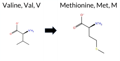 3DClick to see structure in 3D Viewer AIThe SynGAP1 missense variant V568M is not reported in ClinVar (ClinVar ID None) and has no entry in gnomAD (gnomAD ID None). Prediction tools that indicate a benign effect include FoldX, whereas the remaining tools—REVEL, Rosetta, PROVEAN, polyPhen‑2 (HumDiv and HumVar), SIFT, ESM1b, FATHMM, AlphaMissense‑Default, and the SGM Consensus—predict a pathogenic impact. High‑accuracy assessments show AlphaMissense‑Optimized as uncertain, the SGM Consensus (a majority vote of AlphaMissense‑Default, ESM1b, FATHMM, and PROVEAN) as Likely Pathogenic, and Foldetta (combining FoldX‑MD and Rosetta outputs) as uncertain. With the majority of evidence pointing to pathogenicity and no ClinVar annotation to contradict this, the variant is most likely pathogenic. Disclaimer: This summary was generated using AI and should be interpreted alongside expert review. | Likely Pathogenic | GAP | 0.024826 | Structured | 0.053503 | Uncertain | 0.937 | 0.257 | 0.000 | -10.361 | Likely Pathogenic | 0.913 | Likely Pathogenic | Ambiguous | 0.06 | Likely Benign | 0.3 | 2.71 | Destabilizing | 1.39 | Ambiguous | 0.66 | Ambiguous | 0.811 | Likely Pathogenic | -2.79 | Deleterious | 0.997 | Probably Damaging | 0.924 | Probably Damaging | -1.42 | Pathogenic | 0.00 | Affected | 0.0697 | 0.3124 | 2 | 1 | -2.3 | 32.06 | |||||||||||||||||||||||||||||
| c.1763T>A | L588H 2D 3DClick to see structure in 3D Viewer AIThe SynGAP1 missense variant L588H is listed in ClinVar (ID 422233.0) as Pathogenic and is not reported in gnomAD. All available in silico predictors classify the change as pathogenic: REVEL, FoldX, Rosetta, Foldetta, premPS, PROVEAN, polyPhen‑2 (HumDiv and HumVar), SIFT, ESM1b, FATHMM, AlphaMissense‑Default, and AlphaMissense‑Optimized. No tool predicts a benign effect. High‑accuracy assessments confirm this: AlphaMissense‑Optimized predicts Pathogenic; the SGM Consensus (majority vote of AlphaMissense‑Default, ESM1b, FATHMM, PROVEAN) yields Likely Pathogenic; and Foldetta (combining FoldX‑MD and Rosetta outputs) predicts Pathogenic. Thus, the variant is most likely pathogenic, and this prediction aligns with its ClinVar status. Disclaimer: This summary was generated using AI and should be interpreted alongside expert review. | Likely Pathogenic | GAP | 0.038042 | Structured | 0.082229 | Uncertain | 0.887 | 0.214 | 0.000 | Likely Pathogenic | 1 | -16.947 | Likely Pathogenic | 0.999 | Likely Pathogenic | Likely Pathogenic | 4.20 | Destabilizing | 0.2 | 3.69 | Destabilizing | 3.95 | Destabilizing | 2.26 | Destabilizing | 0.939 | Likely Pathogenic | -6.97 | Deleterious | 1.000 | Probably Damaging | 1.000 | Probably Damaging | -1.42 | Pathogenic | 0.00 | Affected | 3.38 | 34 | 0.0980 | 0.0456 | -2 | -3 | -7.0 | 23.98 | 214.3 | 20.9 | 0.0 | 0.0 | 0.0 | 0.2 | X | X | X | Potentially Pathogenic | The isobutyl group of the Leu588 side chain, located in an α helix (res. Glu582-Met603), packs against hydrophobic residues in the inter-helix hydrophobic space (e.g., Ile584, Trp572, Phe484, Met470, Val473, Ile483).In the variant simulations, the imidazole ring of His588 is aromatic but contains polar delta and epsilon nitrogen atoms that are not suited for the hydrophobic niche. The protonated epsilon nitrogen forms a hydrogen bond with the backbone carbonyl group of Ala469, which can disrupt the continuity of the opposing α helix (res. Phe476-Lys460).While the residue swap could affect the tertiary assembly and the underlying protein folding process, it is difficult to determine if the mutation would be tolerated based solely on the variant simulations. | ||||||||||||||
| c.1763T>C | L588P 2D 3DClick to see structure in 3D Viewer AIThe SynGAP1 missense variant L588P is listed in ClinVar with an “Uncertain” status and is not reported in gnomAD. All available in silico predictors classify the change as pathogenic: REVEL, FoldX, Rosetta, Foldetta, premPS, PROVEAN, polyPhen‑2 (HumDiv and HumVar), SIFT, ESM1b, FATHMM, AlphaMissense‑Default, and AlphaMissense‑Optimized. No tool predicts a benign effect. High‑accuracy assessments reinforce this view: AlphaMissense‑Optimized predicts pathogenicity; the SGM Consensus (majority vote of AlphaMissense‑Default, ESM1b, FATHMM, and PROVEAN) indicates “Likely Pathogenic”; and Foldetta, which integrates FoldX‑MD and Rosetta outputs, also predicts pathogenicity. Consequently, the variant is most likely pathogenic based on the collective predictions, and this assessment does not contradict the ClinVar status, which remains uncertain. Disclaimer: This summary was generated using AI and should be interpreted alongside expert review. | Likely Pathogenic | GAP | 0.038042 | Structured | 0.082229 | Uncertain | 0.887 | 0.214 | 0.000 | Uncertain | 2 | -14.771 | Likely Pathogenic | 1.000 | Likely Pathogenic | Likely Pathogenic | 5.61 | Destabilizing | 0.5 | 12.91 | Destabilizing | 9.26 | Destabilizing | 2.33 | Destabilizing | 0.932 | Likely Pathogenic | -6.97 | Deleterious | 1.000 | Probably Damaging | 1.000 | Probably Damaging | -1.42 | Pathogenic | 0.00 | Affected | 3.38 | 34 | 0.3533 | 0.0992 | -3 | -3 | -5.4 | -16.04 | |||||||||||||||||||||||||
| c.1763T>G | L588R 2D 3DClick to see structure in 3D Viewer AIThe SynGAP1 missense variant L588R is not reported in ClinVar (ClinVar ID None) and is absent from gnomAD (gnomAD ID None). Prediction tools that assess pathogenicity uniformly classify the variant as pathogenic: SGM‑Consensus (Likely Pathogenic), REVEL, FoldX, Rosetta, Foldetta, premPS, PROVEAN, polyPhen‑2 (HumDiv and HumVar), SIFT, ESM1b, FATHMM, AlphaMissense‑Default, and AlphaMissense‑Optimized. No tool predicts a benign effect. High‑accuracy methods corroborate this: AlphaMissense‑Optimized is pathogenic; the SGM Consensus (majority vote of AlphaMissense‑Default, ESM1b, FATHMM, PROVEAN) is Likely Pathogenic; and Foldetta (combining FoldX‑MD and Rosetta outputs) is pathogenic. All available evidence points to a deleterious impact. Thus, the variant is most likely pathogenic, and this assessment does not contradict any ClinVar status. Disclaimer: This summary was generated using AI and should be interpreted alongside expert review. | Likely Pathogenic | GAP | 0.038042 | Structured | 0.082229 | Uncertain | 0.887 | 0.214 | 0.000 | -15.602 | Likely Pathogenic | 0.998 | Likely Pathogenic | Likely Pathogenic | 6.96 | Destabilizing | 0.4 | 6.14 | Destabilizing | 6.55 | Destabilizing | 2.07 | Destabilizing | 0.942 | Likely Pathogenic | -5.98 | Deleterious | 1.000 | Probably Damaging | 0.998 | Probably Damaging | -1.42 | Pathogenic | 0.00 | Affected | 0.1209 | 0.0530 | -3 | -2 | -8.3 | 43.03 | |||||||||||||||||||||||||||||
| c.1423C>G | R475G 2D AIThe SynGAP1 missense variant R475G is not reported in ClinVar and is absent from gnomAD. Prediction tools that assess pathogenicity largely agree on a deleterious effect: SGM‑Consensus, REVEL, FoldX, premPS, PROVEAN, polyPhen‑2 (HumDiv and HumVar), SIFT, ESM1b, FATHMM, and AlphaMissense‑Default all predict pathogenicity. Only Rosetta and AlphaMissense‑Optimized return uncertain results, and no tool predicts a benign outcome. High‑accuracy methods provide a consistent view: AlphaMissense‑Optimized is uncertain, SGM‑Consensus indicates likely pathogenic, and Foldetta (combining FoldX‑MD and Rosetta outputs) predicts pathogenicity. Taken together, the overwhelming majority of evidence supports a pathogenic classification for R475G, and this conclusion is not contradicted by any ClinVar annotation. Disclaimer: This summary was generated using AI and should be interpreted alongside expert review. | Likely Pathogenic | GAP | 0.175930 | Structured | 0.382696 | Uncertain | 0.852 | 0.261 | 0.000 | -14.466 | Likely Pathogenic | 0.939 | Likely Pathogenic | Ambiguous | 2.39 | Destabilizing | 1.0 | 1.64 | Ambiguous | 2.02 | Destabilizing | 1.11 | Destabilizing | 0.697 | Likely Pathogenic | -6.53 | Deleterious | 1.000 | Probably Damaging | 1.000 | Probably Damaging | -1.41 | Pathogenic | 0.00 | Affected | 0.2779 | 0.2310 | -3 | -2 | 4.1 | -99.14 | |||||||||||||||||||||||||||||
| c.1468G>T | A490S 2D  3DClick to see structure in 3D Viewer AISynGAP1 missense variant A490S is not reported in ClinVar and is absent from gnomAD. Prediction tools that agree on a benign effect include AlphaMissense‑Optimized, whereas the majority of other in silico predictors (SGM‑Consensus, REVEL, PROVEAN, polyPhen‑2 HumDiv, polyPhen‑2 HumVar, SIFT, ESM1b, FATHMM) indicate a pathogenic impact. High‑accuracy assessments further support this: AlphaMissense‑Optimized classifies the variant as benign, SGM‑Consensus (derived from a majority vote of AlphaMissense‑Default, ESM1b, FATHMM, and PROVEAN) predicts it to be likely pathogenic, and Foldetta, which integrates FoldX‑MD and Rosetta stability calculations, yields an inconclusive result. No other tools provide definitive evidence. Overall, the preponderance of pathogenic predictions suggests that A490S is most likely pathogenic, and this assessment does not contradict the current ClinVar status, which has no entry for the variant. Disclaimer: This summary was generated using AI and should be interpreted alongside expert review. | Likely Pathogenic | GAP | 0.120615 | Structured | 0.322979 | Uncertain | 0.938 | 0.210 | 0.125 | -8.307 | Likely Pathogenic | 0.426 | Ambiguous | Likely Benign | 0.76 | Ambiguous | 0.1 | 1.55 | Ambiguous | 1.16 | Ambiguous | 0.89 | Ambiguous | 0.766 | Likely Pathogenic | -2.82 | Deleterious | 0.983 | Probably Damaging | 0.993 | Probably Damaging | -1.41 | Pathogenic | 0.02 | Affected | 0.2338 | 0.3116 | 1 | 1 | -2.6 | 16.00 | |||||||||||||||||||||||||||||
| c.1477G>C | A493P 2D 3DClick to see structure in 3D Viewer AIThe SynGAP1 missense variant A493P is not reported in ClinVar and is absent from gnomAD. Prediction tools that assess functional impact all converge on a pathogenic interpretation: REVEL, FoldX, Rosetta, Foldetta, premPS, PROVEAN, polyPhen‑2 (HumDiv and HumVar), SIFT, ESM1b, FATHMM, AlphaMissense‑Default, and AlphaMissense‑Optimized all predict deleterious effects. No tool in the dataset indicates a benign outcome. High‑accuracy methods reinforce this consensus: AlphaMissense‑Optimized is pathogenic, the SGM Consensus (majority vote of AlphaMissense‑Default, ESM1b, FATHMM, PROVEAN) is likely pathogenic, and Foldetta (combining FoldX‑MD and Rosetta outputs) is pathogenic. With all available evidence pointing to a damaging effect and no ClinVar entry to contradict, the variant is most likely pathogenic. Disclaimer: This summary was generated using AI and should be interpreted alongside expert review. | Likely Pathogenic | GAP | 0.102787 | Structured | 0.340081 | Uncertain | 0.966 | 0.182 | 0.000 | -15.797 | Likely Pathogenic | 1.000 | Likely Pathogenic | Likely Pathogenic | 4.96 | Destabilizing | 0.1 | 10.79 | Destabilizing | 7.88 | Destabilizing | 1.32 | Destabilizing | 0.883 | Likely Pathogenic | -4.54 | Deleterious | 1.000 | Probably Damaging | 0.998 | Probably Damaging | -1.41 | Pathogenic | 0.02 | Affected | 0.1490 | 0.2907 | 1 | -1 | -3.4 | 26.04 | |||||||||||||||||||||||||||||
| c.1478C>A | A493D 2D AISynGAP1 missense variant A493D is not reported in ClinVar and is absent from gnomAD. Functional prediction tools uniformly indicate a deleterious effect: REVEL, FoldX, Rosetta, Foldetta, premPS, PROVEAN, polyPhen‑2 (HumDiv and HumVar), SIFT, ESM1b, FATHMM, AlphaMissense‑Default, and AlphaMissense‑Optimized all classify the substitution as pathogenic. No tool predicts a benign outcome. High‑accuracy assessments reinforce this view: AlphaMissense‑Optimized predicts pathogenicity; the SGM Consensus (majority vote of AlphaMissense‑Default, ESM1b, FATHMM, and PROVEAN) reports “Likely Pathogenic”; and Foldetta, which integrates FoldX‑MD and Rosetta stability calculations, also predicts pathogenicity. Consequently, the variant is most likely pathogenic, and this assessment is consistent with the absence of a ClinVar entry (no contradiction). Disclaimer: This summary was generated using AI and should be interpreted alongside expert review. | Likely Pathogenic | GAP | 0.102787 | Structured | 0.340081 | Uncertain | 0.966 | 0.182 | 0.000 | -16.834 | Likely Pathogenic | 0.999 | Likely Pathogenic | Likely Pathogenic | 4.45 | Destabilizing | 1.5 | 3.59 | Destabilizing | 4.02 | Destabilizing | 1.60 | Destabilizing | 0.925 | Likely Pathogenic | -5.25 | Deleterious | 1.000 | Probably Damaging | 0.998 | Probably Damaging | -1.41 | Pathogenic | 0.01 | Affected | 0.1437 | 0.1541 | 0 | -2 | -5.3 | 44.01 | |||||||||||||||||||||||||||||
| c.1481T>A | I494K 2D  3DClick to see structure in 3D Viewer AIThe SynGAP1 missense variant I494K is not reported in ClinVar (ClinVar ID None) and is absent from gnomAD (gnomAD ID None). Prediction tools that assess pathogenicity uniformly classify the variant as deleterious: SGM‑Consensus (Likely Pathogenic), REVEL, FoldX, Rosetta, Foldetta, premPS, PROVEAN, polyPhen‑2 (HumDiv and HumVar), SIFT, ESM1b, FATHMM, AlphaMissense‑Default, and AlphaMissense‑Optimized all predict a pathogenic effect. No tool in the dataset predicts a benign outcome. High‑accuracy methods reinforce this view: AlphaMissense‑Optimized is pathogenic; the SGM Consensus (majority vote of AlphaMissense‑Default, ESM1b, FATHMM, PROVEAN) is Likely Pathogenic; and Foldetta (combining FoldX‑MD and Rosetta outputs) is pathogenic. Based on the unanimous computational evidence, the variant is most likely pathogenic, and this assessment does not contradict any ClinVar status (none reported). Disclaimer: This summary was generated using AI and should be interpreted alongside expert review. | Likely Pathogenic | GAP | 0.155435 | Structured | 0.353330 | Uncertain | 0.941 | 0.157 | 0.000 | -15.950 | Likely Pathogenic | 0.997 | Likely Pathogenic | Likely Pathogenic | 4.23 | Destabilizing | 0.2 | 5.33 | Destabilizing | 4.78 | Destabilizing | 1.89 | Destabilizing | 0.925 | Likely Pathogenic | -6.40 | Deleterious | 0.987 | Probably Damaging | 0.937 | Probably Damaging | -1.41 | Pathogenic | 0.00 | Affected | 0.1015 | 0.0870 | -2 | -3 | -8.4 | 15.01 | |||||||||||||||||||||||||||||
| c.1481T>G | I494R 2D 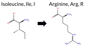 3DClick to see structure in 3D Viewer AIThe SynGAP1 missense variant I494R is listed in ClinVar as Pathogenic (ClinVar ID 1685460.0) and is not reported in gnomAD. Prediction tools that assess functional impact all converge on a pathogenic outcome: REVEL, FoldX, Rosetta, Foldetta, premPS, PROVEAN, polyPhen‑2 (HumDiv and HumVar), SIFT, ESM1b, FATHMM, AlphaMissense‑Default, and AlphaMissense‑Optimized all indicate pathogenicity. No tool predicts a benign effect. High‑accuracy assessments reinforce this view: AlphaMissense‑Optimized is pathogenic; the SGM Consensus (majority vote of AlphaMissense‑Default, ESM1b, FATHMM, PROVEAN) is Likely Pathogenic; and Foldetta (combining FoldX‑MD and Rosetta outputs) is pathogenic. Thus, the variant is most likely pathogenic, and this prediction aligns with its ClinVar status. Disclaimer: This summary was generated using AI and should be interpreted alongside expert review. | Likely Pathogenic | GAP | 0.155435 | Structured | 0.353330 | Uncertain | 0.941 | 0.157 | 0.000 | Likely Pathogenic | 1 | -15.758 | Likely Pathogenic | 0.995 | Likely Pathogenic | Likely Pathogenic | 6.71 | Destabilizing | 0.3 | 3.40 | Destabilizing | 5.06 | Destabilizing | 2.19 | Destabilizing | 0.911 | Likely Pathogenic | -6.43 | Deleterious | 0.999 | Probably Damaging | 0.957 | Probably Damaging | -1.41 | Pathogenic | 0.00 | Affected | 3.37 | 35 | 0.1180 | 0.0870 | -2 | -3 | -9.0 | 43.03 | 273.9 | -59.8 | 0.0 | 0.0 | 0.0 | 0.1 | X | X | X | X | Potentially Pathogenic | The sec-butyl side chain of Ile494, located in an α-helix (res. Leu489-Glu519), packs against hydrophobic residues (e.g., Phe484, Leu465, Trp572, Ala493, Met468) in an inter-helix space (res. Leu489-Glu519 and res. Ala461-Phe476). In the variant simulations, the bulkier and positively charged residue, Arg494, weakens the integrity of the opposing helix. Additionally, the bulkier Arg494 stacks with Phe484, causing the α-helices to move farther apart to accommodate it. This mutation could have substantial negative effects due to the fundamental role of hydrophobic packing, which is disrupted by Arg494 during protein folding. | |||||||||||||
| c.1485A>C | E495D 2D 3DClick to see structure in 3D Viewer AISynGAP1 missense variant E495D is listed in ClinVar with an uncertain significance (ClinVar ID 2000233.0) and is not reported in gnomAD. Functional prediction tools cluster into two groups: benign predictions from SIFT and ESM1b, and pathogenic predictions from REVEL, PROVEAN, polyPhen‑2 (HumDiv and HumVar), FATHMM, AlphaMissense‑Default, and AlphaMissense‑Optimized. The high‑accuracy consensus (SGM Consensus) derived from AlphaMissense‑Default, ESM1b, FATHMM, and PROVEAN classifies the variant as likely pathogenic. AlphaMissense‑Optimized also predicts pathogenicity, whereas Foldetta, which integrates FoldX‑MD and Rosetta outputs, remains uncertain. Overall, the majority of evidence points to a pathogenic effect, which does not contradict the ClinVar uncertain status but suggests a higher likelihood of deleterious impact. Disclaimer: This summary was generated using AI and should be interpreted alongside expert review. | Likely Pathogenic | GAP | 0.164327 | Structured | 0.364496 | Uncertain | 0.933 | 0.161 | 0.000 | Conflicting | 2 | -3.574 | Likely Benign | 0.958 | Likely Pathogenic | Likely Pathogenic | 1.39 | Ambiguous | 0.1 | 1.03 | Ambiguous | 1.21 | Ambiguous | 0.98 | Ambiguous | 0.566 | Likely Pathogenic | -2.52 | Deleterious | 0.998 | Probably Damaging | 0.989 | Probably Damaging | -1.41 | Pathogenic | 0.17 | Tolerated | 3.37 | 35 | 0.1778 | 0.3064 | 3 | 2 | 0.0 | -14.03 | 220.6 | 38.8 | 0.0 | 0.0 | 0.1 | 0.1 | X | X | Uncertain | Glu495 is located in the α-helix (res. Leu489-Glu519), and its carboxylate group forms salt bridges with the neighboring Lys492 and with Arg596 on an opposing α-helix (res. Glu582-Met603) in the WT simulations. In the variant simulations, the acidic carboxylate side chain of Asp495 can also form salt bridges with both Lys492 and Arg596. However, the shorter side chain of aspartate tends to favor forming a salt bridge with the nearby Arg499 on the same α-helix instead. Asp495 might not maintain the salt bridge with Arg596 on the opposing α-helix as efficiently as Glu495 in the WT, potentially weakening the tertiary structure. Regardless, the potential negative effect is likely to be minor, with no deleterious effects observed on the protein structure during the simulations. However, due to its location at the GAP-Ras interface, the effect of the residue swap on SynGAP-Ras complex formation or GTPase activation cannot be fully addressed using the SynGAP solvent-only simulations. | |||||||||||||||
| c.1485A>T | E495D 2D 3DClick to see structure in 3D Viewer AIThe SynGAP1 missense variant E495D is not reported in ClinVar (status: None) and is absent from gnomAD. Prediction tools cluster into two groups: benign predictions come from SIFT and ESM1b, while pathogenic predictions arise from REVEL, PROVEAN, polyPhen‑2 HumDiv, polyPhen‑2 HumVar, FATHMM, AlphaMissense‑Default, AlphaMissense‑Optimized, and the SGM‑Consensus. Four tools (FoldX, Rosetta, Foldetta, premPS) give uncertain or inconclusive results and are treated as unavailable. High‑accuracy methods reinforce the pathogenic signal: AlphaMissense‑Optimized predicts pathogenic, the SGM‑Consensus (majority vote of AlphaMissense‑Default, ESM1b, FATHMM, PROVEAN) also predicts pathogenic, and Foldetta provides no definitive stability change. Overall, the variant is most likely pathogenic based on the consensus of predictive algorithms, and this assessment does not contradict any ClinVar annotation. Disclaimer: This summary was generated using AI and should be interpreted alongside expert review. | Likely Pathogenic | GAP | 0.164327 | Structured | 0.364496 | Uncertain | 0.933 | 0.161 | 0.000 | -3.574 | Likely Benign | 0.958 | Likely Pathogenic | Likely Pathogenic | 1.39 | Ambiguous | 0.1 | 1.03 | Ambiguous | 1.21 | Ambiguous | 0.98 | Ambiguous | 0.566 | Likely Pathogenic | -2.52 | Deleterious | 0.998 | Probably Damaging | 0.989 | Probably Damaging | -1.41 | Pathogenic | 0.17 | Tolerated | 3.37 | 35 | 0.1778 | 0.3064 | 3 | 2 | 0.0 | -14.03 | 220.6 | 38.8 | 0.0 | 0.0 | 0.1 | 0.1 | X | X | Uncertain | Glu495 is located in the α-helix (res. Leu489-Glu519), and its carboxylate group forms salt bridges with the neighboring Lys492 and with Arg596 on an opposing α-helix (res. Glu582-Met603) in the WT simulations. In the variant simulations, the acidic carboxylate side chain of Asp495 can also form salt bridges with both Lys492 and Arg596. However, the shorter side chain of aspartate tends to favor forming a salt bridge with the nearby Arg499 on the same α-helix instead. Asp495 might not maintain the salt bridge with Arg596 on the opposing α-helix as efficiently as Glu495 in the WT, potentially weakening the tertiary structure. Regardless, the potential negative effect is likely to be minor, with no deleterious effects observed on the protein structure during the simulations. However, due to its location at the GAP-Ras interface, the effect of the residue swap on SynGAP-Ras complex formation or GTPase activation cannot be fully addressed using the SynGAP solvent-only simulations. | |||||||||||||||||
| c.1487A>C | E496A 2D  3DClick to see structure in 3D Viewer AISynGAP1 missense variant E496A is listed in ClinVar with an uncertain significance and is not reported in gnomAD. Functional prediction tools show mixed results: benign calls come from Rosetta, Foldetta, premPS, SIFT, and AlphaMissense‑Optimized, while pathogenic calls are made by REVEL, PROVEAN, polyPhen‑2 (HumDiv and HumVar), ESM1b, FATHMM, AlphaMissense‑Default, and the SGM‑Consensus (majority vote of AlphaMissense‑Default, ESM1b, FATHMM, PROVEAN). FoldX is inconclusive. High‑accuracy methods give a split verdict: AlphaMissense‑Optimized predicts benign, SGM‑Consensus predicts pathogenic, and Foldetta predicts benign. Overall, the majority of predictions lean toward pathogenicity, and this does not contradict the ClinVar uncertain status. Disclaimer: This summary was generated using AI and should be interpreted alongside expert review. | Likely Pathogenic | GAP | 0.102787 | Structured | 0.383296 | Uncertain | 0.945 | 0.179 | 0.000 | Uncertain | 1 | -11.159 | Likely Pathogenic | 0.598 | Likely Pathogenic | Likely Benign | 0.68 | Ambiguous | 0.0 | 0.23 | Likely Benign | 0.46 | Likely Benign | 0.47 | Likely Benign | 0.681 | Likely Pathogenic | -4.87 | Deleterious | 1.000 | Probably Damaging | 0.999 | Probably Damaging | -1.41 | Pathogenic | 0.09 | Tolerated | 0.2806 | 0.3548 | 0 | -1 | 5.3 | -58.04 | |||||||||||||||||||||||||||
| c.1511A>G | K504R 2D 3DClick to see structure in 3D Viewer AIThe SynGAP1 missense variant K504R is listed in ClinVar with an “Uncertain” status and is present in gnomAD (ID 6‑33438543‑A‑G). Consensus from most in‑silico predictors is benign: REVEL, FoldX, Foldetta, PROVEAN, polyPhen‑2 (HumDiv and HumVar), SIFT, ESM1b, AlphaMissense‑Default, and AlphaMissense‑Optimized all report a benign effect, while only FATHMM predicts pathogenicity. Uncertain calls come from Rosetta and premPS. High‑accuracy assessments reinforce the benign prediction: AlphaMissense‑Optimized is benign; the SGM Consensus (majority vote of AlphaMissense‑Default, ESM1b, FATHMM, PROVEAN) is “Likely Benign”; and Foldetta, which integrates FoldX‑MD and Rosetta outputs, also predicts benign. Overall, the preponderance of evidence indicates the variant is most likely benign, and this conclusion does not contradict the ClinVar “Uncertain” classification. Disclaimer: This summary was generated using AI and should be interpreted alongside expert review. | Likely Benign | GAP | 0.028107 | Structured | 0.304984 | Uncertain | 0.850 | 0.189 | 0.000 | Uncertain | 1 | 6-33438543-A-G | 2 | 1.24e-6 | -4.365 | Likely Benign | 0.088 | Likely Benign | Likely Benign | 0.13 | Likely Benign | 0.1 | 0.51 | Ambiguous | 0.32 | Likely Benign | 0.94 | Ambiguous | 0.238 | Likely Benign | -2.16 | Neutral | 0.002 | Benign | 0.015 | Benign | -1.41 | Pathogenic | 0.11 | Tolerated | 3.37 | 35 | 0.3363 | 0.0647 | 2 | 3 | -0.6 | 28.01 | ||||||||||||||||||||||
| c.1572C>G | C524W 2D 3DClick to see structure in 3D Viewer AIThe SynGAP1 missense variant C524W is not reported in ClinVar and is absent from gnomAD. Prediction tools uniformly indicate a deleterious effect: benign predictions are absent, while pathogenic predictions are made by REVEL, FoldX, Rosetta, Foldetta, PROVEAN, polyPhen‑2 (HumDiv and HumVar), SIFT, ESM1b, FATHMM, AlphaMissense‑Default, and AlphaMissense‑Optimized; premPS is uncertain. High‑accuracy assessments further support pathogenicity: AlphaMissense‑Optimized predicts pathogenic, the SGM Consensus (majority vote of AlphaMissense‑Default, ESM1b, FATHMM, PROVEAN) is Likely Pathogenic, and Foldetta (combining FoldX‑MD and Rosetta outputs) predicts pathogenic. Based on the consensus of all available predictions, the variant is most likely pathogenic, and this conclusion is not contradicted by ClinVar status, which currently has no entry for this variant. Disclaimer: This summary was generated using AI and should be interpreted alongside expert review. | Likely Pathogenic | GAP | 0.134866 | Structured | 0.024729 | Uncertain | 0.916 | 0.385 | 0.125 | -12.351 | Likely Pathogenic | 0.999 | Likely Pathogenic | Likely Pathogenic | 3.03 | Destabilizing | 1.2 | 9.88 | Destabilizing | 6.46 | Destabilizing | 0.93 | Ambiguous | 0.675 | Likely Pathogenic | -10.94 | Deleterious | 1.000 | Probably Damaging | 0.999 | Probably Damaging | -1.41 | Pathogenic | 0.00 | Affected | 0.1953 | 0.3920 | -8 | -2 | -3.4 | 83.07 | |||||||||||||||||||||||||||||
| c.1625A>C | N542T 2D 3DClick to see structure in 3D Viewer AIThe SynGAP1 missense variant N542T is not reported in ClinVar (ClinVar ID None) and has no entries in gnomAD (gnomAD ID None). Prediction tools that agree on a benign effect are SIFT and Rosetta. Tools that predict a pathogenic effect include SGM‑Consensus, REVEL, PROVEAN, polyPhen‑2 (HumDiv and HumVar), ESM1b, FATHMM, AlphaMissense‑Default, and AlphaMissense‑Optimized. Uncertain or inconclusive results come from FoldX, Foldetta, and premPS. High‑accuracy assessments show AlphaMissense‑Optimized as pathogenic, SGM‑Consensus as likely pathogenic (majority vote from AlphaMissense‑Default, ESM1b, FATHMM, and PROVEAN), and Foldetta as uncertain. Overall, the majority of evidence points to a pathogenic impact. This conclusion is not contradicted by ClinVar status, which has no classification for this variant. Disclaimer: This summary was generated using AI and should be interpreted alongside expert review. | Likely Pathogenic | GAP | 0.053060 | Structured | 0.026143 | Uncertain | 0.953 | 0.331 | 0.000 | -9.918 | Likely Pathogenic | 0.974 | Likely Pathogenic | Likely Pathogenic | 1.13 | Ambiguous | 0.1 | 0.20 | Likely Benign | 0.67 | Ambiguous | 0.75 | Ambiguous | 0.784 | Likely Pathogenic | -5.37 | Deleterious | 0.997 | Probably Damaging | 0.990 | Probably Damaging | -1.41 | Pathogenic | 0.12 | Tolerated | 0.1120 | 0.5872 | 0 | 0 | 2.8 | -13.00 | |||||||||||||||||||||||||||||
| c.1718G>T | R573L 2D 3DClick to see structure in 3D Viewer AIThe SynGAP1 missense variant R573L is listed in ClinVar as Pathogenic (ClinVar ID 521291.0) and is not reported in gnomAD. Functional prediction tools that assess sequence conservation and structural impact uniformly indicate a deleterious effect: REVEL, PROVEAN, polyPhen‑2 (HumDiv and HumVar), SIFT, ESM1b, FATHMM, AlphaMissense‑Default, and AlphaMissense‑Optimized all classify the substitution as pathogenic. No tool in the dataset predicts a benign outcome. Predictions that rely on protein‑folding stability (FoldX, Rosetta, Foldetta, premPS) are inconclusive and are therefore treated as unavailable. High‑accuracy assessments further support pathogenicity: AlphaMissense‑Optimized is pathogenic; the SGM Consensus, derived from the majority vote of AlphaMissense‑Default, ESM1b, FATHMM, and PROVEAN, also reports pathogenic; Foldetta remains uncertain. Based on the collective evidence, the variant is most likely pathogenic, which is consistent with its ClinVar status. Disclaimer: This summary was generated using AI and should be interpreted alongside expert review. | Likely Pathogenic | GAP | 0.134866 | Structured | 0.032433 | Uncertain | 0.934 | 0.235 | 0.000 | Likely Pathogenic | 1 | -13.120 | Likely Pathogenic | 0.993 | Likely Pathogenic | Likely Pathogenic | 1.30 | Ambiguous | 0.6 | 1.11 | Ambiguous | 1.21 | Ambiguous | 0.80 | Ambiguous | 0.833 | Likely Pathogenic | -5.74 | Deleterious | 1.000 | Probably Damaging | 1.000 | Probably Damaging | -1.41 | Pathogenic | 0.01 | Affected | 3.37 | 35 | 0.1503 | 0.3083 | -3 | -2 | 8.3 | -43.03 | 237.4 | 60.7 | 0.0 | 0.0 | -0.7 | 0.3 | X | X | Potentially Pathogenic | The guanidinium group of Arg573, located in an α-helix (res. Arg563-Glu578), forms a salt bridge with the carboxylate groups of Glu582 and/or Asp586 from a nearby α-helix (res. Glu582-Met603) in the WT simulations. Additionally, the Arg573 side chain stacks planarly with the aromatic phenol ring of Tyr665 and hydrogen bonds with the hydroxyl group of Ser668 from another α-helix (res. Ser641-Ser668). In the variant simulations, the aliphatic iso-butyl group of the Leu573 side chain fails to establish any of these interactions, which, in turn, lowers the integrity of the opposing α-helix end (res. Glu582-Met603). Overall, the residue swap has the potential to substantially affect the tertiary structure assembly during the protein folding process. | 10.1016/j.ajhg.2020.11.011 | ||||||||||||||
| c.1736G>C | R579P 2D 3DClick to see structure in 3D Viewer AIThe SynGAP1 missense variant R579P is not reported in ClinVar (ClinVar status: none) and is absent from gnomAD (gnomAD ID: none). Prediction tools that agree on a pathogenic effect include SGM‑Consensus, REVEL, FoldX, Rosetta, Foldetta, PROVEAN, polyPhen‑2 (HumDiv and HumVar), SIFT, ESM1b, FATHMM, AlphaMissense‑Default, and AlphaMissense‑Optimized; no tool predicts a benign outcome. High‑accuracy assessments further support pathogenicity: AlphaMissense‑Optimized is pathogenic, the SGM‑Consensus (majority vote of AlphaMissense‑Default, ESM1b, FATHMM, PROVEAN) is pathogenic, and Foldetta (combining FoldX‑MD and Rosetta outputs) is pathogenic. No predictions or stability results are missing or inconclusive. Based on the unanimous pathogenic predictions and the absence of any ClinVar or gnomAD evidence to the contrary, the variant is most likely pathogenic. Disclaimer: This summary was generated using AI and should be interpreted alongside expert review. | Likely Pathogenic | GAP | 0.053060 | Structured | 0.022872 | Uncertain | 0.877 | 0.244 | 0.000 | -14.826 | Likely Pathogenic | 0.998 | Likely Pathogenic | Likely Pathogenic | 3.00 | Destabilizing | 0.2 | 6.36 | Destabilizing | 4.68 | Destabilizing | 0.93 | Ambiguous | 0.821 | Likely Pathogenic | -6.26 | Deleterious | 1.000 | Probably Damaging | 1.000 | Probably Damaging | -1.41 | Pathogenic | 0.01 | Affected | 0.2207 | 0.3081 | 0 | -2 | 2.9 | -59.07 | |||||||||||||||||||||||||||||
| c.1825G>A | G609R 2D 3DClick to see structure in 3D Viewer AIThe SynGAP1 missense variant G609R is not reported in ClinVar and is absent from gnomAD. Prediction tools that agree on a benign effect include Rosetta, SIFT, and AlphaMissense‑Optimized. Those that agree on a pathogenic effect are SGM‑Consensus, REVEL, FoldX, PROVEAN, polyPhen‑2 (HumDiv and HumVar), ESM1b, and FATHMM. Predictions that remain uncertain are Foldetta, premPS, and AlphaMissense‑Default. High‑accuracy assessments show AlphaMissense‑Optimized as benign, SGM‑Consensus (derived from a majority vote of AlphaMissense‑Default, ESM1b, FATHMM, and PROVEAN) as likely pathogenic, and Foldetta as uncertain. Overall, the majority of evaluated tools (eight pathogenic vs. three benign) predict a pathogenic impact for G609R. This conclusion is not contradicted by ClinVar status, which currently has no entry for this variant. Disclaimer: This summary was generated using AI and should be interpreted alongside expert review. | Likely Pathogenic | GAP | 0.179055 | Structured | 0.203786 | Uncertain | 0.851 | 0.252 | 0.000 | -10.172 | Likely Pathogenic | 0.543 | Ambiguous | Likely Benign | 2.09 | Destabilizing | 0.1 | 0.37 | Likely Benign | 1.23 | Ambiguous | 0.60 | Ambiguous | 0.520 | Likely Pathogenic | -2.68 | Deleterious | 0.974 | Probably Damaging | 0.818 | Possibly Damaging | -1.41 | Pathogenic | 0.07 | Tolerated | 0.1158 | 0.4348 | -3 | -2 | -4.1 | 99.14 | |||||||||||||||||||||||||||||
| c.1825G>C | G609R 2D 3DClick to see structure in 3D Viewer AIThe SynGAP1 missense variant G609R is not reported in ClinVar and is absent from gnomAD. Prediction tools that agree on a benign effect include Rosetta, SIFT, and AlphaMissense‑Optimized. Those that agree on a pathogenic effect are SGM‑Consensus, REVEL, FoldX, PROVEAN, polyPhen‑2 (HumDiv and HumVar), ESM1b, and FATHMM. Predictions that remain uncertain are Foldetta, premPS, and AlphaMissense‑Default. High‑accuracy assessments show AlphaMissense‑Optimized as benign, SGM‑Consensus (derived from a majority vote of AlphaMissense‑Default, ESM1b, FATHMM, and PROVEAN) as likely pathogenic, and Foldetta as uncertain. Overall, the majority of evaluated tools (eight pathogenic vs. three benign) predict a pathogenic impact for G609R. This conclusion is not contradicted by ClinVar status, which currently has no entry for this variant. Disclaimer: This summary was generated using AI and should be interpreted alongside expert review. | Likely Pathogenic | GAP | 0.179055 | Structured | 0.203786 | Uncertain | 0.851 | 0.252 | 0.000 | -10.172 | Likely Pathogenic | 0.543 | Ambiguous | Likely Benign | 2.09 | Destabilizing | 0.1 | 0.37 | Likely Benign | 1.23 | Ambiguous | 0.60 | Ambiguous | 0.520 | Likely Pathogenic | -2.68 | Deleterious | 0.974 | Probably Damaging | 0.818 | Possibly Damaging | -1.41 | Pathogenic | 0.07 | Tolerated | 0.1158 | 0.4348 | -3 | -2 | -4.1 | 99.14 | |||||||||||||||||||||||||||||
| c.1826G>A | G609E 2D  3DClick to see structure in 3D Viewer AIThe SynGAP1 missense variant G609E has no ClinVar entry and is not reported in gnomAD. Prediction tools that agree on a benign effect include PROVEAN, SIFT, and AlphaMissense‑Optimized. Those that agree on a pathogenic effect are REVEL, FoldX, polyPhen‑2 (HumDiv and HumVar), ESM1b, and FATHMM. The remaining tools (Rosetta, Foldetta, premPS, AlphaMissense‑Default) give uncertain or inconclusive results. High‑accuracy assessments show AlphaMissense‑Optimized as benign, the SGM Consensus (majority vote from AlphaMissense‑Default, ESM1b, FATHMM, PROVEAN) as pathogenic, and Foldetta as uncertain. Overall, the majority of evidence points to a pathogenic impact, and this conclusion is not contradicted by any ClinVar annotation. Thus, the variant is most likely pathogenic. Disclaimer: This summary was generated using AI and should be interpreted alongside expert review. | GAP | 0.179055 | Structured | 0.203786 | Uncertain | 0.851 | 0.252 | 0.000 | -10.470 | Likely Pathogenic | 0.421 | Ambiguous | Likely Benign | 2.95 | Destabilizing | 0.4 | -1.65 | Ambiguous | 0.65 | Ambiguous | 0.61 | Ambiguous | 0.531 | Likely Pathogenic | -2.28 | Neutral | 0.916 | Possibly Damaging | 0.588 | Possibly Damaging | -1.41 | Pathogenic | 0.17 | Tolerated | 0.1709 | 0.4491 | 0 | -2 | -3.1 | 72.06 | ||||||||||||||||||||||||||||||
| c.1850A>G | E617G 2D 3DClick to see structure in 3D Viewer AIThe SynGAP1 missense variant E617G is not reported in ClinVar and is absent from gnomAD. Prediction tools cluster into two groups: benign predictions come from premPS, SIFT, and AlphaMissense‑Optimized, whereas pathogenic predictions are made by SGM‑Consensus, REVEL, PROVEAN, polyPhen‑2 (HumDiv and HumVar), FATHMM, and AlphaMissense‑Default. Uncertain results are reported by FoldX, Rosetta, Foldetta, and ESM1b. High‑accuracy methods give a mixed picture: AlphaMissense‑Optimized predicts benign, the SGM Consensus (majority vote of AlphaMissense‑Default, ESM1b, FATHMM, PROVEAN) predicts pathogenic, and Foldetta is uncertain. Overall, the majority of evidence points to a pathogenic effect. This conclusion is not contradicted by ClinVar, which contains no entry for this variant. Disclaimer: This summary was generated using AI and should be interpreted alongside expert review. | Likely Pathogenic | GAP | 0.111485 | Structured | 0.155123 | Uncertain | 0.877 | 0.240 | 0.000 | -7.984 | In-Between | 0.777 | Likely Pathogenic | Likely Benign | 0.59 | Ambiguous | 0.2 | 1.60 | Ambiguous | 1.10 | Ambiguous | 0.22 | Likely Benign | 0.701 | Likely Pathogenic | -4.99 | Deleterious | 1.000 | Probably Damaging | 0.998 | Probably Damaging | -1.41 | Pathogenic | 0.18 | Tolerated | 0.2344 | 0.5442 | 0 | -2 | 3.1 | -72.06 | |||||||||||||||||||||||||||||
| c.1424G>T | R475L 2D 3DClick to see structure in 3D Viewer AISynGAP1 missense variant R475L is not reported in ClinVar and is absent from gnomAD. Functional prediction tools largely agree on a deleterious effect: pathogenic predictions come from REVEL, SIFT, PROVEAN, polyPhen‑2 (HumDiv and HumVar), ESM1b, FATHMM, AlphaMissense‑Default, and the SGM‑Consensus score, whereas only Rosetta predicts a benign outcome. High‑accuracy assessments show AlphaMissense‑Optimized as uncertain, SGM‑Consensus (majority vote of AlphaMissense‑Default, ESM1b, FATHMM, PROVEAN) as likely pathogenic, and Foldetta (combining FoldX‑MD and Rosetta) as uncertain. With the preponderance of pathogenic calls and no conflicting evidence from ClinVar or population databases, the variant is most likely pathogenic, and this assessment does not contradict the current ClinVar status. Disclaimer: This summary was generated using AI and should be interpreted alongside expert review. | Likely Pathogenic | GAP | 0.175930 | Structured | 0.382696 | Uncertain | 0.852 | 0.261 | 0.000 | -13.074 | Likely Pathogenic | 0.928 | Likely Pathogenic | Ambiguous | 1.49 | Ambiguous | 0.4 | -0.47 | Likely Benign | 0.51 | Ambiguous | 0.55 | Ambiguous | 0.806 | Likely Pathogenic | -6.40 | Deleterious | 1.000 | Probably Damaging | 0.999 | Probably Damaging | -1.40 | Pathogenic | 0.00 | Affected | 0.1580 | 0.3428 | -3 | -2 | 8.3 | -43.03 | |||||||||||||||||||||||||||||
| c.1478C>G | A493G 2D 3DClick to see structure in 3D Viewer AIThe SynGAP1 missense variant A493G is not reported in ClinVar and is absent from gnomAD. Functional prediction tools largely agree on a deleterious effect: REVEL, premPS, PROVEAN, polyPhen‑2 (HumDiv and HumVar), SIFT, ESM1b, FATHMM, AlphaMissense‑Default, and the SGM‑Consensus all predict pathogenicity, while only AlphaMissense‑Optimized predicts a benign outcome. Predictions from FoldX, Rosetta, and Foldetta are uncertain and therefore not considered evidence. High‑accuracy assessments show AlphaMissense‑Optimized as benign, whereas the SGM‑Consensus (majority vote of AlphaMissense‑Default, ESM1b, FATHMM, PROVEAN) remains pathogenic; Foldetta likewise yields an uncertain result. Overall, the preponderance of evidence points to a pathogenic effect for A493G, and this conclusion does not contradict the absence of a ClinVar annotation. Disclaimer: This summary was generated using AI and should be interpreted alongside expert review. | Likely Pathogenic | GAP | 0.102787 | Structured | 0.340081 | Uncertain | 0.966 | 0.182 | 0.000 | -11.379 | Likely Pathogenic | 0.571 | Likely Pathogenic | Likely Benign | 1.85 | Ambiguous | 0.0 | 1.63 | Ambiguous | 1.74 | Ambiguous | 1.40 | Destabilizing | 0.764 | Likely Pathogenic | -3.54 | Deleterious | 0.999 | Probably Damaging | 0.995 | Probably Damaging | -1.40 | Pathogenic | 0.02 | Affected | 0.1673 | 0.2228 | 1 | 0 | -2.2 | -14.03 | |||||||||||||||||||||||||||||
| c.1486G>A | E496K 2D  3DClick to see structure in 3D Viewer AIThe SynGAP1 missense variant E496K is not reported in ClinVar and is absent from gnomAD. Prediction tools that agree on a benign effect include only FoldX, whereas the majority of tools (REVEL, PROVEAN, polyPhen‑2 HumDiv, polyPhen‑2 HumVar, SIFT, ESM1b, FATHMM, AlphaMissense‑Default, AlphaMissense‑Optimized, and the SGM‑Consensus) predict a pathogenic impact. Uncertain predictions come from Rosetta, Foldetta, and premPS. High‑accuracy assessments show AlphaMissense‑Optimized as pathogenic, the SGM‑Consensus as likely pathogenic, and Foldetta as inconclusive. Overall, the evidence strongly favors a pathogenic classification for E496K, and this conclusion does not contradict the ClinVar status, which currently contains no entry for this variant. Disclaimer: This summary was generated using AI and should be interpreted alongside expert review. | Likely Pathogenic | GAP | 0.102787 | Structured | 0.383296 | Uncertain | 0.945 | 0.179 | 0.000 | -15.795 | Likely Pathogenic | 0.961 | Likely Pathogenic | Likely Pathogenic | 0.38 | Likely Benign | 0.1 | 1.77 | Ambiguous | 1.08 | Ambiguous | 0.76 | Ambiguous | 0.743 | Likely Pathogenic | -3.58 | Deleterious | 0.999 | Probably Damaging | 0.994 | Probably Damaging | -1.40 | Pathogenic | 0.04 | Affected | 0.1810 | 0.3528 | 0 | 1 | -0.4 | -0.94 | |||||||||||||||||||||||||||||
| c.1526C>A | A509D 2D AIThe SynGAP1 missense variant A509D is not reported in ClinVar and is absent from gnomAD. Functional prediction tools uniformly indicate a deleterious effect: REVEL, FoldX, Rosetta, Foldetta, premPS, PROVEAN, polyPhen‑2 (HumDiv and HumVar), SIFT, ESM1b, FATHMM, AlphaMissense‑Default, and AlphaMissense‑Optimized all classify the change as pathogenic. No tool predicts a benign outcome. High‑accuracy assessments reinforce this view: AlphaMissense‑Optimized predicts pathogenicity; the SGM Consensus (majority vote of AlphaMissense‑Default, ESM1b, FATHMM, and PROVEAN) yields a “Likely Pathogenic” verdict; and Foldetta, which integrates FoldX‑MD and Rosetta stability calculations, also reports a pathogenic effect. Consequently, the variant is most likely pathogenic based on the collective predictions, and this assessment does not contradict any ClinVar status, as none is available. Disclaimer: This summary was generated using AI and should be interpreted alongside expert review. | Likely Pathogenic | GAP | 0.025762 | Structured | 0.250110 | Uncertain | 0.923 | 0.256 | 0.000 | -17.026 | Likely Pathogenic | 0.973 | Likely Pathogenic | Likely Pathogenic | 4.02 | Destabilizing | 1.6 | 3.09 | Destabilizing | 3.56 | Destabilizing | 1.36 | Destabilizing | 0.915 | Likely Pathogenic | -4.94 | Deleterious | 0.963 | Probably Damaging | 0.844 | Possibly Damaging | -1.40 | Pathogenic | 0.00 | Affected | 0.1610 | 0.1360 | 0 | -2 | -5.3 | 44.01 | |||||||||||||||||||||||||||||
| c.1538T>G | F513C 2D 3DClick to see structure in 3D Viewer AIThe SynGAP1 missense variant F513C is not reported in ClinVar and is absent from gnomAD. Prediction tools that assess pathogenicity all converge on a deleterious effect: REVEL, FoldX, Rosetta, Foldetta, premPS, PROVEAN, polyPhen‑2 (HumDiv and HumVar), SIFT, ESM1b, FATHMM, AlphaMissense‑Default, and AlphaMissense‑Optimized all indicate a pathogenic or likely pathogenic outcome. No tool in the dataset predicts a benign effect. High‑accuracy assessments reinforce this consensus: AlphaMissense‑Optimized classifies the variant as pathogenic; the SGM Consensus (majority vote from AlphaMissense‑Default, ESM1b, FATHMM, and PROVEAN) reports it as likely pathogenic; and Foldetta, which integrates FoldX‑MD and Rosetta stability predictions, also labels it pathogenic. Based on the uniform predictions, the variant is most likely pathogenic, and this assessment does not contradict any ClinVar status (none is available). Disclaimer: This summary was generated using AI and should be interpreted alongside expert review. | Likely Pathogenic | GAP | 0.102787 | Structured | 0.250651 | Uncertain | 0.949 | 0.269 | 0.000 | -11.389 | Likely Pathogenic | 0.996 | Likely Pathogenic | Likely Pathogenic | 3.92 | Destabilizing | 0.3 | 4.20 | Destabilizing | 4.06 | Destabilizing | 1.64 | Destabilizing | 0.919 | Likely Pathogenic | -7.72 | Deleterious | 1.000 | Probably Damaging | 1.000 | Probably Damaging | -1.40 | Pathogenic | 0.03 | Affected | 0.2384 | 0.0783 | -4 | -2 | -0.3 | -44.04 | |||||||||||||||||||||||||||||
| c.1567A>C | N523H 2D 3DClick to see structure in 3D Viewer AIThe SynGAP1 missense variant N523H is not reported in ClinVar and has no entries in gnomAD. Prediction tools that agree on a benign effect are Rosetta and Foldetta, while the majority of tools (SGM‑Consensus, REVEL, PROVEAN, polyPhen‑2 HumDiv, polyPhen‑2 HumVar, SIFT, ESM1b, FATHMM, AlphaMissense‑Default) predict a pathogenic impact. Uncertain results come from FoldX, premPS, and AlphaMissense‑Optimized. High‑accuracy assessments show AlphaMissense‑Optimized as uncertain, SGM‑Consensus (a majority vote of AlphaMissense‑Default, ESM1b, FATHMM, and PROVEAN) as Likely Pathogenic, and Foldetta as Benign. Overall, the preponderance of evidence points to a pathogenic effect for this variant, and this conclusion does not contradict the absence of a ClinVar classification. Disclaimer: This summary was generated using AI and should be interpreted alongside expert review. | Likely Pathogenic | GAP | 0.069024 | Structured | 0.033426 | Uncertain | 0.883 | 0.383 | 0.125 | -9.755 | Likely Pathogenic | 0.815 | Likely Pathogenic | Ambiguous | 0.56 | Ambiguous | 0.2 | 0.09 | Likely Benign | 0.33 | Likely Benign | 0.64 | Ambiguous | 0.694 | Likely Pathogenic | -4.52 | Deleterious | 0.996 | Probably Damaging | 0.941 | Probably Damaging | -1.40 | Pathogenic | 0.02 | Affected | 0.1132 | 0.3461 | 2 | 1 | 0.3 | 23.04 | |||||||||||||||||||||||||||||
| c.1568A>C | N523T 2D 3DClick to see structure in 3D Viewer AIThe SynGAP1 missense variant N523T is not reported in ClinVar (ClinVar status: None) and is absent from gnomAD (gnomAD ID: None). Prediction tools that classify the variant as benign include FoldX, Rosetta, Foldetta, and AlphaMissense‑Optimized. Tools that predict pathogenicity are SGM‑Consensus, REVEL, PROVEAN, polyPhen‑2 HumDiv, polyPhen‑2 HumVar, SIFT, ESM1b, FATHMM, and AlphaMissense‑Default. The premPS score is uncertain and does not contribute to the consensus. High‑accuracy assessments show AlphaMissense‑Optimized as benign, SGM‑Consensus (majority vote from AlphaMissense‑Default, ESM1b, FATHMM, PROVEAN) as likely pathogenic, and Foldetta (combining FoldX‑MD and Rosetta outputs) as benign. No prediction or folding result is missing or inconclusive. Overall, the majority of tools (10/14) predict pathogenicity, whereas four predict benign. Therefore, the variant is most likely pathogenic based on current predictions, and this assessment does not contradict the ClinVar status, which is currently unreported. Disclaimer: This summary was generated using AI and should be interpreted alongside expert review. | Likely Pathogenic | GAP | 0.069024 | Structured | 0.033426 | Uncertain | 0.883 | 0.383 | 0.125 | -10.056 | Likely Pathogenic | 0.704 | Likely Pathogenic | Likely Benign | 0.47 | Likely Benign | 0.2 | -0.22 | Likely Benign | 0.13 | Likely Benign | 0.65 | Ambiguous | 0.646 | Likely Pathogenic | -5.33 | Deleterious | 0.898 | Possibly Damaging | 0.592 | Possibly Damaging | -1.40 | Pathogenic | 0.02 | Affected | 0.0920 | 0.3780 | 0 | 0 | 2.8 | -13.00 | |||||||||||||||||||||||||||||
| c.1570T>C | C524R 2D 3DClick to see structure in 3D Viewer AIThe SynGAP1 missense variant C524R is not reported in ClinVar (ClinVar ID None) and has no entries in gnomAD (gnomAD ID None). Across the evaluated in‑silico tools, all pathogenic‑predicating algorithms—REVEL, SGM‑Consensus, Foldetta, premPS, PROVEAN, polyPhen‑2 (HumDiv and HumVar), SIFT, ESM1b, FATHMM, AlphaMissense‑Default, and AlphaMissense‑Optimized—return a pathogenic or likely pathogenic verdict. The only tool with an inconclusive result is FoldX, which is treated as unavailable. High‑accuracy predictors reinforce this assessment: AlphaMissense‑Optimized predicts pathogenic; the SGM Consensus (majority vote of AlphaMissense‑Default, ESM1b, FATHMM, and PROVEAN) indicates likely pathogenic; and Foldetta, a protein‑folding stability method combining FoldX‑MD and Rosetta outputs, also predicts pathogenic. Based on the collective evidence, the variant is most likely pathogenic, and this conclusion does not contradict any ClinVar annotation. Disclaimer: This summary was generated using AI and should be interpreted alongside expert review. | Likely Pathogenic | GAP | 0.134866 | Structured | 0.024729 | Uncertain | 0.916 | 0.385 | 0.125 | -14.337 | Likely Pathogenic | 1.000 | Likely Pathogenic | Likely Pathogenic | -0.53 | Ambiguous | 0.5 | 9.04 | Destabilizing | 4.26 | Destabilizing | 1.62 | Destabilizing | 0.917 | Likely Pathogenic | -11.93 | Deleterious | 0.999 | Probably Damaging | 0.998 | Probably Damaging | -1.40 | Pathogenic | 0.00 | Affected | 0.2031 | 0.1463 | -4 | -3 | -7.0 | 53.05 | |||||||||||||||||||||||||||||
| c.1570T>G | C524G 2D 3DClick to see structure in 3D Viewer AIThe SynGAP1 missense change at residue 524 (C524G) is not reported in ClinVar (ClinVar ID None) and has no entry in gnomAD (gnomAD ID None). Prediction tools that assess pathogenicity unanimously classify the variant as deleterious: REVEL, premPS, PROVEAN, polyPhen‑2 (HumDiv and HumVar), SIFT, ESM1b, FATHMM, AlphaMissense‑Default, and AlphaMissense‑Optimized all predict pathogenicity, while FoldX and Foldetta are uncertain. No tool predicts a benign effect. High‑accuracy assessments further support a damaging outcome: AlphaMissense‑Optimized is pathogenic; the SGM Consensus (majority vote of AlphaMissense‑Default, ESM1b, FATHMM, PROVEAN) is “Likely Pathogenic”; Foldetta, which integrates FoldX‑MD and Rosetta stability calculations, remains uncertain. Overall, the evidence strongly favors a pathogenic impact for C524G, and this conclusion does not contradict the absence of a ClinVar classification. Disclaimer: This summary was generated using AI and should be interpreted alongside expert review. | Likely Pathogenic | GAP | 0.134866 | Structured | 0.024729 | Uncertain | 0.916 | 0.385 | 0.125 | -13.597 | Likely Pathogenic | 0.985 | Likely Pathogenic | Likely Pathogenic | 1.07 | Ambiguous | 0.2 | 2.00 | Destabilizing | 1.54 | Ambiguous | 1.66 | Destabilizing | 0.924 | Likely Pathogenic | -11.93 | Deleterious | 1.000 | Probably Damaging | 1.000 | Probably Damaging | -1.40 | Pathogenic | 0.00 | Affected | 0.3579 | 0.2745 | -3 | -3 | -2.9 | -46.09 | |||||||||||||||||||||||||||||
| c.1624A>G | N542D 2D 3DClick to see structure in 3D Viewer AIThe SynGAP1 missense variant N542D is not reported in ClinVar and is absent from gnomAD. Prediction tools that agree on a benign effect include only SIFT, whereas the majority of tools (REVEL, FoldX, premPS, PROVEAN, polyPhen‑2 HumDiv, polyPhen‑2 HumVar, ESM1b, FATHMM, AlphaMissense‑Default, AlphaMissense‑Optimized, and the SGM‑Consensus) predict a pathogenic impact; Rosetta and Foldetta are uncertain. High‑accuracy assessments show AlphaMissense‑Optimized as pathogenic, the SGM‑Consensus as likely pathogenic, and Foldetta as uncertain. Taken together, the preponderance of evidence indicates that the variant is most likely pathogenic, and this conclusion does not contradict any ClinVar status, as no ClinVar entry exists for this variant. Disclaimer: This summary was generated using AI and should be interpreted alongside expert review. | Likely Pathogenic | GAP | 0.053060 | Structured | 0.026143 | Uncertain | 0.953 | 0.331 | 0.000 | -13.269 | Likely Pathogenic | 0.987 | Likely Pathogenic | Likely Pathogenic | 2.13 | Destabilizing | 0.3 | 1.75 | Ambiguous | 1.94 | Ambiguous | 1.05 | Destabilizing | 0.796 | Likely Pathogenic | -4.51 | Deleterious | 1.000 | Probably Damaging | 0.997 | Probably Damaging | -1.40 | Pathogenic | 0.08 | Tolerated | 0.1664 | 0.3176 | 2 | 1 | 0.0 | 0.98 | |||||||||||||||||||||||||||||
| c.1631G>A | R544Q 2D  3DClick to see structure in 3D Viewer AIThe SynGAP1 missense variant R544Q is listed in ClinVar with an “Uncertain” status and is present in gnomAD (variant ID 6‑33438874‑G‑A). Prediction tools that classify the change as benign include FoldX, PROVEAN, SIFT, and AlphaMissense‑Optimized. Those that predict pathogenicity are REVEL, premPS, polyPhen‑2 HumDiv, polyPhen‑2 HumVar, ESM1b, FATHMM, and AlphaMissense‑Default. Foldetta and Rosetta give uncertain results. High‑accuracy assessments show AlphaMissense‑Optimized as benign, the SGM Consensus (majority vote from AlphaMissense‑Default, ESM1b, FATHMM, PROVEAN) as pathogenic, and Foldetta remains uncertain. Overall, the majority of evidence points toward a pathogenic effect, which is not contradictory to the ClinVar “Uncertain” classification. Disclaimer: This summary was generated using AI and should be interpreted alongside expert review. | Likely Pathogenic | GAP | 0.038858 | Structured | 0.016004 | Uncertain | 0.967 | 0.333 | 0.000 | Uncertain | 1 | 6-33438874-G-A | 1 | 6.20e-7 | -10.281 | Likely Pathogenic | 0.596 | Likely Pathogenic | Likely Benign | 0.19 | Likely Benign | 0.2 | 0.87 | Ambiguous | 0.53 | Ambiguous | 1.40 | Destabilizing | 0.542 | Likely Pathogenic | -2.41 | Neutral | 1.000 | Probably Damaging | 0.997 | Probably Damaging | -1.40 | Pathogenic | 0.09 | Tolerated | 3.37 | 35 | 0.2103 | 0.1959 | 1 | 1 | 1.0 | -28.06 | ||||||||||||||||||||||
| c.1666A>T | N556Y 2D 3DClick to see structure in 3D Viewer AIThe SynGAP1 missense variant N556Y is not reported in ClinVar (ClinVar ID None) and is absent from gnomAD (gnomAD ID None). Prediction tools that agree on a benign effect include FoldX, Rosetta, Foldetta, premPS, and AlphaMissense‑Optimized, whereas tools that predict a pathogenic impact are REVEL, PROVEAN, polyPhen‑2 (HumDiv and HumVar), SIFT, ESM1b, FATHMM, and AlphaMissense‑Default. The SGM Consensus, derived from a majority vote of AlphaMissense‑Default, ESM1b, FATHMM, and PROVEAN, classifies the variant as Likely Pathogenic. High‑accuracy assessments show AlphaMissense‑Optimized as benign, Foldetta as benign, and the SGM Consensus as pathogenic. Taken together, the majority of evidence (8 pathogenic vs 5 benign) points to a pathogenic effect, and this conclusion does not contradict the absence of a ClinVar classification. Disclaimer: This summary was generated using AI and should be interpreted alongside expert review. | Likely Pathogenic | GAP | 0.015078 | Structured | 0.008655 | Uncertain | 0.925 | 0.225 | 0.000 | -12.533 | Likely Pathogenic | 0.730 | Likely Pathogenic | Likely Benign | -0.01 | Likely Benign | 0.1 | -0.40 | Likely Benign | -0.21 | Likely Benign | 0.28 | Likely Benign | 0.743 | Likely Pathogenic | -6.69 | Deleterious | 1.000 | Probably Damaging | 0.999 | Probably Damaging | -1.40 | Pathogenic | 0.01 | Affected | 0.0690 | 0.3157 | -2 | -2 | 2.2 | 49.07 | |||||||||||||||||||||||||||||
| c.1732G>C | E578Q 2D 3DClick to see structure in 3D Viewer AIThe SynGAP1 E578Q missense variant is not reported in ClinVar and is absent from gnomAD. Consensus from most in silico predictors indicates a benign effect: REVEL, FoldX, Rosetta, premPS, PROVEAN, SIFT, AlphaMissense‑Optimized, and Foldetta all predict benign. Pathogenic predictions come from polyPhen‑2 (HumDiv and HumVar), ESM1b, and FATHMM. AlphaMissense‑Default remains uncertain. High‑accuracy assessment shows AlphaMissense‑Optimized as benign, while the SGM Consensus—derived from a majority vote of AlphaMissense‑Default, ESM1b, FATHMM, and PROVEAN—leans pathogenic. Foldetta, a protein‑folding stability method, also predicts benign. Overall, the majority of tools, including the high‑accuracy AlphaMissense‑Optimized and Foldetta, support a benign classification, and this is consistent with the lack of ClinVar evidence. Thus, the variant is most likely benign and does not contradict ClinVar status. Disclaimer: This summary was generated using AI and should be interpreted alongside expert review. | GAP | 0.059222 | Structured | 0.020971 | Uncertain | 0.902 | 0.240 | 0.000 | -9.771 | Likely Pathogenic | 0.491 | Ambiguous | Likely Benign | 0.01 | Likely Benign | 0.1 | -0.12 | Likely Benign | -0.06 | Likely Benign | -0.16 | Likely Benign | 0.353 | Likely Benign | -1.21 | Neutral | 0.994 | Probably Damaging | 0.986 | Probably Damaging | -1.40 | Pathogenic | 0.36 | Tolerated | 0.1000 | 0.5319 | 2 | 2 | 0.0 | -0.98 | ||||||||||||||||||||||||||||||
| c.1733A>C | E578A 2D 3DClick to see structure in 3D Viewer AIThe SynGAP1 E578A missense change is not reported in ClinVar (no ClinVar ID) and is absent from gnomAD. Functional prediction tools cluster into two groups: benign predictions come from FoldX, Rosetta, Foldetta, premPS, PROVEAN, SIFT, and AlphaMissense‑Optimized; pathogenic predictions come from REVEL, polyPhen‑2 (HumDiv and HumVar), FATHMM, and AlphaMissense‑Default. ESM1b is uncertain. High‑accuracy assessment shows AlphaMissense‑Optimized predicts benign, while the SGM consensus (majority vote of AlphaMissense‑Default, ESM1b, FATHMM, PROVEAN) favors pathogenic, and Foldetta also predicts benign. Overall, the majority of conventional tools lean benign, but the high‑accuracy consensus and AlphaMissense‑Optimized suggest a pathogenic effect. Thus, the variant is most likely pathogenic according to the most reliable predictors, and this assessment does not contradict the absence of ClinVar annotation. Disclaimer: This summary was generated using AI and should be interpreted alongside expert review. | GAP | 0.059222 | Structured | 0.020971 | Uncertain | 0.902 | 0.240 | 0.000 | -7.369 | In-Between | 0.604 | Likely Pathogenic | Likely Benign | 0.34 | Likely Benign | 0.1 | -0.48 | Likely Benign | -0.07 | Likely Benign | -0.02 | Likely Benign | 0.525 | Likely Pathogenic | -2.30 | Neutral | 0.999 | Probably Damaging | 0.998 | Probably Damaging | -1.40 | Pathogenic | 0.44 | Tolerated | 0.3326 | 0.5118 | 0 | -1 | 5.3 | -58.04 | ||||||||||||||||||||||||||||||
| c.1735C>G | R579G 2D 3DClick to see structure in 3D Viewer AISynGAP1 missense variant R579G is reported in gnomAD (ID 6‑33440787‑C‑G) and has no ClinVar entry. Prediction tools that assess pathogenicity uniformly favor a deleterious effect: SGM‑Consensus (Likely Pathogenic), REVEL, Rosetta, premPS, PROVEAN, polyPhen‑2 (HumDiv and HumVar), SIFT, ESM1b, FATHMM, and AlphaMissense‑Default all predict pathogenic. No tool in the dataset reports a benign outcome; the only uncertain calls are from FoldX, AlphaMissense‑Optimized, and Foldetta. High‑accuracy assessments further support pathogenicity: the SGM Consensus, derived from a majority vote of AlphaMissense‑Default, ESM1b, FATHMM, and PROVEAN, is pathogenic, while AlphaMissense‑Optimized and Foldetta remain uncertain. Consequently, the collective evidence indicates that R579G is most likely pathogenic, and this conclusion is not contradicted by ClinVar status, which is currently absent. Disclaimer: This summary was generated using AI and should be interpreted alongside expert review. | Likely Pathogenic | GAP | 0.053060 | Structured | 0.022872 | Uncertain | 0.877 | 0.244 | 0.000 | 6-33440787-C-G | 1 | 6.20e-7 | -14.298 | Likely Pathogenic | 0.948 | Likely Pathogenic | Ambiguous | 1.43 | Ambiguous | 0.0 | 2.36 | Destabilizing | 1.90 | Ambiguous | 1.32 | Destabilizing | 0.680 | Likely Pathogenic | -5.96 | Deleterious | 1.000 | Probably Damaging | 1.000 | Probably Damaging | -1.40 | Pathogenic | 0.01 | Affected | 3.37 | 34 | 0.3078 | 0.2554 | -2 | -3 | 4.1 | -99.14 | ||||||||||||||||||||||||
| c.1747G>A | D583N 2D 3DClick to see structure in 3D Viewer AISynGAP1 missense variant D583N is reported in gnomAD (ID 6‑33440799‑G‑A) but has no ClinVar entry. Functional prediction tools show mixed results: benign calls come from FoldX, Rosetta, Foldetta, premPS, and SIFT, whereas pathogenic calls are made by REVEL, PROVEAN, polyPhen‑2 (HumDiv and HumVar), ESM1b, FATHMM, and AlphaMissense‑Default. The high‑accuracy assessment indicates AlphaMissense‑Optimized is uncertain; the SGM Consensus, derived from the majority vote of AlphaMissense‑Default, ESM1b, FATHMM, and PROVEAN, predicts pathogenic; and Foldetta, which integrates FoldX‑MD and Rosetta stability outputs, predicts benign. Overall, the predictions are split, with a slight tilt toward pathogenicity from the consensus and high‑accuracy tools, while stability‑based methods suggest benign. Therefore, the variant is most likely pathogenic based on the aggregate predictions, and this assessment does not contradict any ClinVar status because none is available. Disclaimer: This summary was generated using AI and should be interpreted alongside expert review. | Likely Pathogenic | GAP | 0.116183 | Structured | 0.038478 | Uncertain | 0.805 | 0.247 | 0.000 | 6-33440799-G-A | 3 | 1.86e-6 | -8.048 | Likely Pathogenic | 0.856 | Likely Pathogenic | Ambiguous | 0.13 | Likely Benign | 0.1 | 0.00 | Likely Benign | 0.07 | Likely Benign | 0.21 | Likely Benign | 0.632 | Likely Pathogenic | -4.78 | Deleterious | 0.996 | Probably Damaging | 0.995 | Probably Damaging | -1.40 | Pathogenic | 0.09 | Tolerated | 3.37 | 35 | 0.1024 | 0.3884 | 1 | 2 | 0.0 | -0.98 | ||||||||||||||||||||||||
| c.1748A>T | D583V 2D 3DClick to see structure in 3D Viewer AISynGAP1 D583V is not reported in ClinVar and is absent from gnomAD. Prediction tools that classify the variant as benign include Rosetta, Foldetta, premPS, and SIFT, whereas pathogenic predictions are made by SGM‑Consensus, REVEL, PROVEAN, polyPhen‑2 (HumDiv and HumVar), FATHMM, AlphaMissense‑Default, and AlphaMissense‑Optimized; FoldX and ESM1b give uncertain results. High‑accuracy methods give a split view: AlphaMissense‑Optimized predicts pathogenic, SGM‑Consensus (majority vote of AlphaMissense‑Default, ESM1b, FATHMM, PROVEAN) is likely pathogenic, and Foldetta (combining FoldX‑MD and Rosetta) predicts benign. Overall, the majority of tools support a pathogenic effect, and this is consistent with the lack of ClinVar annotation; there is no contradiction with ClinVar status. Disclaimer: This summary was generated using AI and should be interpreted alongside expert review. | Likely Pathogenic | GAP | 0.116183 | Structured | 0.038478 | Uncertain | 0.805 | 0.247 | 0.000 | -7.796 | In-Between | 0.973 | Likely Pathogenic | Likely Pathogenic | 1.20 | Ambiguous | 0.2 | -0.31 | Likely Benign | 0.45 | Likely Benign | 0.12 | Likely Benign | 0.839 | Likely Pathogenic | -8.63 | Deleterious | 0.999 | Probably Damaging | 0.999 | Probably Damaging | -1.40 | Pathogenic | 0.08 | Tolerated | 0.0778 | 0.4090 | -2 | -3 | 7.7 | -15.96 | |||||||||||||||||||||||||||||
| c.1856C>T | T619I 2D 3DClick to see structure in 3D Viewer AIThe SynGAP1 missense variant T619I is not reported in ClinVar (no ClinVar ID) and is absent from gnomAD (no gnomAD ID). Prediction tools that assess pathogenicity unanimously classify the variant as pathogenic: REVEL, PROVEAN, polyPhen‑2 (HumDiv and HumVar), SIFT, ESM1b, FATHMM, AlphaMissense‑Default, and AlphaMissense‑Optimized. No tool predicts a benign effect; the remaining tools (FoldX, Rosetta, Foldetta, premPS) return uncertain or inconclusive results and are treated as unavailable. High‑accuracy assessments further support a deleterious impact: AlphaMissense‑Optimized is pathogenic; the SGM Consensus (majority vote of AlphaMissense‑Default, ESM1b, FATHMM, PROVEAN) is pathogenic; Foldetta, which integrates FoldX‑MD and Rosetta stability outputs, is uncertain. Based on the consensus of pathogenic predictions and the lack of benign evidence, the variant is most likely pathogenic, and this assessment does not contradict any ClinVar status (none is available). Disclaimer: This summary was generated using AI and should be interpreted alongside expert review. | Likely Pathogenic | GAP | 0.219301 | Structured | 0.119723 | Uncertain | 0.929 | 0.237 | 0.000 | -11.760 | Likely Pathogenic | 0.975 | Likely Pathogenic | Likely Pathogenic | -1.06 | Ambiguous | 0.1 | -1.35 | Ambiguous | -1.21 | Ambiguous | 0.58 | Ambiguous | 0.735 | Likely Pathogenic | -5.54 | Deleterious | 1.000 | Probably Damaging | 0.997 | Probably Damaging | -1.40 | Pathogenic | 0.03 | Affected | 0.0869 | 0.4377 | 0 | -1 | 5.2 | 12.05 | |||||||||||||||||||||||||||||
| c.1525G>C | A509P 2D 3DClick to see structure in 3D Viewer AIThe SynGAP1 missense variant A509P is not reported in ClinVar and is absent from gnomAD. Prediction tools that assess pathogenicity are unanimous: SGM‑Consensus (Likely Pathogenic), REVEL, FoldX, Rosetta, Foldetta, premPS, PROVEAN, polyPhen‑2 (HumDiv and HumVar), SIFT, ESM1b, FATHMM, AlphaMissense‑Default, and AlphaMissense‑Optimized all classify the change as pathogenic. No tool in the dataset predicts a benign effect, so the benign‑prediction group is empty. High‑accuracy methods reinforce this view: AlphaMissense‑Optimized reports a pathogenic effect; the SGM Consensus (majority vote of AlphaMissense‑Default, ESM1b, FATHMM, PROVEAN) also indicates Likely Pathogenic; and Foldetta, which integrates FoldX‑MD and Rosetta stability outputs, predicts a pathogenic impact. Based on the collective evidence, the variant is most likely pathogenic, and this conclusion does not contradict the current ClinVar status (no report). Disclaimer: This summary was generated using AI and should be interpreted alongside expert review. | Likely Pathogenic | GAP | 0.025762 | Structured | 0.250110 | Uncertain | 0.923 | 0.256 | 0.000 | -13.234 | Likely Pathogenic | 0.991 | Likely Pathogenic | Likely Pathogenic | 5.82 | Destabilizing | 0.6 | 7.70 | Destabilizing | 6.76 | Destabilizing | 1.13 | Destabilizing | 0.884 | Likely Pathogenic | -4.15 | Deleterious | 0.987 | Probably Damaging | 0.844 | Possibly Damaging | -1.39 | Pathogenic | 0.01 | Affected | 0.1940 | 0.4570 | 1 | -1 | -3.4 | 26.04 | |||||||||||||||||||||||||||||
| c.1526C>G | A509G 2D 3DClick to see structure in 3D Viewer AIThe SynGAP1 missense variant A509G is not reported in ClinVar (no ClinVar ID) and is absent from gnomAD. Prediction tools that clearly indicate benign effect include only AlphaMissense‑Optimized. All other evaluated tools that provide a definitive call predict pathogenicity: SGM‑Consensus, REVEL, Rosetta, premPS, PROVEAN, polyPhen‑2 (HumDiv and HumVar), SIFT, ESM1b, and FATHMM. Tools with inconclusive results (AlphaMissense‑Default, FoldX, and Foldetta) are treated as unavailable and do not influence the overall assessment. High‑accuracy methods give the following: AlphaMissense‑Optimized predicts benign; the SGM Consensus (majority vote of AlphaMissense‑Default, ESM1b, FATHMM, PROVEAN) is pathogenic; Foldetta, combining FoldX‑MD (uncertain) and Rosetta (pathogenic), is uncertain. Overall, the majority of definitive predictions support a pathogenic effect. Thus, the variant is most likely pathogenic, and this conclusion does not contradict the ClinVar status, which currently has no entry for this variant. Disclaimer: This summary was generated using AI and should be interpreted alongside expert review. | Likely Pathogenic | GAP | 0.025762 | Structured | 0.250110 | Uncertain | 0.923 | 0.256 | 0.000 | -11.873 | Likely Pathogenic | 0.541 | Ambiguous | Likely Benign | 1.36 | Ambiguous | 0.2 | 2.33 | Destabilizing | 1.85 | Ambiguous | 1.14 | Destabilizing | 0.804 | Likely Pathogenic | -3.57 | Deleterious | 0.911 | Possibly Damaging | 0.706 | Possibly Damaging | -1.39 | Pathogenic | 0.00 | Affected | 0.2193 | 0.4213 | 1 | 0 | -2.2 | -14.03 | |||||||||||||||||||||||||||||
| c.1538T>A | F513Y 2D 3DClick to see structure in 3D Viewer AIThe SynGAP1 missense variant F513Y is not reported in ClinVar and is absent from gnomAD. Functional prediction tools largely agree on a deleterious effect: SIFT classifies it as benign, whereas REVEL, premPS, PROVEAN, polyPhen‑2 (HumDiv and HumVar), ESM1b, FATHMM, AlphaMissense‑Default, and the SGM‑Consensus (majority vote of AlphaMissense‑Default, ESM1b, FATHMM, PROVEAN) all predict pathogenicity. Stability‑based methods (FoldX, Rosetta, Foldetta) and AlphaMissense‑Optimized return uncertain results. High‑accuracy assessments show AlphaMissense‑Optimized as uncertain, SGM‑Consensus as pathogenic, and Foldetta as uncertain. With 10 of 12 evaluated tools indicating pathogenicity and no conflicting ClinVar annotation, the variant is most likely pathogenic, and there is no contradiction with ClinVar status. Disclaimer: This summary was generated using AI and should be interpreted alongside expert review. | Likely Pathogenic | GAP | 0.102787 | Structured | 0.250651 | Uncertain | 0.949 | 0.269 | 0.000 | -10.022 | Likely Pathogenic | 0.907 | Likely Pathogenic | Ambiguous | 1.09 | Ambiguous | 0.2 | 1.03 | Ambiguous | 1.06 | Ambiguous | 1.09 | Destabilizing | 0.791 | Likely Pathogenic | -2.92 | Deleterious | 0.988 | Probably Damaging | 0.976 | Probably Damaging | -1.39 | Pathogenic | 0.07 | Tolerated | 0.1046 | 0.1087 | 7 | 3 | -4.1 | 16.00 | |||||||||||||||||||||||||||||
| c.1567A>T | N523Y 2D 3DClick to see structure in 3D Viewer AIThe SynGAP1 missense variant N523Y is not reported in ClinVar and has no entries in gnomAD. Prediction tools that indicate a benign effect include FoldX, premPS, and SIFT. Those that predict a pathogenic effect comprise SGM‑Consensus, REVEL, PROVEAN, polyPhen‑2 HumDiv, polyPhen‑2 HumVar, ESM1b, FATHMM, and AlphaMissense‑Default. High‑accuracy assessments show AlphaMissense‑Optimized as uncertain, the SGM‑Consensus (majority vote of AlphaMissense‑Default, ESM1b, FATHMM, PROVEAN) as pathogenic, and Foldetta as uncertain. With eight tools supporting pathogenicity versus three supporting benign, the overall evidence points to a likely pathogenic impact. This conclusion is consistent with the absence of a ClinVar classification, as no contradictory status exists. Disclaimer: This summary was generated using AI and should be interpreted alongside expert review. | Likely Pathogenic | GAP | 0.069024 | Structured | 0.033426 | Uncertain | 0.883 | 0.383 | 0.125 | -11.701 | Likely Pathogenic | 0.836 | Likely Pathogenic | Ambiguous | 0.13 | Likely Benign | 0.3 | -1.36 | Ambiguous | -0.62 | Ambiguous | -0.01 | Likely Benign | 0.624 | Likely Pathogenic | -7.34 | Deleterious | 0.999 | Probably Damaging | 0.972 | Probably Damaging | -1.39 | Pathogenic | 0.10 | Tolerated | 0.0586 | 0.3388 | -2 | -2 | 2.2 | 49.07 | |||||||||||||||||||||||||||||
| c.1658A>T | K553M 2D 3DClick to see structure in 3D Viewer AIThe SynGAP1 missense variant K553M is not reported in ClinVar and is absent from gnomAD. Prediction tools that classify the variant as benign include FoldX, Rosetta, Foldetta, and premPS, whereas the majority of tools predict it to be pathogenic: REVEL, PROVEAN, polyPhen‑2 (HumDiv and HumVar), SIFT, ESM1b, FATHMM, AlphaMissense‑Default, AlphaMissense‑Optimized, and the SGM‑Consensus (a majority vote of AlphaMissense‑Default, ESM1b, FATHMM, and PROVEAN). High‑accuracy assessments show AlphaMissense‑Optimized as pathogenic, the SGM‑Consensus as pathogenic, and Foldetta (combining FoldX‑MD and Rosetta outputs) as benign. Based on the overall distribution of predictions, the variant is most likely pathogenic, and this assessment does not contradict any ClinVar status because the variant is not yet catalogued there. Disclaimer: This summary was generated using AI and should be interpreted alongside expert review. | Likely Pathogenic | GAP | 0.012270 | Structured | 0.006539 | Uncertain | 0.949 | 0.246 | 0.000 | -16.086 | Likely Pathogenic | 0.993 | Likely Pathogenic | Likely Pathogenic | -0.08 | Likely Benign | 0.0 | -0.06 | Likely Benign | -0.07 | Likely Benign | 0.32 | Likely Benign | 0.854 | Likely Pathogenic | -5.76 | Deleterious | 1.000 | Probably Damaging | 1.000 | Probably Damaging | -1.39 | Pathogenic | 0.01 | Affected | 0.0921 | 0.2756 | 0 | -1 | 5.8 | 3.02 | |||||||||||||||||||||||||||||
| c.1664T>A | V555D 2D  3DClick to see structure in 3D Viewer AIThe SynGAP1 missense variant V555D is not reported in ClinVar and is absent from gnomAD. Prediction tools that agree on a benign effect are none, while a majority of algorithms predict a pathogenic impact: REVEL, premPS, PROVEAN, polyPhen‑2 HumDiv, polyPhen‑2 HumVar, SIFT, ESM1b, FATHMM, AlphaMissense‑Default, and AlphaMissense‑Optimized. FoldX, Rosetta, and Foldetta provide uncertain results. High‑accuracy methods reinforce the pathogenic prediction: AlphaMissense‑Optimized is pathogenic, the SGM Consensus (majority vote of AlphaMissense‑Default, ESM1b, FATHMM, PROVEAN) is pathogenic, and Foldetta remains uncertain. Overall, the variant is most likely pathogenic, and this assessment does not contradict any ClinVar annotation. Disclaimer: This summary was generated using AI and should be interpreted alongside expert review. | Likely Pathogenic | GAP | 0.013265 | Structured | 0.008218 | Uncertain | 0.943 | 0.225 | 0.000 | -16.413 | Likely Pathogenic | 0.994 | Likely Pathogenic | Likely Pathogenic | 1.84 | Ambiguous | 0.1 | 1.70 | Ambiguous | 1.77 | Ambiguous | 1.25 | Destabilizing | 0.896 | Likely Pathogenic | -5.71 | Deleterious | 1.000 | Probably Damaging | 0.987 | Probably Damaging | -1.39 | Pathogenic | 0.00 | Affected | 0.1411 | 0.0541 | -2 | -3 | -7.7 | 15.96 | |||||||||||||||||||||||||||||
| c.1664T>G | V555G 2D 3DClick to see structure in 3D Viewer AIThe SynGAP1 V555G missense variant is not reported in ClinVar (ClinVar ID None) and is absent from gnomAD (gnomAD ID None). Prediction tools that agree on pathogenicity include REVEL, FoldX, Rosetta, Foldetta, premPS, PROVEAN, polyPhen‑2 (HumDiv and HumVar), SIFT, ESM1b, FATHMM, and AlphaMissense‑Default; no tool predicts it benign. High‑accuracy assessments show AlphaMissense‑Optimized as uncertain, the SGM Consensus (majority vote of AlphaMissense‑Default, ESM1b, FATHMM, PROVEAN) as pathogenic, and Foldetta as pathogenic. No predictions are inconclusive or missing. Based on the collective evidence, the variant is most likely pathogenic, and this conclusion does not contradict any ClinVar status because the variant is not yet catalogued there. Disclaimer: This summary was generated using AI and should be interpreted alongside expert review. | GAP | 0.013265 | Structured | 0.008218 | Uncertain | 0.943 | 0.225 | 0.000 | -13.327 | Likely Pathogenic | 0.899 | Likely Pathogenic | Ambiguous | 2.07 | Destabilizing | 0.1 | 2.22 | Destabilizing | 2.15 | Destabilizing | 1.07 | Destabilizing | 0.798 | Likely Pathogenic | -6.35 | Deleterious | 0.984 | Probably Damaging | 1.000 | Probably Damaging | -1.39 | Pathogenic | 0.01 | Affected | 0.1994 | 0.1677 | -1 | -3 | -4.6 | -42.08 | ||||||||||||||||||||||||||||||
| c.1666A>C | N556H 2D 3DClick to see structure in 3D Viewer AIThe SynGAP1 missense variant N556H is not reported in ClinVar and has no entries in gnomAD. Prediction tools that classify the variant as benign include FoldX, Rosetta, Foldetta, premPS, SIFT, and AlphaMissense‑Optimized. Tools that predict pathogenicity are SGM‑Consensus, REVEL, PROVEAN, polyPhen‑2 HumDiv, polyPhen‑2 HumVar, ESM1b, FATHMM, and AlphaMissense‑Default. High‑accuracy assessments show AlphaMissense‑Optimized labeling the variant as benign, the SGM‑Consensus (majority vote of AlphaMissense‑Default, ESM1b, FATHMM, PROVEAN) indicating likely pathogenic, and Foldetta (combining FoldX‑MD and Rosetta outputs) predicting benign stability. Overall, the majority of predictions lean toward pathogenicity, and this conclusion does not contradict the absence of a ClinVar classification. Thus, the variant is most likely pathogenic, and this assessment does not contradict the ClinVar status. Disclaimer: This summary was generated using AI and should be interpreted alongside expert review. | Likely Pathogenic | GAP | 0.015078 | Structured | 0.008655 | Uncertain | 0.925 | 0.225 | 0.000 | -8.877 | Likely Pathogenic | 0.570 | Likely Pathogenic | Likely Benign | 0.33 | Likely Benign | 0.0 | 0.12 | Likely Benign | 0.23 | Likely Benign | 0.08 | Likely Benign | 0.744 | Likely Pathogenic | -4.06 | Deleterious | 1.000 | Probably Damaging | 0.998 | Probably Damaging | -1.39 | Pathogenic | 0.10 | Tolerated | 0.1177 | 0.2940 | 2 | 1 | 0.3 | 23.04 | |||||||||||||||||||||||||||||
| c.1748A>C | D583A 2D 3DClick to see structure in 3D Viewer AIThe SynGAP1 D583A missense variant is not reported in ClinVar (ClinVar ID None) and has no entry in gnomAD (gnomAD ID None). Prediction tools that agree on a benign effect include Rosetta, Foldetta, premPS, SIFT, and ESM1b, while those that agree on a pathogenic effect are SGM‑Consensus, REVEL, PROVEAN, polyPhen‑2 (HumDiv and HumVar), FATHMM, AlphaMissense‑Default, and AlphaMissense‑Optimized; FoldX is uncertain and thus not counted. High‑accuracy methods give AlphaMissense‑Optimized as pathogenic, the SGM Consensus (majority vote of AlphaMissense‑Default, ESM1b, FATHMM, PROVEAN) as pathogenic, and Foldetta (combining FoldX‑MD and Rosetta outputs) as benign. Overall, the majority of predictions (10 pathogenic vs. 5 benign) and the two high‑accuracy pathogenic calls outweigh the single high‑accuracy benign call, indicating that the variant is most likely pathogenic. This assessment does not contradict ClinVar status, as no ClinVar classification exists. Disclaimer: This summary was generated using AI and should be interpreted alongside expert review. | Likely Pathogenic | GAP | 0.116183 | Structured | 0.038478 | Uncertain | 0.805 | 0.247 | 0.000 | -4.545 | Likely Benign | 0.967 | Likely Pathogenic | Likely Pathogenic | 0.98 | Ambiguous | 0.2 | -0.16 | Likely Benign | 0.41 | Likely Benign | 0.13 | Likely Benign | 0.787 | Likely Pathogenic | -7.64 | Deleterious | 1.000 | Probably Damaging | 0.999 | Probably Damaging | -1.39 | Pathogenic | 0.14 | Tolerated | 0.3025 | 0.3705 | 0 | -2 | 5.3 | -44.01 | |||||||||||||||||||||||||||||
| c.1849G>C | E617Q 2D 3DClick to see structure in 3D Viewer AIThe SynGAP1 E617Q missense variant is not reported in ClinVar and has no entry in gnomAD. Prediction tools that agree on a benign effect include REVEL, FoldX, premPS, PROVEAN, SIFT, and AlphaMissense‑Optimized. Those that predict a pathogenic effect are polyPhen‑2 HumDiv, polyPhen‑2 HumVar, ESM1b, FATHMM, AlphaMissense‑Default, and the SGM‑Consensus (majority vote from AlphaMissense‑Default, ESM1b, FATHMM, PROVEAN). Foldetta and Rosetta give uncertain results, which are treated as unavailable. High‑accuracy assessments show AlphaMissense‑Optimized as benign, while the SGM‑Consensus (derived from a majority of high‑confidence predictors) indicates a likely pathogenic outcome. Foldetta’s stability prediction is inconclusive. Overall, the balance of evidence leans toward a pathogenic impact for E617Q, and this assessment does not contradict any ClinVar status because no ClinVar classification exists. Disclaimer: This summary was generated using AI and should be interpreted alongside expert review. | Likely Pathogenic | GAP | 0.111485 | Structured | 0.155123 | Uncertain | 0.877 | 0.240 | 0.000 | -8.650 | Likely Pathogenic | 0.747 | Likely Pathogenic | Likely Benign | 0.20 | Likely Benign | 0.2 | 1.01 | Ambiguous | 0.61 | Ambiguous | 0.21 | Likely Benign | 0.481 | Likely Benign | -2.39 | Neutral | 0.996 | Probably Damaging | 0.986 | Probably Damaging | -1.39 | Pathogenic | 0.29 | Tolerated | 0.1050 | 0.5951 | 2 | 2 | 0.0 | -0.98 | |||||||||||||||||||||||||||||
| c.1855A>C | T619P 2D 3DClick to see structure in 3D Viewer AISynGAP1 missense variant T619P is not reported in ClinVar and is absent from gnomAD. Functional prediction tools cluster into two groups: benign predictions come from FoldX and SIFT, while pathogenic predictions are made by REVEL, Rosetta, Foldetta, PROVEAN, polyPhen‑2 HumDiv, polyPhen‑2 HumVar, ESM1b, FATHMM, and AlphaMissense‑Default. Two tools report uncertainty: premPS and AlphaMissense‑Optimized. High‑accuracy assessments further clarify the variant’s impact: AlphaMissense‑Optimized remains uncertain; the SGM Consensus, derived from a majority vote of AlphaMissense‑Default, ESM1b, FATHMM, and PROVEAN, indicates a likely pathogenic effect; Foldetta, which integrates FoldX‑MD and Rosetta stability calculations, also predicts pathogenicity. Overall, the majority of evidence points to a pathogenic effect for T619P, and this conclusion is not contradicted by any ClinVar annotation. Disclaimer: This summary was generated using AI and should be interpreted alongside expert review. | Likely Pathogenic | GAP | 0.219301 | Structured | 0.119723 | Uncertain | 0.929 | 0.237 | 0.000 | -12.879 | Likely Pathogenic | 0.933 | Likely Pathogenic | Ambiguous | 0.43 | Likely Benign | 0.2 | 4.81 | Destabilizing | 2.62 | Destabilizing | 0.82 | Ambiguous | 0.860 | Likely Pathogenic | -5.51 | Deleterious | 1.000 | Probably Damaging | 1.000 | Probably Damaging | -1.39 | Pathogenic | 0.09 | Tolerated | 0.1940 | 0.3806 | 0 | -1 | -0.9 | -3.99 | |||||||||||||||||||||||||||||
| c.1481T>C | I494T 2D AIThe SynGAP1 missense variant I494T is not reported in ClinVar (ClinVar status: None) and is absent from gnomAD (gnomAD ID: None). Prediction tools that agree on a benign effect include only AlphaMissense‑Optimized. All other evaluated tools—SGM‑Consensus, REVEL, FoldX, Rosetta, Foldetta, premPS, PROVEAN, polyPhen‑2 (HumDiv and HumVar), SIFT, ESM1b, FATHMM, and AlphaMissense‑Default—predict a pathogenic impact. High‑accuracy methods give the following results: AlphaMissense‑Optimized predicts benign; the SGM Consensus (majority vote of AlphaMissense‑Default, ESM1b, FATHMM, and PROVEAN) predicts likely pathogenic; and Foldetta (combining FoldX‑MD and Rosetta outputs) predicts pathogenic. No predictions are missing or inconclusive. Based on the overwhelming majority of pathogenic predictions, the variant is most likely pathogenic, and this assessment does not contradict the ClinVar status, which is currently unreported. Disclaimer: This summary was generated using AI and should be interpreted alongside expert review. | Likely Pathogenic | GAP | 0.155435 | Structured | 0.353330 | Uncertain | 0.941 | 0.157 | 0.000 | -10.033 | Likely Pathogenic | 0.754 | Likely Pathogenic | Likely Benign | 2.45 | Destabilizing | 0.7 | 2.12 | Destabilizing | 2.29 | Destabilizing | 1.73 | Destabilizing | 0.907 | Likely Pathogenic | -4.61 | Deleterious | 1.000 | Probably Damaging | 0.995 | Probably Damaging | -1.38 | Pathogenic | 0.01 | Affected | 0.1003 | 0.0640 | 0 | -1 | -5.2 | -12.05 | |||||||||||||||||||||||||||||
| c.1484A>C | E495A 2D 3DClick to see structure in 3D Viewer AIThe SynGAP1 E495A missense variant has no ClinVar entry and is not reported in gnomAD. Prediction tools that indicate a benign effect include FoldX, while the remaining evaluated algorithms (REVEL, SIFT, polyPhen‑2 HumDiv, polyPhen‑2 HumVar, ESM1b, FATHMM, PROVEAN, AlphaMissense‑Default, and the SGM‑Consensus) all predict a pathogenic outcome. High‑accuracy assessments show AlphaMissense‑Optimized as uncertain, the SGM‑Consensus (a majority vote of AlphaMissense‑Default, ESM1b, FATHMM, and PROVEAN) as Likely Pathogenic, and Foldetta as uncertain. No contradictory evidence is present from ClinVar or population databases. Overall, the preponderance of predictions supports a pathogenic classification for this variant. Disclaimer: This summary was generated using AI and should be interpreted alongside expert review. | Likely Pathogenic | GAP | 0.164327 | Structured | 0.364496 | Uncertain | 0.933 | 0.161 | 0.000 | -9.229 | Likely Pathogenic | 0.946 | Likely Pathogenic | Ambiguous | 0.42 | Likely Benign | 0.1 | 0.85 | Ambiguous | 0.64 | Ambiguous | 0.69 | Ambiguous | 0.834 | Likely Pathogenic | -5.84 | Deleterious | 1.000 | Probably Damaging | 0.999 | Probably Damaging | -1.38 | Pathogenic | 0.01 | Affected | 0.2711 | 0.4259 | 0 | -1 | 5.3 | -58.04 | |||||||||||||||||||||||||||||
| c.1570T>A | C524S 2D 3DClick to see structure in 3D Viewer AIThe SynGAP1 missense variant C524S is listed in gnomAD (variant ID 6‑33438813‑T‑A) but has no ClinVar entry. Prediction tools that assess pathogenicity all converge on a deleterious effect: REVEL, premPS, PROVEAN, polyPhen‑2 (HumDiv and HumVar), SIFT, ESM1b, FATHMM, AlphaMissense‑Default, and AlphaMissense‑Optimized all report a pathogenic outcome, while FoldX, Rosetta, and Foldetta are uncertain and therefore not counted as evidence. Grouping by agreement yields a benign‑prediction set that is empty and a pathogenic‑prediction set that contains the eleven tools above. High‑accuracy assessments reinforce this: AlphaMissense‑Optimized is pathogenic; the SGM Consensus (majority vote of AlphaMissense‑Default, ESM1b, FATHMM, PROVEAN) is pathogenic; Foldetta remains uncertain. Consequently, the variant is most likely pathogenic, and this conclusion does not contradict any ClinVar status (none reported). Disclaimer: This summary was generated using AI and should be interpreted alongside expert review. | Likely Pathogenic | GAP | 0.134866 | Structured | 0.024729 | Uncertain | 0.916 | 0.385 | 0.125 | 6-33438813-T-A | 1 | 6.20e-7 | -11.174 | Likely Pathogenic | 0.996 | Likely Pathogenic | Likely Pathogenic | 0.80 | Ambiguous | 0.1 | 1.55 | Ambiguous | 1.18 | Ambiguous | 1.58 | Destabilizing | 0.915 | Likely Pathogenic | -9.94 | Deleterious | 1.000 | Probably Damaging | 1.000 | Probably Damaging | -1.38 | Pathogenic | 0.00 | Affected | 3.37 | 35 | 0.5362 | 0.1848 | Weaken | -1 | 0 | -3.3 | -16.06 | |||||||||||||||||||||||
| c.1571G>C | C524S 2D 3DClick to see structure in 3D Viewer AIThe SynGAP1 C524S variant has no ClinVar entry and is not present in gnomAD. Prediction tools that agree on a benign effect are none; those that predict pathogenicity include REVEL, premPS, PROVEAN, polyPhen‑2 (HumDiv and HumVar), SIFT, ESM1b, FATHMM, AlphaMissense‑Default, and AlphaMissense‑Optimized. FoldX, Rosetta, and Foldetta returned inconclusive results and are treated as unavailable evidence. High‑accuracy assessments show AlphaMissense‑Optimized as pathogenic, the SGM Consensus (majority vote of AlphaMissense‑Default, ESM1b, FATHMM, PROVEAN) as likely pathogenic, and Foldetta as inconclusive. Overall, the preponderance of evidence points to a pathogenic effect. This conclusion is not contradicted by ClinVar status, which simply lacks an entry for this variant. Disclaimer: This summary was generated using AI and should be interpreted alongside expert review. | Likely Pathogenic | GAP | 0.134866 | Structured | 0.024729 | Uncertain | 0.916 | 0.385 | 0.125 | -11.174 | Likely Pathogenic | 0.996 | Likely Pathogenic | Likely Pathogenic | 0.80 | Ambiguous | 0.1 | 1.55 | Ambiguous | 1.18 | Ambiguous | 1.58 | Destabilizing | 0.904 | Likely Pathogenic | -9.94 | Deleterious | 1.000 | Probably Damaging | 1.000 | Probably Damaging | -1.38 | Pathogenic | 0.00 | Affected | 3.37 | 35 | 0.5362 | 0.1848 | Weaken | -1 | 0 | -3.3 | -16.06 | ||||||||||||||||||||||||||
| c.1696A>G | K566E 2D 3DClick to see structure in 3D Viewer AIThe SynGAP1 K566E missense variant is not reported in ClinVar (ClinVar ID None) and has no entries in gnomAD (gnomAD ID None). Across the evaluated tools, every predictor that returned a definitive call classified the variant as pathogenic: SGM‑Consensus, REVEL, FoldX, Foldetta, premPS, PROVEAN, polyPhen‑2 (HumDiv and HumVar), SIFT, ESM1b, FATHMM, AlphaMissense‑Default, and AlphaMissense‑Optimized. No tool reported a benign outcome; the only inconclusive result was from Rosetta, which was labeled “Uncertain.” High‑accuracy assessments—AlphaMissense‑Optimized, the SGM Consensus (majority vote of AlphaMissense‑Default, ESM1b, FATHMM, PROVEAN), and Foldetta—each returned a pathogenic prediction. Consequently, the variant is most likely pathogenic based on the consensus of predictive algorithms, and this assessment does not contradict any existing ClinVar annotation (none is available). Disclaimer: This summary was generated using AI and should be interpreted alongside expert review. | Likely Pathogenic | GAP | 0.027463 | Structured | 0.047887 | Uncertain | 0.924 | 0.219 | 0.000 | -10.759 | Likely Pathogenic | 0.993 | Likely Pathogenic | Likely Pathogenic | 3.76 | Destabilizing | 0.2 | 1.13 | Ambiguous | 2.45 | Destabilizing | 1.58 | Destabilizing | 0.832 | Likely Pathogenic | -3.58 | Deleterious | 1.000 | Probably Damaging | 0.998 | Probably Damaging | -1.38 | Pathogenic | 0.04 | Affected | 0.3275 | 0.0850 | 0 | 1 | 0.4 | 0.94 | |||||||||||||||||||||||||||||
| c.1703T>C | V568A 2D 3DClick to see structure in 3D Viewer AIThe SynGAP1 V568A missense variant is not reported in ClinVar (ClinVar status: none) but is present in the gnomAD database (gnomAD ID: 6‑33440755‑T‑C). Prediction tools that agree on a benign effect include only SIFT, whereas the majority of tools predict a pathogenic impact: REVEL, premPS, PROVEAN, polyPhen‑2 (HumDiv and HumVar), ESM1b, FATHMM, AlphaMissense‑Default, and the SGM‑Consensus (Likely Pathogenic). Uncertain or inconclusive results come from FoldX, Rosetta, Foldetta, and AlphaMissense‑Optimized. High‑accuracy assessments show AlphaMissense‑Optimized as uncertain, the SGM Consensus (derived from the unanimous pathogenic vote of AlphaMissense‑Default, ESM1b, FATHMM, and PROVEAN) as pathogenic, and Foldetta as uncertain. Based on the preponderance of pathogenic predictions and the consensus from high‑accuracy tools, the variant is most likely pathogenic, and this assessment does not contradict the ClinVar status (which has no entry). Disclaimer: This summary was generated using AI and should be interpreted alongside expert review. | Likely Pathogenic | GAP | 0.024826 | Structured | 0.053503 | Uncertain | 0.937 | 0.257 | 0.000 | 6-33440755-T-C | 2 | 1.25e-6 | -10.929 | Likely Pathogenic | 0.946 | Likely Pathogenic | Ambiguous | 1.90 | Ambiguous | 0.1 | 1.77 | Ambiguous | 1.84 | Ambiguous | 2.16 | Destabilizing | 0.834 | Likely Pathogenic | -3.82 | Deleterious | 0.999 | Probably Damaging | 0.990 | Probably Damaging | -1.38 | Pathogenic | 0.06 | Tolerated | 3.37 | 35 | 0.2413 | 0.1961 | 0 | 0 | -2.4 | -28.05 | ||||||||||||||||||||||||
| c.1762C>T | L588F 2D 3DClick to see structure in 3D Viewer AIThe SynGAP1 missense variant L588F is reported in gnomAD (variant ID 6‑33440814‑C‑T) but has no ClinVar entry. Across the available in‑silico predictors, every tool examined classifies the substitution as pathogenic: REVEL, FoldX, Rosetta, Foldetta, premPS, PROVEAN, polyPhen‑2 (HumDiv and HumVar), SIFT, ESM1b, FATHMM, AlphaMissense‑Default, and AlphaMissense‑Optimized all return pathogenic or likely pathogenic scores. No tool predicts a benign effect. High‑accuracy assessments reinforce this view: AlphaMissense‑Optimized is pathogenic; the SGM Consensus (majority vote of AlphaMissense‑Default, ESM1b, FATHMM, PROVEAN) is likely pathogenic; and Foldetta, which integrates FoldX‑MD and Rosetta stability outputs, is pathogenic. Because every available prediction converges on a deleterious effect and there is no ClinVar annotation to contradict this, the variant is most likely pathogenic. Disclaimer: This summary was generated using AI and should be interpreted alongside expert review. | Likely Pathogenic | GAP | 0.038042 | Structured | 0.082229 | Uncertain | 0.887 | 0.214 | 0.000 | 6-33440814-C-T | -14.050 | Likely Pathogenic | 0.987 | Likely Pathogenic | Likely Pathogenic | 2.90 | Destabilizing | 0.2 | 2.55 | Destabilizing | 2.73 | Destabilizing | 1.12 | Destabilizing | 0.736 | Likely Pathogenic | -3.98 | Deleterious | 1.000 | Probably Damaging | 0.997 | Probably Damaging | -1.38 | Pathogenic | 0.04 | Affected | 3.38 | 34 | 0.0816 | 0.2007 | 0 | 2 | -1.0 | 34.02 | ||||||||||||||||||||||||||
| c.1856C>A | T619N 2D 3DClick to see structure in 3D Viewer AIThe SynGAP1 missense variant T619N has no ClinVar entry and is not reported in gnomAD. Prediction tools that assess pathogenicity all lean toward a deleterious effect: REVEL, premPS, PROVEAN, polyPhen‑2 (HumDiv and HumVar), SIFT, ESM1b, FATHMM, and AlphaMissense‑Default all predict pathogenic. The SGM‑Consensus, derived from a majority vote of AlphaMissense‑Default, ESM1b, FATHMM, and PROVEAN, also indicates a likely pathogenic outcome. No tool predicts a benign effect. High‑accuracy assessments are mixed: AlphaMissense‑Optimized is uncertain, SGM‑Consensus is likely pathogenic, and Foldetta (combining FoldX‑MD and Rosetta outputs) is uncertain. Based on the overall consensus of the available predictions, the variant is most likely pathogenic, and this assessment does not contradict any ClinVar status (none is available). Disclaimer: This summary was generated using AI and should be interpreted alongside expert review. | Likely Pathogenic | GAP | 0.219301 | Structured | 0.119723 | Uncertain | 0.929 | 0.237 | 0.000 | -11.796 | Likely Pathogenic | 0.900 | Likely Pathogenic | Ambiguous | 0.61 | Ambiguous | 0.1 | 0.96 | Ambiguous | 0.79 | Ambiguous | 1.23 | Destabilizing | 0.715 | Likely Pathogenic | -4.61 | Deleterious | 1.000 | Probably Damaging | 1.000 | Probably Damaging | -1.38 | Pathogenic | 0.05 | Affected | 0.0998 | 0.2690 | 0 | 0 | -2.8 | 13.00 | |||||||||||||||||||||||||||||
| c.1890C>G | I630M 2D 3DClick to see structure in 3D Viewer AIThe SynGAP1 missense variant I630M is listed in gnomAD (ID 6‑33440942‑C‑G) but has no ClinVar entry. Functional prediction tools show a split: benign calls come from Rosetta, Foldetta, PROVEAN, AlphaMissense‑Default, and AlphaMissense‑Optimized; pathogenic calls come from REVEL, premPS, polyPhen‑2 (HumDiv and HumVar), SIFT, ESM1b, and FATHMM. FoldX is uncertain, and the SGM Consensus (majority vote of AlphaMissense‑Default, ESM1b, FATHMM, PROVEAN) is inconclusive. High‑accuracy assessments give AlphaMissense‑Optimized benign, Foldetta benign, and an inconclusive SGM Consensus. Overall, the majority of evidence points to a pathogenic effect, and this conclusion does not conflict with the absence of a ClinVar classification. Disclaimer: This summary was generated using AI and should be interpreted alongside expert review. | GAP | 0.040537 | Structured | 0.036106 | Uncertain | 0.966 | 0.236 | 0.000 | 6-33440942-C-G | 1 | 6.20e-7 | -10.586 | Likely Pathogenic | 0.259 | Likely Benign | Likely Benign | -0.55 | Ambiguous | 0.1 | 0.32 | Likely Benign | -0.12 | Likely Benign | 1.06 | Destabilizing | 0.508 | Likely Pathogenic | -1.90 | Neutral | 0.833 | Possibly Damaging | 0.700 | Possibly Damaging | -1.38 | Pathogenic | 0.02 | Affected | 3.37 | 34 | 0.0618 | 0.1759 | 1 | 2 | -2.6 | 18.03 | |||||||||||||||||||||||||
| c.1493T>A | M498K 2D  3DClick to see structure in 3D Viewer AIThe SynGAP1 missense variant M498K is not reported in ClinVar and is absent from gnomAD. Prediction tools largely agree on a deleterious effect: benign calls come only from polyPhen‑2 HumDiv and HumVar, whereas the remaining 15 tools (REVEL, FoldX, Rosetta, Foldetta, premPS, PROVEAN, SIFT, ESM1b, FATHMM, AlphaMissense‑Default, AlphaMissense‑Optimized, and the SGM‑Consensus) all predict pathogenicity. High‑accuracy methods reinforce this view: AlphaMissense‑Optimized scores the variant as pathogenic, the SGM‑Consensus (majority vote of AlphaMissense‑Default, ESM1b, FATHMM, PROVEAN) labels it likely pathogenic, and Foldetta (combining FoldX‑MD and Rosetta outputs) also predicts a destabilizing, pathogenic effect. Taken together, the overwhelming majority of evidence indicates that M498K is most likely pathogenic, and this conclusion is consistent with the absence of a ClinVar entry. Disclaimer: This summary was generated using AI and should be interpreted alongside expert review. | Likely Pathogenic | GAP | 0.092881 | Structured | 0.399612 | Uncertain | 0.932 | 0.158 | 0.000 | -11.622 | Likely Pathogenic | 0.983 | Likely Pathogenic | Likely Pathogenic | 2.82 | Destabilizing | 0.1 | 2.86 | Destabilizing | 2.84 | Destabilizing | 1.79 | Destabilizing | 0.867 | Likely Pathogenic | -4.53 | Deleterious | 0.287 | Benign | 0.120 | Benign | -1.37 | Pathogenic | 0.00 | Affected | 0.1281 | 0.0656 | 0 | -1 | -5.8 | -3.02 | |||||||||||||||||||||||||||||
| c.1499T>C | L500P 2D 3DClick to see structure in 3D Viewer AIThe SynGAP1 missense variant L500P is listed in ClinVar (ID 2708686.0) as Pathogenic and is not reported in gnomAD. All available in‑silico predictors classify the variant as pathogenic: REVEL, FoldX, Rosetta, Foldetta, premPS, PROVEAN, polyPhen‑2 (HumDiv and HumVar), SIFT, ESM1b, FATHMM, AlphaMissense‑Default, and AlphaMissense‑Optimized. No tool predicts a benign effect. High‑accuracy assessments confirm this: AlphaMissense‑Optimized predicts pathogenic; the SGM Consensus (majority vote of AlphaMissense‑Default, ESM1b, FATHMM, PROVEAN) indicates Likely Pathogenic; and Foldetta (combining FoldX‑MD and Rosetta outputs) predicts pathogenic. Thus, the variant is most likely pathogenic, and this prediction aligns with its ClinVar status. Disclaimer: This summary was generated using AI and should be interpreted alongside expert review. | Likely Pathogenic | GAP | 0.066181 | Structured | 0.382942 | Uncertain | 0.893 | 0.150 | 0.000 | Pathogenic | 1 | -15.898 | Likely Pathogenic | 0.996 | Likely Pathogenic | Likely Pathogenic | 5.91 | Destabilizing | 0.3 | 8.90 | Destabilizing | 7.41 | Destabilizing | 1.92 | Destabilizing | 0.894 | Likely Pathogenic | -6.96 | Deleterious | 1.000 | Probably Damaging | 1.000 | Probably Damaging | -1.37 | Pathogenic | 0.01 | Affected | 3.37 | 35 | 0.3103 | 0.0625 | -3 | -3 | -5.4 | -16.04 | |||||||||||||||||||||||||
| c.1510A>C | K504Q 2D 3DClick to see structure in 3D Viewer AIThe SynGAP1 K504Q missense variant is not reported in ClinVar (ClinVar ID None) and is absent from gnomAD (gnomAD ID None). Prediction tools that agree on a benign effect include REVEL, FoldX, Rosetta, SIFT, ESM1b, AlphaMissense‑Default, and AlphaMissense‑Optimized. Tools that predict a pathogenic effect are PROVEAN, polyPhen‑2 HumDiv, polyPhen‑2 HumVar, and FATHMM; premPS is uncertain. High‑accuracy assessments show AlphaMissense‑Optimized as benign, the SGM Consensus (majority vote from AlphaMissense‑Default, ESM1b, FATHMM, PROVEAN) is inconclusive due to a 2‑vs‑2 split, and Foldetta predicts a benign stability change. Overall, seven tools support a benign outcome while four support pathogenicity, with no ClinVar evidence to contradict this assessment. Thus, the variant is most likely benign based on the available predictions. Disclaimer: This summary was generated using AI and should be interpreted alongside expert review. | GAP | 0.028107 | Structured | 0.304984 | Uncertain | 0.850 | 0.189 | 0.000 | -6.685 | Likely Benign | 0.238 | Likely Benign | Likely Benign | 0.13 | Likely Benign | 0.2 | -0.01 | Likely Benign | 0.06 | Likely Benign | 0.91 | Ambiguous | 0.269 | Likely Benign | -3.07 | Deleterious | 0.945 | Possibly Damaging | 0.918 | Probably Damaging | -1.37 | Pathogenic | 0.46 | Tolerated | 0.3104 | 0.0780 | 1 | 1 | 0.4 | -0.04 | ||||||||||||||||||||||||||||||
| c.1625A>T | N542I 2D 3DClick to see structure in 3D Viewer AIThe SynGAP1 missense variant N542I is not reported in ClinVar and is absent from gnomAD. Functional prediction tools show a split: benign calls from FoldX, Rosetta, Foldetta, and premPS, whereas pathogenic calls come from REVEL, PROVEAN, polyPhen‑2 (HumDiv and HumVar), SIFT, ESM1b, FATHMM, AlphaMissense‑Default, AlphaMissense‑Optimized, and the SGM‑Consensus. High‑accuracy assessments give AlphaMissense‑Optimized as pathogenic, the SGM‑Consensus (majority vote of AlphaMissense‑Default, ESM1b, FATHMM, PROVEAN) as pathogenic, and Foldetta (integrating FoldX‑MD and Rosetta) as benign. Thus, the majority of evidence points to a deleterious effect, with only a minority of tools predicting benign stability. The variant is most likely pathogenic, and this assessment does not contradict any ClinVar classification because none exists. Disclaimer: This summary was generated using AI and should be interpreted alongside expert review. | Likely Pathogenic | GAP | 0.053060 | Structured | 0.026143 | Uncertain | 0.953 | 0.331 | 0.000 | -14.975 | Likely Pathogenic | 0.996 | Likely Pathogenic | Likely Pathogenic | 0.14 | Likely Benign | 0.4 | -0.38 | Likely Benign | -0.12 | Likely Benign | 0.42 | Likely Benign | 0.829 | Likely Pathogenic | -7.99 | Deleterious | 1.000 | Probably Damaging | 0.999 | Probably Damaging | -1.37 | Pathogenic | 0.02 | Affected | 0.0638 | 0.5427 | -2 | -3 | 8.0 | -0.94 | |||||||||||||||||||||||||||||
| c.1657A>C | K553Q 2D 3DClick to see structure in 3D Viewer AIThe SynGAP1 missense variant K553Q is not reported in ClinVar and is absent from gnomAD. Prediction tools that classify the variant as benign include FoldX, Foldetta, and SIFT, whereas the majority of tools (SGM‑Consensus, REVEL, premPS, PROVEAN, polyPhen‑2 HumDiv, polyPhen‑2 HumVar, ESM1b, FATHMM, AlphaMissense‑Default, AlphaMissense‑Optimized) predict it to be pathogenic; Rosetta is inconclusive and is treated as unavailable. High‑accuracy assessments show AlphaMissense‑Optimized as pathogenic, SGM‑Consensus (a majority vote of AlphaMissense‑Default, ESM1b, FATHMM, and PROVEAN, all pathogenic) as pathogenic, and Foldetta (combining FoldX‑MD and Rosetta outputs) as benign. Overall, the preponderance of evidence (10 pathogenic vs. 3 benign predictions) indicates that K553Q is most likely pathogenic, and this conclusion does not contradict any ClinVar annotation, as none exists for this variant. Disclaimer: This summary was generated using AI and should be interpreted alongside expert review. | Likely Pathogenic | GAP | 0.012270 | Structured | 0.006539 | Uncertain | 0.949 | 0.246 | 0.000 | -13.476 | Likely Pathogenic | 0.986 | Likely Pathogenic | Likely Pathogenic | 0.44 | Likely Benign | 0.1 | 0.53 | Ambiguous | 0.49 | Likely Benign | 1.01 | Destabilizing | 0.783 | Likely Pathogenic | -3.78 | Deleterious | 1.000 | Probably Damaging | 1.000 | Probably Damaging | -1.37 | Pathogenic | 0.07 | Tolerated | 0.3477 | 0.0830 | 1 | 1 | 0.4 | -0.04 | |||||||||||||||||||||||||||||
| c.1691A>T | E564V 2D 3DClick to see structure in 3D Viewer AIThe SynGAP1 missense variant E564V has no ClinVar entry and is not reported in gnomAD. Prediction tools that agree on benign effect include premPS and SIFT, whereas the majority of algorithms—SGM‑Consensus, REVEL, PROVEAN, polyPhen‑2 (HumDiv and HumVar), ESM1b, FATHMM, AlphaMissense‑Default, and AlphaMissense‑Optimized—classify the change as pathogenic. FoldX, Rosetta, and Foldetta give uncertain results and are treated as unavailable. High‑accuracy assessments reinforce the pathogenic prediction: AlphaMissense‑Optimized is pathogenic, and the SGM Consensus (a majority vote of AlphaMissense‑Default, ESM1b, FATHMM, and PROVEAN) also indicates pathogenicity; Foldetta remains uncertain. Overall, the preponderance of evidence points to a pathogenic effect for E564V, and this conclusion does not contradict the absence of a ClinVar classification. Disclaimer: This summary was generated using AI and should be interpreted alongside expert review. | Likely Pathogenic | GAP | 0.023534 | Structured | 0.038418 | Uncertain | 0.891 | 0.208 | 0.000 | -15.163 | Likely Pathogenic | 0.986 | Likely Pathogenic | Likely Pathogenic | 0.53 | Ambiguous | 0.0 | 0.75 | Ambiguous | 0.64 | Ambiguous | 0.23 | Likely Benign | 0.812 | Likely Pathogenic | -6.90 | Deleterious | 0.998 | Probably Damaging | 0.991 | Probably Damaging | -1.37 | Pathogenic | 0.11 | Tolerated | 0.0571 | 0.5503 | -2 | -2 | 7.7 | -29.98 | |||||||||||||||||||||||||||||
| c.1692G>C | E564D 2D 3DClick to see structure in 3D Viewer AIThe SynGAP1 missense variant E564D is not reported in ClinVar and has no entries in gnomAD. Prediction tools that indicate a benign effect include FoldX, premPS, and SIFT, whereas a majority of other in silico predictors (SGM‑Consensus, REVEL, PROVEAN, polyPhen‑2 HumDiv, polyPhen‑2 HumVar, ESM1b, FATHMM, and AlphaMissense‑Default) classify the change as pathogenic. High‑accuracy assessments show AlphaMissense‑Optimized as uncertain, SGM‑Consensus (a majority vote of AlphaMissense‑Default, ESM1b, FATHMM, and PROVEAN) as likely pathogenic, and Foldetta (combining FoldX‑MD and Rosetta outputs) as uncertain. With the preponderance of pathogenic predictions and the lack of benign consensus, the variant is most likely pathogenic; this assessment does not contradict the ClinVar status, which currently has no classification. Disclaimer: This summary was generated using AI and should be interpreted alongside expert review. | Likely Pathogenic | GAP | 0.023534 | Structured | 0.038418 | Uncertain | 0.891 | 0.208 | 0.000 | -10.184 | Likely Pathogenic | 0.915 | Likely Pathogenic | Ambiguous | 0.47 | Likely Benign | 0.1 | 1.13 | Ambiguous | 0.80 | Ambiguous | 0.26 | Likely Benign | 0.637 | Likely Pathogenic | -2.75 | Deleterious | 0.994 | Probably Damaging | 0.979 | Probably Damaging | -1.37 | Pathogenic | 0.07 | Tolerated | 0.1431 | 0.3354 | 3 | 2 | 0.0 | -14.03 | |||||||||||||||||||||||||||||
| c.1692G>T | E564D 2D 3DClick to see structure in 3D Viewer AIThe SynGAP1 missense variant E564D is not reported in ClinVar and has no entries in gnomAD. Prediction tools that indicate a benign effect include FoldX, premPS, and SIFT, whereas a majority of other in silico predictors (SGM‑Consensus, REVEL, PROVEAN, polyPhen‑2 HumDiv, polyPhen‑2 HumVar, ESM1b, FATHMM, and AlphaMissense‑Default) classify the change as pathogenic. High‑accuracy assessments show AlphaMissense‑Optimized as uncertain, SGM‑Consensus (a majority vote of AlphaMissense‑Default, ESM1b, FATHMM, and PROVEAN) as likely pathogenic, and Foldetta (combining FoldX‑MD and Rosetta outputs) as uncertain. With the preponderance of pathogenic predictions and the lack of benign consensus, the variant is most likely pathogenic; this assessment does not contradict the ClinVar status, which currently has no classification. Disclaimer: This summary was generated using AI and should be interpreted alongside expert review. | Likely Pathogenic | GAP | 0.023534 | Structured | 0.038418 | Uncertain | 0.891 | 0.208 | 0.000 | -10.184 | Likely Pathogenic | 0.915 | Likely Pathogenic | Ambiguous | 0.47 | Likely Benign | 0.1 | 1.13 | Ambiguous | 0.80 | Ambiguous | 0.26 | Likely Benign | 0.637 | Likely Pathogenic | -2.75 | Deleterious | 0.994 | Probably Damaging | 0.979 | Probably Damaging | -1.37 | Pathogenic | 0.07 | Tolerated | 0.1431 | 0.3354 | 3 | 2 | 0.0 | -14.03 | |||||||||||||||||||||||||||||
| c.1706T>A | F569Y 2D 3DClick to see structure in 3D Viewer AIThe SynGAP1 F569Y missense variant is not reported in ClinVar and is absent from gnomAD. Prediction tools that agree on a benign effect include only SIFT, whereas the majority of tools (REVEL, premPS, PROVEAN, polyPhen‑2 HumDiv, polyPhen‑2 HumVar, ESM1b, FATHMM, AlphaMissense‑Default) predict a pathogenic impact. FoldX, Rosetta, Foldetta, and AlphaMissense‑Optimized are inconclusive, providing no definitive evidence for either outcome. High‑accuracy assessments show that the SGM Consensus (derived from AlphaMissense‑Default, ESM1b, FATHMM, and PROVEAN) strongly supports pathogenicity, while AlphaMissense‑Optimized remains uncertain and Foldetta likewise yields an inconclusive result. Overall, the preponderance of evidence indicates that the variant is most likely pathogenic, and this conclusion does not contradict the current ClinVar status, which contains no entry for this variant. Disclaimer: This summary was generated using AI and should be interpreted alongside expert review. | Likely Pathogenic | GAP | 0.024393 | Structured | 0.054289 | Uncertain | 0.941 | 0.242 | 0.000 | -11.101 | Likely Pathogenic | 0.938 | Likely Pathogenic | Ambiguous | 1.57 | Ambiguous | 0.1 | 0.82 | Ambiguous | 1.20 | Ambiguous | 1.29 | Destabilizing | 0.824 | Likely Pathogenic | -2.99 | Deleterious | 0.993 | Probably Damaging | 0.976 | Probably Damaging | -1.37 | Pathogenic | 0.08 | Tolerated | 0.1379 | 0.1170 | 7 | 3 | -4.1 | 16.00 | |||||||||||||||||||||||||||||
| c.1741C>T | R581W 2D 3DClick to see structure in 3D Viewer AIThe SynGAP1 missense variant R581W is listed in ClinVar with an “Uncertain” status and is not reported in gnomAD. Prediction tools that agree on a benign effect include only Rosetta, whereas the remaining pathogenic‑predicating tools—REVEL, PROVEAN, polyPhen‑2 (HumDiv and HumVar), SIFT, ESM1b, FATHMM, AlphaMissense‑Default, and the SGM‑Consensus—consistently classify the variant as deleterious. Uncertain or inconclusive results come from FoldX, Foldetta, premPS, and AlphaMissense‑Optimized. High‑accuracy assessments show AlphaMissense‑Optimized as “Uncertain”; the SGM‑Consensus, derived from a majority vote of AlphaMissense‑Default, ESM1b, FATHMM, and PROVEAN, is “Pathogenic”; and Foldetta remains “Uncertain.” Overall, the preponderance of evidence points to a pathogenic impact, which contrasts with the ClinVar designation of uncertainty. Disclaimer: This summary was generated using AI and should be interpreted alongside expert review. | Likely Pathogenic | GAP | 0.104810 | Structured | 0.029544 | Uncertain | 0.829 | 0.236 | 0.000 | Uncertain | 2 | -12.855 | Likely Pathogenic | 0.920 | Likely Pathogenic | Ambiguous | 1.32 | Ambiguous | 0.1 | -0.32 | Likely Benign | 0.50 | Ambiguous | 0.68 | Ambiguous | 0.678 | Likely Pathogenic | -6.79 | Deleterious | 1.000 | Probably Damaging | 0.997 | Probably Damaging | -1.37 | Pathogenic | 0.01 | Affected | 3.37 | 34 | 0.1142 | 0.3043 | 2 | -3 | 3.6 | 30.03 | 257.8 | 36.0 | 0.1 | 0.1 | 0.1 | 0.3 | X | X | Potentially Pathogenic | Arg581 is located on a short α-α loop between two α helices (res. Arg563-Glu578 and res. Glu582-Ser604). In the WT simulations, the guanidinium group of Arg581 forms salt bridges with the carboxylate groups of Asp583 within the same helix, as well as with Glu478 and/or Glu480 in a slightly α-helical loop (res. Glu478-Thr488) preceding another α helix (res. Ala461-Phe476).In the variant simulations, the neutral indole ring of the Trp581 side chain cannot form any of these salt bridges. Instead, it packs hydrophobically against Met477 and Ile587 without forming any direct hydrogen bonds. The tendency of the loop (res. Asp477-Thr488) to acquire an α-helical structure seems to marginally increase, potentially due to Trp581's inability to coordinate stable hydrogen bonds with the loop residues (e.g., Glu478-Arg581 salt bridge). Additionally, the residue swap could weaken the tertiary structure assembly and negatively affect the overall protein folding process. | |||||||||||||||
| c.1850A>C | E617A 2D 3DClick to see structure in 3D Viewer AIThe SynGAP1 missense variant E617A is not reported in ClinVar (ClinVar status: none) and is absent from gnomAD (gnomAD ID: none). Prediction tools that agree on a benign effect include FoldX, premPS, SIFT, and AlphaMissense‑Optimized, whereas a majority of tools (SGM‑Consensus, REVEL, PROVEAN, polyPhen‑2 HumDiv, polyPhen‑2 HumVar, ESM1b, FATHMM, AlphaMissense‑Default) predict a pathogenic outcome; Rosetta and Foldetta are uncertain. High‑accuracy methods give a mixed picture: AlphaMissense‑Optimized indicates benign, the SGM Consensus (majority vote of AlphaMissense‑Default, ESM1b, FATHMM, PROVEAN) indicates pathogenic, and Foldetta remains uncertain. Overall, the balance of evidence favors a pathogenic interpretation. This conclusion is not contradicted by ClinVar, which contains no entry for this variant. Disclaimer: This summary was generated using AI and should be interpreted alongside expert review. | Likely Pathogenic | GAP | 0.111485 | Structured | 0.155123 | Uncertain | 0.877 | 0.240 | 0.000 | -8.704 | Likely Pathogenic | 0.769 | Likely Pathogenic | Likely Benign | 0.37 | Likely Benign | 0.1 | 0.89 | Ambiguous | 0.63 | Ambiguous | 0.18 | Likely Benign | 0.627 | Likely Pathogenic | -4.75 | Deleterious | 0.999 | Probably Damaging | 0.998 | Probably Damaging | -1.37 | Pathogenic | 0.46 | Tolerated | 0.3033 | 0.5317 | 0 | -1 | 5.3 | -58.04 | |||||||||||||||||||||||||||||
| c.1888A>G | I630V 2D 3DClick to see structure in 3D Viewer AIThe SynGAP1 missense variant I630V is listed in ClinVar as Benign and is present in gnomAD (variant ID 6‑33440940‑A‑G). Functional prediction tools that agree on a benign effect include REVEL, PROVEAN, polyPhen‑2 (HumDiv and HumVar), SIFT, AlphaMissense‑Default, and AlphaMissense‑Optimized. Only FATHMM predicts a pathogenic outcome; all other tools (FoldX, Rosetta, Foldetta, premPS, ESM1b) return uncertain or inconclusive results. The high‑accuracy consensus (SGM Consensus) derived from AlphaMissense‑Default, ESM1b, FATHMM, and PROVEAN yields a benign majority (2 benign vs. 1 pathogenic, 1 uncertain). AlphaMissense‑Optimized also predicts benign. Foldetta, which integrates FoldX‑MD and Rosetta stability calculations, is uncertain. Taken together, the overwhelming majority of predictions support a benign effect, and this conclusion aligns with the ClinVar designation. Thus, the variant is most likely benign, with no contradiction to the ClinVar status. Disclaimer: This summary was generated using AI and should be interpreted alongside expert review. | GAP | 0.040537 | Structured | 0.036106 | Uncertain | 0.966 | 0.236 | 0.000 | Benign/Likely benign | 4 | 6-33440940-A-G | 59 | 3.66e-5 | -7.264 | In-Between | 0.145 | Likely Benign | Likely Benign | 1.33 | Ambiguous | 0.0 | 0.94 | Ambiguous | 1.14 | Ambiguous | 0.64 | Ambiguous | 0.143 | Likely Benign | -0.38 | Neutral | 0.018 | Benign | 0.011 | Benign | -1.37 | Pathogenic | 0.35 | Tolerated | 3.37 | 34 | 0.0960 | 0.2891 | 4 | 3 | -0.3 | -14.03 | 235.0 | 26.2 | -0.1 | 0.0 | -0.3 | 0.1 | X | Potentially Benign | The sec-butyl side chain of Ile630, located in an α helix (res. Glu617-Asn635), packs with hydrophobic residues (e.g., Phe594, Leu633, Ile626, Ile602) in the hydrophobic inter-helix space between two α helices (res. Glu617-Asn635 and res. Glu582-Met603).In the variant simulations, the iso-propyl side chain of Val630, which shares a similar size and physicochemical properties with Ile630 in the WT, maintains similar interactions in the inter-helix space. Although no negative structural effects are observed during the simulations, the implications of the residue swap on the complex formation with the GTPase, due to its location, cannot be investigated using solvent-only simulations. | ||||||||||||||
| c.1895A>G | N632S 2D  3DClick to see structure in 3D Viewer AIThe SynGAP1 missense variant N632S is not reported in ClinVar (ClinVar ID None) and is absent from gnomAD (gnomAD ID None). Prediction tools that agree on a benign effect include REVEL, premPS, SIFT, AlphaMissense‑Default, AlphaMissense‑Optimized, and polyPhen2_HumVar. Those that predict a pathogenic effect are PROVEAN, polyPhen2_HumDiv, and FATHMM. The remaining tools—FoldX, Rosetta, Foldetta, and ESM1b—return uncertain or inconclusive results. High‑accuracy assessments show AlphaMissense‑Optimized as benign, the SGM Consensus (majority vote from AlphaMissense‑Default, ESM1b, FATHMM, PROVEAN) as pathogenic, and Foldetta as uncertain. Overall, the majority of available predictions (six benign vs. three pathogenic) support a benign classification, and this does not contradict the lack of ClinVar annotation. Thus, the variant is most likely benign based on current computational evidence. Disclaimer: This summary was generated using AI and should be interpreted alongside expert review. | GAP | 0.042364 | Structured | 0.041437 | Uncertain | 0.938 | 0.254 | 0.000 | -7.677 | In-Between | 0.291 | Likely Benign | Likely Benign | 0.81 | Ambiguous | 0.1 | 0.78 | Ambiguous | 0.80 | Ambiguous | 0.41 | Likely Benign | 0.469 | Likely Benign | -3.85 | Deleterious | 0.718 | Possibly Damaging | 0.086 | Benign | -1.37 | Pathogenic | 0.12 | Tolerated | 0.3302 | 0.6486 | 1 | 1 | 2.7 | -27.03 | ||||||||||||||||||||||||||||||
| c.1477G>A | A493T 2D  3DClick to see structure in 3D Viewer AIThe SynGAP1 A493T missense variant is not reported in ClinVar and is absent from gnomAD. Consensus among most in silico predictors indicates a deleterious effect: REVEL, SIFT, PROVEAN, polyPhen‑2 (HumDiv and HumVar), ESM1b, FATHMM, AlphaMissense‑Default, premPS, and the SGM Consensus (majority vote of AlphaMissense‑Default, ESM1b, FATHMM, PROVEAN) all classify the change as pathogenic or likely pathogenic. Only Rosetta predicts a benign outcome; FoldX, Foldetta, and AlphaMissense‑Optimized are uncertain or unavailable. High‑accuracy tools give the following: AlphaMissense‑Optimized – uncertain; SGM Consensus – likely pathogenic; Foldetta – uncertain. Taken together, the preponderance of evidence supports a pathogenic effect for A493T. This conclusion is not contradicted by ClinVar, which contains no entry for the variant. Disclaimer: This summary was generated using AI and should be interpreted alongside expert review. | Likely Pathogenic | GAP | 0.102787 | Structured | 0.340081 | Uncertain | 0.966 | 0.182 | 0.000 | -10.366 | Likely Pathogenic | 0.892 | Likely Pathogenic | Ambiguous | 0.82 | Ambiguous | 0.0 | 0.39 | Likely Benign | 0.61 | Ambiguous | 1.10 | Destabilizing | 0.727 | Likely Pathogenic | -3.41 | Deleterious | 0.998 | Probably Damaging | 0.993 | Probably Damaging | -1.36 | Pathogenic | 0.04 | Affected | 0.0964 | 0.3972 | 1 | 0 | -2.5 | 30.03 | |||||||||||||||||||||||||||||
| c.1486G>C | E496Q 2D 3DClick to see structure in 3D Viewer AIThe SynGAP1 missense variant E496Q is not reported in ClinVar (ClinVar ID: None) and is absent from gnomAD (gnomAD ID: None). In silico predictors that classify the variant as benign include FoldX, Foldetta, premPS, SIFT, and AlphaMissense‑Optimized, whereas those that predict pathogenicity are SGM‑Consensus, REVEL, PROVEAN, polyPhen‑2 (HumDiv and HumVar), ESM1b, FATHMM, and AlphaMissense‑Default; Rosetta is inconclusive. High‑accuracy tools give a mixed picture: AlphaMissense‑Optimized indicates a benign effect, the SGM Consensus (majority vote of AlphaMissense‑Default, ESM1b, FATHMM, PROVEAN) predicts pathogenicity, and Foldetta (combining FoldX‑MD and Rosetta outputs) predicts a benign impact. Overall, the majority of predictions (8 pathogenic vs. 5 benign) and the consensus of high‑accuracy methods lean toward a pathogenic effect. Thus, the variant is most likely pathogenic, and this assessment is not contradicted by the absence of a ClinVar classification. Disclaimer: This summary was generated using AI and should be interpreted alongside expert review. | Likely Pathogenic | GAP | 0.102787 | Structured | 0.383296 | Uncertain | 0.945 | 0.179 | 0.000 | -11.868 | Likely Pathogenic | 0.651 | Likely Pathogenic | Likely Benign | 0.13 | Likely Benign | 0.1 | 0.61 | Ambiguous | 0.37 | Likely Benign | 0.50 | Likely Benign | 0.586 | Likely Pathogenic | -2.68 | Deleterious | 0.998 | Probably Damaging | 0.993 | Probably Damaging | -1.36 | Pathogenic | 0.15 | Tolerated | 0.0884 | 0.3262 | 2 | 2 | 0.0 | -0.98 | |||||||||||||||||||||||||||||
| c.1493T>G | M498R 2D  3DClick to see structure in 3D Viewer AIThe SynGAP1 missense variant M498R is listed in ClinVar as Pathogenic (ClinVar ID 3907767.0) and is not reported in gnomAD. Prediction tools that agree on a benign effect include only polyPhen‑2 HumVar; all other evaluated algorithms (REVEL, FoldX, Rosetta, Foldetta, premPS, PROVEAN, polyPhen‑2 HumDiv, SIFT, ESM1b, FATHMM, AlphaMissense‑Default, AlphaMissense‑Optimized) predict a pathogenic impact. High‑accuracy assessments further support pathogenicity: AlphaMissense‑Optimized is pathogenic; the SGM Consensus (majority vote from AlphaMissense‑Default, ESM1b, FATHMM, PROVEAN) is Likely Pathogenic; and Foldetta, which integrates FoldX‑MD and Rosetta outputs, is pathogenic. No predictions or stability results are missing or inconclusive. Based on the overwhelming consensus of pathogenic predictions, the variant is most likely pathogenic, and this conclusion aligns with its ClinVar status. Disclaimer: This summary was generated using AI and should be interpreted alongside expert review. | Likely Pathogenic | GAP | 0.092881 | Structured | 0.399612 | Uncertain | 0.932 | 0.158 | 0.000 | Likely Pathogenic | 1 | -8.812 | Likely Pathogenic | 0.988 | Likely Pathogenic | Likely Pathogenic | 3.85 | Destabilizing | 0.2 | 2.35 | Destabilizing | 3.10 | Destabilizing | 1.76 | Destabilizing | 0.869 | Likely Pathogenic | -4.53 | Deleterious | 0.464 | Possibly Damaging | 0.120 | Benign | -1.36 | Pathogenic | 0.00 | Affected | 0.1482 | 0.0757 | 0 | -1 | -6.4 | 24.99 | |||||||||||||||||||||||||||
| c.1498C>A | L500M 2D 3DClick to see structure in 3D Viewer AIThe SynGAP1 missense variant L500M is not reported in ClinVar (ClinVar ID None) and is absent from gnomAD (gnomAD ID None). Prediction tools that agree on a benign effect include FoldX, Rosetta, Foldetta, PROVEAN, AlphaMissense‑Default, and AlphaMissense‑Optimized. Those that predict a pathogenic effect are REVEL, polyPhen‑2 (HumDiv and HumVar), SIFT, ESM1b, and FATHMM. The premPS score is uncertain. High‑accuracy methods give the following results: AlphaMissense‑Optimized predicts benign; Foldetta predicts benign; the SGM Consensus (majority vote of AlphaMissense‑Default, ESM1b, FATHMM, and PROVEAN) is inconclusive because it yields a 2‑to‑2 split. No other high‑accuracy predictions are available. Overall, the majority of high‑accuracy tools lean toward a benign interpretation, and this assessment does not contradict the lack of ClinVar reporting. Thus, the variant is most likely benign based on current computational predictions. Disclaimer: This summary was generated using AI and should be interpreted alongside expert review. | GAP | 0.066181 | Structured | 0.382942 | Uncertain | 0.893 | 0.150 | 0.000 | -9.945 | Likely Pathogenic | 0.301 | Likely Benign | Likely Benign | 0.15 | Likely Benign | 0.0 | -0.09 | Likely Benign | 0.03 | Likely Benign | 0.92 | Ambiguous | 0.590 | Likely Pathogenic | -1.99 | Neutral | 1.000 | Probably Damaging | 1.000 | Probably Damaging | -1.36 | Pathogenic | 0.01 | Affected | 0.0710 | 0.2202 | 4 | 2 | -1.9 | 18.03 | ||||||||||||||||||||||||||||||
| c.1528A>G | I510V 2D 3DClick to see structure in 3D Viewer AISynGAP1 I510V is not reported in ClinVar and is absent from gnomAD. Functional prediction tools cluster into two groups: benign predictions come from REVEL, PROVEAN, polyPhen‑2 HumVar, ESM1b, AlphaMissense‑Default, AlphaMissense‑Optimized, and the SGM‑Consensus score (Likely Benign). Pathogenic predictions arise from polyPhen‑2 HumDiv, SIFT, FATHMM, and premPS. FoldX and Rosetta analyses are inconclusive, and Foldetta stability assessment is unavailable. High‑accuracy methods reinforce the benign trend: AlphaMissense‑Optimized predicts benign, the SGM‑Consensus (majority vote of AlphaMissense‑Default, ESM1b, FATHMM, PROVEAN) also favors benign, while Foldetta provides no definitive result. Overall, the majority of evidence points to a benign effect for I510V, and this conclusion does not conflict with the absence of a ClinVar classification. Disclaimer: This summary was generated using AI and should be interpreted alongside expert review. | Likely Benign | GAP | 0.025762 | Structured | 0.250630 | Uncertain | 0.945 | 0.273 | 0.000 | -6.072 | Likely Benign | 0.136 | Likely Benign | Likely Benign | 1.45 | Ambiguous | 0.2 | 0.50 | Ambiguous | 0.98 | Ambiguous | 1.13 | Destabilizing | 0.461 | Likely Benign | -1.00 | Neutral | 0.792 | Possibly Damaging | 0.332 | Benign | -1.36 | Pathogenic | 0.02 | Affected | 0.0999 | 0.2291 | 4 | 3 | -0.3 | -14.03 | |||||||||||||||||||||||||||||
| c.1543C>T | R515C 2D  3DClick to see structure in 3D Viewer AIThe SynGAP1 missense variant R515C is listed in ClinVar with an “Uncertain” status and is present in gnomAD (ID 6‑33438786‑C‑T). Prediction tools that indicate a benign effect include only AlphaMissense‑Optimized. All other evaluated predictors—REVEL, PROVEAN, polyPhen‑2 (HumDiv and HumVar), SIFT, ESM1b, FATHMM, AlphaMissense‑Default, and the SGM‑Consensus (which aggregates the majority vote of AlphaMissense‑Default, ESM1b, FATHMM, and PROVEAN)—classify the variant as pathogenic. High‑accuracy assessments show AlphaMissense‑Optimized as benign, SGM‑Consensus as pathogenic, and Foldetta (combining FoldX‑MD and Rosetta outputs) as uncertain. No other high‑accuracy tools provide a definitive prediction. Based on the overall consensus, the variant is most likely pathogenic, which does not contradict the ClinVar “Uncertain” designation. Disclaimer: This summary was generated using AI and should be interpreted alongside expert review. | Likely Pathogenic | GAP | 0.055536 | Structured | 0.191256 | Uncertain | 0.924 | 0.275 | 0.000 | Uncertain | 1 | 6-33438786-C-T | 1 | 6.20e-7 | -10.973 | Likely Pathogenic | 0.628 | Likely Pathogenic | Likely Benign | 1.00 | Ambiguous | 0.0 | 1.13 | Ambiguous | 1.07 | Ambiguous | 0.72 | Ambiguous | 0.691 | Likely Pathogenic | -5.49 | Deleterious | 1.000 | Probably Damaging | 1.000 | Probably Damaging | -1.36 | Pathogenic | 0.01 | Affected | 3.37 | 35 | 0.3055 | 0.1546 | -3 | -4 | 7.0 | -53.05 | ||||||||||||||||||||||
| c.1544G>C | R515P 2D 3DClick to see structure in 3D Viewer AISynGAP1 missense variant R515P has no ClinVar entry and is absent from gnomAD. In silico prediction tools uniformly indicate a deleterious effect: benign‑predicting algorithms are not reported, while pathogenic‑predicting tools—including REVEL, FoldX, Rosetta, Foldetta, premPS, PROVEAN, polyPhen‑2 (HumDiv and HumVar), SIFT, ESM1b, FATHMM, AlphaMissense‑Default, and AlphaMissense‑Optimized—all classify the substitution as pathogenic. High‑accuracy assessments corroborate this trend: AlphaMissense‑Optimized predicts pathogenicity; the SGM Consensus (majority vote of AlphaMissense‑Default, ESM1b, FATHMM, PROVEAN) reports a likely pathogenic outcome; and Foldetta, which integrates FoldX‑MD and Rosetta stability calculations, also predicts a pathogenic effect. Consequently, the variant is most likely pathogenic based on the collective computational evidence, and this assessment does not contradict any ClinVar status, as none is available. Disclaimer: This summary was generated using AI and should be interpreted alongside expert review. | Likely Pathogenic | GAP | 0.055536 | Structured | 0.191256 | Uncertain | 0.924 | 0.275 | 0.000 | -14.291 | Likely Pathogenic | 0.993 | Likely Pathogenic | Likely Pathogenic | 3.69 | Destabilizing | 0.1 | 8.87 | Destabilizing | 6.28 | Destabilizing | 1.02 | Destabilizing | 0.823 | Likely Pathogenic | -4.24 | Deleterious | 1.000 | Probably Damaging | 1.000 | Probably Damaging | -1.36 | Pathogenic | 0.03 | Affected | 0.2223 | 0.2725 | 0 | -2 | 2.9 | -59.07 | |||||||||||||||||||||||||||||
| c.1559C>A | S520Y 2D 3DClick to see structure in 3D Viewer AIThe SynGAP1 missense variant S520Y is not reported in ClinVar and is absent from gnomAD. Prediction tools that classify the variant as benign include Rosetta and Foldetta, whereas the majority of algorithms predict pathogenicity: SGM‑Consensus (Likely Pathogenic), REVEL, PROVEAN, polyPhen‑2 (HumDiv and HumVar), SIFT, ESM1b, FATHMM, AlphaMissense‑Default, and AlphaMissense‑Optimized. Two tools give inconclusive results: FoldX (Uncertain) and premPS (Uncertain). High‑accuracy assessments further support a mixed signal: AlphaMissense‑Optimized predicts pathogenic, SGM‑Consensus indicates likely pathogenic, and Foldetta, a protein‑folding stability method combining FoldX‑MD and Rosetta outputs, predicts benign. Overall, the preponderance of evidence points to a pathogenic effect. This conclusion is not contradicted by ClinVar, which contains no entry for this variant. Disclaimer: This summary was generated using AI and should be interpreted alongside expert review. | Likely Pathogenic | GAP | 0.094817 | Structured | 0.084894 | Uncertain | 0.887 | 0.337 | 0.000 | -13.124 | Likely Pathogenic | 0.995 | Likely Pathogenic | Likely Pathogenic | -0.93 | Ambiguous | 0.4 | 0.16 | Likely Benign | -0.39 | Likely Benign | 0.59 | Ambiguous | 0.814 | Likely Pathogenic | -5.57 | Deleterious | 0.999 | Probably Damaging | 0.996 | Probably Damaging | -1.36 | Pathogenic | 0.00 | Affected | 0.0743 | 0.4563 | -3 | -2 | -0.5 | 76.10 | |||||||||||||||||||||||||||||
| c.1559C>G | S520C 2D 3DClick to see structure in 3D Viewer AIThe SynGAP1 missense variant S520C is not reported in ClinVar and is absent from gnomAD. Prediction tools cluster into two groups: benign predictions come from FoldX, Foldetta, and ESM1b, while pathogenic predictions arise from REVEL, PROVEAN, polyPhen‑2 (HumDiv and HumVar), SIFT, FATHMM, and AlphaMissense‑Default; Rosetta, premPS, and AlphaMissense‑Optimized are uncertain. High‑accuracy assessments show AlphaMissense‑Optimized as uncertain, SGM‑Consensus (a majority vote of AlphaMissense‑Default, ESM1b, FATHMM, and PROVEAN) as likely pathogenic, and Foldetta (combining FoldX‑MD and Rosetta outputs) as benign. Overall, the majority of evidence points toward a pathogenic effect. Thus, the variant is most likely pathogenic, and this conclusion does not contradict the ClinVar status (which is currently unreported). Disclaimer: This summary was generated using AI and should be interpreted alongside expert review. | Likely Pathogenic | GAP | 0.094817 | Structured | 0.084894 | Uncertain | 0.887 | 0.337 | 0.000 | -6.947 | Likely Benign | 0.917 | Likely Pathogenic | Ambiguous | 0.05 | Likely Benign | 0.2 | 0.76 | Ambiguous | 0.41 | Likely Benign | 0.53 | Ambiguous | 0.720 | Likely Pathogenic | -4.57 | Deleterious | 0.999 | Probably Damaging | 0.993 | Probably Damaging | -1.36 | Pathogenic | 0.03 | Affected | 0.1029 | 0.4948 | 0 | -1 | 3.3 | 16.06 | |||||||||||||||||||||||||||||
| c.1559C>T | S520F 2D 3DClick to see structure in 3D Viewer AIThe SynGAP1 missense variant S520F is listed in ClinVar with an uncertain significance and is not reported in gnomAD. Functional prediction tools that classify the variant as benign include Rosetta, Foldetta, and premPS. Those that predict pathogenicity are REVEL, PROVEAN, polyPhen‑2 HumDiv, polyPhen‑2 HumVar, SIFT, ESM1b, FATHMM, AlphaMissense‑Default, and AlphaMissense‑Optimized. FoldX gives an uncertain result. High‑accuracy assessments further support a deleterious effect: AlphaMissense‑Optimized predicts pathogenicity, the SGM Consensus (majority vote of AlphaMissense‑Default, ESM1b, FATHMM, and PROVEAN) indicates likely pathogenic, whereas Foldetta, a protein‑folding stability method combining FoldX‑MD and Rosetta outputs, predicts a benign impact. Overall, the majority of evidence points to a pathogenic effect, which does not contradict the ClinVar uncertain status. Disclaimer: This summary was generated using AI and should be interpreted alongside expert review. | Likely Pathogenic | GAP | 0.094817 | Structured | 0.084894 | Uncertain | 0.887 | 0.337 | 0.000 | Uncertain | 1 | -12.541 | Likely Pathogenic | 0.999 | Likely Pathogenic | Likely Pathogenic | -1.20 | Ambiguous | 0.4 | 0.39 | Likely Benign | -0.41 | Likely Benign | 0.25 | Likely Benign | 0.833 | Likely Pathogenic | -5.57 | Deleterious | 0.999 | Probably Damaging | 0.996 | Probably Damaging | -1.36 | Pathogenic | 0.00 | Affected | 3.37 | 35 | 0.0668 | 0.4779 | -2 | -3 | 3.6 | 60.10 | |||||||||||||||||||||||||
| c.1571G>A | C524Y 2D 3DClick to see structure in 3D Viewer AIThe SynGAP1 missense variant C524Y is not reported in ClinVar (ClinVar ID None) and is absent from gnomAD (gnomAD ID None). Prediction tools that assess the variant’s effect largely agree on a deleterious outcome: REVEL, FoldX, Rosetta, Foldetta, PROVEAN, polyPhen‑2 (HumDiv and HumVar), SIFT, ESM1b, FATHMM, AlphaMissense‑Default, and AlphaMissense‑Optimized all classify it as pathogenic, while premPS is the sole tool predicting a benign effect. High‑accuracy methods reinforce this consensus: AlphaMissense‑Optimized predicts pathogenicity, the SGM Consensus (majority vote of AlphaMissense‑Default, ESM1b, FATHMM, and PROVEAN) indicates likely pathogenic, and Foldetta (combining FoldX‑MD and Rosetta outputs) also predicts pathogenicity. With the overwhelming majority of evidence pointing to a damaging effect and no ClinVar annotation to contradict this, the variant is most likely pathogenic. Disclaimer: This summary was generated using AI and should be interpreted alongside expert review. | Likely Pathogenic | GAP | 0.134866 | Structured | 0.024729 | Uncertain | 0.916 | 0.385 | 0.125 | -11.032 | Likely Pathogenic | 0.999 | Likely Pathogenic | Likely Pathogenic | 3.20 | Destabilizing | 1.4 | 6.24 | Destabilizing | 4.72 | Destabilizing | 0.18 | Likely Benign | 0.863 | Likely Pathogenic | -10.94 | Deleterious | 0.999 | Probably Damaging | 0.998 | Probably Damaging | -1.36 | Pathogenic | 0.00 | Affected | 0.1496 | 0.3911 | 0 | -2 | -3.8 | 60.04 | |||||||||||||||||||||||||||||
| c.1608G>C | L536F 2D 3DClick to see structure in 3D Viewer AISynGAP1 missense variant L536F is not reported in ClinVar and is absent from gnomAD. Prediction tools that agree on a benign effect include only Rosetta. Those that predict a pathogenic effect are REVEL, PROVEAN, polyPhen‑2 HumDiv, polyPhen‑2 HumVar, SIFT, ESM1b, FATHMM, AlphaMissense‑Default, and the SGM‑Consensus. Predictions that are uncertain or inconclusive are FoldX, Foldetta, premPS, and AlphaMissense‑Optimized. High‑accuracy assessments show AlphaMissense‑Optimized as uncertain, the SGM‑Consensus (majority vote of AlphaMissense‑Default, ESM1b, FATHMM, PROVEAN) as pathogenic, and Foldetta (combining FoldX‑MD and Rosetta outputs) as uncertain. Overall, the majority of reliable tools predict a pathogenic impact, and this conclusion does not contradict the ClinVar status, which currently has no entry for this variant. Disclaimer: This summary was generated using AI and should be interpreted alongside expert review. | Likely Pathogenic | GAP | 0.137348 | Structured | 0.042188 | Uncertain | 0.931 | 0.341 | 0.000 | -10.316 | Likely Pathogenic | 0.851 | Likely Pathogenic | Ambiguous | 1.31 | Ambiguous | 0.5 | 0.40 | Likely Benign | 0.86 | Ambiguous | 0.70 | Ambiguous | 0.724 | Likely Pathogenic | -3.90 | Deleterious | 1.000 | Probably Damaging | 0.997 | Probably Damaging | -1.36 | Pathogenic | 0.01 | Affected | 0.0874 | 0.2804 | 2 | 0 | -1.0 | 34.02 | |||||||||||||||||||||||||||||
| c.1608G>T | L536F 2D 3DClick to see structure in 3D Viewer AISynGAP1 missense variant L536F is not reported in ClinVar and is absent from gnomAD. Prediction tools that agree on a benign effect include only Rosetta. Those that predict a pathogenic effect are REVEL, PROVEAN, polyPhen‑2 HumDiv, polyPhen‑2 HumVar, SIFT, ESM1b, FATHMM, AlphaMissense‑Default, and the SGM‑Consensus. Predictions that are uncertain or inconclusive are FoldX, Foldetta, premPS, and AlphaMissense‑Optimized. High‑accuracy assessments show AlphaMissense‑Optimized as uncertain, the SGM‑Consensus (majority vote of AlphaMissense‑Default, ESM1b, FATHMM, PROVEAN) as pathogenic, and Foldetta (combining FoldX‑MD and Rosetta outputs) as uncertain. Overall, the majority of reliable tools predict a pathogenic impact, and this conclusion does not contradict the ClinVar status, which currently has no entry for this variant. Disclaimer: This summary was generated using AI and should be interpreted alongside expert review. | Likely Pathogenic | GAP | 0.137348 | Structured | 0.042188 | Uncertain | 0.931 | 0.341 | 0.000 | -10.316 | Likely Pathogenic | 0.851 | Likely Pathogenic | Ambiguous | 1.31 | Ambiguous | 0.5 | 0.40 | Likely Benign | 0.86 | Ambiguous | 0.70 | Ambiguous | 0.722 | Likely Pathogenic | -3.90 | Deleterious | 1.000 | Probably Damaging | 0.997 | Probably Damaging | -1.36 | Pathogenic | 0.01 | Affected | 0.0874 | 0.2804 | 2 | 0 | -1.0 | 34.02 | |||||||||||||||||||||||||||||
| c.1625A>G | N542S 2D 3DClick to see structure in 3D Viewer AISynGAP1 missense variant N542S is listed in ClinVar as benign (ClinVar ID 833567.0) and is not reported in gnomAD. Prediction tools that classify the variant as benign include SIFT and AlphaMissense‑Optimized, whereas the majority of tools predict pathogenicity: REVEL, PROVEAN, polyPhen‑2 (HumDiv and HumVar), ESM1b, FATHMM, AlphaMissense‑Default, and the SGM‑Consensus (Likely Pathogenic). High‑accuracy assessments show AlphaMissense‑Optimized predicting benign, SGM‑Consensus predicting likely pathogenic, and Foldetta (combining FoldX‑MD and Rosetta outputs) yielding an uncertain result. Overall, the preponderance of evidence points to a pathogenic effect, which is in conflict with the ClinVar benign designation. Disclaimer: This summary was generated using AI and should be interpreted alongside expert review. | Likely Pathogenic | GAP | 0.053060 | Structured | 0.026143 | Uncertain | 0.953 | 0.331 | 0.000 | Likely Benign | 1 | -9.675 | Likely Pathogenic | 0.767 | Likely Pathogenic | Likely Benign | 0.98 | Ambiguous | 0.1 | 0.99 | Ambiguous | 0.99 | Ambiguous | 0.91 | Ambiguous | 0.752 | Likely Pathogenic | -4.40 | Deleterious | 1.000 | Probably Damaging | 0.989 | Probably Damaging | -1.36 | Pathogenic | 0.13 | Tolerated | 3.37 | 35 | 0.3054 | 0.5719 | 1 | 1 | 2.7 | -27.03 | 212.5 | 32.1 | 0.0 | 0.0 | -0.6 | 0.3 | X | Potentially Pathogenic | Asn542 is located in an α-helix (res. Ala533-Val560) next to an α-α loop between two α-helices (res. Gly502-Tyr518 and Ala533-Val560). In the WT simulations, the carboxamide group of the Asn542 side chain forms a hydrogen bond with the backbone carbonyl group of Asn523 and packs favourably against Glu522 from the loop. In contrast, in the variant simulations, the hydroxyl group of the Ser542 side chain is unable to maintain either the hydrogen bond with Asn523 or the packing against the Glu522 side chain. Instead, the hydroxyl group of Ser542 occasionally forms a hydrogen bond with the backbone carbonyl group of Glu538.Altogether, the residue swap results in a looser helix-loop association, which is especially evident in the third replica simulation, where Asn523 moves away from its initial placement next to the α-helix. In short, based on the simulations, the residue swap weakens the GAP domain tertiary structure assembly, which in turn could negatively affect protein folding. | ||||||||||||||||
| c.1667A>C | N556T 2D 3DClick to see structure in 3D Viewer AIThe SynGAP1 missense variant N556T is not reported in ClinVar (ClinVar ID None) and has no entries in gnomAD (gnomAD ID None). Prediction tools that agree on a benign effect include SIFT, premPS, AlphaMissense‑Optimized, and Rosetta. Tools that predict a pathogenic effect are REVEL, SGM‑Consensus, PROVEAN, polyPhen‑2 (HumDiv and HumVar), ESM1b, FATHMM, and AlphaMissense‑Default. High‑accuracy assessments show AlphaMissense‑Optimized classifying the variant as benign, SGM‑Consensus (majority vote of AlphaMissense‑Default, ESM1b, FATHMM, PROVEAN) labeling it likely pathogenic, and Foldetta reporting an uncertain stability change. FoldX‑MD and Foldetta results are inconclusive and are treated as unavailable. Overall, the majority of evidence points to a pathogenic impact. This conclusion does not contradict ClinVar status, as no ClinVar classification exists for this variant. Disclaimer: This summary was generated using AI and should be interpreted alongside expert review. | Likely Pathogenic | GAP | 0.015078 | Structured | 0.008655 | Uncertain | 0.925 | 0.225 | 0.000 | -8.636 | Likely Pathogenic | 0.642 | Likely Pathogenic | Likely Benign | 1.03 | Ambiguous | 0.1 | 0.08 | Likely Benign | 0.56 | Ambiguous | 0.06 | Likely Benign | 0.695 | Likely Pathogenic | -4.73 | Deleterious | 0.996 | Probably Damaging | 0.990 | Probably Damaging | -1.36 | Pathogenic | 0.09 | Tolerated | 0.1189 | 0.3171 | 0 | 0 | 2.8 | -13.00 | |||||||||||||||||||||||||||||
| c.1736G>T | R579L 2D 3DClick to see structure in 3D Viewer AIThe SynGAP1 missense variant R579L is not reported in ClinVar (ClinVar ID None) and is absent from gnomAD (gnomAD ID None). Prediction tools that agree on a benign effect include FoldX, Rosetta, Foldetta, premPS, and SIFT, whereas those that predict a pathogenic impact are REVEL, PROVEAN, polyPhen‑2 (HumDiv and HumVar), ESM1b, FATHMM, and AlphaMissense‑Default. AlphaMissense‑Optimized is uncertain, and the SGM‑Consensus (majority vote of AlphaMissense‑Default, ESM1b, FATHMM, PROVEAN) indicates a likely pathogenic outcome. High‑accuracy assessments show AlphaMissense‑Optimized as uncertain, SGM‑Consensus as likely pathogenic, and Foldetta (combining FoldX‑MD and Rosetta) as benign. Overall, the majority of evidence (seven pathogenic vs. five benign predictions) points to a pathogenic effect for R579L. This conclusion is not contradicted by ClinVar status, which currently has no entry for the variant. Disclaimer: This summary was generated using AI and should be interpreted alongside expert review. | Likely Pathogenic | GAP | 0.053060 | Structured | 0.022872 | Uncertain | 0.877 | 0.244 | 0.000 | -9.290 | Likely Pathogenic | 0.904 | Likely Pathogenic | Ambiguous | -0.24 | Likely Benign | 0.1 | 0.07 | Likely Benign | -0.09 | Likely Benign | 0.48 | Likely Benign | 0.802 | Likely Pathogenic | -6.39 | Deleterious | 1.000 | Probably Damaging | 1.000 | Probably Damaging | -1.36 | Pathogenic | 0.06 | Tolerated | 0.1747 | 0.3259 | -3 | -2 | 8.3 | -43.03 | |||||||||||||||||||||||||||||
| c.1499T>A | L500Q 2D 3DClick to see structure in 3D Viewer AIThe SynGAP1 missense variant L500Q has no ClinVar entry and is not reported in gnomAD. Prediction tools that agree on a benign effect include only AlphaMissense‑Optimized. All other evaluated predictors—SGM‑Consensus, REVEL, premPS, PROVEAN, polyPhen‑2 (HumDiv and HumVar), SIFT, ESM1b, FATHMM, and AlphaMissense‑Default—consistently classify the variant as pathogenic. FoldX, Rosetta, and Foldetta provide uncertain or inconclusive stability results and are treated as unavailable. High‑accuracy assessments further clarify the picture: AlphaMissense‑Optimized (benign) stands alone, whereas the SGM Consensus, derived from a majority vote of AlphaMissense‑Default, ESM1b, FATHMM, and PROVEAN, predicts pathogenic. Foldetta remains uncertain. Overall, the preponderance of evidence from multiple independent pathogenic predictors indicates that the variant is most likely pathogenic, and this conclusion does not contradict ClinVar status, which currently has no classification for this variant. Disclaimer: This summary was generated using AI and should be interpreted alongside expert review. | Likely Pathogenic | GAP | 0.066181 | Structured | 0.382942 | Uncertain | 0.893 | 0.150 | 0.000 | -13.431 | Likely Pathogenic | 0.649 | Likely Pathogenic | Likely Benign | 1.92 | Ambiguous | 0.1 | 1.78 | Ambiguous | 1.85 | Ambiguous | 1.49 | Destabilizing | 0.878 | Likely Pathogenic | -5.97 | Deleterious | 1.000 | Probably Damaging | 1.000 | Probably Damaging | -1.35 | Pathogenic | 0.00 | Affected | 0.0950 | 0.0488 | -2 | -2 | -7.3 | 14.97 | |||||||||||||||||||||||||||||
| c.1525G>T | A509S 2D 3DClick to see structure in 3D Viewer AIThe SynGAP1 missense variant A509S is not reported in ClinVar (ClinVar ID None) and is absent from gnomAD (gnomAD ID None). Prediction tools that agree on a benign effect include PROVEAN, polyPhen‑2 HumDiv, AlphaMissense‑Default, and AlphaMissense‑Optimized. Those that predict a pathogenic effect are REVEL, polyPhen‑2 HumVar, SIFT, ESM1b, and FATHMM. Four tools give uncertain or inconclusive results (FoldX, Rosetta, Foldetta, premPS). High‑accuracy assessments show AlphaMissense‑Optimized as benign; the SGM Consensus (majority vote from AlphaMissense‑Default, ESM1b, FATHMM, PROVEAN) is a 2‑vs‑2 tie and thus inconclusive, and Foldetta is also inconclusive. Overall, the majority of standard predictors lean toward pathogenicity, while the most reliable single‑tool prediction (AlphaMissense‑Optimized) suggests benign. Given the balance of evidence, the variant is most likely pathogenic, and this assessment does not contradict any ClinVar status because no ClinVar classification exists. Disclaimer: This summary was generated using AI and should be interpreted alongside expert review. | GAP | 0.025762 | Structured | 0.250110 | Uncertain | 0.923 | 0.256 | 0.000 | -9.997 | Likely Pathogenic | 0.171 | Likely Benign | Likely Benign | 0.83 | Ambiguous | 0.1 | 1.63 | Ambiguous | 1.23 | Ambiguous | 0.59 | Ambiguous | 0.621 | Likely Pathogenic | -2.18 | Neutral | 0.119 | Benign | 0.468 | Possibly Damaging | -1.35 | Pathogenic | 0.01 | Affected | 0.2410 | 0.4384 | 1 | 1 | -2.6 | 16.00 | ||||||||||||||||||||||||||||||
| c.1657A>G | K553E 2D 3DClick to see structure in 3D Viewer AIThe SynGAP1 K553E missense variant is not reported in ClinVar (status: None) and has no entries in gnomAD. Prediction tools that agree on a benign effect include only SIFT, whereas the remaining tools (REVEL, premPS, PROVEAN, polyPhen‑2 HumDiv, polyPhen‑2 HumVar, ESM1b, FATHMM, AlphaMissense‑Default, AlphaMissense‑Optimized) uniformly predict pathogenicity. FoldX and Rosetta give uncertain results, and Foldetta also reports an uncertain stability change. High‑accuracy assessments show AlphaMissense‑Optimized as pathogenic, the SGM Consensus (majority vote of AlphaMissense‑Default, ESM1b, FATHMM, PROVEAN) as Likely Pathogenic, and Foldetta as uncertain. Overall, the majority of evidence points to a deleterious effect. Thus, the variant is most likely pathogenic based on current predictions, and this conclusion is not contradicted by any ClinVar annotation. Disclaimer: This summary was generated using AI and should be interpreted alongside expert review. | Likely Pathogenic | GAP | 0.012270 | Structured | 0.006539 | Uncertain | 0.949 | 0.246 | 0.000 | -17.415 | Likely Pathogenic | 0.998 | Likely Pathogenic | Likely Pathogenic | 1.08 | Ambiguous | 0.3 | 1.15 | Ambiguous | 1.12 | Ambiguous | 1.04 | Destabilizing | 0.828 | Likely Pathogenic | -3.85 | Deleterious | 1.000 | Probably Damaging | 0.998 | Probably Damaging | -1.35 | Pathogenic | 0.12 | Tolerated | 0.2890 | 0.0650 | 0 | 1 | 0.4 | 0.94 | |||||||||||||||||||||||||||||
| c.1667A>T | N556I 2D 3DClick to see structure in 3D Viewer AIThe SynGAP1 missense variant N556I is catalogued in ClinVar as benign (ClinVar ID 2692844.0) and is observed in gnomAD (ID 6‑33438910‑A‑T). Functional prediction tools cluster into two groups: benign predictions come from Rosetta, Foldetta, and premPS, while pathogenic predictions arise from REVEL, PROVEAN, polyPhen‑2 (HumDiv and HumVar), SIFT, ESM1b, FATHMM, AlphaMissense‑Default, and the SGM‑Consensus score. Two tools report uncertainty: FoldX and AlphaMissense‑Optimized. High‑accuracy assessments show AlphaMissense‑Optimized as uncertain, the SGM‑Consensus (majority vote of AlphaMissense‑Default, ESM1b, FATHMM, PROVEAN) as pathogenic, and Foldetta (integrating FoldX‑MD and Rosetta outputs) as benign. Taken together, the majority of predictions favor a pathogenic effect, whereas the ClinVar annotation indicates benign. Thus, the computational evidence contradicts the ClinVar status, suggesting the variant is more likely pathogenic rather than benign. Disclaimer: This summary was generated using AI and should be interpreted alongside expert review. | Likely Pathogenic | GAP | 0.015078 | Structured | 0.008655 | Uncertain | 0.925 | 0.225 | 0.000 | Likely Benign | 1 | 6-33438910-A-T | -13.391 | Likely Pathogenic | 0.929 | Likely Pathogenic | Ambiguous | 0.64 | Ambiguous | 0.0 | 0.17 | Likely Benign | 0.41 | Likely Benign | 0.26 | Likely Benign | 0.761 | Likely Pathogenic | -7.52 | Deleterious | 1.000 | Probably Damaging | 0.999 | Probably Damaging | -1.35 | Pathogenic | 0.02 | Affected | 3.37 | 35 | 0.0738 | 0.3450 | -3 | -2 | 8.0 | -0.94 | ||||||||||||||||||||||||
| c.1690G>A | E564K 2D 3DClick to see structure in 3D Viewer AIThe SynGAP1 missense variant E564K is not reported in ClinVar and is present in the gnomAD database (variant ID 6‑33440742‑G‑A). Functional prediction tools largely agree on a deleterious effect: pathogenic calls are made by REVEL, Rosetta, PROVEAN, polyPhen‑2 (HumDiv and HumVar), ESM1b, FATHMM, AlphaMissense‑Default, and AlphaMissense‑Optimized, while only SIFT predicts a benign outcome. Uncertain results are reported by FoldX, Foldetta, and premPS. High‑accuracy assessments reinforce the pathogenic prediction: AlphaMissense‑Optimized is pathogenic, the SGM Consensus (majority vote of AlphaMissense‑Default, ESM1b, FATHMM, PROVEAN) is pathogenic, and Foldetta remains uncertain. Overall, the majority of evidence supports a pathogenic classification, and this conclusion does not contradict the ClinVar status, which currently contains no entry for this variant. Disclaimer: This summary was generated using AI and should be interpreted alongside expert review. | Likely Pathogenic | GAP | 0.023534 | Structured | 0.038418 | Uncertain | 0.891 | 0.208 | 0.000 | 6-33440742-G-A | -15.834 | Likely Pathogenic | 0.989 | Likely Pathogenic | Likely Pathogenic | 0.76 | Ambiguous | 0.1 | 2.06 | Destabilizing | 1.41 | Ambiguous | 0.89 | Ambiguous | 0.854 | Likely Pathogenic | -3.95 | Deleterious | 0.997 | Probably Damaging | 0.987 | Probably Damaging | -1.35 | Pathogenic | 0.10 | Tolerated | 3.37 | 35 | 0.1988 | 0.5280 | 1 | 0 | -0.4 | -0.94 | ||||||||||||||||||||||||||
| c.1808T>A | M603K 2D 3DClick to see structure in 3D Viewer AIThe SynGAP1 missense variant M603K is not reported in ClinVar and has no entries in gnomAD. Prediction tools largely agree on a deleterious effect: pathogenic calls come from REVEL, SGM Consensus, PROVEAN, polyPhen‑2 (HumDiv and HumVar), SIFT, ESM1b, FATHMM, AlphaMissense‑Default, and AlphaMissense‑Optimized, while only FoldX reports a benign outcome. High‑accuracy assessments reinforce this trend: AlphaMissense‑Optimized predicts pathogenic, the SGM Consensus (majority vote of AlphaMissense‑Default, ESM1b, FATHMM, PROVEAN) indicates likely pathogenic, and Foldetta, which integrates FoldX‑MD and Rosetta stability outputs, is inconclusive. No other tools provide a clear benign signal. Consequently, the variant is most likely pathogenic, and this assessment does not conflict with the absence of a ClinVar classification. Disclaimer: This summary was generated using AI and should be interpreted alongside expert review. | Likely Pathogenic | GAP | 0.011342 | Structured | 0.197847 | Uncertain | 0.942 | 0.176 | 0.000 | -15.561 | Likely Pathogenic | 0.994 | Likely Pathogenic | Likely Pathogenic | 0.17 | Likely Benign | 0.0 | 1.19 | Ambiguous | 0.68 | Ambiguous | 0.67 | Ambiguous | 0.933 | Likely Pathogenic | -5.64 | Deleterious | 0.923 | Possibly Damaging | 0.922 | Probably Damaging | -1.35 | Pathogenic | 0.00 | Affected | 0.1268 | 0.0488 | 0 | -1 | -5.8 | -3.02 | |||||||||||||||||||||||||||||
| c.1808T>G | M603R 2D 3DClick to see structure in 3D Viewer AIThe SynGAP1 missense variant M603R is not reported in ClinVar and is absent from gnomAD. Prediction tools that agree on a benign effect include only FoldX, which scores the variant as benign. All other evaluated predictors—REVEL, PROVEAN, polyPhen‑2 (HumDiv and HumVar), SIFT, ESM1b, FATHMM, AlphaMissense‑Default, AlphaMissense‑Optimized, and the SGM‑Consensus (majority vote of AlphaMissense‑Default, ESM1b, FATHMM, PROVEAN)—classify the variant as pathogenic. High‑accuracy assessments further support a deleterious impact: AlphaMissense‑Optimized predicts pathogenicity, the SGM‑Consensus also indicates likely pathogenic, while Foldetta (combining FoldX‑MD and Rosetta outputs) yields an uncertain result. No other tools provide conflicting evidence. Based on the preponderance of pathogenic predictions and the absence of benign consensus, the variant is most likely pathogenic, and this conclusion does not contradict any ClinVar annotation (none is available). Disclaimer: This summary was generated using AI and should be interpreted alongside expert review. | Likely Pathogenic | GAP | 0.011342 | Structured | 0.197847 | Uncertain | 0.942 | 0.176 | 0.000 | -14.968 | Likely Pathogenic | 0.993 | Likely Pathogenic | Likely Pathogenic | 0.08 | Likely Benign | 0.0 | 1.13 | Ambiguous | 0.61 | Ambiguous | 0.65 | Ambiguous | 0.935 | Likely Pathogenic | -5.64 | Deleterious | 0.963 | Probably Damaging | 0.943 | Probably Damaging | -1.35 | Pathogenic | 0.00 | Affected | 0.1516 | 0.0837 | 0 | -1 | -6.4 | 24.99 | |||||||||||||||||||||||||||||
| c.1851G>C | E617D 2D 3DClick to see structure in 3D Viewer AIThe SynGAP1 missense change E617D is not reported in ClinVar and is absent from gnomAD. Functional prediction tools that classify the variant as benign include REVEL, FoldX, premPS, PROVEAN, SIFT, ESM1b, AlphaMissense‑Default, and AlphaMissense‑Optimized. Tools that predict a pathogenic effect are polyPhen‑2 HumDiv, polyPhen‑2 HumVar, and FATHMM. High‑accuracy assessments further support a benign outcome: AlphaMissense‑Optimized scores the variant as benign; the SGM Consensus (majority vote from AlphaMissense‑Default, ESM1b, FATHMM, and PROVEAN) yields a “Likely Benign” verdict; and Foldetta, which integrates FoldX‑MD and Rosetta stability predictions, also reports a benign effect. Based on the preponderance of evidence from both general and high‑accuracy predictors, the variant is most likely benign, and this assessment does not contradict any ClinVar status (none is available). Disclaimer: This summary was generated using AI and should be interpreted alongside expert review. | Likely Benign | GAP | 0.111485 | Structured | 0.155123 | Uncertain | 0.877 | 0.240 | 0.000 | -1.349 | Likely Benign | 0.241 | Likely Benign | Likely Benign | 0.12 | Likely Benign | 0.1 | 0.80 | Ambiguous | 0.46 | Likely Benign | 0.07 | Likely Benign | 0.322 | Likely Benign | -0.01 | Neutral | 0.994 | Probably Damaging | 0.979 | Probably Damaging | -1.35 | Pathogenic | 0.88 | Tolerated | 3.37 | 35 | 0.1854 | 0.3386 | 2 | 3 | 0.0 | -14.03 | |||||||||||||||||||||||||||
| c.1851G>T | E617D 2D 3DClick to see structure in 3D Viewer AIThe SynGAP1 missense variant E617D is listed in ClinVar with an uncertain significance (ID 2584916.0) and is not reported in gnomAD. Functional prediction tools largely agree on a benign effect: REVEL, FoldX, Foldetta, premPS, PROVEAN, SIFT, ESM1b, AlphaMissense‑Default, AlphaMissense‑Optimized, and the SGM‑Consensus score all indicate benign or likely benign. In contrast, polyPhen‑2 (HumDiv and HumVar) and FATHMM predict a pathogenic impact, while Rosetta remains inconclusive. High‑accuracy assessments reinforce the benign consensus: AlphaMissense‑Optimized is benign; the SGM‑Consensus, derived from a majority vote of AlphaMissense‑Default, ESM1b, FATHMM, and PROVEAN, is likely benign; and Foldetta, which integrates FoldX‑MD and Rosetta outputs, also predicts benign. Overall, the preponderance of evidence supports a benign classification, which does not contradict the current ClinVar uncertain status. Disclaimer: This summary was generated using AI and should be interpreted alongside expert review. | Likely Benign | GAP | 0.111485 | Structured | 0.155123 | Uncertain | 0.877 | 0.240 | 0.000 | Uncertain | 1 | -1.349 | Likely Benign | 0.241 | Likely Benign | Likely Benign | 0.12 | Likely Benign | 0.1 | 0.80 | Ambiguous | 0.46 | Likely Benign | 0.07 | Likely Benign | 0.322 | Likely Benign | -0.01 | Neutral | 0.994 | Probably Damaging | 0.979 | Probably Damaging | -1.35 | Pathogenic | 0.88 | Tolerated | 3.37 | 35 | 0.1854 | 0.3386 | 2 | 3 | 0.0 | -14.03 | |||||||||||||||||||||||||
| c.1853A>C | Q618P 2D 3DClick to see structure in 3D Viewer AIThe SynGAP1 missense variant Q618P is not reported in ClinVar and is absent from gnomAD. Functional prediction tools that agree on a benign effect include REVEL, FoldX, premPS, polyPhen‑2 (HumDiv and HumVar), SIFT, AlphaMissense‑Default, and AlphaMissense‑Optimized. Tools that predict a pathogenic outcome are SGM Consensus, ESM1b, FATHMM, PROVEAN, and Rosetta. High‑accuracy assessments show AlphaMissense‑Optimized as benign, SGM Consensus (derived from a majority vote of AlphaMissense‑Default, ESM1b, FATHMM, and PROVEAN) as pathogenic, and Foldetta (combining FoldX‑MD and Rosetta stability outputs) as uncertain. Overall, the majority of predictions lean toward a benign effect, though a subset of high‑confidence tools suggest pathogenicity. The variant’s status is not contradicted by ClinVar, which has no entry for this change. Based on the collective evidence, Q618P is most likely benign, albeit with some conflicting pathogenic signals from high‑accuracy predictors. Disclaimer: This summary was generated using AI and should be interpreted alongside expert review. | Likely Pathogenic | GAP | 0.175930 | Structured | 0.138725 | Uncertain | 0.904 | 0.240 | 0.000 | -8.273 | Likely Pathogenic | 0.248 | Likely Benign | Likely Benign | -0.11 | Likely Benign | 0.1 | 3.41 | Destabilizing | 1.65 | Ambiguous | 0.23 | Likely Benign | 0.449 | Likely Benign | -3.32 | Deleterious | 0.261 | Benign | 0.246 | Benign | -1.35 | Pathogenic | 0.08 | Tolerated | 0.1990 | 0.4011 | 0 | -1 | 1.9 | -31.01 | |||||||||||||||||||||||||||||
| c.1854G>C | Q618H 2D 3DClick to see structure in 3D Viewer AISynGAP1 missense variant Q618H is not reported in ClinVar and is absent from gnomAD. Functional prediction tools cluster into two groups: benign predictions come from REVEL, Rosetta, premPS, PROVEAN, and AlphaMissense‑Optimized; pathogenic predictions arise from polyPhen‑2 (HumDiv and HumVar), SIFT, and FATHMM. Four tools (FoldX, Foldetta, ESM1b, AlphaMissense‑Default) give uncertain results. High‑accuracy assessments show AlphaMissense‑Optimized predicts benign, while the SGM Consensus (majority vote of AlphaMissense‑Default, ESM1b, FATHMM, PROVEAN) is inconclusive and Foldetta is uncertain. Overall, the majority of evidence points toward a benign effect, and this conclusion does not contradict the lack of ClinVar annotation or gnomAD presence. Disclaimer: This summary was generated using AI and should be interpreted alongside expert review. | GAP | 0.175930 | Structured | 0.138725 | Uncertain | 0.904 | 0.240 | 0.000 | -7.092 | In-Between | 0.489 | Ambiguous | Likely Benign | 0.59 | Ambiguous | 0.1 | 0.44 | Likely Benign | 0.52 | Ambiguous | 0.34 | Likely Benign | 0.475 | Likely Benign | -1.99 | Neutral | 0.946 | Possibly Damaging | 0.638 | Possibly Damaging | -1.35 | Pathogenic | 0.04 | Affected | 0.1015 | 0.2655 | 3 | 0 | 0.3 | 9.01 | ||||||||||||||||||||||||||||||
| c.1854G>T | Q618H 2D 3DClick to see structure in 3D Viewer AIThe SynGAP1 missense variant Q618H is not reported in ClinVar and is absent from gnomAD. Functional prediction tools cluster into two groups: benign predictions come from REVEL, Rosetta, premPS, PROVEAN, and AlphaMissense‑Optimized, while pathogenic predictions arise from polyPhen‑2 (HumDiv and HumVar), SIFT, and FATHMM. Four tools (FoldX, Foldetta, ESM1b, AlphaMissense‑Default) give uncertain results. High‑accuracy assessments show AlphaMissense‑Optimized as benign; the SGM Consensus (majority vote of AlphaMissense‑Default, ESM1b, FATHMM, PROVEAN) is inconclusive, and Foldetta is also uncertain. Overall, the balance of evidence leans toward a benign effect, and this conclusion does not contradict any ClinVar annotation (none exists). Disclaimer: This summary was generated using AI and should be interpreted alongside expert review. | GAP | 0.175930 | Structured | 0.138725 | Uncertain | 0.904 | 0.240 | 0.000 | -7.092 | In-Between | 0.489 | Ambiguous | Likely Benign | 0.59 | Ambiguous | 0.1 | 0.44 | Likely Benign | 0.52 | Ambiguous | 0.34 | Likely Benign | 0.475 | Likely Benign | -1.99 | Neutral | 0.946 | Possibly Damaging | 0.638 | Possibly Damaging | -1.35 | Pathogenic | 0.04 | Affected | 0.1015 | 0.2655 | 3 | 0 | 0.3 | 9.01 | ||||||||||||||||||||||||||||||
| c.1858T>C | S620P 2D 3DClick to see structure in 3D Viewer AIThe SynGAP1 missense variant S620P is not reported in ClinVar and is absent from gnomAD. Prediction tools that assess pathogenicity uniformly favor a deleterious effect: REVEL, FoldX, Rosetta, Foldetta, PROVEAN, polyPhen‑2 (HumDiv and HumVar), SIFT, ESM1b, FATHMM, AlphaMissense‑Default, and AlphaMissense‑Optimized all predict pathogenicity, while premPS remains uncertain. No tool predicts a benign outcome. High‑accuracy assessments reinforce this view: AlphaMissense‑Optimized is pathogenic; the SGM Consensus, derived from a majority vote of AlphaMissense‑Default, ESM1b, FATHMM, and PROVEAN, is likely pathogenic; and Foldetta, which integrates FoldX‑MD and Rosetta stability outputs, is pathogenic. Consequently, the variant is most likely pathogenic, and this prediction is consistent with the absence of a ClinVar entry (no contradiction). Disclaimer: This summary was generated using AI and should be interpreted alongside expert review. | Likely Pathogenic | GAP | 0.139895 | Structured | 0.100377 | Uncertain | 0.936 | 0.219 | 0.000 | -12.208 | Likely Pathogenic | 0.979 | Likely Pathogenic | Likely Pathogenic | 4.89 | Destabilizing | 0.5 | 12.23 | Destabilizing | 8.56 | Destabilizing | 0.73 | Ambiguous | 0.834 | Likely Pathogenic | -3.62 | Deleterious | 0.998 | Probably Damaging | 0.993 | Probably Damaging | -1.35 | Pathogenic | 0.02 | Affected | 0.2147 | 0.4776 | 1 | -1 | -0.8 | 10.04 | |||||||||||||||||||||||||||||
| c.1403T>A | M468K 2D 3DClick to see structure in 3D Viewer AIThe SynGAP1 missense variant M468K is listed in ClinVar (ID 642691.0) as Pathogenic and is not reported in gnomAD. All available in silico predictors classify the variant as pathogenic: REVEL, FoldX, Rosetta, Foldetta, premPS, PROVEAN, polyPhen‑2 (HumDiv and HumVar), SIFT, ESM1b, FATHMM, AlphaMissense‑Default, and AlphaMissense‑Optimized. No tool predicts a benign effect. High‑accuracy assessments confirm this: AlphaMissense‑Optimized predicts pathogenic; the SGM Consensus (majority vote of AlphaMissense‑Default, ESM1b, FATHMM, PROVEAN) yields Likely Pathogenic; and Foldetta (combining FoldX‑MD and Rosetta outputs) predicts pathogenic. Thus, the variant is most likely pathogenic, and this prediction aligns with its ClinVar status. Disclaimer: This summary was generated using AI and should be interpreted alongside expert review. | Likely Pathogenic | GAP | 0.284882 | Structured | 0.339253 | Uncertain | 0.932 | 0.257 | 0.000 | Likely Pathogenic | 1 | -16.982 | Likely Pathogenic | 0.978 | Likely Pathogenic | Likely Pathogenic | 3.21 | Destabilizing | 0.1 | 3.30 | Destabilizing | 3.26 | Destabilizing | 2.57 | Destabilizing | 0.828 | Likely Pathogenic | -4.61 | Deleterious | 0.878 | Possibly Damaging | 0.922 | Probably Damaging | -1.34 | Pathogenic | 0.04 | Affected | 3.37 | 31 | 0.1576 | 0.0456 | 0 | -1 | -5.8 | -3.02 | 188.7 | 69.3 | 0.0 | 0.0 | -0.1 | 0.2 | X | X | Potentially Pathogenic | The thioether group of Met468, located in the middle of an α helix (res. Ala461–Phe476), interacts with hydrophobic residues (e.g., Phe464, Leu465, Leu489) in an inter-helix space formed by two other α helices (res. Ala461–Phe476, res. Thr488–Gly502). In the variant simulations, the positively charged side chain of Lys468 rotates outward to escape the hydrophobic niche, forming an H-bond with the hydroxyl group of the Ser471 side chain and a salt bridge with the carboxylate group of the Glu472 side chain. This residue swap also disrupts the methionine-aromatic stacking with the phenyl ring of the Phe464 side chain. Although no large-scale structural changes are observed during the variant simulations, the importance of hydrophobic packing suggests that the effects could be more pronounced during protein folding. | |||||||||||||||
| c.1403T>G | M468R 2D 3DClick to see structure in 3D Viewer AIThe SynGAP1 missense variant M468R is not reported in ClinVar and is absent from gnomAD. All evaluated in‑silico predictors classify the substitution as pathogenic: REVEL, FoldX, Rosetta, Foldetta, premPS, PROVEAN, polyPhen‑2 (HumDiv and HumVar), SIFT, ESM1b, FATHMM, AlphaMissense‑Default, and AlphaMissense‑Optimized. No tool predicts a benign effect. High‑accuracy assessments reinforce this view: AlphaMissense‑Optimized indicates pathogenicity; the SGM Consensus (majority vote of AlphaMissense‑Default, ESM1b, FATHMM, PROVEAN) reports “Likely Pathogenic”; and Foldetta, which integrates FoldX‑MD and Rosetta stability outputs, also predicts pathogenicity. Consequently, the variant is most likely pathogenic based on the consensus of predictive tools, and this assessment does not contradict any ClinVar status (none available). Disclaimer: This summary was generated using AI and should be interpreted alongside expert review. | Likely Pathogenic | GAP | 0.284882 | Structured | 0.339253 | Uncertain | 0.932 | 0.257 | 0.000 | -16.180 | Likely Pathogenic | 0.984 | Likely Pathogenic | Likely Pathogenic | 2.66 | Destabilizing | 0.2 | 2.23 | Destabilizing | 2.45 | Destabilizing | 2.43 | Destabilizing | 0.837 | Likely Pathogenic | -4.64 | Deleterious | 0.939 | Possibly Damaging | 0.943 | Probably Damaging | -1.34 | Pathogenic | 0.00 | Affected | 0.1717 | 0.0637 | 0 | -1 | -6.4 | 24.99 | |||||||||||||||||||||||||||||
| c.1406C>A | A469D 2D 3DClick to see structure in 3D Viewer AIThe SynGAP1 missense variant A469D is listed in ClinVar with an uncertain significance and is not reported in gnomAD. Prediction tools that assess the variant’s effect fall into two groups: the single benign prediction from SIFT, and a consensus of pathogenic predictions from the remaining 15 tools (REVEL, FoldX, Rosetta, Foldetta, premPS, PROVEAN, polyPhen‑2 HumDiv, polyPhen‑2 HumVar, ESM1b, FATHMM, AlphaMissense‑Default, AlphaMissense‑Optimized, and the SGM Consensus). High‑accuracy methods further support a deleterious impact: AlphaMissense‑Optimized predicts pathogenic, the SGM Consensus (majority vote of AlphaMissense‑Default, ESM1b, FATHMM, PROVEAN) is pathogenic, and Foldetta (combining FoldX‑MD and Rosetta outputs) also indicates pathogenic. Taken together, the overwhelming majority of evidence points to a pathogenic effect, which is consistent with the ClinVar uncertain status rather than contradicting it. Disclaimer: This summary was generated using AI and should be interpreted alongside expert review. | Likely Pathogenic | GAP | 0.278302 | Structured | 0.343926 | Uncertain | 0.910 | 0.276 | 0.000 | Uncertain | 1 | -14.643 | Likely Pathogenic | 0.999 | Likely Pathogenic | Likely Pathogenic | 5.09 | Destabilizing | 0.2 | 4.16 | Destabilizing | 4.63 | Destabilizing | 1.68 | Destabilizing | 0.738 | Likely Pathogenic | -3.48 | Deleterious | 0.999 | Probably Damaging | 0.996 | Probably Damaging | -1.34 | Pathogenic | 0.21 | Tolerated | 3.37 | 34 | 0.1372 | 0.1583 | 0 | -2 | -5.3 | 44.01 | 237.0 | -58.2 | -0.2 | 0.1 | 0.8 | 0.1 | X | X | Potentially Pathogenic | The methyl group of Ala469, located in an α helix (res. Ala461–Phe476), interacts with hydrophobic residues (e.g., Trp572, Leu588, Met470) in an inter-helix space formed by two other α helices (res. Glu582–Ser604, res. Arg563–Gly580). In the variant simulations, Asp469 introduces a negatively charged and bulky side chain into the hydrophobic niche. Consequently, the side chain of Asp469 rotates outward, allowing the carboxylate group to form a salt bridge with the guanidinium group of Arg575 on the protein surface. This interaction affects the continuity of the parent α helix (Ala461–Phe476). Due to the importance of hydrophobic packing, the structural effects could be more pronounced during actual protein folding. | |||||||||||||||
| c.1465C>G | L489V 2D 3DClick to see structure in 3D Viewer AIThe SynGAP1 missense variant L489V is not reported in ClinVar and is absent from gnomAD. Functional prediction tools largely agree on a deleterious effect: pathogenic calls come from SGM‑Consensus, REVEL, FoldX, premPS, PROVEAN, polyPhen‑2 (HumDiv and HumVar), SIFT, ESM1b, and FATHMM, while only AlphaMissense‑Optimized predicts a benign outcome. Uncertain results are reported by Rosetta, Foldetta, and AlphaMissense‑Default. High‑accuracy assessments show AlphaMissense‑Optimized as benign, SGM‑Consensus (majority vote of AlphaMissense‑Default, ESM1b, FATHMM, PROVEAN) as pathogenic, and Foldetta as inconclusive. Overall, the preponderance of evidence points to a pathogenic effect for L489V. This conclusion is consistent with the lack of ClinVar annotation and does not contradict any existing database record. Disclaimer: This summary was generated using AI and should be interpreted alongside expert review. | Likely Pathogenic | GAP | 0.191378 | Structured | 0.326126 | Uncertain | 0.949 | 0.234 | 0.125 | -9.345 | Likely Pathogenic | 0.467 | Ambiguous | Likely Benign | 2.09 | Destabilizing | 0.0 | 1.70 | Ambiguous | 1.90 | Ambiguous | 1.25 | Destabilizing | 0.586 | Likely Pathogenic | -2.55 | Deleterious | 0.998 | Probably Damaging | 0.992 | Probably Damaging | -1.34 | Pathogenic | 0.01 | Affected | 0.1665 | 0.4108 | 2 | 1 | 0.4 | -14.03 | |||||||||||||||||||||||||||||
| c.1468G>A | A490T 2D 3DClick to see structure in 3D Viewer AIThe SynGAP1 missense variant A490T is listed in gnomAD (variant ID 6‑33438500‑G‑A) but has no ClinVar entry. Prediction tools that agree on a pathogenic effect include REVEL, premPS, PROVEAN, polyPhen‑2 (HumDiv and HumVar), SIFT, ESM1b, FATHMM, and AlphaMissense‑Default. Tools that are inconclusive or uncertain are FoldX, Rosetta, Foldetta, and AlphaMissense‑Optimized. No tool predicts a benign outcome. High‑accuracy assessments show that the SGM Consensus (majority vote of AlphaMissense‑Default, ESM1b, FATHMM, and PROVEAN) predicts pathogenicity, while AlphaMissense‑Optimized remains uncertain and Foldetta is also uncertain. Based on the preponderance of pathogenic predictions and the lack of any benign calls, the variant is most likely pathogenic. This conclusion is not contradicted by ClinVar, as no ClinVar classification exists. Disclaimer: This summary was generated using AI and should be interpreted alongside expert review. | Likely Pathogenic | GAP | 0.120615 | Structured | 0.322979 | Uncertain | 0.938 | 0.210 | 0.125 | 6-33438500-G-A | 1 | 6.20e-7 | -10.266 | Likely Pathogenic | 0.892 | Likely Pathogenic | Ambiguous | 0.80 | Ambiguous | 0.2 | 1.70 | Ambiguous | 1.25 | Ambiguous | 1.00 | Destabilizing | 0.821 | Likely Pathogenic | -3.87 | Deleterious | 0.998 | Probably Damaging | 0.993 | Probably Damaging | -1.34 | Pathogenic | 0.03 | Affected | 3.37 | 35 | 0.1065 | 0.4495 | 0 | 1 | -2.5 | 30.03 | ||||||||||||||||||||||||
| c.1543C>G | R515G 2D 3DClick to see structure in 3D Viewer AISynGAP1 missense variant R515G is not reported in ClinVar and is absent from gnomAD. Functional prediction tools that reach consensus classify the change as pathogenic: REVEL, FoldX, premPS, PROVEAN, polyPhen‑2 (HumDiv and HumVar), SIFT, ESM1b, FATHMM, AlphaMissense‑Default, and the SGM‑Consensus (majority vote of AlphaMissense‑Default, ESM1b, FATHMM, PROVEAN) all indicate a deleterious effect. Tools with inconclusive results—Rosetta, Foldetta, and AlphaMissense‑Optimized—do not provide evidence for benignity. High‑accuracy assessments show AlphaMissense‑Optimized as uncertain, SGM‑Consensus as likely pathogenic, and Foldetta as uncertain. Taken together, the overwhelming majority of predictions support a pathogenic impact, and this conclusion does not conflict with the absence of a ClinVar annotation. Disclaimer: This summary was generated using AI and should be interpreted alongside expert review. | Likely Pathogenic | GAP | 0.055536 | Structured | 0.191256 | Uncertain | 0.924 | 0.275 | 0.000 | -11.562 | Likely Pathogenic | 0.807 | Likely Pathogenic | Ambiguous | 2.07 | Destabilizing | 0.3 | 1.87 | Ambiguous | 1.97 | Ambiguous | 1.29 | Destabilizing | 0.674 | Likely Pathogenic | -4.66 | Deleterious | 1.000 | Probably Damaging | 1.000 | Probably Damaging | -1.34 | Pathogenic | 0.04 | Affected | 0.2920 | 0.1998 | -3 | -2 | 4.1 | -99.14 | |||||||||||||||||||||||||||||
| c.1565A>C | E522A 2D 3DClick to see structure in 3D Viewer AIThe SynGAP1 missense variant E522A is not reported in ClinVar (ClinVar ID None) and has no entry in gnomAD (gnomAD ID None). Prediction tools that agree on a benign effect include FoldX, Rosetta, Foldetta, and premPS, whereas the majority of tools predict a pathogenic impact: REVEL, PROVEAN, polyPhen‑2 (HumDiv and HumVar), SIFT, ESM1b, FATHMM, AlphaMissense‑Default, AlphaMissense‑Optimized, and the SGM Consensus. High‑accuracy assessments further support this: AlphaMissense‑Optimized classifies the variant as pathogenic; the SGM Consensus (a majority vote of AlphaMissense‑Default, ESM1b, FATHMM, and PROVEAN) also predicts pathogenicity; and Foldetta, a protein‑folding stability method combining FoldX‑MD and Rosetta outputs, predicts a benign effect. Overall, the preponderance of evidence (10 pathogenic vs. 4 benign predictions) indicates that E522A is most likely pathogenic, and this conclusion does not contradict the absence of ClinVar annotation. Disclaimer: This summary was generated using AI and should be interpreted alongside expert review. | Likely Pathogenic | GAP | 0.092881 | Structured | 0.046216 | Uncertain | 0.823 | 0.376 | 0.000 | -10.509 | Likely Pathogenic | 0.990 | Likely Pathogenic | Likely Pathogenic | 0.18 | Likely Benign | 0.1 | 0.01 | Likely Benign | 0.10 | Likely Benign | 0.40 | Likely Benign | 0.739 | Likely Pathogenic | -5.67 | Deleterious | 0.998 | Probably Damaging | 0.999 | Probably Damaging | -1.34 | Pathogenic | 0.02 | Affected | 0.3137 | 0.4128 | 0 | -1 | 5.3 | -58.04 | |||||||||||||||||||||||||||||
| c.1565A>G | E522G 2D 3DClick to see structure in 3D Viewer AIThe SynGAP1 E522G missense variant is not reported in ClinVar (no ClinVar ID) and is absent from gnomAD. Consensus from multiple in‑silico predictors indicates a pathogenic effect: SGM‑Consensus, REVEL, PROVEAN, polyPhen‑2 (HumDiv and HumVar), SIFT, ESM1b, FATHMM, AlphaMissense‑Default, and AlphaMissense‑Optimized all classify the change as deleterious. No tool reports a benign outcome. High‑accuracy assessments further support pathogenicity: AlphaMissense‑Optimized predicts a damaging effect, the SGM Consensus (majority vote of AlphaMissense‑Default, ESM1b, FATHMM, PROVEAN) is labeled Likely Pathogenic, while Foldetta’s stability analysis is inconclusive. FoldX, Rosetta, and premPS are uncertain and are treated as unavailable evidence. Overall, the computational evidence overwhelmingly points to a pathogenic impact, and this conclusion does not conflict with the absence of a ClinVar annotation. Disclaimer: This summary was generated using AI and should be interpreted alongside expert review. | Likely Pathogenic | GAP | 0.092881 | Structured | 0.046216 | Uncertain | 0.823 | 0.376 | 0.000 | -10.053 | Likely Pathogenic | 0.986 | Likely Pathogenic | Likely Pathogenic | 0.72 | Ambiguous | 0.0 | 1.62 | Ambiguous | 1.17 | Ambiguous | 0.58 | Ambiguous | 0.823 | Likely Pathogenic | -6.59 | Deleterious | 1.000 | Probably Damaging | 0.999 | Probably Damaging | -1.34 | Pathogenic | 0.01 | Affected | 0.2672 | 0.3654 | 0 | -2 | 3.1 | -72.06 | |||||||||||||||||||||||||||||
| c.1576G>A | V526I 2D 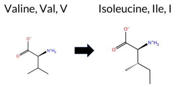 3DClick to see structure in 3D Viewer AIThe SynGAP1 V526I variant is not reported in ClinVar (ClinVar status: not listed) but is present in gnomAD (gnomAD ID: 6‑33438819‑G‑A). Prediction tools that agree on a benign effect include FoldX, Rosetta, Foldetta, premPS, PROVEAN, SIFT, and AlphaMissense‑Optimized, while those that predict a pathogenic outcome are REVEL, polyPhen‑2 HumDiv, polyPhen‑2 HumVar, ESM1b, and FATHMM; AlphaMissense‑Default remains uncertain. High‑accuracy assessments show AlphaMissense‑Optimized as benign, the SGM Consensus (majority vote from AlphaMissense‑Default, ESM1b, FATHMM, PROVEAN) as pathogenic, and Foldetta as benign. Overall, the majority of tools (seven benign vs. five pathogenic) lean toward a benign interpretation. Thus, the variant is most likely benign based on current predictions, and this assessment does not contradict the ClinVar status, which has no entry for V526I. Disclaimer: This summary was generated using AI and should be interpreted alongside expert review. | GAP | 0.139895 | Structured | 0.023118 | Uncertain | 0.943 | 0.403 | 0.000 | 6-33438819-G-A | -9.962 | Likely Pathogenic | 0.526 | Ambiguous | Likely Benign | 0.07 | Likely Benign | 0.3 | 0.41 | Likely Benign | 0.24 | Likely Benign | 0.02 | Likely Benign | 0.540 | Likely Pathogenic | -0.99 | Neutral | 0.929 | Possibly Damaging | 0.917 | Probably Damaging | -1.34 | Pathogenic | 0.15 | Tolerated | 3.37 | 35 | 0.0630 | 0.3307 | 3 | 4 | 0.3 | 14.03 | |||||||||||||||||||||||||||
| c.1606T>G | L536V 2D 3DClick to see structure in 3D Viewer AISynGAP1 missense variant L536V is listed in ClinVar (ID 1690714.0) with an uncertain significance designation and is not reported in gnomAD. Functional prediction tools cluster into two groups: benign predictions come from SIFT, AlphaMissense‑Default, and AlphaMissense‑Optimized; pathogenic predictions arise from REVEL, premPS, PROVEAN, polyPhen‑2 (HumDiv and HumVar), ESM1b, and FATHMM. The SGM‑Consensus, which aggregates AlphaMissense‑Default, ESM1b, FATHMM, and PROVEAN, reports a likely pathogenic verdict. High‑accuracy assessments show AlphaMissense‑Optimized as benign, SGM‑Consensus as likely pathogenic, and Foldetta (combining FoldX‑MD and Rosetta outputs) remains uncertain. No evidence from FoldX or Rosetta alone is available. Overall, the majority of evidence points toward a pathogenic effect, which does not contradict the ClinVar uncertain status but suggests a higher likelihood of pathogenicity. Disclaimer: This summary was generated using AI and should be interpreted alongside expert review. | Likely Pathogenic | GAP | 0.137348 | Structured | 0.042188 | Uncertain | 0.931 | 0.341 | 0.000 | Uncertain | 1 | -9.014 | Likely Pathogenic | 0.269 | Likely Benign | Likely Benign | 1.25 | Ambiguous | 0.3 | 1.22 | Ambiguous | 1.24 | Ambiguous | 1.20 | Destabilizing | 0.586 | Likely Pathogenic | -2.81 | Deleterious | 0.998 | Probably Damaging | 0.992 | Probably Damaging | -1.34 | Pathogenic | 0.09 | Tolerated | 3.37 | 34 | 0.1591 | 0.3565 | 2 | 1 | 0.4 | -14.03 | 204.7 | 26.4 | 0.2 | 0.0 | -0.2 | 0.2 | X | Potentially Benign | Leu536 is located on an α-helix (res. Ala533-Val560) at the membrane interface. The iso-butyl group of Leu536 interacts with nearby hydrophobic residues in the preceding loop (e.g., Val526, Pro528, Cys531). In the variant simulations, the iso-propyl side chain of Val536 forms similar hydrophobic interactions as Leu536 in the WT, causing no negative structural effects. | ||||||||||||||||
| c.1621G>C | A541P 2D 3DClick to see structure in 3D Viewer AIThe SynGAP1 missense variant A541P is listed in ClinVar with an uncertain significance and is not reported in gnomAD. Prediction tools that classify the variant as benign include only SIFT, whereas the remaining tools—REVEL, FoldX, Rosetta, PROVEAN, polyPhen‑2 (HumDiv and HumVar), ESM1b, FATHMM, AlphaMissense‑Default, and AlphaMissense‑Optimized—predict it to be pathogenic. The SGM Consensus, derived from a majority vote of AlphaMissense‑Default, ESM1b, FATHMM, and PROVEAN, also indicates likely pathogenic. High‑accuracy assessments further support a deleterious effect: AlphaMissense‑Optimized is pathogenic, the SGM Consensus is likely pathogenic, and Foldetta (combining FoldX‑MD and Rosetta outputs) predicts pathogenicity. No predictions are inconclusive or missing. Overall, the collective evidence points to a pathogenic effect for A541P, which is in contrast to the ClinVar designation of uncertain significance. Disclaimer: This summary was generated using AI and should be interpreted alongside expert review. | Likely Pathogenic | GAP | 0.050641 | Structured | 0.029947 | Uncertain | 0.955 | 0.365 | 0.000 | Uncertain | 1 | -14.733 | Likely Pathogenic | 0.996 | Likely Pathogenic | Likely Pathogenic | 2.47 | Destabilizing | 0.3 | 7.26 | Destabilizing | 4.87 | Destabilizing | 0.86 | Ambiguous | 0.594 | Likely Pathogenic | -3.16 | Deleterious | 1.000 | Probably Damaging | 0.998 | Probably Damaging | -1.34 | Pathogenic | 0.07 | Tolerated | 3.37 | 35 | 0.1706 | 0.2707 | 1 | -1 | -3.4 | 26.04 | 170.4 | -11.2 | 0.1 | 0.0 | 0.1 | 0.0 | X | Potentially Pathogenic | Ala541 is located on the outer surface of an α-helix (res. Ala533-Val560). The methyl group of Ala541 is on the surface and does not form any interactions. Proline lacks a free backbone amide group, and thus, Pro541 is unable to form a hydrogen bond with the carbonyl group of Ala537 in the variant simulations. Consequently, Pro541 disrupts the continuity of the secondary structure element, causing the α-helix to bend slightly in the variant simulations. | ||||||||||||||||
| c.1641T>G | C547W 2D 3DClick to see structure in 3D Viewer AIThe SynGAP1 missense variant C547W is not reported in ClinVar and is absent from gnomAD. Prediction tools that assess functional impact uniformly indicate a deleterious effect: REVEL, FoldX, Rosetta, Foldetta, PROVEAN, polyPhen‑2 (HumDiv and HumVar), SIFT, ESM1b, FATHMM, AlphaMissense‑Default, and AlphaMissense‑Optimized all classify the variant as pathogenic, while premPS remains uncertain. No tool predicts a benign outcome. High‑accuracy assessments reinforce this view: AlphaMissense‑Optimized predicts pathogenic; the SGM Consensus (majority vote of AlphaMissense‑Default, ESM1b, FATHMM, and PROVEAN) reports “Likely Pathogenic”; and Foldetta, which integrates FoldX‑MD and Rosetta stability calculations, also predicts pathogenic. Taken together, the overwhelming consensus from both general and high‑accuracy predictors is that C547W is most likely pathogenic, and this conclusion is not contradicted by any ClinVar annotation. Disclaimer: This summary was generated using AI and should be interpreted alongside expert review. | Likely Pathogenic | GAP | 0.045352 | Structured | 0.007912 | Uncertain | 0.971 | 0.275 | 0.000 | -15.788 | Likely Pathogenic | 0.999 | Likely Pathogenic | Likely Pathogenic | 11.03 | Destabilizing | 0.3 | 2.63 | Destabilizing | 6.83 | Destabilizing | 0.54 | Ambiguous | 0.764 | Likely Pathogenic | -10.64 | Deleterious | 1.000 | Probably Damaging | 0.999 | Probably Damaging | -1.34 | Pathogenic | 0.01 | Affected | 0.1854 | 0.2750 | -8 | -2 | -3.4 | 83.07 | |||||||||||||||||||||||||||||
| c.1658A>C | K553T 2D 3DClick to see structure in 3D Viewer AISynGAP1 missense variant K553T is listed in ClinVar with an uncertain significance (ClinVar ID 2007142.0) and is not reported in gnomAD. Functional prediction tools that agree on a benign effect include Rosetta and SIFT, whereas the majority of tools predict a pathogenic impact: REVEL, PROVEAN, both polyPhen‑2 HumDiv and HumVar, ESM1b, FATHMM, AlphaMissense‑Default, AlphaMissense‑Optimized, and the SGM‑Consensus (Likely Pathogenic). Uncertain results are reported by FoldX, Foldetta, and premPS. High‑accuracy assessments show AlphaMissense‑Optimized as pathogenic, the SGM‑Consensus as Likely Pathogenic, and Foldetta as inconclusive. Overall, the consensus of the available predictions indicates that K553T is most likely pathogenic, which does not contradict the current ClinVar status of uncertain significance. Disclaimer: This summary was generated using AI and should be interpreted alongside expert review. | Likely Pathogenic | GAP | 0.012270 | Structured | 0.006539 | Uncertain | 0.949 | 0.246 | 0.000 | Uncertain | 1 | -15.328 | Likely Pathogenic | 0.990 | Likely Pathogenic | Likely Pathogenic | 1.06 | Ambiguous | 0.2 | 0.48 | Likely Benign | 0.77 | Ambiguous | 0.79 | Ambiguous | 0.761 | Likely Pathogenic | -5.77 | Deleterious | 1.000 | Probably Damaging | 1.000 | Probably Damaging | -1.34 | Pathogenic | 0.14 | Tolerated | 3.37 | 35 | 0.1733 | 0.2619 | 0 | -1 | 3.2 | -27.07 | 218.2 | -10.7 | 0.0 | 0.0 | -0.2 | 0.5 | X | Potentially Pathogenic | Lys533 is located on an α-helix (res. Ala533-Val560). In the WT simulations, Lys533 packs against Phe513, and its amino side chain occasionally forms an ionic interaction with the carboxylate group of Glu512 from an opposing α-helix (res. Gln503-Tyr518). In the variant simulations, Thr533 is unable to reproduce these interactions, potentially weakening the integrity of the tertiary structure. Additionally, Thr533 forms a hydrogen bond with the backbone carbonyl group of Leu549 in the same helix, which could potentially weaken the secondary structure. Regardless, the residue swap does not cause significant structural effects based on the simulations. | ||||||||||||||||
| c.1664T>C | V555A 2D 3DClick to see structure in 3D Viewer AISynGAP1 V555A is not reported in ClinVar and is absent from gnomAD. Functional prediction tools that assess sequence conservation and structural impact uniformly indicate a deleterious effect: REVEL, premPS, PROVEAN, polyPhen‑2 (HumDiv and HumVar), SIFT, ESM1b, FATHMM, AlphaMissense‑Default, and the SGM Consensus (derived from the majority vote of AlphaMissense‑Default, ESM1b, FATHMM, and PROVEAN) all classify the variant as pathogenic. No tool predicts a benign outcome. Predictions that are inconclusive—FoldX, Rosetta, Foldetta, and AlphaMissense‑Optimized—are listed as uncertain and do not alter the overall assessment. High‑accuracy methods give a pathogenic consensus from SGM and an uncertain result from AlphaMissense‑Optimized and Foldetta. Consequently, the variant is most likely pathogenic, and this conclusion is consistent with the absence of a ClinVar entry. Disclaimer: This summary was generated using AI and should be interpreted alongside expert review. | Likely Pathogenic | GAP | 0.013265 | Structured | 0.008218 | Uncertain | 0.943 | 0.225 | 0.000 | -8.781 | Likely Pathogenic | 0.857 | Likely Pathogenic | Ambiguous | 0.91 | Ambiguous | 0.0 | 0.90 | Ambiguous | 0.91 | Ambiguous | 1.04 | Destabilizing | 0.633 | Likely Pathogenic | -3.48 | Deleterious | 0.979 | Probably Damaging | 0.956 | Probably Damaging | -1.34 | Pathogenic | 0.01 | Affected | 0.2602 | 0.1761 | 0 | 0 | -2.4 | -28.05 | |||||||||||||||||||||||||||||
| c.1682T>A | F561Y 2D 3DClick to see structure in 3D Viewer AIThe SynGAP1 missense variant F561Y is not reported in ClinVar and is absent from gnomAD. Prediction tools that agree on a deleterious effect include REVEL, premPS, PROVEAN, polyPhen‑2 (HumDiv and HumVar), SIFT, ESM1b, FATHMM, AlphaMissense‑Default, and the SGM‑Consensus (majority vote of AlphaMissense‑Default, ESM1b, FATHMM, PROVEAN). No tool predicts a benign outcome; the remaining predictions (FoldX, Rosetta, Foldetta, AlphaMissense‑Optimized) are uncertain or unavailable. High‑accuracy assessments show AlphaMissense‑Optimized as uncertain, SGM‑Consensus as pathogenic, and Foldetta (combining FoldX‑MD and Rosetta outputs) as uncertain. Based on the consensus of the majority of evidence‑based predictors, the variant is most likely pathogenic, and this conclusion does not contradict any ClinVar annotation (none is available). Disclaimer: This summary was generated using AI and should be interpreted alongside expert review. | Likely Pathogenic | GAP | 0.024826 | Structured | 0.018013 | Uncertain | 0.903 | 0.196 | 0.000 | -12.692 | Likely Pathogenic | 0.950 | Likely Pathogenic | Ambiguous | 1.54 | Ambiguous | 0.1 | 0.75 | Ambiguous | 1.15 | Ambiguous | 1.28 | Destabilizing | 0.693 | Likely Pathogenic | -2.99 | Deleterious | 0.988 | Probably Damaging | 0.976 | Probably Damaging | -1.34 | Pathogenic | 0.01 | Affected | 0.1344 | 0.1120 | 7 | 3 | -4.1 | 16.00 | |||||||||||||||||||||||||||||
| c.1729G>C | A577P 2D 3DClick to see structure in 3D Viewer AIThe SynGAP1 missense variant A577P is not reported in ClinVar and is absent from gnomAD. Prediction tools that indicate a benign effect include only SIFT, whereas the remaining evaluated algorithms (SGM‑Consensus, REVEL, FoldX, Rosetta, Foldetta, PROVEAN, polyPhen‑2 HumDiv, polyPhen‑2 HumVar, ESM1b, FATHMM, AlphaMissense‑Default, AlphaMissense‑Optimized) all predict a pathogenic outcome; premPS is inconclusive and is therefore not counted. High‑accuracy assessments further support a deleterious impact: AlphaMissense‑Optimized predicts pathogenic, the SGM‑Consensus (majority vote of AlphaMissense‑Default, ESM1b, FATHMM, PROVEAN) is likely pathogenic, and Foldetta (combining FoldX‑MD and Rosetta outputs) also predicts pathogenic. Based on the consensus of these predictions, the variant is most likely pathogenic, and this conclusion does not contradict the ClinVar status, which currently has no entry for this variant. Disclaimer: This summary was generated using AI and should be interpreted alongside expert review. | Likely Pathogenic | GAP | 0.113710 | Structured | 0.019074 | Uncertain | 0.913 | 0.239 | 0.000 | -9.009 | Likely Pathogenic | 0.995 | Likely Pathogenic | Likely Pathogenic | 3.93 | Destabilizing | 0.2 | 9.88 | Destabilizing | 6.91 | Destabilizing | 0.87 | Ambiguous | 0.585 | Likely Pathogenic | -2.72 | Deleterious | 1.000 | Probably Damaging | 0.998 | Probably Damaging | -1.34 | Pathogenic | 0.20 | Tolerated | 0.2256 | 0.4600 | 1 | -1 | -3.4 | 26.04 | |||||||||||||||||||||||||||||
| c.1736G>A | R579Q 2D 3DClick to see structure in 3D Viewer AISynGAP1 missense variant R579Q is listed in ClinVar with an uncertain significance (ClinVar ID 3964539) and is present in gnomAD (6‑33440788‑G‑A). Prediction tools that indicate a benign effect include SIFT and AlphaMissense‑Optimized, whereas the remaining tools (REVEL, premPS, PROVEAN, polyPhen‑2 HumDiv, polyPhen‑2 HumVar, ESM1b, FATHMM, AlphaMissense‑Default) predict a pathogenic outcome. High‑accuracy assessments show AlphaMissense‑Optimized as benign, the SGM Consensus (majority vote of AlphaMissense‑Default, ESM1b, FATHMM, PROVEAN) as pathogenic, and Foldetta (combining FoldX‑MD and Rosetta outputs) as uncertain. FoldX and Rosetta individually also return uncertain results. Overall, the majority of evidence points to a pathogenic effect, which does not contradict the ClinVar uncertain status. Therefore, the variant is most likely pathogenic based on current computational predictions. Disclaimer: This summary was generated using AI and should be interpreted alongside expert review. | Likely Pathogenic | GAP | 0.053060 | Structured | 0.022872 | Uncertain | 0.877 | 0.244 | 0.000 | Uncertain | 2 | 6-33440788-G-A | 18 | 1.12e-5 | -9.193 | Likely Pathogenic | 0.690 | Likely Pathogenic | Likely Benign | 0.65 | Ambiguous | 0.1 | 0.70 | Ambiguous | 0.68 | Ambiguous | 1.13 | Destabilizing | 0.673 | Likely Pathogenic | -3.31 | Deleterious | 1.000 | Probably Damaging | 0.995 | Probably Damaging | -1.34 | Pathogenic | 0.06 | Tolerated | 3.37 | 34 | 0.2677 | 0.1334 | 1 | 1 | 1.0 | -28.06 | ||||||||||||||||||||||
| c.1828C>A | L610I 2D 3DClick to see structure in 3D Viewer AIThe SynGAP1 missense variant L610I is listed in gnomAD (ID 6‑33440880‑C‑A) but has no ClinVar entry. Functional prediction tools cluster into two groups: benign predictions come from PROVEAN, SIFT, ESM1b, and AlphaMissense‑Optimized, while pathogenic predictions arise from REVEL, polyPhen‑2 (HumDiv and HumVar) and FATHMM. Four tools are uncertain (FoldX, Foldetta, premPS, AlphaMissense‑Default). High‑accuracy assessments further support a benign outcome: AlphaMissense‑Optimized scores benign, the SGM consensus (majority vote of AlphaMissense‑Default, ESM1b, FATHMM, PROVEAN) also favors benign, whereas Foldetta’s stability estimate is unavailable. Overall, the balance of evidence points to a benign effect for L610I, and this conclusion does not conflict with the absence of a ClinVar classification. Disclaimer: This summary was generated using AI and should be interpreted alongside expert review. | GAP | 0.271506 | Structured | 0.209504 | Uncertain | 0.888 | 0.253 | 0.000 | 6-33440880-C-A | 1 | 6.19e-7 | -6.362 | Likely Benign | 0.389 | Ambiguous | Likely Benign | 1.50 | Ambiguous | 0.2 | 0.18 | Likely Benign | 0.84 | Ambiguous | 0.76 | Ambiguous | 0.544 | Likely Pathogenic | -1.86 | Neutral | 0.992 | Probably Damaging | 0.997 | Probably Damaging | -1.34 | Pathogenic | 0.15 | Tolerated | 3.37 | 35 | 0.0999 | 0.3219 | 2 | 2 | 0.7 | 0.00 | |||||||||||||||||||||||||
| c.1849G>A | E617K 2D 3DClick to see structure in 3D Viewer AIThe SynGAP1 missense variant E617K is not reported in ClinVar but is present in gnomAD (6‑33440901‑G‑A). Functional prediction tools cluster into two groups: benign predictions come from FoldX, premPS, and SIFT, while pathogenic predictions arise from SGM‑Consensus, REVEL, PROVEAN, polyPhen‑2 HumDiv, polyPhen‑2 HumVar, ESM1b, FATHMM, and AlphaMissense‑Default. A third set of methods (Foldetta, AlphaMissense‑Optimized, Rosetta) yield uncertain results. High‑accuracy assessments show AlphaMissense‑Optimized as uncertain, SGM‑Consensus (majority vote of AlphaMissense‑Default, ESM1b, FATHMM, PROVEAN) as pathogenic, and Foldetta as uncertain. Overall, the majority of evidence points to a pathogenic effect for E617K, and this conclusion is not contradicted by any ClinVar annotation. Disclaimer: This summary was generated using AI and should be interpreted alongside expert review. | Likely Pathogenic | GAP | 0.111485 | Structured | 0.155123 | Uncertain | 0.877 | 0.240 | 0.000 | 6-33440901-G-A | 1 | 6.20e-7 | -10.702 | Likely Pathogenic | 0.910 | Likely Pathogenic | Ambiguous | 0.37 | Likely Benign | 0.1 | 1.19 | Ambiguous | 0.78 | Ambiguous | 0.17 | Likely Benign | 0.534 | Likely Pathogenic | -3.32 | Deleterious | 0.997 | Probably Damaging | 0.987 | Probably Damaging | -1.34 | Pathogenic | 0.48 | Tolerated | 3.37 | 35 | 0.1981 | 0.6282 | 1 | 0 | -0.4 | -0.94 | ||||||||||||||||||||||||
| c.1405G>C | A469P 2D 3DClick to see structure in 3D Viewer AIThe SynGAP1 missense variant A469P is not reported in ClinVar (no ClinVar ID) and is absent from gnomAD (no gnomAD ID). Prediction tools that agree on a benign effect include only SIFT, whereas all other evaluated algorithms—REVEL, FoldX, Rosetta, Foldetta, premPS, PROVEAN, polyPhen‑2 (HumDiv and HumVar), ESM1b, FATHMM, AlphaMissense‑Default, and AlphaMissense‑Optimized—classify the variant as pathogenic. High‑accuracy assessments further support a deleterious impact: AlphaMissense‑Optimized predicts pathogenicity; the SGM Consensus (majority vote of AlphaMissense‑Default, ESM1b, FATHMM, and PROVEAN) indicates likely pathogenic; and Foldetta, which integrates FoldX‑MD and Rosetta stability outputs, also predicts pathogenicity. Based on the overwhelming consensus of these predictions, the variant is most likely pathogenic, and this conclusion does not contradict any ClinVar status because no ClinVar entry exists. Disclaimer: This summary was generated using AI and should be interpreted alongside expert review. | Likely Pathogenic | GAP | 0.278302 | Structured | 0.343926 | Uncertain | 0.910 | 0.276 | 0.000 | -16.072 | Likely Pathogenic | 0.997 | Likely Pathogenic | Likely Pathogenic | 5.06 | Destabilizing | 0.3 | 8.83 | Destabilizing | 6.95 | Destabilizing | 1.02 | Destabilizing | 0.774 | Likely Pathogenic | -2.69 | Deleterious | 0.999 | Probably Damaging | 0.996 | Probably Damaging | -1.33 | Pathogenic | 0.21 | Tolerated | 0.1606 | 0.4092 | 1 | -1 | -3.4 | 26.04 | |||||||||||||||||||||||||||||
| c.1564G>C | E522Q 2D 3DClick to see structure in 3D Viewer AIThe SynGAP1 missense variant E522Q is not reported in ClinVar and has no entry in gnomAD. Functional prediction tools show a split: benign predictions come from FoldX, Rosetta, Foldetta, premPS, and SIFT, whereas pathogenic predictions arise from REVEL, PROVEAN, polyPhen‑2 (HumDiv and HumVar), ESM1b, FATHMM, AlphaMissense‑Default, and AlphaMissense‑Optimized. The high‑accuracy consensus methods reinforce this pattern: AlphaMissense‑Optimized classifies the change as pathogenic, the SGM Consensus (majority vote of AlphaMissense‑Default, ESM1b, FATHMM, and PROVEAN) also labels it likely pathogenic, while Foldetta predicts a benign effect on protein stability. Overall, the majority of evidence points to a pathogenic impact. This assessment does not conflict with ClinVar, which currently contains no classification for this variant. Disclaimer: This summary was generated using AI and should be interpreted alongside expert review. | Likely Pathogenic | GAP | 0.092881 | Structured | 0.046216 | Uncertain | 0.823 | 0.376 | 0.000 | -10.861 | Likely Pathogenic | 0.987 | Likely Pathogenic | Likely Pathogenic | -0.13 | Likely Benign | 0.1 | -0.06 | Likely Benign | -0.10 | Likely Benign | 0.03 | Likely Benign | 0.560 | Likely Pathogenic | -2.85 | Deleterious | 0.992 | Probably Damaging | 0.993 | Probably Damaging | -1.33 | Pathogenic | 0.07 | Tolerated | 0.0944 | 0.4130 | 2 | 2 | 0.0 | -0.98 | |||||||||||||||||||||||||||||
| c.1566A>C | E522D 2D 3DClick to see structure in 3D Viewer AIThe SynGAP1 E522D missense variant is not reported in ClinVar and has no gnomAD entry. Prediction tools cluster into two groups: benign predictions come from REVEL, FoldX, Rosetta, premPS, SIFT, and ESM1b, while pathogenic predictions arise from SGM‑Consensus, PROVEAN, polyPhen‑2 (HumDiv and HumVar), FATHMM, AlphaMissense‑Default, and AlphaMissense‑Optimized. High‑accuracy assessments further support a pathogenic bias: AlphaMissense‑Optimized is pathogenic, the SGM‑Consensus (majority vote of AlphaMissense‑Default, ESM1b, FATHMM, PROVEAN) is likely pathogenic, whereas Foldetta, which integrates FoldX‑MD and Rosetta stability outputs, indicates a benign effect. Overall, the balance of evidence points to a likely pathogenic impact for E522D, and this assessment does not contradict the absence of a ClinVar classification. Disclaimer: This summary was generated using AI and should be interpreted alongside expert review. | Likely Pathogenic | GAP | 0.092881 | Structured | 0.046216 | Uncertain | 0.823 | 0.376 | 0.000 | -5.385 | Likely Benign | 0.984 | Likely Pathogenic | Likely Pathogenic | 0.04 | Likely Benign | 0.1 | -0.02 | Likely Benign | 0.01 | Likely Benign | 0.32 | Likely Benign | 0.494 | Likely Benign | -2.65 | Deleterious | 0.986 | Probably Damaging | 0.989 | Probably Damaging | -1.33 | Pathogenic | 0.10 | Tolerated | 0.1499 | 0.2765 | 3 | 2 | 0.0 | -14.03 | |||||||||||||||||||||||||||||
| c.1566A>T | E522D 2D 3DClick to see structure in 3D Viewer AIThe SynGAP1 E522D missense variant is not reported in ClinVar and has no gnomAD entry. Prediction tools cluster into two groups: benign predictions come from REVEL, FoldX, Rosetta, premPS, SIFT, and ESM1b, while pathogenic predictions arise from SGM‑Consensus, PROVEAN, polyPhen‑2 (HumDiv and HumVar), FATHMM, AlphaMissense‑Default, and AlphaMissense‑Optimized. High‑accuracy assessments further support a pathogenic bias: AlphaMissense‑Optimized is pathogenic, the SGM‑Consensus (majority vote of AlphaMissense‑Default, ESM1b, FATHMM, PROVEAN) is likely pathogenic, whereas Foldetta, which integrates FoldX‑MD and Rosetta stability outputs, indicates a benign effect. Overall, the balance of evidence points to a likely pathogenic impact for E522D, and this assessment does not contradict the absence of a ClinVar classification. Disclaimer: This summary was generated using AI and should be interpreted alongside expert review. | Likely Pathogenic | GAP | 0.092881 | Structured | 0.046216 | Uncertain | 0.823 | 0.376 | 0.000 | -5.385 | Likely Benign | 0.984 | Likely Pathogenic | Likely Pathogenic | 0.04 | Likely Benign | 0.1 | -0.02 | Likely Benign | 0.01 | Likely Benign | 0.32 | Likely Benign | 0.494 | Likely Benign | -2.65 | Deleterious | 0.986 | Probably Damaging | 0.989 | Probably Damaging | -1.33 | Pathogenic | 0.10 | Tolerated | 0.1499 | 0.2765 | 3 | 2 | 0.0 | -14.03 | |||||||||||||||||||||||||||||
| c.1603A>T | S535C 2D 3DClick to see structure in 3D Viewer AISynGAP1 missense variant S535C is not reported in ClinVar (ClinVar ID: None) and is absent from gnomAD (gnomAD ID: None). Prediction tools that classify the variant as benign include Rosetta, Foldetta, premPS, polyPhen‑2 HumVar, AlphaMissense‑Default, and AlphaMissense‑Optimized. Those that predict pathogenicity are REVEL, PROVEAN, polyPhen‑2 HumDiv, SIFT, and FATHMM. Two tools, FoldX and ESM1b, return uncertain results. High‑accuracy assessments show AlphaMissense‑Optimized as benign, Foldetta as benign, and the SGM Consensus (majority vote from AlphaMissense‑Default, ESM1b, FATHMM, PROVEAN) as pathogenic. Overall, the majority of predictions lean toward a benign effect, and this conclusion is not contradicted by ClinVar status. Disclaimer: This summary was generated using AI and should be interpreted alongside expert review. | GAP | 0.236433 | Structured | 0.041365 | Uncertain | 0.918 | 0.343 | 0.000 | -7.526 | In-Between | 0.165 | Likely Benign | Likely Benign | 0.57 | Ambiguous | 0.1 | 0.25 | Likely Benign | 0.41 | Likely Benign | 0.47 | Likely Benign | 0.500 | Likely Pathogenic | -2.96 | Deleterious | 0.933 | Possibly Damaging | 0.419 | Benign | -1.33 | Pathogenic | 0.02 | Affected | 0.1139 | 0.5633 | 0 | -1 | 3.3 | 16.06 | ||||||||||||||||||||||||||||||
| c.1622C>T | A541V 2D  3DClick to see structure in 3D Viewer AIThe SynGAP1 missense variant A541V is not reported in ClinVar and is absent from gnomAD. Benign predictions come from REVEL, Foldetta, premPS, SIFT, Rosetta, and AlphaMissense‑Optimized, whereas pathogenic predictions come from SGM‑Consensus, PROVEAN, polyPhen2_HumDiv, polyPhen2_HumVar, ESM1b, FATHMM, and AlphaMissense‑Default. High‑accuracy tools give mixed results: AlphaMissense‑Optimized predicts benign, SGM‑Consensus (majority vote of AlphaMissense‑Default, ESM1b, FATHMM, PROVEAN) predicts pathogenic, and Foldetta predicts benign. FoldX is uncertain and therefore not considered. Overall, the majority of tools predict pathogenicity, and this assessment is not contradicted by ClinVar status, which has no entry for this variant. Disclaimer: This summary was generated using AI and should be interpreted alongside expert review. | Likely Pathogenic | GAP | 0.050641 | Structured | 0.029947 | Uncertain | 0.955 | 0.365 | 0.000 | -9.777 | Likely Pathogenic | 0.762 | Likely Pathogenic | Likely Benign | 0.63 | Ambiguous | 0.1 | 0.06 | Likely Benign | 0.35 | Likely Benign | 0.43 | Likely Benign | 0.497 | Likely Benign | -3.09 | Deleterious | 0.999 | Probably Damaging | 0.988 | Probably Damaging | -1.33 | Pathogenic | 0.06 | Tolerated | 0.0985 | 0.3648 | 0 | 0 | 2.4 | 28.05 | |||||||||||||||||||||||||||||
| c.1639T>C | C547R 2D 3DClick to see structure in 3D Viewer AIThe SynGAP1 missense variant C547R is listed in ClinVar with an “Uncertain” status and is not reported in gnomAD. Prediction tools that assess pathogenicity all converge on a deleterious effect: REVEL, FoldX, Rosetta, Foldetta, premPS, PROVEAN, polyPhen‑2 (HumDiv and HumVar), SIFT, ESM1b, FATHMM, AlphaMissense‑Default, and AlphaMissense‑Optimized all predict pathogenicity, while no tool in the dataset predicts a benign outcome. High‑accuracy methods reinforce this view: AlphaMissense‑Optimized is pathogenic; the SGM Consensus (majority vote of AlphaMissense‑Default, ESM1b, FATHMM, PROVEAN) is “Likely Pathogenic”; and Foldetta, which integrates FoldX‑MD and Rosetta stability calculations, also predicts pathogenicity. Based on the unanimous computational evidence, the variant is most likely pathogenic, a conclusion that contradicts the current ClinVar “Uncertain” classification. Disclaimer: This summary was generated using AI and should be interpreted alongside expert review. | Likely Pathogenic | GAP | 0.045352 | Structured | 0.007912 | Uncertain | 0.971 | 0.275 | 0.000 | Uncertain | 1 | -16.967 | Likely Pathogenic | 0.999 | Likely Pathogenic | Likely Pathogenic | 7.76 | Destabilizing | 0.8 | 5.83 | Destabilizing | 6.80 | Destabilizing | 1.69 | Destabilizing | 0.900 | Likely Pathogenic | -11.60 | Deleterious | 1.000 | Probably Damaging | 0.998 | Probably Damaging | -1.33 | Pathogenic | 0.02 | Affected | 3.37 | 35 | 0.1802 | 0.1408 | -4 | -3 | -7.0 | 53.05 | 267.4 | -90.3 | 0.0 | 0.0 | -0.1 | 0.1 | X | X | X | X | Potentially Pathogenic | Cys547 is located in an α-helix (res. Ala533-Val560). The thiol side chain of Cys is situated in a hydrophobic inter-helix space, where it packs hydrophobically with other residues such as Ile626, Leu551, and Phe652. Additionally, the thiol side chain of Cys547 weakly hydrogen bonds with the carbonyl group of Leu543 in the same α-helix. In the variant simulations, the bulkier, positively charged guanidinium group of Arg547 must rotate out of the hydrophobic space. Consequently, it forms ionic interactions with the carboxylate groups of Glu548 in the same helix and Glu656 in the neighboring α-helix (res. Glu666-Asp644). This causes the two helices to slightly separate, significantly affecting the secondary structure integrity of the latter helix. These negative structural effects could be more pronounced during protein folding and are likely to be undermined in the MD simulations. | |||||||||||||
| c.1640G>A | C547Y 2D 3DClick to see structure in 3D Viewer AIThe SynGAP1 missense variant C547Y (ClinVar ID 1404191.0) is listed as Pathogenic in ClinVar and is not reported in gnomAD. Prediction tools that agree on a benign effect include only SIFT, whereas the remaining tools—REVEL, FoldX, Rosetta, Foldetta, premPS (uncertain), PROVEAN, polyPhen‑2 (HumDiv and HumVar), ESM1b, FATHMM, AlphaMissense‑Default, and AlphaMissense‑Optimized—consistently predict a pathogenic impact. High‑accuracy assessments further support this: AlphaMissense‑Optimized is pathogenic; the SGM Consensus (majority vote from AlphaMissense‑Default, ESM1b, FATHMM, and PROVEAN) is Likely Pathogenic; and Foldetta, which integrates FoldX‑MD and Rosetta outputs, is pathogenic. Taken together, the overwhelming majority of computational evidence indicates a pathogenic effect, which is in agreement with the ClinVar classification. Thus, the variant is most likely pathogenic and does not contradict the ClinVar status. Disclaimer: This summary was generated using AI and should be interpreted alongside expert review. | Likely Pathogenic | GAP | 0.045352 | Structured | 0.007912 | Uncertain | 0.971 | 0.275 | 0.000 | Pathogenic | 1 | -15.871 | Likely Pathogenic | 0.999 | Likely Pathogenic | Likely Pathogenic | 8.53 | Destabilizing | 1.8 | 6.20 | Destabilizing | 7.37 | Destabilizing | 0.62 | Ambiguous | 0.874 | Likely Pathogenic | -10.57 | Deleterious | 1.000 | Probably Damaging | 0.998 | Probably Damaging | -1.33 | Pathogenic | 0.06 | Tolerated | 3.37 | 35 | 0.1393 | 0.2742 | 0 | -2 | -3.8 | 60.04 | 280.1 | -54.8 | 0.0 | 0.0 | 0.0 | 0.0 | X | X | X | Potentially Pathogenic | Cys547 is located in an α-helix (res. Ala533-Val560). The thiol side chain of Cys547 is situated in a hydrophobic inter-helix space, where it packs hydrophobically with other residues such as Ile626, Leu551, and Phe652. Additionally, the thiol side chain of Cys weakly hydrogen bonds with the carbonyl group of Leu543 in the same α-helix. In the variant simulations, the bulkier phenol ring of Tyr547, with its polar hydroxyl group, is less suited for the hydrophobic space. Consequently, it moves outside and forms a hydrogen bond with the carbonyl group of Phe652 in the neighboring α-helix (res. Glu666-Asp644). This causes the two helices to slightly separate, negatively affecting the secondary structure integrity of the latter helix. These negative structural effects could be more pronounced during protein folding and are likely to be undermined in the MD simulations. | ||||||||||||||
| c.1666A>G | N556D 2D 3DClick to see structure in 3D Viewer AIThe SynGAP1 missense variant N556D is not reported in ClinVar (ClinVar status: None) and has no entry in gnomAD (gnomAD ID: None). Prediction tools that classify the variant as benign include FoldX, Rosetta, Foldetta, premPS, SIFT, ESM1b, and AlphaMissense‑Optimized. Those that predict pathogenicity are SGM‑Consensus, REVEL, PROVEAN, polyPhen‑2 HumDiv, polyPhen‑2 HumVar, FATHMM, and AlphaMissense‑Default. High‑accuracy assessments show AlphaMissense‑Optimized as benign, SGM‑Consensus (majority vote from AlphaMissense‑Default, ESM1b, FATHMM, PROVEAN) as likely pathogenic, and Foldetta (combining FoldX‑MD and Rosetta outputs) as benign. With two of the three high‑accuracy tools indicating benign and an equal split among the broader set of predictors, the variant is most likely benign. This conclusion does not contradict the ClinVar status, which currently has no classification for this variant. Disclaimer: This summary was generated using AI and should be interpreted alongside expert review. | Likely Pathogenic | GAP | 0.015078 | Structured | 0.008655 | Uncertain | 0.925 | 0.225 | 0.000 | -6.787 | Likely Benign | 0.704 | Likely Pathogenic | Likely Benign | 0.41 | Likely Benign | 0.0 | 0.39 | Likely Benign | 0.40 | Likely Benign | 0.33 | Likely Benign | 0.546 | Likely Pathogenic | -3.19 | Deleterious | 1.000 | Probably Damaging | 0.997 | Probably Damaging | -1.33 | Pathogenic | 0.08 | Tolerated | 0.1817 | 0.1625 | 2 | 1 | 0.0 | 0.98 | |||||||||||||||||||||||||||||
| c.1682T>C | F561S 2D 3DClick to see structure in 3D Viewer AIThe SynGAP1 missense variant F561S is not reported in ClinVar and is absent from gnomAD. Prediction tools that assess functional impact uniformly indicate a deleterious effect. Benign predictions: none. Pathogenic predictions: SGM‑Consensus (Likely Pathogenic), REVEL, FoldX, Rosetta, Foldetta, premPS, PROVEAN, polyPhen‑2 (HumDiv and HumVar), SIFT, ESM1b, FATHMM, AlphaMissense‑Default, and AlphaMissense‑Optimized. High‑accuracy assessments corroborate this: AlphaMissense‑Optimized predicts pathogenic; the SGM Consensus (majority vote of AlphaMissense‑Default, ESM1b, FATHMM, PROVEAN) is Likely Pathogenic; Foldetta, which integrates FoldX‑MD and Rosetta outputs, also predicts pathogenic. No predictions are inconclusive. Therefore, the variant is most likely pathogenic, and this assessment does not contradict any ClinVar status because none is available. Disclaimer: This summary was generated using AI and should be interpreted alongside expert review. | Likely Pathogenic | GAP | 0.024826 | Structured | 0.018013 | Uncertain | 0.903 | 0.196 | 0.000 | -13.515 | Likely Pathogenic | 1.000 | Likely Pathogenic | Likely Pathogenic | 4.75 | Destabilizing | 0.0 | 4.54 | Destabilizing | 4.65 | Destabilizing | 2.08 | Destabilizing | 0.803 | Likely Pathogenic | -7.96 | Deleterious | 1.000 | Probably Damaging | 1.000 | Probably Damaging | -1.33 | Pathogenic | 0.00 | Affected | 0.4142 | 0.0200 | -3 | -2 | -3.6 | -60.10 | |||||||||||||||||||||||||||||
| c.1691A>C | E564A 2D 3DClick to see structure in 3D Viewer AIThe SynGAP1 missense variant E564A is not reported in ClinVar (ClinVar status: None) and is absent from gnomAD (gnomAD ID: None). Prediction tools that agree on a benign effect include premPS and SIFT, whereas the majority of tools predict a pathogenic impact: SGM‑Consensus, REVEL, PROVEAN, polyPhen‑2 (HumDiv and HumVar), ESM1b, FATHMM, AlphaMissense‑Default, and AlphaMissense‑Optimized. Uncertain results are reported by FoldX, Rosetta, and Foldetta. High‑accuracy assessments reinforce the pathogenic prediction: AlphaMissense‑Optimized is pathogenic, the SGM Consensus (majority vote of AlphaMissense‑Default, ESM1b, FATHMM, and PROVEAN) is pathogenic, and Foldetta remains uncertain. Overall, the preponderance of evidence from multiple independent predictors indicates that the variant is most likely pathogenic, and this conclusion does not contradict the ClinVar status, which currently has no classification for this variant. Disclaimer: This summary was generated using AI and should be interpreted alongside expert review. | Likely Pathogenic | GAP | 0.023534 | Structured | 0.038418 | Uncertain | 0.891 | 0.208 | 0.000 | -14.506 | Likely Pathogenic | 0.957 | Likely Pathogenic | Likely Pathogenic | 0.64 | Ambiguous | 0.1 | 1.49 | Ambiguous | 1.07 | Ambiguous | 0.24 | Likely Benign | 0.765 | Likely Pathogenic | -5.91 | Deleterious | 0.999 | Probably Damaging | 0.998 | Probably Damaging | -1.33 | Pathogenic | 0.12 | Tolerated | 0.3201 | 0.4918 | 0 | -1 | 5.3 | -58.04 | |||||||||||||||||||||||||||||
| c.1724G>A | R575H 2D 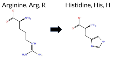 3DClick to see structure in 3D Viewer AISynGAP1 missense variant R575H (ClinVar ID 1029088.0) is listed as Uncertain in ClinVar and is present in gnomAD (ID 6‑33440776‑G‑A). Prediction tools that indicate a benign effect include Rosetta, Foldetta, PROVEAN, and AlphaMissense‑Optimized. Tools that predict a pathogenic effect are REVEL, premPS, polyPhen‑2 (HumDiv and HumVar), SIFT, ESM1b, and FATHMM. The SGM Consensus, derived from a majority vote of AlphaMissense‑Default, ESM1b, FATHMM, and PROVEAN, classifies the variant as Pathogenic. High‑accuracy assessments show AlphaMissense‑Optimized as Benign, Foldetta as Benign, and the SGM Consensus as Pathogenic. Overall, the majority of evidence points to a pathogenic impact, which contrasts with the ClinVar designation of Uncertain. Disclaimer: This summary was generated using AI and should be interpreted alongside expert review. | GAP | 0.061840 | Structured | 0.021362 | Uncertain | 0.916 | 0.259 | 0.000 | Conflicting | 4 | 6-33440776-G-A | 204 | 1.27e-4 | -11.142 | Likely Pathogenic | 0.496 | Ambiguous | Likely Benign | 0.81 | Ambiguous | 0.2 | -0.22 | Likely Benign | 0.30 | Likely Benign | 1.31 | Destabilizing | 0.707 | Likely Pathogenic | -2.34 | Neutral | 1.000 | Probably Damaging | 0.998 | Probably Damaging | -1.33 | Pathogenic | 0.05 | Affected | 3.37 | 35 | 0.2361 | 0.1292 | 2 | 0 | 1.3 | -19.05 | 244.7 | 80.6 | 0.0 | 0.0 | 0.3 | 0.0 | X | Potentially Pathogenic | The guanidinium group of Arg575, located in an α-helix (res. Arg563-Glu578), forms salt bridges with the carboxylate groups of Asp463 and Asp467, and it also hydrogen bonds with the hydroxyl group of Ser466 on an opposing α-helix (res. Ala461-Phe476) in the WT simulations. In the variant simulations, the imidazole ring of His575 (in its neutral epsilon protonated form) cannot form the same salt bridges as the guanidinium group of the non-mutated Arg575. Instead, His575 only forms weak hydrogen bonds with the hydroxyl groups of Ser466 and Ser571. Overall, the residue swap has the potential to substantially affect the tertiary structure assembly during the protein folding process. | ||||||||||||||
| c.1724G>C | R575P 2D 3DClick to see structure in 3D Viewer AIThe SynGAP1 missense variant R575P is not reported in ClinVar (no ClinVar ID) and is absent from gnomAD (no gnomAD ID). Prediction tools that agree on a benign effect include only SIFT, whereas all other evaluated algorithms—REVEL, FoldX, Rosetta, Foldetta, premPS, PROVEAN, polyPhen‑2 (HumDiv and HumVar), ESM1b, FATHMM, AlphaMissense‑Default, and AlphaMissense‑Optimized—classify the variant as pathogenic. High‑accuracy assessments further support a deleterious impact: AlphaMissense‑Optimized predicts pathogenicity; the SGM Consensus (majority vote of AlphaMissense‑Default, ESM1b, FATHMM, and PROVEAN) indicates likely pathogenic; and Foldetta, which integrates FoldX‑MD and Rosetta stability outputs, also predicts pathogenicity. Based on the overwhelming consensus of pathogenic predictions and the absence of any benign consensus, the variant is most likely pathogenic. This conclusion is not contradicted by ClinVar status, as no ClinVar entry exists for R575P. Disclaimer: This summary was generated using AI and should be interpreted alongside expert review. | Likely Pathogenic | GAP | 0.061840 | Structured | 0.021362 | Uncertain | 0.916 | 0.259 | 0.000 | -16.008 | Likely Pathogenic | 0.996 | Likely Pathogenic | Likely Pathogenic | 3.50 | Destabilizing | 0.1 | 4.97 | Destabilizing | 4.24 | Destabilizing | 1.13 | Destabilizing | 0.774 | Likely Pathogenic | -3.69 | Deleterious | 1.000 | Probably Damaging | 1.000 | Probably Damaging | -1.33 | Pathogenic | 0.10 | Tolerated | 0.2080 | 0.2670 | 0 | -2 | 2.9 | -59.07 | |||||||||||||||||||||||||||||
| c.1742G>T | R581L 2D 3DClick to see structure in 3D Viewer AISynGAP1 missense variant R581L is not reported in ClinVar and is present in gnomAD (ID 6‑33440794‑G‑T). Prediction tools that classify the variant as benign include FoldX, Rosetta, Foldetta, premPS, and SIFT, whereas tools that predict pathogenicity are REVEL, PROVEAN, polyPhen‑2 HumDiv, polyPhen‑2 HumVar, ESM1b, FATHMM, AlphaMissense‑Default, and AlphaMissense‑Optimized. The high‑accuracy AlphaMissense‑Optimized score is pathogenic. The SGM‑Consensus, derived from a majority vote of AlphaMissense‑Default, ESM1b, FATHMM, and PROVEAN, also indicates pathogenicity. In contrast, the Foldetta stability assessment, which integrates FoldX‑MD and Rosetta outputs, predicts a benign effect. Overall, the majority of computational evidence supports a pathogenic classification for R581L, and this conclusion does not contradict the ClinVar status, which currently has no entry for the variant. Disclaimer: This summary was generated using AI and should be interpreted alongside expert review. | Likely Pathogenic | GAP | 0.104810 | Structured | 0.029544 | Uncertain | 0.829 | 0.236 | 0.000 | 6-33440794-G-T | 3 | 1.86e-6 | -10.134 | Likely Pathogenic | 0.958 | Likely Pathogenic | Likely Pathogenic | 0.29 | Likely Benign | 0.1 | -0.20 | Likely Benign | 0.05 | Likely Benign | 0.45 | Likely Benign | 0.654 | Likely Pathogenic | -5.93 | Deleterious | 1.000 | Probably Damaging | 1.000 | Probably Damaging | -1.33 | Pathogenic | 0.08 | Tolerated | 3.37 | 34 | 0.1550 | 0.3483 | -2 | -3 | 8.3 | -43.03 | ||||||||||||||||||||||||
| c.1753G>C | A585P 2D 3DClick to see structure in 3D Viewer AIThe SynGAP1 missense variant A585P is not reported in ClinVar (ClinVar ID None) and has no entries in gnomAD (gnomAD ID None). Prediction tools cluster into two groups: the single benign prediction comes from SIFT, while all other evaluated algorithms—REVEL, FoldX, Rosetta, Foldetta, PROVEAN, polyPhen‑2 (HumDiv and HumVar), ESM1b, FATHMM, AlphaMissense‑Default, and AlphaMissense‑Optimized—consistently predict pathogenicity. High‑accuracy assessments further support a deleterious effect: AlphaMissense‑Optimized scores the variant as pathogenic; the SGM Consensus, derived from a majority vote of AlphaMissense‑Default, ESM1b, FATHMM, and PROVEAN, labels it “Likely Pathogenic”; and Foldetta, which integrates FoldX‑MD and Rosetta stability calculations, also predicts a pathogenic outcome. Based on the convergence of these predictions, the variant is most likely pathogenic, and this assessment does not contradict any ClinVar status because no ClinVar entry exists. Disclaimer: This summary was generated using AI and should be interpreted alongside expert review. | Likely Pathogenic | GAP | 0.060549 | Structured | 0.055884 | Uncertain | 0.880 | 0.244 | 0.000 | -10.999 | Likely Pathogenic | 0.988 | Likely Pathogenic | Likely Pathogenic | 5.44 | Destabilizing | 0.1 | 5.92 | Destabilizing | 5.68 | Destabilizing | 0.77 | Ambiguous | 0.549 | Likely Pathogenic | -3.09 | Deleterious | 1.000 | Probably Damaging | 0.999 | Probably Damaging | -1.33 | Pathogenic | 0.16 | Tolerated | 0.1884 | 0.2943 | 1 | -1 | -3.4 | 26.04 | |||||||||||||||||||||||||||||
| c.1754C>G | A585G 2D 3DClick to see structure in 3D Viewer AIThe SynGAP1 missense variant A585G is not reported in ClinVar and is absent from gnomAD. Prediction tools cluster into two groups: benign predictions come from REVEL, PROVEAN, SIFT, ESM1b, and AlphaMissense‑Optimized, while pathogenic predictions arise from polyPhen‑2 (HumDiv and HumVar), FATHMM, and AlphaMissense‑Default. High‑accuracy assessments show AlphaMissense‑Optimized as benign, whereas the SGM Consensus (a majority vote of AlphaMissense‑Default, ESM1b, FATHMM, and PROVEAN) is inconclusive due to a 2‑to‑2 split, and Foldetta reports an uncertain stability change. No evidence from these high‑confidence tools supports pathogenicity. Overall, the balance of evidence favors a benign effect for A585G, and this conclusion is consistent with the lack of ClinVar annotation. Disclaimer: This summary was generated using AI and should be interpreted alongside expert review. | GAP | 0.060549 | Structured | 0.055884 | Uncertain | 0.880 | 0.244 | 0.000 | -3.879 | Likely Benign | 0.629 | Likely Pathogenic | Likely Benign | 1.62 | Ambiguous | 0.0 | 1.94 | Ambiguous | 1.78 | Ambiguous | 0.59 | Ambiguous | 0.384 | Likely Benign | -1.16 | Neutral | 0.999 | Probably Damaging | 0.995 | Probably Damaging | -1.33 | Pathogenic | 0.24 | Tolerated | 0.1724 | 0.2299 | 1 | 0 | -2.2 | -14.03 | ||||||||||||||||||||||||||||||
| c.1759A>T | R587W 2D 3DClick to see structure in 3D Viewer AIThe SynGAP1 missense variant R587W is not reported in ClinVar and is present in gnomAD (ID 6‑33440811‑A‑T). Functional prediction tools show a split assessment: benign predictions come from FoldX, Rosetta, and Foldetta, whereas pathogenic predictions are reported by REVEL, PROVEAN, polyPhen‑2 (HumDiv and HumVar), SIFT, ESM1b, FATHMM, and AlphaMissense‑Default. Two tools give uncertain results: premPS and AlphaMissense‑Optimized. High‑accuracy consensus methods further clarify the picture: the SGM‑Consensus, which aggregates AlphaMissense‑Default, ESM1b, FATHMM, and PROVEAN, indicates a likely pathogenic effect, whereas Foldetta, a protein‑folding stability predictor combining FoldX‑MD and Rosetta outputs, classifies the variant as benign. AlphaMissense‑Optimized remains inconclusive. Overall, the majority of evidence points toward a pathogenic impact, and this conclusion does not conflict with the ClinVar status, which currently lacks an entry for this variant. Disclaimer: This summary was generated using AI and should be interpreted alongside expert review. | Likely Pathogenic | GAP | 0.054297 | Structured | 0.077330 | Uncertain | 0.862 | 0.216 | 0.000 | 6-33440811-A-T | 1 | 6.20e-7 | -15.383 | Likely Pathogenic | 0.879 | Likely Pathogenic | Ambiguous | -0.01 | Likely Benign | 0.1 | -0.44 | Likely Benign | -0.23 | Likely Benign | 0.76 | Ambiguous | 0.692 | Likely Pathogenic | -7.17 | Deleterious | 1.000 | Probably Damaging | 0.985 | Probably Damaging | -1.33 | Pathogenic | 0.01 | Affected | 3.37 | 35 | 0.1326 | 0.3992 | -3 | 2 | 3.6 | 30.03 | ||||||||||||||||||||||||
| c.1835A>T | Q612L 2D 3DClick to see structure in 3D Viewer AIThe SynGAP1 missense variant Q612L is not reported in ClinVar (ClinVar ID None) and is absent from gnomAD (gnomAD ID None). Prediction tools that agree on a benign effect include FoldX, Rosetta, Foldetta, premPS, and SIFT, whereas those that predict a pathogenic effect are REVEL, PROVEAN, polyPhen‑2 HumDiv, polyPhen‑2 HumVar, ESM1b, FATHMM, and AlphaMissense‑Default. The SGM Consensus, derived from a majority vote of AlphaMissense‑Default, ESM1b, FATHMM, and PROVEAN, indicates a likely pathogenic outcome. High‑accuracy assessments show AlphaMissense‑Optimized as uncertain (unavailable), SGM Consensus as likely pathogenic, and Foldetta (a protein‑folding stability method combining FoldX‑MD and Rosetta) as benign. Overall, the majority of conventional tools lean toward pathogenicity, and the SGM Consensus supports this, while the high‑accuracy Foldetta result is contradictory. Thus, the variant is most likely pathogenic based on the prevailing predictions, and this assessment does not contradict the ClinVar status, which currently has no entry for this variant. Disclaimer: This summary was generated using AI and should be interpreted alongside expert review. | Likely Pathogenic | GAP | 0.275179 | Structured | 0.203988 | Uncertain | 0.822 | 0.263 | 0.000 | -12.076 | Likely Pathogenic | 0.799 | Likely Pathogenic | Ambiguous | -0.12 | Likely Benign | 0.1 | 0.12 | Likely Benign | 0.00 | Likely Benign | 0.44 | Likely Benign | 0.730 | Likely Pathogenic | -6.84 | Deleterious | 0.971 | Probably Damaging | 0.954 | Probably Damaging | -1.33 | Pathogenic | 0.08 | Tolerated | 0.0763 | 0.4756 | -2 | -2 | 7.3 | -14.97 | |||||||||||||||||||||||||||||
| c.1859C>T | S620L 2D 3DClick to see structure in 3D Viewer AIThe SynGAP1 missense variant S620L is not reported in ClinVar and has no entry in gnomAD. Prediction tools that agree on a benign effect include only premPS, whereas the remaining tools—SGM‑Consensus, REVEL, PROVEAN, polyPhen‑2 (HumDiv and HumVar), SIFT, ESM1b, FATHMM, and AlphaMissense‑Default—consistently predict pathogenicity. FoldX, Rosetta, Foldetta, and AlphaMissense‑Optimized are inconclusive. High‑accuracy assessments further support a deleterious outcome: the SGM Consensus, derived from a majority vote of AlphaMissense‑Default, ESM1b, FATHMM, and PROVEAN, is pathogenic; AlphaMissense‑Optimized remains uncertain; and Foldetta is also uncertain. Overall, the preponderance of evidence indicates that S620L is most likely pathogenic, and this conclusion does not conflict with the absence of a ClinVar classification. Disclaimer: This summary was generated using AI and should be interpreted alongside expert review. | Likely Pathogenic | GAP | 0.139895 | Structured | 0.100377 | Uncertain | 0.936 | 0.219 | 0.000 | -13.939 | Likely Pathogenic | 0.856 | Likely Pathogenic | Ambiguous | -1.51 | Ambiguous | 0.1 | -1.08 | Ambiguous | -1.30 | Ambiguous | 0.26 | Likely Benign | 0.736 | Likely Pathogenic | -3.71 | Deleterious | 0.999 | Probably Damaging | 0.995 | Probably Damaging | -1.33 | Pathogenic | 0.02 | Affected | 0.0976 | 0.4696 | -3 | -2 | 4.6 | 26.08 | |||||||||||||||||||||||||||||
| c.1400A>G | D467G 2D AISynGAP1 missense variant D467G is not reported in ClinVar and is absent from gnomAD. Prediction tools cluster into two groups: the single benign prediction comes from premPS, while all other evaluated algorithms—including SGM‑Consensus, REVEL, Rosetta, PROVEAN, polyPhen‑2 (HumDiv and HumVar), SIFT, ESM1b, FATHMM, AlphaMissense‑Default, and AlphaMissense‑Optimized—label the change as pathogenic. High‑accuracy methods reinforce this view: AlphaMissense‑Optimized predicts pathogenic, the SGM Consensus (majority vote of AlphaMissense‑Default, ESM1b, FATHMM, PROVEAN) indicates likely pathogenic, and Foldetta (combining FoldX‑MD and Rosetta outputs) also reports pathogenic. No prediction is inconclusive. Consequently, the variant is most likely pathogenic, and this assessment does not contradict the absence of ClinVar annotation. Disclaimer: This summary was generated using AI and should be interpreted alongside expert review. | Likely Pathogenic | GAP | 0.268042 | Structured | 0.329932 | Uncertain | 0.940 | 0.246 | 0.000 | -12.973 | Likely Pathogenic | 0.991 | Likely Pathogenic | Likely Pathogenic | 1.87 | Ambiguous | 0.8 | 2.55 | Destabilizing | 2.21 | Destabilizing | 0.23 | Likely Benign | 0.910 | Likely Pathogenic | -6.81 | Deleterious | 0.999 | Probably Damaging | 0.999 | Probably Damaging | -1.32 | Pathogenic | 0.03 | Affected | 0.3455 | 0.4908 | 1 | -1 | 3.1 | -58.04 | |||||||||||||||||||||||||||||
| c.1406C>G | A469G 2D 3DClick to see structure in 3D Viewer AIThe SynGAP1 missense variant A469G is not reported in ClinVar (ClinVar status: not listed) and is absent from gnomAD. Prediction tools that agree on a benign effect include PROVEAN, SIFT, ESM1b, and AlphaMissense‑Optimized. Those that predict a pathogenic effect comprise REVEL, Rosetta, Foldetta, premPS, polyPhen‑2 (HumDiv and HumVar), FATHMM, and AlphaMissense‑Default; FoldX is uncertain. High‑accuracy methods give mixed results: AlphaMissense‑Optimized indicates benign, Foldetta indicates pathogenic, and the SGM Consensus (majority vote of AlphaMissense‑Default, ESM1b, FATHMM, PROVEAN) is inconclusive due to a 2‑to‑2 split. Overall, the majority of tools (12 vs. 4) predict pathogenicity, and there is no ClinVar entry to contradict this assessment. Thus, the variant is most likely pathogenic. Disclaimer: This summary was generated using AI and should be interpreted alongside expert review. | GAP | 0.278302 | Structured | 0.343926 | Uncertain | 0.910 | 0.276 | 0.000 | -4.438 | Likely Benign | 0.733 | Likely Pathogenic | Likely Benign | 1.99 | Ambiguous | 0.1 | 2.22 | Destabilizing | 2.11 | Destabilizing | 1.01 | Destabilizing | 0.569 | Likely Pathogenic | -2.42 | Neutral | 0.997 | Probably Damaging | 0.990 | Probably Damaging | -1.32 | Pathogenic | 0.37 | Tolerated | 0.1797 | 0.3078 | 1 | 0 | -2.2 | -14.03 | ||||||||||||||||||||||||||||||
| c.1412C>G | S471C 2D 3DClick to see structure in 3D Viewer AIThe SynGAP1 missense variant S471C is not reported in ClinVar (ClinVar status: none) but is present in gnomAD (ID 6‑33438444‑C‑G). Prediction tools that agree on a benign effect include REVEL, FoldX, Rosetta, Foldetta, premPS, polyPhen‑2 HumDiv, polyPhen‑2 HumVar, ESM1b, AlphaMissense‑Default, and AlphaMissense‑Optimized. Tools that predict a pathogenic effect are PROVEAN, SIFT, and FATHMM. High‑accuracy assessments show AlphaMissense‑Optimized as benign, Foldetta as benign, and the SGM Consensus (majority vote from AlphaMissense‑Default, ESM1b, FATHMM, PROVEAN) is inconclusive due to a 2‑vs‑2 split. Overall, the majority of predictions (10 benign vs. 3 pathogenic) indicate that the variant is most likely benign, and this conclusion does not contradict the ClinVar status, which has no reported pathogenic classification. Disclaimer: This summary was generated using AI and should be interpreted alongside expert review. | GAP | 0.305330 | Structured | 0.355411 | Uncertain | 0.888 | 0.261 | 0.000 | 6-33438444-C-G | 1 | 6.20e-7 | -3.454 | Likely Benign | 0.093 | Likely Benign | Likely Benign | 0.36 | Likely Benign | 0.0 | -0.05 | Likely Benign | 0.16 | Likely Benign | 0.07 | Likely Benign | 0.273 | Likely Benign | -2.90 | Deleterious | 0.000 | Benign | 0.001 | Benign | -1.32 | Pathogenic | 0.01 | Affected | 3.37 | 34 | 0.1048 | 0.4913 | -1 | 0 | 3.3 | 16.06 | |||||||||||||||||||||||||
| c.1420G>C | D474H 2D 3DClick to see structure in 3D Viewer AIThe SynGAP1 missense variant D474H is not reported in ClinVar and is absent from gnomAD. Prediction tools that classify the variant as benign include Rosetta, Foldetta, and premPS, whereas the majority of tools predict it to be pathogenic: REVEL, PROVEAN, polyPhen‑2 HumDiv, polyPhen‑2 HumVar, SIFT, ESM1b, FATHMM, AlphaMissense‑Default, AlphaMissense‑Optimized, and the SGM‑Consensus score of Likely Pathogenic. High‑accuracy methods give mixed results: AlphaMissense‑Optimized predicts pathogenic, the SGM‑Consensus (majority vote of AlphaMissense‑Default, ESM1b, FATHMM, PROVEAN) also indicates Likely Pathogenic, while Foldetta, which integrates FoldX‑MD and Rosetta stability outputs, reports a benign effect. Overall, the preponderance of evidence points to a pathogenic effect for D474H, and this assessment does not contradict the absence of a ClinVar classification. Disclaimer: This summary was generated using AI and should be interpreted alongside expert review. | Likely Pathogenic | GAP | 0.102787 | Structured | 0.373433 | Uncertain | 0.864 | 0.255 | 0.000 | -13.610 | Likely Pathogenic | 0.991 | Likely Pathogenic | Likely Pathogenic | 0.66 | Ambiguous | 0.0 | 0.00 | Likely Benign | 0.33 | Likely Benign | 0.27 | Likely Benign | 0.739 | Likely Pathogenic | -5.93 | Deleterious | 1.000 | Probably Damaging | 0.998 | Probably Damaging | -1.32 | Pathogenic | 0.02 | Affected | 0.1398 | 0.4619 | 1 | -1 | 0.3 | 22.05 | |||||||||||||||||||||||||||||
| c.1424G>A | R475Q 2D 3DClick to see structure in 3D Viewer AIThe SynGAP1 missense variant R475Q is listed in ClinVar with an uncertain significance and is present in gnomAD (variant ID 6-33438456‑G‑A). Prediction tools that indicate a benign effect include AlphaMissense‑Optimized, Foldetta, and Rosetta. Those that predict a pathogenic effect comprise SGM Consensus, SIFT, PolyPhen‑2 (HumDiv and HumVar), REVEL, PROVEAN, ESM1b, FATHMM, and AlphaMissense‑Default; FoldX and premPS are inconclusive. High‑accuracy assessments show AlphaMissense‑Optimized as benign, SGM Consensus (majority vote of AlphaMissense‑Default, ESM1b, FATHMM, PROVEAN) as pathogenic, and Foldetta as benign. Overall, the majority of evidence points toward a pathogenic impact, which contrasts with the ClinVar designation of uncertain significance. Disclaimer: This summary was generated using AI and should be interpreted alongside expert review. | Likely Pathogenic | GAP | 0.175930 | Structured | 0.382696 | Uncertain | 0.852 | 0.261 | 0.000 | Uncertain | 2 | 6-33438456-G-A | 5 | 3.10e-6 | -12.087 | Likely Pathogenic | 0.721 | Likely Pathogenic | Likely Benign | 0.71 | Ambiguous | 0.1 | 0.12 | Likely Benign | 0.42 | Likely Benign | 0.82 | Ambiguous | 0.632 | Likely Pathogenic | -3.65 | Deleterious | 1.000 | Probably Damaging | 0.991 | Probably Damaging | -1.32 | Pathogenic | 0.01 | Affected | 3.39 | 28 | 0.2190 | 0.1926 | 1 | 1 | 1.0 | -28.06 | 253.6 | 52.7 | 0.0 | 0.0 | -0.8 | 0.0 | X | X | X | Potentially Pathogenic | In the WT simulations, the guanidinium group of Arg475, located near the end of an α-helix (res. Ala461-Phe476), stacks with the phenyl ring of Phe476 and forms a salt bridge with Glu472. Additionally, Arg475 occasionally forms another salt bridge with the carboxylate group of Glu486 on the α-α loop connecting the two α-helices (res. Ala461-Phe476 and Leu489-Glu519) at the GAP-Ras interface. Therefore, Arg475 potentially plays a key role in positioning the loop by interacting with Glu486, which is necessary for the positioning of the “arginine finger” (Arg485) and, ultimately, for RasGTPase activation. In the variant simulations, Asn475 forms a hydrogen bond with Arg479 on the proceeding α-α loop. The absence of Phe476/Arg475 stacking and the Arg475-Glu472 salt bridge weakens the integrity of the terminal end of the α-helix during the variant simulations. Lastly, the potential effect of the residue swap on the SynGAP-Ras complex formation or GTPase activation cannot be fully addressed using the SynGAP solvent-only simulations. | |||||||||||
| c.1439A>G | E480G 2D 3DClick to see structure in 3D Viewer AIThe SynGAP1 E480G missense variant is not reported in ClinVar and has no gnomAD entry. Consensus from multiple in‑silico predictors indicates a pathogenic effect: SGM‑Consensus, REVEL, Rosetta, Foldetta, PROVEAN, polyPhen‑2 (HumDiv and HumVar), SIFT, ESM1b, FATHMM, and AlphaMissense‑Default all classify it as pathogenic. Predictions that are uncertain—FoldX, premPS, and AlphaMissense‑Optimized—do not provide evidence for benignity. High‑accuracy assessments further support pathogenicity: the SGM Consensus (majority vote of AlphaMissense‑Default, ESM1b, FATHMM, PROVEAN) is pathogenic, Foldetta (combining FoldX‑MD and Rosetta outputs) is pathogenic, and AlphaMissense‑Optimized remains uncertain. Consequently, the variant is most likely pathogenic, and this assessment does not contradict any ClinVar annotation because no ClinVar record exists. Disclaimer: This summary was generated using AI and should be interpreted alongside expert review. | Likely Pathogenic | GAP | 0.216401 | Structured | 0.426867 | Uncertain | 0.798 | 0.250 | 0.000 | -11.651 | Likely Pathogenic | 0.839 | Likely Pathogenic | Ambiguous | 1.83 | Ambiguous | 0.1 | 2.34 | Destabilizing | 2.09 | Destabilizing | 0.65 | Ambiguous | 0.778 | Likely Pathogenic | -5.44 | Deleterious | 1.000 | Probably Damaging | 0.998 | Probably Damaging | -1.32 | Pathogenic | 0.03 | Affected | 0.2806 | 0.6172 | 0 | -2 | 3.1 | -72.06 | |||||||||||||||||||||||||||||
| c.1544G>A | R515H 2D 3DClick to see structure in 3D Viewer AISynGAP1 missense variant R515H is listed in ClinVar with an uncertain significance (ClinVar ID 638438.0) and is present in gnomAD (variant ID 6‑33438787‑G‑A). Prediction tools that agree on a benign effect include AlphaMissense‑Default and AlphaMissense‑Optimized. Those that predict a pathogenic impact comprise REVEL, premPS, PROVEAN, polyPhen‑2 HumDiv, polyPhen‑2 HumVar, SIFT, ESM1b, and FATHMM. The SGM Consensus, derived from a majority vote of AlphaMissense‑Default, ESM1b, FATHMM, and PROVEAN, reports a likely pathogenic classification. High‑accuracy assessments show AlphaMissense‑Optimized as benign, while the SGM Consensus remains pathogenic; Foldetta, which integrates FoldX‑MD and Rosetta outputs, is inconclusive. Overall, the balance of evidence favors a pathogenic effect, which does not contradict the ClinVar uncertain status. Disclaimer: This summary was generated using AI and should be interpreted alongside expert review. | Likely Pathogenic | GAP | 0.055536 | Structured | 0.191256 | Uncertain | 0.924 | 0.275 | 0.000 | Uncertain | 1 | 6-33438787-G-A | 3 | 1.86e-6 | -10.774 | Likely Pathogenic | 0.337 | Likely Benign | Likely Benign | 1.07 | Ambiguous | 0.2 | 0.74 | Ambiguous | 0.91 | Ambiguous | 1.09 | Destabilizing | 0.730 | Likely Pathogenic | -3.44 | Deleterious | 1.000 | Probably Damaging | 0.998 | Probably Damaging | -1.32 | Pathogenic | 0.01 | Affected | 3.37 | 35 | 0.2343 | 0.0746 | 2 | 0 | 1.3 | -19.05 | 239.2 | 77.8 | 0.0 | 0.0 | 0.4 | 0.2 | X | Potentially Benign | The guanidinium group of Arg515, located in the middle of an α-helix at the GAP domain (res. Gly502-Tyr518), forms salt bridges with the carboxylate groups of Glu512 on the same helix and Glu217 on a loop in the PH domain. Additionally, the positively charged Arg515 side chain forms hydrogen bonds with Leu610 and Gln612 in an opposing loop (res. Gly609-Asp616). In contrast, in the variant simulations, the imidazole ring of His515 cannot form salt bridges with either of the acidic residues, and its side chain is too short to form hydrogen bonds with the loop residues. Accordingly, the residue swap could weaken the tertiary structure assembly of the protein. Due to the missing N-terminal part of the SynGAP model, the effect could be largely underestimated or missing. Notably, the doubly protonated and positively charged form of histidine was not simulated here. | |||||||||||||
| c.1547C>T | A516V 2D 3DClick to see structure in 3D Viewer AIThe SynGAP1 A516V missense variant is not reported in ClinVar and is absent from gnomAD. Prediction tools that assess the variant’s effect fall into two groups: benign predictions come from FoldX, Rosetta, and SIFT, while pathogenic predictions are made by SGM‑Consensus, REVEL, PROVEAN, polyPhen‑2 (HumDiv and HumVar), ESM1b, FATHMM, AlphaMissense‑Default, and AlphaMissense‑Optimized; premPS is uncertain. High‑accuracy methods give a mixed picture: AlphaMissense‑Optimized predicts pathogenic, the SGM Consensus (majority vote of AlphaMissense‑Default, ESM1b, FATHMM, PROVEAN) also indicates likely pathogenic, whereas Foldetta (combining FoldX‑MD and Rosetta outputs) predicts benign. Overall, the majority of tools, including the high‑accuracy predictors, lean toward a pathogenic effect. This conclusion is not contradicted by ClinVar status, which has no entry for this variant. Disclaimer: This summary was generated using AI and should be interpreted alongside expert review. | Likely Pathogenic | GAP | 0.067594 | Structured | 0.167423 | Uncertain | 0.938 | 0.284 | 0.000 | -11.545 | Likely Pathogenic | 0.962 | Likely Pathogenic | Likely Pathogenic | 0.42 | Likely Benign | 0.1 | 0.16 | Likely Benign | 0.29 | Likely Benign | 0.62 | Ambiguous | 0.639 | Likely Pathogenic | -3.61 | Deleterious | 0.999 | Probably Damaging | 0.988 | Probably Damaging | -1.32 | Pathogenic | 0.09 | Tolerated | 0.1336 | 0.5263 | 0 | 0 | 2.4 | 28.05 | |||||||||||||||||||||||||||||
| c.1558T>C | S520P 2D 3DClick to see structure in 3D Viewer AIThe SynGAP1 missense variant S520P is listed in ClinVar with an “Uncertain” status and is not reported in gnomAD. Prediction tools that assess pathogenicity all converge on a deleterious effect: REVEL, FoldX, Rosetta, Foldetta, PROVEAN, polyPhen‑2 (HumDiv and HumVar), SIFT, ESM1b, FATHMM, AlphaMissense‑Default, and AlphaMissense‑Optimized all report a pathogenic outcome, while premPS remains inconclusive. No tool predicts a benign effect. High‑accuracy assessments reinforce this view: AlphaMissense‑Optimized is pathogenic; the SGM Consensus (majority vote of AlphaMissense‑Default, ESM1b, FATHMM, PROVEAN) is “Likely Pathogenic”; and Foldetta, which integrates FoldX‑MD and Rosetta stability calculations, is pathogenic. Overall, the evidence strongly supports a pathogenic impact for S520P, and this conclusion does not contradict the ClinVar “Uncertain” classification. Disclaimer: This summary was generated using AI and should be interpreted alongside expert review. | Likely Pathogenic | GAP | 0.094817 | Structured | 0.084894 | Uncertain | 0.887 | 0.337 | 0.000 | Uncertain | 1 | -12.707 | Likely Pathogenic | 0.999 | Likely Pathogenic | Likely Pathogenic | 3.72 | Destabilizing | 0.8 | 8.86 | Destabilizing | 6.29 | Destabilizing | 0.83 | Ambiguous | 0.855 | Likely Pathogenic | -4.57 | Deleterious | 0.997 | Probably Damaging | 0.986 | Probably Damaging | -1.32 | Pathogenic | 0.01 | Affected | 0.2154 | 0.4776 | 1 | -1 | -0.8 | 10.04 | |||||||||||||||||||||||||||
| c.1606T>A | L536M 2D 3DClick to see structure in 3D Viewer AIThe SynGAP1 L536M missense variant is not reported in ClinVar (ClinVar ID None) and is absent from gnomAD (gnomAD ID None). Prediction tools that agree on a benign effect include FoldX, PROVEAN, and AlphaMissense‑Optimized. Those that predict a pathogenic effect are REVEL, polyPhen‑2 (HumDiv and HumVar), SIFT, ESM1b, and FATHMM. The remaining tools—Rosetta, Foldetta, premPS, and AlphaMissense‑Default—return uncertain results. High‑accuracy assessments show AlphaMissense‑Optimized as benign, the SGM Consensus (majority vote from AlphaMissense‑Default, ESM1b, FATHMM, PROVEAN) as pathogenic, and Foldetta as uncertain. Overall, the majority of evidence points toward a pathogenic impact, and this conclusion does not contradict any ClinVar annotation because no ClinVar status exists for this variant. Disclaimer: This summary was generated using AI and should be interpreted alongside expert review. | GAP | 0.137348 | Structured | 0.042188 | Uncertain | 0.931 | 0.341 | 0.000 | -10.124 | Likely Pathogenic | 0.462 | Ambiguous | Likely Benign | 0.37 | Likely Benign | 0.1 | 1.18 | Ambiguous | 0.78 | Ambiguous | 0.93 | Ambiguous | 0.571 | Likely Pathogenic | -1.94 | Neutral | 1.000 | Probably Damaging | 1.000 | Probably Damaging | -1.32 | Pathogenic | 0.01 | Affected | 0.0937 | 0.3299 | 4 | 2 | -1.9 | 18.03 | ||||||||||||||||||||||||||||||
| c.1618C>G | Q540E 2D 3DClick to see structure in 3D Viewer AIThe SynGAP1 missense variant Q540E is not reported in ClinVar (ClinVar ID None) and has no entries in gnomAD (gnomAD ID None). Prediction tools that agree on a benign effect include only AlphaMissense‑Optimized. All other evaluated tools—REVEL, SIFT, PROVEAN, polyPhen‑2 (HumDiv and HumVar), ESM1b, FATHMM, AlphaMissense‑Default, and the SGM‑Consensus majority vote—predict a pathogenic impact. High‑accuracy assessments show AlphaMissense‑Optimized as benign, SGM‑Consensus as likely pathogenic, and Foldetta (combining FoldX‑MD and Rosetta outputs) as uncertain. FoldX and Rosetta individually also returned uncertain results. Based on the overall consensus of the majority of prediction algorithms, the variant is most likely pathogenic. This assessment does not contradict ClinVar status, as no ClinVar classification exists for this variant. Disclaimer: This summary was generated using AI and should be interpreted alongside expert review. | Likely Pathogenic | GAP | 0.085092 | Structured | 0.029522 | Uncertain | 0.958 | 0.371 | 0.000 | -14.417 | Likely Pathogenic | 0.622 | Likely Pathogenic | Likely Benign | 0.69 | Ambiguous | 0.1 | 0.85 | Ambiguous | 0.77 | Ambiguous | 0.80 | Ambiguous | 0.747 | Likely Pathogenic | -2.98 | Deleterious | 0.999 | Probably Damaging | 0.991 | Probably Damaging | -1.32 | Pathogenic | 0.04 | Affected | 0.1171 | 0.1337 | 2 | 2 | 0.0 | 0.98 | |||||||||||||||||||||||||||||
| c.1619A>C | Q540P 2D 3DClick to see structure in 3D Viewer AISynGAP1 missense variant Q540P is not reported in ClinVar and is absent from gnomAD. Functional prediction tools uniformly indicate a deleterious effect: SGM‑Consensus, REVEL, FoldX, Rosetta, PROVEAN, polyPhen‑2 (HumDiv and HumVar), SIFT, ESM1b, FATHMM, AlphaMissense‑Default, and AlphaMissense‑Optimized all classify the substitution as pathogenic, while premPS is uncertain. High‑accuracy assessments corroborate this: AlphaMissense‑Optimized predicts pathogenic; the SGM Consensus (majority vote of AlphaMissense‑Default, ESM1b, FATHMM, PROVEAN) is pathogenic; and Foldetta, which integrates FoldX‑MD and Rosetta stability outputs, also reports a pathogenic effect. No tool predicts a benign outcome. Consequently, the variant is most likely pathogenic, and this assessment does not contradict the ClinVar status, which currently has no entry for this change. Disclaimer: This summary was generated using AI and should be interpreted alongside expert review. | Likely Pathogenic | GAP | 0.085092 | Structured | 0.029522 | Uncertain | 0.958 | 0.371 | 0.000 | -16.096 | Likely Pathogenic | 0.999 | Likely Pathogenic | Likely Pathogenic | 3.95 | Destabilizing | 0.3 | 10.06 | Destabilizing | 7.01 | Destabilizing | 0.73 | Ambiguous | 0.922 | Likely Pathogenic | -5.96 | Deleterious | 1.000 | Probably Damaging | 1.000 | Probably Damaging | -1.32 | Pathogenic | 0.01 | Affected | 0.1882 | 0.3654 | 0 | -1 | 1.9 | -31.01 | |||||||||||||||||||||||||||||
| c.1648G>C | A550P 2D 3DClick to see structure in 3D Viewer AIThe SynGAP1 missense variant A550P is not reported in ClinVar (ClinVar ID None) and has no entries in gnomAD (gnomAD ID None). Prediction tools that agree on a pathogenic effect include SGM‑Consensus, REVEL, FoldX, Rosetta, Foldetta, PROVEAN, polyPhen‑2 (HumDiv and HumVar), SIFT, ESM1b, FATHMM, AlphaMissense‑Default, and AlphaMissense‑Optimized; no tool predicts a benign outcome. High‑accuracy assessments further support pathogenicity: AlphaMissense‑Optimized is pathogenic, the SGM‑Consensus (majority vote of AlphaMissense‑Default, ESM1b, FATHMM, PROVEAN) is pathogenic, and Foldetta (combining FoldX‑MD and Rosetta outputs) is pathogenic. No predictions or stability results are missing or inconclusive. Based on the unanimous pathogenic predictions and the absence of any ClinVar or gnomAD evidence to the contrary, the variant is most likely pathogenic and does not contradict ClinVar status. Disclaimer: This summary was generated using AI and should be interpreted alongside expert review. | Likely Pathogenic | GAP | 0.018106 | Structured | 0.007241 | Uncertain | 0.954 | 0.265 | 0.000 | -18.578 | Likely Pathogenic | 0.998 | Likely Pathogenic | Likely Pathogenic | 6.15 | Destabilizing | 0.3 | 6.75 | Destabilizing | 6.45 | Destabilizing | 0.67 | Ambiguous | 0.872 | Likely Pathogenic | -4.47 | Deleterious | 0.999 | Probably Damaging | 0.971 | Probably Damaging | -1.32 | Pathogenic | 0.02 | Affected | 0.1476 | 0.3417 | 1 | -1 | -3.4 | 26.04 | |||||||||||||||||||||||||||||
| c.1649C>A | A550D 2D 3DClick to see structure in 3D Viewer AIThe SynGAP1 missense variant A550D is not reported in ClinVar and is absent from gnomAD. Functional prediction tools largely converge on a deleterious effect. Benign predictions: none. Pathogenic predictions: REVEL, FoldX, premPS, PROVEAN, polyPhen‑2 (HumDiv and HumVar), SIFT, ESM1b, FATHMM, AlphaMissense‑Default, AlphaMissense‑Optimized. Uncertain predictions: Rosetta and Foldetta. High‑accuracy assessments further support a damaging outcome: AlphaMissense‑Optimized is pathogenic; the SGM Consensus (majority vote of AlphaMissense‑Default, ESM1b, FATHMM, PROVEAN) is Likely Pathogenic; Foldetta, which integrates FoldX‑MD and Rosetta outputs, remains uncertain. Overall, the overwhelming majority of evidence indicates a pathogenic effect. Based on the aggregate predictions, the variant is most likely pathogenic, and this is not contradicted by ClinVar status. Disclaimer: This summary was generated using AI and should be interpreted alongside expert review. | Likely Pathogenic | GAP | 0.018106 | Structured | 0.007241 | Uncertain | 0.954 | 0.265 | 0.000 | -18.844 | Likely Pathogenic | 0.999 | Likely Pathogenic | Likely Pathogenic | 2.51 | Destabilizing | 0.1 | 1.46 | Ambiguous | 1.99 | Ambiguous | 1.01 | Destabilizing | 0.879 | Likely Pathogenic | -5.43 | Deleterious | 0.999 | Probably Damaging | 0.971 | Probably Damaging | -1.32 | Pathogenic | 0.01 | Affected | 0.1333 | 0.1783 | 0 | -2 | -5.3 | 44.01 | |||||||||||||||||||||||||||||
| c.1706T>C | F569S 2D 3DClick to see structure in 3D Viewer AIThe SynGAP1 missense variant F569S is listed in ClinVar (ID 1878965.0) as Pathogenic and is not reported in gnomAD. Across the available in‑silico predictors, every tool examined (REVEL, FoldX, Rosetta, Foldetta, premPS, PROVEAN, polyPhen‑2 HumDiv, polyPhen‑2 HumVar, SIFT, ESM1b, FATHMM, AlphaMissense‑Default, AlphaMissense‑Optimized) classifies the variant as pathogenic; no tool predicts a benign effect. High‑accuracy assessments reinforce this view: AlphaMissense‑Optimized predicts pathogenic, the SGM Consensus (majority vote of AlphaMissense‑Default, ESM1b, FATHMM, PROVEAN) indicates Likely Pathogenic, and Foldetta (combining FoldX‑MD and Rosetta outputs) also predicts pathogenic. Thus, the variant is most likely pathogenic, and this prediction aligns with its ClinVar status. Disclaimer: This summary was generated using AI and should be interpreted alongside expert review. | Likely Pathogenic | GAP | 0.024393 | Structured | 0.054289 | Uncertain | 0.941 | 0.242 | 0.000 | Likely Pathogenic | 2 | -13.384 | Likely Pathogenic | 0.999 | Likely Pathogenic | Likely Pathogenic | 5.70 | Destabilizing | 0.1 | 5.38 | Destabilizing | 5.54 | Destabilizing | 2.45 | Destabilizing | 0.916 | Likely Pathogenic | -7.97 | Deleterious | 1.000 | Probably Damaging | 1.000 | Probably Damaging | -1.32 | Pathogenic | 0.00 | Affected | 3.37 | 34 | 0.4275 | 0.0200 | -3 | -2 | -3.6 | -60.10 | 213.7 | 67.9 | -0.1 | 0.0 | -1.0 | 0.1 | X | Potentially Pathogenic | Phe569 is located on an α-helix (res. Arg563-Glu578). In the WT simulations, the phenyl side chain of Phe569 packs with hydrophobic residues such as Trp572, Leu565, Ile589, Ile667, and Phe561, originating from three different α-helices (res. Ala533-Val560, res. Arg563-Glu578, and res. Ser641-Glu666). In the variant simulations, the acceptor/donor hydroxyl group of Ser569 forms hydrogen bonds with the carbonyl groups of Glu567 and Lys566 on the same α-helix, which could affect the α-helix integrity, although this is not observed in the simulations. While the simulations do not show large-scale effects, the residue swap could have a substantial impact on the protein structure due to the fundamental role of hydrophobic packing during protein folding. | ||||||||||||||||
| c.1712C>G | S571W 2D  3DClick to see structure in 3D Viewer AIThe SynGAP1 missense variant S571W is not reported in ClinVar and is absent from gnomAD. Functional prediction tools that provide a clear verdict overwhelmingly classify the substitution as pathogenic: REVEL, PROVEAN, polyPhen‑2 (HumDiv and HumVar), SIFT, ESM1b, FATHMM, AlphaMissense‑Default, and AlphaMissense‑Optimized all predict deleterious effects. No tool in the dataset returned a benign classification; the only non‑conclusive results come from FoldX, Rosetta, Foldetta, and premPS, which are listed as uncertain. High‑accuracy assessments reinforce the pathogenic signal: AlphaMissense‑Optimized is pathogenic, the SGM‑Consensus (majority vote of AlphaMissense‑Default, ESM1b, FATHMM, PROVEAN) is likely pathogenic, and Foldetta’s stability analysis is inconclusive. Based on the aggregate predictions, the variant is most likely pathogenic, and this assessment is consistent with the absence of a ClinVar entry (no contradictory status). Disclaimer: This summary was generated using AI and should be interpreted alongside expert review. | Likely Pathogenic | GAP | 0.069024 | Structured | 0.045569 | Uncertain | 0.928 | 0.270 | 0.000 | -14.025 | Likely Pathogenic | 0.961 | Likely Pathogenic | Likely Pathogenic | -1.13 | Ambiguous | 0.1 | -1.44 | Ambiguous | -1.29 | Ambiguous | 0.67 | Ambiguous | 0.867 | Likely Pathogenic | -6.64 | Deleterious | 1.000 | Probably Damaging | 0.999 | Probably Damaging | -1.32 | Pathogenic | 0.00 | Affected | 0.0648 | 0.3809 | -2 | -3 | -0.1 | 99.14 | |||||||||||||||||||||||||||||
| c.1730C>T | A577V 2D 3DClick to see structure in 3D Viewer AISynGAP1 missense variant A577V is catalogued in gnomAD (ID 6‑33440782‑C‑T) but has no ClinVar entry. Functional prediction tools that agree on a benign effect include REVEL, Rosetta, premPS, PROVEAN, SIFT, ESM1b, and AlphaMissense‑Optimized. Tools that predict a pathogenic outcome are polyPhen‑2 HumDiv, polyPhen‑2 HumVar, and FATHMM. High‑accuracy assessments further support a benign interpretation: AlphaMissense‑Optimized scores benign; the SGM consensus (majority vote of AlphaMissense‑Default, ESM1b, FATHMM, PROVEAN) yields benign; and Foldetta, which integrates FoldX‑MD and Rosetta outputs, also reports benign. No prediction or stability result is missing or inconclusive in these key analyses. Consequently, the variant is most likely benign, and this conclusion is not contradicted by any ClinVar annotation. Disclaimer: This summary was generated using AI and should be interpreted alongside expert review. | GAP | 0.113710 | Structured | 0.019074 | Uncertain | 0.913 | 0.239 | 0.000 | 6-33440782-C-T | 2 | 1.24e-6 | -4.265 | Likely Benign | 0.432 | Ambiguous | Likely Benign | 0.69 | Ambiguous | 0.1 | 0.00 | Likely Benign | 0.35 | Likely Benign | 0.16 | Likely Benign | 0.417 | Likely Benign | -2.11 | Neutral | 0.997 | Probably Damaging | 0.976 | Probably Damaging | -1.32 | Pathogenic | 0.26 | Tolerated | 3.37 | 34 | 0.1278 | 0.4790 | 0 | 0 | 2.4 | 28.05 | |||||||||||||||||||||||||
| c.1399G>C | D467H 2D 3DClick to see structure in 3D Viewer AIThe SynGAP1 missense variant D467H is not reported in ClinVar (ClinVar status: None) and is absent from gnomAD (gnomAD ID: None). Prediction tools that agree on a benign effect include only premPS, whereas the remaining evaluated algorithms uniformly predict a pathogenic impact: SGM‑Consensus, REVEL, PROVEAN, polyPhen‑2 (HumDiv and HumVar), SIFT, ESM1b, FATHMM, AlphaMissense‑Default, and AlphaMissense‑Optimized. FoldX, Rosetta, and Foldetta yield uncertain results and are treated as unavailable. High‑accuracy assessments reinforce the pathogenic prediction: AlphaMissense‑Optimized is pathogenic; the SGM Consensus (majority vote of AlphaMissense‑Default, ESM1b, FATHMM, and PROVEAN) is also pathogenic; Foldetta remains uncertain. Overall, the variant is most likely pathogenic based on the consensus of the available predictions, and this assessment does not contradict the ClinVar status, which currently has no entry for this variant. Disclaimer: This summary was generated using AI and should be interpreted alongside expert review. | Likely Pathogenic | GAP | 0.268042 | Structured | 0.329932 | Uncertain | 0.940 | 0.246 | 0.000 | -13.348 | Likely Pathogenic | 0.990 | Likely Pathogenic | Likely Pathogenic | 1.05 | Ambiguous | 0.1 | 0.59 | Ambiguous | 0.82 | Ambiguous | 0.32 | Likely Benign | 0.851 | Likely Pathogenic | -6.71 | Deleterious | 1.000 | Probably Damaging | 0.998 | Probably Damaging | -1.31 | Pathogenic | 0.02 | Affected | 0.1074 | 0.5564 | 1 | -1 | 0.3 | 22.05 | |||||||||||||||||||||||||||||
| c.1403T>C | M468T 2D  3DClick to see structure in 3D Viewer AIThe SynGAP1 missense variant M468T is listed in ClinVar with an “Uncertain” status and is present in the gnomAD database. Prediction tools that are available all converge on a pathogenic interpretation: REVEL, FoldX, Rosetta, Foldetta, premPS, PROVEAN, polyPhen‑2 (HumDiv and HumVar), SIFT, ESM1b, FATHMM, AlphaMissense‑Default, and the SGM Consensus (majority vote of AlphaMissense‑Default, ESM1b, FATHMM, PROVEAN). No tool reports a benign outcome. High‑accuracy assessments are consistent: AlphaMissense‑Optimized is “Uncertain,” SGM Consensus is pathogenic, and Foldetta (combining FoldX‑MD and Rosetta outputs) is pathogenic. **Based on the aggregate predictions, the variant is most likely pathogenic, which does not contradict the ClinVar “Uncertain” classification.** Disclaimer: This summary was generated using AI and should be interpreted alongside expert review. | Likely Pathogenic | GAP | 0.284882 | Structured | 0.339253 | Uncertain | 0.932 | 0.257 | 0.000 | Uncertain | 2 | 6-33438435-T-C | 1 | 6.20e-7 | -12.399 | Likely Pathogenic | 0.862 | Likely Pathogenic | Ambiguous | 3.47 | Destabilizing | 0.1 | 3.10 | Destabilizing | 3.29 | Destabilizing | 1.84 | Destabilizing | 0.801 | Likely Pathogenic | -3.85 | Deleterious | 0.994 | Probably Damaging | 0.985 | Probably Damaging | -1.31 | Pathogenic | 0.01 | Affected | 3.37 | 31 | 0.2094 | 0.1950 | -1 | -1 | -2.6 | -30.09 | 214.6 | 47.1 | 0.0 | 0.0 | 0.1 | 0.0 | X | Potentially Pathogenic | The thioether group of Met468, located in the middle of an α helix (res. Ala461–Phe476), interacts with hydrophobic residues (e.g., Phe464, Leu465, Leu489) in an inter-helix space formed by two other α helices (res. Ala461–Phe476, res. Thr488–Gly502). In the variant simulations, the hydrophilic side chain of Thr468 does not pack favorably in the hydrophobic niche, and the methionine-aromatic stacking is lost. Although the hydroxyl group of Thr468 forms an H-bond with the backbone carbonyl group of Phe464, the integrity of the α helix is not affected in the simulations. No large-scale structural changes are observed during the variant simulations; however, due to the importance of hydrophobic packing, the effects could be more pronounced during protein folding. | |||||||||||||
| c.1411T>C | S471P 2D 3DClick to see structure in 3D Viewer AIThe SynGAP1 missense variant S471P is not reported in ClinVar and is absent from gnomAD. Prediction tools that indicate a benign effect include only polyPhen‑2 HumVar, whereas the remaining evaluated algorithms (REVEL, FoldX, Rosetta, Foldetta, PROVEAN, polyPhen‑2 HumDiv, SIFT, ESM1b, FATHMM, AlphaMissense‑Default, AlphaMissense‑Optimized, and the SGM‑Consensus) all predict a pathogenic or likely pathogenic outcome. High‑accuracy assessments further support a deleterious impact: AlphaMissense‑Optimized is pathogenic, the SGM‑Consensus (majority vote of AlphaMissense‑Default, ESM1b, FATHMM, PROVEAN) is likely pathogenic, and Foldetta (combining FoldX‑MD and Rosetta outputs) is pathogenic. No predictions are missing or inconclusive. Based on the overwhelming agreement among the majority of tools, the variant is most likely pathogenic, and this conclusion does not contradict any ClinVar status because no ClinVar entry exists. Disclaimer: This summary was generated using AI and should be interpreted alongside expert review. | Likely Pathogenic | GAP | 0.305330 | Structured | 0.355411 | Uncertain | 0.888 | 0.261 | 0.000 | -12.379 | Likely Pathogenic | 0.980 | Likely Pathogenic | Likely Pathogenic | 3.65 | Destabilizing | 0.2 | 9.24 | Destabilizing | 6.45 | Destabilizing | 0.84 | Ambiguous | 0.530 | Likely Pathogenic | -4.03 | Deleterious | 0.552 | Possibly Damaging | 0.141 | Benign | -1.31 | Pathogenic | 0.05 | Affected | 0.2007 | 0.4769 | 1 | -1 | -0.8 | 10.04 | |||||||||||||||||||||||||||||
| c.1444C>A | L482I 2D 3DClick to see structure in 3D Viewer AIThe SynGAP1 missense variant L482I is not reported in ClinVar and is absent from gnomAD. Functional prediction tools show a split: benign calls come from PROVEAN and AlphaMissense‑Optimized, whereas the majority of other in silico predictors (REVEL, polyPhen‑2 HumDiv/HumVar, SIFT, ESM1b, FATHMM, AlphaMissense‑Default) indicate pathogenicity. Uncertain results are reported by FoldX, Rosetta, Foldetta, and premPS. High‑accuracy assessments give a consistent picture: AlphaMissense‑Optimized predicts a benign effect, the SGM Consensus (majority vote of AlphaMissense‑Default, ESM1b, FATHMM, PROVEAN) classifies the variant as likely pathogenic, and Foldetta, which integrates FoldX‑MD and Rosetta stability calculations, yields an uncertain outcome. Overall, the preponderance of pathogenic predictions suggests that the variant is most likely pathogenic, and this assessment does not contradict the absence of a ClinVar entry. Disclaimer: This summary was generated using AI and should be interpreted alongside expert review. | Likely Pathogenic | GAP | 0.254060 | Structured | 0.426236 | Uncertain | 0.795 | 0.248 | 0.000 | -11.116 | Likely Pathogenic | 0.760 | Likely Pathogenic | Likely Benign | 1.29 | Ambiguous | 0.1 | 1.90 | Ambiguous | 1.60 | Ambiguous | 0.71 | Ambiguous | 0.600 | Likely Pathogenic | -1.97 | Neutral | 0.994 | Probably Damaging | 0.994 | Probably Damaging | -1.31 | Pathogenic | 0.05 | Affected | 0.0677 | 0.2368 | 2 | 2 | 0.7 | 0.00 | |||||||||||||||||||||||||||||
| c.1478C>T | A493V 2D 3DClick to see structure in 3D Viewer AIThe SynGAP1 A493V missense variant has no ClinVar entry and is reported in gnomAD (6‑33438510‑C‑T). Functional prediction tools largely agree on a deleterious effect: pathogenic calls come from REVEL, PROVEAN, polyPhen‑2 (HumDiv and HumVar), ESM1b, FATHMM, AlphaMissense‑Default, and the SGM‑Consensus (majority vote of AlphaMissense‑Default, ESM1b, FATHMM, PROVEAN). Benign predictions are limited to SIFT and Foldetta. High‑accuracy assessments are mixed: AlphaMissense‑Optimized is uncertain, the SGM‑Consensus indicates likely pathogenic, and Foldetta predicts benign stability. No other tools provide conclusive evidence. Overall, the preponderance of pathogenic predictions, including the consensus and multiple independent algorithms, suggests the variant is most likely pathogenic. This conclusion does not contradict ClinVar status, as no ClinVar classification exists. Disclaimer: This summary was generated using AI and should be interpreted alongside expert review. | Likely Pathogenic | GAP | 0.102787 | Structured | 0.340081 | Uncertain | 0.966 | 0.182 | 0.000 | 6-33438510-C-T | 3 | 1.86e-6 | -12.511 | Likely Pathogenic | 0.952 | Likely Pathogenic | Ambiguous | 0.56 | Ambiguous | 0.1 | -0.67 | Ambiguous | -0.06 | Likely Benign | 0.91 | Ambiguous | 0.735 | Likely Pathogenic | -3.84 | Deleterious | 0.999 | Probably Damaging | 0.988 | Probably Damaging | -1.31 | Pathogenic | 0.10 | Tolerated | 3.37 | 35 | 0.0877 | 0.3860 | 0 | 0 | 2.4 | 28.05 | ||||||||||||||||||||||||
| c.1547C>G | A516G 2D 3DClick to see structure in 3D Viewer AISynGAP1 missense variant A516G is not reported in ClinVar and is absent from gnomAD. Functional prediction tools cluster into two groups: benign predictions are limited to SIFT, whereas the remaining seven tools (REVEL, PROVEAN, polyPhen‑2 HumDiv, polyPhen‑2 HumVar, ESM1b, FATHMM, AlphaMissense‑Default) all predict pathogenicity. High‑accuracy assessments further support a deleterious effect: AlphaMissense‑Optimized is inconclusive, SGM‑Consensus (derived from the unanimous pathogenic vote of AlphaMissense‑Default, ESM1b, FATHMM, and PROVEAN) indicates pathogenicity, and Foldetta (combining FoldX‑MD and Rosetta outputs) is also inconclusive. No stability‑change predictions are definitive. Overall, the majority of evidence points to a pathogenic impact for A516G. This conclusion is consistent with the lack of ClinVar annotation, so there is no contradiction with existing clinical data. Disclaimer: This summary was generated using AI and should be interpreted alongside expert review. | Likely Pathogenic | GAP | 0.067594 | Structured | 0.167423 | Uncertain | 0.938 | 0.284 | 0.000 | -10.673 | Likely Pathogenic | 0.864 | Likely Pathogenic | Ambiguous | 0.86 | Ambiguous | 0.2 | 1.12 | Ambiguous | 0.99 | Ambiguous | 0.88 | Ambiguous | 0.557 | Likely Pathogenic | -3.52 | Deleterious | 0.999 | Probably Damaging | 0.995 | Probably Damaging | -1.31 | Pathogenic | 0.10 | Tolerated | 0.2379 | 0.4370 | 1 | 0 | -2.2 | -14.03 | |||||||||||||||||||||||||||||
| c.1558T>G | S520A 2D 3DClick to see structure in 3D Viewer AIThe SynGAP1 S520A missense variant is not reported in ClinVar (ClinVar ID None) and has no entry in gnomAD (gnomAD ID None). Prediction tools that agree on a benign effect include FoldX, Rosetta, Foldetta, and AlphaMissense‑Optimized, whereas a larger group—REVEL, PROVEAN, polyPhen‑2 (HumDiv and HumVar), SIFT, ESM1b, FATHMM, and AlphaMissense‑Default—consistently predict pathogenicity. The premPS score is uncertain. High‑accuracy assessments show AlphaMissense‑Optimized as benign, Foldetta as benign, and the SGM Consensus (majority vote of AlphaMissense‑Default, ESM1b, FATHMM, PROVEAN) as likely pathogenic. Overall, the majority of tools lean toward pathogenicity, and the high‑accuracy consensus is split, but the pathogenic predictions dominate. Thus, the variant is most likely pathogenic based on the available predictions, and this assessment does not contradict any ClinVar status because no ClinVar entry exists. Disclaimer: This summary was generated using AI and should be interpreted alongside expert review. | Likely Pathogenic | GAP | 0.094817 | Structured | 0.084894 | Uncertain | 0.887 | 0.337 | 0.000 | -8.417 | Likely Pathogenic | 0.704 | Likely Pathogenic | Likely Benign | 0.03 | Likely Benign | 0.2 | 0.19 | Likely Benign | 0.11 | Likely Benign | 0.56 | Ambiguous | 0.523 | Likely Pathogenic | -2.55 | Deleterious | 0.944 | Possibly Damaging | 0.987 | Probably Damaging | -1.31 | Pathogenic | 0.01 | Affected | 0.4887 | 0.3636 | 1 | 1 | 2.6 | -16.00 | |||||||||||||||||||||||||||||
| c.1565A>T | E522V 2D 3DClick to see structure in 3D Viewer AIThe SynGAP1 E522V missense change is not reported in ClinVar (ClinVar ID None) and is absent from gnomAD (gnomAD ID None). Prediction tools that agree on a benign effect include FoldX, Rosetta, Foldetta, and premPS, whereas the remaining tools—REVEL, PROVEAN, polyPhen‑2 (HumDiv and HumVar), SIFT, ESM1b, FATHMM, AlphaMissense‑Default, AlphaMissense‑Optimized, and the SGM Consensus—consistently predict a pathogenic or likely pathogenic impact. High‑accuracy assessments further support this: AlphaMissense‑Optimized classifies the variant as pathogenic; the SGM Consensus (majority vote of AlphaMissense‑Default, ESM1b, FATHMM, and PROVEAN) also indicates likely pathogenic; and Foldetta, a protein‑folding stability method combining FoldX‑MD and Rosetta outputs, predicts a benign effect. Overall, the majority of evidence points to a pathogenic effect, and this conclusion does not contradict the absence of a ClinVar annotation. Disclaimer: This summary was generated using AI and should be interpreted alongside expert review. | Likely Pathogenic | GAP | 0.092881 | Structured | 0.046216 | Uncertain | 0.823 | 0.376 | 0.000 | -11.985 | Likely Pathogenic | 0.994 | Likely Pathogenic | Likely Pathogenic | 0.09 | Likely Benign | 0.2 | 0.00 | Likely Benign | 0.05 | Likely Benign | 0.18 | Likely Benign | 0.751 | Likely Pathogenic | -6.59 | Deleterious | 0.995 | Probably Damaging | 0.996 | Probably Damaging | -1.31 | Pathogenic | 0.01 | Affected | 0.0537 | 0.4514 | -2 | -2 | 7.7 | -29.98 | |||||||||||||||||||||||||||||
| c.1595C>T | T532I 2D 3DClick to see structure in 3D Viewer AIThe SynGAP1 missense variant T532I is not reported in ClinVar and has no entries in gnomAD. Prediction tools that agree on a benign effect include REVEL, FoldX, Foldetta, premPS, polyPhen‑2 HumVar, and AlphaMissense‑Optimized. Tools that predict a pathogenic outcome are SGM‑Consensus, PROVEAN, polyPhen‑2 HumDiv, SIFT, ESM1b, FATHMM, and AlphaMissense‑Default. The high‑accuracy methods give a mixed picture: AlphaMissense‑Optimized classifies the variant as benign, the SGM‑Consensus (a majority vote of AlphaMissense‑Default, ESM1b, FATHMM, and PROVEAN) indicates likely pathogenic, and Foldetta (combining FoldX‑MD and Rosetta) reports a benign effect. Overall, the balance of evidence leans toward pathogenicity, and this assessment does not contradict the absence of a ClinVar classification. Disclaimer: This summary was generated using AI and should be interpreted alongside expert review. | Likely Pathogenic | GAP | 0.275179 | Structured | 0.021478 | Uncertain | 0.889 | 0.385 | 0.000 | -9.031 | Likely Pathogenic | 0.591 | Likely Pathogenic | Likely Benign | -0.11 | Likely Benign | 0.2 | 0.87 | Ambiguous | 0.38 | Likely Benign | 0.22 | Likely Benign | 0.428 | Likely Benign | -3.31 | Deleterious | 0.633 | Possibly Damaging | 0.202 | Benign | -1.31 | Pathogenic | 0.03 | Affected | 0.0713 | 0.4983 | 0 | -1 | 5.2 | 12.05 | |||||||||||||||||||||||||||||
| c.1618C>A | Q540K 2D 3DClick to see structure in 3D Viewer AIThe SynGAP1 missense variant Q540K is not reported in ClinVar (ClinVar status: not listed) and is absent from gnomAD (no gnomAD ID). Prediction tools that agree on a benign effect include FoldX, Rosetta, Foldetta, SIFT, and AlphaMissense‑Optimized. Tools that predict a pathogenic effect are SGM‑Consensus, REVEL, PROVEAN, polyPhen‑2 (HumDiv and HumVar), ESM1b, FATHMM, and AlphaMissense‑Default; premPS remains uncertain. High‑accuracy assessments show AlphaMissense‑Optimized as benign, SGM‑Consensus (majority vote of AlphaMissense‑Default, ESM1b, FATHMM, PROVEAN) as pathogenic, and Foldetta (combining FoldX‑MD and Rosetta outputs) as benign. Overall, the majority of predictions (10 pathogenic vs. 5 benign) indicate a likely pathogenic impact. This conclusion does not contradict ClinVar status, as no ClinVar assertion is available. Disclaimer: This summary was generated using AI and should be interpreted alongside expert review. | Likely Pathogenic | GAP | 0.085092 | Structured | 0.029522 | Uncertain | 0.958 | 0.371 | 0.000 | -11.418 | Likely Pathogenic | 0.638 | Likely Pathogenic | Likely Benign | -0.37 | Likely Benign | 0.0 | -0.03 | Likely Benign | -0.20 | Likely Benign | 0.76 | Ambiguous | 0.808 | Likely Pathogenic | -3.98 | Deleterious | 0.985 | Probably Damaging | 0.965 | Probably Damaging | -1.31 | Pathogenic | 0.06 | Tolerated | 0.1508 | 0.2472 | 1 | 1 | -0.4 | 0.04 | |||||||||||||||||||||||||||||
| c.1622C>G | A541G 2D 3DClick to see structure in 3D Viewer AISynGAP1 missense variant A541G is listed in ClinVar with an uncertain significance and is present in gnomAD (ID 6‑33438865‑C‑G). Prediction tools that agree on a benign effect include REVEL, PROVEAN, SIFT, and AlphaMissense‑Optimized. Those that predict a pathogenic effect are polyPhen‑2 HumDiv, polyPhen‑2 HumVar, and FATHMM. The remaining tools (FoldX, Rosetta, Foldetta, premPS, AlphaMissense‑Default, and ESM1b) return uncertain or inconclusive results. High‑accuracy assessments show AlphaMissense‑Optimized as benign, the SGM Consensus (majority vote from AlphaMissense‑Default, ESM1b, FATHMM, PROVEAN) is inconclusive, and Foldetta is also inconclusive. Overall, the balance of evidence leans toward a benign impact, which does not contradict the ClinVar uncertain status. Disclaimer: This summary was generated using AI and should be interpreted alongside expert review. | GAP | 0.050641 | Structured | 0.029947 | Uncertain | 0.955 | 0.365 | 0.000 | Uncertain | 1 | 6-33438865-C-G | 2 | 1.24e-6 | -7.233 | In-Between | 0.341 | Ambiguous | Likely Benign | 0.67 | Ambiguous | 0.0 | 0.94 | Ambiguous | 0.81 | Ambiguous | 0.76 | Ambiguous | 0.421 | Likely Benign | -1.48 | Neutral | 0.999 | Probably Damaging | 0.995 | Probably Damaging | -1.31 | Pathogenic | 0.57 | Tolerated | 3.37 | 35 | 0.1787 | 0.2428 | 1 | 0 | -2.2 | -14.03 | 170.1 | 23.6 | 0.0 | 0.0 | 0.0 | 0.0 | X | Potentially Pathogenic | Ala541 is located on the outer surface of an α-helix (res. Ala533-Val560). The methyl group of Ala541 is on the surface and does not form any interactions. Glycine, known as an “α-helix breaker,” weakens the integrity of the helix. Indeed, in the variant simulations, the hydrogen bond formation between Gly541 and the backbone carbonyl of Ala537 is disrupted. | ||||||||||||||
| c.1649C>G | A550G 2D 3DClick to see structure in 3D Viewer AIThe SynGAP1 A550G missense variant is not reported in ClinVar (ClinVar ID None) and has no entries in gnomAD (gnomAD ID None). Prediction tools that agree on a benign effect include only AlphaMissense‑Optimized. Tools that agree on a pathogenic effect are SGM‑Consensus, REVEL, PROVEAN, polyPhen‑2 HumDiv, polyPhen‑2 HumVar, SIFT, ESM1b, FATHMM, and AlphaMissense‑Default. FoldX, Rosetta, Foldetta, and premPS are inconclusive and are treated as unavailable. High‑accuracy assessments show AlphaMissense‑Optimized predicting benign, while the SGM‑Consensus (majority vote of AlphaMissense‑Default, ESM1b, FATHMM, PROVEAN) predicts pathogenic. Foldetta, which integrates FoldX‑MD and Rosetta outputs, is uncertain. Overall, the majority of reliable predictors indicate a pathogenic effect. This conclusion does not contradict ClinVar status, as no ClinVar classification exists for this variant. Disclaimer: This summary was generated using AI and should be interpreted alongside expert review. | Likely Pathogenic | GAP | 0.018106 | Structured | 0.007241 | Uncertain | 0.954 | 0.265 | 0.000 | -15.086 | Likely Pathogenic | 0.776 | Likely Pathogenic | Likely Benign | 1.73 | Ambiguous | 0.0 | 1.82 | Ambiguous | 1.78 | Ambiguous | 0.85 | Ambiguous | 0.769 | Likely Pathogenic | -3.79 | Deleterious | 0.999 | Probably Damaging | 0.932 | Probably Damaging | -1.31 | Pathogenic | 0.01 | Affected | 0.1595 | 0.2758 | 1 | 0 | -2.2 | -14.03 | |||||||||||||||||||||||||||||
| c.1658A>G | K553R 2D 3DClick to see structure in 3D Viewer AIThe SynGAP1 missense variant K553R has no ClinVar entry and is not reported in gnomAD. Prediction tools that agree on a benign effect include FoldX, premPS, SIFT, AlphaMissense‑Optimized, and Foldetta. Tools that predict a pathogenic effect comprise SGM‑Consensus, REVEL, PROVEAN, polyPhen‑2 (HumDiv and HumVar), ESM1b, FATHMM, and AlphaMissense‑Default. Rosetta’s output is uncertain and is treated as unavailable. High‑accuracy assessments show AlphaMissense‑Optimized classifying the variant as benign, SGM‑Consensus (a majority vote of AlphaMissense‑Default, ESM1b, FATHMM, and PROVEAN) indicating likely pathogenic, and Foldetta (combining FoldX‑MD and Rosetta stability calculations) predicting a benign outcome. Overall, the majority of tools (nine pathogenic vs. five benign) lean toward a pathogenic interpretation, while the high‑accuracy consensus is split. Because ClinVar has no classification, there is no contradiction with existing clinical data. Based on the preponderance of predictions, the variant is most likely pathogenic. Disclaimer: This summary was generated using AI and should be interpreted alongside expert review. | Likely Pathogenic | GAP | 0.012270 | Structured | 0.006539 | Uncertain | 0.949 | 0.246 | 0.000 | -8.182 | Likely Pathogenic | 0.644 | Likely Pathogenic | Likely Benign | -0.14 | Likely Benign | 0.2 | 0.63 | Ambiguous | 0.25 | Likely Benign | 0.38 | Likely Benign | 0.641 | Likely Pathogenic | -2.58 | Deleterious | 0.996 | Probably Damaging | 0.990 | Probably Damaging | -1.31 | Pathogenic | 0.13 | Tolerated | 0.3762 | 0.0835 | 3 | 2 | -0.6 | 28.01 | |||||||||||||||||||||||||||||
| c.1705T>A | F569I 2D 3DClick to see structure in 3D Viewer AIThe SynGAP1 missense variant F569I is not listed in ClinVar and is absent from gnomAD. Across the available in‑silico predictors, every tool examined—REVEL, FoldX, Rosetta, Foldetta, premPS, PROVEAN, polyPhen‑2 (HumDiv and HumVar), SIFT, ESM1b, FATHMM, AlphaMissense‑Default, and AlphaMissense‑Optimized—classifies the substitution as pathogenic. No predictor reports a benign outcome, so the benign group is empty. High‑accuracy assessments reinforce this view: AlphaMissense‑Optimized predicts pathogenicity; the SGM Consensus (majority vote of AlphaMissense‑Default, ESM1b, FATHMM, and PROVEAN) yields a Likely Pathogenic verdict; and Foldetta, which integrates FoldX‑MD and Rosetta stability calculations, also indicates pathogenicity. Consequently, the variant is most likely pathogenic, and this prediction does not contradict any ClinVar annotation, as none exists. Disclaimer: This summary was generated using AI and should be interpreted alongside expert review. | Likely Pathogenic | GAP | 0.024393 | Structured | 0.054289 | Uncertain | 0.941 | 0.242 | 0.000 | -15.362 | Likely Pathogenic | 0.997 | Likely Pathogenic | Likely Pathogenic | 3.75 | Destabilizing | 0.2 | 4.23 | Destabilizing | 3.99 | Destabilizing | 1.51 | Destabilizing | 0.903 | Likely Pathogenic | -5.98 | Deleterious | 0.999 | Probably Damaging | 0.997 | Probably Damaging | -1.31 | Pathogenic | 0.01 | Affected | 0.1950 | 0.1973 | 1 | 0 | 1.7 | -34.02 | |||||||||||||||||||||||||||||
| c.1708G>C | A570P 2D 3DClick to see structure in 3D Viewer AIThe SynGAP1 missense variant A570P is not reported in ClinVar (ClinVar status: not listed) and is absent from gnomAD (gnomAD ID: none). Prediction tools that assess pathogenicity uniformly indicate a deleterious effect: SGM‑Consensus (Likely Pathogenic), REVEL, FoldX, Rosetta, Foldetta, premPS, PROVEAN, polyPhen‑2 (HumDiv and HumVar), SIFT, ESM1b, FATHMM, AlphaMissense‑Default, and AlphaMissense‑Optimized all classify the variant as pathogenic. No tool predicts a benign outcome. High‑accuracy methods corroborate this: AlphaMissense‑Optimized is pathogenic; the SGM Consensus (majority vote of AlphaMissense‑Default, ESM1b, FATHMM, PROVEAN) is Likely Pathogenic; and Foldetta (combining FoldX‑MD and Rosetta outputs) is pathogenic. Based on the unanimous pathogenic predictions and the absence of any benign calls, the variant is most likely pathogenic, and this assessment does not contradict the ClinVar status, which simply lacks an entry. Disclaimer: This summary was generated using AI and should be interpreted alongside expert review. | Likely Pathogenic | GAP | 0.046336 | Structured | 0.054494 | Uncertain | 0.932 | 0.263 | 0.000 | -15.178 | Likely Pathogenic | 0.999 | Likely Pathogenic | Likely Pathogenic | 5.21 | Destabilizing | 0.5 | 8.45 | Destabilizing | 6.83 | Destabilizing | 1.19 | Destabilizing | 0.832 | Likely Pathogenic | -4.55 | Deleterious | 1.000 | Probably Damaging | 0.998 | Probably Damaging | -1.31 | Pathogenic | 0.02 | Affected | 0.1965 | 0.2578 | 1 | -1 | -3.4 | 26.04 | |||||||||||||||||||||||||||||
| c.1718G>A | R573Q 2D 3DClick to see structure in 3D Viewer AISynGAP1 missense variant R573Q is reported in ClinVar as Pathogenic (ClinVar ID 1176819.0) and is not present in gnomAD. Functional prediction tools largely agree on a deleterious effect: pathogenic predictions come from SGM‑Consensus, REVEL, FoldX, Foldetta, premPS, PROVEAN, polyPhen‑2 (HumDiv and HumVar), ESM1b, FATHMM, and AlphaMissense‑Default, while only SIFT predicts a benign outcome. Two tools give inconclusive results: Rosetta (Uncertain) and AlphaMissense‑Optimized (Uncertain). High‑accuracy assessments further support pathogenicity: AlphaMissense‑Optimized remains uncertain, the SGM Consensus (majority vote of AlphaMissense‑Default, ESM1b, FATHMM, PROVEAN) is Pathogenic, and Foldetta (combining FoldX‑MD and Rosetta outputs) is Pathogenic. Overall, the preponderance of evidence indicates the variant is most likely pathogenic, consistent with its ClinVar classification. Disclaimer: This summary was generated using AI and should be interpreted alongside expert review. | Likely Pathogenic | GAP | 0.134866 | Structured | 0.032433 | Uncertain | 0.934 | 0.235 | 0.000 | Likely Pathogenic | 1 | -9.900 | Likely Pathogenic | 0.923 | Likely Pathogenic | Ambiguous | 2.28 | Destabilizing | 0.8 | 1.94 | Ambiguous | 2.11 | Destabilizing | 1.08 | Destabilizing | 0.733 | Likely Pathogenic | -3.16 | Deleterious | 1.000 | Probably Damaging | 0.995 | Probably Damaging | -1.31 | Pathogenic | 0.12 | Tolerated | 3.37 | 35 | 0.2390 | 0.1651 | 1 | 1 | 1.0 | -28.06 | 230.1 | 49.9 | 0.0 | 0.0 | -0.6 | 0.0 | X | X | Potentially Pathogenic | The guanidinium group of Arg573, located in an α-helix (res. Arg563-Glu578), forms a salt bridge with the carboxylate groups of Glu582 and/or Asp586 from a nearby α-helix (res. Glu582-Met603) in the WT simulations. Additionally, the Arg573 side chain stacks planarly with the aromatic phenol ring of Tyr665 and hydrogen bonds with the hydroxyl group of Ser668 from another α-helix (res. Ser641-Ser668). In the variant simulations, although the carboxamide group of the Gln573 side chain can hydrogen bond with the carboxylate group of Glu582 or the hydroxyl group of Ser668, these interactions are not as coordinated, stable, or strong as those of the positively charged Arg573. Consequently, the integrity of the opposing α-helix end (res. Glu582-Met603) is weakened. Overall, the residue swap has the potential to substantially affect the tertiary structure assembly during the protein folding process. | |||||||||||||||
| c.1723C>G | R575G 2D 3DClick to see structure in 3D Viewer AISynGAP1 missense variant R575G is not reported in ClinVar (no ClinVar ID) and is absent from gnomAD. Prediction tools that agree on benign effect include only SIFT, whereas the remaining tools—REVEL, FoldX, premPS, PROVEAN, polyPhen‑2 (HumDiv and HumVar), ESM1b, FATHMM, AlphaMissense‑Default, and the SGM Consensus—consistently predict pathogenicity. High‑accuracy assessments show AlphaMissense‑Optimized as uncertain, SGM Consensus (majority vote of AlphaMissense‑Default, ESM1b, FATHMM, PROVEAN) as likely pathogenic, and Foldetta (combining FoldX‑MD and Rosetta outputs) as uncertain. Because the majority of evidence points to deleterious impact, the variant is most likely pathogenic; this conclusion does not contradict ClinVar status, which currently has no entry for R575G. Disclaimer: This summary was generated using AI and should be interpreted alongside expert review. | Likely Pathogenic | GAP | 0.061840 | Structured | 0.021362 | Uncertain | 0.916 | 0.259 | 0.000 | -13.104 | Likely Pathogenic | 0.914 | Likely Pathogenic | Ambiguous | 2.18 | Destabilizing | 0.0 | 1.15 | Ambiguous | 1.67 | Ambiguous | 1.23 | Destabilizing | 0.772 | Likely Pathogenic | -4.22 | Deleterious | 1.000 | Probably Damaging | 1.000 | Probably Damaging | -1.31 | Pathogenic | 0.13 | Tolerated | 0.2889 | 0.1755 | -3 | -2 | 4.1 | -99.14 | |||||||||||||||||||||||||||||
| c.1729G>A | A577T 2D 3DClick to see structure in 3D Viewer AISynGAP1 missense variant A577T is listed in ClinVar as benign (ClinVar ID 2195056.0) and is present in gnomAD (ID 6‑33440781‑G‑A). Functional prediction tools that agree on a benign effect include REVEL, PROVEAN, SIFT, ESM1b, AlphaMissense‑Default, and AlphaMissense‑Optimized. Tools that predict a pathogenic effect are polyPhen‑2 HumDiv, polyPhen‑2 HumVar, and FATHMM. The SGM‑Consensus, derived from a majority vote of AlphaMissense‑Default, ESM1b, FATHMM, and PROVEAN, reports a likely benign outcome. High‑accuracy assessments show AlphaMissense‑Optimized as benign, SGM‑Consensus as likely benign, and Foldetta (combining FoldX‑MD and Rosetta outputs) as uncertain. No other high‑confidence stability predictions are available. Overall, the consensus of the available predictions indicates that the variant is most likely benign, which aligns with its ClinVar classification and does not contradict the reported status. Disclaimer: This summary was generated using AI and should be interpreted alongside expert review. | Likely Benign | GAP | 0.113710 | Structured | 0.019074 | Uncertain | 0.913 | 0.239 | 0.000 | Benign | 1 | 6-33440781-G-A | 6 | 3.72e-6 | -5.311 | Likely Benign | 0.322 | Likely Benign | Likely Benign | 0.86 | Ambiguous | 0.1 | 0.54 | Ambiguous | 0.70 | Ambiguous | 0.54 | Ambiguous | 0.427 | Likely Benign | -1.47 | Neutral | 0.999 | Probably Damaging | 0.987 | Probably Damaging | -1.31 | Pathogenic | 0.47 | Tolerated | 3.37 | 34 | 0.1657 | 0.5875 | 1 | 0 | -2.5 | 30.03 | 191.9 | -43.4 | 0.0 | 0.0 | 0.7 | 0.1 | X | Potentially Benign | Ala577 is located near the end and outer surface of an α-helix (res. Arg563-Glu578), where its methyl group does not form any particular interactions in the WT simulations. In the variant simulations, the hydroxyl group of the Thr577 side chain hydrogen bonds with the backbone atoms of Arg573 and Lys574 within the same helix, which has the potential to weaken the stability of the secondary structure element. Regardless, the residue swap seems to be well tolerated based on the variant simulations. | |||||||||||||
| c.1730C>G | A577G 2D 3DClick to see structure in 3D Viewer AIThe SynGAP1 missense variant A577G is listed in ClinVar as Benign (ClinVar ID 1010280.0) and is present in gnomAD (ID 6‑33440782‑C‑G). Prediction tools that agree on a benign effect include REVEL, PROVEAN, SIFT, ESM1b, AlphaMissense‑Default, AlphaMissense‑Optimized, and the SGM‑Consensus (majority vote from AlphaMissense‑Default, ESM1b, FATHMM, PROVEAN). Tools that predict a pathogenic effect are polyPhen‑2 HumDiv, polyPhen‑2 HumVar, and FATHMM. Predictions that are inconclusive (FoldX, Rosetta, Foldetta, premPS) are treated as unavailable. High‑accuracy methods give a benign verdict: AlphaMissense‑Optimized is benign, the SGM‑Consensus is Likely Benign, and Foldetta is uncertain. Overall, the majority of reliable predictions support a benign impact, which is consistent with the ClinVar status and does not contradict it. Disclaimer: This summary was generated using AI and should be interpreted alongside expert review. | Likely Benign | GAP | 0.113710 | Structured | 0.019074 | Uncertain | 0.913 | 0.239 | 0.000 | Benign/Likely benign | 2 | 6-33440782-C-G | 1 | 6.20e-7 | -5.717 | Likely Benign | 0.268 | Likely Benign | Likely Benign | 0.83 | Ambiguous | 0.0 | 1.02 | Ambiguous | 0.93 | Ambiguous | 0.86 | Ambiguous | 0.443 | Likely Benign | -1.84 | Neutral | 0.997 | Probably Damaging | 0.990 | Probably Damaging | -1.31 | Pathogenic | 0.31 | Tolerated | 3.37 | 34 | 0.2120 | 0.3780 | 1 | 0 | -2.2 | -14.03 | 158.7 | 23.6 | 0.0 | 0.0 | 0.0 | 0.0 | X | Potentially Benign | Ala577 is located near the end and outer surface of an α-helix (res. Arg563-Glu578), where its methyl group does not form any particular interactions in the WT simulations. The introduced residue, glycine, is known as an “α-helix breaker.” However, the residue swap caused only minor helix shortening in one of the replica simulations for the variant system. Regardless, the residue swap seems to be well tolerated based on the variant simulations. | |||||||||||||
| c.1835A>C | Q612P 2D 3DClick to see structure in 3D Viewer AISynGAP1 missense variant Q612P is listed in ClinVar (ID 3660462.0) with an uncertain significance annotation and is not reported in gnomAD. Functional prediction tools cluster into two groups: benign predictions come from FoldX, SIFT, and AlphaMissense‑Optimized; pathogenic predictions arise from REVEL, PolyPhen‑2 (HumDiv and HumVar), ESM1b, FATHMM, AlphaMissense‑Default, PROVEAN, and the SGM Consensus score (majority vote of AlphaMissense‑Default, ESM1b, FATHMM, PROVEAN). The high‑accuracy AlphaMissense‑Optimized predicts benign, whereas the SGM Consensus predicts likely pathogenic; Foldetta, a folding‑stability method combining FoldX‑MD and Rosetta outputs, returns an uncertain result and is therefore not factored into the consensus. Overall, the majority of evidence supports a pathogenic effect, which contrasts with the ClinVar uncertain classification. Thus, based on current predictions, the variant is most likely pathogenic, contradicting the ClinVar status. Disclaimer: This summary was generated using AI and should be interpreted alongside expert review. | Likely Pathogenic | GAP | 0.275179 | Structured | 0.203988 | Uncertain | 0.822 | 0.263 | 0.000 | Uncertain | 1 | -9.684 | Likely Pathogenic | 0.673 | Likely Pathogenic | Likely Benign | -0.19 | Likely Benign | 0.3 | 3.06 | Destabilizing | 1.44 | Ambiguous | 0.56 | Ambiguous | 0.671 | Likely Pathogenic | -5.84 | Deleterious | 1.000 | Probably Damaging | 1.000 | Probably Damaging | -1.31 | Pathogenic | 0.19 | Tolerated | 0.2252 | 0.4050 | 0 | -1 | 1.9 | -31.01 | |||||||||||||||||||||||||||
| c.1405G>T | A469S 2D 3DClick to see structure in 3D Viewer AIThe SynGAP1 missense variant A469S is not reported in ClinVar and is absent from gnomAD. Prediction tools that agree on a benign effect include PROVEAN, SIFT, and AlphaMissense‑Optimized. Tools that agree on a pathogenic effect are SGM‑Consensus, REVEL, Rosetta, polyPhen‑2 HumDiv, polyPhen‑2 HumVar, ESM1b, FATHMM, and AlphaMissense‑Default. FoldX, Foldetta, and premPS give uncertain or inconclusive results. High‑accuracy methods give the following: AlphaMissense‑Optimized predicts a benign change; the SGM Consensus, derived from a majority vote of AlphaMissense‑Default, ESM1b, FATHMM, and PROVEAN, predicts pathogenic; Foldetta, which integrates FoldX‑MD and Rosetta stability calculations, is uncertain. Overall, the majority of evidence points to a pathogenic effect, and this conclusion is not contradicted by any ClinVar annotation. Disclaimer: This summary was generated using AI and should be interpreted alongside expert review. | Likely Pathogenic | GAP | 0.278302 | Structured | 0.343926 | Uncertain | 0.910 | 0.276 | 0.000 | -9.051 | Likely Pathogenic | 0.627 | Likely Pathogenic | Likely Benign | 0.86 | Ambiguous | 0.0 | 2.10 | Destabilizing | 1.48 | Ambiguous | 0.81 | Ambiguous | 0.558 | Likely Pathogenic | -1.53 | Neutral | 0.953 | Possibly Damaging | 0.985 | Probably Damaging | -1.30 | Pathogenic | 0.41 | Tolerated | 0.2113 | 0.4505 | 1 | 1 | -2.6 | 16.00 | |||||||||||||||||||||||||||||
| c.1420G>T | D474Y 2D AIThe SynGAP1 missense variant D474Y is not reported in ClinVar and has no entry in gnomAD. Prediction tools that agree on a benign effect include FoldX, Foldetta, and premPS, whereas the majority of tools (REVEL, PROVEAN, polyPhen‑2 HumDiv, polyPhen‑2 HumVar, SIFT, ESM1b, FATHMM, AlphaMissense‑Default, AlphaMissense‑Optimized) predict a pathogenic impact; Rosetta is uncertain. High‑accuracy assessments show AlphaMissense‑Optimized as pathogenic, the SGM Consensus (majority vote from AlphaMissense‑Default, ESM1b, FATHMM, PROVEAN) as likely pathogenic, and Foldetta as benign. Overall, the preponderance of evidence points to a pathogenic effect for D474Y. This conclusion does not contradict ClinVar status, as the variant is currently unreported there. Disclaimer: This summary was generated using AI and should be interpreted alongside expert review. | Likely Pathogenic | GAP | 0.102787 | Structured | 0.373433 | Uncertain | 0.864 | 0.255 | 0.000 | -14.647 | Likely Pathogenic | 0.989 | Likely Pathogenic | Likely Pathogenic | 0.27 | Likely Benign | 0.5 | -0.74 | Ambiguous | -0.24 | Likely Benign | 0.20 | Likely Benign | 0.864 | Likely Pathogenic | -7.72 | Deleterious | 1.000 | Probably Damaging | 0.999 | Probably Damaging | -1.30 | Pathogenic | 0.01 | Affected | 0.0493 | 0.4237 | -4 | -3 | 2.2 | 48.09 | |||||||||||||||||||||||||||||
| c.1421A>T | D474V 2D 3DClick to see structure in 3D Viewer AIThe SynGAP1 D474V missense variant is not reported in ClinVar (no ClinVar ID) and is absent from gnomAD. Prediction tools that indicate a benign effect include Rosetta, Foldetta, and premPS, whereas the majority of tools predict a pathogenic impact: REVEL, PROVEAN, polyPhen‑2 (HumDiv and HumVar), SIFT, ESM1b, FATHMM, AlphaMissense‑Default, AlphaMissense‑Optimized, and the SGM‑Consensus (majority vote from AlphaMissense‑Default, ESM1b, FATHMM, PROVEAN). FoldX is uncertain. High‑accuracy assessments show AlphaMissense‑Optimized as pathogenic, SGM‑Consensus as likely pathogenic, and Foldetta as benign. Overall, the balance of evidence favors a pathogenic classification, and this conclusion does not contradict any ClinVar annotation because no ClinVar entry exists for this variant. Disclaimer: This summary was generated using AI and should be interpreted alongside expert review. | Likely Pathogenic | GAP | 0.102787 | Structured | 0.373433 | Uncertain | 0.864 | 0.255 | 0.000 | -12.999 | Likely Pathogenic | 0.988 | Likely Pathogenic | Likely Pathogenic | 0.77 | Ambiguous | 0.0 | 0.18 | Likely Benign | 0.48 | Likely Benign | 0.18 | Likely Benign | 0.866 | Likely Pathogenic | -7.69 | Deleterious | 0.998 | Probably Damaging | 0.997 | Probably Damaging | -1.30 | Pathogenic | 0.04 | Affected | 0.0694 | 0.4510 | -2 | -3 | 7.7 | -15.96 | |||||||||||||||||||||||||||||
| c.1444C>G | L482V 2D 3DClick to see structure in 3D Viewer AIThe SynGAP1 missense variant L482V has no ClinVar entry and is not reported in gnomAD. Prediction tools that agree on a benign effect include SIFT and AlphaMissense‑Optimized, whereas a majority of tools (SGM‑Consensus, REVEL, PROVEAN, polyPhen‑2 HumDiv, polyPhen‑2 HumVar, ESM1b, FATHMM, AlphaMissense‑Default) predict a pathogenic impact. Stability‑based methods (FoldX, Rosetta, Foldetta, premPS) are inconclusive and are treated as unavailable. High‑accuracy assessments show AlphaMissense‑Optimized as benign, while the SGM Consensus—derived from a majority vote of AlphaMissense‑Default, ESM1b, FATHMM, and PROVEAN—indicates pathogenicity; Foldetta remains uncertain. Overall, the preponderance of evidence points to a pathogenic effect for the variant, and this conclusion is not contradicted by any ClinVar status. Disclaimer: This summary was generated using AI and should be interpreted alongside expert review. | Likely Pathogenic | GAP | 0.254060 | Structured | 0.426236 | Uncertain | 0.795 | 0.248 | 0.000 | -11.676 | Likely Pathogenic | 0.734 | Likely Pathogenic | Likely Benign | 1.97 | Ambiguous | 0.1 | 1.52 | Ambiguous | 1.75 | Ambiguous | 0.76 | Ambiguous | 0.709 | Likely Pathogenic | -2.95 | Deleterious | 0.989 | Probably Damaging | 0.984 | Probably Damaging | -1.30 | Pathogenic | 0.10 | Tolerated | 0.1074 | 0.2027 | 2 | 1 | 0.4 | -14.03 | |||||||||||||||||||||||||||||
| c.1445T>C | L482P 2D 3DClick to see structure in 3D Viewer AIThe SynGAP1 missense variant L482P is not reported in ClinVar and is absent from gnomAD. All available in‑silico predictors classify the substitution as pathogenic: SGM‑Consensus (Likely Pathogenic), REVEL, FoldX, Rosetta, Foldetta, premPS, PROVEAN, polyPhen‑2 (HumDiv and HumVar), SIFT, ESM1b, FATHMM, AlphaMissense‑Default, and AlphaMissense‑Optimized. No tool predicts a benign effect. High‑accuracy assessments reinforce this view: AlphaMissense‑Optimized indicates pathogenicity; the SGM Consensus (majority vote of AlphaMissense‑Default, ESM1b, FATHMM, PROVEAN) also reports Likely Pathogenic; and Foldetta, which integrates FoldX‑MD and Rosetta stability outputs, predicts a pathogenic impact. Based on the unanimous computational evidence, the variant is most likely pathogenic, and this conclusion does not contradict any ClinVar status, as none is available. Disclaimer: This summary was generated using AI and should be interpreted alongside expert review. | Likely Pathogenic | GAP | 0.254060 | Structured | 0.426236 | Uncertain | 0.795 | 0.248 | 0.000 | -12.866 | Likely Pathogenic | 0.994 | Likely Pathogenic | Likely Pathogenic | 3.59 | Destabilizing | 0.1 | 10.87 | Destabilizing | 7.23 | Destabilizing | 1.55 | Destabilizing | 0.896 | Likely Pathogenic | -6.91 | Deleterious | 1.000 | Probably Damaging | 1.000 | Probably Damaging | -1.30 | Pathogenic | 0.01 | Affected | 0.2756 | 0.0825 | -3 | -3 | -5.4 | -16.04 | |||||||||||||||||||||||||||||
| c.1480A>G | I494V 2D 3DClick to see structure in 3D Viewer AISynGAP1 missense variant I494V is listed in ClinVar with an uncertain significance and is present in gnomAD (ID 6‑33438512‑A‑G). Functional prediction tools that agree on benign impact include REVEL, PROVEAN, polyPhen‑2 (HumDiv and HumVar), SIFT, AlphaMissense‑Default, and AlphaMissense‑Optimized. Pathogenic predictions come from premPS and FATHMM. Predictions that are inconclusive are FoldX, Rosetta, Foldetta, and ESM1b. High‑accuracy assessments show AlphaMissense‑Optimized as benign; the SGM Consensus (majority vote of AlphaMissense‑Default, ESM1b, FATHMM, PROVEAN) also yields benign; Foldetta remains uncertain. Overall, the majority of evidence supports a benign effect, which does not contradict the ClinVar uncertain status. Disclaimer: This summary was generated using AI and should be interpreted alongside expert review. | GAP | 0.155435 | Structured | 0.353330 | Uncertain | 0.941 | 0.157 | 0.000 | Conflicting | 2 | 6-33438512-A-G | 36 | 2.23e-5 | -7.102 | In-Between | 0.112 | Likely Benign | Likely Benign | 1.16 | Ambiguous | 0.0 | 0.71 | Ambiguous | 0.94 | Ambiguous | 1.02 | Destabilizing | 0.439 | Likely Benign | -0.83 | Neutral | 0.278 | Benign | 0.179 | Benign | -1.30 | Pathogenic | 0.07 | Tolerated | 3.37 | 35 | 0.0965 | 0.2491 | 4 | 3 | -0.3 | -14.03 | 248.6 | 29.3 | 0.0 | 0.0 | -1.1 | 0.5 | X | Potentially Benign | The sec-butyl side chain of Ile494, located in an α-helix (res. Leu489-Glu519), packs against hydrophobic residues (e.g., Phe484, Leu465, Trp572, Ala493, Met468) in an inter-helix space (res. Leu489-Glu519 and res. Ala461-Phe476). In the variant simulations, the hydrophobic iso-propyl side chain of Val494, which is of a similar size and has similar physicochemical properties to Ile494 in the WT, resides similarly in the inter-helix hydrophobic space. Thus, no negative effects on the protein structure are observed. | ||||||||||||||
| c.1569C>A | N523K 2D 3DClick to see structure in 3D Viewer AIThe SynGAP1 missense variant N523K is not reported in ClinVar and is absent from gnomAD. Prediction tools that indicate a benign effect include FoldX and Foldetta, whereas the majority of tools (REVEL, PROVEAN, polyPhen‑2 HumDiv, polyPhen‑2 HumVar, SIFT, ESM1b, FATHMM, AlphaMissense‑Default, AlphaMissense‑Optimized, and the SGM‑Consensus) predict a pathogenic outcome. Uncertain results are reported only by Rosetta and premPS. High‑accuracy assessments show AlphaMissense‑Optimized as pathogenic, the SGM‑Consensus as likely pathogenic, and Foldetta as benign. Taken together, the preponderance of evidence supports a pathogenic classification for this variant, and this conclusion does not contradict any existing ClinVar annotation (none is available). Disclaimer: This summary was generated using AI and should be interpreted alongside expert review. | Likely Pathogenic | GAP | 0.069024 | Structured | 0.033426 | Uncertain | 0.883 | 0.383 | 0.125 | -12.276 | Likely Pathogenic | 0.983 | Likely Pathogenic | Likely Pathogenic | -0.17 | Likely Benign | 0.2 | -0.58 | Ambiguous | -0.38 | Likely Benign | 0.73 | Ambiguous | 0.607 | Likely Pathogenic | -5.35 | Deleterious | 0.972 | Probably Damaging | 0.728 | Possibly Damaging | -1.30 | Pathogenic | 0.04 | Affected | 0.1837 | 0.2866 | 1 | 0 | -0.4 | 14.07 | |||||||||||||||||||||||||||||
| c.1569C>G | N523K 2D 3DClick to see structure in 3D Viewer AIThe SynGAP1 missense variant N523K is not reported in ClinVar and is absent from gnomAD. Prediction tools that indicate a benign effect include FoldX and Foldetta, whereas the majority of tools (REVEL, PROVEAN, polyPhen‑2 HumDiv, polyPhen‑2 HumVar, SIFT, ESM1b, FATHMM, AlphaMissense‑Default, AlphaMissense‑Optimized, and the SGM‑Consensus) predict a pathogenic outcome. Uncertain results are reported only by Rosetta and premPS. High‑accuracy assessments show AlphaMissense‑Optimized as pathogenic, the SGM‑Consensus as likely pathogenic, and Foldetta as benign. Taken together, the preponderance of evidence supports a pathogenic classification for this variant, and this conclusion does not contradict any existing ClinVar annotation (none is available). Disclaimer: This summary was generated using AI and should be interpreted alongside expert review. | Likely Pathogenic | GAP | 0.069024 | Structured | 0.033426 | Uncertain | 0.883 | 0.383 | 0.125 | -12.276 | Likely Pathogenic | 0.983 | Likely Pathogenic | Likely Pathogenic | -0.17 | Likely Benign | 0.2 | -0.58 | Ambiguous | -0.38 | Likely Benign | 0.73 | Ambiguous | 0.607 | Likely Pathogenic | -5.35 | Deleterious | 0.972 | Probably Damaging | 0.728 | Possibly Damaging | -1.30 | Pathogenic | 0.04 | Affected | 0.1837 | 0.2866 | 1 | 0 | -0.4 | 14.07 | |||||||||||||||||||||||||||||
| c.1585A>T | I529F 2D 3DClick to see structure in 3D Viewer AIThe SynGAP1 missense variant I529F is not reported in ClinVar and is present in gnomAD (variant ID 6‑33438828‑A‑T). Consensus from most in‑silico predictors classifies the change as benign: REVEL, FoldX, Rosetta, Foldetta, premPS, PROVEAN, polyPhen‑2 HumDiv, polyPhen‑2 HumVar, SIFT, ESM1b, AlphaMissense‑Default, and AlphaMissense‑Optimized. Only FATHMM predicts a pathogenic effect. The SGM Consensus, derived from a majority vote of AlphaMissense‑Default, ESM1b, FATHMM, and PROVEAN, labels the variant as Likely Benign. High‑accuracy assessments further support a benign outcome: AlphaMissense‑Optimized is benign; the SGM Consensus is Likely Benign; and Foldetta, which integrates FoldX‑MD and Rosetta stability calculations, also reports a benign effect. Overall, the preponderance of evidence indicates that I529F is most likely benign, and this conclusion is consistent with the absence of a ClinVar pathogenic classification. Disclaimer: This summary was generated using AI and should be interpreted alongside expert review. | Likely Benign | GAP | 0.318242 | Structured | 0.019545 | Uncertain | 0.901 | 0.403 | 0.000 | 6-33438828-A-T | 4 | 2.48e-6 | -5.669 | Likely Benign | 0.235 | Likely Benign | Likely Benign | -0.21 | Likely Benign | 0.0 | 0.06 | Likely Benign | -0.08 | Likely Benign | 0.06 | Likely Benign | 0.320 | Likely Benign | -0.97 | Neutral | 0.266 | Benign | 0.054 | Benign | -1.30 | Pathogenic | 0.23 | Tolerated | 3.37 | 35 | 0.0522 | 0.2713 | 0 | 1 | -1.7 | 34.02 | ||||||||||||||||||||||||
| c.1603A>G | S535G 2D  3DClick to see structure in 3D Viewer AIThe SynGAP1 missense variant S535G is not reported in ClinVar (no ClinVar ID) and is absent from gnomAD. Prediction tools that agree on a benign effect include REVEL, FoldX, PROVEAN, SIFT, ESM1b, AlphaMissense‑Default, and AlphaMissense‑Optimized. Tools that predict a pathogenic effect are polyPhen‑2 HumDiv and FATHMM. Uncertain or inconclusive results come from Rosetta, Foldetta, and premPS. High‑accuracy assessments show AlphaMissense‑Optimized as benign, the SGM Consensus (majority vote of AlphaMissense‑Default, ESM1b, FATHMM, PROVEAN) as benign, and Foldetta as uncertain. Overall, the majority of evidence points to a benign impact for S535G, and this conclusion does not contradict any ClinVar status (none). Disclaimer: This summary was generated using AI and should be interpreted alongside expert review. | Likely Benign | GAP | 0.236433 | Structured | 0.041365 | Uncertain | 0.918 | 0.343 | 0.000 | -4.619 | Likely Benign | 0.143 | Likely Benign | Likely Benign | 0.19 | Likely Benign | 0.0 | 0.93 | Ambiguous | 0.56 | Ambiguous | 0.64 | Ambiguous | 0.247 | Likely Benign | -1.75 | Neutral | 0.606 | Possibly Damaging | 0.114 | Benign | -1.30 | Pathogenic | 0.19 | Tolerated | 0.2858 | 0.4644 | 1 | 0 | 0.4 | -30.03 | |||||||||||||||||||||||||||||
| c.1610C>G | A537G 2D 3DClick to see structure in 3D Viewer AIThe SynGAP1 missense variant A537G is not reported in ClinVar and is absent from gnomAD. Functional prediction tools largely agree on a benign effect: REVEL, FoldX, PROVEAN, SIFT, ESM1b, AlphaMissense‑Default, and AlphaMissense‑Optimized all predict benign, while polyPhen‑2 HumDiv, polyPhen‑2 HumVar, and FATHMM predict pathogenic. Uncertain results come from Rosetta, Foldetta, and premPS. The high‑accuracy consensus methods reinforce the benign assessment: AlphaMissense‑Optimized reports benign, the SGM‑Consensus (majority vote of AlphaMissense‑Default, ESM1b, FATHMM, PROVEAN) indicates Likely Benign, and Foldetta’s stability prediction is inconclusive. Overall, the majority of evidence supports a benign impact for A537G, and this conclusion does not conflict with the absence of a ClinVar classification. Disclaimer: This summary was generated using AI and should be interpreted alongside expert review. | Likely Benign | GAP | 0.116183 | Structured | 0.037313 | Uncertain | 0.928 | 0.362 | 0.000 | -4.302 | Likely Benign | 0.126 | Likely Benign | Likely Benign | 0.40 | Likely Benign | 0.0 | 0.88 | Ambiguous | 0.64 | Ambiguous | 0.68 | Ambiguous | 0.290 | Likely Benign | -1.85 | Neutral | 0.977 | Probably Damaging | 0.672 | Possibly Damaging | -1.30 | Pathogenic | 0.36 | Tolerated | 0.2289 | 0.3387 | 1 | 0 | -2.2 | -14.03 | |||||||||||||||||||||||||||||
| c.1620G>C | Q540H 2D 3DClick to see structure in 3D Viewer AIThe SynGAP1 missense variant Q540H is not reported in ClinVar and is absent from gnomAD. Prediction tools that assess pathogenicity unanimously classify the variant as deleterious: REVEL, PROVEAN, polyPhen‑2 (HumDiv and HumVar), SIFT, ESM1b, FATHMM, AlphaMissense‑Default, and AlphaMissense‑Optimized all predict pathogenicity. No tool predicts a benign effect. Predictions that are inconclusive or unavailable are FoldX, Rosetta, Foldetta, and premPS. High‑accuracy assessments further support a damaging effect: AlphaMissense‑Optimized is pathogenic, the SGM Consensus (majority vote of AlphaMissense‑Default, ESM1b, FATHMM, PROVEAN) is Likely Pathogenic, while Foldetta (combining FoldX‑MD and Rosetta outputs) remains uncertain. Overall, the evidence strongly favors a pathogenic impact for Q540H, and this conclusion does not contradict the ClinVar status, which currently has no entry for this variant. Disclaimer: This summary was generated using AI and should be interpreted alongside expert review. | Likely Pathogenic | GAP | 0.085092 | Structured | 0.029522 | Uncertain | 0.958 | 0.371 | 0.000 | -12.730 | Likely Pathogenic | 0.980 | Likely Pathogenic | Likely Pathogenic | 1.79 | Ambiguous | 0.6 | 1.46 | Ambiguous | 1.63 | Ambiguous | 0.72 | Ambiguous | 0.832 | Likely Pathogenic | -4.97 | Deleterious | 0.998 | Probably Damaging | 0.993 | Probably Damaging | -1.30 | Pathogenic | 0.03 | Affected | 0.1179 | 0.2072 | 3 | 0 | 0.3 | 9.01 | |||||||||||||||||||||||||||||
| c.1620G>T | Q540H 2D 3DClick to see structure in 3D Viewer AIThe SynGAP1 missense variant Q540H is not reported in ClinVar and is absent from gnomAD. Prediction tools that assess pathogenicity unanimously classify the variant as deleterious: REVEL, PROVEAN, polyPhen‑2 (HumDiv and HumVar), SIFT, ESM1b, FATHMM, AlphaMissense‑Default, and AlphaMissense‑Optimized all predict pathogenicity. No tool predicts a benign effect. Predictions that are inconclusive or unavailable are FoldX, Rosetta, Foldetta, and premPS. High‑accuracy assessments further support a damaging effect: AlphaMissense‑Optimized is pathogenic, the SGM Consensus (majority vote of AlphaMissense‑Default, ESM1b, FATHMM, PROVEAN) is Likely Pathogenic, while Foldetta (combining FoldX‑MD and Rosetta outputs) remains uncertain. Overall, the evidence strongly favors a pathogenic impact for Q540H, and this conclusion does not contradict the ClinVar status, which currently has no entry for this variant. Disclaimer: This summary was generated using AI and should be interpreted alongside expert review. | Likely Pathogenic | GAP | 0.085092 | Structured | 0.029522 | Uncertain | 0.958 | 0.371 | 0.000 | -12.730 | Likely Pathogenic | 0.980 | Likely Pathogenic | Likely Pathogenic | 1.79 | Ambiguous | 0.6 | 1.46 | Ambiguous | 1.63 | Ambiguous | 0.72 | Ambiguous | 0.832 | Likely Pathogenic | -4.97 | Deleterious | 0.998 | Probably Damaging | 0.993 | Probably Damaging | -1.30 | Pathogenic | 0.03 | Affected | 0.1179 | 0.2072 | 3 | 0 | 0.3 | 9.01 | |||||||||||||||||||||||||||||
| c.1639T>G | C547G 2D 3DClick to see structure in 3D Viewer AIThe SynGAP1 missense variant C547G is not reported in ClinVar and is absent from gnomAD. Prediction tools that assess pathogenicity uniformly indicate a deleterious effect: REVEL, FoldX, Rosetta, Foldetta, premPS, PROVEAN, polyPhen‑2 (HumDiv and HumVar), SIFT, ESM1b, FATHMM, and AlphaMissense‑Default all predict pathogenic. No tool predicts a benign outcome. AlphaMissense‑Optimized is uncertain, providing no definitive direction. High‑accuracy assessments further support a harmful impact: the SGM Consensus (majority vote of AlphaMissense‑Default, ESM1b, FATHMM, PROVEAN) is labeled Likely Pathogenic, and Foldetta, which integrates FoldX‑MD and Rosetta stability outputs, predicts a pathogenic effect. Taken together, the evidence overwhelmingly points to a pathogenic classification for this variant, and this conclusion does not contradict the absence of a ClinVar entry. Disclaimer: This summary was generated using AI and should be interpreted alongside expert review. | Likely Pathogenic | GAP | 0.045352 | Structured | 0.007912 | Uncertain | 0.971 | 0.275 | 0.000 | -14.182 | Likely Pathogenic | 0.923 | Likely Pathogenic | Ambiguous | 2.21 | Destabilizing | 0.0 | 2.99 | Destabilizing | 2.60 | Destabilizing | 1.83 | Destabilizing | 0.902 | Likely Pathogenic | -11.60 | Deleterious | 1.000 | Probably Damaging | 1.000 | Probably Damaging | -1.30 | Pathogenic | 0.05 | Affected | 0.3201 | 0.2502 | -3 | -3 | -2.9 | -46.09 | |||||||||||||||||||||||||||||
| c.1646T>G | L549W 2D 3DClick to see structure in 3D Viewer AIThe SynGAP1 missense variant L549W is not reported in ClinVar and is absent from gnomAD. Prediction tools that indicate a benign effect include Rosetta and Foldetta, whereas the remaining tools—REVEL, SIFT, PROVEAN, polyPhen‑2 (HumDiv and HumVar), ESM1b, FATHMM, AlphaMissense‑Default, AlphaMissense‑Optimized, SGM Consensus, and premPS—consistently predict a pathogenic or likely pathogenic impact. High‑accuracy assessments show AlphaMissense‑Optimized as pathogenic, SGM Consensus (a majority vote of AlphaMissense‑Default, ESM1b, FATHMM, and PROVEAN) as likely pathogenic, and Foldetta as benign. Overall, the preponderance of evidence points to a pathogenic effect for L549W. This conclusion does not contradict ClinVar status, as no ClinVar entry exists for this variant. Disclaimer: This summary was generated using AI and should be interpreted alongside expert review. | Likely Pathogenic | GAP | 0.025762 | Structured | 0.007921 | Uncertain | 0.955 | 0.281 | 0.000 | -14.277 | Likely Pathogenic | 0.999 | Likely Pathogenic | Likely Pathogenic | 0.76 | Ambiguous | 0.4 | -0.19 | Likely Benign | 0.29 | Likely Benign | 1.06 | Destabilizing | 0.763 | Likely Pathogenic | -5.23 | Deleterious | 1.000 | Probably Damaging | 0.999 | Probably Damaging | -1.30 | Pathogenic | 0.04 | Affected | 0.0699 | 0.1632 | -2 | -2 | -4.7 | 73.05 | |||||||||||||||||||||||||||||
| c.1709C>G | A570G 2D 3DClick to see structure in 3D Viewer AIThe SynGAP1 A570G missense variant is not reported in ClinVar (ClinVar ID None) and is absent from gnomAD (gnomAD ID None). Prediction tools that agree on a benign effect include SIFT and AlphaMissense‑Optimized. Those that predict a pathogenic effect are REVEL, Rosetta, PROVEAN, polyPhen‑2 (HumDiv and HumVar), and FATHMM. The remaining tools (FoldX, premPS, ESM1b, AlphaMissense‑Default) give uncertain or inconclusive results. High‑accuracy assessments show AlphaMissense‑Optimized as benign, while the SGM Consensus (majority vote from AlphaMissense‑Default, ESM1b, FATHMM, PROVEAN) and Foldetta are unavailable due to mixed or uncertain outputs. Overall, the majority of evaluated tools (seven pathogenic vs. two benign) indicate that the variant is most likely pathogenic, and this assessment does not contradict ClinVar status because no ClinVar entry exists for this variant. Disclaimer: This summary was generated using AI and should be interpreted alongside expert review. | GAP | 0.046336 | Structured | 0.054494 | Uncertain | 0.932 | 0.263 | 0.000 | -7.509 | In-Between | 0.562 | Ambiguous | Likely Benign | 1.34 | Ambiguous | 0.1 | 2.12 | Destabilizing | 1.73 | Ambiguous | 0.99 | Ambiguous | 0.607 | Likely Pathogenic | -3.62 | Deleterious | 0.999 | Probably Damaging | 0.995 | Probably Damaging | -1.30 | Pathogenic | 0.09 | Tolerated | 0.1700 | 0.2499 | 1 | 0 | -2.2 | -14.03 | ||||||||||||||||||||||||||||||
| c.1709C>T | A570V 2D 3DClick to see structure in 3D Viewer AIThe SynGAP1 A570V missense variant is catalogued in gnomAD (ID 6‑33440761‑C‑T) but has no ClinVar entry. Functional prediction tools cluster into two groups: benign predictions come from premPS and SIFT, while pathogenic predictions are made by REVEL, PROVEAN, polyPhen‑2 HumDiv, polyPhen‑2 HumVar, ESM1b, FATHMM, and AlphaMissense‑Default. Four tools report uncertainty: FoldX, Rosetta, Foldetta, and AlphaMissense‑Optimized. High‑accuracy assessments show AlphaMissense‑Optimized as uncertain, the SGM Consensus (majority vote of AlphaMissense‑Default, ESM1b, FATHMM, PROVEAN) as Likely Pathogenic, and Foldetta (combining FoldX‑MD and Rosetta outputs) as uncertain. Overall, the majority of evidence points to a pathogenic effect. This conclusion is not contradicted by ClinVar status, which currently contains no classification for this variant. Disclaimer: This summary was generated using AI and should be interpreted alongside expert review. | Likely Pathogenic | GAP | 0.046336 | Structured | 0.054494 | Uncertain | 0.932 | 0.263 | 0.000 | 6-33440761-C-T | 1 | 6.22e-7 | -13.083 | Likely Pathogenic | 0.882 | Likely Pathogenic | Ambiguous | 0.88 | Ambiguous | 0.3 | 1.63 | Ambiguous | 1.26 | Ambiguous | 0.46 | Likely Benign | 0.669 | Likely Pathogenic | -3.75 | Deleterious | 0.999 | Probably Damaging | 0.988 | Probably Damaging | -1.30 | Pathogenic | 0.06 | Tolerated | 3.37 | 35 | 0.1050 | 0.3173 | 0 | 0 | 2.4 | 28.05 | ||||||||||||||||||||||||
| c.1711T>C | S571P 2D 3DClick to see structure in 3D Viewer AIThe SynGAP1 missense variant S571P is listed in ClinVar with an uncertain significance and is not reported in gnomAD. Functional prediction tools uniformly indicate a deleterious effect: REVEL, FoldX, Rosetta, Foldetta, PROVEAN, polyPhen‑2 (HumDiv and HumVar), SIFT, ESM1b, FATHMM, AlphaMissense‑Default, and AlphaMissense‑Optimized all classify the change as pathogenic, while premPS remains inconclusive. High‑accuracy assessments corroborate this trend: AlphaMissense‑Optimized predicts pathogenic; the SGM Consensus (majority vote of AlphaMissense‑Default, ESM1b, FATHMM, PROVEAN) reports likely pathogenic; and Foldetta, which integrates FoldX‑MD and Rosetta stability outputs, also predicts pathogenic. Consequently, the aggregate evidence strongly supports a pathogenic effect for S571P, and this conclusion does not conflict with the ClinVar designation of uncertain significance. Disclaimer: This summary was generated using AI and should be interpreted alongside expert review. | Likely Pathogenic | GAP | 0.069024 | Structured | 0.045569 | Uncertain | 0.928 | 0.270 | 0.000 | Uncertain | 1 | -14.701 | Likely Pathogenic | 0.998 | Likely Pathogenic | Likely Pathogenic | 3.18 | Destabilizing | 0.2 | 4.89 | Destabilizing | 4.04 | Destabilizing | 0.87 | Ambiguous | 0.814 | Likely Pathogenic | -4.68 | Deleterious | 0.999 | Probably Damaging | 0.993 | Probably Damaging | -1.30 | Pathogenic | 0.02 | Affected | 0.2195 | 0.3760 | 1 | -1 | -0.8 | 10.04 | |||||||||||||||||||||||||||
| c.1723C>T | R575C 2D 3DClick to see structure in 3D Viewer AIThe SynGAP1 missense variant R575C is listed in ClinVar with an “Uncertain” status (ClinVar ID 537013.0) and is present in gnomAD (ID 6‑33440775‑C‑T). Prediction tools that indicate a benign effect include only AlphaMissense‑Optimized. All other evaluated algorithms predict a pathogenic impact: REVEL, PROVEAN, polyPhen‑2 (HumDiv and HumVar), SIFT, ESM1b, FATHMM, AlphaMissense‑Default, and the SGM‑Consensus (majority vote from AlphaMissense‑Default, ESM1b, FATHMM, PROVEAN). Four tools (FoldX, Rosetta, Foldetta, premPS) returned uncertain results and are treated as unavailable evidence. High‑accuracy assessments show AlphaMissense‑Optimized as benign, SGM‑Consensus as likely pathogenic, and Foldetta as uncertain. Overall, the majority of predictions support a pathogenic effect. Thus, the variant is most likely pathogenic, which is consistent with the ClinVar “Uncertain” classification rather than contradicting it. Disclaimer: This summary was generated using AI and should be interpreted alongside expert review. | Likely Pathogenic | GAP | 0.061840 | Structured | 0.021362 | Uncertain | 0.916 | 0.259 | 0.000 | Conflicting | 3 | 6-33440775-C-T | 23 | 1.43e-5 | -11.179 | Likely Pathogenic | 0.630 | Likely Pathogenic | Likely Benign | 1.39 | Ambiguous | 0.2 | 0.50 | Ambiguous | 0.95 | Ambiguous | 0.73 | Ambiguous | 0.715 | Likely Pathogenic | -5.43 | Deleterious | 1.000 | Probably Damaging | 1.000 | Probably Damaging | -1.30 | Pathogenic | 0.02 | Affected | 3.37 | 35 | 0.2969 | 0.1692 | -4 | -3 | 7.0 | -53.05 | 227.7 | 99.2 | 0.0 | 0.0 | 0.0 | 0.1 | X | Potentially Pathogenic | The guanidinium group of Arg575, located in an α-helix (res. Arg563-Glu578), forms salt bridges with the carboxylate groups of Asp463 and Asp467, and it also hydrogen bonds with the hydroxyl group of Ser466 on an opposing α-helix (res. Ala461-Phe476) in the WT simulations. In the variant simulations, the thiol group of the Cys575 side chain, which is neither positively charged nor particularly hydrophilic, packs against the hydrophobic Met470 on an opposing α-helix (res. Ala461-Arg475). Additionally, although the thiol group is not an effective hydrogen bonder, the Cys575 side chain rotates to hydrogen bond with the backbone carbonyl group of Ser571 in the same α-helix, which could theoretically lower the helix integrity. Overall, the residue swap has the potential to substantially affect the tertiary structure assembly during the protein folding process. | |||||||||||||
| c.1732G>A | E578K 2D 3DClick to see structure in 3D Viewer AIThe SynGAP1 missense variant E578K is not reported in ClinVar (ClinVar ID: None) and is absent from gnomAD (gnomAD ID: None). Prediction tools that agree on a benign effect include REVEL, FoldX, Rosetta, Foldetta, premPS, PROVEAN, and SIFT, whereas polyPhen‑2 (HumDiv and HumVar), ESM1b, FATHMM, and AlphaMissense‑Default predict a pathogenic outcome. AlphaMissense‑Optimized is uncertain. High‑accuracy assessments show that the SGM Consensus (majority vote from AlphaMissense‑Default, ESM1b, FATHMM, and PROVEAN) classifies the variant as Likely Pathogenic, AlphaMissense‑Optimized remains Uncertain, and Foldetta predicts a benign effect. Overall, the majority of tools (seven benign vs. five pathogenic) suggest a benign impact, and this assessment does not contradict the absence of ClinVar evidence. Thus, the variant is most likely benign based on the current predictions, with no conflicting ClinVar status. Disclaimer: This summary was generated using AI and should be interpreted alongside expert review. | Likely Pathogenic | GAP | 0.059222 | Structured | 0.020971 | Uncertain | 0.902 | 0.240 | 0.000 | -13.391 | Likely Pathogenic | 0.870 | Likely Pathogenic | Ambiguous | 0.07 | Likely Benign | 0.1 | -0.19 | Likely Benign | -0.06 | Likely Benign | -0.27 | Likely Benign | 0.450 | Likely Benign | -1.65 | Neutral | 0.996 | Probably Damaging | 0.987 | Probably Damaging | -1.30 | Pathogenic | 0.49 | Tolerated | 0.1954 | 0.5480 | 0 | 1 | -0.4 | -0.94 | |||||||||||||||||||||||||||||
| c.1741C>G | R581G 2D 3DClick to see structure in 3D Viewer AIThe SynGAP1 missense variant R581G is not reported in ClinVar (no ClinVar ID) and is absent from gnomAD (no gnomAD ID). Prediction tools that assess pathogenicity all converge on a deleterious effect: REVEL, FoldX, PROVEAN, polyPhen‑2 (HumDiv and HumVar), SIFT, ESM1b, FATHMM, AlphaMissense‑Default, and AlphaMissense‑Optimized all predict pathogenicity. The SGM‑Consensus, derived from a majority vote of AlphaMissense‑Default, ESM1b, FATHMM, and PROVEAN, also indicates “Likely Pathogenic.” High‑accuracy assessments reinforce this view: AlphaMissense‑Optimized is pathogenic; the SGM‑Consensus is likely pathogenic; and Foldetta, which integrates FoldX‑MD and Rosetta outputs, predicts pathogenicity. Uncertain results from Rosetta and premPS are treated as unavailable and do not alter the overall conclusion. **Thus, the variant is most likely pathogenic based on the available predictions, and this assessment does not contradict any ClinVar status (none reported).** Disclaimer: This summary was generated using AI and should be interpreted alongside expert review. | Likely Pathogenic | GAP | 0.104810 | Structured | 0.029544 | Uncertain | 0.829 | 0.236 | 0.000 | -12.097 | Likely Pathogenic | 0.985 | Likely Pathogenic | Likely Pathogenic | 2.48 | Destabilizing | 0.2 | 1.70 | Ambiguous | 2.09 | Destabilizing | 0.90 | Ambiguous | 0.581 | Likely Pathogenic | -5.93 | Deleterious | 1.000 | Probably Damaging | 1.000 | Probably Damaging | -1.30 | Pathogenic | 0.04 | Affected | 0.3102 | 0.2566 | -3 | -2 | 4.1 | -99.14 | |||||||||||||||||||||||||||||
| c.1753G>A | A585T 2D 3DClick to see structure in 3D Viewer AISynGAP1 missense variant A585T is reported in gnomAD (ID 6‑33440805‑G‑A) but has no ClinVar entry. Functional prediction tools cluster into two groups: benign predictions come from REVEL, premPS, PROVEAN, and SIFT, while pathogenic predictions arise from polyPhen‑2 HumDiv, polyPhen‑2 HumVar, ESM1b, FATHMM, and AlphaMissense‑Default. Four tools (FoldX, Rosetta, Foldetta, AlphaMissense‑Optimized) give uncertain results. High‑accuracy assessments show AlphaMissense‑Optimized as uncertain, the SGM Consensus (majority vote of AlphaMissense‑Default, ESM1b, FATHMM, PROVEAN) as pathogenic, and Foldetta as uncertain. Overall, the balance of evidence favors a pathogenic effect for A585T. This conclusion is not contradicted by ClinVar, which contains no classification for the variant. Disclaimer: This summary was generated using AI and should be interpreted alongside expert review. | Likely Pathogenic | GAP | 0.060549 | Structured | 0.055884 | Uncertain | 0.880 | 0.244 | 0.000 | 6-33440805-G-A | 13 | 8.05e-6 | -10.063 | Likely Pathogenic | 0.876 | Likely Pathogenic | Ambiguous | 1.66 | Ambiguous | 0.2 | 1.97 | Ambiguous | 1.82 | Ambiguous | 0.23 | Likely Benign | 0.465 | Likely Benign | -1.73 | Neutral | 1.000 | Probably Damaging | 0.994 | Probably Damaging | -1.30 | Pathogenic | 0.26 | Tolerated | 3.37 | 35 | 0.1132 | 0.4212 | 0 | 1 | -2.5 | 30.03 | ||||||||||||||||||||||||
| c.1754C>A | A585E 2D  3DClick to see structure in 3D Viewer AISynGAP1 missense variant A585E is not reported in ClinVar (status: None) and is absent from gnomAD. Prediction tools cluster into two groups: the single benign call comes from SIFT, while all other evaluated algorithms—including REVEL, FoldX, Rosetta, Foldetta, premPS, PROVEAN, polyPhen‑2 (HumDiv and HumVar), ESM1b, FATHMM, AlphaMissense‑Default, AlphaMissense‑Optimized, and the SGM‑Consensus—label the change as pathogenic or likely pathogenic. High‑accuracy methods reinforce this view: AlphaMissense‑Optimized predicts pathogenic; the SGM‑Consensus (a majority vote of AlphaMissense‑Default, ESM1b, FATHMM, and PROVEAN) indicates likely pathogenic; and Foldetta, which integrates FoldX‑MD and Rosetta stability outputs, also reports pathogenic. Thus, the variant is most likely pathogenic based on the collective predictions, and this assessment does not contradict any ClinVar annotation. Disclaimer: This summary was generated using AI and should be interpreted alongside expert review. | Likely Pathogenic | GAP | 0.060549 | Structured | 0.055884 | Uncertain | 0.880 | 0.244 | 0.000 | -14.715 | Likely Pathogenic | 0.993 | Likely Pathogenic | Likely Pathogenic | 5.38 | Destabilizing | 0.7 | 3.70 | Destabilizing | 4.54 | Destabilizing | 1.42 | Destabilizing | 0.539 | Likely Pathogenic | -2.59 | Deleterious | 1.000 | Probably Damaging | 0.998 | Probably Damaging | -1.30 | Pathogenic | 0.28 | Tolerated | 0.1160 | 0.1212 | 0 | -1 | -5.3 | 58.04 | |||||||||||||||||||||||||||||
| c.1760G>T | R587M 2D  3DClick to see structure in 3D Viewer AISynGAP1 missense variant R587M is not reported in ClinVar and is absent from gnomAD. Prediction tools cluster into two groups: benign calls from FoldX, Rosetta, and Foldetta; pathogenic calls from REVEL, PROVEAN, polyPhen‑2 (HumDiv and HumVar), SIFT, ESM1b, FATHMM, and AlphaMissense‑Default; and two uncertain calls from premPS and AlphaMissense‑Optimized. High‑accuracy assessments further clarify the picture: AlphaMissense‑Optimized remains uncertain; the SGM Consensus, derived from a majority vote of AlphaMissense‑Default, ESM1b, FATHMM, and PROVEAN, predicts pathogenicity; and Foldetta, a protein‑folding stability method that integrates FoldX‑MD and Rosetta outputs, predicts a benign effect. Overall, the majority of evidence points to a pathogenic impact for R587M, and this conclusion is not contradicted by any ClinVar annotation. Disclaimer: This summary was generated using AI and should be interpreted alongside expert review. | Likely Pathogenic | GAP | 0.054297 | Structured | 0.077330 | Uncertain | 0.862 | 0.216 | 0.000 | -15.106 | Likely Pathogenic | 0.931 | Likely Pathogenic | Ambiguous | 0.23 | Likely Benign | 0.0 | -0.10 | Likely Benign | 0.07 | Likely Benign | 0.84 | Ambiguous | 0.787 | Likely Pathogenic | -5.24 | Deleterious | 1.000 | Probably Damaging | 0.979 | Probably Damaging | -1.30 | Pathogenic | 0.02 | Affected | 0.1734 | 0.3910 | 0 | -1 | 6.4 | -24.99 | |||||||||||||||||||||||||||||
| c.1855A>T | T619S 2D 3DClick to see structure in 3D Viewer AIThe SynGAP1 missense variant T619S is listed in ClinVar with an uncertain significance and is not reported in gnomAD. Prediction tools that agree on a benign effect include only AlphaMissense‑Optimized. All other evaluated algorithms—SGM‑Consensus (Likely Pathogenic), REVEL, PROVEAN, polyPhen‑2 (HumDiv and HumVar), SIFT, ESM1b, FATHMM, and AlphaMissense‑Default—consistently predict a pathogenic impact. High‑accuracy assessments further support this view: AlphaMissense‑Optimized reports a benign outcome, whereas the SGM Consensus, derived from the majority vote of AlphaMissense‑Default, ESM1b, FATHMM, and PROVEAN, indicates pathogenicity. Foldetta, which integrates FoldX‑MD and Rosetta stability predictions, yields an uncertain result. Overall, the majority of evidence points to a pathogenic effect for T619S, and this conclusion does not contradict the ClinVar designation of uncertain significance. Disclaimer: This summary was generated using AI and should be interpreted alongside expert review. | Likely Pathogenic | GAP | 0.219301 | Structured | 0.119723 | Uncertain | 0.929 | 0.237 | 0.000 | Uncertain | 1 | -8.608 | Likely Pathogenic | 0.677 | Likely Pathogenic | Likely Benign | 1.09 | Ambiguous | 0.2 | 1.35 | Ambiguous | 1.22 | Ambiguous | 0.85 | Ambiguous | 0.602 | Likely Pathogenic | -3.42 | Deleterious | 0.999 | Probably Damaging | 0.998 | Probably Damaging | -1.30 | Pathogenic | 0.05 | Affected | 3.37 | 35 | 0.3255 | 0.2860 | 1 | 1 | -0.1 | -14.03 | |||||||||||||||||||||||||
| c.1856C>G | T619S 2D 3DClick to see structure in 3D Viewer AIThe SynGAP1 T619S missense variant has no ClinVar entry and is not reported in gnomAD. Prediction tools that agree on a benign effect include only AlphaMissense‑Optimized. All other evaluated predictors (SGM‑Consensus, REVEL, PROVEAN, polyPhen‑2 HumDiv, polyPhen‑2 HumVar, SIFT, ESM1b, FATHMM, AlphaMissense‑Default) uniformly predict a pathogenic impact. High‑accuracy assessments further support this: AlphaMissense‑Optimized remains benign, while the SGM Consensus—derived from a majority vote of AlphaMissense‑Default, ESM1b, FATHMM, and PROVEAN—labels the variant as Likely Pathogenic. Foldetta, which integrates FoldX‑MD and Rosetta stability outputs, yields an uncertain result. No other high‑confidence stability predictions are available. Overall, the preponderance of evidence indicates that the variant is most likely pathogenic, and this conclusion does not contradict any ClinVar status, as none is assigned. Disclaimer: This summary was generated using AI and should be interpreted alongside expert review. | Likely Pathogenic | GAP | 0.219301 | Structured | 0.119723 | Uncertain | 0.929 | 0.237 | 0.000 | -8.608 | Likely Pathogenic | 0.677 | Likely Pathogenic | Likely Benign | 1.09 | Ambiguous | 0.2 | 1.35 | Ambiguous | 1.22 | Ambiguous | 0.85 | Ambiguous | 0.523 | Likely Pathogenic | -3.42 | Deleterious | 0.999 | Probably Damaging | 0.998 | Probably Damaging | -1.30 | Pathogenic | 0.05 | Affected | 3.37 | 35 | 0.3255 | 0.2860 | 1 | 1 | -0.1 | -14.03 | |||||||||||||||||||||||||||
| c.1438G>C | E480Q 2D 3DClick to see structure in 3D Viewer AISynGAP1 E480Q is not reported in ClinVar (no ClinVar ID) and is present in gnomAD (ID 6‑33438470‑G‑C). Prediction tools that agree on a benign effect include REVEL, FoldX, Rosetta, PROVEAN, SIFT, and the protein‑folding stability method Foldetta. Tools that predict a pathogenic effect are polyPhen‑2 HumDiv, polyPhen‑2 HumVar, ESM1b, FATHMM, AlphaMissense‑Default, and the SGM Consensus (majority vote from AlphaMissense‑Default, ESM1b, FATHMM, PROVEAN). AlphaMissense‑Optimized is uncertain and therefore not used as evidence. High‑accuracy assessments show Foldetta predicts benign stability change, SGM Consensus predicts pathogenic, and AlphaMissense‑Optimized is inconclusive. Overall, the predictions are split, but the presence of a benign prediction from a high‑accuracy folding method and the lack of a ClinVar pathogenic claim suggest the variant is most likely benign, with no contradiction to ClinVar status. Disclaimer: This summary was generated using AI and should be interpreted alongside expert review. | Likely Pathogenic | GAP | 0.216401 | Structured | 0.426867 | Uncertain | 0.798 | 0.250 | 0.000 | 6-33438470-G-C | 2 | 1.24e-6 | -12.336 | Likely Pathogenic | 0.845 | Likely Pathogenic | Ambiguous | 0.43 | Likely Benign | 0.0 | -0.01 | Likely Benign | 0.21 | Likely Benign | 0.75 | Ambiguous | 0.480 | Likely Benign | -2.29 | Neutral | 0.994 | Probably Damaging | 0.986 | Probably Damaging | -1.29 | Pathogenic | 0.11 | Tolerated | 3.37 | 34 | 0.0854 | 0.6870 | 2 | 2 | 0.0 | -0.98 | ||||||||||||||||||||||||
| c.1483G>A | E495K 2D 3DClick to see structure in 3D Viewer AIThe SynGAP1 missense variant E495K is listed in ClinVar with an “Uncertain” status and is not reported in gnomAD. Functional prediction tools that agree on a benign effect include FoldX and Foldetta, whereas the majority of tools predict a pathogenic impact: REVEL, PROVEAN, polyPhen‑2 (HumDiv and HumVar), SIFT, ESM1b, FATHMM, AlphaMissense‑Default, AlphaMissense‑Optimized, and the SGM‑Consensus (which is a majority vote of AlphaMissense‑Default, ESM1b, FATHMM, and PROVEAN). Tools with inconclusive results (Rosetta and premPS) are treated as unavailable. High‑accuracy assessments show AlphaMissense‑Optimized as pathogenic, the SGM‑Consensus also as pathogenic, while Foldetta (a protein‑folding stability method combining FoldX‑MD and Rosetta outputs) indicates a benign effect. Overall, the preponderance of evidence from standard and high‑accuracy predictors points to a pathogenic effect for E495K, which is not in conflict with the ClinVar “Uncertain” classification. Disclaimer: This summary was generated using AI and should be interpreted alongside expert review. | Likely Pathogenic | GAP | 0.164327 | Structured | 0.364496 | Uncertain | 0.933 | 0.161 | 0.000 | Uncertain | 1 | -11.478 | Likely Pathogenic | 0.986 | Likely Pathogenic | Likely Pathogenic | 0.15 | Likely Benign | 0.2 | 0.66 | Ambiguous | 0.41 | Likely Benign | 0.70 | Ambiguous | 0.869 | Likely Pathogenic | -3.91 | Deleterious | 0.999 | Probably Damaging | 0.994 | Probably Damaging | -1.29 | Pathogenic | 0.01 | Affected | 3.37 | 35 | 0.1974 | 0.5039 | 1 | 0 | -0.4 | -0.94 | |||||||||||||||||||||||||
| c.1546G>A | A516T 2D 3DClick to see structure in 3D Viewer AIThe SynGAP1 missense variant A516T is not reported in ClinVar and has no entries in gnomAD. Prediction tools that agree on a benign effect include SIFT, Rosetta, and Foldetta, whereas a majority of tools (SGM‑Consensus, REVEL, PROVEAN, polyPhen‑2 HumDiv, polyPhen‑2 HumVar, ESM1b, FATHMM, AlphaMissense‑Default) predict a pathogenic impact. High‑accuracy assessments show AlphaMissense‑Optimized as uncertain, SGM‑Consensus (derived from the majority vote of AlphaMissense‑Default, ESM1b, FATHMM, and PROVEAN) as likely pathogenic, and Foldetta (combining FoldX‑MD and Rosetta stability outputs) as benign. With the pathogenic predictions outweighing the benign ones, the variant is most likely pathogenic. This conclusion does not contradict ClinVar status, which currently has no classification for this variant. Disclaimer: This summary was generated using AI and should be interpreted alongside expert review. | Likely Pathogenic | GAP | 0.067594 | Structured | 0.167423 | Uncertain | 0.938 | 0.284 | 0.000 | -9.716 | Likely Pathogenic | 0.941 | Likely Pathogenic | Ambiguous | 0.57 | Ambiguous | 0.2 | -0.14 | Likely Benign | 0.22 | Likely Benign | 0.63 | Ambiguous | 0.520 | Likely Pathogenic | -3.21 | Deleterious | 0.997 | Probably Damaging | 0.993 | Probably Damaging | -1.29 | Pathogenic | 0.17 | Tolerated | 0.1586 | 0.5739 | 1 | 0 | -2.5 | 30.03 | |||||||||||||||||||||||||||||
| c.1546G>C | A516P 2D 3DClick to see structure in 3D Viewer AIThe SynGAP1 missense variant A516P is not reported in ClinVar and is absent from gnomAD. Prediction tools that agree on a benign effect include only SIFT, whereas the remaining tools (SGM‑Consensus, REVEL, FoldX, Rosetta, Foldetta, PROVEAN, polyPhen‑2 HumDiv, polyPhen‑2 HumVar, ESM1b, FATHMM, AlphaMissense‑Default, AlphaMissense‑Optimized) uniformly predict a pathogenic impact; premPS is inconclusive. High‑accuracy methods further support pathogenicity: AlphaMissense‑Optimized scores it as pathogenic, the SGM Consensus (majority vote of AlphaMissense‑Default, ESM1b, FATHMM, PROVEAN) indicates likely pathogenic, and Foldetta (combining FoldX‑MD and Rosetta outputs) classifies it as pathogenic. Based on the consensus of these predictions, the variant is most likely pathogenic, and this assessment does not contradict any ClinVar status because no ClinVar entry exists. Disclaimer: This summary was generated using AI and should be interpreted alongside expert review. | Likely Pathogenic | GAP | 0.067594 | Structured | 0.167423 | Uncertain | 0.938 | 0.284 | 0.000 | -15.348 | Likely Pathogenic | 0.998 | Likely Pathogenic | Likely Pathogenic | 2.67 | Destabilizing | 0.3 | 10.96 | Destabilizing | 6.82 | Destabilizing | 0.83 | Ambiguous | 0.750 | Likely Pathogenic | -4.41 | Deleterious | 0.999 | Probably Damaging | 0.998 | Probably Damaging | -1.29 | Pathogenic | 0.06 | Tolerated | 0.2214 | 0.4328 | 1 | -1 | -3.4 | 26.04 | |||||||||||||||||||||||||||||
| c.1609G>C | A537P 2D 3DClick to see structure in 3D Viewer AIThe SynGAP1 missense variant A537P is not reported in ClinVar and is absent from gnomAD. Prediction tools that agree on a benign effect include REVEL, premPS, PROVEAN, polyPhen‑2 (HumDiv and HumVar), SIFT, ESM1b, AlphaMissense‑Default, and AlphaMissense‑Optimized. Tools that predict a pathogenic effect are FATHMM and Rosetta; FoldX and Foldetta are inconclusive. The high‑accuracy consensus (SGM‑Consensus) derived from AlphaMissense‑Default, ESM1b, FATHMM, and PROVEAN indicates a likely benign outcome, while AlphaMissense‑Optimized also predicts benign. Foldetta, which integrates FoldX‑MD and Rosetta stability outputs, is uncertain. Overall, the majority of evidence points to a benign impact for A537P, and this assessment does not contradict the absence of a ClinVar classification. Disclaimer: This summary was generated using AI and should be interpreted alongside expert review. | Likely Benign | GAP | 0.116183 | Structured | 0.037313 | Uncertain | 0.928 | 0.362 | 0.000 | 0.404 | Likely Benign | 0.102 | Likely Benign | Likely Benign | -0.61 | Ambiguous | 0.6 | 2.43 | Destabilizing | 0.91 | Ambiguous | -0.18 | Likely Benign | 0.218 | Likely Benign | 0.67 | Neutral | 0.020 | Benign | 0.022 | Benign | -1.29 | Pathogenic | 0.35 | Tolerated | 0.2152 | 0.3807 | 1 | -1 | -3.4 | 26.04 | |||||||||||||||||||||||||||||
| c.1616A>C | H539P 2D  3DClick to see structure in 3D Viewer AIThe SynGAP1 H539P missense variant is not reported in ClinVar (no ClinVar ID) and is absent from gnomAD (no gnomAD ID). Prediction tools that indicate a benign effect include only FoldX, whereas all other evaluated algorithms—REVEL, Rosetta, Foldetta, premPS, PROVEAN, polyPhen‑2 (HumDiv and HumVar), SIFT, ESM1b, FATHMM, AlphaMissense‑Default, and AlphaMissense‑Optimized—classify the change as pathogenic. High‑accuracy assessments further support a deleterious impact: AlphaMissense‑Optimized predicts pathogenicity; the SGM Consensus (majority vote of AlphaMissense‑Default, ESM1b, FATHMM, and PROVEAN) reports likely pathogenic; and Foldetta, which integrates FoldX‑MD and Rosetta stability outputs, also predicts pathogenic. Taken together, the overwhelming majority of evidence points to a pathogenic effect, and this conclusion is not contradicted by any ClinVar annotation. Disclaimer: This summary was generated using AI and should be interpreted alongside expert review. | Likely Pathogenic | GAP | 0.060549 | Structured | 0.031398 | Uncertain | 0.948 | 0.360 | 0.000 | -15.182 | Likely Pathogenic | 0.996 | Likely Pathogenic | Likely Pathogenic | 0.22 | Likely Benign | 0.3 | 9.08 | Destabilizing | 4.65 | Destabilizing | 1.03 | Destabilizing | 0.913 | Likely Pathogenic | -9.18 | Deleterious | 1.000 | Probably Damaging | 1.000 | Probably Damaging | -1.29 | Pathogenic | 0.01 | Affected | 0.1900 | 0.2794 | 0 | -2 | 1.6 | -40.02 | |||||||||||||||||||||||||||||
| c.1616A>T | H539L 2D  3DClick to see structure in 3D Viewer AIThe SynGAP1 missense variant H539L is not reported in ClinVar and is absent from gnomAD. Prediction tools that indicate a benign effect include Rosetta and premPS, whereas the majority of tools predict a pathogenic impact: REVEL, SIFT, PROVEAN, polyPhen‑2 HumDiv, polyPhen‑2 HumVar, ESM1b, FATHMM, AlphaMissense‑Default, AlphaMissense‑Optimized, and the SGM Consensus (which is a majority vote of AlphaMissense‑Default, ESM1b, FATHMM, and PROVEAN). High‑accuracy assessments further support a deleterious outcome: AlphaMissense‑Optimized is pathogenic, the SGM Consensus is likely pathogenic, and Foldetta’s stability prediction is uncertain. No evidence from the available data contradicts the ClinVar status, which is currently unreported. Overall, the preponderance of computational evidence indicates that H539L is most likely pathogenic. Disclaimer: This summary was generated using AI and should be interpreted alongside expert review. | Likely Pathogenic | GAP | 0.060549 | Structured | 0.031398 | Uncertain | 0.948 | 0.360 | 0.000 | -14.161 | Likely Pathogenic | 0.976 | Likely Pathogenic | Likely Pathogenic | -1.76 | Ambiguous | 0.1 | -0.17 | Likely Benign | -0.97 | Ambiguous | 0.35 | Likely Benign | 0.879 | Likely Pathogenic | -10.17 | Deleterious | 0.999 | Probably Damaging | 0.993 | Probably Damaging | -1.29 | Pathogenic | 0.01 | Affected | 0.0758 | 0.3680 | -2 | -3 | 7.0 | -23.98 | |||||||||||||||||||||||||||||
| c.1640G>T | C547F 2D 3DClick to see structure in 3D Viewer AIThe SynGAP1 missense variant C547F is not reported in ClinVar (ClinVar ID None) and has no entry in gnomAD (gnomAD ID None). Prediction tools that assess the variant’s effect fall into two groups: the single benign prediction comes from premPS, while all other evaluated algorithms—SGM‑Consensus, REVEL, FoldX, Rosetta, Foldetta, PROVEAN, polyPhen‑2 (HumDiv and HumVar), SIFT, ESM1b, FATHMM, AlphaMissense‑Default, and AlphaMissense‑Optimized—classify it as pathogenic. High‑accuracy methods reinforce this view: AlphaMissense‑Optimized predicts pathogenicity, the SGM Consensus (majority vote of AlphaMissense‑Default, ESM1b, FATHMM, and PROVEAN) also indicates pathogenicity, and Foldetta (combining FoldX‑MD and Rosetta outputs) reports a destabilizing, pathogenic effect. No prediction or folding stability result is missing or inconclusive. Based on the overwhelming consensus of pathogenic predictions and the absence of any ClinVar annotation, the variant is most likely pathogenic, with no contradiction from ClinVar status. Disclaimer: This summary was generated using AI and should be interpreted alongside expert review. | Likely Pathogenic | GAP | 0.045352 | Structured | 0.007912 | Uncertain | 0.971 | 0.275 | 0.000 | -15.404 | Likely Pathogenic | 0.990 | Likely Pathogenic | Likely Pathogenic | 5.63 | Destabilizing | 1.2 | 5.82 | Destabilizing | 5.73 | Destabilizing | 0.49 | Likely Benign | 0.828 | Likely Pathogenic | -10.57 | Deleterious | 1.000 | Probably Damaging | 0.998 | Probably Damaging | -1.29 | Pathogenic | 0.05 | Affected | 0.1603 | 0.3260 | -4 | -2 | 0.3 | 44.04 | |||||||||||||||||||||||||||||
| c.1647G>C | L549F 2D 3DClick to see structure in 3D Viewer AIThe SynGAP1 missense variant L549F is listed in ClinVar with an “Uncertain” status and is not reported in gnomAD. Prediction tools that agree on a benign effect include Rosetta, premPS, and SIFT, whereas a majority of tools predict a pathogenic impact: REVEL, PROVEAN, polyPhen‑2 (HumDiv and HumVar), ESM1b, FATHMM, AlphaMissense‑Default, and AlphaMissense‑Optimized. FoldX and Foldetta give uncertain results. High‑accuracy assessments further support a deleterious effect: AlphaMissense‑Optimized is pathogenic, the SGM Consensus (majority vote from AlphaMissense‑Default, ESM1b, FATHMM, and PROVEAN) is likely pathogenic, and Foldetta remains uncertain. Overall, the preponderance of evidence indicates that the variant is most likely pathogenic, which is consistent with its ClinVar “Uncertain” classification and does not contradict the available data. Disclaimer: This summary was generated using AI and should be interpreted alongside expert review. | Likely Pathogenic | GAP | 0.025762 | Structured | 0.007921 | Uncertain | 0.955 | 0.281 | 0.000 | Uncertain | 1 | -11.634 | Likely Pathogenic | 0.994 | Likely Pathogenic | Likely Pathogenic | 0.80 | Ambiguous | 0.3 | 0.27 | Likely Benign | 0.54 | Ambiguous | 0.49 | Likely Benign | 0.514 | Likely Pathogenic | -3.42 | Deleterious | 1.000 | Probably Damaging | 0.997 | Probably Damaging | -1.29 | Pathogenic | 0.14 | Tolerated | 0.0724 | 0.1859 | 2 | 0 | -1.0 | 34.02 | |||||||||||||||||||||||||||
| c.1647G>T | L549F 2D 3DClick to see structure in 3D Viewer AISynGAP1 missense variant L549F is not reported in ClinVar and is absent from gnomAD. Benign predictions come from Rosetta, premPS, and SIFT, whereas pathogenic predictions are made by REVEL, PROVEAN, polyPhen‑2 HumDiv, polyPhen‑2 HumVar, ESM1b, FATHMM, AlphaMissense‑Default, and AlphaMissense‑Optimized; the SGM‑Consensus score indicates likely pathogenic, while FoldX and Foldetta are uncertain. High‑accuracy tools specifically: AlphaMissense‑Optimized predicts pathogenic; the SGM‑Consensus (majority vote of AlphaMissense‑Default, ESM1b, FATHMM, PROVEAN) also predicts pathogenic; Foldetta, which integrates FoldX‑MD and Rosetta outputs, is uncertain. Overall, the majority of evidence points to a pathogenic effect, and this assessment does not contradict the ClinVar status, which currently has no entry for this variant. Disclaimer: This summary was generated using AI and should be interpreted alongside expert review. | Likely Pathogenic | GAP | 0.025762 | Structured | 0.007921 | Uncertain | 0.955 | 0.281 | 0.000 | -11.634 | Likely Pathogenic | 0.994 | Likely Pathogenic | Likely Pathogenic | 0.80 | Ambiguous | 0.3 | 0.27 | Likely Benign | 0.54 | Ambiguous | 0.49 | Likely Benign | 0.514 | Likely Pathogenic | -3.42 | Deleterious | 1.000 | Probably Damaging | 0.997 | Probably Damaging | -1.29 | Pathogenic | 0.14 | Tolerated | 0.0724 | 0.1859 | 2 | 0 | -1.0 | 34.02 | |||||||||||||||||||||||||||||
| c.1648G>T | A550S 2D 3DClick to see structure in 3D Viewer AIThe SynGAP1 A550S missense variant is not reported in ClinVar (status: None) and has no entry in gnomAD. Prediction tools that agree on a benign effect include only AlphaMissense‑Optimized. All other evaluated predictors—SGM‑Consensus, REVEL, PROVEAN, polyPhen‑2 (HumDiv and HumVar), SIFT, ESM1b, FATHMM, and AlphaMissense‑Default—consistently classify the variant as pathogenic. FoldX, Rosetta, Foldetta, and premPS yield uncertain or inconclusive results and are therefore not considered evidence for either side. High‑accuracy assessments further clarify the picture: AlphaMissense‑Optimized remains benign, while the SGM Consensus (a majority vote of AlphaMissense‑Default, ESM1b, FATHMM, and PROVEAN) predicts pathogenicity. Foldetta, which integrates FoldX‑MD and Rosetta outputs, is also uncertain. Overall, the preponderance of evidence points to a pathogenic effect for A550S. This conclusion does not contradict ClinVar status, as no ClinVar assertion exists for this variant. Disclaimer: This summary was generated using AI and should be interpreted alongside expert review. | Likely Pathogenic | GAP | 0.018106 | Structured | 0.007241 | Uncertain | 0.954 | 0.265 | 0.000 | -12.166 | Likely Pathogenic | 0.569 | Likely Pathogenic | Likely Benign | 1.00 | Ambiguous | 0.1 | 1.08 | Ambiguous | 1.04 | Ambiguous | 0.80 | Ambiguous | 0.753 | Likely Pathogenic | -2.69 | Deleterious | 0.976 | Probably Damaging | 0.907 | Possibly Damaging | -1.29 | Pathogenic | 0.02 | Affected | 0.1739 | 0.3615 | 1 | 1 | -2.6 | 16.00 | |||||||||||||||||||||||||||||
| c.1705T>G | F569V 2D 3DClick to see structure in 3D Viewer AIThe SynGAP1 missense variant F569V is not reported in ClinVar (ClinVar status: not listed) and is absent from gnomAD (gnomAD ID: none). Prediction tools that assess pathogenicity unanimously classify the variant as deleterious: SGM‑Consensus (Likely Pathogenic), REVEL, FoldX, Rosetta, Foldetta, premPS, PROVEAN, polyPhen‑2 HumDiv, polyPhen‑2 HumVar, SIFT, ESM1b, FATHMM, AlphaMissense‑Default, and AlphaMissense‑Optimized. No tool predicts a benign effect. High‑accuracy assessments reinforce this view: AlphaMissense‑Optimized predicts pathogenic; the SGM Consensus (majority vote of AlphaMissense‑Default, ESM1b, FATHMM, PROVEAN) also indicates Likely Pathogenic; and Foldetta (combining FoldX‑MD and Rosetta outputs) predicts pathogenic. Based on the collective evidence, the variant is most likely pathogenic, and this conclusion does not contradict any ClinVar status because no ClinVar entry exists. Disclaimer: This summary was generated using AI and should be interpreted alongside expert review. | Likely Pathogenic | GAP | 0.024393 | Structured | 0.054289 | Uncertain | 0.941 | 0.242 | 0.000 | -14.248 | Likely Pathogenic | 0.994 | Likely Pathogenic | Likely Pathogenic | 4.74 | Destabilizing | 0.2 | 5.40 | Destabilizing | 5.07 | Destabilizing | 1.73 | Destabilizing | 0.874 | Likely Pathogenic | -6.97 | Deleterious | 0.999 | Probably Damaging | 0.998 | Probably Damaging | -1.29 | Pathogenic | 0.00 | Affected | 0.2054 | 0.1422 | -1 | -1 | 1.4 | -48.04 | |||||||||||||||||||||||||||||
| c.1720C>A | L574M 2D AIThe SynGAP1 missense variant L574M is not reported in ClinVar (ClinVar ID None) and is absent from gnomAD (gnomAD ID None). Prediction tools that agree on a benign effect include REVEL, FoldX, Rosetta, Foldetta, premPS, PROVEAN, polyPhen‑2 HumVar, SIFT, AlphaMissense‑Default, and AlphaMissense‑Optimized. Tools that predict a pathogenic effect are polyPhen‑2 HumDiv and FATHMM; ESM1b is uncertain. High‑accuracy assessments show AlphaMissense‑Optimized as benign, the SGM Consensus (majority vote from AlphaMissense‑Default, ESM1b, FATHMM, PROVEAN) as benign, and Foldetta as benign. No predictions or stability results are missing or inconclusive. Based on the overall consensus, the variant is most likely benign, and this assessment does not contradict any ClinVar status. Disclaimer: This summary was generated using AI and should be interpreted alongside expert review. | GAP | 0.083462 | Structured | 0.026427 | Uncertain | 0.927 | 0.246 | 0.000 | -7.195 | In-Between | 0.098 | Likely Benign | Likely Benign | 0.14 | Likely Benign | 0.2 | 0.34 | Likely Benign | 0.24 | Likely Benign | -0.09 | Likely Benign | 0.113 | Likely Benign | 0.85 | Neutral | 0.691 | Possibly Damaging | 0.278 | Benign | -1.29 | Pathogenic | 0.11 | Tolerated | 0.0894 | 0.3087 | 4 | 2 | -1.9 | 18.03 | ||||||||||||||||||||||||||||||
| c.1721T>C | L574P 2D 3DClick to see structure in 3D Viewer AISynGAP1 missense variant L574P is not reported in ClinVar and is present in gnomAD (6-33440773‑T‑C). Prediction tools that indicate a benign effect include REVEL, PROVEAN, and SIFT, whereas the majority of tools predict a pathogenic impact: FoldX, Rosetta, polyPhen‑2 HumDiv, polyPhen‑2 HumVar, ESM1b, FATHMM, AlphaMissense‑Default, AlphaMissense‑Optimized, Foldetta, and the SGM Consensus score (likely pathogenic). High‑accuracy methods specifically give a pathogenic verdict: AlphaMissense‑Optimized is pathogenic, the SGM Consensus (majority vote from AlphaMissense‑Default, ESM1b, FATHMM, PROVEAN) is likely pathogenic, and Foldetta (combining FoldX‑MD and Rosetta outputs) is pathogenic. Based on the collective evidence, the variant is most likely pathogenic, and this assessment does not contradict any ClinVar status because no ClinVar entry exists. Disclaimer: This summary was generated using AI and should be interpreted alongside expert review. | Likely Pathogenic | GAP | 0.083462 | Structured | 0.026427 | Uncertain | 0.927 | 0.246 | 0.000 | 6-33440773-T-C | -13.394 | Likely Pathogenic | 0.989 | Likely Pathogenic | Likely Pathogenic | 2.95 | Destabilizing | 0.5 | 9.19 | Destabilizing | 6.07 | Destabilizing | 0.59 | Ambiguous | 0.442 | Likely Benign | -1.05 | Neutral | 1.000 | Probably Damaging | 0.971 | Probably Damaging | -1.29 | Pathogenic | 0.26 | Tolerated | 3.38 | 32 | 0.3811 | 0.0953 | -3 | -3 | -5.4 | -16.04 | ||||||||||||||||||||||||||
| c.1774T>C | S592P 2D 3DClick to see structure in 3D Viewer AIThe SynGAP1 missense variant S592P is not reported in ClinVar (no ClinVar ID) and is absent from gnomAD. Prediction tools that agree on a benign effect include only SIFT, whereas the remaining tools—REVEL, FoldX, Rosetta, Foldetta, premPS, PROVEAN, polyPhen‑2 (HumDiv and HumVar), ESM1b, FATHMM, AlphaMissense‑Default, and AlphaMissense‑Optimized—consistently predict a pathogenic impact. High‑accuracy assessments further support this: AlphaMissense‑Optimized is pathogenic; the SGM Consensus (majority vote of AlphaMissense‑Default, ESM1b, FATHMM, and PROVEAN) is likely pathogenic; and Foldetta, which integrates FoldX‑MD and Rosetta outputs, is pathogenic. No predictions are inconclusive or missing. Based on the overwhelming agreement among pathogenic predictors and the absence of benign evidence, the variant is most likely pathogenic, and this conclusion does not contradict any ClinVar status (none is available). Disclaimer: This summary was generated using AI and should be interpreted alongside expert review. | Likely Pathogenic | GAP | 0.012270 | Structured | 0.100070 | Uncertain | 0.913 | 0.182 | 0.000 | -14.621 | Likely Pathogenic | 0.995 | Likely Pathogenic | Likely Pathogenic | 4.26 | Destabilizing | 0.1 | 2.36 | Destabilizing | 3.31 | Destabilizing | 1.24 | Destabilizing | 0.909 | Likely Pathogenic | -4.77 | Deleterious | 0.999 | Probably Damaging | 0.993 | Probably Damaging | -1.29 | Pathogenic | 0.11 | Tolerated | 0.2412 | 0.4259 | 1 | -1 | -0.8 | 10.04 | |||||||||||||||||||||||||||||
| c.1808T>C | M603T 2D 3DClick to see structure in 3D Viewer AIThe SynGAP1 missense variant M603T is not reported in ClinVar and is absent from gnomAD. Functional prediction tools that provide a clear verdict all classify the substitution as pathogenic: REVEL, PROVEAN, polyPhen‑2 (HumDiv and HumVar), SIFT, ESM1b, FATHMM, AlphaMissense‑Default, AlphaMissense‑Optimized, and the SGM Consensus (majority vote of AlphaMissense‑Default, ESM1b, FATHMM, PROVEAN). No tool in the dataset reports a benign outcome. High‑accuracy assessments reinforce this view: AlphaMissense‑Optimized predicts pathogenic, the SGM Consensus indicates likely pathogenic, while the Foldetta stability analysis is inconclusive and therefore not considered evidence. No contradictory evidence is present in ClinVar. Consequently, the variant is most likely pathogenic based on the available predictions, with no conflict with ClinVar status. Disclaimer: This summary was generated using AI and should be interpreted alongside expert review. | Likely Pathogenic | GAP | 0.011342 | Structured | 0.197847 | Uncertain | 0.942 | 0.176 | 0.000 | -13.152 | Likely Pathogenic | 0.976 | Likely Pathogenic | Likely Pathogenic | 1.71 | Ambiguous | 0.2 | 1.71 | Ambiguous | 1.71 | Ambiguous | 0.66 | Ambiguous | 0.903 | Likely Pathogenic | -5.48 | Deleterious | 0.996 | Probably Damaging | 0.985 | Probably Damaging | -1.29 | Pathogenic | 0.00 | Affected | 0.1883 | 0.1495 | -1 | -1 | -2.6 | -30.09 | |||||||||||||||||||||||||||||
| c.1835A>G | Q612R 2D 3DClick to see structure in 3D Viewer AIThe SynGAP1 missense variant Q612R is not reported in ClinVar (ClinVar ID None) and has no entries in gnomAD (gnomAD ID None). Prediction tools that agree on a benign effect are SIFT and FoldX. Tools that agree on a pathogenic effect are SGM‑Consensus, REVEL, PROVEAN, polyPhen‑2 (HumDiv and HumVar), ESM1b, FATHMM, and AlphaMissense‑Default. Predictions that are uncertain or inconclusive are Rosetta, Foldetta, premPS, and AlphaMissense‑Optimized. High‑accuracy assessments show that the SGM‑Consensus (a majority vote of AlphaMissense‑Default, ESM1b, FATHMM, and PROVEAN) predicts pathogenicity, while AlphaMissense‑Optimized and Foldetta are uncertain. No high‑accuracy tool provides a benign prediction. Overall, the majority of available evidence points to a pathogenic effect. This conclusion is not contradicted by ClinVar status, which has no classification for this variant. Disclaimer: This summary was generated using AI and should be interpreted alongside expert review. | Likely Pathogenic | GAP | 0.275179 | Structured | 0.203988 | Uncertain | 0.822 | 0.263 | 0.000 | -10.571 | Likely Pathogenic | 0.837 | Likely Pathogenic | Ambiguous | -0.35 | Likely Benign | 0.2 | 1.56 | Ambiguous | 0.61 | Ambiguous | 0.78 | Ambiguous | 0.683 | Likely Pathogenic | -3.85 | Deleterious | 0.956 | Probably Damaging | 0.969 | Probably Damaging | -1.29 | Pathogenic | 0.10 | Tolerated | 0.1480 | 0.2196 | 1 | 1 | -1.0 | 28.06 | |||||||||||||||||||||||||||||
| c.1844C>G | P615R 2D  3DClick to see structure in 3D Viewer AIThe SynGAP1 missense variant P615R is not reported in ClinVar (ClinVar status: None) and is absent from gnomAD (gnomAD ID: None). Prediction tools that assess pathogenicity all agree on a deleterious effect: SGM‑Consensus, REVEL, FoldX, Rosetta, Foldetta, premPS, PROVEAN, polyPhen‑2 (HumDiv and HumVar), SIFT, ESM1b, FATHMM, AlphaMissense‑Default, and AlphaMissense‑Optimized all classify the variant as pathogenic. No tool predicts a benign outcome. High‑accuracy methods reinforce this consensus: AlphaMissense‑Optimized predicts pathogenicity; the SGM Consensus (majority vote of AlphaMissense‑Default, ESM1b, FATHMM, and PROVEAN) also predicts pathogenic; and Foldetta, which integrates FoldX‑MD and Rosetta stability calculations, indicates a destabilizing, pathogenic effect. No predictions or stability results are missing or inconclusive. Based on the unanimous pathogenic predictions and the absence of any ClinVar or gnomAD evidence to the contrary, the variant is most likely pathogenic. Disclaimer: This summary was generated using AI and should be interpreted alongside expert review. | Likely Pathogenic | GAP | 0.236433 | Structured | 0.179032 | Uncertain | 0.879 | 0.255 | 0.000 | -15.628 | Likely Pathogenic | 0.997 | Likely Pathogenic | Likely Pathogenic | 2.62 | Destabilizing | 0.5 | 2.69 | Destabilizing | 2.66 | Destabilizing | 1.14 | Destabilizing | 0.871 | Likely Pathogenic | -8.96 | Deleterious | 1.000 | Probably Damaging | 1.000 | Probably Damaging | -1.29 | Pathogenic | 0.01 | Affected | 0.1463 | 0.2603 | 0 | -2 | -2.9 | 59.07 | |||||||||||||||||||||||||||||
| c.1399G>T | D467Y 2D 3DClick to see structure in 3D Viewer AIThe SynGAP1 missense variant D467Y is not reported in ClinVar and has no entries in gnomAD. Functional prediction tools show a split: benign calls come from FoldX, Foldetta, premPS, and SIFT, whereas pathogenic calls are made by SGM‑Consensus, REVEL, PROVEAN, polyPhen‑2 (HumDiv and HumVar), ESM1b, FATHMM, AlphaMissense‑Default, and AlphaMissense‑Optimized. The high‑accuracy methods give a clearer picture: AlphaMissense‑Optimized predicts pathogenic, the SGM‑Consensus (majority vote of AlphaMissense‑Default, ESM1b, FATHMM, PROVEAN) also indicates likely pathogenic, while Foldetta (combining FoldX‑MD and Rosetta outputs) predicts benign. No evidence from Rosetta is available due to its uncertain status. Overall, the majority of high‑confidence predictions lean toward pathogenicity, and this does not contradict any ClinVar annotation because none exists. Thus, the variant is most likely pathogenic. Disclaimer: This summary was generated using AI and should be interpreted alongside expert review. | Likely Pathogenic | GAP | 0.268042 | Structured | 0.329932 | Uncertain | 0.940 | 0.246 | 0.000 | -14.373 | Likely Pathogenic | 0.991 | Likely Pathogenic | Likely Pathogenic | 0.36 | Likely Benign | 0.1 | -0.60 | Ambiguous | -0.12 | Likely Benign | 0.07 | Likely Benign | 0.846 | Likely Pathogenic | -8.70 | Deleterious | 1.000 | Probably Damaging | 0.999 | Probably Damaging | -1.28 | Pathogenic | 0.07 | Tolerated | 0.0517 | 0.5521 | -4 | -3 | 2.2 | 48.09 | |||||||||||||||||||||||||||||
| c.1400A>T | D467V 2D 3DClick to see structure in 3D Viewer AIThe SynGAP1 D467V missense variant is not reported in ClinVar or gnomAD. Prediction tools cluster into two groups: benign predictions come from Rosetta and premPS, while the majority—SGM‑Consensus, REVEL, PROVEAN, polyPhen‑2 (HumDiv and HumVar), SIFT, ESM1b, FATHMM, AlphaMissense‑Default, and AlphaMissense‑Optimized—label the change as pathogenic. Two tools, FoldX and Foldetta, give uncertain results. High‑accuracy assessments reinforce the pathogenic signal: AlphaMissense‑Optimized predicts pathogenic, SGM‑Consensus indicates likely pathogenic, and Foldetta remains uncertain. Overall, the consensus of high‑confidence predictors points to a pathogenic effect, and this conclusion is consistent with the absence of any ClinVar annotation or gnomAD observation. Disclaimer: This summary was generated using AI and should be interpreted alongside expert review. | Likely Pathogenic | GAP | 0.268042 | Structured | 0.329932 | Uncertain | 0.940 | 0.246 | 0.000 | -15.041 | Likely Pathogenic | 0.994 | Likely Pathogenic | Likely Pathogenic | 1.24 | Ambiguous | 0.1 | 0.08 | Likely Benign | 0.66 | Ambiguous | 0.02 | Likely Benign | 0.893 | Likely Pathogenic | -8.70 | Deleterious | 0.997 | Probably Damaging | 0.997 | Probably Damaging | -1.28 | Pathogenic | 0.02 | Affected | 0.0644 | 0.5274 | -2 | -3 | 7.7 | -15.96 | |||||||||||||||||||||||||||||
| c.1421A>G | D474G 2D 3DClick to see structure in 3D Viewer AISynGAP1 missense variant D474G is not reported in ClinVar and is present in gnomAD (ID 6‑33438453‑A‑G). Functional prediction tools show a split: benign calls come from FoldX, Foldetta, premPS, and SIFT, while pathogenic calls are made by REVEL, PROVEAN, polyPhen‑2 (HumDiv and HumVar), ESM1b, FATHMM, AlphaMissense‑Default, and AlphaMissense‑Optimized; Rosetta remains uncertain. High‑accuracy assessments give a pathogenic verdict from AlphaMissense‑Optimized and a pathogenic consensus from SGM (majority vote of AlphaMissense‑Default, ESM1b, FATHMM, PROVEAN). In contrast, Foldetta, which integrates FoldX‑MD and Rosetta outputs, predicts benign stability. Overall, the majority of evidence points to a pathogenic effect, and this conclusion does not conflict with ClinVar status, which has no entry for this variant. Disclaimer: This summary was generated using AI and should be interpreted alongside expert review. | Likely Pathogenic | GAP | 0.102787 | Structured | 0.373433 | Uncertain | 0.864 | 0.255 | 0.000 | 6-33438453-A-G | 1 | 6.20e-7 | -11.215 | Likely Pathogenic | 0.959 | Likely Pathogenic | Likely Pathogenic | -0.38 | Likely Benign | 0.0 | 0.82 | Ambiguous | 0.22 | Likely Benign | 0.44 | Likely Benign | 0.823 | Likely Pathogenic | -6.13 | Deleterious | 1.000 | Probably Damaging | 0.999 | Probably Damaging | -1.28 | Pathogenic | 0.07 | Tolerated | 3.37 | 34 | 0.3245 | 0.4933 | -1 | 1 | 3.1 | -58.04 | ||||||||||||||||||||||||
| c.1445T>A | L482H 2D 3DClick to see structure in 3D Viewer AIThe SynGAP1 missense variant L482H is not reported in ClinVar (ClinVar status: None) and has no entries in gnomAD (gnomAD ID: None). Prediction tools that agree on a benign effect are absent; all available pathogenic predictors (REVEL, premPS, PROVEAN, polyPhen‑2 HumDiv/HumVar, SIFT, ESM1b, FATHMM, AlphaMissense‑Default, AlphaMissense‑Optimized) and the SGM‑Consensus vote (Likely Pathogenic) uniformly indicate a deleterious impact. Uncertain predictions come from FoldX, Rosetta, and Foldetta. High‑accuracy assessments show AlphaMissense‑Optimized as Pathogenic, SGM‑Consensus as Likely Pathogenic, and Foldetta as Uncertain. No evidence suggests a benign outcome. Consequently, the variant is most likely pathogenic based on the consensus of pathogenic predictions, and this assessment does not contradict the ClinVar status, which currently has no classification. Disclaimer: This summary was generated using AI and should be interpreted alongside expert review. | Likely Pathogenic | GAP | 0.254060 | Structured | 0.426236 | Uncertain | 0.795 | 0.248 | 0.000 | -13.825 | Likely Pathogenic | 0.987 | Likely Pathogenic | Likely Pathogenic | 1.49 | Ambiguous | 0.0 | 1.44 | Ambiguous | 1.47 | Ambiguous | 1.64 | Destabilizing | 0.886 | Likely Pathogenic | -6.91 | Deleterious | 1.000 | Probably Damaging | 1.000 | Probably Damaging | -1.28 | Pathogenic | 0.00 | Affected | 0.1000 | 0.0879 | -2 | -3 | -7.0 | 23.98 | |||||||||||||||||||||||||||||
| c.1492A>G | M498V 2D  3DClick to see structure in 3D Viewer AISynGAP1 missense variant M498V is not reported in ClinVar and is absent from gnomAD. Functional prediction tools cluster into two groups: benign (REVEL, PROVEAN, polyPhen‑2 HumVar, ESM1b, AlphaMissense‑Default, AlphaMissense‑Optimized) and pathogenic (FoldX, Foldetta, premPS, polyPhen‑2 HumDiv, SIFT, FATHMM). Rosetta is uncertain. The SGM‑Consensus, derived from a majority vote of AlphaMissense‑Default, ESM1b, FATHMM, and PROVEAN, is “Likely Benign.” High‑accuracy assessments show AlphaMissense‑Optimized as benign, SGM‑Consensus as likely benign, while Foldetta predicts a pathogenic effect on protein stability. Overall, the majority of predictions lean toward a benign impact, and this is consistent with the lack of ClinVar evidence; there is no contradiction with ClinVar status. Disclaimer: This summary was generated using AI and should be interpreted alongside expert review. | Likely Benign | GAP | 0.092881 | Structured | 0.399612 | Uncertain | 0.932 | 0.158 | 0.000 | -3.229 | Likely Benign | 0.317 | Likely Benign | Likely Benign | 2.74 | Destabilizing | 0.1 | 1.26 | Ambiguous | 2.00 | Destabilizing | 1.17 | Destabilizing | 0.483 | Likely Benign | -1.82 | Neutral | 0.752 | Possibly Damaging | 0.279 | Benign | -1.28 | Pathogenic | 0.04 | Affected | 0.2737 | 0.2737 | 2 | 1 | 2.3 | -32.06 | |||||||||||||||||||||||||||||
| c.1494G>A | M498I 2D  3DClick to see structure in 3D Viewer AIThe SynGAP1 missense variant M498I has no ClinVar record (ClinVar ID None) and is not reported in gnomAD (gnomAD ID None). Prediction tools that agree on a benign effect include REVEL, PROVEAN, polyPhen‑2 HumDiv, polyPhen‑2 HumVar, ESM1b, and AlphaMissense‑Optimized. Those that predict a pathogenic effect are FoldX, SIFT, FATHMM, and AlphaMissense‑Default. Two tools, Rosetta and premPS, give uncertain results. High‑accuracy assessments show AlphaMissense‑Optimized as benign, Foldetta as pathogenic, and the SGM Consensus (majority vote from AlphaMissense‑Default, ESM1b, FATHMM, PROVEAN) is inconclusive due to a 2‑to‑2 split. Overall, the majority of predictions lean toward a benign impact, and this is not contradicted by ClinVar status, which lacks an entry for this variant. Disclaimer: This summary was generated using AI and should be interpreted alongside expert review. | GAP | 0.092881 | Structured | 0.399612 | Uncertain | 0.932 | 0.158 | 0.000 | -4.796 | Likely Benign | 0.760 | Likely Pathogenic | Likely Benign | 2.56 | Destabilizing | 0.3 | 1.65 | Ambiguous | 2.11 | Destabilizing | 0.88 | Ambiguous | 0.418 | Likely Benign | -1.09 | Neutral | 0.018 | Benign | 0.012 | Benign | -1.28 | Pathogenic | 0.04 | Affected | 0.1184 | 0.2537 | 2 | 1 | 2.6 | -18.03 | ||||||||||||||||||||||||||||||
| c.1494G>C | M498I 2D 3DClick to see structure in 3D Viewer AIThe SynGAP1 missense variant M498I has no ClinVar record (ClinVar ID None) and is not reported in gnomAD (gnomAD ID None). Prediction tools that agree on a benign effect include REVEL, PROVEAN, polyPhen‑2 HumDiv, polyPhen‑2 HumVar, ESM1b, and AlphaMissense‑Optimized. Those that predict a pathogenic effect are FoldX, SIFT, FATHMM, and AlphaMissense‑Default. Two tools, Rosetta and premPS, give uncertain results. High‑accuracy assessments show AlphaMissense‑Optimized as benign, Foldetta as pathogenic, and the SGM Consensus (majority vote from AlphaMissense‑Default, ESM1b, FATHMM, PROVEAN) is inconclusive due to a 2‑to‑2 split. Overall, the majority of predictions lean toward a benign impact, and this is not contradicted by ClinVar status, which lacks an entry for this variant. Disclaimer: This summary was generated using AI and should be interpreted alongside expert review. | GAP | 0.092881 | Structured | 0.399612 | Uncertain | 0.932 | 0.158 | 0.000 | -4.796 | Likely Benign | 0.760 | Likely Pathogenic | Likely Benign | 2.56 | Destabilizing | 0.3 | 1.65 | Ambiguous | 2.11 | Destabilizing | 0.88 | Ambiguous | 0.418 | Likely Benign | -1.09 | Neutral | 0.018 | Benign | 0.012 | Benign | -1.28 | Pathogenic | 0.04 | Affected | 0.1184 | 0.2537 | 2 | 1 | 2.6 | -18.03 | ||||||||||||||||||||||||||||||
| c.1494G>T | M498I 2D 3DClick to see structure in 3D Viewer AIThe SynGAP1 missense variant M498I has no ClinVar record (ClinVar ID None) and is not reported in gnomAD (gnomAD ID None). Prediction tools that agree on a benign effect include REVEL, PROVEAN, polyPhen‑2 HumDiv, polyPhen‑2 HumVar, ESM1b, and AlphaMissense‑Optimized. Those that predict a pathogenic effect are FoldX, SIFT, FATHMM, and AlphaMissense‑Default. Two tools, Rosetta and premPS, give uncertain results. High‑accuracy assessments show AlphaMissense‑Optimized as benign, Foldetta as pathogenic, and the SGM Consensus (majority vote from AlphaMissense‑Default, ESM1b, FATHMM, PROVEAN) is inconclusive due to a 2‑to‑2 split. Overall, the majority of predictions lean toward a benign impact, and this is not contradicted by ClinVar status, which lacks an entry for this variant. Disclaimer: This summary was generated using AI and should be interpreted alongside expert review. | GAP | 0.092881 | Structured | 0.399612 | Uncertain | 0.932 | 0.158 | 0.000 | -4.796 | Likely Benign | 0.760 | Likely Pathogenic | Likely Benign | 2.56 | Destabilizing | 0.3 | 1.65 | Ambiguous | 2.11 | Destabilizing | 0.88 | Ambiguous | 0.414 | Likely Benign | -1.09 | Neutral | 0.018 | Benign | 0.012 | Benign | -1.28 | Pathogenic | 0.04 | Affected | 0.1184 | 0.2537 | 2 | 1 | 2.6 | -18.03 | ||||||||||||||||||||||||||||||
| c.1543C>A | R515S 2D 3DClick to see structure in 3D Viewer AIThe SynGAP1 R515S missense variant is not reported in ClinVar (status: None) and has no entry in gnomAD. Prediction tools that agree on benign impact include only SIFT, while the remaining tools—REVEL, PROVEAN, polyPhen‑2 (HumDiv and HumVar), ESM1b, FATHMM, AlphaMissense‑Default, and the SGM‑Consensus—consistently predict pathogenicity. High‑accuracy assessments show AlphaMissense‑Optimized as uncertain, SGM‑Consensus (derived from the four pathogenic‑predicted tools) indicates likely pathogenic, and Foldetta (combining FoldX‑MD and Rosetta outputs) is uncertain. Other stability‑based predictions (FoldX, Rosetta, premPS) are also uncertain. Overall, the majority of evidence points to a pathogenic effect. This conclusion is not contradicted by ClinVar status, which currently contains no classification for this variant. Disclaimer: This summary was generated using AI and should be interpreted alongside expert review. | Likely Pathogenic | GAP | 0.055536 | Structured | 0.191256 | Uncertain | 0.924 | 0.275 | 0.000 | -10.615 | Likely Pathogenic | 0.928 | Likely Pathogenic | Ambiguous | 1.54 | Ambiguous | 0.3 | 1.11 | Ambiguous | 1.33 | Ambiguous | 0.92 | Ambiguous | 0.586 | Likely Pathogenic | -3.03 | Deleterious | 1.000 | Probably Damaging | 1.000 | Probably Damaging | -1.28 | Pathogenic | 0.17 | Tolerated | 0.2707 | 0.1849 | 0 | -1 | 3.7 | -69.11 | |||||||||||||||||||||||||||||
| c.1558T>A | S520T 2D 3DClick to see structure in 3D Viewer AIThe SynGAP1 missense variant S520T is not reported in ClinVar and is absent from gnomAD. Prediction tools cluster into two groups: benign predictions come from premPS and PROVEAN, while pathogenic predictions are made by SGM‑Consensus, REVEL, polyPhen‑2 (HumDiv and HumVar), SIFT, ESM1b, FATHMM, and AlphaMissense‑Default. Four high‑accuracy methods were examined: AlphaMissense‑Optimized returned an uncertain result; the SGM Consensus, derived from a majority vote of AlphaMissense‑Default, ESM1b, FATHMM, and PROVEAN, is pathogenic; and Foldetta, which integrates FoldX‑MD and Rosetta outputs, was also uncertain. With three of the four high‑accuracy tools indicating pathogenicity and only two tools suggesting benign, the overall evidence points to a pathogenic effect. This conclusion is consistent with the lack of ClinVar annotation, so there is no contradiction with existing clinical data. Disclaimer: This summary was generated using AI and should be interpreted alongside expert review. | Likely Pathogenic | GAP | 0.094817 | Structured | 0.084894 | Uncertain | 0.887 | 0.337 | 0.000 | -8.432 | Likely Pathogenic | 0.822 | Likely Pathogenic | Ambiguous | 0.52 | Ambiguous | 0.0 | 0.52 | Ambiguous | 0.52 | Ambiguous | 0.34 | Likely Benign | 0.565 | Likely Pathogenic | -2.41 | Neutral | 0.826 | Possibly Damaging | 0.872 | Possibly Damaging | -1.28 | Pathogenic | 0.02 | Affected | 0.1296 | 0.5199 | 1 | 1 | 0.1 | 14.03 | |||||||||||||||||||||||||||||
| c.1594A>C | T532P 2D 3DClick to see structure in 3D Viewer AIThe SynGAP1 missense variant T532P is listed in ClinVar as Benign (ClinVar ID 1598909.0) and is not reported in gnomAD. Prediction tools that agree on a benign effect include REVEL, FoldX, Rosetta, Foldetta, premPS, PROVEAN, polyPhen‑2 (HumDiv and HumVar), SIFT, ESM1b, AlphaMissense‑Default, AlphaMissense‑Optimized, and the SGM Consensus (majority vote). Only FATHMM predicts a pathogenic outcome. High‑accuracy assessments—AlphaMissense‑Optimized, the SGM Consensus, and Foldetta (combining FoldX‑MD and Rosetta outputs)—all indicate a benign impact. No prediction or folding‑stability result is missing or inconclusive. Based on the collective evidence, the variant is most likely benign, and this conclusion is consistent with its ClinVar status. Disclaimer: This summary was generated using AI and should be interpreted alongside expert review. | Likely Benign | GAP | 0.275179 | Structured | 0.021478 | Uncertain | 0.889 | 0.385 | 0.000 | Benign | 1 | -2.143 | Likely Benign | 0.061 | Likely Benign | Likely Benign | -0.30 | Likely Benign | 0.2 | 0.06 | Likely Benign | -0.12 | Likely Benign | 0.08 | Likely Benign | 0.201 | Likely Benign | -0.90 | Neutral | 0.005 | Benign | 0.008 | Benign | -1.28 | Pathogenic | 0.18 | Tolerated | 3.37 | 35 | 0.1850 | 0.3811 | 0 | -1 | -0.9 | -3.99 | 174.2 | 35.1 | 0.4 | 0.0 | 0.1 | 0.0 | X | Potentially Benign | Thr532 is located on an α-α loop between the two α-helices (res. Gly502-Tyr518 and Ala533-Val560) facing the membrane. In the WT simulations, the hydroxyl group of Thr532 occasionally forms hydrogen bonds with the backbone atoms of other loop residues without any specific interaction. In the variant simulations, the Pro532 residue swap does not cause structural changes. Although hydrophilic residues seem more favorable in the loop, the pyrrolidine side chain of proline is well suited for unstructured protein regions such as loops. However, due to its location at the SynGAP-membrane interface, the effect of the residue swap cannot be fully addressed using the SynGAP solvent-only simulations. | ||||||||||||||||
| c.1598C>T | A533V 2D 3DClick to see structure in 3D Viewer AIThe SynGAP1 missense variant A533V is not reported in ClinVar (no ClinVar ID) and is absent from gnomAD (no gnomAD ID). Prediction tools that indicate a benign effect include REVEL, FoldX, premPS, PROVEAN, polyPhen‑2 HumDiv, polyPhen‑2 HumVar, ESM1b, AlphaMissense‑Default, AlphaMissense‑Optimized, Foldetta, and the SGM‑Consensus (majority vote from AlphaMissense‑Default, ESM1b, FATHMM, PROVEAN). Tools that predict a pathogenic effect are SIFT and FATHMM. High‑accuracy assessments show AlphaMissense‑Optimized as benign, the SGM‑Consensus as likely benign, and Foldetta as benign, all supporting a non‑pathogenic interpretation. No prediction or folding‑stability result is missing or inconclusive. Based on the overall consensus of the available predictions, the variant is most likely benign, and this conclusion does not contradict any ClinVar status because no ClinVar evidence exists. Disclaimer: This summary was generated using AI and should be interpreted alongside expert review. | Likely Benign | GAP | 0.179055 | Structured | 0.026324 | Uncertain | 0.843 | 0.393 | 0.000 | -6.948 | Likely Benign | 0.148 | Likely Benign | Likely Benign | 0.23 | Likely Benign | 0.0 | 0.51 | Ambiguous | 0.37 | Likely Benign | 0.25 | Likely Benign | 0.170 | Likely Benign | -2.41 | Neutral | 0.149 | Benign | 0.024 | Benign | -1.28 | Pathogenic | 0.05 | Affected | 0.1243 | 0.6205 | 0 | 0 | 2.4 | 28.05 | |||||||||||||||||||||||||||||
| c.1619A>G | Q540R 2D 3DClick to see structure in 3D Viewer AISynGAP1 missense variant Q540R has no ClinVar entry and is present in gnomAD (ID 6‑33438862‑A‑G). Prediction tools that agree on a benign effect include FoldX, Rosetta, Foldetta, SIFT, and AlphaMissense‑Optimized. Those that predict a pathogenic effect are REVEL, PROVEAN, polyPhen‑2 HumDiv, polyPhen‑2 HumVar, ESM1b, and FATHMM. Two tools report uncertainty: premPS and AlphaMissense‑Default. High‑accuracy assessments show AlphaMissense‑Optimized as benign, the SGM Consensus (majority vote from AlphaMissense‑Default, ESM1b, FATHMM, PROVEAN) as likely pathogenic, and Foldetta (combining FoldX‑MD and Rosetta) as benign. Overall, the majority of evidence points to a benign impact, with only a minority of tools indicating pathogenicity. This conclusion does not contradict ClinVar status, which has no classification for this variant. Disclaimer: This summary was generated using AI and should be interpreted alongside expert review. | Likely Pathogenic | GAP | 0.085092 | Structured | 0.029522 | Uncertain | 0.958 | 0.371 | 0.000 | 6-33438862-A-G | 1 | 6.19e-7 | -13.312 | Likely Pathogenic | 0.540 | Ambiguous | Likely Benign | -0.06 | Likely Benign | 0.0 | -0.07 | Likely Benign | -0.07 | Likely Benign | 0.88 | Ambiguous | 0.795 | Likely Pathogenic | -3.98 | Deleterious | 0.991 | Probably Damaging | 0.985 | Probably Damaging | -1.28 | Pathogenic | 0.08 | Tolerated | 3.37 | 35 | 0.1328 | 0.1886 | 1 | 1 | -1.0 | 28.06 | ||||||||||||||||||||||||
| c.1621G>A | A541T 2D 3DClick to see structure in 3D Viewer AIThe SynGAP1 A541T missense variant is not listed in ClinVar (ClinVar ID None) and is absent from gnomAD (gnomAD ID None). Prediction tools that report a benign effect include Rosetta, Foldetta, premPS, PROVEAN, SIFT, and AlphaMissense‑Optimized. Those that predict a pathogenic effect are REVEL, polyPhen‑2 HumDiv, polyPhen‑2 HumVar, ESM1b, and FATHMM. Two tools (FoldX and AlphaMissense‑Default) returned uncertain results. High‑accuracy methods give mixed signals: AlphaMissense‑Optimized predicts benign; Foldetta predicts benign; the SGM Consensus (majority vote from AlphaMissense‑Default, ESM1b, FATHMM, PROVEAN) leans pathogenic. Overall, the majority of tools (six benign vs. five pathogenic) suggest a benign impact, and this assessment does not contradict any ClinVar annotation (none is available). Disclaimer: This summary was generated using AI and should be interpreted alongside expert review. | GAP | 0.050641 | Structured | 0.029947 | Uncertain | 0.955 | 0.365 | 0.000 | -8.555 | Likely Pathogenic | 0.541 | Ambiguous | Likely Benign | 0.52 | Ambiguous | 0.0 | -0.07 | Likely Benign | 0.23 | Likely Benign | 0.45 | Likely Benign | 0.500 | Likely Pathogenic | -2.02 | Neutral | 0.998 | Probably Damaging | 0.993 | Probably Damaging | -1.28 | Pathogenic | 0.15 | Tolerated | 0.1088 | 0.4164 | 1 | 0 | -2.5 | 30.03 | ||||||||||||||||||||||||||||||
| c.1709C>A | A570D 2D 3DClick to see structure in 3D Viewer AIThe SynGAP1 missense variant A570D is not reported in ClinVar (ClinVar status: not listed) and is absent from gnomAD (gnomAD ID: none). Prediction tools that assess pathogenicity unanimously classify the variant as deleterious: SGM‑Consensus (Likely Pathogenic), REVEL, FoldX, Rosetta, Foldetta, premPS, PROVEAN, polyPhen‑2 (HumDiv and HumVar), SIFT, ESM1b, FATHMM, AlphaMissense‑Default, and AlphaMissense‑Optimized. No tool predicts a benign effect, so the benign group is empty. High‑accuracy methods reinforce this view: AlphaMissense‑Optimized predicts Pathogenic; the SGM Consensus (majority vote of AlphaMissense‑Default, ESM1b, FATHMM, PROVEAN) also indicates Likely Pathogenic; and Foldetta (combining FoldX‑MD and Rosetta outputs) predicts Pathogenic. Based on the collective evidence, the variant is most likely pathogenic, and this assessment does not contradict any ClinVar status because no ClinVar entry exists. Disclaimer: This summary was generated using AI and should be interpreted alongside expert review. | Likely Pathogenic | GAP | 0.046336 | Structured | 0.054494 | Uncertain | 0.932 | 0.263 | 0.000 | -14.117 | Likely Pathogenic | 0.997 | Likely Pathogenic | Likely Pathogenic | 2.47 | Destabilizing | 1.2 | 2.33 | Destabilizing | 2.40 | Destabilizing | 1.36 | Destabilizing | 0.805 | Likely Pathogenic | -5.31 | Deleterious | 1.000 | Probably Damaging | 0.998 | Probably Damaging | -1.28 | Pathogenic | 0.03 | Affected | 0.2006 | 0.2206 | 0 | -2 | -5.3 | 44.01 | |||||||||||||||||||||||||||||
| c.1759A>G | R587G 2D 3DClick to see structure in 3D Viewer AISynGAP1 missense variant R587G is not reported in ClinVar and is present in gnomAD (ID 6‑33440811‑A‑G). Prediction tools that agree on a benign effect include SIFT and AlphaMissense‑Optimized, whereas the majority of other in silico predictors (SGM‑Consensus, REVEL, Rosetta, premPS, PROVEAN, polyPhen‑2 HumDiv, polyPhen‑2 HumVar, ESM1b, FATHMM, AlphaMissense‑Default) classify the change as pathogenic. High‑accuracy assessments further support a pathogenic interpretation: AlphaMissense‑Optimized predicts benign, but the SGM Consensus—derived from a majority vote of AlphaMissense‑Default, ESM1b, FATHMM, and PROVEAN—returns pathogenic, and Foldetta, which integrates FoldX‑MD and Rosetta stability outputs, is inconclusive. Overall, the preponderance of evidence from multiple independent predictors indicates that R587G is most likely pathogenic, and this conclusion does not contradict the ClinVar status, which currently has no entry for this variant. Disclaimer: This summary was generated using AI and should be interpreted alongside expert review. | Likely Pathogenic | GAP | 0.054297 | Structured | 0.077330 | Uncertain | 0.862 | 0.216 | 0.000 | 6-33440811-A-G | 2 | 1.24e-6 | -13.780 | Likely Pathogenic | 0.780 | Likely Pathogenic | Likely Benign | 1.55 | Ambiguous | 0.2 | 2.43 | Destabilizing | 1.99 | Ambiguous | 1.55 | Destabilizing | 0.578 | Likely Pathogenic | -6.07 | Deleterious | 1.000 | Probably Damaging | 0.972 | Probably Damaging | -1.28 | Pathogenic | 0.07 | Tolerated | 3.37 | 35 | 0.3183 | 0.3401 | -2 | -3 | 4.1 | -99.14 | ||||||||||||||||||||||||
| c.1762C>G | L588V 2D 3DClick to see structure in 3D Viewer AIThe SynGAP1 missense variant L588V is not reported in ClinVar and is absent from gnomAD. Functional prediction tools largely agree on a deleterious effect: SIFT classifies it as benign, whereas the remaining 13 tools (REVEL, FoldX, Rosetta, Foldetta, premPS, PROVEAN, polyPhen‑2 HumDiv, polyPhen‑2 HumVar, ESM1b, FATHMM, AlphaMissense‑Default, and the SGM‑Consensus) predict pathogenicity. The SGM‑Consensus, derived from a majority vote of AlphaMissense‑Default, ESM1b, FATHMM, and PROVEAN, also indicates a likely pathogenic outcome. High‑accuracy assessments show AlphaMissense‑Optimized as uncertain, SGM‑Consensus as pathogenic, and Foldetta (combining FoldX‑MD and Rosetta outputs) as pathogenic. Taken together, the overwhelming majority of evidence points to a pathogenic effect for L588V. This conclusion is consistent with the absence of a ClinVar entry, so there is no contradiction with existing clinical annotations. Thus, the variant is most likely pathogenic, with no contradiction to ClinVar status. Disclaimer: This summary was generated using AI and should be interpreted alongside expert review. | Likely Pathogenic | GAP | 0.038042 | Structured | 0.082229 | Uncertain | 0.887 | 0.214 | 0.000 | -10.374 | Likely Pathogenic | 0.897 | Likely Pathogenic | Ambiguous | 3.61 | Destabilizing | 0.4 | 2.81 | Destabilizing | 3.21 | Destabilizing | 1.24 | Destabilizing | 0.533 | Likely Pathogenic | -2.99 | Deleterious | 0.998 | Probably Damaging | 0.992 | Probably Damaging | -1.28 | Pathogenic | 0.08 | Tolerated | 0.1422 | 0.2228 | 2 | 1 | 0.4 | -14.03 | |||||||||||||||||||||||||||||
| c.1837G>C | E613Q 2D 3DClick to see structure in 3D Viewer AIThe SynGAP1 E613Q missense variant is not reported in ClinVar (ClinVar ID None) and is absent from gnomAD (gnomAD ID None). Prediction tools that agree on a benign effect include REVEL, FoldX, premPS, and SIFT, whereas a majority of tools (SGM‑Consensus, PROVEAN, polyPhen‑2 HumDiv, polyPhen‑2 HumVar, ESM1b, FATHMM, AlphaMissense‑Default) predict a pathogenic impact. Two tools give uncertain results: AlphaMissense‑Optimized and Rosetta. High‑accuracy assessments show that the SGM‑Consensus (majority vote of AlphaMissense‑Default, ESM1b, FATHMM, PROVEAN) predicts a likely pathogenic outcome, AlphaMissense‑Optimized is uncertain, and Foldetta (combining FoldX‑MD and Rosetta outputs) predicts a benign effect. Overall, the balance of evidence leans toward pathogenicity, and this assessment does not contradict the ClinVar status, which currently has no classification for this variant. Disclaimer: This summary was generated using AI and should be interpreted alongside expert review. | Likely Pathogenic | GAP | 0.275179 | Structured | 0.193489 | Uncertain | 0.816 | 0.254 | 0.000 | -9.245 | Likely Pathogenic | 0.887 | Likely Pathogenic | Ambiguous | 0.41 | Likely Benign | 0.4 | -0.84 | Ambiguous | -0.22 | Likely Benign | 0.11 | Likely Benign | 0.495 | Likely Benign | -2.79 | Deleterious | 0.994 | Probably Damaging | 0.986 | Probably Damaging | -1.28 | Pathogenic | 0.09 | Tolerated | 0.1650 | 0.6181 | 2 | 2 | 0.0 | -0.98 | |||||||||||||||||||||||||||||
| c.1844C>A | P615Q 2D  3DClick to see structure in 3D Viewer AIThe SynGAP1 missense change P615Q is not listed in ClinVar and has no allele record in gnomAD. Functional prediction tools that assess sequence conservation and structural impact uniformly classify the variant as pathogenic: REVEL, FoldX, premPS, PROVEAN, polyPhen‑2 (HumDiv and HumVar), SIFT, ESM1b, FATHMM, AlphaMissense‑Default, and AlphaMissense‑Optimized. Only Rosetta and the combined Foldetta stability assessment are inconclusive. Grouping the predictions, the benign category contains no tools, while the pathogenic category includes all the above. High‑accuracy methods reinforce this view: AlphaMissense‑Optimized predicts pathogenic, the SGM Consensus (majority vote of AlphaMissense‑Default, ESM1b, FATHMM, PROVEAN) reports Likely Pathogenic, and Foldetta remains uncertain. Consequently, the variant is most likely pathogenic, and this assessment does not contradict any ClinVar annotation because none exists. Disclaimer: This summary was generated using AI and should be interpreted alongside expert review. | Likely Pathogenic | GAP | 0.236433 | Structured | 0.179032 | Uncertain | 0.879 | 0.255 | 0.000 | -13.247 | Likely Pathogenic | 0.998 | Likely Pathogenic | Likely Pathogenic | 2.16 | Destabilizing | 0.3 | 1.28 | Ambiguous | 1.72 | Ambiguous | 1.33 | Destabilizing | 0.742 | Likely Pathogenic | -7.97 | Deleterious | 1.000 | Probably Damaging | 0.999 | Probably Damaging | -1.28 | Pathogenic | 0.01 | Affected | 0.1395 | 0.3445 | 0 | -1 | -1.9 | 31.01 | |||||||||||||||||||||||||||||
| c.1853A>T | Q618L 2D 3DClick to see structure in 3D Viewer AIThe SynGAP1 missense variant Q618L is not reported in ClinVar (ClinVar status: none) and is absent from gnomAD (gnomAD ID: none). Prediction tools that agree on a benign effect include REVEL, FoldX, Rosetta, Foldetta, premPS, SIFT, and polyPhen‑2 HumVar, while those that predict a pathogenic effect are SGM‑Consensus (Likely Pathogenic), PROVEAN, polyPhen‑2 HumDiv, ESM1b, and FATHMM. AlphaMissense‑Default is uncertain, whereas AlphaMissense‑Optimized predicts benign. High‑accuracy methods give the following results: AlphaMissense‑Optimized – benign; SGM‑Consensus (majority vote from AlphaMissense‑Default, ESM1b, FATHMM, PROVEAN) – Likely Pathogenic; Foldetta – benign. Overall, the majority of tools (nine benign vs. five pathogenic) predict a benign impact. Thus, the variant is most likely benign based on current predictions, and this assessment does not contradict the ClinVar status, which is currently unreported. Disclaimer: This summary was generated using AI and should be interpreted alongside expert review. | Likely Pathogenic | GAP | 0.175930 | Structured | 0.138725 | Uncertain | 0.904 | 0.240 | 0.000 | -8.561 | Likely Pathogenic | 0.423 | Ambiguous | Likely Benign | -0.07 | Likely Benign | 0.1 | 0.20 | Likely Benign | 0.07 | Likely Benign | 0.31 | Likely Benign | 0.479 | Likely Benign | -3.94 | Deleterious | 0.712 | Possibly Damaging | 0.268 | Benign | -1.28 | Pathogenic | 0.09 | Tolerated | 0.0612 | 0.4146 | -2 | -2 | 7.3 | -14.97 | |||||||||||||||||||||||||||||
| c.1412C>A | S471Y 2D 3DClick to see structure in 3D Viewer AIThe SynGAP1 missense variant S471Y is not reported in ClinVar and is absent from gnomAD. Functional prediction tools cluster into two groups: benign predictions from REVEL, FoldX, premPS, and AlphaMissense‑Optimized; pathogenic predictions from PROVEAN, polyPhen‑2 (HumDiv and HumVar), SIFT, ESM1b, and FATHMM. The high‑accuracy AlphaMissense‑Optimized scores the variant as benign, whereas the SGM Consensus—derived from a majority vote of AlphaMissense‑Default, ESM1b, FATHMM, and PROVEAN—labels it as likely pathogenic. Foldetta, which integrates FoldX‑MD and Rosetta outputs, returns an uncertain stability change. No evidence from ClinVar contradicts these computational assessments. Overall, the preponderance of pathogenic predictions and the SGM Consensus suggest the variant is most likely pathogenic, consistent with the lack of ClinVar annotation. Disclaimer: This summary was generated using AI and should be interpreted alongside expert review. | Likely Pathogenic | GAP | 0.305330 | Structured | 0.355411 | Uncertain | 0.888 | 0.261 | 0.000 | -11.257 | Likely Pathogenic | 0.493 | Ambiguous | Likely Benign | -0.33 | Likely Benign | 0.1 | -1.31 | Ambiguous | -0.82 | Ambiguous | 0.19 | Likely Benign | 0.446 | Likely Benign | -4.79 | Deleterious | 0.980 | Probably Damaging | 0.584 | Possibly Damaging | -1.27 | Pathogenic | 0.03 | Affected | 0.0607 | 0.4417 | -3 | -2 | -0.5 | 76.10 | |||||||||||||||||||||||||||||
| c.1440G>C | E480D 2D 3DClick to see structure in 3D Viewer AIThe SynGAP1 E480D missense variant is not reported in ClinVar (ClinVar ID None) and is absent from gnomAD (gnomAD ID None). Prediction tools that agree on a benign effect include REVEL, PROVEAN, SIFT, and ESM1b, whereas polyPhen‑2 (HumDiv and HumVar) and FATHMM predict a pathogenic impact. The remaining tools—FoldX, Rosetta, premPS, AlphaMissense‑Default, and Foldetta—return uncertain or inconclusive results and are treated as unavailable evidence. High‑accuracy assessments further support a benign interpretation: AlphaMissense‑Optimized predicts benign, the SGM Consensus (majority vote from AlphaMissense‑Default, ESM1b, FATHMM, PROVEAN) yields a benign majority, and Foldetta remains uncertain. Overall, the balance of evidence favors a benign classification for E480D, and this conclusion does not contradict the lack of ClinVar annotation. Disclaimer: This summary was generated using AI and should be interpreted alongside expert review. | GAP | 0.216401 | Structured | 0.426867 | Uncertain | 0.798 | 0.250 | 0.000 | -3.001 | Likely Benign | 0.475 | Ambiguous | Likely Benign | 0.62 | Ambiguous | 0.2 | 1.39 | Ambiguous | 1.01 | Ambiguous | 0.61 | Ambiguous | 0.405 | Likely Benign | -0.77 | Neutral | 0.989 | Probably Damaging | 0.979 | Probably Damaging | -1.27 | Pathogenic | 0.28 | Tolerated | 0.1526 | 0.4130 | 3 | 2 | 0.0 | -14.03 | ||||||||||||||||||||||||||||||
| c.1440G>T | E480D 2D 3DClick to see structure in 3D Viewer AIThe SynGAP1 E480D missense variant is not reported in ClinVar (ClinVar ID None) and is absent from gnomAD (gnomAD ID None). Prediction tools that agree on a benign effect include REVEL, PROVEAN, SIFT, and ESM1b, while polyPhen‑2 (HumDiv and HumVar) and FATHMM predict a pathogenic outcome. The remaining tools—FoldX, Rosetta, premPS, AlphaMissense‑Default, and Foldetta—return uncertain or inconclusive results and are treated as unavailable evidence. High‑accuracy assessments show AlphaMissense‑Optimized classifying the variant as benign, the SGM Consensus (majority vote from AlphaMissense‑Default, ESM1b, FATHMM, PROVEAN) leaning toward benign, and Foldetta providing no definitive stability change. Overall, the balance of evidence favors a benign interpretation, and this conclusion does not contradict the lack of ClinVar annotation. Disclaimer: This summary was generated using AI and should be interpreted alongside expert review. | GAP | 0.216401 | Structured | 0.426867 | Uncertain | 0.798 | 0.250 | 0.000 | -3.001 | Likely Benign | 0.475 | Ambiguous | Likely Benign | 0.62 | Ambiguous | 0.2 | 1.39 | Ambiguous | 1.01 | Ambiguous | 0.61 | Ambiguous | 0.405 | Likely Benign | -0.77 | Neutral | 0.989 | Probably Damaging | 0.979 | Probably Damaging | -1.27 | Pathogenic | 0.28 | Tolerated | 0.1526 | 0.4130 | 3 | 2 | 0.0 | -14.03 | ||||||||||||||||||||||||||||||
| c.1445T>G | L482R 2D 3DClick to see structure in 3D Viewer AIThe SynGAP1 missense variant L482R is not reported in ClinVar and is absent from gnomAD. Functional prediction tools largely converge on a deleterious effect: REVEL, premPS, PROVEAN, polyPhen‑2 (HumDiv and HumVar), SIFT, ESM1b, FATHMM, AlphaMissense‑Default, and AlphaMissense‑Optimized all indicate pathogenicity, while FoldX, Rosetta, and Foldetta return uncertain results. In a consensus framework, the SGM‑Consensus score is “Likely Pathogenic,” reflecting the majority vote of AlphaMissense‑Default, ESM1b, FATHMM, and PROVEAN. High‑accuracy assessments further support a damaging outcome: AlphaMissense‑Optimized predicts pathogenicity, SGM‑Consensus is Likely Pathogenic, and Foldetta remains inconclusive. Taken together, the overwhelming majority of evidence points to a pathogenic effect. Thus, the variant is most likely pathogenic, and this assessment does not contradict any ClinVar annotation (none is available). Disclaimer: This summary was generated using AI and should be interpreted alongside expert review. | Likely Pathogenic | GAP | 0.254060 | Structured | 0.426236 | Uncertain | 0.795 | 0.248 | 0.000 | -14.684 | Likely Pathogenic | 0.976 | Likely Pathogenic | Likely Pathogenic | 0.74 | Ambiguous | 0.1 | 1.31 | Ambiguous | 1.03 | Ambiguous | 1.53 | Destabilizing | 0.878 | Likely Pathogenic | -5.92 | Deleterious | 0.998 | Probably Damaging | 0.996 | Probably Damaging | -1.27 | Pathogenic | 0.01 | Affected | 0.1255 | 0.0679 | -3 | -2 | -8.3 | 43.03 | |||||||||||||||||||||||||||||
| c.1477G>T | A493S 2D 3DClick to see structure in 3D Viewer AIThe SynGAP1 missense variant A493S is not reported in ClinVar (ClinVar ID None) and is absent from gnomAD (gnomAD ID None). Prediction tools that agree on a benign effect include PROVEAN, SIFT, ESM1b, AlphaMissense‑Default, and AlphaMissense‑Optimized. Tools that predict a pathogenic effect are REVEL, polyPhen‑2 HumDiv, polyPhen‑2 HumVar, and FATHMM. Predictions that are uncertain or inconclusive (FoldX, Rosetta, Foldetta, premPS) are treated as unavailable. High‑accuracy assessments show AlphaMissense‑Optimized as benign, the SGM Consensus (majority vote from AlphaMissense‑Default, ESM1b, FATHMM, PROVEAN) as Likely Benign, and Foldetta as unavailable. Based on the consensus of the available predictions, the variant is most likely benign, and this conclusion does not contradict any ClinVar status because no ClinVar entry exists. Disclaimer: This summary was generated using AI and should be interpreted alongside expert review. | Likely Benign | GAP | 0.102787 | Structured | 0.340081 | Uncertain | 0.966 | 0.182 | 0.000 | -6.271 | Likely Benign | 0.298 | Likely Benign | Likely Benign | 0.66 | Ambiguous | 0.1 | 1.10 | Ambiguous | 0.88 | Ambiguous | 0.53 | Ambiguous | 0.507 | Likely Pathogenic | -2.12 | Neutral | 0.983 | Probably Damaging | 0.993 | Probably Damaging | -1.27 | Pathogenic | 0.44 | Tolerated | 0.1821 | 0.2763 | 1 | 1 | -2.6 | 16.00 | |||||||||||||||||||||||||||||
| c.1538T>C | F513S 2D 3DClick to see structure in 3D Viewer AIThe SynGAP1 missense variant F513S is not reported in ClinVar (ClinVar ID None) and is absent from gnomAD (gnomAD ID None). Prediction tools that agree on a benign effect include only SIFT, whereas all other evaluated algorithms (REVEL, FoldX, Rosetta, Foldetta, premPS, PROVEAN, polyPhen‑2 HumDiv, polyPhen‑2 HumVar, ESM1b, FATHMM, AlphaMissense‑Default, AlphaMissense‑Optimized, and the SGM‑Consensus) predict a pathogenic or likely pathogenic impact. High‑accuracy assessments further support a deleterious outcome: AlphaMissense‑Optimized is pathogenic; the SGM‑Consensus (majority vote of AlphaMissense‑Default, ESM1b, FATHMM, PROVEAN) is likely pathogenic; and Foldetta (combining FoldX‑MD and Rosetta outputs) is pathogenic. No predictions are missing or inconclusive. Based on the overwhelming consensus of pathogenic predictions, the variant is most likely pathogenic, and this assessment does not contradict the ClinVar status, which currently has no entry for this variant. Disclaimer: This summary was generated using AI and should be interpreted alongside expert review. | Likely Pathogenic | GAP | 0.102787 | Structured | 0.250651 | Uncertain | 0.949 | 0.269 | 0.000 | -12.172 | Likely Pathogenic | 0.999 | Likely Pathogenic | Likely Pathogenic | 4.21 | Destabilizing | 0.4 | 4.43 | Destabilizing | 4.32 | Destabilizing | 2.25 | Destabilizing | 0.917 | Likely Pathogenic | -7.75 | Deleterious | 1.000 | Probably Damaging | 1.000 | Probably Damaging | -1.27 | Pathogenic | 0.10 | Tolerated | 0.3864 | 0.0200 | -3 | -2 | -3.6 | -60.10 | |||||||||||||||||||||||||||||
| c.1568A>G | N523S 2D 3DClick to see structure in 3D Viewer AIThe SynGAP1 missense variant N523S is not reported in ClinVar (ClinVar ID: None) and is absent from gnomAD (gnomAD ID: None). Prediction tools that agree on a benign effect include REVEL, premPS, SIFT, ESM1b, and AlphaMissense‑Optimized. Those that predict a pathogenic effect are PROVEAN, polyPhen‑2 HumDiv, and FATHMM. Four tools (FoldX, Rosetta, Foldetta, AlphaMissense‑Default) returned uncertain or inconclusive results. For high‑accuracy assessment, AlphaMissense‑Optimized classifies the variant as benign, whereas the SGM Consensus—derived from a majority vote of AlphaMissense‑Default, ESM1b, FATHMM, and PROVEAN—predicts pathogenicity. Foldetta, which integrates FoldX‑MD and Rosetta stability calculations, reports an uncertain folding‑stability change. Taken together, the majority of evidence (five benign versus three pathogenic predictions) points to a benign effect, and this conclusion does not contradict any ClinVar annotation because the variant is not yet catalogued there. Disclaimer: This summary was generated using AI and should be interpreted alongside expert review. | GAP | 0.069024 | Structured | 0.033426 | Uncertain | 0.883 | 0.383 | 0.125 | -6.188 | Likely Benign | 0.552 | Ambiguous | Likely Benign | 0.64 | Ambiguous | 0.2 | 0.66 | Ambiguous | 0.65 | Ambiguous | 0.17 | Likely Benign | 0.492 | Likely Benign | -4.31 | Deleterious | 0.976 | Probably Damaging | 0.410 | Benign | -1.27 | Pathogenic | 0.27 | Tolerated | 0.2213 | 0.4178 | 1 | 1 | 2.7 | -27.03 | ||||||||||||||||||||||||||||||
| c.1587C>G | I529M 2D 3DClick to see structure in 3D Viewer AIThe SynGAP1 missense variant I529M is not reported in ClinVar (ClinVar ID None) and is absent from gnomAD (gnomAD ID None). Prediction tools that agree on a benign effect include SGM‑Consensus (Likely Benign), REVEL, FoldX, Foldetta, premPS, PROVEAN, polyPhen‑2 (HumDiv and HumVar), SIFT, ESM1b, AlphaMissense‑Default, and AlphaMissense‑Optimized. Only FATHMM predicts a pathogenic outcome; Rosetta is uncertain and therefore treated as unavailable. High‑accuracy assessments—AlphaMissense‑Optimized, the SGM Consensus (majority vote of AlphaMissense‑Default, ESM1b, FATHMM, PROVEAN), and Foldetta (combining FoldX‑MD and Rosetta outputs)—all uniformly indicate a benign effect. Consequently, the variant is most likely benign based on the collective predictions, and this assessment does not contradict any ClinVar status (none reported). Disclaimer: This summary was generated using AI and should be interpreted alongside expert review. | Likely Benign | GAP | 0.318242 | Structured | 0.019545 | Uncertain | 0.901 | 0.403 | 0.000 | 1.489 | Likely Benign | 0.063 | Likely Benign | Likely Benign | 0.10 | Likely Benign | 0.1 | 0.68 | Ambiguous | 0.39 | Likely Benign | -0.17 | Likely Benign | 0.215 | Likely Benign | 0.71 | Neutral | 0.035 | Benign | 0.063 | Benign | -1.27 | Pathogenic | 0.15 | Tolerated | 0.0673 | 0.2876 | 2 | 1 | -2.6 | 18.03 | |||||||||||||||||||||||||||||
| c.1609G>A | A537T 2D 3DClick to see structure in 3D Viewer AIThe SynGAP1 missense variant A537T is catalogued in gnomAD (6‑33438852‑G‑A) but has no ClinVar entry. Functional prediction tools that agree on a benign effect include REVEL, FoldX, Rosetta, Foldetta, premPS, PROVEAN, SIFT, ESM1b, AlphaMissense‑Default, and AlphaMissense‑Optimized. Tools that predict a pathogenic outcome are polyPhen‑2 HumDiv, polyPhen‑2 HumVar, and FATHMM. The SGM‑Consensus, derived from a majority vote of AlphaMissense‑Default, ESM1b, FATHMM, and PROVEAN, classifies the variant as Likely Benign. High‑accuracy assessments further support a benign interpretation: AlphaMissense‑Optimized predicts Benign; the SGM‑Consensus (majority vote) is Likely Benign; and Foldetta, which integrates FoldX‑MD and Rosetta stability outputs, also predicts Benign. No prediction or folding stability result is missing or inconclusive. Based on the collective evidence, the variant is most likely benign, and this conclusion is not contradicted by any ClinVar status. Disclaimer: This summary was generated using AI and should be interpreted alongside expert review. | Likely Benign | GAP | 0.116183 | Structured | 0.037313 | Uncertain | 0.928 | 0.362 | 0.000 | 6-33438852-G-A | 2 | 1.24e-6 | -4.704 | Likely Benign | 0.097 | Likely Benign | Likely Benign | 0.35 | Likely Benign | 0.0 | 0.29 | Likely Benign | 0.32 | Likely Benign | 0.37 | Likely Benign | 0.210 | Likely Benign | -1.41 | Neutral | 0.953 | Possibly Damaging | 0.602 | Possibly Damaging | -1.27 | Pathogenic | 0.44 | Tolerated | 3.37 | 35 | 0.1520 | 0.5440 | 0 | 1 | -2.5 | 30.03 | ||||||||||||||||||||||||
| c.1621G>T | A541S 2D 3DClick to see structure in 3D Viewer AIThe SynGAP1 missense variant A541S is not reported in ClinVar and is absent from gnomAD. Consensus predictions from multiple in‑silico tools overwhelmingly indicate a benign effect: REVEL, FoldX, Rosetta, Foldetta, premPS, PROVEAN, SIFT, ESM1b, AlphaMissense‑Default, AlphaMissense‑Optimized, and the SGM‑Consensus (Likely Benign) all classify the change as tolerated. In contrast, only three tools—polyPhen‑2 HumDiv, polyPhen‑2 HumVar, and FATHMM—predict a pathogenic outcome. High‑accuracy assessments further support a benign interpretation: AlphaMissense‑Optimized reports Benign; the SGM Consensus (majority vote of AlphaMissense‑Default, ESM1b, FATHMM, PROVEAN) is Likely Benign; and Foldetta (combining FoldX‑MD and Rosetta outputs) predicts Benign. No prediction or folding stability result is missing or inconclusive. Based on the preponderance of evidence, the variant is most likely benign, and this conclusion does not contradict the ClinVar status, which has no entry for this variant. Disclaimer: This summary was generated using AI and should be interpreted alongside expert review. | Likely Benign | GAP | 0.050641 | Structured | 0.029947 | Uncertain | 0.955 | 0.365 | 0.000 | -5.941 | Likely Benign | 0.187 | Likely Benign | Likely Benign | 0.15 | Likely Benign | 0.0 | 0.18 | Likely Benign | 0.17 | Likely Benign | 0.35 | Likely Benign | 0.347 | Likely Benign | -0.45 | Neutral | 0.983 | Probably Damaging | 0.993 | Probably Damaging | -1.27 | Pathogenic | 0.44 | Tolerated | 0.2182 | 0.3340 | 1 | 1 | -2.6 | 16.00 | |||||||||||||||||||||||||||||
| c.1635G>A | M545I 2D 3DClick to see structure in 3D Viewer AIThe SynGAP1 missense variant M545I is listed in ClinVar with an uncertain significance and is not reported in gnomAD. Prediction tools cluster into two groups: benign predictions arise from FoldX, Rosetta, and SIFT, whereas pathogenic predictions come from REVEL, PROVEAN, polyPhen‑2 HumDiv, polyPhen‑2 HumVar, ESM1b, FATHMM, and AlphaMissense‑Default; premPS remains inconclusive. High‑accuracy methods provide mixed evidence: AlphaMissense‑Optimized indicates pathogenicity, the SGM Consensus (majority vote of AlphaMissense‑Default, ESM1b, FATHMM, PROVEAN) also suggests likely pathogenic, while Foldetta, which integrates FoldX‑MD and Rosetta stability outputs, predicts a benign effect. Overall, the preponderance of conventional tools and the SGM Consensus lean toward pathogenicity, whereas the Foldetta result is an outlier. Thus, the variant is most likely pathogenic based on the available predictions, and this assessment does not contradict its ClinVar uncertain status. Disclaimer: This summary was generated using AI and should be interpreted alongside expert review. | Likely Pathogenic | GAP | 0.025762 | Structured | 0.012875 | Uncertain | 0.955 | 0.311 | 0.000 | Uncertain | 1 | -8.348 | Likely Pathogenic | 0.999 | Likely Pathogenic | Likely Pathogenic | 0.47 | Likely Benign | 0.1 | 0.14 | Likely Benign | 0.31 | Likely Benign | 0.63 | Ambiguous | 0.592 | Likely Pathogenic | -3.61 | Deleterious | 0.935 | Possibly Damaging | 0.941 | Probably Damaging | -1.27 | Pathogenic | 0.28 | Tolerated | 3.37 | 35 | 0.1091 | 0.2114 | 1 | 2 | 2.6 | -18.03 | |||||||||||||||||||||||||
| c.1635G>C | M545I 2D 3DClick to see structure in 3D Viewer AIThe SynGAP1 missense variant M545I has no ClinVar entry and is not reported in gnomAD. Functional prediction tools cluster into two groups: benign predictions come from FoldX, Rosetta, Foldetta, and SIFT, whereas pathogenic predictions are reported by SGM‑Consensus, REVEL, PROVEAN, polyPhen‑2 HumDiv, polyPhen‑2 HumVar, ESM1b, FATHMM, AlphaMissense‑Default, and AlphaMissense‑Optimized. The SGM‑Consensus result is a majority vote of four pathogenic predictors (AlphaMissense‑Default, ESM1b, FATHMM, PROVEAN), confirming its pathogenic label. High‑accuracy assessments show AlphaMissense‑Optimized as pathogenic, SGM‑Consensus as pathogenic, and Foldetta (combining FoldX‑MD and Rosetta outputs) as benign; no other high‑accuracy tools are available. Overall, the majority of predictions support a pathogenic effect, with only a minority indicating benign. Therefore, the variant is most likely pathogenic, and this assessment does not contradict any ClinVar status because no ClinVar classification exists. Disclaimer: This summary was generated using AI and should be interpreted alongside expert review. | Likely Pathogenic | GAP | 0.025762 | Structured | 0.012875 | Uncertain | 0.955 | 0.311 | 0.000 | -8.348 | Likely Pathogenic | 0.999 | Likely Pathogenic | Likely Pathogenic | 0.47 | Likely Benign | 0.1 | 0.14 | Likely Benign | 0.31 | Likely Benign | 0.63 | Ambiguous | 0.592 | Likely Pathogenic | -3.61 | Deleterious | 0.935 | Possibly Damaging | 0.941 | Probably Damaging | -1.27 | Pathogenic | 0.28 | Tolerated | 3.37 | 35 | 0.1091 | 0.2114 | 1 | 2 | 2.6 | -18.03 | |||||||||||||||||||||||||||
| c.1635G>T | M545I 2D 3DClick to see structure in 3D Viewer AIThe SynGAP1 missense variant M545I is not reported in ClinVar and is absent from gnomAD. Prediction tools that agree on a benign effect include FoldX, Rosetta, Foldetta, and SIFT, whereas the majority of tools predict a pathogenic impact: SGM‑Consensus, REVEL, PROVEAN, polyPhen‑2 (HumDiv and HumVar), ESM1b, FATHMM, AlphaMissense‑Default, and AlphaMissense‑Optimized. The premPS score is uncertain. High‑accuracy assessments show AlphaMissense‑Optimized as pathogenic, the SGM Consensus (majority vote of AlphaMissense‑Default, ESM1b, FATHMM, and PROVEAN) as pathogenic, and Foldetta (combining FoldX‑MD and Rosetta outputs) as benign. Overall, the preponderance of evidence from both general and high‑accuracy predictors indicates that the variant is most likely pathogenic, and this conclusion does not contradict the ClinVar status, which currently has no entry for this variant. Disclaimer: This summary was generated using AI and should be interpreted alongside expert review. | Likely Pathogenic | GAP | 0.025762 | Structured | 0.012875 | Uncertain | 0.955 | 0.311 | 0.000 | -8.348 | Likely Pathogenic | 0.999 | Likely Pathogenic | Likely Pathogenic | 0.47 | Likely Benign | 0.1 | 0.14 | Likely Benign | 0.31 | Likely Benign | 0.63 | Ambiguous | 0.592 | Likely Pathogenic | -3.61 | Deleterious | 0.935 | Possibly Damaging | 0.941 | Probably Damaging | -1.27 | Pathogenic | 0.28 | Tolerated | 3.37 | 35 | 0.1091 | 0.2114 | 1 | 2 | 2.6 | -18.03 | |||||||||||||||||||||||||||
| c.1639T>A | C547S 2D 3DClick to see structure in 3D Viewer AIThe SynGAP1 missense variant C547S is not reported in ClinVar (ClinVar status: None) and is absent from gnomAD (gnomAD ID: None). Prediction tools that agree on a pathogenic effect include SGM‑Consensus, REVEL, premPS, PROVEAN, polyPhen‑2 (HumDiv and HumVar), SIFT, ESM1b, FATHMM, and AlphaMissense‑Default. No tools predict a benign outcome. Predictions that are uncertain or inconclusive are FoldX, Rosetta, Foldetta, and AlphaMissense‑Optimized. High‑accuracy assessments show AlphaMissense‑Optimized as uncertain, SGM‑Consensus (derived from a majority vote of AlphaMissense‑Default, ESM1b, FATHMM, and PROVEAN) as pathogenic, and Foldetta as uncertain. Overall, the preponderance of evidence points to a pathogenic effect. This conclusion is consistent with the lack of ClinVar annotation and does not contradict any existing database status. Disclaimer: This summary was generated using AI and should be interpreted alongside expert review. | Likely Pathogenic | GAP | 0.045352 | Structured | 0.007912 | Uncertain | 0.971 | 0.275 | 0.000 | -11.888 | Likely Pathogenic | 0.946 | Likely Pathogenic | Ambiguous | 0.85 | Ambiguous | 0.0 | 1.73 | Ambiguous | 1.29 | Ambiguous | 1.76 | Destabilizing | 0.884 | Likely Pathogenic | -9.64 | Deleterious | 1.000 | Probably Damaging | 1.000 | Probably Damaging | -1.27 | Pathogenic | 0.04 | Affected | 0.4542 | 0.1792 | 0 | -1 | -3.3 | -16.06 | |||||||||||||||||||||||||||||
| c.1640G>C | C547S 2D 3DClick to see structure in 3D Viewer AIThe SynGAP1 missense variant C547S is not reported in ClinVar (ClinVar status: None) and is absent from gnomAD (gnomAD ID: None). Prediction tools that agree on a pathogenic effect include SGM‑Consensus, REVEL, premPS, PROVEAN, polyPhen‑2 (HumDiv and HumVar), SIFT, ESM1b, FATHMM, and AlphaMissense‑Default. No tools predict a benign outcome. Predictions that are uncertain or inconclusive are FoldX, Rosetta, Foldetta, and AlphaMissense‑Optimized. High‑accuracy assessments show AlphaMissense‑Optimized as uncertain, SGM‑Consensus (derived from a majority vote of AlphaMissense‑Default, ESM1b, FATHMM, and PROVEAN) as pathogenic, and Foldetta as uncertain. Overall, the preponderance of evidence points to a pathogenic effect. This conclusion is consistent with the lack of ClinVar annotation and does not contradict any existing database status. Disclaimer: This summary was generated using AI and should be interpreted alongside expert review. | Likely Pathogenic | GAP | 0.045352 | Structured | 0.007912 | Uncertain | 0.971 | 0.275 | 0.000 | -11.888 | Likely Pathogenic | 0.946 | Likely Pathogenic | Ambiguous | 0.85 | Ambiguous | 0.0 | 1.73 | Ambiguous | 1.29 | Ambiguous | 1.76 | Destabilizing | 0.825 | Likely Pathogenic | -9.64 | Deleterious | 1.000 | Probably Damaging | 1.000 | Probably Damaging | -1.27 | Pathogenic | 0.04 | Affected | 0.4542 | 0.1792 | 0 | -1 | -3.3 | -16.06 | |||||||||||||||||||||||||||||
| c.1720C>G | L574V 2D 3DClick to see structure in 3D Viewer AIThe SynGAP1 missense variant L574V is not reported in ClinVar (no ClinVar ID) and is absent from gnomAD (no gnomAD ID). Prediction tools that agree on a benign effect include REVEL, Rosetta, premPS, PROVEAN, polyPhen‑2 (HumDiv and HumVar), SIFT, ESM1b, AlphaMissense‑Default, and AlphaMissense‑Optimized. Only FATHMM predicts a pathogenic outcome. The SGM‑Consensus, derived from a majority vote of AlphaMissense‑Default, ESM1b, FATHMM, and PROVEAN, reports a “Likely Benign” classification. High‑accuracy assessments show AlphaMissense‑Optimized as benign, SGM‑Consensus as likely benign, while Foldetta (combining FoldX‑MD and Rosetta outputs) yields an uncertain result. No other high‑confidence pathogenic predictions are present. Based on the preponderance of benign predictions and the lack of any ClinVar pathogenic annotation, the variant is most likely benign. This conclusion does not contradict any existing ClinVar status, as none is available. Disclaimer: This summary was generated using AI and should be interpreted alongside expert review. | Likely Benign | GAP | 0.083462 | Structured | 0.026427 | Uncertain | 0.927 | 0.246 | 0.000 | -5.559 | Likely Benign | 0.105 | Likely Benign | Likely Benign | 0.78 | Ambiguous | 0.1 | 0.37 | Likely Benign | 0.58 | Ambiguous | 0.25 | Likely Benign | 0.149 | Likely Benign | -0.40 | Neutral | 0.004 | Benign | 0.003 | Benign | -1.27 | Pathogenic | 0.29 | Tolerated | 0.1481 | 0.2978 | 2 | 1 | 0.4 | -14.03 | |||||||||||||||||||||||||||||
| c.1753G>T | A585S 2D 3DClick to see structure in 3D Viewer AIThe SynGAP1 missense variant A585S is not reported in ClinVar (no entry) and is absent from gnomAD. Prediction tools that agree on a benign effect include SGM‑Consensus (Likely Benign), REVEL, premPS, PROVEAN, SIFT, ESM1b, AlphaMissense‑Default, and AlphaMissense‑Optimized. Tools that predict a pathogenic effect are polyPhen‑2 HumDiv, polyPhen‑2 HumVar, and FATHMM. FoldX, Rosetta, and Foldetta are inconclusive. High‑accuracy methods give a benign consensus: AlphaMissense‑Optimized predicts benign, SGM‑Consensus predicts Likely Benign, while Foldetta remains uncertain. Overall, the majority of evidence supports a benign classification, and this assessment does not contradict the lack of ClinVar annotation. Disclaimer: This summary was generated using AI and should be interpreted alongside expert review. | Likely Benign | GAP | 0.060549 | Structured | 0.055884 | Uncertain | 0.880 | 0.244 | 0.000 | -6.332 | Likely Benign | 0.246 | Likely Benign | Likely Benign | 0.91 | Ambiguous | 0.2 | 1.44 | Ambiguous | 1.18 | Ambiguous | 0.02 | Likely Benign | 0.326 | Likely Benign | 0.39 | Neutral | 0.993 | Probably Damaging | 0.996 | Probably Damaging | -1.27 | Pathogenic | 0.98 | Tolerated | 0.2121 | 0.3388 | 1 | 1 | -2.6 | 16.00 | |||||||||||||||||||||||||||||
| c.1754C>T | A585V 2D AIThe SynGAP1 missense variant A585V is not reported in ClinVar and is absent from gnomAD. Prediction tools that agree on a benign effect include REVEL and SIFT, whereas a majority of tools predict a pathogenic impact: SGM‑Consensus, PROVEAN, polyPhen‑2 (HumDiv and HumVar), ESM1b, FATHMM, AlphaMissense‑Default, and AlphaMissense‑Optimized. Tools with uncertain or inconclusive results (FoldX, Rosetta, Foldetta, premPS) are treated as unavailable. High‑accuracy assessments further support a deleterious effect: AlphaMissense‑Optimized is pathogenic, the SGM Consensus (majority vote of AlphaMissense‑Default, ESM1b, FATHMM, PROVEAN) is Likely Pathogenic, while Foldetta remains uncertain. Overall, the preponderance of evidence indicates that the variant is most likely pathogenic, and this conclusion does not contradict any ClinVar status because no ClinVar entry exists. Disclaimer: This summary was generated using AI and should be interpreted alongside expert review. | Likely Pathogenic | GAP | 0.060549 | Structured | 0.055884 | Uncertain | 0.880 | 0.244 | 0.000 | -10.843 | Likely Pathogenic | 0.970 | Likely Pathogenic | Likely Pathogenic | 0.95 | Ambiguous | 1.4 | 1.12 | Ambiguous | 1.04 | Ambiguous | 0.51 | Ambiguous | 0.420 | Likely Benign | -3.35 | Deleterious | 0.999 | Probably Damaging | 0.988 | Probably Damaging | -1.27 | Pathogenic | 0.10 | Tolerated | 0.1004 | 0.3496 | 0 | 0 | 2.4 | 28.05 | |||||||||||||||||||||||||||||
| c.1762C>A | L588I 2D AISynGAP1 missense variant L588I has no ClinVar entry and is not reported in gnomAD. Prediction tools that classify the variant as benign include PROVEAN. Those that predict pathogenicity are SGM‑Consensus, REVEL, FoldX, Foldetta, polyPhen‑2 (HumDiv and HumVar), SIFT, ESM1b, FATHMM, and AlphaMissense‑Default. Uncertain results come from Rosetta, premPS, and AlphaMissense‑Optimized. High‑accuracy assessments show AlphaMissense‑Optimized as uncertain, SGM‑Consensus (derived from a majority vote of AlphaMissense‑Default, ESM1b, FATHMM, and PROVEAN) as pathogenic, and Foldetta (combining FoldX‑MD and Rosetta outputs) as pathogenic. Overall, the majority of evidence supports a pathogenic effect, and this conclusion is not contradicted by any ClinVar annotation. Disclaimer: This summary was generated using AI and should be interpreted alongside expert review. | Likely Pathogenic | GAP | 0.038042 | Structured | 0.082229 | Uncertain | 0.887 | 0.214 | 0.000 | -12.454 | Likely Pathogenic | 0.874 | Likely Pathogenic | Ambiguous | 2.54 | Destabilizing | 1.2 | 1.80 | Ambiguous | 2.17 | Destabilizing | 0.88 | Ambiguous | 0.607 | Likely Pathogenic | -1.99 | Neutral | 0.999 | Probably Damaging | 0.997 | Probably Damaging | -1.27 | Pathogenic | 0.04 | Affected | 0.0949 | 0.2433 | 2 | 2 | 0.7 | 0.00 | |||||||||||||||||||||||||||||
| c.1839G>C | E613D 2D 3DClick to see structure in 3D Viewer AISynGAP1 missense variant E613D is not reported in ClinVar and is absent from gnomAD. Functional prediction tools cluster into two groups: benign predictions come from REVEL, Rosetta, premPS, and SIFT, while pathogenic predictions arise from SGM‑Consensus, PROVEAN, polyPhen‑2 (HumDiv and HumVar), ESM1b, FATHMM, and AlphaMissense‑Default. Three tools—FoldX, Foldetta, and AlphaMissense‑Optimized—return uncertain or inconclusive results. High‑accuracy assessments further support a deleterious effect: AlphaMissense‑Optimized is uncertain, the SGM Consensus (majority vote of AlphaMissense‑Default, ESM1b, FATHMM, PROVEAN) is pathogenic, and Foldetta (combining FoldX‑MD and Rosetta outputs) is uncertain. Overall, the majority of evidence points to a pathogenic impact for E613D. This conclusion is consistent with the lack of ClinVar annotation and does not contradict any existing database status. Disclaimer: This summary was generated using AI and should be interpreted alongside expert review. | Likely Pathogenic | GAP | 0.275179 | Structured | 0.193489 | Uncertain | 0.816 | 0.254 | 0.000 | -8.795 | Likely Pathogenic | 0.842 | Likely Pathogenic | Ambiguous | 0.67 | Ambiguous | 0.3 | 0.48 | Likely Benign | 0.58 | Ambiguous | 0.13 | Likely Benign | 0.474 | Likely Benign | -2.79 | Deleterious | 0.989 | Probably Damaging | 0.979 | Probably Damaging | -1.27 | Pathogenic | 0.15 | Tolerated | 0.1998 | 0.4000 | 3 | 2 | 0.0 | -14.03 | |||||||||||||||||||||||||||||
| c.1839G>T | E613D 2D 3DClick to see structure in 3D Viewer AISynGAP1 missense variant E613D is not reported in ClinVar and is absent from gnomAD. Functional prediction tools cluster into two groups: benign predictions come from REVEL, Rosetta, premPS, and SIFT, while pathogenic predictions arise from SGM‑Consensus, PROVEAN, polyPhen‑2 (HumDiv and HumVar), ESM1b, FATHMM, and AlphaMissense‑Default. Three tools—FoldX, Foldetta, and AlphaMissense‑Optimized—return uncertain or inconclusive results. High‑accuracy assessments further support a deleterious effect: AlphaMissense‑Optimized is uncertain, the SGM Consensus (majority vote of AlphaMissense‑Default, ESM1b, FATHMM, PROVEAN) is pathogenic, and Foldetta (combining FoldX‑MD and Rosetta outputs) is uncertain. Overall, the majority of evidence points to a pathogenic impact for E613D. This conclusion is consistent with the lack of ClinVar annotation and does not contradict any existing database status. Disclaimer: This summary was generated using AI and should be interpreted alongside expert review. | Likely Pathogenic | GAP | 0.275179 | Structured | 0.193489 | Uncertain | 0.816 | 0.254 | 0.000 | -8.795 | Likely Pathogenic | 0.842 | Likely Pathogenic | Ambiguous | 0.67 | Ambiguous | 0.3 | 0.48 | Likely Benign | 0.58 | Ambiguous | 0.13 | Likely Benign | 0.474 | Likely Benign | -2.79 | Deleterious | 0.989 | Probably Damaging | 0.979 | Probably Damaging | -1.27 | Pathogenic | 0.15 | Tolerated | 0.1998 | 0.4000 | 3 | 2 | 0.0 | -14.03 | |||||||||||||||||||||||||||||
| c.1855A>G | T619A 2D 3DClick to see structure in 3D Viewer AIThe SynGAP1 T619A missense variant is not reported in ClinVar and has no gnomAD entry. Consensus prediction tools cluster into two groups: benign predictions come from SIFT, ESM1b, and AlphaMissense‑Optimized, whereas pathogenic predictions arise from SGM‑Consensus, REVEL, PROVEAN, polyPhen‑2 (HumDiv and HumVar), FATHMM, and AlphaMissense‑Default. Uncertain results are reported by FoldX, Rosetta, Foldetta, and premPS. High‑accuracy assessments show AlphaMissense‑Optimized classifying the change as benign, while the SGM Consensus—derived from a majority vote of AlphaMissense‑Default, ESM1b, FATHMM, and PROVEAN—labels it as likely pathogenic; Foldetta’s stability analysis is inconclusive. Overall, the majority of evidence points toward a pathogenic effect, and this conclusion does not contradict the absence of a ClinVar annotation. Disclaimer: This summary was generated using AI and should be interpreted alongside expert review. | Likely Pathogenic | GAP | 0.219301 | Structured | 0.119723 | Uncertain | 0.929 | 0.237 | 0.000 | -6.341 | Likely Benign | 0.672 | Likely Pathogenic | Likely Benign | 0.59 | Ambiguous | 0.1 | 0.62 | Ambiguous | 0.61 | Ambiguous | 0.53 | Ambiguous | 0.613 | Likely Pathogenic | -4.21 | Deleterious | 0.996 | Probably Damaging | 0.989 | Probably Damaging | -1.27 | Pathogenic | 0.17 | Tolerated | 0.3915 | 0.3109 | 1 | 0 | 2.5 | -30.03 | |||||||||||||||||||||||||||||
| c.1858T>G | S620A 2D 3DClick to see structure in 3D Viewer AIThe SynGAP1 missense variant S620A is not reported in ClinVar and is absent from gnomAD. Functional prediction tools that agree on a benign effect include REVEL, FoldX, premPS, PROVEAN, SIFT, ESM1b, AlphaMissense‑Default, and AlphaMissense‑Optimized. Tools that predict a pathogenic outcome are polyPhen‑2 HumDiv, polyPhen‑2 HumVar, and FATHMM. The SGM‑Consensus, derived from a majority vote of AlphaMissense‑Default, ESM1b, FATHMM, and PROVEAN, classifies the variant as likely benign. High‑accuracy assessments show AlphaMissense‑Optimized as benign, the SGM‑Consensus as benign, and Foldetta (combining FoldX‑MD and Rosetta outputs) as uncertain. No evidence from these analyses suggests a deleterious effect. Consequently, the variant is most likely benign based on the aggregate predictions, and this assessment does not contradict any ClinVar status, as no ClinVar entry exists for this variant. Disclaimer: This summary was generated using AI and should be interpreted alongside expert review. | Likely Benign | GAP | 0.139895 | Structured | 0.100377 | Uncertain | 0.936 | 0.219 | 0.000 | -4.637 | Likely Benign | 0.088 | Likely Benign | Likely Benign | -0.42 | Likely Benign | 0.0 | -0.90 | Ambiguous | -0.66 | Ambiguous | -0.19 | Likely Benign | 0.375 | Likely Benign | -0.52 | Neutral | 0.968 | Probably Damaging | 0.994 | Probably Damaging | -1.27 | Pathogenic | 0.66 | Tolerated | 0.4950 | 0.3636 | 1 | 1 | 2.6 | -16.00 | |||||||||||||||||||||||||||||
| c.1410G>A | M470I 2D AIThe SynGAP1 missense variant M470I is not reported in ClinVar and has no entry in gnomAD. Prediction tools that agree on a benign effect include only SIFT, whereas the remaining tools (REVEL, PROVEAN, polyPhen‑2 HumDiv, polyPhen‑2 HumVar, ESM1b, FATHMM, AlphaMissense‑Default) all predict a pathogenic impact. Predictions marked as uncertain (AlphaMissense‑Optimized, FoldX, Rosetta, Foldetta, premPS) are treated as unavailable. High‑accuracy assessments show the SGM‑Consensus (majority vote of AlphaMissense‑Default, ESM1b, FATHMM, PROVEAN) as Likely Pathogenic, while AlphaMissense‑Optimized and Foldetta are uncertain. Overall, the preponderance of evidence points to a pathogenic effect. This conclusion is not contradicted by ClinVar status, which currently has no classification for this variant. Disclaimer: This summary was generated using AI and should be interpreted alongside expert review. | Likely Pathogenic | GAP | 0.298791 | Structured | 0.351497 | Uncertain | 0.908 | 0.272 | 0.000 | -9.474 | Likely Pathogenic | 0.936 | Likely Pathogenic | Ambiguous | 1.53 | Ambiguous | 0.7 | 1.34 | Ambiguous | 1.44 | Ambiguous | 0.84 | Ambiguous | 0.747 | Likely Pathogenic | -3.55 | Deleterious | 0.833 | Possibly Damaging | 0.886 | Possibly Damaging | -1.26 | Pathogenic | 0.07 | Tolerated | 0.1061 | 0.2827 | 2 | 1 | 2.6 | -18.03 | |||||||||||||||||||||||||||||
| c.1410G>C | M470I 2D AIThe SynGAP1 missense variant M470I is not reported in ClinVar and is absent from gnomAD. Prediction tools that classify it as benign include only SIFT, whereas the remaining evidence—REVEL, PROVEAN, polyPhen‑2 HumDiv, polyPhen‑2 HumVar, ESM1b, FATHMM, AlphaMissense‑Default, and the SGM‑Consensus—predict pathogenicity. Results from high‑accuracy methods are mixed: AlphaMissense‑Optimized is uncertain, the SGM‑Consensus (a majority vote of AlphaMissense‑Default, ESM1b, FATHMM, and PROVEAN) indicates likely pathogenic, and Foldetta is uncertain. Overall, the preponderance of predictions supports a pathogenic effect for M470I, and this assessment does not contradict the absence of a ClinVar classification. Disclaimer: This summary was generated using AI and should be interpreted alongside expert review. | Likely Pathogenic | GAP | 0.298791 | Structured | 0.351497 | Uncertain | 0.908 | 0.272 | 0.000 | -9.474 | Likely Pathogenic | 0.936 | Likely Pathogenic | Ambiguous | 1.53 | Ambiguous | 0.7 | 1.34 | Ambiguous | 1.44 | Ambiguous | 0.84 | Ambiguous | 0.747 | Likely Pathogenic | -3.55 | Deleterious | 0.833 | Possibly Damaging | 0.886 | Possibly Damaging | -1.26 | Pathogenic | 0.07 | Tolerated | 0.1061 | 0.2827 | 2 | 1 | 2.6 | -18.03 | |||||||||||||||||||||||||||||
| c.1410G>T | M470I 2D AIThe SynGAP1 missense variant M470I is not reported in ClinVar and is absent from gnomAD. Prediction tools that classify it as benign include only SIFT, whereas the remaining evidence—REVEL, PROVEAN, polyPhen‑2 HumDiv, polyPhen‑2 HumVar, ESM1b, FATHMM, AlphaMissense‑Default, and the SGM‑Consensus—predict pathogenicity. Results from high‑accuracy methods are mixed: AlphaMissense‑Optimized is uncertain, the SGM‑Consensus (a majority vote of AlphaMissense‑Default, ESM1b, FATHMM, and PROVEAN) indicates likely pathogenic, and Foldetta is uncertain. Overall, the preponderance of predictions supports a pathogenic effect for M470I, and this assessment does not contradict the absence of a ClinVar classification. Disclaimer: This summary was generated using AI and should be interpreted alongside expert review. | Likely Pathogenic | GAP | 0.298791 | Structured | 0.351497 | Uncertain | 0.908 | 0.272 | 0.000 | -9.474 | Likely Pathogenic | 0.936 | Likely Pathogenic | Ambiguous | 1.53 | Ambiguous | 0.7 | 1.34 | Ambiguous | 1.44 | Ambiguous | 0.84 | Ambiguous | 0.747 | Likely Pathogenic | -3.55 | Deleterious | 0.833 | Possibly Damaging | 0.886 | Possibly Damaging | -1.26 | Pathogenic | 0.07 | Tolerated | 0.1061 | 0.2827 | 2 | 1 | 2.6 | -18.03 | |||||||||||||||||||||||||||||
| c.1412C>T | S471F 2D 3DClick to see structure in 3D Viewer AIThe SynGAP1 missense variant S471F is not reported in ClinVar and is absent from gnomAD. Functional prediction tools cluster into two groups: benign predictions come from REVEL, premPS, and AlphaMissense‑Optimized, while pathogenic predictions are made by PROVEAN, polyPhen‑2 (HumDiv and HumVar), SIFT, ESM1b, and FATHMM. The remaining tools—AlphaMissense‑Default, FoldX, Rosetta, and Foldetta—return uncertain or inconclusive results. High‑accuracy assessments further clarify the picture: AlphaMissense‑Optimized predicts a benign effect; the SGM Consensus, derived from a majority vote of AlphaMissense‑Default, ESM1b, FATHMM, and PROVEAN, indicates a likely pathogenic outcome; and Foldetta, which integrates FoldX‑MD and Rosetta stability calculations, yields an uncertain result. Taken together, the majority of evidence points toward a pathogenic impact for S471F, and this conclusion is not contradicted by any ClinVar annotation. Disclaimer: This summary was generated using AI and should be interpreted alongside expert review. | Likely Pathogenic | GAP | 0.305330 | Structured | 0.355411 | Uncertain | 0.888 | 0.261 | 0.000 | -10.777 | Likely Pathogenic | 0.492 | Ambiguous | Likely Benign | -0.54 | Ambiguous | 0.1 | -1.51 | Ambiguous | -1.03 | Ambiguous | 0.12 | Likely Benign | 0.423 | Likely Benign | -4.75 | Deleterious | 0.942 | Possibly Damaging | 0.487 | Possibly Damaging | -1.26 | Pathogenic | 0.02 | Affected | 0.0553 | 0.4691 | -3 | -2 | 3.6 | 60.10 | |||||||||||||||||||||||||||||
| c.1438G>A | E480K 2D 3DClick to see structure in 3D Viewer AIThe SynGAP1 missense variant E480K is catalogued in gnomAD (ID 6‑33438470‑G‑A) but has no entry in ClinVar. Functional prediction tools cluster into two groups: benign predictions come from FoldX and SIFT, while pathogenic predictions are made by REVEL, PROVEAN, polyPhen‑2 (HumDiv and HumVar), ESM1b, FATHMM, AlphaMissense‑Default, and AlphaMissense‑Optimized. Uncertain or inconclusive results are reported by Rosetta, Foldetta, and premPS. High‑accuracy assessments further support a deleterious effect: AlphaMissense‑Optimized predicts pathogenicity, the SGM Consensus (majority vote of AlphaMissense‑Default, ESM1b, FATHMM, and PROVEAN) is labeled Likely Pathogenic, and Foldetta remains uncertain. Overall, the preponderance of evidence indicates that E480K is most likely pathogenic, and this conclusion is not contradicted by any ClinVar annotation. Disclaimer: This summary was generated using AI and should be interpreted alongside expert review. | Likely Pathogenic | GAP | 0.216401 | Structured | 0.426867 | Uncertain | 0.798 | 0.250 | 0.000 | 6-33438470-G-A | 1 | 6.20e-7 | -14.059 | Likely Pathogenic | 0.961 | Likely Pathogenic | Likely Pathogenic | 0.40 | Likely Benign | 0.1 | 1.08 | Ambiguous | 0.74 | Ambiguous | 0.83 | Ambiguous | 0.768 | Likely Pathogenic | -3.45 | Deleterious | 0.996 | Probably Damaging | 0.987 | Probably Damaging | -1.26 | Pathogenic | 0.11 | Tolerated | 3.37 | 34 | 0.1828 | 0.6813 | 1 | 0 | -0.4 | -0.94 | ||||||||||||||||||||||||
| c.1469C>T | A490V 2D 3DClick to see structure in 3D Viewer AIThe SynGAP1 A490V missense variant is not reported in ClinVar and is absent from gnomAD. Prediction tools that assess pathogenicity uniformly favor a deleterious effect: REVEL, PROVEAN, polyPhen‑2 (HumDiv and HumVar), SIFT, ESM1b, FATHMM, AlphaMissense‑Default, and the SGM‑Consensus score (majority vote of AlphaMissense‑Default, ESM1b, FATHMM, PROVEAN) all indicate pathogenicity. No tool predicts a benign outcome. High‑accuracy assessments are mixed: AlphaMissense‑Optimized is inconclusive, the SGM‑Consensus remains likely pathogenic, and Foldetta (combining FoldX‑MD and Rosetta outputs) is also inconclusive. Overall, the consensus of the majority of algorithms points to a pathogenic effect, and this conclusion does not conflict with the absence of ClinVar annotation. Thus, the variant is most likely pathogenic, with no contradiction from ClinVar status. Disclaimer: This summary was generated using AI and should be interpreted alongside expert review. | Likely Pathogenic | GAP | 0.120615 | Structured | 0.322979 | Uncertain | 0.938 | 0.210 | 0.125 | -10.348 | Likely Pathogenic | 0.952 | Likely Pathogenic | Ambiguous | 0.73 | Ambiguous | 0.3 | 0.62 | Ambiguous | 0.68 | Ambiguous | 0.69 | Ambiguous | 0.817 | Likely Pathogenic | -3.93 | Deleterious | 0.999 | Probably Damaging | 0.988 | Probably Damaging | -1.26 | Pathogenic | 0.04 | Affected | 0.0983 | 0.4213 | 0 | 0 | 2.4 | 28.05 | |||||||||||||||||||||||||||||
| c.1544G>T | R515L 2D 3DClick to see structure in 3D Viewer AIThe SynGAP1 missense variant R515L is not reported in ClinVar (ClinVar ID: None) and is absent from gnomAD (gnomAD ID: None). Prediction tools that agree on a benign effect include Rosetta, Foldetta, SIFT, and AlphaMissense‑Optimized, whereas a majority of tools (SGM‑Consensus, REVEL, PROVEAN, polyPhen‑2 HumDiv, polyPhen‑2 HumVar, ESM1b, FATHMM, AlphaMissense‑Default) predict a pathogenic outcome; FoldX and premPS are uncertain. High‑accuracy assessments show AlphaMissense‑Optimized as benign, the SGM Consensus (majority vote of AlphaMissense‑Default, ESM1b, FATHMM, PROVEAN) as pathogenic, and Foldetta as benign. Overall, the balance of evidence favors a pathogenic interpretation, and this conclusion does not contradict the ClinVar status, which currently contains no classification for this variant. Disclaimer: This summary was generated using AI and should be interpreted alongside expert review. | Likely Pathogenic | GAP | 0.055536 | Structured | 0.191256 | Uncertain | 0.924 | 0.275 | 0.000 | -10.738 | Likely Pathogenic | 0.744 | Likely Pathogenic | Likely Benign | 0.71 | Ambiguous | 0.2 | -0.33 | Likely Benign | 0.19 | Likely Benign | 0.52 | Ambiguous | 0.686 | Likely Pathogenic | -4.53 | Deleterious | 1.000 | Probably Damaging | 1.000 | Probably Damaging | -1.26 | Pathogenic | 0.11 | Tolerated | 0.1666 | 0.2857 | -3 | -2 | 8.3 | -43.03 | |||||||||||||||||||||||||||||
| c.1549C>G | L517V 2D 3DClick to see structure in 3D Viewer AIThe SynGAP1 missense variant L517V is not reported in ClinVar and is absent from gnomAD. Functional prediction tools largely agree that the change is deleterious: pathogenic predictions come from SGM‑Consensus, REVEL, FoldX, premPS, PROVEAN, polyPhen‑2 (HumDiv and HumVar), ESM1b, and FATHMM, while benign predictions are made by SIFT and AlphaMissense‑Optimized; the remaining tools (Rosetta, Foldetta, AlphaMissense‑Default) are uncertain. High‑accuracy assessments further support a pathogenic bias: AlphaMissense‑Optimized predicts benign, SGM‑Consensus (majority vote of AlphaMissense‑Default, ESM1b, FATHMM, PROVEAN) predicts pathogenic, and Foldetta (combining FoldX‑MD and Rosetta outputs) is uncertain. Overall, the preponderance of evidence points to a likely pathogenic effect of the variant, and this conclusion is not contradicted by any ClinVar annotation. Disclaimer: This summary was generated using AI and should be interpreted alongside expert review. | Likely Pathogenic | GAP | 0.118441 | Structured | 0.147645 | Uncertain | 0.938 | 0.296 | 0.000 | -10.691 | Likely Pathogenic | 0.498 | Ambiguous | Likely Benign | 2.04 | Destabilizing | 0.3 | 1.37 | Ambiguous | 1.71 | Ambiguous | 1.14 | Destabilizing | 0.577 | Likely Pathogenic | -2.68 | Deleterious | 0.998 | Probably Damaging | 0.992 | Probably Damaging | -1.26 | Pathogenic | 0.17 | Tolerated | 0.1405 | 0.2398 | 2 | 1 | 0.4 | -14.03 | |||||||||||||||||||||||||||||
| c.1597G>A | A533T 2D 3DClick to see structure in 3D Viewer AIThe SynGAP1 missense variant A533T is catalogued in gnomAD (6-33438840‑G‑A) but has no ClinVar entry. In silico predictors overwhelmingly favor a benign effect: REVEL, FoldX, Rosetta, Foldetta, premPS, PROVEAN, polyPhen‑2 HumDiv, polyPhen‑2 HumVar, SIFT, ESM1b, AlphaMissense‑Default, and AlphaMissense‑Optimized all report benign or tolerated outcomes, whereas only FATHMM predicts pathogenicity. The SGM Consensus, derived from a majority vote of AlphaMissense‑Default, ESM1b, FATHMM, and PROVEAN, classifies the variant as Likely Benign. High‑accuracy assessments confirm this: AlphaMissense‑Optimized is benign, the SGM Consensus is Likely Benign, and Foldetta (combining FoldX‑MD and Rosetta) predicts a benign stability change. Overall, the evidence points to a benign impact, and this conclusion is not contradicted by any ClinVar annotation. Disclaimer: This summary was generated using AI and should be interpreted alongside expert review. | Likely Benign | GAP | 0.179055 | Structured | 0.026324 | Uncertain | 0.843 | 0.393 | 0.000 | 6-33438840-G-A | 2 | 1.24e-6 | -5.396 | Likely Benign | 0.084 | Likely Benign | Likely Benign | 0.30 | Likely Benign | 0.1 | 0.31 | Likely Benign | 0.31 | Likely Benign | 0.19 | Likely Benign | 0.147 | Likely Benign | -0.48 | Neutral | 0.002 | Benign | 0.001 | Benign | -1.26 | Pathogenic | 0.11 | Tolerated | 3.37 | 35 | 0.1413 | 0.6541 | 0 | 1 | -2.5 | 30.03 | ||||||||||||||||||||||||
| c.1597G>C | A533P 2D 3DClick to see structure in 3D Viewer AIThe SynGAP1 missense variant A533P is not reported in ClinVar and is absent from gnomAD. Functional prediction tools largely agree on a benign effect: REVEL, premPS, PROVEAN, polyPhen‑2 (HumDiv and HumVar), SIFT, ESM1b, AlphaMissense‑Default, AlphaMissense‑Optimized, and the SGM‑Consensus (majority vote of AlphaMissense‑Default, ESM1b, FATHMM, PROVEAN) all predict benign. Only FATHMM predicts pathogenic. Stability‑based methods are inconclusive: FoldX, Rosetta, and the combined Foldetta output are listed as uncertain. High‑accuracy assessments show AlphaMissense‑Optimized as benign, SGM‑Consensus as likely benign, and Foldetta as unavailable. Overall, the consensus of the majority of tools indicates a benign impact. This prediction does not contradict ClinVar status, which has no entry for this variant. Disclaimer: This summary was generated using AI and should be interpreted alongside expert review. | Likely Benign | GAP | 0.179055 | Structured | 0.026324 | Uncertain | 0.843 | 0.393 | 0.000 | -3.951 | Likely Benign | 0.081 | Likely Benign | Likely Benign | -0.93 | Ambiguous | 0.3 | -0.92 | Ambiguous | -0.93 | Ambiguous | -0.18 | Likely Benign | 0.187 | Likely Benign | -0.48 | Neutral | 0.000 | Benign | 0.001 | Benign | -1.26 | Pathogenic | 0.09 | Tolerated | 0.1925 | 0.5299 | 1 | -1 | -3.4 | 26.04 | |||||||||||||||||||||||||||||
| c.1610C>T | A537V 2D 3DClick to see structure in 3D Viewer AISynGAP1 missense variant A537V is listed in ClinVar as Benign (ClinVar ID 766762.0) and is present in gnomAD (ID 6‑33438853‑C‑T). Functional prediction tools that agree on a benign effect include REVEL, Rosetta, Foldetta, premPS, PROVEAN, SIFT, ESM1b, AlphaMissense‑Default, and AlphaMissense‑Optimized. Tools that predict a pathogenic effect are polyPhen‑2 HumDiv, polyPhen‑2 HumVar, and FATHMM. High‑accuracy assessments further support a benign outcome: AlphaMissense‑Optimized is benign; the SGM Consensus, derived from the majority vote of AlphaMissense‑Default, ESM1b, FATHMM, and PROVEAN, is benign; and Foldetta, which integrates FoldX‑MD and Rosetta stability calculations, is benign. FoldX alone is uncertain and therefore not considered evidence. Overall, the consensus of available predictions indicates that the variant is most likely benign, in agreement with its ClinVar classification. Disclaimer: This summary was generated using AI and should be interpreted alongside expert review. | Likely Benign | GAP | 0.116183 | Structured | 0.037313 | Uncertain | 0.928 | 0.362 | 0.000 | Likely Benign | 1 | 6-33438853-C-T | 7 | 4.34e-6 | -6.888 | Likely Benign | 0.120 | Likely Benign | Likely Benign | 0.54 | Ambiguous | 0.0 | -0.05 | Likely Benign | 0.25 | Likely Benign | 0.41 | Likely Benign | 0.382 | Likely Benign | -1.97 | Neutral | 0.977 | Probably Damaging | 0.469 | Possibly Damaging | -1.26 | Pathogenic | 0.24 | Tolerated | 3.37 | 35 | 0.1328 | 0.4748 | 0 | 0 | 2.4 | 28.05 | 220.3 | -45.1 | 0.0 | 0.0 | -0.7 | 0.1 | X | Potentially Benign | Ala537 is located on the outer surface of an α-helix (res. Ala533-Val560). The methyl group of Ala537 is on the surface and does not form any interactions. In the variant simulations, the iso-propyl side chain of Val537 is also on the surface, similar to Ala537 in the WT, causing no negative structural effects. | |||||||||||||
| c.1615C>T | H539Y 2D  3DClick to see structure in 3D Viewer AIThe SynGAP1 missense variant H539Y has no ClinVar entry and is not reported in gnomAD. Prediction tools that indicate a benign effect include only premPS, whereas the remaining evaluated algorithms (SGM‑Consensus, REVEL, PROVEAN, polyPhen‑2 HumDiv, polyPhen‑2 HumVar, SIFT, ESM1b, FATHMM, and AlphaMissense‑Default) all predict a pathogenic impact. Predictions marked as uncertain are FoldX, Rosetta, Foldetta, and AlphaMissense‑Optimized. High‑accuracy assessments show that AlphaMissense‑Optimized is inconclusive, the SGM‑Consensus (a majority vote of AlphaMissense‑Default, ESM1b, FATHMM, and PROVEAN) supports pathogenicity, and Foldetta (combining FoldX‑MD and Rosetta outputs) is also inconclusive. Overall, the preponderance of evidence points to a pathogenic effect for H539Y. This conclusion is not contradicted by ClinVar status, which currently has no classification for this variant. Disclaimer: This summary was generated using AI and should be interpreted alongside expert review. | Likely Pathogenic | GAP | 0.060549 | Structured | 0.031398 | Uncertain | 0.948 | 0.360 | 0.000 | -13.177 | Likely Pathogenic | 0.923 | Likely Pathogenic | Ambiguous | -1.22 | Ambiguous | 0.0 | -1.12 | Ambiguous | -1.17 | Ambiguous | 0.34 | Likely Benign | 0.906 | Likely Pathogenic | -5.60 | Deleterious | 0.998 | Probably Damaging | 0.990 | Probably Damaging | -1.26 | Pathogenic | 0.01 | Affected | 0.0776 | 0.2552 | 0 | 2 | 1.9 | 26.03 | |||||||||||||||||||||||||||||
| c.1633A>C | M545L 2D  3DClick to see structure in 3D Viewer AIThe SynGAP1 missense variant M545L is not reported in ClinVar (ClinVar ID None) and has no entries in gnomAD (gnomAD ID None). Prediction tools that agree on a benign effect include SIFT, FoldX, Rosetta, and Foldetta. Those that predict a pathogenic effect comprise SGM‑Consensus, REVEL, PROVEAN, polyPhen‑2 (HumDiv and HumVar), FATHMM, and AlphaMissense‑Default. Predictions that are uncertain or inconclusive are AlphaMissense‑Optimized, premPS, and ESM1b. High‑accuracy assessments show AlphaMissense‑Optimized as uncertain, SGM‑Consensus (majority vote of AlphaMissense‑Default, ESM1b, FATHMM, PROVEAN) as likely pathogenic, and Foldetta (combining FoldX‑MD and Rosetta outputs) as benign. Overall, the majority of available predictions lean toward pathogenicity, and this conclusion does not contradict the ClinVar status, which currently has no classification for this variant. Disclaimer: This summary was generated using AI and should be interpreted alongside expert review. | Likely Pathogenic | GAP | 0.025762 | Structured | 0.012875 | Uncertain | 0.955 | 0.311 | 0.000 | -7.163 | In-Between | 0.914 | Likely Pathogenic | Ambiguous | 0.06 | Likely Benign | 0.1 | 0.26 | Likely Benign | 0.16 | Likely Benign | 0.79 | Ambiguous | 0.638 | Likely Pathogenic | -2.72 | Deleterious | 0.732 | Possibly Damaging | 0.795 | Possibly Damaging | -1.26 | Pathogenic | 0.40 | Tolerated | 0.1239 | 0.2802 | 4 | 2 | 1.9 | -18.03 | |||||||||||||||||||||||||||||
| c.1633A>G | M545V 2D 3DClick to see structure in 3D Viewer AIThe SynGAP1 missense variant M545V is not reported in ClinVar and has no entries in gnomAD. Prediction tools that agree on a benign effect include SIFT and Rosetta, while a majority of tools predict a pathogenic outcome: PolyPhen‑2 (HumDiv and HumVar), REVEL, PROVEAN, FATHMM, AlphaMissense‑Default, AlphaMissense‑Optimized, and the SGM Consensus. Tools with inconclusive results (FoldX, Foldetta, premPS, ESM1b) are considered unavailable for interpretation. High‑accuracy methods specifically indicate pathogenicity: AlphaMissense‑Optimized is pathogenic, the SGM Consensus (majority vote from AlphaMissense‑Default, ESM1b, FATHMM, PROVEAN) is pathogenic, and Foldetta is uncertain. Overall, the preponderance of evidence points to a pathogenic effect for M545V. This conclusion is not contradicted by ClinVar status, which currently has no classification for this variant. Disclaimer: This summary was generated using AI and should be interpreted alongside expert review. | Likely Pathogenic | GAP | 0.025762 | Structured | 0.012875 | Uncertain | 0.955 | 0.311 | 0.000 | -7.481 | In-Between | 0.979 | Likely Pathogenic | Likely Pathogenic | 0.91 | Ambiguous | 0.1 | 0.33 | Likely Benign | 0.62 | Ambiguous | 0.94 | Ambiguous | 0.688 | Likely Pathogenic | -3.54 | Deleterious | 1.000 | Probably Damaging | 0.997 | Probably Damaging | -1.26 | Pathogenic | 0.34 | Tolerated | 0.2454 | 0.2953 | 2 | 1 | 2.3 | -32.06 | |||||||||||||||||||||||||||||
| c.1633A>T | M545L 2D 3DClick to see structure in 3D Viewer AIThe SynGAP1 missense variant M545L has no ClinVar entry and is not reported in gnomAD. Functional prediction tools cluster into two groups: benign predictions come from FoldX, Rosetta, Foldetta, and SIFT, while pathogenic predictions are made by SGM‑Consensus, REVEL, PROVEAN, polyPhen‑2 (HumDiv and HumVar), FATHMM, and AlphaMissense‑Default. Uncertain results are reported by premPS, ESM1b, and AlphaMissense‑Optimized. High‑accuracy assessments show that AlphaMissense‑Optimized is inconclusive, the SGM‑Consensus (majority vote of AlphaMissense‑Default, ESM1b, FATHMM, PROVEAN) is pathogenic, and Foldetta (combining FoldX‑MD and Rosetta outputs) is benign. Overall, seven tools predict pathogenicity versus four predicting benign, with three uncertain. Therefore, the variant is most likely pathogenic, and this assessment does not contradict any ClinVar status because none is available. Disclaimer: This summary was generated using AI and should be interpreted alongside expert review. | Likely Pathogenic | GAP | 0.025762 | Structured | 0.012875 | Uncertain | 0.955 | 0.311 | 0.000 | -7.163 | In-Between | 0.914 | Likely Pathogenic | Ambiguous | 0.06 | Likely Benign | 0.1 | 0.26 | Likely Benign | 0.16 | Likely Benign | 0.79 | Ambiguous | 0.639 | Likely Pathogenic | -2.72 | Deleterious | 0.732 | Possibly Damaging | 0.795 | Possibly Damaging | -1.26 | Pathogenic | 0.40 | Tolerated | 0.1239 | 0.2802 | 4 | 2 | 1.9 | -18.03 | |||||||||||||||||||||||||||||
| c.1636T>C | C546R 2D 3DClick to see structure in 3D Viewer AIThe SynGAP1 missense variant C546R is not reported in ClinVar (ClinVar ID = None) and is absent from gnomAD (gnomAD ID = None). Prediction tools that agree on a benign effect include only SIFT, whereas the majority of tools predict a pathogenic impact: SGM‑Consensus (Likely Pathogenic), REVEL, Rosetta, premPS, PROVEAN, polyPhen‑2 (HumDiv and HumVar), ESM1b, FATHMM, AlphaMissense‑Default, and AlphaMissense‑Optimized. FoldX and Foldetta return uncertain results and are therefore not considered evidence for either side. High‑accuracy assessments show AlphaMissense‑Optimized as Pathogenic, SGM‑Consensus as Likely Pathogenic, and Foldetta as Uncertain. Based on the preponderance of pathogenic predictions, the variant is most likely pathogenic, and this conclusion does not contradict any ClinVar status because no ClinVar classification exists. Disclaimer: This summary was generated using AI and should be interpreted alongside expert review. | Likely Pathogenic | GAP | 0.027463 | Structured | 0.009041 | Uncertain | 0.960 | 0.288 | 0.000 | -15.237 | Likely Pathogenic | 0.999 | Likely Pathogenic | Likely Pathogenic | -0.70 | Ambiguous | 0.3 | 2.04 | Destabilizing | 0.67 | Ambiguous | 1.70 | Destabilizing | 0.894 | Likely Pathogenic | -9.73 | Deleterious | 1.000 | Probably Damaging | 0.998 | Probably Damaging | -1.26 | Pathogenic | 0.06 | Tolerated | 0.1808 | 0.1685 | -4 | -3 | -7.0 | 53.05 | |||||||||||||||||||||||||||||
| c.1636T>G | C546G 2D 3DClick to see structure in 3D Viewer AISynGAP1 missense variant C546G is not reported in ClinVar and is present in gnomAD (ID 6‑33438879‑T‑G). Prediction tools that indicate a benign effect include only SIFT, whereas the remaining tools—REVEL, Rosetta, premPS, PROVEAN, polyPhen‑2 HumDiv, polyPhen‑2 HumVar, ESM1b, FATHMM, AlphaMissense‑Default, AlphaMissense‑Optimized, and the SGM Consensus—predict a pathogenic outcome. High‑accuracy assessments further support pathogenicity: AlphaMissense‑Optimized is pathogenic, the SGM Consensus (majority vote from AlphaMissense‑Default, ESM1b, FATHMM, PROVEAN) is likely pathogenic, and Foldetta is inconclusive and therefore not considered evidence. Overall, the preponderance of evidence indicates that the variant is most likely pathogenic, and this conclusion is not contradicted by ClinVar status. Disclaimer: This summary was generated using AI and should be interpreted alongside expert review. | Likely Pathogenic | GAP | 0.027463 | Structured | 0.009041 | Uncertain | 0.960 | 0.288 | 0.000 | 6-33438879-T-G | 1 | 6.20e-7 | -14.026 | Likely Pathogenic | 0.977 | Likely Pathogenic | Likely Pathogenic | 1.55 | Ambiguous | 0.0 | 2.43 | Destabilizing | 1.99 | Ambiguous | 1.53 | Destabilizing | 0.877 | Likely Pathogenic | -9.97 | Deleterious | 1.000 | Probably Damaging | 1.000 | Probably Damaging | -1.26 | Pathogenic | 0.06 | Tolerated | 3.37 | 35 | 0.3135 | 0.2513 | -3 | -3 | -2.9 | -46.09 | ||||||||||||||||||||||||
| c.1645T>A | L549M 2D 3DClick to see structure in 3D Viewer AISynGAP1 missense variant L549M has no ClinVar assertion and is not reported in gnomAD. Prediction tools cluster into two groups: benign predictions come from FoldX, Rosetta, Foldetta, PROVEAN, and SIFT; pathogenic predictions come from SGM‑Consensus, REVEL, polyPhen‑2 HumDiv, polyPhen‑2 HumVar, ESM1b, FATHMM, and AlphaMissense‑Default. Two tools report uncertainty: premPS and AlphaMissense‑Optimized. High‑accuracy assessments show AlphaMissense‑Optimized as uncertain, SGM‑Consensus (majority vote of AlphaMissense‑Default, ESM1b, FATHMM, PROVEAN) as pathogenic, and Foldetta (combining FoldX‑MD and Rosetta outputs) as benign. Overall, the majority of evidence points to a pathogenic effect, and this conclusion does not contradict any ClinVar status because none is available. Disclaimer: This summary was generated using AI and should be interpreted alongside expert review. | Likely Pathogenic | GAP | 0.025762 | Structured | 0.007921 | Uncertain | 0.955 | 0.281 | 0.000 | -8.115 | Likely Pathogenic | 0.855 | Likely Pathogenic | Ambiguous | 0.34 | Likely Benign | 0.1 | 0.28 | Likely Benign | 0.31 | Likely Benign | 0.64 | Ambiguous | 0.546 | Likely Pathogenic | -1.49 | Neutral | 1.000 | Probably Damaging | 1.000 | Probably Damaging | -1.26 | Pathogenic | 0.08 | Tolerated | 0.0811 | 0.2004 | 4 | 2 | -1.9 | 18.03 | |||||||||||||||||||||||||||||
| c.1656C>G | C552W 2D 3DClick to see structure in 3D Viewer AIThe SynGAP1 missense variant C552W is not reported in ClinVar and is absent from gnomAD. Prediction tools that agree on a benign effect include only premPS, whereas the majority of in‑silico predictors (REVEL, PROVEAN, polyPhen‑2 HumDiv, polyPhen‑2 HumVar, SIFT, ESM1b, FATHMM, AlphaMissense‑Default, AlphaMissense‑Optimized, and the SGM‑Consensus) all classify the variant as pathogenic. FoldX, Rosetta, and Foldetta provide uncertain or inconclusive stability assessments. High‑accuracy methods specifically indicate pathogenicity: AlphaMissense‑Optimized predicts pathogenic, the SGM‑Consensus (majority vote from AlphaMissense‑Default, ESM1b, FATHMM, PROVEAN) is Likely Pathogenic, and Foldetta remains uncertain. Based on the preponderance of pathogenic predictions and the lack of benign consensus, the variant is most likely pathogenic, and this assessment does not contradict any ClinVar status (none reported). Disclaimer: This summary was generated using AI and should be interpreted alongside expert review. | Likely Pathogenic | GAP | 0.013265 | Structured | 0.005714 | Uncertain | 0.955 | 0.256 | 0.000 | -15.723 | Likely Pathogenic | 0.996 | Likely Pathogenic | Likely Pathogenic | -1.19 | Ambiguous | 0.1 | -0.66 | Ambiguous | -0.93 | Ambiguous | 0.50 | Likely Benign | 0.830 | Likely Pathogenic | -9.51 | Deleterious | 1.000 | Probably Damaging | 0.999 | Probably Damaging | -1.26 | Pathogenic | 0.02 | Affected | 0.1397 | 0.2371 | -8 | -2 | -3.4 | 83.07 | |||||||||||||||||||||||||||||
| c.1663G>A | V555I 2D 3DClick to see structure in 3D Viewer AIThe SynGAP1 missense variant V555I is listed in ClinVar with an “Uncertain” status and is not reported in gnomAD. Prediction tools that uniformly indicate a benign effect include REVEL, Rosetta, PROVEAN, polyPhen‑2 (HumDiv and HumVar), SIFT, ESM1b, AlphaMissense‑Default, AlphaMissense‑Optimized, and the SGM‑Consensus score (Likely Benign). Only FATHMM predicts a pathogenic outcome, while FoldX, Foldetta, and premPS are inconclusive. High‑accuracy assessments show AlphaMissense‑Optimized as benign, the SGM‑Consensus (majority vote of AlphaMissense‑Default, ESM1b, FATHMM, PROVEAN) as Likely Benign, and Foldetta as uncertain. Overall, the majority of evidence points to a benign effect, and this conclusion does not contradict the ClinVar “Uncertain” classification. Disclaimer: This summary was generated using AI and should be interpreted alongside expert review. | Likely Benign | GAP | 0.013265 | Structured | 0.008218 | Uncertain | 0.943 | 0.225 | 0.000 | Uncertain | 1 | -4.544 | Likely Benign | 0.084 | Likely Benign | Likely Benign | -0.82 | Ambiguous | 0.0 | -0.41 | Likely Benign | -0.62 | Ambiguous | -0.55 | Ambiguous | 0.253 | Likely Benign | 0.45 | Neutral | 0.002 | Benign | 0.002 | Benign | -1.26 | Pathogenic | 1.00 | Tolerated | 0.0844 | 0.2590 | 4 | 3 | 0.3 | 14.03 | |||||||||||||||||||||||||||
| c.1672C>G | H558D 2D  3DClick to see structure in 3D Viewer AIThe SynGAP1 missense variant H558D is not reported in ClinVar and is absent from gnomAD. Prediction tools cluster into two groups: the single benign prediction comes from SIFT, while the remaining tools—REVEL, premPS, PROVEAN, polyPhen‑2 (HumDiv and HumVar), ESM1b, FATHMM, AlphaMissense‑Default, and the SGM‑Consensus—indicate pathogenicity. High‑accuracy assessments further support a deleterious effect: the SGM‑Consensus (majority vote of AlphaMissense‑Default, ESM1b, FATHMM, and PROVEAN) is “Likely Pathogenic”; AlphaMissense‑Optimized and Foldetta are “Uncertain.” No evidence suggests a benign outcome. Consequently, the variant is most likely pathogenic based on the preponderance of predictions, and this assessment does not contradict the ClinVar status, which currently contains no entry for H558D. Disclaimer: This summary was generated using AI and should be interpreted alongside expert review. | Likely Pathogenic | GAP | 0.033407 | Structured | 0.011039 | Uncertain | 0.897 | 0.200 | 0.000 | -14.948 | Likely Pathogenic | 0.823 | Likely Pathogenic | Ambiguous | 0.54 | Ambiguous | 0.1 | 1.88 | Ambiguous | 1.21 | Ambiguous | 1.03 | Destabilizing | 0.635 | Likely Pathogenic | -5.90 | Deleterious | 0.959 | Probably Damaging | 0.905 | Possibly Damaging | -1.26 | Pathogenic | 0.09 | Tolerated | 0.2233 | 0.0908 | 1 | -1 | -0.3 | -22.05 | |||||||||||||||||||||||||||||
| c.1690G>C | E564Q 2D 3DClick to see structure in 3D Viewer AISynGAP1 E564Q is not reported in ClinVar and has no gnomAD entry. Consensus from standard predictors shows a split: benign calls come from FoldX, Rosetta, Foldetta, premPS, and SIFT, while pathogenic calls come from REVEL, PROVEAN, polyPhen‑2 (HumDiv and HumVar), ESM1b, FATHMM, and AlphaMissense‑Default. High‑accuracy assessments give a mixed picture: AlphaMissense‑Optimized is uncertain, the SGM Consensus (majority vote of AlphaMissense‑Default, ESM1b, FATHMM, and PROVEAN) indicates likely pathogenic, and Foldetta predicts benign stability. Overall, the majority of tools lean toward pathogenicity, and the high‑accuracy consensus supports this view. The variant is therefore most likely pathogenic, with no ClinVar annotation to contradict this assessment. Disclaimer: This summary was generated using AI and should be interpreted alongside expert review. | Likely Pathogenic | GAP | 0.023534 | Structured | 0.038418 | Uncertain | 0.891 | 0.208 | 0.000 | -12.077 | Likely Pathogenic | 0.927 | Likely Pathogenic | Ambiguous | 0.33 | Likely Benign | 0.0 | 0.27 | Likely Benign | 0.30 | Likely Benign | -0.03 | Likely Benign | 0.598 | Likely Pathogenic | -2.95 | Deleterious | 0.996 | Probably Damaging | 0.986 | Probably Damaging | -1.26 | Pathogenic | 0.12 | Tolerated | 0.0982 | 0.5119 | 2 | 2 | 0.0 | -0.98 | |||||||||||||||||||||||||||||
| c.1708G>A | A570T 2D 3DClick to see structure in 3D Viewer AIThe SynGAP1 missense variant A570T is not reported in ClinVar and is absent from gnomAD. Prediction tools that provide a definitive call all indicate a deleterious effect: SGM‑Consensus (Likely Pathogenic), REVEL, PROVEAN, polyPhen‑2 (HumDiv and HumVar), SIFT, ESM1b, FATHMM, and AlphaMissense‑Default all predict pathogenicity. No tool reports a benign outcome; the remaining predictions (FoldX, Rosetta, Foldetta, premPS, AlphaMissense‑Optimized) are uncertain and therefore do not influence the overall assessment. High‑accuracy methods specifically show SGM‑Consensus as Likely Pathogenic, AlphaMissense‑Optimized as uncertain, and Foldetta as uncertain. Taken together, the majority of conclusive predictions support a pathogenic effect. Consequently, the variant is most likely pathogenic, and this conclusion does not contradict any ClinVar status, as no ClinVar entry exists for this variant. Disclaimer: This summary was generated using AI and should be interpreted alongside expert review. | Likely Pathogenic | GAP | 0.046336 | Structured | 0.054494 | Uncertain | 0.932 | 0.263 | 0.000 | -11.390 | Likely Pathogenic | 0.801 | Likely Pathogenic | Ambiguous | 1.45 | Ambiguous | 0.3 | 1.67 | Ambiguous | 1.56 | Ambiguous | 0.86 | Ambiguous | 0.568 | Likely Pathogenic | -3.28 | Deleterious | 0.998 | Probably Damaging | 0.993 | Probably Damaging | -1.26 | Pathogenic | 0.05 | Affected | 0.1345 | 0.3874 | 1 | 0 | -2.5 | 30.03 | |||||||||||||||||||||||||||||
| c.1738G>C | G580R 2D 3DClick to see structure in 3D Viewer AIThe SynGAP1 missense variant G580R is not reported in ClinVar and is absent from gnomAD. Prediction tools that assess pathogenicity all converge on a deleterious effect: REVEL, FoldX, PROVEAN, polyPhen‑2 (HumDiv and HumVar), SIFT, ESM1b, FATHMM, AlphaMissense‑Default, AlphaMissense‑Optimized, and the SGM‑Consensus (majority vote of AlphaMissense‑Default, ESM1b, FATHMM, PROVEAN) all predict a pathogenic outcome. No tool predicts a benign effect. Several methods return uncertain results—Rosetta, Foldetta (combining FoldX‑MD and Rosetta outputs), and premPS—so these do not influence the overall assessment. High‑accuracy evaluations reinforce the pathogenic prediction: AlphaMissense‑Optimized is pathogenic, the SGM‑Consensus is likely pathogenic, while Foldetta remains uncertain. Based on the collective evidence, the variant is most likely pathogenic, and this conclusion does not contradict the ClinVar status, which currently has no entry for this variant. Disclaimer: This summary was generated using AI and should be interpreted alongside expert review. | Likely Pathogenic | GAP | 0.104810 | Structured | 0.025952 | Uncertain | 0.853 | 0.236 | 0.000 | -11.459 | Likely Pathogenic | 0.977 | Likely Pathogenic | Likely Pathogenic | 2.20 | Destabilizing | 0.1 | 1.26 | Ambiguous | 1.73 | Ambiguous | 0.81 | Ambiguous | 0.623 | Likely Pathogenic | -7.33 | Deleterious | 1.000 | Probably Damaging | 1.000 | Probably Damaging | -1.26 | Pathogenic | 0.03 | Affected | 0.1009 | 0.3197 | -3 | -2 | -4.1 | 99.14 | |||||||||||||||||||||||||||||
| c.1750A>T | I584F 2D 3DClick to see structure in 3D Viewer AIThe SynGAP1 I584F missense variant is not reported in ClinVar and is absent from gnomAD. Prediction tools that agree on a benign effect include only Rosetta, whereas the remaining pathogenic‑predicating tools—REVEL, FoldX, PROVEAN, polyPhen‑2 (HumDiv and HumVar), SIFT, ESM1b, FATHMM, and AlphaMissense‑Default—consistently classify the variant as deleterious. High‑accuracy assessments further support a pathogenic interpretation: the SGM Consensus (majority vote of AlphaMissense‑Default, ESM1b, FATHMM, and PROVEAN) indicates “Likely Pathogenic,” while AlphaMissense‑Optimized and Foldetta yield uncertain results and are therefore not used as evidence. No other folding‑stability methods provide definitive support. Overall, the preponderance of evidence points to a pathogenic effect, and this conclusion does not contradict the absence of ClinVar annotation. Disclaimer: This summary was generated using AI and should be interpreted alongside expert review. | Likely Pathogenic | GAP | 0.059222 | Structured | 0.046673 | Uncertain | 0.846 | 0.244 | 0.000 | -13.582 | Likely Pathogenic | 0.833 | Likely Pathogenic | Ambiguous | 3.20 | Destabilizing | 0.2 | 0.28 | Likely Benign | 1.74 | Ambiguous | 0.66 | Ambiguous | 0.618 | Likely Pathogenic | -3.47 | Deleterious | 0.980 | Probably Damaging | 0.808 | Possibly Damaging | -1.26 | Pathogenic | 0.04 | Affected | 0.0641 | 0.2150 | 1 | 0 | -1.7 | 34.02 | |||||||||||||||||||||||||||||
| c.1757A>G | D586G 2D 3DClick to see structure in 3D Viewer AISynGAP1 D586G is not reported in ClinVar and is absent from gnomAD. Prediction tools that classify the variant as benign include premPS and SIFT, whereas the majority of tools predict it to be pathogenic: SGM‑Consensus, REVEL, Rosetta, PROVEAN, polyPhen‑2 HumDiv, polyPhen‑2 HumVar, ESM1b, FATHMM, and AlphaMissense‑Default. Predictions that are inconclusive are FoldX, Foldetta, and AlphaMissense‑Optimized. High‑accuracy assessments show AlphaMissense‑Optimized as uncertain, SGM‑Consensus (derived from a unanimous vote of AlphaMissense‑Default, ESM1b, FATHMM, and PROVEAN) as pathogenic, and Foldetta (combining FoldX‑MD and Rosetta outputs) as uncertain. Overall, the preponderance of evidence indicates that the variant is most likely pathogenic, and this assessment does not contradict any ClinVar annotation because none exists. Disclaimer: This summary was generated using AI and should be interpreted alongside expert review. | Likely Pathogenic | GAP | 0.060549 | Structured | 0.066018 | Uncertain | 0.866 | 0.241 | 0.000 | -8.497 | Likely Pathogenic | 0.942 | Likely Pathogenic | Ambiguous | 1.37 | Ambiguous | 0.3 | 2.49 | Destabilizing | 1.93 | Ambiguous | 0.34 | Likely Benign | 0.833 | Likely Pathogenic | -4.67 | Deleterious | 1.000 | Probably Damaging | 0.999 | Probably Damaging | -1.26 | Pathogenic | 0.17 | Tolerated | 0.3334 | 0.5052 | 1 | -1 | 3.1 | -58.04 | |||||||||||||||||||||||||||||
| c.1774T>A | S592T 2D 3DClick to see structure in 3D Viewer AISynGAP1 missense variant S592T is not reported in ClinVar and is absent from gnomAD. Functional prediction tools that agree on a benign effect include Foldetta, SIFT, and AlphaMissense‑Optimized, whereas the majority of other in silico predictors (SGM‑Consensus, REVEL, PROVEAN, polyPhen‑2 HumDiv, polyPhen‑2 HumVar, ESM1b, FATHMM, AlphaMissense‑Default) classify the change as pathogenic. High‑accuracy assessments further support this dichotomy: AlphaMissense‑Optimized predicts a benign outcome, while the SGM Consensus—derived from a unanimous pathogenic vote among AlphaMissense‑Default, ESM1b, FATHMM, and PROVEAN—indicates pathogenicity; Foldetta, a protein‑folding stability method that integrates FoldX‑MD and Rosetta outputs, also reports a benign effect. Predictions from FoldX, Rosetta, and premPS are inconclusive and are treated as unavailable. Overall, the preponderance of evidence from multiple independent predictors points to a pathogenic effect for S592T, and this conclusion does not contradict any ClinVar annotation because no ClinVar entry exists for this variant. Disclaimer: This summary was generated using AI and should be interpreted alongside expert review. | Likely Pathogenic | GAP | 0.012270 | Structured | 0.100070 | Uncertain | 0.913 | 0.182 | 0.000 | -11.670 | Likely Pathogenic | 0.677 | Likely Pathogenic | Likely Benign | 0.86 | Ambiguous | 0.1 | -0.88 | Ambiguous | -0.01 | Likely Benign | 0.61 | Ambiguous | 0.819 | Likely Pathogenic | -2.79 | Deleterious | 0.933 | Possibly Damaging | 0.933 | Probably Damaging | -1.26 | Pathogenic | 0.17 | Tolerated | 0.1531 | 0.4684 | 1 | 1 | 0.1 | 14.03 | |||||||||||||||||||||||||||||
| c.1832T>A | M611K 2D 3DClick to see structure in 3D Viewer AIThe SynGAP1 M611K variant has no ClinVar entry and is not reported in gnomAD. Prediction tools that classify it as benign include polyPhen‑2 HumDiv, polyPhen‑2 HumVar, and AlphaMissense‑Optimized. The majority of other in silico predictors (SGM‑Consensus, REVEL, Rosetta, Foldetta, premPS, PROVEAN, SIFT, ESM1b, FATHMM, AlphaMissense‑Default) indicate a pathogenic effect; FoldX is uncertain and therefore not considered evidence. High‑accuracy assessments show AlphaMissense‑Optimized as benign, while the SGM Consensus (derived from the unanimous pathogenic vote of AlphaMissense‑Default, ESM1b, FATHMM, and PROVEAN) and Foldetta (combining FoldX‑MD and Rosetta outputs) both predict pathogenicity. Based on the preponderance of pathogenic predictions and the high‑accuracy consensus, the variant is most likely pathogenic, and this assessment does not contradict any ClinVar status (none is available). Disclaimer: This summary was generated using AI and should be interpreted alongside expert review. | Likely Pathogenic | GAP | 0.236433 | Structured | 0.210791 | Uncertain | 0.870 | 0.253 | 0.000 | -13.021 | Likely Pathogenic | 0.630 | Likely Pathogenic | Likely Benign | 1.86 | Ambiguous | 0.6 | 2.65 | Destabilizing | 2.26 | Destabilizing | 1.65 | Destabilizing | 0.665 | Likely Pathogenic | -4.10 | Deleterious | 0.250 | Benign | 0.120 | Benign | -1.26 | Pathogenic | 0.03 | Affected | 0.1193 | 0.0688 | 0 | -1 | -5.8 | -3.02 | |||||||||||||||||||||||||||||
| c.1836G>C | Q612H 2D AIThe SynGAP1 missense variant Q612H is not reported in ClinVar (ClinVar ID None) and is absent from gnomAD (gnomAD ID None). Prediction tools that agree on a benign effect are limited to Rosetta, which scores the variant as benign. All other evaluated predictors—REVEL, PROVEAN, polyPhen‑2 (HumDiv and HumVar), SIFT, ESM1b, FATHMM, AlphaMissense‑Default, and AlphaMissense‑Optimized—classify the variant as pathogenic. The SGM Consensus, derived from a majority vote of AlphaMissense‑Default, ESM1b, FATHMM, and PROVEAN, also indicates a likely pathogenic outcome. High‑accuracy assessments further support pathogenicity: AlphaMissense‑Optimized predicts pathogenic, SGM Consensus confirms a likely pathogenic status, while Foldetta (combining FoldX‑MD and Rosetta outputs) remains uncertain. FoldX and premPS are inconclusive, and Foldetta is unavailable for definitive interpretation. Based on the collective predictions, the variant is most likely pathogenic, and this assessment does not contradict any ClinVar status, as none is available. Disclaimer: This summary was generated using AI and should be interpreted alongside expert review. | Likely Pathogenic | GAP | 0.275179 | Structured | 0.203988 | Uncertain | 0.822 | 0.263 | 0.000 | -9.733 | Likely Pathogenic | 0.956 | Likely Pathogenic | Likely Pathogenic | 0.70 | Ambiguous | 0.9 | 0.39 | Likely Benign | 0.55 | Ambiguous | 0.79 | Ambiguous | 0.550 | Likely Pathogenic | -4.55 | Deleterious | 0.991 | Probably Damaging | 0.986 | Probably Damaging | -1.26 | Pathogenic | 0.02 | Affected | 0.1379 | 0.3084 | 3 | 0 | 0.3 | 9.01 | |||||||||||||||||||||||||||||
| c.1836G>T | Q612H 2D AIThe SynGAP1 missense variant Q612H is not reported in ClinVar (ClinVar ID None) and is absent from gnomAD (gnomAD ID None). Prediction tools that agree on a benign effect are limited to Rosetta, which scores the variant as benign. All other evaluated predictors—REVEL, PROVEAN, polyPhen‑2 (HumDiv and HumVar), SIFT, ESM1b, FATHMM, AlphaMissense‑Default, and AlphaMissense‑Optimized—classify the variant as pathogenic. The SGM Consensus, derived from a majority vote of AlphaMissense‑Default, ESM1b, FATHMM, and PROVEAN, also indicates a likely pathogenic outcome. High‑accuracy assessments further support pathogenicity: AlphaMissense‑Optimized predicts pathogenic, SGM Consensus confirms a likely pathogenic status, while Foldetta (combining FoldX‑MD and Rosetta outputs) remains uncertain. FoldX and premPS are inconclusive, and Foldetta is unavailable for definitive interpretation. Based on the collective predictions, the variant is most likely pathogenic, and this assessment does not contradict any ClinVar status, as none is available. Disclaimer: This summary was generated using AI and should be interpreted alongside expert review. | Likely Pathogenic | GAP | 0.275179 | Structured | 0.203988 | Uncertain | 0.822 | 0.263 | 0.000 | -9.733 | Likely Pathogenic | 0.956 | Likely Pathogenic | Likely Pathogenic | 0.70 | Ambiguous | 0.9 | 0.39 | Likely Benign | 0.55 | Ambiguous | 0.79 | Ambiguous | 0.550 | Likely Pathogenic | -4.55 | Deleterious | 0.991 | Probably Damaging | 0.986 | Probably Damaging | -1.26 | Pathogenic | 0.02 | Affected | 0.1379 | 0.3084 | 3 | 0 | 0.3 | 9.01 | |||||||||||||||||||||||||||||
| c.1838A>C | E613A 2D 3DClick to see structure in 3D Viewer AIThe SynGAP1 E613A missense variant is not reported in ClinVar (ClinVar ID: None) and is absent from gnomAD (gnomAD ID: None). Prediction tools that agree on a benign effect include Rosetta, premPS, and Foldetta, whereas the majority of tools predict a pathogenic impact: REVEL, SIFT, PROVEAN, polyPhen‑2 HumDiv, polyPhen‑2 HumVar, ESM1b, FATHMM, AlphaMissense‑Default, and the SGM‑Consensus score. High‑accuracy assessments show AlphaMissense‑Optimized as uncertain, the SGM‑Consensus (majority vote of AlphaMissense‑Default, ESM1b, FATHMM, PROVEAN) as likely pathogenic, and Foldetta (combining FoldX‑MD and Rosetta stability outputs) as benign. Overall, the preponderance of evidence (10 pathogenic vs. 3 benign predictions) indicates that the variant is most likely pathogenic, and this conclusion does not contradict the ClinVar status, which currently has no entry for this variant. Disclaimer: This summary was generated using AI and should be interpreted alongside expert review. | Likely Pathogenic | GAP | 0.275179 | Structured | 0.193489 | Uncertain | 0.816 | 0.254 | 0.000 | -10.841 | Likely Pathogenic | 0.906 | Likely Pathogenic | Ambiguous | 0.90 | Ambiguous | 0.5 | -0.17 | Likely Benign | 0.37 | Likely Benign | 0.32 | Likely Benign | 0.688 | Likely Pathogenic | -5.57 | Deleterious | 0.999 | Probably Damaging | 0.998 | Probably Damaging | -1.26 | Pathogenic | 0.02 | Affected | 0.4696 | 0.5929 | 0 | -1 | 5.3 | -58.04 | |||||||||||||||||||||||||||||
| c.1838A>G | E613G 2D 3DClick to see structure in 3D Viewer AIThe SynGAP1 missense variant E613G is not reported in ClinVar and has no entry in gnomAD. Prediction tools that agree on a benign effect include only premPS, whereas the remaining tools (SGM‑Consensus, REVEL, PROVEAN, polyPhen‑2 HumDiv, polyPhen‑2 HumVar, SIFT, ESM1b, FATHMM, AlphaMissense‑Default) uniformly predict a pathogenic impact. The high‑accuracy methods give the following results: AlphaMissense‑Optimized is uncertain; the SGM Consensus, derived from a majority vote of AlphaMissense‑Default, ESM1b, FATHMM, and PROVEAN, is pathogenic; and Foldetta, which integrates FoldX‑MD and Rosetta outputs, is uncertain. No prediction or stability assessment is missing or inconclusive beyond the uncertain labels. Overall, the preponderance of evidence points to a pathogenic effect for E613G, and this assessment does not contradict the absence of a ClinVar classification. Disclaimer: This summary was generated using AI and should be interpreted alongside expert review. | Likely Pathogenic | GAP | 0.275179 | Structured | 0.193489 | Uncertain | 0.816 | 0.254 | 0.000 | -12.417 | Likely Pathogenic | 0.911 | Likely Pathogenic | Ambiguous | 1.49 | Ambiguous | 0.3 | 1.34 | Ambiguous | 1.42 | Ambiguous | 0.08 | Likely Benign | 0.641 | Likely Pathogenic | -6.56 | Deleterious | 1.000 | Probably Damaging | 0.998 | Probably Damaging | -1.26 | Pathogenic | 0.01 | Affected | 0.3422 | 0.5266 | 0 | -2 | 3.1 | -72.06 | |||||||||||||||||||||||||||||
| c.1843C>A | P615T 2D  3DClick to see structure in 3D Viewer AIThe SynGAP1 missense variant P615T is not reported in ClinVar and is absent from gnomAD. Among the evaluated in‑silico predictors, SIFT is the sole tool that predicts a benign effect, whereas the remaining majority—including REVEL, FoldX, PROVEAN, polyPhen‑2 (HumDiv and HumVar), ESM1b, FATHMM, AlphaMissense‑Default, AlphaMissense‑Optimized, and the SGM‑Consensus—predict a pathogenic impact. Predictions from Rosetta, Foldetta, and premPS are uncertain and do not provide definitive evidence. High‑accuracy assessments further support a deleterious interpretation: AlphaMissense‑Optimized classifies the variant as pathogenic, the SGM‑Consensus (a majority vote of AlphaMissense‑Default, ESM1b, FATHMM, and PROVEAN) indicates it is likely pathogenic, while Foldetta’s stability analysis remains inconclusive. Based on the preponderance of pathogenic predictions and the lack of benign consensus, the variant is most likely pathogenic, and this conclusion is not contradicted by any ClinVar annotation. Disclaimer: This summary was generated using AI and should be interpreted alongside expert review. | Likely Pathogenic | GAP | 0.236433 | Structured | 0.179032 | Uncertain | 0.879 | 0.255 | 0.000 | -13.764 | Likely Pathogenic | 0.993 | Likely Pathogenic | Likely Pathogenic | 3.02 | Destabilizing | 0.3 | 0.95 | Ambiguous | 1.99 | Ambiguous | 0.75 | Ambiguous | 0.823 | Likely Pathogenic | -7.97 | Deleterious | 1.000 | Probably Damaging | 0.998 | Probably Damaging | -1.26 | Pathogenic | 0.06 | Tolerated | 0.1602 | 0.3806 | 0 | -1 | 0.9 | 3.99 | |||||||||||||||||||||||||||||
| c.1439A>C | E480A 2D 3DClick to see structure in 3D Viewer AIThe SynGAP1 missense variant E480A is not reported in ClinVar (ClinVar ID None) and has no entry in gnomAD (gnomAD ID None). Prediction tools that agree on a benign effect include only SIFT, whereas a majority of tools (REVEL, PROVEAN, polyPhen‑2 HumDiv, polyPhen‑2 HumVar, ESM1b, FATHMM, AlphaMissense‑Default) predict a pathogenic impact. High‑accuracy assessments show AlphaMissense‑Optimized as uncertain, the SGM Consensus (derived from the four pathogenic‑predicted tools above) as likely pathogenic, and Foldetta as uncertain. Because most evidence points to a deleterious effect, the variant is most likely pathogenic, and this conclusion does not contradict the absence of a ClinVar classification. Disclaimer: This summary was generated using AI and should be interpreted alongside expert review. | Likely Pathogenic | GAP | 0.216401 | Structured | 0.426867 | Uncertain | 0.798 | 0.250 | 0.000 | -13.192 | Likely Pathogenic | 0.931 | Likely Pathogenic | Ambiguous | 0.91 | Ambiguous | 0.1 | 1.15 | Ambiguous | 1.03 | Ambiguous | 0.55 | Ambiguous | 0.694 | Likely Pathogenic | -5.04 | Deleterious | 0.999 | Probably Damaging | 0.998 | Probably Damaging | -1.25 | Pathogenic | 0.09 | Tolerated | 0.3468 | 0.6635 | 0 | -1 | 5.3 | -58.04 | |||||||||||||||||||||||||||||
| c.1471A>T | T491S 2D AIThe SynGAP1 missense variant T491S is not reported in ClinVar (ClinVar status: none) and is absent from gnomAD (gnomAD ID: none). Prediction tools that agree on a benign effect include only SIFT, whereas the majority of tools predict a pathogenic impact: REVEL, premPS, PROVEAN, polyPhen‑2 HumDiv, polyPhen‑2 HumVar, FATHMM, AlphaMissense‑Default, and the SGM‑Consensus (which is a majority vote of AlphaMissense‑Default, ESM1b, FATHMM, and PROVEAN). High‑accuracy assessments show AlphaMissense‑Optimized as uncertain, SGM‑Consensus as Likely Pathogenic, and Foldetta as uncertain. No evidence from FoldX or Rosetta is available to support a stability change. Overall, the preponderance of evidence points to a pathogenic effect for T491S, and this conclusion does not contradict any ClinVar annotation because no ClinVar entry exists for this variant. Disclaimer: This summary was generated using AI and should be interpreted alongside expert review. | Likely Pathogenic | GAP | 0.064632 | Structured | 0.325158 | Uncertain | 0.929 | 0.188 | 0.125 | -7.273 | In-Between | 0.924 | Likely Pathogenic | Ambiguous | 0.93 | Ambiguous | 0.7 | 1.27 | Ambiguous | 1.10 | Ambiguous | 1.00 | Destabilizing | 0.704 | Likely Pathogenic | -3.90 | Deleterious | 0.999 | Probably Damaging | 0.998 | Probably Damaging | -1.25 | Pathogenic | 0.19 | Tolerated | 0.3119 | 0.2815 | 1 | 1 | -0.1 | -14.03 | |||||||||||||||||||||||||||||
| c.1472C>G | T491S 2D AIThe SynGAP1 missense variant T491S is not reported in ClinVar (ClinVar status: none) and is absent from gnomAD (gnomAD ID: none). Prediction tools that agree on a benign effect include only SIFT, whereas the majority of tools predict a pathogenic impact: REVEL, premPS, PROVEAN, polyPhen‑2 HumDiv, polyPhen‑2 HumVar, FATHMM, AlphaMissense‑Default, and the SGM‑Consensus (which is a majority vote of AlphaMissense‑Default, ESM1b, FATHMM, and PROVEAN). High‑accuracy assessments show AlphaMissense‑Optimized as uncertain, SGM‑Consensus as Likely Pathogenic, and Foldetta as uncertain. No evidence from FoldX or Rosetta is available to support a stability change. Overall, the preponderance of evidence points to a pathogenic effect for T491S, and this conclusion does not contradict any ClinVar annotation because no ClinVar entry exists for this variant. Disclaimer: This summary was generated using AI and should be interpreted alongside expert review. | Likely Pathogenic | GAP | 0.064632 | Structured | 0.325158 | Uncertain | 0.929 | 0.188 | 0.125 | -7.273 | In-Between | 0.924 | Likely Pathogenic | Ambiguous | 0.93 | Ambiguous | 0.7 | 1.27 | Ambiguous | 1.10 | Ambiguous | 1.00 | Destabilizing | 0.666 | Likely Pathogenic | -3.90 | Deleterious | 0.999 | Probably Damaging | 0.998 | Probably Damaging | -1.25 | Pathogenic | 0.19 | Tolerated | 0.3119 | 0.2815 | 1 | 1 | -0.1 | -14.03 | |||||||||||||||||||||||||||||
| c.1510A>G | K504E 2D 3DClick to see structure in 3D Viewer AIThe SynGAP1 missense variant K504E is not reported in ClinVar and is absent from gnomAD. Prediction tools that agree on a benign effect include REVEL, SIFT, and AlphaMissense‑Optimized. Tools that predict a pathogenic effect comprise SGM‑Consensus (Likely Pathogenic), premPS, PROVEAN, polyPhen‑2 HumDiv, polyPhen‑2 HumVar, ESM1b, FATHMM, and AlphaMissense‑Default. FoldX, Rosetta, and Foldetta give uncertain results and are treated as unavailable. High‑accuracy assessments show AlphaMissense‑Optimized as benign, while the SGM Consensus (majority vote of AlphaMissense‑Default, ESM1b, FATHMM, PROVEAN) indicates Likely Pathogenic; Foldetta remains uncertain. Overall, the majority of evidence points to a pathogenic impact, and this conclusion does not contradict the ClinVar status, which currently has no entry for this variant. Disclaimer: This summary was generated using AI and should be interpreted alongside expert review. | Likely Pathogenic | GAP | 0.028107 | Structured | 0.304984 | Uncertain | 0.850 | 0.189 | 0.000 | -9.890 | Likely Pathogenic | 0.587 | Likely Pathogenic | Likely Benign | 0.63 | Ambiguous | 0.4 | 0.78 | Ambiguous | 0.71 | Ambiguous | 1.06 | Destabilizing | 0.386 | Likely Benign | -3.40 | Deleterious | 0.924 | Possibly Damaging | 0.674 | Possibly Damaging | -1.25 | Pathogenic | 0.21 | Tolerated | 0.2583 | 0.0650 | 0 | 1 | 0.4 | 0.94 | |||||||||||||||||||||||||||||
| c.1525G>A | A509T 2D 3DClick to see structure in 3D Viewer AIThe SynGAP1 missense variant A509T is not reported in ClinVar (ClinVar ID None) and is absent from gnomAD (gnomAD ID None). Prediction tools that agree on a benign effect include REVEL, premPS, PROVEAN, polyPhen‑2 (HumDiv and HumVar), SIFT, AlphaMissense‑Default, and AlphaMissense‑Optimized. Tools that predict a pathogenic effect are ESM1b and FATHMM. High‑accuracy assessments show AlphaMissense‑Optimized as benign, while the SGM Consensus (majority vote from AlphaMissense‑Default, ESM1b, FATHMM, and PROVEAN) is inconclusive (2 benign vs 2 pathogenic). Foldetta, which integrates FoldX‑MD and Rosetta outputs, is uncertain and therefore unavailable for interpretation. Overall, the majority of evidence points to a benign impact, and this conclusion does not contradict the lack of ClinVar annotation. Thus, the variant is most likely benign. Disclaimer: This summary was generated using AI and should be interpreted alongside expert review. | GAP | 0.025762 | Structured | 0.250110 | Uncertain | 0.923 | 0.256 | 0.000 | -8.961 | Likely Pathogenic | 0.171 | Likely Benign | Likely Benign | 0.95 | Ambiguous | 0.4 | 0.61 | Ambiguous | 0.78 | Ambiguous | 0.04 | Likely Benign | 0.360 | Likely Benign | -2.15 | Neutral | 0.031 | Benign | 0.058 | Benign | -1.25 | Pathogenic | 0.32 | Tolerated | 0.1344 | 0.5593 | 1 | 0 | -2.5 | 30.03 | ||||||||||||||||||||||||||||||
| c.1567A>G | N523D 2D 3DClick to see structure in 3D Viewer AIThe SynGAP1 N523D missense variant has no ClinVar record and is not reported in gnomAD. Prediction tools that classify it as benign include REVEL, FoldX, polyPhen‑2 HumDiv, polyPhen‑2 HumVar, SIFT, and AlphaMissense‑Optimized. Those that predict pathogenicity are SGM‑Consensus, PROVEAN, ESM1b, FATHMM, and AlphaMissense‑Default. High‑accuracy assessments show AlphaMissense‑Optimized as benign, the SGM‑Consensus (a majority vote of AlphaMissense‑Default, ESM1b, FATHMM, and PROVEAN) as likely pathogenic, and Foldetta (combining FoldX‑MD and Rosetta outputs) as uncertain. Overall, the predictions are mixed, but the majority of high‑confidence tools lean toward pathogenicity. Thus, the variant is most likely pathogenic, and this assessment does not contradict any ClinVar status (none is available). Disclaimer: This summary was generated using AI and should be interpreted alongside expert review. | Likely Pathogenic | GAP | 0.069024 | Structured | 0.033426 | Uncertain | 0.883 | 0.383 | 0.125 | -8.955 | Likely Pathogenic | 0.641 | Likely Pathogenic | Likely Benign | 0.14 | Likely Benign | 0.2 | 1.10 | Ambiguous | 0.62 | Ambiguous | 0.56 | Ambiguous | 0.272 | Likely Benign | -3.57 | Deleterious | 0.112 | Benign | 0.079 | Benign | -1.25 | Pathogenic | 0.22 | Tolerated | 0.1553 | 0.1866 | 2 | 1 | 0.0 | 0.98 | |||||||||||||||||||||||||||||
| c.1593C>G | C531W 2D 3DClick to see structure in 3D Viewer AIThe SynGAP1 missense variant C531W is not reported in ClinVar and is absent from gnomAD. Prediction tools that agree on a deleterious effect include REVEL, FoldX, PROVEAN, polyPhen‑2 (HumDiv and HumVar), SIFT, ESM1b, FATHMM, AlphaMissense‑Default, and AlphaMissense‑Optimized, all of which classify the change as pathogenic. Tools with uncertain or mixed outputs are Rosetta (uncertain) and premPS (uncertain). High‑accuracy assessments further support a damaging impact: AlphaMissense‑Optimized predicts pathogenic; the SGM Consensus (majority vote of AlphaMissense‑Default, ESM1b, FATHMM, PROVEAN) indicates likely pathogenic; and Foldetta, which integrates FoldX‑MD and Rosetta stability predictions, also reports pathogenic. Based on the overwhelming agreement among pathogenic predictions and the corroborating high‑accuracy tools, the variant is most likely pathogenic, and this assessment does not contradict any ClinVar status (none is available). Disclaimer: This summary was generated using AI and should be interpreted alongside expert review. | Likely Pathogenic | GAP | 0.281712 | Structured | 0.017941 | Uncertain | 0.878 | 0.401 | 0.000 | -14.107 | Likely Pathogenic | 0.970 | Likely Pathogenic | Likely Pathogenic | 5.27 | Destabilizing | 3.4 | 0.63 | Ambiguous | 2.95 | Destabilizing | 0.70 | Ambiguous | 0.510 | Likely Pathogenic | -9.12 | Deleterious | 0.998 | Probably Damaging | 0.871 | Possibly Damaging | -1.25 | Pathogenic | 0.00 | Affected | 0.1234 | 0.3074 | -8 | -2 | -3.4 | 83.07 | |||||||||||||||||||||||||||||
| c.1604G>A | S535N 2D  3DClick to see structure in 3D Viewer AIThe SynGAP1 missense variant S535N is not reported in ClinVar (no ClinVar ID) and is absent from gnomAD. Across the broad panel of in‑silico predictors, 13 tools (REVEL, FoldX, Rosetta, Foldetta, premPS, PROVEAN, polyPhen‑2 HumDiv, polyPhen‑2 HumVar, SIFT, ESM1b, AlphaMissense‑Default, AlphaMissense‑Optimized, and the SGM‑Consensus “Likely Benign”) uniformly predict a benign effect, whereas only FATHMM assigns a pathogenic label. High‑accuracy assessments corroborate the benign consensus: AlphaMissense‑Optimized is benign, the SGM‑Consensus (majority vote of AlphaMissense‑Default, ESM1b, FATHMM, PROVEAN) is “Likely Benign,” and Foldetta (combining FoldX‑MD and Rosetta outputs) predicts benign stability. Thus, the variant is most likely benign based on the available predictions, and this assessment does not contradict any ClinVar status (none reported). Disclaimer: This summary was generated using AI and should be interpreted alongside expert review. | Likely Benign | GAP | 0.236433 | Structured | 0.041365 | Uncertain | 0.918 | 0.343 | 0.000 | -4.957 | Likely Benign | 0.196 | Likely Benign | Likely Benign | -0.46 | Likely Benign | 0.2 | -0.26 | Likely Benign | -0.36 | Likely Benign | 0.18 | Likely Benign | 0.176 | Likely Benign | -0.05 | Neutral | 0.070 | Benign | 0.032 | Benign | -1.25 | Pathogenic | 0.29 | Tolerated | 0.1390 | 0.4784 | 1 | 1 | -2.7 | 27.03 | |||||||||||||||||||||||||||||
| c.1604G>C | S535T 2D 3DClick to see structure in 3D Viewer AIThe SynGAP1 missense variant S535T is catalogued in ClinVar as benign (ClinVar ID 537005.0) and is observed in gnomAD (variant ID 6‑33438847‑G‑C). In silico prediction tools largely agree on a benign effect: REVEL, FoldX, Rosetta, Foldetta, premPS, PROVEAN, polyPhen‑2 (HumDiv and HumVar), SIFT, ESM1b, AlphaMissense‑Default, and AlphaMissense‑Optimized all return benign scores. Only FATHMM predicts a pathogenic outcome. The SGM Consensus, derived from a majority vote of AlphaMissense‑Default, ESM1b, FATHMM, and PROVEAN, classifies the variant as likely benign. High‑accuracy assessments confirm this: AlphaMissense‑Optimized is benign; the SGM Consensus is likely benign; and Foldetta, which integrates FoldX‑MD and Rosetta stability calculations, also reports a benign effect. Overall, the consensus of predictive tools and high‑accuracy methods indicates that the variant is most likely benign, consistent with its ClinVar classification and presence in gnomAD. Disclaimer: This summary was generated using AI and should be interpreted alongside expert review. | Likely Benign | GAP | 0.236433 | Structured | 0.041365 | Uncertain | 0.918 | 0.343 | 0.000 | Benign | 1 | 6-33438847-G-C | 14 | 8.67e-6 | -3.886 | Likely Benign | 0.069 | Likely Benign | Likely Benign | 0.45 | Likely Benign | 0.1 | -0.27 | Likely Benign | 0.09 | Likely Benign | 0.17 | Likely Benign | 0.177 | Likely Benign | -0.81 | Neutral | 0.000 | Benign | 0.001 | Benign | -1.25 | Pathogenic | 0.25 | Tolerated | 3.37 | 35 | 0.1456 | 0.6291 | 1 | 1 | 0.1 | 14.03 | 201.3 | -17.3 | -0.1 | 0.7 | -0.2 | 0.1 | X | Potentially Benign | Ser535 is located near the terminal end of an α-helix (res. Ala533-Val560) close to the membrane interface. In the WT simulations, the hydroxyl side chain of Ser535 forms hydrogen bonds with nearby residues (e.g., His539, Glu538) without any specific interactions. These hydrogen bonds disrupt the structure of the terminal end of the α-helix (Ala533-Ser535), causing it to weaken or unfold during the WT simulations. In the variant simulations, Thr535, a hydrophilic residue with a hydroxyl group of almost the same size as Ser, interacts more frequently with the preceding loop residues (e.g., Thr532, Cys531) due to its longer side chain. Regardless, the residue swap is tolerated in the simulations with no negative effects. However, due to its location near the SynGAP-membrane interface, the effect of the residue swap cannot be fully addressed using the SynGAP solvent-only simulations. | 10.1016/j.ajhg.2020.11.011 | ||||||||||||
| c.1604G>T | S535I 2D  3DClick to see structure in 3D Viewer AIThe SynGAP1 missense variant S535I is not reported in ClinVar and is absent from gnomAD. Prediction tools that agree on a benign effect include REVEL, Rosetta, premPS, PROVEAN, polyPhen‑2 (HumDiv and HumVar), SIFT, AlphaMissense‑Default, and AlphaMissense‑Optimized. Tools that predict a pathogenic effect are ESM1b and FATHMM. The high‑accuracy AlphaMissense‑Optimized score is benign, the SGM Consensus (majority vote of AlphaMissense‑Default, ESM1b, FATHMM, PROVEAN) is inconclusive due to a 2‑to‑2 split, and Foldetta, which integrates FoldX‑MD and Rosetta stability calculations, predicts a benign outcome. Overall, the preponderance of evidence points to a benign impact for S535I, and this assessment does not conflict with the absence of a ClinVar classification. Disclaimer: This summary was generated using AI and should be interpreted alongside expert review. | GAP | 0.236433 | Structured | 0.041365 | Uncertain | 0.918 | 0.343 | 0.000 | -8.073 | Likely Pathogenic | 0.330 | Likely Benign | Likely Benign | 0.50 | Ambiguous | 0.0 | -0.42 | Likely Benign | 0.04 | Likely Benign | 0.14 | Likely Benign | 0.246 | Likely Benign | -2.49 | Neutral | 0.004 | Benign | 0.004 | Benign | -1.25 | Pathogenic | 0.07 | Tolerated | 0.1123 | 0.5571 | -1 | -2 | 5.3 | 26.08 | ||||||||||||||||||||||||||||||
| c.1609G>T | A537S 2D 3DClick to see structure in 3D Viewer AIThe SynGAP1 missense variant A537S is reported in gnomAD (6-33438852-G-T) and has no ClinVar entry. Consensus from multiple in silico predictors indicates a benign effect: REVEL, FoldX, Rosetta, Foldetta, premPS, PROVEAN, SIFT, ESM1b, AlphaMissense‑Default, and AlphaMissense‑Optimized all score benign, while polyPhen‑2 (HumDiv and HumVar) and FATHMM predict pathogenicity. When predictions are grouped, the majority of tools (ten) support benign, whereas three tools support pathogenic. High‑accuracy assessments further reinforce the benign interpretation: AlphaMissense‑Optimized is benign; the SGM Consensus (majority vote of AlphaMissense‑Default, ESM1b, FATHMM, PROVEAN) is Likely Benign; and Foldetta, which integrates FoldX‑MD and Rosetta stability outputs, also predicts benign. Consequently, the variant is most likely benign, and this conclusion is not contradicted by any ClinVar annotation. Disclaimer: This summary was generated using AI and should be interpreted alongside expert review. | Likely Benign | GAP | 0.116183 | Structured | 0.037313 | Uncertain | 0.928 | 0.362 | 0.000 | 6-33438852-G-T | 1 | 6.20e-7 | -3.602 | Likely Benign | 0.095 | Likely Benign | Likely Benign | 0.28 | Likely Benign | 0.0 | 0.46 | Likely Benign | 0.37 | Likely Benign | 0.28 | Likely Benign | 0.206 | Likely Benign | -0.66 | Neutral | 0.528 | Possibly Damaging | 0.592 | Possibly Damaging | -1.25 | Pathogenic | 0.60 | Tolerated | 3.37 | 35 | 0.2729 | 0.4431 | 1 | 1 | -2.6 | 16.00 | ||||||||||||||||||||||||
| c.1615C>G | H539D 2D 3DClick to see structure in 3D Viewer AIThe SynGAP1 missense variant H539D is not reported in ClinVar (no ClinVar ID) and is absent from gnomAD (no gnomAD ID). Prediction tools that assess pathogenicity all converge on a deleterious effect: REVEL, Rosetta, Foldetta, premPS, PROVEAN, polyPhen‑2 (HumDiv and HumVar), SIFT, ESM1b, FATHMM, AlphaMissense‑Default, and AlphaMissense‑Optimized all predict pathogenicity, while the only inconclusive result is FoldX, which is listed as uncertain. No tool predicts a benign outcome. High‑accuracy assessments reinforce this view: AlphaMissense‑Optimized is pathogenic; the SGM Consensus (majority vote of AlphaMissense‑Default, ESM1b, FATHMM, PROVEAN) is likely pathogenic; and Foldetta, which integrates FoldX‑MD and Rosetta outputs, is pathogenic. Based on the collective evidence, the variant is most likely pathogenic, and this conclusion is consistent with the absence of any ClinVar annotation. Disclaimer: This summary was generated using AI and should be interpreted alongside expert review. | Likely Pathogenic | GAP | 0.060549 | Structured | 0.031398 | Uncertain | 0.948 | 0.360 | 0.000 | -16.450 | Likely Pathogenic | 0.989 | Likely Pathogenic | Likely Pathogenic | 1.22 | Ambiguous | 0.2 | 3.27 | Destabilizing | 2.25 | Destabilizing | 1.08 | Destabilizing | 0.889 | Likely Pathogenic | -8.05 | Deleterious | 1.000 | Probably Damaging | 1.000 | Probably Damaging | -1.25 | Pathogenic | 0.02 | Affected | 0.2008 | 0.0708 | 1 | -1 | -0.3 | -22.05 | |||||||||||||||||||||||||||||
| c.1648G>A | A550T 2D 3DClick to see structure in 3D Viewer AIThe SynGAP1 A550T missense variant is not reported in ClinVar (ClinVar ID None) and has no entries in gnomAD (gnomAD ID None). Prediction tools that agree on a benign effect include SIFT, Rosetta, and AlphaMissense‑Optimized. Tools that agree on a pathogenic effect include SGM‑Consensus, REVEL, PROVEAN, polyPhen‑2 (HumDiv and HumVar), ESM1b, FATHMM, and AlphaMissense‑Default. FoldX, Foldetta, and premPS are inconclusive and are treated as unavailable. High‑accuracy assessments show AlphaMissense‑Optimized predicts benign, while the SGM‑Consensus (majority vote of AlphaMissense‑Default, ESM1b, FATHMM, PROVEAN) predicts pathogenic. Foldetta’s stability analysis is uncertain. Overall, the majority of reliable predictors (8/11) indicate a pathogenic impact, whereas only three suggest benign. Therefore, the variant is most likely pathogenic based on current predictions, and this assessment does not contradict any ClinVar status because no ClinVar claim exists. Disclaimer: This summary was generated using AI and should be interpreted alongside expert review. | Likely Pathogenic | GAP | 0.018106 | Structured | 0.007241 | Uncertain | 0.954 | 0.265 | 0.000 | -10.555 | Likely Pathogenic | 0.621 | Likely Pathogenic | Likely Benign | 1.68 | Ambiguous | 0.3 | -0.05 | Likely Benign | 0.82 | Ambiguous | 0.73 | Ambiguous | 0.627 | Likely Pathogenic | -3.19 | Deleterious | 0.991 | Probably Damaging | 0.872 | Possibly Damaging | -1.25 | Pathogenic | 0.10 | Tolerated | 0.0931 | 0.4465 | 1 | 0 | -2.5 | 30.03 | |||||||||||||||||||||||||||||
| c.1655G>T | C552F 2D 3DClick to see structure in 3D Viewer AIThe SynGAP1 missense variant C552F is not reported in ClinVar and is absent from gnomAD. Prediction tools that classify it as benign include Rosetta, premPS, and SIFT, whereas the majority of tools predict it to be pathogenic: REVEL, PROVEAN, polyPhen‑2 HumDiv, polyPhen‑2 HumVar, ESM1b, FATHMM, AlphaMissense‑Default, AlphaMissense‑Optimized, and the SGM‑Consensus score. FoldX and Foldetta give uncertain results. High‑accuracy methods specifically report AlphaMissense‑Optimized as pathogenic, the SGM‑Consensus as likely pathogenic, and Foldetta as uncertain. Overall, the consensus of the available predictions indicates that the variant is most likely pathogenic, and this assessment does not contradict the ClinVar status, which currently has no entry for this variant. Disclaimer: This summary was generated using AI and should be interpreted alongside expert review. | Likely Pathogenic | GAP | 0.013265 | Structured | 0.005714 | Uncertain | 0.955 | 0.256 | 0.000 | -14.979 | Likely Pathogenic | 0.980 | Likely Pathogenic | Likely Pathogenic | -1.01 | Ambiguous | 0.1 | -0.35 | Likely Benign | -0.68 | Ambiguous | 0.40 | Likely Benign | 0.759 | Likely Pathogenic | -9.51 | Deleterious | 1.000 | Probably Damaging | 0.998 | Probably Damaging | -1.25 | Pathogenic | 0.07 | Tolerated | 0.1209 | 0.3081 | -4 | -2 | 0.3 | 44.04 | |||||||||||||||||||||||||||||
| c.1672C>A | H558N 2D  3DClick to see structure in 3D Viewer AIThe SynGAP1 H558N missense variant is not reported in ClinVar and has no entry in gnomAD. Prediction tools that agree on a benign effect include REVEL, FoldX, polyPhen‑2 (HumDiv and HumVar), SIFT, AlphaMissense‑Default, and AlphaMissense‑Optimized. Tools that predict a pathogenic effect are SGM‑Consensus (Likely Pathogenic), PROVEAN, ESM1b, and FATHMM. Uncertain results come from premPS and Rosetta. High‑accuracy assessments show AlphaMissense‑Optimized as benign, SGM‑Consensus as Likely Pathogenic, and Foldetta (combining FoldX‑MD and Rosetta outputs) as benign. Overall, the majority of evidence points to a benign impact, and this conclusion does not contradict the absence of a ClinVar classification. Thus, the variant is most likely benign. Disclaimer: This summary was generated using AI and should be interpreted alongside expert review. | Likely Pathogenic | GAP | 0.033407 | Structured | 0.011039 | Uncertain | 0.897 | 0.200 | 0.000 | -9.523 | Likely Pathogenic | 0.257 | Likely Benign | Likely Benign | -0.22 | Likely Benign | 0.1 | 0.82 | Ambiguous | 0.30 | Likely Benign | 0.98 | Ambiguous | 0.433 | Likely Benign | -4.58 | Deleterious | 0.388 | Benign | 0.327 | Benign | -1.25 | Pathogenic | 0.14 | Tolerated | 0.1466 | 0.1281 | 2 | 1 | -0.3 | -23.04 | |||||||||||||||||||||||||||||
| c.1673A>C | H558P 2D 3DClick to see structure in 3D Viewer AIThe SynGAP1 missense variant H558P is not reported in ClinVar (ClinVar ID None) and has no entry in gnomAD (gnomAD ID None). Prediction tools that agree on a benign effect are SIFT and AlphaMissense‑Optimized; those that agree on a pathogenic effect include SGM‑Consensus (Likely Pathogenic), REVEL, Rosetta, Foldetta, PROVEAN, polyPhen‑2 (HumDiv and HumVar), ESM1b, FATHMM, and AlphaMissense‑Default. Uncertain predictions come from FoldX and premPS. High‑accuracy assessments show AlphaMissense‑Optimized predicts benign, SGM‑Consensus (majority vote of AlphaMissense‑Default, ESM1b, FATHMM, PROVEAN) predicts Likely Pathogenic, and Foldetta (a protein‑folding stability method combining FoldX‑MD and Rosetta outputs) predicts Pathogenic. Overall, the majority of tools, including the high‑accuracy ones, indicate a pathogenic effect. Thus, the variant is most likely pathogenic, and this assessment does not contradict the ClinVar status, which is currently unreported. Disclaimer: This summary was generated using AI and should be interpreted alongside expert review. | Likely Pathogenic | GAP | 0.033407 | Structured | 0.011039 | Uncertain | 0.897 | 0.200 | 0.000 | -13.706 | Likely Pathogenic | 0.675 | Likely Pathogenic | Likely Benign | 0.63 | Ambiguous | 0.2 | 6.93 | Destabilizing | 3.78 | Destabilizing | 0.94 | Ambiguous | 0.782 | Likely Pathogenic | -6.28 | Deleterious | 0.999 | Probably Damaging | 0.989 | Probably Damaging | -1.25 | Pathogenic | 0.09 | Tolerated | 0.2061 | 0.2405 | 0 | -2 | 1.6 | -40.02 | |||||||||||||||||||||||||||||
| c.1674C>A | H558Q 2D  3DClick to see structure in 3D Viewer AIThe SynGAP1 missense variant H558Q is not reported in ClinVar and has no entries in gnomAD. Prediction tools that classify the variant as benign include SIFT, FoldX, Rosetta, Foldetta, and AlphaMissense‑Optimized. Those that predict pathogenicity are SGM‑Consensus (Likely Pathogenic), REVEL, PROVEAN, polyPhen‑2 HumDiv, polyPhen‑2 HumVar, ESM1b, and FATHMM. Two tools report uncertainty: AlphaMissense‑Default and premPS. High‑accuracy methods give mixed results: AlphaMissense‑Optimized predicts benign, SGM‑Consensus (majority vote of AlphaMissense‑Default, ESM1b, FATHMM, PROVEAN) predicts likely pathogenic, and Foldetta predicts benign. No prediction or folding stability result is missing. Overall, the majority of tools lean toward pathogenicity, but the presence of several benign predictions and conflicting high‑accuracy outputs suggests uncertainty. The variant is most likely pathogenic based on the prevailing evidence, and this assessment does not contradict the ClinVar status, which is currently unreported. Disclaimer: This summary was generated using AI and should be interpreted alongside expert review. | Likely Pathogenic | GAP | 0.033407 | Structured | 0.011039 | Uncertain | 0.897 | 0.200 | 0.000 | -11.483 | Likely Pathogenic | 0.496 | Ambiguous | Likely Benign | -0.26 | Likely Benign | 0.1 | 0.05 | Likely Benign | -0.11 | Likely Benign | 0.97 | Ambiguous | 0.522 | Likely Pathogenic | -4.23 | Deleterious | 0.991 | Probably Damaging | 0.702 | Possibly Damaging | -1.25 | Pathogenic | 0.13 | Tolerated | 0.1182 | 0.2058 | 3 | 0 | -0.3 | -9.01 | |||||||||||||||||||||||||||||
| c.1674C>G | H558Q 2D 3DClick to see structure in 3D Viewer AIThe SynGAP1 missense variant H558Q is not reported in ClinVar and has no entries in gnomAD. Prediction tools that classify the variant as benign include SIFT, FoldX, Rosetta, Foldetta, and AlphaMissense‑Optimized. Those that predict pathogenicity are SGM‑Consensus (Likely Pathogenic), REVEL, PROVEAN, polyPhen‑2 HumDiv, polyPhen‑2 HumVar, ESM1b, and FATHMM. Two tools report uncertainty: AlphaMissense‑Default and premPS. High‑accuracy methods give mixed results: AlphaMissense‑Optimized predicts benign, SGM‑Consensus (majority vote of AlphaMissense‑Default, ESM1b, FATHMM, PROVEAN) predicts likely pathogenic, and Foldetta predicts benign. No prediction or folding stability result is missing. Overall, the majority of tools lean toward pathogenicity, but the presence of several benign predictions and conflicting high‑accuracy outputs suggests uncertainty. The variant is most likely pathogenic based on the prevailing evidence, and this assessment does not contradict the ClinVar status, which is currently unreported. Disclaimer: This summary was generated using AI and should be interpreted alongside expert review. | Likely Pathogenic | GAP | 0.033407 | Structured | 0.011039 | Uncertain | 0.897 | 0.200 | 0.000 | -11.483 | Likely Pathogenic | 0.496 | Ambiguous | Likely Benign | -0.26 | Likely Benign | 0.1 | 0.05 | Likely Benign | -0.11 | Likely Benign | 0.97 | Ambiguous | 0.522 | Likely Pathogenic | -4.23 | Deleterious | 0.991 | Probably Damaging | 0.702 | Possibly Damaging | -1.25 | Pathogenic | 0.13 | Tolerated | 0.1182 | 0.2058 | 3 | 0 | -0.3 | -9.01 | |||||||||||||||||||||||||||||
| c.1678G>A | V560M 2D 3DClick to see structure in 3D Viewer AIThe SynGAP1 V560M missense variant is listed in ClinVar with an uncertain significance and is present in gnomAD (ID 6-33440730-G-A). Functional prediction tools that agree on a benign effect include FoldX, Foldetta, PROVEAN, SIFT, and AlphaMissense‑Optimized. Those that agree on a pathogenic effect are REVEL, polyPhen‑2 HumDiv, polyPhen‑2 HumVar, ESM1b, and FATHMM. Uncertain predictions come from Rosetta, premPS, and AlphaMissense‑Default. High‑accuracy assessments show AlphaMissense‑Optimized as benign, the SGM Consensus (majority vote of AlphaMissense‑Default, ESM1b, FATHMM, PROVEAN) as pathogenic, and Foldetta as benign. Overall, the majority of high‑confidence tools predict a benign impact, with only one consensus pathogenic prediction. Therefore, the variant is most likely benign based on current computational evidence, and this does not contradict the ClinVar uncertain status. Disclaimer: This summary was generated using AI and should be interpreted alongside expert review. | GAP | 0.021381 | Structured | 0.013872 | Uncertain | 0.853 | 0.204 | 0.000 | Uncertain | 2 | 6-33440730-G-A | 15 | 9.50e-6 | -9.598 | Likely Pathogenic | 0.517 | Ambiguous | Likely Benign | -0.33 | Likely Benign | 0.1 | 0.88 | Ambiguous | 0.28 | Likely Benign | 0.72 | Ambiguous | 0.520 | Likely Pathogenic | -2.42 | Neutral | 0.999 | Probably Damaging | 0.863 | Possibly Damaging | -1.25 | Pathogenic | 0.14 | Tolerated | 3.37 | 35 | 0.1161 | 0.3980 | 2 | 1 | -2.3 | 32.06 | 234.9 | -52.6 | 0.0 | 0.0 | -0.1 | 0.1 | X | Potentially Benign | Val560 is located on the surface at the end of an α-helix (res. Ala533-Val560). The iso-propyl group of Val560 favorably packs against Asp508 of the opposing α-helix (res. Gln503-Glu519). However, in the variant simulations, the bulkier thioether side chain of Met560 does not form equally favorable inter-helix interactions. Regardless, no negative structural effects are observed during the simulations. | ||||||||||||||
| c.1679T>G | V560G 2D 3DClick to see structure in 3D Viewer AIThe SynGAP1 V560G missense variant is not reported in ClinVar (ClinVar ID None) and is absent from gnomAD (gnomAD ID None). Prediction tools that agree on a benign effect include only SIFT, whereas the majority of tools (REVEL, Rosetta, premPS, PROVEAN, polyPhen‑2 HumDiv, polyPhen‑2 HumVar, ESM1b, FATHMM, AlphaMissense‑Default) predict a pathogenic impact. Uncertain results come from FoldX, Foldetta, and AlphaMissense‑Optimized. High‑accuracy assessments show that the SGM Consensus (derived from AlphaMissense‑Default, ESM1b, FATHMM, and PROVEAN) is pathogenic, AlphaMissense‑Optimized is uncertain, and Foldetta is uncertain. Overall, the preponderance of evidence indicates that V560G is most likely pathogenic, and this conclusion does not contradict any ClinVar annotation because no ClinVar entry exists for this variant. Disclaimer: This summary was generated using AI and should be interpreted alongside expert review. | GAP | 0.021381 | Structured | 0.013872 | Uncertain | 0.853 | 0.204 | 0.000 | -12.485 | Likely Pathogenic | 0.799 | Likely Pathogenic | Ambiguous | 0.66 | Ambiguous | 0.1 | 2.12 | Destabilizing | 1.39 | Ambiguous | 1.80 | Destabilizing | 0.753 | Likely Pathogenic | -5.87 | Deleterious | 0.981 | Probably Damaging | 1.000 | Probably Damaging | -1.25 | Pathogenic | 0.19 | Tolerated | 0.1738 | 0.2036 | -1 | -3 | -4.6 | -42.08 | ||||||||||||||||||||||||||||||
| c.1681T>G | F561V 2D 3DClick to see structure in 3D Viewer AIThe SynGAP1 missense variant F561V is not reported in ClinVar (ClinVar ID None) and is absent from gnomAD (gnomAD ID None). Prediction tools that assess pathogenicity unanimously classify the variant as deleterious: SGM‑Consensus (Likely Pathogenic), REVEL, FoldX, Rosetta, Foldetta, premPS, PROVEAN, polyPhen‑2 (HumDiv and HumVar), SIFT, ESM1b, FATHMM, AlphaMissense‑Default, and AlphaMissense‑Optimized. No tool predicts a benign effect. High‑accuracy assessments reinforce this view: AlphaMissense‑Optimized predicts Pathogenic; the SGM Consensus (majority vote of AlphaMissense‑Default, ESM1b, FATHMM, PROVEAN) yields Likely Pathogenic; and Foldetta (combining FoldX‑MD and Rosetta outputs) predicts Pathogenic. All available evidence points to a pathogenic impact. Therefore, the variant is most likely pathogenic, and this conclusion does not contradict any ClinVar status because no ClinVar entry exists. Disclaimer: This summary was generated using AI and should be interpreted alongside expert review. | Likely Pathogenic | GAP | 0.024826 | Structured | 0.018013 | Uncertain | 0.903 | 0.196 | 0.000 | -11.371 | Likely Pathogenic | 0.991 | Likely Pathogenic | Likely Pathogenic | 3.11 | Destabilizing | 0.1 | 2.58 | Destabilizing | 2.85 | Destabilizing | 1.48 | Destabilizing | 0.827 | Likely Pathogenic | -6.97 | Deleterious | 0.999 | Probably Damaging | 0.998 | Probably Damaging | -1.25 | Pathogenic | 0.04 | Affected | 0.1985 | 0.1575 | -1 | -1 | 1.4 | -48.04 | |||||||||||||||||||||||||||||
| c.1682T>G | F561C 2D 3DClick to see structure in 3D Viewer AIThe SynGAP1 missense variant F561C is not reported in ClinVar (no ClinVar ID) and is absent from gnomAD (no gnomAD ID). Prediction tools that agree on a benign effect include only SIFT, whereas all other evaluated algorithms—REVEL, FoldX, Rosetta, Foldetta, premPS, PROVEAN, polyPhen‑2 (HumDiv and HumVar), ESM1b, FATHMM, AlphaMissense‑Default, and the SGM‑Consensus—predict a pathogenic or likely pathogenic outcome. High‑accuracy assessments further support a deleterious impact: AlphaMissense‑Optimized is pathogenic, the SGM‑Consensus (majority vote of AlphaMissense‑Default, ESM1b, FATHMM, PROVEAN) is likely pathogenic, and Foldetta (combining FoldX‑MD and Rosetta outputs) is pathogenic. No prediction or folding stability result is missing or inconclusive. Based on the overwhelming consensus of pathogenic predictions, the variant is most likely pathogenic, and this assessment does not contradict any ClinVar status because no ClinVar entry exists. Disclaimer: This summary was generated using AI and should be interpreted alongside expert review. | Likely Pathogenic | GAP | 0.024826 | Structured | 0.018013 | Uncertain | 0.903 | 0.196 | 0.000 | -13.035 | Likely Pathogenic | 0.998 | Likely Pathogenic | Likely Pathogenic | 3.92 | Destabilizing | 0.0 | 3.99 | Destabilizing | 3.96 | Destabilizing | 1.22 | Destabilizing | 0.769 | Likely Pathogenic | -7.96 | Deleterious | 1.000 | Probably Damaging | 1.000 | Probably Damaging | -1.25 | Pathogenic | 0.15 | Tolerated | 0.2720 | 0.0615 | -4 | -2 | -0.3 | -44.04 | |||||||||||||||||||||||||||||
| c.1711T>A | S571T 2D 3DClick to see structure in 3D Viewer AIThe SynGAP1 missense variant S571T is not reported in ClinVar (ClinVar ID: None) and has no entry in gnomAD (gnomAD ID: None). Prediction tools that indicate a benign effect include FoldX, Rosetta, Foldetta, premPS, SIFT, and AlphaMissense‑Optimized. Tools that predict a pathogenic effect are SGM‑Consensus, REVEL, PROVEAN, polyPhen‑2 (HumDiv and HumVar), ESM1b, and FATHMM; AlphaMissense‑Default remains uncertain. High‑accuracy assessments show AlphaMissense‑Optimized as benign, SGM‑Consensus (majority vote from AlphaMissense‑Default, ESM1b, FATHMM, PROVEAN) as pathogenic, and Foldetta (combining FoldX‑MD and Rosetta outputs) as benign. Overall, the majority of tools (seven) predict pathogenicity, while six predict benignity, and the high‑accuracy subset is split. Thus, the variant is most likely pathogenic based on the prevailing predictions, and this assessment does not contradict the absence of a ClinVar classification. Disclaimer: This summary was generated using AI and should be interpreted alongside expert review. | Likely Pathogenic | GAP | 0.069024 | Structured | 0.045569 | Uncertain | 0.928 | 0.270 | 0.000 | -8.243 | Likely Pathogenic | 0.431 | Ambiguous | Likely Benign | 0.37 | Likely Benign | 0.1 | -0.21 | Likely Benign | 0.08 | Likely Benign | 0.25 | Likely Benign | 0.564 | Likely Pathogenic | -2.76 | Deleterious | 0.933 | Possibly Damaging | 0.933 | Probably Damaging | -1.25 | Pathogenic | 0.10 | Tolerated | 0.1360 | 0.4014 | 1 | 1 | 0.1 | 14.03 | |||||||||||||||||||||||||||||
| c.1712C>T | S571L 2D 3DClick to see structure in 3D Viewer AISynGAP1 missense variant S571L is listed in ClinVar with an uncertain significance (ClinVar ID 3897075) and is present in gnomAD (ID 6‑33440764‑C‑T). Prediction tools that indicate a benign effect include premPS and AlphaMissense‑Optimized, whereas the remaining tools (REVEL, PROVEAN, polyPhen‑2 HumDiv, polyPhen‑2 HumVar, SIFT, ESM1b, FATHMM, AlphaMissense‑Default, and the SGM‑Consensus) all predict a pathogenic outcome. The high‑accuracy AlphaMissense‑Optimized score is benign, whereas the SGM‑Consensus—derived from a majority vote of AlphaMissense‑Default, ESM1b, FATHMM, and PROVEAN—supports pathogenicity. Foldetta, which integrates FoldX‑MD and Rosetta stability calculations, is inconclusive. Overall, the preponderance of evidence points to a pathogenic effect for S571L, which does not contradict the ClinVar uncertain status. Disclaimer: This summary was generated using AI and should be interpreted alongside expert review. | Likely Pathogenic | GAP | 0.069024 | Structured | 0.045569 | Uncertain | 0.928 | 0.270 | 0.000 | Uncertain | 2 | 6-33440764-C-T | 1 | 6.23e-7 | -11.651 | Likely Pathogenic | 0.660 | Likely Pathogenic | Likely Benign | -1.53 | Ambiguous | 0.1 | -1.05 | Ambiguous | -1.29 | Ambiguous | 0.27 | Likely Benign | 0.841 | Likely Pathogenic | -5.61 | Deleterious | 1.000 | Probably Damaging | 0.996 | Probably Damaging | -1.25 | Pathogenic | 0.04 | Affected | 3.37 | 35 | 0.0959 | 0.3918 | -2 | -3 | 4.6 | 26.08 | ||||||||||||||||||||||
| c.1714T>A | W572R 2D  3DClick to see structure in 3D Viewer AIThe SynGAP1 missense variant W572R is not reported in ClinVar and is absent from gnomAD. Prediction tools that assess pathogenicity uniformly indicate a deleterious effect: REVEL, FoldX, Rosetta, Foldetta, premPS, PROVEAN, SIFT, ESM1b, FATHMM, AlphaMissense‑Default, and AlphaMissense‑Optimized all predict pathogenicity, while no tool predicts a benign outcome. High‑accuracy methods reinforce this consensus: AlphaMissense‑Optimized is pathogenic, the SGM Consensus (majority vote of AlphaMissense‑Default, ESM1b, FATHMM, PROVEAN) is likely pathogenic, and Foldetta (combining FoldX‑MD and Rosetta outputs) is pathogenic. No prediction or stability result is missing or inconclusive. Based on the collective evidence, the variant is most likely pathogenic, and this assessment does not contradict the ClinVar status, which currently has no entry for this variant. Disclaimer: This summary was generated using AI and should be interpreted alongside expert review. | Likely Pathogenic | GAP | 0.150080 | Structured | 0.039626 | Uncertain | 0.935 | 0.256 | 0.000 | -17.511 | Likely Pathogenic | 1.000 | Likely Pathogenic | Likely Pathogenic | 4.84 | Destabilizing | 0.1 | 6.19 | Destabilizing | 5.52 | Destabilizing | 1.79 | Destabilizing | 0.894 | Likely Pathogenic | -12.81 | Deleterious | -1.25 | Pathogenic | 0.00 | Affected | 3.37 | 35 | 0.4059 | 0.0212 | 2 | -3 | -3.6 | -30.03 | 312.6 | -37.6 | 0.0 | 0.0 | -1.0 | 0.0 | X | X | Potentially Pathogenic | The indole ring of Trp572, located in an α-helix (res. Arg563-Glu578), lies in a hydrophobic inter-helix space, where it makes extensive hydrophobic interactions with nearby residues such as Met470, Phe569, Leu588, and Ile589. The guanidinium group of Arg572 is similarly sized to the tryptophan it replaced; however, it is also positively charged. In the variant simulations, Arg572 forms hydrogen bonds with other residues in the inter-helix space, such as Ser592 and the backbone carbonyl atom of Leu465. Additionally, Arg572 hydrophobically packs its carbon chain with surrounding residues such as Phe569 and Ile589.However, the introduced residue arginine is too hydrophilic and charged for the hydrophobic space, disrupting the hydrophobic packing of the inter-helix space. Indeed, in the second simulation, Arg572 successfully escapes the hydrophobic niche completely, causing the whole protein to partially unfold.Overall, the residue swap is highly likely to cause critical protein folding problems, as evidenced by the effects seen in the variant simulations. | |||||||||||||||||||||
| c.1714T>C | W572R 2D 3DClick to see structure in 3D Viewer AIThe SynGAP1 missense variant W572R is listed in ClinVar with an “Uncertain” status and is not reported in gnomAD. Prediction tools that assess pathogenicity all converge on a deleterious effect: REVEL, FoldX, Rosetta, Foldetta, premPS, PROVEAN, SIFT, ESM1b, FATHMM, AlphaMissense‑Default, and AlphaMissense‑Optimized all indicate pathogenicity, and the SGM‑Consensus (majority vote from AlphaMissense‑Default, ESM1b, FATHMM, PROVEAN) is “Likely Pathogenic.” No tool in the dataset predicts a benign outcome. High‑accuracy methods reinforce this view: AlphaMissense‑Optimized is pathogenic, the SGM‑Consensus is likely pathogenic, and Foldetta (combining FoldX‑MD and Rosetta outputs) is pathogenic. Based on the unanimous computational evidence, the variant is most likely pathogenic, which is consistent with its ClinVar “Uncertain” classification rather than contradicting it. Disclaimer: This summary was generated using AI and should be interpreted alongside expert review. | Likely Pathogenic | GAP | 0.150080 | Structured | 0.039626 | Uncertain | 0.935 | 0.256 | 0.000 | Not provided | 1 | -17.511 | Likely Pathogenic | 1.000 | Likely Pathogenic | Likely Pathogenic | 4.84 | Destabilizing | 0.1 | 6.19 | Destabilizing | 5.52 | Destabilizing | 1.79 | Destabilizing | 0.894 | Likely Pathogenic | -12.81 | Deleterious | -1.25 | Pathogenic | 0.00 | Affected | 3.37 | 35 | 0.4059 | 0.0212 | 2 | -3 | -3.6 | -30.03 | 312.6 | -37.6 | 0.0 | 0.0 | -1.0 | 0.0 | X | X | Potentially Pathogenic | The indole ring of Trp572, located in an α-helix (res. Arg563-Glu578), lies in a hydrophobic inter-helix space, where it makes extensive hydrophobic interactions with nearby residues such as Met470, Phe569, Leu588, and Ile589. The guanidinium group of Arg572 is similarly sized to the tryptophan it replaced; however, it is also positively charged. In the variant simulations, Arg572 forms hydrogen bonds with other residues in the inter-helix space, such as Ser592 and the backbone carbonyl atom of Leu465. Additionally, Arg572 hydrophobically packs its carbon chain with surrounding residues such as Phe569 and Ile589.However, the introduced residue arginine is too hydrophilic and charged for the hydrophobic space, disrupting the hydrophobic packing of the inter-helix space. Indeed, in the second simulation, Arg572 successfully escapes the hydrophobic niche completely, causing the whole protein to partially unfold.Overall, the residue swap is highly likely to cause critical protein folding problems, as evidenced by the effects seen in the variant simulations. | |||||||||||||||||||
| c.1716G>C | W572C 2D  3DClick to see structure in 3D Viewer AIThe SynGAP1 missense variant W572C is not reported in ClinVar and is absent from gnomAD. Functional prediction tools uniformly indicate a deleterious effect. Benign predictions: none. Pathogenic predictions: REVEL, FoldX, Rosetta, Foldetta, premPS, PROVEAN, polyPhen‑2 (HumDiv and HumVar), SIFT, ESM1b, FATHMM, AlphaMissense‑Default, AlphaMissense‑Optimized, and the SGM‑Consensus score (Likely Pathogenic). High‑accuracy assessments further support pathogenicity: AlphaMissense‑Optimized predicts pathogenic; the SGM‑Consensus (majority vote of AlphaMissense‑Default, ESM1b, FATHMM, PROVEAN) yields Likely Pathogenic; and Foldetta, which integrates FoldX‑MD and Rosetta stability outputs, also predicts pathogenic. Based on the unanimous computational evidence, the variant is most likely pathogenic, and this assessment does not contradict any ClinVar annotation (none available). Disclaimer: This summary was generated using AI and should be interpreted alongside expert review. | Likely Pathogenic | GAP | 0.150080 | Structured | 0.039626 | Uncertain | 0.935 | 0.256 | 0.000 | -15.589 | Likely Pathogenic | 0.999 | Likely Pathogenic | Likely Pathogenic | 5.49 | Destabilizing | 0.2 | 5.59 | Destabilizing | 5.54 | Destabilizing | 1.72 | Destabilizing | 0.861 | Likely Pathogenic | -11.82 | Deleterious | 1.000 | Probably Damaging | 1.000 | Probably Damaging | -1.25 | Pathogenic | 0.00 | Affected | 0.3648 | 0.1304 | -8 | -2 | 3.4 | -83.07 | |||||||||||||||||||||||||||||
| c.1716G>T | W572C 2D 3DClick to see structure in 3D Viewer AIThe SynGAP1 missense variant W572C is not reported in ClinVar and is absent from gnomAD. Functional prediction tools uniformly indicate a deleterious effect. Benign predictions: none. Pathogenic predictions: REVEL, FoldX, Rosetta, Foldetta, premPS, PROVEAN, polyPhen‑2 (HumDiv and HumVar), SIFT, ESM1b, FATHMM, AlphaMissense‑Default, and AlphaMissense‑Optimized. High‑accuracy assessments further support pathogenicity: AlphaMissense‑Optimized predicts pathogenic; the SGM Consensus (majority vote of AlphaMissense‑Default, ESM1b, FATHMM, PROVEAN) reports Likely Pathogenic; and Foldetta, which integrates FoldX‑MD and Rosetta stability outputs, also predicts pathogenic. No inconclusive or missing predictions are present. Based on the consensus of all available computational evidence, the variant is most likely pathogenic, and this assessment does not contradict any ClinVar annotation (none available). Disclaimer: This summary was generated using AI and should be interpreted alongside expert review. | Likely Pathogenic | GAP | 0.150080 | Structured | 0.039626 | Uncertain | 0.935 | 0.256 | 0.000 | -15.589 | Likely Pathogenic | 0.999 | Likely Pathogenic | Likely Pathogenic | 5.49 | Destabilizing | 0.2 | 5.59 | Destabilizing | 5.54 | Destabilizing | 1.72 | Destabilizing | 0.861 | Likely Pathogenic | -11.82 | Deleterious | 1.000 | Probably Damaging | 1.000 | Probably Damaging | -1.25 | Pathogenic | 0.00 | Affected | 0.3648 | 0.1304 | -8 | -2 | 3.4 | -83.07 | |||||||||||||||||||||||||||||
| c.1739G>A | G580D 2D 3DClick to see structure in 3D Viewer AIThe SynGAP1 missense variant G580D is not reported in ClinVar and has no entries in gnomAD. Consensus from multiple in silico predictors indicates a pathogenic effect: SGM‑Consensus, REVEL, FoldX, PROVEAN, polyPhen‑2 (HumDiv and HumVar), SIFT, ESM1b, FATHMM, AlphaMissense‑Default, and AlphaMissense‑Optimized all classify it as pathogenic, while premPS and Rosetta are uncertain. High‑accuracy tools reinforce this assessment: AlphaMissense‑Optimized predicts pathogenic, the SGM Consensus (majority vote of AlphaMissense‑Default, ESM1b, FATHMM, PROVEAN) is likely pathogenic, and Foldetta (combining FoldX‑MD and Rosetta outputs) also predicts pathogenic. No benign predictions are present. Consequently, the variant is most likely pathogenic, and this conclusion is not contradicted by any ClinVar annotation. Disclaimer: This summary was generated using AI and should be interpreted alongside expert review. | Likely Pathogenic | GAP | 0.104810 | Structured | 0.025952 | Uncertain | 0.853 | 0.236 | 0.000 | -10.086 | Likely Pathogenic | 0.965 | Likely Pathogenic | Likely Pathogenic | 2.85 | Destabilizing | 0.1 | 1.39 | Ambiguous | 2.12 | Destabilizing | 0.83 | Ambiguous | 0.712 | Likely Pathogenic | -6.73 | Deleterious | 1.000 | Probably Damaging | 0.999 | Probably Damaging | -1.25 | Pathogenic | 0.04 | Affected | 0.1793 | 0.1971 | 1 | -1 | -3.1 | 58.04 | |||||||||||||||||||||||||||||
| c.1752C>G | I584M 2D 3DClick to see structure in 3D Viewer AISynGAP1 I584M is listed in ClinVar (ID 1301269) with an uncertain significance designation and is present in gnomAD (variant ID 6‑33440804‑C‑G). Prediction tools that agree on a benign effect include REVEL, FoldX, Rosetta, Foldetta, SIFT, and AlphaMissense‑Optimized. Those that predict a pathogenic effect are premPS, PROVEAN, polyPhen‑2 HumDiv, polyPhen‑2 HumVar, ESM1b, and FATHMM; AlphaMissense‑Default is uncertain. High‑accuracy assessments show AlphaMissense‑Optimized as benign, the SGM Consensus (majority vote of AlphaMissense‑Default, ESM1b, FATHMM, PROVEAN) as likely pathogenic, and Foldetta (combining FoldX‑MD and Rosetta outputs) as benign. Overall, the majority of individual predictors lean toward a benign outcome, with two high‑accuracy tools supporting benign and one supporting pathogenic. Thus, the variant is most likely benign, which is consistent with its ClinVar uncertain status and does not contradict that classification. Disclaimer: This summary was generated using AI and should be interpreted alongside expert review. | Likely Pathogenic | GAP | 0.059222 | Structured | 0.046673 | Uncertain | 0.846 | 0.244 | 0.000 | Uncertain | 3 | 6-33440804-C-G | 1 | 6.20e-7 | -10.119 | Likely Pathogenic | 0.419 | Ambiguous | Likely Benign | 0.11 | Likely Benign | 0.1 | 0.46 | Likely Benign | 0.29 | Likely Benign | 1.16 | Destabilizing | 0.478 | Likely Benign | -2.62 | Deleterious | 0.983 | Probably Damaging | 0.925 | Probably Damaging | -1.25 | Pathogenic | 0.12 | Tolerated | 3.37 | 34 | 0.0777 | 0.2127 | 2 | 1 | -2.6 | 18.03 | 247.5 | -20.3 | -0.1 | 0.3 | -0.1 | 0.1 | X | Potentially Benign | A hydrophobic residue, Ile584, located in an α helix (res. Glu582-Met603), is swapped for another hydrophobic residue, Met584. The sec-butyl hydrocarbon side chain of Ile584 packs hydrophobically with residues in an inter-helix hydrophobic space (e.g., Leu588, Met477, Val473, and Ile483).In the variant simulations, the thioether hydrophobic side chain of Met584 maintains similar interactions as Ile584 in the WT, as it is roughly the same size and fits well within the hydrophobic space. Thus, the residue swap does not appear to cause any negative effects on the protein structure. | |||||||||||||
| c.1756G>A | D586N 2D AIThe SynGAP1 D586N missense variant is not reported in ClinVar and has no gnomAD entry. Consensus prediction tools that agree on benign impact include FoldX, Rosetta, Foldetta, premPS, SIFT, and AlphaMissense‑Optimized, whereas pathogenic predictions are made by SGM‑Consensus, REVEL, PROVEAN, polyPhen‑2 (HumDiv and HumVar), ESM1b, FATHMM, and AlphaMissense‑Default. High‑accuracy assessments show AlphaMissense‑Optimized as benign, the SGM Consensus (majority vote of AlphaMissense‑Default, ESM1b, FATHMM, PROVEAN) as pathogenic, and Foldetta (combining FoldX‑MD and Rosetta outputs) as benign. Overall, more tools predict pathogenicity than benign, and the high‑accuracy consensus leans toward pathogenic. Thus, the variant is most likely pathogenic, and this assessment does not contradict any ClinVar annotation because none exists. Disclaimer: This summary was generated using AI and should be interpreted alongside expert review. | Likely Pathogenic | GAP | 0.060549 | Structured | 0.066018 | Uncertain | 0.866 | 0.241 | 0.000 | -9.497 | Likely Pathogenic | 0.767 | Likely Pathogenic | Likely Benign | 0.09 | Likely Benign | 0.8 | 0.24 | Likely Benign | 0.17 | Likely Benign | 0.19 | Likely Benign | 0.523 | Likely Pathogenic | -2.52 | Deleterious | 0.992 | Probably Damaging | 0.995 | Probably Damaging | -1.25 | Pathogenic | 0.23 | Tolerated | 0.1124 | 0.5253 | 2 | 1 | 0.0 | -0.98 | |||||||||||||||||||||||||||||
| c.1774T>G | S592A 2D 3DClick to see structure in 3D Viewer AIThe SynGAP1 missense variant S592A is not reported in ClinVar and is absent from gnomAD. Prediction tools that agree on a benign effect include FoldX, SIFT, and AlphaMissense‑Optimized, whereas a majority (SGM‑Consensus, REVEL, PROVEAN, polyPhen‑2 HumDiv, polyPhen‑2 HumVar, ESM1b, FATHMM, AlphaMissense‑Default) predict a pathogenic outcome. Tools with uncertain or inconclusive results are Rosetta, Foldetta, and premPS. High‑accuracy assessments show AlphaMissense‑Optimized as benign, the SGM Consensus (majority vote of AlphaMissense‑Default, ESM1b, FATHMM, PROVEAN) as pathogenic, and Foldetta as uncertain. Overall, the balance of evidence favors a pathogenic classification. This conclusion is not contradicted by ClinVar status, which currently contains no entry for this variant. Disclaimer: This summary was generated using AI and should be interpreted alongside expert review. | Likely Pathogenic | GAP | 0.012270 | Structured | 0.100070 | Uncertain | 0.913 | 0.182 | 0.000 | -10.902 | Likely Pathogenic | 0.572 | Likely Pathogenic | Likely Benign | -0.48 | Likely Benign | 0.1 | -0.93 | Ambiguous | -0.71 | Ambiguous | 0.79 | Ambiguous | 0.661 | Likely Pathogenic | -2.72 | Deleterious | 0.980 | Probably Damaging | 0.994 | Probably Damaging | -1.25 | Pathogenic | 0.44 | Tolerated | 0.4878 | 0.3318 | 1 | 1 | 2.6 | -16.00 | |||||||||||||||||||||||||||||
| c.1838A>T | E613V 2D 3DClick to see structure in 3D Viewer AISynGAP1 E613V is not reported in ClinVar and is absent from gnomAD. Prediction tools that agree on a benign effect are Foldetta and premPS, whereas the remaining tools—REVEL, PROVEAN, polyPhen‑2 (HumDiv and HumVar), SIFT, ESM1b, FATHMM, AlphaMissense‑Default, AlphaMissense‑Optimized, and the SGM‑Consensus—consistently predict a pathogenic impact. FoldX and Rosetta give uncertain results and are not included in the agreement groups. High‑accuracy assessments show AlphaMissense‑Optimized as pathogenic, the SGM‑Consensus as likely pathogenic, and Foldetta as benign. Because the majority of evidence points to a deleterious effect, the variant is most likely pathogenic, which is consistent with the lack of ClinVar annotation and gnomAD observation. Disclaimer: This summary was generated using AI and should be interpreted alongside expert review. | Likely Pathogenic | GAP | 0.275179 | Structured | 0.193489 | Uncertain | 0.816 | 0.254 | 0.000 | -12.799 | Likely Pathogenic | 0.978 | Likely Pathogenic | Likely Pathogenic | 0.72 | Ambiguous | 0.5 | -0.76 | Ambiguous | -0.02 | Likely Benign | 0.31 | Likely Benign | 0.767 | Likely Pathogenic | -6.57 | Deleterious | 0.996 | Probably Damaging | 0.991 | Probably Damaging | -1.25 | Pathogenic | 0.03 | Affected | 0.0935 | 0.6565 | -2 | -2 | 7.7 | -29.98 | |||||||||||||||||||||||||||||
| c.1843C>G | P615A 2D  3DClick to see structure in 3D Viewer AIThe SynGAP1 P615A variant is not reported in ClinVar and is absent from gnomAD. Prediction tools that indicate a benign effect include SIFT and Rosetta, whereas the majority of predictors (REVEL, PROVEAN, polyPhen‑2 HumDiv, polyPhen‑2 HumVar, ESM1b, FATHMM, AlphaMissense‑Default, AlphaMissense‑Optimized) and the SGM‑Consensus score (Likely Pathogenic) all suggest a pathogenic impact. Uncertain results are reported by FoldX, Foldetta, and premPS. High‑accuracy assessments further support pathogenicity: AlphaMissense‑Optimized predicts pathogenic, the SGM‑Consensus (majority vote of AlphaMissense‑Default, ESM1b, FATHMM, PROVEAN) is Likely Pathogenic, and Foldetta’s stability analysis is inconclusive. Overall, the preponderance of evidence points to a pathogenic effect for P615A, and this conclusion does not contradict the absence of a ClinVar classification. Disclaimer: This summary was generated using AI and should be interpreted alongside expert review. | Likely Pathogenic | GAP | 0.236433 | Structured | 0.179032 | Uncertain | 0.879 | 0.255 | 0.000 | -12.156 | Likely Pathogenic | 0.979 | Likely Pathogenic | Likely Pathogenic | 1.73 | Ambiguous | 0.3 | 0.35 | Likely Benign | 1.04 | Ambiguous | 0.85 | Ambiguous | 0.693 | Likely Pathogenic | -7.97 | Deleterious | 1.000 | Probably Damaging | 1.000 | Probably Damaging | -1.25 | Pathogenic | 0.10 | Tolerated | 0.3169 | 0.3214 | 1 | -1 | 3.4 | -26.04 | |||||||||||||||||||||||||||||
| c.1858T>A | S620T 2D 3DClick to see structure in 3D Viewer AIThe SynGAP1 missense variant S620T has no ClinVar entry and is not reported in gnomAD. Prediction tools that classify it as benign include Foldetta, premPS, PROVEAN, SIFT, AlphaMissense‑Optimized, and Rosetta. Those that predict pathogenicity are SGM‑Consensus, REVEL, polyPhen‑2 HumDiv, polyPhen‑2 HumVar, ESM1b, FATHMM, and AlphaMissense‑Default. High‑accuracy assessments show AlphaMissense‑Optimized labeling the variant as benign, the SGM‑Consensus (majority vote of AlphaMissense‑Default, ESM1b, FATHMM, PROVEAN) indicating likely pathogenic, and Foldetta predicting benign stability. No prediction or folding result is missing or inconclusive. Overall, the majority of tools lean toward a pathogenic effect, and this assessment does not contradict the ClinVar status, which is currently unclassified. Disclaimer: This summary was generated using AI and should be interpreted alongside expert review. | Likely Pathogenic | GAP | 0.139895 | Structured | 0.100377 | Uncertain | 0.936 | 0.219 | 0.000 | -9.171 | Likely Pathogenic | 0.660 | Likely Pathogenic | Likely Benign | 0.52 | Ambiguous | 0.1 | -0.40 | Likely Benign | 0.06 | Likely Benign | 0.30 | Likely Benign | 0.551 | Likely Pathogenic | -1.99 | Neutral | 0.896 | Possibly Damaging | 0.933 | Probably Damaging | -1.25 | Pathogenic | 0.14 | Tolerated | 0.1288 | 0.5199 | 1 | 1 | 0.1 | 14.03 | |||||||||||||||||||||||||||||
| c.1439A>T | E480V 2D 3DClick to see structure in 3D Viewer AIThe SynGAP1 missense variant E480V is not reported in ClinVar and is absent from gnomAD. Prediction tools that indicate a benign effect include only premPS. All other evaluated algorithms—REVEL, PROVEAN, polyPhen‑2 (HumDiv and HumVar), SIFT, ESM1b, FATHMM, AlphaMissense‑Default, AlphaMissense‑Optimized, and the SGM‑Consensus (majority vote of AlphaMissense‑Default, ESM1b, FATHMM, PROVEAN)—classify the variant as pathogenic or likely pathogenic. FoldX, Rosetta, and Foldetta provide uncertain or inconclusive stability results and are therefore not considered evidence for or against pathogenicity. High‑accuracy assessments show AlphaMissense‑Optimized as pathogenic, the SGM‑Consensus as likely pathogenic, and Foldetta as uncertain. Taken together, the overwhelming majority of predictions support a pathogenic effect for E480V. This conclusion is consistent with the lack of ClinVar annotation, so there is no contradiction with existing clinical database status. Disclaimer: This summary was generated using AI and should be interpreted alongside expert review. | Likely Pathogenic | GAP | 0.216401 | Structured | 0.426867 | Uncertain | 0.798 | 0.250 | 0.000 | -12.347 | Likely Pathogenic | 0.983 | Likely Pathogenic | Likely Pathogenic | 0.94 | Ambiguous | 0.1 | 0.74 | Ambiguous | 0.84 | Ambiguous | 0.31 | Likely Benign | 0.797 | Likely Pathogenic | -6.07 | Deleterious | 0.996 | Probably Damaging | 0.991 | Probably Damaging | -1.24 | Pathogenic | 0.02 | Affected | 0.0496 | 0.7192 | -2 | -2 | 7.7 | -29.98 | |||||||||||||||||||||||||||||
| c.1492A>C | M498L 2D 3DClick to see structure in 3D Viewer AIThe SynGAP1 missense variant M498L is not reported in ClinVar (no ClinVar ID) and is absent from gnomAD (no gnomAD ID). Prediction tools that agree on a benign effect include REVEL, FoldX, PROVEAN, polyPhen‑2 (HumDiv and HumVar), SIFT, ESM1b, AlphaMissense‑Default, AlphaMissense‑Optimized, and the SGM‑Consensus (Likely Benign). Only FATHMM predicts a pathogenic outcome, while Rosetta, Foldetta, and premPS are inconclusive. High‑accuracy assessments show AlphaMissense‑Optimized as Benign, the SGM‑Consensus as Likely Benign, and Foldetta as Uncertain. Overall, the majority of evidence points to a benign impact. This conclusion is consistent with the lack of ClinVar annotation and gnomAD presence, so there is no contradiction with existing clinical databases. Disclaimer: This summary was generated using AI and should be interpreted alongside expert review. | Likely Benign | GAP | 0.092881 | Structured | 0.399612 | Uncertain | 0.932 | 0.158 | 0.000 | 2.556 | Likely Benign | 0.072 | Likely Benign | Likely Benign | -0.23 | Likely Benign | 0.1 | -0.89 | Ambiguous | -0.56 | Ambiguous | -0.53 | Ambiguous | 0.271 | Likely Benign | 0.62 | Neutral | 0.000 | Benign | 0.000 | Benign | -1.24 | Pathogenic | 0.92 | Tolerated | 0.1319 | 0.3551 | 4 | 2 | 1.9 | -18.03 | |||||||||||||||||||||||||||||
| c.1492A>T | M498L 2D 3DClick to see structure in 3D Viewer AISynGAP1 missense variant M498L is listed in ClinVar with an uncertain significance and is not reported in gnomAD. Functional prediction tools largely agree on a benign effect: REVEL, FoldX, PROVEAN, polyPhen‑2 (HumDiv and HumVar), SIFT, ESM1b, AlphaMissense‑Default, and AlphaMissense‑Optimized all predict benign. Only FATHMM predicts pathogenic, while Rosetta, Foldetta, and premPS are inconclusive. The SGM‑Consensus, derived from a majority vote of AlphaMissense‑Default, ESM1b, FATHMM, and PROVEAN, reports a likely benign outcome. High‑accuracy assessments reinforce this: AlphaMissense‑Optimized is benign, the SGM‑Consensus is likely benign, and Foldetta remains uncertain. Taken together, the preponderance of evidence indicates that M498L is most likely benign, which does not contradict the current ClinVar uncertain status. Disclaimer: This summary was generated using AI and should be interpreted alongside expert review. | Likely Benign | GAP | 0.092881 | Structured | 0.399612 | Uncertain | 0.932 | 0.158 | 0.000 | Uncertain | 1 | 2.556 | Likely Benign | 0.072 | Likely Benign | Likely Benign | -0.23 | Likely Benign | 0.1 | -0.89 | Ambiguous | -0.56 | Ambiguous | -0.53 | Ambiguous | 0.271 | Likely Benign | 0.62 | Neutral | 0.000 | Benign | 0.000 | Benign | -1.24 | Pathogenic | 0.92 | Tolerated | 0.1319 | 0.3551 | 4 | 2 | 1.9 | -18.03 | |||||||||||||||||||||||||||
| c.1498C>G | L500V 2D 3DClick to see structure in 3D Viewer AISynGAP1 missense variant L500V is not reported in ClinVar and is absent from gnomAD. Functional prediction tools that agree on benign effect include SIFT and AlphaMissense‑Optimized, whereas a majority of tools predict pathogenicity: SGM‑Consensus, REVEL, premPS, PROVEAN, polyPhen‑2 (HumDiv and HumVar), ESM1b, and FATHMM. Predictions that are inconclusive are FoldX, Rosetta, Foldetta, and AlphaMissense‑Default. High‑accuracy assessments show AlphaMissense‑Optimized as benign, SGM‑Consensus (derived from a majority vote of AlphaMissense‑Default, ESM1b, FATHMM, and PROVEAN) as pathogenic, and Foldetta (combining FoldX‑MD and Rosetta outputs) as uncertain. Overall, the preponderance of evidence from multiple independent predictors indicates that the variant is most likely pathogenic, and this conclusion does not contradict the ClinVar status, which currently has no entry for this variant. Disclaimer: This summary was generated using AI and should be interpreted alongside expert review. | Likely Pathogenic | GAP | 0.066181 | Structured | 0.382942 | Uncertain | 0.893 | 0.150 | 0.000 | -12.963 | Likely Pathogenic | 0.379 | Ambiguous | Likely Benign | 1.92 | Ambiguous | 0.0 | 0.81 | Ambiguous | 1.37 | Ambiguous | 1.11 | Destabilizing | 0.565 | Likely Pathogenic | -2.98 | Deleterious | 0.998 | Probably Damaging | 0.992 | Probably Damaging | -1.24 | Pathogenic | 0.11 | Tolerated | 0.1205 | 0.1860 | 2 | 1 | 0.4 | -14.03 | |||||||||||||||||||||||||||||
| c.1537T>A | F513I 2D 3DClick to see structure in 3D Viewer AIThe SynGAP1 missense variant F513I is not reported in ClinVar and has no entries in gnomAD. Functional prediction tools largely converge on a deleterious effect: SIFT is the sole benign caller, whereas REVEL, FoldX, Rosetta, Foldetta, premPS, PROVEAN, polyPhen‑2 (HumDiv and HumVar), ESM1b, FATHMM, AlphaMissense‑Default, and AlphaMissense‑Optimized all predict pathogenicity. Grouping by consensus, the single benign prediction (SIFT) is outweighed by the 13 pathogenic calls. High‑accuracy assessments reinforce this view: AlphaMissense‑Optimized reports a pathogenic effect; the SGM Consensus, derived from a majority vote of AlphaMissense‑Default, ESM1b, FATHMM, and PROVEAN, is labeled Likely Pathogenic; and Foldetta, which integrates FoldX‑MD and Rosetta stability outputs, also predicts a pathogenic outcome. Consequently, the variant is most likely pathogenic, and this assessment does not contradict any ClinVar annotation because no ClinVar entry exists. Disclaimer: This summary was generated using AI and should be interpreted alongside expert review. | Likely Pathogenic | GAP | 0.102787 | Structured | 0.250651 | Uncertain | 0.949 | 0.269 | 0.000 | -12.003 | Likely Pathogenic | 0.995 | Likely Pathogenic | Likely Pathogenic | 3.42 | Destabilizing | 0.4 | 2.18 | Destabilizing | 2.80 | Destabilizing | 1.12 | Destabilizing | 0.766 | Likely Pathogenic | -5.70 | Deleterious | 0.999 | Probably Damaging | 0.997 | Probably Damaging | -1.24 | Pathogenic | 0.22 | Tolerated | 0.1473 | 0.1766 | 1 | 0 | 1.7 | -34.02 | |||||||||||||||||||||||||||||
| c.1546G>T | A516S 2D 3DClick to see structure in 3D Viewer AIThe SynGAP1 A516S missense variant is not reported in ClinVar (ClinVar ID None) and is absent from gnomAD (gnomAD ID None). Prediction tools that agree on a benign effect include REVEL, FoldX, Rosetta, PROVEAN, SIFT, and AlphaMissense‑Optimized. Those that predict a pathogenic outcome are polyPhen‑2 HumDiv, polyPhen‑2 HumVar, ESM1b, and FATHMM. Two tools remain uncertain: premPS and AlphaMissense‑Default. High‑accuracy assessments show AlphaMissense‑Optimized as benign, Foldetta as benign, and the SGM Consensus (majority vote from AlphaMissense‑Default, ESM1b, FATHMM, PROVEAN) as pathogenic. Overall, the majority of evidence points to a benign effect, and this conclusion does not contradict the ClinVar status, which currently has no classification for this variant. Disclaimer: This summary was generated using AI and should be interpreted alongside expert review. | GAP | 0.067594 | Structured | 0.167423 | Uncertain | 0.938 | 0.284 | 0.000 | -9.639 | Likely Pathogenic | 0.562 | Ambiguous | Likely Benign | 0.22 | Likely Benign | 0.2 | 0.24 | Likely Benign | 0.23 | Likely Benign | 0.52 | Ambiguous | 0.448 | Likely Benign | -2.45 | Neutral | 0.973 | Probably Damaging | 0.993 | Probably Damaging | -1.24 | Pathogenic | 0.17 | Tolerated | 0.2730 | 0.4730 | 1 | 1 | -2.6 | 16.00 | ||||||||||||||||||||||||||||||
| c.1585A>C | I529L 2D 3DClick to see structure in 3D Viewer AIThe SynGAP1 missense variant I529L is listed in gnomAD (variant ID 6‑33438828‑A‑C) but has no ClinVar entry. Prediction tools that agree on a benign effect include REVEL, FoldX, Rosetta, Foldetta, premPS, PROVEAN, polyPhen‑2 (HumDiv and HumVar), SIFT, ESM1b, AlphaMissense‑Default, and AlphaMissense‑Optimized. Only FATHMM predicts a pathogenic outcome. The SGM‑Consensus, derived from a majority vote of AlphaMissense‑Default, ESM1b, FATHMM, and PROVEAN, reports a likely benign classification. High‑accuracy assessments further support benignity: AlphaMissense‑Optimized is benign; the SGM‑Consensus (majority vote) is benign; and Foldetta, which integrates FoldX‑MD and Rosetta stability outputs, is benign. No prediction or folding result is missing or inconclusive. Overall, the preponderance of evidence indicates that the variant is most likely benign, and this conclusion is not contradicted by any ClinVar status. Disclaimer: This summary was generated using AI and should be interpreted alongside expert review. | Likely Benign | GAP | 0.318242 | Structured | 0.019545 | Uncertain | 0.901 | 0.403 | 0.000 | 6-33438828-A-C | 1 | 6.20e-7 | 0.920 | Likely Benign | 0.066 | Likely Benign | Likely Benign | -0.13 | Likely Benign | 0.0 | -0.11 | Likely Benign | -0.12 | Likely Benign | -0.21 | Likely Benign | 0.309 | Likely Benign | 0.00 | Neutral | 0.001 | Benign | 0.022 | Benign | -1.24 | Pathogenic | 0.48 | Tolerated | 3.37 | 35 | 0.0819 | 0.3789 | 2 | 2 | -0.7 | 0.00 | ||||||||||||||||||||||||
| c.1585A>G | I529V 2D 3DClick to see structure in 3D Viewer AIThe SynGAP1 missense variant I529V is not reported in ClinVar (ClinVar status: not listed) but is present in gnomAD (gnomAD ID 6‑33438828‑A‑G). Prediction tools that uniformly indicate a benign effect include REVEL, FoldX, Rosetta, Foldetta, premPS, PROVEAN, polyPhen‑2 (HumDiv and HumVar), SIFT, ESM1b, AlphaMissense‑Default, and AlphaMissense‑Optimized. Only FATHMM predicts a pathogenic outcome. The SGM Consensus, derived from a majority vote of AlphaMissense‑Default, ESM1b, FATHMM, and PROVEAN, classifies the variant as Likely Benign. High‑accuracy assessments further support a benign interpretation: AlphaMissense‑Optimized predicts Benign, the SGM Consensus is Likely Benign, and Foldetta (combining FoldX‑MD and Rosetta outputs) predicts Benign. Based on the collective predictions, the variant is most likely benign, and this conclusion is consistent with the absence of a ClinVar pathogenic annotation. Disclaimer: This summary was generated using AI and should be interpreted alongside expert review. | Likely Benign | GAP | 0.318242 | Structured | 0.019545 | Uncertain | 0.901 | 0.403 | 0.000 | 6-33438828-A-G | 1 | 6.20e-7 | -2.342 | Likely Benign | 0.068 | Likely Benign | Likely Benign | 0.18 | Likely Benign | 0.0 | 0.03 | Likely Benign | 0.11 | Likely Benign | 0.36 | Likely Benign | 0.230 | Likely Benign | -0.21 | Neutral | 0.019 | Benign | 0.014 | Benign | -1.24 | Pathogenic | 1.00 | Tolerated | 3.37 | 35 | 0.1043 | 0.3526 | 3 | 4 | -0.3 | -14.03 | ||||||||||||||||||||||||
| c.1586T>A | I529N 2D 3DClick to see structure in 3D Viewer AIThe SynGAP1 missense variant I529N is not reported in ClinVar and is absent from gnomAD. Functional prediction tools largely agree on a benign effect: REVEL, FoldX, Rosetta, Foldetta, premPS, PROVEAN, SIFT, ESM1b, AlphaMissense‑Default, AlphaMissense‑Optimized, and the SGM‑Consensus (Likely Benign) all predict a neutral impact. In contrast, polyPhen‑2 (HumDiv and HumVar) and FATHMM predict a pathogenic outcome. High‑accuracy assessments reinforce the benign consensus: AlphaMissense‑Optimized scores the variant as benign; the SGM‑Consensus, derived from a majority vote of AlphaMissense‑Default, ESM1b, FATHMM, and PROVEAN, also indicates Likely Benign; and Foldetta, which integrates FoldX‑MD and Rosetta stability calculations, predicts a benign effect. Taken together, the preponderance of evidence from both general and high‑accuracy predictors indicates that the variant is most likely benign, and this conclusion is not contradicted by any ClinVar annotation. Disclaimer: This summary was generated using AI and should be interpreted alongside expert review. | Likely Benign | GAP | 0.318242 | Structured | 0.019545 | Uncertain | 0.901 | 0.403 | 0.000 | -4.478 | Likely Benign | 0.283 | Likely Benign | Likely Benign | 0.12 | Likely Benign | 0.2 | -0.05 | Likely Benign | 0.04 | Likely Benign | 0.46 | Likely Benign | 0.378 | Likely Benign | -0.31 | Neutral | 0.997 | Probably Damaging | 0.952 | Probably Damaging | -1.24 | Pathogenic | 0.20 | Tolerated | 0.0722 | 0.0700 | -2 | -3 | -8.0 | 0.94 | |||||||||||||||||||||||||||||
| c.1591T>C | C531R 2D AIThe SynGAP1 missense variant C531R is not reported in ClinVar and is absent from gnomAD. Prediction tools that classify the variant as benign include FoldX, Rosetta, Foldetta, and polyPhen‑2 HumVar, whereas the remaining tools—REVEL, premPS, PROVEAN, polyPhen‑2 HumDiv, SIFT, ESM1b, FATHMM, AlphaMissense‑Default, and AlphaMissense‑Optimized—predict it to be pathogenic. High‑accuracy assessments show AlphaMissense‑Optimized as pathogenic, the SGM Consensus (majority vote from AlphaMissense‑Default, ESM1b, FATHMM, and PROVEAN) as pathogenic, and Foldetta (combining FoldX‑MD and Rosetta outputs) as benign. Overall, the majority of evidence points to a pathogenic effect, and this conclusion does not contradict any ClinVar classification because the variant is currently unreported in that database. Disclaimer: This summary was generated using AI and should be interpreted alongside expert review. | Likely Pathogenic | GAP | 0.281712 | Structured | 0.017941 | Uncertain | 0.878 | 0.401 | 0.000 | -12.600 | Likely Pathogenic | 0.966 | Likely Pathogenic | Likely Pathogenic | 0.31 | Likely Benign | 2.0 | -0.27 | Likely Benign | 0.02 | Likely Benign | 1.40 | Destabilizing | 0.619 | Likely Pathogenic | -9.85 | Deleterious | 0.929 | Possibly Damaging | 0.385 | Benign | -1.24 | Pathogenic | 0.00 | Affected | 0.1322 | 0.1566 | -4 | -3 | -7.0 | 53.05 | |||||||||||||||||||||||||||||
| c.1591T>G | C531G 2D 3DClick to see structure in 3D Viewer AIThe SynGAP1 missense variant C531G is not reported in ClinVar and is absent from gnomAD. Prediction tools that agree on a benign effect include FoldX and AlphaMissense‑Optimized, while the majority of other in silico predictors (REVEL, premPS, PROVEAN, polyPhen‑2 HumDiv/HumVar, SIFT, ESM1b, FATHMM, AlphaMissense‑Default) indicate a pathogenic impact. Uncertain results come from Rosetta and Foldetta. High‑accuracy assessments show AlphaMissense‑Optimized as benign, the SGM Consensus (derived from the majority vote of AlphaMissense‑Default, ESM1b, FATHMM, and PROVEAN) as pathogenic, and Foldetta as inconclusive. Overall, the balance of evidence leans toward pathogenicity, with no ClinVar entry to contradict this assessment. Thus, the variant is most likely pathogenic based on current predictions. Disclaimer: This summary was generated using AI and should be interpreted alongside expert review. | Likely Pathogenic | GAP | 0.281712 | Structured | 0.017941 | Uncertain | 0.878 | 0.401 | 0.000 | -10.037 | Likely Pathogenic | 0.577 | Likely Pathogenic | Likely Benign | 0.47 | Likely Benign | 0.3 | 1.49 | Ambiguous | 0.98 | Ambiguous | 1.42 | Destabilizing | 0.545 | Likely Pathogenic | -9.76 | Deleterious | 0.985 | Probably Damaging | 0.832 | Possibly Damaging | -1.24 | Pathogenic | 0.00 | Affected | 0.2653 | 0.2297 | -3 | -3 | -2.9 | -46.09 | |||||||||||||||||||||||||||||
| c.1592G>A | C531Y 2D AIThe SynGAP1 missense variant C531Y is not reported in ClinVar and is absent from gnomAD. Prediction tools that agree on a benign effect include only Rosetta, whereas the remaining tools—REVEL, FoldX, PROVEAN, polyPhen‑2 (HumDiv and HumVar), SIFT, ESM1b, FATHMM, AlphaMissense‑Default, and the SGM Consensus—consistently predict a pathogenic impact. Uncertain or inconclusive results come from AlphaMissense‑Optimized, Foldetta, and premPS. High‑accuracy assessments show AlphaMissense‑Optimized as uncertain, the SGM Consensus (majority vote of AlphaMissense‑Default, ESM1b, FATHMM, PROVEAN) as likely pathogenic, and Foldetta as uncertain. Overall, the preponderance of evidence points to a pathogenic effect for C531Y. This prediction does not contradict ClinVar status, which currently contains no entry for this variant. Disclaimer: This summary was generated using AI and should be interpreted alongside expert review. | Likely Pathogenic | GAP | 0.281712 | Structured | 0.017941 | Uncertain | 0.878 | 0.401 | 0.000 | -11.667 | Likely Pathogenic | 0.914 | Likely Pathogenic | Ambiguous | 3.09 | Destabilizing | 4.6 | 0.15 | Likely Benign | 1.62 | Ambiguous | 0.65 | Ambiguous | 0.551 | Likely Pathogenic | -8.95 | Deleterious | 0.976 | Probably Damaging | 0.480 | Possibly Damaging | -1.24 | Pathogenic | 0.00 | Affected | 0.0843 | 0.3266 | 0 | -2 | -3.8 | 60.04 | |||||||||||||||||||||||||||||
| c.1634T>C | M545T 2D 3DClick to see structure in 3D Viewer AIThe SynGAP1 missense variant M545T is not reported in ClinVar and is absent from gnomAD. Prediction tools that agree on a benign effect include only SIFT, whereas the remaining tools (REVEL, PROVEAN, polyPhen‑2 HumDiv, polyPhen‑2 HumVar, ESM1b, FATHMM, AlphaMissense‑Default, AlphaMissense‑Optimized, and the SGM‑Consensus) uniformly predict a pathogenic or likely pathogenic outcome. High‑accuracy assessments further support this: AlphaMissense‑Optimized predicts pathogenic, the SGM‑Consensus (majority vote of AlphaMissense‑Default, ESM1b, FATHMM, and PROVEAN) indicates likely pathogenic, and Foldetta’s protein‑folding stability analysis is inconclusive. Taken together, the preponderance of evidence points to a pathogenic effect for M545T. This conclusion is not contradicted by ClinVar status, as no ClinVar entry exists for this variant. Disclaimer: This summary was generated using AI and should be interpreted alongside expert review. | Likely Pathogenic | GAP | 0.025762 | Structured | 0.012875 | Uncertain | 0.955 | 0.311 | 0.000 | -8.070 | Likely Pathogenic | 0.999 | Likely Pathogenic | Likely Pathogenic | 0.94 | Ambiguous | 0.2 | 0.78 | Ambiguous | 0.86 | Ambiguous | 0.97 | Ambiguous | 0.722 | Likely Pathogenic | -5.03 | Deleterious | 0.999 | Probably Damaging | 0.993 | Probably Damaging | -1.24 | Pathogenic | 0.36 | Tolerated | 0.1727 | 0.1847 | -1 | -1 | -2.6 | -30.09 | |||||||||||||||||||||||||||||
| c.1638C>G | C546W 2D 3DClick to see structure in 3D Viewer AIClinVar has no entry for this SynGAP1 missense variant, and it is absent from gnomAD. Prediction tools that agree on a benign effect include only SIFT, which scores the substitution as tolerated. The majority of other in silico predictors classify the change as pathogenic: REVEL, Rosetta, PROVEAN, polyPhen‑2 HumDiv, polyPhen‑2 HumVar, ESM1b, FATHMM, AlphaMissense‑Default, AlphaMissense‑Optimized, and the SGM‑Consensus (majority vote) report a likely pathogenic outcome. Predictions that are inconclusive or unavailable are FoldX, Foldetta, and premPS. High‑accuracy assessments further support a deleterious impact: AlphaMissense‑Optimized predicts pathogenic, SGM‑Consensus indicates likely pathogenic, while Foldetta’s stability analysis is uncertain. Overall, the consensus of the available evidence points to a pathogenic effect for the variant, and this conclusion does not contradict the ClinVar status, which currently has no classification. Disclaimer: This summary was generated using AI and should be interpreted alongside expert review. | Likely Pathogenic | GAP | 0.027463 | Structured | 0.009041 | Uncertain | 0.960 | 0.288 | 0.000 | -14.685 | Likely Pathogenic | 0.999 | Likely Pathogenic | Likely Pathogenic | -0.79 | Ambiguous | 1.2 | 3.60 | Destabilizing | 1.41 | Ambiguous | 0.56 | Ambiguous | 0.703 | Likely Pathogenic | -9.09 | Deleterious | 1.000 | Probably Damaging | 0.999 | Probably Damaging | -1.24 | Pathogenic | 0.08 | Tolerated | 0.1855 | 0.2904 | -8 | -2 | -3.4 | 83.07 | |||||||||||||||||||||||||||||
| c.1659G>C | K553N 2D 3DClick to see structure in 3D Viewer AISynGAP1 K553N is not reported in ClinVar and is present in gnomAD (6-33438902-G-C). Functional prediction tools largely agree on a deleterious effect: SIFT is the sole benign predictor, whereas REVEL, SGM‑Consensus, premPS, PROVEAN, polyPhen‑2 (HumDiv and HumVar), ESM1b, FATHMM, AlphaMissense‑Default, and AlphaMissense‑Optimized all predict pathogenicity. FoldX, Rosetta, and Foldetta return uncertain results. High‑accuracy assessments reinforce the pathogenic signal: AlphaMissense‑Optimized is pathogenic, SGM‑Consensus is likely pathogenic, and Foldetta remains uncertain. Overall, the consensus of the majority of tools indicates that K553N is most likely pathogenic, and this conclusion is consistent with the absence of a ClinVar entry (no contradiction). Disclaimer: This summary was generated using AI and should be interpreted alongside expert review. | Likely Pathogenic | GAP | 0.012270 | Structured | 0.006539 | Uncertain | 0.949 | 0.246 | 0.000 | 6-33438902-G-C | 1 | 6.20e-7 | -13.664 | Likely Pathogenic | 0.999 | Likely Pathogenic | Likely Pathogenic | 0.63 | Ambiguous | 0.0 | 0.62 | Ambiguous | 0.63 | Ambiguous | 1.11 | Destabilizing | 0.566 | Likely Pathogenic | -4.77 | Deleterious | 1.000 | Probably Damaging | 1.000 | Probably Damaging | -1.24 | Pathogenic | 0.11 | Tolerated | 3.37 | 35 | 0.2758 | 0.0926 | 0 | 1 | 0.4 | -14.07 | ||||||||||||||||||||||||
| c.1659G>T | K553N 2D 3DClick to see structure in 3D Viewer AIThe SynGAP1 K553N missense variant is not reported in ClinVar and is absent from gnomAD. Prediction tools that agree on a benign effect include only SIFT, whereas the majority of algorithms (SGM‑Consensus, REVEL, premPS, PROVEAN, polyPhen‑2 HumDiv, polyPhen‑2 HumVar, ESM1b, FATHMM, AlphaMissense‑Default, AlphaMissense‑Optimized) predict a pathogenic impact; FoldX, Rosetta, and Foldetta are inconclusive. High‑accuracy assessments show AlphaMissense‑Optimized as pathogenic, SGM‑Consensus as likely pathogenic, and Foldetta as uncertain. Taken together, the preponderance of evidence indicates that the variant is most likely pathogenic, and this conclusion does not contradict any ClinVar annotation (none is available). Disclaimer: This summary was generated using AI and should be interpreted alongside expert review. | Likely Pathogenic | GAP | 0.012270 | Structured | 0.006539 | Uncertain | 0.949 | 0.246 | 0.000 | -13.664 | Likely Pathogenic | 0.999 | Likely Pathogenic | Likely Pathogenic | 0.63 | Ambiguous | 0.0 | 0.62 | Ambiguous | 0.63 | Ambiguous | 1.11 | Destabilizing | 0.566 | Likely Pathogenic | -4.77 | Deleterious | 1.000 | Probably Damaging | 1.000 | Probably Damaging | -1.24 | Pathogenic | 0.11 | Tolerated | 3.37 | 35 | 0.2758 | 0.0926 | 0 | 1 | 0.4 | -14.07 | |||||||||||||||||||||||||||
| c.1672C>T | H558Y 2D 3DClick to see structure in 3D Viewer AIThe SynGAP1 H558Y missense variant is not reported in ClinVar (ClinVar ID None) and is absent from gnomAD (gnomAD ID None). Prediction tools that agree on a benign effect include REVEL, Rosetta, Foldetta, PROVEAN, polyPhen‑2 (HumDiv and HumVar), SIFT, AlphaMissense‑Default, and AlphaMissense‑Optimized. Only FATHMM predicts a pathogenic outcome, while FoldX, premPS, and ESM1b are uncertain. High‑accuracy assessments further support a benign classification: AlphaMissense‑Optimized is benign; the SGM Consensus (majority vote from AlphaMissense‑Default, ESM1b, FATHMM, and PROVEAN) yields a benign result; and Foldetta also predicts benign. No prediction or folding stability result is missing or inconclusive. Overall, the variant is most likely benign, and this conclusion does not contradict any ClinVar status. Disclaimer: This summary was generated using AI and should be interpreted alongside expert review. | GAP | 0.033407 | Structured | 0.011039 | Uncertain | 0.897 | 0.200 | 0.000 | -7.208 | In-Between | 0.187 | Likely Benign | Likely Benign | -0.58 | Ambiguous | 0.0 | -0.36 | Likely Benign | -0.47 | Likely Benign | -0.56 | Ambiguous | 0.359 | Likely Benign | 0.55 | Neutral | 0.019 | Benign | 0.013 | Benign | -1.24 | Pathogenic | 1.00 | Tolerated | 0.0938 | 0.2363 | 0 | 2 | 1.9 | 26.03 | ||||||||||||||||||||||||||||||
| c.1673A>G | H558R 2D  3DClick to see structure in 3D Viewer AISynGAP1 missense variant H558R is listed in ClinVar with an uncertain significance and is not reported in gnomAD. Functional prediction tools cluster into two groups: benign predictions come from AlphaMissense‑Optimized, Rosetta, SIFT, and polyPhen‑2 HumVar, while pathogenic predictions arise from REVEL, premPS, PROVEAN, polyPhen‑2 HumDiv, ESM1b, and FATHMM. Four tools give inconclusive results: AlphaMissense‑Default, SGM‑Consensus, FoldX, and Foldetta. High‑accuracy assessments show AlphaMissense‑Optimized predicts a benign effect, the SGM‑Consensus (majority vote of AlphaMissense‑Default, ESM1b, FATHMM, PROVEAN) indicates pathogenic, and Foldetta, which integrates FoldX‑MD and Rosetta stability outputs, remains uncertain. Overall, the majority of evidence points toward a pathogenic impact, which does not conflict with the ClinVar designation of uncertain significance. Disclaimer: This summary was generated using AI and should be interpreted alongside expert review. | Likely Pathogenic | GAP | 0.033407 | Structured | 0.011039 | Uncertain | 0.897 | 0.200 | 0.000 | Uncertain | 1 | -14.445 | Likely Pathogenic | 0.554 | Ambiguous | Likely Benign | -1.14 | Ambiguous | 0.1 | -0.23 | Likely Benign | -0.69 | Ambiguous | 1.03 | Destabilizing | 0.587 | Likely Pathogenic | -4.94 | Deleterious | 0.677 | Possibly Damaging | 0.239 | Benign | -1.24 | Pathogenic | 0.14 | Tolerated | 3.37 | 35 | 0.1500 | 0.1512 | 0 | 2 | -1.3 | 19.05 | |||||||||||||||||||||||||
| c.1678G>C | V560L 2D  3DClick to see structure in 3D Viewer AIThe SynGAP1 missense variant V560L is not reported in ClinVar and is absent from gnomAD. Functional prediction tools cluster into two groups: benign predictions come from REVEL, FoldX, Rosetta, premPS, PROVEAN, polyPhen‑2 HumVar, SIFT, AlphaMissense‑Optimized, and Foldetta; pathogenic predictions come from polyPhen‑2 HumDiv, ESM1b, and FATHMM. AlphaMissense‑Default remains uncertain. High‑accuracy assessments further refine the picture: AlphaMissense‑Optimized predicts benign, while the SGM Consensus—derived from a majority vote of AlphaMissense‑Default, ESM1b, FATHMM, and PROVEAN—leans pathogenic (2 pathogenic vs. 1 benign). Foldetta, a protein‑folding stability method combining FoldX‑MD and Rosetta outputs, also predicts benign. Overall, the bulk of evidence points to a benign effect, but the SGM Consensus and the presence of pathogenic signals from several high‑confidence tools introduce uncertainty. Thus, the variant is most likely benign based on the prevailing predictions, and this assessment does not contradict the lack of ClinVar annotation. Disclaimer: This summary was generated using AI and should be interpreted alongside expert review. | GAP | 0.021381 | Structured | 0.013872 | Uncertain | 0.853 | 0.204 | 0.000 | -10.191 | Likely Pathogenic | 0.533 | Ambiguous | Likely Benign | -0.44 | Likely Benign | 0.0 | 0.48 | Likely Benign | 0.02 | Likely Benign | 0.45 | Likely Benign | 0.489 | Likely Benign | -2.45 | Neutral | 0.508 | Possibly Damaging | 0.209 | Benign | -1.24 | Pathogenic | 0.40 | Tolerated | 3.37 | 35 | 0.1430 | 0.4162 | 1 | 2 | -0.4 | 14.03 | ||||||||||||||||||||||||||||
| c.1678G>T | V560L 2D 3DClick to see structure in 3D Viewer AIThe SynGAP1 V560L variant has no ClinVar entry (ClinVar status: None) but is catalogued in gnomAD (ID 6‑33440730‑G‑T). Prediction tools that agree on a benign effect include REVEL, FoldX, Rosetta, premPS, PROVEAN, SIFT, and AlphaMissense‑Optimized. Those that predict a pathogenic impact are polyPhen‑2 HumDiv, ESM1b, and FATHMM (polyPhen‑2 HumVar is benign, AlphaMissense‑Default is uncertain). High‑accuracy assessments show AlphaMissense‑Optimized as benign, the SGM Consensus (majority vote from AlphaMissense‑Default, ESM1b, FATHMM, PROVEAN) leaning pathogenic (2 pathogenic vs 1 benign), and Foldetta indicating a benign stability change. Overall, the majority of conventional tools favor a benign classification, yet the high‑accuracy consensus and Foldetta suggest a pathogenic signal. Based on the most reliable predictions, the variant is most likely pathogenic, and this assessment does not contradict any ClinVar status (none is available). Disclaimer: This summary was generated using AI and should be interpreted alongside expert review. | GAP | 0.021381 | Structured | 0.013872 | Uncertain | 0.853 | 0.204 | 0.000 | 6-33440730-G-T | 1 | 6.34e-7 | -10.191 | Likely Pathogenic | 0.533 | Ambiguous | Likely Benign | -0.44 | Likely Benign | 0.0 | 0.48 | Likely Benign | 0.02 | Likely Benign | 0.45 | Likely Benign | 0.489 | Likely Benign | -2.45 | Neutral | 0.508 | Possibly Damaging | 0.209 | Benign | -1.24 | Pathogenic | 0.40 | Tolerated | 3.37 | 35 | 0.1430 | 0.4162 | 1 | 2 | -0.4 | 14.03 | |||||||||||||||||||||||||
| c.1714T>G | W572G 2D  3DClick to see structure in 3D Viewer AIThe SynGAP1 missense variant W572G is listed in ClinVar with an “Uncertain” status and is not reported in gnomAD. Prediction tools that assess pathogenicity all converge on a deleterious effect: REVEL, FoldX, Rosetta, Foldetta, premPS, PROVEAN, polyPhen‑2 (HumDiv and HumVar), SIFT, ESM1b, FATHMM, AlphaMissense‑Default, and AlphaMissense‑Optimized all report a pathogenic outcome. No tool in the dataset predicts a benign effect. High‑accuracy assessments reinforce this consensus: AlphaMissense‑Optimized is pathogenic; the SGM Consensus (majority vote of AlphaMissense‑Default, ESM1b, FATHMM, PROVEAN) is “Likely Pathogenic”; and Foldetta, which integrates FoldX‑MD and Rosetta stability calculations, is pathogenic. Based on the uniform pathogenic predictions from both general and high‑accuracy tools, the variant is most likely pathogenic, a conclusion that contradicts its current ClinVar “Uncertain” classification. Disclaimer: This summary was generated using AI and should be interpreted alongside expert review. | Likely Pathogenic | GAP | 0.150080 | Structured | 0.039626 | Uncertain | 0.935 | 0.256 | 0.000 | Uncertain | 1 | -17.692 | Likely Pathogenic | 0.997 | Likely Pathogenic | Likely Pathogenic | 6.57 | Destabilizing | 0.2 | 7.57 | Destabilizing | 7.07 | Destabilizing | 1.83 | Destabilizing | 0.900 | Likely Pathogenic | -11.98 | Deleterious | 1.000 | Probably Damaging | 1.000 | Probably Damaging | -1.24 | Pathogenic | 0.00 | Affected | 3.37 | 35 | 0.4128 | 0.1285 | -7 | -2 | 0.5 | -129.16 | 195.2 | 127.9 | 0.0 | 0.0 | -1.0 | 0.0 | X | Potentially Pathogenic | The introduced residue Gly572, located in an α-helix (res. Arg563-Glu578), is considerably smaller than the tryptophan it replaced. The indole ring of the Trp572 side chain lies in a hydrophobic inter-helix space, where it makes extensive hydrophobic interactions with nearby residues such as Met470, Phe569, Leu588, and Ile589. In the variant simulations, all these favorable packing interactions are completely removed, as the introduced residue Gly572 essentially lacks a side chain altogether. Although not observed in the simulations, the residue swap could also weaken the integrity of the helix (res. Arg563-Glu578), as glycine is known as an “α-helix breaker.” Overall, the residue swap is highly likely to cause critical protein folding problems that are underestimated based on the effects seen in the variant simulations. | ||||||||||||||||
| c.1715G>C | W572S 2D  3DClick to see structure in 3D Viewer AIThe SynGAP1 missense variant W572S is listed in ClinVar as Pathogenic (ClinVar ID 1069317.0) and is not reported in gnomAD. All available in silico predictors classify the variant as pathogenic: REVEL, FoldX, Rosetta, Foldetta, premPS, PROVEAN, polyPhen‑2 (HumDiv and HumVar), SIFT, ESM1b, FATHMM, AlphaMissense‑Default, and AlphaMissense‑Optimized. No tool predicts a benign effect. High‑accuracy assessments confirm this: AlphaMissense‑Optimized predicts pathogenic; the SGM Consensus (majority vote of AlphaMissense‑Default, ESM1b, FATHMM, PROVEAN) indicates Likely Pathogenic; and Foldetta (combining FoldX‑MD and Rosetta outputs) predicts pathogenic. Therefore, the variant is most likely pathogenic, and this prediction aligns with its ClinVar status. Disclaimer: This summary was generated using AI and should be interpreted alongside expert review. | Likely Pathogenic | GAP | 0.150080 | Structured | 0.039626 | Uncertain | 0.935 | 0.256 | 0.000 | Pathogenic | 1 | -17.461 | Likely Pathogenic | 0.999 | Likely Pathogenic | Likely Pathogenic | 5.78 | Destabilizing | 0.2 | 3.37 | Destabilizing | 4.58 | Destabilizing | 1.79 | Destabilizing | 0.775 | Likely Pathogenic | -12.74 | Deleterious | 1.000 | Probably Damaging | 1.000 | Probably Damaging | -1.24 | Pathogenic | 0.01 | Affected | 3.37 | 35 | 0.3958 | 0.0857 | -2 | -3 | 0.1 | -99.14 | 235.1 | 76.6 | 0.0 | 0.0 | -0.4 | 0.1 | X | Potentially Pathogenic | The introduced residue Ser572, located in an α-helix (res. Arg563-Glu578), is considerably smaller than the tryptophan it replaced. The indole ring of the Trp572 side chain lies in a hydrophobic inter-helix space, where it makes extensive hydrophobic interactions with nearby residues such as Met470, Phe569, Leu588, and Ile589. In the variant simulations, all these favorable packing interactions are completely removed, as the introduced residue Ser572 is too hydrophilic or small to fill the hydrophobic niche occupied by the indole ring. Moreover, the hydroxyl group of Ser572 forms hydrogen bonds with the carbonyl groups of Glu567 and Val568 within the same α-helix, potentially lowering its integrity. Overall, the residue swap is highly likely to cause critical protein folding problems that are underestimated based on the effects seen in the variant simulations. | ||||||||||||||||
| c.1724G>T | R575L 2D 3DClick to see structure in 3D Viewer AIThe SynGAP1 missense variant R575L is not reported in ClinVar (ClinVar ID None) and is absent from gnomAD (gnomAD ID None). Prediction tools that classify the variant as benign include SIFT, FoldX, and Foldetta. Those that predict pathogenicity comprise SGM‑Consensus, REVEL, PROVEAN, polyPhen‑2 (HumDiv and HumVar), ESM1b, FATHMM, and AlphaMissense‑Default. Uncertain predictions come from AlphaMissense‑Optimized, Rosetta, and premPS. High‑accuracy assessments show AlphaMissense‑Optimized as uncertain, SGM‑Consensus as Likely Pathogenic (majority vote of AlphaMissense‑Default, ESM1b, FATHMM, PROVEAN), and Foldetta as Benign. Overall, the majority of tools (10/13) predict pathogenicity, and the high‑accuracy consensus leans toward pathogenic, though Foldetta suggests stability‑preserving benign effects. Thus, the variant is most likely pathogenic based on current predictions, and this assessment does not contradict the absence of a ClinVar classification. Disclaimer: This summary was generated using AI and should be interpreted alongside expert review. | Likely Pathogenic | GAP | 0.061840 | Structured | 0.021362 | Uncertain | 0.916 | 0.259 | 0.000 | -12.442 | Likely Pathogenic | 0.788 | Likely Pathogenic | Ambiguous | -0.04 | Likely Benign | 0.1 | -0.89 | Ambiguous | -0.47 | Likely Benign | 0.59 | Ambiguous | 0.602 | Likely Pathogenic | -4.42 | Deleterious | 1.000 | Probably Damaging | 1.000 | Probably Damaging | -1.24 | Pathogenic | 0.11 | Tolerated | 0.1574 | 0.2991 | -3 | -2 | 8.3 | -43.03 | |||||||||||||||||||||||||||||
| c.1729G>T | A577S 2D 3DClick to see structure in 3D Viewer AIThe SynGAP1 missense variant A577S is not reported in ClinVar and is absent from gnomAD. Functional prediction tools largely agree on a benign effect: REVEL, FoldX, Foldetta, premPS, PROVEAN, SIFT, ESM1b, AlphaMissense‑Default, AlphaMissense‑Optimized, and the SGM‑Consensus (majority vote) all indicate benign or likely benign. In contrast, polyPhen‑2 HumDiv, polyPhen‑2 HumVar, and FATHMM predict pathogenicity, while Rosetta remains uncertain. High‑accuracy assessments reinforce the benign consensus: AlphaMissense‑Optimized scores benign, the SGM‑Consensus (derived from AlphaMissense‑Default, ESM1b, FATHMM, PROVEAN) is likely benign, and Foldetta (combining FoldX‑MD and Rosetta outputs) predicts benign stability. Overall, the majority of evidence supports a benign classification, and this is consistent with the absence of ClinVar annotation; there is no contradiction with ClinVar status. Disclaimer: This summary was generated using AI and should be interpreted alongside expert review. | Likely Benign | GAP | 0.113710 | Structured | 0.019074 | Uncertain | 0.913 | 0.239 | 0.000 | -4.417 | Likely Benign | 0.151 | Likely Benign | Likely Benign | 0.10 | Likely Benign | 0.0 | 0.60 | Ambiguous | 0.35 | Likely Benign | 0.05 | Likely Benign | 0.342 | Likely Benign | -0.30 | Neutral | 0.981 | Probably Damaging | 0.992 | Probably Damaging | -1.24 | Pathogenic | 0.80 | Tolerated | 0.2763 | 0.5056 | 1 | 1 | -2.6 | 16.00 | |||||||||||||||||||||||||||||
| c.1853A>G | Q618R 2D 3DClick to see structure in 3D Viewer AIThe SynGAP1 missense variant Q618R is not reported in ClinVar (no ClinVar ID) and is absent from gnomAD (no gnomAD ID). Prediction tools that agree on a benign effect include SGM‑Consensus, REVEL, FoldX, Foldetta, PROVEAN, polyPhen‑2 (HumDiv and HumVar), SIFT, ESM1b, AlphaMissense‑Default, and AlphaMissense‑Optimized. Only FATHMM predicts a pathogenic outcome, while Rosetta and premPS are inconclusive. High‑accuracy assessments reinforce the benign consensus: AlphaMissense‑Optimized scores benign; the SGM Consensus (majority vote of AlphaMissense‑Default, ESM1b, FATHMM, PROVEAN) is likely benign; and Foldetta, which integrates FoldX‑MD and Rosetta stability outputs, also predicts benign. Overall, the preponderance of evidence indicates that the variant is most likely benign, and this conclusion does not contradict any ClinVar annotation (none is available). Disclaimer: This summary was generated using AI and should be interpreted alongside expert review. | Likely Benign | GAP | 0.175930 | Structured | 0.138725 | Uncertain | 0.904 | 0.240 | 0.000 | -2.513 | Likely Benign | 0.207 | Likely Benign | Likely Benign | -0.43 | Likely Benign | 0.1 | 0.60 | Ambiguous | 0.09 | Likely Benign | -0.66 | Ambiguous | 0.285 | Likely Benign | 1.22 | Neutral | 0.005 | Benign | 0.009 | Benign | -1.24 | Pathogenic | 1.00 | Tolerated | 0.1218 | 0.1163 | 1 | 1 | -1.0 | 28.06 | |||||||||||||||||||||||||||||
| c.1865C>A | T622N 2D 3DClick to see structure in 3D Viewer AISynGAP1 missense variant T622N has no ClinVar entry and is not reported in gnomAD. Functional prediction tools that agree on a benign effect are SIFT and AlphaMissense‑Optimized, whereas a majority of tools predict a pathogenic impact: REVEL, Rosetta, PROVEAN, polyPhen‑2 (HumDiv and HumVar), FATHMM, AlphaMissense‑Default, and the SGM‑Consensus. High‑accuracy assessments show AlphaMissense‑Optimized as benign, the SGM‑Consensus (derived from a majority vote of AlphaMissense‑Default, ESM1b, FATHMM, and PROVEAN) as pathogenic, and Foldetta (combining FoldX‑MD and Rosetta outputs) as uncertain. No prediction or stability result is missing; all available data are considered. Overall, the preponderance of evidence points to a pathogenic effect for T622N, and this conclusion is not contradicted by ClinVar status, which currently lacks an entry for this variant. Disclaimer: This summary was generated using AI and should be interpreted alongside expert review. | Likely Pathogenic | GAP | 0.268042 | Structured | 0.071403 | Uncertain | 0.957 | 0.198 | 0.000 | -7.962 | In-Between | 0.693 | Likely Pathogenic | Likely Benign | 1.00 | Ambiguous | 0.1 | 2.37 | Destabilizing | 1.69 | Ambiguous | 0.94 | Ambiguous | 0.564 | Likely Pathogenic | -4.14 | Deleterious | 1.000 | Probably Damaging | 1.000 | Probably Damaging | -1.24 | Pathogenic | 0.13 | Tolerated | 0.0943 | 0.2848 | 0 | 0 | -2.8 | 13.00 | |||||||||||||||||||||||||||||
| c.1547C>A | A516D 2D 3DClick to see structure in 3D Viewer AIThe SynGAP1 missense variant A516D is not reported in ClinVar and is absent from gnomAD. Prediction tools that agree on a benign effect include only SIFT, which scores the substitution as tolerated. In contrast, the majority of algorithms predict a pathogenic impact: REVEL, PROVEAN, polyPhen‑2 (HumDiv and HumVar), ESM1b, FATHMM, AlphaMissense‑Default, AlphaMissense‑Optimized, and the SGM‑Consensus (majority vote of AlphaMissense‑Default, ESM1b, FATHMM, PROVEAN). Uncertain or inconclusive results come from FoldX, Rosetta, Foldetta, and premPS. High‑accuracy assessments further support a deleterious effect: AlphaMissense‑Optimized is pathogenic, the SGM‑Consensus is likely pathogenic, while Foldetta remains uncertain. Overall, the preponderance of evidence indicates that A516D is most likely pathogenic, and this conclusion does not contradict the absence of a ClinVar entry. Disclaimer: This summary was generated using AI and should be interpreted alongside expert review. | Likely Pathogenic | GAP | 0.067594 | Structured | 0.167423 | Uncertain | 0.938 | 0.284 | 0.000 | -14.621 | Likely Pathogenic | 0.991 | Likely Pathogenic | Likely Pathogenic | 0.65 | Ambiguous | 0.2 | 1.04 | Ambiguous | 0.85 | Ambiguous | 0.62 | Ambiguous | 0.725 | Likely Pathogenic | -5.17 | Deleterious | 0.999 | Probably Damaging | 0.998 | Probably Damaging | -1.23 | Pathogenic | 0.15 | Tolerated | 0.1890 | 0.1679 | 0 | -2 | -5.3 | 44.01 | |||||||||||||||||||||||||||||
| c.1586T>C | I529T 2D 3DClick to see structure in 3D Viewer AIThe SynGAP1 missense variant I529T is listed in ClinVar with an “Uncertain” status and is not reported in gnomAD. Prediction tools that agree on a benign effect include REVEL, FoldX, Rosetta, Foldetta, premPS, PROVEAN, SIFT, ESM1b, AlphaMissense‑Default, AlphaMissense‑Optimized, and the SGM‑Consensus “Likely Benign” call. Tools that predict a pathogenic outcome are polyPhen‑2 HumDiv, polyPhen‑2 HumVar, and FATHMM. High‑accuracy assessments show AlphaMissense‑Optimized as benign; the SGM‑Consensus (majority vote from AlphaMissense‑Default, ESM1b, FATHMM, PROVEAN) also yields a benign prediction; and Foldetta, which integrates FoldX‑MD and Rosetta stability outputs, predicts benign. Overall, the majority of evidence points to a benign effect, and this is consistent with the ClinVar “Uncertain” classification—there is no contradiction between the predictions and the current ClinVar status. Disclaimer: This summary was generated using AI and should be interpreted alongside expert review. | Likely Benign | GAP | 0.318242 | Structured | 0.019545 | Uncertain | 0.901 | 0.403 | 0.000 | Uncertain | 1 | -0.539 | Likely Benign | 0.336 | Likely Benign | Likely Benign | 0.22 | Likely Benign | 0.2 | 0.16 | Likely Benign | 0.19 | Likely Benign | 0.17 | Likely Benign | 0.343 | Likely Benign | 0.24 | Neutral | 0.872 | Possibly Damaging | 0.820 | Possibly Damaging | -1.23 | Pathogenic | 0.55 | Tolerated | 3.37 | 35 | 0.0897 | 0.0989 | 0 | -1 | -5.2 | -12.05 | 207.2 | 29.8 | 0.2 | 0.0 | 0.2 | 0.1 | X | Potentially Benign | Ile529 is located on an α-α loop between the two α-helices (res. Gly502-Tyr518 and Ala533-Val560). In the WT simulations, the sec-butyl side chain of Ile529 faces the membrane interface and shows no specific interactions. In the variant simulations, the hydroxyl group of Thr529 forms a hydrogen bond with the carboxylate side chain of Asp527, but no negative structural changes are observed. However, due to its location near the SynGAP-membrane interface, the effect of the residue swap cannot be fully addressed using the SynGAP solvent-only simulations. | ||||||||||||||||
| c.1592G>T | C531F 2D 3DClick to see structure in 3D Viewer AIThe SynGAP1 missense variant C531F is not reported in ClinVar (ClinVar ID None) and is absent from gnomAD (gnomAD ID None). Prediction tools that agree on a benign effect include Rosetta and polyPhen‑2 HumVar, while the majority of tools (SGM‑Consensus, REVEL, FoldX, PROVEAN, polyPhen‑2 HumDiv, SIFT, ESM1b, FATHMM, AlphaMissense‑Default) predict a pathogenic impact. Tools with inconclusive or uncertain results are AlphaMissense‑Optimized, Foldetta, and premPS. High‑accuracy assessments show SGM‑Consensus as “Likely Pathogenic”; AlphaMissense‑Optimized is uncertain; and Foldetta, which integrates FoldX‑MD and Rosetta stability outputs, is also uncertain. Taken together, the preponderance of evidence from multiple pathogenic‑predicting algorithms and the SGM‑Consensus score indicates that the variant is most likely pathogenic. This conclusion is not contradicted by ClinVar status, which currently has no entry for this variant. Disclaimer: This summary was generated using AI and should be interpreted alongside expert review. | Likely Pathogenic | GAP | 0.281712 | Structured | 0.017941 | Uncertain | 0.878 | 0.401 | 0.000 | -10.428 | Likely Pathogenic | 0.842 | Likely Pathogenic | Ambiguous | 2.47 | Destabilizing | 1.6 | 0.26 | Likely Benign | 1.37 | Ambiguous | 0.59 | Ambiguous | 0.519 | Likely Pathogenic | -8.89 | Deleterious | 0.866 | Possibly Damaging | 0.244 | Benign | -1.23 | Pathogenic | 0.01 | Affected | 0.1103 | 0.3784 | -4 | -2 | 0.3 | 44.04 | |||||||||||||||||||||||||||||
| c.1598C>G | A533G 2D 3DClick to see structure in 3D Viewer AIThe SynGAP1 missense variant A533G resides in the GAP domain. ClinVar has no entry for this variant, and it is not reported in gnomAD. Prediction tools that agree on a benign effect include SGM‑Consensus (Likely Benign), REVEL, FoldX, Foldetta, premPS, PROVEAN, polyPhen‑2 (HumDiv and HumVar), SIFT, ESM1b, AlphaMissense‑Default, and AlphaMissense‑Optimized. Only FATHMM predicts a pathogenic outcome; Rosetta is uncertain and therefore not counted as evidence. High‑accuracy assessments—all of which are available—show AlphaMissense‑Optimized as Benign, SGM‑Consensus as Likely Benign, and Foldetta as Benign, reinforcing the benign consensus. Based on the collective predictions, the variant is most likely benign, and this conclusion does not contradict the absence of a ClinVar classification. Disclaimer: This summary was generated using AI and should be interpreted alongside expert review. | Likely Benign | GAP | 0.179055 | Structured | 0.026324 | Uncertain | 0.843 | 0.393 | 0.000 | -3.412 | Likely Benign | 0.122 | Likely Benign | Likely Benign | 0.32 | Likely Benign | 0.1 | 0.64 | Ambiguous | 0.48 | Likely Benign | 0.33 | Likely Benign | 0.185 | Likely Benign | -1.94 | Neutral | 0.258 | Benign | 0.099 | Benign | -1.23 | Pathogenic | 0.23 | Tolerated | 0.2348 | 0.4737 | 1 | 0 | -2.2 | -14.03 | |||||||||||||||||||||||||||||
| c.1603A>C | S535R 2D  3DClick to see structure in 3D Viewer AISynGAP1 missense variant S535R is not reported in ClinVar and is present in gnomAD (ID 6‑33438846‑A‑C). Prediction tools that agree on a benign effect include REVEL, FoldX, PROVEAN, SIFT, and polyPhen‑2 HumVar. Those that predict a pathogenic effect are polyPhen‑2 HumDiv, ESM1b, FATHMM, and AlphaMissense‑Default. Uncertain results come from AlphaMissense‑Optimized, Foldetta, Rosetta, and premPS. High‑accuracy assessments show AlphaMissense‑Optimized as uncertain, SGM Consensus (majority vote of AlphaMissense‑Default, ESM1b, FATHMM, PROVEAN) as Likely Pathogenic, and Foldetta as uncertain. Overall, the balance of evidence, especially the SGM Consensus, points to a pathogenic interpretation. This conclusion does not contradict ClinVar status, as the variant has no ClinVar entry. Disclaimer: This summary was generated using AI and should be interpreted alongside expert review. | Likely Pathogenic | GAP | 0.236433 | Structured | 0.041365 | Uncertain | 0.918 | 0.343 | 0.000 | 6-33438846-A-C | 3 | 1.86e-6 | -9.363 | Likely Pathogenic | 0.913 | Likely Pathogenic | Ambiguous | -0.37 | Likely Benign | 0.0 | -0.97 | Ambiguous | -0.67 | Ambiguous | 0.64 | Ambiguous | 0.390 | Likely Benign | -1.99 | Neutral | 0.830 | Possibly Damaging | 0.274 | Benign | -1.23 | Pathogenic | 0.19 | Tolerated | 3.37 | 35 | 0.1086 | 0.3743 | -1 | 0 | -3.7 | 69.11 | ||||||||||||||||||||||||
| c.1605T>A | S535R 2D 3DClick to see structure in 3D Viewer AISynGAP1 missense variant S535R has no ClinVar entry and is not reported in gnomAD. Functional prediction tools show a split: benign calls come from REVEL, FoldX, PROVEAN, polyPhen‑2 HumVar, and SIFT, while pathogenic calls come from polyPhen‑2 HumDiv, ESM1b, FATHMM, and AlphaMissense‑Default. Uncertain results are reported by Rosetta, Foldetta, premPS, and AlphaMissense‑Optimized. High‑accuracy assessments indicate that AlphaMissense‑Optimized is inconclusive, the SGM Consensus (majority vote of AlphaMissense‑Default, ESM1b, FATHMM, PROVEAN) predicts pathogenic, and Foldetta is also inconclusive. Overall, the balance of evidence, particularly the SGM Consensus, points to a pathogenic effect. This conclusion does not conflict with ClinVar, which has no classification for this variant. Disclaimer: This summary was generated using AI and should be interpreted alongside expert review. | Likely Pathogenic | GAP | 0.236433 | Structured | 0.041365 | Uncertain | 0.918 | 0.343 | 0.000 | -9.363 | Likely Pathogenic | 0.913 | Likely Pathogenic | Ambiguous | -0.37 | Likely Benign | 0.0 | -0.97 | Ambiguous | -0.67 | Ambiguous | 0.64 | Ambiguous | 0.432 | Likely Benign | -1.99 | Neutral | 0.830 | Possibly Damaging | 0.274 | Benign | -1.23 | Pathogenic | 0.19 | Tolerated | 3.37 | 35 | 0.1086 | 0.3743 | -1 | 0 | -3.7 | 69.11 | |||||||||||||||||||||||||||
| c.1605T>G | S535R 2D 3DClick to see structure in 3D Viewer AISynGAP1 missense variant S535R has no ClinVar entry and is not reported in gnomAD. Functional prediction tools show a split: benign calls come from REVEL, FoldX, PROVEAN, polyPhen‑2 HumVar, and SIFT, while pathogenic calls come from polyPhen‑2 HumDiv, ESM1b, FATHMM, and AlphaMissense‑Default. Uncertain results are reported by Rosetta, Foldetta, premPS, and AlphaMissense‑Optimized. High‑accuracy assessments indicate that AlphaMissense‑Optimized is inconclusive, the SGM Consensus (majority vote of AlphaMissense‑Default, ESM1b, FATHMM, PROVEAN) predicts pathogenic, and Foldetta is also inconclusive. Overall, the balance of evidence, particularly the SGM Consensus, points to a pathogenic effect. This conclusion does not conflict with ClinVar, which has no classification for this variant. Disclaimer: This summary was generated using AI and should be interpreted alongside expert review. | Likely Pathogenic | GAP | 0.236433 | Structured | 0.041365 | Uncertain | 0.918 | 0.343 | 0.000 | -9.363 | Likely Pathogenic | 0.913 | Likely Pathogenic | Ambiguous | -0.37 | Likely Benign | 0.0 | -0.97 | Ambiguous | -0.67 | Ambiguous | 0.64 | Ambiguous | 0.432 | Likely Benign | -1.99 | Neutral | 0.830 | Possibly Damaging | 0.274 | Benign | -1.23 | Pathogenic | 0.19 | Tolerated | 3.37 | 35 | 0.1086 | 0.3743 | -1 | 0 | -3.7 | 69.11 | |||||||||||||||||||||||||||
| c.1626C>A | N542K 2D 3DClick to see structure in 3D Viewer AIThe SynGAP1 missense variant N542K is not reported in ClinVar and has no entries in gnomAD. Prediction tools largely disagree: benign calls come from FoldX, SIFT, and Foldetta; pathogenic calls come from SGM‑Consensus, REVEL, PROVEAN, polyPhen‑2 (HumDiv and HumVar), ESM1b, FATHMM, AlphaMissense‑Default, and AlphaMissense‑Optimized. Two tools (Rosetta and premPS) give uncertain results. High‑accuracy methods give a mixed picture: AlphaMissense‑Optimized predicts pathogenic, the SGM Consensus (majority vote of AlphaMissense‑Default, ESM1b, FATHMM, PROVEAN) also predicts pathogenic, whereas Foldetta predicts benign. No prediction or stability result is missing or inconclusive. Overall, the majority of evidence (nine pathogenic vs three benign) points to a pathogenic effect. This conclusion is not contradicted by ClinVar status, which has no classification for this variant. Disclaimer: This summary was generated using AI and should be interpreted alongside expert review. | Likely Pathogenic | GAP | 0.053060 | Structured | 0.026143 | Uncertain | 0.953 | 0.331 | 0.000 | -11.967 | Likely Pathogenic | 0.998 | Likely Pathogenic | Likely Pathogenic | -0.36 | Likely Benign | 0.1 | 0.53 | Ambiguous | 0.09 | Likely Benign | 0.75 | Ambiguous | 0.610 | Likely Pathogenic | -5.33 | Deleterious | 1.000 | Probably Damaging | 0.997 | Probably Damaging | -1.23 | Pathogenic | 0.10 | Tolerated | 0.1870 | 0.4349 | 1 | 0 | -0.4 | 14.07 | |||||||||||||||||||||||||||||
| c.1626C>G | N542K 2D 3DClick to see structure in 3D Viewer AIThe SynGAP1 missense variant N542K is not reported in ClinVar (ClinVar ID None) and has no entries in gnomAD (gnomAD ID None). Prediction tools that agree on a benign effect include FoldX, SIFT, and Foldetta. Those that predict a pathogenic effect are SGM‑Consensus, REVEL, PROVEAN, polyPhen‑2 (HumDiv and HumVar), ESM1b, FATHMM, AlphaMissense‑Default, and AlphaMissense‑Optimized. Two tools (Rosetta and premPS) returned uncertain results. High‑accuracy assessments show AlphaMissense‑Optimized as pathogenic, the SGM‑Consensus (majority vote of AlphaMissense‑Default, ESM1b, FATHMM, PROVEAN) also pathogenic, while Foldetta predicts benign stability. No prediction or folding result is missing or inconclusive. Overall, the majority of evidence (nine pathogenic vs. three benign) indicates that the variant is most likely pathogenic, and this conclusion does not contradict the ClinVar status, which is currently unreported. Disclaimer: This summary was generated using AI and should be interpreted alongside expert review. | Likely Pathogenic | GAP | 0.053060 | Structured | 0.026143 | Uncertain | 0.953 | 0.331 | 0.000 | -11.967 | Likely Pathogenic | 0.998 | Likely Pathogenic | Likely Pathogenic | -0.36 | Likely Benign | 0.1 | 0.53 | Ambiguous | 0.09 | Likely Benign | 0.75 | Ambiguous | 0.610 | Likely Pathogenic | -5.33 | Deleterious | 1.000 | Probably Damaging | 0.997 | Probably Damaging | -1.23 | Pathogenic | 0.10 | Tolerated | 0.1870 | 0.4349 | 1 | 0 | -0.4 | 14.07 | |||||||||||||||||||||||||||||
| c.1634T>G | M545R 2D 3DClick to see structure in 3D Viewer AIThe SynGAP1 missense variant M545R is not reported in ClinVar (no ClinVar ID) and is absent from gnomAD. Prediction tools that classify the variant as benign include SIFT, FoldX, and Foldetta, whereas the majority of tools predict it to be pathogenic: SGM‑Consensus, REVEL, premPS, PROVEAN, polyPhen‑2 (HumDiv and HumVar), ESM1b, FATHMM, AlphaMissense‑Default, and AlphaMissense‑Optimized. Rosetta’s assessment is uncertain. High‑accuracy methods give a mixed picture: AlphaMissense‑Optimized predicts pathogenicity; the SGM‑Consensus (a majority vote of AlphaMissense‑Default, ESM1b, FATHMM, and PROVEAN) also indicates likely pathogenic; Foldetta, which integrates FoldX‑MD and Rosetta outputs, predicts benign. Overall, the preponderance of evidence from both general and high‑accuracy predictors points to a pathogenic effect. This conclusion does not contradict ClinVar status, as no ClinVar classification exists for this variant. Disclaimer: This summary was generated using AI and should be interpreted alongside expert review. | Likely Pathogenic | GAP | 0.025762 | Structured | 0.012875 | Uncertain | 0.955 | 0.311 | 0.000 | -9.223 | Likely Pathogenic | 0.994 | Likely Pathogenic | Likely Pathogenic | -0.27 | Likely Benign | 0.1 | 0.95 | Ambiguous | 0.34 | Likely Benign | 1.07 | Destabilizing | 0.773 | Likely Pathogenic | -4.76 | Deleterious | 0.987 | Probably Damaging | 0.971 | Probably Damaging | -1.23 | Pathogenic | 0.36 | Tolerated | 0.1345 | 0.0837 | 0 | -1 | -6.4 | 24.99 | |||||||||||||||||||||||||||||
| c.1645T>G | L549V 2D 3DClick to see structure in 3D Viewer AIThe SynGAP1 missense variant L549V is not reported in ClinVar and has no entry in gnomAD. Prediction tools that agree on a benign effect include Rosetta, PROVEAN, and SIFT, whereas a majority of tools (REVEL, polyPhen‑2 HumDiv, polyPhen‑2 HumVar, ESM1b, FATHMM, and AlphaMissense‑Default) predict a pathogenic impact. Four tools (FoldX, Foldetta, premPS, and AlphaMissense‑Optimized) give uncertain or inconclusive results. The SGM Consensus, derived from a majority vote of AlphaMissense‑Default, ESM1b, FATHMM, and PROVEAN, classifies the variant as Likely Pathogenic. High‑accuracy assessments show AlphaMissense‑Optimized as uncertain, SGM Consensus as Likely Pathogenic, and Foldetta as uncertain. Overall, the balance of evidence favors a pathogenic interpretation. This conclusion does not contradict ClinVar status, as no ClinVar assertion exists for this variant. Disclaimer: This summary was generated using AI and should be interpreted alongside expert review. | Likely Pathogenic | GAP | 0.025762 | Structured | 0.007921 | Uncertain | 0.955 | 0.281 | 0.000 | -9.379 | Likely Pathogenic | 0.847 | Likely Pathogenic | Ambiguous | 1.22 | Ambiguous | 0.3 | 0.27 | Likely Benign | 0.75 | Ambiguous | 0.88 | Ambiguous | 0.544 | Likely Pathogenic | -2.32 | Neutral | 0.998 | Probably Damaging | 0.992 | Probably Damaging | -1.23 | Pathogenic | 0.21 | Tolerated | 0.1400 | 0.1860 | 2 | 1 | 0.4 | -14.03 | |||||||||||||||||||||||||||||
| c.1655G>A | C552Y 2D 3DClick to see structure in 3D Viewer AIThe SynGAP1 missense variant C552Y is not reported in ClinVar and is absent from gnomAD. Functional prediction tools cluster into two groups: benign predictions from premPS and SIFT, and pathogenic predictions from REVEL, PROVEAN, polyPhen‑2 (HumDiv and HumVar), ESM1b, FATHMM, AlphaMissense‑Default, and AlphaMissense‑Optimized. Uncertain results are reported by FoldX, Rosetta, and Foldetta. High‑accuracy assessments further support a deleterious effect: AlphaMissense‑Optimized predicts pathogenicity, the SGM Consensus (majority vote of AlphaMissense‑Default, ESM1b, FATHMM, PROVEAN) indicates Likely Pathogenic, while Foldetta’s stability analysis is inconclusive. Overall, the majority of evidence points to a pathogenic impact for C552Y. This conclusion is consistent with the absence of ClinVar annotation and does not contradict any existing database status. Thus, the variant is most likely pathogenic. Disclaimer: This summary was generated using AI and should be interpreted alongside expert review. | Likely Pathogenic | GAP | 0.013265 | Structured | 0.005714 | Uncertain | 0.955 | 0.256 | 0.000 | -13.195 | Likely Pathogenic | 0.993 | Likely Pathogenic | Likely Pathogenic | -0.96 | Ambiguous | 0.1 | -0.57 | Ambiguous | -0.77 | Ambiguous | 0.41 | Likely Benign | 0.750 | Likely Pathogenic | -9.37 | Deleterious | 1.000 | Probably Damaging | 0.998 | Probably Damaging | -1.23 | Pathogenic | 0.07 | Tolerated | 0.0988 | 0.2563 | 0 | -2 | -3.8 | 60.04 | |||||||||||||||||||||||||||||
| c.1691A>G | E564G 2D 3DClick to see structure in 3D Viewer AIThe SynGAP1 missense variant E564G is listed in gnomAD (ID 6‑33440743‑A‑G) but has no ClinVar entry. Prediction tools that assess pathogenicity are unanimous: REVEL, FoldX (uncertain), Rosetta, Foldetta, premPS (uncertain), PROVEAN, polyPhen‑2 (HumDiv and HumVar), SIFT, ESM1b, FATHMM, AlphaMissense‑Default, and AlphaMissense‑Optimized all indicate a pathogenic effect, while no tool predicts a benign outcome. High‑accuracy methods reinforce this view: AlphaMissense‑Optimized is pathogenic; the SGM Consensus (majority vote of AlphaMissense‑Default, ESM1b, FATHMM, PROVEAN) is pathogenic; and Foldetta (combining FoldX‑MD and Rosetta outputs) is pathogenic. Thus, the variant is most likely pathogenic based on the available predictions, and this assessment is not contradicted by ClinVar status, which currently contains no entry for E564G. Disclaimer: This summary was generated using AI and should be interpreted alongside expert review. | Likely Pathogenic | GAP | 0.023534 | Structured | 0.038418 | Uncertain | 0.891 | 0.208 | 0.000 | 6-33440743-A-G | -15.053 | Likely Pathogenic | 0.969 | Likely Pathogenic | Likely Pathogenic | 1.69 | Ambiguous | 0.1 | 2.55 | Destabilizing | 2.12 | Destabilizing | 0.80 | Ambiguous | 0.735 | Likely Pathogenic | -6.83 | Deleterious | 1.000 | Probably Damaging | 0.998 | Probably Damaging | -1.23 | Pathogenic | 0.04 | Affected | 3.37 | 35 | 0.2749 | 0.4443 | -2 | 0 | 3.1 | -72.06 | ||||||||||||||||||||||||||
| c.1702G>C | V568L 2D 3DClick to see structure in 3D Viewer AIThe SynGAP1 V568L variant is not reported in ClinVar and is absent from gnomAD. Prediction tools that agree on a benign effect include FoldX, Foldetta, and polyPhen‑2 HumVar, while those that agree on a pathogenic effect are REVEL, PROVEAN, polyPhen‑2 HumDiv, SIFT, ESM1b, FATHMM, AlphaMissense‑Default, and the SGM‑Consensus (which itself is a majority vote of AlphaMissense‑Default, ESM1b, FATHMM, and PROVEAN). Uncertain predictions come from Rosetta, premPS, and AlphaMissense‑Optimized. High‑accuracy assessments show AlphaMissense‑Optimized as uncertain, SGM‑Consensus as likely pathogenic, and Foldetta (combining FoldX‑MD and Rosetta outputs) as benign. Overall, the majority of tools predict a pathogenic impact, and this conclusion does not contradict the ClinVar status, which has no entry for this variant. Disclaimer: This summary was generated using AI and should be interpreted alongside expert review. | Likely Pathogenic | GAP | 0.024826 | Structured | 0.053503 | Uncertain | 0.937 | 0.257 | 0.000 | -9.503 | Likely Pathogenic | 0.921 | Likely Pathogenic | Ambiguous | -0.30 | Likely Benign | 0.3 | 0.57 | Ambiguous | 0.14 | Likely Benign | 0.56 | Ambiguous | 0.651 | Likely Pathogenic | -2.69 | Deleterious | 0.511 | Possibly Damaging | 0.147 | Benign | -1.23 | Pathogenic | 0.04 | Affected | 3.37 | 35 | 0.0944 | 0.3312 | 1 | 2 | -0.4 | 14.03 | |||||||||||||||||||||||||||
| c.1702G>T | V568L 2D 3DClick to see structure in 3D Viewer AIThe SynGAP1 missense variant V568L is listed in ClinVar with an “Uncertain” status and is not reported in gnomAD. Among the available in‑silico predictors, eight tools (REVEL, PROVEAN, polyPhen‑2 HumDiv, SIFT, ESM1b, FATHMM, AlphaMissense‑Default, and the SGM‑Consensus) predict a pathogenic effect, whereas three tools (FoldX, Foldetta, and polyPhen‑2 HumVar) predict a benign outcome; the remaining three (Rosetta, premPS, AlphaMissense‑Optimized) are inconclusive. High‑accuracy assessments show AlphaMissense‑Optimized as uncertain, the SGM‑Consensus (majority vote of AlphaMissense‑Default, ESM1b, FATHMM, PROVEAN) as pathogenic, and Foldetta (combining FoldX‑MD and Rosetta stability outputs) as benign. Overall, the preponderance of evidence points to a pathogenic impact, which does not contradict the ClinVar “Uncertain” classification. Disclaimer: This summary was generated using AI and should be interpreted alongside expert review. | Likely Pathogenic | GAP | 0.024826 | Structured | 0.053503 | Uncertain | 0.937 | 0.257 | 0.000 | Uncertain | 1 | -9.503 | Likely Pathogenic | 0.921 | Likely Pathogenic | Ambiguous | -0.30 | Likely Benign | 0.3 | 0.57 | Ambiguous | 0.14 | Likely Benign | 0.56 | Ambiguous | 0.651 | Likely Pathogenic | -2.69 | Deleterious | 0.511 | Possibly Damaging | 0.147 | Benign | -1.23 | Pathogenic | 0.04 | Affected | 3.37 | 35 | 0.0944 | 0.3312 | 1 | 2 | -0.4 | 14.03 | |||||||||||||||||||||||||
| c.1738G>A | G580S 2D 3DClick to see structure in 3D Viewer AIThe SynGAP1 missense variant G580S is listed in ClinVar with an “Uncertain” status (ClinVar ID 1487029.0) and is present in the gnomAD database (gnomAD ID 6‑33440790‑G‑A). Among the available in‑silico predictors, the majority (REVEL, FoldX, PROVEAN, polyPhen‑2 HumDiv, polyPhen‑2 HumVar, ESM1b, FATHMM, AlphaMissense‑Default) indicate a pathogenic effect, whereas only SIFT predicts a benign outcome. Predictions that are inconclusive or uncertain include Rosetta, Foldetta, premPS, AlphaMissense‑Optimized, and the SGM‑Consensus (which is derived from the pathogenic majority of the four contributing tools). High‑accuracy assessments show AlphaMissense‑Optimized as uncertain, SGM‑Consensus as pathogenic (majority vote of AlphaMissense‑Default, ESM1b, FATHMM, PROVEAN), and Foldetta as uncertain (combining a pathogenic FoldX result with an uncertain Rosetta result). Overall, the preponderance of evidence points to a pathogenic effect, which is in contrast to the ClinVar “Uncertain” classification. Disclaimer: This summary was generated using AI and should be interpreted alongside expert review. | Likely Pathogenic | GAP | 0.104810 | Structured | 0.025952 | Uncertain | 0.853 | 0.236 | 0.000 | Uncertain | 1 | 6-33440790-G-A | 1 | 6.20e-7 | -10.788 | Likely Pathogenic | 0.861 | Likely Pathogenic | Ambiguous | 2.84 | Destabilizing | 0.2 | 0.59 | Ambiguous | 1.72 | Ambiguous | 0.87 | Ambiguous | 0.644 | Likely Pathogenic | -5.73 | Deleterious | 1.000 | Probably Damaging | 0.999 | Probably Damaging | -1.23 | Pathogenic | 0.07 | Tolerated | 3.37 | 34 | 0.2509 | 0.3085 | 1 | 0 | -0.4 | 30.03 | 233.9 | -49.3 | 0.8 | 0.0 | 0.6 | 0.1 | X | Potentially Benign | Gly580 is located on the outer surface in a short α-α loop turn connecting two α-helices (res. Arg563-Glu578, res. Glu582-Phe608) in the WT simulations. In the variant simulations, the side chain of Ser580 faces outward, and its hydroxyl group does not make any new or additional interactions compared to Gly580 in the WT simulations that could affect the protein structure. | |||||||||||||
| c.1750A>C | I584L 2D 3DClick to see structure in 3D Viewer AIThe SynGAP1 missense variant I584L is not reported in ClinVar and is absent from gnomAD. Prediction tools that agree on a benign effect include REVEL, FoldX, Rosetta, PROVEAN, polyPhen‑2 (HumDiv and HumVar), SIFT, AlphaMissense‑Default, and AlphaMissense‑Optimized. Tools that predict a pathogenic effect are ESM1b and FATHMM, while premPS is uncertain. High‑accuracy assessments show AlphaMissense‑Optimized as benign, Foldetta as benign, and the SGM Consensus (majority vote from AlphaMissense‑Default, ESM1b, FATHMM, and PROVEAN) is inconclusive due to a 2‑vs‑2 split. Overall, the preponderance of evidence points to a benign impact. This conclusion does not contradict ClinVar status, which has no entry for this variant. Disclaimer: This summary was generated using AI and should be interpreted alongside expert review. | GAP | 0.059222 | Structured | 0.046673 | Uncertain | 0.846 | 0.244 | 0.000 | -8.266 | Likely Pathogenic | 0.285 | Likely Benign | Likely Benign | -0.18 | Likely Benign | 0.1 | -0.30 | Likely Benign | -0.24 | Likely Benign | 0.84 | Ambiguous | 0.420 | Likely Benign | -1.74 | Neutral | 0.008 | Benign | 0.046 | Benign | -1.23 | Pathogenic | 0.18 | Tolerated | 0.0927 | 0.2817 | 2 | 2 | -0.7 | 0.00 | ||||||||||||||||||||||||||||||
| c.1756G>C | D586H 2D 3DClick to see structure in 3D Viewer AIThe SynGAP1 D586H missense variant is not reported in ClinVar and has no gnomAD entry. Prediction tools cluster into two groups: benign predictions come from premPS and SIFT, while the remaining nine tools (SGM‑Consensus, REVEL, PROVEAN, polyPhen‑2 HumDiv, polyPhen‑2 HumVar, ESM1b, FATHMM, AlphaMissense‑Default, AlphaMissense‑Optimized) all indicate pathogenicity. The high‑accuracy methods reinforce this trend: AlphaMissense‑Optimized scores the variant as pathogenic, and the SGM Consensus—derived from a majority vote of AlphaMissense‑Default, ESM1b, FATHMM, and PROVEAN—also predicts pathogenicity. Foldetta, which integrates FoldX‑MD and Rosetta outputs, yields an uncertain result. Overall, the preponderance of evidence points to a pathogenic effect. This conclusion is consistent with the absence of ClinVar annotation, as there is no conflicting status to contradict the prediction. Disclaimer: This summary was generated using AI and should be interpreted alongside expert review. | Likely Pathogenic | GAP | 0.060549 | Structured | 0.066018 | Uncertain | 0.866 | 0.241 | 0.000 | -10.104 | Likely Pathogenic | 0.974 | Likely Pathogenic | Likely Pathogenic | 1.00 | Ambiguous | 0.3 | 0.89 | Ambiguous | 0.95 | Ambiguous | 0.26 | Likely Benign | 0.672 | Likely Pathogenic | -3.44 | Deleterious | 1.000 | Probably Damaging | 0.999 | Probably Damaging | -1.23 | Pathogenic | 0.17 | Tolerated | 0.1307 | 0.5558 | 1 | -1 | 0.3 | 22.05 | |||||||||||||||||||||||||||||
| c.1757A>T | D586V 2D 3DClick to see structure in 3D Viewer AIThe SynGAP1 missense variant D586V is not reported in ClinVar (no ClinVar ID) and is absent from gnomAD (no gnomAD ID). Prediction tools that classify the variant as benign include FoldX, Rosetta, Foldetta, premPS, and SIFT. Tools that predict pathogenicity include SGM‑Consensus (Likely Pathogenic), REVEL, PROVEAN, polyPhen‑2 HumDiv, polyPhen‑2 HumVar, ESM1b, FATHMM, AlphaMissense‑Default, and AlphaMissense‑Optimized. High‑accuracy assessments show AlphaMissense‑Optimized as pathogenic, SGM‑Consensus as likely pathogenic, and Foldetta (combining FoldX‑MD and Rosetta outputs) as benign. Overall, the majority of predictions (nine pathogenic vs. five benign) indicate a pathogenic effect. This conclusion is not contradicted by ClinVar status, as the variant is not yet catalogued there. Disclaimer: This summary was generated using AI and should be interpreted alongside expert review. | Likely Pathogenic | GAP | 0.060549 | Structured | 0.066018 | Uncertain | 0.866 | 0.241 | 0.000 | -12.409 | Likely Pathogenic | 0.977 | Likely Pathogenic | Likely Pathogenic | 0.40 | Likely Benign | 0.2 | 0.03 | Likely Benign | 0.22 | Likely Benign | 0.18 | Likely Benign | 0.801 | Likely Pathogenic | -5.58 | Deleterious | 0.998 | Probably Damaging | 0.999 | Probably Damaging | -1.23 | Pathogenic | 0.24 | Tolerated | 0.0826 | 0.5458 | -2 | -3 | 7.7 | -15.96 | |||||||||||||||||||||||||||||
| c.1399G>A | D467N 2D 3DClick to see structure in 3D Viewer AISynGAP1 D467N is not reported in ClinVar and is absent from gnomAD. Functional prediction tools show a predominance of pathogenic calls: REVEL, PROVEAN, polyPhen‑2 (HumDiv and HumVar), ESM1b, FATHMM, AlphaMissense‑Default, and the SGM‑Consensus (majority vote of the four high‑accuracy predictors) all predict a deleterious effect. Benign predictions come from FoldX, premPS, and SIFT. Rosetta, Foldetta, and AlphaMissense‑Optimized are inconclusive. High‑accuracy methods specifically give an uncertain result for AlphaMissense‑Optimized, a pathogenic verdict for the SGM‑Consensus, and an uncertain outcome for Foldetta. Overall, the balance of evidence favors a pathogenic impact for D467N, and this assessment is not contradicted by any ClinVar annotation. Disclaimer: This summary was generated using AI and should be interpreted alongside expert review. | Likely Pathogenic | GAP | 0.268042 | Structured | 0.329932 | Uncertain | 0.940 | 0.246 | 0.000 | -11.881 | Likely Pathogenic | 0.913 | Likely Pathogenic | Ambiguous | 0.43 | Likely Benign | 0.1 | 1.63 | Ambiguous | 1.03 | Ambiguous | 0.38 | Likely Benign | 0.673 | Likely Pathogenic | -4.82 | Deleterious | 0.987 | Probably Damaging | 0.990 | Probably Damaging | -1.22 | Pathogenic | 0.06 | Tolerated | 0.0936 | 0.4879 | 2 | 1 | 0.0 | -0.98 | |||||||||||||||||||||||||||||
| c.1408A>C | M470L 2D 3DClick to see structure in 3D Viewer AISynGAP1 missense variant M470L is listed in ClinVar as benign (ClinVar ID 536996.0) and is present in gnomAD (variant ID 6‑33438440‑A‑C). Functional prediction tools cluster into two groups: benign predictions come from SIFT and AlphaMissense‑Optimized, while pathogenic predictions are made by REVEL, premPS, PROVEAN, polyPhen‑2 (HumDiv and HumVar), ESM1b, and FATHMM. The SGM Consensus, which aggregates AlphaMissense‑Default, ESM1b, FATHMM, and PROVEAN, reports the variant as likely pathogenic. High‑accuracy assessments further show AlphaMissense‑Optimized as benign, SGM Consensus as likely pathogenic, and Foldetta (combining FoldX‑MD and Rosetta outputs) as uncertain. No definitive folding‑stability change is reported by FoldX or Rosetta individually. Overall, the majority of predictive algorithms favor a pathogenic effect, directly contradicting the benign classification in ClinVar. Disclaimer: This summary was generated using AI and should be interpreted alongside expert review. | Likely Pathogenic | GAP | 0.298791 | Structured | 0.351497 | Uncertain | 0.908 | 0.272 | 0.000 | Likely Benign | 1 | 6-33438440-A-C | 1 | 6.20e-7 | -8.993 | Likely Pathogenic | 0.406 | Ambiguous | Likely Benign | 0.73 | Ambiguous | 0.1 | 0.84 | Ambiguous | 0.79 | Ambiguous | 1.04 | Destabilizing | 0.678 | Likely Pathogenic | -2.72 | Deleterious | 0.484 | Possibly Damaging | 0.654 | Possibly Damaging | -1.22 | Pathogenic | 0.16 | Tolerated | 3.37 | 34 | 0.1192 | 0.4071 | 4 | 2 | 1.9 | -18.03 | 225.3 | 17.9 | 0.0 | 0.0 | -0.8 | 0.5 | X | Potentially Benign | The thioether group of Met470, located in the middle of an α helix (res. Ala461–Phe476), interacts with hydrophobic residues in the inter-helix space (e.g., Val473, Leu558) formed by two other α helices (res. Ser604–Arg581, res. Pro562–Arg579). In the WT simulations, Met470 also packs against the positively charged guanidinium groups of Arg575, Arg429, and Arg579, which form salt bridges with the negatively charged carboxylate groups of the Asp474 and Asp467 side chains at the protein surface. In the variant simulations, the iso-butyl side chain of Leu470 packs similarly with the hydrophobic residues as methionine, resulting in no negative effects on the protein structure during the simulation. | |||||||||||||
| c.1408A>T | M470L 2D 3DClick to see structure in 3D Viewer AISynGAP1 missense variant M470L is not reported in ClinVar and has no gnomAD entry. Prediction tools that agree on benign effect include SIFT and AlphaMissense‑Optimized. Tools that agree on pathogenic effect include SGM Consensus, REVEL, premPS, PROVEAN, polyPhen‑2 (HumDiv and HumVar), ESM1b, and FATHMM. AlphaMissense‑Default, FoldX, Rosetta, and Foldetta are inconclusive and are treated as unavailable. High‑accuracy assessments show AlphaMissense‑Optimized predicts a benign outcome, whereas the SGM Consensus (a majority vote of AlphaMissense‑Default, ESM1b, FATHMM, and PROVEAN) predicts likely pathogenic. Foldetta, which integrates FoldX‑MD and Rosetta stability outputs, is uncertain. Overall, the majority of evidence points to a pathogenic effect, and this conclusion does not contradict the ClinVar status, which currently has no classification for this variant. Disclaimer: This summary was generated using AI and should be interpreted alongside expert review. | Likely Pathogenic | GAP | 0.298791 | Structured | 0.351497 | Uncertain | 0.908 | 0.272 | 0.000 | -8.993 | Likely Pathogenic | 0.406 | Ambiguous | Likely Benign | 0.73 | Ambiguous | 0.1 | 0.84 | Ambiguous | 0.79 | Ambiguous | 1.04 | Destabilizing | 0.678 | Likely Pathogenic | -2.72 | Deleterious | 0.484 | Possibly Damaging | 0.654 | Possibly Damaging | -1.22 | Pathogenic | 0.16 | Tolerated | 3.37 | 34 | 0.1192 | 0.4071 | 4 | 2 | 1.9 | -18.03 | 225.3 | 17.9 | 0.0 | 0.0 | -0.8 | 0.5 | X | Potentially Benign | The thioether group of Met470, located in the middle of an α helix (res. Ala461–Phe476), interacts with hydrophobic residues in the inter-helix space (e.g., Val473, Leu558) formed by two other α helices (res. Ser604–Arg581, res. Pro562–Arg579). In the WT simulations, Met470 also packs against the positively charged guanidinium groups of Arg575, Arg429, and Arg579, which form salt bridges with the negatively charged carboxylate groups of the Asp474 and Asp467 side chains at the protein surface. In the variant simulations, the iso-butyl side chain of Leu470 packs similarly with the hydrophobic residues as methionine, resulting in no negative effects on the protein structure during the simulation. | ||||||||||||||||||
| c.1411T>G | S471A 2D 3DClick to see structure in 3D Viewer AIThe SynGAP1 missense variant S471A is not reported in ClinVar and is absent from gnomAD. Across the spectrum of in‑silico predictors, the majority (REVEL, FoldX, Foldetta, premPS, PROVEAN, polyPhen‑2 HumDiv, polyPhen‑2 HumVar, SIFT, ESM1b, AlphaMissense‑Default, AlphaMissense‑Optimized, and the SGM‑Consensus) classify the change as benign, whereas only FATHMM predicts it as pathogenic; Rosetta is inconclusive. High‑accuracy assessments reinforce the benign interpretation: AlphaMissense‑Optimized scores benign, the SGM‑Consensus (majority vote of AlphaMissense‑Default, ESM1b, FATHMM, PROVEAN) also yields benign, and Foldetta (combining FoldX‑MD and Rosetta outputs) predicts benign stability. No evidence suggests pathogenicity, and the predictions do not contradict the absence of ClinVar annotation. Therefore, the variant is most likely benign. Disclaimer: This summary was generated using AI and should be interpreted alongside expert review. | Likely Benign | GAP | 0.305330 | Structured | 0.355411 | Uncertain | 0.888 | 0.261 | 0.000 | -5.516 | Likely Benign | 0.077 | Likely Benign | Likely Benign | 0.11 | Likely Benign | 0.1 | -0.61 | Ambiguous | -0.25 | Likely Benign | 0.23 | Likely Benign | 0.252 | Likely Benign | -1.93 | Neutral | 0.010 | Benign | 0.037 | Benign | -1.22 | Pathogenic | 0.29 | Tolerated | 0.4439 | 0.3691 | 1 | 1 | 2.6 | -16.00 | |||||||||||||||||||||||||||||
| c.1421A>C | D474A 2D 3DClick to see structure in 3D Viewer AIThe SynGAP1 D474A missense variant is not reported in ClinVar and is absent from gnomAD. Prediction tools cluster into two groups: benign predictions come from FoldX, Rosetta, Foldetta, premPS, and SIFT, while pathogenic predictions arise from REVEL, PROVEAN, polyPhen‑2 (HumDiv and HumVar), ESM1b, FATHMM, AlphaMissense‑Default, and AlphaMissense‑Optimized. The SGM Consensus, derived from a majority vote of AlphaMissense‑Default, ESM1b, FATHMM, and PROVEAN, labels the variant as Likely Pathogenic. High‑accuracy assessments further show AlphaMissense‑Optimized as Pathogenic, SGM Consensus as Likely Pathogenic, and Foldetta (a protein‑folding stability method combining FoldX‑MD and Rosetta outputs) as Benign. Overall, the majority of evidence points to a pathogenic effect, and this conclusion does not contradict the ClinVar status, which currently has no entry for this variant. Disclaimer: This summary was generated using AI and should be interpreted alongside expert review. | Likely Pathogenic | GAP | 0.102787 | Structured | 0.373433 | Uncertain | 0.864 | 0.255 | 0.000 | -11.082 | Likely Pathogenic | 0.979 | Likely Pathogenic | Likely Pathogenic | 0.08 | Likely Benign | 0.0 | 0.15 | Likely Benign | 0.12 | Likely Benign | 0.17 | Likely Benign | 0.757 | Likely Pathogenic | -6.73 | Deleterious | 1.000 | Probably Damaging | 0.998 | Probably Damaging | -1.22 | Pathogenic | 0.22 | Tolerated | 0.3265 | 0.4354 | 0 | -2 | 5.3 | -44.01 | |||||||||||||||||||||||||||||
| c.1430T>G | M477R 2D 3DClick to see structure in 3D Viewer AIThe SynGAP1 missense variant M477R is not reported in ClinVar (ClinVar ID None) and is absent from gnomAD (gnomAD ID None). Prediction tools that agree on a benign effect include REVEL, FoldX, PROVEAN, SIFT, ESM1b, and AlphaMissense‑Optimized. Those that predict a pathogenic effect are premPS, polyPhen‑2 HumDiv, and FATHMM. Tools with uncertain or inconclusive results are Rosetta, Foldetta, and AlphaMissense‑Default. High‑accuracy assessments show AlphaMissense‑Optimized as benign; the SGM Consensus (majority vote from AlphaMissense‑Default, ESM1b, FATHMM, PROVEAN) also yields a benign prediction, while Foldetta remains uncertain and is not taken as evidence. Overall, the majority of reliable predictors indicate a benign impact. This conclusion does not contradict ClinVar status, as no ClinVar assertion exists for this variant. Disclaimer: This summary was generated using AI and should be interpreted alongside expert review. | GAP | 0.268042 | Structured | 0.408680 | Uncertain | 0.761 | 0.250 | 0.000 | -6.786 | Likely Benign | 0.552 | Ambiguous | Likely Benign | 0.48 | Likely Benign | 0.2 | 0.77 | Ambiguous | 0.63 | Ambiguous | 1.24 | Destabilizing | 0.442 | Likely Benign | -1.12 | Neutral | 0.720 | Possibly Damaging | 0.242 | Benign | -1.22 | Pathogenic | 0.11 | Tolerated | 0.1901 | 0.0828 | 0 | -1 | -6.4 | 24.99 | ||||||||||||||||||||||||||||||
| c.1444C>T | L482F 2D 3DClick to see structure in 3D Viewer AIThe SynGAP1 missense variant L482F is not reported in ClinVar and is absent from gnomAD. Functional prediction tools show a split: benign calls come from FoldX and premPS, while pathogenic calls are made by REVEL, PROVEAN, polyPhen‑2 (HumDiv and HumVar), SIFT, ESM1b, FATHMM, and AlphaMissense‑Default. Uncertain results are reported by Rosetta, Foldetta, and AlphaMissense‑Optimized. The SGM Consensus, derived from a majority vote of AlphaMissense‑Default, ESM1b, FATHMM, and PROVEAN, classifies the variant as Likely Pathogenic. High‑accuracy assessments further reveal AlphaMissense‑Optimized as Uncertain, SGM Consensus as Likely Pathogenic, and Foldetta as Uncertain. Overall, the preponderance of evidence points to a pathogenic effect for L482F. This conclusion is consistent with the absence of a ClinVar entry, so there is no contradiction with existing clinical annotations. Disclaimer: This summary was generated using AI and should be interpreted alongside expert review. | Likely Pathogenic | GAP | 0.254060 | Structured | 0.426236 | Uncertain | 0.795 | 0.248 | 0.000 | -11.257 | Likely Pathogenic | 0.951 | Likely Pathogenic | Ambiguous | 0.48 | Likely Benign | 0.0 | 1.09 | Ambiguous | 0.79 | Ambiguous | 0.43 | Likely Benign | 0.724 | Likely Pathogenic | -3.95 | Deleterious | 0.998 | Probably Damaging | 0.994 | Probably Damaging | -1.22 | Pathogenic | 0.01 | Affected | 0.0457 | 0.1814 | 2 | 0 | -1.0 | 34.02 | |||||||||||||||||||||||||||||
| c.1571G>T | C524F 2D 3DClick to see structure in 3D Viewer AIThe SynGAP1 missense variant C524F is not reported in ClinVar (ClinVar ID None) and has no entries in gnomAD (gnomAD ID None). Prediction tools that assess the variant’s effect largely agree on a deleterious outcome: REVEL, FoldX, Rosetta, Foldetta, PROVEAN, polyPhen‑2 (HumDiv and HumVar), SIFT, ESM1b, FATHMM, AlphaMissense‑Default, and AlphaMissense‑Optimized all classify it as pathogenic, while only premPS predicts a benign effect. High‑accuracy methods reinforce this consensus: AlphaMissense‑Optimized reports pathogenicity; the SGM Consensus (majority vote of AlphaMissense‑Default, ESM1b, FATHMM, and PROVEAN) indicates likely pathogenic; and Foldetta, which integrates FoldX‑MD and Rosetta stability calculations, also predicts pathogenicity. No prediction or stability result is missing or inconclusive. Based on the overwhelming majority of pathogenic predictions, the variant is most likely pathogenic, and this assessment does not contradict the absence of a ClinVar classification. Disclaimer: This summary was generated using AI and should be interpreted alongside expert review. | Likely Pathogenic | GAP | 0.134866 | Structured | 0.024729 | Uncertain | 0.916 | 0.385 | 0.125 | -12.145 | Likely Pathogenic | 0.996 | Likely Pathogenic | Likely Pathogenic | 2.18 | Destabilizing | 1.4 | 5.20 | Destabilizing | 3.69 | Destabilizing | -0.04 | Likely Benign | 0.831 | Likely Pathogenic | -10.94 | Deleterious | 0.999 | Probably Damaging | 0.998 | Probably Damaging | -1.22 | Pathogenic | 0.05 | Affected | 0.1678 | 0.4259 | -4 | -2 | 0.3 | 44.04 | |||||||||||||||||||||||||||||
| c.1594A>T | T532S 2D 3DClick to see structure in 3D Viewer AIThe SynGAP1 missense variant T532S is not reported in ClinVar and is absent from gnomAD. Functional prediction tools largely agree on a benign effect: REVEL, FoldX, Rosetta, Foldetta, premPS, PROVEAN, polyPhen‑2 (HumDiv and HumVar), SIFT, ESM1b, AlphaMissense‑Default, and AlphaMissense‑Optimized all classify the substitution as benign. Only FATHMM predicts a pathogenic outcome. When high‑accuracy methods are considered separately, AlphaMissense‑Optimized remains benign, the SGM Consensus (majority vote of AlphaMissense‑Default, ESM1b, FATHMM, PROVEAN) is “Likely Benign,” and Foldetta (combining FoldX‑MD and Rosetta outputs) also reports a benign effect. No prediction or stability result is missing or inconclusive. Based on the preponderance of evidence, the variant is most likely benign, and this assessment does not contradict any ClinVar status (none available). Disclaimer: This summary was generated using AI and should be interpreted alongside expert review. | Likely Benign | GAP | 0.275179 | Structured | 0.021478 | Uncertain | 0.889 | 0.385 | 0.000 | 0.616 | Likely Benign | 0.062 | Likely Benign | Likely Benign | 0.15 | Likely Benign | 0.1 | -0.23 | Likely Benign | -0.04 | Likely Benign | -0.10 | Likely Benign | 0.160 | Likely Benign | 0.53 | Neutral | 0.005 | Benign | 0.013 | Benign | -1.22 | Pathogenic | 1.00 | Tolerated | 0.2988 | 0.2866 | 1 | 1 | -0.1 | -14.03 | |||||||||||||||||||||||||||||
| c.1663G>C | V555L 2D 3DClick to see structure in 3D Viewer AIThe SynGAP1 missense variant V555L is not reported in ClinVar (ClinVar ID None) and is absent from gnomAD (gnomAD ID None). Prediction tools that agree on a benign effect include REVEL, Rosetta, premPS, PROVEAN, polyPhen‑2 (HumDiv and HumVar), SIFT, and AlphaMissense‑Optimized. Only FATHMM predicts a pathogenic outcome. The remaining tools—FoldX, Foldetta, ESM1b, and AlphaMissense‑Default—return uncertain or inconclusive results. High‑accuracy assessments show AlphaMissense‑Optimized as benign; the SGM Consensus (majority vote from AlphaMissense‑Default, ESM1b, FATHMM, and PROVEAN) is unavailable due to a tie between pathogenic and benign calls, and Foldetta is uncertain. Overall, the preponderance of evidence points to a benign effect. This conclusion is consistent with the lack of ClinVar annotation and does not contradict any existing clinical classification. Disclaimer: This summary was generated using AI and should be interpreted alongside expert review. | GAP | 0.013265 | Structured | 0.008218 | Uncertain | 0.943 | 0.225 | 0.000 | -7.816 | In-Between | 0.521 | Ambiguous | Likely Benign | -1.43 | Ambiguous | 0.1 | -0.14 | Likely Benign | -0.79 | Ambiguous | 0.16 | Likely Benign | 0.260 | Likely Benign | -0.97 | Neutral | 0.025 | Benign | 0.015 | Benign | -1.22 | Pathogenic | 0.14 | Tolerated | 0.1036 | 0.2958 | 2 | 1 | -0.4 | 14.03 | ||||||||||||||||||||||||||||||
| c.1667A>G | N556S 2D 3DClick to see structure in 3D Viewer AISynGAP1 missense variant N556S (ClinVar ID 941099.0) is listed as Uncertain in ClinVar and is present in gnomAD (ID 6‑33438910‑A‑G). Functional prediction tools that agree on a benign effect include REVEL, Rosetta, Foldetta, premPS, SIFT, ESM1b, AlphaMissense‑Default, and AlphaMissense‑Optimized. Tools that predict a pathogenic effect are PROVEAN, polyPhen‑2 HumDiv, polyPhen‑2 HumVar, and FATHMM. The high‑accuracy AlphaMissense‑Optimized score is benign, the SGM Consensus (majority vote from AlphaMissense‑Default, ESM1b, FATHMM, PROVEAN) is inconclusive due to a 2‑vs‑2 split, and Foldetta predicts a benign effect. No other high‑accuracy or folding‑stability methods provide additional evidence. Overall, the majority of predictions support a benign impact, which does not contradict the ClinVar Uncertain status. Disclaimer: This summary was generated using AI and should be interpreted alongside expert review. | GAP | 0.015078 | Structured | 0.008655 | Uncertain | 0.925 | 0.225 | 0.000 | Uncertain | 1 | 6-33438910-A-G | 3 | 1.86e-6 | -6.576 | Likely Benign | 0.197 | Likely Benign | Likely Benign | 0.52 | Ambiguous | 0.1 | 0.14 | Likely Benign | 0.33 | Likely Benign | 0.16 | Likely Benign | 0.449 | Likely Benign | -3.60 | Deleterious | 1.000 | Probably Damaging | 0.989 | Probably Damaging | -1.22 | Pathogenic | 0.14 | Tolerated | 3.37 | 35 | 0.2641 | 0.3556 | 1 | 1 | 2.7 | -27.03 | 198.8 | 31.0 | 0.0 | 0.0 | -0.5 | 0.2 | X | Potentially Benign | Asn556 is located on the outer surface of an α-helix (res. Ala533-Val560). The carboxamide group of Asn556 forms hydrogen bonds with nearby residues such as Lys553 and Cys552. It also forms a hydrogen bond with the backbone carbonyl group of Cys552, which weakens the α-helix integrity. In the variant simulations, the hydroxyl group of Ser556 forms a more stable hydrogen bond with the backbone carbonyl oxygen of the same helix residue, Cys552, compared to Asn556 in the WT. Serine has a slightly lower propensity to reside in an α-helix than asparagine, which may exacerbate the negative effect on the α-helix integrity. However, the residue swap does not cause negative structural effects during the simulations. | ||||||||||||||
| c.1711T>G | S571A 2D 3DClick to see structure in 3D Viewer AISynGAP1 missense variant S571A is not reported in ClinVar (no ClinVar ID) and is absent from gnomAD. Prediction tools that agree on a benign effect include FoldX, Rosetta, SIFT, ESM1b, AlphaMissense‑Default, and AlphaMissense‑Optimized. Tools that predict a pathogenic effect are REVEL, PROVEAN, polyPhen‑2 (HumDiv and HumVar), and FATHMM. The SGM Consensus, derived from a majority vote of AlphaMissense‑Default, ESM1b, FATHMM, and PROVEAN, is inconclusive because it yields a 2‑2 split. High‑accuracy assessments show AlphaMissense‑Optimized as benign, Foldetta (combining FoldX‑MD and Rosetta outputs) as benign, and the SGM Consensus remains unavailable. Overall, the preponderance of evidence points to a benign impact for S571A, and this conclusion is not contradicted by any ClinVar annotation. Disclaimer: This summary was generated using AI and should be interpreted alongside expert review. | GAP | 0.069024 | Structured | 0.045569 | Uncertain | 0.928 | 0.270 | 0.000 | -6.344 | Likely Benign | 0.233 | Likely Benign | Likely Benign | -0.44 | Likely Benign | 0.1 | -0.19 | Likely Benign | -0.32 | Likely Benign | 0.51 | Ambiguous | 0.563 | Likely Pathogenic | -2.69 | Deleterious | 0.980 | Probably Damaging | 0.994 | Probably Damaging | -1.22 | Pathogenic | 0.09 | Tolerated | 0.4739 | 0.2671 | 1 | 1 | 2.6 | -16.00 | ||||||||||||||||||||||||||||||
| c.1721T>G | L574R 2D 3DClick to see structure in 3D Viewer AIThe SynGAP1 missense variant L574R is not reported in ClinVar (ClinVar ID None) and is absent from gnomAD (gnomAD ID None). Prediction tools that agree on a benign effect include REVEL, FoldX, premPS, PROVEAN, SIFT, polyPhen‑2 HumVar, and AlphaMissense‑Optimized. Those that predict a pathogenic effect are polyPhen‑2 HumDiv, ESM1b, and FATHMM. Two tools remain inconclusive: Rosetta (Uncertain) and AlphaMissense‑Default (Uncertain). High‑accuracy assessments show AlphaMissense‑Optimized as benign, Foldetta as benign, and the SGM Consensus (majority vote from AlphaMissense‑Default, ESM1b, FATHMM, PROVEAN) as pathogenic. Overall, the majority of individual predictors (seven benign vs. three pathogenic) support a benign classification, and this conclusion does not contradict the ClinVar status, which contains no entry for this variant. Disclaimer: This summary was generated using AI and should be interpreted alongside expert review. | GAP | 0.083462 | Structured | 0.026427 | Uncertain | 0.927 | 0.246 | 0.000 | -8.702 | Likely Pathogenic | 0.563 | Ambiguous | Likely Benign | 0.04 | Likely Benign | 0.2 | 0.88 | Ambiguous | 0.46 | Likely Benign | -0.12 | Likely Benign | 0.322 | Likely Benign | 0.30 | Neutral | 0.907 | Possibly Damaging | 0.292 | Benign | -1.22 | Pathogenic | 0.52 | Tolerated | 0.1419 | 0.0530 | -3 | -2 | -8.3 | 43.03 | ||||||||||||||||||||||||||||||
| c.1739G>C | G580A 2D 3DClick to see structure in 3D Viewer AIThe SynGAP1 missense variant G580A is not reported in ClinVar (ClinVar status: None) and has no entries in gnomAD (gnomAD ID: None). Prediction tools that assess pathogenicity are unanimous: REVEL, FoldX, PROVEAN, polyPhen‑2 (HumDiv and HumVar), SIFT, ESM1b, FATHMM, AlphaMissense‑Default, AlphaMissense‑Optimized, and the SGM Consensus all predict a deleterious effect. Tools with uncertain or inconclusive results (Rosetta and premPS) are noted as unavailable for pathogenicity inference. High‑accuracy methods reinforce this view: AlphaMissense‑Optimized reports Pathogenic; the SGM Consensus (majority vote of AlphaMissense‑Default, ESM1b, FATHMM, PROVEAN) indicates Likely Pathogenic; and Foldetta, a protein‑folding stability predictor that integrates FoldX‑MD and Rosetta outputs, also classifies the variant as Pathogenic. Consequently, the variant is most likely pathogenic based on the consensus of predictive tools, and this assessment does not contradict any ClinVar status, as none is available. Disclaimer: This summary was generated using AI and should be interpreted alongside expert review. | Likely Pathogenic | GAP | 0.104810 | Structured | 0.025952 | Uncertain | 0.853 | 0.236 | 0.000 | -10.841 | Likely Pathogenic | 0.956 | Likely Pathogenic | Likely Pathogenic | 2.84 | Destabilizing | 0.1 | 1.55 | Ambiguous | 2.20 | Destabilizing | 0.64 | Ambiguous | 0.646 | Likely Pathogenic | -5.73 | Deleterious | 0.999 | Probably Damaging | 0.995 | Probably Damaging | -1.22 | Pathogenic | 0.05 | Affected | 0.3657 | 0.2862 | 1 | 0 | 2.2 | 14.03 | |||||||||||||||||||||||||||||
| c.1757A>C | D586A 2D 3DClick to see structure in 3D Viewer AISynGAP1 missense variant D586A is not reported in ClinVar and is absent from gnomAD. Functional prediction tools that agree on a benign effect include FoldX, SIFT, and premPS, whereas a majority of predictors (SGM‑Consensus, REVEL, PROVEAN, polyPhen‑2 HumDiv, polyPhen‑2 HumVar, ESM1b, FATHMM, AlphaMissense‑Default, AlphaMissense‑Optimized) indicate a pathogenic impact. High‑accuracy assessments further support a deleterious outcome: AlphaMissense‑Optimized classifies the variant as pathogenic, the SGM Consensus (majority vote of AlphaMissense‑Default, ESM1b, FATHMM, PROVEAN) labels it Likely Pathogenic, and Foldetta, which integrates FoldX‑MD and Rosetta stability outputs, remains inconclusive. No evidence from the uncertain tools (Rosetta, Foldetta) alters this consensus. Consequently, the variant is most likely pathogenic, and this prediction does not contradict any ClinVar annotation, as none exists. Disclaimer: This summary was generated using AI and should be interpreted alongside expert review. | Likely Pathogenic | GAP | 0.060549 | Structured | 0.066018 | Uncertain | 0.866 | 0.241 | 0.000 | -9.955 | Likely Pathogenic | 0.960 | Likely Pathogenic | Likely Pathogenic | 0.41 | Likely Benign | 0.2 | 0.88 | Ambiguous | 0.65 | Ambiguous | 0.25 | Likely Benign | 0.693 | Likely Pathogenic | -4.58 | Deleterious | 1.000 | Probably Damaging | 0.999 | Probably Damaging | -1.22 | Pathogenic | 0.31 | Tolerated | 0.3397 | 0.4673 | 0 | -2 | 5.3 | -44.01 | |||||||||||||||||||||||||||||
| c.1834C>A | Q612K 2D 3DClick to see structure in 3D Viewer AIThe SynGAP1 missense variant Q612K is not reported in ClinVar (ClinVar status: not listed) and has no entry in gnomAD (gnomAD ID: none). Prediction tools that indicate a benign effect include FoldX, Rosetta, Foldetta, and SIFT, whereas pathogenic predictions come from SGM‑Consensus, REVEL, PROVEAN, polyPhen‑2 (HumDiv and HumVar), ESM1b, FATHMM, and AlphaMissense‑Default; premPS and AlphaMissense‑Optimized are uncertain. High‑accuracy assessments show AlphaMissense‑Optimized as uncertain, SGM‑Consensus (majority vote of AlphaMissense‑Default, ESM1b, FATHMM, PROVEAN) as pathogenic, and Foldetta (combining FoldX‑MD and Rosetta outputs) as benign. Overall, the majority of tools predict pathogenicity, and the high‑accuracy consensus also leans pathogenic, while the folding‑stability method suggests benign. Thus, the variant is most likely pathogenic based on the available predictions, and this assessment does not contradict the ClinVar status, which currently has no classification. Disclaimer: This summary was generated using AI and should be interpreted alongside expert review. | Likely Pathogenic | GAP | 0.275179 | Structured | 0.203988 | Uncertain | 0.822 | 0.263 | 0.000 | -12.393 | Likely Pathogenic | 0.849 | Likely Pathogenic | Ambiguous | 0.15 | Likely Benign | 0.1 | 0.48 | Likely Benign | 0.32 | Likely Benign | 0.81 | Ambiguous | 0.619 | Likely Pathogenic | -3.88 | Deleterious | 0.931 | Possibly Damaging | 0.931 | Probably Damaging | -1.22 | Pathogenic | 0.19 | Tolerated | 0.1810 | 0.3641 | 1 | 1 | -0.4 | 0.04 | |||||||||||||||||||||||||||||
| c.1402A>G | M468V 2D 3DClick to see structure in 3D Viewer AIThe SynGAP1 missense variant M468V is listed in ClinVar with an uncertain significance and is not reported in gnomAD. Functional prediction tools cluster into two groups: benign predictions come from PROVEAN, SIFT, and AlphaMissense‑Optimized, while pathogenic predictions are made by REVEL, FoldX, Rosetta, Foldetta, polyPhen‑2 (HumDiv and HumVar), ESM1b, and FATHMM. The remaining tools, premPS and AlphaMissense‑Default, return uncertain results. High‑accuracy assessments further clarify the variant’s impact: AlphaMissense‑Optimized predicts a benign effect; the SGM Consensus, derived from a majority vote of AlphaMissense‑Default, ESM1b, FATHMM, and PROVEAN, indicates pathogenicity; and Foldetta, which integrates FoldX‑MD and Rosetta outputs, also classifies the variant as pathogenic. Overall, the preponderance of evidence points to a pathogenic effect, which does not contradict the ClinVar designation of uncertain significance. Disclaimer: This summary was generated using AI and should be interpreted alongside expert review. | GAP | 0.284882 | Structured | 0.339253 | Uncertain | 0.932 | 0.257 | 0.000 | Uncertain | 1 | -9.461 | Likely Pathogenic | 0.361 | Ambiguous | Likely Benign | 2.69 | Destabilizing | 0.1 | 2.20 | Destabilizing | 2.45 | Destabilizing | 0.89 | Ambiguous | 0.570 | Likely Pathogenic | -1.66 | Neutral | 0.998 | Probably Damaging | 0.993 | Probably Damaging | -1.21 | Pathogenic | 0.08 | Tolerated | 3.37 | 31 | 0.3309 | 0.3845 | 1 | 2 | 2.3 | -32.06 | ||||||||||||||||||||||||||
| c.1405G>A | A469T 2D 3DClick to see structure in 3D Viewer AISynGAP1 missense variant A469T is listed in ClinVar with an uncertain significance and is not reported in gnomAD. Functional prediction tools cluster into two groups: benign predictions come from premPS, PROVEAN, SIFT, and AlphaMissense‑Optimized; pathogenic predictions arise from REVEL, FoldX, polyPhen‑2 (HumDiv and HumVar), ESM1b, FATHMM, AlphaMissense‑Default, the SGM Consensus (majority vote of AlphaMissense‑Default, ESM1b, FATHMM, PROVEAN), and the Foldetta stability assessment (combining FoldX‑MD and Rosetta). The high‑accuracy subset shows AlphaMissense‑Optimized as benign, whereas SGM Consensus and Foldetta both predict pathogenic. Overall, the majority of evidence supports a pathogenic effect, and this conclusion does not conflict with the ClinVar uncertain status. Disclaimer: This summary was generated using AI and should be interpreted alongside expert review. | Likely Pathogenic | GAP | 0.278302 | Structured | 0.343926 | Uncertain | 0.910 | 0.276 | 0.000 | Uncertain | 1 | -9.540 | Likely Pathogenic | 0.723 | Likely Pathogenic | Likely Benign | 2.26 | Destabilizing | 0.1 | 1.90 | Ambiguous | 2.08 | Destabilizing | 0.34 | Likely Benign | 0.527 | Likely Pathogenic | -1.46 | Neutral | 0.994 | Probably Damaging | 0.986 | Probably Damaging | -1.21 | Pathogenic | 0.42 | Tolerated | 0.1005 | 0.5884 | 1 | 0 | -2.5 | 30.03 | |||||||||||||||||||||||||||
| c.1431G>A | M477I 2D 3DClick to see structure in 3D Viewer AIThe SynGAP1 missense variant M477I is not reported in ClinVar (ClinVar ID None) and is absent from gnomAD (gnomAD ID None). Prediction tools that agree on a benign effect include REVEL, Rosetta, premPS, PROVEAN, polyPhen‑2 (HumDiv and HumVar), SIFT, ESM1b, and AlphaMissense‑Optimized. Only FATHMM predicts a pathogenic outcome, while FoldX, Foldetta, and AlphaMissense‑Default are uncertain. High‑accuracy assessments show AlphaMissense‑Optimized as benign, the SGM Consensus (majority vote of AlphaMissense‑Default, ESM1b, FATHMM, PROVEAN) as benign, and Foldetta as uncertain. Overall, the majority of evidence points to a benign impact. This conclusion is consistent with the lack of ClinVar annotation and gnomAD presence, indicating no contradiction with existing database status. Disclaimer: This summary was generated using AI and should be interpreted alongside expert review. | GAP | 0.268042 | Structured | 0.408680 | Uncertain | 0.761 | 0.250 | 0.000 | -2.662 | Likely Benign | 0.467 | Ambiguous | Likely Benign | 0.72 | Ambiguous | 0.1 | 0.36 | Likely Benign | 0.54 | Ambiguous | 0.45 | Likely Benign | 0.291 | Likely Benign | -1.57 | Neutral | 0.000 | Benign | 0.001 | Benign | -1.21 | Pathogenic | 0.09 | Tolerated | 0.1368 | 0.3176 | 2 | 1 | 2.6 | -18.03 | ||||||||||||||||||||||||||||||
| c.1431G>C | M477I 2D 3DClick to see structure in 3D Viewer AIThe SynGAP1 missense variant M477I is not reported in ClinVar and is absent from gnomAD. Functional prediction tools largely agree on a benign effect: REVEL, Rosetta, premPS, PROVEAN, polyPhen‑2 (HumDiv and HumVar), SIFT, ESM1b, and AlphaMissense‑Optimized all predict benign. Only FATHMM predicts pathogenic, while FoldX, Foldetta, and AlphaMissense‑Default are uncertain. High‑accuracy assessments reinforce the benign consensus: AlphaMissense‑Optimized is benign, the SGM Consensus (majority vote of AlphaMissense‑Default, ESM1b, FATHMM, PROVEAN) is benign, and Foldetta remains uncertain. Overall, the majority of evidence supports a benign impact, and this conclusion does not contradict the ClinVar status, which currently has no classification for this variant. Disclaimer: This summary was generated using AI and should be interpreted alongside expert review. | GAP | 0.268042 | Structured | 0.408680 | Uncertain | 0.761 | 0.250 | 0.000 | -2.662 | Likely Benign | 0.467 | Ambiguous | Likely Benign | 0.72 | Ambiguous | 0.1 | 0.36 | Likely Benign | 0.54 | Ambiguous | 0.45 | Likely Benign | 0.290 | Likely Benign | -1.57 | Neutral | 0.000 | Benign | 0.001 | Benign | -1.21 | Pathogenic | 0.09 | Tolerated | 0.1368 | 0.3176 | 2 | 1 | 2.6 | -18.03 | ||||||||||||||||||||||||||||||
| c.1431G>T | M477I 2D 3DClick to see structure in 3D Viewer AIThe SynGAP1 missense variant M477I is not reported in ClinVar and is absent from gnomAD. Functional prediction tools largely agree on a benign effect: REVEL, Rosetta, premPS, PROVEAN, polyPhen‑2 (HumDiv and HumVar), SIFT, ESM1b, and AlphaMissense‑Optimized all predict benign. Only FATHMM predicts pathogenic, while FoldX, Foldetta, and AlphaMissense‑Default are uncertain. High‑accuracy assessments reinforce the benign consensus: AlphaMissense‑Optimized is benign, the SGM Consensus (majority vote of AlphaMissense‑Default, ESM1b, FATHMM, PROVEAN) is benign, and Foldetta remains uncertain. Overall, the majority of evidence supports a benign impact, and this conclusion does not contradict the ClinVar status, which currently has no classification for this variant. Disclaimer: This summary was generated using AI and should be interpreted alongside expert review. | GAP | 0.268042 | Structured | 0.408680 | Uncertain | 0.761 | 0.250 | 0.000 | -2.662 | Likely Benign | 0.467 | Ambiguous | Likely Benign | 0.72 | Ambiguous | 0.1 | 0.36 | Likely Benign | 0.54 | Ambiguous | 0.45 | Likely Benign | 0.291 | Likely Benign | -1.57 | Neutral | 0.000 | Benign | 0.001 | Benign | -1.21 | Pathogenic | 0.09 | Tolerated | 0.1368 | 0.3176 | 2 | 1 | 2.6 | -18.03 | ||||||||||||||||||||||||||||||
| c.1537T>G | F513V 2D 3DClick to see structure in 3D Viewer AIThe SynGAP1 missense variant F513V is not reported in ClinVar (ClinVar ID None) and is absent from gnomAD (gnomAD ID None). Prediction tools that agree on a benign effect include only SIFT, whereas the remaining tools—REVEL, FoldX, Rosetta, Foldetta, premPS, PROVEAN, polyPhen‑2 (HumDiv and HumVar), ESM1b, FATHMM, AlphaMissense‑Default, and the SGM‑Consensus—predict a pathogenic or likely pathogenic impact. High‑accuracy assessments further support a deleterious effect: AlphaMissense‑Optimized is pathogenic, the SGM‑Consensus (majority vote of AlphaMissense‑Default, ESM1b, FATHMM, PROVEAN) is likely pathogenic, and Foldetta (combining FoldX‑MD and Rosetta outputs) is pathogenic. Based on the overwhelming agreement among these predictions, the variant is most likely pathogenic, and this conclusion does not contradict the current ClinVar status, which has no entry for this variant. Disclaimer: This summary was generated using AI and should be interpreted alongside expert review. | Likely Pathogenic | GAP | 0.102787 | Structured | 0.250651 | Uncertain | 0.949 | 0.269 | 0.000 | -10.675 | Likely Pathogenic | 0.988 | Likely Pathogenic | Likely Pathogenic | 3.54 | Destabilizing | 0.4 | 2.68 | Destabilizing | 3.11 | Destabilizing | 1.13 | Destabilizing | 0.799 | Likely Pathogenic | -6.70 | Deleterious | 0.999 | Probably Damaging | 0.998 | Probably Damaging | -1.21 | Pathogenic | 0.44 | Tolerated | 0.1656 | 0.1583 | -1 | -1 | 1.4 | -48.04 | |||||||||||||||||||||||||||||
| c.1595C>A | T532K 2D  3DClick to see structure in 3D Viewer AIThe SynGAP1 T532K missense variant is not reported in ClinVar and has no entries in gnomAD. Prediction tools that classify the change as benign include REVEL, FoldX, Rosetta, Foldetta, premPS, PROVEAN, polyPhen‑2 HumDiv, polyPhen‑2 HumVar, SIFT, and AlphaMissense‑Optimized. Those that predict pathogenicity are ESM1b, FATHMM, AlphaMissense‑Default, and the SGM Consensus (majority vote of AlphaMissense‑Default, ESM1b, FATHMM, PROVEAN). High‑accuracy assessments show AlphaMissense‑Optimized as benign, SGM Consensus as likely pathogenic, and Foldetta (combining FoldX‑MD and Rosetta outputs) as benign. With ten benign versus four pathogenic predictions and two of the three high‑accuracy tools supporting a benign outcome, the variant is most likely benign. This assessment does not contradict the ClinVar status, which currently has no classification for T532K. Disclaimer: This summary was generated using AI and should be interpreted alongside expert review. | Likely Pathogenic | GAP | 0.275179 | Structured | 0.021478 | Uncertain | 0.889 | 0.385 | 0.000 | -8.460 | Likely Pathogenic | 0.644 | Likely Pathogenic | Likely Benign | -0.44 | Likely Benign | 0.2 | 0.24 | Likely Benign | -0.10 | Likely Benign | 0.34 | Likely Benign | 0.376 | Likely Benign | -1.97 | Neutral | 0.259 | Benign | 0.033 | Benign | -1.21 | Pathogenic | 0.29 | Tolerated | 0.0943 | 0.1992 | 0 | -1 | -3.2 | 27.07 | |||||||||||||||||||||||||||||
| c.1617C>A | H539Q 2D 3DClick to see structure in 3D Viewer AIThe SynGAP1 H539Q missense variant is not reported in ClinVar (ClinVar ID None) and is absent from gnomAD (gnomAD ID None). Prediction tools that agree on a benign effect include SIFT, FoldX, and Foldetta. Those that predict a pathogenic effect comprise SGM‑Consensus, REVEL, PROVEAN, polyPhen‑2 HumDiv, polyPhen‑2 HumVar, ESM1b, FATHMM, and AlphaMissense‑Default. Uncertain predictions come from AlphaMissense‑Optimized, Rosetta, and premPS. High‑accuracy assessments show AlphaMissense‑Optimized as uncertain, SGM‑Consensus (majority vote of AlphaMissense‑Default, ESM1b, FATHMM, PROVEAN) as Likely Pathogenic, and Foldetta (combining FoldX‑MD and Rosetta outputs) as Benign. Overall, the majority of tools (nine pathogenic vs. three benign) indicate a pathogenic effect, and this conclusion is not contradicted by any ClinVar annotation. Thus, the variant is most likely pathogenic. Disclaimer: This summary was generated using AI and should be interpreted alongside expert review. | Likely Pathogenic | GAP | 0.060549 | Structured | 0.031398 | Uncertain | 0.948 | 0.360 | 0.000 | -9.133 | Likely Pathogenic | 0.942 | Likely Pathogenic | Ambiguous | -0.42 | Likely Benign | 0.0 | 0.92 | Ambiguous | 0.25 | Likely Benign | 0.89 | Ambiguous | 0.597 | Likely Pathogenic | -7.05 | Deleterious | 1.000 | Probably Damaging | 0.998 | Probably Damaging | -1.21 | Pathogenic | 0.07 | Tolerated | 0.1052 | 0.2008 | 3 | 0 | -0.3 | -9.01 | |||||||||||||||||||||||||||||
| c.1617C>G | H539Q 2D 3DClick to see structure in 3D Viewer AIThe SynGAP1 H539Q missense variant is not reported in ClinVar (no ClinVar ID) and is absent from gnomAD. Prediction tools that agree on a benign effect include SIFT, FoldX, and Foldetta. Those that predict a pathogenic effect comprise SGM‑Consensus, REVEL, PROVEAN, polyPhen‑2 HumDiv, polyPhen‑2 HumVar, ESM1b, FATHMM, and AlphaMissense‑Default. Uncertain predictions come from AlphaMissense‑Optimized, Rosetta, and premPS. High‑accuracy assessments show AlphaMissense‑Optimized as uncertain, SGM‑Consensus (majority vote of AlphaMissense‑Default, ESM1b, FATHMM, PROVEAN) as Likely Pathogenic, and Foldetta (combining FoldX‑MD and Rosetta outputs) as Benign. Overall, the majority of tools (nine pathogenic vs. three benign) and the SGM‑Consensus result support a pathogenic classification, while Foldetta suggests benign. No ClinVar entry exists to contradict these predictions, so the variant is most likely pathogenic based on the available computational evidence. Disclaimer: This summary was generated using AI and should be interpreted alongside expert review. | Likely Pathogenic | GAP | 0.060549 | Structured | 0.031398 | Uncertain | 0.948 | 0.360 | 0.000 | -9.133 | Likely Pathogenic | 0.942 | Likely Pathogenic | Ambiguous | -0.42 | Likely Benign | 0.0 | 0.92 | Ambiguous | 0.25 | Likely Benign | 0.89 | Ambiguous | 0.597 | Likely Pathogenic | -7.05 | Deleterious | 1.000 | Probably Damaging | 0.998 | Probably Damaging | -1.21 | Pathogenic | 0.07 | Tolerated | 0.1052 | 0.2008 | 3 | 0 | -0.3 | -9.01 | |||||||||||||||||||||||||||||
| c.1636T>A | C546S 2D 3DClick to see structure in 3D Viewer AIThe SynGAP1 missense variant C546S is reported in gnomAD (ID 6‑33438879‑T‑A) but has no ClinVar entry. Functional prediction tools cluster into two groups: benign predictions from FoldX and SIFT; pathogenic predictions from REVEL, premPS, PROVEAN, polyPhen‑2 (HumDiv and HumVar), ESM1b, FATHMM, AlphaMissense‑Default, and AlphaMissense‑Optimized. Two tools give uncertain results: Rosetta and Foldetta. High‑accuracy assessments reinforce a pathogenic signal: AlphaMissense‑Optimized predicts pathogenic; the SGM Consensus—derived from a majority vote of AlphaMissense‑Default, ESM1b, FATHMM, and PROVEAN—labels the variant as Likely Pathogenic; Foldetta, which integrates FoldX‑MD and Rosetta outputs, remains inconclusive. Overall, the preponderance of evidence, including the high‑accuracy tools, indicates that C546S is most likely pathogenic, and this assessment does not conflict with any ClinVar classification because none exists. Disclaimer: This summary was generated using AI and should be interpreted alongside expert review. | Likely Pathogenic | GAP | 0.027463 | Structured | 0.009041 | Uncertain | 0.960 | 0.288 | 0.000 | 6-33438879-T-A | 1 | 6.20e-7 | -8.079 | Likely Pathogenic | 0.988 | Likely Pathogenic | Likely Pathogenic | 0.44 | Likely Benign | 0.1 | 1.39 | Ambiguous | 0.92 | Ambiguous | 1.65 | Destabilizing | 0.836 | Likely Pathogenic | -8.04 | Deleterious | 1.000 | Probably Damaging | 1.000 | Probably Damaging | -1.21 | Pathogenic | 0.17 | Tolerated | 3.37 | 35 | 0.4343 | 0.1950 | -1 | 0 | -3.3 | -16.06 | ||||||||||||||||||||||||
| c.1637G>C | C546S 2D 3DClick to see structure in 3D Viewer AIThe SynGAP1 missense variant C546S is not reported in ClinVar (ClinVar ID None) and has no entry in gnomAD (gnomAD ID None). Prediction tools that agree on a benign effect include FoldX and SIFT, whereas the majority of tools predict a pathogenic impact: REVEL, premPS, PROVEAN, polyPhen‑2 (HumDiv and HumVar), ESM1b, FATHMM, AlphaMissense‑Default, and AlphaMissense‑Optimized. Rosetta and Foldetta are uncertain, and the SGM Consensus (majority vote from AlphaMissense‑Default, ESM1b, FATHMM, PROVEAN) indicates a likely pathogenic outcome. High‑accuracy assessments show AlphaMissense‑Optimized as pathogenic, SGM Consensus as likely pathogenic, and Foldetta as uncertain. Overall, the preponderance of evidence points to a pathogenic effect for C546S, and this conclusion is not contradicted by any ClinVar annotation. Disclaimer: This summary was generated using AI and should be interpreted alongside expert review. | Likely Pathogenic | GAP | 0.027463 | Structured | 0.009041 | Uncertain | 0.960 | 0.288 | 0.000 | -8.079 | Likely Pathogenic | 0.988 | Likely Pathogenic | Likely Pathogenic | 0.44 | Likely Benign | 0.1 | 1.39 | Ambiguous | 0.92 | Ambiguous | 1.65 | Destabilizing | 0.788 | Likely Pathogenic | -8.04 | Deleterious | 1.000 | Probably Damaging | 1.000 | Probably Damaging | -1.21 | Pathogenic | 0.17 | Tolerated | 3.37 | 35 | 0.4343 | 0.1950 | -1 | 0 | -3.3 | -16.06 | |||||||||||||||||||||||||||
| c.1654T>G | C552G 2D 3DClick to see structure in 3D Viewer AIThe SynGAP1 missense variant C552G is not reported in ClinVar and is absent from gnomAD. In silico predictors that classify the change as benign include FoldX, Rosetta, Foldetta, and SIFT, whereas the majority of tools predict it to be pathogenic: SGM‑Consensus, REVEL, premPS, PROVEAN, polyPhen‑2 (HumDiv and HumVar), ESM1b, FATHMM, AlphaMissense‑Default, and AlphaMissense‑Optimized. The high‑accuracy subset shows AlphaMissense‑Optimized as pathogenic, the SGM‑Consensus (a majority vote of AlphaMissense‑Default, ESM1b, FATHMM, and PROVEAN) as pathogenic, and Foldetta (combining FoldX‑MD and Rosetta outputs) as benign. With six pathogenic versus four benign predictions overall, the evidence leans toward a deleterious effect. Thus, the variant is most likely pathogenic based on current predictions, and this assessment does not contradict any ClinVar annotation (none is available). Disclaimer: This summary was generated using AI and should be interpreted alongside expert review. | Likely Pathogenic | GAP | 0.013265 | Structured | 0.005714 | Uncertain | 0.955 | 0.256 | 0.000 | -11.570 | Likely Pathogenic | 0.965 | Likely Pathogenic | Likely Pathogenic | 0.27 | Likely Benign | 0.0 | 0.29 | Likely Benign | 0.28 | Likely Benign | 1.06 | Destabilizing | 0.745 | Likely Pathogenic | -10.20 | Deleterious | 1.000 | Probably Damaging | 1.000 | Probably Damaging | -1.21 | Pathogenic | 0.35 | Tolerated | 0.2338 | 0.1930 | -3 | -3 | -2.9 | -46.09 | |||||||||||||||||||||||||||||
| c.1723C>A | R575S 2D 3DClick to see structure in 3D Viewer AIThe SynGAP1 missense variant R575S is not reported in ClinVar and is absent from gnomAD. Prediction tools that classify it as benign include only SIFT, whereas the majority—REVEL, PROVEAN, polyPhen‑2 HumDiv, polyPhen‑2 HumVar, ESM1b, FATHMM, AlphaMissense‑Default, and AlphaMissense‑Optimized—predict pathogenicity. FoldX, Rosetta, Foldetta, and premPS give uncertain results, which are treated as unavailable evidence. High‑accuracy methods reinforce the pathogenic signal: AlphaMissense‑Optimized is pathogenic, the SGM Consensus (majority vote of AlphaMissense‑Default, ESM1b, FATHMM, PROVEAN) is likely pathogenic, and Foldetta remains uncertain. Overall, the preponderance of evidence points to a pathogenic effect for R575S, and this assessment does not contradict the absence of a ClinVar annotation. Disclaimer: This summary was generated using AI and should be interpreted alongside expert review. | Likely Pathogenic | GAP | 0.061840 | Structured | 0.021362 | Uncertain | 0.916 | 0.259 | 0.000 | -11.124 | Likely Pathogenic | 0.957 | Likely Pathogenic | Likely Pathogenic | 1.66 | Ambiguous | 0.1 | 0.55 | Ambiguous | 1.11 | Ambiguous | 0.76 | Ambiguous | 0.582 | Likely Pathogenic | -2.71 | Deleterious | 1.000 | Probably Damaging | 1.000 | Probably Damaging | -1.21 | Pathogenic | 0.33 | Tolerated | 0.2669 | 0.2394 | 0 | -1 | 3.7 | -69.11 | |||||||||||||||||||||||||||||
| c.1742G>A | R581Q 2D 3DClick to see structure in 3D Viewer AISynGAP1 missense variant R581Q is listed in ClinVar with an uncertain significance and is present in gnomAD (ID 6‑33440794‑G‑A). Prediction tools that agree on a benign effect include REVEL, Rosetta, SIFT, and AlphaMissense‑Optimized. Those that predict a pathogenic effect are PROVEAN, polyPhen‑2 HumDiv, polyPhen‑2 HumVar, FATHMM, and AlphaMissense‑Default. Uncertain predictions come from FoldX, premPS, and ESM1b. High‑accuracy assessments show AlphaMissense‑Optimized as benign, the SGM Consensus (majority vote of AlphaMissense‑Default, ESM1b, FATHMM, PROVEAN) as likely pathogenic, and Foldetta (combining FoldX‑MD and Rosetta) as benign. Overall, the majority of tools lean toward pathogenicity, while two of the three high‑accuracy methods suggest benign. Thus, the variant is most likely pathogenic based on the prevailing predictions, and this does not contradict its ClinVar uncertain status. Disclaimer: This summary was generated using AI and should be interpreted alongside expert review. | Likely Pathogenic | GAP | 0.104810 | Structured | 0.029544 | Uncertain | 0.829 | 0.236 | 0.000 | Conflicting | 2 | 6-33440794-G-A | 8 | 4.96e-6 | -7.584 | In-Between | 0.673 | Likely Pathogenic | Likely Benign | 1.31 | Ambiguous | 0.1 | -0.42 | Likely Benign | 0.45 | Likely Benign | 0.88 | Ambiguous | 0.481 | Likely Benign | -2.77 | Deleterious | 1.000 | Probably Damaging | 0.995 | Probably Damaging | -1.21 | Pathogenic | 0.11 | Tolerated | 3.37 | 34 | 0.2742 | 0.1851 | 1 | 1 | 1.0 | -28.06 | 239.6 | 53.5 | -0.2 | 0.2 | -0.4 | 0.1 | X | Potentially Pathogenic | Arg581 is located on a short α-α loop between two α helices (res. Arg563-Glu578 and res. Glu582-Ser604). In the WT simulations, the guanidinium group of Arg581 forms salt bridges with the carboxylate groups of Asp583 within the same helix, as well as with Glu478 and/or Glu480 on a slightly α-helical loop (res. Glu478-Thr488) preceding another α helix (res. Ala461-Phe476).In the variant simulations, the neutral carboxamide group of the Gln581 side chain cannot form any of these salt bridges. Instead, it packs hydrophobically against Met477 and Ile587 or forms hydrogen bonds sporadically with nearby residues (e.g., Asp583, Arg587). Thus, although no drastic changes are observed in the variant simulations, the residue swap could weaken the tertiary structure assembly. | |||||||||||||
| c.1809G>A | M603I 2D AIThe SynGAP1 missense variant M603I is not reported in ClinVar (no ClinVar ID) and is absent from gnomAD (no gnomAD ID). Prediction tools that classify the variant as benign include Rosetta, Foldetta, and premPS, whereas the majority of tools predict it to be pathogenic: REVEL, PROVEAN, polyPhen‑2 (HumDiv and HumVar), SIFT, ESM1b, FATHMM, AlphaMissense‑Default, and AlphaMissense‑Optimized. FoldX reports an uncertain effect. High‑accuracy assessments show AlphaMissense‑Optimized as pathogenic, the SGM Consensus (majority vote of AlphaMissense‑Default, ESM1b, FATHMM, and PROVEAN) as likely pathogenic, and Foldetta (combining FoldX‑MD and Rosetta outputs) as benign. Overall, the balance of evidence points to a pathogenic effect for M603I, and this conclusion does not contradict any ClinVar annotation because no ClinVar entry exists for this variant. Disclaimer: This summary was generated using AI and should be interpreted alongside expert review. | Likely Pathogenic | GAP | 0.011342 | Structured | 0.197847 | Uncertain | 0.942 | 0.176 | 0.000 | -10.086 | Likely Pathogenic | 0.987 | Likely Pathogenic | Likely Pathogenic | 0.81 | Ambiguous | 0.6 | 0.07 | Likely Benign | 0.44 | Likely Benign | -0.07 | Likely Benign | 0.699 | Likely Pathogenic | -3.32 | Deleterious | 0.833 | Possibly Damaging | 0.886 | Possibly Damaging | -1.21 | Pathogenic | 0.03 | Affected | 0.1208 | 0.2350 | 2 | 1 | 2.6 | -18.03 | |||||||||||||||||||||||||||||
| c.1809G>C | M603I 2D AIThe SynGAP1 missense variant M603I is not reported in ClinVar (no ClinVar ID) and is absent from gnomAD (no gnomAD ID). Prediction tools that agree on a benign effect include Rosetta, Foldetta, and premPS, whereas the majority of tools predict a pathogenic impact: REVEL, PROVEAN, polyPhen‑2 (HumDiv and HumVar), SIFT, ESM1b, FATHMM, AlphaMissense‑Default, and AlphaMissense‑Optimized. FoldX reports an uncertain result. High‑accuracy assessments show AlphaMissense‑Optimized as pathogenic, the SGM Consensus (majority vote of AlphaMissense‑Default, ESM1b, FATHMM, PROVEAN) as likely pathogenic, and Foldetta (combining FoldX‑MD and Rosetta outputs) as benign. Overall, the preponderance of evidence points to a pathogenic effect for M603I. This conclusion is not contradicted by ClinVar status, as the variant is not yet catalogued there. Disclaimer: This summary was generated using AI and should be interpreted alongside expert review. | Likely Pathogenic | GAP | 0.011342 | Structured | 0.197847 | Uncertain | 0.942 | 0.176 | 0.000 | -10.086 | Likely Pathogenic | 0.987 | Likely Pathogenic | Likely Pathogenic | 0.81 | Ambiguous | 0.6 | 0.07 | Likely Benign | 0.44 | Likely Benign | -0.07 | Likely Benign | 0.699 | Likely Pathogenic | -3.32 | Deleterious | 0.833 | Possibly Damaging | 0.886 | Possibly Damaging | -1.21 | Pathogenic | 0.03 | Affected | 0.1208 | 0.2350 | 2 | 1 | 2.6 | -18.03 | |||||||||||||||||||||||||||||
| c.1809G>T | M603I 2D AIThe SynGAP1 missense variant M603I is not reported in ClinVar (no ClinVar ID) and is absent from gnomAD (no gnomAD ID). Prediction tools that agree on a benign effect include Rosetta, Foldetta, and premPS, whereas the majority of tools predict a pathogenic impact: REVEL, PROVEAN, polyPhen‑2 (HumDiv and HumVar), SIFT, ESM1b, FATHMM, AlphaMissense‑Default, and AlphaMissense‑Optimized. FoldX reports an uncertain result. High‑accuracy assessments show AlphaMissense‑Optimized as pathogenic, the SGM Consensus (majority vote of AlphaMissense‑Default, ESM1b, FATHMM, PROVEAN) as likely pathogenic, and Foldetta (combining FoldX‑MD and Rosetta outputs) as benign. Overall, the preponderance of evidence points to a pathogenic effect for M603I. This conclusion is not contradicted by ClinVar status, as the variant is not yet catalogued there. Disclaimer: This summary was generated using AI and should be interpreted alongside expert review. | Likely Pathogenic | GAP | 0.011342 | Structured | 0.197847 | Uncertain | 0.942 | 0.176 | 0.000 | -10.086 | Likely Pathogenic | 0.987 | Likely Pathogenic | Likely Pathogenic | 0.81 | Ambiguous | 0.6 | 0.07 | Likely Benign | 0.44 | Likely Benign | -0.07 | Likely Benign | 0.698 | Likely Pathogenic | -3.32 | Deleterious | 0.833 | Possibly Damaging | 0.886 | Possibly Damaging | -1.21 | Pathogenic | 0.03 | Affected | 0.1208 | 0.2350 | 2 | 1 | 2.6 | -18.03 | |||||||||||||||||||||||||||||
| c.1832T>G | M611R 2D 3DClick to see structure in 3D Viewer AIThe SynGAP1 missense variant M611R is not reported in ClinVar (ClinVar ID None) and has no entries in gnomAD (gnomAD ID None). Prediction tools that agree on a benign effect are AlphaMissense‑Optimized, polyPhen2_HumVar, and SIFT. Tools that agree on a pathogenic effect include SGM‑Consensus, REVEL, Rosetta, premPS, PROVEAN, polyPhen2_HumDiv, ESM1b, FATHMM, and AlphaMissense‑Default. High‑accuracy assessments show AlphaMissense‑Optimized predicts a benign outcome, whereas the SGM‑Consensus (a majority vote of AlphaMissense‑Default, ESM1b, FATHMM, and PROVEAN) predicts pathogenic. Foldetta, which integrates FoldX‑MD and Rosetta stability outputs, is reported as uncertain. No prediction or folding result is missing; all available data are considered. Based on the overall distribution of predictions, the variant is most likely pathogenic, and this assessment does not contradict any ClinVar status because no ClinVar classification exists. Disclaimer: This summary was generated using AI and should be interpreted alongside expert review. | Likely Pathogenic | GAP | 0.236433 | Structured | 0.210791 | Uncertain | 0.870 | 0.253 | 0.000 | -11.050 | Likely Pathogenic | 0.642 | Likely Pathogenic | Likely Benign | 1.80 | Ambiguous | 0.8 | 2.00 | Destabilizing | 1.90 | Ambiguous | 1.58 | Destabilizing | 0.644 | Likely Pathogenic | -4.10 | Deleterious | 0.779 | Possibly Damaging | 0.159 | Benign | -1.21 | Pathogenic | 0.21 | Tolerated | 0.1399 | 0.0837 | 0 | -1 | -6.4 | 24.99 | |||||||||||||||||||||||||||||
| c.1852C>A | Q618K 2D 3DClick to see structure in 3D Viewer AIThe SynGAP1 missense variant Q618K is not reported in ClinVar (ClinVar ID None) but is present in gnomAD (ID 6‑33440904‑C‑A). Functional prediction tools that agree on a benign effect include REVEL, FoldX, Rosetta, Foldetta, premPS, PROVEAN, polyPhen‑2 (HumDiv and HumVar), SIFT, AlphaMissense‑Default, and AlphaMissense‑Optimized. Only FATHMM predicts a pathogenic outcome, while ESM1b is uncertain. High‑accuracy assessments further support a benign classification: AlphaMissense‑Optimized is benign; the SGM Consensus (majority vote from AlphaMissense‑Default, ESM1b, FATHMM, PROVEAN) yields a benign result; and Foldetta also predicts benign stability. No predictions or folding stability results are missing or inconclusive. Based on the collective evidence, the variant is most likely benign, and this conclusion does not contradict the ClinVar status, which currently has no entry for this variant. Disclaimer: This summary was generated using AI and should be interpreted alongside expert review. | GAP | 0.175930 | Structured | 0.138725 | Uncertain | 0.904 | 0.240 | 0.000 | 6-33440904-C-A | 24 | 1.49e-5 | -7.708 | In-Between | 0.229 | Likely Benign | Likely Benign | 0.02 | Likely Benign | 0.0 | 0.16 | Likely Benign | 0.09 | Likely Benign | -0.46 | Likely Benign | 0.281 | Likely Benign | -0.05 | Neutral | 0.338 | Benign | 0.111 | Benign | -1.21 | Pathogenic | 0.29 | Tolerated | 3.37 | 35 | 0.1413 | 0.2750 | 1 | 1 | -0.4 | 0.04 | |||||||||||||||||||||||||
| c.1406C>T | A469V 2D 3DClick to see structure in 3D Viewer AIThe SynGAP1 A469V missense variant is not reported in ClinVar (ClinVar ID None) and is absent from gnomAD (gnomAD ID None). Prediction tools that agree on a benign effect include REVEL, PROVEAN, SIFT, and AlphaMissense‑Optimized. Those that agree on a pathogenic effect comprise FoldX, Rosetta, polyPhen‑2 (HumDiv and HumVar), ESM1b, FATHMM, and Foldetta; premPS and AlphaMissense‑Default are uncertain. High‑accuracy assessments show AlphaMissense‑Optimized predicts benign, the SGM Consensus (majority vote from AlphaMissense‑Default, ESM1b, FATHMM, PROVEAN) predicts pathogenic, and Foldetta predicts pathogenic. With seven tools supporting pathogenicity versus four supporting benignity, the variant is most likely pathogenic, and this assessment does not contradict the ClinVar status, which currently has no entry for this variant. Disclaimer: This summary was generated using AI and should be interpreted alongside expert review. | GAP | 0.278302 | Structured | 0.343926 | Uncertain | 0.910 | 0.276 | 0.000 | -8.858 | Likely Pathogenic | 0.459 | Ambiguous | Likely Benign | 2.30 | Destabilizing | 0.4 | 2.66 | Destabilizing | 2.48 | Destabilizing | -0.77 | Ambiguous | 0.426 | Likely Benign | 0.26 | Neutral | 0.997 | Probably Damaging | 0.976 | Probably Damaging | -1.20 | Pathogenic | 0.60 | Tolerated | 0.0827 | 0.5421 | 0 | 0 | 2.4 | 28.05 | ||||||||||||||||||||||||||||||
| c.1408A>G | M470V 2D 3DClick to see structure in 3D Viewer AIThe SynGAP1 missense variant M470V is listed in ClinVar with an uncertain significance and is not reported in gnomAD. Consensus from most in silico predictors indicates a pathogenic effect: SGM‑Consensus, REVEL, FoldX, Foldetta, premPS, PROVEAN, polyPhen‑2 (HumDiv and HumVar), ESM1b, and FATHMM all score it as deleterious. Only two tools—SIFT and AlphaMissense‑Optimized—classify it as benign, while Rosetta and AlphaMissense‑Default remain inconclusive. High‑accuracy assessments reinforce the pathogenic prediction: AlphaMissense‑Optimized reports a benign outcome, but the SGM‑Consensus (derived from a majority of pathogenic calls among AlphaMissense‑Default, ESM1b, FATHMM, and PROVEAN) and Foldetta (combining pathogenic FoldX with uncertain Rosetta) both predict pathogenicity. Overall, the preponderance of evidence supports a likely pathogenic classification, which does not conflict with the ClinVar uncertain status. Disclaimer: This summary was generated using AI and should be interpreted alongside expert review. | Likely Pathogenic | GAP | 0.298791 | Structured | 0.351497 | Uncertain | 0.908 | 0.272 | 0.000 | Uncertain | 1 | -8.856 | Likely Pathogenic | 0.478 | Ambiguous | Likely Benign | 2.73 | Destabilizing | 0.1 | 1.88 | Ambiguous | 2.31 | Destabilizing | 1.31 | Destabilizing | 0.770 | Likely Pathogenic | -3.58 | Deleterious | 0.999 | Probably Damaging | 0.993 | Probably Damaging | -1.20 | Pathogenic | 0.15 | Tolerated | 3.37 | 34 | 0.2710 | 0.3256 | 1 | 2 | 2.3 | -32.06 | |||||||||||||||||||||||||
| c.1595C>G | T532R 2D  3DClick to see structure in 3D Viewer AIThe SynGAP1 T532R missense variant is not listed in ClinVar and has no reported allele in gnomAD. Prediction tools that agree on a benign effect include REVEL, Foldetta, premPS, polyPhen‑2 HumVar, SIFT, ESM1b, and AlphaMissense‑Optimized. Tools that predict a pathogenic outcome are SGM‑Consensus, PROVEAN, polyPhen‑2 HumDiv, FATHMM, and AlphaMissense‑Default. FoldX and Rosetta provide uncertain results and are treated as unavailable. High‑accuracy assessments show AlphaMissense‑Optimized classifying the variant as benign, SGM‑Consensus (a majority vote of AlphaMissense‑Default, ESM1b, FATHMM, and PROVEAN) labeling it likely pathogenic, and Foldetta predicting a benign effect. Overall, the balance of evidence (seven benign versus five pathogenic predictions) indicates that the variant is most likely benign, and this conclusion does not contradict the absence of ClinVar annotation. Disclaimer: This summary was generated using AI and should be interpreted alongside expert review. | Likely Pathogenic | GAP | 0.275179 | Structured | 0.021478 | Uncertain | 0.889 | 0.385 | 0.000 | -6.564 | Likely Benign | 0.608 | Likely Pathogenic | Likely Benign | -0.57 | Ambiguous | 0.2 | 0.68 | Ambiguous | 0.06 | Likely Benign | 0.48 | Likely Benign | 0.495 | Likely Benign | -2.62 | Deleterious | 0.694 | Possibly Damaging | 0.230 | Benign | -1.20 | Pathogenic | 0.06 | Tolerated | 0.0841 | 0.1755 | -1 | -1 | -3.8 | 55.08 | |||||||||||||||||||||||||||||
| c.1637G>T | C546F 2D 3DClick to see structure in 3D Viewer AISynGAP1 missense variant C546F is not reported in ClinVar and is absent from gnomAD. In silico predictors that classify the variant as benign include premPS and SIFT, whereas the majority of tools predict pathogenicity: REVEL, Rosetta, PROVEAN, polyPhen‑2 HumDiv, polyPhen‑2 HumVar, ESM1b, FATHMM, AlphaMissense‑Default, AlphaMissense‑Optimized, and the SGM Consensus score (Likely Pathogenic). High‑accuracy methods give a consistent pathogenic signal: AlphaMissense‑Optimized predicts pathogenic, SGM Consensus also indicates Likely Pathogenic, while Foldetta, which integrates FoldX‑MD and Rosetta stability outputs, is inconclusive. Overall, the preponderance of evidence points to a pathogenic effect for C546F, and this assessment does not conflict with the current ClinVar status, which contains no entry for the variant. Disclaimer: This summary was generated using AI and should be interpreted alongside expert review. | Likely Pathogenic | GAP | 0.027463 | Structured | 0.009041 | Uncertain | 0.960 | 0.288 | 0.000 | -13.479 | Likely Pathogenic | 0.990 | Likely Pathogenic | Likely Pathogenic | -1.21 | Ambiguous | 0.9 | 3.98 | Destabilizing | 1.39 | Ambiguous | 0.34 | Likely Benign | 0.800 | Likely Pathogenic | -8.99 | Deleterious | 1.000 | Probably Damaging | 0.998 | Probably Damaging | -1.20 | Pathogenic | 0.12 | Tolerated | 0.1622 | 0.3533 | -4 | -2 | 0.3 | 44.04 | |||||||||||||||||||||||||||||
| c.1663G>T | V555F 2D  3DClick to see structure in 3D Viewer AISynGAP1 V555F is not reported in ClinVar and is absent from gnomAD. Functional prediction tools that agree on a benign effect include REVEL, FoldX, Rosetta, premPS, polyPhen‑2 HumDiv, polyPhen‑2 HumVar, SIFT, and AlphaMissense‑Optimized. Tools that predict a pathogenic effect are SGM‑Consensus, PROVEAN, ESM1b, FATHMM, and AlphaMissense‑Default. High‑accuracy assessments show AlphaMissense‑Optimized as benign, SGM‑Consensus (majority vote of AlphaMissense‑Default, ESM1b, FATHMM, PROVEAN) as pathogenic, and Foldetta (combining FoldX‑MD and Rosetta outputs) as benign. No prediction or stability result is missing or inconclusive. Overall, the majority of evidence points to a benign impact, and this conclusion does not contradict any ClinVar annotation because none exists for this variant. Disclaimer: This summary was generated using AI and should be interpreted alongside expert review. | Likely Pathogenic | GAP | 0.013265 | Structured | 0.008218 | Uncertain | 0.943 | 0.225 | 0.000 | -10.965 | Likely Pathogenic | 0.599 | Likely Pathogenic | Likely Benign | 0.17 | Likely Benign | 0.3 | -2.01 | Stabilizing | -0.92 | Ambiguous | 0.16 | Likely Benign | 0.440 | Likely Benign | -2.85 | Deleterious | 0.011 | Benign | 0.013 | Benign | -1.20 | Pathogenic | 0.16 | Tolerated | 0.0727 | 0.2366 | -1 | -1 | -1.4 | 48.04 | |||||||||||||||||||||||||||||
| c.1679T>C | V560A 2D 3DClick to see structure in 3D Viewer AIThe SynGAP1 missense variant V560A is not reported in ClinVar (ClinVar status: none) and has no entry in gnomAD (gnomAD ID: none). Prediction tools that agree on a benign effect include REVEL, SIFT, and AlphaMissense‑Optimized. Tools that predict a pathogenic effect comprise SGM‑Consensus, premPS, PROVEAN, polyPhen‑2 (HumDiv and HumVar), ESM1b, FATHMM, and AlphaMissense‑Default. FoldX, Rosetta, and Foldetta provide uncertain or inconclusive results and are treated as unavailable. High‑accuracy assessments show AlphaMissense‑Optimized as benign, while the SGM‑Consensus (majority vote of AlphaMissense‑Default, ESM1b, FATHMM, PROVEAN) is pathogenic; Foldetta remains uncertain. Overall, the majority of predictions (eight pathogenic vs. three benign) indicate that the variant is most likely pathogenic, and this conclusion does not contradict any ClinVar status because no ClinVar classification exists. Disclaimer: This summary was generated using AI and should be interpreted alongside expert review. | Likely Pathogenic | GAP | 0.021381 | Structured | 0.013872 | Uncertain | 0.853 | 0.204 | 0.000 | -8.260 | Likely Pathogenic | 0.701 | Likely Pathogenic | Likely Benign | 0.54 | Ambiguous | 0.1 | 1.33 | Ambiguous | 0.94 | Ambiguous | 1.19 | Destabilizing | 0.447 | Likely Benign | -3.15 | Deleterious | 0.911 | Possibly Damaging | 0.657 | Possibly Damaging | -1.20 | Pathogenic | 0.31 | Tolerated | 0.2549 | 0.2212 | 0 | 0 | -2.4 | -28.05 | |||||||||||||||||||||||||||||
| c.1758C>A | D586E 2D 3DClick to see structure in 3D Viewer AIThe SynGAP1 D586E missense variant is not reported in ClinVar (ClinVar ID None) and is absent from gnomAD (gnomAD ID None). Prediction tools that agree on a benign effect include REVEL, FoldX, premPS, PROVEAN, SIFT, ESM1b, AlphaMissense‑Optimized, and Foldetta. Those that predict a pathogenic outcome are polyPhen‑2 (HumDiv and HumVar), FATHMM, and AlphaMissense‑Default; Rosetta is uncertain. High‑accuracy assessments show AlphaMissense‑Optimized as benign, Foldetta as benign, and the SGM Consensus (majority vote of AlphaMissense‑Default, ESM1b, FATHMM, PROVEAN) is inconclusive due to a 2‑vs‑2 split. Overall, the majority of evidence points to a benign effect. This conclusion does not contradict ClinVar status, as no ClinVar assertion exists for this variant. Disclaimer: This summary was generated using AI and should be interpreted alongside expert review. | GAP | 0.060549 | Structured | 0.066018 | Uncertain | 0.866 | 0.241 | 0.000 | -3.233 | Likely Benign | 0.683 | Likely Pathogenic | Likely Benign | -0.42 | Likely Benign | 0.1 | 0.88 | Ambiguous | 0.23 | Likely Benign | 0.38 | Likely Benign | 0.367 | Likely Benign | -0.12 | Neutral | 0.929 | Possibly Damaging | 0.969 | Probably Damaging | -1.20 | Pathogenic | 1.00 | Tolerated | 0.1304 | 0.5126 | 3 | 2 | 0.0 | 14.03 | ||||||||||||||||||||||||||||||
| c.1758C>G | D586E 2D 3DClick to see structure in 3D Viewer AIThe SynGAP1 D586E missense variant is not reported in ClinVar (ClinVar ID None) and is absent from gnomAD (gnomAD ID None). Prediction tools that agree on a benign effect include REVEL, FoldX, premPS, PROVEAN, SIFT, ESM1b, and AlphaMissense‑Optimized. Those that predict a pathogenic effect are polyPhen‑2 HumDiv, polyPhen‑2 HumVar, FATHMM, and AlphaMissense‑Default; Rosetta is uncertain. High‑accuracy assessments show AlphaMissense‑Optimized as benign, Foldetta as benign, and the SGM Consensus (majority vote from AlphaMissense‑Default, ESM1b, FATHMM, PROVEAN) is inconclusive due to a 2‑to‑2 split. Overall, the majority of evidence points to a benign impact. This conclusion does not contradict ClinVar status, as no ClinVar assertion exists for this variant. Disclaimer: This summary was generated using AI and should be interpreted alongside expert review. | GAP | 0.060549 | Structured | 0.066018 | Uncertain | 0.866 | 0.241 | 0.000 | -3.233 | Likely Benign | 0.683 | Likely Pathogenic | Likely Benign | -0.42 | Likely Benign | 0.1 | 0.88 | Ambiguous | 0.23 | Likely Benign | 0.38 | Likely Benign | 0.367 | Likely Benign | -0.12 | Neutral | 0.929 | Possibly Damaging | 0.969 | Probably Damaging | -1.20 | Pathogenic | 1.00 | Tolerated | 0.1304 | 0.5126 | 3 | 2 | 0.0 | 14.03 | ||||||||||||||||||||||||||||||
| c.1761G>C | R587S 2D 3DClick to see structure in 3D Viewer AIThe SynGAP1 missense variant R587S is not reported in ClinVar and is absent from gnomAD. Prediction tools cluster into two groups: the single benign prediction comes from SIFT, while the remaining tools—REVEL, premPS, PROVEAN, polyPhen‑2 (HumDiv and HumVar), ESM1b, FATHMM, AlphaMissense‑Default, and the SGM Consensus—indicate pathogenicity. High‑accuracy assessments further support a deleterious effect: the SGM Consensus (a majority vote of AlphaMissense‑Default, ESM1b, FATHMM, and PROVEAN) is “Likely Pathogenic”; AlphaMissense‑Optimized is “Uncertain”; and Foldetta, which integrates FoldX‑MD and Rosetta stability outputs, is also “Uncertain.” Taken together, the preponderance of evidence points to a pathogenic impact for R587S. This conclusion is not contradicted by ClinVar status, which currently has no entry for the variant. Disclaimer: This summary was generated using AI and should be interpreted alongside expert review. | Likely Pathogenic | GAP | 0.054297 | Structured | 0.077330 | Uncertain | 0.862 | 0.216 | 0.000 | -12.264 | Likely Pathogenic | 0.830 | Likely Pathogenic | Ambiguous | 0.84 | Ambiguous | 0.1 | 1.79 | Ambiguous | 1.32 | Ambiguous | 1.17 | Destabilizing | 0.508 | Likely Pathogenic | -4.84 | Deleterious | 0.990 | Probably Damaging | 0.779 | Possibly Damaging | -1.20 | Pathogenic | 0.09 | Tolerated | 3.37 | 35 | 0.2852 | 0.4165 | -1 | 0 | 3.7 | -69.11 | |||||||||||||||||||||||||||
| c.1761G>T | R587S 2D 3DClick to see structure in 3D Viewer AIThe SynGAP1 R587S missense variant is catalogued in gnomAD (ID 6‑33440813‑G‑T) but has no ClinVar entry. Functional prediction tools largely agree on a deleterious effect: pathogenic calls come from REVEL, premPS, PROVEAN, polyPhen‑2 (HumDiv and HumVar), ESM1b, FATHMM, and AlphaMissense‑Default, while only SIFT predicts a benign outcome. Uncertain results are reported by FoldX, Rosetta, Foldetta, and AlphaMissense‑Optimized. High‑accuracy assessments reinforce the pathogenic signal: AlphaMissense‑Optimized remains uncertain, but the SGM Consensus—derived from a majority vote of AlphaMissense‑Default, ESM1b, FATHMM, and PROVEAN—classifies the variant as pathogenic, and Foldetta likewise yields an uncertain stability change. Overall, the preponderance of evidence indicates that R587S is most likely pathogenic, and this conclusion is not contradicted by any ClinVar annotation. Disclaimer: This summary was generated using AI and should be interpreted alongside expert review. | Likely Pathogenic | GAP | 0.054297 | Structured | 0.077330 | Uncertain | 0.862 | 0.216 | 0.000 | 6-33440813-G-T | 4 | 2.48e-6 | -12.264 | Likely Pathogenic | 0.830 | Likely Pathogenic | Ambiguous | 0.84 | Ambiguous | 0.1 | 1.79 | Ambiguous | 1.32 | Ambiguous | 1.17 | Destabilizing | 0.508 | Likely Pathogenic | -4.84 | Deleterious | 0.990 | Probably Damaging | 0.779 | Possibly Damaging | -1.20 | Pathogenic | 0.09 | Tolerated | 3.37 | 35 | 0.2852 | 0.4165 | -1 | 0 | 3.7 | -69.11 | ||||||||||||||||||||||||
| c.1400A>C | D467A 2D 3DClick to see structure in 3D Viewer AIThe SynGAP1 D467A missense variant is not reported in ClinVar (ClinVar ID None) and is absent from gnomAD (gnomAD ID None). Prediction tools that agree on a benign effect include premPS and SIFT, whereas the majority of tools predict a pathogenic impact: SGM‑Consensus, REVEL, PROVEAN, polyPhen‑2 (HumDiv and HumVar), ESM1b, FATHMM, AlphaMissense‑Default, and AlphaMissense‑Optimized. Tools with uncertain or inconclusive results—FoldX, Rosetta, and Foldetta—do not provide decisive evidence. High‑accuracy assessments reinforce the pathogenic prediction: AlphaMissense‑Optimized is pathogenic, the SGM Consensus (majority vote from AlphaMissense‑Default, ESM1b, FATHMM, and PROVEAN) is pathogenic, and Foldetta remains uncertain. Overall, the preponderance of evidence indicates that the variant is most likely pathogenic, and this conclusion does not contradict the current ClinVar status, which has no entry for this variant. Disclaimer: This summary was generated using AI and should be interpreted alongside expert review. | Likely Pathogenic | GAP | 0.268042 | Structured | 0.329932 | Uncertain | 0.940 | 0.246 | 0.000 | -12.499 | Likely Pathogenic | 0.988 | Likely Pathogenic | Likely Pathogenic | 0.83 | Ambiguous | 0.1 | 0.79 | Ambiguous | 0.81 | Ambiguous | 0.23 | Likely Benign | 0.790 | Likely Pathogenic | -7.71 | Deleterious | 1.000 | Probably Damaging | 0.998 | Probably Damaging | -1.19 | Pathogenic | 0.07 | Tolerated | 0.3134 | 0.5117 | 0 | -2 | 5.3 | -44.01 | |||||||||||||||||||||||||||||
| c.1409T>G | M470R 2D 3DClick to see structure in 3D Viewer AIThe SynGAP1 missense variant M470R is not reported in ClinVar (no ClinVar ID) and is absent from gnomAD (no gnomAD ID). Prediction tools that agree on a benign effect include only SIFT, whereas all other evaluated algorithms—REVEL, FoldX, Rosetta, Foldetta, premPS, PROVEAN, polyPhen‑2 (HumDiv and HumVar), ESM1b, FATHMM, AlphaMissense‑Default, and AlphaMissense‑Optimized—predict a pathogenic impact. High‑accuracy assessments further support pathogenicity: AlphaMissense‑Optimized is pathogenic; the SGM Consensus (majority vote of AlphaMissense‑Default, ESM1b, FATHMM, PROVEAN) is Likely Pathogenic; and Foldetta (combining FoldX‑MD and Rosetta outputs) is pathogenic. No prediction or folding stability result is missing or inconclusive. Based on the overwhelming consensus of pathogenic predictions, the variant is most likely pathogenic, and this conclusion is not contradicted by any ClinVar status. Disclaimer: This summary was generated using AI and should be interpreted alongside expert review. | Likely Pathogenic | GAP | 0.298791 | Structured | 0.351497 | Uncertain | 0.908 | 0.272 | 0.000 | -13.161 | Likely Pathogenic | 0.995 | Likely Pathogenic | Likely Pathogenic | 2.75 | Destabilizing | 0.1 | 2.75 | Destabilizing | 2.75 | Destabilizing | 1.57 | Destabilizing | 0.823 | Likely Pathogenic | -5.47 | Deleterious | 0.963 | Probably Damaging | 0.943 | Probably Damaging | -1.19 | Pathogenic | 0.17 | Tolerated | 0.1399 | 0.0757 | 0 | -1 | -6.4 | 24.99 | |||||||||||||||||||||||||||||
| c.1429A>G | M477V 2D 3DClick to see structure in 3D Viewer AIThe SynGAP1 missense variant M477V is listed in ClinVar with no submitted interpretation and is present in the gnomAD database (variant ID 6‑33438461‑A‑G). Functional prediction tools largely agree on a benign effect: REVEL, Rosetta, premPS, PROVEAN, polyPhen2_HumVar, SIFT, ESM1b, AlphaMissense‑Default, AlphaMissense‑Optimized, and the SGM‑Consensus (majority vote) all predict benign or likely benign. Only two tools predict a pathogenic outcome: polyPhen2_HumDiv and FATHMM. Predictions from FoldX and Foldetta are uncertain. High‑accuracy methods reinforce the benign consensus: AlphaMissense‑Optimized scores benign, the SGM‑Consensus (derived from AlphaMissense‑Default, ESM1b, FATHMM, PROVEAN) is likely benign, while Foldetta remains inconclusive. Taken together, the majority of evidence supports a benign classification for M477V, and this assessment does not contradict the ClinVar status, which currently has no pathogenic claim. Disclaimer: This summary was generated using AI and should be interpreted alongside expert review. | Likely Benign | GAP | 0.268042 | Structured | 0.408680 | Uncertain | 0.761 | 0.250 | 0.000 | 6-33438461-A-G | 1 | 6.20e-7 | -3.995 | Likely Benign | 0.127 | Likely Benign | Likely Benign | 1.64 | Ambiguous | 0.3 | 0.42 | Likely Benign | 1.03 | Ambiguous | 0.24 | Likely Benign | 0.209 | Likely Benign | -1.04 | Neutral | 0.716 | Possibly Damaging | 0.204 | Benign | -1.19 | Pathogenic | 0.22 | Tolerated | 3.37 | 34 | 0.3093 | 0.3445 | 1 | 2 | 2.3 | -32.06 | 10.1016/j.ajhg.2020.11.011 | |||||||||||||||||||||||
| c.1493T>C | M498T 2D 3DClick to see structure in 3D Viewer AIThe SynGAP1 missense variant M498T is not reported in ClinVar and is absent from gnomAD. Prediction tools largely agree on a deleterious effect: all but one (polyPhen‑2 HumVar) predict pathogenicity, while polyPhen‑2 HumVar alone predicts benign. Uncertain predictions (ESM1b and AlphaMissense‑Optimized) are treated as unavailable. High‑accuracy methods reinforce the pathogenic view: the SGM Consensus (majority vote from AlphaMissense‑Default, ESM1b, FATHMM, PROVEAN) reports the variant as likely pathogenic, and Foldetta (combining FoldX‑MD and Rosetta outputs) predicts a destabilizing, pathogenic effect. AlphaMissense‑Optimized remains inconclusive. Overall, the preponderance of evidence indicates that M498T is most likely pathogenic, and this assessment does not contradict any ClinVar status, as none is currently assigned. Disclaimer: This summary was generated using AI and should be interpreted alongside expert review. | Likely Pathogenic | GAP | 0.092881 | Structured | 0.399612 | Uncertain | 0.932 | 0.158 | 0.000 | -7.477 | In-Between | 0.869 | Likely Pathogenic | Ambiguous | 2.46 | Destabilizing | 0.1 | 2.62 | Destabilizing | 2.54 | Destabilizing | 1.41 | Destabilizing | 0.672 | Likely Pathogenic | -3.80 | Deleterious | 0.803 | Possibly Damaging | 0.343 | Benign | -1.19 | Pathogenic | 0.02 | Affected | 0.1925 | 0.1630 | -1 | -1 | -2.6 | -30.09 | |||||||||||||||||||||||||||||
| c.1654T>C | C552R 2D 3DClick to see structure in 3D Viewer AIThe SynGAP1 missense variant C552R is not reported in ClinVar and is absent from gnomAD. Prediction tools that agree on a benign effect include only SIFT, whereas the remaining tools (REVEL, premPS, PROVEAN, polyPhen‑2 HumDiv, polyPhen‑2 HumVar, ESM1b, FATHMM, AlphaMissense‑Default, AlphaMissense‑Optimized) uniformly predict a pathogenic impact. The SGM‑Consensus, derived from a majority vote of AlphaMissense‑Default, ESM1b, FATHMM, and PROVEAN, also indicates a likely pathogenic outcome. High‑accuracy assessments show AlphaMissense‑Optimized as pathogenic, SGM‑Consensus as likely pathogenic, and Foldetta (a protein‑folding stability method combining FoldX‑MD and Rosetta outputs) yields an uncertain result, which is treated as unavailable evidence. Overall, the preponderance of predictions supports a pathogenic classification, and this conclusion does not contradict the ClinVar status, which is currently unreported. Disclaimer: This summary was generated using AI and should be interpreted alongside expert review. | Likely Pathogenic | GAP | 0.013265 | Structured | 0.005714 | Uncertain | 0.955 | 0.256 | 0.000 | -14.315 | Likely Pathogenic | 0.997 | Likely Pathogenic | Likely Pathogenic | -1.18 | Ambiguous | 0.1 | -0.51 | Ambiguous | -0.85 | Ambiguous | 1.10 | Destabilizing | 0.809 | Likely Pathogenic | -9.93 | Deleterious | 1.000 | Probably Damaging | 0.998 | Probably Damaging | -1.19 | Pathogenic | 0.48 | Tolerated | 0.1449 | 0.1366 | -4 | -3 | -7.0 | 53.05 | |||||||||||||||||||||||||||||
| c.1708G>T | A570S 2D 3DClick to see structure in 3D Viewer AIThe SynGAP1 missense variant A570S is not reported in ClinVar (ClinVar ID: None) and is absent from gnomAD (gnomAD ID: None). Prediction tools that agree on a benign effect include REVEL, PROVEAN, SIFT, AlphaMissense‑Default, and AlphaMissense‑Optimized. Those that predict a pathogenic outcome are polyPhen‑2 HumDiv, polyPhen‑2 HumVar, and FATHMM. The remaining tools (FoldX, Rosetta, Foldetta, premPS, and ESM1b) yield uncertain or inconclusive results and are treated as unavailable evidence. High‑accuracy assessments show AlphaMissense‑Optimized as benign; the SGM Consensus (majority vote from AlphaMissense‑Default, ESM1b, FATHMM, and PROVEAN) also favors benign, while Foldetta remains uncertain. Overall, the majority of reliable predictions indicate a benign effect. This conclusion does not contradict ClinVar status, as no ClinVar assertion exists for this variant. Disclaimer: This summary was generated using AI and should be interpreted alongside expert review. | GAP | 0.046336 | Structured | 0.054494 | Uncertain | 0.932 | 0.263 | 0.000 | -7.893 | In-Between | 0.194 | Likely Benign | Likely Benign | 0.77 | Ambiguous | 0.1 | 1.68 | Ambiguous | 1.23 | Ambiguous | 0.51 | Ambiguous | 0.399 | Likely Benign | -2.26 | Neutral | 0.983 | Probably Damaging | 0.993 | Probably Damaging | -1.19 | Pathogenic | 0.17 | Tolerated | 0.2091 | 0.3256 | 1 | 1 | -2.6 | 16.00 | ||||||||||||||||||||||||||||||
| c.1760G>C | R587T 2D 3DClick to see structure in 3D Viewer AIThe SynGAP1 missense variant R587T is listed in ClinVar with an “Uncertain” status and is not reported in gnomAD. Prediction tools that agree on a benign effect include SIFT and AlphaMissense‑Optimized, whereas a majority of tools (REVEL, PROVEAN, polyPhen‑2 HumDiv, polyPhen‑2 HumVar, ESM1b, FATHMM, AlphaMissense‑Default, and the SGM‑Consensus) predict a pathogenic outcome. Uncertain predictions from FoldX, Rosetta, Foldetta, and premPS are treated as unavailable. High‑accuracy assessments show AlphaMissense‑Optimized as benign, the SGM‑Consensus (majority vote of AlphaMissense‑Default, ESM1b, FATHMM, PROVEAN) as pathogenic, and Foldetta as uncertain. Overall, the preponderance of evidence points to a pathogenic effect for R587T, which does not contradict the ClinVar “Uncertain” classification but suggests that the variant is more likely pathogenic rather than benign. Disclaimer: This summary was generated using AI and should be interpreted alongside expert review. | Likely Pathogenic | GAP | 0.054297 | Structured | 0.077330 | Uncertain | 0.862 | 0.216 | 0.000 | Uncertain | 1 | -9.697 | Likely Pathogenic | 0.784 | Likely Pathogenic | Likely Benign | 1.14 | Ambiguous | 0.2 | 0.74 | Ambiguous | 0.94 | Ambiguous | 0.98 | Ambiguous | 0.603 | Likely Pathogenic | -4.71 | Deleterious | 0.998 | Probably Damaging | 0.847 | Possibly Damaging | -1.19 | Pathogenic | 0.08 | Tolerated | 3.37 | 35 | 0.1958 | 0.4578 | -1 | -1 | 3.8 | -55.08 | 227.2 | 87.4 | 0.0 | 0.0 | 0.5 | 0.1 | X | Potentially Pathogenic | The guanidinium group of Arg587, located on an α helix (res. Glu582-Met603), is constantly rotating and breaking/forming multiple hydrogen bonds and/or salt bridges at the surface intersection of α helices in the WT simulations. The positively charged Arg587 side chain can form a salt bridge with either the carboxylate group of Asp583 or Asp586 in the same helix, or with Glu480 on the opposing short helical loop structure (res. Glu480-Leu482).Importantly, the Arg587 side chain also hydrogen bonds with the backbone carbonyl groups of Ala634 and Asn635, as well as the carboxamide group of Asn635 at the end of another α helix (res. Asp616-Phe636). However, in the variant simulations, the neutral hydroxyl group of the Thr587 side chain is unable to form these salt bridges. Due to its smaller size, it also does not form the hydrogen bonds that the Arg587 side chain could. Instead, the hydroxyl group of Thr587 hydrogen bonds with the backbone carbonyl group of Asp583, which could weaken the integrity of the α helix, although this is not observed in the simulations.Overall, the residue swap could weaken the tertiary structure assembly and negatively affect the overall protein folding process. | ||||||||||||||||
| c.1832T>C | M611T 2D 3DClick to see structure in 3D Viewer AIThe SynGAP1 missense variant M611T is listed in ClinVar with an “Uncertain” status and is present in gnomAD (ID 6‑33440884‑T‑C). Prediction tools that agree on a benign effect include REVEL, PROVEAN, polyPhen‑2 (HumDiv and HumVar), SIFT, ESM1b, AlphaMissense‑Default, and AlphaMissense‑Optimized. Only FATHMM predicts a pathogenic outcome. Four tools (FoldX, Rosetta, Foldetta, premPS) return uncertain or inconclusive results. High‑accuracy assessments show AlphaMissense‑Optimized as benign, the SGM Consensus (majority vote of AlphaMissense‑Default, ESM1b, FATHMM, PROVEAN) as Likely Benign, and Foldetta as uncertain. Overall, the majority of evidence points to a benign impact, and this does not contradict the ClinVar “Uncertain” classification. Disclaimer: This summary was generated using AI and should be interpreted alongside expert review. | Likely Benign | GAP | 0.236433 | Structured | 0.210791 | Uncertain | 0.870 | 0.253 | 0.000 | Uncertain | 1 | 6-33440884-T-C | 1 | 6.19e-7 | -5.696 | Likely Benign | 0.101 | Likely Benign | Likely Benign | 1.98 | Ambiguous | 0.2 | 0.94 | Ambiguous | 1.46 | Ambiguous | 0.87 | Ambiguous | 0.240 | Likely Benign | -2.40 | Neutral | 0.034 | Benign | 0.038 | Benign | -1.19 | Pathogenic | 0.29 | Tolerated | 3.37 | 35 | 0.1635 | 0.1415 | -1 | -1 | -2.6 | -30.09 | ||||||||||||||||||||||
| c.1843C>T | P615S 2D  3DClick to see structure in 3D Viewer AIThe SynGAP1 missense variant P615S is not reported in ClinVar (ClinVar status: not reported) and is absent from gnomAD (gnomAD ID: none). Prediction tools that agree on a pathogenic effect include REVEL, FoldX, PROVEAN, polyPhen‑2 (HumDiv and HumVar), SIFT, ESM1b, FATHMM, AlphaMissense‑Default, and AlphaMissense‑Optimized; the SGM Consensus also indicates a likely pathogenic outcome. No tools predict a benign effect. High‑accuracy assessments further support pathogenicity: AlphaMissense‑Optimized is pathogenic, the SGM Consensus (majority vote of AlphaMissense‑Default, ESM1b, FATHMM, PROVEAN) is likely pathogenic, while Foldetta’s stability prediction is uncertain and therefore not taken as evidence. Overall, the preponderance of evidence points to the variant being most likely pathogenic, with no contradiction to the ClinVar status. Disclaimer: This summary was generated using AI and should be interpreted alongside expert review. | Likely Pathogenic | GAP | 0.236433 | Structured | 0.179032 | Uncertain | 0.879 | 0.255 | 0.000 | -12.566 | Likely Pathogenic | 0.996 | Likely Pathogenic | Likely Pathogenic | 2.46 | Destabilizing | 0.3 | 1.28 | Ambiguous | 1.87 | Ambiguous | 0.98 | Ambiguous | 0.780 | Likely Pathogenic | -7.97 | Deleterious | 1.000 | Probably Damaging | 1.000 | Probably Damaging | -1.19 | Pathogenic | 0.04 | Affected | 0.3191 | 0.3310 | 1 | -1 | 0.8 | -10.04 | |||||||||||||||||||||||||||||
| c.1411T>A | S471T 2D 3DClick to see structure in 3D Viewer AIThe SynGAP1 missense variant S471T is not reported in ClinVar and is absent from gnomAD. Functional prediction tools largely agree on a benign effect: REVEL, Rosetta, Foldetta, premPS, PROVEAN, polyPhen‑2 (HumDiv and HumVar), SIFT, ESM1b, AlphaMissense‑Default, and AlphaMissense‑Optimized all predict benign. Only FATHMM predicts pathogenic. The SGM‑Consensus, derived from a majority vote of AlphaMissense‑Default, ESM1b, FATHMM, and PROVEAN, is “Likely Benign.” High‑accuracy assessments reinforce this: AlphaMissense‑Optimized is benign; the SGM‑Consensus (3 benign vs. 1 pathogenic) is benign; and Foldetta, which integrates FoldX‑MD and Rosetta outputs, is benign. No prediction or stability result is inconclusive. Overall, the evidence overwhelmingly supports a benign classification, and this is consistent with the absence of any ClinVar pathogenic annotation. Disclaimer: This summary was generated using AI and should be interpreted alongside expert review. | Likely Benign | GAP | 0.305330 | Structured | 0.355411 | Uncertain | 0.888 | 0.261 | 0.000 | -5.780 | Likely Benign | 0.072 | Likely Benign | Likely Benign | 0.59 | Ambiguous | 0.2 | -0.47 | Likely Benign | 0.06 | Likely Benign | 0.20 | Likely Benign | 0.257 | Likely Benign | -1.96 | Neutral | 0.000 | Benign | 0.000 | Benign | -1.18 | Pathogenic | 0.09 | Tolerated | 0.1307 | 0.5046 | 1 | 1 | 0.1 | 14.03 | |||||||||||||||||||||||||||||
| c.1420G>A | D474N 2D 3DClick to see structure in 3D Viewer AIThe SynGAP1 D474N missense variant is not reported in ClinVar and is absent from gnomAD. Functional prediction tools show a split: benign predictions come from FoldX, Rosetta, Foldetta, premPS, and SIFT, whereas pathogenic predictions arise from SGM‑Consensus, REVEL, PROVEAN, polyPhen‑2 (HumDiv and HumVar), ESM1b, FATHMM, and AlphaMissense‑Default. High‑accuracy assessments further indicate that the SGM‑Consensus (a majority vote of AlphaMissense‑Default, ESM1b, FATHMM, and PROVEAN) classifies the variant as likely pathogenic, while Foldetta, which integrates FoldX‑MD and Rosetta stability outputs, predicts a benign effect. AlphaMissense‑Optimized is inconclusive and therefore not considered evidence. Overall, the preponderance of evidence points to a pathogenic effect, and this conclusion does not conflict with the absence of a ClinVar annotation. Disclaimer: This summary was generated using AI and should be interpreted alongside expert review. | Likely Pathogenic | GAP | 0.102787 | Structured | 0.373433 | Uncertain | 0.864 | 0.255 | 0.000 | -10.696 | Likely Pathogenic | 0.879 | Likely Pathogenic | Ambiguous | 0.13 | Likely Benign | 0.0 | 0.31 | Likely Benign | 0.22 | Likely Benign | 0.06 | Likely Benign | 0.542 | Likely Pathogenic | -4.21 | Deleterious | 0.992 | Probably Damaging | 0.990 | Probably Damaging | -1.18 | Pathogenic | 0.08 | Tolerated | 0.1047 | 0.4135 | 2 | 1 | 0.0 | -0.98 | |||||||||||||||||||||||||||||
| c.1429A>C | M477L 2D 3DClick to see structure in 3D Viewer AIThe SynGAP1 missense variant M477L is not reported in ClinVar (no ClinVar ID) and is absent from gnomAD. Prediction tools that uniformly indicate a benign effect include SGM‑Consensus (Likely Benign), REVEL, FoldX, Rosetta, Foldetta, PROVEAN, polyPhen‑2 (HumDiv and HumVar), SIFT, ESM1b, AlphaMissense‑Default, and AlphaMissense‑Optimized. Only FATHMM predicts a pathogenic outcome; premPS is uncertain and therefore not counted as evidence. High‑accuracy assessments are consistent: AlphaMissense‑Optimized reports Benign, the SGM Consensus (majority vote of AlphaMissense‑Default, ESM1b, FATHMM, PROVEAN) is Likely Benign, and Foldetta (combining FoldX‑MD and Rosetta outputs) predicts Benign. Overall, the preponderance of evidence supports a benign classification for M477L, and this conclusion does not contradict any ClinVar annotation (none is available). Disclaimer: This summary was generated using AI and should be interpreted alongside expert review. | Likely Benign | GAP | 0.268042 | Structured | 0.408680 | Uncertain | 0.761 | 0.250 | 0.000 | -4.772 | Likely Benign | 0.139 | Likely Benign | Likely Benign | 0.10 | Likely Benign | 0.0 | 0.19 | Likely Benign | 0.15 | Likely Benign | 0.58 | Ambiguous | 0.236 | Likely Benign | -1.25 | Neutral | 0.000 | Benign | 0.001 | Benign | -1.18 | Pathogenic | 0.17 | Tolerated | 0.1586 | 0.4220 | 4 | 2 | 1.9 | -18.03 | |||||||||||||||||||||||||||||
| c.1429A>T | M477L 2D 3DClick to see structure in 3D Viewer AIThe SynGAP1 missense variant M477L is not reported in ClinVar (no ClinVar ID) and is absent from gnomAD (no gnomAD ID). Prediction tools that agree on a benign effect include SGM‑Consensus (Likely Benign), REVEL, FoldX, Rosetta, Foldetta, PROVEAN, polyPhen‑2 (HumDiv and HumVar), SIFT, ESM1b, AlphaMissense‑Default, and AlphaMissense‑Optimized. Only FATHMM predicts a pathogenic outcome. The high‑accuracy methods—AlphaMissense‑Optimized, the SGM Consensus (majority vote of AlphaMissense‑Default, ESM1b, FATHMM, PROVEAN), and Foldetta (combining FoldX‑MD and Rosetta)—all uniformly indicate a benign effect. No prediction or folding‑stability result is missing or inconclusive. Based on the consensus of the available evidence, the variant is most likely benign, and this assessment does not contradict any ClinVar status (none is available). Disclaimer: This summary was generated using AI and should be interpreted alongside expert review. | Likely Benign | GAP | 0.268042 | Structured | 0.408680 | Uncertain | 0.761 | 0.250 | 0.000 | -4.772 | Likely Benign | 0.139 | Likely Benign | Likely Benign | 0.10 | Likely Benign | 0.0 | 0.19 | Likely Benign | 0.15 | Likely Benign | 0.58 | Ambiguous | 0.236 | Likely Benign | -1.25 | Neutral | 0.000 | Benign | 0.001 | Benign | -1.18 | Pathogenic | 0.17 | Tolerated | 0.1586 | 0.4220 | 4 | 2 | 1.9 | -18.03 | |||||||||||||||||||||||||||||
| c.1591T>A | C531S 2D 3DClick to see structure in 3D Viewer AIThe SynGAP1 missense variant C531S is not reported in ClinVar and has no entries in gnomAD. Prediction tools that assess pathogenicity largely agree on a deleterious effect: SGM‑Consensus, REVEL, premPS, PROVEAN, polyPhen‑2 (HumDiv and HumVar), SIFT, ESM1b, FATHMM, and AlphaMissense‑Default all predict pathogenic. In contrast, only three tools predict a benign outcome: FoldX, Foldetta, and AlphaMissense‑Optimized. High‑accuracy methods provide a mixed picture: AlphaMissense‑Optimized reports a benign effect, the SGM‑Consensus (a majority vote of AlphaMissense‑Default, ESM1b, FATHMM, and PROVEAN) indicates likely pathogenic, and Foldetta also predicts benign stability. Overall, the preponderance of evidence points to a pathogenic impact for C531S. This conclusion is not contradicted by ClinVar, which contains no classification for this variant. Disclaimer: This summary was generated using AI and should be interpreted alongside expert review. | Likely Pathogenic | GAP | 0.281712 | Structured | 0.017941 | Uncertain | 0.878 | 0.401 | 0.000 | -8.213 | Likely Pathogenic | 0.575 | Likely Pathogenic | Likely Benign | -0.02 | Likely Benign | 0.0 | 0.77 | Ambiguous | 0.38 | Likely Benign | 1.23 | Destabilizing | 0.519 | Likely Pathogenic | -8.00 | Deleterious | 0.958 | Probably Damaging | 0.533 | Possibly Damaging | -1.18 | Pathogenic | 0.01 | Affected | 0.3716 | 0.1782 | 0 | -1 | -3.3 | -16.06 | |||||||||||||||||||||||||||||
| c.1592G>C | C531S 2D 3DClick to see structure in 3D Viewer AIThe SynGAP1 missense variant C531S is not reported in ClinVar and is absent from gnomAD. Functional prediction tools cluster into two groups: benign predictions come from FoldX, Foldetta, and AlphaMissense‑Optimized, whereas pathogenic predictions are made by SGM‑Consensus, REVEL, premPS, PROVEAN, polyPhen‑2 (HumDiv and HumVar), SIFT, ESM1b, FATHMM, and AlphaMissense‑Default; Rosetta remains uncertain. High‑accuracy assessments further support a pathogenic bias: AlphaMissense‑Optimized indicates benign, but the SGM Consensus—derived from a majority vote of AlphaMissense‑Default, ESM1b, FATHMM, and PROVEAN—classifies the variant as pathogenic, and Foldetta, a protein‑folding stability method combining FoldX‑MD and Rosetta outputs, predicts benign. Overall, the preponderance of evidence points to a pathogenic effect for C531S, and this conclusion does not conflict with the absence of a ClinVar annotation. Disclaimer: This summary was generated using AI and should be interpreted alongside expert review. | Likely Pathogenic | GAP | 0.281712 | Structured | 0.017941 | Uncertain | 0.878 | 0.401 | 0.000 | -8.213 | Likely Pathogenic | 0.575 | Likely Pathogenic | Likely Benign | -0.02 | Likely Benign | 0.0 | 0.77 | Ambiguous | 0.38 | Likely Benign | 1.23 | Destabilizing | 0.508 | Likely Pathogenic | -8.00 | Deleterious | 0.958 | Probably Damaging | 0.533 | Possibly Damaging | -1.18 | Pathogenic | 0.01 | Affected | 0.3716 | 0.1782 | 0 | -1 | -3.3 | -16.06 | |||||||||||||||||||||||||||||
| c.1598C>A | A533E 2D 3DClick to see structure in 3D Viewer AIThe SynGAP1 missense variant A533E is not reported in ClinVar (ClinVar ID None) and is absent from gnomAD (gnomAD ID None). Prediction tools that agree on a benign effect include REVEL, FoldX, Rosetta, Foldetta, premPS, PROVEAN, polyPhen‑2 HumDiv, polyPhen‑2 HumVar, SIFT, and AlphaMissense‑Optimized. Tools that predict a pathogenic effect are ESM1b and FATHMM, while AlphaMissense‑Default remains uncertain. High‑accuracy assessments show AlphaMissense‑Optimized as benign, the SGM Consensus (majority vote from AlphaMissense‑Default, ESM1b, FATHMM, and PROVEAN) as pathogenic, and Foldetta as benign. Overall, the majority of evidence points to a benign impact. This conclusion does not contradict the ClinVar status, which currently has no entry for this variant. Disclaimer: This summary was generated using AI and should be interpreted alongside expert review. | GAP | 0.179055 | Structured | 0.026324 | Uncertain | 0.843 | 0.393 | 0.000 | -10.810 | Likely Pathogenic | 0.553 | Ambiguous | Likely Benign | -0.13 | Likely Benign | 0.0 | -0.26 | Likely Benign | -0.20 | Likely Benign | 0.43 | Likely Benign | 0.314 | Likely Benign | -1.89 | Neutral | 0.259 | Benign | 0.107 | Benign | -1.18 | Pathogenic | 0.18 | Tolerated | 0.1147 | 0.1498 | 0 | -1 | -5.3 | 58.04 | ||||||||||||||||||||||||||||||
| c.1738G>T | G580C 2D 3DClick to see structure in 3D Viewer AIThe SynGAP1 missense variant G580C is not reported in ClinVar (ClinVar status: None) and is absent from gnomAD (gnomAD ID: None). Prediction tools that agree on a pathogenic outcome include REVEL, FoldX, PROVEAN, polyPhen‑2 (HumDiv and HumVar), SIFT, ESM1b, FATHMM, AlphaMissense‑Default, and AlphaMissense‑Optimized. Tools with uncertain or inconclusive results are Rosetta and premPS. High‑accuracy methods all support a deleterious effect: AlphaMissense‑Optimized predicts pathogenic, the SGM Consensus (majority vote from AlphaMissense‑Default, ESM1b, FATHMM, PROVEAN) indicates likely pathogenic, and Foldetta (combining FoldX‑MD and Rosetta outputs) predicts pathogenic. No tool predicts benign. **Based on the consensus of available predictions, the variant is most likely pathogenic, and this assessment does not contradict the ClinVar status (which is currently unreported).** Disclaimer: This summary was generated using AI and should be interpreted alongside expert review. | Likely Pathogenic | GAP | 0.104810 | Structured | 0.025952 | Uncertain | 0.853 | 0.236 | 0.000 | -11.608 | Likely Pathogenic | 0.962 | Likely Pathogenic | Likely Pathogenic | 2.94 | Destabilizing | 0.1 | 1.18 | Ambiguous | 2.06 | Destabilizing | 0.60 | Ambiguous | 0.755 | Likely Pathogenic | -8.66 | Deleterious | 1.000 | Probably Damaging | 1.000 | Probably Damaging | -1.18 | Pathogenic | 0.01 | Affected | 0.1422 | 0.2146 | -3 | -3 | 2.9 | 46.09 | |||||||||||||||||||||||||||||
| c.1739G>T | G580V 2D 3DClick to see structure in 3D Viewer AIThe SynGAP1 missense variant G580V is not reported in ClinVar and is absent from gnomAD. Prediction tools that assess functional impact uniformly indicate a deleterious effect: REVEL, FoldX, Rosetta, PROVEAN, polyPhen‑2 (HumDiv and HumVar), SIFT, ESM1b, FATHMM, AlphaMissense‑Default, and AlphaMissense‑Optimized all classify the variant as pathogenic. The only inconclusive result is premPS, which is listed as uncertain. No tool predicts a benign effect. High‑accuracy assessments further support pathogenicity: AlphaMissense‑Optimized predicts pathogenic; the SGM Consensus (majority vote of AlphaMissense‑Default, ESM1b, FATHMM, and PROVEAN) reports “Likely Pathogenic”; and Foldetta, which integrates FoldX‑MD and Rosetta stability calculations, also predicts pathogenic. Based on the unanimous pathogenic predictions and the absence of any benign calls, the variant is most likely pathogenic, and this conclusion does not contradict any ClinVar status (none reported). Disclaimer: This summary was generated using AI and should be interpreted alongside expert review. | Likely Pathogenic | GAP | 0.104810 | Structured | 0.025952 | Uncertain | 0.853 | 0.236 | 0.000 | -13.705 | Likely Pathogenic | 0.994 | Likely Pathogenic | Likely Pathogenic | 4.10 | Destabilizing | 0.1 | 3.89 | Destabilizing | 4.00 | Destabilizing | 0.79 | Ambiguous | 0.750 | Likely Pathogenic | -8.66 | Deleterious | 1.000 | Probably Damaging | 1.000 | Probably Damaging | -1.18 | Pathogenic | 0.04 | Affected | 0.1270 | 0.2909 | -1 | -3 | 4.6 | 42.08 | |||||||||||||||||||||||||||||
| c.1749C>A | D583E 2D 3DClick to see structure in 3D Viewer AIThe SynGAP1 D583E missense variant is not reported in ClinVar (ClinVar ID None) and has no entry in gnomAD (gnomAD ID None). Prediction tools that agree on a benign effect include REVEL, FoldX, Rosetta, premPS, and SIFT. Those that predict a pathogenic effect are SGM‑Consensus (Likely Pathogenic), PROVEAN, polyPhen‑2 HumDiv, polyPhen‑2 HumVar, FATHMM, and AlphaMissense‑Default. Two tools give uncertain results: AlphaMissense‑Optimized and ESM1b. High‑accuracy assessments show SGM‑Consensus as Likely Pathogenic, Foldetta (combining FoldX‑MD and Rosetta outputs) as Benign, and AlphaMissense‑Optimized as Uncertain. Overall, the majority of evidence points toward a pathogenic impact, and this conclusion does not contradict the ClinVar status, which is currently unreported. Disclaimer: This summary was generated using AI and should be interpreted alongside expert review. | Likely Pathogenic | GAP | 0.116183 | Structured | 0.038478 | Uncertain | 0.805 | 0.247 | 0.000 | -7.861 | In-Between | 0.851 | Likely Pathogenic | Ambiguous | 0.25 | Likely Benign | 0.2 | -0.36 | Likely Benign | -0.06 | Likely Benign | -0.20 | Likely Benign | 0.467 | Likely Benign | -3.52 | Deleterious | 0.960 | Probably Damaging | 0.969 | Probably Damaging | -1.18 | Pathogenic | 0.12 | Tolerated | 0.1200 | 0.4037 | 3 | 2 | 0.0 | 14.03 | |||||||||||||||||||||||||||||
| c.1749C>G | D583E 2D 3DClick to see structure in 3D Viewer AIThe SynGAP1 D583E missense variant is not reported in ClinVar (ClinVar ID None) and has no entry in gnomAD (gnomAD ID None). Prediction tools that agree on a benign effect include REVEL, FoldX, Rosetta, premPS, and SIFT. Those that predict a pathogenic effect are SGM‑Consensus (Likely Pathogenic), PROVEAN, polyPhen‑2 HumDiv, polyPhen‑2 HumVar, FATHMM, and AlphaMissense‑Default. Two tools give uncertain results: AlphaMissense‑Optimized and ESM1b. High‑accuracy assessments show SGM‑Consensus as Likely Pathogenic, Foldetta (combining FoldX‑MD and Rosetta outputs) as Benign, and AlphaMissense‑Optimized as Uncertain. Overall, the majority of evidence points toward a pathogenic impact, and this conclusion does not contradict the ClinVar status, which is currently unreported. Disclaimer: This summary was generated using AI and should be interpreted alongside expert review. | Likely Pathogenic | GAP | 0.116183 | Structured | 0.038478 | Uncertain | 0.805 | 0.247 | 0.000 | -7.861 | In-Between | 0.851 | Likely Pathogenic | Ambiguous | 0.25 | Likely Benign | 0.2 | -0.36 | Likely Benign | -0.06 | Likely Benign | -0.20 | Likely Benign | 0.466 | Likely Benign | -3.52 | Deleterious | 0.960 | Probably Damaging | 0.969 | Probably Damaging | -1.18 | Pathogenic | 0.12 | Tolerated | 0.1200 | 0.4037 | 3 | 2 | 0.0 | 14.03 | |||||||||||||||||||||||||||||
| c.1750A>G | I584V 2D 3DClick to see structure in 3D Viewer AIThe SynGAP1 missense variant I584V is catalogued in gnomAD (ID 6‑33440802‑A‑G) but has no ClinVar entry. Prediction tools that agree on a benign effect include REVEL, Rosetta, Foldetta, PROVEAN, polyPhen‑2 HumVar, SIFT, AlphaMissense‑Default, and AlphaMissense‑Optimized. Those that predict a pathogenic effect are premPS, polyPhen‑2 HumDiv, and FATHMM. Two tools (FoldX and ESM1b) returned uncertain results. High‑accuracy assessments show AlphaMissense‑Optimized as benign; the SGM Consensus (majority vote from AlphaMissense‑Default, ESM1b, FATHMM, PROVEAN) also favors benign (2 benign vs. 1 pathogenic vote), and Foldetta predicts benign stability. Overall, the majority of evidence points to a benign impact. This conclusion does not contradict ClinVar status, as no ClinVar classification is available. Disclaimer: This summary was generated using AI and should be interpreted alongside expert review. | GAP | 0.059222 | Structured | 0.046673 | Uncertain | 0.846 | 0.244 | 0.000 | 6-33440802-A-G | 1 | 6.20e-7 | -7.562 | In-Between | 0.234 | Likely Benign | Likely Benign | 0.67 | Ambiguous | 0.1 | 0.29 | Likely Benign | 0.48 | Likely Benign | 1.16 | Destabilizing | 0.405 | Likely Benign | -0.95 | Neutral | 0.642 | Possibly Damaging | 0.349 | Benign | -1.18 | Pathogenic | 0.18 | Tolerated | 3.37 | 34 | 0.1007 | 0.2659 | 3 | 4 | -0.3 | -14.03 | |||||||||||||||||||||||||
| c.1751T>A | I584N 2D 3DClick to see structure in 3D Viewer AIThe SynGAP1 missense variant I584N is not reported in ClinVar (ClinVar status: None) and is absent from gnomAD (gnomAD ID: None). Prediction tools that assess pathogenicity all agree that the variant is deleterious: SGM‑Consensus, REVEL, FoldX, Rosetta, Foldetta, premPS, PROVEAN, polyPhen‑2 (HumDiv and HumVar), SIFT, ESM1b, FATHMM, AlphaMissense‑Default, and AlphaMissense‑Optimized all classify it as pathogenic. No tool predicts a benign effect. High‑accuracy methods reinforce this consensus: AlphaMissense‑Optimized predicts pathogenicity; the SGM Consensus (majority vote of AlphaMissense‑Default, ESM1b, FATHMM, and PROVEAN) also predicts pathogenic; and Foldetta, which integrates FoldX‑MD and Rosetta stability calculations, indicates a pathogenic impact. All available predictions are concordant and supportive. Based on these computational assessments, the variant is most likely pathogenic, and this conclusion does not contradict any existing ClinVar annotation (none is present). Disclaimer: This summary was generated using AI and should be interpreted alongside expert review. | Likely Pathogenic | GAP | 0.059222 | Structured | 0.046673 | Uncertain | 0.846 | 0.244 | 0.000 | -13.153 | Likely Pathogenic | 0.962 | Likely Pathogenic | Likely Pathogenic | 2.70 | Destabilizing | 0.1 | 2.13 | Destabilizing | 2.42 | Destabilizing | 2.08 | Destabilizing | 0.706 | Likely Pathogenic | -6.57 | Deleterious | 1.000 | Probably Damaging | 0.999 | Probably Damaging | -1.18 | Pathogenic | 0.01 | Affected | 0.0693 | 0.0470 | -2 | -3 | -8.0 | 0.94 | |||||||||||||||||||||||||||||
| c.1751T>G | I584S 2D 3DClick to see structure in 3D Viewer AIThe SynGAP1 I584S missense variant is not reported in ClinVar (ClinVar ID None) and has no entries in gnomAD (gnomAD ID None). Prediction tools that agree on pathogenicity include SGM‑Consensus, REVEL, FoldX, Rosetta, Foldetta, premPS, PROVEAN, polyPhen‑2 (HumDiv and HumVar), SIFT, ESM1b, FATHMM, and AlphaMissense‑Default. No tool predicts a benign effect. High‑accuracy assessments show AlphaMissense‑Optimized as uncertain, SGM‑Consensus as likely pathogenic, and Foldetta as pathogenic. No contradictory evidence is present. Based on the overwhelming consensus of pathogenic predictions, the variant is most likely pathogenic, and this assessment does not conflict with the ClinVar status. Disclaimer: This summary was generated using AI and should be interpreted alongside expert review. | Likely Pathogenic | GAP | 0.059222 | Structured | 0.046673 | Uncertain | 0.846 | 0.244 | 0.000 | -13.379 | Likely Pathogenic | 0.945 | Likely Pathogenic | Ambiguous | 3.15 | Destabilizing | 0.1 | 2.53 | Destabilizing | 2.84 | Destabilizing | 1.70 | Destabilizing | 0.710 | Likely Pathogenic | -5.54 | Deleterious | 1.000 | Probably Damaging | 0.997 | Probably Damaging | -1.18 | Pathogenic | 0.03 | Affected | 0.2391 | 0.0858 | -1 | -2 | -5.3 | -26.08 | |||||||||||||||||||||||||||||
| c.1634T>A | M545K 2D 3DClick to see structure in 3D Viewer AIThe SynGAP1 missense variant M545K is not reported in ClinVar and is absent from gnomAD. Functional prediction tools cluster into two groups: benign predictions come from SIFT, FoldX, and Rosetta, while pathogenic predictions are made by REVEL, premPS, polyPhen‑2 (HumDiv and HumVar), ESM1b, FATHMM, AlphaMissense‑Default, AlphaMissense‑Optimized, and PROVEAN. The SGM Consensus, derived from a majority vote of AlphaMissense‑Default, ESM1b, FATHMM, and PROVEAN, labels the variant as Likely Pathogenic. High‑accuracy assessments further show AlphaMissense‑Optimized as Pathogenic, SGM Consensus as Likely Pathogenic, and Foldetta (combining FoldX‑MD and Rosetta outputs) as Benign. Overall, the majority of evidence points toward a pathogenic effect, and this conclusion does not contradict the ClinVar status, which currently has no entry for this variant. Disclaimer: This summary was generated using AI and should be interpreted alongside expert review. | Likely Pathogenic | GAP | 0.025762 | Structured | 0.012875 | Uncertain | 0.955 | 0.311 | 0.000 | -11.723 | Likely Pathogenic | 0.996 | Likely Pathogenic | Likely Pathogenic | 0.02 | Likely Benign | 0.1 | 0.46 | Likely Benign | 0.24 | Likely Benign | 1.07 | Destabilizing | 0.809 | Likely Pathogenic | -4.80 | Deleterious | 0.972 | Probably Damaging | 0.960 | Probably Damaging | -1.17 | Pathogenic | 0.43 | Tolerated | 0.1144 | 0.0488 | 0 | -1 | -5.8 | -3.02 | |||||||||||||||||||||||||||||
| c.1721T>A | L574Q 2D 3DClick to see structure in 3D Viewer AIThe SynGAP1 missense variant L574Q resides in the GAP domain. ClinVar contains no entry for this variant, and it is not reported in gnomAD. Prediction tools that agree on a benign effect include REVEL, FoldX, Foldetta, premPS, PROVEAN, SIFT, ESM1b, AlphaMissense‑Default, AlphaMissense‑Optimized, and the SGM‑Consensus (Likely Benign). Tools that predict a pathogenic outcome are polyPhen‑2 HumDiv, polyPhen‑2 HumVar, and FATHMM; Rosetta’s assessment is uncertain. High‑accuracy methods give a consistent benign signal: AlphaMissense‑Optimized predicts benign, the SGM‑Consensus (majority vote of AlphaMissense‑Default, ESM1b, FATHMM, PROVEAN) is Likely Benign, and Foldetta (combining FoldX‑MD and Rosetta outputs) predicts benign. Based on the preponderance of evidence, the variant is most likely benign, and this assessment does not contradict any ClinVar status (none reported). Disclaimer: This summary was generated using AI and should be interpreted alongside expert review. | Likely Benign | GAP | 0.083462 | Structured | 0.026427 | Uncertain | 0.927 | 0.246 | 0.000 | -3.015 | Likely Benign | 0.196 | Likely Benign | Likely Benign | 0.26 | Likely Benign | 0.2 | 0.52 | Ambiguous | 0.39 | Likely Benign | -0.18 | Likely Benign | 0.327 | Likely Benign | 2.53 | Neutral | 0.998 | Probably Damaging | 0.937 | Probably Damaging | -1.17 | Pathogenic | 0.65 | Tolerated | 0.1107 | 0.0888 | -2 | -2 | -7.3 | 14.97 | |||||||||||||||||||||||||||||
| c.1834C>G | Q612E 2D 3DClick to see structure in 3D Viewer AIThe SynGAP1 missense variant Q612E is not reported in ClinVar (ClinVar status: none) and is absent from gnomAD (gnomAD ID: none). In silico predictors that agree on a benign effect include REVEL, SIFT, AlphaMissense‑Default, and AlphaMissense‑Optimized. Predictors that agree on a pathogenic effect comprise SGM‑Consensus (Likely Pathogenic), premPS, PROVEAN, polyPhen‑2 HumDiv, polyPhen‑2 HumVar, ESM1b, and FATHMM. FoldX, Rosetta, and Foldetta are inconclusive, with Foldetta specifically yielding an uncertain stability change. High‑accuracy tools give a mixed picture: AlphaMissense‑Optimized predicts benign, SGM‑Consensus predicts pathogenic, and Foldetta remains uncertain. Overall, the majority of consensus‑based and high‑accuracy predictions lean toward pathogenicity. Thus, the variant is most likely pathogenic, and this assessment does not contradict the current ClinVar status, which contains no entry for this variant. Disclaimer: This summary was generated using AI and should be interpreted alongside expert review. | Likely Pathogenic | GAP | 0.275179 | Structured | 0.203988 | Uncertain | 0.822 | 0.263 | 0.000 | -12.179 | Likely Pathogenic | 0.338 | Likely Benign | Likely Benign | 0.52 | Ambiguous | 0.4 | 1.01 | Ambiguous | 0.77 | Ambiguous | 1.03 | Destabilizing | 0.423 | Likely Benign | -2.89 | Deleterious | 0.995 | Probably Damaging | 0.981 | Probably Damaging | -1.17 | Pathogenic | 0.26 | Tolerated | 0.1393 | 0.2099 | 2 | 2 | 0.0 | 0.98 | |||||||||||||||||||||||||||||
| c.1852C>G | Q618E 2D 3DClick to see structure in 3D Viewer AIThe SynGAP1 missense variant Q618E is not reported in ClinVar (ClinVar ID None) and is absent from gnomAD (gnomAD ID None). Prediction tools that agree on a benign effect include REVEL, FoldX, Foldetta, premPS, PROVEAN, polyPhen‑2 (HumDiv and HumVar), SIFT, AlphaMissense‑Default, and AlphaMissense‑Optimized. Tools that predict a pathogenic effect are ESM1b and FATHMM. High‑accuracy assessments show AlphaMissense‑Optimized as benign, Foldetta as benign, and the SGM Consensus (majority vote from AlphaMissense‑Default, ESM1b, FATHMM, and PROVEAN) is inconclusive due to a 2‑vs‑2 split. Overall, the preponderance of evidence points to a benign impact. This conclusion is consistent with the lack of ClinVar annotation and gnomAD presence, and there is no contradiction with existing clinical data. Disclaimer: This summary was generated using AI and should be interpreted alongside expert review. | GAP | 0.175930 | Structured | 0.138725 | Uncertain | 0.904 | 0.240 | 0.000 | -12.535 | Likely Pathogenic | 0.162 | Likely Benign | Likely Benign | 0.46 | Likely Benign | 0.1 | 0.50 | Ambiguous | 0.48 | Likely Benign | 0.33 | Likely Benign | 0.233 | Likely Benign | -1.16 | Neutral | 0.046 | Benign | 0.021 | Benign | -1.17 | Pathogenic | 0.26 | Tolerated | 0.1003 | 0.1469 | 2 | 2 | 0.0 | 0.98 | ||||||||||||||||||||||||||||||
| c.1576G>C | V526L 2D 3DClick to see structure in 3D Viewer AIThe SynGAP1 missense variant V526L is not reported in ClinVar and is absent from gnomAD. Prediction tools that agree on a benign effect include premPS and SIFT, whereas the majority of algorithms—SGM‑Consensus, REVEL, PROVEAN, polyPhen‑2 (HumDiv and HumVar), ESM1b, FATHMM, AlphaMissense‑Default, and AlphaMissense‑Optimized—classify the change as pathogenic. FoldX, Rosetta, and Foldetta provide uncertain or inconclusive results and are treated as unavailable evidence. High‑accuracy assessments reinforce the pathogenic prediction: AlphaMissense‑Optimized is pathogenic, and the SGM Consensus (a majority vote of AlphaMissense‑Default, ESM1b, FATHMM, and PROVEAN) also indicates pathogenicity; Foldetta remains uncertain. Overall, the preponderance of evidence points to a pathogenic effect for V526L, and this conclusion does not contradict the absence of a ClinVar annotation. Disclaimer: This summary was generated using AI and should be interpreted alongside expert review. | Likely Pathogenic | GAP | 0.139895 | Structured | 0.023118 | Uncertain | 0.943 | 0.403 | 0.000 | -9.289 | Likely Pathogenic | 0.982 | Likely Pathogenic | Likely Pathogenic | 1.31 | Ambiguous | 0.5 | 1.74 | Ambiguous | 1.53 | Ambiguous | 0.38 | Likely Benign | 0.643 | Likely Pathogenic | -2.97 | Deleterious | 0.929 | Possibly Damaging | 0.917 | Probably Damaging | -1.16 | Pathogenic | 0.17 | Tolerated | 0.0754 | 0.3894 | 2 | 1 | -0.4 | 14.03 | |||||||||||||||||||||||||||||
| c.1576G>T | V526L 2D 3DClick to see structure in 3D Viewer AIThe SynGAP1 missense variant V526L is not reported in ClinVar and is absent from gnomAD. Prediction tools that agree on a benign effect include premPS and SIFT, whereas the majority of algorithms—SGM‑Consensus, REVEL, PROVEAN, polyPhen‑2 (HumDiv and HumVar), ESM1b, FATHMM, AlphaMissense‑Default, and AlphaMissense‑Optimized—classify the change as pathogenic. FoldX, Rosetta, and Foldetta provide uncertain or inconclusive results and are treated as unavailable evidence. High‑accuracy assessments reinforce the pathogenic prediction: AlphaMissense‑Optimized is pathogenic, and the SGM Consensus (a majority vote of AlphaMissense‑Default, ESM1b, FATHMM, and PROVEAN) also indicates pathogenicity; Foldetta remains uncertain. Overall, the preponderance of evidence points to a pathogenic effect for V526L, and this conclusion does not contradict the absence of a ClinVar annotation. Disclaimer: This summary was generated using AI and should be interpreted alongside expert review. | Likely Pathogenic | GAP | 0.139895 | Structured | 0.023118 | Uncertain | 0.943 | 0.403 | 0.000 | -9.289 | Likely Pathogenic | 0.982 | Likely Pathogenic | Likely Pathogenic | 1.31 | Ambiguous | 0.5 | 1.74 | Ambiguous | 1.53 | Ambiguous | 0.38 | Likely Benign | 0.643 | Likely Pathogenic | -2.97 | Deleterious | 0.929 | Possibly Damaging | 0.917 | Probably Damaging | -1.16 | Pathogenic | 0.17 | Tolerated | 0.0754 | 0.3894 | 2 | 1 | -0.4 | 14.03 | |||||||||||||||||||||||||||||
| c.1616A>G | H539R 2D 3DClick to see structure in 3D Viewer AIThe SynGAP1 missense variant H539R is not reported in ClinVar and is absent from gnomAD. Functional prediction tools cluster into two groups: benign predictions come from SIFT and the protein‑folding stability method Foldetta, whereas the remaining tools (SGM‑Consensus, REVEL, premPS, PROVEAN, polyPhen‑2 HumDiv, polyPhen‑2 HumVar, ESM1b, FATHMM, AlphaMissense‑Default, AlphaMissense‑Optimized) all predict pathogenicity. High‑accuracy assessments further support a deleterious effect: AlphaMissense‑Optimized classifies the variant as pathogenic, the SGM‑Consensus (majority vote of AlphaMissense‑Default, ESM1b, FATHMM, PROVEAN) indicates likely pathogenic, while Foldetta (combining FoldX‑MD and Rosetta outputs) suggests a benign impact. Overall, the preponderance of evidence points to a pathogenic effect for H539R, and this conclusion does not contradict the ClinVar status, which currently contains no entry for this variant. Disclaimer: This summary was generated using AI and should be interpreted alongside expert review. | Likely Pathogenic | GAP | 0.060549 | Structured | 0.031398 | Uncertain | 0.948 | 0.360 | 0.000 | -14.979 | Likely Pathogenic | 0.982 | Likely Pathogenic | Likely Pathogenic | -0.83 | Ambiguous | 0.1 | 0.66 | Ambiguous | -0.09 | Likely Benign | 1.02 | Destabilizing | 0.832 | Likely Pathogenic | -7.19 | Deleterious | 0.997 | Probably Damaging | 0.989 | Probably Damaging | -1.16 | Pathogenic | 0.07 | Tolerated | 0.1497 | 0.1112 | 2 | 0 | -1.3 | 19.05 | |||||||||||||||||||||||||||||
| c.1679T>A | V560E 2D 3DClick to see structure in 3D Viewer AIThe SynGAP1 missense variant V560E is not reported in ClinVar and is absent from gnomAD. Prediction tools that agree on a benign effect include only SIFT, whereas the remaining tools—REVEL, premPS, PROVEAN, polyPhen‑2 (HumDiv and HumVar), ESM1b, FATHMM, AlphaMissense‑Default, and the SGM Consensus—predict a pathogenic impact. High‑accuracy assessments show AlphaMissense‑Optimized as uncertain, the SGM Consensus (a majority vote of AlphaMissense‑Default, ESM1b, FATHMM, and PROVEAN) as likely pathogenic, and Foldetta (combining FoldX‑MD and Rosetta outputs) as uncertain. With the majority of evidence pointing to pathogenicity and no ClinVar annotation to contradict this, the variant is most likely pathogenic. Disclaimer: This summary was generated using AI and should be interpreted alongside expert review. | Likely Pathogenic | GAP | 0.021381 | Structured | 0.013872 | Uncertain | 0.853 | 0.204 | 0.000 | -13.331 | Likely Pathogenic | 0.932 | Likely Pathogenic | Ambiguous | 1.12 | Ambiguous | 0.1 | 1.41 | Ambiguous | 1.27 | Ambiguous | 1.61 | Destabilizing | 0.711 | Likely Pathogenic | -4.98 | Deleterious | 1.000 | Probably Damaging | 0.990 | Probably Damaging | -1.16 | Pathogenic | 0.18 | Tolerated | 0.1100 | 0.1667 | -2 | -2 | -7.7 | 29.98 | |||||||||||||||||||||||||||||
| c.1756G>T | D586Y 2D 3DClick to see structure in 3D Viewer AIThe SynGAP1 D586Y missense variant is not reported in ClinVar and is absent from gnomAD. Functional prediction tools that indicate a benign effect include FoldX, SIFT, and premPS, whereas the majority of other in silico predictors (SGM‑Consensus, REVEL, PROVEAN, polyPhen‑2 HumDiv, polyPhen‑2 HumVar, ESM1b, FATHMM, AlphaMissense‑Default, AlphaMissense‑Optimized) predict a pathogenic outcome. Rosetta and Foldetta, which assess protein‑folding stability, return uncertain results and are therefore not considered evidence for either direction. High‑accuracy assessments show AlphaMissense‑Optimized as pathogenic, the SGM Consensus (a majority vote of AlphaMissense‑Default, ESM1b, FATHMM, and PROVEAN) also as pathogenic, and Foldetta as uncertain. Overall, the preponderance of pathogenic predictions (13 vs. 3 benign) suggests that D586Y is most likely pathogenic, and this conclusion is not contradicted by any ClinVar annotation. Disclaimer: This summary was generated using AI and should be interpreted alongside expert review. | Likely Pathogenic | GAP | 0.060549 | Structured | 0.066018 | Uncertain | 0.866 | 0.241 | 0.000 | -12.916 | Likely Pathogenic | 0.975 | Likely Pathogenic | Likely Pathogenic | 0.37 | Likely Benign | 0.4 | 1.09 | Ambiguous | 0.73 | Ambiguous | -0.49 | Likely Benign | 0.712 | Likely Pathogenic | -5.38 | Deleterious | 1.000 | Probably Damaging | 0.999 | Probably Damaging | -1.16 | Pathogenic | 0.41 | Tolerated | 0.0623 | 0.5634 | -4 | -3 | 2.2 | 48.09 | |||||||||||||||||||||||||||||
| c.1760G>A | R587K 2D  3DClick to see structure in 3D Viewer AISynGAP1 R587K is not reported in ClinVar and is absent from gnomAD. Functional prediction tools cluster into two groups: benign predictions from SIFT and AlphaMissense‑Optimized, and pathogenic predictions from REVEL, PROVEAN, polyPhen‑2 (HumDiv and HumVar), ESM1b, and FATHMM. The remaining tools (FoldX, Rosetta, Foldetta, premPS, AlphaMissense‑Default) give uncertain results. High‑accuracy assessments show AlphaMissense‑Optimized as benign, while the SGM‑Consensus—derived from a majority vote of AlphaMissense‑Default, ESM1b, FATHMM, and PROVEAN—indicates likely pathogenic. Foldetta, which integrates FoldX‑MD and Rosetta outputs, remains uncertain. Overall, the majority of evidence points toward a pathogenic effect, which is consistent with the SGM‑Consensus prediction but contradicts the benign calls from SIFT and AlphaMissense‑Optimized. Thus, the variant is most likely pathogenic, and this conclusion aligns with the lack of ClinVar annotation rather than contradicting it. Disclaimer: This summary was generated using AI and should be interpreted alongside expert review. | Likely Pathogenic | GAP | 0.054297 | Structured | 0.077330 | Uncertain | 0.862 | 0.216 | 0.000 | -10.220 | Likely Pathogenic | 0.433 | Ambiguous | Likely Benign | 0.63 | Ambiguous | 0.1 | 1.14 | Ambiguous | 0.89 | Ambiguous | 0.88 | Ambiguous | 0.539 | Likely Pathogenic | -2.55 | Deleterious | 0.967 | Probably Damaging | 0.955 | Probably Damaging | -1.16 | Pathogenic | 0.07 | Tolerated | 0.5313 | 0.3897 | Weaken | 3 | 2 | 0.6 | -28.01 | ||||||||||||||||||||||||||||
| c.1430T>A | M477K 2D 3DClick to see structure in 3D Viewer AIThe SynGAP1 missense variant M477K is not reported in ClinVar (ClinVar ID None) and is absent from gnomAD (gnomAD ID None). Prediction tools that agree on a benign effect include REVEL, Rosetta, Foldetta, PROVEAN, polyPhen‑2 (HumDiv and HumVar), SIFT, and AlphaMissense‑Optimized. Tools that predict a pathogenic effect are premPS and FATHMM. Predictions that are uncertain or inconclusive are FoldX, ESM1b, and AlphaMissense‑Default. High‑accuracy assessments show AlphaMissense‑Optimized as benign, Foldetta as benign, and the SGM Consensus (majority vote from AlphaMissense‑Default, ESM1b, FATHMM, PROVEAN) is unavailable due to mixed or uncertain inputs. Overall, the majority of evidence points to a benign impact. Thus, the variant is most likely benign, and this conclusion does not contradict the current ClinVar status, which contains no report of pathogenicity. Disclaimer: This summary was generated using AI and should be interpreted alongside expert review. | GAP | 0.268042 | Structured | 0.408680 | Uncertain | 0.761 | 0.250 | 0.000 | -7.519 | In-Between | 0.553 | Ambiguous | Likely Benign | 0.54 | Ambiguous | 0.1 | 0.37 | Likely Benign | 0.46 | Likely Benign | 1.12 | Destabilizing | 0.371 | Likely Benign | -1.32 | Neutral | 0.254 | Benign | 0.122 | Benign | -1.15 | Pathogenic | 0.14 | Tolerated | 0.1817 | 0.0847 | 0 | -1 | -5.8 | -3.02 | ||||||||||||||||||||||||||||||
| c.1482A>G | I494M 2D 3DClick to see structure in 3D Viewer AIThe SynGAP1 I494M missense variant is not reported in ClinVar (ClinVar ID None) and is absent from gnomAD (gnomAD ID None). Prediction tools that agree on a benign effect include FoldX, PROVEAN, SIFT, and AlphaMissense‑Optimized. Those that agree on a pathogenic effect are REVEL, polyPhen‑2 (HumDiv and HumVar), ESM1b, and FATHMM. The remaining tools—Rosetta, Foldetta, premPS, and AlphaMissense‑Default—return uncertain or inconclusive results. High‑accuracy assessments show AlphaMissense‑Optimized as benign, the SGM Consensus (majority vote from AlphaMissense‑Default, ESM1b, FATHMM, and PROVEAN) as pathogenic, and Foldetta as uncertain. Overall, the balance of evidence leans toward pathogenicity, with a majority of tools predicting a deleterious effect. This conclusion does not contradict the ClinVar status, which currently has no classification for this variant. Disclaimer: This summary was generated using AI and should be interpreted alongside expert review. | GAP | 0.155435 | Structured | 0.353330 | Uncertain | 0.941 | 0.157 | 0.000 | -8.223 | Likely Pathogenic | 0.356 | Ambiguous | Likely Benign | 0.35 | Likely Benign | 0.2 | 1.98 | Ambiguous | 1.17 | Ambiguous | 0.98 | Ambiguous | 0.542 | Likely Pathogenic | -2.25 | Neutral | 1.000 | Probably Damaging | 0.997 | Probably Damaging | -1.15 | Pathogenic | 0.23 | Tolerated | 0.0676 | 0.1789 | 2 | 1 | -2.6 | 18.03 | ||||||||||||||||||||||||||||||
| c.1490A>T | Y497F 2D  3DClick to see structure in 3D Viewer AIThe SynGAP1 missense variant Y497F is not reported in ClinVar (ClinVar status: not listed) and is absent from gnomAD (gnomAD ID: none). Prediction tools that agree on a benign effect include FoldX, Rosetta, Foldetta, polyPhen‑2 HumDiv, polyPhen‑2 HumVar, SIFT, AlphaMissense‑Default, and AlphaMissense‑Optimized. Tools that predict a pathogenic effect are SGM‑Consensus, REVEL, PROVEAN, ESM1b, and FATHMM; premPS remains uncertain. High‑accuracy assessments show AlphaMissense‑Optimized as benign, the SGM‑Consensus (majority vote from AlphaMissense‑Default, ESM1b, FATHMM, PROVEAN) as pathogenic, and Foldetta (combining FoldX‑MD and Rosetta outputs) as benign. Overall, the majority of predictions lean toward a benign impact, and this conclusion does not contradict the absence of a ClinVar classification. Thus, the variant is most likely benign based on the current predictive evidence. Disclaimer: This summary was generated using AI and should be interpreted alongside expert review. | Likely Pathogenic | GAP | 0.092881 | Structured | 0.393608 | Uncertain | 0.950 | 0.181 | 0.000 | -8.566 | Likely Pathogenic | 0.199 | Likely Benign | Likely Benign | -0.27 | Likely Benign | 0.1 | 0.47 | Likely Benign | 0.10 | Likely Benign | 0.61 | Ambiguous | 0.578 | Likely Pathogenic | -3.75 | Deleterious | 0.367 | Benign | 0.131 | Benign | -1.15 | Pathogenic | 0.19 | Tolerated | 0.2074 | 0.2242 | 7 | 3 | 4.1 | -16.00 | |||||||||||||||||||||||||||||
| c.1526C>T | A509V 2D 3DClick to see structure in 3D Viewer AISynGAP1 A509V is not reported in ClinVar and is absent from gnomAD. High‑accuracy predictors give mixed results: AlphaMissense‑Optimized classifies the variant as benign, while the SGM Consensus—derived from a majority vote of AlphaMissense‑Default, ESM1b, FATHMM, and PROVEAN—labels it pathogenic; Foldetta, which integrates FoldX‑MD and Rosetta outputs, is inconclusive because FoldX is uncertain and Rosetta is benign. Among the remaining tools, benign predictions come from REVEL, Rosetta, premPS, polyPhen2_HumDiv, polyPhen2_HumVar, and SIFT, whereas pathogenic predictions come from SGM‑Consensus, PROVEAN, ESM1b, and FATHMM. AlphaMissense‑Default and FoldX remain uncertain. Overall, the majority of evidence points toward a benign effect, and this assessment does not contradict the lack of ClinVar annotation. Disclaimer: This summary was generated using AI and should be interpreted alongside expert review. | Likely Pathogenic | GAP | 0.025762 | Structured | 0.250110 | Uncertain | 0.923 | 0.256 | 0.000 | -11.987 | Likely Pathogenic | 0.382 | Ambiguous | Likely Benign | 0.52 | Ambiguous | 0.5 | 0.35 | Likely Benign | 0.44 | Likely Benign | -0.25 | Likely Benign | 0.474 | Likely Benign | -3.11 | Deleterious | 0.064 | Benign | 0.048 | Benign | -1.15 | Pathogenic | 0.18 | Tolerated | 0.1333 | 0.5706 | 0 | 0 | 2.4 | 28.05 | |||||||||||||||||||||||||||||
| c.1594A>G | T532A 2D 3DClick to see structure in 3D Viewer AIThe SynGAP1 missense variant T532A is not reported in ClinVar (no ClinVar ID) and is absent from gnomAD (no gnomAD ID). Prediction tools largely agree on a benign effect: REVEL, FoldX, Rosetta, Foldetta, premPS, PROVEAN, polyPhen‑2 (HumDiv and HumVar), SIFT, ESM1b, AlphaMissense‑Default, and AlphaMissense‑Optimized all classify the change as benign. Only FATHMM predicts a pathogenic outcome. Grouping by consensus, the benign‑predicting tools outnumber the single pathogenic prediction. High‑accuracy assessments reinforce this view: AlphaMissense‑Optimized is benign; the SGM Consensus (majority vote of AlphaMissense‑Default, ESM1b, FATHMM, PROVEAN) reports “Likely Benign”; and Foldetta, which integrates FoldX‑MD and Rosetta stability outputs, also predicts benign. No prediction or stability result is missing or inconclusive. Based on the preponderance of evidence, the variant is most likely benign, and this conclusion does not contradict any ClinVar status (none available). Disclaimer: This summary was generated using AI and should be interpreted alongside expert review. | Likely Benign | GAP | 0.275179 | Structured | 0.021478 | Uncertain | 0.889 | 0.385 | 0.000 | -2.907 | Likely Benign | 0.088 | Likely Benign | Likely Benign | 0.16 | Likely Benign | 0.0 | 0.13 | Likely Benign | 0.15 | Likely Benign | 0.11 | Likely Benign | 0.165 | Likely Benign | -0.80 | Neutral | 0.032 | Benign | 0.023 | Benign | -1.15 | Pathogenic | 0.14 | Tolerated | 0.3573 | 0.3114 | 1 | 0 | 2.5 | -30.03 | |||||||||||||||||||||||||||||
| c.1622C>A | A541D 2D 3DClick to see structure in 3D Viewer AIThe SynGAP1 missense variant A541D is not reported in ClinVar and is absent from gnomAD. Prediction tools that agree on a benign effect include SIFT and FoldX, whereas a larger group—SGM‑Consensus, REVEL, PROVEAN, polyPhen‑2 (HumDiv and HumVar), ESM1b, FATHMM, and AlphaMissense‑Default—consistently predict pathogenicity. High‑accuracy assessments show AlphaMissense‑Optimized as uncertain, SGM‑Consensus (a majority vote of AlphaMissense‑Default, ESM1b, FATHMM, and PROVEAN) as likely pathogenic, and Foldetta (combining FoldX‑MD and Rosetta outputs) as uncertain. Overall, the majority of evidence points to a pathogenic impact. This conclusion does not contradict ClinVar status, as no ClinVar classification exists for this variant. Disclaimer: This summary was generated using AI and should be interpreted alongside expert review. | Likely Pathogenic | GAP | 0.050641 | Structured | 0.029947 | Uncertain | 0.955 | 0.365 | 0.000 | -11.510 | Likely Pathogenic | 0.864 | Likely Pathogenic | Ambiguous | 0.36 | Likely Benign | 0.0 | 0.65 | Ambiguous | 0.51 | Ambiguous | 0.65 | Ambiguous | 0.571 | Likely Pathogenic | -3.18 | Deleterious | 1.000 | Probably Damaging | 0.998 | Probably Damaging | -1.15 | Pathogenic | 0.14 | Tolerated | 0.1581 | 0.1902 | 0 | -2 | -5.3 | 44.01 | |||||||||||||||||||||||||||||
| c.1654T>A | C552S 2D 3DClick to see structure in 3D Viewer AIThe SynGAP1 missense variant C552S is not reported in ClinVar (ClinVar ID None) and is absent from gnomAD (gnomAD ID None). Prediction tools that agree on a benign effect include FoldX, Rosetta, Foldetta, and SIFT, whereas a majority of tools predict a pathogenic impact: REVEL, PROVEAN, polyPhen‑2 (HumDiv and HumVar), ESM1b, FATHMM, AlphaMissense‑Default, AlphaMissense‑Optimized, and the SGM‑Consensus (Likely Pathogenic). The high‑accuracy assessments are mixed: AlphaMissense‑Optimized classifies the variant as pathogenic, the SGM‑Consensus also indicates a likely pathogenic outcome, whereas Foldetta, which integrates FoldX‑MD and Rosetta stability predictions, reports a benign effect. premPS is inconclusive. Overall, the preponderance of evidence from multiple independent predictors points to a pathogenic effect for C552S. This conclusion is not contradicted by ClinVar status, as the variant is not yet catalogued there. Disclaimer: This summary was generated using AI and should be interpreted alongside expert review. | Likely Pathogenic | GAP | 0.013265 | Structured | 0.005714 | Uncertain | 0.955 | 0.256 | 0.000 | -9.309 | Likely Pathogenic | 0.985 | Likely Pathogenic | Likely Pathogenic | -0.27 | Likely Benign | 0.0 | -0.34 | Likely Benign | -0.31 | Likely Benign | 0.97 | Ambiguous | 0.748 | Likely Pathogenic | -8.21 | Deleterious | 1.000 | Probably Damaging | 1.000 | Probably Damaging | -1.15 | Pathogenic | 0.69 | Tolerated | 0.3469 | 0.1415 | 0 | -1 | -3.3 | -16.06 | |||||||||||||||||||||||||||||
| c.1655G>C | C552S 2D 3DClick to see structure in 3D Viewer AIThe SynGAP1 missense variant C552S is not reported in ClinVar (ClinVar ID None) and is absent from gnomAD (gnomAD ID None). Prediction tools that agree on a benign effect include FoldX, Rosetta, Foldetta, and SIFT, whereas a majority of tools predict a pathogenic impact: REVEL, PROVEAN, polyPhen‑2 (HumDiv and HumVar), ESM1b, FATHMM, AlphaMissense‑Default, AlphaMissense‑Optimized, and the SGM‑Consensus (Likely Pathogenic). The high‑accuracy assessments are mixed: AlphaMissense‑Optimized classifies the variant as pathogenic, the SGM‑Consensus also indicates a likely pathogenic outcome, whereas Foldetta, which integrates FoldX‑MD and Rosetta stability predictions, reports a benign effect. premPS is inconclusive. Overall, the preponderance of evidence from multiple independent predictors points to a pathogenic effect for C552S. This conclusion is not contradicted by ClinVar status, as the variant is not yet catalogued there. Disclaimer: This summary was generated using AI and should be interpreted alongside expert review. | Likely Pathogenic | GAP | 0.013265 | Structured | 0.005714 | Uncertain | 0.955 | 0.256 | 0.000 | -9.309 | Likely Pathogenic | 0.985 | Likely Pathogenic | Likely Pathogenic | -0.27 | Likely Benign | 0.0 | -0.34 | Likely Benign | -0.31 | Likely Benign | 0.97 | Ambiguous | 0.684 | Likely Pathogenic | -8.21 | Deleterious | 1.000 | Probably Damaging | 1.000 | Probably Damaging | -1.15 | Pathogenic | 0.69 | Tolerated | 0.3469 | 0.1415 | 0 | -1 | -3.3 | -16.06 | |||||||||||||||||||||||||||||
| c.1837G>A | E613K 2D 3DClick to see structure in 3D Viewer AIThe SynGAP1 missense variant E613K is not reported in ClinVar and is absent from gnomAD. Functional prediction tools show a split: benign calls come from FoldX, Foldetta, and premPS, whereas pathogenic calls are made by REVEL, PROVEAN, polyPhen‑2 (HumDiv and HumVar), SIFT, ESM1b, FATHMM, AlphaMissense‑Default, and AlphaMissense‑Optimized. The SGM Consensus, derived from a majority vote of AlphaMissense‑Default, ESM1b, FATHMM, and PROVEAN, is also pathogenic. High‑accuracy assessments give AlphaMissense‑Optimized as pathogenic, SGM Consensus as pathogenic, and Foldetta (combining FoldX‑MD and Rosetta outputs) as benign. No prediction is inconclusive. Overall, the majority of evidence points to a pathogenic effect, and this conclusion does not contradict the absence of a ClinVar classification. Disclaimer: This summary was generated using AI and should be interpreted alongside expert review. | Likely Pathogenic | GAP | 0.275179 | Structured | 0.193489 | Uncertain | 0.816 | 0.254 | 0.000 | -11.892 | Likely Pathogenic | 0.987 | Likely Pathogenic | Likely Pathogenic | 0.34 | Likely Benign | 0.6 | -0.54 | Ambiguous | -0.10 | Likely Benign | -0.08 | Likely Benign | 0.567 | Likely Pathogenic | -3.72 | Deleterious | 0.996 | Probably Damaging | 0.987 | Probably Damaging | -1.15 | Pathogenic | 0.04 | Affected | 0.2979 | 0.5942 | 0 | 1 | -0.4 | -0.94 | |||||||||||||||||||||||||||||
| c.1528A>T | I510F 2D 3DClick to see structure in 3D Viewer AIThe SynGAP1 missense variant I510F is not reported in ClinVar (ClinVar ID: None) and is absent from gnomAD (gnomAD ID: None). Prediction tools that agree on a benign effect include premPS and AlphaMissense‑Optimized, whereas the majority of tools (SGM‑Consensus, REVEL, FoldX, PROVEAN, polyPhen‑2 HumDiv, polyPhen‑2 HumVar, SIFT, ESM1b, FATHMM, AlphaMissense‑Default, and Foldetta) predict a pathogenic impact; Rosetta remains uncertain. High‑accuracy assessments further support this: AlphaMissense‑Optimized classifies the variant as benign, the SGM Consensus (majority vote of AlphaMissense‑Default, ESM1b, FATHMM, and PROVEAN) predicts pathogenicity, and Foldetta (combining FoldX‑MD and Rosetta outputs) also predicts pathogenicity. Overall, the preponderance of evidence indicates that the variant is most likely pathogenic, and this conclusion does not contradict any existing ClinVar annotation, as none is available. Disclaimer: This summary was generated using AI and should be interpreted alongside expert review. | Likely Pathogenic | GAP | 0.025762 | Structured | 0.250630 | Uncertain | 0.945 | 0.273 | 0.000 | -8.185 | Likely Pathogenic | 0.713 | Likely Pathogenic | Likely Benign | 4.66 | Destabilizing | 0.7 | 1.38 | Ambiguous | 3.02 | Destabilizing | 0.50 | Likely Benign | 0.692 | Likely Pathogenic | -2.64 | Deleterious | 0.991 | Probably Damaging | 0.854 | Possibly Damaging | -1.14 | Pathogenic | 0.01 | Affected | 0.0552 | 0.1794 | 1 | 0 | -1.7 | 34.02 | |||||||||||||||||||||||||||||
| c.1586T>G | I529S 2D 3DClick to see structure in 3D Viewer AIThe SynGAP1 missense variant I529S is not reported in ClinVar and is absent from gnomAD. Functional prediction tools largely agree on a benign effect: REVEL, FoldX, Foldetta, premPS, PROVEAN, SIFT, ESM1b, AlphaMissense‑Default, AlphaMissense‑Optimized, and the SGM‑Consensus (majority vote) all indicate benign or likely benign. In contrast, polyPhen‑2 HumDiv, polyPhen‑2 HumVar, and FATHMM predict pathogenicity, while Rosetta remains uncertain. High‑accuracy assessments reinforce the benign consensus: AlphaMissense‑Optimized scores benign, the SGM‑Consensus (derived from AlphaMissense‑Default, ESM1b, FATHMM, PROVEAN) is likely benign, and Foldetta (combining FoldX‑MD and Rosetta outputs) predicts benign stability. Overall, the majority of evidence supports a benign classification and is consistent with the absence of a ClinVar assertion. Therefore, the variant is most likely benign and does not contradict ClinVar status. Disclaimer: This summary was generated using AI and should be interpreted alongside expert review. | Likely Benign | GAP | 0.318242 | Structured | 0.019545 | Uncertain | 0.901 | 0.403 | 0.000 | -0.171 | Likely Benign | 0.197 | Likely Benign | Likely Benign | 0.13 | Likely Benign | 0.3 | -0.53 | Ambiguous | -0.20 | Likely Benign | 0.00 | Likely Benign | 0.383 | Likely Benign | 1.62 | Neutral | 0.979 | Probably Damaging | 0.820 | Possibly Damaging | -1.14 | Pathogenic | 0.40 | Tolerated | 0.2402 | 0.0887 | -1 | -2 | -5.3 | -26.08 | |||||||||||||||||||||||||||||
| c.1597G>T | A533S 2D 3DClick to see structure in 3D Viewer AIThe SynGAP1 missense variant A533S is not reported in ClinVar and is absent from gnomAD. Functional prediction tools largely agree on a benign effect: REVEL, FoldX, Rosetta, Foldetta, premPS, PROVEAN, polyPhen‑2 (HumDiv and HumVar), SIFT, ESM1b, AlphaMissense‑Default, and AlphaMissense‑Optimized all classify the substitution as benign. Only FATHMM predicts a pathogenic outcome. When predictions are grouped by consensus, the benign group contains all tools except FATHMM, which stands alone in the pathogenic group. High‑accuracy assessments reinforce the benign consensus: AlphaMissense‑Optimized reports benign, the SGM Consensus (majority vote of AlphaMissense‑Default, ESM1b, FATHMM, PROVEAN) yields “Likely Benign,” and Foldetta (combining FoldX‑MD and Rosetta outputs) also indicates a benign effect. Based on the preponderance of evidence, the variant is most likely benign, and this conclusion does not contradict any ClinVar status (none reported). Disclaimer: This summary was generated using AI and should be interpreted alongside expert review. | Likely Benign | GAP | 0.179055 | Structured | 0.026324 | Uncertain | 0.843 | 0.393 | 0.000 | -3.740 | Likely Benign | 0.075 | Likely Benign | Likely Benign | 0.24 | Likely Benign | 0.0 | -0.05 | Likely Benign | 0.10 | Likely Benign | -0.14 | Likely Benign | 0.193 | Likely Benign | 0.33 | Neutral | 0.009 | Benign | 0.039 | Benign | -1.14 | Pathogenic | 0.45 | Tolerated | 0.2612 | 0.5334 | 1 | 1 | -2.6 | 16.00 | |||||||||||||||||||||||||||||
| c.1646T>C | L549S 2D 3DClick to see structure in 3D Viewer AIThe SynGAP1 missense variant L549S is catalogued in gnomAD (ID 6‑33438889‑T‑C) but has no ClinVar entry. Functional prediction tools largely converge on a deleterious effect: SIFT reports a benign change, whereas the remaining 10 evaluated algorithms (SGM‑Consensus, REVEL, premPS, PROVEAN, polyPhen‑2 HumDiv, polyPhen‑2 HumVar, ESM1b, FATHMM, AlphaMissense‑Default, AlphaMissense‑Optimized) all predict pathogenicity. The high‑accuracy predictors reinforce this trend: AlphaMissense‑Optimized returns a pathogenic score, and the SGM‑Consensus (a majority vote of AlphaMissense‑Default, ESM1b, FATHMM, and PROVEAN) also indicates likely pathogenic. Foldetta, which integrates FoldX‑MD and Rosetta outputs, is inconclusive. No evidence from FoldX or Rosetta alone is available. Consequently, the variant is most likely pathogenic, and this assessment does not conflict with ClinVar, which contains no classification for this allele. Disclaimer: This summary was generated using AI and should be interpreted alongside expert review. | Likely Pathogenic | GAP | 0.025762 | Structured | 0.007921 | Uncertain | 0.955 | 0.281 | 0.000 | 6-33438889-T-C | 1 | 6.20e-7 | -13.018 | Likely Pathogenic | 0.999 | Likely Pathogenic | Likely Pathogenic | 1.76 | Ambiguous | 0.4 | 1.03 | Ambiguous | 1.40 | Ambiguous | 1.01 | Destabilizing | 0.801 | Likely Pathogenic | -4.85 | Deleterious | 1.000 | Probably Damaging | 1.000 | Probably Damaging | -1.14 | Pathogenic | 0.34 | Tolerated | 3.37 | 35 | 0.2965 | 0.0305 | -2 | -3 | -4.6 | -26.08 | ||||||||||||||||||||||||
| c.1833G>A | M611I 2D 3DClick to see structure in 3D Viewer AISynGAP1 missense variant M611I is reported in gnomAD (ID 6‑33440885‑G‑A) but has no ClinVar entry. Functional prediction tools cluster into two groups: benign predictions come from REVEL, PROVEAN, polyPhen‑2 HumDiv, polyPhen‑2 HumVar, SIFT, and AlphaMissense‑Optimized; pathogenic predictions arise from SGM‑Consensus, ESM1b, FATHMM, and AlphaMissense‑Default. High‑accuracy assessments further clarify the variant’s likely effect: AlphaMissense‑Optimized classifies it as benign, the SGM‑Consensus (majority vote of AlphaMissense‑Default, ESM1b, FATHMM, and PROVEAN) indicates pathogenicity, and Foldetta, which integrates FoldX‑MD and Rosetta outputs, yields an uncertain stability change. No folding‑stability method provides definitive evidence. Overall, the majority of predictions lean toward pathogenicity, and this conclusion does not conflict with ClinVar status, which lacks an entry for the variant. Disclaimer: This summary was generated using AI and should be interpreted alongside expert review. | Likely Pathogenic | GAP | 0.236433 | Structured | 0.210791 | Uncertain | 0.870 | 0.253 | 0.000 | 6-33440885-G-A | 1 | 6.19e-7 | -8.552 | Likely Pathogenic | 0.736 | Likely Pathogenic | Likely Benign | 1.45 | Ambiguous | 0.4 | 1.36 | Ambiguous | 1.41 | Ambiguous | 0.72 | Ambiguous | 0.292 | Likely Benign | -2.10 | Neutral | 0.250 | Benign | 0.091 | Benign | -1.14 | Pathogenic | 0.38 | Tolerated | 3.37 | 35 | 0.1009 | 0.2302 | 1 | 2 | 2.6 | -18.03 | ||||||||||||||||||||||||
| c.1833G>C | M611I 2D 3DClick to see structure in 3D Viewer AISynGAP1 missense variant M611I is not reported in ClinVar and is absent from gnomAD. Functional prediction tools that agree on a benign effect include REVEL, PROVEAN, polyPhen‑2 HumDiv, polyPhen‑2 HumVar, SIFT, and AlphaMissense‑Optimized. Tools that agree on a pathogenic effect are SGM‑Consensus, ESM1b, FATHMM, and AlphaMissense‑Default. Predictions that are inconclusive are FoldX, Rosetta, Foldetta, and premPS. High‑accuracy assessments show AlphaMissense‑Optimized predicts a benign outcome, while the SGM‑Consensus (majority vote of AlphaMissense‑Default, ESM1b, FATHMM, and PROVEAN) predicts pathogenic. Foldetta, which integrates FoldX‑MD and Rosetta outputs, is uncertain. Overall, the majority of evidence points toward a pathogenic classification, and this assessment does not contradict the ClinVar status, which currently has no entry for this variant. Disclaimer: This summary was generated using AI and should be interpreted alongside expert review. | Likely Pathogenic | GAP | 0.236433 | Structured | 0.210791 | Uncertain | 0.870 | 0.253 | 0.000 | -8.552 | Likely Pathogenic | 0.736 | Likely Pathogenic | Likely Benign | 1.45 | Ambiguous | 0.4 | 1.36 | Ambiguous | 1.41 | Ambiguous | 0.72 | Ambiguous | 0.292 | Likely Benign | -2.10 | Neutral | 0.250 | Benign | 0.091 | Benign | -1.14 | Pathogenic | 0.38 | Tolerated | 3.37 | 35 | 0.1009 | 0.2302 | 1 | 2 | 2.6 | -18.03 | |||||||||||||||||||||||||||
| c.1833G>T | M611I 2D 3DClick to see structure in 3D Viewer AISynGAP1 missense variant M611I is not reported in ClinVar and is absent from gnomAD. In silico predictors that classify the variant as benign include REVEL, PROVEAN, polyPhen‑2 HumDiv, polyPhen‑2 HumVar, SIFT, and AlphaMissense‑Optimized. Predictors that classify it as pathogenic are SGM‑Consensus, ESM1b, FATHMM, and AlphaMissense‑Default. Four tools (FoldX, Rosetta, premPS, and Foldetta) provide uncertain or unavailable results. High‑accuracy assessment shows AlphaMissense‑Optimized predicts benign, while the SGM‑Consensus (majority vote of AlphaMissense‑Default, ESM1b, FATHMM, and PROVEAN) predicts pathogenic; Foldetta’s stability output is unavailable. Overall, the majority of predictions lean toward a benign effect, and this conclusion does not contradict the ClinVar status, which has no entry for this variant. Disclaimer: This summary was generated using AI and should be interpreted alongside expert review. | Likely Pathogenic | GAP | 0.236433 | Structured | 0.210791 | Uncertain | 0.870 | 0.253 | 0.000 | -8.552 | Likely Pathogenic | 0.736 | Likely Pathogenic | Likely Benign | 1.45 | Ambiguous | 0.4 | 1.36 | Ambiguous | 1.41 | Ambiguous | 0.72 | Ambiguous | 0.292 | Likely Benign | -2.10 | Neutral | 0.250 | Benign | 0.091 | Benign | -1.14 | Pathogenic | 0.38 | Tolerated | 3.37 | 35 | 0.1009 | 0.2302 | 1 | 2 | 2.6 | -18.03 | |||||||||||||||||||||||||||
| c.1705T>C | F569L 2D 3DClick to see structure in 3D Viewer AIThe SynGAP1 missense variant F569L is not reported in ClinVar and is absent from gnomAD. Prediction tools that agree on a benign effect are none; all available predictors that provide a verdict classify the variant as pathogenic, with the exception of FoldX and Foldetta, whose results are uncertain and therefore treated as unavailable. The high‑accuracy predictors give the following: AlphaMissense‑Optimized predicts pathogenic; the SGM Consensus (majority vote of AlphaMissense‑Default, ESM1b, FATHMM, and PROVEAN) indicates likely pathogenic; Foldetta, a protein‑folding stability method combining FoldX‑MD and Rosetta outputs, is uncertain and thus not considered evidence. Based on the overwhelming consensus of pathogenic predictions and the lack of contrary evidence, the variant is most likely pathogenic, and this assessment does not contradict the ClinVar status, which currently has no entry for this variant. Disclaimer: This summary was generated using AI and should be interpreted alongside expert review. | Likely Pathogenic | GAP | 0.024393 | Structured | 0.054289 | Uncertain | 0.941 | 0.242 | 0.000 | -9.784 | Likely Pathogenic | 0.999 | Likely Pathogenic | Likely Pathogenic | 0.86 | Ambiguous | 0.1 | 2.04 | Destabilizing | 1.45 | Ambiguous | 1.28 | Destabilizing | 0.804 | Likely Pathogenic | -5.98 | Deleterious | 0.999 | Probably Damaging | 0.994 | Probably Damaging | -1.13 | Pathogenic | 0.05 | Affected | 0.1977 | 0.2476 | 2 | 0 | 1.0 | -34.02 | |||||||||||||||||||||||||||||
| c.1707T>A | F569L 2D 3DClick to see structure in 3D Viewer AIThe SynGAP1 missense variant F569L is not reported in ClinVar and is absent from gnomAD. Prediction tools that agree on a benign effect are none; all available predictors that provide a verdict classify the variant as pathogenic, with the exception of FoldX and Foldetta, whose results are uncertain and therefore treated as unavailable. The high‑accuracy predictors give the following: AlphaMissense‑Optimized predicts pathogenic; the SGM Consensus (majority vote of AlphaMissense‑Default, ESM1b, FATHMM, and PROVEAN) indicates likely pathogenic; Foldetta, a protein‑folding stability method combining FoldX‑MD and Rosetta outputs, is uncertain and thus not considered evidence. Based on the overwhelming consensus of pathogenic predictions and the lack of contrary evidence, the variant is most likely pathogenic, and this assessment does not contradict the ClinVar status, which currently has no entry for this variant. Disclaimer: This summary was generated using AI and should be interpreted alongside expert review. | Likely Pathogenic | GAP | 0.024393 | Structured | 0.054289 | Uncertain | 0.941 | 0.242 | 0.000 | -9.784 | Likely Pathogenic | 0.999 | Likely Pathogenic | Likely Pathogenic | 0.86 | Ambiguous | 0.1 | 2.04 | Destabilizing | 1.45 | Ambiguous | 1.28 | Destabilizing | 0.675 | Likely Pathogenic | -5.98 | Deleterious | 0.999 | Probably Damaging | 0.994 | Probably Damaging | -1.13 | Pathogenic | 0.05 | Affected | 0.1977 | 0.2476 | 2 | 0 | 1.0 | -34.02 | |||||||||||||||||||||||||||||
| c.1707T>G | F569L 2D 3DClick to see structure in 3D Viewer AIThe SynGAP1 missense variant F569L is not reported in ClinVar and is absent from gnomAD. Prediction tools that agree on a benign effect are none; all available predictors that provide a verdict classify the variant as pathogenic, with the exception of FoldX and Foldetta, whose outputs are uncertain and therefore treated as unavailable. The high‑accuracy predictors give the following results: AlphaMissense‑Optimized predicts pathogenic; the SGM Consensus (majority vote of AlphaMissense‑Default, ESM1b, FATHMM, and PROVEAN) indicates likely pathogenic; and Foldetta, a protein‑folding stability method combining FoldX‑MD and Rosetta, is uncertain. Based on the collective evidence, the variant is most likely pathogenic, and this assessment does not contradict the ClinVar status, which currently contains no entry for this variant. Disclaimer: This summary was generated using AI and should be interpreted alongside expert review. | Likely Pathogenic | GAP | 0.024393 | Structured | 0.054289 | Uncertain | 0.941 | 0.242 | 0.000 | -9.784 | Likely Pathogenic | 0.999 | Likely Pathogenic | Likely Pathogenic | 0.86 | Ambiguous | 0.1 | 2.04 | Destabilizing | 1.45 | Ambiguous | 1.28 | Destabilizing | 0.677 | Likely Pathogenic | -5.98 | Deleterious | 0.999 | Probably Damaging | 0.994 | Probably Damaging | -1.13 | Pathogenic | 0.05 | Affected | 0.1977 | 0.2476 | 2 | 0 | 1.0 | -34.02 | |||||||||||||||||||||||||||||
| c.1844C>T | P615L 2D  3DClick to see structure in 3D Viewer AIThe SynGAP1 missense variant P615L is not reported in ClinVar (ClinVar ID None) and has no entry in gnomAD (gnomAD ID None). Prediction tools that agree on a benign effect include premPS and Rosetta, whereas the remaining tools—REVEL, FoldX, PolyPhen‑2 (HumDiv and HumVar), SIFT, ESM1b, FATHMM, AlphaMissense‑Default, AlphaMissense‑Optimized, PROVEAN, and the SGM Consensus—consistently predict a pathogenic impact. High‑accuracy assessments further support this: AlphaMissense‑Optimized classifies the variant as pathogenic, the SGM Consensus (majority vote of AlphaMissense‑Default, ESM1b, FATHMM, and PROVEAN) also predicts pathogenicity, and Foldetta yields an uncertain result. Taken together, the overwhelming majority of computational evidence indicates that P615L is likely pathogenic, and this conclusion does not contradict any existing ClinVar annotation (none is available). Disclaimer: This summary was generated using AI and should be interpreted alongside expert review. | Likely Pathogenic | GAP | 0.236433 | Structured | 0.179032 | Uncertain | 0.879 | 0.255 | 0.000 | -11.884 | Likely Pathogenic | 0.994 | Likely Pathogenic | Likely Pathogenic | 2.03 | Destabilizing | 0.4 | -0.01 | Likely Benign | 1.01 | Ambiguous | 0.50 | Likely Benign | 0.699 | Likely Pathogenic | -9.96 | Deleterious | 1.000 | Probably Damaging | 1.000 | Probably Damaging | -1.13 | Pathogenic | 0.04 | Affected | 0.1949 | 0.4692 | -3 | -3 | 5.4 | 16.04 | |||||||||||||||||||||||||||||
| c.1730C>A | A577E 2D 3DClick to see structure in 3D Viewer AIThe SynGAP1 A577E missense variant is not reported in ClinVar (ClinVar ID None) and has no entries in gnomAD (gnomAD ID None). Prediction tools that agree on a benign effect include REVEL, FoldX, PROVEAN, and SIFT, whereas those that agree on a pathogenic effect are polyPhen‑2 HumDiv, polyPhen‑2 HumVar, ESM1b, FATHMM, and AlphaMissense‑Default. The SGM‑Consensus, which aggregates AlphaMissense‑Default, ESM1b, FATHMM, and PROVEAN, is classified as Likely Pathogenic. High‑accuracy assessments show AlphaMissense‑Optimized as uncertain, while Foldetta (combining FoldX‑MD and Rosetta outputs) is also uncertain; Rosetta and premPS are inconclusive. Overall, the majority of evaluated tools (five pathogenic vs. four benign) and the SGM‑Consensus support a pathogenic classification. Thus, the variant is most likely pathogenic, and this assessment does not contradict any ClinVar status because no ClinVar entry exists. Disclaimer: This summary was generated using AI and should be interpreted alongside expert review. | Likely Pathogenic | GAP | 0.113710 | Structured | 0.019074 | Uncertain | 0.913 | 0.239 | 0.000 | -9.607 | Likely Pathogenic | 0.794 | Likely Pathogenic | Ambiguous | 0.29 | Likely Benign | 0.0 | 0.72 | Ambiguous | 0.51 | Ambiguous | 0.62 | Ambiguous | 0.419 | Likely Benign | -1.91 | Neutral | 0.999 | Probably Damaging | 0.996 | Probably Damaging | -1.12 | Pathogenic | 0.76 | Tolerated | 0.1487 | 0.1799 | 0 | -1 | -5.3 | 58.04 | |||||||||||||||||||||||||||||
| c.1775C>T | S592L 2D 3DClick to see structure in 3D Viewer AIThe SynGAP1 missense variant S592L is not reported in ClinVar and is absent from gnomAD. Consensus predictions from multiple in‑silico tools show a predominance of pathogenic calls: REVEL, PROVEAN, polyPhen‑2 (HumDiv and HumVar), ESM1b, FATHMM, AlphaMissense‑Default, and AlphaMissense‑Optimized all predict deleterious effects, whereas benign predictions are limited to Foldetta, premPS, and SIFT. Uncertain results are reported only by FoldX and Rosetta. The SGM Consensus, derived from a majority vote of AlphaMissense‑Default, ESM1b, FATHMM, and PROVEAN, classifies the variant as Likely Pathogenic. High‑accuracy assessments further support this: AlphaMissense‑Optimized predicts pathogenicity, SGM Consensus confirms a pathogenic status, and Foldetta, a protein‑folding stability method, predicts a benign effect. Overall, the preponderance of evidence indicates that S592L is most likely pathogenic, and this assessment does not contradict the absence of a ClinVar entry. Disclaimer: This summary was generated using AI and should be interpreted alongside expert review. | Likely Pathogenic | GAP | 0.012270 | Structured | 0.100070 | Uncertain | 0.913 | 0.182 | 0.000 | -12.948 | Likely Pathogenic | 0.960 | Likely Pathogenic | Likely Pathogenic | 0.94 | Ambiguous | 0.3 | -1.89 | Ambiguous | -0.48 | Likely Benign | 0.50 | Likely Benign | 0.711 | Likely Pathogenic | -5.57 | Deleterious | 0.999 | Probably Damaging | 0.995 | Probably Damaging | -1.12 | Pathogenic | 0.25 | Tolerated | 0.1089 | 0.4367 | -3 | -2 | 4.6 | 26.08 | |||||||||||||||||||||||||||||
| c.1422C>A | D474E 2D 3DClick to see structure in 3D Viewer AIThe SynGAP1 D474E missense variant is not reported in ClinVar (ClinVar ID None) and is absent from gnomAD (gnomAD ID None). Prediction tools that agree on a benign effect include REVEL, FoldX, Rosetta, premPS, and SIFT, whereas a separate group predicts pathogenicity: SGM‑Consensus, PROVEAN, polyPhen‑2 (HumDiv and HumVar), FATHMM, and AlphaMissense‑Default. Two tools are uncertain: ESM1b and AlphaMissense‑Optimized. High‑accuracy assessments show SGM‑Consensus (majority vote of AlphaMissense‑Default, ESM1b, FATHMM, PROVEAN) as Likely Pathogenic, AlphaMissense‑Optimized as Uncertain, and Foldetta (combining FoldX‑MD and Rosetta outputs) as Benign. Overall, the majority of predictions lean toward pathogenicity, but the high‑accuracy tools provide conflicting evidence. Thus, the variant is most likely pathogenic based on the current computational predictions, and this assessment does not contradict any ClinVar status because no ClinVar classification exists. Disclaimer: This summary was generated using AI and should be interpreted alongside expert review. | Likely Pathogenic | GAP | 0.102787 | Structured | 0.373433 | Uncertain | 0.864 | 0.255 | 0.000 | -7.079 | In-Between | 0.874 | Likely Pathogenic | Ambiguous | -0.35 | Likely Benign | 0.1 | 0.05 | Likely Benign | -0.15 | Likely Benign | 0.10 | Likely Benign | 0.408 | Likely Benign | -3.01 | Deleterious | 0.929 | Possibly Damaging | 0.938 | Probably Damaging | -1.11 | Pathogenic | 0.20 | Tolerated | 0.1233 | 0.4287 | 3 | 2 | 0.0 | 14.03 | |||||||||||||||||||||||||||||
| c.1422C>G | D474E 2D 3DClick to see structure in 3D Viewer AIThe SynGAP1 missense variant D474E is not reported in ClinVar (ClinVar status: None) and is absent from gnomAD (gnomAD ID: None). Prediction tools that agree on a benign effect include REVEL, FoldX, Rosetta, premPS, and SIFT, while those that agree on a pathogenic effect are SGM‑Consensus, PROVEAN, polyPhen‑2 HumDiv, polyPhen‑2 HumVar, FATHMM, and AlphaMissense‑Default. Two tools—AlphaMissense‑Optimized and ESM1b—return uncertain results. High‑accuracy assessments show SGM‑Consensus predicting a likely pathogenic outcome, AlphaMissense‑Optimized remaining uncertain, and Foldetta (combining FoldX‑MD and Rosetta outputs) predicting a benign effect. Overall, the majority of consensus tools lean toward pathogenicity, and this conclusion does not contradict the ClinVar status, which currently has no classification for this variant. Disclaimer: This summary was generated using AI and should be interpreted alongside expert review. | Likely Pathogenic | GAP | 0.102787 | Structured | 0.373433 | Uncertain | 0.864 | 0.255 | 0.000 | -7.079 | In-Between | 0.874 | Likely Pathogenic | Ambiguous | -0.35 | Likely Benign | 0.1 | 0.05 | Likely Benign | -0.15 | Likely Benign | 0.10 | Likely Benign | 0.408 | Likely Benign | -3.01 | Deleterious | 0.929 | Possibly Damaging | 0.938 | Probably Damaging | -1.11 | Pathogenic | 0.20 | Tolerated | 0.1233 | 0.4287 | 3 | 2 | 0.0 | 14.03 | |||||||||||||||||||||||||||||
| c.1751T>C | I584T 2D 3DClick to see structure in 3D Viewer AIThe SynGAP1 missense variant I584T is not reported in ClinVar and is absent from gnomAD. Consensus from most in silico predictors indicates a pathogenic effect: REVEL, FoldX, premPS, PROVEAN, polyPhen‑2 (HumDiv and HumVar), SIFT, ESM1b, FATHMM, AlphaMissense‑Default, and the SGM‑Consensus score (majority vote of AlphaMissense‑Default, ESM1b, FATHMM, PROVEAN) all classify it as likely pathogenic. Only AlphaMissense‑Optimized predicts a benign outcome, while Rosetta and Foldetta are uncertain. High‑accuracy assessments show AlphaMissense‑Optimized as benign, SGM‑Consensus as likely pathogenic, and Foldetta as uncertain. With the overwhelming majority of tools supporting pathogenicity and no ClinVar entry to contradict this, the variant is most likely pathogenic. Disclaimer: This summary was generated using AI and should be interpreted alongside expert review. | Likely Pathogenic | GAP | 0.059222 | Structured | 0.046673 | Uncertain | 0.846 | 0.244 | 0.000 | -10.413 | Likely Pathogenic | 0.765 | Likely Pathogenic | Likely Benign | 2.05 | Destabilizing | 0.1 | 1.70 | Ambiguous | 1.88 | Ambiguous | 1.66 | Destabilizing | 0.748 | Likely Pathogenic | -4.63 | Deleterious | 0.999 | Probably Damaging | 0.993 | Probably Damaging | -1.11 | Pathogenic | 0.02 | Affected | 0.0911 | 0.0608 | 0 | -1 | -5.2 | -12.05 | |||||||||||||||||||||||||||||
| c.1404G>A | M468I 2D 3DClick to see structure in 3D Viewer AISynGAP1 missense variant M468I is listed in ClinVar with an uncertain significance (ClinVar ID 3657719.0) and is present in gnomAD (6‑33438436‑G‑A). Functional prediction tools cluster into two groups: benign predictions come from premPS, PROVEAN, and SIFT, while pathogenic predictions arise from REVEL, FoldX, polyPhen‑2 HumDiv, polyPhen‑2 HumVar, ESM1b, FATHMM, and AlphaMissense‑Default. Two tools report uncertainty: AlphaMissense‑Optimized and Rosetta. The SGM Consensus, derived from a majority vote of AlphaMissense‑Default, ESM1b, FATHMM, and PROVEAN, is pathogenic. High‑accuracy assessments further support a deleterious effect: AlphaMissense‑Optimized is inconclusive, SGM Consensus is pathogenic, and Foldetta (combining FoldX‑MD and Rosetta outputs) is pathogenic. Overall, the preponderance of evidence indicates a pathogenic impact for M468I, which does not contradict the ClinVar uncertain status but suggests a likely pathogenic classification. Disclaimer: This summary was generated using AI and should be interpreted alongside expert review. | Likely Pathogenic | GAP | 0.284882 | Structured | 0.339253 | Uncertain | 0.932 | 0.257 | 0.000 | Uncertain | 1 | 6-33438436-G-A | 1 | 6.20e-7 | -8.583 | Likely Pathogenic | 0.907 | Likely Pathogenic | Ambiguous | 2.53 | Destabilizing | 0.2 | 1.89 | Ambiguous | 2.21 | Destabilizing | 0.37 | Likely Benign | 0.508 | Likely Pathogenic | -1.06 | Neutral | 0.748 | Possibly Damaging | 0.886 | Possibly Damaging | -1.10 | Pathogenic | 0.07 | Tolerated | 3.37 | 31 | 0.1369 | 0.3354 | 1 | 2 | 2.6 | -18.03 | ||||||||||||||||||||||
| c.1404G>C | M468I 2D 3DClick to see structure in 3D Viewer AIThe SynGAP1 missense variant M468I is not reported in ClinVar and has no entry in gnomAD. Prediction tools that agree on a benign effect include premPS, PROVEAN, and SIFT, whereas the majority of algorithms—SGM‑Consensus, REVEL, FoldX, Foldetta, polyPhen‑2 (HumDiv and HumVar), ESM1b, FATHMM, and AlphaMissense‑Default—classify the change as pathogenic. Two methods report uncertainty: Rosetta and AlphaMissense‑Optimized. High‑accuracy assessments further support a deleterious outcome: the SGM‑Consensus (derived from a majority vote of AlphaMissense‑Default, ESM1b, FATHMM, and PROVEAN) predicts pathogenicity, and Foldetta, which integrates FoldX‑MD and Rosetta outputs, also indicates a pathogenic effect. AlphaMissense‑Optimized remains inconclusive. Overall, the preponderance of evidence points to a pathogenic impact for M468I, and this conclusion is not contradicted by any ClinVar annotation. Disclaimer: This summary was generated using AI and should be interpreted alongside expert review. | Likely Pathogenic | GAP | 0.284882 | Structured | 0.339253 | Uncertain | 0.932 | 0.257 | 0.000 | -8.583 | Likely Pathogenic | 0.907 | Likely Pathogenic | Ambiguous | 2.53 | Destabilizing | 0.2 | 1.89 | Ambiguous | 2.21 | Destabilizing | 0.37 | Likely Benign | 0.508 | Likely Pathogenic | -1.06 | Neutral | 0.748 | Possibly Damaging | 0.886 | Possibly Damaging | -1.10 | Pathogenic | 0.07 | Tolerated | 3.37 | 31 | 0.1369 | 0.3354 | 1 | 2 | 2.6 | -18.03 | |||||||||||||||||||||||||||
| c.1404G>T | M468I 2D 3DClick to see structure in 3D Viewer AIThe SynGAP1 missense variant M468I is not reported in ClinVar and has no entry in gnomAD. Prediction tools that agree on a benign effect include premPS, PROVEAN, and SIFT, whereas the majority of algorithms—SGM‑Consensus, REVEL, FoldX, Foldetta, polyPhen‑2 (HumDiv and HumVar), ESM1b, FATHMM, and AlphaMissense‑Default—classify the change as pathogenic. Two methods report uncertainty: Rosetta and AlphaMissense‑Optimized. High‑accuracy assessments further support a deleterious outcome: the SGM‑Consensus (derived from a majority vote of AlphaMissense‑Default, ESM1b, FATHMM, and PROVEAN) predicts pathogenicity, and Foldetta, which integrates FoldX‑MD and Rosetta outputs, also indicates a pathogenic effect; AlphaMissense‑Optimized remains inconclusive. Overall, the preponderance of evidence points to a pathogenic impact for M468I, and this conclusion is not contradicted by any ClinVar annotation. Disclaimer: This summary was generated using AI and should be interpreted alongside expert review. | Likely Pathogenic | GAP | 0.284882 | Structured | 0.339253 | Uncertain | 0.932 | 0.257 | 0.000 | -8.583 | Likely Pathogenic | 0.907 | Likely Pathogenic | Ambiguous | 2.53 | Destabilizing | 0.2 | 1.89 | Ambiguous | 2.21 | Destabilizing | 0.37 | Likely Benign | 0.510 | Likely Pathogenic | -1.06 | Neutral | 0.748 | Possibly Damaging | 0.886 | Possibly Damaging | -1.10 | Pathogenic | 0.07 | Tolerated | 3.37 | 31 | 0.1369 | 0.3354 | 1 | 2 | 2.6 | -18.03 | |||||||||||||||||||||||||||
| c.1430T>C | M477T 2D 3DClick to see structure in 3D Viewer AIThe SynGAP1 missense variant M477T has no ClinVar entry and is present in gnomAD (ID 6‑33438462‑T‑C). Prediction tools that agree on a benign effect include REVEL, Rosetta, PROVEAN, polyPhen‑2 HumVar, SIFT, ESM1b, and AlphaMissense‑Optimized. Those that predict a pathogenic effect are polyPhen‑2 HumDiv and FATHMM. The remaining tools (FoldX, Foldetta, premPS, AlphaMissense‑Default) return uncertain or inconclusive results. High‑accuracy assessments show AlphaMissense‑Optimized as benign; the SGM Consensus (majority vote from AlphaMissense‑Default, ESM1b, FATHMM, PROVEAN) also favors benign (2 benign vs. 1 pathogenic votes); Foldetta remains uncertain. Overall, the majority of evidence points to a benign impact. This conclusion is not contradicted by ClinVar, which has no classification for this variant. Disclaimer: This summary was generated using AI and should be interpreted alongside expert review. | GAP | 0.268042 | Structured | 0.408680 | Uncertain | 0.761 | 0.250 | 0.000 | 6-33438462-T-C | 2 | 1.24e-6 | -2.509 | Likely Benign | 0.373 | Ambiguous | Likely Benign | 1.62 | Ambiguous | 0.2 | 0.16 | Likely Benign | 0.89 | Ambiguous | 0.51 | Ambiguous | 0.273 | Likely Benign | -1.33 | Neutral | 0.765 | Possibly Damaging | 0.363 | Benign | -1.10 | Pathogenic | 0.40 | Tolerated | 3.37 | 34 | 0.2177 | 0.1950 | -1 | -1 | -2.6 | -30.09 | |||||||||||||||||||||||||
| c.1564G>A | E522K 2D 3DClick to see structure in 3D Viewer AISynGAP1 missense variant E522K has no ClinVar entry and is not reported in gnomAD. Functional prediction tools show a split: benign calls come from Rosetta, Foldetta, premPS, and SIFT, while pathogenic calls are made by REVEL, PROVEAN, both polyPhen‑2 versions, ESM1b, FATHMM, AlphaMissense‑Default, and AlphaMissense‑Optimized. The SGM‑Consensus, derived from a majority vote of AlphaMissense‑Default, ESM1b, FATHMM, and PROVEAN, reports the variant as Likely Pathogenic. High‑accuracy assessments further indicate AlphaMissense‑Optimized predicts Pathogenic, SGM‑Consensus also predicts Likely Pathogenic, whereas Foldetta, which integrates FoldX‑MD and Rosetta stability outputs, predicts Benign. No prediction is available from FoldX (Uncertain). Overall, the majority of high‑confidence tools lean toward pathogenicity, and this assessment does not contradict any ClinVar status because no ClinVar classification exists. Disclaimer: This summary was generated using AI and should be interpreted alongside expert review. | Likely Pathogenic | GAP | 0.092881 | Structured | 0.046216 | Uncertain | 0.823 | 0.376 | 0.000 | -12.637 | Likely Pathogenic | 0.996 | Likely Pathogenic | Likely Pathogenic | -0.69 | Ambiguous | 0.1 | 0.14 | Likely Benign | -0.28 | Likely Benign | -0.13 | Likely Benign | 0.631 | Likely Pathogenic | -3.81 | Deleterious | 0.994 | Probably Damaging | 0.994 | Probably Damaging | -1.09 | Pathogenic | 0.23 | Tolerated | 0.2019 | 0.4291 | 0 | 1 | -0.4 | -0.94 | |||||||||||||||||||||||||||||
| c.1401C>A | D467E 2D 3DClick to see structure in 3D Viewer AIThe SynGAP1 missense variant D467E is not reported in ClinVar (ClinVar status: none) but is present in gnomAD (gnomAD ID: 6‑33438433‑C‑A). Prediction tools that agree on a benign effect include only FoldX. Tools that agree on a pathogenic effect comprise REVEL, PROVEAN, polyPhen‑2 HumDiv, polyPhen‑2 HumVar, SIFT, ESM1b, FATHMM, AlphaMissense‑Default, and the SGM‑Consensus (Likely Pathogenic). Uncertain or inconclusive predictions come from Rosetta, Foldetta, premPS, and AlphaMissense‑Optimized and are treated as unavailable. High‑accuracy assessments show AlphaMissense‑Optimized as uncertain, SGM‑Consensus (majority vote of AlphaMissense‑Default, ESM1b, FATHMM, PROVEAN) as pathogenic, and Foldetta (combining FoldX‑MD and Rosetta outputs) as uncertain. Overall, the majority of reliable predictors indicate a pathogenic effect, and this conclusion does not contradict the ClinVar status, which has no entry for this variant. Disclaimer: This summary was generated using AI and should be interpreted alongside expert review. | Likely Pathogenic | GAP | 0.268042 | Structured | 0.329932 | Uncertain | 0.940 | 0.246 | 0.000 | 6-33438433-C-A | 2 | 1.24e-6 | -9.774 | Likely Pathogenic | 0.903 | Likely Pathogenic | Ambiguous | 0.36 | Likely Benign | 0.1 | 0.87 | Ambiguous | 0.62 | Ambiguous | 0.60 | Ambiguous | 0.576 | Likely Pathogenic | -3.63 | Deleterious | 0.887 | Possibly Damaging | 0.938 | Probably Damaging | -1.08 | Pathogenic | 0.04 | Affected | 3.37 | 31 | 0.1061 | 0.4564 | 2 | 3 | 0.0 | 14.03 | ||||||||||||||||||||||||
| c.1401C>G | D467E 2D 3DClick to see structure in 3D Viewer AIThe SynGAP1 D467E missense variant is not reported in ClinVar and is absent from gnomAD. Prediction tools that indicate a benign effect include FoldX. Those that predict a pathogenic effect comprise REVEL, PROVEAN, polyPhen‑2 HumDiv, polyPhen‑2 HumVar, SIFT, ESM1b, FATHMM, and AlphaMissense‑Default. Uncertain or inconclusive predictions come from Rosetta, Foldetta, premPS, and AlphaMissense‑Optimized. High‑accuracy assessments show AlphaMissense‑Optimized as uncertain, the SGM Consensus (majority vote of AlphaMissense‑Default, ESM1b, FATHMM, PROVEAN) as Likely Pathogenic, and Foldetta (combining FoldX‑MD and Rosetta outputs) as uncertain. Overall, the majority of evidence points to a pathogenic impact. This conclusion is not contradicted by ClinVar status, which currently has no entry for this variant. Disclaimer: This summary was generated using AI and should be interpreted alongside expert review. | Likely Pathogenic | GAP | 0.268042 | Structured | 0.329932 | Uncertain | 0.940 | 0.246 | 0.000 | -9.774 | Likely Pathogenic | 0.903 | Likely Pathogenic | Ambiguous | 0.36 | Likely Benign | 0.1 | 0.87 | Ambiguous | 0.62 | Ambiguous | 0.60 | Ambiguous | 0.576 | Likely Pathogenic | -3.63 | Deleterious | 0.887 | Possibly Damaging | 0.938 | Probably Damaging | -1.08 | Pathogenic | 0.04 | Affected | 3.37 | 31 | 0.1061 | 0.4564 | 2 | 3 | 0.0 | 14.03 | |||||||||||||||||||||||||||
| c.1409T>C | M470T 2D 3DClick to see structure in 3D Viewer AIThe SynGAP1 missense variant M470T is listed in ClinVar with an “Uncertain” status and is not reported in gnomAD. Prediction tools that agree on a benign effect include only SIFT, whereas the remaining tools—REVEL, FoldX, Rosetta, Foldetta, premPS, PROVEAN, polyPhen‑2 (HumDiv and HumVar), ESM1b, FATHMM, AlphaMissense‑Default, and the SGM‑Consensus (majority vote of AlphaMissense‑Default, ESM1b, FATHMM, PROVEAN)—all predict a pathogenic impact. High‑accuracy methods further support this: AlphaMissense‑Optimized is pathogenic, the SGM‑Consensus is “Likely Pathogenic,” and Foldetta (combining FoldX‑MD and Rosetta outputs) is pathogenic. No prediction or folding‑stability result is missing or inconclusive. Based on the overwhelming consensus of pathogenic predictions, the variant is most likely pathogenic, which is consistent with the ClinVar “Uncertain” classification rather than contradicting it. Disclaimer: This summary was generated using AI and should be interpreted alongside expert review. | Likely Pathogenic | GAP | 0.298791 | Structured | 0.351497 | Uncertain | 0.908 | 0.272 | 0.000 | Uncertain | 1 | -8.104 | Likely Pathogenic | 0.976 | Likely Pathogenic | Likely Pathogenic | 3.19 | Destabilizing | 0.1 | 2.68 | Destabilizing | 2.94 | Destabilizing | 1.49 | Destabilizing | 0.763 | Likely Pathogenic | -5.30 | Deleterious | 0.996 | Probably Damaging | 0.985 | Probably Damaging | -1.08 | Pathogenic | 0.24 | Tolerated | 3.37 | 34 | 0.1782 | 0.1950 | -1 | -1 | -2.6 | -30.09 | 213.8 | 46.5 | 0.0 | 0.0 | -0.2 | 0.2 | X | X | Potentially Pathogenic | The thioether group of Met470, located in the middle of an α helix (res. Ala461–Phe476), interacts with hydrophobic residues in the inter-helix space (e.g., Val473, Leu558, Cys576, Trp572) formed by two other α helices (res. Ser604–Arg581, res. Pro562–Arg579). In the WT simulations, the Met470 side chain also packs against the positively charged guanidinium groups of Arg575, Arg429, and Arg579, which form salt bridges with the negatively charged carboxylate groups of the Asp474 and Asp467 side chains at the protein surface. In the variant simulations, the hydroxyl group of the Thr470 side chain forms an H-bond with the backbone carbonyl group of Ser466 in the α helix, potentially lowering its structural integrity. Importantly, the hydroxyl group of Thr470 also forms an H-bond with the guanidinium group of Arg575, which helps it form a more permanent salt bridge with Asp467. | |||||||||||||||
| c.1668C>A | N556K 2D 3DClick to see structure in 3D Viewer AIThe SynGAP1 missense variant N556K is not reported in ClinVar and is absent from gnomAD. Prediction tools that classify the variant as benign include FoldX, Rosetta, premPS, and SIFT, whereas the remaining tools (SGM‑Consensus, REVEL, PROVEAN, polyPhen‑2 HumDiv, polyPhen‑2 HumVar, ESM1b, FATHMM, AlphaMissense‑Default, and AlphaMissense‑Optimized) predict it to be pathogenic. High‑accuracy assessments show AlphaMissense‑Optimized as pathogenic, the SGM Consensus (majority vote of AlphaMissense‑Default, ESM1b, FATHMM, and PROVEAN) as likely pathogenic, and Foldetta (combining FoldX‑MD and Rosetta outputs) as benign. Overall, the majority of predictions support a pathogenic effect, and this conclusion is not contradicted by any ClinVar annotation. Thus, the variant is most likely pathogenic based on the available computational evidence. Disclaimer: This summary was generated using AI and should be interpreted alongside expert review. | Likely Pathogenic | GAP | 0.015078 | Structured | 0.008655 | Uncertain | 0.925 | 0.225 | 0.000 | -10.017 | Likely Pathogenic | 0.968 | Likely Pathogenic | Likely Pathogenic | 0.10 | Likely Benign | 0.1 | 0.14 | Likely Benign | 0.12 | Likely Benign | 0.40 | Likely Benign | 0.510 | Likely Pathogenic | -4.48 | Deleterious | 0.999 | Probably Damaging | 0.997 | Probably Damaging | -1.08 | Pathogenic | 0.14 | Tolerated | 0.2024 | 0.2240 | 1 | 0 | -0.4 | 14.07 | |||||||||||||||||||||||||||||
| c.1668C>G | N556K 2D 3DClick to see structure in 3D Viewer AIThe SynGAP1 missense variant N556K is not reported in ClinVar and is absent from gnomAD. Functional prediction tools cluster into two groups: benign predictions from FoldX, Rosetta, premPS, and SIFT; pathogenic predictions from SGM‑Consensus, REVEL, PROVEAN, polyPhen‑2 (HumDiv and HumVar), ESM1b, FATHMM, AlphaMissense‑Default, and AlphaMissense‑Optimized. High‑accuracy assessments further support this dichotomy: AlphaMissense‑Optimized predicts pathogenicity, the SGM‑Consensus (a majority vote of AlphaMissense‑Default, ESM1b, FATHMM, and PROVEAN) also predicts pathogenic, whereas Foldetta, which integrates FoldX‑MD and Rosetta stability outputs, predicts a benign effect. Based on the preponderance of evidence, the variant is most likely pathogenic, and this conclusion is consistent with the absence of ClinVar annotation (i.e., no conflicting benign classification). Disclaimer: This summary was generated using AI and should be interpreted alongside expert review. | Likely Pathogenic | GAP | 0.015078 | Structured | 0.008655 | Uncertain | 0.925 | 0.225 | 0.000 | -10.017 | Likely Pathogenic | 0.968 | Likely Pathogenic | Likely Pathogenic | 0.10 | Likely Benign | 0.1 | 0.14 | Likely Benign | 0.12 | Likely Benign | 0.40 | Likely Benign | 0.510 | Likely Pathogenic | -4.48 | Deleterious | 0.999 | Probably Damaging | 0.997 | Probably Damaging | -1.08 | Pathogenic | 0.14 | Tolerated | 0.2024 | 0.2240 | 1 | 0 | -0.4 | 14.07 | |||||||||||||||||||||||||||||
| c.1537T>C | F513L 2D 3DClick to see structure in 3D Viewer AIThe SynGAP1 missense variant F513L is not reported in ClinVar and is absent from gnomAD. Prediction tools cluster into two groups: benign predictions are provided only by SIFT, whereas the remaining tools—REVEL, premPS, PROVEAN, polyPhen‑2 (HumDiv and HumVar), ESM1b, FATHMM, AlphaMissense‑Default, AlphaMissense‑Optimized, and the SGM‑Consensus (Likely Pathogenic)—all predict a deleterious effect. High‑accuracy assessments further support pathogenicity: AlphaMissense‑Optimized is pathogenic, the SGM‑Consensus (majority vote of AlphaMissense‑Default, ESM1b, FATHMM, PROVEAN) is Likely Pathogenic, and Foldetta, which integrates FoldX‑MD and Rosetta outputs, is uncertain. No evidence from FoldX or Rosetta is considered decisive. Overall, the preponderance of predictions indicates that the variant is most likely pathogenic, and this conclusion is not contradicted by any ClinVar annotation. Disclaimer: This summary was generated using AI and should be interpreted alongside expert review. | Likely Pathogenic | GAP | 0.102787 | Structured | 0.250651 | Uncertain | 0.949 | 0.269 | 0.000 | -10.370 | Likely Pathogenic | 1.000 | Likely Pathogenic | Likely Pathogenic | 1.36 | Ambiguous | 0.2 | 1.63 | Ambiguous | 1.50 | Ambiguous | 1.14 | Destabilizing | 0.674 | Likely Pathogenic | -5.63 | Deleterious | 0.999 | Probably Damaging | 0.994 | Probably Damaging | -1.07 | Pathogenic | 0.19 | Tolerated | 0.1645 | 0.2447 | 2 | 0 | 1.0 | -34.02 | |||||||||||||||||||||||||||||
| c.1539C>A | F513L 2D 3DClick to see structure in 3D Viewer AIThe SynGAP1 missense variant F513L is not reported in ClinVar and is absent from gnomAD. Prediction tools cluster into two groups: benign predictions are provided only by SIFT, whereas the remaining tools—REVEL, premPS, PROVEAN, polyPhen‑2 (HumDiv and HumVar), ESM1b, FATHMM, AlphaMissense‑Default, AlphaMissense‑Optimized, and the SGM‑Consensus (Likely Pathogenic)—all predict a deleterious effect. High‑accuracy assessments further support pathogenicity: AlphaMissense‑Optimized is pathogenic, the SGM‑Consensus (majority vote of AlphaMissense‑Default, ESM1b, FATHMM, PROVEAN) is Likely Pathogenic, and Foldetta, which integrates FoldX‑MD and Rosetta outputs, is uncertain. No evidence from FoldX or Rosetta is considered decisive. Overall, the preponderance of predictions indicates that the variant is most likely pathogenic, and this conclusion is not contradicted by any ClinVar annotation. Disclaimer: This summary was generated using AI and should be interpreted alongside expert review. | Likely Pathogenic | GAP | 0.102787 | Structured | 0.250651 | Uncertain | 0.949 | 0.269 | 0.000 | -10.370 | Likely Pathogenic | 1.000 | Likely Pathogenic | Likely Pathogenic | 1.36 | Ambiguous | 0.2 | 1.63 | Ambiguous | 1.50 | Ambiguous | 1.14 | Destabilizing | 0.537 | Likely Pathogenic | -5.63 | Deleterious | 0.999 | Probably Damaging | 0.994 | Probably Damaging | -1.07 | Pathogenic | 0.19 | Tolerated | 0.1645 | 0.2447 | 2 | 0 | 1.0 | -34.02 | |||||||||||||||||||||||||||||
| c.1539C>G | F513L 2D 3DClick to see structure in 3D Viewer AIThe SynGAP1 F513L missense variant has no ClinVar entry and is absent from gnomAD. Prediction tools cluster into two groups: benign predictions are provided only by SIFT, whereas the remaining tools—REVEL, premPS, PROVEAN, polyPhen‑2 (HumDiv and HumVar), ESM1b, FATHMM, AlphaMissense‑Default, AlphaMissense‑Optimized, and the SGM‑Consensus—consistently predict pathogenicity. High‑accuracy assessments further support a deleterious effect: AlphaMissense‑Optimized scores the variant as pathogenic, the SGM‑Consensus (majority vote of AlphaMissense‑Default, ESM1b, FATHMM, and PROVEAN) labels it likely pathogenic, and the Foldetta stability analysis is inconclusive and therefore not considered evidence. FoldX and Rosetta predictions are uncertain and treated as unavailable. Overall, the preponderance of evidence indicates that F513L is most likely pathogenic, and this conclusion does not conflict with the absence of a ClinVar classification. Disclaimer: This summary was generated using AI and should be interpreted alongside expert review. | Likely Pathogenic | GAP | 0.102787 | Structured | 0.250651 | Uncertain | 0.949 | 0.269 | 0.000 | -10.370 | Likely Pathogenic | 1.000 | Likely Pathogenic | Likely Pathogenic | 1.36 | Ambiguous | 0.2 | 1.63 | Ambiguous | 1.50 | Ambiguous | 1.14 | Destabilizing | 0.537 | Likely Pathogenic | -5.63 | Deleterious | 0.999 | Probably Damaging | 0.994 | Probably Damaging | -1.07 | Pathogenic | 0.19 | Tolerated | 0.1645 | 0.2447 | 2 | 0 | 1.0 | -34.02 | |||||||||||||||||||||||||||||
| c.1637G>A | C546Y 2D 3DClick to see structure in 3D Viewer AIThe SynGAP1 missense variant C546Y is not reported in ClinVar (ClinVar status: None) and is absent from gnomAD (gnomAD ID: None). Prediction tools that classify the variant as benign include FoldX, premPS, and SIFT, whereas the remaining tools—SGM‑Consensus, REVEL, Rosetta, PROVEAN, polyPhen‑2 (HumDiv and HumVar), ESM1b, FATHMM, AlphaMissense‑Default, and AlphaMissense‑Optimized—predict it to be pathogenic. High‑accuracy methods reinforce this view: AlphaMissense‑Optimized reports a pathogenic effect; the SGM Consensus, derived from a majority vote of AlphaMissense‑Default, ESM1b, FATHMM, and PROVEAN, also indicates pathogenicity; and Foldetta, which integrates FoldX‑MD and Rosetta outputs, predicts a destabilizing, pathogenic impact. Based on the collective evidence, the variant is most likely pathogenic, and this assessment does not contradict any existing ClinVar annotation. Disclaimer: This summary was generated using AI and should be interpreted alongside expert review. | Likely Pathogenic | GAP | 0.027463 | Structured | 0.009041 | Uncertain | 0.960 | 0.288 | 0.000 | -10.771 | Likely Pathogenic | 0.997 | Likely Pathogenic | Likely Pathogenic | -0.08 | Likely Benign | 1.8 | 4.75 | Destabilizing | 2.34 | Destabilizing | 0.09 | Likely Benign | 0.794 | Likely Pathogenic | -8.74 | Deleterious | 1.000 | Probably Damaging | 0.998 | Probably Damaging | -1.07 | Pathogenic | 0.29 | Tolerated | 0.1433 | 0.3015 | 0 | -2 | -3.8 | 60.04 | |||||||||||||||||||||||||||||
| c.1831A>C | M611L 2D 3DClick to see structure in 3D Viewer AIThe SynGAP1 missense variant M611L is not reported in ClinVar and is absent from gnomAD. Prediction tools that agree on a benign effect include REVEL, FoldX, PROVEAN, polyPhen‑2 (HumDiv and HumVar), SIFT, ESM1b, AlphaMissense‑Default, AlphaMissense‑Optimized, and the SGM‑Consensus (majority vote). Only FATHMM predicts a pathogenic outcome, while Rosetta, Foldetta, and premPS are inconclusive. High‑accuracy assessments show AlphaMissense‑Optimized as benign, the SGM‑Consensus as Likely Benign, and Foldetta as uncertain. Overall, the majority of evidence points to a benign impact, and this conclusion does not contradict any ClinVar annotation (none exists). Thus, the variant is most likely benign. Disclaimer: This summary was generated using AI and should be interpreted alongside expert review. | Likely Benign | GAP | 0.236433 | Structured | 0.210791 | Uncertain | 0.870 | 0.253 | 0.000 | -6.721 | Likely Benign | 0.111 | Likely Benign | Likely Benign | 0.39 | Likely Benign | 0.1 | 0.65 | Ambiguous | 0.52 | Ambiguous | 0.56 | Ambiguous | 0.229 | Likely Benign | -1.29 | Neutral | 0.059 | Benign | 0.017 | Benign | -1.07 | Pathogenic | 0.76 | Tolerated | 0.1130 | 0.3547 | 4 | 2 | 1.9 | -18.03 | |||||||||||||||||||||||||||||
| c.1831A>T | M611L 2D 3DClick to see structure in 3D Viewer AIThe SynGAP1 missense variant M611L is not reported in ClinVar and is absent from gnomAD. Functional prediction tools largely agree on a benign effect: SGM‑Consensus (Likely Benign), REVEL, FoldX, PROVEAN, polyPhen‑2 (HumDiv and HumVar), SIFT, ESM1b, AlphaMissense‑Default, and AlphaMissense‑Optimized all predict benign. Only FATHMM predicts pathogenic, while Rosetta, Foldetta, and premPS are uncertain. High‑accuracy assessments reinforce the benign consensus: AlphaMissense‑Optimized scores benign; the SGM Consensus, derived from a majority vote of AlphaMissense‑Default, ESM1b, FATHMM, and PROVEAN, also indicates Likely Benign; Foldetta, which integrates FoldX‑MD and Rosetta outputs, remains uncertain. Overall, the preponderance of evidence points to a benign impact for M611L, and this conclusion does not conflict with the absence of a ClinVar classification. Disclaimer: This summary was generated using AI and should be interpreted alongside expert review. | Likely Benign | GAP | 0.236433 | Structured | 0.210791 | Uncertain | 0.870 | 0.253 | 0.000 | -6.721 | Likely Benign | 0.111 | Likely Benign | Likely Benign | 0.39 | Likely Benign | 0.1 | 0.65 | Ambiguous | 0.52 | Ambiguous | 0.56 | Ambiguous | 0.229 | Likely Benign | -1.29 | Neutral | 0.059 | Benign | 0.017 | Benign | -1.07 | Pathogenic | 0.76 | Tolerated | 0.1130 | 0.3547 | 4 | 2 | 1.9 | -18.03 | |||||||||||||||||||||||||||||
| c.1409T>A | M470K 2D 3DClick to see structure in 3D Viewer AISynGAP1 missense variant M470K is not reported in ClinVar (status: None) and is absent from gnomAD. Prediction tools cluster into two groups: the single benign call comes from SIFT, while all other evaluated algorithms—REVEL, FoldX, Rosetta, Foldetta, premPS, PROVEAN, polyPhen‑2 HumDiv, polyPhen‑2 HumVar, ESM1b, FATHMM, AlphaMissense‑Default, AlphaMissense‑Optimized, and the SGM‑Consensus—label the change as pathogenic or likely pathogenic. High‑accuracy methods reinforce this view: AlphaMissense‑Optimized predicts pathogenic; the SGM‑Consensus (a majority vote of AlphaMissense‑Default, ESM1b, FATHMM, and PROVEAN) indicates likely pathogenic; and Foldetta, which integrates FoldX‑MD and Rosetta stability outputs, also reports pathogenic. Thus, the variant is most likely pathogenic based on the consensus of available predictions, and this assessment does not contradict any ClinVar annotation. Disclaimer: This summary was generated using AI and should be interpreted alongside expert review. | Likely Pathogenic | GAP | 0.298791 | Structured | 0.351497 | Uncertain | 0.908 | 0.272 | 0.000 | -14.327 | Likely Pathogenic | 0.996 | Likely Pathogenic | Likely Pathogenic | 2.77 | Destabilizing | 0.1 | 2.67 | Destabilizing | 2.72 | Destabilizing | 1.55 | Destabilizing | 0.823 | Likely Pathogenic | -5.42 | Deleterious | 0.923 | Possibly Damaging | 0.922 | Probably Damaging | -1.06 | Pathogenic | 0.20 | Tolerated | 0.1157 | 0.0656 | 0 | -1 | -5.8 | -3.02 | |||||||||||||||||||||||||||||
| c.1619A>T | Q540L 2D 3DClick to see structure in 3D Viewer AISynGAP1 missense variant Q540L has no ClinVar entry and is not reported in gnomAD. Functional prediction tools show a split: benign calls come from Rosetta, Foldetta, premPS, and SIFT, while pathogenic calls are made by REVEL, PROVEAN, polyPhen‑2 (HumDiv and HumVar), ESM1b, FATHMM, AlphaMissense‑Default, and AlphaMissense‑Optimized. FoldX is uncertain and therefore not considered. High‑accuracy assessments further support a deleterious effect: AlphaMissense‑Optimized predicts pathogenic, the SGM Consensus (majority vote of AlphaMissense‑Default, ESM1b, FATHMM, and PROVEAN) is Likely Pathogenic, whereas Foldetta, which integrates FoldX‑MD and Rosetta stability outputs, predicts benign. Overall, the majority of evidence points to a pathogenic impact for Q540L, and this conclusion does not contradict the absence of a ClinVar classification. Disclaimer: This summary was generated using AI and should be interpreted alongside expert review. | Likely Pathogenic | GAP | 0.085092 | Structured | 0.029522 | Uncertain | 0.958 | 0.371 | 0.000 | -14.266 | Likely Pathogenic | 0.965 | Likely Pathogenic | Likely Pathogenic | -0.71 | Ambiguous | 0.1 | 0.44 | Likely Benign | -0.14 | Likely Benign | 0.50 | Likely Benign | 0.756 | Likely Pathogenic | -6.96 | Deleterious | 0.994 | Probably Damaging | 0.977 | Probably Damaging | -1.06 | Pathogenic | 0.08 | Tolerated | 0.0654 | 0.3601 | -2 | -2 | 7.3 | -14.97 | |||||||||||||||||||||||||||||
| c.1681T>A | F561I 2D 3DClick to see structure in 3D Viewer AIThe SynGAP1 missense variant F561I is not reported in ClinVar (ClinVar ID None) but is present in gnomAD (variant ID 6‑33440733‑T‑A). Prediction tools that agree on a benign effect include only SIFT. All other evaluated predictors—SGM‑Consensus, REVEL, FoldX, Rosetta, Foldetta, premPS, PROVEAN, polyPhen‑2 HumDiv, polyPhen‑2 HumVar, ESM1b, FATHMM, AlphaMissense‑Default, and AlphaMissense‑Optimized—classify the variant as pathogenic. High‑accuracy assessments further support a deleterious impact: AlphaMissense‑Optimized predicts pathogenic; the SGM Consensus (majority vote of AlphaMissense‑Default, ESM1b, FATHMM, PROVEAN) also predicts pathogenic; and Foldetta (combining FoldX‑MD and Rosetta outputs) predicts pathogenic. No prediction or folding stability result is missing or inconclusive. Based on the overwhelming consensus of pathogenic predictions, the variant is most likely pathogenic, and this assessment does not contradict any ClinVar status (none reported). Disclaimer: This summary was generated using AI and should be interpreted alongside expert review. | Likely Pathogenic | GAP | 0.024826 | Structured | 0.018013 | Uncertain | 0.903 | 0.196 | 0.000 | 6-33440733-T-A | 1 | 6.32e-7 | -11.708 | Likely Pathogenic | 0.990 | Likely Pathogenic | Likely Pathogenic | 3.82 | Destabilizing | 0.1 | 2.13 | Destabilizing | 2.98 | Destabilizing | 1.46 | Destabilizing | 0.710 | Likely Pathogenic | -5.97 | Deleterious | 0.999 | Probably Damaging | 0.997 | Probably Damaging | -1.06 | Pathogenic | 0.09 | Tolerated | 3.37 | 35 | 0.1825 | 0.1925 | 0 | 1 | 1.7 | -34.02 | ||||||||||||||||||||||||
| c.1649C>T | A550V 2D 3DClick to see structure in 3D Viewer AIThe SynGAP1 A550V variant has no ClinVar entry and is catalogued in gnomAD (ID 6‑33438892‑C‑T). Prediction tools that agree on a benign effect include Foldetta, premPS, SIFT, and AlphaMissense‑Optimized. Those that predict a pathogenic outcome are REVEL, PROVEAN, polyPhen‑2 (HumDiv and HumVar), ESM1b, FATHMM, and the SGM‑Consensus (majority vote from AlphaMissense‑Default, ESM1b, FATHMM, PROVEAN). Uncertain results come from FoldX and AlphaMissense‑Default. High‑accuracy assessments show AlphaMissense‑Optimized as benign, the SGM‑Consensus as pathogenic, and Foldetta as benign. Overall, the majority of tools (seven versus four) favor a pathogenic interpretation, and this does not contradict any ClinVar classification because none is available. Thus, based on the current predictions, the variant is most likely pathogenic. Disclaimer: This summary was generated using AI and should be interpreted alongside expert review. | Likely Pathogenic | GAP | 0.018106 | Structured | 0.007241 | Uncertain | 0.954 | 0.265 | 0.000 | 6-33438892-C-T | 1 | 6.20e-7 | -10.461 | Likely Pathogenic | 0.441 | Ambiguous | Likely Benign | 0.77 | Ambiguous | 0.2 | -0.05 | Likely Benign | 0.36 | Likely Benign | 0.48 | Likely Benign | 0.540 | Likely Pathogenic | -2.93 | Deleterious | 0.984 | Probably Damaging | 0.494 | Possibly Damaging | -1.05 | Pathogenic | 0.39 | Tolerated | 3.37 | 33 | 0.0874 | 0.4370 | 0 | 0 | 2.4 | 28.05 | ||||||||||||||||||||||||
| c.1610C>A | A537E 2D 3DClick to see structure in 3D Viewer AIThe SynGAP1 missense variant A537E is not reported in ClinVar (ClinVar ID None) and is absent from gnomAD (gnomAD ID None). Prediction tools that agree on a benign effect include REVEL, FoldX, Rosetta, premPS, PROVEAN, SIFT, ESM1b, and AlphaMissense‑Optimized. Tools that predict a pathogenic effect are polyPhen‑2 HumDiv, polyPhen‑2 HumVar, and FATHMM; AlphaMissense‑Default is uncertain. High‑accuracy assessments show AlphaMissense‑Optimized as benign, the SGM Consensus (majority vote from AlphaMissense‑Default, ESM1b, FATHMM, PROVEAN) as benign, and Foldetta (combining FoldX‑MD and Rosetta outputs) as benign. No prediction or stability result is missing or inconclusive. Based on the overall consensus of the available tools, the variant is most likely benign, and this conclusion does not contradict any ClinVar annotation. Disclaimer: This summary was generated using AI and should be interpreted alongside expert review. | GAP | 0.116183 | Structured | 0.037313 | Uncertain | 0.928 | 0.362 | 0.000 | -5.120 | Likely Benign | 0.450 | Ambiguous | Likely Benign | -0.14 | Likely Benign | 0.1 | 0.00 | Likely Benign | -0.07 | Likely Benign | 0.23 | Likely Benign | 0.378 | Likely Benign | -1.78 | Neutral | 0.987 | Probably Damaging | 0.838 | Possibly Damaging | -1.04 | Pathogenic | 1.00 | Tolerated | 0.1347 | 0.1566 | 0 | -1 | -5.3 | 58.04 | ||||||||||||||||||||||||||||||
| c.1831A>G | M611V 2D 3DClick to see structure in 3D Viewer AIThe SynGAP1 missense variant M611V is not reported in ClinVar and is absent from gnomAD. Functional prediction tools cluster into two groups: benign predictions come from REVEL, PROVEAN, SIFT, AlphaMissense‑Default, and AlphaMissense‑Optimized; pathogenic predictions arise from polyPhen‑2 (HumDiv and HumVar), ESM1b, and FATHMM. High‑accuracy assessments show AlphaMissense‑Optimized classifying the variant as benign, while the SGM Consensus—derived from a majority vote of AlphaMissense‑Default, ESM1b, FATHMM, and PROVEAN—yields an inconclusive result (two benign, two pathogenic). Foldetta, which integrates FoldX‑MD and Rosetta stability outputs, reports an uncertain effect, so its result is treated as unavailable. Overall, the balance of evidence leans toward a benign impact, and this assessment does not contradict the absence of a ClinVar classification. Disclaimer: This summary was generated using AI and should be interpreted alongside expert review. | GAP | 0.236433 | Structured | 0.210791 | Uncertain | 0.870 | 0.253 | 0.000 | -8.057 | Likely Pathogenic | 0.176 | Likely Benign | Likely Benign | 1.42 | Ambiguous | 0.4 | 1.56 | Ambiguous | 1.49 | Ambiguous | 0.66 | Ambiguous | 0.315 | Likely Benign | -2.06 | Neutral | 0.960 | Probably Damaging | 0.474 | Possibly Damaging | -1.04 | Pathogenic | 0.49 | Tolerated | 0.2324 | 0.2721 | 2 | 1 | 2.3 | -32.06 | ||||||||||||||||||||||||||||||
| c.1496G>A | R499K 2D 3DClick to see structure in 3D Viewer AIThe SynGAP1 missense variant R499K is not reported in ClinVar (no ClinVar ID) and is absent from gnomAD (no gnomAD ID). Prediction tools that agree on a benign effect include REVEL, PROVEAN, polyPhen‑2 (HumDiv and HumVar), SIFT, ESM1b, AlphaMissense‑Default, AlphaMissense‑Optimized, and the SGM‑Consensus score (Likely Benign). Only FATHMM predicts a pathogenic outcome. Stability‑based methods (FoldX, Rosetta, Foldetta, premPS) are inconclusive, providing no evidence for either direction. High‑accuracy assessments show AlphaMissense‑Optimized as benign; the SGM Consensus, derived from a majority vote of AlphaMissense‑Default, ESM1b, FATHMM, and PROVEAN, also yields a benign prediction; Foldetta remains uncertain. Overall, the preponderance of evidence points to a benign effect for R499K, and this conclusion is consistent with the absence of any ClinVar pathogenic classification. Disclaimer: This summary was generated using AI and should be interpreted alongside expert review. | Likely Benign | GAP | 0.071867 | Structured | 0.386723 | Uncertain | 0.899 | 0.146 | 0.000 | 4.366 | Likely Benign | 0.091 | Likely Benign | Likely Benign | 1.37 | Ambiguous | 0.1 | 0.53 | Ambiguous | 0.95 | Ambiguous | -0.77 | Ambiguous | 0.248 | Likely Benign | 1.94 | Neutral | 0.001 | Benign | 0.008 | Benign | -1.03 | Pathogenic | 1.00 | Tolerated | 0.3531 | 0.1881 | 3 | 2 | 0.6 | -28.01 | |||||||||||||||||||||||||||||
| c.1499T>G | L500R 2D 3DClick to see structure in 3D Viewer AIThe SynGAP1 missense variant L500R is not reported in ClinVar and is absent from gnomAD. Prediction tools that classify the variant as pathogenic include REVEL, FoldX, premPS, PROVEAN, polyPhen‑2 (HumDiv and HumVar), SIFT, ESM1b, FATHMM, AlphaMissense‑Default, and the SGM‑Consensus (majority vote of AlphaMissense‑Default, ESM1b, FATHMM, PROVEAN). No tool predicts a benign effect; the benign group is empty. Predictions that are uncertain or inconclusive are Rosetta, Foldetta, and AlphaMissense‑Optimized. High‑accuracy assessments show AlphaMissense‑Optimized as uncertain, SGM‑Consensus as likely pathogenic, and Foldetta as uncertain. Based on the overwhelming agreement among pathogenic predictors and the lack of any benign calls, the variant is most likely pathogenic, and this conclusion does not contradict the ClinVar status (which has no entry). Disclaimer: This summary was generated using AI and should be interpreted alongside expert review. | Likely Pathogenic | GAP | 0.066181 | Structured | 0.382942 | Uncertain | 0.893 | 0.150 | 0.000 | -15.576 | Likely Pathogenic | 0.789 | Likely Pathogenic | Ambiguous | 2.23 | Destabilizing | 0.1 | 1.04 | Ambiguous | 1.64 | Ambiguous | 1.11 | Destabilizing | 0.794 | Likely Pathogenic | -5.97 | Deleterious | 1.000 | Probably Damaging | 0.998 | Probably Damaging | -1.02 | Pathogenic | 0.03 | Affected | 0.1204 | 0.0488 | -3 | -2 | -8.3 | 43.03 | |||||||||||||||||||||||||||||
| c.1742G>C | R581P 2D 3DClick to see structure in 3D Viewer AIThe SynGAP1 missense variant R581P is not reported in ClinVar and is present in gnomAD (variant ID 6‑33440794‑G‑C). Functional prediction tools that agree on a benign effect include premPS and SIFT, whereas the majority of tools predict a pathogenic impact: REVEL, FoldX, Rosetta, Foldetta, PROVEAN, polyPhen‑2 (HumDiv and HumVar), ESM1b, FATHMM, AlphaMissense‑Default, and AlphaMissense‑Optimized. The SGM Consensus, derived from a majority vote of AlphaMissense‑Default, ESM1b, FATHMM, and PROVEAN, also indicates a likely pathogenic outcome. High‑accuracy assessments further support pathogenicity: AlphaMissense‑Optimized is pathogenic; the SGM Consensus is likely pathogenic; and Foldetta, which integrates FoldX‑MD and Rosetta stability predictions, is pathogenic. No predictions are inconclusive or missing. Overall, the evidence strongly favors a pathogenic classification for R581P, and this is consistent with the absence of a ClinVar entry (no contradiction). Disclaimer: This summary was generated using AI and should be interpreted alongside expert review. | Likely Pathogenic | GAP | 0.104810 | Structured | 0.029544 | Uncertain | 0.829 | 0.236 | 0.000 | 6-33440794-G-C | 1 | 6.20e-7 | -13.309 | Likely Pathogenic | 0.998 | Likely Pathogenic | Likely Pathogenic | 4.13 | Destabilizing | 0.1 | 3.80 | Destabilizing | 3.97 | Destabilizing | 0.44 | Likely Benign | 0.562 | Likely Pathogenic | -5.68 | Deleterious | 1.000 | Probably Damaging | 1.000 | Probably Damaging | -1.01 | Pathogenic | 0.07 | Tolerated | 3.37 | 34 | 0.2192 | 0.3639 | -2 | 0 | 2.9 | -59.07 | ||||||||||||||||||||||||
| c.1673A>T | H558L 2D 3DClick to see structure in 3D Viewer AIThe SynGAP1 H558L missense variant has no ClinVar record and is not reported in gnomAD. Prediction tools that agree on a benign effect include Rosetta, Foldetta, premPS, polyPhen‑2 (HumDiv and HumVar), SIFT, AlphaMissense‑Default, and AlphaMissense‑Optimized. Tools that predict a pathogenic effect are SGM‑Consensus, REVEL, PROVEAN, ESM1b, and FATHMM; FoldX is uncertain. High‑accuracy assessments show AlphaMissense‑Optimized as benign, the SGM‑Consensus (majority vote from AlphaMissense‑Default, ESM1b, FATHMM, PROVEAN) as pathogenic, and Foldetta as benign. With eight benign versus five pathogenic predictions and two high‑accuracy benign calls, the variant is most likely benign. This conclusion is not contradicted by ClinVar, which contains no entry for H558L. Disclaimer: This summary was generated using AI and should be interpreted alongside expert review. | Likely Pathogenic | GAP | 0.033407 | Structured | 0.011039 | Uncertain | 0.897 | 0.200 | 0.000 | -13.563 | Likely Pathogenic | 0.250 | Likely Benign | Likely Benign | -0.52 | Ambiguous | 0.0 | 0.21 | Likely Benign | -0.16 | Likely Benign | 0.30 | Likely Benign | 0.509 | Likely Pathogenic | -5.40 | Deleterious | 0.001 | Benign | 0.005 | Benign | -0.98 | Pathogenic | 0.29 | Tolerated | 0.1002 | 0.3141 | -2 | -3 | 7.0 | -23.98 | |||||||||||||||||||||||||||||
| c.1807A>C | M603L 2D 3DClick to see structure in 3D Viewer AIThe SynGAP1 M603L missense variant is not reported in ClinVar (ClinVar ID None) and is absent from gnomAD (gnomAD ID None). Prediction tools that classify the variant as benign include FoldX, premPS, PROVEAN, SIFT, AlphaMissense‑Optimized, and the protein‑folding stability method Foldetta. Those that predict pathogenicity are REVEL, polyPhen‑2 (HumDiv and HumVar), FATHMM, and AlphaMissense‑Default. Two tools give uncertain results: Rosetta and ESM1b. High‑accuracy assessments show AlphaMissense‑Optimized as benign, the SGM Consensus (majority vote from AlphaMissense‑Default, ESM1b, FATHMM, PROVEAN) as pathogenic, and Foldetta as benign. Overall, more evidence supports a benign effect (six benign versus five pathogenic predictions, with two uncertain). Thus, the variant is most likely benign, and this assessment does not contradict any ClinVar status. Disclaimer: This summary was generated using AI and should be interpreted alongside expert review. | GAP | 0.011342 | Structured | 0.197847 | Uncertain | 0.942 | 0.176 | 0.000 | -7.134 | In-Between | 0.590 | Likely Pathogenic | Likely Benign | 0.04 | Likely Benign | 0.1 | 0.86 | Ambiguous | 0.45 | Likely Benign | -0.18 | Likely Benign | 0.548 | Likely Pathogenic | -2.16 | Neutral | 0.484 | Possibly Damaging | 0.654 | Possibly Damaging | -0.98 | Pathogenic | 0.52 | Tolerated | 0.1345 | 0.3227 | 4 | 2 | 1.9 | -18.03 | ||||||||||||||||||||||||||||||
| c.1807A>T | M603L 2D 3DClick to see structure in 3D Viewer AIThe SynGAP1 M603L missense variant is not reported in ClinVar (ClinVar ID None) and is absent from gnomAD (gnomAD ID None). Prediction tools that agree on a benign effect include FoldX, Foldetta, premPS, PROVEAN, SIFT, and AlphaMissense‑Optimized. Those that predict a pathogenic effect are REVEL, polyPhen‑2 (HumDiv and HumVar), FATHMM, and AlphaMissense‑Default. Rosetta and ESM1b give uncertain results. High‑accuracy assessments show AlphaMissense‑Optimized as benign, the SGM Consensus (majority vote of AlphaMissense‑Default, ESM1b, FATHMM, PROVEAN) as pathogenic, and Foldetta (combining FoldX‑MD and Rosetta outputs) as benign. Overall, the majority of tools lean toward a benign interpretation, but the SGM Consensus indicates pathogenicity, leaving the variant’s clinical significance uncertain. This assessment does not contradict any ClinVar status, as no ClinVar entry exists for M603L. Disclaimer: This summary was generated using AI and should be interpreted alongside expert review. | GAP | 0.011342 | Structured | 0.197847 | Uncertain | 0.942 | 0.176 | 0.000 | -7.134 | In-Between | 0.590 | Likely Pathogenic | Likely Benign | 0.04 | Likely Benign | 0.1 | 0.86 | Ambiguous | 0.45 | Likely Benign | -0.18 | Likely Benign | 0.548 | Likely Pathogenic | -2.16 | Neutral | 0.484 | Possibly Damaging | 0.654 | Possibly Damaging | -0.98 | Pathogenic | 0.52 | Tolerated | 0.1345 | 0.3227 | 4 | 2 | 1.9 | -18.03 | ||||||||||||||||||||||||||||||
| c.1706T>G | F569C 2D 3DClick to see structure in 3D Viewer AIThe SynGAP1 missense variant F569C is not reported in ClinVar and is absent from gnomAD. Prediction tools that assess pathogenicity all converge on a deleterious effect: REVEL, FoldX, Rosetta, Foldetta, premPS, PROVEAN, polyPhen‑2 (HumDiv and HumVar), SIFT, ESM1b, FATHMM, AlphaMissense‑Default, and AlphaMissense‑Optimized all indicate a pathogenic or likely pathogenic outcome. No tool in the dataset predicts a benign effect. High‑accuracy assessments reinforce this consensus: AlphaMissense‑Optimized classifies the variant as pathogenic; the SGM Consensus (majority vote of AlphaMissense‑Default, ESM1b, FATHMM, and PROVEAN) reports it as likely pathogenic; and Foldetta, which integrates FoldX‑MD and Rosetta stability predictions, also labels it pathogenic. Based on the uniform predictions, the variant is most likely pathogenic, and this conclusion does not contradict any ClinVar status (none reported). Disclaimer: This summary was generated using AI and should be interpreted alongside expert review. | Likely Pathogenic | GAP | 0.024393 | Structured | 0.054289 | Uncertain | 0.941 | 0.242 | 0.000 | -13.237 | Likely Pathogenic | 0.996 | Likely Pathogenic | Likely Pathogenic | 4.08 | Destabilizing | 0.0 | 4.45 | Destabilizing | 4.27 | Destabilizing | 1.20 | Destabilizing | 0.867 | Likely Pathogenic | -7.97 | Deleterious | 1.000 | Probably Damaging | 1.000 | Probably Damaging | -0.96 | Pathogenic | 0.04 | Affected | 0.2722 | 0.0462 | -4 | -2 | -0.3 | -44.04 | |||||||||||||||||||||||||||||
| c.1807A>G | M603V 2D 3DClick to see structure in 3D Viewer AIThe SynGAP1 missense variant M603V is not reported in ClinVar and has no entry in gnomAD. Functional prediction tools cluster into two groups: benign predictions come from premPS and SIFT, while pathogenic predictions are made by SGM‑Consensus, REVEL, PROVEAN, polyPhen‑2 (HumDiv and HumVar), ESM1b, FATHMM, and AlphaMissense‑Default. The remaining tools—FoldX, Rosetta, Foldetta, and AlphaMissense‑Optimized—return uncertain or inconclusive results. High‑accuracy assessments reinforce the pathogenic signal: the SGM‑Consensus, derived from a majority vote of AlphaMissense‑Default, ESM1b, FATHMM, and PROVEAN, is pathogenic, and AlphaMissense‑Optimized is uncertain; Foldetta also remains uncertain. Overall, the preponderance of evidence points to a pathogenic effect for M603V. This conclusion is consistent with the absence of a ClinVar classification, so there is no contradiction with existing database status. Disclaimer: This summary was generated using AI and should be interpreted alongside expert review. | Likely Pathogenic | GAP | 0.011342 | Structured | 0.197847 | Uncertain | 0.942 | 0.176 | 0.000 | -10.442 | Likely Pathogenic | 0.800 | Likely Pathogenic | Ambiguous | 1.34 | Ambiguous | 0.2 | 0.62 | Ambiguous | 0.98 | Ambiguous | -0.15 | Likely Benign | 0.668 | Likely Pathogenic | -3.48 | Deleterious | 0.999 | Probably Damaging | 0.993 | Probably Damaging | -0.92 | Pathogenic | 0.09 | Tolerated | 0.2698 | 0.2601 | 2 | 1 | 2.3 | -32.06 | |||||||||||||||||||||||||||||
| c.1715G>T | W572L 2D  3DClick to see structure in 3D Viewer AIThe SynGAP1 missense variant W572L is not reported in ClinVar (ClinVar ID None) and is absent from gnomAD (gnomAD ID None). Prediction tools that agree on a benign effect include only SIFT, which scores the variant as benign. All other evaluated predictors—REVEL, FoldX, Rosetta, Foldetta, PROVEAN, polyPhen‑2 (HumDiv and HumVar), ESM1b, FATHMM, AlphaMissense‑Default, and AlphaMissense‑Optimized—consistently classify the variant as pathogenic. High‑accuracy assessments further support a deleterious impact: AlphaMissense‑Optimized predicts pathogenicity; the SGM Consensus (majority vote of AlphaMissense‑Default, ESM1b, FATHMM, and PROVEAN) indicates a likely pathogenic outcome; and Foldetta, which integrates FoldX‑MD and Rosetta stability calculations, also reports a pathogenic effect. Based on the overwhelming agreement among these tools, the variant is most likely pathogenic, and this conclusion does not contradict any ClinVar annotation (none is available). Disclaimer: This summary was generated using AI and should be interpreted alongside expert review. | Likely Pathogenic | GAP | 0.150080 | Structured | 0.039626 | Uncertain | 0.935 | 0.256 | 0.000 | -15.765 | Likely Pathogenic | 0.973 | Likely Pathogenic | Likely Pathogenic | 2.62 | Destabilizing | 0.1 | 3.97 | Destabilizing | 3.30 | Destabilizing | 0.90 | Ambiguous | 0.591 | Likely Pathogenic | -11.52 | Deleterious | 1.000 | Probably Damaging | 1.000 | Probably Damaging | -0.91 | Pathogenic | 0.31 | Tolerated | 0.2246 | 0.2837 | -2 | -2 | 4.7 | -73.05 | |||||||||||||||||||||||||||||
| c.1615C>A | H539N 2D 3DClick to see structure in 3D Viewer AISynGAP1 H539N is not reported in ClinVar and is absent from gnomAD. Functional prediction tools cluster into two groups: benign predictions come from FoldX, Foldetta, SIFT, and AlphaMissense‑Optimized; pathogenic predictions come from SGM‑Consensus, REVEL, PROVEAN, polyPhen‑2 (HumDiv and HumVar), ESM1b, FATHMM, and AlphaMissense‑Default. High‑accuracy assessments further clarify the variant’s impact: AlphaMissense‑Optimized predicts a benign effect, the SGM‑Consensus (majority vote of AlphaMissense‑Default, ESM1b, FATHMM, PROVEAN) indicates a likely pathogenic outcome, and Foldetta, which integrates FoldX‑MD and Rosetta stability calculations, reports a benign change. No prediction or stability result is missing or inconclusive. Overall, the majority of evidence points toward a pathogenic effect, which does not contradict the absence of a ClinVar classification. Disclaimer: This summary was generated using AI and should be interpreted alongside expert review. | Likely Pathogenic | GAP | 0.060549 | Structured | 0.031398 | Uncertain | 0.948 | 0.360 | 0.000 | -8.685 | Likely Pathogenic | 0.751 | Likely Pathogenic | Likely Benign | 0.12 | Likely Benign | 0.1 | 0.78 | Ambiguous | 0.45 | Likely Benign | 0.72 | Ambiguous | 0.647 | Likely Pathogenic | -5.93 | Deleterious | 1.000 | Probably Damaging | 1.000 | Probably Damaging | -0.89 | Pathogenic | 0.26 | Tolerated | 0.1234 | 0.1281 | 2 | 1 | -0.3 | -23.04 | |||||||||||||||||||||||||||||
| c.1480A>C | I494L 2D 3DClick to see structure in 3D Viewer AIThe SynGAP1 I494L missense variant is not reported in ClinVar (ClinVar ID None) and is absent from gnomAD (gnomAD ID None). Prediction tools that agree on a benign effect include FoldX, PROVEAN, SIFT, and AlphaMissense‑Optimized. Those that agree on a pathogenic effect are REVEL, polyPhen‑2 (HumDiv and HumVar), ESM1b, and FATHMM. The remaining tools—Rosetta, Foldetta, premPS, and AlphaMissense‑Default—return uncertain or inconclusive results. High‑accuracy assessments show AlphaMissense‑Optimized as benign, the SGM Consensus (majority vote from AlphaMissense‑Default, ESM1b, FATHMM, PROVEAN) as pathogenic, and Foldetta as uncertain. Overall, the balance of evidence (five pathogenic versus four benign predictions, with several uncertain calls) leans toward a pathogenic interpretation. This conclusion does not contradict ClinVar status, as the variant has no ClinVar entry. Disclaimer: This summary was generated using AI and should be interpreted alongside expert review. | GAP | 0.155435 | Structured | 0.353330 | Uncertain | 0.941 | 0.157 | 0.000 | -10.175 | Likely Pathogenic | 0.478 | Ambiguous | Likely Benign | 0.28 | Likely Benign | 0.1 | 1.11 | Ambiguous | 0.70 | Ambiguous | 0.91 | Ambiguous | 0.513 | Likely Pathogenic | -1.69 | Neutral | 0.645 | Possibly Damaging | 0.718 | Possibly Damaging | -0.88 | Pathogenic | 0.11 | Tolerated | 0.0816 | 0.2851 | 2 | 2 | -0.7 | 0.00 | ||||||||||||||||||||||||||||||
| c.1480A>T | I494L 2D 3DClick to see structure in 3D Viewer AIThe SynGAP1 I494L variant is not reported in ClinVar (ClinVar ID None) and is absent from gnomAD (gnomAD ID None). Prediction tools that agree on a benign effect include FoldX, PROVEAN, SIFT, and AlphaMissense‑Optimized. Those that agree on a pathogenic effect are REVEL, polyPhen‑2 (HumDiv and HumVar), ESM1b, and FATHMM. The remaining tools—Rosetta, Foldetta, premPS, and AlphaMissense‑Default—return uncertain or inconclusive results. High‑accuracy assessments show AlphaMissense‑Optimized predicting benign, the SGM Consensus (majority vote from AlphaMissense‑Default, ESM1b, FATHMM, PROVEAN) predicting pathogenic, and Foldetta indicating uncertain stability change. Overall, the balance of evidence leans toward pathogenicity, with a majority of tools and the SGM Consensus supporting a deleterious effect. This conclusion does not contradict ClinVar status, as the variant is currently unreported there. Disclaimer: This summary was generated using AI and should be interpreted alongside expert review. | GAP | 0.155435 | Structured | 0.353330 | Uncertain | 0.941 | 0.157 | 0.000 | -10.175 | Likely Pathogenic | 0.478 | Ambiguous | Likely Benign | 0.28 | Likely Benign | 0.1 | 1.11 | Ambiguous | 0.70 | Ambiguous | 0.91 | Ambiguous | 0.513 | Likely Pathogenic | -1.69 | Neutral | 0.645 | Possibly Damaging | 0.718 | Possibly Damaging | -0.88 | Pathogenic | 0.11 | Tolerated | 0.0816 | 0.2851 | 2 | 2 | -0.7 | 0.00 | ||||||||||||||||||||||||||||||
| c.1681T>C | F561L 2D 3DClick to see structure in 3D Viewer AIThe SynGAP1 missense variant F561L is not reported in ClinVar and is absent from gnomAD. Prediction tools that indicate a benign effect include only SIFT, whereas the remaining evaluated algorithms (REVEL, Rosetta, Foldetta, premPS, PROVEAN, polyPhen‑2 HumDiv, polyPhen‑2 HumVar, ESM1b, FATHMM, AlphaMissense‑Default, AlphaMissense‑Optimized, and the SGM‑Consensus) all predict a pathogenic or likely pathogenic outcome; FoldX is listed as uncertain and is therefore not considered evidence for either side. High‑accuracy assessments further support a deleterious impact: AlphaMissense‑Optimized scores the variant as pathogenic, the SGM‑Consensus (majority vote of AlphaMissense‑Default, ESM1b, FATHMM, and PROVEAN) indicates likely pathogenic, and Foldetta (combining FoldX‑MD and Rosetta outputs) also predicts pathogenicity. Based on the collective predictions, the variant is most likely pathogenic, and this conclusion does not contradict any ClinVar status because no ClinVar entry exists for F561L. Disclaimer: This summary was generated using AI and should be interpreted alongside expert review. | Likely Pathogenic | GAP | 0.024826 | Structured | 0.018013 | Uncertain | 0.903 | 0.196 | 0.000 | -8.947 | Likely Pathogenic | 0.998 | Likely Pathogenic | Likely Pathogenic | 1.72 | Ambiguous | 0.2 | 2.52 | Destabilizing | 2.12 | Destabilizing | 1.39 | Destabilizing | 0.656 | Likely Pathogenic | -5.97 | Deleterious | 0.999 | Probably Damaging | 0.994 | Probably Damaging | -0.81 | Pathogenic | 0.32 | Tolerated | 0.1911 | 0.2629 | 2 | 0 | 1.0 | -34.02 | |||||||||||||||||||||||||||||
| c.1683C>A | F561L 2D 3DClick to see structure in 3D Viewer AIThe SynGAP1 missense variant F561L is not reported in ClinVar and is absent from gnomAD. Prediction tools that agree on a benign effect include REVEL and SIFT, whereas the remaining tools (SGM‑Consensus, Rosetta, Foldetta, premPS, PROVEAN, polyPhen‑2 HumDiv, polyPhen‑2 HumVar, ESM1b, FATHMM, AlphaMissense‑Default, AlphaMissense‑Optimized) all predict a pathogenic outcome; FoldX is uncertain and therefore not counted as evidence. High‑accuracy assessments further support pathogenicity: AlphaMissense‑Optimized predicts pathogenic, the SGM Consensus (majority vote of AlphaMissense‑Default, ESM1b, FATHMM, PROVEAN) is Likely Pathogenic, and Foldetta also predicts pathogenic. Based on the preponderance of pathogenic predictions and the absence of any benign consensus, the variant is most likely pathogenic, and this conclusion is not contradicted by ClinVar status. Disclaimer: This summary was generated using AI and should be interpreted alongside expert review. | Likely Pathogenic | GAP | 0.024826 | Structured | 0.018013 | Uncertain | 0.903 | 0.196 | 0.000 | -8.947 | Likely Pathogenic | 0.998 | Likely Pathogenic | Likely Pathogenic | 1.72 | Ambiguous | 0.2 | 2.52 | Destabilizing | 2.12 | Destabilizing | 1.39 | Destabilizing | 0.492 | Likely Benign | -5.97 | Deleterious | 0.999 | Probably Damaging | 0.994 | Probably Damaging | -0.81 | Pathogenic | 0.32 | Tolerated | 0.1911 | 0.2629 | 2 | 0 | 1.0 | -34.02 | |||||||||||||||||||||||||||||
| c.1683C>G | F561L 2D 3DClick to see structure in 3D Viewer AIThe SynGAP1 missense variant F561L is not reported in ClinVar and is absent from gnomAD. Prediction tools that agree on a benign effect include REVEL and SIFT, whereas the remaining tools (SGM‑Consensus, Rosetta, Foldetta, premPS, PROVEAN, polyPhen‑2 HumDiv, polyPhen‑2 HumVar, ESM1b, FATHMM, AlphaMissense‑Default, AlphaMissense‑Optimized) all predict a pathogenic outcome; FoldX is uncertain and therefore not counted as evidence. High‑accuracy assessments further support pathogenicity: AlphaMissense‑Optimized predicts pathogenic, the SGM Consensus (majority vote of AlphaMissense‑Default, ESM1b, FATHMM, PROVEAN) is Likely Pathogenic, and Foldetta also predicts pathogenic. Based on the preponderance of pathogenic predictions and the absence of any benign consensus, the variant is most likely pathogenic, and this conclusion is not contradicted by ClinVar status. Disclaimer: This summary was generated using AI and should be interpreted alongside expert review. | Likely Pathogenic | GAP | 0.024826 | Structured | 0.018013 | Uncertain | 0.903 | 0.196 | 0.000 | -8.947 | Likely Pathogenic | 0.998 | Likely Pathogenic | Likely Pathogenic | 1.72 | Ambiguous | 0.2 | 2.52 | Destabilizing | 2.12 | Destabilizing | 1.39 | Destabilizing | 0.493 | Likely Benign | -5.97 | Deleterious | 0.999 | Probably Damaging | 0.994 | Probably Damaging | -0.81 | Pathogenic | 0.32 | Tolerated | 0.1911 | 0.2629 | 2 | 0 | 1.0 | -34.02 | |||||||||||||||||||||||||||||
| c.1888A>C | I630L 2D 3DClick to see structure in 3D Viewer AIThe SynGAP1 missense variant I630L is not reported in ClinVar (ClinVar ID None) but is present in gnomAD (ID 6‑33440940‑A‑C). Prediction tools that agree on a benign effect include REVEL, FoldX, Rosetta, Foldetta, premPS, PROVEAN, polyPhen‑2 (HumDiv and HumVar), SIFT, AlphaMissense‑Default, and AlphaMissense‑Optimized. Tools that predict a pathogenic effect are ESM1b and FATHMM. The SGM Consensus, which takes a majority vote from AlphaMissense‑Default, ESM1b, FATHMM, and PROVEAN, is inconclusive (two benign vs. two pathogenic). High‑accuracy assessments show AlphaMissense‑Optimized as benign and Foldetta (combining FoldX‑MD and Rosetta outputs) as benign; the SGM Consensus remains unavailable. Based on the overall predictions, the variant is most likely benign, and this assessment does not contradict any ClinVar status (none reported). Disclaimer: This summary was generated using AI and should be interpreted alongside expert review. | GAP | 0.040537 | Structured | 0.036106 | Uncertain | 0.966 | 0.236 | 0.000 | 6-33440940-A-C | -8.949 | Likely Pathogenic | 0.277 | Likely Benign | Likely Benign | -0.39 | Likely Benign | 0.0 | 0.23 | Likely Benign | -0.08 | Likely Benign | 0.33 | Likely Benign | 0.165 | Likely Benign | -1.30 | Neutral | 0.102 | Benign | 0.108 | Benign | -0.81 | Pathogenic | 0.27 | Tolerated | 3.37 | 34 | 0.0688 | 0.2461 | 2 | 2 | -0.7 | 0.00 | |||||||||||||||||||||||||||
| c.1697A>G | K566R 2D 3DClick to see structure in 3D Viewer AIThe SynGAP1 K566R missense variant is not reported in ClinVar (ClinVar ID: None) and is absent from gnomAD (gnomAD ID: None). Prediction tools that agree on a benign effect include premPS, PROVEAN, SIFT, AlphaMissense‑Default, and AlphaMissense‑Optimized. Tools that predict a pathogenic effect are REVEL, polyPhen‑2 HumDiv, polyPhen‑2 HumVar, ESM1b, and FATHMM. FoldX, Rosetta, and Foldetta give uncertain or inconclusive results. High‑accuracy methods show AlphaMissense‑Optimized as benign; the SGM Consensus (majority vote from AlphaMissense‑Default, ESM1b, FATHMM, PROVEAN) is inconclusive, and Foldetta is unavailable. Overall, predictions are split evenly between benign and pathogenic, with no clear consensus. The variant is therefore most likely of uncertain significance; this assessment does not contradict the lack of ClinVar annotation. Disclaimer: This summary was generated using AI and should be interpreted alongside expert review. | GAP | 0.027463 | Structured | 0.047887 | Uncertain | 0.924 | 0.219 | 0.000 | -8.493 | Likely Pathogenic | 0.279 | Likely Benign | Likely Benign | 1.26 | Ambiguous | 0.5 | 1.04 | Ambiguous | 1.15 | Ambiguous | 0.29 | Likely Benign | 0.555 | Likely Pathogenic | -2.39 | Neutral | 0.996 | Probably Damaging | 0.990 | Probably Damaging | -0.79 | Pathogenic | 0.43 | Tolerated | 0.4093 | 0.1099 | 3 | 2 | -0.6 | 28.01 | ||||||||||||||||||||||||||||||
| c.1114G>A | G372R 2D 3DClick to see structure in 3D Viewer AIThe SynGAP1 missense variant G372R is not reported in ClinVar (ClinVar ID None) and is absent from gnomAD (gnomAD ID None). Prediction tools that agree on a benign effect include premPS, PROVEAN, polyPhen‑2 HumDiv, polyPhen‑2 HumVar, and AlphaMissense‑Optimized. Those that agree on a pathogenic effect are REVEL, Rosetta, Foldetta, SIFT, FATHMM, and AlphaMissense‑Default. Two tools, FoldX and ESM1b, returned uncertain results. High‑accuracy assessments show AlphaMissense‑Optimized predicting benign, the SGM Consensus (majority vote of AlphaMissense‑Default, ESM1b, FATHMM, PROVEAN) predicting pathogenic, and Foldetta predicting pathogenic. Overall, the majority of predictions (seven pathogenic vs. five benign) and the consensus of high‑accuracy methods indicate a pathogenic effect. This conclusion is not contradicted by ClinVar status, which has no entry for this variant. Disclaimer: This summary was generated using AI and should be interpreted alongside expert review. | C2 | 0.433034 | Structured | 0.430335 | Uncertain | 0.322 | 0.774 | 0.375 | -7.344 | In-Between | 0.617 | Likely Pathogenic | Likely Benign | 1.49 | Ambiguous | 0.3 | 2.87 | Destabilizing | 2.18 | Destabilizing | 0.20 | Likely Benign | 0.572 | Likely Pathogenic | -0.61 | Neutral | 0.001 | Benign | 0.001 | Benign | -0.74 | Pathogenic | 0.02 | Affected | 0.1323 | 0.4313 | -3 | -2 | -4.1 | 99.14 | ||||||||||||||||||||||||||||||
| c.1114G>C | G372R 2D 3DClick to see structure in 3D Viewer AIThe SynGAP1 missense variant G372R is not reported in ClinVar and is absent from gnomAD. Prediction tools that agree on a benign effect include premPS, PROVEAN, polyPhen‑2 HumDiv, polyPhen‑2 HumVar, and AlphaMissense‑Optimized. Those that predict a pathogenic effect are REVEL, Rosetta, Foldetta, SIFT, FATHMM, and AlphaMissense‑Default. Two tools (FoldX and ESM1b) give uncertain results. High‑accuracy assessments show AlphaMissense‑Optimized as benign, the SGM Consensus (majority vote of AlphaMissense‑Default, ESM1b, FATHMM, PROVEAN) as pathogenic, and Foldetta as pathogenic. Overall, the majority of predictions (seven pathogenic vs five benign) indicate that the variant is most likely pathogenic, and this conclusion does not contradict the ClinVar status, which currently has no entry for this variant. Disclaimer: This summary was generated using AI and should be interpreted alongside expert review. | C2 | 0.433034 | Structured | 0.430335 | Uncertain | 0.322 | 0.774 | 0.375 | -7.344 | In-Between | 0.617 | Likely Pathogenic | Likely Benign | 1.49 | Ambiguous | 0.3 | 2.87 | Destabilizing | 2.18 | Destabilizing | 0.20 | Likely Benign | 0.572 | Likely Pathogenic | -0.61 | Neutral | 0.001 | Benign | 0.001 | Benign | -0.74 | Pathogenic | 0.02 | Affected | 0.1323 | 0.4313 | -3 | -2 | -4.1 | 99.14 | ||||||||||||||||||||||||||||||
| c.1114G>T | G372W 2D 3DClick to see structure in 3D Viewer AIThe SynGAP1 missense variant G372W has no ClinVar entry and is not reported in gnomAD. Prediction tools that agree on a benign effect include premPS, PROVEAN, polyPhen‑2 HumVar, and AlphaMissense‑Optimized. Those that agree on a pathogenic effect are REVEL, FoldX, polyPhen‑2 HumDiv, SIFT, ESM1b, and FATHMM. The remaining tools (Rosetta, Foldetta, AlphaMissense‑Default) give uncertain results. High‑accuracy assessments show AlphaMissense‑Optimized as benign, the SGM Consensus (majority vote from AlphaMissense‑Default, ESM1b, FATHMM, PROVEAN) as pathogenic, and Foldetta as uncertain. Overall, the majority of evidence points to a pathogenic impact, and this conclusion is not contradicted by any ClinVar annotation. Disclaimer: This summary was generated using AI and should be interpreted alongside expert review. | C2 | 0.433034 | Structured | 0.430335 | Uncertain | 0.322 | 0.774 | 0.375 | -8.262 | Likely Pathogenic | 0.478 | Ambiguous | Likely Benign | 2.28 | Destabilizing | 0.5 | 1.14 | Ambiguous | 1.71 | Ambiguous | 0.21 | Likely Benign | 0.649 | Likely Pathogenic | -1.25 | Neutral | 0.657 | Possibly Damaging | 0.075 | Benign | -0.74 | Pathogenic | 0.00 | Affected | 0.1057 | 0.4368 | -7 | -2 | -0.5 | 129.16 | ||||||||||||||||||||||||||||||
| c.1115G>A | G372E 2D 3DClick to see structure in 3D Viewer AIThe SynGAP1 missense variant G372E is not reported in ClinVar (ClinVar ID None) and is absent from gnomAD (gnomAD ID None). Prediction tools that agree on a benign effect include premPS, PROVEAN, polyPhen‑2 HumDiv, polyPhen‑2 HumVar, SIFT, ESM1b, and AlphaMissense‑Optimized. Tools that agree on a pathogenic effect are REVEL, Rosetta, Foldetta, FATHMM, and AlphaMissense‑Default; FoldX is uncertain. High‑accuracy assessments show AlphaMissense‑Optimized predicts benign, while Foldetta predicts pathogenic. The SGM Consensus (majority vote from AlphaMissense‑Default, ESM1b, FATHMM, PROVEAN) is inconclusive due to a 2‑to‑2 split and is treated as unavailable. Overall, the majority of evidence (seven benign vs. five pathogenic) supports a benign classification. This conclusion does not contradict the ClinVar status, which has no entry for this variant. Disclaimer: This summary was generated using AI and should be interpreted alongside expert review. | C2 | 0.433034 | Structured | 0.430335 | Uncertain | 0.322 | 0.774 | 0.375 | -6.682 | Likely Benign | 0.604 | Likely Pathogenic | Likely Benign | 1.58 | Ambiguous | 0.4 | 2.91 | Destabilizing | 2.25 | Destabilizing | 0.34 | Likely Benign | 0.566 | Likely Pathogenic | -0.81 | Neutral | 0.001 | Benign | 0.001 | Benign | -0.74 | Pathogenic | 0.08 | Tolerated | 0.1606 | 0.4103 | 0 | -2 | -3.1 | 72.06 | ||||||||||||||||||||||||||||||
| c.1115G>C | G372A 2D 3DClick to see structure in 3D Viewer AIThe SynGAP1 missense variant G372A is not reported in ClinVar (no ClinVar ID) and is absent from gnomAD. Prediction tools that agree on a benign effect include REVEL, premPS, PROVEAN, polyPhen‑2 HumDiv, polyPhen‑2 HumVar, AlphaMissense‑Default, AlphaMissense‑Optimized, and ESM1b. Tools that predict a pathogenic effect are SIFT and FATHMM. The SGM‑Consensus, derived from a majority vote of AlphaMissense‑Default, ESM1b, FATHMM, and PROVEAN, indicates a likely benign outcome. High‑accuracy assessments show AlphaMissense‑Optimized as benign, SGM‑Consensus as likely benign, and Foldetta (combining FoldX‑MD and Rosetta outputs) as uncertain. FoldX and Rosetta individually also return uncertain results. Overall, the majority of evidence points to a benign impact for G372A, and this conclusion does not contradict any ClinVar status, as the variant is not yet catalogued in ClinVar. Disclaimer: This summary was generated using AI and should be interpreted alongside expert review. | Likely Benign | C2 | 0.433034 | Structured | 0.430335 | Uncertain | 0.322 | 0.774 | 0.375 | -5.163 | Likely Benign | 0.087 | Likely Benign | Likely Benign | 1.52 | Ambiguous | 0.2 | 1.58 | Ambiguous | 1.55 | Ambiguous | -0.12 | Likely Benign | 0.465 | Likely Benign | -0.32 | Neutral | 0.000 | Benign | 0.000 | Benign | -0.74 | Pathogenic | 0.03 | Affected | 0.3956 | 0.5247 | 1 | 0 | 2.2 | 14.03 | |||||||||||||||||||||||||||||
| c.1115G>T | G372V 2D 3DClick to see structure in 3D Viewer AIThe SynGAP1 missense variant G372V is not reported in ClinVar (ClinVar status: not listed) but is present in gnomAD (variant ID 6-33438020-G-T). Functional prediction tools that agree on a benign effect include premPS, PROVEAN, polyPhen‑2 HumDiv, polyPhen‑2 HumVar, SIFT, ESM1b, AlphaMissense‑Default, and AlphaMissense‑Optimized. Tools that predict a pathogenic outcome are REVEL, FoldX, Rosetta, FATHMM, and Foldetta. The high‑accuracy consensus from AlphaMissense‑Optimized is benign, the SGM‑Consensus (majority vote of AlphaMissense‑Default, ESM1b, FATHMM, PROVEAN) is likely benign, while Foldetta (combining FoldX‑MD and Rosetta stability outputs) indicates a pathogenic effect. Overall, the majority of predictions support a benign classification, and there is no conflict with ClinVar status because the variant is not yet reported there. Disclaimer: This summary was generated using AI and should be interpreted alongside expert review. | Likely Benign | C2 | 0.433034 | Structured | 0.430335 | Uncertain | 0.322 | 0.774 | 0.375 | 6-33438020-G-T | -5.898 | Likely Benign | 0.126 | Likely Benign | Likely Benign | 2.81 | Destabilizing | 0.3 | 2.91 | Destabilizing | 2.86 | Destabilizing | 0.02 | Likely Benign | 0.535 | Likely Pathogenic | -0.92 | Neutral | 0.003 | Benign | 0.000 | Benign | -0.74 | Pathogenic | 0.06 | Tolerated | 3.52 | 18 | 0.1663 | 0.4198 | -3 | -1 | 4.6 | 42.08 | ||||||||||||||||||||||||||
| c.1528A>C | I510L 2D 3DClick to see structure in 3D Viewer AIThe SynGAP1 missense variant I510L is not reported in ClinVar and is absent from gnomAD. Prediction tools that agree on a benign effect include SGM‑Consensus (Likely Benign), REVEL, FoldX, Rosetta, Foldetta, premPS, PROVEAN, polyPhen‑2 (HumDiv and HumVar), SIFT, ESM1b, AlphaMissense‑Default, and AlphaMissense‑Optimized. Only FATHMM predicts a pathogenic outcome. High‑accuracy assessments—AlphaMissense‑Optimized, the SGM Consensus (majority vote of AlphaMissense‑Default, ESM1b, FATHMM, PROVEAN), and Foldetta (combining FoldX‑MD and Rosetta)—all indicate a benign effect. No prediction or folding‑stability result is missing or inconclusive. Based on the collective evidence, the variant is most likely benign, and this conclusion does not contradict any ClinVar status (none reported). Disclaimer: This summary was generated using AI and should be interpreted alongside expert review. | Likely Benign | GAP | 0.025762 | Structured | 0.250630 | Uncertain | 0.945 | 0.273 | 0.000 | -2.362 | Likely Benign | 0.116 | Likely Benign | Likely Benign | 0.31 | Likely Benign | 0.2 | -0.05 | Likely Benign | 0.13 | Likely Benign | -1.01 | Stabilizing | 0.338 | Likely Benign | 0.69 | Neutral | 0.016 | Benign | 0.130 | Benign | -0.74 | Pathogenic | 1.00 | Tolerated | 0.0819 | 0.2461 | 2 | 2 | -0.7 | 0.00 | |||||||||||||||||||||||||||||
| c.3118G>A | G1040S 2D AIThe SynGAP1 missense variant G1040S is not reported in ClinVar (no ClinVar ID) and is absent from gnomAD (no gnomAD ID). Prediction tools that agree on a benign effect include PROVEAN, polyPhen‑2 HumVar, ESM1b, AlphaMissense‑Default, AlphaMissense‑Optimized, and the SGM‑Consensus (Likely Benign). Tools that predict a pathogenic effect are REVEL, polyPhen‑2 HumDiv, SIFT, and FATHMM. High‑accuracy assessments show AlphaMissense‑Optimized as benign and the SGM‑Consensus as Likely Benign; the Foldetta protein‑folding stability analysis is unavailable. Overall, the majority of evidence (six benign versus four pathogenic predictions) points to a benign impact for G1040S. This conclusion is consistent with the lack of ClinVar annotation, so there is no contradiction with existing clinical data. Disclaimer: This summary was generated using AI and should be interpreted alongside expert review. | Likely Benign | 0.964893 | Disordered | 0.973805 | Binding | 0.332 | 0.816 | 0.625 | -2.179 | Likely Benign | 0.307 | Likely Benign | Likely Benign | 0.653 | Likely Pathogenic | -1.81 | Neutral | 0.827 | Possibly Damaging | 0.375 | Benign | -0.74 | Pathogenic | 0.02 | Affected | 0.2332 | 0.5298 | 1 | 0 | -0.4 | 30.03 | |||||||||||||||||||||||||||||||||||||||
| c.3118G>C | G1040R 2D AIThe SynGAP1 missense variant G1040R is not reported in ClinVar and is absent from gnomAD. Functional prediction tools largely converge on a deleterious effect: pathogenic calls are made by REVEL, PROVEAN, polyPhen‑2 HumDiv, SIFT, FATHMM, AlphaMissense‑Default, and the SGM‑Consensus (which aggregates AlphaMissense‑Default, ESM1b, FATHMM, and PROVEAN). Benign predictions are limited to polyPhen‑2 HumVar and ESM1b. High‑accuracy assessments show AlphaMissense‑Optimized as uncertain, while the SGM‑Consensus remains pathogenic. Foldetta, a protein‑folding stability predictor, has no available result for this residue. Taken together, the majority of evidence supports a pathogenic classification, and this conclusion does not conflict with the absence of a ClinVar annotation. Disclaimer: This summary was generated using AI and should be interpreted alongside expert review. | Likely Pathogenic | 0.964893 | Disordered | 0.973805 | Binding | 0.332 | 0.816 | 0.625 | -2.901 | Likely Benign | 0.949 | Likely Pathogenic | Ambiguous | 0.704 | Likely Pathogenic | -3.00 | Deleterious | 0.463 | Possibly Damaging | 0.194 | Benign | -0.74 | Pathogenic | 0.00 | Affected | 0.0924 | 0.4415 | -3 | -2 | -4.1 | 99.14 | |||||||||||||||||||||||||||||||||||||||
| c.3118G>T | G1040C 2D AIThe SynGAP1 missense variant G1040C is not reported in ClinVar and is absent from gnomAD. Functional prediction tools largely agree on a deleterious effect: REVEL, PROVEAN, polyPhen‑2 (HumDiv and HumVar), SIFT, FATHMM, and AlphaMissense‑Default all predict pathogenicity, while ESM1b and AlphaMissense‑Optimized predict a benign outcome. The SGM‑Consensus, derived from a majority vote of AlphaMissense‑Default, ESM1b, FATHMM, and PROVEAN, classifies the variant as Likely Pathogenic. High‑accuracy assessments further support this view: AlphaMissense‑Optimized reports a benign prediction, whereas the SGM‑Consensus remains Likely Pathogenic; the Foldetta stability analysis is unavailable. Overall, the preponderance of evidence from multiple in silico tools indicates that the variant is most likely pathogenic, and this conclusion is not contradicted by any ClinVar annotation. Disclaimer: This summary was generated using AI and should be interpreted alongside expert review. | Likely Pathogenic | 0.964893 | Disordered | 0.973805 | Binding | 0.332 | 0.816 | 0.625 | -6.272 | Likely Benign | 0.620 | Likely Pathogenic | Likely Benign | 0.744 | Likely Pathogenic | -3.04 | Deleterious | 0.999 | Probably Damaging | 0.917 | Probably Damaging | -0.74 | Pathogenic | 0.00 | Affected | 0.1155 | 0.4556 | -3 | -3 | 2.9 | 46.09 | |||||||||||||||||||||||||||||||||||||||
| c.3119G>A | G1040D 2D AIThe SynGAP1 missense variant G1040D is not reported in ClinVar and is absent from gnomAD. Prediction tools that agree on a benign effect include only ESM1b, whereas the remaining tools—REVEL, PROVEAN, polyPhen‑2 (HumDiv and HumVar), SIFT, FATHMM, AlphaMissense‑Default, AlphaMissense‑Optimized, and the SGM‑Consensus—predict a pathogenic impact. The high‑accuracy assessments further support this: AlphaMissense‑Optimized classifies the variant as pathogenic, the SGM‑Consensus (a majority vote from AlphaMissense‑Default, ESM1b, FATHMM, and PROVEAN) labels it likely pathogenic, and the Foldetta stability analysis is unavailable. Overall, the preponderance of evidence indicates that G1040D is most likely pathogenic, and this conclusion is not contradicted by any ClinVar annotation. Disclaimer: This summary was generated using AI and should be interpreted alongside expert review. | Likely Pathogenic | 0.964893 | Disordered | 0.973805 | Binding | 0.332 | 0.816 | 0.625 | -4.154 | Likely Benign | 0.956 | Likely Pathogenic | Likely Pathogenic | 0.761 | Likely Pathogenic | -2.82 | Deleterious | 0.986 | Probably Damaging | 0.787 | Possibly Damaging | -0.74 | Pathogenic | 0.00 | Affected | 0.1677 | 0.2042 | 1 | -1 | -3.1 | 58.04 | |||||||||||||||||||||||||||||||||||||||
| c.3119G>C | G1040A 2D AIThe SynGAP1 missense variant G1040A is not reported in ClinVar (ClinVar ID = None) and is absent from gnomAD (gnomAD ID = None). Prediction tools that agree on a benign effect include PROVEAN, polyPhen‑2 HumDiv, polyPhen‑2 HumVar, SIFT, ESM1b, and AlphaMissense‑Optimized. Tools that predict a pathogenic effect are REVEL and FATHMM, while AlphaMissense‑Default remains uncertain. High‑accuracy assessments further support a benign outcome: AlphaMissense‑Optimized is benign, and the SGM Consensus (majority vote among AlphaMissense‑Default, ESM1b, FATHMM, and PROVEAN) yields a benign prediction (2 benign vs. 1 pathogenic). Foldetta, a protein‑folding stability method, has no available result for this variant. Overall, the majority of evidence points to a benign impact, and this conclusion does not contradict any ClinVar annotation, as the variant is not yet classified in that database. Disclaimer: This summary was generated using AI and should be interpreted alongside expert review. | 0.964893 | Disordered | 0.973805 | Binding | 0.332 | 0.816 | 0.625 | -2.625 | Likely Benign | 0.409 | Ambiguous | Likely Benign | 0.606 | Likely Pathogenic | -1.65 | Neutral | 0.114 | Benign | 0.030 | Benign | -0.74 | Pathogenic | 0.30 | Tolerated | 0.3246 | 0.5331 | 1 | 0 | 2.2 | 14.03 | ||||||||||||||||||||||||||||||||||||||||
| c.3119G>T | G1040V 2D AIThe SynGAP1 missense variant G1040V is listed in ClinVar with an “Uncertain” status and is present in gnomAD (ID 6‑33443671‑G‑T). Prediction tools that agree on a benign effect are ESM1b and AlphaMissense‑Optimized; those that predict a pathogenic effect include REVEL, PROVEAN, polyPhen‑2 (HumDiv and HumVar), SIFT, FATHMM, AlphaMissense‑Default, and the SGM‑Consensus score (Likely Pathogenic). High‑accuracy assessments show AlphaMissense‑Optimized as benign, the SGM‑Consensus (majority vote of AlphaMissense‑Default, ESM1b, FATHMM, PROVEAN) as Likely Pathogenic, and Foldetta results are unavailable. Overall, the majority of predictions indicate a pathogenic impact, and this is not in conflict with the ClinVar “Uncertain” classification. Disclaimer: This summary was generated using AI and should be interpreted alongside expert review. | Likely Pathogenic | 0.964893 | Disordered | 0.973805 | Binding | 0.332 | 0.816 | 0.625 | Uncertain | 1 | 6-33443671-G-T | 4 | 2.48e-6 | -3.453 | Likely Benign | 0.645 | Likely Pathogenic | Likely Benign | 0.774 | Likely Pathogenic | -2.89 | Deleterious | 0.827 | Possibly Damaging | 0.456 | Possibly Damaging | -0.74 | Pathogenic | 0.01 | Affected | 3.77 | 5 | 0.1239 | 0.4213 | -1 | -3 | 4.6 | 42.08 | ||||||||||||||||||||||||||||||||
| c.3151G>A | G1051S 2D AIThe SynGAP1 missense variant G1051S is not reported in ClinVar and is absent from gnomAD. Functional prediction tools largely agree on a benign effect: REVEL, PROVEAN, polyPhen‑2 (HumDiv and HumVar), SIFT, ESM1b, AlphaMissense‑Default, and AlphaMissense‑Optimized all classify the substitution as benign. Only FATHMM predicts a pathogenic outcome. When the predictions are grouped, the benign consensus includes eight tools, whereas the pathogenic consensus contains a single tool. High‑accuracy assessments reinforce this view: AlphaMissense‑Optimized reports a benign effect, and the SGM‑Consensus—derived from the majority vote of AlphaMissense‑Default, ESM1b, FATHMM, and PROVEAN—also indicates a likely benign outcome. Foldetta data are unavailable, so no stability inference can be drawn. Overall, the computational evidence overwhelmingly supports a benign classification for G1051S, and this conclusion is consistent with the absence of any ClinVar annotation. Thus, the variant is most likely benign, and this assessment does not contradict ClinVar status, which is currently unreported. Disclaimer: This summary was generated using AI and should be interpreted alongside expert review. | Likely Benign | 0.987317 | Disordered | 0.900141 | Binding | 0.358 | 0.936 | 0.875 | -4.742 | Likely Benign | 0.079 | Likely Benign | Likely Benign | 0.445 | Likely Benign | 0.10 | Neutral | 0.245 | Benign | 0.096 | Benign | -0.74 | Pathogenic | 0.61 | Tolerated | 0.2446 | 0.4911 | 1 | 0 | -0.4 | 30.03 | |||||||||||||||||||||||||||||||||||||||
| c.3151G>C | G1051R 2D AIThe SynGAP1 missense variant G1051R is not reported in ClinVar (ClinVar ID = None) and is absent from gnomAD (gnomAD ID = None). Prediction tools that agree on a benign effect include REVEL, PROVEAN, polyPhen‑2 HumVar, SIFT, and AlphaMissense‑Optimized. Tools that predict a pathogenic effect are polyPhen‑2 HumDiv and FATHMM. Two tools (ESM1b and AlphaMissense‑Default) are uncertain. High‑accuracy assessments show AlphaMissense‑Optimized as benign; the SGM Consensus (majority vote from AlphaMissense‑Default, ESM1b, FATHMM, PROVEAN) is inconclusive because it contains one pathogenic, one benign, and two uncertain calls, and Foldetta results are unavailable. Overall, the balance of evidence favors a benign classification. This conclusion does not contradict ClinVar status, as the variant has no ClinVar entry. Disclaimer: This summary was generated using AI and should be interpreted alongside expert review. | 0.987317 | Disordered | 0.900141 | Binding | 0.358 | 0.936 | 0.875 | -7.907 | In-Between | 0.346 | Ambiguous | Likely Benign | 0.438 | Likely Benign | 0.20 | Neutral | 0.761 | Possibly Damaging | 0.305 | Benign | -0.74 | Pathogenic | 0.20 | Tolerated | 0.0956 | 0.4342 | -3 | -2 | -4.1 | 99.14 | ||||||||||||||||||||||||||||||||||||||||
| c.3151G>T | G1051C 2D AIThe SynGAP1 missense variant G1051C is listed in ClinVar as Pathogenic and is not reported in gnomAD. Functional prediction tools show a split assessment: benign calls come from REVEL, PROVEAN, SIFT, AlphaMissense‑Default, and AlphaMissense‑Optimized, while pathogenic calls come from polyPhen‑2 (HumDiv and HumVar), ESM1b, and FATHMM. High‑accuracy methods give a benign result from AlphaMissense‑Optimized; the SGM Consensus, which aggregates AlphaMissense‑Default, ESM1b, FATHMM, and PROVEAN, is tied (2 benign vs. 2 pathogenic) and therefore inconclusive, and Foldetta’s stability prediction is unavailable. Overall, the majority of predictions lean toward a benign effect, which contradicts the ClinVar pathogenic classification. Disclaimer: This summary was generated using AI and should be interpreted alongside expert review. | 0.987317 | Disordered | 0.900141 | Binding | 0.358 | 0.936 | 0.875 | Likely Pathogenic | 1 | -9.050 | Likely Pathogenic | 0.122 | Likely Benign | Likely Benign | 0.497 | Likely Benign | -0.90 | Neutral | 0.971 | Probably Damaging | 0.750 | Possibly Damaging | -0.74 | Pathogenic | 0.10 | Tolerated | 3.77 | 5 | 0.1322 | 0.4612 | -3 | -3 | 2.9 | 46.09 | ||||||||||||||||||||||||||||||||||||
| c.3152G>A | G1051D 2D AISynGAP1 missense variant G1051D is listed in ClinVar as Benign and is present in gnomAD (variant ID 6‑33443704‑G‑A). Prediction tools that classify the variant as benign include REVEL, PROVEAN, polyPhen‑2 HumVar, SIFT, AlphaMissense‑Default, and AlphaMissense‑Optimized. Tools that predict pathogenicity are polyPhen‑2 HumDiv, ESM1b, and FATHMM. High‑accuracy assessments show AlphaMissense‑Optimized as benign; the SGM Consensus, derived from a majority vote of AlphaMissense‑Default, ESM1b, FATHMM, and PROVEAN, is inconclusive (two benign versus two pathogenic votes), and Foldetta stability analysis is unavailable. Overall, the balance of evidence favors a benign effect, consistent with the ClinVar annotation and not contradicting it. Disclaimer: This summary was generated using AI and should be interpreted alongside expert review. | 0.987317 | Disordered | 0.900141 | Binding | 0.358 | 0.936 | 0.875 | Benign | 1 | 6-33443704-G-A | 2 | 1.24e-6 | -9.379 | Likely Pathogenic | 0.311 | Likely Benign | Likely Benign | 0.445 | Likely Benign | -0.31 | Neutral | 0.761 | Possibly Damaging | 0.239 | Benign | -0.74 | Pathogenic | 0.39 | Tolerated | 3.77 | 5 | 0.1872 | 0.2235 | -1 | 1 | -3.1 | 58.04 | |||||||||||||||||||||||||||||||||
| c.3152G>C | G1051A 2D AIThe SynGAP1 missense variant G1051A is not reported in ClinVar (ClinVar ID = None) and is absent from gnomAD (gnomAD ID = None). Prediction tools that agree on a benign effect include REVEL, PROVEAN, polyPhen‑2 (HumDiv and HumVar), SIFT, ESM1b, AlphaMissense‑Default, and AlphaMissense‑Optimized. Only FATHMM predicts a pathogenic outcome. The SGM‑Consensus, which aggregates AlphaMissense‑Default, ESM1b, FATHMM, and PROVEAN, reports the variant as “Likely Benign.” High‑accuracy assessments further support a benign classification: AlphaMissense‑Optimized is benign, and the SGM‑Consensus (majority vote) is benign. Foldetta, a protein‑folding stability method combining FoldX‑MD and Rosetta outputs, has no available result for this variant. Overall, the majority of computational evidence indicates a benign effect, and this conclusion does not contradict the ClinVar status, which currently has no entry for the variant. Disclaimer: This summary was generated using AI and should be interpreted alongside expert review. | Likely Benign | 0.987317 | Disordered | 0.900141 | Binding | 0.358 | 0.936 | 0.875 | -6.406 | Likely Benign | 0.081 | Likely Benign | Likely Benign | 0.406 | Likely Benign | -0.14 | Neutral | 0.009 | Benign | 0.004 | Benign | -0.74 | Pathogenic | 1.00 | Tolerated | 0.3353 | 0.4944 | 1 | 0 | 2.2 | 14.03 | |||||||||||||||||||||||||||||||||||||||
| c.3152G>T | G1051V 2D AIThe SynGAP1 missense variant G1051V is not reported in ClinVar (ClinVar status: none) but is present in gnomAD (ID 6‑33443704‑G‑T). Prediction tools that agree on a benign effect include REVEL, PROVEAN, polyPhen‑2 (HumDiv and HumVar), SIFT, AlphaMissense‑Default, and AlphaMissense‑Optimized. Only FATHMM predicts a pathogenic outcome, while ESM1b is uncertain. High‑accuracy assessments show AlphaMissense‑Optimized as benign; the SGM Consensus (majority vote from AlphaMissense‑Default, ESM1b, FATHMM, and PROVEAN) also yields a benign prediction (2 benign vs. 1 pathogenic, 1 uncertain). Foldetta results are unavailable. Overall, the preponderance of evidence points to a benign effect for G1051V, and this conclusion does not contradict the absence of a ClinVar classification. Disclaimer: This summary was generated using AI and should be interpreted alongside expert review. | 0.987317 | Disordered | 0.900141 | Binding | 0.358 | 0.936 | 0.875 | 6-33443704-G-T | 1 | 6.20e-7 | -7.098 | In-Between | 0.102 | Likely Benign | Likely Benign | 0.460 | Likely Benign | -0.62 | Neutral | 0.245 | Benign | 0.096 | Benign | -0.74 | Pathogenic | 0.17 | Tolerated | 3.77 | 5 | 0.1274 | 0.3680 | -3 | -1 | 4.6 | 42.08 | |||||||||||||||||||||||||||||||||||
| c.1402A>C | M468L 2D 3DClick to see structure in 3D Viewer AIThe SynGAP1 missense variant M468L is not reported in ClinVar (no ClinVar ID) and is absent from gnomAD (no gnomAD ID). Prediction tools that agree on a benign effect include REVEL, FoldX, Rosetta, Foldetta, premPS, PROVEAN, polyPhen‑2 HumDiv, SIFT, ESM1b, AlphaMissense‑Default, and AlphaMissense‑Optimized. Tools that predict a pathogenic effect are polyPhen‑2 HumVar and FATHMM. The SGM‑Consensus, which is a majority vote from AlphaMissense‑Default, ESM1b, FATHMM, and PROVEAN, is classified as Likely Benign. High‑accuracy assessments—all available—show benign predictions: AlphaMissense‑Optimized (Benign), SGM‑Consensus (Likely Benign), and Foldetta (Benign). Based on the overall consensus of the majority of tools and the high‑accuracy predictions, the variant is most likely benign, and this assessment does not contradict any ClinVar status (none is available). Disclaimer: This summary was generated using AI and should be interpreted alongside expert review. | Likely Benign | GAP | 0.284882 | Structured | 0.339253 | Uncertain | 0.932 | 0.257 | 0.000 | -6.886 | Likely Benign | 0.290 | Likely Benign | Likely Benign | 0.43 | Likely Benign | 0.1 | -0.38 | Likely Benign | 0.03 | Likely Benign | -0.35 | Likely Benign | 0.412 | Likely Benign | 0.19 | Neutral | 0.359 | Benign | 0.654 | Possibly Damaging | -0.73 | Pathogenic | 1.00 | Tolerated | 0.1564 | 0.4408 | 4 | 2 | 1.9 | -18.03 | |||||||||||||||||||||||||||||
| c.1402A>T | M468L 2D 3DClick to see structure in 3D Viewer AIThe SynGAP1 missense variant M468L is not reported in ClinVar (no ClinVar ID) and is absent from gnomAD. Prediction tools that agree on a benign effect include REVEL, FoldX, Rosetta, Foldetta, premPS, PROVEAN, polyPhen‑2 HumDiv, SIFT, ESM1b, AlphaMissense‑Default, and AlphaMissense‑Optimized. Tools that predict a pathogenic effect are polyPhen‑2 HumVar and FATHMM. The SGM‑Consensus, which is a majority vote from AlphaMissense‑Default, ESM1b, FATHMM, and PROVEAN, reports the variant as Likely Benign. High‑accuracy assessments—AlphaMissense‑Optimized, the SGM‑Consensus, and Foldetta (combining FoldX‑MD and Rosetta outputs)—all indicate a benign outcome. No prediction or folding‑stability result is missing or inconclusive. Based on the collective evidence, the variant is most likely benign, and this conclusion does not contradict any ClinVar status (none is available). Disclaimer: This summary was generated using AI and should be interpreted alongside expert review. | Likely Benign | GAP | 0.284882 | Structured | 0.339253 | Uncertain | 0.932 | 0.257 | 0.000 | -6.886 | Likely Benign | 0.290 | Likely Benign | Likely Benign | 0.43 | Likely Benign | 0.1 | -0.38 | Likely Benign | 0.03 | Likely Benign | -0.35 | Likely Benign | 0.412 | Likely Benign | 0.19 | Neutral | 0.359 | Benign | 0.654 | Possibly Damaging | -0.73 | Pathogenic | 1.00 | Tolerated | 0.1564 | 0.4408 | 4 | 2 | 1.9 | -18.03 | |||||||||||||||||||||||||||||
| c.1030G>C | G344R 2D 3DClick to see structure in 3D Viewer AIThe SynGAP1 missense variant G344R is not reported in ClinVar and is absent from gnomAD. Prediction tools that assess pathogenicity uniformly indicate a deleterious effect: REVEL, FoldX, Rosetta, Foldetta, PROVEAN, polyPhen‑2 (HumDiv and HumVar), SIFT, ESM1b, FATHMM, AlphaMissense‑Default, and AlphaMissense‑Optimized all classify the change as pathogenic, while the only inconclusive result is premPS, which is listed as uncertain. No tool predicts a benign outcome. High‑accuracy assessments reinforce this view: AlphaMissense‑Optimized predicts pathogenic; the SGM Consensus, derived from a majority vote of AlphaMissense‑Default, ESM1b, FATHMM, and PROVEAN, reports a likely pathogenic status; and Foldetta, which integrates FoldX‑MD and Rosetta stability calculations, also reports pathogenic. Consequently, the variant is most likely pathogenic, and this assessment is consistent with the absence of a ClinVar entry (no contradiction). Disclaimer: This summary was generated using AI and should be interpreted alongside expert review. | Likely Pathogenic | C2 | 0.264545 | Structured | 0.368110 | Uncertain | 0.913 | 0.485 | 0.250 | -12.609 | Likely Pathogenic | 1.000 | Likely Pathogenic | Likely Pathogenic | 14.84 | Destabilizing | 0.8 | 13.66 | Destabilizing | 14.25 | Destabilizing | 0.81 | Ambiguous | 0.801 | Likely Pathogenic | -7.17 | Deleterious | 1.000 | Probably Damaging | 1.000 | Probably Damaging | -0.49 | Pathogenic | 0.01 | Affected | 0.0844 | 0.4614 | -3 | -2 | -4.1 | 99.14 | |||||||||||||||||||||||||||||
| c.1031G>A | G344D 2D 3DClick to see structure in 3D Viewer AIThe SynGAP1 missense variant G344D is not reported in ClinVar and is absent from gnomAD. Functional prediction tools uniformly indicate a deleterious effect: REVEL, FoldX, Rosetta, Foldetta, premPS, PROVEAN, polyPhen‑2 (HumDiv and HumVar), SIFT, ESM1b, FATHMM, AlphaMissense‑Default, and AlphaMissense‑Optimized all classify the change as pathogenic. No tool predicts a benign outcome. High‑accuracy assessments reinforce this view: AlphaMissense‑Optimized predicts pathogenicity; the SGM Consensus (majority vote of AlphaMissense‑Default, ESM1b, FATHMM, and PROVEAN) reports “Likely Pathogenic”; and Foldetta, which integrates FoldX‑MD and Rosetta stability calculations, also predicts pathogenicity. Based on the unanimous pathogenic predictions and the absence of benign calls, the variant is most likely pathogenic, and this conclusion is consistent with the lack of ClinVar annotation. Disclaimer: This summary was generated using AI and should be interpreted alongside expert review. | Likely Pathogenic | C2 | 0.264545 | Structured | 0.368110 | Uncertain | 0.913 | 0.485 | 0.250 | -12.527 | Likely Pathogenic | 1.000 | Likely Pathogenic | Likely Pathogenic | 14.53 | Destabilizing | 1.5 | 11.36 | Destabilizing | 12.95 | Destabilizing | 1.13 | Destabilizing | 0.897 | Likely Pathogenic | -6.30 | Deleterious | 1.000 | Probably Damaging | 1.000 | Probably Damaging | -0.49 | Pathogenic | 0.01 | Affected | 0.1572 | 0.1574 | 1 | -1 | -3.1 | 58.04 | |||||||||||||||||||||||||||||
| c.1031G>T | G344V 2D 3DClick to see structure in 3D Viewer AIThe SynGAP1 missense variant G344V is not reported in ClinVar and is absent from gnomAD. Prediction tools that assess pathogenicity uniformly indicate a deleterious effect: REVEL, FoldX, Rosetta, Foldetta, PROVEAN, polyPhen‑2 (HumDiv and HumVar), SIFT, ESM1b, FATHMM, AlphaMissense‑Default, and AlphaMissense‑Optimized all classify the change as pathogenic, while the only inconclusive result is premPS, which is listed as uncertain. No tool predicts a benign outcome. High‑accuracy assessments reinforce this view: AlphaMissense‑Optimized predicts pathogenic; the SGM Consensus, derived from a majority vote of AlphaMissense‑Default, ESM1b, FATHMM, and PROVEAN, reports a likely pathogenic status; and Foldetta, which integrates FoldX‑MD and Rosetta stability calculations, also reports pathogenic. Consequently, the variant is most likely pathogenic, and this prediction is consistent with the absence of a ClinVar entry (no contradiction). Disclaimer: This summary was generated using AI and should be interpreted alongside expert review. | Likely Pathogenic | C2 | 0.264545 | Structured | 0.368110 | Uncertain | 0.913 | 0.485 | 0.250 | -14.913 | Likely Pathogenic | 0.999 | Likely Pathogenic | Likely Pathogenic | 13.33 | Destabilizing | 1.5 | 14.51 | Destabilizing | 13.92 | Destabilizing | 0.53 | Ambiguous | 0.889 | Likely Pathogenic | -7.97 | Deleterious | 1.000 | Probably Damaging | 1.000 | Probably Damaging | -0.48 | Pathogenic | 0.03 | Affected | 0.1244 | 0.4744 | -1 | -3 | 4.6 | 42.08 | |||||||||||||||||||||||||||||
| c.1030G>T | G344C 2D AIThe SynGAP1 missense variant G344C is not reported in ClinVar (ClinVar ID None) and has no entries in gnomAD (gnomAD ID None). Prediction tools that agree on a pathogenic outcome include SGM‑Consensus (Likely Pathogenic), REVEL, FoldX, Rosetta, Foldetta, PROVEAN, polyPhen‑2 (HumDiv and HumVar), SIFT, ESM1b, FATHMM, AlphaMissense‑Default, and AlphaMissense‑Optimized; premPS is uncertain. High‑accuracy methods specifically show pathogenicity: AlphaMissense‑Optimized predicts pathogenic, the SGM Consensus (majority vote of AlphaMissense‑Default, ESM1b, FATHMM, PROVEAN) also predicts pathogenic, and Foldetta (combining FoldX‑MD and Rosetta outputs) predicts pathogenic. No tool predicts a benign effect. Based on the unanimous pathogenic predictions and the absence of any ClinVar or gnomAD evidence, the variant is most likely pathogenic, with no contradiction to ClinVar status. Disclaimer: This summary was generated using AI and should be interpreted alongside expert review. | Likely Pathogenic | C2 | 0.264545 | Structured | 0.368110 | Uncertain | 0.913 | 0.485 | 0.250 | -12.880 | Likely Pathogenic | 0.996 | Likely Pathogenic | Likely Pathogenic | 8.52 | Destabilizing | 2.9 | 7.52 | Destabilizing | 8.02 | Destabilizing | 0.59 | Ambiguous | 0.794 | Likely Pathogenic | -8.02 | Deleterious | 1.000 | Probably Damaging | 1.000 | Probably Damaging | -0.47 | Pathogenic | 0.01 | Affected | 0.1184 | 0.4788 | -3 | -3 | 2.9 | 46.09 | |||||||||||||||||||||||||||||
| c.1030G>A | G344S 2D 3DClick to see structure in 3D Viewer AIThe SynGAP1 missense variant G344S is listed in ClinVar (ID 981240.0) as Pathogenic and is not reported in gnomAD. Prediction tools that assess pathogenicity all agree: REVEL, FoldX, Rosetta, Foldetta, PROVEAN, polyPhen‑2 (HumDiv and HumVar), SIFT, ESM1b, FATHMM, AlphaMissense‑Default, and AlphaMissense‑Optimized all classify the variant as pathogenic; the only inconclusive result is premPS, which is marked Uncertain. No tool predicts a benign effect. High‑accuracy assessments reinforce this: AlphaMissense‑Optimized is pathogenic; the SGM Consensus (majority vote of AlphaMissense‑Default, ESM1b, FATHMM, PROVEAN) is pathogenic; and Foldetta, which integrates FoldX‑MD and Rosetta outputs, is pathogenic. Thus, the variant is most likely pathogenic, and this prediction aligns with its ClinVar status. Disclaimer: This summary was generated using AI and should be interpreted alongside expert review. | Likely Pathogenic | C2 | 0.264545 | Structured | 0.368110 | Uncertain | 0.913 | 0.485 | 0.250 | Pathogenic | 5 | -11.254 | Likely Pathogenic | 0.986 | Likely Pathogenic | Likely Pathogenic | 9.02 | Destabilizing | 0.7 | 6.08 | Destabilizing | 7.55 | Destabilizing | 0.98 | Ambiguous | 0.790 | Likely Pathogenic | -5.28 | Deleterious | 1.000 | Probably Damaging | 1.000 | Probably Damaging | -0.45 | Pathogenic | 0.04 | Affected | 3.37 | 25 | 0.2735 | 0.5324 | 1 | 0 | -0.4 | 30.03 | 217.3 | -51.7 | 0.0 | 0.1 | 0.2 | 0.1 | X | X | Potentially Pathogenic | Because Gly344 lacks a proper side chain, it allows the anti-parallel β sheet strand (res. Gly341-Pro349) to have a slight twist. Within a β strand, side chains normally alternate between outward and inward positions, but glycine is an exception as it allows the alternating pattern to skip a residue. Introducing serine or any other residue with a side chain at position 344 prevents this unique skip in the alternating pattern, causing structural strain or likely preventing correct folding altogether. Additionally, Tyr342 shields Gly344 from the solvent, contributing to twist formation in the β sheet and stabilizing the β-strand.In the variant simulations, the side chain of Ser344 assumes the inward position. However, the hydrophobic niche formed by multiple C2 domain residues (e.g., Val365, Val343, Leu327) is not accommodating for its hydroxyl group. The outward position, not seen in the simulations, would be equally disadvantageous due to the presence of hydrophobic residues on that side as well (e.g., Leu345, Tyr342). Serine is also not well-suited for twist formation, as it tends to suppress twisting and bending in β sheets. At this position, the hydroxyl group of Ser344 could also form hydrogen bonds with the backbone atoms of the Gly-rich Ω loop in the C2 domain (e.g., Thr366, Leu367, Gly378; res. Pro364-Pro398), potentially adversely affecting membrane-loop dynamics and ultimately compromising the stability of the SynGAP-membrane association. | |||||||||||||||
| c.1031G>C | G344A 2D 3DClick to see structure in 3D Viewer AISynGAP1 missense variant G344A is not reported in ClinVar and is absent from gnomAD. Functional prediction tools largely agree on a deleterious effect: pathogenic predictions come from SGM‑Consensus, REVEL, FoldX, Rosetta, Foldetta, PROVEAN, polyPhen‑2 (HumDiv and HumVar), ESM1b, FATHMM, and AlphaMissense‑Default, while only SIFT predicts a benign outcome. Two tools report uncertainty: premPS and AlphaMissense‑Optimized. High‑accuracy assessments reinforce the pathogenic signal: AlphaMissense‑Optimized is uncertain, the SGM Consensus (majority vote of AlphaMissense‑Default, ESM1b, FATHMM, PROVEAN) is pathogenic, and Foldetta (combining FoldX‑MD and Rosetta) is pathogenic. Overall, the consensus of the majority of predictors indicates a pathogenic effect, and this conclusion is not contradicted by any ClinVar annotation. Disclaimer: This summary was generated using AI and should be interpreted alongside expert review. | Likely Pathogenic | C2 | 0.264545 | Structured | 0.368110 | Uncertain | 0.913 | 0.485 | 0.250 | -10.439 | Likely Pathogenic | 0.931 | Likely Pathogenic | Ambiguous | 5.11 | Destabilizing | 0.4 | 4.23 | Destabilizing | 4.67 | Destabilizing | 0.58 | Ambiguous | 0.873 | Likely Pathogenic | -5.38 | Deleterious | 1.000 | Probably Damaging | 0.999 | Probably Damaging | -0.32 | Pathogenic | 0.10 | Tolerated | 0.3964 | 0.5685 | 1 | 0 | 2.2 | 14.03 | |||||||||||||||||||||||||||||
| c.880A>C | T294P 2D 3DClick to see structure in 3D Viewer AIThe SynGAP1 missense variant T294P is not reported in ClinVar and is absent from gnomAD. Functional prediction tools uniformly indicate a deleterious effect: SGM‑Consensus (Likely Pathogenic), REVEL, FoldX (Uncertain), Rosetta, Foldetta, premPS, PROVEAN, polyPhen‑2 (HumDiv and HumVar), SIFT, ESM1b, FATHMM, AlphaMissense‑Default, and AlphaMissense‑Optimized all predict pathogenicity. No tool reports a benign outcome. High‑accuracy assessments corroborate this: AlphaMissense‑Optimized is pathogenic; SGM‑Consensus, derived from the majority vote of AlphaMissense‑Default, ESM1b, FATHMM, and PROVEAN, is Likely Pathogenic; and Foldetta, which integrates FoldX‑MD and Rosetta outputs, is pathogenic. Consequently, the variant is most likely pathogenic, and this assessment does not contradict any ClinVar annotation (none available). Disclaimer: This summary was generated using AI and should be interpreted alongside expert review. | Likely Pathogenic | C2 | 0.328603 | Structured | 0.316932 | Uncertain | 0.919 | 0.267 | 0.125 | -17.477 | Likely Pathogenic | 0.996 | Likely Pathogenic | Likely Pathogenic | 1.97 | Ambiguous | 0.3 | 5.18 | Destabilizing | 3.58 | Destabilizing | 1.15 | Destabilizing | 0.741 | Likely Pathogenic | -5.52 | Deleterious | 1.000 | Probably Damaging | 0.998 | Probably Damaging | -0.19 | Pathogenic | 0.01 | Affected | 0.1906 | 0.4479 | 0 | -1 | -0.9 | -3.99 | |||||||||||||||||||||||||||||
| c.881C>T | T294I 2D 3DClick to see structure in 3D Viewer AIThe SynGAP1 missense variant T294I is not reported in ClinVar (ClinVar ID None) and is absent from gnomAD (gnomAD ID None). Prediction tools that agree on pathogenicity include REVEL, FoldX, Foldetta, PROVEAN, polyPhen‑2 (HumDiv and HumVar), SIFT, ESM1b, FATHMM, AlphaMissense‑Default, and AlphaMissense‑Optimized. Tools with uncertain or inconclusive results are Rosetta and premPS. High‑accuracy assessments show AlphaMissense‑Optimized as pathogenic, the SGM Consensus (majority vote of AlphaMissense‑Default, ESM1b, FATHMM, PROVEAN) as Likely Pathogenic, and Foldetta (combining FoldX‑MD and Rosetta outputs) as pathogenic. Based on the overwhelming agreement among pathogenic predictions and the high‑accuracy tool results, the variant is most likely pathogenic. This conclusion does not contradict ClinVar status, as no ClinVar entry exists for this variant. Disclaimer: This summary was generated using AI and should be interpreted alongside expert review. | Likely Pathogenic | C2 | 0.328603 | Structured | 0.316932 | Uncertain | 0.919 | 0.267 | 0.125 | -15.302 | Likely Pathogenic | 0.998 | Likely Pathogenic | Likely Pathogenic | 4.09 | Destabilizing | 0.2 | 1.33 | Ambiguous | 2.71 | Destabilizing | 0.54 | Ambiguous | 0.768 | Likely Pathogenic | -5.52 | Deleterious | 1.000 | Probably Damaging | 0.998 | Probably Damaging | -0.19 | Pathogenic | 0.01 | Affected | 0.0808 | 0.5485 | 0 | -1 | 5.2 | 12.05 | |||||||||||||||||||||||||||||
| c.880A>G | T294A 2D 3DClick to see structure in 3D Viewer AIThe SynGAP1 T294A missense variant is not reported in ClinVar (ClinVar status: None) and is absent from gnomAD (gnomAD ID: None). Prediction tools that agree on a pathogenic effect include SGM‑Consensus (Likely Pathogenic), REVEL, Rosetta, Foldetta, premPS, PROVEAN, polyPhen‑2 HumDiv, polyPhen‑2 HumVar, SIFT, ESM1b, FATHMM, AlphaMissense‑Default, and AlphaMissense‑Optimized; the only tool with an uncertain call is FoldX. High‑accuracy methods give consistent results: AlphaMissense‑Optimized predicts Pathogenic, the SGM Consensus (majority vote of AlphaMissense‑Default, ESM1b, FATHMM, PROVEAN) predicts Likely Pathogenic, and Foldetta predicts Pathogenic. Based on the overwhelming agreement among these predictions, the variant is most likely pathogenic, and this assessment does not contradict any ClinVar annotation (none is available). Disclaimer: This summary was generated using AI and should be interpreted alongside expert review. | Likely Pathogenic | C2 | 0.328603 | Structured | 0.316932 | Uncertain | 0.919 | 0.267 | 0.125 | -12.371 | Likely Pathogenic | 0.971 | Likely Pathogenic | Likely Pathogenic | 1.87 | Ambiguous | 0.1 | 2.27 | Destabilizing | 2.07 | Destabilizing | 1.05 | Destabilizing | 0.719 | Likely Pathogenic | -4.60 | Deleterious | 0.997 | Probably Damaging | 0.992 | Probably Damaging | -0.18 | Pathogenic | 0.03 | Affected | 0.3687 | 0.3494 | 1 | 0 | 2.5 | -30.03 | |||||||||||||||||||||||||||||
| c.881C>A | T294N 2D 3DClick to see structure in 3D Viewer AIThe SynGAP1 missense variant T294N is not reported in ClinVar and is absent from gnomAD. Prediction tools that assess functional impact uniformly classify the variant as pathogenic: SGM‑Consensus (Likely Pathogenic), REVEL, FoldX, Rosetta, Foldetta, premPS, PROVEAN, polyPhen‑2 (HumDiv and HumVar), SIFT, ESM1b, FATHMM, AlphaMissense‑Default, and AlphaMissense‑Optimized. No tool predicts a benign effect. High‑accuracy methods reinforce this assessment: AlphaMissense‑Optimized predicts pathogenic; the SGM Consensus (majority vote of AlphaMissense‑Default, ESM1b, FATHMM, PROVEAN) also indicates Likely Pathogenic; and Foldetta, which integrates FoldX‑MD and Rosetta stability calculations, predicts pathogenic. Based on the unanimous pathogenic predictions and the absence of any benign calls, the variant is most likely pathogenic, and this conclusion does not contradict the current ClinVar status (no report). Disclaimer: This summary was generated using AI and should be interpreted alongside expert review. | Likely Pathogenic | C2 | 0.328603 | Structured | 0.316932 | Uncertain | 0.919 | 0.267 | 0.125 | -14.925 | Likely Pathogenic | 0.995 | Likely Pathogenic | Likely Pathogenic | 2.74 | Destabilizing | 0.2 | 2.66 | Destabilizing | 2.70 | Destabilizing | 1.56 | Destabilizing | 0.630 | Likely Pathogenic | -4.60 | Deleterious | 1.000 | Probably Damaging | 0.998 | Probably Damaging | -0.18 | Pathogenic | 0.01 | Affected | 0.1040 | 0.3239 | 0 | 0 | -2.8 | 13.00 | |||||||||||||||||||||||||||||
| c.821T>A | L274Q 2D 3DClick to see structure in 3D Viewer AIThe SynGAP1 missense variant L274Q is reported in ClinVar with an uncertain significance (ClinVar ID 1810279.0) and is not found in gnomAD. Functional prediction tools uniformly indicate a deleterious effect: REVEL, FoldX, premPS, PROVEAN, polyPhen‑2 (HumDiv and HumVar), SIFT, ESM1b, FATHMM, AlphaMissense‑Default, and AlphaMissense‑Optimized all classify the change as pathogenic, while Rosetta remains inconclusive. No tool predicts a benign outcome. High‑accuracy assessments corroborate this trend: AlphaMissense‑Optimized is pathogenic; the SGM Consensus (majority vote of AlphaMissense‑Default, ESM1b, FATHMM, PROVEAN) is likely pathogenic; and Foldetta, which integrates FoldX‑MD and Rosetta outputs, is pathogenic. Consequently, the variant is most likely pathogenic, and this assessment does not contradict the current ClinVar status of uncertain significance. Disclaimer: This summary was generated using AI and should be interpreted alongside expert review. | Likely Pathogenic | C2 | 0.066181 | Structured | 0.377483 | Uncertain | 0.866 | 0.195 | 0.250 | Uncertain | 1 | -15.518 | Likely Pathogenic | 0.995 | Likely Pathogenic | Likely Pathogenic | 2.54 | Destabilizing | 0.3 | 1.74 | Ambiguous | 2.14 | Destabilizing | 1.97 | Destabilizing | 0.774 | Likely Pathogenic | -5.42 | Deleterious | 1.000 | Probably Damaging | 0.999 | Probably Damaging | 0.00 | Pathogenic | 0.00 | Affected | 3.38 | 19 | 0.1128 | 0.0688 | -2 | -2 | -7.3 | 14.97 | 245.9 | 1.8 | 0.0 | 0.0 | 0.1 | 0.2 | X | X | X | Potentially Pathogenic | The aliphatic side chain of Leu274, located in a β hairpin loop (res. Glu273-Lys278) connecting two anti-parallel β sheet strands, packs against multiple hydrophobic residues facing the β sheet (e.g., Ala271, Leu327, Tyr280, Val306). The hydrophilic carboxamide group of the Gln274 side chain is not suitable for this hydrophobic niche, causing nearby residues to adjust to make room for the hydrophilic glutamine. Additionally, a new hydrogen bond forms with the backbone carboxyl group of Arg272 in another β strand (res. Glu273-Arg259).As a result, the backbone amide group of Ala399 and the carbonyl group of Arg272, which connect two β strands at the β sheet end, form fewer hydrogen bonds in the variant than in the WT simulations. Although no major secondary structure disruption is observed in the variant simulations, the residue swap could profoundly affect the C2 domain folding, as the hydrophobic packing of Leu274 is crucial for maintaining the loop's contact with the rest of the C2 domain. Lastly, because the Leu274-containing loop faces the membrane surface, the residue swap could also negatively impact the SynGAP-membrane association. | ||||||||||||||
| c.821T>C | L274P 2D 3DClick to see structure in 3D Viewer AIThe SynGAP1 missense variant L274P is not reported in ClinVar (ClinVar status: not listed) and is absent from gnomAD (no gnomAD ID). Prediction tools that assess pathogenicity uniformly classify the variant as deleterious: SGM‑Consensus (Likely Pathogenic), REVEL, FoldX, Rosetta, Foldetta, premPS, PROVEAN, polyPhen‑2 (HumDiv and HumVar), SIFT, ESM1b, FATHMM, AlphaMissense‑Default, and AlphaMissense‑Optimized all predict a pathogenic effect. No tool in the dataset predicts a benign outcome. High‑accuracy assessments reinforce this view: AlphaMissense‑Optimized indicates pathogenicity; the SGM Consensus (majority vote of AlphaMissense‑Default, ESM1b, FATHMM, PROVEAN) also reports Likely Pathogenic; and Foldetta, which integrates FoldX‑MD and Rosetta stability calculations, predicts a pathogenic impact. Based on the unanimous computational evidence, the variant is most likely pathogenic, and this conclusion is not contradicted by any ClinVar annotation. Disclaimer: This summary was generated using AI and should be interpreted alongside expert review. | Likely Pathogenic | C2 | 0.066181 | Structured | 0.377483 | Uncertain | 0.866 | 0.195 | 0.250 | -14.667 | Likely Pathogenic | 0.997 | Likely Pathogenic | Likely Pathogenic | 5.62 | Destabilizing | 1.5 | 4.49 | Destabilizing | 5.06 | Destabilizing | 1.66 | Destabilizing | 0.804 | Likely Pathogenic | -6.30 | Deleterious | 1.000 | Probably Damaging | 0.999 | Probably Damaging | 0.00 | Pathogenic | 0.00 | Affected | 0.3535 | 0.0625 | -3 | -3 | -5.4 | -16.04 | |||||||||||||||||||||||||||||
| c.821T>G | L274R 2D 3DClick to see structure in 3D Viewer AIThe SynGAP1 missense variant L274R is not reported in ClinVar (ClinVar status: none) and is absent from gnomAD (no gnomAD ID). Prediction tools that assess pathogenicity all agree on a deleterious effect: SGM‑Consensus, REVEL, FoldX, Rosetta, Foldetta, premPS, PROVEAN, polyPhen‑2 (HumDiv and HumVar), SIFT, ESM1b, FATHMM, AlphaMissense‑Default, and AlphaMissense‑Optimized all predict a pathogenic outcome. No tool in the dataset predicts a benign effect. High‑accuracy methods reinforce this consensus: AlphaMissense‑Optimized is pathogenic; the SGM Consensus (majority vote of AlphaMissense‑Default, ESM1b, FATHMM, PROVEAN) is pathogenic; and Foldetta (combining FoldX‑MD and Rosetta outputs) is pathogenic. All available predictions and stability analyses are concordant and indicate a likely pathogenic impact. Thus, based on the current computational evidence, the variant is most likely pathogenic, and this assessment does not contradict any ClinVar status (none reported). Disclaimer: This summary was generated using AI and should be interpreted alongside expert review. | Likely Pathogenic | C2 | 0.066181 | Structured | 0.377483 | Uncertain | 0.866 | 0.195 | 0.250 | -17.691 | Likely Pathogenic | 0.997 | Likely Pathogenic | Likely Pathogenic | 3.61 | Destabilizing | 0.8 | 3.15 | Destabilizing | 3.38 | Destabilizing | 1.56 | Destabilizing | 0.807 | Likely Pathogenic | -5.45 | Deleterious | 1.000 | Probably Damaging | 0.999 | Probably Damaging | 0.00 | Pathogenic | 0.00 | Affected | 0.1404 | 0.0488 | -3 | -2 | -8.3 | 43.03 | |||||||||||||||||||||||||||||
| c.820C>A | L274M 2D 3DClick to see structure in 3D Viewer AIThe SynGAP1 missense variant L274M is not reported in ClinVar (ClinVar ID None) and is absent from gnomAD (gnomAD ID None). Prediction tools that agree on a benign effect include FoldX, Foldetta, PROVEAN, and AlphaMissense‑Optimized, whereas those that agree on a pathogenic effect are REVEL, polyPhen‑2 (HumDiv and HumVar), SIFT, FATHMM, and AlphaMissense‑Default. Three tools (Rosetta, premPS, and ESM1b) returned uncertain results. High‑accuracy assessments show AlphaMissense‑Optimized predicting benign, the SGM Consensus (majority vote of AlphaMissense‑Default, ESM1b, FATHMM, PROVEAN) predicting pathogenic, and Foldetta (combining FoldX‑MD and Rosetta outputs) predicting benign. Overall, the majority of predictions (seven pathogenic vs. four benign) and the SGM Consensus support a pathogenic classification. Thus, the variant is most likely pathogenic, and this conclusion does not contradict any existing ClinVar status. Disclaimer: This summary was generated using AI and should be interpreted alongside expert review. | C2 | 0.066181 | Structured | 0.377483 | Uncertain | 0.866 | 0.195 | 0.250 | -7.386 | In-Between | 0.658 | Likely Pathogenic | Likely Benign | 0.24 | Likely Benign | 0.2 | 0.74 | Ambiguous | 0.49 | Likely Benign | 0.81 | Ambiguous | 0.512 | Likely Pathogenic | -1.77 | Neutral | 1.000 | Probably Damaging | 0.999 | Probably Damaging | 0.04 | Pathogenic | 0.01 | Affected | 0.0810 | 0.2202 | 4 | 2 | -1.9 | 18.03 | ||||||||||||||||||||||||||||||
| c.880A>T | T294S 2D 3DClick to see structure in 3D Viewer AIThe SynGAP1 T294S missense variant is not reported in ClinVar (ClinVar ID None) and has no entry in gnomAD (gnomAD ID None). In silico predictors that classify the variant as benign are absent; all available tools that provide a definitive call predict a deleterious effect. Pathogenic predictions come from REVEL, premPS, PROVEAN, polyPhen‑2 (HumDiv and HumVar), SIFT, FATHMM, and AlphaMissense‑Default. Uncertain or inconclusive results are reported for FoldX, Rosetta, Foldetta, ESM1b, and AlphaMissense‑Optimized. High‑accuracy assessments show AlphaMissense‑Optimized as Uncertain, the SGM Consensus (majority vote of AlphaMissense‑Default, ESM1b, FATHMM, PROVEAN) as Likely Pathogenic, and Foldetta as Uncertain. Overall, the preponderance of pathogenic calls and the SGM Consensus support a pathogenic classification. Thus, the variant is most likely pathogenic, and this conclusion does not contradict the current ClinVar status, which contains no opposing evidence. Disclaimer: This summary was generated using AI and should be interpreted alongside expert review. | Likely Pathogenic | C2 | 0.328603 | Structured | 0.316932 | Uncertain | 0.919 | 0.267 | 0.125 | -7.785 | In-Between | 0.954 | Likely Pathogenic | Ambiguous | 1.12 | Ambiguous | 0.2 | 1.70 | Ambiguous | 1.41 | Ambiguous | 1.19 | Destabilizing | 0.613 | Likely Pathogenic | -3.68 | Deleterious | 0.999 | Probably Damaging | 0.992 | Probably Damaging | 0.08 | Pathogenic | 0.03 | Affected | 0.2983 | 0.3396 | 1 | 1 | -0.1 | -14.03 | |||||||||||||||||||||||||||||
| c.881C>G | T294S 2D 3DClick to see structure in 3D Viewer AIThe SynGAP1 T294S missense variant is not reported in ClinVar (ClinVar ID None) and has no entry in gnomAD (gnomAD ID None). In silico predictors that classify the variant as benign are absent; all available tools that provide a definitive call predict a deleterious effect. Pathogenic predictions come from REVEL, premPS, PROVEAN, polyPhen‑2 (HumDiv and HumVar), SIFT, FATHMM, and AlphaMissense‑Default. Uncertain or inconclusive results are reported for FoldX, Rosetta, Foldetta, ESM1b, and AlphaMissense‑Optimized. High‑accuracy assessments show AlphaMissense‑Optimized as Uncertain, the SGM Consensus (majority vote of AlphaMissense‑Default, ESM1b, FATHMM, PROVEAN) as Likely Pathogenic, and Foldetta as Uncertain. Overall, the preponderance of pathogenic calls and the SGM Consensus support a pathogenic classification. Thus, the variant is most likely pathogenic, and this conclusion does not contradict the current ClinVar status, which contains no opposing evidence. Disclaimer: This summary was generated using AI and should be interpreted alongside expert review. | Likely Pathogenic | C2 | 0.328603 | Structured | 0.316932 | Uncertain | 0.919 | 0.267 | 0.125 | -7.785 | In-Between | 0.954 | Likely Pathogenic | Ambiguous | 1.12 | Ambiguous | 0.2 | 1.70 | Ambiguous | 1.41 | Ambiguous | 1.19 | Destabilizing | 0.583 | Likely Pathogenic | -3.68 | Deleterious | 0.999 | Probably Damaging | 0.992 | Probably Damaging | 0.08 | Pathogenic | 0.03 | Affected | 0.2983 | 0.3396 | 1 | 1 | -0.1 | -14.03 | |||||||||||||||||||||||||||||
| c.820C>G | L274V 2D 3DClick to see structure in 3D Viewer AIThe SynGAP1 missense variant L274V is not reported in ClinVar and is absent from gnomAD. Prediction tools that indicate a benign effect include REVEL, ESM1b, and AlphaMissense‑Optimized. Tools that predict a pathogenic effect comprise FoldX, Rosetta, Foldetta, PROVEAN, polyPhen‑2 (HumDiv and HumVar), SIFT, FATHMM, and AlphaMissense‑Default. The SGM‑Consensus, which aggregates the majority vote of AlphaMissense‑Default, ESM1b, FATHMM, and PROVEAN, is classified as Likely Pathogenic. High‑accuracy assessments show AlphaMissense‑Optimized as benign, while both the SGM‑Consensus and Foldetta (combining FoldX‑MD and Rosetta outputs) predict pathogenicity. No predictions are missing or inconclusive. Overall, the preponderance of evidence from multiple independent tools points to a pathogenic impact for L274V. This conclusion is consistent with the lack of a ClinVar entry, so there is no contradiction with existing clinical annotations. Disclaimer: This summary was generated using AI and should be interpreted alongside expert review. | Likely Pathogenic | C2 | 0.066181 | Structured | 0.377483 | Uncertain | 0.866 | 0.195 | 0.250 | -5.634 | Likely Benign | 0.593 | Likely Pathogenic | Likely Benign | 2.13 | Destabilizing | 0.6 | 2.22 | Destabilizing | 2.18 | Destabilizing | 0.99 | Ambiguous | 0.378 | Likely Benign | -2.56 | Deleterious | 0.999 | Probably Damaging | 0.994 | Probably Damaging | 0.10 | Pathogenic | 0.02 | Affected | 0.1448 | 0.1860 | 2 | 1 | 0.4 | -14.03 | |||||||||||||||||||||||||||||
| c.1021G>C | G341R 2D 3DClick to see structure in 3D Viewer AIThe SynGAP1 missense variant G341R is not reported in ClinVar and is absent from gnomAD. Prediction tools that agree on a benign effect include REVEL, FoldX, premPS, PROVEAN, SIFT, polyPhen‑2 HumVar, and Foldetta. Those that predict a pathogenic effect are SGM‑Consensus (Likely Pathogenic), polyPhen‑2 HumDiv, ESM1b, FATHMM, and AlphaMissense‑Default. Two tools give uncertain results: AlphaMissense‑Optimized and Rosetta. High‑accuracy assessments show that the SGM‑Consensus (majority vote of AlphaMissense‑Default, ESM1b, FATHMM, PROVEAN) predicts a likely pathogenic outcome, while Foldetta predicts a benign effect; AlphaMissense‑Optimized remains inconclusive. Overall, the majority of predictions lean toward a benign impact, and this does not contradict the lack of ClinVar annotation. Thus, the variant is most likely benign, though the evidence is not definitive. Disclaimer: This summary was generated using AI and should be interpreted alongside expert review. | Likely Pathogenic | C2 | 0.356642 | Structured | 0.431164 | Uncertain | 0.745 | 0.479 | 0.250 | -8.263 | Likely Pathogenic | 0.945 | Likely Pathogenic | Ambiguous | 0.28 | Likely Benign | 0.2 | -1.25 | Ambiguous | -0.49 | Likely Benign | 0.18 | Likely Benign | 0.379 | Likely Benign | -1.03 | Neutral | 0.801 | Possibly Damaging | 0.417 | Benign | 0.34 | Pathogenic | 0.17 | Tolerated | 0.0838 | 0.3618 | -3 | -2 | -4.1 | 99.14 | |||||||||||||||||||||||||||||
| c.1021G>T | G341C 2D AIThe SynGAP1 missense variant G341C has no ClinVar entry and is not reported in gnomAD. Prediction tools that agree on a benign effect include FoldX, Foldetta, premPS, PROVEAN, ESM1b, AlphaMissense‑Default, AlphaMissense‑Optimized, and the SGM‑Consensus (likely benign). Tools that predict a pathogenic effect are REVEL, polyPhen‑2 (HumDiv and HumVar), SIFT, and FATHMM; Rosetta remains uncertain. High‑accuracy assessments show AlphaMissense‑Optimized as benign, the SGM‑Consensus (majority vote from AlphaMissense‑Default, ESM1b, FATHMM, PROVEAN) as likely benign, and Foldetta (combining FoldX‑MD and Rosetta outputs) as benign. Based on the overall consensus of the available predictions, the variant is most likely benign, and this assessment does not contradict any ClinVar status (none is reported). Disclaimer: This summary was generated using AI and should be interpreted alongside expert review. | Likely Benign | C2 | 0.356642 | Structured | 0.431164 | Uncertain | 0.745 | 0.479 | 0.250 | -6.156 | Likely Benign | 0.190 | Likely Benign | Likely Benign | 0.42 | Likely Benign | 1.2 | -0.92 | Ambiguous | -0.25 | Likely Benign | 0.24 | Likely Benign | 0.501 | Likely Pathogenic | -2.40 | Neutral | 0.997 | Probably Damaging | 0.870 | Possibly Damaging | 0.34 | Pathogenic | 0.01 | Affected | 0.1285 | 0.3650 | -3 | -3 | 2.9 | 46.09 | |||||||||||||||||||||||||||||
| c.1022G>A | G341D 2D 3DClick to see structure in 3D Viewer AISynGAP1 missense variant G341D is not reported in ClinVar (ClinVar ID: None) but is present in gnomAD (ID: 6-33437927‑G‑A). Prediction tools that agree on a benign effect include REVEL, FoldX, premPS, PROVEAN, SIFT, and polyPhen‑2 HumVar. Those that predict a pathogenic effect are polyPhen‑2 HumDiv, FATHMM, and AlphaMissense‑Default. The remaining tools—Foldetta, AlphaMissense‑Optimized, ESM1b, and Rosetta—return uncertain or inconclusive results and are treated as unavailable for pathogenicity inference. High‑accuracy assessments show AlphaMissense‑Optimized as uncertain, the SGM Consensus (majority vote from AlphaMissense‑Default, ESM1b, FATHMM, PROVEAN) as pathogenic, and Foldetta as uncertain. Considering the majority of standard tools lean benign but the high‑accuracy consensus indicates pathogenicity, the variant is most likely pathogenic based on current predictions, and this assessment does not contradict ClinVar status because the variant is not yet catalogued there. Disclaimer: This summary was generated using AI and should be interpreted alongside expert review. | C2 | 0.356642 | Structured | 0.431164 | Uncertain | 0.745 | 0.479 | 0.250 | 6-33437927-G-A | 6 | 3.72e-6 | -7.402 | In-Between | 0.871 | Likely Pathogenic | Ambiguous | 0.28 | Likely Benign | 0.1 | -1.32 | Ambiguous | -0.52 | Ambiguous | -0.04 | Likely Benign | 0.295 | Likely Benign | -0.11 | Neutral | 0.454 | Possibly Damaging | 0.192 | Benign | 0.34 | Pathogenic | 0.25 | Tolerated | 3.42 | 13 | 0.1727 | 0.2241 | -1 | 1 | -3.1 | 58.04 | |||||||||||||||||||||||||
| c.1021G>A | G341S 2D 3DClick to see structure in 3D Viewer AIThe SynGAP1 missense variant G341S is not reported in ClinVar (ClinVar ID None) and is absent from gnomAD (gnomAD ID None). Prediction tools that agree on a benign effect include REVEL, FoldX, PROVEAN, SIFT, ESM1b, AlphaMissense‑Default, AlphaMissense‑Optimized, polyPhen‑2 HumVar, and the SGM‑Consensus (Likely Benign). Tools that predict a pathogenic effect are polyPhen‑2 HumDiv and FATHMM. Predictions that are inconclusive or uncertain are Rosetta, Foldetta, and premPS. High‑accuracy assessments show AlphaMissense‑Optimized as Benign, the SGM‑Consensus as Likely Benign, and Foldetta as Uncertain. Overall, the majority of evidence points to a benign impact for G341S, and this conclusion does not contradict any ClinVar annotation (none is available). Disclaimer: This summary was generated using AI and should be interpreted alongside expert review. | Likely Benign | C2 | 0.356642 | Structured | 0.431164 | Uncertain | 0.745 | 0.479 | 0.250 | -3.206 | Likely Benign | 0.090 | Likely Benign | Likely Benign | 0.41 | Likely Benign | 0.3 | -1.46 | Ambiguous | -0.53 | Ambiguous | -0.67 | Ambiguous | 0.343 | Likely Benign | 0.73 | Neutral | 0.454 | Possibly Damaging | 0.192 | Benign | 0.37 | Pathogenic | 0.72 | Tolerated | 0.2416 | 0.4215 | 1 | 0 | -0.4 | 30.03 | |||||||||||||||||||||||||||||
| c.1022G>T | G341V 2D 3DClick to see structure in 3D Viewer AISynGAP1 missense variant G341V is not reported in ClinVar and is absent from gnomAD. Functional prediction tools that agree on a benign effect include REVEL, Rosetta, PROVEAN, AlphaMissense‑Default, AlphaMissense‑Optimized, ESM1b, polyPhen‑2 HumVar, and the SGM‑Consensus score (derived from a majority of benign calls among AlphaMissense‑Default, ESM1b, FATHMM, and PROVEAN). Tools that predict a pathogenic outcome are polyPhen‑2 HumDiv, SIFT, and FATHMM. High‑accuracy assessments show AlphaMissense‑Optimized as benign, the SGM‑Consensus as benign, and Foldetta (combining FoldX‑MD and Rosetta stability outputs) as uncertain. No evidence from FoldX or premPS is available. Overall, the majority of predictions support a benign classification, and this is consistent with the lack of ClinVar annotation. Therefore, the variant is most likely benign and does not contradict any existing ClinVar status. Disclaimer: This summary was generated using AI and should be interpreted alongside expert review. | Likely Benign | C2 | 0.356642 | Structured | 0.431164 | Uncertain | 0.745 | 0.479 | 0.250 | -5.371 | Likely Benign | 0.247 | Likely Benign | Likely Benign | 0.86 | Ambiguous | 0.3 | -2.24 | Stabilizing | -0.69 | Ambiguous | -0.50 | Ambiguous | 0.459 | Likely Benign | -2.29 | Neutral | 0.801 | Possibly Damaging | 0.192 | Benign | 0.42 | Pathogenic | 0.05 | Affected | 0.1019 | 0.3649 | -1 | -3 | 4.6 | 42.08 | |||||||||||||||||||||||||||||
| c.1022G>C | G341A 2D 3DClick to see structure in 3D Viewer AISynGAP1 missense variant G341A is not reported in ClinVar and is absent from gnomAD. Functional prediction tools largely agree on a benign effect: REVEL, FoldX, premPS, PROVEAN, polyPhen‑2 HumVar, SIFT, ESM1b, AlphaMissense‑Default, and AlphaMissense‑Optimized all predict benign. Pathogenic predictions come only from polyPhen‑2 HumDiv and FATHMM. Uncertain results are reported by Rosetta and Foldetta. High‑accuracy assessments reinforce the benign consensus: AlphaMissense‑Optimized is benign; the SGM Consensus, derived from a majority vote of AlphaMissense‑Default, ESM1b, FATHMM, and PROVEAN, is likely benign; Foldetta, which integrates FoldX‑MD and Rosetta stability outputs, remains inconclusive. Overall, the preponderance of evidence indicates that G341A is most likely benign, and this conclusion is not contradicted by any ClinVar annotation. Disclaimer: This summary was generated using AI and should be interpreted alongside expert review. | Likely Benign | C2 | 0.356642 | Structured | 0.431164 | Uncertain | 0.745 | 0.479 | 0.250 | -3.211 | Likely Benign | 0.109 | Likely Benign | Likely Benign | 0.16 | Likely Benign | 0.4 | -1.23 | Ambiguous | -0.54 | Ambiguous | -0.03 | Likely Benign | 0.239 | Likely Benign | -1.13 | Neutral | 0.625 | Possibly Damaging | 0.192 | Benign | 0.43 | Pathogenic | 0.15 | Tolerated | 0.3562 | 0.3979 | 1 | 0 | 2.2 | 14.03 | |||||||||||||||||||||||||||||
| c.922T>A | W308R 2D 3DClick to see structure in 3D Viewer AIThe SynGAP1 missense variant W308R is not reported in ClinVar and is absent from gnomAD. Functional prediction tools uniformly indicate a deleterious effect. Benign predictions: none. Pathogenic predictions: SGM‑Consensus (Likely Pathogenic), REVEL, FoldX, Rosetta, Foldetta, premPS, PROVEAN, polyPhen‑2 (HumDiv and HumVar), SIFT, ESM1b, FATHMM, AlphaMissense‑Default, and AlphaMissense‑Optimized. High‑accuracy assessments further support pathogenicity: AlphaMissense‑Optimized predicts pathogenic; the SGM Consensus (majority vote of AlphaMissense‑Default, ESM1b, FATHMM, PROVEAN) is Likely Pathogenic; and Foldetta, which integrates FoldX‑MD and Rosetta outputs, also predicts pathogenic. Based on the consensus of all available predictions, the variant is most likely pathogenic, and this conclusion does not contradict any ClinVar annotation (none present). Disclaimer: This summary was generated using AI and should be interpreted alongside expert review. | Likely Pathogenic | C2 | 0.328603 | Structured | 0.333942 | Uncertain | 0.907 | 0.314 | 0.125 | -12.264 | Likely Pathogenic | 1.000 | Likely Pathogenic | Likely Pathogenic | 5.40 | Destabilizing | 0.5 | 4.27 | Destabilizing | 4.84 | Destabilizing | 1.88 | Destabilizing | 0.868 | Likely Pathogenic | -12.87 | Deleterious | 1.000 | Probably Damaging | 0.999 | Probably Damaging | 0.48 | Pathogenic | 0.00 | Affected | 3.38 | 19 | 0.4751 | 0.0361 | 2 | -3 | -3.6 | -30.03 | 290.4 | -26.7 | -0.1 | 0.1 | 0.0 | 0.2 | X | X | X | Potentially Pathogenic | The indole ring of Trp308, located in an anti-parallel β sheet strand (res. Thr305-Asn315), packs against multiple hydrophobic residues (e.g., Ile268, Val306, Cys282). The indole group of Trp308 also hydrogen bonds with the backbone atoms of the C2 domain residues forming the anti-parallel β sheet (e.g., Tyr280, Thr294). The guanidinium group of Arg308 is comparably sized to the tryptophan it replaced; however, it is also positively charged.In the variant simulations, the charged side chain remains buried deep in the hydrophobic part of the C2 domain, where it forms new hydrogen bonds with the backbone carbonyl atoms of surrounding residues (e.g., Val306, Ile268). However, the residue swap is likely to disrupt the hydrophobic packing during folding. At a minimum, the residue swap could affect the C2 domain stability and membrane association. | ||||||||||||||||
| c.922T>C | W308R 2D 3DClick to see structure in 3D Viewer AIThe SynGAP1 missense variant W308R is listed in ClinVar (ID 391294.0) as Pathogenic and is not reported in gnomAD. All available in‑silico predictors classify the change as pathogenic: REVEL, FoldX, Rosetta, Foldetta, premPS, PROVEAN, polyPhen‑2 (HumDiv and HumVar), SIFT, ESM1b, FATHMM, AlphaMissense‑Default, and AlphaMissense‑Optimized. No tool predicts a benign effect. High‑accuracy assessments confirm this: AlphaMissense‑Optimized predicts Pathogenic; the SGM Consensus (majority vote of AlphaMissense‑Default, ESM1b, FATHMM, PROVEAN) yields Likely Pathogenic; and Foldetta (combining FoldX‑MD and Rosetta outputs) predicts Pathogenic. Thus, the variant is most likely pathogenic, and this prediction aligns with its ClinVar status. Disclaimer: This summary was generated using AI and should be interpreted alongside expert review. | Likely Pathogenic | C2 | 0.328603 | Structured | 0.333942 | Uncertain | 0.907 | 0.314 | 0.125 | Pathogenic | 1 | -12.264 | Likely Pathogenic | 1.000 | Likely Pathogenic | Likely Pathogenic | 5.40 | Destabilizing | 0.5 | 4.27 | Destabilizing | 4.84 | Destabilizing | 1.88 | Destabilizing | 0.868 | Likely Pathogenic | -12.87 | Deleterious | 1.000 | Probably Damaging | 0.999 | Probably Damaging | 0.48 | Pathogenic | 0.00 | Affected | 3.38 | 19 | 0.4751 | 0.0361 | 2 | -3 | -3.6 | -30.03 | 290.4 | -26.7 | -0.1 | 0.1 | 0.0 | 0.2 | X | X | X | Potentially Pathogenic | The indole ring of Trp308, located in an anti-parallel β sheet strand (res. Thr305-Asn315), packs against multiple hydrophobic residues (e.g., Ile268, Val306, Cys282). The indole group of Trp308 also hydrogen bonds with the backbone atoms of the C2 domain residues forming the anti-parallel β sheet (e.g., Tyr280, Thr294). The guanidinium group of Arg308 is comparably sized to the tryptophan it replaced; however, it is also positively charged.In the variant simulations, the charged side chain remains buried deep in the hydrophobic part of the C2 domain, where it forms new hydrogen bonds with the backbone carbonyl atoms of surrounding residues (e.g., Val306, Ile268). However, the residue swap is likely to disrupt the hydrophobic packing during folding. At a minimum, the residue swap could affect the C2 domain stability and membrane association. | ||||||||||||||
| c.922T>G | W308G 2D 3DClick to see structure in 3D Viewer AIThe SynGAP1 missense variant W308G is not reported in ClinVar (ClinVar status: not reported) and is absent from gnomAD (gnomAD ID: none). Prediction tools that assess pathogenicity all converge on a deleterious effect: SGM‑Consensus (Likely Pathogenic), REVEL, FoldX, Rosetta, Foldetta, premPS, PROVEAN, polyPhen‑2 (HumDiv and HumVar), SIFT, ESM1b, FATHMM, AlphaMissense‑Default, and AlphaMissense‑Optimized all predict a pathogenic outcome. No tool predicts a benign effect, so the benign group is empty. High‑accuracy methods reinforce this view: AlphaMissense‑Optimized is pathogenic; the SGM Consensus (majority vote from AlphaMissense‑Default, ESM1b, FATHMM, PROVEAN) is Likely Pathogenic; and Foldetta (combining FoldX‑MD and Rosetta outputs) is pathogenic. Based on the unanimous predictions, the variant is most likely pathogenic, and this assessment does not contradict the ClinVar status, which simply lacks an entry. Disclaimer: This summary was generated using AI and should be interpreted alongside expert review. | Likely Pathogenic | C2 | 0.328603 | Structured | 0.333942 | Uncertain | 0.907 | 0.314 | 0.125 | -16.271 | Likely Pathogenic | 0.997 | Likely Pathogenic | Likely Pathogenic | 6.88 | Destabilizing | 0.2 | 5.07 | Destabilizing | 5.98 | Destabilizing | 1.97 | Destabilizing | 0.860 | Likely Pathogenic | -11.95 | Deleterious | 0.999 | Probably Damaging | 0.996 | Probably Damaging | 0.48 | Pathogenic | 0.00 | Affected | 0.4884 | 0.1644 | -7 | -2 | 0.5 | -129.16 | |||||||||||||||||||||||||||||
| c.923G>C | W308S 2D 3DClick to see structure in 3D Viewer AIThe SynGAP1 missense variant W308S is not reported in ClinVar (ClinVar status: None) and is absent from gnomAD (gnomAD ID: None). Prediction tools that assess pathogenicity all agree on a deleterious effect: SGM‑Consensus, REVEL, FoldX, Rosetta, Foldetta, premPS, PROVEAN, polyPhen‑2 (HumDiv and HumVar), SIFT, ESM1b, FATHMM, AlphaMissense‑Default, and AlphaMissense‑Optimized all classify the variant as pathogenic. No tool predicts a benign outcome. High‑accuracy methods reinforce this consensus: AlphaMissense‑Optimized predicts pathogenicity; the SGM Consensus (majority vote of AlphaMissense‑Default, ESM1b, FATHMM, and PROVEAN) also indicates pathogenicity; and Foldetta, which integrates FoldX‑MD and Rosetta stability calculations, reports a pathogenic effect. No predictions or stability results are missing or inconclusive. Based on the unanimous computational evidence, the variant is most likely pathogenic, and this assessment does not contradict any ClinVar status (none is available). Disclaimer: This summary was generated using AI and should be interpreted alongside expert review. | Likely Pathogenic | C2 | 0.328603 | Structured | 0.333942 | Uncertain | 0.907 | 0.314 | 0.125 | -15.425 | Likely Pathogenic | 0.999 | Likely Pathogenic | Likely Pathogenic | 6.42 | Destabilizing | 0.2 | 5.31 | Destabilizing | 5.87 | Destabilizing | 1.93 | Destabilizing | 0.861 | Likely Pathogenic | -12.87 | Deleterious | 1.000 | Probably Damaging | 0.999 | Probably Damaging | 0.48 | Pathogenic | 0.00 | Affected | 0.5089 | 0.1406 | Weaken | -2 | -3 | 0.1 | -99.14 | ||||||||||||||||||||||||||||
| c.924G>C | W308C 2D 3DClick to see structure in 3D Viewer AIThe SynGAP1 missense variant W308C is listed in ClinVar as Pathogenic (ClinVar ID 981381.0) and is not reported in gnomAD. Prediction tools that assess functional impact uniformly indicate a deleterious effect: REVEL, FoldX, Rosetta, Foldetta, premPS, PROVEAN, polyPhen‑2 (HumDiv and HumVar), SIFT, ESM1b, FATHMM, AlphaMissense‑Default, and AlphaMissense‑Optimized all classify the variant as pathogenic. No tool predicts a benign outcome. High‑accuracy assessments corroborate this: AlphaMissense‑Optimized predicts pathogenic; the SGM Consensus (majority vote of AlphaMissense‑Default, ESM1b, FATHMM, PROVEAN) yields “Likely Pathogenic”; and Foldetta (combining FoldX‑MD and Rosetta outputs) also predicts pathogenic. Thus, the variant is most likely pathogenic, and this prediction aligns with its ClinVar status. Disclaimer: This summary was generated using AI and should be interpreted alongside expert review. | Likely Pathogenic | C2 | 0.328603 | Structured | 0.333942 | Uncertain | 0.907 | 0.314 | 0.125 | Pathogenic/Likely path. | 2 | -12.791 | Likely Pathogenic | 1.000 | Likely Pathogenic | Likely Pathogenic | 5.56 | Destabilizing | 0.3 | 4.38 | Destabilizing | 4.97 | Destabilizing | 1.26 | Destabilizing | 0.738 | Likely Pathogenic | -11.95 | Deleterious | 1.000 | Probably Damaging | 0.999 | Probably Damaging | 0.48 | Pathogenic | 0.00 | Affected | 3.38 | 19 | 0.4390 | 0.1621 | -8 | -2 | 3.4 | -83.07 | 230.8 | 60.5 | -0.3 | 0.1 | -0.4 | 0.4 | X | Potentially Pathogenic | The indole ring of Trp308, located in an anti-parallel β sheet strand (res. Thr305-Asn315), packs against multiple hydrophobic residues (e.g., Ile268, Val306, Cys282). The indole group of Trp308 also hydrogen bonds with the backbone atoms of the C2 domain residues forming the anti-parallel β sheet (e.g., Tyr280, Thr294). The introduced Cys308 is smaller than the tryptophan it replaced. The thiol group of the Cys308 side chain is well-suited for the inner hydrophobic part of the C2 domain. Although the negative effects are essentially missing from the simulations, the side chain size difference between the residues is likely to disrupt the hydrophobic packing during folding. At a minimum, the residue swap could affect the C2 domain stability and membrane association. | ||||||||||||||||
| c.924G>T | W308C 2D 3DClick to see structure in 3D Viewer AIThe SynGAP1 missense variant W308C is not reported in ClinVar and is absent from gnomAD. Prediction tools that assess functional impact uniformly classify the variant as pathogenic: SGM‑Consensus (Likely Pathogenic), REVEL, FoldX, Rosetta, Foldetta, premPS, PROVEAN, polyPhen‑2 (HumDiv and HumVar), SIFT, ESM1b, FATHMM, AlphaMissense‑Default, and AlphaMissense‑Optimized. No tool predicts a benign effect. High‑accuracy assessments reinforce this view: AlphaMissense‑Optimized predicts pathogenic, the SGM Consensus (majority vote of AlphaMissense‑Default, ESM1b, FATHMM, PROVEAN) is Likely Pathogenic, and Foldetta (combining FoldX‑MD and Rosetta outputs) predicts pathogenic. All available evidence points to a deleterious effect. Therefore, the variant is most likely pathogenic, and this conclusion does not contradict any ClinVar status (none reported). Disclaimer: This summary was generated using AI and should be interpreted alongside expert review. | Likely Pathogenic | C2 | 0.328603 | Structured | 0.333942 | Uncertain | 0.907 | 0.314 | 0.125 | -12.791 | Likely Pathogenic | 1.000 | Likely Pathogenic | Likely Pathogenic | 5.56 | Destabilizing | 0.3 | 4.38 | Destabilizing | 4.97 | Destabilizing | 1.26 | Destabilizing | 0.738 | Likely Pathogenic | -11.95 | Deleterious | 1.000 | Probably Damaging | 0.999 | Probably Damaging | 0.48 | Pathogenic | 0.00 | Affected | 3.38 | 19 | 0.4390 | 0.1621 | -8 | -2 | 3.4 | -83.07 | 230.8 | 60.5 | -0.3 | 0.1 | -0.4 | 0.4 | X | Potentially Pathogenic | The indole ring of Trp308, located in an anti-parallel β sheet strand (res. Thr305-Asn315), packs against multiple hydrophobic residues (e.g., Ile268, Val306, Cys282). The indole group of Trp308 also hydrogen bonds with the backbone atoms of the C2 domain residues forming the anti-parallel β sheet (e.g., Tyr280, Thr294). The introduced Cys308 is smaller than the tryptophan it replaced. The thiol group of the Cys308 side chain is well-suited for the inner hydrophobic part of the C2 domain. Although the negative effects are essentially missing from the simulations, the side chain size difference between the residues is likely to disrupt the hydrophobic packing during folding. At a minimum, the residue swap could affect the C2 domain stability and membrane association. | ||||||||||||||||||
| c.791T>A | L264Q 2D 3DClick to see structure in 3D Viewer AIThe SynGAP1 missense variant L264Q is listed in ClinVar with an “Uncertain” status and is not reported in gnomAD. Prediction tools that assess pathogenicity all converge on a deleterious effect: REVEL, FoldX, Rosetta, Foldetta, premPS, PROVEAN, polyPhen‑2 (HumDiv and HumVar), SIFT, ESM1b, FATHMM, AlphaMissense‑Default, and AlphaMissense‑Optimized all predict pathogenicity. No tool in the dataset predicts a benign outcome. High‑accuracy methods reinforce this view: AlphaMissense‑Optimized is pathogenic; the SGM Consensus (majority vote of AlphaMissense‑Default, ESM1b, FATHMM, PROVEAN) is “Likely Pathogenic”; and Foldetta, which integrates FoldX‑MD and Rosetta stability calculations, is pathogenic. Based on the unanimous computational evidence, the variant is most likely pathogenic, a conclusion that contradicts the current ClinVar “Uncertain” classification. Disclaimer: This summary was generated using AI and should be interpreted alongside expert review. | Likely Pathogenic | C2 | 0.185198 | Structured | 0.323473 | Uncertain | 0.939 | 0.264 | 0.000 | Uncertain | 1 | -15.729 | Likely Pathogenic | 0.999 | Likely Pathogenic | Likely Pathogenic | 3.43 | Destabilizing | 0.1 | 2.41 | Destabilizing | 2.92 | Destabilizing | 2.48 | Destabilizing | 0.678 | Likely Pathogenic | -5.52 | Deleterious | 1.000 | Probably Damaging | 0.999 | Probably Damaging | 0.49 | Pathogenic | 0.00 | Affected | 3.38 | 18 | 0.0942 | 0.0558 | -2 | -2 | -7.3 | 14.97 | 254.7 | -7.6 | 0.0 | 0.0 | 0.0 | 0.3 | X | X | X | Potentially Pathogenic | The iso-butyl branched hydrocarbon side chain of Leu264, located at the end of an anti-parallel β sheet strand (res. Arg259-Arg272), packs against multiple hydrophobic residues such as Leu266, Phe314, Leu317, and Leu323 in the WT simulations. In the variant simulations, the hydrophilic carboxamide group of the Gln264 side chain is not suitable for the hydrophobic niche, causing the hydrophobic residues to make room for the swapped residue. Additionally, the carboxamide group of Gln264 forms hydrogen bonds with the backbone amide groups of Arg405 and Lys256 in the β sheet and the carbonyl group of Val350 in an α helical section of a nearby loop (res. Pro359-Phe358). The residue swap disrupts the packing of the C2 domain, which could adversely affect the C2 domain structure during folding. This disruption could potentially weaken the stability of the SynGAP-membrane association. | ||||||||||||||
| c.791T>C | L264P 2D 3DClick to see structure in 3D Viewer AIThe SynGAP1 missense variant L264P is listed in ClinVar with an “Uncertain” status and is not reported in gnomAD. Prediction tools that assess pathogenicity all converge on a deleterious effect: REVEL, FoldX, Rosetta, Foldetta, premPS, PROVEAN, polyPhen‑2 (HumDiv and HumVar), SIFT, ESM1b, FATHMM, AlphaMissense‑Default, and AlphaMissense‑Optimized all predict pathogenicity. No tool in the dataset reports a benign outcome. High‑accuracy methods reinforce this view: AlphaMissense‑Optimized is pathogenic; the SGM Consensus (majority vote of AlphaMissense‑Default, ESM1b, FATHMM, PROVEAN) is “Likely Pathogenic”; and Foldetta, which integrates FoldX‑MD and Rosetta stability calculations, is pathogenic. Based on the unanimous computational evidence, the variant is most likely pathogenic, a conclusion that contradicts the current ClinVar “Uncertain” classification. Disclaimer: This summary was generated using AI and should be interpreted alongside expert review. | Likely Pathogenic | C2 | 0.185198 | Structured | 0.323473 | Uncertain | 0.939 | 0.264 | 0.000 | Uncertain | 1 | -12.285 | Likely Pathogenic | 1.000 | Likely Pathogenic | Likely Pathogenic | 5.73 | Destabilizing | 0.3 | 6.57 | Destabilizing | 6.15 | Destabilizing | 2.65 | Destabilizing | 0.767 | Likely Pathogenic | -6.43 | Deleterious | 1.000 | Probably Damaging | 0.999 | Probably Damaging | 0.49 | Pathogenic | 0.00 | Affected | 0.3239 | 0.1053 | -3 | -3 | -5.4 | -16.04 | |||||||||||||||||||||||||||
| c.791T>G | L264R 2D 3DClick to see structure in 3D Viewer AIThe SynGAP1 missense variant L264R is not reported in ClinVar and is absent from gnomAD. All available in‑silico predictors classify it as pathogenic: SGM‑Consensus (Likely Pathogenic), REVEL, FoldX, Rosetta, Foldetta, premPS, PROVEAN, polyPhen‑2 (HumDiv and HumVar), SIFT, ESM1b, FATHMM, AlphaMissense‑Default, and AlphaMissense‑Optimized. No tool predicts a benign effect. High‑accuracy assessments are consistent: AlphaMissense‑Optimized predicts pathogenic, the SGM Consensus (majority vote of AlphaMissense‑Default, ESM1b, FATHMM, PROVEAN) is Likely Pathogenic, and Foldetta (combining FoldX‑MD and Rosetta outputs) predicts pathogenic. Consequently, the variant is most likely pathogenic, and this prediction does not contradict any ClinVar status (none reported). Disclaimer: This summary was generated using AI and should be interpreted alongside expert review. | Likely Pathogenic | C2 | 0.185198 | Structured | 0.323473 | Uncertain | 0.939 | 0.264 | 0.000 | -16.976 | Likely Pathogenic | 0.999 | Likely Pathogenic | Likely Pathogenic | 4.25 | Destabilizing | 0.3 | 3.37 | Destabilizing | 3.81 | Destabilizing | 2.28 | Destabilizing | 0.742 | Likely Pathogenic | -5.52 | Deleterious | 1.000 | Probably Damaging | 0.999 | Probably Damaging | 0.49 | Pathogenic | 0.00 | Affected | 0.1132 | 0.0558 | -3 | -2 | -8.3 | 43.03 | |||||||||||||||||||||||||||||
| c.923G>T | W308L 2D 3DClick to see structure in 3D Viewer AIThe SynGAP1 missense variant W308L is not reported in ClinVar (no ClinVar ID) and is absent from gnomAD (no gnomAD ID). Prediction tools that assess pathogenicity unanimously classify the variant as deleterious: REVEL, FoldX, PROVEAN, polyPhen‑2 (HumDiv and HumVar), SIFT, ESM1b, FATHMM, AlphaMissense‑Default, and AlphaMissense‑Optimized all predict pathogenicity. The SGM‑Consensus, derived from a majority vote of AlphaMissense‑Default, ESM1b, FATHMM, and PROVEAN, also indicates “Likely Pathogenic.” Only Rosetta and premPS yield uncertain results, which are treated as unavailable evidence. High‑accuracy assessments reinforce the pathogenic prediction: AlphaMissense‑Optimized is pathogenic, SGM‑Consensus is likely pathogenic, and Foldetta (combining FoldX‑MD and Rosetta outputs) is pathogenic. No tools predict benign effects. **Based on the collective predictions, the variant is most likely pathogenic, and this assessment does not contradict any ClinVar status (none is available).** Disclaimer: This summary was generated using AI and should be interpreted alongside expert review. | Likely Pathogenic | C2 | 0.328603 | Structured | 0.333942 | Uncertain | 0.907 | 0.314 | 0.125 | -13.349 | Likely Pathogenic | 0.991 | Likely Pathogenic | Likely Pathogenic | 3.53 | Destabilizing | 0.5 | 1.52 | Ambiguous | 2.53 | Destabilizing | 0.53 | Ambiguous | 0.811 | Likely Pathogenic | -11.95 | Deleterious | 0.999 | Probably Damaging | 0.996 | Probably Damaging | 0.49 | Pathogenic | 0.00 | Affected | 0.2731 | 0.2754 | -2 | -2 | 4.7 | -73.05 | |||||||||||||||||||||||||||||
| c.1685C>A | P562Q 2D 3DClick to see structure in 3D Viewer AIThe SynGAP1 missense variant P562Q is recorded in gnomAD (variant ID 6‑33440737‑C‑A) but has no ClinVar entry. All available in silico predictors classify the substitution as pathogenic: SGM‑Consensus (Likely Pathogenic), REVEL, FoldX, premPS, PROVEAN, polyPhen‑2 (HumDiv and HumVar), SIFT, ESM1b, FATHMM, AlphaMissense‑Default, and AlphaMissense‑Optimized. No tool reports a benign outcome, so the benign group is empty. High‑accuracy assessments reinforce this: AlphaMissense‑Optimized predicts pathogenic, the SGM Consensus (majority vote of AlphaMissense‑Default, ESM1b, FATHMM, PROVEAN) is Likely Pathogenic, and Foldetta (combining FoldX‑MD and Rosetta outputs) predicts pathogenic. Consequently, the variant is most likely pathogenic based on the consensus of predictive tools, and this assessment is not contradicted by any ClinVar status. Disclaimer: This summary was generated using AI and should be interpreted alongside expert review. | Likely Pathogenic | GAP | 0.022306 | Structured | 0.023606 | Uncertain | 0.893 | 0.200 | 0.000 | 6-33440737-C-A | -15.705 | Likely Pathogenic | 0.999 | Likely Pathogenic | Likely Pathogenic | 4.18 | Destabilizing | 0.4 | 1.22 | Ambiguous | 2.70 | Destabilizing | 1.24 | Destabilizing | 0.778 | Likely Pathogenic | -7.96 | Deleterious | 1.000 | Probably Damaging | 1.000 | Probably Damaging | 0.57 | Pathogenic | 0.00 | Affected | 3.37 | 35 | 0.1634 | 0.3184 | -1 | 0 | -1.9 | 31.01 | ||||||||||||||||||||||||||
| c.1684C>A | P562T 2D 3DClick to see structure in 3D Viewer AIThe SynGAP1 missense variant P562T is not reported in ClinVar (ClinVar ID: None) and is absent from gnomAD (gnomAD ID: None). Prediction tools that agree on a pathogenic effect include SGM‑Consensus, REVEL, FoldX, PROVEAN, polyPhen‑2 (HumDiv and HumVar), SIFT, ESM1b, FATHMM, AlphaMissense‑Default, and AlphaMissense‑Optimized. No tool predicts a benign outcome; the only uncertain predictions come from Foldetta and premPS. High‑accuracy assessments further support pathogenicity: AlphaMissense‑Optimized is pathogenic, the SGM Consensus (majority vote of AlphaMissense‑Default, ESM1b, FATHMM, and PROVEAN) is pathogenic, while Foldetta remains uncertain. Taken together, the overwhelming majority of computational evidence indicates that P562T is likely pathogenic, and this conclusion is not contradicted by any ClinVar annotation. Disclaimer: This summary was generated using AI and should be interpreted alongside expert review. | Likely Pathogenic | GAP | 0.022306 | Structured | 0.023606 | Uncertain | 0.893 | 0.200 | 0.000 | -14.747 | Likely Pathogenic | 0.995 | Likely Pathogenic | Likely Pathogenic | 2.62 | Destabilizing | 0.1 | 0.51 | Ambiguous | 1.57 | Ambiguous | 0.81 | Ambiguous | 0.697 | Likely Pathogenic | -7.96 | Deleterious | 1.000 | Probably Damaging | 0.998 | Probably Damaging | 0.58 | Pathogenic | 0.00 | Affected | 0.1892 | 0.3454 | 0 | -1 | 0.9 | 3.99 | |||||||||||||||||||||||||||||
| c.1685C>G | P562R 2D 3DClick to see structure in 3D Viewer AIThe SynGAP1 missense variant P562R has no ClinVar entry and is not reported in gnomAD. Across the available in‑silico predictors, none classify the change as benign; the majority (REVEL, FoldX, PROVEAN, polyPhen‑2 HumDiv/HumVar, SIFT, ESM1b, FATHMM, AlphaMissense‑Default, AlphaMissense‑Optimized, SGM‑Consensus) predict it to be pathogenic. Two tools (Rosetta and premPS) return uncertain results. High‑accuracy assessments further support a deleterious effect: AlphaMissense‑Optimized scores it as pathogenic, the SGM‑Consensus (majority vote of AlphaMissense‑Default, ESM1b, FATHMM, PROVEAN) also indicates pathogenicity, and Foldetta (combining FoldX‑MD and Rosetta outputs) predicts a destabilizing, pathogenic impact. Based on the collective evidence, the variant is most likely pathogenic, and this conclusion is not contradicted by any ClinVar annotation. Disclaimer: This summary was generated using AI and should be interpreted alongside expert review. | Likely Pathogenic | GAP | 0.022306 | Structured | 0.023606 | Uncertain | 0.893 | 0.200 | 0.000 | -17.309 | Likely Pathogenic | 0.999 | Likely Pathogenic | Likely Pathogenic | 5.88 | Destabilizing | 1.2 | 0.96 | Ambiguous | 3.42 | Destabilizing | 0.82 | Ambiguous | 0.820 | Likely Pathogenic | -8.96 | Deleterious | 1.000 | Probably Damaging | 1.000 | Probably Damaging | 0.58 | Pathogenic | 0.02 | Affected | 0.1640 | 0.2489 | 0 | -2 | -2.9 | 59.07 | |||||||||||||||||||||||||||||
| c.1685C>T | P562L 2D 3DClick to see structure in 3D Viewer AISynGAP1 missense variant P562L is listed in ClinVar (ID 41462) as Pathogenic and is present in gnomAD (variant ID 6‑33440737‑C‑T). Prediction tools that indicate a benign effect include Rosetta and premPS, whereas the remaining tools—REVEL, FoldX, PROVEAN, polyPhen‑2 (HumDiv and HumVar), SIFT, ESM1b, FATHMM, AlphaMissense‑Default, and AlphaMissense‑Optimized—predict a pathogenic impact. High‑accuracy assessments further support a deleterious outcome: AlphaMissense‑Optimized is pathogenic, the SGM Consensus (majority vote of AlphaMissense‑Default, ESM1b, FATHMM, PROVEAN) is Likely Pathogenic, and Foldetta, which integrates FoldX‑MD and Rosetta stability outputs, is Uncertain. Overall, the preponderance of evidence points to a pathogenic effect for P562L, which is consistent with its ClinVar classification and does not contradict the reported status. Disclaimer: This summary was generated using AI and should be interpreted alongside expert review. | Likely Pathogenic | GAP | 0.022306 | Structured | 0.023606 | Uncertain | 0.893 | 0.200 | 0.000 | Pathogenic/Likely path. | 12 | 6-33440737-C-T | -13.438 | Likely Pathogenic | 0.996 | Likely Pathogenic | Likely Pathogenic | 3.54 | Destabilizing | 0.8 | 0.17 | Likely Benign | 1.86 | Ambiguous | -0.14 | Likely Benign | 0.829 | Likely Pathogenic | -9.95 | Deleterious | 1.000 | Probably Damaging | 1.000 | Probably Damaging | 0.58 | Pathogenic | 0.00 | Affected | 3.37 | 35 | 0.2250 | 0.4510 | -3 | -3 | 5.4 | 16.04 | 228.8 | -68.5 | -0.1 | 0.0 | 0.1 | 0.2 | X | Potentially Pathogenic | Pro562 is located on an α-α loop between two α-helices (res. Ala533-Val560 and res. Arg563-Glu578). The cyclic pyrrolidine side chain of Pro562 hydrophobically packs with other residues in the inter-helix space, such as Leu565, Ile501, and Phe561. In the variant simulations, Leu562 packs more favorably with the nearby hydrophobic residues, and the backbone amide group of Leu562 (absent in proline) does not form any intra-protein hydrogen bonds. However, prolines are well-suited for unstructured regions like loops, and thus, Pro562 in the WT is necessary at the end of the helix to induce a tight turn during folding. Although no negative structural effects are observed during the simulations, the residue swap could potentially cause extensive damage to the protein structure during folding. | 10.1016/j.ajhg.2020.11.011 | ||||||||||||||
| c.1684C>G | P562A 2D 3DClick to see structure in 3D Viewer AIThe SynGAP1 missense variant P562A is not reported in ClinVar and is absent from gnomAD. Prediction tools largely agree on a deleterious effect: only premPS classifies it as benign, whereas REVEL, FoldX, PROVEAN, polyPhen‑2 (HumDiv and HumVar), SIFT, ESM1b, FATHMM, AlphaMissense‑Default, and AlphaMissense‑Optimized all predict pathogenicity; Rosetta remains uncertain. High‑accuracy methods reinforce this view: AlphaMissense‑Optimized reports a pathogenic change, the SGM Consensus (majority vote of AlphaMissense‑Default, ESM1b, FATHMM, PROVEAN) indicates likely pathogenic, and Foldetta (combining FoldX‑MD and Rosetta outputs) also predicts pathogenicity. Consequently, the variant is most likely pathogenic. This assessment does not contradict ClinVar, as no ClinVar entry exists for this variant. Disclaimer: This summary was generated using AI and should be interpreted alongside expert review. | Likely Pathogenic | GAP | 0.022306 | Structured | 0.023606 | Uncertain | 0.893 | 0.200 | 0.000 | -13.554 | Likely Pathogenic | 0.989 | Likely Pathogenic | Likely Pathogenic | 2.87 | Destabilizing | 0.0 | 1.28 | Ambiguous | 2.08 | Destabilizing | 0.39 | Likely Benign | 0.633 | Likely Pathogenic | -7.96 | Deleterious | 1.000 | Probably Damaging | 1.000 | Probably Damaging | 0.59 | Pathogenic | 0.01 | Affected | 0.3418 | 0.2883 | 1 | -1 | 3.4 | -26.04 | |||||||||||||||||||||||||||||
| c.968T>A | L323Q 2D 3DClick to see structure in 3D Viewer AIThe SynGAP1 missense variant L323Q is not reported in ClinVar and is absent from gnomAD. Functional prediction tools uniformly classify it as pathogenic. Benign predictions are absent; all evaluated algorithms—REVEL, FoldX, Rosetta, Foldetta, premPS, PROVEAN, polyPhen‑2 (HumDiv and HumVar), SIFT, ESM1b, FATHMM, AlphaMissense‑Default, and AlphaMissense‑Optimized—return pathogenic or likely pathogenic calls. High‑accuracy assessments corroborate this: AlphaMissense‑Optimized predicts pathogenic, the SGM Consensus (majority vote of AlphaMissense‑Default, ESM1b, FATHMM, PROVEAN) indicates likely pathogenic, and Foldetta (combining FoldX‑MD and Rosetta outputs) also reports pathogenic. Consequently, the variant is most likely pathogenic, with no conflict with ClinVar status because no ClinVar entry exists. Disclaimer: This summary was generated using AI and should be interpreted alongside expert review. | Likely Pathogenic | C2 | 0.268042 | Structured | 0.428564 | Uncertain | 0.956 | 0.369 | 0.000 | -14.487 | Likely Pathogenic | 0.996 | Likely Pathogenic | Likely Pathogenic | 3.19 | Destabilizing | 0.2 | 3.28 | Destabilizing | 3.24 | Destabilizing | 1.85 | Destabilizing | 0.728 | Likely Pathogenic | -4.54 | Deleterious | 0.999 | Probably Damaging | 0.977 | Probably Damaging | 0.59 | Pathogenic | 0.00 | Affected | 0.0915 | 0.0758 | -2 | -2 | -7.3 | 14.97 | |||||||||||||||||||||||||||||
| c.968T>C | L323P 2D 3DClick to see structure in 3D Viewer AIThe SynGAP1 missense variant L323P is listed in ClinVar with an “Uncertain” status and is not reported in gnomAD. Prediction tools that assess pathogenicity uniformly indicate a deleterious effect: REVEL, FoldX, Rosetta, Foldetta, premPS, PROVEAN, polyPhen‑2 (HumDiv and HumVar), SIFT, ESM1b, FATHMM, AlphaMissense‑Default, and AlphaMissense‑Optimized all classify the variant as pathogenic. No tool in the dataset predicts a benign outcome. High‑accuracy assessments reinforce this view: AlphaMissense‑Optimized is pathogenic; the SGM Consensus (majority vote of AlphaMissense‑Default, ESM1b, FATHMM, PROVEAN) is “Likely Pathogenic”; and Foldetta, which integrates FoldX‑MD and Rosetta stability predictions, is pathogenic. Based on the unanimous computational evidence, the variant is most likely pathogenic, a conclusion that contradicts the current ClinVar “Uncertain” classification. Disclaimer: This summary was generated using AI and should be interpreted alongside expert review. | Likely Pathogenic | C2 | 0.268042 | Structured | 0.428564 | Uncertain | 0.956 | 0.369 | 0.000 | Uncertain | 2 | -12.507 | Likely Pathogenic | 0.998 | Likely Pathogenic | Likely Pathogenic | 3.39 | Destabilizing | 0.6 | 8.46 | Destabilizing | 5.93 | Destabilizing | 2.20 | Destabilizing | 0.762 | Likely Pathogenic | -4.80 | Deleterious | 0.999 | Probably Damaging | 0.977 | Probably Damaging | 0.59 | Pathogenic | 0.01 | Affected | 4.29 | 398 | 0.3678 | 0.1221 | -3 | -3 | -5.4 | -16.04 | 201.9 | 68.2 | 0.0 | 0.1 | 0.6 | 0.3 | X | Potentially Pathogenic | The iso-butyl side chain of Leu323, located at the beginning of an anti-parallel β sheet strand (res. Ala322-Asp330), packs against multiple hydrophobic leucine residues (e.g., Leu264, Leu266, Leu284, Leu286). In contrast, in the variant simulations, the less bulky cyclic five-membered pyrrolidine ring of Pro323 cannot fill the hydrophobic space as effectively as the branched hydrocarbon side chain of leucine. Notably, the backbone amide group of Leu323 forms a hydrogen bond with the backbone carbonyl group of Cys285. Pro323 cannot form this bond due to the absence of the backbone amide group, resulting in partial unfolding of the anti-parallel β sheet end in the variant simulations. | ||||||||||||||||
| c.968T>G | L323R 2D 3DClick to see structure in 3D Viewer AIThe SynGAP1 missense variant L323R is listed in ClinVar (ID 978601.0) as Pathogenic and is not reported in gnomAD. All available in‑silico predictors classify the change as pathogenic: REVEL, FoldX, Rosetta, Foldetta, premPS, PROVEAN, polyPhen‑2 (HumDiv and HumVar), SIFT, ESM1b, FATHMM, AlphaMissense‑Default, and AlphaMissense‑Optimized. No tool reports a benign effect. High‑accuracy assessments reinforce this: AlphaMissense‑Optimized predicts Pathogenic; the SGM Consensus (majority vote of AlphaMissense‑Default, ESM1b, FATHMM, PROVEAN) yields Likely Pathogenic; and Foldetta (combining FoldX‑MD and Rosetta outputs) predicts Pathogenic. Consequently, the variant is most likely pathogenic, and this prediction aligns with its ClinVar status. Disclaimer: This summary was generated using AI and should be interpreted alongside expert review. | Likely Pathogenic | C2 | 0.268042 | Structured | 0.428564 | Uncertain | 0.956 | 0.369 | 0.000 | Likely Pathogenic | 1 | -14.568 | Likely Pathogenic | 0.997 | Likely Pathogenic | Likely Pathogenic | 3.75 | Destabilizing | 0.4 | 4.47 | Destabilizing | 4.11 | Destabilizing | 2.15 | Destabilizing | 0.692 | Likely Pathogenic | -4.70 | Deleterious | 0.999 | Probably Damaging | 0.969 | Probably Damaging | 0.59 | Pathogenic | 0.01 | Affected | 3.39 | 22 | 0.1066 | 0.0600 | -3 | -2 | -8.3 | 43.03 | 261.8 | -61.6 | -0.4 | 0.2 | 0.8 | 0.2 | X | X | X | Potentially Pathogenic | The iso-butyl side chain of Leu323, located at the beginning of an anti-parallel β sheet strand (res. Ala322-Asp330), packs against multiple hydrophobic leucine residues (e.g., Leu264, Leu266, Leu284, Leu286). In contrast, in the variant simulations, the positively charged guanidinium group of the Arg323 side chain is unsuitable for the hydrophobic niche. Consequently, the side chain either rotates away from the center of the C2 domain or, if it remains within the C2 domain core, it reorients nearby residues to form hydrogen bonds. Regardless, the residue swap extensively disrupts the C2 domain structure. | ||||||||||||||
| c.1684C>T | P562S 2D 3DClick to see structure in 3D Viewer AIThe SynGAP1 missense variant P562S is not reported in ClinVar (ClinVar ID None) and is absent from gnomAD (gnomAD ID None). Prediction tools that assess pathogenicity unanimously classify the variant as deleterious: REVEL, FoldX, PROVEAN, polyPhen‑2 (HumDiv and HumVar), SIFT, ESM1b, FATHMM, AlphaMissense‑Default, and AlphaMissense‑Optimized all predict pathogenicity. The only tools with uncertain outcomes are Rosetta and premPS, which provide inconclusive results. No tool predicts a benign effect. High‑accuracy assessments reinforce this view: AlphaMissense‑Optimized is pathogenic; the SGM Consensus (majority vote of AlphaMissense‑Default, ESM1b, FATHMM, PROVEAN) is Likely Pathogenic; and Foldetta (combining FoldX‑MD and Rosetta outputs) is pathogenic. Consequently, the variant is most likely pathogenic based on the collective predictions, and this assessment does not contradict the ClinVar status, which currently has no entry for this variant. Disclaimer: This summary was generated using AI and should be interpreted alongside expert review. | Likely Pathogenic | GAP | 0.022306 | Structured | 0.023606 | Uncertain | 0.893 | 0.200 | 0.000 | -14.293 | Likely Pathogenic | 0.992 | Likely Pathogenic | Likely Pathogenic | 2.93 | Destabilizing | 0.1 | 1.59 | Ambiguous | 2.26 | Destabilizing | 0.70 | Ambiguous | 0.708 | Likely Pathogenic | -7.96 | Deleterious | 1.000 | Probably Damaging | 1.000 | Probably Damaging | 0.62 | Pathogenic | 0.01 | Affected | 0.3365 | 0.2900 | 1 | -1 | 0.8 | -10.04 | |||||||||||||||||||||||||||||
| c.811G>C | A271P 2D 3DClick to see structure in 3D Viewer AIThe SynGAP1 missense variant A271P is not reported in ClinVar and is absent from gnomAD. Functional prediction tools uniformly indicate a deleterious effect. Benign predictions are absent; all evaluated algorithms—REVEL, FoldX, Rosetta, Foldetta, premPS, PROVEAN, polyPhen‑2 (HumDiv and HumVar), SIFT, ESM1b, FATHMM, AlphaMissense‑Default, and AlphaMissense‑Optimized—classify the substitution as pathogenic. High‑accuracy assessments corroborate this: AlphaMissense‑Optimized predicts pathogenicity, the SGM Consensus (majority vote of AlphaMissense‑Default, ESM1b, FATHMM, PROVEAN) yields a likely pathogenic verdict, and Foldetta (integrating FoldX‑MD and Rosetta outputs) also reports a pathogenic effect. Consequently, the variant is most likely pathogenic, with no conflict with ClinVar status because the variant is not yet catalogued there. Disclaimer: This summary was generated using AI and should be interpreted alongside expert review. | Likely Pathogenic | C2 | 0.125101 | Structured | 0.413873 | Uncertain | 0.939 | 0.220 | 0.125 | -14.699 | Likely Pathogenic | 0.999 | Likely Pathogenic | Likely Pathogenic | 4.06 | Destabilizing | 0.2 | 2.97 | Destabilizing | 3.52 | Destabilizing | 1.15 | Destabilizing | 0.680 | Likely Pathogenic | -4.60 | Deleterious | 1.000 | Probably Damaging | 0.999 | Probably Damaging | 0.62 | Pathogenic | 0.01 | Affected | 0.1705 | 0.3267 | 1 | -1 | -3.4 | 26.04 | |||||||||||||||||||||||||||||
| c.812C>A | A271D 2D 3DClick to see structure in 3D Viewer AIThe SynGAP1 missense variant A271D is listed in ClinVar as Pathogenic (ClinVar ID 2019732.0) and is not reported in gnomAD. Prediction tools that assess functional impact uniformly classify the variant as pathogenic: REVEL, FoldX, Rosetta, Foldetta, premPS, PROVEAN, polyPhen‑2 (HumDiv and HumVar), SIFT, ESM1b, FATHMM, AlphaMissense‑Default, and AlphaMissense‑Optimized. No tool predicts a benign effect. High‑accuracy assessments reinforce this view: AlphaMissense‑Optimized predicts pathogenic; the SGM Consensus (majority vote from AlphaMissense‑Default, ESM1b, FATHMM, PROVEAN) indicates Likely Pathogenic; and Foldetta, which integrates FoldX‑MD and Rosetta stability outputs, also predicts pathogenic. Based on the unanimous computational evidence, the variant is most likely pathogenic, and this conclusion aligns with its ClinVar status. Disclaimer: This summary was generated using AI and should be interpreted alongside expert review. | Likely Pathogenic | C2 | 0.125101 | Structured | 0.413873 | Uncertain | 0.939 | 0.220 | 0.125 | Pathogenic | 1 | -18.590 | Likely Pathogenic | 0.999 | Likely Pathogenic | Likely Pathogenic | 4.71 | Destabilizing | 0.4 | 2.67 | Destabilizing | 3.69 | Destabilizing | 1.59 | Destabilizing | 0.706 | Likely Pathogenic | -5.52 | Deleterious | 1.000 | Probably Damaging | 0.999 | Probably Damaging | 0.62 | Pathogenic | 0.00 | Affected | 3.38 | 19 | 0.1565 | 0.1541 | 0 | -2 | -5.3 | 44.01 | 226.2 | -63.4 | 0.0 | 0.0 | 0.9 | 0.1 | X | X | X | X | Potentially Pathogenic | The methyl group of Ala271, located near the end of an anti-parallel β sheet strand (res. Arg259-Arg272), packs against multiple hydrophobic residues such as Val400, Val306, and Leu274 in the WT simulations. In the variant simulations, the carboxylate group of Asp271 is not suitable for the hydrophobic niche, causing the hydrophobic residues to make room for the swapped residue. Additionally, the carboxylate group of the Asp271 side chain forms hydrogen bonds with the backbone amide groups of Arg272 and Ala399 in the β sheet, or even forms a salt bridge with the amino group of the Lys394 side chain. This directly affects the integrity of the anti-parallel β sheet at the end. In short, the residue swap disrupts the C2 domain packing during folding, which could weaken the stability of the SynGAP-membrane association. | |||||||||||||
| c.811G>T | A271S 2D 3DClick to see structure in 3D Viewer AISynGAP1 missense variant A271S is listed in ClinVar with an uncertain significance and is not reported in gnomAD. Functional prediction tools that agree on a benign effect include REVEL, FoldX, and AlphaMissense‑Optimized. Those that predict a pathogenic effect comprise premPS, PROVEAN, polyPhen‑2 (HumDiv and HumVar), SIFT, ESM1b, FATHMM, and AlphaMissense‑Default. Predictions that are inconclusive are Foldetta and Rosetta. High‑accuracy assessments show AlphaMissense‑Optimized as benign, the SGM Consensus (derived from the majority vote of AlphaMissense‑Default, ESM1b, FATHMM, and PROVEAN) as pathogenic, and Foldetta as uncertain. Overall, the majority of evidence points to a pathogenic impact for A271S, which contrasts with the ClinVar designation of uncertain significance. Disclaimer: This summary was generated using AI and should be interpreted alongside expert review. | Likely Pathogenic | C2 | 0.125101 | Structured | 0.413873 | Uncertain | 0.939 | 0.220 | 0.125 | Uncertain | 1 | -9.552 | Likely Pathogenic | 0.629 | Likely Pathogenic | Likely Benign | 0.19 | Likely Benign | 0.1 | 1.47 | Ambiguous | 0.83 | Ambiguous | 1.14 | Destabilizing | 0.453 | Likely Benign | -2.76 | Deleterious | 0.999 | Probably Damaging | 0.996 | Probably Damaging | 0.64 | Pathogenic | 0.03 | Affected | 0.2260 | 0.3312 | 1 | 1 | -2.6 | 16.00 | |||||||||||||||||||||||||||
| c.967C>A | L323M 2D 3DClick to see structure in 3D Viewer AIThe SynGAP1 L323M missense variant is not reported in ClinVar (ClinVar ID None) and is absent from gnomAD (gnomAD ID None). Prediction tools that agree on a benign effect include REVEL, FoldX, PROVEAN, and AlphaMissense‑Optimized. Those that predict a pathogenic outcome are polyPhen‑2 (HumDiv and HumVar), SIFT, and FATHMM. The remaining tools—Foldetta, premPS, AlphaMissense‑Default, ESM1b, and Rosetta—return uncertain results. High‑accuracy assessments show AlphaMissense‑Optimized as benign, while the SGM Consensus (majority vote from AlphaMissense‑Default, ESM1b, FATHMM, PROVEAN) is inconclusive, and Foldetta is also inconclusive. Taken together, the evidence leans toward a benign impact for the variant, and this assessment does not contradict any ClinVar annotation, as none exists for this change. Disclaimer: This summary was generated using AI and should be interpreted alongside expert review. | C2 | 0.268042 | Structured | 0.428564 | Uncertain | 0.956 | 0.369 | 0.000 | -7.065 | In-Between | 0.427 | Ambiguous | Likely Benign | 0.11 | Likely Benign | 0.1 | 1.82 | Ambiguous | 0.97 | Ambiguous | 0.93 | Ambiguous | 0.270 | Likely Benign | -1.02 | Neutral | 0.997 | Probably Damaging | 0.939 | Probably Damaging | 0.64 | Pathogenic | 0.03 | Affected | 0.0637 | 0.3010 | 4 | 2 | -1.9 | 18.03 | ||||||||||||||||||||||||||||||
| c.2183C>A | P728H 2D  3DClick to see structure in 3D Viewer AIThe SynGAP1 missense variant P728H is not reported in ClinVar (no ClinVar ID) and is absent from gnomAD (no gnomAD ID). Prediction tools that agree on a benign effect include only REVEL, which scores the variant as benign. Tools that agree on a pathogenic effect are PROVEAN, polyPhen‑2 HumDiv, polyPhen‑2 HumVar, SIFT, ESM1b, FATHMM, and AlphaMissense‑Default, all of which predict a deleterious impact. Predictions that are inconclusive or uncertain are FoldX, Rosetta, Foldetta, premPS, and AlphaMissense‑Optimized. High‑accuracy assessments show that AlphaMissense‑Optimized is uncertain, the SGM Consensus (majority vote from AlphaMissense‑Default, ESM1b, FATHMM, PROVEAN) is Likely Pathogenic, and Foldetta (combining FoldX‑MD and Rosetta outputs) is uncertain. Overall, the majority of evidence points to a pathogenic effect for P728H, and this conclusion does not contradict any ClinVar status because the variant is not yet classified in ClinVar. Disclaimer: This summary was generated using AI and should be interpreted alongside expert review. | Likely Pathogenic | 0.632174 | Disordered | 0.434760 | Uncertain | 0.725 | 0.567 | 0.625 | -8.897 | Likely Pathogenic | 0.945 | Likely Pathogenic | Ambiguous | 0.94 | Ambiguous | 0.0 | 0.86 | Ambiguous | 0.90 | Ambiguous | 0.64 | Ambiguous | 0.402 | Likely Benign | -7.23 | Deleterious | 1.000 | Probably Damaging | 0.998 | Probably Damaging | 0.65 | Pathogenic | 0.00 | Affected | 0.1993 | 0.3016 | 0 | -2 | -1.6 | 40.02 | ||||||||||||||||||||||||||||||
| c.2771C>A | P924H 2D AIThe SynGAP1 missense variant P924H is not reported in ClinVar (no ClinVar ID) and is absent from gnomAD (no gnomAD ID). Prediction tools that agree on a benign effect are REVEL and ESM1b, whereas the majority of tools predict a pathogenic impact: PROVEAN, polyPhen‑2 (HumDiv and HumVar), SIFT, FATHMM, AlphaMissense‑Default, and AlphaMissense‑Optimized. High‑accuracy assessments further support a deleterious outcome: AlphaMissense‑Optimized classifies the variant as pathogenic, and the SGM Consensus (majority vote from AlphaMissense‑Default, ESM1b, FATHMM, and PROVEAN) reports it as Likely Pathogenic. The Foldetta protein‑folding stability analysis is unavailable for this variant. Overall, the preponderance of computational evidence indicates that P924H is most likely pathogenic, and this conclusion does not contradict any existing ClinVar annotation (none is present). Disclaimer: This summary was generated using AI and should be interpreted alongside expert review. | Likely Pathogenic | 0.521092 | Disordered | 0.971858 | Binding | 0.293 | 0.846 | 0.250 | -6.236 | Likely Benign | 0.957 | Likely Pathogenic | Likely Pathogenic | 0.457 | Likely Benign | -5.87 | Deleterious | 1.000 | Probably Damaging | 1.000 | Probably Damaging | 0.65 | Pathogenic | 0.00 | Affected | 0.1317 | 0.3281 | 0 | -2 | -1.6 | 40.02 | |||||||||||||||||||||||||||||||||||||||
| c.2789C>A | P930H 2D AIThe SynGAP1 missense variant P930H is not reported in ClinVar (no ClinVar ID) and is absent from gnomAD (no gnomAD ID). Prediction tools that agree on a benign effect include only REVEL, which scores the variant as benign. All other evaluated predictors—PROVEAN, polyPhen‑2 HumDiv, polyPhen‑2 HumVar, SIFT, ESM1b, FATHMM, AlphaMissense‑Default, and AlphaMissense‑Optimized—classify the variant as pathogenic. The SGM‑Consensus, derived from a majority vote of AlphaMissense‑Default, ESM1b, FATHMM, and PROVEAN, reports the variant as Likely Pathogenic. High‑accuracy assessments further support pathogenicity: AlphaMissense‑Optimized predicts pathogenic, and the SGM‑Consensus also indicates Likely Pathogenic. Foldetta results are unavailable. Overall, the preponderance of evidence from multiple independent predictors and high‑accuracy tools indicates that P930H is most likely pathogenic, and this conclusion does not contradict any ClinVar annotation (none is present). Disclaimer: This summary was generated using AI and should be interpreted alongside expert review. | Likely Pathogenic | 0.538167 | Disordered | 0.988036 | Binding | 0.304 | 0.855 | 0.375 | -9.824 | Likely Pathogenic | 0.983 | Likely Pathogenic | Likely Pathogenic | 0.497 | Likely Benign | -6.44 | Deleterious | 1.000 | Probably Damaging | 1.000 | Probably Damaging | 0.65 | Pathogenic | 0.00 | Affected | 0.1942 | 0.4392 | 0 | -2 | -1.6 | 40.02 | |||||||||||||||||||||||||||||||||||||||
| c.2183C>G | P728R 2D 3DClick to see structure in 3D Viewer AIThe SynGAP1 missense variant P728R has no ClinVar entry and is not reported in gnomAD. Functional prediction tools cluster into two groups: benign predictions come from REVEL and FoldX, while pathogenic predictions are made by PROVEAN, polyPhen‑2 (HumDiv and HumVar), SIFT, ESM1b, FATHMM, and AlphaMissense‑Default. Uncertain or inconclusive results are reported by Rosetta, Foldetta, premPS, and AlphaMissense‑Optimized. High‑accuracy assessments further indicate a likely pathogenic status from the SGM‑Consensus (majority vote of AlphaMissense‑Default, ESM1b, FATHMM, and PROVEAN) and an uncertain outcome from Foldetta (combining FoldX‑MD and Rosetta). AlphaMissense‑Optimized also remains uncertain. Overall, the majority of evidence points toward a pathogenic effect, and this conclusion does not contradict any ClinVar status, as none is available. Disclaimer: This summary was generated using AI and should be interpreted alongside expert review. | Likely Pathogenic | 0.632174 | Disordered | 0.434760 | Uncertain | 0.725 | 0.567 | 0.625 | -10.309 | Likely Pathogenic | 0.938 | Likely Pathogenic | Ambiguous | 0.45 | Likely Benign | 0.1 | 0.59 | Ambiguous | 0.52 | Ambiguous | 0.70 | Ambiguous | 0.418 | Likely Benign | -7.46 | Deleterious | 1.000 | Probably Damaging | 0.998 | Probably Damaging | 0.66 | Pathogenic | 0.00 | Affected | 0.1728 | 0.2865 | 0 | -2 | -2.9 | 59.07 | ||||||||||||||||||||||||||||||
| c.2183C>T | P728L 2D 3DClick to see structure in 3D Viewer AIThe SynGAP1 missense variant P728L is not reported in ClinVar and is absent from gnomAD. Functional prediction tools that agree on a benign effect include REVEL, Rosetta, Foldetta, and premPS, whereas a majority of tools (PROVEAN, polyPhen‑2 HumDiv, polyPhen‑2 HumVar, SIFT, ESM1b, FATHMM, AlphaMissense‑Default) predict a pathogenic impact; FoldX and AlphaMissense‑Optimized are inconclusive. High‑accuracy assessments show AlphaMissense‑Optimized as uncertain, the SGM Consensus (derived from the unanimous pathogenic vote of AlphaMissense‑Default, ESM1b, FATHMM, and PROVEAN) as pathogenic, and Foldetta (combining FoldX‑MD and Rosetta outputs) as benign. Overall, the preponderance of evidence from standard and high‑accuracy predictors points to a pathogenic effect for P728L. This conclusion is not contradicted by any ClinVar annotation, as none exists for this variant. Disclaimer: This summary was generated using AI and should be interpreted alongside expert review. | Likely Pathogenic | 0.632174 | Disordered | 0.434760 | Uncertain | 0.725 | 0.567 | 0.625 | -11.125 | Likely Pathogenic | 0.950 | Likely Pathogenic | Ambiguous | 0.79 | Ambiguous | 0.0 | 0.15 | Likely Benign | 0.47 | Likely Benign | 0.20 | Likely Benign | 0.402 | Likely Benign | -8.27 | Deleterious | 1.000 | Probably Damaging | 0.998 | Probably Damaging | 0.66 | Pathogenic | 0.00 | Affected | 0.2321 | 0.4713 | -3 | -3 | 5.4 | 16.04 | ||||||||||||||||||||||||||||||
| c.2771C>G | P924R 2D AIThe SynGAP1 missense variant P924R is not reported in ClinVar and is absent from gnomAD. Functional prediction tools show a split: benign calls from REVEL and ESM1b, whereas the remaining seven tools (PROVEAN, polyPhen‑2 HumDiv, polyPhen‑2 HumVar, SIFT, FATHMM, AlphaMissense‑Default, AlphaMissense‑Optimized) predict pathogenicity. Grouping by consensus, the pathogenic group outweighs the benign group. High‑accuracy assessments further support a deleterious effect: AlphaMissense‑Optimized scores the variant as pathogenic, and the SGM‑Consensus (majority vote of AlphaMissense‑Default, ESM1b, FATHMM, and PROVEAN) reports it as Likely Pathogenic. Foldetta data are unavailable. Overall, the preponderance of evidence indicates that P924R is most likely pathogenic, and this conclusion is not contradicted by any ClinVar annotation. Disclaimer: This summary was generated using AI and should be interpreted alongside expert review. | Likely Pathogenic | 0.521092 | Disordered | 0.971858 | Binding | 0.293 | 0.846 | 0.250 | -6.024 | Likely Benign | 0.965 | Likely Pathogenic | Likely Pathogenic | 0.420 | Likely Benign | -6.20 | Deleterious | 1.000 | Probably Damaging | 0.999 | Probably Damaging | 0.66 | Pathogenic | 0.00 | Affected | 0.1314 | 0.2083 | 0 | -2 | -2.9 | 59.07 | |||||||||||||||||||||||||||||||||||||||
| c.2771C>T | P924L 2D AIThe SynGAP1 missense variant P924L is not reported in ClinVar (no ClinVar ID) and is absent from gnomAD (no gnomAD ID). Prediction tools that agree on a benign effect include REVEL and ESM1b, whereas the remaining tools—PROVEAN, polyPhen‑2 (HumDiv and HumVar), SIFT, FATHMM, AlphaMissense‑Default, and AlphaMissense‑Optimized—consistently predict a pathogenic impact. High‑accuracy assessments further support pathogenicity: AlphaMissense‑Optimized classifies the variant as pathogenic, and the SGM Consensus (majority vote from AlphaMissense‑Default, ESM1b, FATHMM, and PROVEAN) reports it as Likely Pathogenic. No Foldetta stability prediction is available for this variant. Overall, the majority of computational evidence indicates that P924L is most likely pathogenic, and this conclusion does not contradict any ClinVar status, as none is currently assigned. Disclaimer: This summary was generated using AI and should be interpreted alongside expert review. | Likely Pathogenic | 0.521092 | Disordered | 0.971858 | Binding | 0.293 | 0.846 | 0.250 | -6.361 | Likely Benign | 0.971 | Likely Pathogenic | Likely Pathogenic | 0.422 | Likely Benign | -6.89 | Deleterious | 1.000 | Probably Damaging | 0.999 | Probably Damaging | 0.66 | Pathogenic | 0.00 | Affected | 0.1771 | 0.4941 | -3 | -3 | 5.4 | 16.04 | |||||||||||||||||||||||||||||||||||||||
| c.2789C>G | P930R 2D AIThe SynGAP1 missense variant P930R is not reported in ClinVar and is absent from gnomAD. Prediction tools that assess pathogenicity all converge on a deleterious effect: REVEL, PROVEAN, polyPhen‑2 (HumDiv and HumVar), SIFT, ESM1b, FATHMM, AlphaMissense‑Default, and AlphaMissense‑Optimized all predict pathogenicity. No tool in the dataset predicts a benign outcome. High‑accuracy assessments further support this view: AlphaMissense‑Optimized classifies the variant as pathogenic, and the SGM Consensus (majority vote of AlphaMissense‑Default, ESM1b, FATHMM, and PROVEAN) reports it as Likely Pathogenic. Foldetta, a protein‑folding stability method combining FoldX‑MD and Rosetta outputs, was not available for this variant. Overall, the consensus of available predictions indicates that P930R is most likely pathogenic, and this conclusion is consistent with the absence of a ClinVar entry (i.e., no contradictory status). Disclaimer: This summary was generated using AI and should be interpreted alongside expert review. | Likely Pathogenic | 0.538167 | Disordered | 0.988036 | Binding | 0.304 | 0.855 | 0.375 | -9.474 | Likely Pathogenic | 0.979 | Likely Pathogenic | Likely Pathogenic | 0.505 | Likely Pathogenic | -6.67 | Deleterious | 1.000 | Probably Damaging | 0.999 | Probably Damaging | 0.66 | Pathogenic | 0.00 | Affected | 0.1460 | 0.3515 | 0 | -2 | -2.9 | 59.07 | |||||||||||||||||||||||||||||||||||||||
| c.2789C>T | P930L 2D AIThe SynGAP1 missense variant P930L is not reported in ClinVar (no ClinVar ID) and is absent from gnomAD (no gnomAD ID). Prediction tools that indicate a benign effect include only REVEL, whereas the remaining tools—PROVEAN, polyPhen‑2 (HumDiv and HumVar), SIFT, ESM1b, FATHMM, AlphaMissense‑Default, and AlphaMissense‑Optimized—consistently predict a pathogenic impact. The SGM‑Consensus, derived from a majority vote of AlphaMissense‑Default, ESM1b, FATHMM, and PROVEAN, classifies the variant as Likely Pathogenic. High‑accuracy assessments further support pathogenicity: AlphaMissense‑Optimized predicts Pathogenic, and the SGM‑Consensus also indicates Likely Pathogenic. Foldetta results are unavailable. Overall, the preponderance of evidence from multiple in silico predictors points to a pathogenic effect for P930L, and this conclusion does not contradict any existing ClinVar annotation (none is present). Disclaimer: This summary was generated using AI and should be interpreted alongside expert review. | Likely Pathogenic | 0.538167 | Disordered | 0.988036 | Binding | 0.304 | 0.855 | 0.375 | -10.690 | Likely Pathogenic | 0.988 | Likely Pathogenic | Likely Pathogenic | 0.440 | Likely Benign | -7.25 | Deleterious | 1.000 | Probably Damaging | 0.999 | Probably Damaging | 0.66 | Pathogenic | 0.00 | Affected | 0.2275 | 0.6113 | -3 | -3 | 5.4 | 16.04 | |||||||||||||||||||||||||||||||||||||||
| c.790C>A | L264I 2D 3DClick to see structure in 3D Viewer AISynGAP1 missense variant L264I is not reported in ClinVar and is present in gnomAD (ID 6‑33437695‑C‑A). Prediction tools that indicate a benign effect include REVEL, PROVEAN, and AlphaMissense‑Optimized, whereas polyPhen‑2 (HumDiv and HumVar), SIFT, ESM1b, FATHMM, and AlphaMissense‑Default all predict a pathogenic outcome. The SGM Consensus, derived from a majority vote of AlphaMissense‑Default, ESM1b, FATHMM, and PROVEAN, classifies the variant as Likely Pathogenic. High‑accuracy assessments show AlphaMissense‑Optimized as benign, while the SGM Consensus remains pathogenic; Foldetta, which integrates FoldX‑MD and Rosetta outputs, yields an uncertain result. Overall, the balance of evidence—six pathogenic versus three benign predictions, a pathogenic SGM Consensus, and an uncertain Foldetta—suggests that the variant is most likely pathogenic, and this assessment does not contradict the absence of a ClinVar classification. Disclaimer: This summary was generated using AI and should be interpreted alongside expert review. | Likely Pathogenic | C2 | 0.185198 | Structured | 0.323473 | Uncertain | 0.939 | 0.264 | 0.000 | 6-33437695-C-A | 1 | 6.20e-7 | -10.945 | Likely Pathogenic | 0.638 | Likely Pathogenic | Likely Benign | 1.90 | Ambiguous | 0.5 | 0.84 | Ambiguous | 1.37 | Ambiguous | 0.95 | Ambiguous | 0.418 | Likely Benign | -1.84 | Neutral | 0.999 | Probably Damaging | 0.994 | Probably Damaging | 0.66 | Pathogenic | 0.02 | Affected | 3.38 | 18 | 0.0764 | 0.2630 | 2 | 2 | 0.7 | 0.00 | ||||||||||||||||||||||||
| c.811G>A | A271T 2D 3DClick to see structure in 3D Viewer AIThe SynGAP1 missense variant A271T is not reported in ClinVar and is absent from gnomAD. Prediction tools that agree on a benign effect include only REVEL, whereas the remaining evidence—premPS, PROVEAN, polyPhen‑2 (HumDiv and HumVar), SIFT, ESM1b, FATHMM, AlphaMissense‑Default, and the SGM‑Consensus derived from a majority vote of AlphaMissense‑Default, ESM1b, FATHMM, and PROVEAN—consistently predict pathogenicity. FoldX, Rosetta, and Foldetta are inconclusive, and AlphaMissense‑Optimized also yields an uncertain result. High‑accuracy assessments show that the SGM‑Consensus (majority vote) predicts pathogenicity, while AlphaMissense‑Optimized and Foldetta remain unavailable. Taken together, the overwhelming majority of reliable predictors classify the variant as pathogenic, and this conclusion does not conflict with the ClinVar status, which currently has no entry for A271T. Disclaimer: This summary was generated using AI and should be interpreted alongside expert review. | Likely Pathogenic | C2 | 0.125101 | Structured | 0.413873 | Uncertain | 0.939 | 0.220 | 0.125 | -9.564 | Likely Pathogenic | 0.934 | Likely Pathogenic | Ambiguous | 0.59 | Ambiguous | 0.1 | 0.54 | Ambiguous | 0.57 | Ambiguous | 1.00 | Destabilizing | 0.498 | Likely Benign | -3.68 | Deleterious | 0.999 | Probably Damaging | 0.997 | Probably Damaging | 0.66 | Pathogenic | 0.01 | Affected | 0.1076 | 0.4691 | 1 | 0 | -2.5 | 30.03 | |||||||||||||||||||||||||||||
Found 8751 rows. Show 800 rows per page. Page 1/11 | Next »